aim of the cystic fibrosis consensus document - SAGES
68
THE SOUTH AFRICAN CYSTIC FIBROSIS CONSENSUS DOCUMENT THIRD EDITION 2007
-
Upload
khangminh22 -
Category
Documents
-
view
0 -
download
0
Transcript of aim of the cystic fibrosis consensus document - SAGES

THE SOUTH AFRICAN CYSTIC FIBROSIS CONSENSUS DOCUMENT
THIRD EDITION 2007

1
AIM OF THE CYSTIC FIBROSIS
CONSENSUS DOCUMENT This document, although initially based on a European document, has been modified with input from SOUTH AFRICAN doctors who treat cystic fibrosis (CF) patients and scientists who have looked at the genetic basis of CF in SOUTH AFRICAN populations. It is a consensus document detailing the diagnosis, appropriate treatment and counselling for the South African CF community. In general, South Africa offers services and expertise similar to those that are available worldwide for CF patients. Financial and staffing constraints present a challenge. TARGET AUDIENCE CF patients and their families, general practitioners and specialists diagnosing and treating CF patients, physiotherapists, dieticians, mental health professionals, health service administrators, hospital staff and counsellors. It may be used as a reference text for teachers and employers.
SACFA SOUTH AFRICAN CYSTIC FIBROSIS ASSOCIATION
Please contact the regional SACFA representative for further information or additional copies. (Contact information is given in Appendix 10 at the back of this document).

2
Acknowledgments: Professor James Littlewood provided us with the initial document and many helpful comments during several visits to South Africa. Consensus Group Members: Professor Robin Green Professor Mervyn Mer
Professor Michele Ramsay Professor Paul Willcox Dr Cathy Baird
Dr Graham Ducasse Dr Jonathan Egner Dr Bertram Henderson Dr Sue Klugman Dr Tammy Urquhart Dr Tony Westwood The contributions of Ms Mandy Read, Ms Mary Rudd, Professor Francois Bonnici and Professor John Ireland are acknowledged with gratitude. (Dr Dave Richard, Dr Fanie Naude, Dr Paul Gebers, and Ms Esta-Lee Tannenbaum contributed to the first and second editions.) The valuable contributions of SACFA are gratefully acknowledged:
Mr Alan Dunn (President) Mr George Pearson (past Executive Officer) Mr Alain Woolf (past President)

3
CONTENTS PAGE 1. INTRODUCTION
1.1 THE GENETIC BASIS OF CF 6 1.2 THE BASIC PROBLEM 6 1.3 CYSTIC FIBROSIS – A MULTI SYSTEM DISORDER 7
2. CLINICAL PRESENTATION AND DIAGNOSIS
2.1 COMMON PRESENTATIONS 8 2.2 PRESENTATION BY AGE 8 2.3 DIAGNOSIS 9 2.4 SCREENING NEWBORN INFANTS 10
3. TESTING AND GENETIC COUNSELLING 11
3.1 PRENATAL DIAGNOSIS (diagnosis before birth) 14
4. GENERAL MANAGEMENT AND APPROACH TO TREATMENT 4.1 GENERAL MANAGEMENT 15 4.2 GENERAL MANAGEMENT BY A NON-CF SPECIALIST15 4.3 IMMUNISATION 18 4.4 ANNUAL REVIEW 19
5. MANAGEMENT OF RESPIRATORY PROBLEMS
5.1 LUNG DISEASE 20 5.1.1 PHYSIOTHERAPY 20 5.1.2 ANTIMICROBIALS 23
5.1.2.1 Use of intravenous antibiotics 26 5.1.2.2 Home intravenous antibiotics 26 5.1.2.3. Nebulised antibiotics 27 5.1.2.4 Bacterial resistance to antibiotics 28 5.1.2.5 Allergy to antibiotics 28
5.1.3 ANTI-INFLAMMATORY DRUGS 29 5.1.3.1 Macrolide antibiotics 29
5.1.4 OTHER IMPORTANT INFECTIONS 30 5.1.5 HAEMOPTYSIS 31 5.1.6 PNEUMOTHORAX 32 5.1.7 ASTHMA 33 5.1.8 NEBULISERS AND COMPRESSORS 33
5.1.8.1 Nebulised medications 35 5.1.9 VENOUS ACCESS 35 5.1.10 RESPIRATORY FAILURE AND VENTILATION 37 5.1.11 LUNG TRANSPLANTATION 38
5.2 SINUSITIS and NASAL POLYPOSIS 39 6. NUTRITION
6.1 FEEDING OF INFANTS 40 6.2 PANCREATIC ENZYME SUPPLEMENTION 40 6.3 NUTRITIONAL MANAGEMENT 41 6.4 DIETARY SUPPLEMENTS 42
6.4.1 SALT 42

4
6.4.2 VITAMINS 43 6.4.3 MINERALS 43
6.5 NASOGASTRIC AND ENTEROSTOMY FEEDS 43 7. GASTROINTESTINAL PROBLEMS
7.1 ABDOMINAL PAIN AND PERSISTING BOWEL SYMPTOMS 45 7.2 GASTRO-OESOPHAGEAL REFLUX 45 7.3 LIVER DISEASE 46
8. OTHER PROBLEMS 8.1 IMPAIRED GLUCOSE TOLERANCE AND CYSTIC FIBROSIS-
RELATED DIABETES MELLITUS 47 8.2 SKELETAL PROBLEMS 48
8.2.1 OSTEOPOROSIS AND OSTEOPAENIA 48 8.2.2 JOINTS 49
8.3 FERTILITY AND PREGNANCY 50 8.4 PAIN MANAGEMENT 51
9. PSYCHOSOCIAL ISSUES 9.1 PARENTING 52 9.2 PATIENT ADHERENCE TO THERAPY 52 9.3 TRANSFER FROM THE PAEDIATRIC TO THE ADULT CLINIC 52 9.4 DEPRESSION 53 9.5 LIFESTYLE CHOICES 53 9.6 TERMINAL CARE 54
10. THE HEALTHCARE TEAM
10.1 CF CENTRES 56 10.2 SHARED CARE 56 10.3 ALTERNATIVE THERAPIES 56
11. CONCLUSION 57

5
APPENDICES PAGE
1. ORAL ANTIBIOTICS 58 2. INTRAVENOUS ANTIBIOTICS 59 3. ANTIFUNGALS 60 4. NEBULISED ANTIBIOTICS 60 5. GASTROINTESTINAL TRACT 61 6. PANCREATIC ENZYMES 61 7. OTHER NEBULISED DRUGS 62 8. DESENSITISATION REGIME 63 9. NUTRITIONAL SUPPLEMENTS 64 10 CONTACT INFORMATION 65

6
1. INTRODUCTION In recent years there have been major changes in the treatment of cystic fibrosis (CF). It has become apparent that a more meticulous and aggressive approach to conventional treatment has been responsible for the improved health and survival of CF patients. The identification of the CF gene and its mutations has increased understanding of CF and provides hope for new and improved treatments. A team approach within the framework of specialised centres is essential for optimal care. 1.1 THE GENETIC BASIS OF CYSTIC FIBROSIS CF is caused by the inheritance of two mutated CF genes, one from each parent. CF is inherited in an autosomal recessive manner. This means that each parent of a child with CF is a carrier of one abnormal CF gene, but is individually healthy. In South Africa 1 in 27 individuals in the white population, 1 in 50 in the Coloured population and at least 1 in 90 in the black population carries a CF mutation. The incidence of CF is approximately 1 in every 2800 white babies, 1 in every 10 000 Coloured babies and at least 1 in every 32 000 black babies. When a couple has a CF child, they are both obligate carriers and there is a 1 in 4 chance of them having another baby with CF with every pregnancy. In 1985 the CF gene was shown to be on the long arm of chromosome number 7 and in 1989 the CF gene was identified and named the cystic fibrosis transmembrane conductance regulator (CFTR). Already well over a thousand different mutations have been described. The most common mutation in the white population is the �F508 (delta F508) mutation and the most common mutation in the black population is the 3120+1G→A mutation. Both are present in the Coloured population. It is not possible accurately to predict the clinical outcome or course of CF in individuals based on their CFTR genotype, but some mutations have been correlated with more severe or milder clinical symptoms and signs. However, it is important to know which CF mutation(s) is present in a family as this information could be used for several purposes. It could be used to test relatives and will be helpful when siblings or other family members are planning their own families. If an individual is identified as a CF carrier, their partner should also be tested to assess their risk of having a child with CF. With a high population risk of being a CF carrier and a good mutation detection rate in some populations, CF carrier testing will detect the majority of carriers. A blood sample or cheek scrape is required for these tests. 1.2 THE BASIC PROBLEM Many of the body's secretions in patients with CF are very sticky. The abnormal transport of chloride and sodium across epithelial membranes is a major factor in causing these abnormal secretions. This affects primarily two systems: the respiratory and gastrointestinal tracts.

7
1.3 CYSTIC FIBROSIS – A MULTI-SYSTEM DISORDER In the respiratory tract, sticky secretions lead to an increased tendency to infections of the sinuses and lower airways. The chest infections, if not treated, become more severe and chronic, leading to progressive lung damage and respiratory failure. In the pancreas, the sticky secretions lead to blockage of the ducts with secondary damage to the secretory gland tissue. This results in deficiency of the pancreatic digestive juices (both enzymes and bicarbonate) that causes severe intestinal malabsorption of food, particularly fats. Fortunately, with modern pancreatic enzyme replacement therapy (PERT), the majority of infants and children grow normally and most have few gastrointestinal symptoms. CF is associated with an increased amount of sodium and chloride in the sweat - usually more than 60mmol/l.

8
2. CLINICAL PRESENTATION AND DIAGNOSIS
Cystic fibrosis can produce many symptoms but some are very much more common than others. This chapter describes the commonest presentations and some less common ones. It is important that CF is thought of in many clinical situations. The diagnosis requires special testing once CF has been considered as a result of the clinical symptoms and signs. 2.1 COMMON PRESENTATIONS 1. Meconium Ileus In 15% to 20% of newborn CF infants, the bowel is blocked by sticky secretions. There are signs of intestinal obstruction soon after birth with bilious vomiting, abdominal distension and delay in passing meconium. The obstruction can often be relieved by Gastrografin® enemas, but some infants require surgery. The outlook for these infants is now good as a result of the impressive improvements in neonatal surgery, anaesthesia and nutritional support. 2. Intestinal malabsorption / Poor weight gain Approximately 85% of CF individuals have malabsorption and in most cases this is evident in infancy. The main cause is a severe deficiency of pancreatic enzymes and bicarbonate although there is also evidence that the transport of some substances across the wall of the intestine is abnormal. These infants present with failure to thrive and/or steatorrhoea (offensive, fatty stools). 3. Chest infections Virtually all CF patients have chest infections, or wheezing usually from an early age. The viscid mucus in the airways is particularly prone to bacterial infections, which, once established, are difficult to eradicate. CF children often present with recurrent or chronic lower respiratory tract infections. Symptoms include persistent coughing, that is often productive of sputum, and wheezing. 2.2 PRESENTATION BY AGE Antenatal (ultrasound): Thickened bowel wall (echogenic bowel) Bowel obstruction (dilated loops of bowel) Meconium peritonitis Newborn: Meconium ileus Meconium plug Ileal and other intestinal micro-atresias Meconium peritonitis

9
Infant and child: Recurrent chest infections or wheeze
Persistent chest symptoms/pneumonia with slow response to antibiotics Severe “bronchiolitis” Uncontrolled “asthma” Bronchiectasis Chronic sinusitis/nasal polyposis Clubbing Failure to thrive
Conjugated hyperbilirubinaemia Anaemia, oedema and rash in infancy (mimicking kwashiorkor)
Steatorrhoea/chronic diarrhoea Rectal prolapse Recurrent intussusception Salty tasting skin/salt crystals on the skin Hypochloraemic alkalosis
Hyponatraemic dehydration/heat prostration Adolescent and adult: Chronic obstructive airways disease
Persistent chest symptoms/pneumonia with slow response to antibiotics Uncontrolled “asthma” Bronchiectasis Sinusitis/Nasal polyposis Male infertility/azoospermia Recurrent pancreatitis It is important to note that many people with CF do not have growth problems at the time of diagnosis. Normal growth does not exclude cystic fibrosis! The severity of presentation for cystic fibrosis is noted to be very variable, even within a family. Atypical presentations "Milder" presentations of CF include male infertility (due to congenital bilateral absence of the vas deferens - CBAVD), and adult bronchiectasis. Both may have negative sweat tests but mutations in both CF genes are present. This book does not deal with these mild cases but rather with the classical sweat test positive cases. 2.3 DIAGNOSIS The sweat test remains the most important and frequently used clinical test for the diagnosis of the classical form of the condition. The diagnosis of CF rests on the presence of an excessive quantity of sodium and chloride (salt) in the sweat of an individual who has clinical features compatible with a diagnosis of cystic fibrosis. Every child considered to have CF should have two

10
positive sweat tests performed by laboratory personnel experienced in the technique. If the patient does not possess two recognised CF mutations, a third sweat test should be performed a year or so later, NO MATTER HOW CERTAIN THE DIAGNOSIS MAY APPEAR. It is advised that all CF patients have their blood examined to identify their CF causing mutations. This is essential if the sweat test has not been performed by a laboratory recognised as reliable by the regional CF Clinic. The regional CF Centre should be contacted for advice. Details are found in Appendix 10. Reduced faecal chymotrypsin and faecal human pancreatic elastase 1 indicate pancreatic dysfunction. This test should be done on every patient diagnosed with CF. Mistakes in diagnosis do occur. Both over-and under-diagnosis are possible and repeat testing is often required. References Westwood T, Henderson B, Ramsay M. Diagnosing cystic fibrosis in South Africa. S Afr Med J 2006;96:304-305 Rosenstein BJ, Cutting GR. The diagnosis of cystic fibrosis: A consensus statement. J Pediatr 1998;132:589-595 2.4 SCREENING NEWBORN INFANTS FOR CYSTIC FIBROSIS Although neonatal CF screening is not generally undertaken in South Africa, it is possible to identify most CF infants in the first days of life by measuring the blood immunoreactive trypsin (IRT) and CFTR mutations. In South Africa, population screening tests for CF are not available. Selective screening in high risk populations may be the route of the future. There are many advantages to early diagnosis and treatment of the CF infant before chest and nutritional problems become established. In addition, many parents become demoralised by the poor progress and often inappropriate reassurance of their doctor before their infant is eventually diagnosed. It is increasingly important to diagnose the condition at an early stage before chest damage has developed in view of the possibility of more specific gene therapy for CF in the future.

11
3. TESTING AND GENETIC COUNSELLING Although CF is a genetic disorder, there is often no history of CF in either the mother’s or father’s families. The altered CF gene can be passed down in families in the carrier form for many generations, without being detected. Genetic testing and counselling are appropriate and important in the following situations:
• All couples with a newly diagnosed CF child. • Couples at risk for having a child with CF and who are planning a pregnancy. • Siblings, cousins and other close relatives of a patient with cystic fibrosis. • A woman with CF who is planning a pregnancy. • A man with CF who is planning a child through assisted reproduction
techniques. When a CF child has been diagnosed, the family can be offered genetic counselling. This will support the family by giving them practical information and will help them to understand the genetics and the recurrence risks for CF. The parents of a CF child are obligate CF carriers and have a 1 in 4 risk of having another CF child with every pregnancy (Figure 1). The counsellor will help the family understand the options available to them for dealing with the risks and will explain which genetic tests are available and the implications of these tests. If they choose genetic testing, the family will be supported throughout the testing process. CF is caused by mutations in the CFTR gene and different mutations are common in different populations. The ethnic origin of the patient and family must be considered when doing genetic tests. In South Africa studies have been done on CF in the white, Coloured and black populations. Since very few Asian CF patients have been diagnosed in South Africa there are no statistics on the CFTR mutations in this group. When there is a CF individual in a family, other healthy family members are at increased risk of being CF carriers (Figure 2). It is important to inform the family and to discuss the implications with a genetic counsellor. A blood test can be performed to determine if they are carriers. If they are identified as being carriers, their partners can be tested. There are increased risks for having CF children when CF patients or their relatives have children. The risks to the children can be modified if the individuals and their partners are genetically tested to assess their risk of being CF carriers.

12
Figure 1: Cystic fibrosis is inherited as an autosomal recessive trait. An individual has to have two abnormal CF alleles (genes) to have CF. Two carrier parents have a 1 in 4 risk with every pregnancy of having a CF child. The alleles from the mother are underlined. (CF = CF causing allele, N = non-CF allele)
Figure 2: Family members of individuals with CF have an increased risk of being CF carriers. The relationship to the CF individual is shown in the pedigree and the risk of being a carrier is given in the circles. A sibling has a 2 in 3 risk, a cousin has a 1 in 4 risk and a second cousin has a 1 in 16 risk of being a CF carrier, provided there are no other CF carriers in the family. If there are other CF carriers in the family, the risk will be higher.
13
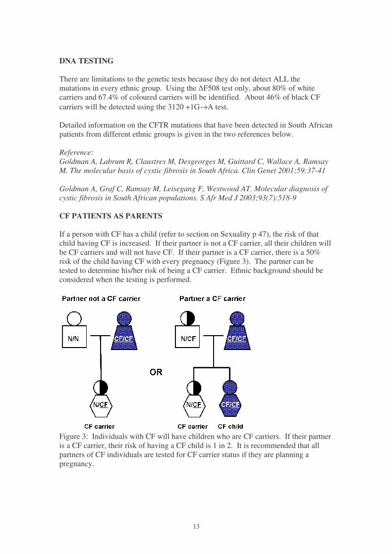
DNA TESTING There are limitations to the genetic tests because they do not detect ALL the mutations in every ethnic group. Using the �F508 test only, about 80% of white carriers and 67.4% of coloured carriers will be identified. About 46% of black CF carriers will be detected using the 3120 +1G→A test. Detailed information on the CFTR mutations that have been detected in South African patients from different ethnic groups is given in the two references below. Reference: Goldman A, Labrum R, Claustres M, Desgeorges M, Guittard C, Wallace A, Ramsay M. The molecular basis of cystic fibrosis in South Africa. Clin Genet 2001;59:37-41 Goldman A, Graf C, Ramsay M, Leisegang F, Westwood AT. Molecular diagnosis of cystic fibrosis in South African populations. S Afr Med J 2003;93(7):518-9 CF PATIENTS AS PARENTS If a person with CF has a child (refer to section on Sexuality p 47), the risk of that child having CF is increased. If their partner is not a CF carrier, all their children will be CF carriers and will not have CF. If their partner is a CF carrier, there is a 50% risk of the child having CF with every pregnancy (Figure 3). The partner can be tested to determine his/her risk of being a CF carrier. Ethnic background should be considered when the testing is performed.
Figure 3: Individuals with CF will have children who are CF carriers. If their partner is a CF carrier, their risk of having a CF child is 1 in 2. It is recommended that all partners of CF individuals are tested for CF carrier status if they are planning a pregnancy.

14
OTHER COUPLES AT INCREASED RISK FOR CF CHILDREN Other individuals who may be at increased risk include more distantly related family members (refer to Figure 2 above) and couples from consanguineous marriages (marriages between relatives e.g. first cousins). These individuals could be tested for CF mutations appropriate to their ethnic group and this information may be used to guide reproductive choices. 3.1 PRENATAL DIAGNOSIS (diagnosis before birth) When a couple is at high risk of having a child with CF they have the option of prenatal diagnosis and selective termination of an affected fetus. Every couple will have their own views and need to make their own choices. A genetic counsellor will ensure that they have all the facts and assist them with the decision making process. A family work-up in the laboratory is essential to determine which genetic testing is appropriate and feasible in each family. Reliable prenatal diagnosis is possible using chorionic villus sampling (CVS) or amniocentesis. Both are available in South Africa at certain centres. Chorionic villus sampling (CVS) CVS is performed under ultrasound guidance (sonar) and a small sample of the developing placenta is removed. This has the same genetic constitution (makeup) as the fetus and can be tested to determine whether the fetus has CF or not. CVS is performed between 10 and 12 weeks of pregnancy and is used to determine whether the fetus has CF. CVS carries a 1 - 2% risk for a miscarriage. Amniocentesis Amniocentesis is also performed under ultrasound guidance (sonar) and a sample of the amniotic fluid surrounding the fetus is removed. The amniotic fluid contains fetal cells that are used for the DNA test. Amniocentesis is usually performed between 16 and 20 weeks of pregnancy and has less than 1% risk of miscarriage. Preimplantation genetic diagnosis (PGD) Preimplantation genetic diagnosis is at present only available at specialist centres overseas. Eggs and sperm are harvested from prospective parents who have already had a child with CF and therefore have a 1 in 4 recurrence risk. Following the procedure of in vitro fertilisation, the developing embryos are screened at a very early stage for their CF status. Selected embryos, free of CF, are implanted into the mother's uterus to continue their development. Because of the risk of contamination and an incorrect result, it is recommended that the PGD be followed by a CVS or amniocentesis to confirm the results. Parents of a CF child who are planning to have more children and who wish to have prenatal tests performed to see if the fetus has CF, should consult with their doctor and with geneticists before embarking on a new pregnancy. A list of Genetic Counselling Clinics in South Africa is attached (Appendix 10).

15
4. GENERAL MANAGEMENT AND APPROACH TO TREATMENT
4.1 GENERAL MANAGEMENT The outlook for the individual with CF has improved dramatically. Many of the clinical features previously thought to be inevitable can be prevented, delayed or improved by intensive treatment. The introduction of a more positive attitude to management and the more widespread use of aggressive treatment regimes have been major factors in improving longevity and quality of life. Better survival is associated with more frequent use of antibiotics and more frequent review at CF clinics. As insulin is to people with diabetes, so are regular, high doses of antibiotics to people who have cystic fibrosis. 4.1.1 Communication at the Time of Diagnosis It is difficult for parents and/or patients to obtain more than a general impression of the condition when it is explained for the first time. Not only is CF a very complex disorder but parents are usually shocked and unable to follow detailed explanations at that time. There is a need to consolidate the information they receive at their first visit. A team approach must be followed. Clinic visits should include consultation with medical personnel, physiotherapists, dieticians, clinic sisters, pharmacists, social workers, psychologists and parent support groups. Information must be made available to general practitioners, caregivers, teachers, relatives and friends. This information should be available in hard copy from the clinic. Additional sites of information, such as the Internet, may be of use. It may be helpful for relatives to talk to the families of other affected individuals. Mutual support is generally most beneficial. "Remember, you are not alone". 4.2 GENERAL MANAGEMENT BY A NON-CF SPECIALIST Some CF patients will be living far from a CF clinic and will find it difficult to attend CF clinics. They will be cared for by general practitioners (GPs) who do not have much experience with CF and who do not have ready access to the support of allied medical staff. It is essential that the GPs align themselves with the nearest CF Clinic and send their CF patient(s) at least yearly for assessment at the CF Clinic. Summaries of CF clinic visits should be sent to primary care doctors. As a relationship is built up with clinic doctors, so the GP will feel more comfortable about telephoning for advice and referring the patient when necessary. 4.2.1 General Facts Discussed at the Time of Diagnosis • CF remains a serious disorder despite the major advances of recent years.

16
• The condition of the patient and the long-term outlook depends on the effectiveness and aggressiveness of the treatment.
• The outlook continues to improve year by year.
• Individuals who have CF will always need regular follow-up at a hospital. The condition is so complex and advances in treatment so rapid that all patients must be cared for under the guidance of the CF specialist at the regional CF clinic.
• The hereditary aspects of CF.
• Families are told about the Cystic Fibrosis Association. If they agree, their names are forwarded to the relevant CF association. For contact addresses, see Appendix 10.
4.2.2 General Precautions for the Individual who has CF There are a number of reasonable precautions that should be observed by the CF individual and the family. • Immunisation is very important (see Section 4.3, p 18).
• NO SMOKING (active or passive). Smoking is particularly bad for people with CF.
• Starting nursery school or crèche should be delayed (ideally until 3 years of age).
• Reduction of exposure to friends and relatives who have just started with a "cold" as this is when they are at their most infectious.
• If an infant with CF is admitted to hospital, every effort should be made to provide a cubicle to reduce the risk of acquiring an acute viral infection from other acutely ill children.
• Avoidance of close contact with stables, compost or other forms of rotting vegetation is advised because of the risk of the inhaling Aspergillus spores or infection by Burkholderia cepacia.
4.2.3 CF patients should attend a specialist CF Clinic • Patients should attend a CF Clinic every one to three months.
• Here the patient's progress must be reviewed by the entire team, if possible.
• At every visit the patient must be weighed and measured.
• At every visit a sputum sample or cough swab should be sent for microscopy, culture and sensitivity.
• Parents should have a sputum container at home to send to the laboratory in the event of new respiratory infection or production of unusually purulent sputum.
• From the age of five years, spirometry should be performed (by experienced personnel).
• Oxygen saturation should be measured using a pulse oximeter.

17
• To avoid cross infection when using all respiratory function equipment, the use of bacterial filters is advised.
• All staff must wash their hands between patients.
• A comprehensive CF assessment is recommended at diagnosis and annually (see sections 4.2.4.and 4.4 respectively).
4.2.4. Details of the Initial Comprehensive CF Assessment History/examination Anthropometrics (height, weight, etc) Immunisation status Family/personal smoking
Confirmation of diagnosis Sweat test DNA testing Lung status and Tests Respiratory function Bronchodilator test Physiotherapist's assessment X-ray chest Sputum culture Aspergillus precipitins & RAST (depending on age and symptoms) Total IgE Gastrointestinal status Dietician's assessment Electrolytes Faecal human pancreatic elastase 1 (at diagnosis) or Faecal chymotrypsin (at diagnosis) Modified GTT (if >10 years or younger if losing weight or there are symptoms suggestive of diabetes mellitus) Vitamin A, D, E serum levels (if available) Adult diagnosis: ultrasound of liver and portal system Additional tests Full blood count Liver function tests (C reactive protein may be helpful) Adult diagnosis: bone mineral density estimation Other Social worker consult Genetic counselling (Section 3, p11), diagnostic testing of siblings.

18
4.3 IMMUNISATION Normal childhood immunisations should be administered since viral respiratory tract infection can have a detrimental effect on the patient’s lung function and disease progression. An annual influenza vaccine covering the expected strains for that season should be given as a routine in March/April except if there has been anaphylaxis to egg. Passive immunisation against the respiratory syncytial virus (Synergis®) for children under the age of 2 years is thought to be useful during epidemics. Immunisation against chicken pox and hepatitis A is recommended. A vaccine to Pseudomonas aeruginosa is under trial at present. When/If it becomes available it should be given prior to colonisation with the organism. Multivalent pneumococcal vaccine is recommended for the young child.

19
4.4 ANNUAL REVIEW History/examination Anthropometrics (height, weight, etc) and review of progress over the year Immunisation status Lung status and Tests Respiratory function and review of the year Physiotherapist's assessment X-ray chest Sputum culture and review of the year Full blood count Total IgE, Aspergillus precipitins & RAST (depending on age and symptoms) Gastrointestinal/Nutritional status Dietician's assessment Sodium, potassium, urea, creatinine, cholesterol, calcium, magnesium, alkaline phosphatase blood levels Faecal human pancreatic elastase 1 if pancreatic sufficient at time of review Modified GTT (if >10 years) Vitamin A, D, E levels (if available) Ultrasound of liver and portal system (>10 years) Additional tests Adults: bone mineral density estimation Other Social worker review Review and discussion of genetic/family issues. The results of assessments and tests should be discussed with the patient and/or parents. Included in the discussions should be:
� Current health status � Meaning of the changes (if any) over the year reviewed (good and not-
so-good news) � Adjustments to treatment regimes for the coming year � Aims of the adjustments � Discussion of the patient’s CF care in general � Planning for life events in the coming year e.g. school, employment

20
5. MANAGEMENT OF RESPIRATORY
PROBLEMS
5.1 LUNG DISEASE Management should aim to minimise the extent and effects of the chest infection and includes early diagnosis, good nutrition, minimising exposure to colds and acute viral respiratory infections, ensuring adequate immunisation, avoidance of smoking (active and passive) and most importantly, early use of appropriate antibiotics and physiotherapy. 5.1.1 PHYSIOTHERAPY Chest physiotherapy (CPT) forms an integral part of the treatment for CF. The mucous secretions in CF are thick and tenacious. CPT facilitates the loosening and expectoration of this mucus and enhances mucociliary clearance. The aims of CPT are to prevent airway obstruction, prevent respiratory complications (pneumonia, bronchiectasis and atelectasis) and to maintain or improve pulmonary function. Compliance with CPT is generally poor and, when patients reach adolescence, it becomes a greater problem. For this reason it may need to be individualised. The physiotherapy modality must be effective, time-efficient and allow for independence. Parts of one modality can be applied and integrated in other methods and a combination can be beneficial. Physiotherapy at different ages: 0 - 3 years Parent(s) and caregivers are largely responsible for CPT. CPT is performed before meals to prevent vomiting. The technique used is modified postural drainage (PD) with percussion, vibration or shaking. The recommended PD positions are supine lying, alternate side lying and sitting. Head down position should NOT be used because of the risk of reflux and aspiration. From 2 years, blowing games can be introduced and huffing on to mirrors can begin. At this age, children cough and swallow. This can be accepted. 3 years - adolescence From the age of 3 years, some patients will begin to expectorate. The 3-year-old can be taught the Forced Expiration Technique (FET). The FET is incorporated into the Active Cycle of Breathing Techniques (ACBT) that children can manage independently by the age of 8 or 9 years.

21
The Active Cycle of Breathing Techniques (ACBT) consist of the following: • Relaxation and breathing control (normal gentle breathing at tidal volume,
using the lower chest, with relaxation of the upper chest and shoulders). • Thoracic (chest) expansion exercises - 4 or 5 deep breaths, emphasising
inspiration with a quiet unforced expiration. This may be combined with chest shaking or clapping.
• FET or “huff” is used to mobilise and clear secretions. One or two forced expirations are combined with a period of breathing control. A huff from high lung volume (when a breath has been taken in) will clear secretions from the upper airways. A huff from mid to low volume will clear secretions from the lower more peripheral airways.
The ACBT encourages independence and is a means of clearing secretions without the assistance of another person or mechanical device. Play activities and physical games should be encouraged at an early age. The older child should be encouraged to participate in school physical activities on all possible levels. The choice of exercise should be determined by the patient and depends on the severity of their lung disease, their environment and their motivation. To stay physically fit is of great importance. Games and exercise do not replace regular physiotherapy. During pulmonary exacerbations, assisted treatments are preferred. This would entail PD with percussion, vibration or shaking. Patients with minimal or no chest symptoms are encouraged to do CPT once a day. Those with more frequent chest symptoms should do CPT twice a day or 3 to 4 times during acute or sub-acute chest infections. Adolescence - adulthood The same techniques and principles apply as in category three years to adolescence. Adolescents and adults become responsible for their own chest care and need to recognise the signs of increased chest infection. They need to be aware of correct posture and of its influence on the mechanical dynamics of respiration. Incorrect posture creates a mechanical disadvantage and contributes to increased work of breathing and muscle fatigue. At all ages, physiotherapy should include exercises to promote improved posture awareness with stretches and strengthening exercises for the relevant musculature. OTHER PHYSIOTHERAPY TECHNIQUES There is no evidence that one technique of physiotherapy is necessarily better than any other. Using a number of modalities enhances compliance through a reduction in boredom.

22
Positive expiratory pressure (PEP) mask The treatment consists of a facemask and a one-way valve to which expiratory resistors are attached. The rationale for use of PEP is that it will open and recruit obstructed regions of the lung allowing air to move behind secretions and assist in their removal. The PEP pressure must not be more than 20cm of water. Treatment is performed in the sitting position. The patient inspires slowly and deeply through the mask. Expiration is active but not forced. The cycle is continued for 10-15 respirations, the patient then removes the mask and does FET to expectorate mobilised secretions. The PEP mask is a useful adjunct method of CPT and should be used in conjunction with other techniques. The technique is contra-indicated where patients have a history of pneumothorax or if cysts are evident on the chest X-Ray. Flutter VRPI / Bronch-u-Vibe These devices are small, simple hand held devices that contain a large ball bearing. As the patient exhales, the ball is displaced and then rolls back into place. The fluctuations in pressure result in oscillations of positive pressure and airflow. This PEP vibrates the airway walls, loosens mucus, decreases the collapsibility of the airways and accelerates airflow. Use of these devices: In a sitting position, the patient breathes out normally and deeply through the device. The cycle is repeated 8-10 times. Mobilised secretions are then expectorated. Hyperinflation could result in dizziness and, should this occur, frequent short interruptions every 5 cycles are encouraged. The devices should be cleaned between physiotherapy sessions. The Bronch-u-Vibe is available from MediTool, P.O. Box 2479, Pietermaritzburg 3200. Mechanical percussors / Vests Percussors and vests produce rapid oscillations of the chest wall, loosening mucus. They are comfortable pain-free devices that can be applied to a rigid, thin thoracic wall and may be used for longer periods than the manual techniques. They are as effective as manual physiotherapy techniques. Autogenic drainage Autogenic drainage (AD) is a technique based on the basic physiology of breathing. Expiratory flow becomes an active force that is used to mobilise the mucus. By adjusting the tidal breath at low-mid or high lung volume level, better flow rates are generated without causing appreciable increases of the airway resistance. This technique requires special training by an experienced physiotherapist. Main E, Prasad A, van der Schans C. Conventional chest physiotherapy compared to other airway clearance techniques for cystic fibrosis. The Cochrane Database of Systematic Reviews 2005, Issue 1. Art. No.: CD002011.pub2. DOI: 10.1002/14651858.CD002011.pub2.

23
BRONCHODILATORS AND PHYSIOTHERAPY: When bronchodilatation is used together with CPT, its timing is important. Metered dose inhalers (MDI) with spacers are more effective, time- and cost-efficient than nebulisers and are recommended. 1st USE THE BRONCHODILATOR (to open up the airways) 2nd GIVE CHEST PHYSIO (to aid removal of secretions) 3rd NEBULISE INHALED ANTIBIOTICS (if prescribed) Cross infection is a risk: devices that are breathed into should NOT be shared CONCLUSION CPT is essential for CF patients, even those with minimal chest symptoms. Patients should be involved in decision making. This will enhance adherence both in and out of hospital. Physiotherapy should be incorporated into the patient's lifestyle so that it is not seen as another "chore". It must become routine. 5.1.2 ANTIMICROBIALS The liberal, frequent and early use of high doses of antibiotics, both orally and intravenously, has been the most important factor improving the outlook for CF. The most frequently encountered bacterial pathogens are Haemophilus influenzae, Staphylococcus aureus and Pseudomonas aeruginosa. All patients require regular cultures of lower respiratory tract secretions and should receive appropriate antibiotic treatment in an attempt to prevent chronic infection. It is important to recognise that chronic infection of the lungs can be postponed with early use of antibiotics. Acute exacerbations are treated on their own merit. Recognition of respiratory infections in CF The symptoms of significant lower respiratory infection in patients with CF differ from those in people who do not have CF. The symptoms are often less obvious and the respiratory signs may be subtle. Cough is one of the most important early signs of increasing infection of the respiratory tract. There are other symptoms and signs that suggest an increase in activity of the chest infection
� lethargy � a reduction in appetite � reduced or absent weight gain or, more significantly, loss of weight � a change of sputum colour from white to yellow or green � a change in sputum quantity and smell, and/or � an increase in cough or breathlessness.
Fever is uncommon. These signs are commonly associated with deterioration in the respiratory function tests (reduction of peak expiratory flow rate or FEV1 and FVC) and there may be new X-ray changes. However, a change in the cough pattern is the most sensitive early sign.

24
Significant new infection within the airways may not be detected by listening with the stethoscope or by a chest X-ray. It may only be evident by the persistence of a cough which had not been present before. Such a new cough represents new bronchial infection and requires aggressive antibiotic treatment even if the patient is generally well. Lower airway obstruction (and asthma) may be a factor in ongoing cough. Chest X-ray is NOT necessary with each exacerbation. Regular (1-3 monthly) cultures of lower respiratory tract secretions are recommended for the early detection of new pathogens. Note: Growth of S aureus or P aeruginosa in the sputum from a colonised patient who does not have new symptoms does not represent an exacerbation. Viral respiratory infection in CF When a patient with CF presents with a probable viral infection, bacterial superinfection or exacerbation of existing bacterial infection is very likely to occur. Antibiotic therapy is usually indicated. The choice of antibiotic needs to be individualised. In young children who are free of colonising organisms, co-amoxyclavulanate is the antibiotic of choice. Approach to common bacterial infections Staphylococcus aureus Chronic infection of the respiratory tract by S aureus can be postponed by the use of prophylactic anti-staphylococcal antibiotics. However this may allow early infection with P aeruginosa. The risk of bacterial resistance to the antibiotic will also be raised by prolonged continuous use. Chronic infection with S aureus may also be postponed by giving a 2 week course of antibiotic each time S aureus is grown from respiratory secretions regardless of symptoms. This will only be achieved if regular specimens are collected (surveillance). Management of infective episodes. Asymptomatic or not previously colonised and not ill: A minimum of 2 weeks with flucloxacillin unless sensitivity indicates otherwise. Ill: Oral or IV antibiotic depending on tolerance of the high oral doses and degree of illness (for 2 to 4 weeks). While combinations of antibiotics are often used, there is no evidence that these are better than a single agent. First line: Flucloxacillin or cloxacillin Alternatives: Macrolides, clindamycin, cephalosporins (1st or 2nd generation), cotrimoxazole. Methicillin resistance: Depends on sensitivities (discuss with microbiologist). Drugs include rifampicin (not alone), fusidic acid (not alone), vancomycin, teicoplanin or linezolid. Cotrimoxazole may be useful in community acquired MRSA.

25
Haemophilus influenzae If the patient is well, there is no need to use an antibiotic if this organism is grown. If the child is ill, at least a week of an appropriate antibiotic is indicated. First line: Amoxycillin (depending on local sensitivities), co-amoxyclavulanic acid. Alternatives: 2nd generation cephalosporin, one of the newer macrolides. Pseudomonas aeruginosa Pseudomonas infection is always significant. It is now clear that chronic infection of respiratory tract with mucoid P aeruginosa can be postponed by regular surveillance and early use of anti-pseudomonal antibiotics. The presence of anti-pseudomonal antibodies indicates early infection with the organism (at present this test is not available in South Africa). Every effort should be made to postpone chronic infection as permanent infection with P aeruginosa increases the rate of damage to the lung. Suggested regimes on first detection or suspicion of infection: Patient well: 3 weeks of oral ciprofloxacin together with nebulised colistin or aminoglycoside. Sputum culture at 2 weeks. If still positive, either admit for a minimum of 2 weeks of IV antibiotics, or continue oral and nebulised regime for 3 months. Patient unwell: At least 2 weeks of intravenous antibiotics. Eradication can be deemed to have been achieved if, despite monthly sampling, the organism is not cultured within six months. Reappearance of the organism (or a rise in anti-pseudomonal antibodies) thereafter necessitates reinstitution of the above regime. Prophylactic therapy in the chronically infected patient There is good evidence that regular therapy with an inhaled antibiotic reduces the rate of deterioration of lung function. Gentamicin, tobramycin or amikacin (the intravenous forms) by inhalation are widely used in South Africa seemingly with good effect. Colistin and a special formulation of tobramycin (TOBI®) have been shown to have beneficial effects. These two antibiotics are considerably more expensive. In choosing an antibiotic sensitivity of the organism should be used as a guide (For details of administration see Section 5.1.2.3, page 27). Macrolides are recommended as prophylaxis in chronically infected patients. This is discussed in Section 5.1.3.1, page 29. Treatment of pulmonary exacerbation in the chronically infected patient Early recognition of an exacerbation is vital. Symptoms that suggest an exacerbation include
� a decrease in energy, appetite or weight,

26
� increase in cough, shortness of breath or increased sputum production, or � significant decrease in lung function.
IV treatment (traditionally with 2 antibiotics) for a minimum of 2 weeks is strongly recommended under these circumstances. Antibiotic blood levels should be done when using aminoglycosides (trough ~ toxicity; peak ~ efficacy). Oral ciprofloxacin for the same period may be used in mild exacerbations. Oral ciprofloxacin has few side effects although photosensitive skin rashes may occur. Cartilage problems have not been documented in humans and are not a contraindication to using the drug in children. Ciprofloxacin should not be used in CF patients who are pregnant. Other infections Infections with unusual bacteria such as Burkholderia cepacia and Stenotrophomonas maltophilia may occur in CF. Management of these infections should be undertaken in specialised CF centres. See Section 5.1.4 page 30. 5.1.2.1 THE USE OF INTRAVENOUS ANTIBIOTICS Indications for intravenous antibiotics are given in Section 5.1.2 (pages 23-6). Admission for Intensive Treatment and Intravenous Antibiotics: It is important to stress that the "hospital treatment package" should include removal from the home environment (where there may be exposure to cigarette smoke!), some rest, temporary transfer of the responsibility of treatment from the patient/family to the hospital staff, expert dietary advice, regular meals with possible increased compliance with pancreatic enzymes and vitamin supplements. These are additional advantages over and above the regular professional CPT and intravenous antibiotics. The duration of a course of intravenous therapy varies but must not be less than 2 weeks. The frequency of courses varies between patients. Usually a combination of an aminoglycoside plus ceftazidime or cefipime is used. Other antibiotics are used according to sensitivities. The doses must be large as CF patients tend to utilise some drugs, including antibiotics, more rapidly than normal (see Appendix 2). Improvement during a course of IV treatment can be demonstrated by performing regular respiratory function tests and carefully assessing the other signs including body weight. 5.1.2.2 HOME INTRAVENOUS ANTIBIOTIC TREATMENT Many studies have demonstrated that adequately supervised home IV antibiotic treatment (or OPAT) is a practicable, effective and acceptable alternative to hospital treatment for many CF patients. Some patients have the first few days of treatment in hospital and complete the course at home. Adequate support and training of the caregivers is essential. Antibiotic blood levels should be done where appropriate and IV technique reviewed. At the end of the two-week course of home IV antibiotics the patient ideally attends the CF Unit: Respiratory function tests are performed and sputum is obtained. The

27
patient should also be seen by the doctor and physiotherapist. The CF team decides whether maximal improvement has occurred and whether further treatment is required. Simple cost effective devices may make ambulatory home and school based IV therapy practical. The Springfusor pump (Cobros Medical Supplies) provides one such option. Totally Implantable Venous Access Devices (TIVADs) (e.g. Port A Cath® or Implantofix®, Braun®) have proved valuable in overcoming problems of venous access for many patients having regular IV antibiotic therapy. It is essential that both family and professionals are familiar with the use of these devices. Complications limit their use and peripheral IV sites remain a first choice where possible. Supervised follow-up must be meticulous. (For more detail see Section 5.1.9, p35) 5.1.2.3 NEBULISED ANTIBIOTICS Nebulised antibiotics have an established place in the management of CF. Their major use is in attempting to prevent chronic infection and to control established chronic infection when it occurs. Although colistin is the antibiotic used in most of the initial studies on this mode of therapy, it has no clear advantage over aminoglycosides (tobramycin, gentamicin, and amikacin) or other antipseudomonal antibiotics (provided they can be nebulised). The choice of antibiotic is usually based on the bacterial sensitivities found on sputum culture (although these results do not always equate to in vivo efficacy). Nebulised antibiotics do not cause toxicity (systemic absorption of aminoglycosides in one study was only + 0.5% of the dose). Nebulised antibiotics often cause some degree of bronchoconstriction (related mainly to hypertonicity of the solution and/or possibly preservatives) and it is recommended that they be administered after a bronchodilator. (Note: The bronchodilatator is best administered via an MDI (with or without a spacer device) and not a nebuliser. This technique reduces the time needed for nebulisation therapy and increases patient adherence.) In order to facilitate maximal deposition of drug in the lungs for as long as possible, the following are recommended: • Administer nebulised antibiotics after sputum clearance (physiotherapy +
RhDNase or hypertonic saline) • Use a mouthpiece. Small children may require a mask. • Breathing at a relaxed tidal volume through the mouth (rather than “big
breaths”) • Use a suitable nebuliser and compressor: The best being an active Venturi
nebuliser (breath assisted) with a 6 l/min flow rate.

28
Preparation of antibiotics for nebulisation Standard IV preparations of antibiotics are used in South Africa. The solutions should be reconstituted with sterile water or saline to a volume of 4 mls. The preparation of isotonic solutions of colistin appears in Appendix 3. Clinical indications are discussed in Section 5.1.2 (pages 23-6). Campbell PW, Saiman L. Use of Aerosolized Antibiotics in Patients with Cystic Fibrosis. Chest 1999;116:775-788 5.1.2.4 BACTERIAL RESISTANCE TO ANTIBIOTICS Frequent antibiotic use, either for prophylaxis or treatment of exacerbations of infections, is associated with the risk of resistance. However frequent high dose antibiotic therapy is an essential part of CF management. Monotherapy is usually not recommended for the treatment of Pseudomonas infections because it may lead to resistance. Regular surveillance of sputum cultures is essential to have current information on the patient’s flora and susceptibility patterns. The choice of antibiotic treatment of exacerbations is influenced by bacterial susceptibilities. It is recommended that nebuliser therapy with an antibiotic continues even if there is laboratory resistance to the antibiotic as the concentration delivered to the lungs is very high. Successful treatment may still occur when antibiotics to which the organism is resistant are used. Where aminoglycoside resistance occurs they should still be used in combination with another class of anti-pseudomonal agent as synergy can occur rendering the combination more effective than the non-aminoglycoside agent on its own. Carbapenems ideally should not follow quinolones within the same antibiotic course as resistance is likely to occur. Fourth generation cephalosporins such as cefepime can be given as a continuous infusion. This may reduce bacterial resistance and is cost-effective. 5.1.2.5 ALLERGY TO ANTIBIOTICS Allergic reactions can be problematic especially with �-lactam antibiotics but can occur with any of the agents used to treat infections. Whether the symptoms associated with antibiotics are allergic in nature should be confirmed by a doctor. Reactions usually take the form of skin rashes that may range from mild erythematous reactions to full blown Stevens Johnson syndrome. Angio-oedema and interstitial nephritis are rare reactions as is anaphylaxis. When an allergic reaction is diagnosed, the offending drug should be withdrawn and not used again. Mild skin reactions respond to drug withdrawal and antihistamines but severe skin reactions will require steroid therapy. The patient must be requested to apply for a Medic Alert bracelet.

29
Treatment of pulmonary infections can be problematic in the face of combined drug resistance and hypersensitivity. In extreme circumstances desensitisation can be considered according to standard protocols in an intensive care unit setting (see Appendix 9). 5.1.3 ANTI-INFLAMMATORY DRUGS Corticosteroids have a very complex but impressive anti-inflammatory action in a number of clinical situations. In CF they are well established as the treatment of choice for allergic bronchopulmonary aspergillosis (ABPA) (see page 30). Oral steroids have been used to try to suppress the harmful effects of inflammation but, although patients treated with a dose of 1mg/kg had better respiratory function, the side effects of growth retardation and diabetes were unacceptable if long term therapy was used. Corticosteroids can be very helpful, particularly in young patients who are wheezy and have associated asthma. Short courses of oral steroids may be used for acute asthma. The use of inhaled steroid therapy is controversial. However they are indicated in the long term management of CF patients who also have asthma. (For the use of corticosteroids in asthma see Section 5.1.7) Occasionally, corticosteroid treatment should be considered in the adult patient with CF when other standard therapies have failed to control wheeze, when sputum production remains copious despite appropriate IV antibiotic treatment, or when inflammatory markers remain persistently elevated. It is common for oral steroids to cause abnormalities of glucose metabolism in CF patients and precipitate diabetes mellitus. Ibuprofen at high doses has been shown to slow the rate of annual decline in FEV1 in children below 13 years of age. Dosage-finding pharmacokinetic studies need to be done. Liver and kidney disease and a bleeding diathesis are contraindications to ibuprofen therapy. 5.1.3.1 Macrolide antibiotics as anti-inflammatories in CF Inflammation plays a major role in the pathophysiology of lung disease in CF. This response is probably triggered primarily as a reaction to the inability of the affected lung to resist the invasion of the most common bacterial pathogens. Debate continues as to whether or not there is a pre-inflammation of the lungs as part of the basic functional defect of CFTR. The anti-inflammatory treatment modalities most tested to date are: oral corticosteroids, inhaled corticosteroids and ibuprofen. Novel antimicrobial and anti-inflammatory properties of macrolides may result in clinical benefits, particularly in conditions where the infectious agent is inherently resistant to macrolides. The most promising newcomer is azithromycin, acting as a long-term anti-inflammatory agent with an excellent safety profile. • A meta-analysis confirmed a significant improvement in FEV1 among the 286
CF subjects.

30
• Most short term studies (three to six months) have not shown the development of increased bacterial resistance or the emergence of new pathogens but this should be monitored locally.
• There is no clear guideline on dosage but even a weekly dose may be beneficial. Suggested dosage: 15-40 kg: 250 mg daily; >40kg: 500 mg daily; alternative is 3 times per week.
Recommendation for use: • Azithromycin if available rather than other macrolides • Patients chronically infected with P aeruginosa • Low dose daily or 3 times per week or even weekly • Monitor organism sensitivities with regular sputum culture (3-6 monthly) 1. Dinwiddie R. Anti-inflammatory therapy in cystic fibrosis. J Cyst Fibros 2005;suppl 2:45-48 2. Saiman L. The use of macrolide antibiotics in patients with cystic fibrosis. Curr Opin Pulm Med 2004;10:515-523 3. Prescott WA Jr, Johnson CE. Antiinflammatory therapies for cystic fibrosis: past, present, and future. Pharmacotherapy 2005;25:555-573 5.1.4 OTHER IMPORTANT INFECTIONS These should be considered in patients not responding to conventional treatment and include TB (very important in South Africa), Mycoplasma, Nocardia, Chlamydaphyla (Chlamydia), Stenotrophomonas, Burkholderia species, Achromobacter xylosidans and fungi. All these organisms should be considered to be potential pathogenic and eradication attempted. Non-tuberculous mycobacteria (MOTT) only require treatment if they are thought to be the cause of symptoms. Allergic Bronchopulmonary Aspergillosis (ABPA) ABPA is an allergic reaction to inhaled Aspergillus spores. ABPA is a type 3 allergic response with proximal bronchiectasis. Unrecognised and untreated it may lead to an acceleration of bronchiectasis. Diagnostic criteria are shown in the Box on the following page.

31
Diagnostic criteria for allergic bronchopulmonary aspergillosis in cystic fibrosis patients All immunologic parameters required • Skin Prick Test positive to Af or IgE-Af (RAST) • IgE elevation >500 iu/ml3 • IgG antibodies to Af or precipitins • Hypereosinophilia1 >400/ml • Reduction by >50% in IgE after 2 weeks of daily systemic corticosteroid therapy Supportive (at least 3 required) • Airway obstruction/wheezing • Bronchiectasis on chest CT • Pulmonary infiltrates on chest radiograph • Af in sputum culture • Decrease in pulmonary function (>19% decreases in FEV1) _____________________________________________________________________ 1 Hypereosinophilia not required when on systemic steroids 2 1 iu = 2.4ng Af – Aspergillus fumigatus CT – computed tomography FEV1 – forced expiratory volume in 1 second SPT – skin prick test RAST – radioallergosorbent test Since Aspergillus spores are widespread in the environment, avoidance of this fungus is difficult. Patients should avoid situations where there is an increased risk of inhaling these spores - in stables, compost and other forms of rotting vegetation. Likewise, when gardening, mixing compost should not be undertaken. Oral steroids are the therapy of choice. The lowest doses possible should be used. ABPA does not respond to inhaled steroids. There is some evidence that the anti-fungal itraconazole and voriconazole may confer benefit when added to steroid therapy and prevent invasive disease. Long term therapy may be needed. Suggested dosage regime for oral steroids in ABPA: 6 weeks of tapering doses of prednisone orally according to response. The starting dose is 0.5-1mg per kg. It may not be possible to stop steroid therapy in the short term. Serological markers (see Box on this page) should be monitored to evaluate response to therapy. 5.1.5 HAEMOPTYSIS Haemoptysis (coughing up blood) is mostly seen in older CF patients with advanced lung disease. It may occur in as many as 60% of adolescents or adults. It usually represents an exacerbation of infection. Other factors are coughing spells, atypical lung infections including TB and clotting disorders (particularly where there is CF liver disease). In most cases there is only a small amount of bleeding with blood flecks in the sputum. However, life-threatening bleeds (greater than 250mls/24 hrs) can occur (in 5 – 10% of adolescents and adults).

32
Primary treatment remains conservative with reassurance and possibly a cough suppressant e.g. codeine phosphate for the first 48 hours only. Gentle physiotherapy can be continued. Antibiotics must be given. RhDNase can safely be continued. With large bleeds, blood transfusion may be necessary, together with fresh frozen plasma or cryoprecipitate. Pro-coagulants may be useful. Vitamin K should be administered although its benefit is not immediate. Any non steroidal anti-inflammatory or aspirin-containing preparation must be discontinued. In the event of larger, recurrent or unrelenting haemoptysis, bronchial artery embolisation should be undertaken in specialised centres. This should be performed sooner rather than later. As a last resort thoracotomy with ligation of the affected artery and possible lobectomy are necessary. This is associated with a poor prognosis. 5.1.6 PNEUMOTHORAX A pneumothorax is an air leak into the pleural space secondary to rupture of a subpleural bleb, alveolus and/or air tracking via the pulmonary lymphatic and interstitial spaces. It may be associated with pneumomediastinum, surgical emphysema and more significantly, generalised interstitial emphysema. The air leak is often confined because, in advanced CF lung disease, the lung is very stiff and may not collapse to the same degree as a healthy lung. If the air leak is under tension, there is acute collapse of the lung with a rise in the carbon dioxide level and respiratory distress. Pneumothorax may complicate severe infection, coughing or the placement of central lines. It is relatively uncommon in young children with CF. An incidence of up to 20% has been reported in adolescents and adult patients. Pneumothorax is more common in males than females, affects either side equally and is usually associated with advanced disease and marked airflow obstruction. Presentation is often subtle. Acute onset pleuritic pain and some respiratory distress are often evident in the absence of overt infection. A high index of suspicion should be maintained. Chest X-ray is used for confirmation. Recurrences are common (up to 60%) on the same side or even on the other side. Treatment:
• Small pneumothorax: Conservative with high FiO2. • If after 24 - 48 hours the lung is not re-expanded or if the pneumothorax is
significant, intercostal tube drainage with a suitable sized drain is required. • Chemical or limited surgical pleurodesis (with non-resolution or
recurrences), keeping in mind that this procedure would be a relative contraindication to future lung transplantation.
Treatment must include step up therapy for the lung infection. The presence of an intercostal drain may exacerbate underlying chest infection if pain is not relieved. Gentle negative pressure using suction may be applied to the drain to help to expand the lung fully.

33
5.1.7 ASTHMA Asthma is an inflammatory disorder of the airways characterised by recurrent, reversible airway obstruction. The reversibility should be demonstrated before maintenance therapy is established. 1. As many as 40% of cystic fibrosis patients will have varying degrees of bronchial
hyperreactivity which manifests as wheezing or coughing. 2. Bronchodilator therapy will be of value in these patients. 3. Bronchodilators in the form of β2 agonists (e.g. salbutamol, fenoterol) can be
given in a number of ways: MDIs, MDIs with spacer (especially good for children), breath activated devices, dry powder inhalers or via a nebuliser. A long acting β2 agonist may be used in combination with inhaled steroid therapy.
All CF patients with asthma should have an inhaler device. Nebulisers for asthma control are less effective than other inhaler systems. Role of steroids: Inhaled steroids are mandatory in all CF patients with persistent asthma symptoms. The dose of the inhaled steroid is adjusted according to the individual's need. Inhaled steroids, often with a long acting β2 agonist such a salmeterol or formoterol, will benefit patients with asthma. Further details are available in the SATS guidelines on asthma for children and adults (http://www.pulmonology.co.za/guidelines.htm) To date, clinical experience and research data on the anti-inflammatory effects of leukotriene receptor antagonists such as montelukast in CF are limited. 5.1.8 NEBULISERS AND COMPRESSORS Choosing a compressor and nebuliser There must be attention to detail or the exercise is waste of time. Ultrasonic nebulisers should not be used for treatments in CF as they denature nebulised antibiotics and rhDNase. Compressor: Minimum specification: 10 litres/min with nebuliser attached. Cheap compressors are NOT cost effective as little of the active medication may reach the lungs. Recommended: Turbimed® (Medix), Parihaler® (Boehringer) or equivalent Nebuliser: Mouthpiece, not mask except in younger children
3-5 �m median droplet diameter Minimum residual volume
Nebulisation should not last longer than 10 minutes. If the chamber is not empty at this stage, there is something wrong. When a mask is used, it should be closely applied to the face. The face should be washed after the nebulisation to prevent irritation of the skin.

34
Ideally: Electronic inhalation triggered nebulisation In-line bacterial filters to prevent contamination from the compressor CLEANING OF HOME NEBULISERS: Nebulisers are designed to convert liquid into small particles of water vapour. The droplet nuclei produced by jet-type nebulisers vary from 0.5 to 10�m in size and are therefore large enough to disseminate microorganisms and small enough to reach the lower respiratory tract. Hence any reservoir nebuliser is capable of generating a bacterial aerosol that can cause lung infections in persons using contaminated equipment. Studies have shown that contamination of home nebuliser equipment and inhalation solutions poses a serious problem. About half of all nebulisers studied were contaminated by microorganisms such as P aeruginosa, Klebsiella and S aureus. Nebuliser reservoirs and syringes used for periods longer than one month were found to be contaminated. The same was true in cases where nebulisers and masks were not cleaned at all or were only rinsed with tap water.
• If a drug is available in both a MDI formulation or as a solution that can be nebulised, the MDI remains the desired form in which the drug should be used.
• Hand-washing is of the utmost importance in preventing contamination of equipment and inhalation solutions. Hands should always be washed before and after performing nebulisation, whether gloves are worn or not. For routine hand-washing, lathered hands should be rubbed together vigorously for at least 10 seconds, followed by thorough rinsing under running water. If bar soap is used for hand washing, a small bar is preferable and the soap should be kept on a rack to allow excess water to drain away. Should liquid soap be used, the dispenser should always be cleaned and dried before being refilled with soap.
• All nebulisers should be sold with specified instructions regarding effective cleaning and maintenance of the equipment.
• After the use of the nebuliser, all residual fluid should be discarded from the reservoir. The masks/mouthpiece and reservoirs should be washed with warm soapy water and dried thoroughly with disposable paper towels. The equipment should either be stored dry or, alternatively, stored submerged in a diluted disinfectant solution.
• Disinfectant solutions that may be used to decontaminate equipment include a 1.25% acetic acid (AA) (white vinegar) solution, a quaternary ammonia compound (QAC) (bleach) diluted in a ratio of 1 to 8 with sterile water, or a 2% solution of glutaraldehyde (Cidex�). In addition to soaking the reservoir and mouthpiece/mask in the solution for 10min in the case of QAC and 30 min in the case of AA and Cidex, the solution should also be nebulised for at least 10 min to ensure sterilisation of the jet. The use of disinfecting solutions should be followed by rinsing and thorough drying of equipment. Where the equipment is only washed and dried after use, disinfecting should be undertaken twice weekly.
• Only sterile medication and fluid should be used in nebulisers. Single dose vials are therefore preferred to large containers, especially in the case of

35
solutions not containing antibacterial agents, such as saline. In the case of multi-dose vials the container should be kept securely closed between usages and should not be kept for prolonged periods. Refrigerating of multidose vials should also be considered to minimise bacterial proliferation. Drugs and solutions to be nebulised should only be poured into the reservoirs immediately before use. Syringes used to draw up drugs should also be replaced regularly to reduce the risk of contamination.
• Disposable nebuliser reservoirs with jets should not be used for more than three months and should preferably be changed once a month. Long-use (durable) nebuliser reservoirs and jets should be changed once a year. The nebuliser compressor should be serviced annually and the filters on the compressor should be changed when they become discoloured.
5.1.8.1 NEBULISED MEDICATIONS Hypertonic saline:
Hypertonic saline (5 - 7.5%) has been evaluated and shown to benefit some patients. It can be helpful where secretions are particularly tenacious. It is by far the most cost-effective mucolytic. This therapy should be initiated under controlled conditions, monitoring for bronchospasm.
RhDNase:
RhDNase (Pulmozyme®) represents an important treatment for CF patients and good clinical trials have demonstrated that it works well and is safe. An improvement of 5 - 7% in lung function can be achieved but the high cost is a limiting factor. Optimally rhDNase should be available in selected patients with demonstrable responsiveness to the drug in whom the FEV1 is <70% of expected. RhDNase should only be used as an add-on therapy to patients on established pulmonary treatments. Ultrasonic nebulisers should not be used for rhDNase.
Other Mucolytics e.g. N-acetyl cysteine: No proven benefit, but some patients feel better using them.
Mucolytics should be used alone in the nebuliser and not mixed with other medication. See also Appendix 7. 5.1.9 VENOUS ACCESS Peripheral intravenous cannulae are the preferred option for venous access. Distal veins should be used where possible. For children, topical anaesthetic creams should be applied prior to siting intravenous cannulae. When peripheral access becomes difficult, alternatives are needed. Peripherally inserted long lines (PICC lines) or Midlines can be placed. Silastic catheters may remain in situ for extended periods. They are easy to handle and are often preferred by patients. The use of such catheters should be considered as an alternative in ambulatory IV treatment.

36
Buck C. Holl R. Kohne E. Wolf A. Silastic catheters: An alternative to the conventional peripheral venous infusions access in patients requiring IV therapy for an extended period for home antibiotic therapy in patients with cystic fibrosis: European Journal of Pediatrics. 1997;156(3):209-211 Where peripheral access is no longer possible, the use of Totally Implantable Venous Access Devices (TIVADs) provides a safe, effective and convenient means of venous access. The site where TIVADs are implanted is chosen according to the patient’s medical condition, their way of life and the treatment required. These ports can be implanted into the anterior chest wall or on the ventral aspect of the upper arm or forearm. Complications Despite the obvious benefits of the TIVADs, the complications are potentially serious and can cause appreciable morbidity. ♦ Catheter-related sepsis including bloodstream infections ♦ Thrombosis. Other complications are rare but include ♦ Superior vena cava syndrome ♦ Air embolism ♦ Pneumothorax ♦ Pulmonary thromboembolism. ♦ Mechanical problems ♦ Blockage ♦ Leakage ♦ Dislodgement of the catheter ♦ Inversion or extrusion of the port chamber. Lifestyle issues that might be affected by implanted TIVADs include unacceptable cosmetic scars and a bulge on the chest wall or interference with clothing, seatbelts, as well as interference with sport. Tissue overlying the implanted TIVAD port can be sensitive and may interfere with regular chest physiotherapy needed by these patients. Girls may find the scar on the upper anterior chest wall cosmetically unacceptable. Selecting the right port system, proper installation of the TIVAD, and efficient handling and maintenance by trained staff and patients can prevent most port-related complications. To reduce the risk of port and catheter sepsis, they should not be used as multipurpose devices but purely for the administration of antibiotics. Furthermore the strictest adherence to aseptic technique must be observed at all times when administering antibiotics or flushing the system with an anticoagulant. Periodic ultrasonography with doppler to detect early thrombosis which may reduce the morbidity due to this not uncommon complication that can be serious but asymptomatic. It is important that all patients should be well informed about the maintenance requirements and possible complications of TIVADs before such devices are implanted in them. It is also essential that surgeons with the necessary experience place TIVADs. X-ray documentation of final catheter position is obligatory.

37
To maintain a patent TIVAD when the TIVAD is not actively used between antibiotic courses, these devices should be flushed with an anticoagulant every 4 to 6 weeks. 10-20 ml of 100 units/ml heparin/saline solution should be used for this. Intracatheter streptokinase is to be used in cases of blocked catheters. Force-feeding heparin into the catheter and leaving it overnight may unblock the line. Standard hypodermic needles should not be used to penetrate the TIVAD’s port septum as this will damage the septum and may result in leakage. Non-coring needles should be used. The needle should not be tilted or rocked once the septum has been punctured as this will also damage the port septum. In port and catheter related infections, antimicrobial therapy should be initiated on the basis of the patient’s acute illness and the potential pathogen(s) involved. The catheter must be removed. 5.1.10 RESPIRATORY FAILURE AND VENTILATION As the disease progresses, patients with CF become more hypoxic and eventually an elevation of carbon dioxide also occurs. When this stage is reached, patients should ideally and where feasible be given the opportunity to be assessed for lung transplantation if they so wish. Oxygen therapy: This can be prescribed for ambulatory as well as long term therapy. When assessing whether a patient requires oxygen, the following should be considered: • blood gases should be measured. • optimum medical management should have been carried out. Long term oxygen may be prescribed in patients with CF when the PaO2 is < 8kPa. Supplemental oxygen may also improve exercise capacity in patients who desaturate on exercise. Oxygen concentrators are the most practical way of providing domiciliary oxygen continuously. Flow rates are adjustable up to 5l/min. Portable cylinders with conserver devices are available for ambulatory oxygen. Oxygen concentrators and portable cylinders are available from VitalAire and Afrox. Acute and acute-on-chronic respiratory failure are treated with oxygen, bronchodilators, physiotherapy and appropriate antibiotics. In patients with end-stage respiratory failure who have already received maximal medical treatment, initiation of intubation and mechanical ventilation requires careful consideration as this may only prolong the process of dying. However, it can tide a patient over an acute exacerbation. In centres where transplantation is feasible, ventilation may allow for sufficient time to enable patients to receive lung transplants. In this setting, non-invasive ventilation techniques using nasal intermittent positive pressure ventilation (NIPPV) may be effective. This technique allows the patient to eat, talk and communicate. NIPPV may also be useful in the longer term for patients with chronic respiratory failure at home. The goals of noninvasive ventilation are presented in the Box on the following page.

38
Goals of non-invasive ventilation Short-term (including acute): Relieve symptoms Reduce work of breathing Improve or stabilise gas exchange Optimise patient contact Good patient-ventilator synchrony Minimise risk Avoid intubation Long-term Improve sleep duration and quality Maximise quality of life Enhance functional status Prolong survival Melitas S, Hill NS. Noninvasive Ventilation. Am J Respir Crit Care Med 2001; 163:540-577 Hodson ME, Madden BP, Steven MH, Tsang VT, Yacoub MH. Non-invasive mechanical ventilation for cystic fibrosis patients - a potential bridge to transplantation. Eur Respir J 1991; 4:524-527 5.1.11 LUNG TRANSPLANTATION Transplantation remains the best option for prolonging life for many patients with CF who are nearing death. In South Africa, transplantation for CF is in its infancy and limited facilities exist. This is compounded by the universal problem of a shortage of donor organs. Bilateral sequential cadaver lung transplantation is the usual procedure of choice with survival rates in established centres at 1 year of between 70 and 80%. There are now several patients from centres abroad who have survived more than 10 years after transplantation. Progress is also being made with living-donor lobar transplantation. Appropriate selection and referral is essential in order to try and achieve the most favourable outcome. Basic selection criteria for lung transplantation appear in the Box on the following page.

39
Selection criteria for lung transplantation Indications: Severe respiratory failure (FEV <30% predicted normal, despite optimal medical therapy) Severely impaired quality of life Patient positively wants transplant Strong contraindications: Active Aspergillus or mycobacterial infection Pulmonary bacterial pathogens resistant to all available antibiotics Non-compliance with treatment Negative psychological state Prednisone therapy >10mg/day Other end-organ failure Gross malnutrition Risk factors: Pre-operative ventilation Previous thoracic surgery (pleurectomy, abrasion pleurodesis Chemical pleurodesis Severe liver disease necessitating combined transplantation Madden BM: Lung transplantation; in Hodson ME, Geddes DM (eds): Cystic Fibrosis, Edition 2. London, Arnold, 2000, Chapter 18 Smeritschnig B, Jaksch P, Kocher A, et al. Quality of life after lung transplantation: a cross-sectional study. J Heart Lung Transplant 2005;24:474-480
5.2 SINUSITIS and NASAL POLYPOSIS Paranasal sinuses are affected in 90% of CF patients but acute purulent sinusitis is uncommon. Regular examination for nasal polyposis is needed. If the polyps are symptomatic, nasal steroids should be tried. If this fails, surgical treatment is indicated and should preferably be undertaken by a surgeon who is familiar with CF. Acute sinusitis should be treated with topical steroids and antibiotics as dictated by sputum cultures. Patients with chronic sinus symptoms such as headache and congestion should be referred to an ENT surgeon who is skilled in functional endoscopic surgery, the management of choice for chronic sinusitis. Allergic rhinitis may occur in CF patients and especially in the atopic patient with asthma. Standard guidelines-based treatment principles for allergic rhinitis should be followed. Topical corticosteroids form the basis of this therapy. Older antihistamines should not be used.
Gysin C, Alothman GA, Papsin BC. Sinonasal disease in cystic fibrosis: clinical characteristics, diagnosis, and management. Pediatr Pulmonol. 2000;30(6):481-489

40
6 NUTRITION The secretion of digestive juice from the pancreas is severely reduced in most CF patients from an early age and, unless treated with pancreatic enzyme supplements, the digestion and absorption of food are severely impaired. Inadequate absorption of food from the bowel will lead to unpleasant digestive symptoms, malnutrition, poor growth and specific deficiencies of fat soluble vitamins A, D, E and K. Well-nourished patients have fewer infections, better quality of life and increased life span. It is, therefore, essential that CF patients be referred to a dietician experienced in the management of CF. Every effort must be made to achieve normal growth in CF as good nutrition promotes good quality of life and longevity. 6.1 FEEDING OF INFANTS Most infants with CF will thrive on breast milk (or a standard infant milk formula if breast feeding is problematic). A predigested medium chain triglyceride fat containing formula such as Pregestimil® or Alfare® may be beneficial for infants who have undergone bowel resection for meconium ileus or those who have co-existing cow milk intolerance. If the infant is breast fed and thriving, this method of feeding should be encouraged. Bottle fed infants may require up to 200ml/kg body weight/day. Nutritional outcome is no better when babies are formula fed. If the infant is failing to thrive despite adequate pancreatic enzyme supplements and an adequate oral intake, additional energy supplements are added to the milk. Early weaning is not usually necessary. Some babies may require sodium supplementation (see Section 6.4.1, p41). An acid-resistant microsphere enzyme preparation (e.g. Creon®, Pankrease®) should be given to all pancreatic insufficient infants from diagnosis. Capsules are opened and the microspheres mixed with breast milk or puree fruit (e.g. apple puree) and given by spoon at the beginning of the feed. One third to half a capsule per feed is a reasonable starting dose. As requirements vary greatly, doses need to be reassessed regularly on an individual basis. To avoid oral mucosal erosions, microspheres should be mixed with a little expressed breast milk. In infants beyond 6 months, microspheres can be given with yoghurt or puree. If too much enzyme is given, excess active microspheres in the stool may cause an irritant nappy rash. 6.2 PANCREATIC ENZYME SUPPLEMENTATION Virtually all CF patients (95%) require pancreatic enzyme supplementation owing to inadequate pancreatic secretion. A number of preparations are available. Higher doses than those recommended in the manufacturers' literature are usually required. The acid-resistant microsphere preparations are significantly more effective than the older pancreatic enzyme preparations which should NOT be used for CF patients.

41
High Lipase Pancreatic Preparations Pancreatic preparations containing three to five times the quantity of lipase (Creon 25000®) are available for older children. Care should be taken to avoid total daily lipase intake of greater than 10000 U/kg/day. General guidelines on use of pancreatic enzyme supplements
1. Type Use one of the acid-resistant microsphere preparations.
2. Time Enzymes are best given at the beginning or early in the meal. Half the dose at the beginning and half in the middle of the meal is recommended.
3. Method Capsules should be swallowed whole from as early an age as possible. If removed from the capsule, the microspheres should not be sprinkled on or mixed with the whole meal. Microspheres should be mixed with a little fluid and taken in one swallow. If mixed with food or fruit puree, they should be mixed with one teaspoonful and taken in one or two swallows. Microspheres must not be chewed.
4. Dose Enzymes are required with all meals and drinks that contain fat.
Start with � or ½ capsule (i.e. 3000 to 5000 units) in infants and one or two capsules per meal in older patients. Increase gradually until the bowel symptoms are controlled.
Increase the dose with more fatty meals. It is advisable not to exceed a dose of 3000 units of lipase/kg body weight/meal or 10 000 units of lipase/kg body weight/day. Some patients may require higher doses.
Changes in dose should be made gradually to avoid constipation.
Insufficient pancreatic enzyme will cause symptoms of malabsorption e.g. abdominal pain, pale, loose, fatty, offensive stools, and will eventually lead to growth failure.
Patients who require larger doses than recommended may warrant the addition of an H2 blocker or proton pump inhibitor to reduce gastric acid secretion. This may permit a reduction in the number of capsules required. 6.3 NUTRITIONAL MANAGEMENT Most individuals who have CF have higher than normal energy requirements due to incompletely controlled intestinal malabsorption, increased energy expenditure, chest infections and physical therapy. A diet that is high in both energy and protein is required to achieve normal weight gain and growth. Individual requirements vary but most patients need 20 to 80% more energy than an unaffected individual of the same age. The food intake of most patients does not meet this increased energy requirement.

42
Patients are encouraged to take foods rich in energy such as fried foods, crisps and chocolate and those rich in protein such as milk, cheese and meat as part of their total balanced diet. Dietary sources of fat such as butter, margarine, cream or mayonnaise can be added to food to increase the energy density. Dietary fat intake should never be restricted as this nutrient is essential to achieve a high energy intake and a normal nutritional state with growth. If foods with a high fat content cause abdominal pain or more frequent and paler stools, the dose of pancreatic enzymes should be increased whenever that food is taken. The dose is gradually increased until the food is tolerated and the steatorrhoea resolves. Many CF patients do not eat enough. Dietary intakes must be assessed regularly to ensure that energy requirements are being met. As part of their annual assessment, patients should record a 3-day dietary diary from which their nutritional intake is analyzed and advice is given accordingly. It is very important that children are given frequent meals and snacks (5-6 per day) from a young age to maximise daily energy intake. Toddlers should get into the habit of regular eating. This habit will stand them in good stead as they grow up. Psychological factors may play a major role in poor food intake patterns in some children and adolescents. A booklet by Dr. Tony Westwood of the Red Cross Children's Hospital in Cape Town provides useful recipes and advice on CF nutrition (available from SACFA or CF centres). 6.4 DIETARY SUPPLEMENTS If the patient's weight gain is inadequate or the appetite poor, dietary energy supplements can improve energy intake. See Appendix 9 for a list of available products and their use. The type and amount of supplement recommended depends on the patient's age, preference and requirements and should be prescribed on an individual basis. The supplements should be taken in addition to normal food to increase the total daily energy intake. They should not replace a meal. They should be given with a snack between meals or as a drink after meals. 6.4.1 SALT There is excessive loss of salt in sweat in CF. Most South African diets contain sufficient salt to compensate for this. There are two circumstances under which excess salt loss may cause clinical problems for someone with CF. All infants with CF lose about 0.5 mmol/kg more sodium than non-CF infants. Salt deficiency can contribute to poor weight gain and must be sought where other explanations (e.g. inadequate pancreatic supplementation) are not present. Hyponatraemic dehydration may be a presenting feature of CF. In these circumstances, 0.5-1mmol/kg of salt per day in a 3% solution should be given until the age when solids constitute most of the child’s diet.

43
Older children who live in and all who exercise in conditions of high environmental temperature should take salt tablets (1-3 per day) or increase their dietary salt and also increase their fluid intake. Salt intake should not be restricted but excessive salt intake is dangerous. 6.4.2 VITAMINS All CF individuals should receive supplements of the fat soluble vitamins A, D and E. The recommended daily supplements that usually achieve normal plasma levels are
� vitamin A 4000 - 10000 units � vitamin D 400 - 800 units, and � vitamin E 100 - 200 IU.
These are considerably greater than the usual daily recommended intake. The plasma fat soluble vitamin levels should be checked annually and the dose adjusted depending on the levels. Vitamin A Vitamin A deficiency may cause night blindness in older patients. Clinical progress improves when low levels of vitamin A detected at assessment are corrected. Vitamin D Vitamin D deficiency may cause rickets (which is rare) and osteomalacia. Osteoporosis and low levels of vitamin D metabolites are well documented, particularly in older patients. Vitamin E Vitamin E deficiency may cause haemolytic anaemia in infants. In older CF individuals Vitamin E deficiency may cause neurological problems. Vitamin E appears to be important as an anti-oxidant in CF. Vitamin K Vitamin K may be low, particularly if there is an associated liver problem, and supplements of Vitamin K, 10mg daily, may be required if the INR is abnormal or if elective surgery is planned. Vitamin K is also important in bone disease. 6.4.3 MINERALS IRON Iron supplements are not routinely needed. Full blood count should be monitored annually. Patients with moderate to severe lung disease require iron supplementation. CALCIUM Many CF patients take insufficient dietary calcium. Calcium supplementation is recommended to maximise bone mineral accretion. 6.5 NASOGASTRIC AND ENTEROSTOMY FEEDS Supplemental tube feeds are frequently useful in patients with severe lung involvement. Before embarking on these forms of feeding the diet must be optimised,

44
pancreatic enzyme replacement therapy has to be maximised and H2 blockers/protein pump inhibitors have been introduced. Indications: • Children less than 5 years: weight/height less than 85% expected; weight loss or
plateau in weight gain over 4 months. • Children 5-18 years: weight/height <85% expected; weight loss or plateau in
weight gain over 6 months • Adults: a BMI of <19; weight loss of >5% body weight for more than 2 months
duration Methods: • Fine nasogastric tube left in permanently or replaced every morning (not usually
well tolerated in the long term). • Gastrostomy:
• Percutaneous endoscopic gastrostomy (PEG) with gastrostomy button. • Traditional surgical placement.
Technique: • Feed for 10 - 12 hours at night (stop 2 hours before morning physiotherapy
session). • Eat normally during the day. At least 40-50% of the total daily energy
requirement should be given at night. • Ideally use a peristaltic pump to avoid the tube blocking. • Use Ensure® as food source (not semi-elemental expensive preparations). • Take two thirds of enzymes at beginning and one third in morning on wakening or
half on starting and half on going to sleep. Dosage to be adjusted according to usual enzyme requirement per gram of fat for the patient.
• Patients tolerate smaller volumes of higher concentration feeds (1 or 1.5kcal/ml) better than larger volumes of less concentrated formulae.
• Prokinetic agents may be required. Patients should be encouraged to sleep with the head elevated (30%).
Dangers: • Vomiting, aspiration and increase in gastro-oesophageal reflux. • Hyperglycaemia. Baseline oral GTT should be done before this type of feeding is
commenced. Some patients will require insulin supplementation during feeding at night (see Diabetes mellitus section 8.1 page 48)
• Leakage, bleeding or ulceration at the gastrostomy site.

45
7. GASTROINTESTINAL PROBLEMS
7.1 ABDOMINAL PAIN AND PERSISTING BOWEL SYMPTOMS Abdominal symptoms may be acute, chronic, recurrent or obstructive. Ongoing bowel symptoms may be due to constipation, intussusception, malrotation, giardiasis, gallstones, appendicitis, colonic stricture, or inflammatory bowel disease. Distal intestinal obstruction syndrome (DIOS) Contributory factors: • Poor compliance and insufficient enzyme supplementation. • Insufficient fluid and water intake. • Low fibre intake. In DIOS there is an accumulation of the products of maldigestion mainly in the ileum and the right side of the large colon. The clinical effect is to produce colicky abdominal pain associated in many but not all cases with symptoms (vomiting, constipation) and signs (abdominal distension) of intestinal obstruction. Anorexia may also be a consequence. Often a right lower quadrant abdominal mass is felt. Faecal masses may also be felt elsewhere in the abdomen. Management: • Optimise diet, enzymes, fluid and fibre intake. • X-ray abdomen to determine degree of faecal loading. • Clear the bowel with: a) Gastrografin® mixed with fruit juice; or b) balanced
electrolyte polyethylene glycol solution (Kleen-Prep®, Golytely®). Gastrografin® is the preparation of choice in severe cases. N-acetyl cysteine (Parvolex®) may be added by mouth.
• If acutely obstructed admit, keep nil per mouth (NPM), improve hydration and use Gastrografin®, Kleen-Prep® or Golytely®. If the obstruction persists, surgery may be required (uncommon).
Hyperacidity/Dyspepsia Hyperacidity (with decreased bicarbonate secretions) with epigastric pain may respond to the introduction of an H2 receptor blocker or proton pump inhibitors. 7.2 GASTRO-OESOPHAGEAL REFLUX DISEASE (GORD) The incidence of GORD is significantly increased in CF patients. Factors playing a role are lower airway obstruction with flattening of the diaphragm, increased negative intra-thoracic pressure with inspiration, increased intra-abdominal positive pressure during coughing and also delayed gastric emptying. As the signs and symptoms of GORD are perfectly mimicked by the traditional symptoms of CF, making a definitive diagnosis is difficult. GORD should be

46
considered in any CF patient who, despite compliance with CF therapy, continues to have problems with progressive pulmonary disease, vomiting, abdominal pain or failure to thrive. A high index of awareness is essential. Investigations may include a barium swallow which gives good structural differentiation but the presence or absence of GOR may be over called or missed. Milk scans (scintigraphy) are more physiological and more informative but are only available at a few of the big centres. The same applies to oesophageal pH monitoring. If complications of severe oesophagitis such as stricture or bleeding are suspected, endoscopy is the definitive diagnostic test. Posture, head of bed elevation and thickened feeds have little effect on GORD. There are no pro-kinetic agents available that do not have medico-legal implications. The use of Cisapride must be limited to children with a normal ECG and who are not on medications such as azithromycin or anti- fungals. Acid suppression with omeprazole remains the most effective therapeutic option. Current experience with long term use of omeprazole in young children has revealed few problems but the potential increase in infections remains a worry. Surgical intervention with a Nissen fundoplication has a much greater failure rate and breakdown in CF patients, most likely due to the recurrent stresses of coughing. 7.2 LIVER DISEASE Liver function tests should be done annually on all CF patients. 5% of CF patients have obvious liver disease. 20% of patients have minor abnormalities of liver function. An increased incidence of gallstones and cholecystitis is noted in older CF patients Of this 5%, a major proportion will have portal hypertension. Severe liver failure is uncommon with most problems being associated with portal hypertension (bleeding varices, etc.). Diagnosis is made based on clinical features such as a palpable liver, abnormal liver function tests, or ultrasound features. Annual review of liver function is recommended. The place of ultrasound in the early detection of CF-related liver disease is not clear. It is important to consider causes of liver disease not directly related to CF such as drug toxicity and infections. Management: • All patients with persistently abnormal liver function tests should receive
ursodeoxycholic acid (20mg/kg/day) in 2 or 3 divided doses. • Bleeding oesophageal varices should be sclerosed endoscopically. • Liver transplant may be indicated in a few cases.

47
8. OTHER PROBLEMS 8.1 IMPAIRED GLUCOSE TOLERANCE AND CYSTIC FIBROSIS-RELATED DIABETES MELLITUS CF-related diabetes mellitus (CFRD) is the most common co-morbidity affecting CF patients. The reported prevalence of CFRD ranges from 4.9% to 17.3% and may be increasing as the lifespan of patients with CF increases. The primary cause of diabetes and impaired glucose tolerance in CF is insulin deficiency. Obstruction of the pancreatic ducts by thick exocrine secretions leads to progressive damage to both the exocrine and endocrine pancreas and fibro-adipose replacement of the pancreatic tissue. Insulin resistance may also play an important role. In CF patients there seems to be a continuum of glucose tolerance ranging from
� Normal glucose tolerance, to � Increasingly severe glucose intolerance, to � Diabetes without fasting hyperglyemia, and ending in � Diabetes with fasting hyperglycemia
Where patients fall in this spectrum is determined primarily by their insulin secretory capacity. During acute infection or glucocorticoid therapy, or, as chronic inflammation becomes more severe, patients become more insulin resistant. Glucose metabolism is also negatively influenced by factors unique to CF, including:
� Under-nutrition � Acute episodes of infection superimposed on chronic infection � Elevated energy expenditure � Glucagon deficiency � Malabsorption � Increased intestinal transit time � Liver dysfunction, and � Increased work of breathing
By adulthood 40% of CF patients have diabetes and a further 30% have impaired glucose tolerance. Ketoacidosis is unusual in CFRD. Potential symptoms of CFRD include
� Polydipsia and polyuria � Failure to gain or maintain weight � Poor growth velocity � Unexplained decline in pulmonary function or increased number of
exacerbations, and � Failure to progress normally through puberty
The presence of symptoms alone has proven unreliable in the diagnosis of diabetes in adults with CF, the majority of whom are asymptomatic. The only way to diagnose impaired glucose tolerance or diabetes without fasting hyperglycemia is with an oral glucose tolerance test. HbA1C levels are not

48
appropriate for screening for CFRD because they are usually normal, perhaps because of increased red blood cell turnover. Criteria for diagnosis of CFRD:
� 2-hour plasma glucose during OGTT � 11.0 mmol/L � fasting plasma glucose � 7.0 mmol/L on two or more occasions � fasting plasma glucose � 7.0mmol/L plus a random glucose � 11.1 mmol/L � random plasma glucose of � 11.1 mmol/L on two or more occasions
Criteria for the diagnosis of other hyperglycaemic states: � Impaired Fasting Glucose (IFG): fasting plasma glucose of 5.6-6.9 mmol/l � Impaired Glucose Tolerance (IGT): 2hr post OGTT of 7.8-11.1 mmol/l
All patients with fasting hyperglycemia must be treated with insulin. The treatment of patients with impaired glucose tolerance (without fasting hyperglycaemia) is more problematic because so little data are available. Because these patients are at high risk of progression to fasting hyperglycemia, home glucose monitoring is recommended. Some of these patients may benefit from initiation of small doses of insulin. There is a body of evidence that fast acting insulin analogues given before meals and snacks and calculated on carbohydrate counts and postprandial glucose targets can successfully reduce long term glycaemic load and improve weight gain and lung function, long before fasting hyperglycaemia is evident. The use of long acting insulin analogues before fasting hyperglycaemia appears is also being investigated, in the hope of reducing glucotoxicity and maintaining C-peptide reserve for longer, as well as improving lung function. In contrast to patients with type 2 diabetes, dietary restriction is never an appropriate means of treatment for patients with CF and the data on the effectiveness of oral diabetes agents are not promising. Insulin therapy is not routinely provided unless symptoms of diabetes such as poor growth, inability to maintain normal weight or an unexpected decline in pulmonary function are present. The diet in CFRD remains high energy (>100% recommended daily intake), high in fat (about 40%) and no restriction on carbohydrate intake including refined sugars. Identification of patients with abnormal glucose tolerance is important because these patients are at greatest risk for deterioration of their lung function. Whereas the optimal treatment of their insulin deficient state is debatable and awaits the completion of ongoing studies, these high risk patients should be closely monitored and their pulmonary status aggressively treated. Patients with CFRD should have annual eye examinations. 8.2 SKELETAL PROBLEMS 8.2.1 OSTEOPOROSIS AND OSTEOPAENIA Osteoporosis and osteopaenia are common findings in patients with CF. Patients at most risk are those with severe disease and those who have used corticosteroids.

49
Patients with CF have multiple risk factors for inadequate bone mineralisation. These include poor nutrition, pancreatic insufficiency and malabsorption, calcium and vitamin D deficiency, reduced weight bearing activity, delayed puberty, hypogonadism, diabetes mellitus and chronic infection. Osteoporosis increases the risk of fracture 2 to 3 fold for each standard deviation fall in the bone mineral density (BMD) below the mean for a healthy population of the same age. Fractures in patients with CF may result in pain and disability with impairment in cough efficiency and the ability to perform chest physiotherapy. Structural chest wall changes following vertebral and rib fractures may result in an accelerated decline in lung function. Encouragement of regular weight bearing exercise (where feasible), attention to nutrition including a higher fibre intake, vitamin D supplementation with adjustments according to serum levels and judicious use of corticosteroid therapy are measures that should be routinely undertaken in all patients with CF in an attempt to limit this problem. It is recommended that all adolescents and adults be screened for osteoporosis and osteopaenia with measurement of BMD. Because puberty is often delayed, an X-ray of the hands should be done to allow for correction to bone age. In addition to the previously mentioned measures, the use of bisphosphonates such as Alendronate may be of benefit in patients with deteriorating bone mineral density.
Brenckmann C, Papaioannou A. Bisphosphonates for osteoporosis in people with cystic fibrosis. Cochrane Database Syst Rev. 2001;(4):CD002010 8.2.2 JOINTS Arthritis may complicate CF and be a cause of significant morbidity. Two main rheumatological syndromes are recognised. These are an episodic and recurrent arthritis which usually remits spontaneously and hypertrophic pulmonary osteoarthritopathy (HPOA) which is seen less frequently. Various rheumatological conditions may also co-exist with CF and manifest with joint symptoms, notably seropositive rheumatoid arthritis, sarcoidosis and psoriatic arthritis. Arthritis has also been reported in association with a variety of drugs used in CF, the commonest being the quinolone antibiotics. The episodic form of arthropathy is characterised by recurrent episodes of joint pain, swelling, tenderness and limitation of movement. Exacerbations are of sudden onset and tend to last from 5 to 7 days. A low grade fever and skin eruptions may accompany the arthritis. HPOA tends to occur in older patients with CF. There is usually symmetrical involvement with associated joint effusions. Gynaecomastia or mastalgia may be present. The disorder tends to occur in patients with more severe lung disease and often presents or worsens during intercurrent pulmonary infections.

50
Investigations of joint symptoms in CF should aim to confirm a clinical suspicion of episodic arthritis or HPOA and exclude drug reactions and co-existent disease. Radiographs of affected joints in episodic arthritis tend to be normal or show soft tissue swelling, although periarticular osteopaenia and erosions may occur. In HPOA plain radiographs may reveal symmetrical periosteal new bone formation at the distal ends of the tibiae, radii, fibulae and ulnae. Isotope scanning is more sensitive and may be diagnostic when plain radiographs are normal. Treatment of the episodic arthritis should be aimed at alleviating pain and stiffness during acute attacks. Non-steroidal anti-inflammatory agents (NSAIDS) may be useful in this context (provided no contraindications exist). Short courses of corticosteroids are often useful in longer attacks and in those unresponsive to NSAIDS. Treatment of the chronic lung infection with intravenous antibiotics in addition to symptomatic therapy is frequently helpful in reducing pain, erythema and swelling in patients with HPOA. 8.3 FERTILITY AND PREGNANCY Men who have CF are usually infertile. This should be confirmed after puberty using sperm analysis. Although sperm is produced in the testes and sexual drive and performance are normal, there is blockage or absence of the vas deferens preventing the sperm travelling from the testis to the penis. In certain centres it is now possible to use sperm aspirated from the testes of men with CF for in vitro fertilisation. Although women with CF may have diminished fertility, they can conceive normally. If a woman with CF intends becoming pregnant, this should be discussed with a CF physician. Those with advanced lung disease (FEV1 <1,6l) should be advised against pregnancy. In cases where the lung function is satisfactory, genetic counselling should be offered. If the partner is a CF carrier the chance of offspring being affected is 1 in 2. Prospective partners should be screened for carrier status. (Refer to Section 3, p11). Pregnancy and lactation exert a nutritional strain on the mother and the nutrition of every mother who has CF should be augmented according to her clinical condition and circumstances. Successful breast feeding in mothers who have CF is possible. Women with CF can use any of the methods of contraception available. It should however be discussed with a doctor. Men with CF should not assume that they are infertile. Safe sex should be practised to avoid unintended pregnancy and sexually transmitted infections. Michel SH, Mueller DH. Impact of lactation on women with cystic fibrosis and their infants: a review of five cases. J Am Diet Assoc. 1994;94(2):159-165

51
8.4 PAIN MANAGEMENT Pain is common in CF both due to procedures and complications of the disease. Pain relief is an essential part of CF care. Its management is discussed in the following sections: Procedural pain Section 5.1.9, p35 DIOS Section 7.1, p45 Skeletal pain Section 8.2, p48 Terminal care Section 9.5, p53

52
9. PSYCHOSOCIAL ISSUES 9.1 PARENTING It is not surprising that a serious life-long disease such as CF requiring daily treatment, frequent hospital attendance and admission to hospital should be associated with considerable emotional stress for the patient and family. Marital strain is likely and parents need to make time for each other. The parents’ emotional health impacts on the child’s health. Most patients and families cope reasonably well but may need help in anticipation of and at crucial times such as at diagnosis, starting school, admission to hospital, adolescence, deteriorating health or bereavement. Disciplining of the child with CF should be no different from family norms. Agreement between parents on limit setting is necessary. Overindulgence is a risk. Siblings without CF should not be neglected. All professionals in contact with the family should be aware of the emotional consequences of CF and aim to assist the family particularly at times of heightened difficulty. Regular discussion of issues is necessary. Most CF units have specialised social workers who can help relieve stress and enable families to cope more effectively by providing a range of services. These include counselling and emotional support. 9.2 PATIENT ADHERENCE TO THERAPY CF is a complex disease requiring a significant number of treatments each day. It is also a chronic disease, requiring these treatments for life. These two factors significantly impact on adherence to regular therapy. Since adherence is the fundamental therapeutic step, every attempt should be made to ensure and facilitate patient and family adherence. Some of the measures that have been shown to work include
� Written treatment plans � Reward charts � Adherence counselling by a staff member � Time set aside during the medical consultation to talk about adherence � Creative use of team members � Involvement of family members
The important issues that need to be stressed are
� Adequate nutrition and diet � Chest care including physiotherapy � Psychosocial issues
9.3 TRANSFER FROM PAEDIATRIC TO THE ADULT CLINIC - TRANSITION Patients should be prepared early for eventual transfer to an adult clinic at around the age of sixteen years. This transition requires close liaison between the paediatric team and the adult caregivers. Ideally transition should be gradual. There is good evidence

53
that meeting the adult team before transfer aids smooth transition. Consideration should be given to establishing a transition clinic involving paediatric and adult professionals. The needs of parents should not be forgotten, but independence of the patient needs to be encouraged. 9.4 DEPRESSION Many patients with CF will experience transient or protracted depression during their lifetime. Ongoing encouragement and skilled management from the CF care team is essential. This may include the services of psychologists, psychiatrists, social workers as well as a chaplain or religious advisor. It is vital that caregivers bear the possibility of underlying depression in mind, as in addition to supportive and counselling measures, the introduction of antidepressant therapy may substantially improve the quality of patients’ lives. 9.5 LIFESTYLE CHOICES As the child reaches adolescence, and then adulthood, important life decisions need to be made in the short and long term. These relate to future educational and career choices, suitability of partners and fertility issues. Open discussions surrounding these issues should occur between patient and health professionals. Factors affecting these choices would be the state of health of individual patients, fitness, frequency and severity of pulmonary exacerbation, and the ability to live independently. Career choices No field should be totally closed to the patient, but consideration should be given to the amount and intensity of manual work, cleanliness in the work environment, exposure to extremes of temperature, dust, smoke or fumes, and the ability of the patient to cope with the lifestyle demanded of his/her chosen profession. Potential employers need to be informed and understand the chronic nature of the condition so that work absenteeism for appointments or during acute exacerbation of illness will be tolerated. Severely ill patients should be encouraged to apply for disability grants. Sexuality It has been noted that adolescents with CF tend to date later, date less often, feel less attractive, and have a reduced sexual desire than non affected or less severely affected individuals. Sexual issues including sexually transmitted infections need to be discussed and a balanced approach to sexual issues encouraged. Although male sterility is usual, sexual function is normal. Because sterility is not always present, unprotected sex should be avoided. In females fertility is frequently normal. Contraception needs to be discussed (see Section 3, p11).

54
CF adults need to consider the chance of premature parental death should they decide to have children. They also need to give consideration to the maternal and fetal health during the pregnancy. 9.6 TERMINAL CARE Almost all deaths from CF are related to respiratory disease. While premature death is usual in CF, even in the presence of severe lung disease, prognostication is not accurate. While an FEV1 of <30% has been associated with death within 2 years, this does not apply to all patients. Even in the late stages of the disease management may have preventive (e.g. vitamin supplementation), therapeutic (e.g. antibiotic therapy) and palliative (e.g. opioid use) components. Decision-making at this time may be complicated by questions surrounding lung transplantation. Defining when palliative care should dominate is difficult but needs to be done if the sufferer and family are to have the best chance of coping with death, dying and the grieving process. Thus sensitive and honest discussion around therapeutic options and the purpose of all interventions needs to take place. It is important that the caring team and the patient and family understand and agree on any treatment plan. Prevention and treatment of symptoms The help of hospice organisations and counsellors should be sought early in the terminal stages of the disease. Defined and open channels of communication between the family and caring teams should be established at this stage. Physical discomfort Dyspnoea is the dominant physical symptom. Management options include domiciliary oxygen, opioids (oral or intravenous) and anxiolytics. In certain circumstances non-invasive ventilation may be considered (see Section 5.1.10, p37). Difficulty clearing secretions may be overcome with positioning, saline inhalations and gentle physiotherapy. There is some evidence that oral steroid therapy may help. Suction is usually not tolerated. Pain (especially headache) is common and should be treated with titrated doses of non-steroidal analgesics and/or opioids. Addiction is not a risk but many families fear this, resulting in unnecessary suffering for the dying individual. While opioids may suppress respiration, fear of this should not prevent adequate dosing. Complementary therapies may play a major role in promoting comfort. Anxiety aggravates pain. Sleep disturbance is common. Mild sedatives should be encouraged. Fears (e.g. dying unsupported in the middle of the night, concerns about those who will survive the individual) should be allayed where possible. Emotional discomfort Anxiety, fear and depression are all common in the late stages of CF. Good communication between family members and with the therapeutic team is essential.

55
Decisions around where the sufferer will die need to be taken. Anxiolytics and antidepressants can be helpful. In consultation with the patient, unnecessary and burdensome therapies should be withdrawn. This requires regular discussion. Spiritual discomfort Open and honest discussion of spiritual concerns should be encouraged. Involvement of counsellors from religious groups acceptable to the patient and family is helpful. The relationship between parents and the health team need not end at the time of the CF patient’s death. Families should feel free to contact the CF clinic at any time.

56
10. THE HEALTHCARE TEAM Many people are involved in the management of the CF patient. The patient, parents and relatives must carry out the actual treatment that has been prescribed by the CF team at the hospital. Their understanding of the treatment prescribed and the reasons for the treatment is absolutely essential if the patient and those at home are to be motivated to comply with the treatment advised. The better the patient and relatives understand a particular treatment, the more efficiently it will be carried out. 10.1 CF CENTRES All CF patients should be known to and have access to specialist advice from a CF clinic. Specialist CF clinics should be set up in each region at tertiary hospitals (ideally one clinic for children and one for adults). The clinic must have a multidisciplinary team including CF specialist doctors and nurses, a physiotherapist, a dietician, genetic counsellor and a mental health professional (social worker or psychologist). Access to surgical, radiological, pharmaceutical and laboratory expertise is essential. All patients living within reasonable distance of a specialised CF clinic should attend these for most of their CF care and advice. 10.2 SHARED CARE A satisfactory pattern of care for patients who live some distance from the regional CF Units is "shared care". A comprehensive assessment at the CF clinic each year or so, with the rest of the care being given at the local hospital, works well for some patients with co-operative and knowledgeable local doctors. 10.3 ALTERNATIVE THERAPY There is no major objection to using any alternative therapy provided it does NOT counteract/contradict or distract from regular, conventional accepted therapies. Dietary therapy, vitamin or trace element supplementation, or homoeopathy may all be useful, provided they are used in combination with conventional therapies. Spiritual healing techniques are often used and may prove to be helpful, if only to improve attitude of the patient and to provide a positive attitude and hope for the future. Self hypnosis and massage therapy have been used for relaxation and pain and headache relief. Music therapy as an adjunct to chest physiotherapy has been used with some success. Herbal remedies (e.g. Ginseng) may cause the activation of neutrophils enhancing the clearance of and modulating the immunoglobulin response to P aeruginosa. Remember - NO ALTERNATIVE THERAPY CAN REPLACE CONVENTIONAL THERAPY.

57
11. CONCLUSION Cystic fibrosis is a very complex and chronic disease affecting almost every system in the body. The outlook improves every year and the identification of the CF gene has brought the prospect of more effective and specific treatment. There is no doubt that the prognosis will continue to improve with new aggressive treatments. It is for this reason that every effort must be made to maintain CF patients in the very best possible condition so they may obtain maximum benefit from the improved treatments as they become available.

58
APPENDICES APPENDIX 1: ORAL ANTIBIOTICS
DRUG Amoxycillin
DOSE 100mg/kg/day Adult 500mg tds
FREQUENCY 3 divided doses
COMMENTS Rashes and loose stools may occur. 20% H influenzae are resistant to Amoxil in parts of SA Maximum dose: 6g/day
Azithromycin Short term treatment only: Child 10mg/kg po for 3 days Adult: 500mg po for 3 days For chronic suppressive treatment see section 5.1.3.1
Daily Daily
Give on an empty stomach
Cefuroxime 10mg/kg/dose (not/kg) 2 divided doses
Ciprofloxacin >5 years: 40mg/kg/day orally <5 years: 30mg/kg/day Adult: 1.5-2g/day
2 divided doses
Drug interactions with theophylline and other drugs. Photosensitivity is common. C/I patients with joint disease. Max: 3g/day
Clarithromycin <8kg: 15 mg/kg/day 1-2yrs: 62.5mg/dose (not/kg) 3-6yrs: 125mg/dose (not/kg) 7-9yrs: 187.5mg/dose (not/kg) >10yrs: 250mg/dose (not/kg)
2 divided doses Suitable for use when erythromycin is not tolerated. Active against S aureus and H influenzae.
Clindamycin 20-30 mg/kg/day
3-4 divided doses Max 2.4g/day. GIT effects. Rarely pseudomembranous colitis. Advise stop if diarrhoea.
Co-amoxy-clavulanate
<6 yrs: 0.5 ml/kg/dose of 125/31 susp. 6-12 yrs: 10 ml/dose of 250/62 susp. 12-18 yrs: 1 tablet/dose of 500/125 tabs.
3 doses/day Give for at least 2 weeks.
Co-trimoxazole <6 months: 240 mg/dose 6 months – 6 years: 480 mg/dose 6 -12 yrs: 960 mg/dose >12 yrs: 1920 mg/dose
2 doses/day
Erythromycin <2 yrs: 500 mg/day (not/kg) 2-8 yrs: 1g/day (not/kg) >8 yrs: 1-2g/day (not/kg)
4 divided doses
Flucloxacillin 50-100 mg/kg/day 3-4 divided doses Max 4g/day
Sodium fusidate 50mg/kg/day as fusidate
3 divided doses Note: Susp. Is in form of fusidic acid 250mg/5mls (therapeutically equivalent to 175mg sodium fusidate). Caution in liver disease. Use with another antibiotic to avoid resistance. Max: 2.25g.day

59
APPENDIX 2: INTRAVENOUS ANTIBIOTICS Note: The Pharmacokinetics and Pharmacodynamics of the individual drug need to taken into account when using these drugs in cystic fibrosis. Refer to Section 5.1.2 for guidelines on when to use these drugs.
DRUG Amikacin
DOSE 35mg/kg/day
FREQUENCY Daily
COMMENTS Peak 25-30mg/l taken 1 hr post-dose. Trough <2µg/ml for daily dosing. Max starting dose 1g daily
Aztreonam 200-250mg/kg/day
3-4 divided doses
Max 8g/day
Cefepime 150-200mg/kg/day
12 hourly
Maximum of 4g/day
Ceftazidime 150-300mg/kg/day
3 divided doses.
Max 9g/day
Ciprofloxacin <5 years: 8-16mg/kg/day 5-17 years: 30mg/kg/day Adult 800
2 divided doses. 2 divided doses
< 5years: Max 800mg/day Adult: Maximum 1.6g/day
Colomycin 75000 units/kg/day Adults >40kg: 2MU
3 divided doses 3 divided doses
Bolus dosing of 2 MU in 10ml 0.9% NaCl over 5 mins can be given to adults patients.
Clindamycin 30-40mg/kg/day 3 divided doses. Max 2.7g/day
Gentamicin 10-12mg/kg/day Daily Maximum 480mg/day until levels known. Peak 10-12mg/l Trough <2mg/l
Imipenem 90mg/kg/day 4 divided doses Max 4g/day
Linezolid <12 yrs:10 mg/kg >12 yrs: 1.2g/day
<12 yrs:3 divided doses >12 years: 2 divided doses
Max 1.8g/day. Only to be used for MRSA
Meropenem 120mg/kg/day 3 divided doses Maximum 6g/day. Bolus or infusion
Piperacillin-Tazobactam
Child: 300mg/kg/day Adult: 6.75-13.5g/day
3 divided doses Hypersensitivity/skin rashes
Teicoplanin 10mg/kg/dose Adults 400mg/dose
12hrly for first 3 doses then once daily 12hrly for first 3 doses then once daily
Dose can be increased to 15mg/kg in severe infections. Measure drug levels.
Tobramycin 10 mg/kg Daily Max 600mg. Infusion over 20-30 mins. Serum trough level <1.5mg/l
Vancomycin 45mg/kg/day 4 divided doses
MUST be infused over minimum of one hour (monitor levels). Trough 5-10mg/l Peak 18-25mg/l

60
APPENDIX 3: ANTIFUNGALS Amphotericin (nebulised)
10mg 2-4 times a day or 25mg bd
Dissolve injection with water, not saline.
Itraconazole 2.5 mg/kg bd (Can also use 5 mg/kg daily)
Take with cola or other acidic liquid. Stop antacids if possible, or give 1 hour later. Consider monitoring LFTs. Consider monitoring levels (ideal trough serum level is 0.5-1mg/l).
Voriconazole <12 yrs: 6 mg/kg bd (max 200 mg)for 1 day, then 4 mg/kg bd (max 100mg) >12 yrs (<40 kg): 200 mg bd for 1day, then 100 mg bd >12 yrs: (>40 kg) 400 mg bd for 1day, then 200 mg bd
Warn of photosensitivity and temporary visual problems Monitor LFTs.
APPENDIX 4: NEBULISED ANTIBIOTICS Colomycin
<10 yrs: 1 megaunit bd >10 yrs: 2 megaunits bd
For a dose of 1 megaunit make up to 4ml with saline. Dose can be increased to 2 megaunits in 4ml saline.
Gentamycin
<5yrs: 40mg bd >5yrs: 80-160mg bd
Use injection and make up volume to 4ml with saline.
Tobramycin
5-10 yrs: 80mg bd >10yrs: 160mg bd
Use injection and make up volume to 4ml with saline.
Amikacin
<10yrs: 250mg bd >10yrs: 500mg bd
Use injection and make up volume to 4ml with saline.
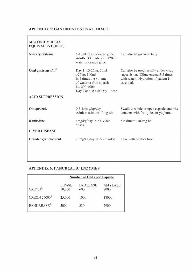
61
APPENDIX 5: GASTROINTESTINAL TRACT MECONIUM ILEUS EQUIVALENT (DIOS)
N-acetylcysteine
5-10ml qds in orange juice. Adults: 30ml tds with 120ml water or orange juice.
Can also be given rectally.
Oral gastrografin®
Day 1: 15-25kg: 50ml >25kg: 100ml in 4 times the volume of water or fruit squash i.e. 200-400ml Day 2 and 3; half Day 1 dose
Can also be used rectally under x-ray supervision. Dilute enema 3-5 times with water. Hydration of patient is essential.
ACID SUPPRESSION
Omeprazole
0.7-1.4mg/kg/day Adult maximum 10mg tds
Swallow whole or open capsule and mix contents with fruit juice or yoghurt.
Ranitidine
4mg/kg/day in 2 divided doses.
Maximum: 300mg bd.
LIVER DISEASE Ursodeoxycholic acid
20mg/kg/day in 2-3 divided
Take with or after food.
APPENDIX 6: PANCREATIC ENZYMES Number of Units per Capsule CREON®
LIPASE 10,000
PROTEASE 600
AMYLASE 8000
CREON 25000® 25,000 1000 18000 PANKREASE® 5000 330 2900

62
APPENDIX 7: OTHER NEBULISED DRUGS BRONCHODILATORS
Salbutamol 2.5mg/2.5ml 5mg/2.5ml
6mths-5yrs: 2.5mg >5yrs: 5mg
Repeat up to 6 times daily.
STEROIDS
Budesonide 250µg/ml 500µg/ml
3mths-12yrs: 0.25-1mg bd 12yrs: 1mg bd
MUCOLYTICS
Hypertonic saline 2-4 mls of 5% or 7% solution
Up to twice per day 30 mins before physiotherapy
rhDNase
2.5mg (1 vial) daily Dose can be increased to twice per day if necessary.

63
APPENDIX 8: DESENSITISATION REGIME
Piperacillin/Tazobactam (Tazocin®) (per continuous infusion)
1st give 100mg over 6 hours 2nd give 500mg over 6 hours 3rd give 2g over 6 hours 4th give normal adult dose (13.5g / 24hours)
Ceftazidime (per continuous infusion)
1st give 50mg over 6-8 hours 2nd give 250mg over 6-8 hours 3rd give 1g over 6-8 hours 4th give normal adult dose (100 mg/kg/24hour or 6g/24 hours)
From the beginning to the end of treatment patients should be on H1 & H2 receptor blockers i.e. Cetirizine 20mg twice per day po, Ranitidine 600mg twice per day po.

64
APPENDIX 9. NUTRITIONAL SUPPLEMENTS
Polymeric feeds (1Kcal/ml) Powdered Ensure
Nutren Activ (name change was Build-up) Nutren Optimun Nutren Fibre
Ready to feed Osmolite & Osmolite HN Jevity Fresubin original Fresubin fibre
Polymeric feeds (1.2 to 1.5 kcal/ml) Ready to feed Ensure Plus
Prosure Jevity Plus Provide Xtra Fresubin HP energy Fresubin energy fibre
Modular feeds Carbonhydrate Polycose Protein Promod Fat MCT oil Peadiatric Powdered Pediasure
Nutren junior Ready to feed Pediasure
Pedisure Plus
Specialised products Powdered Alitraq (semi elemental with glutamine)
Vital HN (semi elemental) Glucerna SR (diabetic) Peptamen (semi elemental) Peptamen Junior (semi elemental for children)
Ready to feed Perative (semi elemental, high energy) Oxepa (EFA and antioxidants) Pulmocare (high fat, low carbohydrate, high energy) Glucerna (diabetic) Peptamen (semi elemental) Supportan (semi elemental) Fresubin diben (diabetic)
Nutricia products have not been included as the availability of products is too unreliable and costly.

65
APPENDIX 10: CONTACT INFORMATION
CYSTIC FIBROSIS CLINICS Johannesburg Hospital – Paediatric CF Clinic Dr Susan Klugman Tel: (011) 488 3983 Johannesburg Hospital – Adult Respiratory Clinic Prof. Mervyn Mer Tel: (011) 488 4911 / 488 3496 (page no. 1025) Sandton Clinic Dr David Richard Tel: (011) 706 6060 Pretoria Academic Hospital – CF Clinic Prof. R Green Tel: (012) 354 6244 / 354 1564 Addington Hospital (Durban) – CF Clinic Tel: (031) 3272000 St. Augustine’s Hospital (Durban) Dr Jonathan Egner Tel: (031) 201 0214/5 Red Cross Children’s Hospital (Cape Town) Dr Tony Westwood, Prof Heather Zar Tel: (021) 658 5137 Fax: (021) 685 3891
Groote Schuur (Cape Town) – Adult CF Clinic Prof. Paul Willcox Tel: (021) 404 4369 Fax: (021) 448 2348 UCT Private Academic Hospital, Cape Town Prof Paul Willcox Tel: (021) 442 1966/1817 Cell 083 261 2064 Fax (021) 442 1813 Port Elizabeth Greenacres Hospital Dr Paul Gebers Tel: (041) 363 3900 Fax: (041) 363 7864 Universitas Academic Complex (Bloemfontein) – Paediatric CF clinic Dr Jeanette Kriel and Dr Bertram Henderson Tel: (051) 405 3783 Fax: (051) 444 5041 Universitas Academic Complex (Bloemfontein) – Adult CF clinic Dr Michiel Prins Tel: (051) 405 3612 Fax: (051) 444 4130

66
GENETIC CLINICS DURBAN Prof. WS Winship Genetic Clinic NBTS 10 Eden Road PINETOWN 3610 Tel: (031) 719 6556 Tel: (031) 765 5039 Dr H Ramdhani Genetic Clinic Inkosi Albert Luthuli CentraI Hospital 800 Bellair Road Cato Manor Durban, 4001 Tel: (031) 240 2341 PRETORIA Dr E Honey Dept of Human Genetics P O Box 2034 University of Pretoria PRETORIA 0001 Tel: (012) 319 2269 BLOEMFONTEIN Dr Bertram Henderson Division Human Genetics University of the Orange Free State P O Box 339 (G11) BLOEMFONTEIN 9300 Tel: (051) 405 3047
CAPE TOWN/STELLENBOSCH Prof. G de Jong Dept. of Human Genetics/Faculty of Health Sciences P O Box 19063 TYGERBERG 7505 Tel: (021) 938 9509 Dr Karen Fieggen Division of Human Genetics UCT Medical School Anzio road Observatory 7925 Tel: (021) 406 6304 JOHANNESBURG Dept. of Human Genetics NHLS P O Box 1038 JOHANNESBURG 2000 Tel: (011) 489 9224 Fax: (011) 489 9226 Chris Hani-Baragwanath Clinic Tel: (011) 933 8000 ext 3258 Genetic Counselling Clinic at ward 284 (Paediatrics) Johannesburg Hospital Tel: (011) 488 4286 Genetic Counselling Clinic at ward 496 (Adult) Johannesburg Hospital Tel: (011) 488 3496
SACFA REPRESENTATIVES CENTRAL REGION Alan Dunn (011) 294-3849 Marylyn Coney (011) 312-0220 [email protected] CAPE TOWN Karin Kratz (021) 782-1226 Ruth Ireland (021) 557-0323 [email protected]
67
KWA ZULU NATAL Debbie Long (031) 765-6687 Marie Allen (031) 202-1644 [email protected]
PLEASE BECOME A MEMBER OF YOUR LOCAL CF ASSOCIATION AND ENQUIRE ABOUT REGISTRATION OF CYSTIC FIBROSIS PATIENTS/FAMILIES ON THE ‘NATIONAL CYSTIC FIBROSIS (CF) DATABASE’ SACFA WEBSITE http://www.sacfa.org.za