Table of Contents - SAGES
195
3 www.12thworldcongress.org 12 th World Congress of Endoscopic Surgery Table of Contents 3 Registration Hours, Exhibit Hours 4 General Information, Shuttle Information 5-7 Schedule at a Glance 6 Speaker Prep. & AV Information 8 CME Worksheet 9 Conflict of Interest Policy 11 Childcare Services, CyberCafé 12-13 National Harbor Map, Gaylord Floor Plan 14-15 World Congress Meeting Leaders 15 SAGES Accreditation 44 International Webcast Sessions 17 WEDNESDAY, APRIL 14, 2010 18 Hands-on Colon Cadaver Lab 19 Postgraduate Course: MIS & Cancer – Endocrine/Solid Organ 19 Postgraduate Course: MIS & Cancer - GI 20 Postgraduate Course: Bariatrics – New Technology, Revisions, Endolumenal, Single Port Access Surgery 20 Postgraduate Course: Bariatrics Around the World 21 SAGES Foundation Awards Lunch 27 Hands-on Course w/Lab: Advanced Suturing and Anastomotic Techniques 28 Pediatric Session: Next Generation Pediatric MAS 28 Exhibits Opening Welcome Reception 31 THURSDAY, APRIL 15, 2010 32 Hands-On Course: Digital Video 33 Postgraduate Course: Avoid Pitfalls in Cholecystectomy and CBD Exploration 33 Postgraduate Course: Challenging Hernias 34 Hands-on Course w/Lab: Fundamentals of Laparoscopic Surgery 35 Educator’s Lunch: Utilizing SAGES Educational Offerings for Residents 36 Device Development Lunch 36 Postgraduate Course: Laparoscopic IBD & Colectomy 39 Hands-On Course w/Lab: Single Port Access 40 MBA for Surgeons Panel 40 Hands-On Course w/Lab: Endolumenal / NOTES® 41 Symposium: Robotics – What’s New? 41 Symposium: Metabolic Surgery – Current Status 41 Debate: Barrett’s – How to Follow, How to Treat 32, 42 Industry Symposia 45 Water Taxi Service 45 FRIDAY, APRIL 16, 2010: SCIENTIFIC SESSION 47 SAGES Presidential Address 48 Gerald Marks Keynote Lecture 51 Panel: Endolumenal Therapies 51 Symposium: NOTES® – Alive and Well, or RIP? 51 Panel: Laparoscopic Education – Do MIS Fellowships Have a Future? 52 Video Complications Lunch 52 Go Global – Report from the Field 53 SAGES/EAES Session: Peer Review Training 53 Presidential Debates 54 Conflict of Interest Panel 54 Residents/Fellows Scientific Session 55 Panel: Single Port Access Surgery 58 Emerging Technology Session 59 Meet the Leadership Reception 61 SATURDAY, APRIL 17, 2010: SCIENTIFIC SESSION 62 Panel: Live from Afghanistan: Video Conference Military Coalition 62 Panel: Hernia Debates 63 Karl Storz Keynote Lecture 63 Royal College of Physicians and Surgeons Lecture 64 Lunch in Exhibit Hall for All Meeting Attendees 64 Fellowship Council Lunch 65 Panel: Uh, Oh, What Now? – Video Session with the Experts 65 CAGS Session: Simulation in the Training of Surgeons 70 Panel: FES Roll-Out 70 IFSES Panel: Surgical Education Around the World 70 World Congress Main Event & International Sing-Off 74 2010 Learning Center 77 Faculty & Presenters 83 Faculty & Presenter Disclosures 94 Social Programs 95 Tours 97 Scientific Session Oral Abstracts 132 Resident/Fellow Oral Abstracts 136 Video Abstracts 148 Video Channel Loop Listing 150 Posters of Distinction Abstracts 158 Poster Listing 180 Emerging Technology Oral Abstracts 189 Emerging Technology Poster Listing 193 Exhibit Hall Floor Plan 194 Exhibitor Profiles 12 th World Congress of Endoscopic Surgery Location Gaylord National Hotel & Convention Center 201 Waterfront Street National Harbor, MD 20745 Hosted By Society of American Gastrointestinal and Endoscopic Surgeons (SAGES) 11300 W. Olympic Blvd., Suite 600 Los Angeles, CA 90064 Phone: 310-437-0544 Fax: 310-437-0585 Email: [email protected] Website: www.sages.org Canadian Association of General Surgeons (CAGS) 774 Echo Drive Ottawa, ON K18 5NB Phone: 613-730-6280 Fax: 613-730-1116 Email: [email protected] Website: www.cags-accg.ca World Congress Registration Hours Tuesday, April 13, 2010: 12:00 PM - 5:00 PM Wednesday, April 14, 2010: 6:30 AM - 6:00 PM Thursday, April 15, 2010: 6:30 AM - 5:30 PM Friday, April 16, 2010: 6:30 AM - 5:30 PM Saturday, April 17, 2010: 6:30 AM - 2:00 PM Exhibit Dates and Times Wednesday, April 14, 2010 World Congress Opening Reception: 5:00 PM - 7:00 PM Thursday, April 15, 2010 Hall Open: 10:00 AM - 2:30 PM Friday, April 16, 2010 Hall Open: 10:00 AM - 2:30 PM Saturday, April 17, 2010 Hall Open: 10:00 AM - 1:00 PM Free Lunch for All Attendees: 11:30 AM - 12:30 PM World Congress exhibits will take place at the Gaylord Convention Center in Prince George’s Exhibit Hall A-C. SAGES & CAGS thank IFSES, the IFSES member societies (ALACE, CSLES, EAES, ELSA, FELAC, IAGES, JSES) and corporate partners for the opportunity to host the World Congress of Endoscopic Surgery. IFSES President: Prof. Alberto Chousleb, MD IFSES Secretary General/Treasurer: Prof. Tatsuo Yamakawa, MD Visit the IFSES website for future IFSES activities and World Congress meetings: http://www.ifses.org/
-
Upload
khangminh22 -
Category
Documents
-
view
0 -
download
0
Transcript of Table of Contents - SAGES

3 www.12thworldcongress.org
12th W
orld
Co
ng
ress of En
do
scop
ic Surg
eryTa b l e o f C o n t e n t s 3 Registration Hours, Exhibit Hours 4 General Information, Shuttle Information 5-7 Schedule at a Glance 6 Speaker Prep. & AV Information 8 CME Worksheet 9 Conflict of Interest Policy 11 Childcare Services, CyberCafé 12-13 National Harbor Map, Gaylord Floor Plan 14-15 World Congress Meeting Leaders 15 SAGES Accreditation 44 International Webcast Sessions 17 WEdnESday, april 14, 2010 18 Hands-on Colon Cadaver Lab 19 Postgraduate Course: MIS & Cancer –
Endocrine/Solid Organ 19 Postgraduate Course: MIS & Cancer - GI 20 Postgraduate Course:
Bariatrics – New Technology, Revisions, Endolumenal, Single Port Access Surgery
20 Postgraduate Course: Bariatrics Around the World
21 SAGES Foundation Awards Lunch 27 Hands-on Course w/Lab: Advanced
Suturing and Anastomotic Techniques 28 Pediatric Session:
Next Generation Pediatric MAS 28 Exhibits Opening Welcome Reception 31 ThurSday, april 15, 2010 32 Hands-On Course: Digital Video 33 Postgraduate Course: Avoid Pitfalls in
Cholecystectomy and CBD Exploration 33 Postgraduate Course:
Challenging Hernias 34 Hands-on Course w/Lab:
Fundamentals of Laparoscopic Surgery 35 Educator’s Lunch: Utilizing SAGES
Educational Offerings for Residents 36 Device Development Lunch 36 Postgraduate Course:
Laparoscopic IBD & Colectomy 39 Hands-On Course w/Lab:
Single Port Access 40 MBA for Surgeons Panel 40 Hands-On Course w/Lab:
Endolumenal / NOTES® 41 Symposium: Robotics – What’s New? 41 Symposium:
Metabolic Surgery – Current Status 41 Debate:
Barrett’s – How to Follow, How to Treat 32, 42 Industry Symposia 45 Water Taxi Service
45 Friday, april 16, 2010: SCiEnTiFiC SESSion
47 SAGES Presidential Address 48 Gerald Marks Keynote Lecture 51 Panel: Endolumenal Therapies 51 Symposium: NOTES® –
Alive and Well, or RIP? 51 Panel: Laparoscopic Education –
Do MIS Fellowships Have a Future? 52 Video Complications Lunch 52 Go Global – Report from the Field 53 SAGES/EAES Session:
Peer Review Training 53 Presidential Debates 54 Conflict of Interest Panel 54 Residents/Fellows Scientific Session 55 Panel: Single Port Access Surgery 58 Emerging Technology Session 59 Meet the Leadership Reception 61 SaTurday, april 17, 2010:
SCiEnTiFiC SESSion 62 Panel: Live from Afghanistan:
Video Conference Military Coalition 62 Panel: Hernia Debates 63 Karl Storz Keynote Lecture 63 Royal College of Physicians and
Surgeons Lecture 64 Lunch in Exhibit Hall for
All Meeting Attendees 64 Fellowship Council Lunch 65 Panel: Uh, Oh, What Now? –
Video Session with the Experts 65 CAGS Session:
Simulation in the Training of Surgeons 70 Panel: FES Roll-Out 70 IFSES Panel:
Surgical Education Around the World 70 World Congress Main Event &
International Sing-Off 74 2010 Learning Center 77 Faculty & Presenters 83 Faculty & Presenter Disclosures 94 Social Programs 95 Tours 97 Scientific Session Oral Abstracts 132 Resident/Fellow Oral Abstracts 136 Video Abstracts 148 Video Channel Loop Listing 150 Posters of Distinction Abstracts 158 Poster Listing 180 Emerging Technology Oral Abstracts 189 Emerging Technology Poster Listing 193 Exhibit Hall Floor Plan 194 Exhibitor Profiles
12th World Congress of Endoscopic SurgeryLocationGaylord National Hotel & Convention Center201 Waterfront StreetNational Harbor, MD 20745
Hosted BySociety of American Gastrointestinal and Endoscopic Surgeons (SAGES)11300 W. Olympic Blvd., Suite 600 Los Angeles, CA 90064 Phone: 310-437-0544 Fax: 310-437-0585 Email: [email protected] Website: www.sages.org
Canadian Association of General Surgeons (CAGS)774 Echo Drive Ottawa, ON K18 5NB Phone: 613-730-6280 Fax: 613-730-1116 Email: [email protected] Website: www.cags-accg.ca
World Congress registration hours
Tuesday, April 13, 2010: 12:00 PM - 5:00 PMWednesday, April 14, 2010: 6:30 AM - 6:00 PM Thursday, April 15, 2010: 6:30 AM - 5:30 PMFriday, April 16, 2010: 6:30 AM - 5:30 PMSaturday, April 17, 2010: 6:30 AM - 2:00 PM
Exhibit dates and TimesWednesday, april 14, 2010 World Congress Opening Reception: 5:00 PM - 7:00 PMThursday, april 15, 2010 Hall Open: 10:00 AM - 2:30 PMFriday, april 16, 2010 Hall Open: 10:00 AM - 2:30 PMSaturday, april 17, 2010 Hall Open: 10:00 AM - 1:00 PM Free Lunch for All Attendees: 11:30 AM - 12:30 PM
World Congress exhibits will take place at the Gaylord Convention Center in Prince George’s Exhibit Hall A-C.
SAGES & CAGS thank IFSES, the IFSES member societies (ALACE, CSLES, EAES, ELSA, FELAC, IAGES, JSES) and corporate partners for the opportunity to host the World Congress of Endoscopic Surgery.
iFSES president: prof. alberto Chousleb, MdiFSES Secretary General/Treasurer: prof. Tatsuo yamakawa, Md
Visit the IFSES website for future IFSES activities and World Congress meetings: http://www.ifses.org/

12th World Congress of Endoscopic Surgery 4
12th W
orld
Co
ng
ress of En
do
scop
ic Surg
ery
leadership for the 2010 World CongressWorld Congress program Chairs:Daniel M. Herron, M.D. (SAGES Co-Chair)
Barry A. Salky, M.D. (SAGES Chair)Christopher M. Schlachta, M.D. (CAGS Chair)
World Congress presidents:Gerald M. Fried, M.D. (CAGS)David W. Rattner, M.D. (SAGES)
SAGES President: C. Daniel Smith, M.D.CAGS President: Chris Jamieson, M.D.IFSES President: Alberto Chousleb, M.D.
G e n e ra l I n f o r m a t i o n
World Congress Corporate SupportersDIAmonD DonorS
CoVidiEnEThiCon Endo-SurGEry, inC.
SaGES EduCaTion and rESEarCh FoundaTion
PLATInUm DonorSKarl STorZ EndoSCopy-aMEriCa
olyMpuS
GoLD DonorSaSCEnT hEalThCarE SoluTionS
STryKEr EndoSCopy
SILVEr DonorSBOSTON SCIENTIFIC DAVOL INC., A BARD COMPANY GORE & ASSOCIATES
BronZE DonorSSYNOVIS SURGICAL INNOVATIONS
d.C. Shuttle Service From Gaylord nationalVisit the Transportation Desk (Lobby Level) or Call 301-839-5261 hours of operation: 8:00am - 10:00pm (major credit cards accepted)
Shuttle management will make every effort to maintain the schedule but may experience delays due to traffic conditions beyond our control especially during peak business hours. Subject to availability. Dates, times, and prices subject to change. Additional restrictions may apply.
Roundabout Tour and ShuttleVisit more than a dozen stops in and around the region and jump on and off as you please! OnBoard Tours’ Roundabout is the best way to see the Capital Region – and at the best value! 3-Day Unlimited Passes Start at Just $35 Per Person!
Sightseeing Tours and Roundabout ServicesChoose from the area’s most exciting tours, including “The DC It All Tour,” “DC the Lights” (the only downtown night tour), and more!
Downtown D.C. Shuttle ServiceFeaturing continuous service to:
– Old Post Office (1100 Pennsylvania Avenue NW) – Union Station (H. Street and 2nd Street NE)
Departing every hour, on the hour, from Gaylord National’s main entrance Seven days a week: 8:00 a.m. - 9:00 p.m.
One-way tickets: $13 Round-trip tickets: $20 Unlimited, 3-Day Pass: $49
King Street Metrorail Station and Old Town Alexandria Shuttle ServiceEnjoy Metrorail’s unparalleled accessibility to the area (including the museums and monuments of the National Mall) or the dining, shopping, and entertainment of Old Town’s historic waterfront community. Metrorail fare-cards may also be purchased at the Gaylord National Transportation Desk.
One-way tickets are $5 per person, with continuous service to:
– King Street Metrorail Station (Yellow and Blue lines) – Old Town Alexandria (King Street and Route 1)
Departing every 30 minutes* from Gaylord National’s main entrance Sunday - Thursday: 6:30am - 9:00pm Friday - Saturday: 6:30am - 10:00pm
Water Taxis to Old Town, Georgetown, and Mount VernonPotomac Riverboat Company’s Water Taxis depart regularly from Gaylord National’s pier, offering exciting trips along the storied Potomac River to and from Old Town Alexandria, historic Georgetown, and George Washington’s Mount Vernon Estate and Gardens.
Airport Shuttle ServicesHourly shuttle service is available from Reagan National Airport (DCA). And SuperShuttle service, private sedans, and taxis are available to all major airports and most locations locally.

5 www.12thworldcongress.org
12th W
orld
Co
ng
ress of En
do
scop
ic Surg
eryWo r l d C o n g r e s s S c h e d u l e - a t - a - G l a n ceWednesday, april 14, 2010
Time Session location7:00 AM - 11:30 AM Hands-on Colon Cadaver Lab **Offsite Lab
7:30 AM - 12:00 PM MIS & Cancer Endocrine/Solid Organ Postgraduate Course Potomac Ballroom B
1:00 PM - 5:00 PM MIS Gastrointestinal Cancer Postgraduate Course Potomac Ballroom B
7:30 AM - 12:00 PMBariatric Postgraduate Course: New Tech/Revisions/Endolumenal/Single Port Access Surgery
Potomac Ballroom A
1:00 PM - 5:00 PM Bariatric Postgraduate Course: Around the World Potomac Ballroom A
12:00 PM - 1:00 PM SAGES Education and Research Foundation Awards Luncheon Maryland Ballroom C
1:00 PM - 5:00 PM Hands-on Advanced Suturing and Anastomotic Techniques Lab Maryland Ballroom B-D
1:00 PM - 5:00 PMPediatrics Session: Next-Generation Pediatric MAS – A Move Toward “Scarless” Surgery
Potomac Ballroom C
5:00 PM - 7:00 PM World Congress Welcome Exhibit Opening Reception Prince George’s Exhibit Hall A-C
Thursday, april 15, 2010Time Session location
6:00 AM - 7:15 AMIndustry Satellite Symposia Covidien – “Advances in SILS™ Technology, Technique and Evidence” Potomac Ballroom D
7:30 AM - 11:30 AMPostgraduate Surgeon in the Digital Age: Video Editing Course – Basic Video Editing with an Introduction to Advanced Techniques Chesapeake Conference Rooms D-E
7:30 AM - 11:30 AM Avoid Pitfalls in Cholecystectomy and CBD Exploration Potomac Ballroom B
7:30 AM - 11:30 AM Challenging Hernias Postgraduate Course Potomac Ballroom A
7:30 AM - 12:00 PM Fundamentals of Laparoscopic Surgery Hands-on CourseLectures – Maryland Ballroom C Lab – Maryland Ballroom A
9:30 AM - 11:30 AM SS01 Best of Videos 1 Potomac Ballroom C
10:00 AM - 2:30 PM World Congress Exhibits, Posters & Learning Center Open Prince George’s Exhibit Hall A-C
11:30 AM - 1:00 PM BREAK: Exhibits, Posters, Learning Center
11:30 AM - 1:30 PMEducators Luncheon: Utilizing SAGES Educational Offerings for Residents Potomac Ballroom D
11:30 AM - 1:30 PM Device Development Luncheon: From Funding to Freedom to Operate Maryland Ballroom C
1:30 PM - 5:00 PMLaparoscopic IBD and Colectomy Postgraduate Course: The Status and Direction of Laparoscopic Colorectal Surgery in the Treatment of Inflammatory Bowel Disease
Potomac Ballroom A
1:30 PM - 5:00 PM Single Port Access Surgery Hands-On Course **Offsite lab
1:30 PM - 5:00 PMMBA for Surgeons Panel: Asset Management and Protection for Surgeons Potomac Ballroom C
1:30 PM - 5:00 PM Hands On Endolumenal/NOTES® Lab Maryland Ballroom B-D
1:30 PM - 2:30 PM Robotics Symposium: What’s New? Potomac Ballroom B
2:30 PM - 4:00 PM Metabolic Surgery Symposium: Current Status Potomac Ballroom B
4:00 PM - 5:30 PM Barrett’s Debate: How to Follow, How to Treat? Potomac Ballroom B
5:30 PM - 7:30 PM Industry Satellite Symposia (No Registration Required)
Boston Scientific – “ Intraluminal Stents and the Surgeon – Who, What, Where, When and Why?” Chesapeake D-E
Covidien – “ Insights into the Future of Surgical Stapling: Integrated Materials, Tri-Staple™ Technology, Powered Staplers” Potomac Ballroom D
Davol Inc., a BARD Company – “ Advanced Endoscopic Techniques for Abdominal Wall Reconstruction” Maryland Ballroom C
Ethicon Endo-Surgery, Inc. – “ Minimally Invasive Surgery – Where is it Going?”
Potomac Ballroom C
Karl Storz Endoscopy – “The Great Debate: S-PORTAL (Single Portal Access) Surgery vs. Mini-Laparoscopy” Maryland Ballroom A

12th World Congress of Endoscopic Surgery 6
12th W
orld
Co
ng
ress of En
do
scop
ic Surg
ery
Friday, april 16, 2010
Time Session location7:00 AM - 8:00 AM Posters of Distinction Potomac Ballroom C
8:00 AM - 9:00 AM SS02 Plenary Session 1 Potomac Ballroom A-B
9:00 AM - 9:30 AM SAGES Presidential Address: Everyone Knows Plan A: Its All About Plan B C. Daniel Smith, M.D. Potomac Ballroom A-B
9:30 AM - 10:00 AMGerald Marks Lecture: The Making of a Surgeon – Revisited Richard H. Bell, M.D. Potomac Ballroom A-B
10:00 AM - 2:30 PM Exhibits, Posters, Learning Center Open Prince George’s Exhibit Hall A-C
10:00 AM -11:00 AM
Concurrent Sessions (accepted oral & video presentations)
SS03 Solid Organ Maryland Ballroom B-D
SS04 Basic Science Maryland Ballroom A
10:00 AM - 11:00 AM Endolumenal Therapies Session Potomac Ballroom C
10:00 AM - 11:00 AM NOTES® Symposium – Alive & Well or RIP? Potomac Ballroom A-B
10:00 AM - 11:00 AM Laparoscopic Education Panel – Do MIS Fellowships Have a Future? Potomac Ballroom D
11:00 AM - 12:30 PM Video Complications Luncheon: What Has Happened and What Do We Have to Do? Maryland Ballroom C
12:30 PM - 1:30 PM Go Global Report from the Field Panel: Teaching Laparoscopic Surgery Abroad Maryland Ballroom A
12:30 PM - 2:30 PM Peer Review Training Session Potomac Ballroom D
12:30 PM - 2:30 PM SAGES Presidential Debates Potomac Ballroom A-B
1:30 PM - 2:30 PM Conflict of Interest Panel Maryland Ballroom A
2:30 PM - 5:30 PM Resident and Fellows Scientific Session Maryland Ballroom C
2:30 PM - 4:00 PM Single Port Access Surgery Panel Potomac Ballroom A-B
2:30 PM - 4:00 PM
Concurrent Sessions
SS05 Hepatobiliary/Pancreatic Maryland Ballroom B-D
SS06 Hernia Potomac Ballroom D
4:00 PM - 5:30 PM
Concurrent Sessions
SS07 Best of Video 2 Maryland Ballroom B-D
SS08 Education/Simulation Maryland Ballroom A
SS09 NOTES® Potomac Ballroom D
2:30 PM - 5:30 PM Emerging Technology Session Potomac Ballroom C
6:00 PM - 7:00 PM Meet the Leadership Reception For Residents, Fellows & New Members Pose, 18-19th Floor
Wo r l d C o n g r e s s S c h e d u l e - a t - a - G l a n ce
iMporTanT aV inForMaTion:You may now upload your presentation on line at any point during the meeting. Please load your presentation online (http://sages.presentationman.com/) or on the show computer in the Speaker Prep room no later than 2 hours before your presentation.
please note: Even if you have submitted your presentation online you must visit the Speaker Prep room to check in, your session moderator may not allow you to present if you do not.
Speaker prep hours:4/13/10: 7:00 AM - 5:00 PM4/14/10: 5:30 AM - 5:00 PM4/15/10: 6:00 AM - 5:00 PM4/16/10: 5:30 AM - 5:30 PM4/17/10: 5:30 AM - 5:00 PM
7 www.12thworldcongress.org
12th W
orld
Co
ng
ress of En
do
scop
ic Surg
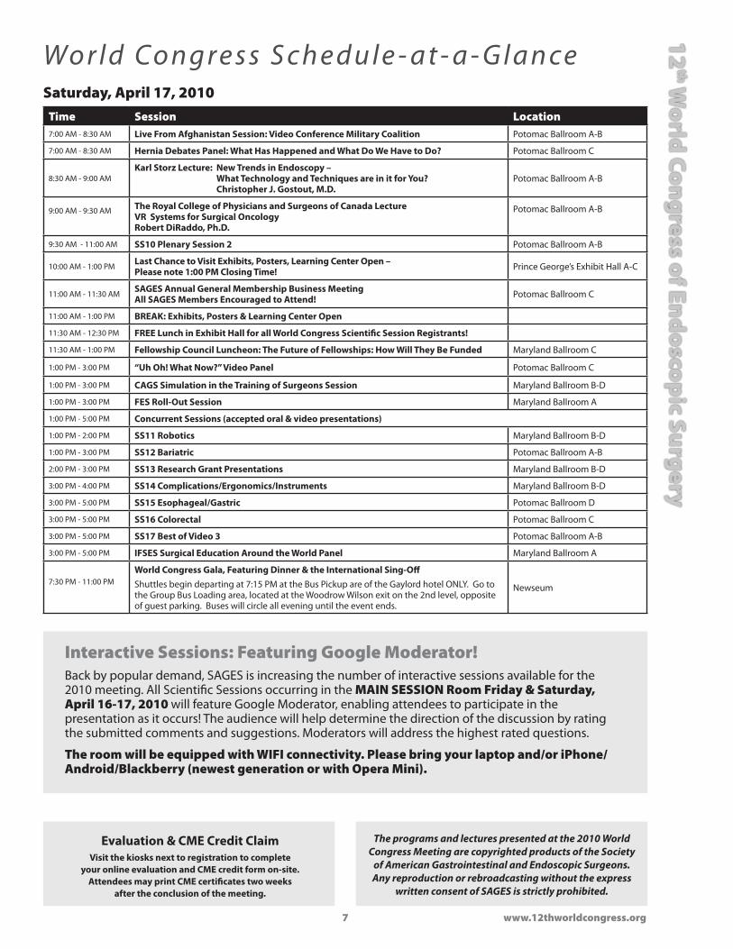
eryWo r l d C o n g r e s s S c h e d u l e - a t - a - G l a n ceSaturday, april 17, 2010
Time Session location7:00 AM - 8:30 AM Live From Afghanistan Session: Video Conference Military Coalition Potomac Ballroom A-B
7:00 AM - 8:30 AM Hernia Debates Panel: What Has Happened and What Do We Have to Do? Potomac Ballroom C
8:30 AM - 9:00 AMKarl Storz Lecture: New Trends in Endoscopy –
What Technology and Techniques are in it for You? Christopher J. Gostout, M.D.
Potomac Ballroom A-B
9:00 AM - 9:30 AM The Royal College of Physicians and Surgeons of Canada Lecture VR Systems for Surgical Oncology Robert DiRaddo, Ph.D.
Potomac Ballroom A-B
9:30 AM - 11:00 AM SS10 Plenary Session 2 Potomac Ballroom A-B
10:00 AM - 1:00 PM Last Chance to Visit Exhibits, Posters, Learning Center Open – Please note 1:00 PM Closing Time! Prince George’s Exhibit Hall A-C
11:00 AM - 11:30 AMSAGES Annual General Membership Business Meeting All SAGES Members Encouraged to Attend! Potomac Ballroom C
11:00 AM - 1:00 PM BREAK: Exhibits, Posters & Learning Center Open
11:30 AM - 12:30 PM FREE Lunch in Exhibit Hall for all World Congress Scientific Session Registrants!
11:30 AM - 1:00 PM Fellowship Council Luncheon: The Future of Fellowships: How Will They Be Funded Maryland Ballroom C
1:00 PM - 3:00 PM “Uh Oh! What Now?” Video Panel Potomac Ballroom C
1:00 PM - 3:00 PM CAGS Simulation in the Training of Surgeons Session Maryland Ballroom B-D
1:00 PM - 3:00 PM FES Roll-Out Session Maryland Ballroom A
1:00 PM - 5:00 PM Concurrent Sessions (accepted oral & video presentations)
1:00 PM - 2:00 PM SS11 Robotics Maryland Ballroom B-D
1:00 PM - 3:00 PM SS12 Bariatric Potomac Ballroom A-B
2:00 PM - 3:00 PM SS13 Research Grant Presentations Maryland Ballroom B-D
3:00 PM - 4:00 PM SS14 Complications/Ergonomics/Instruments Maryland Ballroom B-D
3:00 PM - 5:00 PM SS15 Esophageal/Gastric Potomac Ballroom D
3:00 PM - 5:00 PM SS16 Colorectal Potomac Ballroom C
3:00 PM - 5:00 PM SS17 Best of Video 3 Potomac Ballroom A-B
3:00 PM - 5:00 PM IFSES Surgical Education Around the World Panel Maryland Ballroom A
7:30 PM - 11:00 PM World Congress Gala, Featuring Dinner & the International Sing-OffShuttles begin departing at 7:15 PM at the Bus Pickup are of the Gaylord hotel ONLY. Go to the Group Bus Loading area, located at the Woodrow Wilson exit on the 2nd level, opposite of guest parking. Buses will circle all evening until the event ends.
Newseum
interactive Sessions: Featuring Google Moderator!Back by popular demand, SAGES is increasing the number of interactive sessions available for the 2010 meeting. All Scientific Sessions occurring in the Main SESSion room Friday & Saturday, april 16-17, 2010 will feature Google Moderator, enabling attendees to participate in the presentation as it occurs! The audience will help determine the direction of the discussion by rating the submitted comments and suggestions. Moderators will address the highest rated questions.
The room will be equipped with WiFi connectivity. please bring your laptop and/or iphone/android/Blackberry (newest generation or with opera Mini).
The programs and lectures presented at the 2010 World Congress Meeting are copyrighted products of the Society
of American Gastrointestinal and Endoscopic Surgeons. Any reproduction or rebroadcasting without the express
written consent of SAGES is strictly prohibited.
Evaluation & CME Credit ClaimVisit the kiosks next to registration to complete
your online evaluation and CME credit form on-site. Attendees may print CME certificates two weeks
after the conclusion of the meeting.

12th World Congress of Endoscopic Surgery 8
12th W
orld
Co
ng
ress of En
do
scop
ic Surg
ery
C ME WorksheetSaGES accreditation
Accreditation: The Society of American Gastrointestinal and Endoscopic Surgeons (SAGES) is accredited by the Accreditation Council for Continuing Medical Education (ACCME) to sponsor Continuing Medical Education for physicians. SAGES designates this Continuing Education activity for a maximum of 41.5 AMA PRA Category 1 Credit(s)™. Physicians should only claim credit commensurate with the extent of their participation in the activity.
CME Worksheet for the 12th World Congress of Endoscopic Surgery: This is NOT your CME credit form. Please use the worksheet below to track the number of CME hours you attend for each activity. SAGES has instituted a new process for claiming CME credits and printing certificates. All attendees wishing to receive a CME certificate for activities attended at the 2010 SAGES Annual Meeting (12th World Congress of Endoscopic Surgery) must first complete an on-line meeting evaluation form. Attendees will be able to print and re-print their certificates throughout the year beginning two weeks after the conclusion of the meeting.
• On-site:There will be on-site kiosks located near the registration area to complete the meeting evaluation and credit claim form. Two weeks after the conclusion of the meeting, an email will notify attendees that the certificates are available to print.
• Duringorafterthemeeting: Attendees will also have access to the on-line meeting evaluation and credit claim form via a link on the World Congress website.
Be sure to retain your Conference Badge as the ID number will be your online PIN number. An email will also be sent, reminding attendees of this service. Those wishing to obtain a simple certificate of attendance may do so at the Evaluation Kiosks.
WednesdayaCTiViTy hourS i aTTEndEd CrEdiTS aVailaBlE
Bariatric New Tech/Revisions/Endolumenal/Single Port PG Course 4.25
MIS & Cancer: Endocrine/Solid Organ Postgraduate Course 4.25
HO Course: Colon Surgery 4.5
Bariatrics Around the World Postgraduate Course 3.75
MIS Gastrointestinal Cancer Postgraduate Course 3.75
HO Course: Advanced Suturing and Anastomotic Techniques 4.0
Pediatrics Session 4.0
SuBToTal MAX – 8.5
ThursdayaCTiViTy hourS i aTTEndEd CrEdiTS aVailaBlE
Postgraduate Surgeon in the Digital Age: Video Editing Course 3.75
Challenging Hernias Postgraduate Course 3.75
HO Course: Fundamentals of Laparoscopic Surgery 4.25
Avoid Pitfalls in Cholecystectomy and CBD Exploration 3.75
Scientific Session 1 2.0
Educators Luncheon: Utilizing SAGES Educational Offerings for Residents 2.0
Device Development Luncheon: From Funding to Freedom to Operate 2.0
HO Course: Endolumenal/NOTES 3.5
HO Course: Single Port Access Surgery 3.5
Lap IBD & Colectomy Postgraduate Course 3.25
MBA for Surgeons Panel 0-NO credit available
Symposium: Robotics: What’s New? 1.0
Symposium: Metabolic Surgery :Current Status 1.5
Barrett’s Debate: How to Follow, How to Treat? 1.5
SuBToTal MAX – 10
FridayaCTiViTy hourS i aTTEndEd CrEdiTS aVailaBlE
Scientific Sessions (panels, debates, lectures and abstract presentations including plenary) 9.0
Video Complications Luncheon: What has Happened and What We Have to Do 1.5
Emerging Technology Session 0-NO credit available
SuBToTal MAX – 10.5
SaturdayaCTiViTy hourS i aTTEndEd CrEdiTS aVailaBlE
Learning Center (*although the Learning Center is open Thurs-Sat, only 3.0 credits are available) 3.0
Scientific Sessions (panels, debates, lectures and abstract presentations including plenary) 8.0
Fellowship Council Lunch 1.5
SuBToTal MAX – 12.5

9 www.12thworldcongress.org
12th W
orld
Co
ng
ress of En
do
scop
ic Surg
ery
a. identifying Conflicts of interestSAGES has implemented a five-tiered approach towards identifying potential conflicts of interest.
1. Members of committees involved in the planning of CME activities, including the Board of Governors, must provide a financial disclosure. These disclosures are sent to the committee in advance of each committee meeting. Attendees are reminded about the disclosure policy at each committee meeting, and any committee member with a conflict is asked to recuse him or herself from the discussion of any CME activities.
2. Course Directors for CME activities must provide their financial disclosures along with their suggested course outline and faculty. This information is forwarded to the Conflict of Interest Task Force, who then determines whether or not a potential conflict exists and makes suggested edits.
3. Invited faculty for CME activities must provide their financial disclosures upon invitation to serve as faculty.
4. For abstract submissions for the scientific session, the presenting and senior authors must provide disclosures. Abstracts are peer reviewed in a blinded fashion by multiple reviewers and are selected for presentation based on scientific merit. All disclosures are provided to the Program Committee during the “Put-The-Program-Together” meeting at which abstracts are selected for presentation.
5. All speakers at SAGES CME activities must display a list of financial disclosures on the first slide of their presentation.
B. Managing potential Conflicts of interestSAGES has implemented several mechanisms to manage conflicts of interest prior to an educational activity.
1. Self-management, such as the committee member recusing him or herself from discussion of CME activities.
2. The SAGES Conflict of Interest Task force reviews all Course Director’s disclosures, proposed course outlines and faculty lists. The Conflict of Interest Task Force will make edits to the course outline or faculty list if necessary.
3. The SAGES disclosure form requires faculty to provide management suggestions if there is a relationship with a commercial entity. This information is forwarded to the Course Director, who is responsible for determining whether or not a conflict exists and if so, how to manage this conflict.
4. If a conflict is determined, then a letter is sent to the faculty member, requiring them to adhere to the management technique or else recuse him or herself from the presentation.
5. During the session, the Course Director observes the presentations and makes note of commercial bias. If any is perceived, this is immediately reported to the staff.
6. All attendees of CME activities are requested to make note of perceived commercial bias in activity evaluations. The Conflict of Interest Task Force and/or the CME Committee will investigate substantive concerns.
Commercial BiasThe Society of American Gastrointestinal and Endoscopic Surgeons (SAGES) has an obligation to the medical profession and society as a whole to elucidate bias in order to protect the objectivity, scientific integrity and quality of its continuing medical education (CME) programs and to provide CME in an ethical and impartial manner. Bias is defined when a preference or predisposition exist toward a particular perspective or result that interferes with an individual’s ability to be impartial, unprejudiced or objective in order to further personal gain and disregard for data. Particular preferences may be favorable or unfavorable. When bias exists, impartial judgment and neutrality may be compromised. Bias may be minimized through a declaration of conflict of interest or commercial interests, an evaluation of peer-reviewed evidence-based medicine with an integration of clinical expertise and/or experience, and an assertion of published sources for evidence-based reporting. SAGES requires presenters at all educational events to specifically avoid introducing bias, commercial or otherwise, into their presentations.
Confl ic t of Interest Pol ic yRevised and approved by SAGES Executive Committee, March, 2010
SaGES Mission Statement“ our mission is to provide leadership in surgery, particularly gastrointestinal and
endoscopic surgery, to optimize patient care through education, research and innovation.”– SAGES has evolved over the last 25 years into a leading society for gastrointestinal surgery,
endoscopy and minimal invasive technology.
– Not only does SAGES provide leadership in clinical care, but it also helps surgeons optimize patient care by providing direction for cutting edge technology, basic and translational science, and educational opportunities.
– SAGES represents leadership in the surgical world for gastrointestinal disease.
– SAGES is the society to improve your clinical skills.

12th World Congress of Endoscopic Surgery 10
12th W
orld
Co
ng
ress of En
do
scop
ic Surg
ery
N o t e s

11 www.12thworldcongress.org
12th W
orld
Co
ng
ress of En
do
scop
ic Surg
eryG e n e ra l I n f o r m a t i o n (co n t i n u e d )
Save the date!CaGS Canadian Surgery ForumSeptember 2 - 5, 2010, Québec City Convention Centre, Canada
SaGES Scientific Session & postgraduate CourseMarch 30 - april 2, 2011, San antonio, Texas
CaGS Canadian Surgery ForumSeptember 15 - 18, 2011, london Convention Centre, Canada
SaGES Scientific Session & postgraduate CourseMarch 7 - 10, 2012, San diego, Ca
SaGES Scientific Session & postgraduate Courseapril 17 - 20, 2013, Baltimore Convention Center, Baltimore, Md
SaGES Scientific Session & postgraduate Courseapril 2 - 5 , 2014, Salt lake Convention Center, Salt lake City, uT
A free Cyber Café is available for all World Congress attendees and exhibitors, located in the Registration area and open during normal registration hours. No message center is available this year.
Please leave the following numbers with your offices and families, in case they cannot reach you on your cell phone:
World Congress On-Site Office:
Phone: (301) 965-5343 Fax: (301) 965-5344
Childcare ServicesWhite house nannies www.whitehousenannies.com (301) 652-8088 Temporary Division (800) 266-9024 Toll Free
White House Nannies, Inc. is owned and managed by Barbara G. Kline. Mrs. Kline is a current member and former board member of the International Nanny Association (INA). The agency is also a member of the Association of Premier Nanny Agencies (APNA). White House Nannies. Inc. was awarded national recognition as APNA’s 2000 Agency of the Year. The hallmark of the agency is the thoughtful matching of Client and Caregiver needs to assure the best possible in-home childcare placements. With 25 years of experience, the agency has become the premier agency in the Washington area.
Caregiver rates: Negotiated with Caregiver, generally $16-$20 per hour.
additional agency referral rates:Hotel Child Care, booked in advance $65.00 per day per Caregiver Hotel Child Care, booked within 48hrs of need $75.00 per day per Caregiver
a Gentle reminder about Safety/Security:We have taken every precaution to assure the safety and security of our guests and their possessions. However, we urge you to be aware and take simple steps to guard your possessions.
– Do not leave your purse or briefcase unattended.
– Do not leave your laptop, phone or other electronic devices on the floor or out of your sight in a darkened room
– Be aware of your surroundings, in the Gaylord Hotel, in and around the National Harbor area and in Washington, DC.
Have a safe & secure meeting!

12th World Congress of Endoscopic Surgery 12
12th W
orld
Co
ng
ress of En
do
scop
ic Surg
ery
N a t i o n a l H a r b o r, M D M a p

13 www.12thworldcongress.org
12th W
orld
Co
ng
ress of En
do
scop
ic Surg
eryG a y l o r d N a t i o n a l Fl o o r P l a n s
GaylordNational.com | 1-877-677-9352 | 201 Waterfront Street, National Harbor, MD 20745 (WASHINGTON D.C . AREA )
F W hi B d
L K J
I H G
F E D
C B A
12 11 10
9 8 7
6 5 4
1 2 3
Chesapeake Conference Rooms
To Hotel Ballroom Level
6
B D
A C
5
4
3
2
4
Maryland Ballroom
B
A
D
C
1
2
3
4
5
6
Potomac BallroomStage
Escalators
Maryland & Potomac BallroomsConvention Center | Level 2
World Congress Exhibits, posters, learning Center (see pg. 193)
Chesapeake Conference Rooms: Speaker Prep World Congress Office SAGES Foundation Lounge FLS Testing Thursday Digital Hands-On Course Industry Education
Potomac Ballrooms: Wednesday through Saturday Main and Concurrent Session Rooms Postgraduate Courses Scientific Sessions Keynote Lectures Panels, Symposia Lunches Industry Education
Maryland Ballrooms: Wednesday through Saturday Concurrent Session Rooms Hands-On Courses Scientific Sessions Panels, Symposia Lunches Industry Education

12th World Congress of Endoscopic Surgery 14
12th W
orld
Co
ng
ress of En
do
scop
ic Surg
ery
Wo r l d C o n g r e s s 2 0 1 0 Le a d e r s
Program Co-Chair (SAGES): Program Chair (SAGES): Program Chair (CAGS): Daniel Herron, M.D. Barry Salky, M.D. Christopher Schlachta, M.D.
2010 Course Chairs & unit CoordinatorsEquipment Czar Chair: Kevin M. Reavis, M.D.
Equipment Czar Co-Chair: Gretchen Purcell-Jackson, M.D.
Poster Chair: Subhash U. Kini, M.D.
Poster Co-Chair: Melina C. Vassiliou, M.D.
Video Chair: Donald J. Selzer, M.D.
Video Co-Chair: Leena Khaitan, M.D.
Learning Center Chair: Allan E. Okrainec, M.D.
Learning Center Co-Chair: Brian P. Jacob, M.D.
Advanced Suturing and Anastomotic Techniques HO Course Chair: Kelvin D. Higa, M.D.
Advanced Suturing and Anastomotic Techniques HO Course Co-Chair: Aureo L. De Pauala, M.D.
Colon HO Course Chair: Mark H. Whiteford, M.D.
Colon HO Course Co-Chair: Conor P. Delaney, M.D.
Digital Video HO Course Chair: Dmitry Oleynikov, M.D.
Digital Video HO Course Co-Chair: John R. Romanelli, M.D.
Endolumenal/NOTES® HO Course Chair: Santiago Horgan, M.D.
Endolumenal/NOTES® HO Course Co-Chair: Christopher C. Thompson, M.D.
Single Port Access Surgery HO Course Chair: Paul G. Curcillo II, M.D.
Single Port Access Surgery HO Course Co-Chair: Daniel J. Scott, M.D.
Avoid Pitfalls in Cholecystectomy and CBD Exploration PG Course Chair: Michael B. Edye, M.D.
Avoid Pitfalls in Cholecystectomy and CBD Exploration PG Course Co-Chair: Bertrand Millat, M.D.
Bariatric Around the World PG Course Chair: Alfons Pomp, M.D.
Bariatric Around the World PG Course Co-Chair: Manolo Cortez, M.D.
Bariatric New Techniques PG Course Chair: Scott A. Shikora, M.D.
Bariatric New Techniques PG Course Co-Chair: Raul J. Rosenthal, M.D.
Challenging Hernias PG Course Chair: Kristi Lee Harold, M.D.
Challenging Hernias PG Course Co-Chair: Shirin Towfigh, M.D.
FLS PG Course Chair: E. Matthew Ritter, M.D.
FLS PG Course Co-Chair: Gerald M. Fried, M.D.
Lap IBD & Colectomy PG Course Chair: John H. Marks, M.D.
Lap IBD & Colectomy PG Course Co-Chair: Eric Glenn Weiss, M.D.
MIS & Cancer Endocrine/Solid Organ PG Course Chair: William Barry Inabnet III, M.D.
MIS & Cancer Endocrine/Solid Organ PG Course Co-Chair: Miguel Herrera, M.D.
MIS & Cancer GI PG Course Chair: Horacio J. Asbun, M.D.
MIS & Cancer GI PG Course Co-Chair: Seigo Kitano, M.D.
Pediatric Surgery Symposium Chair: Sanjeev Dutta, M.D.
Pediatric Surgery Symposium Co-Chair: Jacob Langer, M.D.
3-Hour MBA for Surgeons Chair: Demetrius E.M. Litwin, M.D.
3-Hour MBA for Surgeons Co-Chair: Fredrick J. Brody, M.D.
Device Development Luncheon Chair: Raymond P. Onders, M.D.
Device Development Luncheon Co-Chair: Dennis L. Fowler, M.D.
Education Luncheon Chair: L. Michael Brunt, M.D.
Education Luncheon Co-Chair: Daniel J. Gagne, M.D.
Fellowship Council Lunch Chair: Adrian E. Park, M.D.
Fellowship Council Lunch Co-Chair: Bruce D. Schirmer, M.D.
Video Complications Lunch Chair: Bipan Chand, M.D.
Video Complications Lunch Co-Chair: Manabu Yamamoto, M.D.
Emerging Technology Session Chair: Steven D. Schwaitzberg, M.D.
Emerging Technology Session Co-Chair: Alex Gandsas, M.D.
Resident’s Day Coordinators: Gregory F. Dakin, M.D. & Adheesh A. Sabnis, M.D.
SAGES Panel/Session/Symposium/Debates Chairs/Co-Chairs:Barrett’s Debate Chair: John Hunter, M.D.Barrett’s Debate Co-Chair: Karl H. Fuchs, M.D.SAGES Presidential Debate Chair: Daniel J. Deziel, M.D.SAGES Presidential Debate Co-Chair: Nathaniel J. Soper, M.D.Hernia Debates Chair: Guy R. Voeller, M.D.Hernia Debates Co-Chair: Edward H. Phillips, M.D.Conflict of Interest Panel Chairs: Steve Eubanks, M.D. & Neely Panton, M.D.IFSES Panel Chair: Alberto Chousleb, M.D.IFSES Panel Co-Chair: Natan Zundel, M.D.Lap Education Panel Chair: Bruce D. Schirmer, M.D.Lap Education Panel Co-Chair: Joseph Mamazza, M.D.Global Panel Chair: Raul J. Rosenthal, M.D.Global Panel Co-Chair: Horacio J. Asbun, M.D.Single Port Access Surgery Panel Chair: Joel Leroy, M.D.Single Port Access Surgery Panel Co-Chair: Andrew A. Gumbs, M.D.“Uh Oh, What Now?” Video Panel Chair: David R. Urbach, M.D.“Uh Oh, What Now?” Video Panel Co-Chair: David Bryan Earle, M.D.Endolumenal Therapies Session Chair: Dean J. Mikami, M.D.Endolumenal Therapies Session Co-Chair: Simon Bergman, M.D.FES Roll-out Session Chair: Brian J. Dunkin, M.D.FES Roll-out Session Co-Chair: Jeffrey M. Marks, M.D.Live from Fellujah Session Chair: Steven P. Bowers, M.D.Live from Fellujah Session Co-Chair: Richard M. Satava, M.D.CAGS Simulation in the Training of Surgeons Session Chair: Liane S. Feldman, M.D.CAGS Simulation in the Training of Surgeons Session Co-Chair: Teodor P. Grantcharov, M.D.Peer Review Training Session Chair: Abe L. Fingerhut, M.D.Peer Review Training Session Co-Chair: Sir Alfred Cuschieri, M.D.Metabolic Surgery Symposium Chair: Philip R. Schauer, M.D.Metabolic Surgery Symposium Co-Chair: Francesco Rubino, M.D.NOTES® Symposium Chair: David W. Rattner, M.D.NOTES® Symposium Co-Chair: G. V. Rao, M.D.Robotics Symposium Chair: Mehran Anvari, M.D.Robotics Symposium Co-Chair: Jacques Marescaux, M.D.Conflict of Interest Panel Chair: Steve Eubanks, M.D.Conflict of Interest Panel Co-Chair: Neely Panton, M.D.

15 www.12thworldcongress.org
12th W
orld
Co
ng
ress of En
do
scop
ic Surg
eryWo r l d C o n g r e s s 2 0 1 0 Le a d e r s World Congress program CommitteeChair: Steven D. Schwaitzberg, M.D.
Horacio J. Asbun, M.D.Yves Bendavid, M.D.Simon Bergman, M.D.Daniel Birch, M.D.Steven P. Bowers, M.D.Fredrick J. Brody, M.D.Robin Boushey, M.D.L. Michael Brunt, M.D.James Ellsmere, M.D.Liane Feldman, M.D.Edward L. Felix, M.D.Denise W. Gee, M.D.Teodor Grantcharov, M.D.Carroll M. Harmon, M.D.Daniel M. Herron, M.D.Michael D. Holzman, M.D.Santiago Horgan, M.D.Gretchen Purcell Jackson, M.D.Timothy D. Kane, M.D.Namir Katkhouda, M.D.Dimitrios A. Linos, M.D.John H. Marks, M.D.Brent D. Matthews, M.D.Marian P. McDonald, M.D.Stephen S. McNatt, M.D.Adam Meneghetti, M.D.Michael S. Nussbaum, M.D.Dmitry Oleynikov, M.D.Allan Okrainec, M.D.Neely Panton, M.D.Edward H. Phillips, M.D.William S. Richardson, M.D.Raul J. Rosenthal, M.D.Barry A. Salky, M.D.Cliff Sample, M.D.Christopher M. Schlachta, M.D.Daniel J. Scott, M.D.Paul A. Severson, M.D.Neal E. Seymour, M.D.Carl J. Westcott, M.D.Manabu Yamamoto, M.D.Tonia M. Young-Fadok, M.D.Natan Zundel, M.D.
To fully comply with aCCME regulations, all World Congress attendees must have their badge scanned before entering any course or session room
in order to receive CME credit for that event.
FlS Testing available!Wednesday, april 14 - Saturday, april 17, 2010location: Chesapeake Conference rooms 7-9
all testing appointments must be made by april 9 – no onsite appointments available
Contact [email protected] for more details or to schedule your test.
SaGES accreditationThe Society of American Gastrointestinal and Endoscopic Surgeons (SAGES) is accredited by the Accreditation Council for Continuing Medical Education (ACCME) to sponsor Continuing Medical Education for physicians.
The Society of American Gastrointestinal and Endoscopic Surgeons (SAGES) designates this educational activity for a maximum of 41.5 hours AMA PRA Category 1 Credit(s)™. Physicians should only claim credit commensurate with the extent of their participation in the activity.
new process for Claiming CME Credit for Meeting attendees!SAGES has instituted a new process for claiming CME credits and printing certificates. All attendees wishing to receive a CME certificate for activities attended at the 2010 SAGES Annual Meeting (12th World Congress of Endoscopic Surgery) must first complete an on-line meeting evaluation form. Attendees will be able to print and re-print their certificates throughout the year beginning two weeks after the conclusion of the meeting.
• On-site:There will be on-site kiosks located near the registration area to complete the meeting evaluation and credit claim form. Two weeks after the conclusion of the meeting, an email will notify attendees that the certificates are available to print.
• Duringorafterthemeeting: Attendees will also have access to the on-line meeting evaluation and credit claim form via a link on the World Congress website (www.12thworldcongress.org).
Be sure to retain your Conference Badge as the id number will be your online pin number. an email will also be sent, reminding attendees of this service. Those wishing to obtain a simple certificate of attendance may do so at the Evaluation Kiosks.

17 www.12thworldcongress.org
12th W
orld
Co
ng
ress of En
do
scop
ic Surg
eryWednesday, April 14, 2010
Time Session location7:00 AM - 11:30 AM Hands-on Colon Cadaver Lab **Offsite Lab
7:30 AM - 12:00 PM MIS & Cancer Endocrine/Solid Organ Postgraduate Course Potomac Ballroom B
1:00 PM - 5:00 PM MIS Gastrointestinal Cancer Postgraduate Course Potomac Ballroom B
7:30 AM - 12:00 PM Bariatric Postgraduate Course: New Tech/Revisions/Endolumenal/Single Port Access Surgery Potomac Ballroom A
1:00 PM - 5:00 PM Bariatric Postgraduate Course: Around the World Potomac Ballroom A
12:00 PM - 1:00 PM SAGES Education and Research Foundation Awards Luncheon Maryland Ballroom C
1:00 PM - 5:00 PM Hands-on Advanced Suturing and Anastomotic Techniques Lab Maryland Ballroom B-D
1:00 PM - 5:00 PM Pediatrics Session: Next-Generation Pediatric MAS – A Move Toward “Scarless” Surgery Potomac Ballroom C
5:00 PM - 7:00 PM World Congress Welcome Exhibit Opening Reception Prince George’s Exhibit Hall A-C
iMporTanT aV inForMaTion:You may now upload your presentation on line at any point during the meeting. Please load your presentation online (http://sages.presentationman.com/) or on the show computer in the Speaker Prep room no later than 2 hours before your presentation.
please note: Even if you have submitted your presentation online you must visit the Speaker Prep room to check in, your session moderator may not allow you to present if you do not.
Speaker prep hours:4/13/10: 7:00 AM - 5:00 PM4/14/10: 5:30 AM - 5:00 PM4/15/10: 6:00 AM - 5:00 PM4/16/10: 5:30 AM - 5:30 PM4/17/10: 5:30 AM - 5:00 PM
World Congress Goes Green!In an effort to support the environment, you will see less paper for the 12th Annual World Congress. The printed Final Program will include the regular schedule and course/panel outlines, as well as oral abstracts, Poster of Distinction abstracts and poster listing. However, electronic copies of all the abstracts, digital posters, and Postgraduate course syllabi will be available on thumb drive for all attendees. The “Electronic Meeting Guide” will be completely navigational and searchable. Print kiosks will also be available throughout the Convention Center.

12th World Congress of Endoscopic Surgery 18
12th W
orld
Co
ng
ress of En
do
scop
ic Surg
ery
Wednesday, April 14, 20107:00 AM - 11:30 AM *Separate Registration Fee
hands-on Colon Cadaver lab **offsite lab
Chair: Mark h. Whiteford, M.d.; Co-Chair: Conor p. delaney, M.d. location: Washington institute of Surgical Endoscopy (WiSE) The George Washington university Medical Center 2300 i Street, nW, ross hall, Washington dC, 20037
Shuttles for faculty and course registrants will depart at 6:15AM from the Gaylord National Hotel and Convention Center. Go to the Group Bus Loading area, located at the Woodrow Wilson exit on the 2nd level, opposite of guest parking.
This half-day practical cadaver lab course is designed for general and colorectal surgeons, fourth year or chief residents and MIS or colorectal fellows. All applicants should be familiar with advanced laparoscopic techniques and wish to expand their skills in laparoscopic colon and rectal surgery. Techniques for straight laparoscopic and single incision colectomies, bowel mobilization, vessel division, and anastomoses will be taught with an emphasis on oncologic principles. The course will emphasize common alternative approaches including lateral-to-medial, medial-to-lateral, and hand-assisted techniques, to facilitate resection of the entire intra-abdominal colon and the rectum. Lab stations will have a 1:3 faculty:participant ratio.
objectives:At the conclusion of this session, participants will be able to:
• Discussmultipleapproachestomobilization,resectionandanastomosisoftherightandleftcolon
• Listtechniques,tipsandtricksfortotalmesorectaldissectionoftherectum
• Understandtheprinciplesinlaparoscopiccolorectalsurgeryforbothbenignandmalignantdisease
SChEdulE6:30 AM Shuttles depart Gaylord hotel
7:00 AM introduction Mark H. Whiteford, M.D. & Conor P. Delaney, M.D.
7:10 AM Video: Single incision laparoscopic right and Transverse Colectomy Mark Whiteford, M.D.
7:30 AM Video: laparoscopic left Colectomy and proctectomy Conor Delaney, M.D.
7:50 AM right Colectomy Techniques Transverse Colectomy Techniques left Colectomy Techniques rectal dissection Techniques Lab Instructors
11:10 AM Questions/discussion All Faculty
11:30 AM Shuttles return to Gaylord hotel
lab instructors:Christopher Cunningham, M.D. Jonathan Efron, M.D. Matthew Kalady, M.D.Michael K. W. Li, M.D. John Park, M.D. Sonia Ramamoorthy, M.D.
SAGES acknowledges educational grants in support of this World Congress course from: Applied Medical, Covidien, Ethicon Endo-Surgery, Inc., Olympus and Stryker Endoscopy
SAGES acknowledges contributions in-kind in support of this World Congress course from: Applied Medical, Cambridge Endoscopy, Covidien, Ethicon Endo-Surgery, Inc., Ethicon Inc.,
Karl Storz Endoscopy-America, Microline Surgical, Novare Surgical Systems, Olympus and Stryker Endoscopy
To fully comply with aCCME regulations, all World Congress attendees must have their badge scanned before entering any
course or session room in order to receive CME credit for that event.

19 www.12thworldcongress.org
12th W
orld
Co
ng
ress of En
do
scop
ic Surg
eryWednesday, April 14, 20107:30 AM - 12:00 PM *included in Registration SuperPass (Option A) or Registration Option B
MiS & Cancer Endocrine/Solid organ postgraduate CourseChair: William B. inabnet iii, M.d.; Co-Chair: Miguel herrera, M.d. location: potomac Ballroom Bdescription:The Endocrine/Solid Organ course will provide a comprehensive update on disorders of the thyroid, adrenal and pancreas. The course will combine didactic presentations with video-based education and panel discussions to emphasize established and novel minimally invasive techniques.
objectives:At the conclusion of this session, participants will be able to:
• Citethechangingparadigminthework-upandmanagementofthyroidmalignancy• Describecurrentminimallyinvasivetechniquesforthyroid,adrenalandpancreassurgery• Differentiatebenignfrommalignantadrenalpathology• Describetheappropriatework-upandtreatmentofisletcelltumorsofthepancreas
SChEdulE7:30 AM introduction William B. Inabnet III, M.D. & Miguel Herrera, M.D.7:35 AM Thyroid Cancer update Allan Siperstein, M.D.7:50 AM Video-Endoscopic and robotic-assisted Thyroidectomy W. Y. Chung, M.D.8:05 AM Minimally invasive Thyroidectomy with intra-operative nerve Monitoring Allan Dackiw, M.D.8:20 AM Video-Endoscopic and Thoracoscopic parathyroidectomy James Lee, M.D.8:35 AM discussion 8:55 AM adrenal incidentaloma Steven Schwaitzberg, M.D.9:10 AM pheochormocytoma Quan Yang Duh, M.D.9:25 AM adrenal Malignancy and MiS Vivian Strong, M.D.9:40 AM laparoscopic Transabdominal and retroperitoneal adrenalectomy Martin Walz, M.D.10:00 AM discussion10:15 AM BrEaK10:30 AM Functioning islet Cell Tumors Juan-Pablo Pantoja, M.D.10:45 AM pancreatic incidentaloma Horacio Asbun, M.D.11:00 AM Gastric Bypass induced hyperinsulinemic hypoglycemia Sayeed Ikramuddin, M.D.11:15 AM laparoscopic Enucleation and distal pancreatic resection Andrew Gumbs, M.D.11:30 AM laparoscopic resection of hepatic neuroendocrine Metastases Brice Gayet, M.D.11:45 AM discussion
1:00 PM - 5:00 PM *included in Registration SuperPass (Option A) or Registration Option B
MiS Gastrointestinal Cancer postgraduate CourseChair: horacio J. asbun, M.d.; Co-Chair: Seigo Kitano, M.d. location: potomac Ballroom B
description:We will discuss on Indication, Procedures of MIS and Outcome etc. of minimally invasive surgery (MIS) for Gastrointestinal Cancer. Additionally, we will define the role of MIS for cancer treatment according to the clinical evidences. In this session, we will have a great opportunity that World leading surgeons discuss on minimally invasive surgery for gastrointestinal cancer.
objectives:
At the conclusion of this session, participants will be able to:
• ChooseappropriateIndicationofMISforGastrointestinalCancer
• AssesswhatthesafetechniquesofMISforGastrointestinalCancer
• DeepentheirknowledgeonClinicalEvidencerelatedtoMISforGastrointestinalCancer
• DefinetheproblemsregardingMISforGastrointestinalCancerandstatetheprospectforthefuture
SChEdulE1:00 AM introduction Horacio J. Asbun, M.D. & Seigo Kitano, M.D.1:05 PM Thoracoscopic Esophagectomy for Cancer Haruhiro Inoue, M.D.1:25 PM laparoscopic Transhiatal Esophagectomy for Early Cancer Abeezar Sarela, M.D.1:45 PM laparoscopic Gastrectomy for Early Cancer Han-Kwang Yang, M.D.2:05 PM laparoscopic Gastrectomy for advanced Cancer Vivian E.M. Strong, M.D.2:25 PM discussion 2:45 PM BrEaK 3:05 PM Clinical Evidences of laparoscopic Surgery for advanced Colorectal Cancer R. Larry Whelan, M.D.3:25 PM laparoscopic Total Mesorectal Excision for rectal Cancer Eric Rullier, M.D.3:45 PM laparoscopic procedures for rectal Cancer after radiochemotherapy Joel Leroy, M.D.4:05 PM laparoscopic approach for obstructing Colorectal Cancer Nicolas Demartines, M.D.4:25 PM Current Status of robotic Colorectal Surgery Richard M. Satava, M.D.4:45 PM discussion
SAGES acknowledges an educational grant in support of this World Congress course from Ethicon Endo-Surgery, Inc.

12th World Congress of Endoscopic Surgery 20
12th W
orld
Co
ng
ress of En
do
scop
ic Surg
ery
Wednesday, April 14, 20107:30 AM - 12:00 PM *Included in Registration SuperPass (Option A) or Registration Option B
Bariatric postgraduate Course:New Tech/Revisions/Endolumenal/Single Port Access SurgeryChair: Scott a. Shikora, M.d.; Co-Chair: raul J. rosenthal, M.d. location: potomac Ballroom aThis postgraduate course will review some of the latest and novel technologies being proposed or currently introduced into the field of bariatric surgery. These operative techniques and devices all claim to offer less invasive and lower risk options for patients who qualify for bariatric surgery. Some even offer new and innovative mechanisms of action. The faculty, all of whom have an experience with these procedures, will offer their opinions concerning their feasibility, cost-effectiveness, and clinical relevance.
objectives: At the conclusion of this session, participants will be able to:• Reviewthefeasibilitiesandtechniquesforendolumenalbariatricproceduresandinparticular,gastricpartitioningforweightloss• Becomefamiliarwiththetechniquesandapplicationsofsingleportsurgeryandtounderstandthecontroversysurroundingitsbenefit• Understandthetheoreticalbenefitsandclinicalresultsofneuromodulation• Becomefamiliarwiththecurrentandpossiblefuturesurgicaloptionsforrevisionoffailedbariatricoperations
SChEdulE7:30 AM introduction Scott A. Shikora, M.D. & Raul J. Rosenthal, M.D.7:35 AM overview of Endolumenal procedures – do They really Work? Jacques M. Himpens, M.D.7:55 AM Endolumenal Gastric partitioning Roberto Fogel, M.D.8:15 AM Can a Gastric Bypass be Created Endoscopically? Alfonso Torquati, M.D.8:35 AM Single port Bariatric Surgery – an analysis of Feasibility and Benefit Julio Teixeira, M.D.8:55 AM Single port is noT Beneficial Raul J. Rosenthal, M.D.9:15 AM update on neuromodulation Scott A. Shikora, M.D.9:35 AM discussion 10:10 AM BrEaK 10:30 AM Banding a Failed Gastric Bypass (Fixed and adjustable) Matthew Hutter, M.D.10:45 AM how to rescue a patient after a Failed Band Luigi Angrisani, M.D.11:00 AM are there any options for a Failed Biliopancreatic diversion With or Without duodenal Switch? Michel Gagner, M.D.11:15 AM Endolumenal pouch and anastomosis reduction: an analysis Christopher Thompson, M.D.11:30 AM discussion
SAGES acknowledges educational grants in support of this World Congress course from Covidien, Gore & Associates and Stryker Endoscopy.
1:00 PM - 5:00 PM *Included in Registration SuperPass (Option A) or Registration Option B
Bariatric postgraduate Course: Around the WorldChair: alfons pomp M.d.; Co-Chair: Manolo Cortez, M.d. location: potomac Ballroom adescription:During this half-day course international surgeons will provide expert commentary on how they choose the appropriate weight loss operation. Experienced clinicians will discuss their therapeutic strategy when patients who have been submitted to surgery fail to sustain weight loss. Surgeons will discuss the prevention and treatment of complications of bariatric surgery. An introduction to the mechanisms of “metabolic” surgery and a brief overview of emerging techniques, including single port access and endoluminal techniques will complete this synopsis
objectives: At the conclusion of this session, participants will be able to:• Differentiateandcompareweightlosssurgeryoptions• Analyzeandcomposeastrategytodealwithbariatricsurgeryfailures• Recognize,assessandtreatcomplicationsofweightlosssurgery• Integrateandreviewtheindicationsofmetabolicsurgeryintoabariatricpractice• Reviewandappraisetheindicationsfornewtherapeuticweightlosssurgeryoptions
SChEdulE1:00 PM introduction Alfons Pomp, M.D. & Manolo Cortez, M.D.1:05 PM Gastric Banding is the Best Weight loss operation Amiki Szold, M.D.1:20 PM Why i no longer perform Gastric Banding Jacques Himpens, M.D.1:35 PM discussion 1:45 PM Gastric Bypass is the Best operation for BMi > 50 Ninh Nguyen, M.D.2:00 PM Bilopancreatic diversion is the Best operation for BMi > 50 Simon Biron, M.D.2:15 PM discussion 2:25 PM What i do When the Band does not Work John Dixon, M.D.2:40 PM What i do When Gastric Bypass does not Work Michel Gagner, M.D.2:55 PM discussion 3:05 PM BrEaK 3:25 PM Complications of Gastric Banding (and What i do with Them) Karl Miller, M.D. 3:40 PM Complications of Gastric Sleeve Antonio Lacy, M.D.3:55 PM Complications of Gastric V Bypass Scott Shikora, M.D.4:10 PM discussion 4:20 PM Metabolic Surgery – What’s it all about Ricardo V. Cohen, M.D. 4:35 PM Single access and Endoluminal Bariatric Surgery Marc Bessler, M.D.4:50 PM discussion
SAGES acknowledges an educational grant in support of this World Congress course from Ethicon Endo-Surgery, Inc.

21 www.12thworldcongress.org
12th W
orld
Co
ng
ress of En
do
scop
ic Surg
eryWednesday, April 14, 201012:00 PM - 1:00 PM location: Maryland Ballroom C
SaGES Education and research Foundation awards luncheonThe 2010 Awards Luncheon will recognize distinguished leaders for their work in minimally invasive surgery and raise funds that will keep patient safety and minimal access surgery in the forefront.
Cost: $125 per ticket / $1,100 per table (10 seats) please bring your ticket or name of company hosting you.
SAGES Foundation thanks the following sponsors of this event: Applied Medical, Atrium Medical, Covidien, Gore & Associates, Karl Storz Endoscopy, Simbionix
host: Bruce Schirmer, Md, SaGES Foundation president
To register on-site, visit the World Congress Registration desk by Wednesday at 10:00 AM. A portion of your contribution is tax-deductible to the extent permitted by law.
SaGES does not offer CME credits for this lunch.
2010 SaGES Career development award & research Grant Winnerspresented by: aurora pryor, Md, research Committee Chair & representatives of Supporting Companies as follows.
Career development award – TBa on-SiteSAGES thanks the SAGES Foundation for their support of this award.
research Grant awards:
honoring Gerald Marks on his 85th Birthday He started it all and is considered the father of SAGES. Gerry Marks was and is a man of vision, tenacity and audacity. He created the formula for SAGES success and lived by it. SAGES would have a strong founding leadership that would rotate so that no one personality would dominate the organization. The organization would cultivate young cutting edge surgeons and give them a seat at the table. The organization had the word “American” in its name but would begin and continue to be an international society. SAGES would lead in education and research and become a force to be reckoned with. He stepped back as president after a few years. He stayed on to serve in many other capacities including as an editor of SAGES Journal, our representative to the ACS Board of Governors and as a member of the SAGES Foundation board. He was instrumental in organizing the International Federation of Societies Endoscopic Surgeons and was its founding president. He is still a voice for visionary thought. He still has a warm smile, quick wit and dreams for better endoscopic surgery.
Why are we telling you this? Gerry Marks is about to celebrate his 85th Birthday. Who could believe it!? Still sporting a bounce in his step, a gleam in his eye and looking like he stepped off the cover of GQ, Gerry is still the quintessential surgeon’s surgeon.
To celebrate 85 years of surgical splendor, please honor Gerald Marks and his work for SAGES by making a contribution in his honor to the SAGES Education and Research Foundation. You may donate on line at www.sagesfoundation.org or stop by the Foundation Donor Lounge or SAGES membership booth to fill out your commemorative donation form.
Gerry, we love you. happy Birthday!
name: Sarah Evans, Md institution: duke university Title: Gastric Bypass Surgery Alters the Secretion of the Anorexogenic
Gut-Derived Hormones Glucagon-like Peptide-1 and Peptide YY Supported by Covidien
name: Toshitaka hoppo, Md institution: The heart, lung and Esophageal Surgery institute Title: Prevention of Stricture Formation Following Subtotal Endoscopic
Mucosal Sleeve Resection in the Swine Model Supported by Covidien
name: Kyle perry, Md institution: The ohio State university Title: Identifying the Optimal Duration of Gastric Ischemic
Conditioning to Improve Gastroesophageal Anastomotic Wound Healing Supported by Covidien
name: Corey deeken, Md institution: Washington university School of Medicine Title: Fixation of Biologic Mesh at the Hiatus with Fibrin or
Polyethylene Glycol (PEG) Sealeant in a Porcine Model Supported by Ethicon Endo-Surgery
name: Karem harth, Md institution: university hospitals Case Medical Center Title: Tension Free Ventral Hernia Repair: Is This the Wrong Operation?
Supported by Ethicon Endo-Surgery
name: Brian dunkin, Md institution: The Methodist hospital Title: A Pilot Study to Determine the Risk of Graft Contamination
Following Transvaginal Extraction of the Kidney During Laparoscopic Living Donor Nephrectomy Supported by Karl Storz Endoscopy
name: liane Feldman, Md institution: McGill university health Centre Title: Mastery Versus Standard Proficiency Laparoscopic Technical
Skills Training: A Randomized Controlled Trial Supported by SAGES Foundation
name: Eric hungness, Md institution: northwestern university department of Surgery Title: Laparoscopic Common Bile Duct Exploration: Simulator
Development Supported by SAGES Foundation
name: William richards, Md institution: university of South alabama Title: Downregulation of G6PD Activity is a Mechanism of Action of
Improvement of Type II Diabetes after Bariatric Surgery Supported by SAGES Foundation

12th World Congress of Endoscopic Surgery 22
12th W
orld
Co
ng
ress of En
do
scop
ic Surg
ery
Wednesday, April 14, 2010
2010 SaGES young researcher award Winnerpresented by: aurora pryor, Md, research Committee Chair & representative from olympus
recipient: Vivian Strong, Md
The Young Researcher Award is given to a SAGES Candidate or Active member in a residency program, fellowship or within 5 years of training, who has demonstrated an interest and ability in research.
Vivian Stong, this year’s Young Researcher recipient, is Assistant Professor of Surgery, Weill Medical College of Cornell University, New York, NY and Assistant Attending Surgeon, Memorial Sloan-Kettering Cancer Center, New York, NY.
Dr. Strong has a research profile that is both broad and deep. She began performing research projects while still in secondary school. During her residency she spent time completing productive basic science research related to
oncology. Following her fellowship in minimally invasive surgery, she was recruited to join the faculty at Memorial Sloan-Kettering Cancer Center. There she has distinguished herself by bringing minimally invasive surgery to cancer patients who formerly rarely had that option, and then studying the effect of minimally invasive surgery in that setting. She has developed new technology for using a beta probe to identify tumor sites intraoperatively during either open or laparoscopic surgery using PET scanning technology (including a SAGES grant to study this). She has also focused on minimally invasive treatment of gastric cancer and has recently published a significant series of oncologically sound minimally invasive gastric resections. She has developed collaboration with Asian surgeons to further the development of better minimally invasive techniques for treating gastric cancer. Her research is a well-rounded body of work including basic science, education, and development and assessment of better clinical techniques for cancer patients.
SAGES gratefully acknowledges Olympus for their support of the Young Researcher Award.
2010 SaGES researcher in Training award Winnerpresented by: aurora pryor, Md, research Committee Chair
recipient: Erica Moran, Md
The Researcher in Training Award is new this year, given to a SAGES Candidate member in a residency program or fellowship, who shows great promise for a career in academic GI/endoscopic practice or potential for significant contributions to the advancement of minimally invasive or endoscopic surgery.
Erica Moran is a General Surgery Resident (PGY4) that has been active in an interdisciplinary research laboratory from January 2008 until December 2009. During that time, she completed the Clinician Investigator Training Program at the Mayo Medical Foundation. She has conducted research projects in three different units of the Mayo Medical School. The research involves the randomized comparisons of laparoscopic and transluminal procedures as well as new applications
of endoscopic tools. During her laboratory experience, she was instrumental in securing three extramural research grants as the primary investigator. She had participated in close to ten different experimental studies. In addition, she has presented the data that she collected at regional, national (including SAGES), and international meetings. Her outstanding work has been recognized at the Balfour Surgical Society meeting, where she was awarded the Resident Best Paper Award as well as the Minnesota Surgical Society Best Paper Presentation Award, for her excellent presentations.
She has given six oral presentations, presented 16 posters and authored 6 original articles
Her current research projects include: “Feasibility of translumenal endoscopic omental patch closure of perforated viscus”—Clinical Trial; as well as: “Feasibility of transvaginal cholecystectomy “—Clinical Trial.
2010 SaGES irCad Fellowship award Winnerpresented by: lee Swanstrom, Md, awards Committee Chair & representative from Karl Storz Endoscopy
recipient: Basil yurcisin, Md
The SAGES IRCAD Traveling Fellowship Award is available to a SAGES Candidate member who is enrolled in a Fellowship Council recognized program.
Dr. Yurcisin first stepped onto the SAGES horizon when he worked in the SAGES Learning Center as a second year clinical resident. He is now in the second and clinical year of a two-year fellowship in Minimally Invasive and Bariatric Surgery at Duke University Medical Center. In his fellowship he has exposure to a wide range of minimally invasive surgical as well as some endoscopic procedures. He believes that the future of surgery lies within minimally invasive surgery. His work to date is extraordinary and there is no doubt he will benefit from his fellowship at IRCAD.
Dr. Yurcisin did his residency training at the University of Pittsburgh Medical Center Mercy Hospital, Pittsburgh, PA. He earned his MD at Southern Illinois University School of Medicine, Springfield, IL.
He has to date served as faculty for the 2006, 2007 and 2008 SAGES Learning Centers and was a recipient of the Resident Achievement Award - Society of Laparoendoscopic Surgeons, University of Pittsburgh Medical Center Mercy Hospital, June 2005 & 2007. He has already given 5 presentations, written 7 peer reviewed papers and published one book chapter.
SAGES gratefully acknowledges Karl Storz Endoscopy for their support of the IRCAD Fellowship Award.

12th World Congress of Endoscopic Surgery 24
12th W
orld
Co
ng
ress of En
do
scop
ic Surg
ery
Wednesday, April 14, 2010
2010 SaGES Excellence in Clinical Care award Winnerpresented by: lee Swanstrom, Md, awards Committee Chair
recipient: paul hansen, Md
The Excellence in Clinical Care Award, new this year, is designated for a clinician who is recognized by the surgical/GI community for excellence in patient care and surgical practice, and has significant surgical/endoscopic skills, and contributions to the community and/or volunteerism.
Paul Hansen is Clinical Associate Professor of Surgery, Oregon Health Sciences University and Director, Hepatobiliary Program, Providence Portland Medical Center. He also serves as Program Director, HPB postgraduate fellowship program, Providence Portland Medical Center. Dr. Hansen is Triple Fellowship trained and has been active in SAGES since he was a resident. He helped to pioneer minimally invasive approaches to liver and pancreatic cancers and has done original
research on technology and approaches for liver tumor ablation (RFA).
Paul Hansen was a leader in laparoscopic liver resections and has advanced research in Laparoscopic assisted liver chemotheratpy, including Laparoscopic chemotherapy pump placement, Laparoscopic assisted chemo-embolization. His work in robotic assisted laparoscopic Whipple procedure is well known.
He was the driving force is the creation of a regional “center of excellence” for pancreatic and liver cancer and established one of the few postgraduate fellowships in hepatobiliary/pancreatic surgery. His educational efforts have included local MIS resident, nursing and community surgeon courses, national MIS courses as well as having developed and run an annual surgery day for local school children to come and tour operating rooms and practice laparoscopic surgery in his training lab.
He annually volunteers for overseas medical missions: Pakistan, Guatemala, Vietnam, Cambodia, and elsewhere. He is an example of the fully realized surgeon: dedicated family man, brilliant clinician, productive researcher, constant teacher and selfless volunteer. He continues to push the frontiers of therapy in quest of better care of his patients. He is the paragon for clinical excellence.
2010 Jeffrey l. ponsky Master Educator in Endoscopy award – a SaGES Foundation awardpresented by: david duppler, Md & Jeffrey Marks, Md
recipient: Carol Scott-Conner, Md
Carol Scott-Conner, MD, PhD, MBA, has been selected to receive the Jeffrey L. Ponsky Master Educator in Endoscopy Award. The Ponsky Flexible Endoscopy Research Fund was established by the SAGES Foundation in 2007 as a tribute to Dr. Jeffrey Ponsky for his outstanding contributions to endoscopy and surgical education. The Master Educator award recognizes a distinguished SAGES leader who exemplifies Dr. Ponsky’s visionary leadership and his dedication to teaching of surgical endoscopy.
Dr. Scott-Conner’s lifelong commitment to education and research, and her trailblazing achievements as a scholar and surgeon, embody the qualities that define compassionate greatness. As Chair of Surgery at University of Iowa College
of Medicine from 1995 through 2004, she was only the second woman to lead a surgery department at an academic medical center. She currently serves as Professor of Surgery at the University of Iowa’s Roy J. and Lucille A. Carver College of Medicine.
Carol Scott-Conner has enriched the world as a mentor and many of us have been impacted by her teachings and by volumes of seminal writings, including eight medical textbooks that have been translated into at least five languages.
Since joining SAGES in 1984, she has served as Vice-President, a member of the SAGES Board of Governors, Chair of the Research Grants Committee, and has made an invaluable contribution as a key figure in the development and growth of the organization. She edited the first SAGES manual and two subsequent volumes, Herculean tasks, all. We are in her debt as our teacher.
2010 Gerald Marks rectal Cancer award – a SaGES Foundation awardpresented by: Gerald Marks, Md & Bruce Schirmer, Md
recipient: Bastiaan Klarenbeek, Md
The Gerald Marks Rectal Cancer award is selected from each year’s submitted abstracts. This award is chosen from the hundreds of abstracts submitted by a special committee of reviewers and given to one individual each year in honor of Dr. Gerald Marks, SAGES first President and Founder.

25 www.12thworldcongress.org
12th W
orld
Co
ng
ress of En
do
scop
ic Surg
eryWednesday, April 14, 2010
2010 SaGES distinguished Service award Winnerpresented by: lee Swanstrom, Md, awards Committee Chair
recipient: nathaniel Soper, Md
Nat Soper clearly did not have a mentor or parent who told him “don’t volunteer.” Or he did not get the message. Dr. Soper was President of SAGES from April 2000-2001. He served on the Board of Governors from 1993 to present and is currently SAGES representative to the American College of Surgeons Board of Governors. His work as one of the founding fathers of FLS and as its project chair for several years (including currently) has made him one of the major forces of surgical education in this country and the world. Along the way he has served on the Development, Continuing Education, Program and Publications Committees. He was the Poster Chairman of the 1993 and 1994 annual scientific sessions, Postgraduate Course Director of the 1998 meeting, and was the Program Chairman of the 1999 meeting. He is
on the Editorial Board of eight journals, and his publications include more than 200 manuscripts and book chapters. He was among the first academic American surgeons to perform laparoscopic surgery and has been involved in its maturation and development over the ensuing years.
He has distinguished himself by the service he has given to SAGES and the surgical community. He has done it with good humor (sometimes a challenge), great patience, common sense and refreshing humility.
2010 Berci lifetime achievement award Winnerpresented by: lee Swanstrom, Md, awards Committee Chair
recipient: Jacques Marescaux, Md
If you don’t know who Jacques Marescaux is, you are simply not paying attention. Since his early presentations in the mid 1990’s related to laparoscopic surgery he has contributed an enormous amount to the surgical body of knowledge and shared that knowledge with literally thousands of surgeons. His world renowned training facility in Strasborg, IRCAD, has set the bar for training and the dissemination of clinical training in the world of minimal access surgery. IRCAD was founded in 1994 and has run courses covering a wide variety of subjects. These courses attract world class faculty to provide high content didactic teaching in addition to one of the finest animal laboratories ever established. Dr. Marescaux gave the Karl Storz Keynote Lecture in New Technology at the SAGES 2001 Meeting in St. Louis.
There is no doubt that Dr. Marescaux is one of the world leaders in terms of innovation in minimally invasive surgery and surgical education. His vision and success in creating a world leading center for internet based surgical education is unmatched. The website that he created, WebSurg is the #1 source of information for people looking on the internet for educational content about minimally invasive surgery. Many such ventures have come and gone, but WebSurg has not only survived the shake out in a dot com industry, but became the world leader in this area. Every surgical resident knows about this website and most use it.
World Congress Corporate SupportersDIAmonD DonorS
CoVidiEnEThiCon Endo-SurGEry, inC.
SaGES EduCaTion and rESEarCh FoundaTion
PLATInUm DonorSKarl STorZ EndoSCopy-aMEriCa
olyMpuS
GoLD DonorSaSCEnT hEalThCarE SoluTionS
STryKEr EndoSCopy
SILVEr DonorSBOSTON SCIENTIFIC DAVOL, INC., A BARD COMPANY GORE & ASSOCIATES
BronZE DonorSSYNOVIS SURGICAL INNOVATIONS

12th World Congress of Endoscopic Surgery 26
12th W
orld
Co
ng
ress of En
do
scop
ic Surg
ery
N o t e s

27 www.12thworldcongress.org
12th W
orld
Co
ng
ress of En
do
scop
ic Surg
eryWednesday, April 14, 20101:00 PM - 5:00 PM *Separate Registration Fee
hands-on advanced Suturing and anastomotic Techniques labChair: Kelvin d. higa, M.d.; Co-Chair: aureo l. de paula, M.d. location: Maryland Ballroom B-d
description:Laparoscopic suturing skills allow smooth transition from basic to advanced surgery. In this lab, world-recognized experts in advanced suturing and anastomotic techniques will provide direct, hands-on training to take your suturing skills to the next level. If you want to take your skills from advanced to expert, this is an opportunity you don’t want to miss.
objectives:At the conclusion of this session, participants will be able to:
• Becomefamiliarwithadvancedsuturingtechniques:tissueapproximation,knottying,anastomoses.
• Understandtheadvantagesanddisadvantagesofintracorporalsutures.
• Adopttheprinciplesforcontinuedefficiencyandskill.
• Applythesetechniquestocommonsurgicalproblemsencountered
SChEdulE1:00 PM introduction – overview of Tools and Suture Material Kelvin D. Higa, M.D. & Aureo L. De Paula, M.D.1:15 PM principles of Efficient laparoscopic Suturing Kelvin D. Higa, M.D.1:30 PM how to Tie a Knot Aureo L. De Paula, M.D.1:45 PM Specific applications: anastomoses, internal hernia Closure, Staple-line reinforcement Aureo L. De Paula, M.D.2:00 PM lab instructions Kelvin D. Higa, M.D. & Aureo L. DePaula, M.D.2:05 PM hands-on lab
lab instructors:Jose Ribamar Azevedo, M.D.Daniel Birch, M.D.Elias Chousleb, M.D. Pradeep Chowbey, M.D.Edward Felix, M.D.Kazunori Kasama, M.D. Nilton Kawahara, M.D.Marina Kurian, M.D.Adam Meneghetti, M.D.Chinnasamy Palenivelu, M.D.Paresh Shah, M.D.Surendra Ugale, M.D.Tony Vine, M.D.
SAGES acknowledges contributions in-kind in support of this World Congress course from: Covidien, Ethicon Endo-Surgery, Inc., Karl Storz Endoscopy-America, Olympus and Stryker Endoscopy.
The programs and lectures presented at the 2010 World Congress Meeting are copyrighted products of the Society
of American Gastrointestinal and Endoscopic Surgeons. Any reproduction or rebroadcasting without the express
written consent of SAGES is strictly prohibited.
Evaluation & CME Credit ClaimVisit the kiosks next to registration to complete
your online evaluation and CME credit form on-site. Attendees may print CME certificates two weeks
after the conclusion of the meeting.
pencil us in for next year:
CaGS Canadian Surgery ForumSeptember 2 - 5, 2010, Québec City Convention Centre, Canada
SaGES Scientific Session & postgraduate CourseMarch 30 - april 2, 2011, San antonio, Texas

12th World Congress of Endoscopic Surgery 28
12th W
orld
Co
ng
ress of En
do
scop
ic Surg
ery
Wednesday, April 14, 20101:00 PM - 5:00 PM *Included in Registration SuperPass (Option A) or Registration Option B
pediatrics Session: Next-Generation Pediatric MAS – A Move Toward “Scarless” SurgeryChairs: Sanjeev dutta, M.d. and Jacob langer, M.d. location: potomac Ballroom C
Pediatric minimal access surgery (MAS) has grown to encompass virtually every pediatric surgical condition, including even complex neonatal anomalies. This has enabled faster recovery and less pain, with smaller scars. The next wave of innovation in pediatric MAS involves further minimizing incisions, transferring them to inconspicuous sites, or avoiding them altogether. Termed “stealth surgery” in the pediatric surgical community, the intent is to perform complex procedures without leaving evidence they occurred, and thereby address the psychological consequences that surgical scars can have on children.
objectives:At the conclusion of this session, participants will have a better understanding of current cutting edge minimal access procedures in pediatric surgery. They will be able to:
• Describethecurrentexperience,instruments,andoperativetechniquesrelevanttoperformingtransaxillarysubcutaneousendoscopicsurgery including benign lesions of the neck and chest, as well as thyroid and parathyroid lesions.
• Describethecurrentexperience,instruments,andoperativetechniquesrelevanttopediatricsingleincisionlaparoscopicsurgeryincluding appendectomy, cholecystectomy, splenectomy, and neonatal procedures.
• Describecurrentexperience,instruments,andoperativetechniquesrelevanttoendolumenaltherapiesinchildrenincludingantirefluxprocedures and gastroduodenal lesions.
• DescribefuturepotentialMAStherapiesinchildrensuchasnaturalorificetranslumenalendoscopicsurgery(NOTES®).
SChEdulE1:00 PM introduction Sanjeev Dutta, M.D.
1:05 PM Transaxillary Subcutaneous Endoscopic Surgery Moderator: Sanjeev Dutta, M.D.
Transaxillary excision of Benign neck and Truncal lesions Sanjeev Dutta, M.D.
1:20 PM Transaxillary Excision of the Thyroid and parathyroids Thom Lobe, M.D.
1:35 PM discussion
1:45 PM Single incision laparoscopic Surgery Moderator: Jacob Langer, MD
Transumbilical non-laparoscopic Surgery in the neonate Jacob Langer, M.D.
2:00 PM overview of Single incision Surgery Tool Box and Technique Jeff Ponsky, M.D.
2:15 PM Single incision Surgery Splenectomy and Cholecystectomy Sanjeev Dutta, M.D.
2:30 PM neonatal Single incision Surgery Carroll “Mac” Harmon, M.D.
2:45 PM discussion
2:55 PM 10 min BrEaK
3:05 PM Endolumenal Surgery and noTES® Moderator: Timothy Kane, M.D.
3:10 PM Endolumenal to Translumenal: The prospect of noTES® in Children Lee Swanstrom, M.D.
3:25 PM Endolumenal Therapy for GErd in Children – Early Experience with Transoral incisionless Fundoplication Michael Thomson, M.D.
3:35 PM GErd in Children – a Surgical perspective Steven S. Rothenberg, M.D.
3:40 PM Endolumenal Therapy for pancreatic and Gastroduodenal lesions Timothy Kane, M.D.
3:55 PM discussion
Video Session Moderators: panel4:05 PM Video – Thyroid Matias Bruzoni, M.D.
4:15 PM Video – Gall bladder Single incision Surgery Katherine Barsness, M.D.
4:25 PM Video – Spleen-Single incision Surgery Zachary Kastenberg, M.D.
4:35 PM Video – Endoscopic-assisted Esophagectomy Jose Prince, M.D.
4:45 PM Video – Single incision Surgery pyloromyotomy Erik Hansen, M.D.
4:55 PM Closing remarks Jacob Langer, M.D
please join us for the
World Congress Welcome Exhibit opening reception5:00 pM - 7:00 pM
World Congress exhibits will take place at the Gaylord Convention Center in the prince George’s Exhibit hall a-C. The learning Center and posters will noT be open until Thursday.
13th World Congress of Endoscopic Surgery leaders will be introduced.
SAGES 2 0 1 1
Scientific Session & Postgraduate Course
March 30 - April 2, 2011San Antonio Convention Center San Antonio, TX
ProGrAM ChAir: Brian Dunkin, MD
registration & program information will be available Fall, 2010
www.sages.org

31 www.12thworldcongress.org
12th W
orld
Co
ng
ress of En
do
scop
ic Surg
eryThursday, April 15, 2010
Time Session location
6:00 AM - 7:15 AM
Industry Satellite SymposiaCovidien – “ Advances in SILS™ Technology,
Technique and Evidence”Potomac Ballroom D
7:30 AM - 11:30 AMPostgraduate Surgeon in the Digital Age: Video Editing Course – Basic Video Editing with an Introduction to Advanced Techniques
Chesapeake Conference Rooms D-E
7:30 AM - 11:30 AM Avoid Pitfalls in Cholecystectomy and CBD Exploration Potomac Ballroom B
7:30 AM - 11:30 AM Challenging Hernias Postgraduate Course Potomac Ballroom A
7:30 AM - 12:00 PM Fundamentals of Laparoscopic Surgery Hands-on Course Lectures – Maryland Ballroom C Lab – Maryland Ballroom A
9:30 AM - 11:30 AM SS01 Best of Videos 1 Potomac Ballroom C
10:00 AM - 2:30 PM World Congress Exhibits, Posters & Learning Center Open Prince George’s Exhibit Hall A-C
11:30 AM - 1:00 PM BREAK: Exhibits, Posters, Learning Center
11:30 AM - 1:30 PM Educators Luncheon: Utilizing SAGES Educational Offerings for Residents Potomac Ballroom D
11:30 AM - 1:30 PM Device Development Luncheon: From Funding to Freedom to Operate Maryland Ballroom C
1:30 PM - 5:00 PMLaparoscopic IBD and Colectomy Postgraduate Course: The Status and Direction of Laparoscopic Colorectal Surgery in the Treatment of Inflammatory Bowel Disease
Potomac Ballroom A
1:30 PM - 5:00 PM Single Port Access Surgery Hands-On Course **Offsite lab
1:30 PM - 5:00 PM MBA for Surgeons Panel: Asset Management and Protection for Surgeons Potomac Ballroom C
1:30 PM - 5:00 PM Hands On Endolumenal/NOTES® Lab Maryland Ballroom B-D
1:30 PM - 2:30 PM Robotics Symposium: What’s New? Potomac Ballroom B
2:30 PM - 4:00 PM Metabolic Surgery Symposium: Current Status Potomac Ballroom B
4:00 PM - 5:30 PM Barrett’s Debate: How to Follow, How to Treat? Potomac Ballroom B
5:30 PM - 7:30 PM Industry Satellite Symposia (No Registration Required)
Boston Scientific, Inc. – “ Intraluminal Stents and the Surgeon – Who, What, Where, When and Why?” Chesapeake D-E
Covidien – “ Insights into the Future of Surgical Stapling: Integrated Materials, Tri-Staple™ Technology, Powered Staplers”
Potomac Ballroom D
Davol Inc., a BARD Company – “ Advanced Endoscopic Techniques for Abdominal Wall Reconstruction”
Maryland Ballroom C
Ethicon Endo-Surgery, Inc. – “ Minimally Invasive Surgery – Where is it Going?” Potomac Ballroom C
Karl Storz Endoscopy – “ The Great Debate: S-PORTAL (Single Portal Access) Surgery vs. Mini-Laparoscopy”
Maryland Ballroom A
To fully comply with aCCME regulations, all World Congress attendees must have their badge scanned before entering any course or session room in order to receive CME credit for that event.
2010 poster Sessionposters will be on display, Thursday, Friday & Saturday.
poster presenters will be available for Q&a on Friday, from 11:15 aM - 12:15 pMSAGES acknowledges our Diamond and Platinum Level Donors for their support of the poster session:
Diamond: Covidien, Ethicon Endo-Surgery, Inc. and SAGES Education and Research Foundation Platinum: Karl Storz Endoscopy, Olympus

12th World Congress of Endoscopic Surgery 32
12th W
orld
Co
ng
ress of En
do
scop
ic Surg
ery
Thursday, April 15, 20106:00 AM - 7:15 AM Free for any World Congress Attendee
industry Satellite Symposia These events are not planned nor accredited for CME by SAGES.
Industry presentations will take place on Thursday morning, immediately prior to Congress sessions. registration is FrEE for any World Congress attendee.
Covidien - “advances in SilS™ Technology, Technique and Evidence” location: potomac Ballroom dPresenters: Jerry Stirman, MD
Paul Enochs, MD Shawn Garber, MD, FACS
This is a non-CME activity presented and supported by Covidien.
7:30 AM - 11:30 AM *Separate registration fee
postgraduate Surgeon in the digital age: Video Editing CourseBasic Video Editing with an Introduction to Advanced TechniquesChair: dmitry oleynikov, M.d.; Co-Chair: John romanelli, M.d. location: Chesapeake Conference rooms d-E
This course is a hands-on (1 to a station) PC-based course and workshop using the latest video editing software. This course is designed to take users through the basic steps of digital video editing. The skills learned in this course will be applicable to digital video editing on most Widows-based computers and editing programs. Led in real time by an experienced surgeon facilitator, participants will import clips, edit them, create transitions and add voice-over sound tracks. Additional advanced techniques which are useful in medical videos will be reviewed. Final products will be transcoded for a variety of purposes. A CD with the course files is included for later practice. Once this course is completed, users should be able to have the skills necessary to create edited digital videos for medical meetings, education or personal use.
objectives:At the completion of this course the surgeon (or other participant) will be able to:
• Demonstrateaworkingknowledgeastohowthevideosignalusedtoperformlaparoscopicsurgicalproceduresistransferredtoavarietyof digital formats (e.g. – CD, DVD, digital tape) for the purposes of creating materials that could be used for:
– Electronic medical records – Formal presentation at medical meeting – Patient education – Website presentation for patient or physician education – Archival needs
• Manipulatetheacquireddigitizedvideomaterialforthepurposesabovebymethodsutilizingthefollowingskillsinordertocreateeffective medical educational materials:
– Editing – Titling – Transitions – Annotation with audio – Addition of still photography – Selected special effects
• Produceoutputoftheacquiredandmanipulateddigitalmaterialforspecificneedsinthemedicaleducationalarenaincluding:
– Videotape – CD-ROM – Web-based media files – DVD
SChEdulE7:30 AM The operation 7:40 AM image acquisition from different devices 7:50 AM Getting video footage onto the computer 8:15 AM Break 8:30 AM Editing 8:45 AM Editing Clips in the Timeline 9:30 AM Transitions 10:15 AM outputting your project 10:30 AM internet publishing and podcasting 11:30 AM Conclusion
Faculty:Shanu Kothari, M.D. John Feng, M.D. Sergey Kantsevoy, M.D. Matthew Goede, M.D. Rhonda Prewitt, M.D.
SAGES acknowledges an educational grant in support of this World Congress course from Stryker Endoscopy.

33 www.12thworldcongress.org
12th W
orld
Co
ng
ress of En
do
scop
ic Surg
eryThursday, April 15, 20107:30 AM - 11:30 AM *included in Registration SuperPass (Option A) or Registration Option B
avoid pitfalls in Cholecystectomy and CBd ExplorationChair: Michael B. Edye, M.d.; Co-Chair: Bertrand Millat, M.d. location: potomac Ballroom Bdescription:The essence of surgery is to be aware of potential pitfalls in advance and to take steps that are known to avoid them. This session will build on a sound anatomic foundation for laparoscopic cholecystectomy and common duct exploration. Practical recommendations for safe techniques that work in straightforward and complicated settings, and a final syntehsis will provide a basis for best practice.
objectives: At the conclusion of this session, participants will be able to:• Describecommonanatomicanomaliesofthebiliarytreethatcancomplicatelaparoscopiccholecystectomyandstrategiestoavoidthese
complications• Recognizehownewsurgicalapproachestocholecystectomycanincreasetheriskofbileductinjuryanddevisemethodstopreventthese
injuries in their subsequent practice• Approachcomplicatedpresentationsofcholecystitiswithgreatconfidence• Reviewtheircurrentapproachtocommonductstonesandrecognizethebenefitsanddifficultiesofsurgicalversusendoscopicbileductclearance
SChEdulE7:30 AM introduction Michael B. Edye, M.D. & Bertrand Millat, M.D.7:35 AM anatomic pitfalls of Cholecystectomy and CBd Exploration Brice Gayet, M.D.7:50 AM lessons from a high Volume laparoscopic Biliary Surgeon Surendra Ugale, M.D. 8:05 AM rationale for Cholangiography William Traverso, M.D.8:20 AM operative Bile duct Clearance vs. preoperative ErCp: other Combined approaches to duct Clearance Augusto Tinoco, M.D.8:40 AM pitfalls of T-Tubes and other Biliary drains, and the place of primary duct Closure Ian Martin, M.D. 9:00 AM discussion 9:20 AM Break 9:40 AM Cholecystectomy in Chirrhotics: if, When and how? Luis Burbano, M.D.9:50 AM acute Cholecystitis: Tricks to Success, When to Convert and When to Start open Bertrand Millat, M.D.10:05 AM Single port Cholecystectomy: umbilical anatomic Techniques and devices Paul G. Curcillo, M.D.10:20 AM “SKin” Cholecystectomy (Skinny instrument i.e. 2-3 mm): The Forgotten alternatives to noTES® Michael B. Edye, M.D.10:35 AM Synthesis of recommendations Michael B. Edye, M.D. & Bertrand Millat, M.D.10:50 AM discussion
7:30 AM - 11:30 AM *included in Registration SuperPass (Option A) or Registration Option B
Challenging hernias postgraduate CourseChair: Kristi l. harold M.d.; Co-Chair: Shirin Towfigh, M.d. location: potomac Ballroom aHernia repair remains one of the most common surgical procedures performed around the world. Despite many standardized techniques new procedures, instruments, and biomaterials continue to emerge to address the problem of challenging hernias. Surgeons attending SAGES will benefit from this half-day course which will utilize evidence- based medicine to cover a wide variety of topics surrounding challenging hernias and the modern approaches to their repair.
objectives:At the conclusion of this session, participants will be able to:• Discussusesandshortcomingsforvarioustypesofsyntheticandbiologicalmeshes• Recognizetheroleofbariatricoperationsinherniapatients• Distinguishpatientswhomaybenefitfromacombinedapproachbygeneralandplasticsurgeryforherniarepair• Formulateatailoredapproachtopatientswithsportshernias
SChEdulE7:30 AM introduction Kristi L. Harold, M.D. & Shrin Towfigh, M.D.7:35 AM parastomal hernia repair – Best Techniques Kristi Harold, M.D.7:50 AM loss of domain – definition and Management Michael Rosen, M.D.8:05 AM Biological Meshes – indications and Shortcomings Gina Adrales, M.D.8:20 AM discussion 8:30 AM What are we doing to predict and prevent hernias? B. Todd Heniford, M.D.8:45 AM hernias in difficult locations: Flank, Supra-pubic, Sub-xyphoid Alfredo Carbonell, D.O.9:00 AM lVhr: dealing with Complications Pradeep Chowbey, M.D.9:15 AM discussion 9:25 AM BrEaK 9:40 AM Biological Mesh for paraesophageal hernia repair – pros & Cons Steven P. Bowers, M.D.9:55 AM is There a role for Synthetic Mesh in an infected Field? Shirin Towfigh, M.D.10:10 AM options in the Management of infected abdominal Wall Mesh Brent Matthews, M.D.10:25 AM discussion 10:35 AM Ventral hernia in the Morbidly obese patient – Timing of repair / role of Weight loss Surgery / hernia during Weight loss Surgery Yuri Novitsky, M.D.10:50 AM Chronic pain Following inguinal hernia repair TBA11:05 AM Best approach to recurrent inguinal hernia Abe Fingerhut, M.D.11:20 AM discussion
SAGES acknowledges educational grants in support of this World Congress course from: Covidien and Gore & Associates.

12th World Congress of Endoscopic Surgery 34
12th W
orld
Co
ng
ress of En
do
scop
ic Surg
ery
Thursday, April 15, 20107:30 AM - 12:00 PM *Separate Registration Fee
Fundamentals of laparoscopic Surgery hands-on CourseChair: Gerald M. Fried, M.d.; Co-Chair: E. Matthew ritter, M.d. location: lectures – Maryland Ballroom C lab – Maryland Ballroom aThe course will be based on the Fundamentals of Laparoscopic Surgery (FLS) Program, a collaborative effort between Society of American Gastrointestinal and Endoscopic Surgeons and the American College of Surgeons. This introductory course will begin with an overview of the history, science, and development of the FLS Program. It will continue with various chapters from the FLS curriculum, such as physiology, intraoperative considerations, basic laparoscopic procedures, and procedural complications. Part of the course will also cover the psychomotor and technical skills required in basic laparoscopic surgery. The course syllabus will outline the contents of the FLS online didactic curriculum.
All course participants will receive online access to the FLS didactic curriculum and will be expected to review the materials prior to the course. The hands-on component will utilize a ratio of 3:1 participants to FLS trainer boxes and faculty for mentored skills practice.
After completing the course, participants will be able to schedule an appointment to take the FLS exam (written and practical). The test fee is included in the course registration, although the test is optional. There will be testing appointments available starting directly after the course and throughout the SAGES meeting. Participants may also opt to take the FLS exam at an upcoming ACS Clinical Congress or SAGES Annual Meeting or one of over 40 FLS Test Centers.
objectives:At the conclusion of this session participants will be able to:
• Discusstheimportantpreoperativeconsiderationswhenfacedwithapatientrequiringlaparoscopicsurgery.
• Listthekeyintraoperativeconsiderationsduringlaparoscopicoperations.
• Discusstheimportantconceptsinpostoperativecareofpatientsundergoinglaparoscopicsurgery.
SChEdulE7:30 AM introduction and rationale for FlS Lee L. Swanstrom, M.D.
7:45 AM preoperative Considerations Ashley Vernon, M.D.
8:05 AM intraoperative Considerations E. Matthew Ritter, M.D.
8:25 AM Basic laparoscopic procedures Shawn T. Tsuda, M.D.
8:45 AM postoperative Considerations Melina C. Vassiliou, M.D.
9:05 AM Technical Skills: development of FlS Curriculum, Validation of Metrics, and relationship to operative performance Gerald M. Fried, M.D.
9:30 AM implementing FlS: The Experience in the u.S. department of defense E. Matthew Ritter, M.D.
9:50 AM implementing FlS: The Experience of the royal australasian College of Surgeons Gerald M. Fried, M.D.
10:10 AM FlS: FaQs – Who, When, Where, and how? Nathaniel J. Soper, M.D.
10:25 AM BrEaK
10:40 AM hands-on Workshop Lee L. Swanstrom, M.D. Ashley Vernon, M.D. E. Matthew Ritter, M.D. Shawn T. Tsuda, M.D. Jonathan Pearl, M.D. Gerald M. Fried, M.D. Nathaniel J. Soper, M.D. Melina C. Vassiliou, M.D. Allan E. Okrainec, M.D.
SAGES acknowledges educational grants in support of this World Congress course from Covidien, Ethicon Endo-Surgery, Inc. and Stryker Endoscopy.
SAGES acknowledges contributions in-kind in support of this World Congress course from: Covidien and Karl Storz Endoscopy-America.
World Congress Goes Green!In an effort to support the environment, you will see less paper for the 12th Annual World Congress. The printed Final Program will include the regular schedule and course/panel outlines, as well as oral abstracts, Poster of Distinction abstracts and poster listing. However, electronic copies of all the abstracts, digital posters, and Postgraduate course syllabi will be available on thumb drive for all attendees. The “Electronic Meeting Guide” will be completely navigational and searchable. Print kiosks will also be available throughout the Convention Center.

35 www.12thworldcongress.org
12th W
orld
Co
ng
ress of En
do
scop
ic Surg
eryThursday, April 15, 20109:30 AM - 11:30 AM *included in Registration SuperPass (Option A) or Registration Option B)
SS01 Best of Videos 1Moderators: donald Selzer, Md and Fredrick Brody, Md location: potomac Ballroom C
V001 IMPROVING FUNCTIONAL ESOPHAGEAL SURGERY WITH A “SMART” BOUGIE: ENDOFLIP® Silvana Perretta, MD, Bernard Dallemagne, MD, Barry Macmahon, MD, Jacopo D’Agostino, MD, Hurng-Sheng Wu, MD, Jacques Marescaux, MD, IRCAD, University Hospital of Strasbourg, France - Show Chwan Memorial Hospital, Changhua, Taiwan
V002 REPAIR OF A HIATAL DEFECT USING A ROTATIONAL FALCIFORM FLAP Erica Sutton, MD, Adrian Park, MD, University of Maryland School of Medicine
V003 LAPAROSCOPIC CHOLECYSTECTOMY USING INTRAOPERATIVE FLUORESCENT CHOLANGIOGRAPHY Takeaki Ishizawa, MD PhD, Nobuhiro Harada, MD, Arata Muraoka, MD, Masayoshi Ijichi, MD PhD, Koji Kusaka, MD PhD, Masayuki Shibasaki, MD PhD, Yasutsugu Bandai, MD PhD, Norihiro Kokudo, MD PhD, Department of Surgery, Central Hospital of Social Health Insurance
V004 LAPAROSCOPIC D2 LYMPH NODE DISSECTION WITH TOTAL GASTRECTOMY AND HUNT LAWRENC JEJUNAL POUCH RECONSTRUCTION FOR CARCINOMA STOMACH C Palanivelu, MCh FACS FRCS, P Senthilnathan, MS DNB FRCS, S Rajapandian, MSFRCS, V Vaithiswaran, MS MRCS, R Sathiyamurthy, MS, P Praveen Raj, MS, GEM Hospital
V005 TRANSCERVICAL VIDEOSCOPIC ESOPHAGEAL DISSECTION IN MINIMALLY INVASIVE ESOPHAGECTOMY Michael Parker, MD, Jason M Pfluke, MD, Kyle K Shaddix, MD, Leslie A Dowling, BS, Timothy A Woodward, MD, Horacio J Asbun, MD, C D Smith, MD, Steven P Bowers, MD, Mayo Clinic Florida, Jacksonville, FL, USA
V006 NOTES-ASSISTED TRASVAGINAL SPLENECTOMY: THE NEXT STEP FOR THE MINIMAL INVASIVE APPROACH TO THE SPLEEN EM Targarona, MD, C Gomez- Oliva, MD, R Rovira, MD, JC Pernas, MD, C Balague, MD, C Guarner-Argente, MD, S Sainz, MD, M Trias, MD, Service of Surgery, Digestive Pathology, Gynecology and Radiology. Hospital de Sant Pau, UAB, Barcelona, Spain.
V007 TOTAL ENDOSCOPIC GASTRIC BAND REMOVAL Kari Thompson, MD, Brian Wong, MD, Tom Savides, MD, Garth R Jacobsen, MD, Bryan Sandler, MD, Mark A Talamini, MD, Santiago Horgan, MD, University of California, San Diego, San Diego, California, USA
V008 LAPROSCOPIC COMPLETION RADICAL CHOLECYSTECTOMY Shailesh Puntambekar, MS, Seema Puntambekar, MD, Geetanjali Agarwal, MS, Neeraj Rayate, MS, Galaxy-Care Laparoscopy Institute
V009 THORACOSCOPIC ESOPHAGECTOMY IN DORSOLATERAL POSITION : AN INNOVATIVE APPROACH - THE PAWAR TECHNIQUE Suraj B Pawar, MS FICS FAIS FMAS, Prashant Mullerpatan, MS FRCS, Reshma S Pawar, MBBS DGO, Kolhapur Cancer Centre, India
V010 SINGLE-INCISION LAPAROSCOPIC PROCTOCOLECTOMY WITH ILEAL J-POUCH-ANAL ANASTOMOSIS Alexandre Bouchard, MD, Jesse Lackey, SA, Tonia Young-Fadok, MD MS, Mayo Clinic - Arizona
10:00 AM - 2:30 PM
World Congress Exhibits, posters & learning Center open11:30 AM - 1:00 PM
BrEaK: Exhibits, posters, learning Center11:30 AM - 1:30 PM **Separate Registration Fee: $55
Educators luncheon: Utilizing SAGES Educational Offerings for ResidentsChair: l. Michael Brunt, M.d.; Co-Chair: daniel Gagné, M.d. location: potomac Ballroom d
This session examines the broad array of materials that are available to complement education and skills training in minimally invasive surgery and flexible endoscopy. Topics to be covered include the Web Surg model of video education, SAGES video offerings and their integration into the SCORE national curriculum, and SAGES Fundamentals of Laparoscopic Surgery (FLS) and Fundamentals of Endoscopic Surgery (FES) programs. Practical methods by which these materials can be incorporated into resident and fellow education will be reviewed.
objectives:At the conclusion of this activity, the participant will be able to:
• DescribeSAGESeducationalresourcesavailabletocomplementresidentandfellowtraininginminimallyinvasivesurgery
• UtilizeeffectivestrategiesforincorporatingFLSintoresidencytraining
• Identifynewmethodsforskillsdevelopmentandassessmentinflexibleendoscopy
SChEdulE11:30 AM introduction L. Michael Brunt, M.D.
11:35 AM The Web Surg Model of Video Education Didier Mutter, M.D.
11:50 AM From Top 14 to the SCorE Curriculum: Daniel Gagné, M.D. and integrating SaGES Videos into resident Education L. Michael Brunt, M.D.
12:10 PM Strategies for Effective integration of FlS into your Skills program Liane S. Feldman M.D.
12:30PM FES: The next Evolution in Endoscopic Skills Training Jeffrey Marks, M.D.
12:45 PM discussion
SAGES acknowledges our Diamond Level Donors for their support of this session: Covidien, Ethicon Endo-Surgery, Inc. and SAGES Education and Research Foundation.

12th World Congress of Endoscopic Surgery 36
12th W
orld
Co
ng
ress of En
do
scop
ic Surg
ery
Thursday, April 15, 201011:30 AM - 1:30 PM **Separate Registration Fee: $55
device development luncheon: From Funding to Freedom to OperateChair: raymond p. onders, M.d.; Co-Chair: dennis l. Fowler, M.d. location: Maryland Ballroom C
The process from initial “idea or device” to help patient, to funding that idea, to working with the FDA to make sure that idea is safe and finally developing a company to manufacture and provide that idea to patients may seem daunting, but this session’s speakers will help make that journey easier with their wealth of experience.
objectives: At the conclusion of this session, participants will be able to:
• DescribefundingopportunitiesfromtheNIHandothersources
• Understandtheroleofauniversitytechnologytransferoffice
• UnderstandtheroleoftheFDAinregulatingdevices
• Appreciatetheprocessoffoundingandfundingenterprisesforcommercializingdevices
SChEdulE11:30 AM introduction – The Story of the diaphragm – Why Surgeons innovate Raymond P. Onders, M.D.
11:40 AM Funding – The nih Can help with device development Dennis Fowler, M.D.
12:00 PM Technology Transfer – Friend or Foe to the Surgeon innovator or to industry Donna See, M.D.
12:20 PM The role of the Fda: From patient Safety, 510(k), and from idE to pMa – Why designing a Trial and Endpoints are important Markham C. Luke, M.D., Ph.D.
12:40 PM Business development – The device Works – now What? Baiju R. Shah
1:00 PM discussion
SAGES acknowledges educational grants in support of this World Congress course from: Covidien and Ethicon Endo-Surgery, Inc.
1:30 PM - 5:00 PM *included in Registration SuperPass (Option A) or Registration Option B)
laparoscopic iBd and Colectomy postgraduate Course: The Status and Direction of Laparoscopic Colorectal Surgery in the Treatment of Inflammatory Bowel DiseaseChair: John h. Marks M.d.; Co-Chair: Eric G. Weiss, M.d. location: potomac Ballroom a
This course will inform the practicing surgeon and trainee alike on the current status and the role of laparoscopic colorectal surgery in the treatment of inflammatory bowel disease. In a video intensive manner, controversies and technical “pearls” will be illustrated in the treatment of inflammatory bowel disease. The techniques and rationale for new minimally invasive colorectal surgical approaches as well as the implications of medical therapy on surgical approaches will be described.
objectives: At the conclusion of this session, participants will be able to:
• Definetheroleoflaparoscopiccolorectalsurgeryinthetreatmentofinflammatoryboweldisease
• Restatelaparoscopicapproachestoilealpouch-analanastomosisforadultandpediatricpatients
• Indicatetheimpactofimmunotherapyonthesurgicaltreatmentofinflammatoryboweldisease
• DescribethebenefitsoflaparoscopictreatmentofCrohn’sDisease
SChEdulE1:30 PM introduction John H. J Marks, M.D. & Eric G. Weiss, M.D.
1:35 PM presentation and differentiation: ulcerative Colitis & Crohn’s disease John Monson, M.D.
1:45 PM Medical management, Cancer risk Surveillance, and indications for Surgery for ulcerative Colitis and Crohn’s disease Conor Delaney, M.D.
1:59 PM laparoscopic ileal pouch-anal anastomosis: how to do it Tonia Young-Fadok, M.D.
2:13 PM laparoscopic pouches: problem area, role of the hand, addressing inadequate length John H. Marks, M.D.
2:27 PM laparoscopic pouches in the pediatric patient: When and how Keith E. Georgeson, M.D.
2:41 PM role of Single port proctocolectomy and pouch: Fad? Trend? how i do it Dan Geisler, M.D.
2:55 PM discussion
3:15 PM BrEaK
3:30 PM Crohn’s disease: Special Challenges of Surgery: impact on Surgery of immuno-Modulation – When Should patients Stop? port placement and planning Future Surgery. Strategy of approach Toyooki Sonoda, M.D.
3:44 PM laparoscopy for Crohn’s Colitis – Total abdominal Colectomy vs. Segmental resection Kirk Ludwig, M.D.
3:58 PM lapaproscopic approach to Crohn’s disease: how i approach Fistulous disease, abscess, and Strictures Petachia Reissman, M.D.
4:12 PM hand assisted approaches in Crohn’s disease: When and Why Eric G. Weiss, M.D.
4:26 PM impact of laparoscopy on outcome for Crohn’s disease Andre D’hoore, M.D.
4:40 PM discussion

37 www.12thworldcongress.org
12th W
orld
Co
ng
ress of En
do
scop
ic Surg
eryN o t e s

12th World Congress of Endoscopic Surgery 38
12th W
orld
Co
ng
ress of En
do
scop
ic Surg
ery
N o t e s
hotel Video loop
Attention Guests at the Gaylord Hotel: The World Congress Program Chairs have created an additional avenue for excellent videos to be viewed by meeting attendees. You may view these videos in your hotel rooms on Thursday, Friday, and Saturday. Please turn to channel 57 (subject to change) in your Gaylord Hotel room to view the 2010 Video Channel Loop videos. The Video Channel Loop listing is available on page 148 of your program.

39 www.12thworldcongress.org
12th W
orld
Co
ng
ress of En
do
scop
ic Surg
eryThursday, April 15, 20101:30 PM - 5:00 PM *Separate Registration Fee
Single port access Surgery hands-on Course **offsite lab
Chair: paul G. Curcillo ii, M.d.; Co-Chair: daniel J. Scott, M.d. location: Washington institute of Surgical Endoscopy (WiSE) The George Washington university Medical Center 2300 i Street, nW, ross hall, Washington dC, 20037
Shuttles for faculty and course registrants will depart at 12:30 PM from the Gaylord National Hotel and Convention Center. Go to the Group Bus Loading area, located at the Woodrow Wilson exit on the 2nd level, opposite of guest parking.
This session is designed to familiarize the participants with the developing field of Single Port Access surgery. Exposure to the tools, devices and techniques available will form the foundation for participants to begin to assimilate this technique into their practice.
This course will consist of a hands-on lab in which participants will receive instruction by experts in various single port access laparoscopic techniques and procedures. Techniques for low-profile port layout and insertion, as well as the use of multi-port devices will be demonstrated with practice afforded. Participants will be supervised by experts and perform single port access laparoscopic cholecystectomy, fundoplication, adjustable gastric band placement, splenectomy and nephrectomy. Participants will have the opportunity to use a variety of specialized instrumentation, including novel imaging technologies, retraction devices, suturing instruments, and articulating devices. Lab stations will have a 1:3 faculty to participant ratio
objectives:At the conclusion of this session, participants will be able to:
• Reproducethevariousaccesstechniquesandidentifythebenefitsanddrawbacksofthemulti-trocaredvs.themulti-portdevices
• Identifyandusethevariousinstrumentsandtoolsthatmayfacilitatesingleportaccessoperations
• Describetheoperativestrategiesforperformingsingleportaccesslaparoscopicprocedures(including–cholecstectomy,fundoplication,adjustable gastric band placement, splenectomy and nephrectomy)
• Identifyandbeawareofthecurrentdataaddressingtheimpactintermsofrisksandbenefitsthisnewfieldishavingonpatients
SChEdulE1:30 PM introduction Paul G. Curcillo II, M.D. & Daniel J. Scott, M.D.
1:35 PM Single port access Surgery: Techniques and approaches 1:35 PM opuS – one port umbilical access Prashanth Rao, M.D.
1:45 PM Single incision Surgery – Single incision laparoscopic approach Homero Rivas, M.D.
1:55 PM Single incision Surgery – Single port access approach Paul G. Curcillo II, M.D.
2:05 PM Flexible Endoscopic approach Julio Teixiera, M.D.
2:15 PM Bariatric Considerations Daniel J. Scott, M.D.
2:25 PM access device Considerations Abhay Rane, M.D.
2:35 PM hands on lab laparoscopic Cholecystectomy
laparoscopic GE Jxn (nissen / Banding)
laparoscopic Splenectomy / nephrectomy
lab instructors: Casey Graybeal, M.D. Andrew Gumbs, M.D. Eric Hungness, M.D. Shuji Kitashiro, M.D. Abhay Rane, M.D.Prashanth Rao, M.D. Homero Rivas, M.D. Julio Teixiera, M.D.
SAGES acknowledges educational grants in support of this World Congress course from: Applied Medical, Covidien, Ethicon Endo-Surgery, Inc., Karl Storz Endoscopy-America, Olympus and Stryker Endoscopy.
SAGES acknowledges contributions in-kind in support of this World Congress course from: Aesculap, Allergan, Apple Medical, Applied Medical, Cambridge Endoscopy, Cambridge Health Alliance,
Carefusion, Covidien, Davol Inc., a BARD Company, Ethicon Endo-Surgery, Inc., Karl Storz Endoscopy-America, Microline Surgical, Novare Surgical Systems, Olympus, Stryker Endoscopy, Surgiquest and Virtual Ports.
The programs and lectures presented at the 2010 World Congress Meeting are copyrighted products of the Society
of American Gastrointestinal and Endoscopic Surgeons. Any reproduction or rebroadcasting without the express
written consent of SAGES is strictly prohibited.
Evaluation & CME Credit ClaimVisit the kiosks next to registration to complete
your online evaluation and CME credit form on-site. Attendees may print CME certificates two weeks
after the conclusion of the meeting.

12th World Congress of Endoscopic Surgery 40
12th W
orld
Co
ng
ress of En
do
scop
ic Surg
ery
Thursday, April 15, 20101:30 PM - 5:00 PM *included in Registration SuperPass (Option A) or Registration Option B)
MBa for Surgeons panel: Asset Management and Protection for SurgeonsChair: demetrius E. M. litwin, M.d.; Co-Chair: Fredrick J. Brody, M.d. location: potomac Ballroom C
description: The rapidly changing economics of practicing medicine will affect all of us. This practical session will help understand what changes you can make in your practice and investments to adjust to these turbulent times. The session will be full of practical, real-world advice and potential solutions to difficult financial problems you face on a daily basis.
objectives: At the conclusion of this session, participants will be able to:• Understandmoneymanagementoptionsduringapracticelifetime.• Recognizeandunderstandthefundamentalsofinvestmentprinciplesduringapracticelifetime.• Identifytheimportanceofleadershipinorganizations.• Understandtheimportanceofstrategicplanningexercisesforhighperformanceorganizations.
SChEdulE1:30PM introduction Demetrius E. M. Litwin, M.D.; Fredrick Brody, M.D.Wealth Management, asset protection1:35PM Who to Entrust with your Money and What relationships Should you have? David Field, CFA1:50PM investing 101 – The Business School perspective Bruce Behn, M.D.2:05PM investing – practical Tips Tim Pitney – Sapers & Wallack2:20PM retirement, insurance and Estate Management – Aviva Sapers – Sapers & Wallack how Much do you really need and What do you do With it?2:45PM discussion panel & Questions3:15PM BrEaKleadership3:30PM physician Executive MBa – What Can it do For you? Mike Stahl, M.D.3:45PM The leadership Moment – The Business School perspective Bruce Behn, M.D.4:00PM The leadership Moment – practical Examples in the World of Surgery Isadore Lieberman, M.D.4:15PM Strategic planning To Create high performance organizations Alec Glover, M.B.A.4:30PM discussion panel & Questions5:00PM adjourn
SaGES does noT offer CME credits for this panel
1:30 PM - 5:00 PM **Separate Registration Fee
hands on Endolumenal/noTES® labChair: Santiago horgan, M.d.; Co-Chair: Christopher C. Thompson, M.d. location: Maryland Ballroom B-d
description: This will be a multiple station hands-on course that will allow a technical experience in a variety of endolumenal procedures and technology. This will also include an introduction to NOTES® specifically, a transvaginal and transgastric peritoneal access, and transgastric closure. The latest endolumenal tissue fixation devices, energy application devices and intraluminal stents will be included. Participants should have basic endoscopic skills and be willing to try new procedures and use new technology.
objectives: At the conclusion of this session, participants will be able to:• Experiencetheuseofmodernendolumenaltechniquesforenergyablation(argonplasmacoagulation,heaterprobe,BICAP,etc.)and
stent placement• DemonstratetechniquesforEndoscopicMucosalResection(EMR)andEndoscopicSubmucosalTunneling• Recognizetheprinciplesofendolumenaltissuefixation• Evaluateandpracticetransgastricandtransvaginalapproachestoperitonealaccess(NaturalOrificeTranslumenalEndoscopicSurgery,NOTES®)• Demonstratetechniquesfortransgastricclosure
SChEdulE1:30 PM introduction – Video demonstration of Basic Technique Santiago Horgan, M.D. & Christopher C. Thompson, M.D. Barrett’s ablation Steven Edmundowicz, M.D. Endoscopic Mucosal resection (EMr) and Endoscopic Submucosal Tunneling Christopher J. Gostout, M.D. & Abraham Mathew, M.D., M.Sc. Endoscopic revision for GErd Jeffrey W. Hazey, M.D. intraluminal Stents: placements and removal, Colonic and Esophageal Jose Martinez, M.D. Treatment of Bleeding Bipan Chand, M.D. Endosuturing Techniques for GErd and redo Bypass Garth Jacobsen, M.D.
noTES® Stations: Transgastric and Transvaginal approaches to peritoneal access and Transgastric Closure Techniques Marc Bessler, M.D., Brian J. Dunkin, M.D., Michael Marohn, M.D., Jeffrey Marks, M.D., Patricia Sylla, M.D., Yoav Mintz, M.D.
SAGES acknowledges educational grants in support of this World Congress course from: BÂRRX Medical, Boston Scientific, Covidien, Endogastric Solutions, Olympus and Stryker Endoscopy.
SAGES acknowledges contributions in-kind in support of this World Congress course from: Alveolus Inc., Apollo Endosurgery, Aponos Medical Corp., BÂRRX Medical, Boston Scientific, Covidien, Davol Inc., a BARD Company,
Endogastric Solutions, Endosim, Erbe, Karl Storz Endoscopy-America, Novare Surgical Systems, Olympus, US Endoscopy, Virtual Ports.

41 www.12thworldcongress.org
12th W
orld
Co
ng
ress of En
do
scop
ic Surg
eryThursday, April 15, 20101:30 PM - 2:30 PM *included in Registration SuperPass (Option A) or Registration Option B
robotics Symposium: What’s New?Chair: Mehran anvari, M.d.; Co-Chair: Jacques Marescaux, M.d. location: potomac Ballroom B
description:Robotic surgery has the potential to become a significant force in the future of minimally invasive surgery.This session will demonstrate new applications of robotics with particular emphasis on the application to reduced port surgery and image-guided applications.
objectives: At the conclusion of this session, participants will be able to:
• Highlightsomeofthenewdevelopmentsinapplicationofroboticsinsurgery• Discusstheuseofroboticsinaugmentedrealityandimageguidedsurgery• ProvideandoverviewofroboticdevelopmentforNOTES®andsingleincisionsurgery
SChEdulE1:30 PM introduction Mehran Anvari, M.D. & Jacques Marescaux, M.D.1:35 PM augmented reality and image Guided robotic Surgery Luc Soler, M.D.1:45 PM new applications of Surgical robot Richard Satava, M.D.1:55 PM The Eu araKnES robotic System Sir Alfred Cuschieri, M.D.2:05 PM Flexible robotic platform for noTES® or Single incision Surgery Bernard Dallemagne, M.D.2:15 PM discussion
2:30 PM - 4:00 PM *included in Registration SuperPass (Option A) or Registration Option B)
Metabolic Surgery Symposium: Current StatusChair: philip r. Schauer, M.d.; Co-Chair: Francesco rubino, M.d. location: potomac Ballroom B
description:The metabolic syndrome is a worldwide problem – can it be solved by surgery? Internationally-renowned experts will discuss the relevant issues in this rapidly developing field.
objectives: At the conclusion of this session, participants will be able to:
• UnderstandhowtodiagnosethevariousstagesofBarrett’s
• Understandthedifferentminimallyinvasivetreatmentoptionsavailable
• Discussnewtechnologiesandtheirimpactonthemanagementprocess
SChEdulE2:30 PM introduction Philip R. Schauer, M.D. & Francesco Rubino, M.D.2:35 PM Effect of Gastric Banding and Sleeve Gastrectomy on Metabolic disease Alfonso Torquati, M.D.2:50 PM Effect of Bypass procedures Philip R. Schauer, M.D.3:05 PM Mechanisms of remission of Metabolic disease David Cummings, M.D.3:20 PM The diabetes Surgery Summit Guidelines for Surgical Treatment of diabetes Francesco Rubino, M.D.3:35 PM The american diabetes association Guidelines for Surgical Treatment of diabetes Sue Kirkman, M.D.3:50 PM discussion
SAGES acknowledges an educational grant in support of this World Congress panel from Covidien.
4:00 PM - 5:30 PM *included in Registration SuperPass (Option A) or Registration Option B)
Barrett’s debate: How to Follow, How to Treat?Chair: John hunter, M.d.; Co-Chair: Karl h. Fuchs, M.d. location: potomac Ballroom B
description:Cancer in Barrett’s esophagus continues to be a serious worldwide problem. Can we identify the patients at high risk to impact this disease? This session will provide a critical update on strategies in the diagnosis and management of Barrett’s.
objectives: At the conclusion of this session, participants will be able to:
• UnderstandhowtodiagnosethevariousstagesofBarrett’s• Understandthedifferentminimallyinvasivetreatmentoptionsavailable• Discussnewtechnologiesandtheirimpactonthemanagementprocess
SChEdulE4:00 PM introduction John Hunter, M.D. & Karl H. Fuchs, M.D.4:05 PM The indications and Strategy for Screening and Surveillance of Barrett’s Esophagus Jeffrey Peters, M.D.4:20 PM ablation Therapy – Who Should have it and Who Should do it? John Hunter, M.D.4:35 PM Endoscopic Mucosal resection of Early Esophageal neoplasia – The Current State of the art Haruhiro Inoue, M.D.4:50 PM limited Surgical resection – or En-Bloc Karl H. Fuchs, M.D. 5:05 PM Emerging Technology in the diagnosis and Management of Barrett’s Esophagus Blair Jobe, M.D.5:20 PM discussion

12th World Congress of Endoscopic Surgery 42
12th W
orld
Co
ng
ress of En
do
scop
ic Surg
ery
Thursday, April 15, 20105:30 PM - 7:30 PM Free for any World Congress Attendee
industry Satellite Symposia (no registration required)Industry presentations will take place on Thursday evening, immediately following the Congress sessions. Three symposia on varying topics will be offered in the World Congress session rooms. registration is FrEE for any World Congress attendee. These events are not planned nor accredited for CME by SAGES.
BoSTon SCiEnTiFiC – “ intraluminal Stents and the Surgeon – Who, What, Where, When and Why?”
location: Chesapeake d-E
Boston Scientific invites you to join your colleagues for a presentation on the role of stents in managing malignant and benign disease. Topics covered will include emerging treatment algorithms, current clinical data, new stent technologies, and case experiences. A Q&A session will follow the presentation.
Light refreshments will be provided as a courtesy for people attending this event. Due to restrictions imposed by Massachusetts and Vermont law, we ask that health care professionals who are licensed by Massachusetts or Vermont not partake of them. Thank you for your cooperation.
This is a non-CME activity presented and supported by Boston Scientific.
CoVidiEn – “ insights into the Future of Surgical Stapling: integrated Materials, Tri-Staple™ Technology, powered Staplers”
location: potomac potomac d-4-5-6
presenters: Mark S. Soberman, Md, FaCS, Moderator Aurora Pryor, MD, FACS Garrett M. Nash, MD, MPH Samer G. Mattar, MD, FACS
This is a non-CME activity presented and supported by Covidien.
daVol inC., a Bard CoMpany: “ advanced Endoscopic Technique for abdominal Wall reconstruction”
location: Maryland C
Speaker: J. Scott roth, Md, FaCS Associate Professor of Surgery Chief, Gastrointestinal Surgery Director, Minimally Invasive Surgery University of Kentucky Medical Center
overview: Dr. J. Scott Roth from the University of Kentucky will discuss the evolution of the Component Separation Technique for complex abdominal wall reconstruction. Dr. Roth will walk the attendees through the evolution of his current technique for endoscopic assisted component separation, including the advantages of this technique over open component separation. Dr. Roth will also discuss such topics as patient selection for component separation and graft selection, as well as current clinical data on the procedure.
This is a non-CME activity presented and supported by Davol Inc., a BARD Company.
EThiCon Endo-SurGEry, inC.: “ Minimally invasive Surgery – Where is it Going?”location: potomac C-1-2-3
Surgeons will be discussing the following topics:
Advances in General Surgery: Santiago Horgan, MD
Advances in Colorectal Surgery: Conor Delaney, MD
Advances in Bariatric Surgery: Keith Zuccala, MD
This is a non-CME activity presented and supported by Ethicon Endo-Surgery, Inc.
Karl STorZ EndoSCopy-aMEriCa, inC.: “ The Great debate: S-porTal (Single portal access) Surgery vs. Mini-laparoscopy”
location: Maryland a-1-2-3
Dr. Jeffrey Ponsky (Case Western Reserve University, Cleveland, Ohio) will act as moderator as four representatives engage in the debate.
proponents of S-porTal – Dr. Paul Curcillo (Drexel University, Philadelphia, Pennsylvania) Professor Giovanni Dapri (Saint-Pierre University Hospital, Brussels, Belgium)
proponents of Mini-laparoscopy – Dr. Michel Gagner (Mount Sinai Medical Center, Miami Beach, Florida) Dr. Joseph Mamazza (Ottawa Hospital, Ottawa, Canada)
This is a non-CME activity presented and supported by Karl Storz Endoscopy-America.

12th World Congress of Endoscopic Surgery 44
12th W
orld
Co
ng
ress of En
do
scop
ic Surg
ery
N o t e s
2010 World Congress International Webcast Sessions To participate in the international Webcast Sessions, please visit:
http://www.sages.org/meetings/annual_meeting/2010/ password: sages
Friday, April 16, 2010TIME SESSION CHAIR (S)/SPEAKER8:00 AM- 9:00 AM Plenary Session C. Daniel Smith, MD & C. Jamieson, MD
9:00 AM - 9:30 AM SAGES Presidential Address C. Daniel Smith, MD
9:30 AM - 10:00 M Gerald Marks Keynote Lecture Richard H. Bell, MD
10:00 AM - 11:00 AM Symposium: NOTES® - Alive and Well, or RIP? Chair: David W. Rattner, MD Co-chair: Venkat G. Rao, MD
12:30 PM - 2:30 PM Presidential Debates Chair: Daniel J. Deziel, MD Co-chair: Nathaniel Soper, MD
2:30 PM - 4:00 PM Panel: Single Port Access Surgery Chair: Joel Leroy, MD Co-chair: Andrew A. Gumbs, MD
Saturday, April 17, 2010TIME SESSION CHAIR (S)/SPEAKER
7:00 AM - 8:30 AM Live from Afghanistan Session: Video Conference Military Coalition Chair: Steven P. Bowers, MD Co-chair: Richard M. Satava, MD
8:30 AM - 9:00 AM Karl Storz Keynote Lecture Christopher J. Gostout, MD
9:00 AM - 9:30 AM CAGS / Royal College of Surgeons Lecture Robert DiRaddo, Ph.D.
9:30 AM - 11:00 AM Plenary Session II Moderators: J. Buyske, MD & A. Chousleb, MD
SAGES gratefully acknowledges Covidien, Ethicon Endo-Surgery, Inc. and the SAGES Education & Research Foundation for their generous support towards the SAGES Global Proctoring Initiative.
To fully comply with ACCME regulations, all World Congress attendees must have their badge scanned before entering any course or session room in order to receive CME credit for that event.

45 www.12thworldcongress.org
12th W
orld
Co
ng
ress of En
do
scop
ic Surg
eryFriday, April 16, 2010 Scientific Sessions & Panels
Time Session location7:00 AM - 8:00 AM Posters of Distinction Potomac Ballroom C
8:00 AM - 9:00 AM SS02 Plenary Session 1 Potomac Ballroom A-B
9:00 AM - 9:30 AM
SAGES Presidential Address: Everyone Knows Plan A: Its All About Plan BC. Daniel Smith, M.D.
Potomac Ballroom A-B
9:30 AM - 10:00 AMGerald Marks Lecture: The Making of a Surgeon – RevisitedRichard H. Bell, M.D.
Potomac Ballroom A-B
10:00 AM - 2:30 PM Exhibits, Posters, Learning Center Open
10:00 AM -11:00 AM
Concurrent Sessions (accepted oral & video presentations)
SS03 Solid Organ Maryland Ballroom B-D
SS04 Basic Science Maryland Ballroom A
10:00 AM - 11:00 AM Endolumenal Therapies Session Potomac Ballroom C
10:00 AM - 11:00 AM NOTES® Symposium – Alive & Well or RIP? Potomac Ballroom A-B
10:00 AM - 11:00 AM Laparoscopic Education Panel – Do MIS Fellowships Have a Future? Potomac Ballroom D
11:00 AM - 12:30 PM Video Complications Luncheon: What Has Happened and What Do We Have to Do? Maryland Ballroom C
12:30 PM - 1:30 PM Go Global Report from the Field Panel: Teaching Laparoscopic Surgery Abroad Maryland Ballroom A
12:30 PM - 2:30 PM Peer Review Training Session Potomac Ballroom D
12:30 PM - 2:30 PM SAGES Presidential Debates Potomac Ballroom A-B
1:30 PM - 2:30 PM Conflict of Interest Panel Maryland Ballroom A
2:30 PM - 5:30 PM Resident and Fellows Scientific Session Maryland Ballroom C
2:30 PM - 4:00 PM Single Port Access Surgery Panel Potomac Ballroom A-B
2:30 PM - 4:00 PM
Concurrent Sessions
SS05 Hepatobiliary/Pancreatic Maryland Ballroom B-D
SS06 Hernia Potomac Ballroom D
4:00 PM - 5:30 PM
Concurrent Sessions
SS07 Best of Video 2 Maryland Ballroom B-D
SS08 Education/Simulation Maryland Ballroom A
SS09 NOTES® Potomac Ballroom D
2:30 PM - 5:30 PM Emerging Technology Session Potomac Ballroom C
6:00 PM - 7:00 PM Meet the Leadership Reception For Residents, Fellows & New Members Pose, 18-19th Floor
Water Taxi ServiceIn addition to cabs and rental cars, the National Harbor offers a fun and practical way to get to various destinations…a water taxi! Water taxi service is available to and from points around Washington D.C., Virginia and Maryland for your convenience. The Potomac Riverboat Company operates water taxi service between National Harbor and Old Town Alexandria, with additional boat service to Mount Vernon and Georgetown. Water taxis will run every half hour between National Harbor and Old Town Alexandria. There will be three daily trips to Georgetown’s Washington Harbor, and a single daily trip to George Washington’s Mount Vernon Estate. Ask the concierge for more information and about pricing.

12th World Congress of Endoscopic Surgery 46
12th W
orld
Co
ng
ress of En
do
scop
ic Surg
ery
Friday, April 16, 2010 Scientific Sessions & Panels
Scientific Session Concurrent Sessions (accepted oral & video presentations)
description:This section of the World Congress includes panels with invited faculty who will speak on specific topics, and sessions of oral & video presentations of abstracts selected by the World Congress Program Committee.
What is included: The Scientific Session is included in Registration SuperPass (Option A). Thursday sessions (concurrent only) are also included in Registration Option B. Friday/Saturday sessions and panels are included in Registration Option C. All fees include entrance to all didactic session rooms (not including hands-on course labs or lunches), Final Program, entrance to the Exhibit Hall, Learning Center, Posters, Digital Meeting Guide, continental breakfast & breaks, and lunch in the Exhibit Hall on Saturday.
7:00 AM - 8:00 AM *Included in Registration SuperPass (Option A) or Registration Option C
posters of distinctionModerators: Subhash u. Kini, M.d. & Melina C. Vassiliou, M.d. location: potomac Ballroom C
P001 EVALUATION OF THE SLEEVE GASTRECTOMY AS A SINGLE-STAGE TREATMENT OF MORBID OBESITY Philippe A Topart, MD, Cyril Chazelet, MD, Pierre Verhaeghe, MD FACS, Societe de Chirurgie Viscerale, Clinique de l’Anjou, Angers ; Clinique St Brice, Provins, Chirurgie Viscerale, CHU, Amiens, France
P002 PATIENTS EXPECT TO LOSE MORE THAN AVERAGE WEIGHT AFTER BARIATRIC SURGERY Brad E Snyder, MD, Erik B Wilson, MD FACS, Todd Wilson, MD, Connie Klein, RN NP, University of Texas Health Sciences Center at Houston
P003 ANASTOMOTIC LEAKS AFTER 899 LAPAROSCOPIC COLORECTAL SURGERIES:WAY TO AN OPTIMAL INTESTINAL ANASTOMOSIS Song Liang, MD PhD, Morris E Franklin Jr, MD FACS, The Texas Endosurgery Institute
P004 SINGLE INCISION LAPAROSCOPIC GASTRIC BANDING: EVOLUTION TOWARDS SCARLESS SURGERY DURING 50 CONSECUTIVE CASES Sarah C Oltmann, MD, Lauren Mashaud, MD, Stephanie Morris, BS, Arsalla Islam, MD, Homero Rivas, MD MBA, Daniel J Scott, MD, Southwestern Center for Minimally Invasive Surgery, Department of Surgery, University of Texas Southwestern Medical Center, Dallas, TX
P005 CLINICAL OUTCOMES OF ATYPICAL SYMPTOMS FOLLOWING LAPAROSCOPIC ANTIREFLUX SURGERY Shaun R Brown, DO, C. Prakash Gyawali, MD, Lora Melman, MD, Eric D Jenkins, MD, Julia Bader, PhD, Margaret M Frisella, RN, L. Michael Brunt, MD, J. Christopher Eagon, MD, Michael M Awad, MD PhD, Brent D Matthews, MD, Department of Surgery, Section of Minimally Invasive Surgery, Washington University School of Medicine, Saint Louis, MO
P006 PREOPERATIVE ULTRASOUND AS A PREDICTOR OF GALLBLADDER EXTRACTION DURING TRANSGASTRIC NOTES CHOLECYSTECTOMY Byron F Santos, MD, Edward D Auyang, MD, Eric S Hungness, MD, Kush R Desai, MD, Edward S Chan, BA, Darren B van Beek, BS, Edward C Wang, PhD, Nathaniel J Soper, MD, Northwestern University, Department of Surgery
P007 A COMPARISON OF PRE-OPERATIVE COMORBIDITIES AND POST-OPERATIVE OUTCOMES AMONG PATIENTS UNDERGOING LAPAROSCOPIC NISSEN FUNDOPLICAITON AT HIGH AND LOW VOLUME CENTERS Oliver A Varban, MD, Thomas McCoy, MS, Carl Westcott, MD, Wake Forest University Baptist Medical Center
P008 102 CONSECUTIVE ROBOTIC ASSISTED MINIMALLY INVASIVE COLECTOMIES – AN OUTCOME AND TECHNICAL UPDATE Franziska Huettner, MD PhD, Paul E Pacheco, MD, Jamie L Doubet, RN BS, Michael J Ryan, MS, Danuta I Dynda, MD, David L Crawford, MD, Division of Minimally Invasive Surgery, Department of Surgery, University of Illinois College of Medicine at Peoria
P009 238 LAPAROSCOPIC LEFT-SIDE COLORECTAL SURGERIES WITH N.O.S.E.: A 10-YEAR EXPERIENCE WITH TRANSANAL SPECIMEN EXTRACTION Morris E Franklin, Jr, MD FACS, Song Liang, MD PHD, The Texas Endosurgery Institute
P010 SINGLE-INCISION LAPAROSCOPIC COLECTOMY FOR COLON CANCER: EXPERIENCE OF 35 CASES Bac H Nguyen, PhD, Thinh H Nguyen, MD, Viet V Ung, MD, University Medical Center at Ho Chi Minh City
P011 SAFETY AND USEFULNESS OF LAPAROSCOPIC METHOD FOR TREATMENT OF PERITONITIS DUE TO ANASTOMOSIS SITE LEAKAGE AFTER RECTAL CANCER RESECTION Gyu-Seog Choi, MD, Kyoung-Hoon Lim, MD, Jun Seok Park, MD, You Seok Jang, MD, Soo-Han Jun, MD, Department of Surgery, School of Medicine, Kyungpook National University, Daegu, Korea
P012 INDICATIONS, COMPLICATIONS AND LONG TERM OUTCOMES OF REMNANT GASTRECTOMY FOR GASTRO-GASTRIC FISTULA AFTER DIVIDED ROUX-EN-Y GASTRIC BYPASS FOR MORBID OBESITY Emeka Acholonu, Jeremy Eckstein, Sheetal Patel, Wasef Abu-Jaish, Samuel Szomstein, Raul Rosenthal, Cleveland Clinic Florida
P013 NEEDLESCOPIC SURGERY: THE COSMETIC ALTERNATIVE TO SILS Morris E Franklin, MD, Karla Russek, MD, Jojy George, MD, Texas Endosurgery Institute
P014 ROLE OF INTERLEUKIN-6 (IL-6) IN THE GROWTH OF CT26 COLORECTAL CANCER IN A MURINE MODEL Melissa A Donigan, BS, Heidi Bahna, MD, Bryan D Loh, MD, Laurie S Norcross, MD, John Aversa, DO, Paul R Williamson, MD, Samuel DeJesus, MD, Andrea Ferrara, MD, Joseph T Gallagher, MD, Cheryl H Baker, PhD, M. D. Anderson Cancer Center Orlando, University of Central Florida, Colon and Rectal Clinic of Orlando, Orlando Regional Medical Center
P015 LAPAROSCOPIC LIVER SURGERY IN CANCER PATIENTS-EXPERIENCE AT A NATIONAL CANCER INSTITUTE Ronald Matteotti, MD, John Hoffman, MD, Elie Chouillard, MD, Veeraiah Siripurapu, MD, Andrew Gumbs, MD, FOX CHASE CANCER CENTER, Philadelphia-PA, USA
P016 IS SURGERY A BETTER OPTION AS FIRST LINE TREATMENT FOR ACHALASIA?: A COMPARISON OF LAPAROSCOPIC ESOPHAGOMYOTOMY WITH FUNDOPLICATION AND ENDOSCOPIC DILATION Jason F Reynoso, MD, Manish M Tiwari, MD PhD MPH, Albert W Tsang, MD, Dmitry Oleynikov, MD, Department of Surgery, University of Nebraska Medical Center
P017 PERFORMANCE RAMIFICATIONS OF SINGLE PORT LAPAROSCOPIC SURGERY: MEASURING DIFFERENCES IN TASK PERFORMANCE USING SIMULATION Nathan E Conway, MD, John R Romanelli, MD, Ron W Bush, BS, Neal E Seymour, MD, Baystate Medical Center, Tufts University School of Medicine, Springfield, MA

47 www.12thworldcongress.org
12th W
orld
Co
ng
ress of En
do
scop
ic Surg
eryFriday, April 16, 2010 Scientific Sessions & Panels
P018 ONCOLOGIC AND PERIOPERATIVE OUTCOMES OF LAPAROSCOPIC ASSISTED VS HAND-ASSISSTED VS OPEN RESECTIONS FOR RECTAL CANCER RESECTION: A CASE MATCH STUDY. Pornthape Prathanvanich, Jirawat Pattana Arun, Chucheep Sahakitrungruang, Puttarat Atithansakul, Arun Rojanasakul, Chulalongkorn University
P019 “INCISIONLESS” LAPAROSCOPIC PROCTECTOMY: AN IDEAL NATURAL ORIFICE AND LAPAROSCOPIC SURGERY HYBRID John Marks, MD, Radu Nedelcoviciu, MD, Eileen Larkin, BA, Albert DeNittis, MD, Gerald Marks, MD, Lankenau Hospital and Institute for Medical Research: Section of Colorectal Surgery, Wynnewood, PA
P020 COLORECTAL CANCER RISK REDUCTION IN INFLAMMATORY BOWEL DISEASE WITH AMINOSALICYLATE THERAPY: A META-ANALYSIS AND COMPARISON TO COLORECTAL CANCER RISK IN THE GENERAL POPULATION Omer Nasir, MD, Amit Kaul, MD, Tejwant Datta, MD, Maryanne Franco, M Gilson, MD, Michael Marohn, DO, Hien Nguyen, MD, Johns Hopkins University School of Medicine
P021 CONVENTIONAL 4-PORT LAPAROSCOPIC CHOLECYSTECTOMY VERSUS SILS™ PORT LAPAROSCOPIC CHOLECYSTECTOMY- EARLY RESULTS OF THE FIRST PROSPECTIVE RANDOMIZED SHAM CONTROLLED TRIAL Jeffrey Marks, MD, Raymond Onders, MD, George DeNoto, MD, Berry Paraskeva, PhD, Homero Rivas, MD, Kurt Roberts, MD, Alexander Rosemurgy, MD, Sajani Shah, MD, Nathaniel Soper, MD, Roberto Tacchino, MD, University Hospitals Case Medical Center, North Shore University Hospital, Imperial College of London, UT Southwestern Medical Center, Yale New Haven Medical Center, Tampa General Hospital, Tufts Medical Center, Northwestern Memorial Hospital, Catholic U
P022 COLON RESECTIONS IN AN ERA OF NONAGENARIANS: NSQIP PREDICTORS OF MORTALITY Dan Ruiz, MD, Alexander Kraev, MD, Darshak Shah, MD, Allapagan W Annamalai, MD, James W Turner, MD, Howard Tiszenkel, MD, New York Hospital Queens
8:00 AM - 9:00 AM *Included in Registration SuperPass (Option A) or Registration Option C
SS02 plenary Session 1Moderators: C. daniel Smith, Md & Christopher Jamieson, Md location: potomac Ballroom a-B
S001 MOST RECENT NATIONAL SURGICAL QUALITY IMPROVEMENT PROJECT DATA (NSQIP), COMPARISON OF LAPAROSCOPIC VS. OPEN COLON RESECTIONS Dan Ruiz, MD, Alexander Kraev, MD, Steven Nurkin, MD, Darshak Shah, MD, James W Turner, MD, Howard Tiszenkel, MD, New York Hospital Queens
S002 INCISIONAL HERNIA – MIDLINE VS LOW TRANSVERSE INCISION: WHAT IS THE IDEAL INCISION FOR SPECIMEN EXTRACTION/HALS? Ashwin L deSouza, MS MRCSEd DNB FCPS MNAMS, Bastian Domajnko, MD, John J Park, MD, Slawomir J Marecik, MD, Leela M Prasad, MD MSSurg FRCSE FRCSC FACS FASCRS, Herand Abcarian, MD, Advocate Lutheran General Hospital, Park Ridge, Illinois
S003 LIMITED VALUE OF HAPTICS IN VIRTUAL REALITY LAPAROSCOPIC CHOLECYSTECTOMY TRAINING Jonathan R Thompson, MD, Charles R Doarn, MBA, Matt J Roesch, Brian D Henry, MD, Timothy J Broderick, MD, University of Cincinnati, Department of Surgery
V011 LAPAROSCOPIC REPAIR OF BOCHDALEK HERNIA Mohamad D Saad, DO, Jonathan Eng, MD, Frances Allocco, MD, Brian J Dunkin, MD, Patrick R Reardon, MD, The Methodist Hospital
SAGES acknowledges our Diamond Level Donors for their support of this session: Covidien, Ethicon Endo-Surgery, Inc. and SAGES Education and Research Foundation
9:00 AM - 9:30 AM *included in Registration SuperPass (Option A) or Registration Option C
SaGES presidential address location: potomac Ballroom a-B
C. Daniel Smith, M.D.professor and Chair, department of Surgery, Surgeon in Chief, Mayo Clinic Florida, Jacksonville, Fl
Everyone Knows Plan A: Its All About Plan B
To fully comply with aCCME regulations, all World Congress attendees must have their badge scanned before entering any
course or session room in order to receive CME credit for that event.
FlS Testing available!
Wednesday, april 14 - Saturday, april 17, 2010location: Chesapeake Conference rooms 7-9
all testing appointments must be made by april 9 – no onsite appointments available
Contact [email protected] for more details or to schedule your test.

12th World Congress of Endoscopic Surgery 48
12th W
orld
Co
ng
ress of En
do
scop
ic Surg
ery
Friday, April 16, 2010 Scientific Sessions & Panels
1994 Prof. Alex Walt 1995 Prof. Kenneth Forde 1996 Prof. John Wickham 1997 Prof. Thomas Dent1998 Prof. Jacques J. Perissat1999 Prof. Michael Trede2000 Prof. Tom R. DeMeester2001 Prof. Layton F. Rikkers
9:30 AM - 10:00 AM *included in Registration SuperPass (Option A) or Registration Option C
Gerald Marks lecture: The Making of a Surgeon – Revisited Richard H. Bell, M.D. location: potomac Ballroom a-B
assistant Executive director, american Board of Surgery, philadelphia, pa
Richard H. Bell Jr., M.D. is the Assistant Executive Director of the American Board of Surgery.Dr. Bell earned his Bachelor’s degree at Princeton University and MD degree at Northwestern. He completed his general surgical residency at the University of Colorado Medical Center in Denver. Following military service, he served on the faculty at the University of California at San Diego and then as Associate Professor, then Chief of General Surgery at the University of Cincinnati College of Medicine. In 1994 he joined the University of Washington School of Medicine in Seattle as Vice Chair of Surgery and Chief of Surgery at the Seattle VA Medical Center. In 2000 he became the Loyal and Edith Davis Professor and Chair of the Department of Surgery at Northwestern University in Chicago.
Dr. Bell is a Past President of the Association for Academic Surgery (AAS) and the Central Surgical Association. He has served as Vice-President of the Society of Surgery for the Alimentary Tract (SSAT) and is a past member of the Board of Governors of the American College of Surgeons.
He has a long-standing interest in pancreatic diseases and pancreatic cancer. He has written more than 150 articles and book chapters and edited a major textbook in digestive tract surgery and a surgical handbook. He has served on the editorial boards of the Journal of Gastrointestinal Surgery, Surgery, and Pancreas.
Dr. Bell was recruited to the American Board of Surgery in August, 2006 to head up an effort to define and implement a national curriculum for residency training in general surgery.
SAGES acknowledges our Diamond and Platinum Level Donors for their support of this lecture:
Diamond: Covidien, Ethicon Endo-Surgery, Inc. and SAGES Education and Research Foundation Platinum: Karl Storz Endoscopy, olympus
The Marks lecture – a history
1987 Prof. William Wolfe (not named Marks Lecture in ‘87)1988 Prof. Worth Boyce 1989 Prof. Peter Cotton 1990 Prof. Alfred Cuschieri 1991 Prof. George Berci 1992 Prof. Theodore Schrock 1993 Prof. John Terblanche
10:00 AM - 2:30 PM
Exhibits, posters, learning Center open
2002 Prof. Hans G. Beger2003 Prof. R. Scott Jones2004 Prof. Jeffrey L. Ponsky2005 Prof. Andrew L. Warshaw2006 Prof. Gregory V. Stiegmann2007 Prof. Lester Rosen2008 Prof. James “Butch” Rosser
2009 Prof. John Cameron
2010 poster Sessionposters will be on display, Thursday, Friday & Saturday.
poster presenters will be available for Q&a on Friday, from 11:15 aM - 12:15 pMSAGES acknowledges our Diamond and Platinum Level Donors for their support of the poster session:
Diamond: Covidien, Ethicon Endo-Surgery, Inc. and SAGES Education and Research Foundation Platinum: Karl Storz Endoscopy, Olympus
a Gentle reminder about Safety/Security:We have taken every precaution to assure the safety and security of our guests and their possessions. However, we urge you to be aware and take simple steps to guard your possessions.
– Do not leave your purse or briefcase unattended.
– Do not leave your laptop, phone or other electronic devices on the floor or out of your sight in a darkened room
– Be aware of your surroundings, in the Gaylord Hotel, in and around the National Harbor area and in Washington, DC.
Have a safe & secure meeting!

12th World Congress of Endoscopic Surgery 50
12th W
orld
Co
ng
ress of En
do
scop
ic Surg
ery
Friday, April 16, 2010 Scientific Sessions & Panels
10:00 AM -11:00 AM *included in Registration SuperPass (Option A) or Registration Option C
Concurrent Sessions (accepted oral & video presentations)
SS03 Solid orGan location: Maryland Ballroom B-dModerators: amir Szold, Md & allan Siperstein, MdS004 UTILIZATION OF LAPAROSCOPIC SPLENECTOMY: AN ANALYSIS FROM THE NATIONAL SURGICAL QUALITY IMPROVEMENT
PROGRAM DATABASE Vikram Attaluri, MD, Philip I Niles, BA, Louisa W Chiu, MD, Eric D Hixson, MBA, Haris Khwaja, MD, Steven Rosenblatt, MD, J Michael Henderson, MD, Allan Siperstein, MD, Cleveland Clinic, Cleveland, OH USA; Case Western School of Medicine and Weatherhead School of Management, Cleveland, OH USA
S005 LAPAROSCOPIC ADRENALECTOMY FOR LARGE TUMORS. SINGLE TEAM EXPERIENCE. Abhay N Dalvi, MS, Pinky M Thapar, MS, Vinay M Thapar, MS, Sameer A Rege, MS, Seth G S Medical College & KEM Hospital, Mumbai, INDIA
V012 PNEUMORETROPERITONEUM: AN ALTERNATIVE APPROACH FOR COMPLETION ADRENALECTOMY IN A HOSTILE ABDOMEN David E Skarda, MD, Martin Walz, MD, Sayeed Ikramuddin, MD, University of Minnesota, Minneapolis, USA, Kliniken Essen-Mitte, Essen, Germany
S006 COMPARISON OF OPEN LIVE DONOR NEPHRECTOMY, LAPAROSCOPIC LIVE DONOR NEPHRECTOMY, AND HAND-ASSISTED LIVE DONOR NEPHRECTOMY: A COST-EFFECTIVENESS ANALYSIS Derek E Moore, MD MPH, Matthew Landman, MD, Deonna Moore, MSN, Irene Feurer, PhD, C W Pinson, MD MBA, Vanderbilt University Medical Center
S007 MULTICENTER COMPARISON OF INTERMEDIATE ONCOLOGIC OUTCOMES OF LAPAROSCOPIC PARTIAL NEPHRECTOMY AND RENAL CRYOABLATION Sean Stroup, MD, John Malcolm, MD, James L’Esperance, MD, Robert Wake, MD, Michael Fabrizio, MD, Ithaar H Derweesh, MD, University of California San Diego, University of Tennessee at Memphis Health Science Center, Eastern Virginia Medical School
V013 LAPAROSCOPIC BILATERAL PARTIAL ADRENALECTOMY FOR HEREDITARY PHEOCHROMOCYTOMA William W Hope, MD, Stanton T Smith, MD, Damon E Sheneman, DO, Jorge Gonzalez, MD, Cyrus A Kotwall, MD, New Hanover Regional Medical Center
SS04 BaSiC SCiEnCE location: Maryland Ballroom aModerators: Steven Stain, Md & Frederick Greene, MdS008 REAL-TIME INTRAOPERATIVE DETECTION OF TISSUE HYPOXIA IN ENDOSCOPIC GASTROINTESTINAL SURGERY BY A NOVEL
WIRELESS PULSE OXIMETER (WIPOX) Elliot L Servais, MD, Nabil P Rizk, MD, Luis Oliviera, MS, Marom Bikson, PhD, Valerie W Rusch, MD, Prasad S Adusumilli, MD, Department of Thoracic Surgery, Memorial Sloan-Kettering Cancer Center; Department of Biomedical Engineering, City College of New York
S009 A PILOT STUDY OF USING MULTIPHOTON MICROSCOPY TO DIAGNOSE GASTRIC CANCER Jun Yan, MD, Gang Chen, MD, Jianxin Chen, PhD, Shuangmu Zhuo, PhD, Hui Yu, MD, Mingang Ying, MD, Fujian Provincial Tumor Hospital, Fuzhou, 350014, China
S010 EFFICACY OF TURMERIC (CURCUMIN) IN PAIN AND POSTOPERATIVE FATIGUE AFTER LAPAROSCOPIC CHOLECYSTECTOMY – A DOUBLE BLIND, RANDOMIZED PLACEBO CONTROLLED STUDY Krishna A Agarwal, C D Tripathi, MD, Brij B Agarwal, MS, VMMC & Safdarjung Hospital, Dr. Agarwal’s Surgery & Yoga and Sir Ganga Ram Hospital, New Delhi, India
S011 3-CCD (CHARGE COUPLED DEVICE) IMAGE ENHANCEMENT FOR BOWEL ISCHEMIA John C Graybill, MD, Nicole Crane, PhD, Susan M Gillern, MD, Eric A Elster, MD, Jonathan P Pearl, MD, Naval Medical Research Center; National Naval Medical Center; Walter Reed Army Medical Center
S012 SENSING FORCES IN NATURAL ORIFICE SURGERY Ana Luisa Trejos, MASc, Shiva Jayaraman, MD, Rajni V Patel, PhD, Michael D Naish, PhD, Christopher M Schlachta, MD, Canadian Surgical Technologies & Advanced Robotics (CSTAR), Lawson Health Research Institute. Departments of Electrical and Computer Engineering, Mechanical and Materials Engineering, Surgery, The University of Western Ontario, London, Ontario, Canada
S013 DOWNREGULATION OF LEPTIN AND RESISTIN EXPRESSION IN BLOOD FOLLOWING BARIATRIC SURGERY Claire Edwards, MD, A. Katharine Hindle, MD, Sidney Fu, MD PhD, Fred Brody, MD MBA, Dept. of Surgery, Dept. of Biochemistry & Molecular Biology and McCormick Genomics Center, George Washington University Medical Center, Washington, DC
interactive Sessions: Featuring Google Moderator!Back by popular demand, SAGES is increasing the number of interactive sessions available for the 2010 meeting. All Scientific Sessions occurring in the Main SESSion room Friday & Saturday, april 16-17, 2010 will feature Google Moderator, enabling attendees to participate in the presentation as it occurs! The audience will help determine the direction of the discussion by rating the submitted comments and suggestions. Moderators will address the highest rated questions.
The room will be equipped with WiFi connectivity. please bring your laptop and/ or iphone/android/Blackberry (newest generation or with opera Mini).

51 www.12thworldcongress.org
12th W
orld
Co
ng
ress of En
do
scop
ic Surg
eryFriday, April 16, 2010 Scientific Sessions & Panels
10:00 AM - 11:00 AM *included in Registration SuperPass (Option A) or Registration Option C
Endolumenal Therapies SessionChair: dean J. Mikami, M.d.; Co-Chair: Simon Bergman, M.d. location: potomac Ballroom CWith the emergence of new technology and the refinement of old techniques, the field of endoscopy has rapidly evolved from simple diagnostics to cutting edge therapeutics. In this session, international experts will share their experience with endolumenal therapies in the care and management of patients suffering from morbid obesity and diseases of the foregut.
objectives:At the conclusion of this session, participants will be able to:
• Discusstheeffectivenessofnewendoscopicmodalitiesinthetreatmentofmorbidobesityandforegutdisease• Identifythepatients(anddiseasesprocesses)thatarebestsuitedfortherapeuticmanagementviatheendolumenalapproach• Describethepropertechniquesandpotentialcomplicationsofendolumenaltherapies
SChEdulE10:00 AM introduction Dean J. Mikami, M.D. & Simon Bergman, M.D.10:05 AM Endolumenal Bariatrics: revisional Techniques Sergio Roll, M.D.10:15 AM Endolumenal Foregut: Established and upcoming Technology W. Scott Melvin, M.D.10:25 AM EMr and ESd: Techniques, Tips, and Tricks Daniel von Renteln, M.D.10:45 AM discussion
10:00 AM - 11:00 AM *included in Registration SuperPass (Option A) or Registration Option C
noTES® Symposium – alive & Well or rip?Chair: david W. rattner, M.d.; Co-Chair: G. V. rao, M.d. location: potomac Ballroom a-B
description:While intense interest continues in the development of NOTES®, its place as a useful therapy remains to be defined. Is NOTES® alive and well? Is it sleeping? Is it dead? This session seeks to provide insight into the current state of NOTES® and its potential for the future.
objectives:At the conclusion of this session, participants will be able to:
• UnderstandrecentdevelopmentsinNOTES®
• Discussif,whenandhowNOTES®willbeaddedtothesurgicalarmamentarium
SChEdulE10:00 AM introduction David W. Ratther, M.D. & G. V. Rao, M.D.10:05 AM Endolumenal or Translumenal Surgery Jeffrey Marks, M.D.10:15 AM Time to abandon noTES® and perfect Single port Surgery Paul G. Curcillo, M.D.10:25 AM noTES® – Steady progress has Been Made Eric Hungness, M.D.10:35 AM noTES® in 2020 Yoav Mintz, M.D.10:45 AM discussion
SAGES acknowledges educational grants in support of this World Congress symposium from Covidien, Ethicon Endo-Surgery, Inc. and Olympus.
10:00 AM - 11:00 AM
laparoscopic Education panel – do MiS Fellowships have a Future?Chair: Bruce d. Schirmer, M.d.; Co-Chair: Joseph Mamazza, M.d. location: potomac Ballroom d
This debate format session will focus on the current state of MIS fellowships in training: are they compatible with strong general surgery training programs, or do they conflict with them? Does the current and planned future training experience of general surgery residents suggest MIS fellowships are still needed?
objectives:At the conclusion of this session, participants will be able to:
• QuantifythecurrentbreadthanddepthofMIStrainingdonebyfinishingresidentsofU.S.trainingprograms
• Describeevidence-basedstudieswhichsuggestnumbersnecessarytoachievecompetenceinanygivenoperativeprocedure
• CompareandcontrastexamplesoftrainingprogramswhichhaveencounteredconflictsbetweenresidentsandMISfellowsandthosethat have not
• DescribethecurrentstateofMISfellowshipsinNorthAmericaandtheirroleinMIStrainingfortheGeneralSurgeon
SChEdulE10:00 AM introduction Bruce D. Schirmer, M.D. & Joseph Mamazza M.D.10:05 AM Can residents Become Fully Skilled MiS Surgeons during Their Training? yES Fady Balaa, M.D.10:15 AM Can residents Become Fully Skilled MiS Surgeons during Their Training? no Rebecca Minter, M.D.10:25 AM Can a Good MiS Fellowship Co-Exist with a residency designed to Train MiS Surgeons? yES Peter Hallowell, M.D.10:35 AM Can a Good MiS Fellowship Co-Exist with a residency designed to Train MiS Surgeons? no Ronnie Clements, M.D.10:45 AM discussion
SAGES acknowledges an educational grant in support of this World Congress panel from Ethicon Endo-Surgery, Inc.

12th World Congress of Endoscopic Surgery 52
12th W
orld
Co
ng
ress of En
do
scop
ic Surg
ery
Friday, April 16, 2010 Scientific Sessions & Panels
11:00 AM - 12:30 PM **Separate Registration Fee: $55
Video Complications luncheon: What Has Happened and What Do We Have to Do?Chair: Bipan Chand, M.d.; Co-Chair: Manabu yamamoto, M.d. location: Maryland Ballroom C
In this Lunch Session participants will watch popular and rare complications during laparoscopic surgery and discuss its prevention and solution.
objectives:At the conclusion of this session, participants will be able to:
• Listthepopular/rarecomplicationsoflaparoscopicsurgery
• Discusshowtomanageandpreventthecomplications
• Lessenthecomplicationstoobtainbetterpatientoutcomes
SChEdulE11:00 AM introduction Bipan Chand, M.D. & Manabu Yamamoto M.D.11:05 AM GErd Lee L. Swanstrom, M.D.11:13 AM arS 11:20 AM Colon Steve D. Wexner, M.D.11:28 AM arS 11:35 AM liver Go Wakabayashi, M.D. 11:43 AM arS 11:50 AM Bariatric Kazunori Kasama, M.D. 11:58 AM arS 12:05 PM hernia Edward Phillips, M.D.12:13 PM arS 12:20 PM discussion
SAGES acknowledges our Diamond Level Donors for their support of this session: Covidien, Ethicon Endo-Surgery, Inc. and SAGES Education and Research Foundation.
12:30 PM - 1:30 PM *included in Registration SuperPass (Option A) or Registration Option C
Global report from the Field panel: Teaching Laparoscopic Surgery AbroadChair: raul rosenthal, M.d.; Co-Chair: horacio asbun M.d. location: Maryland Ballroom a
As a result of SAGES growth and evolution, our society has become one of the world’s leaders in minimally invasive and endoscopic surgery. The practice and evolution of minimally invasive surgery has been enriched by SAGES members from around the globe, and it is important to analyze the challenges that have occurred outside the United States and how SAGES has participated in helping overcome adversity. The Global Initiative program was conceived to participate in basic and advanced surgical education worldwide. This symposium will highlight the achievements of the Global Initiative since its inception and highlight how surgeons manage some unusual disease processes in other parts of the world.
objectives:At the conclusion of this session, participants will be able to:
• UnderstandsentinelcontributionsmadebySAGESmembersinminimallyinvasivesurgeryworldwide
• IdentifycontributionsmadebySAGESandtheGlobalInitiativeinimplementingMinimallyInvasiveSurgicaltechniquesinothercountries
• Discusstheimpactandapplicationofminimallyinvasivesurgicaltechniquesinotherpartsoftheworld
SChEdulE12:30PM introduction Raul Rosenthal, M.D. & Horacio Asbun, M.D.
12:35 PM Can We help address the Worldwide Burden of Surgical disease? Ramon Berguer, M.D.
12:45 PM problems and Solutions When dealing with laparoscopic Equipment: humanitarian and Military Experiences Steve Bowers M.D.
12:55 PM a Trainer perspective: peru Experience Allan Okrainec, M.D.
1:05 PM a Trainee perspective: peru Experience Edmundo Dedios, M.D.
1:15 PM discussion
The programs and lectures presented at the 2010 World Congress Meeting are copyrighted products of the Society
of American Gastrointestinal and Endoscopic Surgeons. Any reproduction or rebroadcasting without the express
written consent of SAGES is strictly prohibited.
Evaluation & CME Credit ClaimVisit the kiosks next to registration to complete
your online evaluation and CME credit form on-site. Attendees may print CME certificates two weeks
after the conclusion of the meeting.

53 www.12thworldcongress.org
12th W
orld
Co
ng
ress of En
do
scop
ic Surg
eryFriday, April 16, 2010 Scientific Sessions & Panels
12:30 PM - 2:30 PM *included in Registration SuperPass (Option A) or Registration Option C
peer review Training SessionChair: abe l. Fingerhut, M.d.; Co-Chair: Sir alfred Cuschieri, M.d. location: Maryland Ballroom B-dThe peer review process has been critical to the dissemination of up-to-date scientific knowledge and best practices in medicine. This session outlines the issues involved in the peer review process as well as the intent. There will be didactic lectures and an examination and critique of several papers previously published in Surgical Endoscopy to provide a practical example to session attendees.
objectives: At the conclusion of this session, participants will be able to:• Describewhatthepeerreviewprocessentails • Incorporatewaysofdoingpeerreviewwell• Understandthecriticalimportanceandintentofpeerreview • Identifyethicalissuesregardingthepeerreviewprocess
SChEdulE1:00 PM introduction Abe L. Fingerhut, M.D. & Sir Alfred Cuschieri, M.D.1:05 PM What is peer review? Kenneth A. Forde, M.D.1:15 PM What do We need it? Sir Alfred Cuschieri, M.D.1:25 PM how Should it Be done? Abe L. Fingerhut, M.D.1:35 PM how to learn to do it Well Mark A. Talamini, M.D.1:45 PM Ethical issues Peter Crookes, M.D.1:55 PM discussion 2:10 PM Examples of articles published in Surgical Endoscopy Faculty2:45 PM discussion
12:30 PM - 2:30 PM *included in Registration SuperPass (Option A) or Registration Option C
SaGES presidential debatesChair: daniel J. deziel, M.d.; Co-Chair: nathaniel J. Soper, M.d. location: potomac Ballroom a-B
description:This popular session will address practical and philosphical issues that are important to SAGES surgeons in an animated and entertaining format.
objectives: At the conclusion of this session, participants will be able to:• Understandtherelevantapplicationsofroboticsingeneralsurgery• Discusstheissuespertinenttolaparoscopicrepairofgroinhernias• Appreciatethepracticalandethicalconcernsaboutreimbursementforinvestigationalproceduresandabouttheomnipresent
dissemination of minimal access surgery
SChEdulE12:30 PM introduction Daniel J. Deziel, M.D. & Nathaniel J. Soper, M.D.
robotics: a real role in Gi Surgery 12:35 PM “right on” Mark A. Talamini, M.D.12:45 PM “no Way” David W. Rattner, M.D.12:55 PM discussion
For My Groin hernia... 1:00 PM “laparoscopy is lovely” Desmond H. Birkett, M.D.1:10 PM “laparoscopy is ludicrous” Greg V. Stiegmann, M.D.1:20 PM discussion
Should investigational procedures be reimbursed? 1:25 PM “yes” Steven D. Wexner, M.D.1:35 PM “no” Jeffrey H. Peters, M.D.1:45 PM discussion
The Title Bout 1:50 PM “laparoscopy Must be universal” Lee Swanstrom, M.D.2:00 PM “laparoscopy Cannot be universal” Kenneth A. Forde, M.D.2:10 PM discussion
SAGES acknowledges our Silver Level Donors for their support of this debate: Boston Scientific, Davol Inc., a BArD Company, Gore & Associates
hotel Video loop
Attention Guests at the Gaylord Hotel: The World Congress Program Chairs have created an additional avenue for excellent videos to be viewed by meeting attendees. You may view these videos in your hotel rooms on Thursday, Friday, and Saturday. Please turn to channel 57 (subject to change) in your Gaylord Hotel room to view the 2010 Video Channel Loop videos. The Video Channel Loop listing is available on page 148 of your program.

12th World Congress of Endoscopic Surgery 54
12th W
orld
Co
ng
ress of En
do
scop
ic Surg
ery
Friday, April 16, 2010 Scientific Sessions & Panels
1:30 PM - 2:30 PM *included in Registration SuperPass (Option A) or Registration Option C
Conflict of interest panel Chair: Steve Eubanks, M.d.; Co-Chair: neely panton, M.d. location: Maryland Ballroom adescription:This panel provides a well-rounded perspective of the current issues that revolve around Conflict of Interest. The topics covered outline not only the pros and cons of the newly set rules for Physician-Industry Relationships, but also define avenues for healthy partnerships amidst strict regulations.
objectives: At the conclusion of this session, participants will be able to:
• IdentifythenewConflictofInterestrulesaffectingPhysician-Industryrelationships• Understandtheimpetusforcurrentrulesandoversight• IntegratenewapproachestomanagingConflictofInterestrules• UtilizeeffectivestrategiesforcopingwithConflictofInterestrules
SChEdulE1:30 PM introduction Steve Eubanks, M.D. & Neely Panton, M.D.1:35 PM The Current Status of physician-industry relationships and Conflict of interest (Coi) rules Gerald M. Fried, M.D. 1:45 PM Why physician-industry relationships require Strict Conflict of interest (Coi) rules and oversight Michael D. Holzman, M.D.1:55 PM how Excessive Coi rules are Killing healthy physician-industry relationships R. Larry Whelan, M.D.2:05 PM Surviving and Succeeding in an Era of Tightened Coi regulations Bruce D. Schirmer, M.D.2:15 PM discussion
2:30 PM - 5:30 PM *included in Registration SuperPass (Option A) or Registration Option C
resident and Fellows Scientific SessionChair: Gregory F. dakin, M.d.; Co-Chair: adheesh a. Sabnis, M.d. location: Maryland Ballroom CIn this session, selected residents and fellows will have the opportunity to present their research to a panel of distinguished faculty – all prominent surgeon-scientists who contribute regularly to the field of minimally invasive surgery. The panel will discuss and critique each presentation with a particular focus on study design, methodology, and critical review of results. Speakers will be judged not only on the content and originality of their work but on their presentation skills as well. An award for the best scientific presentation will be given at the conclusion of the session.
objectives: At the conclusion of this session, participants will be able to:
• Describemethodologicalpitfallsthatdetractfromscientificstudies• Identifysolutionstotheseproblemsandappletheminordertoimprovestudydesign• Optimizecommunicationandtransferofinformationinthecontextofa10minutepresentation• Recognizethedepthandcomplexityofcurrentresearchendeavorsofsurgeons-in-training
SChEdulE2:30 PM introduction Gregory F. Dakin, M.D. & Adheesh A. Sabnis, M.D. discussion Frederick L. Greene, M.D., Bruce MacFadyen, Jr., M.D., William Barry Inabnet, M.D., William Traverso, M.D., Christopher Jamieson, M.D.
S093 SINGLE INCISION LAPAROSCOPIC CHOLECYSTECTOMIES COST COMPARISON Katie Love, MD, Michael P Meara, MD, Christopher A Durham, MD, Curtis E Bower, MD FACS, ECU Brody School of Medicine
S094 SINGLE-INCISION LAPAROSCOPIC SURGERY (SILS) VERSUS STANDARD LAPAROSCOPIC SURGERY: A COMPARISON OF PERFORMANCE USING A SURGICAL SIMULATOR Byron F Santos, MD, Daniel H Enter, BA, Nathaniel J Soper, MD, Eric S Hungness, MD, Northwestern University, Department of Surgery
S095 DOES SPEED MATTER? THE IMPACT OF OPERATIVE TIME ON OUTCOMES IN LAPAROSCOPIC SURGERY Timothy D Jackson, MD MPH, Jeffrey J Wannares, BS, Robert T Lancaster, MD MPH, David W Rattner, MD, Matthew M Hutter, MD MPH, The Codman Center for Clinical Effectiveness in Surgery, Massachusetts General Hospital, Boston, MA
S096 MORTALITY RISK ASSOCIATED WITH INSURANCE DENIALS IN BARIATRIC SURGERY CANDIDATES Raghid S Bitar, MD, Mastafa Springston, Geoffrey P Kohn, MD, Lindsee E McPhail, MD, David W Overby, MD, Timothy M Farrell, MD, University of North Carolina at Chapel Hill
S097 INTRAOPERATIVE COLONOSCOPIES WITH LAPAROSCOPIC ASSISTANCE REDUCES UNNECESSARY BOWEL RESECTIONS AND HOSPITILIZATION Donald M Davis, MD, Valentine Nfonsam, MD, Jorge E Marcet, MD, Jared C Frattini, MD, University of South Florida
S098 IMPROVING SURGICAL TRAINING; THE USE OF FEEDBACK TO REDUCE ERRORS AND IMPROVE RETENTION DURING A SIMULATED SURGICAL PROCEDURE E Boyle, MRCS, M Al-Akash, MRCS, O Traynor, FRCS, AG Gallagher, PhD Bsc, ADK Hill, MD FRCS, PC Neary, MD FRCS, National Surgical Training Centre, RCSI, Dublin, Ireland
S099 SAFETY OF LAPAROSCOPIC PARAESOPHAGEAL HERNIA REPAIR IN OCTOGENARIANS Heidi L Fitzgerald, MD, Gina L Adrales, MD, Thadeus L Trus, MD, Song Li, BA, William S Laycock, MD, Dartmouth Hitchcock Medical Center
S100 A COMPARATIVE STUDY OF HAND-SEWN VERSUS STAPLED GASTROJEJUNAL ANASTOMOSIS IN LAPARASCOPIC ROUX-EN-Y GASTRIC BYPASS Amanda J Kravetz, MD, Subhash Reddy, MD, Shean Satgunam, MD, Panduranga Yenumula, MD, Michigan State University Department of Surgery
S101 HOW RELIABLE IS LAPAROSCOPIC COLORECTAL SURGERY COMPARED TO OPEN SURGERY FOR OCTOGENARIANS? Rodrigo A Pinto, Dan Ruiz, Yair Edden, Eric G Weiss, Juan J Nogueras, Steven D Wexner, Cleveland Clinic Florida
S102 BARRETT’S ESOPHAGUS TREATMENT DILEMMA: PILLS VERSUS OPERATION: STATISTICAL ANALYSIS AND COST/EFFECTIVENESS Victor Bochkarev, MD, Dmitry Oleynikov, MD, UNMC
SAGES acknowledges an educational grant in support of this World Congress session from Ethicon Endo-Surgery, Inc.

55 www.12thworldcongress.org
12th W
orld
Co
ng
ress of En
do
scop
ic Surg
eryFriday, April 16, 2010 Scientific Sessions & Panels
2:30 PM - 4:00 PM *included in Registration SuperPass (Option A) or Registration Option C
Single port access Surgery panelChair: Joel leroy, M.d.; Co-Chair: andrew a. Gumbs, M.d. location: potomac Ballroom a-B
description:Reduced port surgery has generated a tremendous amount of excitement in the surgical community. However, its clinical benefits remain poorly defined. This session will provide an update on the latest developments in this rapidly evolving area.
objectives:At the conclusion of this session, participants will be able to:• TracethehistoryandevolutionofSinglePortAccessSurgeryinthespectrumofMinimallyInvasiveSurgery• IdentifythekeyconceptsingettingSinglePortAccessandthestepsnecessarytosafelyperformappendectomy,cholecystectomy,and
colectomies• DiscusstheindicationsforSinglePortAccesssurgeryandwhentoconverttomultipleportlaparoscopy• RecognizeoncologicprinciplesandpitfallsspecifictoSinglePortAccesssurgeryandindications/contraindicationstoSinglePortAccess
surgery in cancer patients
SChEdulE2:30 PM introduction Joel Leroy, M.D. & Andrew A. Gumbs, M.D.2:35 PM The history of Single port access Surgery and Basic Technical principles Paul G. Curcillo, M.D.2:45 PM Single port access appendectomy Elie Chouillard, M.D.2:55 PM Single port Cholecystectomy: Techniques and overview of the literature Prashanth Rao, M.D.3:05 PM Single port access Bariatric Surgery: Gastric Sleeves and laparoscopic adjustable Gastric Band placement Marc Bessler, M.D.3:15 PM Colorectal Surgery with Single port: Techniques and overview of the indications Feza Remzi, M.D.3:25 PM Single port access in Endocrine Surgery (adrenal, Spleen, Thyroid?) Eduardo Targarona, M.D.3:35 PM Single port Surgery: a Step to noTES® Ronan Cahill, M.D.3:45 PM discussion
SAGES acknowledges educational grants in support of this World Congress panel from Covidien, Ethicon Endo-Surgery, Inc. and Olympus.
2:30 PM - 4:00 PM *included in Registration SuperPass (Option A) or Registration Option C
Concurrent SessionsSS05 hEpaToBiliary/panCrEaTiC location: Maryland Ballroom B-dModerators: namir Katkhouda, Md & Brice Gayet, Md
S014 COMMON BILE DUCT EXPLORATION IN DECLINE: MAJORITY OF THESE PROCEDURES ARE DONE IN NON-TEACHING HOSPITALS IN THE US. Samuel Jacob, MD, Marek Rudnicki, MD PhD, Metropolitan Group Hospitals/University of Illinois Surgery Residency Program Chicago, USA
S015 DISPARITIES IN ACCESS TO BASIC LAPAROSCOPIC SURGERY AT U.S. ACADEMIC CENTERS Esteban Varela, MD FACS, Ninh Nguyen, MD FACS, Department of Surgery, Washington University in St. Louis
S016 REAL-TIME FLUORESCENCE IMAGING OF BILIARY ANATOMY DURING LAPAROSCOPIC CHOLECYSTECTOMY Nobumi Tagaya, PhD, Aya Nakagawa, PhD, Akihito Abe, PHD, Yoshimi Iwasaki, PhD, Mitsugi Shimoda, PhD, Masato Kato, PhD, Keiichi Kubota, phD, Second Department of Surgery, Dokkyo Medical University, Tochigi, Japan
V014 LAPAROSCOPIC LEFT HEPATECTOMY WITHOUT INFLOW OCCLUSION USING MODIFIED ANTERIOR APPROACH C Palanivelu, MCh FACS FRCS, P Senthilnathan, MS DNB MRCS, N Anand Vijai, MS, R Rohit, MS, P Praveen Raj, MS, GEM Hospital
S017 INITIAL EXPERIENCES IN 70 CASES OF TOTALLY LAPAROSCOPIC LIVER RESECTION FOR HCC Nguyen Hoang Bac, PhD, Tran Cong Duy Long, MD, Nguyen Duc Thuan, MD, Le Tien Dat, MD, Medical University Center at Hochiminh city
S018 LAPAROSCOPIC MANAGEMENT OF CBD STONES: AN INDIAN EXPERIENCE Jagdish Chander, MD, Pawanindra Lal, MDDNBFRCSEdFRCSGlasg, Anubhav Vindal, MD, Vinod K Ramteke, MD, Maulana Azad Medical College, New Delhi, India
S019 GERIATRIC SURGERY: IMPACT OF AGE ON THE OPERATIVE DECISION IN GALLSTONE DISEASE Simon Bergman, MD MSc, Nadia Sourial, MSc, Shannon A Fraser, MD MSc, Wael C Hanna, MD, Isabelle Vedel, MSc, Michèle Monette, Gabriela Ghitulescu, MD, Issie Weissglas, MD, Johanne Monette, MD, Department of Surgery, Sir Mortimer B. Davis Jewish General Hospital, Montreal, QC; Solidage Research Group on Frailty and Aging, Montreal, QC; Division of Geriatric Medicine, Sir Mortimer B. Davis Jewish General Hospital
S020 IS THE USE OF T-TUBE NECESSARY AFTER LAPAROSCOPIC CHOLEDOCHOTOMY? Ahmed R El-Geidie, MD, Gastroenterology Surgical Center, Mansoura University, Egypt
S021 BILE DUCT INJURY AFTER LAPAROSCOPIC CHOLECYSTECTOMY IN HOSPITALS WITH AND WITHOUT SURGICAL TRAINING PROGRAMS. IS THERE A DIFFERENCE? Vincent L Harrison, MD, Thai H Pham, MD, Brian S Diggs, PhD, Alexander J Greenstein, MD, James P Dolan, MD, Brett C Sheppard, MD, John G Hunter, MD, Oregon Health & Science University
SS06 hErnia location: potomac Ballroom dModerators: William laycock, Md & Minhua Zheng, Md
S022 EVALUATION OF INTRAPERITONEAL PLACEMENT OF ABSORBABLE AND NONABSORBABLE BARRIER COATED MESH SECURED WITH FIBRIN SEALANT (NEW ZEALAND WHITE RABBIT MODEL) Eric D Jenkins, MD, Lora Melman, MD, Salil Desai, BA, Shaun R Brown, DO, Margaret M Frisella, RN, Corey R Deeken, PhD, Brent D Matthews, MD, Department of Surgery, Washington University, St. Louis, Missouri
S023 A PROSPECTIVE RANDOMIZED STUDY COMPARING SUTURE MESH FIXATION VS TACKER MESH FIXATION FOR LAPAROSCOPIC REPAIR OF INCISIONAL AND VENTRAL HERNIAS Mahesh C Misra, MS FRCS, Virinder K Bansal, MS, Subodh Kumar, MS, Keerthi Y Rao, MBBS, Department of Surgical Disciplines, All India Institute of Medical Sciences, Ansari Nagar, New Delhi, India

12th World Congress of Endoscopic Surgery 56
12th W
orld
Co
ng
ress of En
do
scop
ic Surg
ery
Friday, April 16, 2010 Scientific Sessions & Panels
S024 OPEN VERSUS ENDOSCOPIC COMPONENT SEPARATION – A COST EFFECTIVENESS ANALYSIS Karem C Harth, MD MHS, Conor P Delaney, MD PhD, Johnie Rose, MD, Michael J Rosen, MD, University Hospital at Case Medical Center
V015 LAPAROSCOPIC LUMBAR HERNIA REPAIR WITH BONE ANCHOR FIXATION Vanessa P Ho, MD, Gregory F Dakin, MD, Weill Cornell Medical College-NewYork Presbyterian Hospital
S025 OUTCOMES OF LAPAROSCOPIC VENTRAL HERNIA REPAIR WITH ROUTINE DEFECT CLOSURE USING “SHOELACING” TECHNIQUE Sean B Orenstein, MD, Julie Monteagudo, Jillian Fortier, Mun Jye Poi, MD, Yuri W Novitsky, MD, Connecticut Comprehensive Center for Hernia Repair, Department of Surgery, University of Connecticut Health Center, Farmington, Connecticut
S026 OPEN TENSION-FREE MESH-PLUG VERSUS TRANSABDOMINAL PREPERITONEAL(TAPP) VERSUS TOTALLY EXTRAPERITONEAL(TEP) LAPAROSCOPIC TECHNIQUES FOR PRIMARY UNILATERAL INGUINAL HERNIA REPAIR: A PROSPECTIVE RANDOMIZED CONTROLLED TRIAL Ke Gong, MD, Nengwei Zhang, MD, Yiping LU, MD, Zhanzhi Zhang, MD, Dexiao Du, MD, Beijing Shijitan Hospital, Beijing, China
S027 A COMPARISON OF OUTCOMES IN OPEN VERSUS LAPAROSCOPIC SURGICAL REPAIR OF RECURRENT INGUINAL HERNIAS Nilay R Shah, MD, Clayton T Hodges, BS, Vanchad R Memark, MD, Eric T Volckmann, MD, Clinton R Hall, MD, Bradley J Needleman, MD, Dean J Mikami, MD, Jeffrey W Hazey, MD, W. Scott Melvin, MD, Vimal K Narula, MD, Center for Minimally Invasive Surgery, Division of Gastrointestinal Surgery, The Ohio State University Medical Center, Columbus, Ohio, USA
S028 30-DAY READMISSION AFTER VENTRAL HERNIA REPAIR: PREDICTABLE OR PREVENTABLE? Jeffrey A Blatnik, MD, Karem C Harth, MD MHS, Mark I Aeder, MD, Michael J Rosen, MD, Department of Surgery, University Hospitals Case Medical Center, Cleveland, OH
V016 RECONSTRUCTION OF THE ANTERIOR ABDOMINAL WALL WITH BILATERAL ENDOSCOPIC COMPONENT SEPARATION, LAPAROSCOPIC ASSISTED MEDIALIZATION OF THE RECTUS AND LAPAROSCOPIC MESH PLACEMENT Jawaid Kalim, MD, Scott Philipp, MD, Archana Ramaswamy, MD, University of Missouri, Columbia
4:00 PM - 5:30 PM
Concurrent SessionsSS07 BEST oF VidEo 2 location: Maryland Ballroom B-dModerators: natan Zundel, Md & david hazzan, Md
V017 SINGLE INCISION LAPAROSCOPIC RIGHT COLECTOMY Wai Lun Law, MD, Joe Fan, MD, Jensen Poon, MD, The University of Hong KongV018 LAPAROSCOPIC REPAIR OF TRAUMATIC FLANK HERNIA Mun Jye Poi, MD, Yuri W Novitsky, MD, University Of Connecticut Health
Center, Farmington, ConnecticutV019 LAPAROSCOPIC REPAIR OF A DUODENAL ATRESIA AND LADD’S PROCEDURE IN A NEONATE Steven S Rothenberg, MD, Rocky
Mountain Hospital For ChildrenV020 LAPAROSCOPIC REPAIR OF ACUTELY INCARCERATED PARAESOPHAGEAL HERNIA Elizabeth Honigsberg, MD, Barry Salky, MD FACS,
The Mount Sinai Hospital, New YorkV021 GALLBLADDER VOLVULUS: A VIDEO CASE REPORT Justin K Lawrence, MD, Nikhil Pawa, MD, Matthew G Tutton, MD, Antonio
Privitera, MD, Colchester University Hospital, UKV022 TOTALLY EXTRAPERITONEAL REPAIR OF A SPIGELIAN HERNIA Frances Allocco, MD, Patrick Reardon, MD, Mohamed Saad, DO, Brian
Dunkin, MD, The Methodist HospitalV023 LAPAROSCOPIC RESECTION OF A PRESACRAL SCHWANNOMA. Alexander Ramirez, Samuel Szomstein, Raul Rosenthal, Cleveland
Clinic Florida
SS08 EduCaTion/SiMulaTion location: Maryland Ballroom aModerators: Teodor Grantcharov, Md & Michael nussbaum, Md
S029 SURGEON IMPRESSIONS AND TECHNICAL DIFFICULTY ASSOCIATED WITH LAPARO-ENDOSCOPIC SINGLE-SITE SURGERY: A SAGES LEARNING CENTER STUDY Antonio O Castellvi, MD, Arsalla Islam, MD, Seifu T Tesfay, RN, Alejandro D Castellvi, BS, Andrew S Wright, MD, Daniel J Scott, MD, Spartanburg Regional Medical Center, UT Southwestern Medical Center, St. Matthews Medical School, University of Washington
S030 TRENDS AND RESULTS OF THE FIRST FIVE YEARS OF FUNDAMENTALS OF LAPAROSCOPIC SURGERY (FLS) CERTIFICATION TESTING Allan Okrainec, MD, Nathaniel J Soper, MD, Lee L Swanstrom, MD, Gerald M Fried, MD, University of Toronto, Toronto, ON, Northwestern University Feinberg School of Medicine, Chicago, IL, Legacy Health System, Portland, OR, and McGill University, Montreal, QC
S031 A NATIONAL TRAINING PROGRAM FOR LAPAROSCOPIC COLORECTAL SURGERY IN THE UK Danilo Miskovic, MD FRCS, Susannah M Wyles, MSc MRCS, Mark G Coleman, MD FRCS, George B Hanna, PhD FRCS, Department of Biosurgery and Surgical Technology, Imperial College London/UK for the National Training Programme for Laparoscopic Colorectal Surgery
S032 ANALYSIS OF STANDARD MULTI-PORT VS. SINGLE SITE ACCESS FOR LAPAROSCOPIC SKILLS TRAINING AND ACQUISITION Daniel R Cox, BS, Wenjing Zeng, BS, L Michael Brunt, MD, Washington University School of Medicine
S033 A DEFICIENCY IN KNOWLEDGE OF BASIC PRINCIPLES OF LAPAROSCOPY AMONG ATTENDEES OF AN ADVANCED LAPAROSCOPIC SURGERY COURSE Carlos A Menezes, MD, Daniel W Birch, MD, Andrey Vizhul, MD, Xinzhe Shi, MPH, Vadim Sherman*, MD, Shahzeer Karmali, MD, Centre for the Advancement of Minimally Invasive Surgery, University Of Alberta, Edmonton, Alberta, Canada; *Baylor College of Medicine, Houston, Texas, USA.
S034 EVALUATION OF SURGICAL PERFORMANCE DURING LAPAROSCOPIC INCISIONAL HERNIA REPAIR- A MULTICENTER STUDY Iman Ghaderi, MD, Marilou Vaillancourt, MD, Liane S Feldman, Gideon Sroka, MD, Pepa A Kaneva, MS, Melina C Vassiliou, MD, Jacob Seagull, PhD, Erica Sutton, MD, Carlos Godinez, MD, Ivan George, BSc, Adrian E Park, MD, Ian Choy, MD, Allan Okrainec, MD, Rita, Steinberg-Bernstein Centre for Minimally Invasive Surgery, McGill University, Montreal, QC, University of Western Ontario, London, ON, University of Maryland, Baltimore, MD, University of Toronto, Toronto, ON, Carolinas Medical Center, Charlotte, NC
S035 SEX ISN’T EVERYTHING: THE ROLE OF GENDER IN EARLY PERFORMANCE OF A FUNDAMENTAL LAPAROSCOPIC SKILL Nicoleta O Kolozsvari, MD, Amin Andalib, MD, Pepa Kaneva, MSc, Jiguo Cao, PhD, Liane S Feldman, MD, Gerald M Fried, MD, Steinberg-Bernstein Centre for Minimally Invasive Surgery and Innovation, McGill University, Montreal, Qc, Canada

57 www.12thworldcongress.org
12th W
orld
Co
ng
ress of En
do
scop
ic Surg
eryFriday, April 16, 2010 Scientific Sessions & Panels
S036 A NOVEL SENSORIZED INSTRUMENT-BASED MINIMALLY INVASIVE SURGERY (SIMIS) TOOL: INITIAL CONSTRUCT VALIDATION OF FORCE SENSING Shiva Jayaraman, MD MESc, Ana Luisa Trejos, MASc, Andrew C Lyle, Michael D Naish, PhD, Rajni V Patel, PhD, Christopher M Schlachta, MD, CSTAR (Canadian Surgical Technologies & Advanced Robotics), Lawson Health Research Institute and Department of Surgery, Schulich School of Medicine and Dentistry, The University of Western Ontario, London, Ontario, Canada
S037 MENTORED TRAINEES OBTAIN COMPARABLE OPERATIVE RESULTS TO EXPERTS IN COMPLEX LAPAROSCOPIC COLORECTAL SURGERY Federico Costantino, MD, Jacopo D’Agostino, MD, Cosimo Callari, MD, Joël Leroy, MD, Hurng-Sheng Wu, MD, Didier Mutter, MD, Jacques Marescaux, MD, IRCAD, University Hospital of Strasbourg, France - Show Chwan Memorial Hospital, Changhua, Taiwan
SS09 noTES® location: potomac Ballroom dModerators: W. Scott Melvin, Md & dean Mikami, Md
S038 TRANSGASTRIC APPROACH DOES NOT INCREASE POSTOPERATIVE INFECTIONS OR COMPLICATIONS IN SWINE UTERINE HORN RESECTION Asghar Azadani, MD, Henrik Jonsson, MD, Maria Bergstrom, MD PhD, Per-Ola Park, MD PhD, Dept of Surgery, Sahlgrenska University Hospital, Gothenburg, Sweden, Dept of Surgery, South Alvsborg Hospital, Boras, Sweden
S039 STUDY OF HUMAN PERITONEAL BACTERIAL CONTAMINATION AFTER NOTES TRANSGASTRIC AND TRANSVAGINAL CHOLECYSTECTOMY Silvana Perretta, MD, Bernard Dallemagne, MD, Pierre Allemann, MD, Gianfranco Donatelli, MD, Cosimo Callari, MD, Hurng-Shen Wu, MD, Jacques Marescaux, MD, IRCAD, University Hospital of Strasbourg, France - Show Chwan Memorial Hospital, Changhua, Taiwan
S040 PROSPECTIVE RANDOMIZED TRIAL COMPARING LAPAROSCOPIC AND NOTES CHOLECYSTECTOMY: PRELIMINARY RESULTS Alberto R Ferreres, MD PhD MPH FACS, Anibal Rondán, MD, Santiago Horgan, MD, Julieta Paleari, MD, Mariano E Giménez, MD, Oscar O Laudanno, MD, Vicente P Gutiérrez, Department of Surgery. University of Buenos Aires
V024 ENDOSCOPIC TRANSESOPHAGEAL MEDIASTINAL AND THORACIC LYMPH NODE DISSECTION WITH EN BLOC RESECTION (VIDEO SUBMISSION) Brian G Turner, MD, Denise W Gee, MD, Sevdenur Cizginer, MD, Min-Chan Kim, MD, Yusuf Konuk, MD, Mari Mino-Kenudson, MD, Patricia Sylla, MD, David W Rattner, MD, William R Brugge, MD FASGE, (1) Gastrointestinal Unit, Massachusetts General Hospital, (2) Department of Surgery, (3) Department of Pathology, Massachusetts General Hospital, Boston, MA
S041 FIRST HUMAN EXPERIENCE WITH ENDOLUMINAL, ENDOSCOPIC GASTRIC BYPASS Bryan J Sandler, MD, C. Paul Swain, MD, Roberto Rumbaut, MD, Gustavo Torres, MD, Luis Morales, MD, Lizcelly Gonzales, MD, Santiago Horgan, MD, University of California, San Diego, San Diego, CA, USA; Imperial College of London, London, England; San Jose Tec de Monterrey, Monterrey, Mexico
S042 ESOPHAGEAL STENT PLACEMENT PROVIDES SAFE CLOSURE FOLLOWING TRANSESOPHAGEAL ACCESS FOR NOTES® THORACIC PROCEDURES Brian G Turner, MD, Denise W Gee, MD, Min-Chan Kim, MD, Sevdenur Cizginer, MD, Mari Mino-Kenudson, MD, Patricia Sylla, MD, David W Rattner, MD, William R Brugge, MD FASGE, (1) Gastrointestinal Unit, Dept of Medicine, (2) Department of Surgery, (3) Department of Pathology, Massachusetts General Hospital, Boston, MA
S043 QUANTITATIVE ERGONOMICS ASSESSMENT OF NOTES TECHNIQUES: A STUDY OF PHYSICAL WORKLOAD, BODY MOVEMENT AND POSTURE Gyusung Lee, PhD, Erica Sutton, MD, Yassar Youssef, MD, Tameka Clanton, MS, Adrian Park, MD, University of Maryland
S044 PROSPECTIVE NON-RANDOMIZED STUDY OF ENDOSCOPIC TRANSVAGINAL CHOLECYSTECTOMY VERSUS LAPAROSCOPIC CHOLECYSTECTOMY José F Noguera, MD PhD, Angel Cuadrado, MD PhD, Carlos Dolz, MD, José M Olea, MD, Rafael Morales, MD, Luis C Lozano, MD, José C Vicens, MD, Hospital Son Llàtzer
V025 LAPAROSCOPIC SLEEVE GASTRECTOMY AS A TREATMENT OPTION FOR ACUTE GASTRIC FUNDUS NECROSIS AFTER LAPROSCOPIC REDO PARAESOPHAGEAL HERNIA REPAIR AND SLIPPED REDO NISSEN FUNDOPLICATION Ismael Court, Omar Bellorin, Sheetal Patel, Samuel Szomstein, Raul J Rosenthal, Cleveland Clinic Florida
iMporTanT aV inForMaTion:You may now upload your presentation on line at any point during the meeting. Please load your presentation online (http://sages.presentationman.com/) or on the show computer in the Speaker Prep room no later than 2 hours before your presentation.
please note: Even if you have submitted your presentation online you must visit the Speaker Prep room to check in, your session moderator may not allow you to present if you do not.
Speaker prep hours:4/13/10: 7:00 AM - 5:00 PM4/14/10: 5:30 AM - 5:00 PM4/15/10: 6:00 AM - 5:00 PM
4/16/10: 5:30 AM - 5:30 PM4/17/10: 5:30 AM - 5:00 PM
pencil us in for next year:CaGS Canadian Surgery Forum
September 2 - 5, 2010, Québec City Convention Centre, Canada
SaGES Scientific Session & postgraduate CourseMarch 30 - april 2, 2011, San antonio, Texas

12th World Congress of Endoscopic Surgery 58
12th W
orld
Co
ng
ress of En
do
scop
ic Surg
ery
Friday, April 16, 2010 Scientific Sessions & Panels
2:30 PM - 5:30 PM *included in Registration SuperPass (Option A) or Registration Option C
Emerging Technology SessionChair: Steven d. Schwaitzberg, M.d.; Co-Chair: alex Gandsas, M.d. location: potomac Ballroom C
For the 6th year, SAGES, as part of the SAGES Technology Initiative, will present the Emerging Technology Session. Surgeons, physicians, scientists from academic centers as well as industry are invited to submit abstracts for consideration. Submissions that reflect “late braking”, “cutting-edge” or novel information are greatly encouraged. Submission of preliminary results for new technologies is encouraged as well.
SaGES does noT offer CME for this session.
ET01 NOVEL HANDHELD PET PROBES PROVIDE INTRAOPERATIVE LOCALIZATION OF MALIGNANT LYMPH NODES Segundo J Gonzalez, MD, Joyce Wong, MD, Lorena Gonzalez, MD, Peter Brader, MD, Mithat Gönen, Maureen Zakowski, MD, Yuman Fong, MD, Vivian Strong, MD, Departments of Surgery,1 Radiology,2 Pathology,3 and Epidemiology and Biostatistics,4; Memorial Sloan-Kettering Cancer Center, New York, NY, 10065 USA
ET02 SINGLE CENTER EXPERIENCE WITH A NOVEL PURELY ENDOLUMINAL FUNDOPLICATION DEVICE Ozanan R Meireles, MD, Julietta Paleari, MD, Noam Belkind, MD, Kari Thompson, MD, Michael Sedrak, MD, Garth Jacobsen, MD, Mark A Talamini, MD, Santiago Horgan, MD, University of California-San Diego
ET03 CLEANOSCOPE: A CLEAR IMAGE AT ALL TIMES Barry Salky, MD, Daniel Sherwin, Mr, Ori Nesher, Mr, Noam Danenberg, Mr, Gadi Lotan, MD, Cleanoscope, Ltd; The Mount Sinai Hospital, New York
ET04 LAPAROSCOPIC INFRARED IMAGING – THE FUTURE VASCULAR MAP Noam Shussman, MD, Mahmoud Abu Gazala, MD, Avraham Schlager, MD, Ram Elazary, MD, Abed Khalaileh, MD, Gideon Zamir, MD, Avraham I Rivkind, MD FACS, Yoav Mintz, MD, Department of General Surgery, Hadassah Hebrew University Medical Center, Jerusalem, Israel
ET05 TRANSGASTRIC SMALL BOWEL RESECTION WITH A NEW MULTITASKING PLATFORM ENDOSAMURAI(TM) Karl H Fuchs, Wolfram Breithaupt, title, Markus-Krankenhaus, Dept of Surgery
ET06 A NOVEL FLEXIBLE BIPOLAR HEMOSTASIS FORCEPS (BELA, ETHICON ENDO-SURGERY, NOTES TOOLBOX) OVERCOMES THE CURRENT SHORTCOMINGS OF ENDOSCOPIC ZENKER´S DIVERTICULOTOMIES: AN EXPERIMENTAL MODEL. Erwin Rieder, MD, Danny V Martinec, BS, Christy M Dunst, MD, Lee L Swanstrom, MD, Dept. of Minimally Invasive Surgery, Legacy Health, Portland, OR
ET07 INSERTABLE ROBOTIC EFFECTOR PLATFORM Dennis L Fowler, MD MPH, Peter Allen, PhD, Jayson Ding, PhD, Roger Goldman, BA, Austin Reiter, BA, Nabil Simaan, PhD, Columbia University
ET08 INTRA-LUMINAL INJECTION OF FERROMAGNETIC GLUE-BASED MEDIA FOR BOWEL RETRACTION IN LAPAROSCOPIC SURGERY Z Wang, PhD, A W Brown, S I Brown, PhD, D Liu, L Wang, PhD MD, A Cuschieri, Professor, Institute for Medical Science and Technology, University of Dundee, UK
ET09 “THE DUNDEE ENDOCONE”: A NEW REUSABLE SILS DEVICE Andrea Pietrabissa, MD, Mario Alessiani, MD, Andrea Peri, MD, Sandro Zonta, MD, Lorenzo Cobianchi, MD, Paolo Dionigi, MD, SI Brown, MD, TG Frank, MD, Alfred Cuschieri, MD, Department of Surgical Science, University of Pavia; Institute for Medical Science and Technology - Dundee U.K
ET10 FLEXIBLE CO2 LASER AND SUBMUCOSAL GEL INJECTION FOR SAFE ENDOSCOPIC AND LAPAROSCOPIC SURGERY OF THE INTESTINES Joyce T Au, MD, Arjun Mittra, MD, Joyce Wong, MD, Iain Nixon, MD, Paula Ezell, DVM, Snehal Patel, MD, Yuman Fong, MD, Memorial Sloan Kettering Cancer Center
ET11 TOWARDS INTRA OPERATIVE DIAGNOSIS OF MESENTERIC TISSUE OXYGENATION WITH HYPERSPECTRAL IMAGING David James, MRCS MBBS BSc, Vincent Sauvage, Neil T Clancy, James Clark, MRCS MBBS, Ara W Darzi, KBE FRCS FACS MD, Guang-Zhong Yang, PhD, Daniel S Elson, PhD, Dept. Biosurgery and Surgical Technology and Institute of Biomedical Engineering, Imperial College, London
ET12 LAPAROSCOPIC INTESTINAL ANASTOMOSIS WITH THE CAIMAN™ LEKTRAFUSE DEVICE Dirk W Meijer, MD MSc PhD, H Jaap Bonjer, MD PhD, BRG, VUMC
ET13 NOVEL METHODOLOGY FOR COMPARING TISSUE ANALOGS Jenna Turocy, John Hryb, Danyel Racenet, Andrew Miesse, Thomas Wenchell, Covidien
ET14 BETA TESTING OF A V-BAND SIMULATOR Jamie D Adair, MD, Ganesh Sankaranarayanan, PhD, Tansel Halic, MS, Zhonghua Lu, MS, Woojin Ahn, PhD, Mark A Gromski, BA, Daniel B Jones, MD, Suvranu De, ScD, Beth Israel Deaconess Medical Center and Rensselaer Polytechnic Institute
ET15 WIRELESS ENDOLUMINAL APPLICATION OF A MAGNETIC DRIVEN CAMERA Nicola Di Lorenzo, MD Phd, Arianna Menciassi, Prof, Pietro Valdastri, Eng Phd, Livia Cenci, MD, Luca Iezzi, Eng Phd, Achille Lucio Gaspari, Prof, Paolo Dario, Prof, Scuola superiore Sant’ Anna Pisa - Department of surgey-University of Rome Tor Vergata
ET16 AUGMENTED REALITY SYSTEM FOR ENDOSCOPIC AND NOTES PROCEDURES Luc Soler, PhD, Vincent Agnus, PhD, Stephane Nicolau, PhD, Julien Waechter, Eng, Oliver Burckhardt, MD, Silvana Perretta, MD, Bernard Dallemagne, MD, Didier Mutter, MD PhD, Jacques Marescaux, MD, IRCAD
ET17 360° IMMERSIVE PANOMORPH ENDOSCOPIC DEVICE: A NEW ADVANCED VISION SYSTEM CONCEPT Pierre Garneau, MD, Patrice Roulet, Pierre Konen, Mathieu Villegas, Simon Thibault, MSc PhD ing, Sacré-Coeur Hospital of Montreal, ImmerVision, Université Laval
ET18 ROBOTIC TRANSAXILLARY THYROIDECTOMY: REPORT OF TWO CASES AND DESCRIPTION OF THE TECHNIQUE Katherine B Heiden, MD, Allan Siperstein, MD, Mira Milas, MD, Joyce Shin, MD, Jamie Mitchell, MD, Eren Berber, MD, Cleveland Clinic
ET19 TRANS-ORAL ENDOSCOPIC RESTRICTIVE IMPLANT SYSTEM (TERIS) FOR THE TREATMENT OF MORBID OBESITY: A 12 MONTHS REVIEW OF SAFETY AND RESULTANT WEIGHT LOSS Fady Moustarah, MD MPH, Stéfane Lebel, MD, Laurent Beirtho, MD, Frédéric-Simon Hould, MD, Simon Biron, MD, Institut Universitaire de Cardiologie et de Pneumologie de Québec, Université Laval
ET20 HEMORRHOIDAL LASER PROCEDURE (HELP) IN THE TREATMENT OF SYMPTOMATIC HEMORRHOIDS Paolo Giamundo, MD, Maria Geraci, MD, Livio Tibaldi, MD, Marco Valente, MD, Department of General Surgery -Hospital S. Spirito - Bra (CN) - ITALY

59 www.12thworldcongress.org
12th W
orld
Co
ng
ress of En
do
scop
ic Surg
eryFriday, April 16, 2010 Scientific Sessions & Panels
ET21 TRANSCOLONIC NOTES- PRELIMINARY HUMAN EXPERIENCE: NOTES TRANSRECTAL RECTOSIGMOID RESECTION AND TME Ricardo Zorron, Djalma Coelho, Luciana Flach, Fabiano Lemos, Moacyr Moreira, Priscila Oliveira, Alain Barbosa, Department of Surgery – University Hospital Teresópolis HCTCO-FESO;Hospital Municipal Lourenco Jorge, Rio de Janeiro, Brazil
ET22 DUAL CONSOLE ROBOTIC SURGERY ENHANCES OPERATIVE TASK EFFICIENCY Michael M Awad, MD PhD, Eric Jenkins, MD, Lora Melman, MD, Brent Matthews, MD, Washington University in St. Louis - School of Medicine
SAGES acknowledges our Gold Level Donors for their support of this World Congress session: Ascent Healthcare Solutions, Stryker Endoscopy
Fr i d a y E v e n i n g6:00 PM - 7:00 PM
Meet the leadership reception For residents, Fellows & new Members location: pose, 18-19th Floor
Water Taxi ServiceIn addition to cabs and rental cars, the National Harbor offers a fun and practical way to get to various destinations…a water taxi! Water taxi service is available to and from points around Washington D.C., Virginia and Maryland for your convenience. The Potomac Riverboat Company operates water taxi service between National Harbor and Old Town Alexandria, with additional boat service to Mount Vernon and Georgetown. Water taxis will run every half hour between National Harbor and Old Town Alexandria. There will be three daily trips to Georgetown’s Washington Harbor, and a single daily trip to George Washington’s Mount Vernon Estate. Ask the concierge for more information and about pricing.
2010 World Congress International Webcast Sessions To participate in the international Webcast Sessions, please visit:
http://www.sages.org/meetings/annual_meeting/2010/ password: sages
Friday, April 16, 2010TIME SESSION CHAIR (S)/SPEAKER8:00 AM- 9:00 AM Plenary Session C. Daniel Smith, MD & C. Jamieson, MD
9:00 AM - 9:30 AM SAGES Presidential Address C. Daniel Smith, MD
9:30 AM - 10:00 M Gerald Marks Keynote Lecture Richard H. Bell, MD
10:00 AM - 11:00 AM Symposium: NOTES® - Alive and Well, or RIP? Chair: David W. Rattner, MD Co-chair: Venkat G. Rao, MD
12:30 PM - 2:30 PM Presidential Debates Chair: Daniel J. Deziel, MD Co-chair: Nathaniel Soper, MD
2:30 PM - 4:00 PM Panel: Single Port Access Surgery Chair: Joel Leroy, MD Co-chair: Andrew A. Gumbs, MD
Saturday, April 17, 2010TIME SESSION CHAIR (S)/SPEAKER
7:00 AM - 8:30 AM Live from Afghanistan Session: Video Conference Military Coalition Chair: Steven P. Bowers, MD Co-chair: Richard M. Satava, MD
8:30 AM - 9:00 AM Karl Storz Keynote Lecture Christopher J. Gostout, MD
9:00 AM - 9:30 AM CAGS / Royal College of Surgeons Lecture Robert DiRaddo, Ph.D.
9:30 AM - 11:00 AM Plenary Session II Moderators: J. Buyske, MD & A. Chousleb, MD
SAGES gratefully acknowledges Covidien, Ethicon Endo-Surgery, Inc. and the SAGES Education & Research Foundation for their generous support towards the SAGES Global Proctoring Initiative.

61 www.12thworldcongress.org
12th W
orld
Co
ng
ress of En
do
scop
ic Surg
erySaturday, April 17, 2010 Scientific Sessions & Panels
Time Session location
7:00 AM - 8:30 AM Live From Afghanistan Session: Video Conference Military Coalition Potomac Ballroom A-B
7:00 AM - 8:30 AM Hernia Debates Panel: What Has Happened and What Do We Have to Do? Potomac Ballroom C
8:30 AM - 9:00 AMKarl Storz Lecture: New Trends in Endoscopy – What Technology and Techniques are in it for You? Christopher J. Gostout, M.D.
Potomac Ballroom A-B
9:00 AM - 9:30 AM The Royal College of Physicians and Surgeons of Canada Lecture VR Systems for Surgical Oncology Robert DiRaddo, Ph.D.
Potomac Ballroom A-B
9:30 AM - 11:00 AM SS10 Plenary Session 2 Potomac Ballroom A-B
10:00 AM - 1:00 PM Last Chance to Visit Exhibits, Posters, Learning Center Open – Please note 1:00 PM Closing Time! Prince George’s Exhibit Hall A-C
11:00 AM - 11:30 AMSAGES Annual General Membership Business MeetingAll SAGES Members Encouraged to Attend!
Potomac Ballroom C
11:00 AM - 1:00 PM BREAK: Exhibits, Posters & Learning Center Open
11:30 AM - 12:30 PM FREE Lunch in Exhibit Hall for all World Congress Scientific Session Registrants!
11:30 AM - 1:00 PM Fellowship Council Luncheon The Future of Fellowships: How Will They Be Funded Maryland Ballroom C
1:00 PM - 3:00 PM “Uh Oh! What Now?” Video Panel Potomac Ballroom C
1:00 PM - 3:00 PM CaGS Simulation in the Training of Surgeons Session Maryland Ballroom B-D
1:00 PM - 3:00 PM FES Roll-Out Session Maryland Ballroom A
1:00 PM - 5:00 PM Concurrent Sessions (accepted oral & video presentations)1:00 PM - 2:00 PM SS11 Robotics Maryland Ballroom B-D
1:00 PM - 3:00 PM SS12 Bariatric Potomac Ballroom A-B
2:00 PM - 3:00 PM SS13 Research Grant Presentations Maryland Ballroom B-D
3:00 PM - 4:00 PM SS14 Complications/Ergonomics/Instruments Maryland Ballroom B-D
3:00 PM - 5:00 PM SS15 Esophageal/Gastric Potomac Ballroom D
3:00 PM - 5:00 PM SS16 Colorectal Potomac Ballroom C
3:00 PM - 5:00 PM SS17 Best of Video 3 Potomac Ballroom A-B
3:00 PM - 5:00 PM IFSES Surgical Education Around the World Panel Maryland Ballroom A
7:30 PM - 11:00 PM
World Congress Gala, Featuring Dinner & the International Sing-OffShuttles begin departing at 7:15 PM at the Bus Pickup are of the Gaylord hotel ONLY. Go to the Group Bus Loading area, located at the Woodrow Wilson exit on the 2nd level, opposite of guest parking. Buses will circle all evening until the event ends.
Newseum
2010 poster Sessionposters will be on display, Thursday, Friday & Saturday.
poster presenters will be available for Q&a on Friday, from 11:15 aM - 12:15 pMSAGES acknowledges our Diamond and Platinum Level Donors for their support of the poster session:
Diamond: Covidien, Ethicon Endo-Surgery, Inc. and SAGES Education and Research Foundation Platinum: Karl Storz Endoscopy, Olympus

12th World Congress of Endoscopic Surgery 62
12th W
orld
Co
ng
ress of En
do
scop
ic Surg
ery
Saturday, April 17, 2010 Scientific Sessions & Panels
7:00 AM - 8:30 AM *included in Registration SuperPass (Option A) or Registration Option C
live From afghanistan Session: Video Conference Military CoalitionChair: Col (ret) Steven p. Bowers, M.d.; Co-Chair: Col (ret) richard M. Satava, M.d. location: potomac Ballroom a-B
description:The military has adapted laparoscopic and endoscopic technologies to the battlefield setting. This session will provide a unique window into how active duty military personnel use new surgical technologies in challenging environments.
objectives:At the conclusion of this session, participants will be able to:
• Tounderstandhowthemilitaryusesminimallyinvasivesurgicaltechniques
• Tolearnhowsurgeryinchallengingmilitaryenvironmentsdiffersfromtheelectivesetting
SChEdulE7:00 AM introduction
7:05 AM live from Salerno afghanistan – use of MiS and Endoscopy in the Field hospital Josh Alley, M.D., USAF
7:20 AM live from Kandahar – humanitarian Care and Care of host nationals Ron Brisebois, M.D., Canadian Forces
7:35 AM Management of the abdominal Wall in Combat Casualties Scott Rehrig, M.D., Army
7:50 AM Military Battlefield Surgery Hon Ward Casscels, Immediate Assistant Secretary of Defense for Health Affairs
8:20 AM discussion
7:00 AM - 8:30 AM *included in Registration SuperPass (Option A) or Registration Option C
hernia debates panel: What Has Happened and What Do We Have to Do?Chair: Guy r. Voeller, M.d.; Co-Chair: Edward h. phillips, M.d. location: potomac Ballroom C
description:Is laparoscopic therapy for the treatment of hernia better, worse, or equivalent to the open approach? This question remains incompletely answered even in 2009. This panel will address recent advances in the evaluation and treatment of inguinal, paraeshophageal, incisional and sportsman’s hernia.
objectives:At the conclusion of this session, participants will be able to:
• Learntreatmentoptionsforsportsmanhernia
• Learnthebestsurgicalapproachforparaesophagealherniarepair
• Learnifmeshfixationisrequiredforlaparoscopicinguinalherniarepair
• Learniflapaoroscopicrepairofincisionalherniaadequatelyreconstructstheabdominalwall
SChEdulE7:00 AM introduction Guy R. Voeller, M.D. & Edward H. Phillips, M.D.
debate 1 – Biologic Mesh Should Be Widely applied 7:05 AM Biologic Mesh Should Be Widely applied TBA
7:15 AM Biologic Mesh Should noT Be Widely applied Shirin Towfigh, M.D.
debate 2 – paraesophageal hiatal hernia 7:25 AM laparoscopic approach is preferred James D. Luketich, M.D.
7:35 AM open Thoracic approach is preferred Jeffrey Peters, M.D.
debate 3 – Fixation of Mesh for laparoscopic inguinal hernia repair 7:45 AM Fixation of Mesh is required Bruce Ramshaw, M.D.
7:55 AM Fixation of Mesh is not required Craig Taylor, M.D.
debate 4 – laparoscopic repair of incisional hernia Cannot adequately reconstruct the abdominal Wall 8:05 AM agree and Believe the open approach is preferred Jean Bernard Flament, M.D.
8:15 AM disagree and Believe the laparoscopic approach is preferred Michael J. Rosen, M.D.
8:25 PM discussion
SAGES acknowledges educational grants in support of this World Congress panel from Covidien and Gore & Associates.

63 www.12thworldcongress.org
12th W
orld
Co
ng
ress of En
do
scop
ic Surg
erySaturday, April 17, 2010 Scientific Sessions & Panels
8:30 AM - 9:00 AM *included in Registration SuperPass (Option A) or Registration Option C
Karl Storz lecture: New Trends in Endoscopy – What Technology and Techniques are in it for You? location: potomac Ballroom a-B
Christopher J. Gostout, M.D. Mayo Clinic, rochester, Mn
associate professor of Medicine, Mayo Clinic
Dr. Gostout attended Medical School at the State University of New York, Brooklyn, NY and completed is residency at the Mayo Graduate School of Medicine, Mayo Clinic, Rochester, MN
His clinical and research interests have been therapeutic endoscopy, gastroenterology and hepatology. His work in innovative flexible endoscopic procedures has made him a leader in his field. Dr. Gostout served as President, American Society for
Gastrointestinal Endoscopy, in 2004-2005.
He has 187 peer reviewed papers and has presented all over the world. He holds two US Patents
- Apparatus and methods for internal surgical procedures 03-12-2009 and (2) Methods And
- Systems For Performing Submucosal Medical Procedures 01-15-2009.
His recent research protocols include: Function and comparative evaluation of flexible monopolar endoscopic scissors; Anatomical Considerations for NOTES® surgery
9:00 AM - 9:30 AM *included in Registration SuperPass (Option A) or Registration Option C
The royal College of physicians and Surgeons of Canada lectureVR Systems for Surgical Oncology location: potomac Ballroom a-B
Robert DiRaddo, Ph.D.Group leader, Simulation of deformable Materials
national research Council of Canada
Robert DiRaddo graduated from McGill University in Engineering in the early 1980’s. He obtained his PhD in Engineering in the use of computer-systems for manufacturing processes. Dr DiRaddo worked in the petrochemical sector for several years, before joining the National Research Council of Canada (NRC). He currently leads a team working on the development of simulation technology for both industrial manufacturing operations and surgery. Since 2008 he has led a large pan-Canadian effort on the development
of surgical simulation technology focused on surgical oncology. The program includes the participation of teaching hospitals from across Canada, from Halifax to Victoria. Part of Dr. Di Raddo’s project development includes a new simulator that lets neurosurgeons rehearse before operating--like pilots on a flight simulator. It is said that this idea could revolutionize how doctors train for and handle brain surgery. The prototype simulator developed by and several other research groups under his tutelage, gives surgeons a dry run in virtual reality before entering the operating room, potentially reducing mistakes.
9:30 AM - 11:00 AM *included in Registration SuperPass (Option A) or Registration Option C
SS10 plenary Session 2Moderators: Jo Buyske, Md & alberto Chousleb, M.d. location: potomac Ballroom a-B
S045 HIATAL MESH IS ASSOCIATED WITH MAJOR RESECTION AT REVISIONAL OPERATION. Michael Parker, MD, Jillian M Bray, MD MPH, Adam S Harris, MD, Erol V Belli, MD, Jason M Pfluke, MD, Susanne S Preissler, Horacio J Asbun, MD, C D Smith, MD, Steven P Bowers, MD, Mayo Clinic Florida, Jacksonville, FL, USA
S046 SAFETY, EFFICACY AND COST-EFFECTIVENESS OF COMMON LAPAROSCOPIC PROCEDURES Manish M Tiwari, MD PhD MPH, Jason F Reynoso, MD, Albert W Tsang, MD, Dmitry Oleynikov, MD FACS, Department of Surgery, University of Nebraska Medical Center, Omaha, NE
V026 THE FEAR OF TRANSGASTRIC CHOLECYSTECTOMY: MISINTERPRETATION OF THE BILIARY ANATOMY Silvana Perretta, MD, Bernard Dallemagne, MD, Gianfranco Donatelli, MD, Didier Mutter, MD, Pierre Allemann, MD, Hurng-Sheng Wu, MD, Jacques Marescaux, MD, IRCAD, University Hospital of Strasbourg, France - Show Chwan Memorial Hospital, Changhua, Taiwan
S047 LIFETIME MEDICATION COST-SAVINGS FOR TREATING HYPERTENSION AND DIABETES AFTER GASTRIC BYPASS Saber Ghiassi, MD MPH, John Morton, MD MPH, Dan Eisenberg, MD MS, Stanford school of medicine and Palo Alto VA Health Care System
S048 MINILAPROTOMY APPROACH TO PERFORATED DUODENAL ULCER Tomonori Ohsawa, MD, Toru Ishiguro, MD, Norimichi Okada, MD, Keiichiro Ishibashi, PhD, Norihiro Haga, PhD, Hideyuki Ishida, PhD, Department of Digestive Tract and General Surgery, Saitama Medical Center, Saitama Medical Schoo
V027 LAPAROSCOPIC MEDIAN ARCUATE LIGAMENT RELEASE FOR CELIAC ARTERY DECOMPRESSION Arthur Rawlings, MD MDiv, Margaret Frisella, RN, L. Michael Brunt, MD, Washington University
SAGES acknowledges our Diamond Level Donors for their support of this session: Covidien, Ethicon Endo-Surgery, Inc. and SAGES Education and Research Foundation.
10:00 AM - 1:00 PM
last Chance to Visit Exhibits, posters, learning Center openPlease note 1:00 PM Closing Time!

12th World Congress of Endoscopic Surgery 64
12th W
orld
Co
ng
ress of En
do
scop
ic Surg
ery
Saturday, April 17, 2010 Scientific Sessions & Panels
11:00 AM - 11:30 AM
SaGES annual General Membership Business MeetingAll SAGES Members Encouraged to Attend! location: potomac Ballroom C
aGEndapresident’s introduction C. Daniel Smith, MD
report of Ballots, Explanation of Bylaws Changes, introduction of new officers/Board Members Adrian Park, MD
Committee reports
Finance/assets development Membership legislative publications / Journal Guidelines Flexible Endoscopy Endolumenal Task Force / noTES® Educational resources resident Education Continuing Education FlS & FES research & Career development Quality, outcomes & Safety program Technology & Web Task Force Global affairs Military Working Group liaison Groups (Bariatric, Ethics, pediatric) advisory Groups (Business development, Communications, industry relations)
remarks by incoming president Jo Buyske, MD
11:00 AM - 1:00 PM
BrEaK: Exhibits, posters & learning Center open11:30 AM - 12:30 PM
FrEE lunch in Exhibit hall for all World Congress Scientific Session registrants!
11:30 AM - 1:00 PM **Separate Registration Fee: $55
Fellowship Council luncheon The Future of Fellowships: How Will They Be FundedChair: adrian E. park, M.d.; Co-Chair: Bruce d. Schirmer, M.d. location: Maryland Ballroom Cdescription:New guidelines in the funding of fellowships will pose great challenges to Program Directors and fellows alike. This session will bring our members up to date with the current status of fellowship funding and discuss options that may be available for the future.
objectives:At the conclusion of this session, participants will be able to:• Tounderstandtherecentchangesinindustrythatwillaffectfellowshipfunding• TolearnofalternativefundingapproachesforMISfellowships
SChEdulE11:30 AM introduction Adrian E. Park, M.D. & Bruce D. Schirmer, M.D.11:35 AM non-aCGME Fellowships: how Many & What Types are out There?! Samer Matter, M.D.11:50 AM Current Funding of non-aCGME Fellowships: a Variety of options Daniel J. Scott, M.D.12:05 PM The industry perspective on Funding of Fellowships Pamela Martin (Ethicon) & Chuck Kennedy (Covidien)12:25 PM discussion
SAGES acknowledges an unrestricted educational grant in support of this session from Covidien.

65 www.12thworldcongress.org
12th W
orld
Co
ng
ress of En
do
scop
ic Surg
erySaturday, April 17, 2010 Scientific Sessions & Panels
1:00 PM - 3:00 PM *included in Registration SuperPass (Option A) or Registration Option C
“uh oh! What now?” Video panelChair: david r. urbach, M.d.; Co-Chair: david B. Earle, M.d. location: potomac Ballroom C
This panel invovles interactive case presentations with video and still photos of intraoperative findings. Both the audience and the expert panel will weigh in on solutions to the problems. The audience will be able to interact with an audience response system, and microphones on the floor. The moderator will poll the expert panel, and review recent, pertinent literature related to the topic.
objectives:At the conclusion of this session, participants will be able to:• Developastrategyforemergentconversiontoopensurgery
• Resolveanintraoperativeexposureduringsingleportcholecystectomy
• Improveoperativeexposureduringsingleportcholecsystectomy
• Understandandimplementstrategiesforcontrollinghemorrhagelaparoscopically
SChEdulE1:00 PM introduction David R. Urbach, M.D. & David B. Earle, M.D.1:05 PM leaking anastomosis after Gastric Bypass John Romanelli, M.D.1:20 PM laparoscopic Treatment of ruptured diverticulitis Morris Franklin, M.D.1:35 PM Enhancing operative Exposure during Gastric Bypass with larger liver Daniel Birch, M.D.1:50 PM Enhancing operative Exposure during Single port Cholecystectomy L. Michael Brunt, M.D.2:05 PM difficult Fundus Mobilization and Bleeding Short Gastrics during Foregut Surgery Allan Okrainec, M.D.2:20 PM rapid Conversion to open Surgery – pro Sports Team or Circus Circus? David Earle, M.D.2:35 PM discussion
1:00 PM - 3:00 PM *included in Registration SuperPass (Option A) or Registration Option C
CaGS Simulation in the Training of Surgeons SessionChair: liane S. Feldman, M.d.; Co-Chair: Teodor p. Grantcharov, M.d. location: potomac Ballroom d
While growing evidence supports the transfer of skills from the simulated to the operating room environment, barriers remain to its wide adoption and integration into clinical training practices. This session will review recent evidence supporting the use of simulation for surgical training (including integration in comprehensive curricula) and assessment (selection, certification, re-certification), as well as discuss current obstacles to implementation.
objectives:At the conclusion of this session, participants will be able to:
• Summarizetheevidencesupportingtransferofskillsfromthesimulatedtotheoperatingroomenvironment[competence]
• Listimportantfactorsrequiredforeffectiveteachinginthesimulatedenvironment[competence]
• Integratecomprehensivesimulation-basedtrainingandassessmentcurriculauntoclinicaltraining[performance]
• Discusswhatlevelofevidenceisrequiredfortheuseofsimulationforcertification[competence]
• Applytheprinciplesofobjectiveassessmentandevidence-basedtrainingtoimprovequalityofsurgicaleducationattheircenter[outcomes]
SChEdulE12:30 PM introduction Liane S. Feldman, M.D. & Teodor P. Grantcharov, M.D.
12:35 PM Transfer of Skills from the Simulation lab to the operation room – What is the Evidence? Liane S. Feldman, M.D.
12:50 PM how to provide Effective Teaching in a Simulated Environment Karim Qayumi, M.D., Ph.D.
1:05 PM integrating Comprehensive Simulation-Based Curricula into Clinical Training Teodor P. Grantcharov, M.D.
1:20 PM introducing Team Training into residency Training – What Can We Teach and how Can We Measure it? Daniel B. Jones, M.D.
1:35 PM Simulation as a Tool for Certification – are We ready? Jo Buyske, M.D.
1:50 PM Fulfilling the promises of Simulation Training – What are the obstacles? Carlos Pellegrini, M.D.
2:05 PM discussion
The programs and lectures presented at the 2010 World Congress Meeting are copyrighted products of the Society
of American Gastrointestinal and Endoscopic Surgeons. Any reproduction or rebroadcasting without the express
written consent of SAGES is strictly prohibited.
Evaluation & CME Credit ClaimVisit the kiosks next to registration to complete
your online evaluation and CME credit form on-site. Attendees may print CME certificates two weeks
after the conclusion of the meeting.

12th World Congress of Endoscopic Surgery 66
12th W
orld
Co
ng
ress of En
do
scop
ic Surg
ery
Saturday, April 17, 2010 Scientific Sessions & Panels
1:00 PM - 3:00 PM *included in Registration SuperPass (Option A) or Registration Option C
FES roll-out SessionChair: Brian J. dunkin, M.d.; Co-Chair: Jeffrey M. Marks, M.d. location: Maryland Ballroom aThis session will introduce the new FES testing program – the first validated test of flexible endoscopy skills. FES is modeled after FLS (Fundamentals of Laparoscopic Surgery) with web-based reference material, a written examination, and a hands-on skills test. The process for developing FES, data on its validation, and plans for making it available to the medical community will be described.
objectives:At the conclusion of this session, participants will be able to:• Recognizethecurrentrequirementsforendoscopictraining• DescribethecomponentsofFES• DefinetheoverallgoalsofFES
SChEdulE1:00 PM introduction Brian J. Dunkin, M.D. & Jeffrey M. Marks, M.D.1:05 PM The numbers Game – Current requirements for Training & Credentialing in Flexible Endoscopy John Mellinger, M.D.1:20 PM Why Create FES? – inspiration and Timeline Brian J. Dunkin, M.D.1:35 PM What is FES – Web-Based didactic Material, Cognitive Exam, hands-on Skills Test Jeffrey M. Marks, M.D.1:55 PM how was FES Validated? – Cognitive Exam Benjamin Poulose, M.D.2:10 PM how was FES Validated? – hands-on Skills Test Melina Vassilou, M.D.2:25 PM Future of FES – linking to Clinical performance, requirement in Surgery Training Gerald Fried, M.D.2:40 PM discussion
1:00 PM - 5:00 PM *included in Registration SuperPass (Option A) or Registration Option C
Concurrent Sessions (accepted oral & video presentations)
1:00 - 2:00 PM SS11 roBoTiCS location: Maryland Ballroom B-d Moderators: Mehran anvari, Md & alessio pigazzi, MdS049 ROBOTIC GASTRECTOMIES OFFER A SOUND ONCOLOGIC SURGICAL ALTERNATIVE FOR THE TREATMENT OF EARLY GASTRIC
CANCERS COMPARING FAVORABLY WITH LAPAROSCOPIC RESECTIONS Yanghee Woo, MD, Kazuki Inaba, MD, Woo Jin Hyung, MD PhD, Sung Hoon Noh, MD PhD, Yonsei University Health System, Seoul Korea. Columbia University Medical Center , NY NY USA
S050 NEUROERGONOMIC ASSESSMENT OF THE ROBOTIC ENHANCEMENT OF MINIMALLY INVASIVE SURGERY David James, MRCS MBBS BScHons, Felipe Orihuela-Espina, PhD, Daniel R Leff, PhD MRCS MBBS, George Mylonas, Ka-Wai Kwok, Ara W Darzi, KBE MD FACS FRCS, Guang-Zhong Yang, PhD, Dept Biosurgery and Surgical Technology and Royal Wolfson Royal Wolfson Image Computing Laboratory, Imperial College London, United Kingdom
S051 ROBOTIC GASTRIC BYPASS: THE FUTURE OF BARIATRIC SURGERY? Chan W Park, MD, Edward Lam, BS, Cedric Lorenzo, MD, Kenric Murayama, MD, Racquel Bueno, MD, University of Hawaii, Queen’s Medical Center
S052 A COMPARISON OF SHORT-TERM OUTCOMES FOR OPEN, LAPAROSCOPIC, AND ROBOT-ASSISTED RECTAL RESECTION FOR CANCER Gyu-Seog Choi, MD, Jun Seok Park, MD, Kyoung Hoon Lim, MD, You Seok Jang, MD, Soo Han Jun, MD, Department of Surgery, School of Medicine, Kyungpook National University, Daegu, Korea
S053 MULTI-FUNCTIONAL ROBOT FOR LAPAROENDOSCOPIC SINGLE-SITE SURGERY Amy C Lehman, MS, Tyler D Wortman, BS, Kyle W Strabala, MS, Nathan A Wood, MS, Manish M Tiwari, MD, Matthew R Goede, MD, Shane M Farritor, PhD, Dmitry Oleynikov, MD, University of Nebraska-Lincoln, University of Nebraska Medical Center
S054 ROBOTIC VS. CONVENTIONAL LAPAROSCOPIC GASTRIC BANDING: A COMPARISON OF 407 CASES Paula K Edelson, BSc, Kristoffel R Dumon, MD, Seema S Sonnad, PhD, Bilal M Shafi, MD, Noel N Williams, MD, Bariatric Surgery Program, Deptartment of Surgery, University of Pennsylvania School of Medicine
1:00 - 3:00 PM SS12 BariaTriC location: potomac Ballroom a-B Moderators: William richards, Md & alfons pomp, MdS055 PROPHYLACTIC INFERIOR VENA CAVA FILTERS IN HIGH-RISK BARIATRIC SURGERY Khashayar Vaziri, MD, John D Watson, BS, Amy P
Harper, MSN ACNPBC, Juliet Lee, MD, Fred Brody, MD, Shawn Sarin, MD, Elizabeth A Ignacio, MD, Albert Chun, MD, Anthony C Venbrux, MD, Paul P Lin, MD, The George Washington University Medical Center
S056 B-TYPE NATRIURETIC PEPTIDE INCREASES AFTER GASTRIC BYPASS AND CORRELATES TO WEIGHT LOSS Eric Changchien, MD, Gavitt A Woodard, BS, Tina H Boussard, PhD, John M Morton, MD MPH, Stanford University
S057 LAGB WITH TRUNCAL VAGOTOMY: ANY INCREASED WEIGHT LOSS? Kristen R Earle, MD, Matt B Martin, MD, Moses H. Cone Hospital Systems, Central Carolina Surgery, PA
V028 LAPAROSCOPIC GASTRIC ACCESS FOR ENDOSCOPIC RETROGRADE CHOLANGIOPANCREATOGRAPHY (ERCP) FOLLOWING ROUX-EN-Y GASTRIC BYPASS (RYGB) Sachin S Kukreja, MD, Matthew I Goldblatt, MD, James R Wallace, MD PhD, Froedtert Memorial Lutheran Hospital and the Medical College of Wisconsin
S058 NUTRITIONAL EFFECT OF ORAL SUPPLEMENT ENRICHED IN BETA-HYDROXY-BETA -METHYLBUTYRATE, GLUTAMINE AND ARGININE ON RESTING METABOLIC RATE AFTER LAPAROSCOPIC GASTRIC BYPASS. R H Clements, MD, N Saraf, MPH, M Kakade, MPH, M White, RN, J Hackett, RN, Division of Gastrointestinal Surgery, University of Alabama at Birmingham, AL

67 www.12thworldcongress.org
12th W
orld
Co
ng
ress of En
do
scop
ic Surg
erySaturday, April 17, 2010 Scientific Sessions & Panels
S059 DETERMINANTS OF RESOURCE UTILIZATION IN LAPAROSCOPIC ROUX-EN-Y GASTRIC BYPASS: A MULTICENTER ANALYSIS OF 6322 PATIENTS Prateek K Gupta, MD, Himani Gupta, MD, Weldon J Miller, MS, Robert A Forse, MD PhD, Creighton University Medical Center
S060 GASTROJEJUNOSTOMY STENOSIS FOLLOWING LAPAROSCOPIC ROUX-EN-Y GASTRIC BYPASS: 21 VS. 25-MM CIRCULAR STAPLER, A FOLLOW-UP STUDY OF LONG TERM WEIGHT LOSS. Carter T Smith, MD, Michael Garren, MD, Jon Gould, MD, University of Wisconsin School of Medicine and Public Health, Department of Surgery, Madison, WI, USA
V029 SLEEVE GASTRECTOMY WITH ILEAL INTERPOSITION FOR TYPE 2 DIABETES Augusto C Tinoco, Luciana J El-Kadre, Renam Tinoco, São Jose do Avaí Hospital
S061 EARLY GHRELIN AND GLP-1 CHANGES AFTER SLEEVE GASTRECTOMY IN DIABETIC OBESE PATIENTS. Nicola Basso, MD, Frida Leonetti, MD, Paola Mariani, MD, Mario Rizzello, PhD, Francesca Abbatini, MD, Giorgio Alessandri, MD, Giovanni Casella, MD, Danila Capoccia, MD, Gianluca Borgonuovo, MD, Maria L De Luca, MD, Policlinico “Umberto I”, University of Rome “Sapienza”
S062 OUTCOMES OF LAPAROSCOPIC ROUX-EN-Y GASTRIC BYPASS AS A PRIMARY VERSUS REVISIONAL BARIATRIC SURGERY. Balaji Jangam, MD, Kristine M O’Hara, MD, Bruce Bernstein, PhD, Ravi Chhatrala, MD, Ioannis Raftopoulos, MD PhD FACS, Bariatric Center at Saint Francis Hospital and Medical Center, Hartford, CT.
S063 LONG-TERM RESULTS AFTER LAPAROSCOPIC ADJUSTABLE GASTRIC BANDING IN ADOLESCENT PATIENTS. Gerd R Silberhumer, Karl Miller, Professor, Antonia Pump, Stefan Kriwanek, Professor, Kurt Widhalm, Professor, Gerhard Prager, Medical University Vienna, Austria; Department of Surgery
V030 LAPAROSCOPIC RESECTION OF AN OCCLUDED ROUX LIMB Frances Allocco, MD, Patrick Reardon, MD, Brian Dunkin, MD, Mohamed Saad, DO, The Methodist Hospital
2:00 - 3:00 PM SS13 rESEarCh GranT prESEnTaTionS location: Maryland Ballroom B-d Moderators: aurora pryor, Md & Tehemton udwadia, Md S064 COLLAGEN TYPE I/III RATIO IN THE SUPPORTING LIGAMENTS OF THE GASTROESOPHAGEAL JUNCTION IN PATIENTS WITH PARAESOPHAGEAL HERNIAS Shaun R Brown, DO, Lora Melman, MD, Eric D Jenkins, MD, Corey R Deeken, PhD, Margaret M Frisella, RN, L. Michael Brunt, MD, J. Christopher Eagon, MD, Brent D Matthews, MD, Department of Surgery, Section of Minimally Invasive Surgery, Washington University School of Medicine, Saint Louis, MO. Discussant: Alfredo Carbonell, D.O.S065 IN VIVO FLUORESCENCE IMAGING OF STAPHYLOCOCCUS AUREUS BIOLOGIC MESH INFECTION – A 30 DAY ANALYSIS Karem C
Harth, MD MHS, Ann-Marie Broome, PhD, Michael R Jacobs, MD PhD, Jeffrey A Blatnik, MD, Michael J Rosen, MD, University Hospitals Case Medical Center. Discussant: Yuri Novitsky, MD
S066 FIBRIN GLUE FOR INTRAPERITONEAL LAPAROSCOPIC MESH FIXATION: A COMPARATIVE STUDY IN A SWINE MODEL Namir Katkhouda, MD, Tatyan Clarke, MD, Jeffrey Algra, MD, Bon C Cheng, BA, Rodney J Mason, MD, Maryam Balouch, BS, Helen J Sohn, MD, Ashkan Moazzez, MD, Jaisa Olasky, MD, Thomas V Berne, MD, USC Keck School of Medicine, Department of Surgery, Los Angeles, CA, USA. Discussant: Namir Katkhouda, MD
S067 INITIAL EXPERIENCE WITH SENTINEL LYMPH NODE BIOPSY FOR ESOPHAGEAL CANCER Sarah K Thompson, MD, Dylan Bartholomeusz, MBBS MD, Peter G Devitt, MBBS, University of Adelaide, Royal Adelaide Hospital, South Australia, Australia. Discussant: John Hunter, MD
S068 THE SELF-APPROXIMATING TRANSLUMENAL ACCESS TECHNIQUE (STAT) RELIABLY PERMITS TRANSGASTRIC ORGAN RESECTION AND RETRIEVAL Eric M Pauli, MD, Jegan Gopal, MD, Matthew T Moyer, MD, Abraham Mathew, MD, Randy S Haluck, MD, Ann M Rogers, MD, Penn State Milton S. Hershey Medical Center. Discussant: Mark Talamini, MD
3:00 - 4:00 PM SS14 CoMpliCaTionS/ErGonoMiCS/inSTruMEnTS location: Maryland Ballroom B-d Moderators: Maurice arregui, Md & aaron Fink, Md
S069 SHORT-STAY SURGERY: WHAT REALLY HAPPENS AFTER DISCHARGE? Tung T Tran, MD, Pepa Kaneva, MSc, Nancy E Mayo, PhD, Gerald M Fried, MD, Liane S Feldman, MD, Steinberg-Bernstein Centre for Minimally Invasive Surgery, McGill University, Montreal, Quebec, Canada
V031 LAPAROSCOPIC HEPATICODUODENOSTOMY David J Kaczorowski, MD, Steven J Hughes, MD, University of Pittsburgh Medical Center
S070 LAPAROSCOPIC APPENDECTOMY IS SAFE AND EFFICACIOUS IN THE ELDERLY: AN ANALYSIS USING THE NSQIP DATABASE. Michael J Kim, MD MA, Fergal J Fleming, MD, Douglas D Gunzler, MS MA, Susan Messing, MA MS, Rabih M Salloum, MD MPH, John R Monson, MD, University of Rochester
V032 USE OF A NOVEL PERCUTANEOUS RETRACTION DEVICE AND MAGNETIC ANCHORING AND GUIDANCE SYSTEM (MAGS) HELPS RE-ESTABLISH THE CRITICAL VIEW AND IMPROVES SURGEON PERFORMANCE WHILE MIMICKING THE FOUR PORT TECHNIQUE IN SINGLE SITE LAPAROSCOPIC (SSL) CHOLECYSTECTOMY Rohan A Joseph, MD, Nilson A Salas, MD, Michael A Donovan, MS, Patrick R Reardon, MD, Barbara L Bass, MD, Brian J Dunkin, MD, Methodist Institute for Technology Innovation and Education (MITIE™), Department of Surgery, The Methodist Hospital, Houston- TX
S071 SINGLE SITE LAPAROSCOPIC (SSL) CHOLECYSTECTOMY IN HUMAN CADAVERS USING NOVEL PERCUTANEOUS RETRACTION AND A MAGNETIC ANCHORING AND GUIDANCE SYSTEM (MAGS): RE-ESTABLISHING THE CRITICAL VIEW Rohan A Joseph, MD, Nilson A Salas, MD, Michael A Donovan, MS, Patrick R Reardon, MD, Barbara L Bass, MD, Brian J Dunkin, MD, Methodist Institute for Technology Innovation and Education (MITIE™), Department of Surgery, The Methodist Hospital,Houston- TX
S072 LAPAROSCOPIC CHOLECYSTECTOMY POSES PHYSICAL INJURY RISK TO SURGEONS: ANALYSIS OF HAND TECHNIQUE AND STANDING POSITION Yassar Youssef, MD, Gyusung Lee, PhD, Carlos Godinez, MD, Erica Sutton, MD, Rosemary Klein, MA, Ivan George, Jacob Seagull, PhD, Adrian Park, MD, Sinai Hospital of Baltimore, MD, USA and University of Maryland Medical Center, Baltimore, MD, USA.

12th World Congress of Endoscopic Surgery 68
12th W
orld
Co
ng
ress of En
do
scop
ic Surg
ery
Saturday, April 17, 2010 Scientific Sessions & Panels
3:00 - 5:00 PM SS15 ESophaGEal/GaSTriC location: potomac Ballroom d Moderators: raul rosenthal, Md & Estuardo Behrens, Md
S073 EFFECTS OF NISSEN FUNDOPLICATION ON ABLATION OF BARRETT’S ESOPHAGUS WITH ENDOSCOPIC, ENDOLUMINAL RADIOFREQUENCY ABLATION Kathleen O’Connell, BS, Vic Velanovich, MD, Henry Ford Hospital, Detroit, Michigan
S074 LAPARO-ENDOSCOPIC SINGLE SITE (LESS) HELLER MYOTOMY AND ANTERIOR FUNDOPLICATION FOR ACHALASIA Linda Barry, MD, Sharona B Ross, MD, Sujat Dahal, MD, Melissa Rosas, Chinyere Okpaleke, BS, Desiree Villadolid, MPH, Alexander S Rosemurgy, MD, Department of Surgery, University of South Florida, Tampa, Florida
S075 DAY TO DAY DISCREPANCY IN BRAVO CAPSULE PH MONITORING: THE IMPACT OF MANOMETRIC PLACEMENT AND STATUS OF LOWER ESOPHAGEAL SPHINCTER Shahin Ayazi, MD, Jeffrey A Hagen, MD, Farzaneh Banki, MD, Joerg Zehetner, MD, Florian Augustin, MD, Helen J Sohn, MD, Steven R DeMeester, MD, John C Lipham, MD, Tom R DeMeester, MD, University of Southern California
S076 REOPERATIVE ANTIREFLUX SURGERY FOR DYSPHAGIA Andras Legner, MD, Kazuto Tsuboi, MD, Lokesh Bathla, MD, Morrow E Lee, MD MS, Tommy Lee, MD, Sumeet K Mittal, MD FACS, Creighton University Medical Center Department of Surgery, Creighton University Medical Center Pulmonary and Critical Care Division
S077 CAN DYSPHAGIA BE OBJECTIVELY CHARACTERIZED USING MULTICHANNEL INTRALUMINAL IMPEDANCE? Ushast Dhir, MD, Leena Khaitan, MD MPH, University Hospitals Case Medical Center
V033 RECURRENT PARAESOPHAGEAL HERNIA WITH GASTROBRONCHIAL FISTULA: LAPAROSCOPIC REPAIR Brian P Jacob, MD, Anthony Vine, MD, Mark Reiner, MD, L. Brian Katz, MD, Mount Sinai Medical Center
S078 LAPAROSCOPIC REPAIR OF LARGE HIATAL HERNIAS – IMPACT ON PULMONARY FUNCTION Jacqui C. Zhu, MBBS, Guillemo Becerril, MBBS, Gregory L. Falk, MBBS, Deptartment of Upper Gastrointestinal Surgery and Endosurgery, Concord Hospital
S079 THE RISK OF RECURRENCE IN LAPAROSCOPY-ASSISTED RADICAL GASTRECTOMY Young-Joon Lee, PhD, Sang-Ho Jeong, MD, Soon-Tae Park, PhD, Sang-Kyung Choi, PhD, Soon-Chan Hong, PhD, Young-tae Joo, PhD, Chi-Young Jeong, MD, Hyeong-Gon Moon, MD, Woo-Song Ha, PhD, Department of Surgery, Gyeongsang National University Hospital, Gyeongnam Regional Cancer Center, Gyeongsang Institute of Health Sciences, Jinju, South Korea.
S080 INTRAOPERATIVE INJECTION OF BUPIVACAINE TO THE DIAPHRAGMATIC CRURA SIGNIFICANTLY ELIMINATES THE NEED FOR OPIOIDS AFTER LAPAROSCOPIC NISSENS FUNDOPLICATION Ismail H Ozerhan, MD, Onur C Kutlu, MD, Yusuf Meker, MD, Sadettin Cetiner, MD, GATA Medical Academy Ankara Turkey, Etimesgut 600 Bed Army Hospital Etimesgut Ankara Turkey
S081 OUTCOMES AFTER REPAIR OF THE INTRA-THORACIC STOMACH: OBJECTIVE FOLLOW-UP UP TO 5 YEARS Jai Bikhchandani, MD, Ona Gurney, F Yano, Tommy Lee, MD, Sumeet K Mittal, MD, Creighton University Medical Center
S082 REOPERATIVE LAPAROSCOPIC PARAESOPHAGEAL HERNIORRAPHY CAN PRODUCE EXCELLENT OUTCOMES Albert W Tsang, MD, Manish M Tiwaria, MD PhD MPH, Jason F Reynoso, MD, Dmitry Oleynikov, MD, Department of Surgery, University of Nebraska Medical Center
V034 TAILORED APPROACH TO MINIMALLY INVASIVE RESECTION OF GASTIRC GIST S. Al-Sabah, MD MBA, GM Fried, MD, MC Vassiliou, MD MEd, Y. Kurashima, MD PhD, LE Ferri, MD PhD, LS Feldman, MD, Steinberg- Bernstein Centre for Minimally Invasive Surgery and Innovation, McGill University Health Centre, Montreal, Canada
3:00 - 5:00 PM SS16 ColorECTal location: potomac Ballroom C Moderators: James Fleshman, Md & TBa
S083 THE IMPACT OF SURGEON BEHAVIOR ON THE COST OF PERFORMING LAPAROSCOPIC APPENDECTOMY Ryan A Chandhoke, BS MS, Thomas Chu, BA, Paul C Smith, BA, Steven D Schwaitzberg, MD, Cambridge Health Alliance, Tufts University School of Medicine, Heller School for Social Policy & Management and Harvard Medical school
S084 TRANSANAL ENDOSCOPIC MICROSURGERY VERSUS LAPAROSCOPIC RESECTIONS IN 100 T2-N0 RECTAL CANCERS FOLLOWING NEOADJUVANT TREATMENT: A PROSPECTIVE RANDOMIZED TRIAL WITH FIVE YEARS MINIMUM FOLLOW-UP Emanuele Lezoche, Professor Phd, Maddalena Baldarelli, MD, Massimiliano Rimini, MD, Alessandro Maria Paganini, Professor Phd, Roberto Campagnacci, MD, Mario Guerrieri, Professor, Department of Surgery University Politecnica delle Marche “Ancona” Italy
S085 LAPAROSCOPIC VERSUS OPEN ELECTIVE SIGMOID RESECTION IN DIVERTICULAR DISEASE: SIX MONTHS FOLLOW-UP OF THE RANDOMIZED CONTROL SIGMA-TRIAL Bastiaan R Klarenbeek, MD, Roberto Bergamaschi, MD PhD FRCS, Alexander A Veenhof, MD, Donald L van der Peet, MD PhD, Wim T van der Broek, MD PhD, Elly S de Lange, PhD, Willem A Bemelman, MD PhD, Piet Heres, MD, Antonio M Lacy, MD PhD, Miguel A Cuesta, MD, VU University Medical Center, Amsterdam, The Netherlands; Forde Health System, Bergen University, Forde, Norway; Academic Medical Center, Amsterdam, The Netherlands; Waterland Hospital, Purmerend, The Netherlands; Hospital Clinic, Barcelona, Spain
S086 TOTAL INTRACORPOREAL COLON SURGERY USING THE N.O.S.E. (NATURAL ORIFICE SPECIMEN EXTRACTION) TECHNIQUE. Morris E Franklin, MD, Karla Russek, MD, Allen Alvarez, MD, Texas Endosurgery Institute
S087 SINGLE INCISION LAPAROSCOPIC TOTAL PROCTOCOLECTOMY WITH ILEOANAL ANASTOMOSIS: INITIAL OPERATIVE EXPERIENCE Daniel P Geisler, MD, Hasan T Kirat, MD, Feza H Remzi, MD, Department of Colorectal Surgery Cleveland Clinic Foundation
V035 SINGLE PORT ANTERIOR RESECTION Hester Cheung, MD, Catherine Co, MD, Cliff Chung, MD, KK Yau, MD, Michael Li, Prof, Pamela Youde Nethersole Eastern Hospital
S088 STAGED LAPAROSCOPIC ADJUVANT INTRAPERITONEAL CHEMOHYPERTHERMIA (IPCH) AFTER COMPLETE RESECTION FOR LOCALLY ADVANCED COLORECTAL OR GASTRIC CANCER Elie K Chouillard, MD, Poissy Medical Center (FRANCE)
S089 A CASE CONTROL COMPARISON OF LAPAROSCOPIC (LX) AND OPEN (OP) APPROACH FOR THE SURGICAL MANAGEMENT OF SMALL BOWEL OBSTRUCTION (SBO) Eran Itzkowitz, MD, Yehuda Kariv, MD, Boaz Sagie, MD, Joseph Koriansky, MD, Joseph Klausner, MD, Tel Aviv Sourasky Medical Center, and Sackler Faculty of Medicine, Tel Aviv University, Tel Aviv, Israel.

69 www.12thworldcongress.org
12th W
orld
Co
ng
ress of En
do
scop
ic Surg
erySaturday, April 17, 2010 Scientific Sessions & Panels
S090 140 CASES EXPERIENCE OF SELF-EXPANDABLE METALIC STENT INSERTION FOR COLON AND RECTUM Yoshihisa Saida, MD, Toshiyuki Enomoto, MD, Kazuhiro Takabayashi, MD, Ayako Otsuji, MD, Yoichi Nakamura, MD, Miwa Katagiri, MD, Sayaka Nagao, MD, Shinya Kusachi, MD, Manabu Watanabe, MD, Koji ASAI, MD, Yasushi Okamoto, MD, Jiro Nagao, MD, Third Department of Surgery, Toho University Ohashi Medical Center
S091 DO DIETARY SPICES IMPAIR THE PATIENT REPORTED OUTCOMES IN STAPLED HEMORRHOIDOPEXY ? A RANDOMIZED CONTROLLED STUDY Brij B Agarwal, MS, Kumar Manish, MBBS, Tapish Sahu, MBBS, Rathindra Sarangi, MS, Krishan C Mahajan, FRCS, Dr. Agarwal’s Surgery & Yoga and Sir Ganga Ram Hospital, New Delhi, India
S092 IMPLEMENTATION OF COLONOSCOPIC PROCESS MEASURES: DOES IT IMPROVE QUALITY? Theodor Asgeirsson, MD, Anthony J Senagore, MD MBA FACS FASCRS, Martin A Luchtefeld, MD FASCRS, Spectrum Health Research, Michigan State University, Ferguson Clinic
V036 THE ADVANTAGES OF LAPAROSCOPIC APPROACH FOR INTERSPHINCTERIC RESECTION M Hamada, MD, T Matsumura, MD, T Matsumoto, MD, F Teraishi, MD, K Ozaki, MD, T Nakamura, MD, Y Fukui, MD, Y Nishioka, MD, T Taniki, MD, T Horimi, MD, Kochi Health Sciences Center
3:00 - 5:00 PM SS17 BEST oF VidEo 3 location: potomac Ballroom a-B Moderators: roberto Gallardo, Md, atul Madan, Md, Kenric Murayama, Md
V037 LAPAROSCOPIC PARAESOPHAGEAL HERNIA REPAIR WITH BIOSYNTHETIC MESH AND COLLIS GASTROPLASTY Angel M Caban, MD, Daniel J Scott, MD FACS, UT Southwestern, Dallas Texas
V038 LAPAROSCOPIC NISSEN FUNDOPLICATION: IT’S ROLE IN THE TREATMENT OF A FAILED ESOPHYX™ PROCEDURE Barry A Salky, MD FACS, The Mount Sinai Hospital, New York
V039 LAPAROSCOPIC DUODENAL POLYPECTOMY WITH INTRAOPERATIVE ENDOSCOPY IN PEUTZ-JEGHERS SYNDROME Eugenius J Harvey, MBBS, Kervin Arroyo, MD, Blair Lewis, MD, Lester Katz, MD, Mount Sinai School of Medicine
V040 SINGLE PORT ACCESS: A FEASIBLE ALTERNATIVE TO CONVENTIONAL LAPAROSCOPIC SPLENECTOMY EM Targarona, MD, C Balague, MD, L Pallares, MD, F Marinello, MD, C Rodriguez-Luppi, MD, C Martinez, MD, MP Hernandez, MD, M Trias, MD, Hospital Santpau, UAB, Barcelona, Spain.
V041 USE OF A NOVEL, SELF-CONTAINED TISSUE RETRACTION DEVICE TO REDUCE TROCAR SITE NUMBER IN LAPAROSCOPIC AND NOTES-BASED SURGICAL PROCEDURES Trudie A Goers, MD, Richard A Pierce, MD PhD, Danny V Martinec, BS, Lee L Swanstrom, MD FACS, Legacy Health System, Portland, OR
V042 AGAINST OCCAM’S RAZOR: TAP REPAIR OF RECURRENT INGUINAL HERNIA WITH CONCOMITANT INVERSION OF ORIGINAL POLYPROPYLENE PLUG Antonio D Lassaletta, MD, Erika Fellinger, MD, Steven D Schwaitzberg, MD FACS, Beth Israel Deaconess Medical Center, Cambridge Health Alliance, Harvard Medical School
V043 LAPAROSCOPIC AND THORACOSCOPIC IVOR LEWIS ESOPHAGECTOMY Kazunori Sato, MD, Beemen N Khalil, MD, Pierre Theodore, MD, David Jablons, MD, Guilherme M Campos, MD, University of Wisconsin School of Medicine and Public Health and University of California San Francisco
V044 LAPAROSCOPIC TREATMENT OF RECTOCELE BY ANTERIOR RECTOPEXY David Lechaux, PhD, Adrian Marius Nedelcu, Aurelie Lemarrec, MD, “Yves Le Foll’ Hospital - Saint Brieuc
V045 LAPAROSCOPIC SPLEN-PANCREATECTOMY, FOR MUCINOUS PANCREATIC-CYST Jorge E Nefa, MD, Pablo E Omelanczuk, MD, Mario D Masrur, MD, Sergio E Bustos, MD, Surgical Service, Hospital Italiano, Mendoza-Argentina
V046 LAPAROSCOPIC EXTIRPATION OF A FORK FROM THE DUODENUM Konrad W Karcz, MD, Birte Kulemann, MD, Gabriel J Seifert, MD, Hans J Schrag, MD, Simon Küsters, MD, Goran Marjanovic, MD, Jodok M Grüneberger, MD, Cheng Zhou, MD, Philipp Holzner, MD, Alexander Braun, MD, Videosurgery Division, Department of General and Abdominal Surgery, University Hospital Freiburg, Hugstetter Str 55, D-79106 Freiburg, Germany
FlS Testing available!
Wednesday, april 14 - Saturday, april 17, 2010location: Chesapeake Conference rooms 7-9
all testing appointments must be made by april 9 – no onsite appointments available
Contact [email protected] for more details or to schedule your test.
a Gentle reminder about Safety/Security:We have taken every precaution to assure the safety and security of our guests and their possessions. However, we urge you to be aware and take simple steps to guard your possessions.
– Do not leave your purse or briefcase unattended.
– Do not leave your laptop, phone or other electronic devices on the floor or out of your sight in a darkened room
– Be aware of your surroundings, in the Gaylord Hotel, in and around the National Harbor area and in Washington, DC.
Have a safe & secure meeting!

12th World Congress of Endoscopic Surgery 70
12th W
orld
Co
ng
ress of En
do
scop
ic Surg
ery
Saturday, April 17, 2010 Scientific Sessions & Panels
3:00 PM - 5:00 PM *included in Registration SuperPass (Option A) or Registration Option C
iFSES Surgical Education around the World panelChair: alberto Chousleb, M.d.; Co-Chair: natan Zundel, M.d.; Co-Chair: Tatsuo yamakawa location: Maryland Ballroom a
description:Each member society of IFSES has educational challenges as related to laparoscopic/endoscopic surgery. This session will feature a variety of educational issues amongst our member societies. How do we compare with each other? Can we offer similar solutions from different backgrounds? This is an international, star-studded cast to peak your interest in education.
objectives:At the conclusion of this session, participants will be able to:• Realizethedifferentwaysandoptionsforsurgicaleducationaroundtheworld• Explainhownewtechnologiesarebeingimplementedandtheirimpactinsurgicaltraining• Recognizewhatthewidespreaduseofnewtechnologiesisindifferentpartsoftheworld
SChEdulE3:00 PM introduction Alberto Chousleb, M.D. & Natan Zundel M.D.
3:05 PM Telementoring and Telemedicine as an Educational Tool Mehran Anvari, M.D.
3:15 PM Fellowship Training in the united States, Where do We Stand? Steve Eubanks, M.D.
3:25 PM Minimally invasive Surgery Training in latin america Samuel Shuchleib, M.D.
3:35 PM how to adapt Surgical Education to new Technology Sir Alfred Cuschieri, M.D.
3:45 PM 3-d/robotic Surgery as Emerging Technologies Current applications and near Future Expectation Seigo Kitano, M.D.
3:55 PM application of advanced Minimally invasive Techniques in rural areas Pradeep Chowbey, M.D.
4:05 PM discussion
4:15 PM Training residents Versus training Surgeons – Should They be Trained the Same? Michael Li, M.D.
4:25 PM Should Single incision Surgery, noTES®, and robotics integrate the Current Surgical Curriculum or be part of a Fellowship program? Natan Zundel, M.D.
4:35 PM What is the role of international Surgical Societies in Education Gerald Marks, M.D.
4:45 PM 1990 - 2010 Twenty years of Minimally invasive Surgery Extensive development; present and Future of Their Educational programs – a European Viewpoint Jacques Perissat, M.D.
4:55 PM discussion
SAGES acknowledges educational grants in support of this World Congress panel from Covidien and Ethicon Endo-Surgery, Inc.
Saturday Evening7:30 PM - 11:00 PM
don’t miss the World Congress Gala, Featuring dinner & the international Sing-offlocation: newseum. See page 94 for details.
Shuttles begin departing at 7:15 PM at the Bus Pickup are of the Gaylord hotel ONLY. Go to the Group Bus Loading area, located at the Woodrow Wilson exit on the 2nd level, opposite of guest parking. Buses will circle all evening until the event ends.
To purchase tickets on-site, please visit the registration desk.

s a g e s . o r g2010
tV
SAGES TV is a central “searchable and fully navigational” depository for SAGES related videos. SAGES members will be able to submit, view, and rate videos on the site.
SAGES TV is the new home for all o�cial SAGES videos including the existing the classic SAGES video library and past SAGES meeting content.
Features:
· 305 free videos and counting· Narrated by leading laparoscopic surgeons· Free access with no registration required· Classic videos from 1995-2003· Keynote Lectures· SAGES PG Courses· And much more!!
Share your own videos with the Surgical Community.
SAGES Video Library
S-WIKI is a surgical “Wikipedia” editable by all SAGES members.
This has signi�cant potential to become the most authoritative and frequently accesses surgical reference on the web.
The SAGES Wiki will be open for public viewing but may be edited only by SAGES membership.
S-WIKI
The SAGES Surgical WIKI
SAGES SURGICAL WIKI
what does it do? what does it do? what does it do?
iMAGES provides SAGES Members with access to various digital images, photos and graphics. All images are donated by SAGES Members to be used by other SAGES Members to advance education and excellence in minimally invasive surgery.
Content may be uploaded, shared, down-loaded, edited and used for publication and presentation by any SAGES member as long as appropriate credit is given to SAGES and the original submitter(s).
Th
e S
AGES Image Resource Library
i M A G E S a t S A G E S
SAGES iMAGES Library
Presenting…SAGES M edia M adness! . . .Now avai lable on sages.org

12th World Congress of Endoscopic Surgery 74
12th W
orld
Co
ng
ress of En
do
scop
ic Surg
ery
2 0 1 0 Le a r n i n g C e n t e r Chair: Allan Okrainec, M.D.; Co-Chair: Brian Jacob, M.D.
hours of operation:Thursday, April 15, 2010 10:00 AM - 2:30 PM Friday, April 16, 2010 10:00 AM - 2:30 PM Saturday, April 17, 2010 10:00 AM - 1:00 PM
The Learning Center is a set of educational classrooms where attendees can gain knowledge and practice skills relevant to minimally invasive surgery. Station coordinators instruct individuals and small groups on topics that range from basic instrumentation to advanced laparoscopic skills. Participants may visit one or more stations that address their educational objectives and spend whatever time is necessary to meet their learning objectives
1. natural orifice Transluminal Endoscopic Surgery (noTES®)
Coordinator: Kai Matthes, MD, PhDNatural Orifice Transluminal Endoscopic Surgery (NOTES®) is an emerging research area of minimally-invasive surgery. The development of new surgical procedures and devices can be simulated effectively in a training model. For the NOTES® Station of the SAGES learning center, a novel ex-vivo simulator is used to provide a realistic training experience using commercially available laparoscopic and flexible endoscopic devices. The ex-vivo model consist of a complete porcine peritoneal cavity explant, which is harvested from the meat production industry, thoroughly cleaned, embalmed and modified to closely resemble human anatomy. Real tissue provides a realistic tactile feedback, which is essential to assess and train new techniques such as NOTES®. Laparoscopic surgeons without flexible endoscopic experience can learn how to operate a flexible endoscope and how to establish transgastric, transvaginal or transcolonic access in order to perform a peritoneal exploration. For the more advanced ‘digestivists’ with flexible endoscopic experience, organ resection (appendectomy, cholecystectomy, distal pancreatectomy, nephrectomy, liver lobe resection, hysterectomy, oophorectomy) or gastrointestinal anastomosis techniques (gastrojejunostomy, partial gastrectomy, colectomy) will be simulated.
Objectives:
At the conclusion of this activity, the participant will be able to:
• Performtransluminalaccessoftheperitonealcavity by using a transgastric, transcolonic or transvaginal approach using flexible endoscopes with or without laparoscopic assistance
• PerformNOTES®appendectomy,cholecystectomy, distal pancreatectomy, nephrectomy, liver lobe resection, hysterectomy, oophorectomy
• Performasecureclosureofthetransluminalaccess port using various techniques such as t-tags, clips or sophisticated closure devices
• Reviewthelimitationofcurrentlyavailablestandard endoscopic devices and the advantage of additional laparoscopic ports for visualization and retraction using a hybrid-NOTES® approach
2. Single Incision Surgery: Entry methods
Coordinators: Gregg Kai Nishi, MD & Marina Kurian, MD
Single incision or single port access is emerging as an optional technique for entry into the abdominal cavity to perform a variety of different laparoscopic procedures. To date, there is dominantly-preferred entry method, but instead a variety of options exist that include using multiple trocars through a single skin incision or using one of many specially designed single port access devices. At this station, you will become familiar with both options. A variety of low-profile trocars that are routinely used in single incision surgery will be available for use in an inanimate model. Additionally, you will be able to practice inserting and setting up a variety of single port access devices that are currently available for clinical use. By the end of your visit, you will be more familiarized with and more able to compare and contrast the different entry methods available to perform single incision surgery operations.
Objectives:
At the conclusion of this activity, the participant will be able to:
• Compareandcontrastdifferententrymethods available to perform single incision surgery operations
• Demonstratehowtoinsertandsetupavariety of single port access devices
3. Single Incision Surgery: Instruments & Techniques
Coordinators: Brian Jacob, MD, Greg Dakin, MD, & Julio Teixeiria, MD
In conjunction with the single incision surgery Entry Methods Station, at this station participants will gain experience with the ability to perform single incisions procedures. The highlight of this station will be an opportunity to suture using single incision techniques in a trainer box and then to compare your skill to traditional laparoscopic suturing. Both straight instruments and articulating instruments will be compared. In addition, you will learn to insert liver and gallbladder retractors, and then practice performing different single incision tasks like suturing, running bowel, or dissecting. Participants will have the opportunity to use a variety of single incision surgery instruments. In addition, at this station, experts in the various single incision surgery procedures like lap adjustable gastric banding, gallbladder, colon, and hernia, will be showing videos that demonstrate their techniques and offer a unique one-on-one opportunity to learn and ask questions.
Objectives:
At the conclusion of this activity, the participant will be able to:
• Performdifferentsingleincisionstaskssuchassuturing, running bowel, or dissecting
• Demonstratehowtoinsertandsetupavariety of single port access devices
4. Fundamentals of Laparoscopic Surgery
Coordinator: Melina Vassiliou, MDThis station will introduce participants to the Fundamentals of Laparoscopic Surgery (FLS) didactic and technical skills modules. FLS was designed to teach the physiology, fundamental knowledge, and technical skills required to perform basic laparoscopic surgery, and is a joint ACS-SAGES program. Participants will use the interactive web-based format and the lap trainer boxes to become familiar with the program while working on their laparoscopic knowledge and skills. This station will also give new program directors the opportunity to have hands-on time with the module and to learn about the Covidien Educational Fund.
Objectives:
At the conclusion of this activity, the participant will be able to:
• DescribethecomponentsoftheFLSprogram• Explainsomeofthepreoperative,
intraoperative, and postoperative considerations fundamental to laparoscopic surgery
5. Fundamentals of Endoscopic Surgery
Coordinators: Brian Dunkin, MD & Thadeus Trus, MD
Come get hands-on experience in flexible endoscopy. This station will showcase the newly developed Fundamentals of Endoscopic Surgery (FES) testing platform (the flexible endoscopy equivalent of FLS) - the first validated hands-on test for gastrointestinal endoscopic skills. FES will be loaded on a Simbionix virtual reality simulator for you to “test drive”. Finally, video based education material is available to review the new SAGES flexible endoscopy hands-on training curriculum and preview the developing FES website.
This is your chance to practice your endoscopic skills with the help of expert proctors, or throw your hat into the ring to pit your skills against others in an FES shootout. The winner of the FES shootout will receive a SAGES Top 14 DVD video set.
Objectives:
At the conclusion of this activity, the participant will be able to:
• EvaluatetheFESmanualskillstestingmodule• Assesshis/herflexibleendoscopyskills• Developbasicendoscopicskillsonvirtual
reality and real tissue simulation platforms• EvaluatetheSAGESflexibleendoscopyhands-
on training curriculum

75 www.12thworldcongress.org
12th W
orld
Co
ng
ress of En
do
scop
ic Surg
ery6. Laparoscopic Ventral Hernia
and Inguinal Hernia repairCoordinator: Adrian Park, MD,
Yo Kurashima, MD, Gerald Fried, MD
The laparoscopic ventral hernia repair has become a very commonly performed procedure. Both virtual reality and box trainer hernia modules will allow participants to practice and become facile with this technique. This station will also feature new simulation models for laparoscopic inguinal hernia repair. Participants will gain experience with the steps of the procedures, as well as positioning and securing the mesh.
Objectives:
At the conclusion of this activity, the participant will be able to:
• Tolearnnormalanatomicrelationships.
• Practicemeshdeploymentandsecuringtechniques.
7. SuturingCoordinators: Zoltan Szabo, PhD &
Neal Seymour, MDParticipants receive intense hands-on suturing including intracorporeal techniques with instantaneous feedback. Laparoscopic tissue handling and complex suturing maneuvers will also be demonstrated. Virtual reality suturing simulators will be used to allow “virtual” suturing practice – no suture required, just a fancy videogame with needle driver handles instead of joysticks. Trainees will be able to compare their scores with established expert levels for both types of simulators.
Objectives:
At the conclusion of this activity, the participant will be able to:
• Todescribethekeystepsforintracorporealsuturing and knot-tying.
• Topracticeintracorporealsuturingandknot-tying in inanimate and virtual reality environments.
• Todemonstrateproficiencycomparedto“experts”.
8. Top GunCoordinator: James “Butch” Rosser, MDThe Top Gun Laparoscopic Skill Shootout Station will allow participants to establish and enhance basic laparoscopic skills and suturing ability. All participants can gain skill advancement no matter their baseline. The station will feature the validated “Rosser TOP GUN” skill development stations developed by Dr. Rosser and made famous at Yale. To date, over 6000 surgeons have participated around the world. Instructors will show tactics and techniques that will transfer readily into the clinical environment. In addition, participants will be competing for slots in the Top Gun Shoot Out that will crown one SAGES 2010 TOP GUN.
Objectives:
At the conclusion of this activity, the participant will be able to:
• ToreviewtheRossersuturingalgorithmandbe able to list and recite
• Toperformdexterityskillsandsuturingexcercises using the “Rosser TOP GUN” training stations
• ToperformwithothersurgeonsintheTopGun Shoot Out (no CME credits will be given for this competition)
9. Laparoscopic Common Bile Duct Exploration
Coordinator: Benjamin Poulose, MD, Brian Katz, MD
At this station, participants will gain exposure to the laparoscopic transcystic method of common bile duct stone management. The station utilizes the latest in inanimate model technology to simulate an actual situation for the management and retrieval of common bile duct stones.
Objectives:
At the conclusion of this activity, the participant will be able to:
• Identifytheindicationsforlaparoscopiccommon bile duct exploration.
• Describethenecessaryequipmentutilizedduring laparoscopic transcystic common bile duct exploration including common bile duct access instrumentation, choledochoscopy, and stone retrieval methods.
• Workaspartofasimulatedoperatingroomteam to perform laparoscopic transcystic common bile duct exploration.
10. Intraoperative UltrasoundCoordinator: Leonardo Villegas, MD &
David Sindram, MDThis station will focus on Intraoperative Ultrasound techniques and applications. Participants will use the latest ultrasound technology with a new inanimate phantom that was developed to mimic the ultrasound properties of abdominal organs. Instructors will demonstrate and help participants perform intraoperative ultrasound for liver, biliary and pancreatic disease, as well as discuss their surgical applications.
Objectives:
•Todescribebasicultrasonographytechniques.
•Todescribehowultrasoundmaybeusedto diagnose and treat biliary and pancreatic disease.
11. Laparoscopic Weight Loss Surgery
Coordinator: Shanu Kothari, MD & John Morton, MD
The learning curve for gaining proficiency in weight-loss procedures may be shortened with an effective simulator. The Laparoscopic Adjustable Gastric Band Simulator allows participants to introduce a laparoscopic
adjustable band, “run” the tubing, place the band in its correct anatomic position, and fixate the port into the subcutaneous tissue. The technical aspects of laparoscopic adjustable gastric band placement may be enhanced by simulation and allow a greater preprocedural understanding for the trainee and expert alike. A virtual laparoscopic gastric bypass trainer will similarly allow for participants to familiarize themselves with the steps and techniques necessary to perform gastric bypass.
objectives:At the conclusion of this activity, the participant will be able to:
• Toreviewlaparoscopicadjustablegastricband anatomy, structure, and function.
• Todescribethebasicstepsofplacingalaparoscopic adjustable gastric band.
• Toexplainthestepsandtechniquesnecessaryto perform laparoscopic gastric bypass surgery.
(Please note this does not meet the FDA requirements for qualifications to perform lap band surgery)
12. robotic SurgeryCoordinators: Dmitry Oleynikov, MD &
Matt Goede, MDSurgical robotic systems allow for the surgeon to comfortably sit at a console and ergonomically manipulate and control the robotic arms and instruments. This offers the surgeon increased range of motion, three-dimensional imaging, the benefit of motion scaling, as well as eliminating tremor. The purpose of this station will be to all participants to have hands-on exposure to the latest technologies in robotic surgery. Participants will be able to perform various simulated tasks using a surgical robot. Additionally, new in-vivo robots, including deployable robotically controlled laparoscopic visualization systems will be demonstrated.
Objectives:
At the conclusion of this activity, the participant will be able to:
• Performvarioussimulatedtasksusingasurgical robot
• Identifylatesttechnologiesinroboticsurgery
13. Video Editing Coordinators: Adheesh Sabnis, MD,
Yuri Novitsky, MDThe presentation of surgical videos is now an integral component of scientific meetings both for educational reasons, as well as for the demonstration of new techniques. The purpose of this station will be to learn the basic tools needed for capturing video in the operating room, and become familiar with the various software available for video editing on both PC and Mac platforms. Participants will have the opportunity to perform basic video editing of short videos as the station.
2 0 1 0 Le a r n i n g C e n t e r

12th World Congress of Endoscopic Surgery 76
12th W
orld
Co
ng
ress of En
do
scop
ic Surg
ery
Objectives:
At the conclusion of this activity, the participant will be able to:
• Identifybasictoolsneededtocapturevideoin operating room
• Performbasicvideoeditingofshortvideos
14. Team SimulationsCoordinator: Shawn Tsuda, MDTeam training is on the forefront of the surgical simulation revolution. Patient safety, health care cost, decreased training time, and advances in learning theory are driving forces for the optimization of multi-disciplinary team performance in a safe environment. This station will feature a mock endosuite that will allow surgeons to plan, execute, debrief, and improve upon best practices during surgery, with a focus on cost-containment, compliance, and quality of care.
2 0 1 0 Le a r n i n g C e n t e r Objectives:
At the conclusion of this activity, the participant will be able to:
• utilizeteam-basedconceptsinoptimizingtime, compliance, performance, and quality during a common surgical procedure
• becomefamiliarwithtechnologyandtechniques in team-based training, including video-capture and debriefing
• simulatealaparoscopiccholecystectomyinamock endosuite
15. Patient Safety Coordinator: Gretchen Purcell Jackson, MD,
PhD & Joshua Glenn, MDThe last decade has seen increasing emphasis on patient safety and quality of care from government agencies, regulatory bodies, and payers. At this station, participants can explore a variety topics related to surgical
patient safety including proper use of operative instrumentation, informed consent, care-team communications, and avoiding malpractice lawsuits through videos, web-based instructional modules, and interactive instruction.
At the conclusion of this activity, the participant will be able to:
• Identifycriticalpatientsafetyissuesrelevantto the practicing surgeon
• Enumerateeducationalresourcesforlearningabout surgical patient safety
• Demonstratecompetenciesinoneormoreareas of patient safety
d
SAGES acknowledges educational grants in support of this World Congress educational venue from: Aloka Ultrasound, B-K Medical Systems, Covidien and Olympus.
SAGES acknowledges contributions in-kind in support of this World Congress educational venue from: 3-DMED, Allergan, Aloka Ultrasound, Apollo Endosurgery, B-K Medical Systems, Cambridge Endoscopy, Carefusion,
Covidien, Endosim, Ethicon Endo-Surgery, Inc., Ethicon Inc., Gore & Associates, Karl Storz Endoscopy-America, Kyoto Kagaku, Novare Surgical Systems, Olympus, Ovesco, Simbionix, Simulab and Stryker Endoscopy.

77 www.12thworldcongress.org
12th W
orld
Co
ng
ress of En
do
scop
ic Surg
eryWorld Congress Invited Faculty ListGina l. adrales, Md, Assistant Professor,
Dartmouth Medical School, Dartmouth-Hitchcock Medical Center, Lebanon, NH
luigi angrisani, Md, Consultant surgeon, University of Naples Federico II, Laparoscopic Surgery Unit, S.Giovanni Bosco Hospital, Napoli, Italy
Mehran anvari, Md, Professor of Surgery, McMaster University, Hamilton, ON, Canada
Maurice E. arregui, Md, Chairman Department of General Surgery, Director of Adult MIS, Director of Fellowship in Advanced La, St. Vincent Hospital, Indianapolis, IN
horacio J. asbun, Md, Director of Hepatobiliary and Pancreas Program, Mayo Clinic Florida, Mayo Clinic, Jacksonville, FL
Sharon l. Bachman, Md, Assistant Professor of Clinical Surgery, University of Missouri, Columbia, MO
Fady Kamal Balaa, Md, Assistant Professor of Surgery, University of Ottawa, The Ottawa Hospital, Ottawa, ON, Canada
Katherine a. Barsness, Md, Assistant professor of Surgery, Northwestern University’s Feinberg School of Medicine, Attending physician, Pediatric Surgery, Northwestern University, Chicago, IL
Bruce Behn, Md, Ergen Professor and CBER Faculty Fellow, The University of Tennessee, Knoxville, TN
Estuardo J. Behrens, Md, FaCS, General Surgeon / Bariatric Surgeon, Hospital de las Américas, Miami, FL
richard h. Bell, Md, Assistant Executive Director, The American Board of Surgery, Inc., Philadelphia, PA
Simon Bergman, Md, Assistant Professor of Surgery, McGill University, Faculty, Department of Surgery and Associate Member, Division of Gastroenterology, Sir Mortimer B Davis Jewish General Hospital, Montreal, PQ, Canada
ramon Berguer, Md, FaCS, Clinical Professor of Surgery, University of California Davis, Chief of Surgery, Contra Costa Regional Medical Center, Martinez, CA
Marc Bessler, Md, Assistant Professor of Surgery, Columbia University, Director Minimal Access Surgery Center, New York Presbyterian Hospital, New York, NY
daniel W. Birch, Md, Associate Professor of Surgery, University of Alberta, Medical Director, CAMIS, Royal Alexandra Hospital, Edmonton, AB, Canada
desmond h. Birkett, Md, Clinical Professor of Surgery, Tufts University School of Medicine, Chairman, Department of General Surgery, Lahey Clinic Medical Center, Burlington, MA
Simon Biron, Md, Professor, Laval University, Director General Suregery Division, Laval University, Quebec City, Quebec, Canada
Steven p. Bowers, Md, Assistant Professor, Mayo Clinic, Florida, Senior Associate Consultant, Mayo Clinic, Jacksonville, FL
Fredrick J. Brody, Md, Associate Professor of Surgery, The George Washington University Medical Center, Director of Minimally Invasive Surgery, Washington, DC
l. Michael Brunt, Md, Professor of Surgery, Washington University School of Medicine, Barnes-Jewish Hospital, St Louis, MO
Matias Bruzoni, Md, Palo Alto, CA
luis E. Burbano, Md, Professor of Surgery, Metropolitan Hospital Quito, Ecuador, Quito, Ecuador
Jo Buyske, Md, Adjunct Professor of Surgery, University of Pennsylvania School of Medicine, Associate Executive Director, American Board of Surgery, Philadelphia, PA
ronan a. Cahill, Md, Oxford Radcliffe Hospitals, John Radcliffe Hospital, Sh’llorgan, Dublin, Ireland
alfredo M. Carbonell, do, Assistant Professor of Clinical Surgery, University of South Carolina School of Medicine, Division of Minimal Access and Bariatric Surgery, Greenville Hospital System University Medical Cent, Greenville, SC
Bipan Chand, Md, Director of Surgical Endoscopy, Cleveland Clinic Foundation, Staff Surgeon, Cleveland Clinic Hospital, Cleveland, OH
Elie Chouillard, Md, President, Intercontinental Society of Natural Orifice, Endoscopic, and Laproscopic Surgery (i-NOELS), Chief of Surgery, Poissy-Saint-Germain-En-Laye Medical Center, Poissy, France
Elias Chousleb, Md, Clinical Fellow, Cleveland Clinic Florida, Hallandale Beach, FL
alberto Chousleb, Md, Chief Experimental Surgery, ABC Medical Center, Medical Director, ABC Medical Center, Mexico, Mexico City, Mexico
pradeep K. Chowbey, Md, Chief of Surgery, Director, Max Healthcare Institute, New Delhi, India
W. y. Chung, Md, Yonsei University College of Medicine, Seoul, Seodaemun-gu, South Korea
ronald hanson Clements, Md, Professor of Surgery, University of Alabama-Birmingham, Director Bariatric Surgery, University Hospital, Nashville, TN
ricardo V. Cohen, Md, Director, The Center for the Surgical Treatment of Morbid Obesity and Metabolic Disorders, Baros Surgical Associates, Sao Paulo, Brazil
Manolo Cortez, Md, Medical Doctor, International University of Ecuador, General & Bariatric Surgeon, Metropolitano Hospital, Quito, Ecuador
david Cummings, Md, Professor of Medicine, University of Washington, Deputy Director, Diabetes Endocrinology Research Center, University of Washington, Seattle, WA
Christopher Cunningham, Md, Consultant Colorectal Surgeon, Oxford Radcliffe Hospitals, Oxford, United Kingdom
paul G. Curcillo ii, Md, Associate Professor & Vice Chairman of Surgery, Drexel University, Director, Robotics and Minimally Invasive Surgery, Flourtown, PA
alfred Cuschieri, Md, Professor Sir, University of Dundee, Sir Alfred, Institute for Medical Science and Technology, Dundee, Scotland, United Kingdom
allan dackiw, Md, Assistant Professor of Surgery, Johns Hopkins University, Baltimore, MD
Gregory F. dakin, Md, Assistant Professor of Surgery, Weill Cornell Medical College, Attending Surgeon, New York Presbyterian Hospital, New York, NY
Bernard dallemagne, Md, Scientific Co-Director, Digestive Cancer Research Institute (IRCAD), MD, University Hospital of Strasbourg, Strasbourg - France, Strasbourg, France
aureo l. de paula, Md, Hospital de Especialidades, Goiania, Goias, Brazil
Edmundo dedios orozco, Md, General Surgeon, Universidad Nacional , Chief Surgeon, Hospital II EsSalud , Piura, Peru
Conor p. delaney, Md, phd, Professor of Surgery, Case Western Reserve University, Chief, Division of Colorectal Surgery, University Hospitals Case Medical Center, Cleveland, OH
nicolas demartines, Md, Professor , University of Lausanne, Switzerland, Chairman, University Hospital CHUV, Lausanne, Switzerland, Switzerland
daniel J. deziel, Md, Professor of Surgery, Rush University, Senior Attending Surgeon, Rush University Medical Center, Chicago, IL
andre dhoore, Md, Professor, University Hospital Leuven Belgium, UZ Gasthuisberg, Leuven, Belgium
robert diraddo, Md, Group Leader, Simulation of Deformable Materials, Industrial Materials Institute, National Research Council Canada, Boucherville, Québec, Canada
Quan-yang duh, Md, Professor, University of California, San Francisco, Attending Surgeon, VA Medical Center, San Francisco, San Francisco, CA
Brian J. dunkin, Md, Professor of Clinical Surgery, Weill Cornell Medical College, Head, Section of Endoscopic Surgery, The Methodist Hospital, Houston, TX
Sanjeev dutta, Md, Assistant Professor, Stanford University, Lucile Packard Children’s Hospital, Stanford, CA

12th World Congress of Endoscopic Surgery 78
12th W
orld
Co
ng
ress of En
do
scop
ic Surg
ery
World Congress Invited Faculty Listdavid Bryan Earle, Md, Assistant Professor of
Surgery, Tufts University School of Medicine, Director of Minimally Invasive Surgery, Baystate Medical Center, Springfield, MA
Steven Edmundowicz, Md, Professor of Medicine, Washington University, Chief of Endoscopy, Barnes Jewish Hospital, St. Louis, MO
Michael B. Edye, Md, FaCS, Adjunct Associate Professor of Surgery, Mount Sinai School of Med, Attending, The Mount Sinai Hospital, New York, New York, NY
Jonathan E. Efron, Md, Associate Professor of Surgery, Mayo Clinic College of Medicine, Consultant Surgeon, Mayo Clinic Arizona, Scottsdale, AZ
Steve Eubanks, Md, Professor, University of Missouri- Columbia, University of Missouri- Columbia, Columbia, MO
liane S. Feldman, Md, Associate Professor of Surgery, McGill University, surgeon, McGill University Health Centre, Montreal, PQ, Canada
Edward l. Felix, Md, Assistant Clinical Professor of Surgery UCSF, UCSF- Fresno, Director of Bariatric Surgery, Clovis Hospital, Fresno, CA
John J. Feng, Md, Director, Bariatric Surgery, Sequoia Hospital, Redwood City, CA
david Field, CFa, Chicago, IL
abe l. Fingerhut, Md, Professor, College de médecins de Paris, past chief of Surgery, Centre Hospitalier Intercommunal, Poissy, France
aaron S. Fink, Md, Professor, Department of Surgery, Emory University School of Medicine, VISN Chief Surgical Consultant, VAMC - Atlanta, Decatur, GA
Jean Bernard Flament, Md, 1st Class Professor, University Hospital of Rheims, Staff Surgeon, Hopital Robert Debre, France
James W. Fleshman, Md, Professor of Surgery, Chief Section of Colon and Rectal Surgery, Washington University in St. Louis, St Louis, MO
roberto Fogel, Md, Endoscopic Specialist, Jackson South Hospital, Miami, FL
Kenneth a. Forde, Md, Jose M. Ferrer Professor Emeritus of Surgery, Columbia University, Dobbs Ferry, NY
dennis l. Fowler, Md, Professor of Clinical Surgery, Columbia University College of Physicians and Surgeons, NA, NA, Boston, MA
Morris E. Franklin Jr, Md, Director, Texas Endosurgery Institute, General Surgeon, Southeast Baptist Hospital, San Antonio, TX
Gerald M. Fried, Md, Adair Family Professor and Chairman, Department of Surgery, McGill University Faculty of Medicine, Surgeon-in-Chief, McGill University Health Centre Hospitals, Montreal, PQ, Canada
Karl h. Fuchs, Md, Prof. Dr., Goethe University Frankfu, Markus-Krankenhaus, Frankfurt, Germany
daniel J. Gagne, Md, Clinical Associate Professor of Surgery, Temple University School of Medicine, Director of Bariatric Surgery, The Western Pennsylvania Hospital, Pittsburgh, PA
Michel Gagner, Md, Clinical Professor of Surgery, Florida International University, Chairman, Department of Surgery, Mount Sinai Medical Center, Scarsdale, NY
roberto Gallardo, Md, President of ALACE / President of FECCAP, ALACE Asociación Latinoamericana de Cirujanos Endoscopistas AND FECCAP Federacion de Cirugia de Centroamerica y Panamá, Titular Surgeon, Sanatorio Nuestra Señora del Pilar , Ciudad De Guatemala, Guatemala
alex Gandsas, Md, Professor of Surgery, UMDNJ-SOM, Chairman, Department of Surgery, UMDNJ - SOM, Statford, NJ
Brice Gayet, Md, Professor of surgery, Université Paris Descartes, Head of department, Institut Mutualiste Montsouris (IMM), Paris, France
daniel p. Geisler, Md, Associate Professor, Cleveland Clinic Foundation, Cleveland, OH
Keith E. Georgeson, Md, Professor of Surgery, University of Alabama School of Medicine, Director, Division of Pediatric Surgery, The Children’s Hospital of Alabam, Birmingham, AL
alec Glover, MBa, Palo Alto, CA
Matthew randall Goede, Md, Assistant Professor of Surgery, University of Nebraska Medical Center, Omaha, NE
Christopher J. Gostout, Md, Professor of Medicine, Mayo Clinic School of Medicine, Director of Developmental Endoscopy, Mayo Clinic, Rochester, MN
Teodor p. Grantcharov, Md, Assistant Professor, University of Toronto, Staff Surgeon, St. Michael’s Hospital, Toronto, ON, Canada
Casey J. Graybeal, Md, Northeast Georgia Medical Center, Gainesville, GA, Gainesville, GA
Frederick l. Greene, Md, Clinical Professor of Surgery, University of North Carolina School of Medicine, Chairman, Department of Surgery, Carolinas Medical Center, Charlotte, NC
andrew a. Gumbs, Md, Assistant Professor of Surgery, Fox Chase Cancer Center, Director of Minimally Invasive HPB Surgery, Fox Chase Cancer Center, Philadelphia, PA
peter T. hallowell, Md, Assistant Professor, University of Virginia, Staff, University of Virginia Health System, Charlottesville, VA
Erik n. hansen, Md, Senior Pediatric Surgery Resident, University of Alabama at Birmingham, The Children’s Hospital of Alabama, Birmingham, AL
Carroll M. harmon, Md, Professor of Surgery, Division of Pediatric Surgery, University of Alabama at Birmingham, Pediatric Surgeon, Children’s Hospital of Alabama, Birmingham, AL
Kristi lee harold, Md, Associate Professor of Surgery, Mayo Clinic Arizona, Director of Minimally Invasive Surgery, Mayo Clinic Arizona, Phoenix, AZ
Jeffrey W. hazey, Md, Associate Professor of Surgery, The Ohio State University, Attending Surgeon, The Ohio State University Medical Center, Columbus, OH
david hazzan, Md, Director, Minimally Invasive Surgery, Carmel Medical Center, Haifa, Israel
B. Todd heniford, Md, Professor, University of North Carolina, Chief, Division of Gastrointestinal and Minimally Invasive Surgery, Carolinas Medical Center, Charlotte, NC
Miguel herrera, Md, Profesor of Endocrine / Minimally Invasive Surgery, Universidad Nacional Autónoma de México, Attending Surgeon, Instituto Nacional de la Nutrición, Mexico DF, Mexico
daniel M. herron, Md, Associate Professor of Surgery, Mount Sinai School of Medicine, Chief, Bariatric Surgery, Mount Sinai Medical Center, New York, NY
Kelvin d. higa, Md, Clinical Professor of Surgery, UCSF, Director of Minimally Invasive and Bariatric Surgery, Fresno Heart and Surgical Hospital, Fresno, CA
Jacques M. himpens, Md, Vinderhoute, OV, Belgium
Michael d. holzman, Md, Lester & Sara Jayne Williams, Chair of Academic Surgery; Associate Professor of Surgery, Vanderbilt University, Vanderbilt University Medical Center, Nashville, TN
Santiago horgan, Md, Professor of Surgery., University of California San Diego, Director Minimally Invasive Surgery, UCSD Medical Center, San Diego, CA
Eric Steven hungness, Md, Assistant Professor of Surgery, Northwestern University, Chicago, IL
John hunter, Md, Mackenzie Professor and Chair, Dept. of Surgery, Oregon Health & Science University, Portland, OR
Matthew M. hutter, Md, Assistant Professor, Harvard Medical School, Director, Codman Center for Clinical Effectiveness in Surgery, Mass. General Hospital, Boston, MA
Sayeed ikramuddin, Md, Director, Bariatric Surgery, University of Minnesota, Co-Director, Center for Minimially Invasive Surgery, University of Minnesota, Minneapolis, MN
William Barry inabnet iii, Md, FACS, Associate Professor of Clinical Surgery, Columbia University, New-York Presbyterian , New York, NY

79 www.12thworldcongress.org
12th W
orld
Co
ng
ress of En
do
scop
ic Surg
eryWorld Congress Invited Faculty Listharuhiro inoue, Md, Professor, Digestive
Disease Center, Showa University Northern Yokohama Hospital, Professor, Digestive Disease Center, Showa University Northern Yokhama Hospital, Japan
Gretchen purcell Jackson, Md, Assistant Professor of Surgery and Biomedical Informatics, Vanderbilt University, Assistant Professor of Surgery, Monroe Carell Jr. Children’s Hospital at Vanderbil, Nashville, TN
Brian p. Jacob, Md, Attending, Assistant Clinical Professor of Surgery, Mt. Sinai Hospital, New York, NY
Garth r. Jacobsen, Md, Assistant Professor, UCSD, Rancho Santa Fe, CA
Christopher Jamieson, Md, Professor, Dept of Surgery., Dalhousie University, Staff Surgeon, Victoria General Hospital
Blair a. Jobe, Md, Professor of Surgery , University of Pittsburgh , Surgeon, UPMC Shadyside, Pittsburgh, PA
daniel Bougere Jones, Md, Associate Professor, Harvard Medical School, Chief, Minimally Invasive Surgical Services; Director,Simulation and Skills Center; Director, Ba, Beth Israel Deaconess Medical Center, Boston, MA, USA
Matthew F. Kalady, Md, Staff Surgeon, Cleveland Clinic, Cleveland Heights, OH
Timothy d. Kane, Md, Assistant Professor of Surgery, University of Pittsburgh School of Medicine, Pediatric Surgeon; Clinical Director / Div. of Pediatric Gen. & Thoracic Surgery; Director, Minimall, Children’s Hospital of Pittsburgh of UPMC, Pittsburgh, PA
Sergey V. Kantsevoy, Md, Director of Theapeutic Endoscopy, Institute for Digestive Health and Liver Disease, Mercy Medical Center, Baltimore, MD
Kazunori Kasama, Md, Director, Dept of Weight Loss Surgery, Minimally Invasive Surgery Center, Yotsuya Medical Cube, Tokyo, Japan
Zachary Kastenberg, Md, Surgical Resident, Surgical Resident, Stanford University Hospital and Clinics
namir Katkhouda, Md, Professor and Vice Chairman , University of Southern California, Chief, Division of General and Laparoscopic Surgery; Director, Bariatric surgery program, USC University hospital and LAC -USC Medical Cente, Los Angeles, CA
l. Brian Katz, Md, Attending, Associate Clinical Professor, General and Laparoscopic Surgery, Mt. Sinai Hospital, New York, NY
nilton Kawahara, Md, Assistant Professor, MD PhD, University of Sao Paulo School of Medicine, General Surgeon, Hospital das Clinicas da FMUSP, Brazil
Chuck Kennedy, Global VP Professional Affairs & Clinical Education, Covidien, Norwalk, CT
leena Khaitan, Md, Associate Professor of Surgery, University Hospitals, Case Medical Center, Director of Minimally Invasive and Bariatric Surgery, University Hospital, Geauga Medical Center, Chagrin Falls, OH
Subhash u. Kini, Md, Asst. Professor Division of Laparoscopic Surgery, Mount Sinai Medical Center, Scarsdale, NY
Sue Kirkman, Md, Vice President, Clinical Affairs, American Diabetes Association, Staff Physician, Washington Hospital Center, Indianapolis
Seigo Kitano, Md, Professor, Department of Surgery I, Oita University Faculty of Medicine, Chairman, Oita University Hospital, Yufu, Japan
Shuji Kitashiro, Md, Director of Surgery, Center for Digestive Disease, Tonan Hospital, Sapporo, Japan
Shanu n. Kothari, Md, Director of Minimally Invasive Bariatric Surgery, Gundersen Clinic, Gundersen Lutheran, La Crosse, WI
yo Kurashima, Md, Research Fellow, Steinberg-Bernstein Center for Minimally Invasive Surgery, McGill University, Japan
Marina Kurian, Md, Asst Professor of surgery, NYU School of Medicine, Attending Surgeon, NYU medical center, New York, NY
antonio M. lacy, Md, Professor of Surgery, University of Barcelona, Chief of Gastrointestinal Surgery, Hospital Clínic, Barcelona, Spain
Jacob C. langer, Md, Professor of Surgery, University of Toronto, Chief, Paediatric General Surgery, Hospital for Sick Children, Toronto, ON, Canada
James lee, Md, Chief, Endocrine Surgery, Columbia University Medical Center, Assistant Professor of Surgery, New York Presbyterian Hospital- Columbia University, New York, NY
Joel leroy, Md, Associated Professor, University Hospital of Starsbourg, France, MD, University Hospital of Strasbourg, France, Strasbourg, Cedex, France
Michael K.W. li, Md, FrCS, FrCSE, FCShK, FhKaM (Surgery), The University of Hong Kong, The Chinese University of Hong Kong, Chief of Service, Department of Surgery, Pamela Youde Nethersole Eastern Hospital, Hong Kong, China
isador lieberman, Md, Professor of Surgery, Cleveland Clinic Lerner College of Medicine, Chairman Spine Department, Cleveland Clinic Florida, Weston, FL
demetrius E.M. litwin, Md, Professor and Chairman, Department of Surgery, University of Massachusetts Medical School, Professor and Chairman, Department of Surgery, UMass Memorial, Worcester, MA
Thom E. lobe, Md, Professor, University of Tennessee Health Sciences Center, Memphis, TN, Blank Children’s Hospital, Des Moines, IA
Kirk allen ludwig, Md, Associate Professor of Surgery, Medical College of Wisconsin, Chief of Colorectal Surgery, Froedtert Memorial Lutheran Hospital, Milwaukee, WI
Markham C. luke, Md, Clinical Deputy Office Director/Chief Medical Officer, Food and Drug Administration, HHS, Bethesda
James d. luketich, Md, Henry T. Bahnson Professor of Cardiothoracic Surgery, University of Pittsburgh, Chief, The Heart, Lung, and Esophageal Surgery Institute, University of Pittsburgh Medical Center, Pittsburgh, PA
Bruce V. MacFadyen Jr, Md, Professor of Surgery, Medical College of Surgery, Chairman of Surgery, Medical College of Georgia Hospital, Augusta, GA
atul K. Madan, Md, Miami, FL
Joseph Mamazza, Md, Chairman, Division of General Surgery, Director of Minimally Invasive Surgery, University of Ottawa, Chief General Surgery, The Ottawa Hospital, Ottawa, ON, Canada
Jacques Marescaux, Md, Professor of Surgery, University Louis Pasteur Strasbourg, Chairman Department Digestive and Endocrine Surgery, University Hospital of Strasbourg, Strasbourg, France
Gerald Marks, Md, Professor, Lankenau Institute for Medical Research, Surgeon, Section of Colorectal Surgery, Lankenau Hospital, Wynnewood, PA 19096, Wynnewood, PA
Jeffrey M. Marks, Md, Associate Professor, Case Medical Center, Director of Surgical Endoscopy, University Hospital, Cleveland, OH
John h. Marks, Md, Professor, Lankenau Institute for Medical Research, Chief, Section of Colorectal Surgery, Main Line Health System (3 hospitals), Wynnewood, PA
Michael r. Marohn, Md, Associate Professor of Surgery, Johns Hopkins University School of Medicine, Director, Minimally Invasive Surgery Fellowship, Johns Hopkins, Baltimore, MD
pamela Martin, Md, Director of Medical Education, Ethicon Endo-Surgery
Jose M. Martinez, Md, Assistant Professor of Surgery, University of Miami, Miller School of Medicine, Chief, Section of Surgical Endoscopy, Jackson Memorial Hospital, Miami, FL
abraham Mathew, Md, Associate Professor of Medicine, Penn State Hershey Medical Center, Director of Endoscopy, The Milton S Hershey Medical Center, Hershey, PA
Samer G. Mattar, Md, Associate Professor of Surgery, Indiana University, Medical Director, Clarian Bariatrics, Clarian North Medical Center, Indianapolis, IN
Kai Matthes, Md, Chief Resident, Harvard Medical School, Beth Israel Deaconess Medical Center, Berlin, MA

12th World Congress of Endoscopic Surgery 80
12th W
orld
Co
ng
ress of En
do
scop
ic Surg
ery
World Congress Invited Faculty ListBrent d. Matthews, Md, Professor of
Surgery, Department of Surgery, Washington University in St Louis, Chief, Section of Minimally Invasive Surgery, Barnes-Jewish Hospital, St Louis, MO
John d. Mellinger, Md, Professor of Surgery, Medical College of Georgia, Chief Gastrointestinal Surgery, Residency Program Director, Medical College of Georgia, Augusta, GA
W. Scott Melvin, Md, Professor of Surgery, The Ohio State University, Director, Center for Minimally Invasive Surgery, Tho Oho State University Medical Center, Columbus, OH
adam T. Meneghetti, Md, Assistant Professor, Director- Research, Education and Development, Centre of Excellence for Surgical Education and Innovation, University of British Columbia, Vancouver, BC, Canada
dean J. Mikami, Md, Assistant Professor of Surgery, The Ohio State University, Assistant Professor of Surgery, The Ohio State University Medical Center, Columbus, OH
Bertrand Millat, Md, Head of Department of Surgery, Saint Eloi Hospital, Montpellier, France
Karl a. Miller, Md, Associate Professor of Surgery, Hallein Clinic, General Surgeon, Bariatric Surgeon, Hallein Clinic, Hallein, Austria
rebecca Manget Minter, Md, Assistant Professor of Surgery, University of Michigan Department of Surgery, Ann Arbor, MI
yoav Mintz, Md, Senior Physician, Dept of General Surgery, Hadassah-Hebrew University Medical Center, Jerusalem, Israel
John r.T. Monson, Md, Chief of Colorectal Surgery, Vice Chair of Dept of Surgery, University of Rochester Medical Center, Rochester, NY
John M. Morton, Md, Associate Professor, Stanford University, Director of Bariatric Surgery and Surgical Quality, Stanford University Medical Center, Menlo Park, CA
Kenric M. Murayama, Md, Professor of Surgery, Univ. of Pennsylvania, Chief of Surgery, Penn Presbyterian Medical Center, Philadelphia, PA
didier Mutter, Md, IRCAD/EITS Digestive and Endocrine Surgery, Strasbourg Cedex, France
leslie K. nathanson, Md, VMo, Royal Brisbane Womens Hospital, Brisbane, QLD, Australia
ninh Tuan nguyen, Md, Professor of Surgery, University of California Irvine Medical Center, Chief, Division of Gastrointestinal Surgery, University of California, Irvine Medical Center, Orange, CA
Gregg K. nishi, Md, Assistant Clinical Professor of Surgery, UCLA/Cedars-Sinai Medical Center, Director of Outcomes, Minimally Invasive Surgery, Cedars-Sinai Medical Center, Los Angeles, CA
yuri W. novitsky, Md, Assistant Professor of Surgery, University of Connecticut School of Medicine, Chief, Laparoscopic Surgery; Director, Connecticut Comprehensive Center for Hernia Repair, University of Connecticut Health Center, Farmington, CT
Michael S. nussbaum, Md, Professor and Chair Department of Surgery, University of Florida College of Medicine - Jacksonville, Surgeon-in-Chief, Shands Jacksonville, Jacksonville, FL
Brant K. oelschlager, Md, Associate Professor, University of Washington Department of Surgery, Seattle, WA
allan E. okrainec, Md, Assistant Professor, University of Toronto, Deputy Head, Division of General Surgery, Toronto Western Hospital - University Health Netwo, Toronto, ON, Canada
dmitry oleynikov, Md, Associate profesor of surgery, University of Nebraska Medical Center, Director of Minimally Invasive and Computer Aided Surgery, nebraska medical center, Omaha, NE
raymond p. onders, Md, Associate Professor of Surgery, Case Western Reserve University, Director of Minimally Invasive Surgery, University Hospitals Case Medical Center, Shaker Heights, OH
Chinnasamy palanivelu, MCh(GE), GEM Hospital and Research Center, Coimbatore, Tamil Nadu, India
Juan p. pantoja, Md, Attending Surgeon, Instituto Nacional de Ciencias Medicas y Nutricion Salvador Zubiran, Attending Surgeon, Instituto Nacional de Ciencias Medicas y Nutricion, Mexco DF, Mexico
neely panton, Md, Clinical Professor, Head, Division of General Surgery, UBC, University of British Columbia, Head, Division of General Surgery, Vancouver General Hospital, Vancouver, Canada
adrian E. park, Md, Campbell & Jeannette Plugge Professor of Surgery, University of Maryland, Head General Surgery, University of Maryland Medical Center, Baltimore, MD
John J. park, Md, Clinical Assistant Professor of Surgery, University of Illinois, Clinical Assistant Professor of Surgery, University of Illinois Medical Center, Park Ridge, IL
Carlos a. pellegrini, Md, The Henry N. Harkins Professor and Chair, University of Washington, Seattle, WA
Jacques J. perissat, Md, Professeur Emeritus, DETERCA Université Victor Ségalen Bordeaux II, Bordeaux, France
Jeffrey h. peters, Md, Professor & Chairman Department of Surgery, University of Rochester, Surgeon in Chief, Strong Memorial Hospital, Rochester, NY
Edward h. phillips, Md, Clinical Associate Professor of Surgery, University of Southern California, Los Angeles County Hospital, Executive Vice-Chairman, Department of Surgery, Cedars-Sinai Medical Center, Los Angeles, CA
alessio pigazzi, Md, Assistant Professor of Surgery, City of Hope , City of Hope National Medical Center, Duarte, CA
Tim pitney, Vice President, Investment Advisory, Sapers & Wallack, Inc., Newton, MA
alfons pomp, Md, Leon C. Hirsch Professor of Surgery, Weill Medical College of Cornell University, Attending Surgeon, New York Presbyterian Hospital, New York, NY
Jeffrey l. ponsky, Md, Professor and Chairman, Department of Surgery, Case Western Reserve University School of Medicine, Surgeon-in-Chief, University Hospitals Case Medical Center, Moreland Hill, OH
Benjamin K. poulose, Md, Mph, Vanderbilt University, Assistant Professor, Surgery, Vanderbilt University Medical Center, Nashville, TN
rhonda J. prewitt, MIS Program Associate, MIS Fellowship Coordinator, University Nebraska Medical Center, Omaha, NE
Jose Manuel prince, Md, Fellow, University of Pittsburgh, Fellow, Children’s Hospital of Pittsburgh, Pittsburgh, PA
aurora dawn pryor, Md, Associate Professor of Surgery, Duke University Medical Center, Division Chief, General Surgery, Durham Regional Hospital, Durham, NC
Karim Qayumi, Md, Professor of Surgery Director of the Center of Excellence for Simulation Education & Innovation, University of BC, VGH, Vancouver General Hospital, Vancouver, B.C., Canada
Sonia l. ramamoorthy, Md, Assistant Professor of Surgery, UC San Diego Medical Ctr., UCSD Medical Center, La Jolla, CA
Bruce J. ramshaw, Md, Associate Professor, Surgery, University of Missouri, Chief, General Surgery, University of Missouri, Columbia, MO
abhay rane, Md, Consultant Urological Surgeon, East Surrey Hospital, United Kingdom
prashanth prabhakar rao, Md, Chief, Dept. of MAS, Mamata Hospital, Dombivli (E), India
G. V. rao, MS, MaMS, Chief of GI and Minimally Invasive Surgery, Asian Institute of Gastroenterology, Director, Asian Institute of Gastroenterology, Sohajiguda, Hyderabad, India
david W. rattner, Md, Professor of Surgery, Harvard Medical School, Chief Division of General and Gastrointestinal Surgery, Massachusetts General Hospital, 15 Parkman St #460, MA
Kevin M. reavis, Md, Assistant Clinical Professor, University of California, Irvine Medical Center, Assistant Clinical Professor, University of California, Irvine Medical Center, Orange, CA
petachia reissman, Md, Associate Professor of Surgery, Hebrew University School of Medicine Jerusalem, Israel, Chairman, Department of General Surgery, Shaare-Zedek Medical Center, Jerusalem, Israel, Jerusalem, Israel

81 www.12thworldcongress.org
12th W
orld
Co
ng
ress of En
do
scop
ic Surg
eryWorld Congress Invited Faculty ListFeza remzi, Md, Md, FaCS, FaSCrS, Ed and
Joey Chair in Colorectal Surgery, Chairman, Dept. of Colorectal Surgery, The Cleveland Clinic, Department of Colorectal Surgery, The Cleveland Clinic, Cleveland, OH
Jose ribamar azevedo, Md, Doctor of General Surgery from Federal University of Rio de Janeiro-RJ-Brazil, Private, Teacher of General Surgery from Federal University of Rio de Janeiro-RJ-Brazil, Samaritano Hospital and Copa D’Or Hospital (both in Rio de Janeiro), Sao Paulo, Brazil
William o. richards, Md, Ingram Professor of Surgical Sciences, Vanderbilt University, Director of Center for Surgical Weight Loss, Vanderbilt University Medical Center, Mobile, AL
E. Matt ritter, Md, Chief, Minimally Invasive Surgery and Emerging Technologies Assistant Professor of Surgery, Uniformed Services University, Chief, Laparoscopic Surgery, National Naval Medical Center, Gaithersburg, MD
homero rivas, Md, Assistant Professor, Stanford University, MD, MBA, FACS, Stanford University Medical Center, Dallas, TX
Sergio roll, Md, Md, phd, Clinical Professor of Surgery, University Positivo-School of Medicine, Brazil, O. Cruz German Hospital, O. Cruz German Hospital, Sao Paulo, Brazil
John r. romanelli, Md, Assistant Professor of Surgery, Tufts University School of Medicine, Medical Director, Bariatric Surgery, Baystate Medical Center, Springfield, MA
Michael J. rosen, Md, Assistant Professor of Surgery, Case Medical Center, Chief, Division of Gastrointestinal and General Surgery, University Hospitals of Cleveland, Cleveland, OH
raul J. rosenthal, Md, Associate Professor of Surgery, University of South Florida , Chairman, Bariatric and metabolic Institute and Section Head of Minimally Invasive Surgery, Cleveland Clinic Florida, Weston, FL
James B. rosser Jr, Md, Professor of Clinical Surgery, Morehouse School of Medicine, Attending Surgeon, Grady Memorial Hospial, Spring, TX
Francesco rubino, Md, Associate Professor of Surgery, Weill-Cornell Medical College, Chief, Section of Gastrointestinal Metabolic Surgery, New York Presbyterian Hospital, Strasbourg, France
Eric rullier, Md, Professor of Surgery, University of Bordeaux, Chairman department of colorectal surgery, Saint-Andre Hospital, France
adheesh a. Sabnis, Md, Co-Driector of Minimally Invasive and Laparoscopic Surgery, Good Samaritan Hospital, Baltimore, MD
Barry a. Salky, Md, Professor of Surgery; Division of Laparoscopic Surgery, Mount Sinai School of Medicine, Attending Surgeon, Mount Sinai Hospital, New York, NY
aviva Sapers, CEO, Sapers & Wallack, Inc., Newton, MA
abeezar i. Sarela, Md, Honorary Senior Lecturer, The University of Leeds, Consultant in Upper GI & Bariatric Surgery, St James’s University Hospital, Leeds, United Kingdom
richard M. Satava, Md, Professor of Surgery, University of Washingotn Medical Center, Professor of Surgery, University of Washington Medical Center, Seattle, WA
philip r. Schauer, Md, Professor of Surgery, Lerner College of Medicine, Director, Advanced Laparoscopic & Bariatric Surgery, Clevleand Clinic, Cleveland, OH
Bruce d. Schirmer, Md, Stephen H. Watts Professor of Surgery, University of Virginia Health System, Vice Chair, Department of Surgery, University of Virginia Health System, Charlottesville, VA
Christopher M. Schlachta, Md, Associate Professor, Departments of Surgery and Oncology, University of Western Ontario, Medical Director, CSTAR (Canadian Surgical Technologies and Advanced Robotics), London Health Sciences Centre, London, ON, Canada
Steven d. Schwaitzberg, Md, Associate Professor of Surgery, Harvard Medical School, Chief of Surgery, Cambridge Health Alliance, Cambridge, MA
daniel J. Scott, Md, Associate Professor of Surgery, University of Texas Southwestern Medical Center, Director, Southwestern Center for Minimally Invasive Surgery, Dallas, TX
Carol E. Scott-Conner, Md, Professor, Division of Surgical Oncology and Endocrine Surgery, University of Iowa Hospitals & Clinics, Iowa City, IA
donna See, Md, Columbia University Science & Technology Ventures, New York, NY
don J. Selzer, Md, Associate Professor of Surgery, Indiana University School of Medicine, General Surgeon, Clarian Health Partners, Inc., Indianapolis, IN
neal E. Seymour, Md, Professor, Tufts University, Division Chief General Surgery, Baystate Medical Center, Springfield, MA
paresh C. Shah, Md, Program Director General Surgery, Lenox Hill Hospital, Chief of Laparoscopic Surgery, Lenox Hill Hospital, New York, NY
Baiju r. Shah, CEO, BioEnterprise, Cleveland, OH
Scott a. Shikora, Md, Professor of Surgery, Tufts University School of Medicine, Chief, Divisions of General, Bariatric and Minimally Invasive Surgery, Tufts Medical Center, Sharon, MA
Samuel Shuchleib, Md, Professor, National University of Mexico, Executive Director, Latin American Federation of Surgical Societies, Mexico City, Mexico
allan E. Siperstein, Md, Professor of Surgery, Cleveland Clinic, Chair, Surgery Institute, Cleveland Clinic, Cleveland, OH
C. daniel Smith, Md, Professor and Chair, Mayo School of Medicine, Mayo Clinic - Florida, Jacksonville, FL
luc Soler, Md, Research Project Manager, IRCAD, Strasbourg, France
Toyooki Sonoda, Md, Assistant Attending Surgeon, NewYork-Presbyterian Hospital/Weill Cornell Medical Center, Assistant Professor of Surgery, Weill Cornell Medical College, New York, NY
nathaniel J. Soper, Md, Loyal and Edith Davis Professor and Chair of Surgery, Northwestern University Feinberg School of Medicine, Surgeon-in-Chief, Northwestern Memorial Hospital, Chicago, IL
Mike Stahl, Md, Distinguished Professor of Management, Director of Physician Executive MBA Program, University of Tennessee, Knoxville, TN
Steven C. Stain, Md, Professor and Chair, Albany Medical College, Chief of Surgery, Albany Medical Center Hospital, Albany, NY
Greg V. Stiegmann, Md, Professor of Surgery, University of Colorado Denver School of Medicine, Vice President Clinical Affairs, University of Colorado Hospital, Aurora, CO
Vivian E.M. Strong, Md, Assistant Attending Surgeon, Memorial Sloan-Kettering Cancer Center, Assistant Professor of Surgery, Weill Medical College of Cornell University, New York, NY
lee l. Swanstrom, Md, Clinical Professor of Surgery, Oregon Health Sciences University, Director, Division of Minimally Invasive Surgery, Legacy Health System, Portland, OR
patricia Sylla, Md, Instructor in Surgery, Harvard University, Assistant in Surgery, Massachusetts General Hospital, Boston, MA
Zoltan Szabo, phd, Visiting Professor, Institute of Surgical Research, University of Szeged Medical School, Hungary, Director, MOET Institute, San Francisco, CA, San Francisco, CA
amir Szold, Md, Senior Lecturer, Sackler School of Medicine, Tel Aviv University, Department of Surgery B, Tel Aviv Sourasky Medical Center, Tel Aviv, Israel
Mark a. Talamini, Md, Professor and Chairman, Department of Surgery, University of California, San Diego, Surgeon-in-Chief, UCSD Medical Center, San Diego, CA
Eduardo Targarona, Md, Professor of Surgery, Autonomous University of Barcelona, Director Unit Gastrointestinal & Hematological Surgery, Hospital de Santpau, Barcelona, Spain
Craig J. Taylor, Md, Dr, University of Sydney, Dr, Concord General Hospital, Balgowlah, NSW, Australia
Julio a. Teixeira, Md, Associate Clinical professor of Surgery, Columbia University, Chief Division of Minimally :invasive Surgery, St.Luke’s-Roosevelt Hospital Center, New York, NY

12th World Congress of Endoscopic Surgery 82
12th W
orld
Co
ng
ress of En
do
scop
ic Surg
ery
World Congress Invited Faculty ListMichael h. Thomson, Md, Consultant
in Pediatric Gastroenterology, Sheffield Childrens NHS Trust, Bristol, United Kingdom
Christopher C. Thompson, Md, Assistant Professor of Medicine, Harvard Medical School, Director of Developmental Endoscopy, Brigham and Women’s Hospital, Boston, MA
augusto almeida Tinoco, Md, Chief of Laparoscopic Surgery, Hospital Sao Jose do Avai, Itaperuna, Brazil
alfonso Torquati, Md, Associate Professor of Surgery, Duke University, Director Minimally Invasive Surgery Research, Durham, NC
Shirin Towfigh, Md, Visiting Associate Professor of Clinical Surgery, Cedars-Sinai Medical Center, Los Angeles, CA
l. William Traverso, Md, Clinical Professor of Surgery, University of Washington, Director Digestive Disease Institute - Pancreas Section, Virginia Mason Medical Center, Seattle, WA
Thadeus l. Trus, Md, Associate Professor of Surgery, Dartmouth-Hitchcock Medical Center, Dartmouth-Hitchcock Medical Center, Lebanon, NH
Shawn T. Tsuda, Md, Assistant Professor of Surgery, University of Nevada School of Medicine, Surgeon, University Health System, Las Vegas, NV
Surendra M. ugale, Md, Director of Metabolic & Advanced Laparoscopic Surgery, Kirloskar Hospital, Hyderabad, India
david r. urbach, Md, Associate Professor of Surgery and Health Policy, Management and Evaluation, University of Toronto, Staff Surgeon, University Health Network, Toronto, ON, Canada
Melina C. Vassiliou, Md, Assistant Professor of Surgery, McGill University Health Centre, Assistant Professor of Surgery, Montreal General Hospital, Montreal, PQ, Canada
ashley Vernon, Md, Instructor, Harvard Medical School, Associate Surgeon, Brigham & Women’s Hospital, Boston, MA
leonardo Villegas, Md, General Surgery, UMKC, Durham, NC
anthony J. Vine, Md, Attending, Assistant Clinical Professor, General and Laparoscopic Surgery, Mt. Sinai Hospital, New York, NY
Guy r. Voeller, Md, Professor of Surgery, University of Tennessee, Memphis, TN
daniel Von renteln, Md, Department of Interdisciplinary Endoscopy, University Hospital Hamburg-Eppendorf, Hamburg, Germany
Go Wakabayashi, Md, Professor and Chairman, Department of Surgery, Iwate Medical University, Surgeon-in-Chief, Iwate Medical University Hospital, Morioka-city, Japan
Martin Walz, Md, Prof. Dr. Dr., University of Essen, Kliniken Essen-Mitte, Germany
Eric Glenn Weiss, Md, Vice Chairman, Department of Colorectal Surgery, DIO & Chairman of Graduate Medical Education, Residency Program Director, Cleveland Clinic Florida, Weston, FL
Steven d. Wexner, Md, Professor of Surgery, Ohio State University, Chief Academic Officer, Cleveland Clinic Florida, Weston, FL
r. larry Whelan, Md, Professor of Surgery, Columbia University, Chief Of Surgical Oncology, St. Luke’s Roosevelt HospitaL, New York, NY
Mark h. Whiteford, Md, Clinical Associate Professor, Oregon Health & Science University, Director, Colorectal Surgery Program, Providence Portland Medical Center, Portland, OR
Tatsuo yamakawa, Md, Emeritus Professor of of Surgery, Teiyo University School of Medicine, Professor of Surgery, Teikyo Uiversity Hospita, at Mizonokuchi, Tokyo, Japan
Manabu yamamoto, Md, Director and Chairman, Adachi Kyosai Hospital, Tokyo, Japan
han-Kwang yang, Md, Professor, Seoul National University College of Medicine, Professor, Seoul National University Hospital, South Korea
Tonia M. young-Fadok, Md, Professor of Surgery, Mayo Clinic, Chair, Division of Colon and Rectal Surgery, Mayo Clinic, Phoenix, AZ
natan Zundel, Md, Clinical Professor of Surgery, F I U, Miami Beach, FL
83 www.12thworldcongress.org
12th W
orld
Co
ng
ress of En
do
scop
ic Surg
eryWorld Congress Faculty & Presenter DisclosuresThe following presenters, faculty, SAGES Board of Governors, Program and Continuing Education Committee Members do not have any relevant financial relationships or significant commercial interests associated with their participation at the 2010 World Congress meeting. If name is not listed below, please refer to the following pages.
Brij B AgarwalKrishna A AgarwalRajesh AggarwalCraig T. AlbaneseFrances AlloccoS. Al-SabahLuigi AngrisaniChandrakanth AreMaurice E. ArreguiTheodor AsgeirssonVikram AttaluriShahin AyaziAsghar AzadaniJose Ribamar AzevedoSharon L. BachmanHeidi BahnaFady Kamal BalaaVirinder K BansalLimaris BarriosLinda BarryKatherine A. BarsnessNicola BassoBruce BehnEstuardo J. BehrensRichard H. BellSimon BergmanJai BikhchandaniDaniel W. BirchSimon BironRaghid S BitarJames G. BittnerVictor BochkarevAlexandre BouchardSteven P. BowersMatias BruzoniLuis E. BurbanoAngel M CabanMichael J. CahalaneRonan A. CahillGuilherme M CamposAntonio O CastellviRose S.M. ChanBipan ChandRyan A ChandhokeEric ChangchienHester CheungGyu-Seog ChoiElie ChouillardElie K ChouillardAlberto ChouslebElias ChouslebPradeep K. ChowbeyW. Y. ChungTatyan ClarkeRicardo V. CohenNathan E ConwayManolo CortezFederico CostantinoIsmael CourtAngel Cuadrado
Christopher CunninghamSir Alfred CuschieriAllan DackiwGregory F. DakinBernard DallemagneAbhay N DalviDonald M DavisAureo L. De PaulaEdmundo Dedios OrozcoNicolas DemartinesIthaar H DerweeshAshwin L deSouzaDaniel J. DezielUshast DhirAndre DhooreRobert DiRaddoQuan-Yang DuhKristen R EarlePaula K EdelsonClaire EdwardsMichael B. EdyeJonathan E. EfronAhmed R El-GeidieCasey J. GrabealTimothy M. FarrellNicole M. Fearing John J. FengAlberto R FerreresDavid FieldAbe L. FingerhutAaron S. FinkHeidi L FitzgeraldJean Bernard FlamentRoberto FogelKenneth A. FordeMorris E FranklinKarl H. FuchsDaniel J. GagneRoberto GallardoAlex GandsasDenise W. GeeDaniel P. GeislerKeith E. GeorgesonIman GhaderiSaber GhiassiMatthew Randall GoedeTrudie A GoersKe GongJon GouldTeodor P. GrantcharovJohn C GraybillFrederick L. GreeneAndrew A. GumbsPrateek K GuptaPeter T. HallowellM HamadaErik N. HansenKristi Lee HaroldVincent L HarrisonEugenius J Harvey
David HazzanVanessa P HoElizabeth HonigsbergWilliam W HopeJohn HunterMatthew M. HutterHaruhiro InoueTakeaki IshizawaEran ItzkowitzGretchen Purcell JacksonTimothy D JacksonSamuel JacobGarth R. JacobsenDavid JamesChristopher JamiesonBalaji JangamShiva JayaramanDavid J KaczorowskiMatthew F. KaladyJawaid KalimTimothy D. KaneSergey V. KantsevoyKazunori KasamaZachary KastenbergNilton KawaharaLeena KhaitanMichael J KimSubhash U. KiniSue KirkmanSeigo KitanoShuji KitashiroBastiaan R KlarenbeekAmanda J KravetzSachin S KukrejaBirte KulemannYo KurashimaAntonio M. LacyPawanindra LalJacob C. LangerAntonio D LassalettaWai Lun LawGyusung LeeJames LeeYoung-Joon LeeAndras LegnerEli N. Lerner Joel LeroyEmanuele LezocheMichael K.W. LiSong LiangDimitrios A. LinosDemetrius E.M. LitwinMarkham C. LukeBruce V. MacFadyen JrAtul K. MadanJacques MarescauxGerald MarksMichael R. MarohnAbraham MathewRonald Matteotti
David A. McCluskyMarian P. McDonaldStephen S. McNattAdam T. MeneghettiCarlos A MenezesBertrand MillatKarl A. MillerRebecca Manget MinterDanilo MiskovicDerek E MooreJohn M. MortonDidier MutterOmer NasirLeslie K. NathansonAdrian Marius NedelcuScott Quy NguyenThinh H NguyenJuan J. NoguerasMichael S. NussbaumKathleen O’ConnellTomonori OhsawaAllan OkrainecPablo E OmelanczukSean B OrensteinIsmail H OzerhanJohn T. Paige C PalaniveluChinnasamy PalaniveluJuan P. PantojaChan W ParkJohn J. ParkMichael ParkerMarco G. PattiEric M PauliNikhil PawaSuraj B PawarJacques J. PerissatSilvana PerrettaEdward H. PhillipsAlessio PigazziRodrigo A PintoTimothy PitneyMun Jye PoiBenjamin K. PoulosePornthape PrathanvanichRhonda J. PrewittJose Manuel PrinceShailesh PuntambekarKarim QayumiAbhay RaneG. V. RaoPrashanth Prabhakar RaoPetachia ReissmanFeza RemziJason F ReynosoWilliam S. RichardsonHomero RivasSergio RollJohn R. RomanelliRaul Rosenthal
James B. Rosser JrDan RuizEric RullierMohamad D SaadAdheesh A. SabnisAviva SapersAbeezar I. SarelaBruce D. SchirmerWayne H. Schwesinger Carol E. Scott-ConnerDonna SeeP SenthilnathanElliot L ServaisPaul A. Severson Neal E. SeymourBaiju R. ShahScott A. ShikoraSamuel ShuchleibGerd R SilberhumerAllan E. SipersteinC. Daniel SmithBrad E SnyderLuc SolerMichael StahlSteven C. StainVivian E.M. StrongErica SuttonJohn F. SweeneyNobumi TagayaEduardo TargaronaEM TargaronaCraig J. TaylorSarah K ThompsonMichael H. ThomsonAugusto Almeida TinocoAugusto c TinocoManish M TiwariShirin TowfighL. William TraversoAlbert W TsangDavid R. UrbachKent R. Van Sickle Oliver A VarbanEsteban VarelaMelina C. VassiliouKhashayar VaziriAshley VernonLeonardo VillegasDaniel Von RentelnGo WakabayashiMartin WalzCarl J. Westcott Yanghee WooTatsuo YamakawaManabu YamamotoJun YanHan-Kwang YangTonia M. Young-FadokYassar YoussefJacqui C. Zhu

12th World Congress of Endoscopic Surgery 84
12th W
orld
Co
ng
ress of En
do
scop
ic Surg
ery
World Congress Faculty & Presenter DisclosuresThe following presenters, faculty, SAGES Board of Governors, Program and Continuing Education Committee Members provided information indicating they have a financial relationship with a proprietary entity producing health care goods or services, with the exemption of non-profit or government organizations and non-health care related companies. (Financial relationships can include such things as grants or research support, employee, consultant, major stockholder, member of speaker’s bureau, etc.)
name Commercial interest What was received For what role other
Gina L. Adrales WL Gore Honoraria Speaking/Teaching
Mehran Anvari
Ethicon Endosurgery Honoraria Speaking/Teaching
Astra-Zeneca Honoraria Speaking/Teaching
Enteromedics Honoraria Advisory Committee
Ethicon Endosurgery Honoraria Advisory Committee
Horacio J. Asbun Doctors Company Honoraria Consultant
Ziad T. Awad Genzyme Biosurgery Honoraria Speaking/Teaching
Simon BergmanPaladin Labs Consulting Fee Consulting
Covidien Consulting Fee Consulting
Ramon Berguer Ethicon Honoraria Speaking/Teaching
Marc Bessler
Covidien Consulting Fee Consultant
Satiety Other Other Investigator in Trial
GI Dynamics Consulting Fee Advisory Committee
Olympus Consulting Fee Consultant
Jeffrey A BlatnikLifeCell Honorarium Speaking and/or Teaching
Covidien Honorarium Speaking and/or Teaching
Fredrick J. BrodyEthicon Honoraria Other Educational grant
Nutricia North America Consulting Fee Consultant
Covidien Consulting Fee
Shaun R Brown
Muskuloskeletal Transplant Foundation Consulting Fee Consulting
Ethicon EndoSurgery Consulting Fee Consulting
Atrium Medical Consulting Fee Consulting
L. Michael Brunt
Covidien Honoraria Speaking/Teaching
Ethicon Endosurgery Honoraria Speaking/Teaching
Ethicon Endosurgery Other Other
Karl Storz Endoscopy Other Other
Lifecell Corp Other Other
Stryker Honoraria Consultant
Stryker Endoscopy Other Other
Cook Medical Honoraria Speaking/Teaching
Jo BuyskeAmerican Board of Surgery Salary Employment
Sunstones Biotech Ownership Interest Other my spouse is chief scientific officer
Alfredo M. CarbonellW.L. Gore and Associates Honoraria Speaking/Teaching
W.L. Gore and Associates Consulting Fee Consultant
Ethicon Inc. Consulting Fee Advisory Committee
Ronald Hanson Clements
Abbott Ross Laboratories Other Financial Benefit Other Activities Grant/Financial Support
Cardinal Healthcare/ Snowden-Pencer Honoraria Advisory Committee
Covidien Consulting Fee Review Panel
Olympus Consulting Fee Consultant
Daniel R CoxKarl Storz Endoscopy Other Financial Benefit Other Activities Educational grant
support
Covidien Honorarium Speaking and/or Teaching
Ethicon Endosurgery Honorarium Speaking and/or Teaching
David L. Crawford Intuitive Surgical Honoraria Other Preceptor/Proctor
Paul G. Curcillo II
Storz Honoraria Speaking/Teaching
Olympus Honoraria Speaking/Teaching
Ethicon Honoraria Speaking/Teaching
Microline Honoraria Speaking/Teaching
Conor P. DelaneyCovidien Honoraria Speaking/Teaching
Adolor Honoraria Advisory Committee

85 www.12thworldcongress.org
12th W
orld
Co
ng
ress of En
do
scop
ic Surg
eryWorld Congress Faculty & Presenter Disclosuresname Commercial interest What was received For what role other
Brian J. DunkinCovidien Consulting Fee
Ethicon Consulting Fee Consultant
Sanjeev DuttaStryker Endoscopy Consulting Fee Consultant
Novare Consulting Fee Advisory Committee
David Bryan Earle
Lifeguard Other Advisory Committee
Covidien Other Other Fellowship Grant
Atrium Medical Consulting Fee Consultant
Surgiquest Honoraria Advisory Committee
David W. EasterCubist Honoraria
MedWaves Other Advisory Committee
Steven EdmundowiczOlympus Consulting Fee Consultant
Boston Scientific Consulting Fee Advisory Committee
BARRX Consulting Fee Speaking/Teaching
Steve Eubanks
Barosense Ownership Interest Other Major Stockholder
Ethicon Other Other Grant Funding
Stryker Other Other Grant Funding
Transenterix Honoraria Advisory Committee
Liane S. Feldman
Ethicon Canada Other Other Unrestricted Educational grant
Covidien Canada Consulting Fee Speaking/Teaching
Covidien Canada Other Other Unrestricted education grant
Edward L. Felix
Paré Surgical Other Consultant
Power Medical Consulting Fee Consultant
Cardica Consulting Fee Consultant
Covidien Honoraria Speaking/Teaching
Dennis L. Fowler Applied Medical Ownership Interest Board Member
Morris E. Franklin Jr
W.L. Gore Other Advisory Committee
Cook Consulting Fee Speaking/Teaching
Covidien Honoraria Speaking/Teaching
Stryker Other Advisory Committee
Ethicon Honoraria Speaking/Teaching
Michel Gagner
Synovis Other Consultant
Ethicon Honoraria Speaking/Teaching
Bariatric Times Honoraria Speaking/Teaching
Cine Med Inc. Honoraria Speaking/Teaching
Gore Honoraria Speaking/Teaching
Covidien Honoraria Speaking/Teaching
Olympus Other Advisory Committee
EndoMetabolic Solutions Ownership Interest Board Member
Brice Gayet Olympus Consulting Fee Consultant
Daniel P Geisler Covidien Consulting Fee Consulting
Alec GloverDpeartment of Surgery, Consulting Fee Consultant
UMass-Memorial Medical Center/School
Christopher J. GostoutVysera Royalty Advisory Committee
Softscope Medical Consulting Fee Consultant
Apollo Endosurgery Royalty Advisory Committee
Carroll M. Harmon Stryker Endoscopy Other Advisory Committee
Karem C HarthCovidien Honorarium Consulting
Lifecell Honorarium Consulting
Jeffrey W. Hazey
Stryker Endoscopy Other Other Research Grant
Covidien Honoraria Speaking/Teaching
Boston Scientific Other Other Fellowship funding support
Ethicon Endosurgery Other Speaking/Teaching

12th World Congress of Endoscopic Surgery 86
12th W
orld
Co
ng
ress of En
do
scop
ic Surg
ery
World Congress Faculty & Presenter Disclosuresname Commercial interest What was received For what role other
B. Todd Heniford
MTF-Musculoskeletal Transplant Foundation Other Other Research Grant
WL Gore Other Other Research Grants
Ethicon Endo Surgery Other Other Research Grant
Ethicon, Inc. Other Speaking/Teaching
Miguel HerreraJohnson & Johnson Medical Mexico Other Other Education grant
Enteromedics Inc Other Other Research grant
Daniel M. HerronCovidien Other Other Educational grant
Gore Consulting Fee Consultant
Hourglass Technology Consulting Fee Advisory Committee
Kelvin D. Higa Surgiquest Other Advisory Committee
Jacques M. HimpensEthicon Consulting Fee Speaking/Teaching
Covidien Consulting Fee Speaking/Teaching
Michael D. Holzman UK Specialty Hospitals Consulting Fee Advisory Committee
Santiago Horgan
USGI Other Advisory Committee
Stryker Other Consultant
Olympus America Honoraria Consultant
Novare Other Advisory Committee
intuitive surgical Honoraria Speaking/Teaching
Ethicon Endosurgery Honoraria Speaking/Teaching
Ethicon Consulting Fee
Valentx Other Advisory Committee
Franziska Huettner Intuitive Honorarium Speaking and/or Teaching
Eric Steven HungnessBoston Scientific Consulting Fee Consultant
Olympus America Consulting Fee Consultant
Terumo Consulting Fee Consultant
Sayeed IkramuddinAllergan Consulting Fee Consultant
Covidien Consulting Fee Consultant
Ethicon Endo-Surgery Honoraria Speaking/Teaching
William Barry Inabnet III Covidien Other Other
Brian P JacobCovidien Honorarium Speaking and/or Teaching
Ethicon Consulting Fee Consultant
Covidien Honoraria Speaking/Teaching
Eric D Jenkins
Ethicon EndoSurgery Consulting Fee Consulting
Atrium Medical Consulting Fee Consulting
Muskuloskeletal Transplant Foundation Consulting Fee Consulting 4th will be emailed to
SAGES office
Blair A. Jobe
Olympus Other Other research support
Crospon Other Other research support
Sandhill Other Other research support
Torax Medical Other Other research support
Covidien Other Other Research
Daniel Bougere Jones
Covidien Consulting Fee
Access Surgical Royalty
Surgiquest Consulting Fee
Cine-Med Royalty
Ethicon Other
Allergan Consulting Fee
Rohan A JosephEthicon Endo Surgery Consulting Fee Consulting
Ethicon Endo-Surgery Consulting Fee Consulting
Namir Katkhouda
Ethicon Endosurgery Other Other
Ethicon Endosurgery Honoraria Consultant
WL Gore Honoraria Speaking/Teaching
Storz Consulting Fee Consultant
L. Brian Katz Neat Stitch Ownership Interest Advisory Committee
Chuck Kennedy Covidien Salary Employment

87 www.12thworldcongress.org
12th W
orld
Co
ng
ress of En
do
scop
ic Surg
eryWorld Congress Faculty & Presenter Disclosuresname Commercial interest What was received For what role other
Nicoleta O Kolozsvari Covidien Canada Other Financial Benefit Other Activities Unrestricted educational grant
Shanu N. KothariCovidien Consulting Fee Speaking/Teaching
ValleyLab Consulting Fee Speaking/Teaching
Lifecell Consulting Fee Consultant
Marina KurianCovidien Honoraria Speaking/Teaching
Allergan Honoraria Speaking/Teaching
Isador Lieberman
Axiomed Spine Corp Royalty Consultant
Mazor Surgical Royalty Consultant
Merlot OrthopediX Royalty Board Member
Stryker Spine Royalty Consultant
Thom E. LobeVisual Medical Solutions Ownership Interest Other Partner
Neatstich Ownership Interest Board Member
Katie Love Covidien Consulting Fee Consulting
Kirk Allen Ludwig Applied Medical Honoraria Speaking/Teaching
James D. Luketich
Stryker, Covidien, Axcan Speaking/Teaching
Accuray Other Other Research Grant/Principal Invesitgator
Intuitive Surgical, Boston Other Other Stock
Scientific, Covidien, Options/Recipient
Stryker, Axcan,
Covidien, Stryker and Other Other Research
Oncotech Grant/Principal Investigator
Joseph Mamazza
Covidien Other OtherMinimally Invasive Surgery program support
Storz Other OtherMinimally Invasive Surgery program support
Jeffrey M. Marks
Ethicon Honoraria Consultant
Covidien Consulting Fee Consultant
Olympus Consulting Fee Consultant
WL Gore Consulting Fee Consultant
Apollo Endusurgery Consulting Fee Advisory Committee
John H. Marks
SurgiQuest Honoraria Advisory Committee
Wolf Consulting Fee Consultant
Stryker Consulting Fee Consultant
Glaxo Smith Kline Consulting Fee Consultant
Covidien Honoraria Speaking/Teaching
Covidien Consulting Fee Consultant
Zassi Consulting Fee Consultant
Pamela Martin Ethicon Endo-Surgery Salary Employment
Jose M. Martinez
Transenterix Other Advisory Committee
Ethicon Inc Other
Boston Scientific Honoraria Speaking/Teaching
Lifecell Honoraria Speaking/Teaching
Olympus Honoraria Speaking/Teaching
Transenterix Consulting Fee Advisory Committee
Covidien Other Speaking/Teaching
Samer G. MattarEthicon Honoraria Speaking/Teaching
Covidien Honoraria Speaking/Teaching
Kai MatthesEndoSim, LLC Ownership Interest Management Position
Olympus America Inc. Other Other equipment support
Brent D. Matthews
WL Gore Honoraria Speaking/Teaching
Atrium Medical Consulting Fee Consultant
Musculoskeletal Transplantation Foundation Consulting Fee Consultant
Ethicon Endosurgery Other Speaking/Teaching

12th World Congress of Endoscopic Surgery 88
12th W
orld
Co
ng
ress of En
do
scop
ic Surg
ery
World Congress Faculty & Presenter Disclosuresname Commercial interest What was received For what role other
John D. Mellinger Gore Honoraria Speaking/Teaching
W. Scott Melvin
Endogastric Solutions Other Advisory Committee
Transenterix Other Advisory Committee
Surgiquest Other Advisory Committee
Covidien Other Other research and training grants
W L Gore Other Other research funding
Stryker Consulting Fee Consultant
Dean J. MikamiEndogastric Solutions Honoraria Speaking/Teaching
Gore Consulting Fee Consultant
Covidien Honoraria Speaking/Teaching
Yoav Mintz
EasyLap Consulting Fee Advisory Committee
Virtual Ports Other Advisory Committee
Lumenis Consulting Fee Consultant
Ethicon Endosurgery Israel Honoraria Speaking/Teaching
John R.T. Monson Covidien Honoraria Speaking/Teaching
Kenric M. Murayama Covidien Honoraria Speaking/Teaching
Radu Nedelcoviciu
Stryker Honorarium Membership on Advisory Committees or Review Panels
Covidien Honorarium Speaking and/or Teaching
Adolor Honorarium Speaking and/or Teaching Surgiquest, Wolfe,
Olympus
Ninh Tuan NguyenSurgiquest Other Consultant
Covidien Honoraria Speaking/Teaching
Yuri W. Novitsky
LifeCell Consulting Fee Speaking/Teaching
Nerites Consulting Fee Review Panel
Davol, Inc Consulting Fee Consultant
Covidien Consulting Fee Speaking/Teaching
Brant K. Oelschlager
Cook Surgical Other Speaking/Teaching
W.L. Gore Other Other Educational grants
Torax Medical Other Other Research grant
Sandhill Scientific Other Other Educational grant
Covidien Other OtherEducational grants, research grants, honorarium
Allan E. Okrainec Ethicon Honoraria Speaking/Teaching
Dmitry Oleynikov
Gore Other Other Research Grant
life cell Honoraria Speaking/Teaching
Virtual Incision corp Ownership Interest Board Member
Covidien Honoraria Speaking/Teaching
Virtual Incision Corporation Ownership Interest Board Membership
Sarah C OltmannEthicon Licensing agreement,
research grants
Allergan Speaking and/or Teaching
Covidien Speaking and/or Teaching Research Grants
Raymond P. OndersCovidian Consulting Fee Consultant
Synapse Biomedical Ownership Interest Board Member
Neely PantonEthicon EndoSurgical Consulting Fee Advisory Committee
Ethicon EndoSurgical Consulting Fee Consultant
Adrian E. Park
WL Gore Honoraria Consultant
Surgiquest Honoraria Advisory Committee
Stryker Endoscopy Honoraria Advisory Committee
Apollo Endosurgery Other Advisory Committee

89 www.12thworldcongress.org
12th W
orld
Co
ng
ress of En
do
scop
ic Surg
eryWorld Congress Faculty & Presenter Disclosuresname Commercial interest What was received For what role other
Carlos A. Pellegrini
Cook Other Other Received: Grant Role: PI
Covidien (Spouse) Salary Employment
METI Other Other Received:Grant Role:PI
Stryker Other Other Received: Grant Role:PI
Jeffrey H. Peters
MediGus Inc Other Other Grant Support
Ethicon Endosurgery Honoraria Advisory Committee
Torax Inc Other Other Grant Support
EndoVx Inc. Ownership Interest Consultant
Alfons PompCovidien Honoraria Speaking/Teaching
Ethicon Endosurgery Honoraria Speaking/Teaching
W.L. Gore and Associates Honoraria Speaking/Teaching
Jeffrey L. Ponsky US Endoscopy Consulting Fee Consultant
Aurora Dawn Pryor
Barosense Ownership Interest Advisory Committee
Immersion Consulting Fee Consultant
Synecor Consulting Fee Consultant
TransEnterix Ownership Interest Advisory Committee
Covidien Honoraria Speaking/Teaching
Sonia L. Ramamoorthy Applied MEdical Honoraria Speaking/Teaching
Bruce J. Ramshaw
LifeCell Honoraria Speaking/Teaching
MTF Consulting Fee Advisory Committee
Atrium Other Other Research support
Ethicon Honoraria Speaking/Teaching
WL Gore Honoraria Speaking/Teaching
Covidien Honoraria Speaking/Teaching
David W. RattnerTransEnterix Honoraria Advisory Committee
Olmpus Honoraria Speaking/Teaching
Arthur Rawlings
Covidien Consulting Fee Speaking and/or Teaching Education Grant
Support
Stryker Endoscopy Consulting Fee Speaking and/or Teaching Grant Support
Ethicon Endosurgery Consulting Fee Speaking and/or Teaching Education Grant Support
Kevin M. ReavisGore Honoraria Speaking/Teaching
Endogastric Solutions Honoraria Speaking/Teaching
Covidien Honoraria Speaking/Teaching
William O. Richards Covidien Honoraria Advisory Committee
E. Matt Ritter
Covidien Other Other
Unrestricted Educational Grants and Equipment via Henry M. Jackson Foundation
ConMed Linvatec Other OtherVideo Imaging equipment for skills training lab
Ethicon Endosurgery Other Other
Unrestricted Educational Grants and Equipment via Henry M. Jackson Foundation
Henry M. Jackson Foundation Other Other Unrestricted Educational Grants
Michael J. RosenLifecell Consulting Fee Consultant
Covidien Consulting Fee Consultant
Raul J. Rosenthal
Sinovys Consulting Fee Speaking/Teaching
Karl Storz Endoscopy Consulting Fee Consultant
Karl Storz Consulting Fee Speaking/Teaching
Ethicon Endosurgery Consulting Fee Consultant
Ethicon Consulting Fee Speaking/Teaching
Covidien Consulting Fee Speaking/Teaching
Covidien Consulting Fee Consultant
Synovis Consulting Fee Consultant

12th World Congress of Endoscopic Surgery 90
12th W
orld
Co
ng
ress of En
do
scop
ic Surg
ery
World Congress Faculty & Presenter Disclosuresname Commercial interest What was received For what role other
Steven S Rothenberg Storz Consulting Fee Consulting
Francesco Rubino
Covidien Consulting Fee Consultant
Ethicon Honoraria Speaking/Teaching
GI Dynamic Consulting Fee Consultant
NGM Biotech Consulting Fee Advisory Committee
Yoshihisa Saida Boston JAPAN Consulting Fee Consulting
Barry A SalkyNeatStitch, LTD Ownership Interest
Membership on Advisory Committees or Review Panels, Board Member
Cleanoscope, LTD Shares in Company Advisory Board
Bryan J Sandler ValenTx Consulting Fee Consulting
Byron F Santos
Ethicon Endosurgery Other Financial Benefit Speaking and/or Teaching Research funding for lab
Ethicon Endosurgery Other Financial Benefit Contracted Research Research funding for lab
Covidien Honorarium Speaking and/or Teaching
Karl Storz Other Financial Benefit Contracted Research Equipmnt for research
Karl Storz Other Financial Benefit Contracted Research Received Equipment
Richard M. SatavaInTouch Technologies Ownership Interest Other own stock in company
Karl Storz Consulting Fee Consultant
Philip R. Schauer
SurgiQuest Ownership Interest Advisory Committee
Ethicon Endosurgery Consulting Fee Advisory Committee
Gore Consulting Fee Consultant
Covidien Honoraria Speaking/Teaching
Barosense Ownership Interest Advisory Committee
Bard-Davol Other Other Research Funding
Stryker Consulting Fee Advisory Committee
Christopher M. Schlachta
Johnson and Johnson Medical Products Consulting Fee Advisory Committee
Ethicon Endosurgery Honoraria Speaking/Teaching
Steven D. Schwaitzberg
Surgiquest Ownership Interest Advisory Committee
Stryker Honoraria Advisory Committee
Endocore Consulting Fee Independent Contractor
MITI Ownership Interest Advisory Committee
Cambridge Endo Ownership Interest Advisory Committee
Neatstitch Ownership Interest Advisory Committee
MMDI Consulting Fee Consultant
Olympus Consulting Fee Consultant
Daniel J. Scott
Accelerrated Technologies, Inc. Consulting Fee Consultant
Covidien Other Other Sponsored research, advisory panel, teaching
Ethicon Other Other sponsored research
Magnetically Anchored Instruments IP Rights Other Co-inventor
NeatStitch Consulting Fee Consultant
Storz Other Other Equipment Grant
Niazy M. SelimLuxor Software Services, LLC Ownership Interest Other Owner
Intuitive Surgical Honoraria Speaking/Teaching
Don J. Selzer
Cambridge Endo Ownership Interest Advisory Committee
Cook Biotech, Inc. Salary Advisory Committee
Covidien Other Other Educational Grant
Elemental Healthcare /Cambridge Endo Honoraria Speaking/Teaching
Ethicon Endosurgery Other Other Educational grant
Nilay R ShahMMDI Other Financial Benefit Contracted Research
Covidien Other Financial Benefit Contracted Research
BarrX Other Financial Benefit Contracted Research

91 www.12thworldcongress.org
12th W
orld
Co
ng
ress of En
do
scop
ic Surg
eryWorld Congress Faculty & Presenter Disclosuresname Commercial interest What was received For what role other
Paresh C. Shah
Suturtek Ownership Interest Consultant
transenterix Ownership Interest Consultant
Ventralfix Ownership Interest Management Position
Covidien Consulting Fee Consultant
Ethicon Consulting Fee Consultant
David E SkardaCovidien Consulting Fee Speaking and/or Teaching
Ethicon Consulting Fee Speaking and/or Teaching
Toyooki SonodaOlympus Honoraria Speaking/Teaching
Applied Medical Honoraria Speaking/Teaching
Tyco Honoraria Speaking/Teaching
Nathaniel J. Soper
Terumo Honoraria Advisory Committee
TransEnterix Consulting Fee Advisory Committee
Karl Storz Other Other equipment used for education and research
Greg V. Stiegmann Valley Lab Honoraria Speaking/Teaching
Lee L. SwanstromBoston Scientific Other
Ethicon Other Other education grants
Patricia Sylla Covidien Consulting Fee Consultant
Zoltan Szabo Karl Storz Endoscopy Royalty
Amir Szold
Surgica Structure Ownership Interest
MST Consulting Fee
Nicast Consulting Fee
Niti Medical Consulting Fee
Visionsense Other
Virtual Ports IP Rights
Mark A. Talamini
Apollo Endosurgery Other Consultant
Covidien Other Advisory Committee
Max Endoscopy Other Advisory Committee
Sanofi-Aventis Other Consultant
Julio A. Teixeira
Novare Consulting Fee Consultant
Covidien Honoraria Speaking/Teaching
Allergan Inc. Honoraria Advisory Committee
Ethicon Endosurgery Consulting Fee Consultant
Christopher C. Thompson
USGI Medical Consulting Fee Consultant
USGI Medical Honoraria Advisory Committee
Power Medical Honoraria Advisory Committee
Covidien Honoraria Advisory Committee
Covidien Consulting Fee Consultant
Boston Scientific Consulting Fee Consultant
Bard Royalty Other Licensing Agreement
Bard Other Other Grants for research support
Bard Honoraria Consultant
Bard Honoraria Advisory Committee
Bard Consulting Fee Consultant
Valentx Consulting Fee Consultant
Jonathan R Thompson Ethicon Endosurgery Consulting Fee Consulting
Kari ThompsonAllergan Other Financial Benefit Speaking and/or Teaching
Olympus Honorarium Consulting
Ethicon Other Financial Benefit Speaking and/or Teaching
Philippe A TopartETHICON Honorarium Speaking and/or Teaching
GORE Other Financial Benefit Other Activities
Alfonso TorquatiDuke
Covidien Consulting Fee Advisory Committee

12th World Congress of Endoscopic Surgery 92
12th W
orld
Co
ng
ress of En
do
scop
ic Surg
ery
World Congress Faculty & Presenter Disclosuresname Commercial interest What was received For what role other
Tung T Tran Covidien Other Financial Benefit Other Activities Unrestricted educational grant
Thadeus L. TrusEthicon Endosurgery Other Other
Boston Scientific Consulting Fee Consultant
Shawn T. TsudaCovidien Honoraria Speaking/Teaching
Ethicon Honoraria Speaking/Teaching
Surendra M. Ugale Ethicon Endosurgery Other Speaking/Teaching
Anthony J. Vine Ethicon Endosurgery Consulting Fee Speaking/Teaching
Guy R. Voeller
W.L. Gore Consulting Fee Consultant
Atrium Consulting Fee Consultant
Covidien Honoraria Speaking/Teaching
Davol Honoraria Speaking/Teaching
Steven D. Wexner
Century Medical Incorporated Consulting Fee Consultant
Signalomics Gmbh Consulting Fee Consultant
Power Medical Interventions IP Rights Other inventor
Niti Consulting Fee Consultant
Neatstitch Ownership Interest Board Member
Neatstitch Consulting Fee Consultant
Medtronics Consulting Fee Consultant
Karl Storz Endoscopy America IP Rights Other inventor
Karl Storz Endoscopy America Consulting Fee Consultant
Intuitive Surgical Ownership Interest Consultant
Renew Medical Consulting Fee Consultant
Incontinence Devices Inc Consulting Fee Consultant
EZ Surgical Ownership Interest Consultant
EZ Surgical Consulting Fee Consultant
Ethicon Inc Consulting Fee Consultant
Unique Surgical Innovations Ownership Interest Management Position
Covidien IP Rights Other inventor
Advanced Surgical Innovations Ownership Interest Management Position
Adolor/Glaxo Smith Kline Honoraria Speaking/Teaching
SurgRX Consulting Fee Consultant
SurgRX Ownership Interest Consultant
Cr Bard Consulting Fee Consultant
R. Larry Whelan
Bristol Meyers Squibb Other Other PI on Phase 1 Drug trial
Olympus Corporation Honoraria Speaking/Teaching
Applied Medical Other Other multi-center study
PolyE Pharma Corporation Other OtherMurine and human study testing drug
Mark H. WhitefordSanofi Aventis Honoraria Speaking/Teaching
Richard Wolf Medical Instruments Consulting Fee Consultant
Applied Medical Honoraria Speaking/Teaching
Natan ZundelEthicon Endosurgery Honoraria Consultant
Cinemed Honoraria Speaking/Teaching

93 www.12thworldcongress.org
12th W
orld
Co
ng
ress of En
do
scop
ic Surg
eryN o t e s

12th World Congress of Endoscopic Surgery 94
12th W
orld
Co
ng
ress of En
do
scop
ic Surg
ery
Wo r l d C o n g r e s s S o c i a l Pr o g ra m sWelcome receptionDate: Wednesday evening, April 14, 2010
Time: 5:00 - 7:00 PM
Place: Prince George’s Exhibit Hall A-C
Fee: No Fee for Registrants & registered guests. Ticketed Event.
Dress: Business casual
Special promotions, presentations and entertainment.
Great food! open bar!Note: Children under the age of 14 will not be permitted in the Exhibit Hall due to safety considerations.
SaGES Meet the leadership reception
for new SAGES members residents and Fellows
Date: Friday Evening, April 16, 2010
Time: 6:00 - 7:00 PM
Place: Gaylord Hotel, Pose 18th-19th floor
Dress: Casual
“all The news” World Congress Gala
an Evening at “The newseum” Dinner and Sing-off
Date: Saturday Evening, April 17, 2010
Place: “The Newseum” Pennsylvania Ave & Sixth St. NW, Washington
Time: 7:30 - 11:30 PM
Dress: Business Casual
Fee: Included in Registration for SAGES SuperPass (Option A), & registered guests.
Tickets: $140.00 (for additional guests and SAGES Registration Options B & C) available at Registration desk until Friday 5:00 PM
Join us for an international evening at “The Newseum” – a museum of news that blends five centuries of news history with up-to-the-second technology and hands-on exhibits. As you enter you’ll see a 74-foot-high marble engraving of the First Amendment. With seven levels of galleries and theaters, The Newseum is a unique environment where we go behind the scenes to experience how and why news is made. Fabulous food by world renowned caterer Wolfgang Puck and plenty of bars.
The evening will conclude with the World Congress International Sing-off.

95 www.12thworldcongress.org
12th W
orld
Co
ng
ress of En
do
scop
ic Surg
eryWo r l d C o n g r e s s To u r sTour 1: Wonders of WashingtonDate: Thursday, April 15, 2010Time: Depart 9:00 AM – 4 1/2 hours
Buses load beginning at 8:45 AMFee: $50.00 per personIncludes: Deluxe Climate-Controlled
Motor coach D.C. Licensed, Uniformed Tour Guide, All Admissions and Appointments
Washington, DC, is one of only three planned federal capitals. George Washington chose this site in 1790, and selected Pierre Charles L’Enfant to design the city as the capital for the new United States.
One of the city’s finest tour guides will introduce you to the history, architecture and grandeur of Washington, DC. Engaging stories, little known facts, and trivia will be told as you explore the monuments, memorials and notable buildings. Drive by the The “Church of the Presidents”, the White House, Smithsonian Institution Complex and the Washington Monument. See the Capitol Dome and travel the inaugural parade route and see the FBI Building. Then stop at one of the most visited areas of the city, the National Park that boasts as its centerpieces the Lincoln, Vietnam War and Korean War Veterans Memorials. See the National World War II Memorial and walk where Martin Luther King gave his “I Have a Dream” speech. Enjoy a short ride along the Tidal Basin to the Jefferson and Franklin Delano Roosevelt Memorials.
Tour 2: Where Fabulous Lives – Hillwood museum and GardensDate: Friday, April 16, 2010Time: Depart 9:00 AM – 5 hours
Buses load beginning at 8:45 AMFee: $105.00 per personIncludes: Deluxe Climate-Controlled
Motor coach, D.C. Licensed, Uniformed Tour Guide, Tour of Estate, All Admissions, Elegant lunch at Hillwood
Travel to the former home of Marjorie Merriweather Post to experience the opulent lifestyle and aristocratic luxury of her 25-acre estate, known as Hillwood. Mrs. Post was the heiress of the Post cereal fortune and mother of famous actress Dina Merrill. She furnished her 40-room Georgian-Style Mansion with a collection of 18th and 19th century Russian and French decorative art, including Fabergé eggs, Russian Imperial icons, rare tapestries and priceless objects d’art. See period pieces from the reign of Catherine the Great and the nuptial crown worn by Empress Alexandra at her wedding to Nicolas II in 1894. A stroll through the lavish gardens and greenhouse will charm you. A Russian dacha (summer house) and Greenhouse shelters a stunning display of 5000 orchids. You will enjoy lunch in the beautiful café on the grounds of the estate.
Tour 3: Washington’s Great Private museums – The Kreeger and the PhillipsDate: Saturday, April 17, 2010Time: Depart 9:30 AM – 6 hours
Buses load beginning at 9:15 AMFee: $95.00 per personIncludes: Deluxe Climate-Controlled
Motor coach, D.C. Licensed, Uniformed Tour Guide, Tours of Both Museums, All Admissions, (Lunch on your own)
Begin with a docent-led tour of the Kreeger Museum. Renowned architect Philip Johnson, famous for such masterpieces as the pre-Columbian gallery at Dumbarton Oaks in Georgetown and the Lincoln Center in New York, designed this magnificent home for David Kreeger, the insurance magnate. Completed in 1967 the Kreeger is a Showcase for its collection of 19th- and 20th-century Masters, with more than 180 works by Picasso, Monet, Miro, Rodin and Kandinsky, as well as other contemporary artists. The 66-foot great hall was designed for chamber music ensembles where artists such as Isaac Stern have performed. The Phillips Collection was the first permanent museum of Modern art in the U.S. and served as the home of Duncan Phillips. This Georgian Revival Style Mansion boasts as the centerpiece of its collection, Renoir’s “Luncheon of the Boating Party”. This outstanding collection concentrates on French Impressionist, Post Impressionist and American Modernist paintings.
Fyi! Getting around national harbor:
Water Taxi ServiceIn addition to cabs and rental cars, the National Harbor offers a fun and practical way to get to various destinations…a water taxi! Water taxi service is available to and from points around Washington D.C., Virginia and Maryland for your convenience. The Potomac Riverboat Company operates water taxi service between National Harbor and Old Town Alexandria, with additional boat service to Mount Vernon and Georgetown. Water taxis will run every half hour between National Harbor and Old Town Alexandria. There will be three daily trips to Georgetown’s Washington Harbor, and a single daily trip to George Washington’s Mount Vernon Estate. Ask the concierge for more information.
Tour tickets may be
purchased at registration
desk until tours are sold out

12th World Congress of Endoscopic Surgery 96
12th W
orld
Co
ng
ress of En
do
scop
ic Surg
ery
N o t e s

97 www.12thworldcongress.org
12th W
orld
Co
ng
ress of En
do
scop
ic Surg
eryS c i e n t i f i c S e s s i o n O ra l A b s t ra c t sS001MOST RECENT NATIONAL SURGICAL QUALITY IMPROVEMENT PROJECT DATA (NSQIP), COMPARISON OF LAPAROSCOPIC VS. OPEN COLON RESECTIONS Dan Ruiz, MD, Alexander Kraev, MD, Steven Nurkin, MD, Darshak Shah, MD, James W Turner, MD, Howard Tiszenkel, MD New York Hospital Queens
BACKGROUND. The ACS National Surgical Quality Improvement Program (ACS NSQIP) is the first nationally validated, risk-adjusted, outcomes-based program to measure and improve the quality of surgical care. The program employs a prospective, peer controlled, validated database to quantify 30-day risk-adjusted surgical outcomes, which allows valid comparison of outcomes among all hospitals in the program. The most recent 2008 National Surgical Quality Improvement Project (NSQIP) database, contains over 240,000 surgical cases.
METHODS. The 2008 NSQIP database was queried for all patients undergoing colon resection. Patients were divided into laparoscopic and open groups based on CPT procedure codes. Difference in 30-day survival between subgroups was evaluated using Kaplan-Meier and logistic regression analysis. Pre-operative morbidity was compared using the unpaired t-test.
RESULTS. There were 17224 patients who underwent colon resection in 2008. Patients who underwent laparoscopic operation had a significantly better survival at thirty days over open (98.8% vs 94.7%, logrank p-value <.0001, c2 194). Length of stay was shorter in laprascopic group (5.9days vs 9.1days, p-value <.0001). The patients who underwent open colon resection were older (61.7y/o vs 63.4y/o, p-value <.0001), had higher pre-operative ASA Class (2.3 vs 2.6, p-value <.0001), and higher pre-operative NSQIP defined morbidity score (.156 vs .244, p-value <.0001).
CONCLUSIONS. Laparoscopic colon resection carries a significantly better outcome than open resection. Yet, these patients also had a significantly less pre-operative comorbidities.
S002INCISIONAL HERNIA - MIDLINE VS LOW TRANSVERSE INCISION: WHAT IS THE IDEAL INCISION FOR SPECIMEN EXTRACTION/HALS? Ashwin L deSouza, MS MRCSEd DNB FCPS MNAMS, Bastian Domajnko, MD, John J Park, MD, Slawomir J Marecik, MD, Leela M Prasad, MD MSSurg FRCSE FRCSC FACS FASCRS, Herand Abcarian, MD Advocate Lutheran General Hospital, Park Ridge, Illinois
Purpose: This study was designed to compare the rates of incisional hernia associated with a standard midline laparotomy (open surgery), a midline incision of reduced length (laparoscopic/hand-assisted surgery) and a Pfannenstiel incision (hand-assisted surgery). Methods: A retrospective review of a prospectively maintained database was performed to identify and evaluate all patients undergoing a pure laparoscopic, hand-assisted or open colorectal procedure between
March 2004 and July 2007, at a single institution. Patients in whom post operative follow up was not possible (e.g. in-house death), open procedures not involving a laparotomy (e.g. stoma reversal), and laparoscopic procedures not involving an incision for specimen retrieval/hand assistance (e.g. APR) were excluded. Depending on the type of incision, the remaining 512 patients were grouped into three groups open, midline and Pfannenstiel. Demographic variables, incidence of incisional hernia, and risk factors for hernia were compared among the groups. A hernia was defined as a palpable defect at the site of incision or a defect detected on CT scan performed for any indication. Trocar site and stoma related hernias were excluded.
Results: There were 142, 231 and 139 patients in the open, midline and Pfannenstiel groups respectively. Procedural break up is presented in table 1. All three groups were comparable with respect to age, gender, steroid use, diabetes, number of patients with cancer and duration of follow up (17-21 months). The Pfannenstiel group had a higher mean BMI (p=0.015) and the open group had a higher rate of wound infection (28.2%) as compared to the other groups. There was no difference in the rate of incisional hernia between the open and midline groups (19.71% and 16.01%, p=0.36). Not a single patient with a Pfannenstiel incision developed an incisional hernia (p<0.001). On univariate analysis, duration of follow up, BMI, wound infection and diabetes were significantly associated with incisional hernia. BMI (p=0.019), follow up (p<0.001) and type of incision (p<0.001) remained significant on multivariate analysis.
Conclusions: With the exception of APR, the majority of colorectal procedures even when performed laparoscopically, require an incision for intact specimen retrieval. This fact has been used to evolve hand-assisted laparoscopic techniques to overcome the technical challenges faced with standard laparoscopy. A Pfannenstiel incision offers excellent access to the pelvis and can be used to supplement laparoscopy with open techniques especially for rectal dissection, transection and anastomosis, which are challenging to accomplish laparoscopically. As the Pfannenstiel incision is also associated with the lowest rate of incisional hernia, it should be the incision of choice for hand assistance/specimen extraction in minimally invasive colorectal resections wherever applicable.
Table 1. Procedural break up
Procedures Open n=142 Midline n=231 Pfannenstiel n=139Total/Subtotal/Procto-colectomy 10 18 3
Small bowel resection 17 2 0Left hemicolectomy 8 6 1Right hemicolectomy 23 146 0Segmental colectomy 2 1 1Modified MACE procedure 1 0 0Anterior resection 47 56 119Completion proctectomy +IPAA 9 2 15
APR 8 0 0Hartman reversal 2 0 0Ileostomy reversal (ileo-proctostomy) 4 0 0
Exploratory laparotomy 11 0 0
S003LIMITED VALUE OF HAPTICS IN VIRTUAL REALITY LAPAROSCOPIC CHOLECYSTECTOMY TRAINING Jonathan R Thompson, MD, Charles R Doarn, MBA, Matt J Roesch, Brian D Henry, MD, Timothy J Broderick, MD University of Cincinnati, Department of Surgery
INTRODUCTION: Haptics is an expensive addition to virtual reality simulators, and the added value to training has not been proven. This study evaluated the benefit of haptics in virtual reality laparoscopic surgery training of novices. METHODS AND PROCEDURES: The Lap Mentor II (Simbionix, Cleveland, OH) haptic virtual reality simulator was used in the study. Laparoscopic novice students were randomly placed in three groups: control (n=10), haptics trained (n=6) and non-haptics trained (n=4). The control group performed the nine basic laparoscopy tasks and four cholecystectomy

12th World Congress of Endoscopic Surgery 98
12th W
orld
Co
ng
ress of En
do
scop
ic Surg
ery
S c i e n t i f i c S e s s i o n O ra l A b s t ra c t sprocedural tasks one time with haptics engaged at the default setting. The haptics trained group was sequentially trained to proficiency in the basic tasks and then performed each of the procedural tasks one time with haptics engaged. The non-haptics group used the same training protocol except haptics was disengaged. Proficiency was to previously published expert values. Each group was assessed in the performance of ten laparoscopic cholecystectomies (alternating with and without haptics). Performance was measured via automatically collected simulator data. Unpaired student t-test was used to compare the groups. A p value <0.05 was considered significant. RESULTS: There were no statistical differences between training and control groups with regard to gender, age, hand dominance, video game experience, musical instrument experience, surgical experience, or simulator experience. In basic task training, the number of attempts to achieve expert proficiency did not differ between haptics and non-haptics groups. In simulated laparoscopic cholecystectomy, the average performance of the non-haptics group on ten cholecystectomies was superior to the haptics group in total time, time to extract gallbladder, time cautery is applied without appropriate contact with adhesions, total cautery time, efficiency of cautery, safe cautery, number of movements of the right instrument, and total path length of the right instrument. The haptics and non-haptics groups both outperformed the control group in total time, time to extract gallbladder, time cautery is applied without appropriate contact with adhesions, total cautery time, efficiency of cautery, number of non-cauterized bleeding, number of movements of the left instrument, total path length of left instrument, and average speed of left instrument. In addition, the non-haptics group outperformed the control group on safe clipping, safe cutting, number of perforations, number of movements of right instrument, total path length of right instrument, and average speed of right instrument. CONCLUSIONS: Haptics does not improve the efficiency or effectiveness of LapMentor II virtual reality laparoscopic surgery training. Subjects trained to the same metrics without haptics performed simulated laparoscopic cholecystectomies better than subjects trained with haptics. Limited benefit and significant cost of haptics suggests haptics should not routinely be included in virtual reality laparoscopic surgery training. Further research is recommended to determine the value of haptics in virtual reality training of specific procedures primarily performed using the sense of touch.
S004UTILIZATION OF LAPAROSCOPIC SPLENECTOMY: AN ANALYSIS FROM THE NATIONAL SURGICAL QUALITY IMPROVEMENT PROGRAM DATABASE Vikram Attaluri, MD, Philip I Niles, BA, Louisa W Chiu, MD, Eric D Hixson, MBA, Haris Khwaja, MD, Steven Rosenblatt, MD, J Michael Henderson, MD, Allan Siperstein, MD Cleveland Clinic, Cleveland, OH USA; Case Western School of Medicine and Weatherhead School of Management, Cleveland, OH USA
Introduction: Laparoscopic splenectomy is increasingly performed for both benign and malignant disorders. With advancements in laparoscopic techniques, the indications for laparoscopic splenectomy have broadened. To determine if clinical practice nationwide reflects the published superiority of the laparoscopic over the open approach, we compared the utilization of laparoscopic versus open splenectomy by analyzing the National Surgical Quality Improvement Program (NSQIP) dataset.
Methods: The NSQIP participant user file from 2005 through 2008 was used for data analysis. Pre-operative risk factors, intra-operative events, and post-operative complications were compared between the two groups. A multivariate model was constructed to evaluate the association between laparoscopic surgery, open surgery, and pre-operative risk factors with morbidity, mortality, and length of stay (LOS). ICD-9 codes were used to organize patients into five subgroups: (I) benign splenic disease, (II) malignancy associated with the spleen, (III) splenic injury, (IV) splenomegaly, and (V) miscellaneous diagnosis.
Results: CPT codes identified 2167 patients, of whom 48% (1045 patients)
underwent laparoscopic and 52% (1122 patients) underwent open splenectomy. There was no significant change in the ratio of laparoscopic to open splenectomies as time progressed. Laparoscopic surgery was significantly (p<0.05) associated with younger age (51.6yo vs 56.9yo), higher BMI (28.3 vs 26.9), and female sex (55.6% vs 47.1%). In addition, laparoscopic patients were significantly less likely to have a history of COPD (3.5% vs 6.1%), renal failure (0.5% vs 1.4%), ascites (0.8% vs 4.5%), functional dependency on others (0.9% vs 1.4%), and to undergo emergent surgery (1.9% vs 26.5%). Subgroup analysis revealed that laparoscopic surgery was used more often than open surgery for group I (627 vs. 277 cases) and less often for group III (4 vs. 132 cases).
Overall morbidity rate was 20.6% (446 patients) with 28.9% in open and 11.7% in laparoscopic splenectomy. Overall mortality rate was 3.7% (81 patients) with 5.3% in open and 2.1% in laparoscopic splenectomy. With multivariate analysis, laparoscopic surgery was not significantly associated with a lower mortality rate but was significantly associated with lower morbidity and shorter LOS (p<0.001)
Excluding emergency cases, subgroup multivariate analysis revealed that laparoscopy was associated with a lower morbidity for groups I (OR 0.43, 95% CI 0.29 to 0.65) and group V (OR 0.28, 95% CI 0.12 to 0.67), but there was not a significant difference for either group II or IV. LOS continued to be significantly lower for laparoscopy in groups I-IV. There were too few laparoscopic cases in group III for meaningful analysis.
Conclusion: Despite literature proposing the superiority of laparoscopic over open splenectomy, less than half of the splenectomies identified in the NSQIP database were performed laparoscopically. While laparoscopic patients in this dataset were less likely to have certain risk factors, multivariate analysis demonstrates that there is still significantly less morbidity and shorter LOS associated with the laparoscopic approach, especially for those with benign splenic disease. Although NSQIP data does not indicate the reason for laparoscopy versus open surgery, subset analysis by ICD-9 code and patient profiles suggest surgeon preference rather than clinical indications.
S005LAPAROSCOPIC ADRENALECTOMY FOR LARGE TUMORS. SINGLE TEAM EXPERIENCE. Abhay N Dalvi, MS, Pinky M Thapar, MS, Vinay M Thapar, MS, Sameer A Rege, MS Seth G S Medical College & KEM Hospital, Mumbai, INDIA
We present our series of 46 Laparoscopic adrenalectomies (LA) for large adrenal tumors (> 5 cm) performed from July 2003 till September 2009. The department of endocrinology primarily evaluated these patients. Contrast-enhanced computerized tomography (CECT scan) or magnetic resonance imaging (MRI) were relied upon in all cases to look for the size of the gland, relation to IVC on right side, renal vein on left side and presence or absence of lymph nodes. Tumors with invasion of adjacent organs were excluded. Transperitoneal approach was used. Ports were standard but modified in some cases due to large size of the tumor. No hand-port or hand-assist was used. Difficulties encountered were overhanging nature of tumor, close proximity to renal vessels, desmoplastic reaction in pheochromocytoma and intact specimen retrieval. Literature seems markedly silent on retrieval of larger tumors. Joining of two ports and/or their extension was sufficient for intact extraction. 42 patients underwent LA (n = 46) for large adrenal tumors (bilateral - 4 patients). Mean age of the patients was 33.38 (14-62) years. The diagnosis confirmed on histopathology was pheochromocytoma (n=26), paraganglioma (n=4), adenomyolipoma (n=7), Cushing’s disease (n=4), schwannoma (n=1), tuberculosis (n=1), carcinoma (n=2) and adrenocortical sarcoma (n=1). The mean size of these tumors was 7.03cm (5-15 cm). 16 patients had tumor size more than 8 cm. The mean operative time was 116 min (45-270 min) and mean blood loss was 112.39 cc (20-400 cc). Conversion rate 10.87% (n = 5). Bleeding was the cause of conversion in three patients of large pheochromocytomas (size 8 cm), of which two patients were pregnant, technical difficulty in one patient of paraganglioma in the aorta caval window and local invasion in the patient of sarcoma. The mean in-hospital stay was 4 days (2-8 days) with no major complications. One patient (adrenocortical sarcoma) died of metastasis

99 www.12thworldcongress.org
12th W
orld
Co
ng
ress of En
do
scop
ic Surg
eryS c i e n t i f i c S e s s i o n O ra l A b s t ra c t s3 months after surgery. The mean follow up is 26 months. The series shows that mere size should not be considered as a contraindication to laparoscopic approach in large adrenal masses. Graded approach to LA1, adherence to strict anatomical principles, minimum handling of the tumor and dissection of the body away from the tumor is the key to success. We suggest that large adrenal masses should be tackled by experienced laparoscopic surgeons. 1. Laparoscopic adrenalectomy:Gaining experience by graded approach. Dalvi A, Thapar P et al. J Minimal Access Surgery 2006;2:59-66
S006COMPARISON OF OPEN LIVE DONOR NEPHRECTOMY, LAPAROSCOPIC LIVE DONOR NEPHRECTOMY, AND HAND-ASSISTED LIVE DONOR NEPHRECTOMY: A COST-EFFECTIVENESS ANALYSIS Derek E Moore, MD MPH, Matthew Landman, MD, Deonna Moore, MSN, Irene Feurer, PhD, C W Pinson, MD MBA Vanderbilt University Medical Center
INTRODUCTION: Live donor kidney transplantation is the treatment of choice for end-stage renal failure. Open Donor Nephrectomy (ODN) was the standard until the introduction of Laparoscopic Donor Nephrectomy (LDN) in 1995. Hand-Assisted Laparoscopic Donor Nephrectomy (HALDN) was added shortly thereafter. While decreasing post-operative analgesic requirements and speeding return to normal activity, the laparoscopic techniques are associated with longer operative times and a minor increase in peri-operative complications. The aim of this study is to evaluate the cost-effectiveness of the different techniques.
METHODS: A decision analytic model was created to simulate outcomes for donors undergoing ODN, LDN, and HALDN. Baseline values and ranges were determined from the SRTR database and a literature review. Sensitivity analyses were conducted to test model strength and parameter variability.
RESULTS: Donors undergoing LDN and HALDN accrued less cost and had a superior quality of life when all factors were considered in this model. HALDN costs are lower $13,000 vs LDN $15,000 and ODN $17,000. HALDN was the dominant scenario in all models.
CONCLUSIONS: By requiring less post-operative analgisia, returning to normal activity faster, and having a more favorable peri-operative complication rate; the HALDN was the more cost-effective procedure for live donor nephrectomy.
S007MULTICENTER COMPARISON OF INTERMEDIATE ONCOLOGIC OUTCOMES OF LAPAROSCOPIC PARTIAL NEPHRECTOMY AND RENAL CRYOABLATION Sean Stroup, MD, John Malcolm, MD, James L’Esperance, MD, Robert Wake, MD, Michael Fabrizio, MD, Ithaar H Derweesh, MD University of California San Diego, University of Tennessee at Memphis Health Science Center, Eastern Virginia Medical School
Introduction: The surgical paradigm for small renal masses has evolved towards less invasive nephron sparing intervention. While partial nephrectomy remains the gold standard for the management of most small renal masses, increasing experience with renal cryoablation has suggested a viable alternative with a favorable morbidity profile and good efficacy. We compare intermediate-term oncologic outcomes following laparoscopic partial nephrectomy (LPN) and renal cryoablation (RC) from a multi-center experience.
Methods: We performed a retrospective review of our LPN and RC experience between 9/1998 and 3/2008. Patients with at least 6 months follow-up were included in the analysis. LPN was performed via a transperitoneal approach. RC was performed via a percutaneous or transperitoneal laparoscopic approach. Follow-up consisted of imaging and laboratory studies at regular intervals. Persistent mass enhancement or interval tumor growth was considered a treatment failure following RC, and repeat biopsy and retreatment were recommended. Residual enhancing tumor was evidence of treatment failure following LPN.
Results: 119 patients (60%male, 39%black, mean BMI: 29.3) underwent RC with a mean follow-up of 29.5 months (range: 7-120). 123 patients (41%male, 34%black, mean BMI: 27.5) underwent LPN with a mean follow-
up of 23.8 months (range: 7.7-74.6). Average patient age was 58 for LPN and 68 for RC (p<0.001). Overall, co-morbid conditions were prevalent with 20% DM, 61% HTN, and 68% smoking history in the LPN cohort and 24% DM (p=0.40), 78% HTN (p<0.05), and 53% smoking history (p<0.05) in the RC cohort. Mean tumor size was 2.7cm for LPN and 2.6cm for RC (p=0.84). 70% of LPN specimens were RCC while 48% of RC were biopsy-confirmed RCC (p=0.001). 5 positive margins were reported in the LPN cohort. Local failures after primary RC were successfully salvaged with repeat RC in 8 patients. Disease free survival was 99% in the LPN cohort with only 1 local recurrence and no metastatic recurrences. Disease free survival in the RC cohort was 97% with 4 patients having evidence of disease at last follow-up (p=0.19). Overall survival was 96% and 97% in the LPN and RC cohorts, respectively (p=0.71).
Conclusions: Intermediate oncologic outcomes appear to be nearly equivalent in this multi-center study of well-matched LPN and RC cohorts. RC had higher primary treatment failure rates than LPN, but salvage retreatment afforded statistically equivalent overall cancer-free survival.
S008REAL-TIME INTRAOPERATIVE DETECTION OF TISSUE HYPOXIA IN ENDOSCOPIC GASTROINTESTINAL SURGERY BY A NOVEL WIRELESS PULSE OXIMETER (WIPOX) Elliot L Servais, MD, Nabil P Rizk, MD, Luis Oliviera, MS, Marom Bikson, PhD, Valerie W Rusch, MD, Prasad S Adusumilli, MD Department of Thoracic Surgery, Memorial Sloan-Kettering Cancer Center; Department of Biomedical Engineering, City College of New York
Objective: Dehiscence or leakage following bowel anastomoses occurs at a rate of 0-50% with a high associated mortality. Vascular perfusion and tissue oxygenation (SpO
2) at the anastomotic site are fundamental
determinates of anastomotic viability. We aimed to construct a wireless pulse oximeter (WiPOX) to monitor intraoperative real-time tissue oxygenation, permitting the identification of unrecognized compromised blood supply at potential anastomotic sites. Methods: We have: (a) designed a handheld device capable of real-time monitoring of SpO
2 through endoscopic ports with wireless data
transmission to standard intraoperative monitors, (b) constructed the WiPOX using materials meeting FDA regulations for intraoperative use and re-use, (c) performed accuracy testing in human volunteers by comparing the WiPOX SpO
2 readout to standard pulse oximeters, and (d)
tested WiPOX reliability and efficacy in detecting early tissue hypoxia in stomach and intestines following mesenteric and/or gastric artery occlusion in anesthetized rats and pigs. Results: The onboard micro-controller and LED array permitted non-invasive pulse oximetry using modified signal processing algorithms for intraoperative application. The contact pressure-sensing head allowed for consistent, high quality SpO
2 waveform readouts even in the presence of
body fluids and blood. WiPOX testing in humans demonstrated a relative accuracy within 3% when compared to commercially available pulse oximeters (see Graph). Application of the WiPOX in rats and pigs demonstrated normal tissue SpO
2 and pulse rates in healthy small bowel
and stomach. Furthermore, within 30 seconds of mesenteric vessel occlusion, the WiPOX detected bowel hypoxia over a wide range of oxygen saturations (see Figures).

12th World Congress of Endoscopic Surgery 100
12th W
orld
Co
ng
ress of En
do
scop
ic Surg
ery
S c i e n t i f i c S e s s i o n O ra l A b s t ra c t s
Conclusions: We have validated a novel wireless pulse oximeter capable of detecting intraoperative tissue hypoxia. A clinical trial in esophageal and gastric anastomotic surgery is planned in order to test the efficacy of the WiPOX in reducing anastomotic complications.
S009A PILOT STUDY OF USING MULTIPHOTON MICROSCOPY TO DIAGNOSE GASTRIC CANCER Jun Yan, MD, Gang Chen, MD, Jianxin Chen, PhD, Shuangmu Zhuo, PhD, Hui Yu, MD, Mingang Ying, MD Fujian Provincial Tumor Hospital, Fuzhou, 350014, China
Introduction: Using a combination of autofluorescence from cells and second harmonic generation signal from collagen, multiphoton microscopy (MPM) imaging can provide detailed information on tissue architecture and cellular morphology. The purpose of this study was to evaluate the feasibility of using MPM to diagnose gastric cancer, compared with gold standard hematoxylin-eosin (H-E) stained histopathology. Methods: A pilot study was performed between June 2009 and September 2009. 10 cases with gastric cancer confirmed by pre-op endoscopic biopsy underwent gastrectomy in Fujian Provincial Tumor Hospital. Each fresh specimen was opened by an attending pathologist. Cancer area and normal area in the specimen received examination under MPM in Key Laboratory of Optoelectronic Science and Technology for Medicine of Fujian University. After MPM examination, the same cancer area and the normal area in the specimen were received routing histopathological examination. MPM images and H-E stained images were compared by the same attending pathologist. Results: MPM images were acquired in two channels: broadband autofluorescence from cells, and second harmonic generation from tissue collagen. Cancer cells were identified in MPM images in the cancer area of 10 specimens, which were confirmed by H-E stained slides. Normal area in 10 specimens didn’t have cancer cells in MPM images and H-E stained images. Conclusions: It is feasible to use Multiphoton microscopy to diagnose gastric cancer. With development and equipped into endoscopy, multiphoton microscopy maybe provide a real-time diagnosis without invasive biopsy for gastric cancer in the future.
S010EFFICACY OF TURMERIC (CURCUMIN) IN PAIN AND POSTOPERATIVE FATIGUE AFTER LAPAROSCOPIC CHOLECYSTECTOMY - A DOUBLE BLIND, RANDOMIZED PLACEBO CONTROLLED STUDY Krishna A Agarwal, C D Tripathi, MD, Brij B Agarwal, MS VMMC & Safdarjung Hospital, Dr. Agarwal’s Surgery & Yoga and Sir Ganga Ram Hospital, New Delhi, India
Objective of Study: Popularity of laparoscopic surgery owes largely to improved patient reported outcomes (PRO). Postoperative pain and fatigue are important indices of return to activity based quality of life PROs. Turmeric has been successfully used in India as a monotherapy for traumatic pain & fatigue. Curcumin, the active ingredient of turmeric has anti-inflammatory / anti-oxidant & tissue modulation / healing properties reported even at nanomolar levels without any toxicity & a wide therapeutic window. We studied the effect of curcumin on pain and postoperative fatigue in patients of Laparoscopic cholecystectomy (LC), an index Laparoscopic procedure.
Methods and procedures: From July - September 2009 fifty unselected consecutive day care LC candidates were enrolled for this prospective, double blind randomized placebo (well matched & label blind) controlled study with ethics and informed consent protocol. Pregnant / lactating ladies, patient with psychosis/ neuropathic pain / alcohol or drug dependence / on self-administered analgesics /on regular analgesics or immunosuppressive or cytotoxic or steroid therapy were excluded.
Any perioperative adverse event was the withdrawal criteria. A standard general anesthesia and perioperative analgesia protocol was followed for the LCs. No analgesic was prescribed at discharge except for either of the label blinded curcumin / placebo in 6 hourly dosage. Patients were asked to maintain pain / fatigue / any adverse event diaries based upon 100 point visual analog scale (VAS) for pain and 10-point interval rating scale (IRS) for fatigue. Paracetamol 650 mg was the rescue analgesic. Patients were asked to record the number of tablets used during the 3 weeks. Patients were followed up on 3rd day (D3), 1st week (W1), 2nd week (W2) and 3rd week (W3) for prospective data. The random labels assigned were opened & entered for the corresponding patients only at the end of study for analysis.
Results: The patients in the study (n=25) and the control group (n=25) were well matched for demographics, co-morbidity and gallbladder inflammation parameters. There was no withdrawal. All patients were pain free at W3 follow up. Analgesic tablet usage in 3 weeks was 7 (5-11) in the study group versus 39 (18-79) in the control group.

101 www.12thworldcongress.org
12th W
orld
Co
ng
ress of En
do
scop
ic Surg
eryS c i e n t i f i c S e s s i o n O ra l A b s t ra c t sTable showing the mean (range) scores of post-operative pain & fatigue.
Group Pain Scores Fatigue ScoresD3 W1 W2 D3 W1 W2 W3
Study 45(30-85) 15(10-25) 15(10-25) 6(4-10) 2(1-8) 1 0Control 40(35-80) 30(15-50) 32(18-56) 6(5-10) 5(4-9) 4(1-7) 1
Conclusions: Turmeric (Curcumin) improves the postoperative pain & fatigue related PROs in patients of laparoscopic cholecystectomy. In view of encouraging results, benefits of a non-toxic common food ingredient i.e. turmeric (Curcumin) need to be studied in large, multi-centric, multi-ethnic studies.
S0113-CCD (CHARGE COUPLED DEVICE) IMAGE ENHANCEMENT FOR BOWEL ISCHEMIA John C Graybill, MD, Nicole Crane, PhD, Susan M Gillern, MD, Eric A Elster, MD, Jonathan P Pearl, MD Naval Medical Research Center; National Naval Medical Center; Walter Reed Army Medical Center
Introduction: Determination of bowel perfusion in cases of strangulating hernias, SMA occlusion, and other ischemic states is currently a subjective decision of surgeons. 3-CCD image enhancement allows objective and quantitative criteria to be included in a surgeon’s decision as to whether bowel resection is warranted secondary to ischemia. 3-CCD technology separates visible light into its three primary wavelength regions (red, green, and blue); by using an easily implemented mathematical algorithm, the amount of light detected by the red, green, and blue CCDs can be directly correlated with tissue oxygenation. We have previously demonstrated the utility of this approach in vessel identification, partial nephrectomy, donor nephrectomy, and now extend these studies to bowel ischemia.
Materials and Methods: In a pilot study, two cases of ischemic bowel were analyzed using 3-CCD laparoscopic images and 3-CCD image enhancement. Ischemic bowel presented with lower values than healthy bowel. Watershed areas were then designated as either ischemic or well-perfused. To adjust for variations in brightness and clarity inherent to laparoscopic imaging, images were standardized such that fat always received a value of “1”. Fat is ubiquitous and well-perfused due to its numerous capillaries.
Results: In this scenario, healthy bowel received a value of “0.727” while ischemic bowel received a value of “0.486,” with a p-value <0.01. In transition areas, i.e. borderline areas between pink, healthy-appearing bowel and blue-black, swollen, ischemic appearing bowel, values greater than 0.653 suggested viable bowel.
Conclusions: Use of 3-CCD technology in laparoscopic surgery could help surgeons better identify areas of ischemic bowel and preserve healthy tissue. Further studies are planned to use 3-CCD technology to predict bowel viability in primates with ischemic injury.
S012SENSING FORCES IN NATURAL ORIFICE SURGERY Ana Luisa Trejos, MASc, Shiva Jayaraman, MD, Rajni V Patel, PhD, Michael D Naish, PhD, Christopher M Schlachta, MD Canadian Surgical Technologies & Advanced Robotics (CSTAR), Lawson Health Research Institute. Departments of Electrical and Computer Engineering, Mechanical and Materials Engineering, Surgery, The University of Western Ontario, London, Ontario, Canada
INTRODUCTION. The purpose of this study is to determine the typical forces that instruments must be able to exert on tissue when performing surgery in the abdomen while accessing the tissue through natural orifices. The feasibility of NOTES is currently limited by a number of factors including the lack of suitable instruments. Current instruments are unstable and it is not possible to exert forces in directions other than the main axis of the instrument. Although the development of devices for NOTES are well underway there is currently very little published regarding how much force is required to manipulate tissue during NOTES procedures. METHODS. Sensorized instruments were used during experiments to measure typical tissue handling forces during NOTES procedures in our
porcine appendectomy test bed. A two-channel gastroscope (Olympus, Model GIF-2T160) was used during the experiments. Two instruments were selected for performing the measurements: the grasping forceps and the endoscopic scissors. These instruments were modified such that two strain gauges were placed close to the tip of the instruments to measure the forces applied by the gripper when manipulating tissue and by the scissors when placing the tool for proper cutting. Strain gauge calibration was performed prior to the data collection. By comparing the calibrated signals to force sensor measurements, sensing accuracy was determined to be 0.18 N. After obtaining ethics approval, force data and video were recorded in two female pigs while performing transgastric and transperineal appendectomy. The data were divided into groups: pull tissue, push tissue in, push tissue sideways, or cut. Maximum forces and average forces were calculated in each case. Furthermore, the total average forces in each of the approaches were compared using the Mann-Whitney non-parametric test for two-sided data assuming non-equal variances. RESULTS. The results showed that for the transgastric approach the average forces required were significantly less than in the transperineal (retroflexed) approach (43% less, p < 0.0001), and that the maximum forces required were about 8 N and 16 N in the transgastric and transperineal approaches respectively. The forces were higher than 5 N in 1.6% of the measurements in the transgastric approach and 2.9% in the transperineal approach. The results also showed that the forces were highest when the grasper was pulling on the tissue (as opposed to pushing inwards or sideways). The results of these experiments indicate that instruments proposed in the literature for NOTES, with a design limit of 5 N, might not be able to handle tissues properly. CONCLUSIONS. This study presents an experimental measurement of tissue manipulation forces in an in vivo NOTES procedure. This information is valuable for research groups looking to develop NOTES devices. It is recommended that NOTES instruments be designed to easily handle forces as high as 16 N.
S013DOWNREGULATION OF LEPTIN AND RESISTIN EXPRESSION IN BLOOD FOLLOWING BARIATRIC SURGERY Claire Edwards, MD, A. Katharine Hindle, MD, Sidney Fu, MD PhD, Fred Brody, MD MBA Dept. of Surgery, Dept. of Biochemistry & Molecular Biology and McCormick Genomics Center, George Washington University Medical Center, Washington, DC
Introduction: Morbidly obese patients are at increased risk for developing type 2 diabetes (T2D). Interestingly, T2D resolves rapidly after bariatric surgery, even before substantial weight is lost. However, the underlying molecular pathways by which T2D resolves after bariatric surgery remain unclear. Previous microarray data has shown the genes for leptin and resistin are differentially expressed in diabetic vs. nondiabetic and pre vs. post op groups of patients undergoing bariatric surgery. Both leptin and resistin are derived from adipose tissue, circulate in the blood, and are associated with obesity and insulin resistance. This study examines expression of these genes pre- and post-operatively in diabetic and non-diabetic morbidly obese patients undergoing bariatric surgery.
Methods: The study included eight morbidly obese patients undergoing bariatric surgery including laparoscopic gastric bypass or an adjustable band. Four patients were diabetic, requiring oral medication for glucose control. Pre- and post-operative blood samples were collected and incubated in PAXgene tubes (PreAnalytix) in order to stabilize the mRNA expression. Post-operative samples were collected approximately three months after surgery. Blood samples were also collected from four non-obese, non-diabetic volunteers. Total RNA was extracted and purified (PreAnalytix, Qiagen) according to the manufacturer’s protocol, then cDNA was synthesized using the iScript cDNA Synthesis Kit (Bio-Rad Laboratories). Finally, real time quantitative PCR (ABI 7300 System) was used to quantify gene expression. Results were analyzed using Student’s t-test with a p<0.05 significant.
Results: At three months post-operatively, three of the morbidly obese diabetic patients had discontinued hypoglycemic medications while the fourth patient showed improved glycemic control with fewer

12th World Congress of Endoscopic Surgery 102
12th W
orld
Co
ng
ress of En
do
scop
ic Surg
ery
S c i e n t i f i c S e s s i o n O ra l A b s t ra c t smedications. Post-operatively, mean leptin expression was decreased for all morbidly obese patients. However, this value was not significantly decreased (p=0.16). Post-operatively, mean resistin expression was unchanged for the entire cohort (p=0.36). However, resistin expression was significantly lower (p=0.05) after surgery in diabetic morbidly obese patients. Post-operative resistin expression in non-diabetic morbidly obese patients was not significantly different from pre-op values (p=0.32).
Conclusions: This unique data shows that decreased gene expression of resistin and leptin may play a significant role in normalizing insulin resistance in morbidly obese patients following bariatric surgery. Also, this preliminary data suggests that modulation of resistin expression plays a role in diabetic obese patients and not necessarily in morbidly obese patients without T2D. Patient recruitment is ongoing to further elucidate these trends. Specific understanding of the pathways by which these adipocytokines contribute to both obesity and T2D could potentially lead to novel therapies for these conditions.
S014COMMON BILE DUCT EXPLORATION IN DECLINE: MAJORITY OF THESE PROCEDURES ARE DONE IN NON-TEACHING HOSPITALS IN THE US. Samuel Jacob, MD, Marek Rudnicki, MD PhD Metropolitan Group Hospitals/University of Illinois Surgery Residency Program Chicago, USA
BACKGROUND: Common bile duct (CBD) exploration used to be one of the relatively frequent procedures in armamentarium of the general surgeon. With raise of minimally invasive techniques one may expect diminished needs and fewer indications for that surgery. OBJECTIVE: The purpose of this study was to determine nationwide trends of cholecystectomy and common bile duct exploration over 10 years period. In addition, this investigation was designed to evaluate utilization of these procedures in the teaching and non-teaching US Hospitals contributing to the National Inpatient Sample database.
METHODS: The National Inpatient Sample from the Healthcare Cost and Utilization Project was analyzed for 1997-2007 nationwide trends for open and laparoscopic cholecystectomy using ICD-9 procedure codes 51.22, 51.23 respectively, and exploration of CBD with and without CBD stone removal (51.41, 51.51). The data was subcategorized based on two main hospital characteristics: teaching or non-teaching hospitals. Analyzed data were weighted to provide national estimates. Statistical testing was performed using Z-test. The trends were studied using trendlines with R-square values as the coefficient of determination.
RESULTS: Number of cholecystectomies did not change over the observed period and ranged from 442,260 in 1997 to 429,805 in 2007 (R=0.0005). Open cholecystectomies declined from 138,116 in 1997 to 86,582 in 2007 (p<0.001), and number of laparoscopic cholecystectomies increased from 304,144 to 343,223 (p<0.01) during that period. Interestingly, more open cholecystectomies are done currently in teaching hospitals (52%) than in 1997 (44%). Number of CBD explorations showed declining trend from 25,984 in 1997 to 9,518 in 2007 (63%, y=-1503.2x + 25367, R= 0.9603). This trend was similar in CBD exploration with stone removal (13,990 to 5,385 respectively, 62% decline, z=14.369, p<0.001) and without: 11,994 in 1997 to 4,132 in 2007 (66% decline, z=3.684, p<0.001). There was one CBD exploration per 17 cholecystectomies in 1997 and one per 45 cholecystectomies in 2007. 63.4% of CBD explorations were done in nonteaching hospitals in 1997 versus 56.5% in 2007.
CONCLUSIONS: Although there was no change in total number of cholecystectomies performed annually between 1997 - 2007, number of laparoscopic procedures increased. During the same period, there was a steady decline in number of CBD explorations. This trend has been consistent in both teaching and non-teaching hospitals. Approximately 60% of choledochotomies are done in nonteaching hospitals. Because of declining exposure to problems related to CBD exploration, the future general surgeons might have insufficient experience in this procedure.
S015DISPARITIES IN ACCESS TO BASIC LAPAROSCOPIC SURGERY AT U.S. ACADEMIC CENTERS Esteban Varela, MD FACS, Ninh Nguyen, MD FACS Department of Surgery, Washington University in St. Louis
Background: Laparoscopy is the standard approach for basic gastrointestinal procedures such appendectomy and cholecystectomy. We determined the disparities in access to basic laparoscopic surgery at U.S. academic centers. Methods: A retrospective analysis of a large administrative, clinical, and financial database (University Health System Consortium) of US Academic Medical Centers was conducted. Using appropriate ICD-9-CM procedure codes and diagnosis we identified 112,540 laparoscopic (n=82,062; 72.9%) and open (n=30,478; 27.1%) appendectomies and cholecystectomies over a 4-year period (2005-2009). The odds ratio (OR) for laparoscopic vs. open procedures were calculated and stratified by age (< or ≥ 65 years), gender, race/ethnicity, admission status, severity of illness (surgery risk) and primary payer status.
Results:
Variables OR 95% CIYoung vs. old 1.33 1.27-1.39*Female vs. male 1.79 1.75-1.84*Caucasian vs. Other 1.07 1.03-1.11*Elective vs. emergent 1.00 0.96-1.05Low vs. high risk 1.96 1.86-2.06*Private payer vs. Medicare 1.25 1.21-1.29*
95% CI = Confidence interval; *=p<.05 by Chi2
Conclusion: A young, Caucasian female who is low risk and has private insurance is more likely to receive a laparoscopic approach for the procedures studied. Disparities in access to basic laparoscopic surgery exist at U.S. academic centers.
S016REAL-TIME FLUORESCENCE IMAGING OF BILIARY ANATOMY DURING LAPAROSCOPIC CHOLECYSTECTOMY Nobumi Tagaya, PhD, Aya Nakagawa, PhD, Akihito Abe, PHD, Yoshimi Iwasaki, PhD, Mitsugi Shimoda, PhD, Masato Kato, PhD, Keiichi Kubota, phD Second Department of Surgery, Dokkyo Medical University, Tochigi, Japan
Background: We evaluate real-time fluorescence imaging of biliary anatomy using indocyanine green (ICG) during laparoscopic cholecystectomy.
Patients and Methods: This study enrolled eight patients who underwent laparoscopic cholecystectomy. ICG was injected 2 hours before exploration. Under general anesthesia, we observed biliary tract under the guidance of real-time fluorescence imaging produced by prototype laparoscope. The flow of the cystic artery after re-injection of ICG was also observed by the same laparoscope.
Results: We obtained the clear vision of biliary tract in all patients. The local compression of transparent and hemispheric plastic cap after re-injection of ICG provided with a further clear anatomy of biliary tract. The cystic artery was also identified in 6 of 8 patients. There were no particular perioperative complications related with an intravenous injection of ICG.
Conclusion: The advantages of this method are no cannulation manner into cystic duct, no arrangement of X-ray equipment and no radio-activity. However, we have to consider the adequate timing of ICG injection and obtain the similar visualization of biliary tract compared with standard intraoperative cholangiography.

103 www.12thworldcongress.org
12th W
orld
Co
ng
ress of En
do
scop
ic Surg
eryS c i e n t i f i c S e s s i o n O ra l A b s t ra c t sS017INITIAL EXPERIENCES IN 70 CASES OF TOTALLY LAPAROSCOPIC LIVER RESECTION FOR HCC Nguyen Hoang Bac, PhD, Tran Cong Duy Long, MD, Nguyen Duc Thuan, MD, Le Tien Dat, MD Medical University Center at Hochiminh city
Initial experiences in 70 cases of Totally Laparoscopic Liver resection for HCC
Background: Laparoscopic surgery now is commonly applied for several kinds of different operations, but laparoscopic liver resection is still under survey. The objective of this study was to evaluate the feasibility and safety of this technique for HCC Patients and methods: Perspective and descriptive study. From Sep 2005 to Feb 2008, we performed laparoscopic liver resections for 70 patients with HCC.The accompanied cirrhosis was in fifty two patients – 74% (51 with Child A and 1 with Child B); mean tumor size 3.75 cm (2-10) and tumor locations were as follows:
• 31(44.3%)inleftlateralsegment,
• 4(5.7%)insegmentIVb,
• 13(18.6%)insegmentV,
• 12(17.1%)insegmentVI,
• 2(2.9%)insegmentVII,
• 1(1.4%)insegmentVIII
• 3(4.3%)intheposteriorsegments
• 4(5.7%)insegmentVandVI
Results: We performed 31 cases of one segmentectomy include:
• 4casesofsegmentII,2ofsegmentIII,4ofsegmentIV,8ofsegmentV,11 of segment VI, 1 of segment VII and 1 of segment VIII.
There were 33 cases of bisegmentectomy include:
• 24(34.3%)casesofleftlateralsegmentectomies,
• 3(4.3%)posteriorsegmentectomy,
• 6(8.6%)V-VIbisegmentectomy.
After Laparoscopic Diagnostic operation, three (4,3%) patients were not suitable for liver resection because of severe cirrhosis. One of them underwent RFA and 2 had TACE after operation. We had 3 (4.3%) patients converted to other procedures because of severe intraoperative bleeding. We converted to open operation after right hepatic vein was injured in one patient, or performed Minilaparotomy to control bleeding and continued to transect the parechymal laparoscopically in one, and converted to hand-assissted to define the tumor and divided the parechymal in the other.
Mean operation time was 118 minutes (30-240).
Mean Blood loss was 221 ml (0-1000ml) that required no blood transfusion.
Hospital stay was 6.23 days (4-17 days).
The Surgical margin less than 1cm was in 13 (18.5%) patients; from 1 to 2cm was in 37 (61.5%) and more than 2cm in 14 (20%) patients. But we had only one positive surgical margin.
There was no postoperative complication and no mortality.
After 10-month follow-up (1–31 months) there are 3 recurrence cases and two of them can be reoperated (one open and 1 laparscopic operations).
Conclusion: Laparoscopic liver resection is feasible and safe for selective patients with peripheral tumors located in the left lobe or in the segment IVb, V, VI, VII of the right liver.
S018LAPAROSCOPIC MANAGEMENT OF CBD STONES: AN INDIAN EXPERIENCE Jagdish Chander, MD, Pawanindra Lal, MDDNBFRCSEdFRCSGlasg, Anubhav Vindal, MD, Vinod K Ramteke, MD Maulana Azad Medical College, New Delhi, India
Introduction: India has a high incidence of gall stones and consequently of CBD stones (CBDS) which are very different from that seen in the west. It is not infrequent to see multiple, large, impacted stones in a hugely dilated CBD. Therefore many of these patients have been managed by open CBD exploration, even after advent of laparoscopic cholecystectomy (LC), since these large stones pose significant challenges for extraction by ERCP. This series presents the largest experience of managing CBDS using a laparoscopic approach from the Indian subcontinent.
Material and Methods: Over the last 7 years, 150 patients of documented CBDS were treated laparoscopically in a single surgical unit at a tertiary care hospital in New Delhi. Of these 4 patients were managed through transcystic route and 140 through the trans-choledochal route. Of the latter, 29 patients were managed with a T-tube, 60 patients with an endobiliary stent, 31 patients had a primary closure of CBD, and 20 had a choledochoduodenostomy. There were 6 conversions to open procedures.
Results: There were 34 male and 116 female patients with age ranging from 15 to 72 years (mean of 46.9±12.4 years). The mean size of the CBD on ultrasound was 11.7±3.7mm (range of 4.0mm to 23.0mm) while that on MRCP was 13.8±4.7mm (range of 4.1mm to 30.0mm). The average number of stones extracted per patient was 7.5±11.8 with a range of 1 to 70. The size of the extracted stones varied from 2mm to 30mm, with the average stone size being 11.5±4.8mm.The average duration of surgery was 139.9±26.3 min with a range of 90 to 205 min. The maximum duration was seen with conversion to open procedures followed by those with T-tube drainage. The mean intra-operative blood loss was 103.4±85.9 ml (range 10 to 500 ml). In patients with T-tube drainage, the average duration for which the T-tube was kept was 13.1±5.0 days (range 9 to 36 days). There was one post-operative death (0.7%). Twenty three patients (15%) had nonfatal postoperative complications (25 events in total) ranging from minor complications like wound infection to more serious ones like bile leakage. There were 3 cases of retained stones (2%), all of which were managed effectively with post-operative ERCP.Post-operative stay ranged from 2 to 33 days with an average of 4.6±4.1 days. The patients with conversion to open procedures had the longest stays, while those with primary closure had the shortest stays. 96% of the procedures could thus be completed by laparoscopic techniques.
Conclusions: This study shows that for the subgroup of patients with multiple, large, impacted stones in a dilated CBD who were traditionally subjected to open CBD exploration owing to inefficiency of ERCP, minimally invasive procedure with its attendant benefits, in the form of laparoscopic CBD exploration (LCBDE) is highly effective and safe procedure. LCBDE when performed by an experienced surgeon results in no additional morbidity or mortality as compared to open surgery, with excellent success rates and 98% stone clearance from the CBD.
S019GERIATRIC SURGERY: IMPACT OF AGE ON THE OPERATIVE DECISION IN GALLSTONE DISEASE Simon Bergman, MD MSc, Nadia Sourial, MSc, Shannon A Fraser, MD MSc, Wael C Hanna, MD, Isabelle Vedel, MSc, Michèle Monette, Gabriela Ghitulescu, MD, Issie Weissglas, MD, Johanne Monette, MD Department of Surgery, Sir Mortimer B. Davis Jewish General Hospital, Montreal, QC; Solidage Research Group on Frailty and Aging, Montreal, QC; Division of Geriatric Medicine, Sir Mortimer B. Davis Jewish General Hospital
Introduction: Elderly patients with gallstone disease often present and are treated differently than their younger counterparts. We hypothesize that these differences also exist within age subgroups of elderly patients. The purpose of this study is to evaluate the relationship between age and disease severity and management of gallstone disease, and to identify the

12th World Congress of Endoscopic Surgery 104
12th W
orld
Co
ng
ress of En
do
scop
ic Surg
ery
S c i e n t i f i c S e s s i o n O ra l A b s t ra c t spredictors of performing a cholecystectomy. Methods: This is a single institution retrospective chart review of hospital visits for patients ≥65 with biliary colic, cholecystitis, choledocholithiasis, cholangitis, or biliary pancreatitis, between April 2004 and April 2008. All prior visits for these patients were also reviewed. Disease severity outcomes were diagnosis (colic vs. complicated) at first visit, visit type (emergency department (ED) visit only vs. urgent admission vs. elective admission) and clinical severity score (calculated upon presentation to ED and based on temperature, pulse, BP, WBC, and symptom duration). Management outcomes were occurrence of cholecystectomy, occurrence of cholecystostomy tube, and admitting ward at first visit. Stratified age groups (Group 1: 65-75, Group 2: 75-85, and Group 3: ≥85) were compared using Mantel-Haenszel for proportions. Multivariable logistic regression was used to identify predictors of undergoing cholecystectomy. Results: 433 patients over 622 hospital visits were included in the analysis. Mean Charlson Comorbidity Index (CCI) for Group 1 (n=203), Group 2 (n=169), and Group 3 (n=61) were 1.5±1.8, 2.2±2.1, and 2.3±1.9, respectively. Disease severity: At first visit, diagnosis was complicated disease in 34.0%, 63.1%, and 70.5% of patients (p<0.0001). 33.5% of group 1 visits represented urgent admissions, increasing to 63.7% and 75.4% in groups 2 and 3. The clinical severity score was 1.1±1.0, 1.4±1.1, and 1.6±1.2 for Groups 1, 2, and 3. Management: Over all visits, cholecystectomy was performed in 90.9% in Group 1, 65.4% in Group 2, and 24.7% in Group 3 (p<0.0001) with equivalent proportions of conversion to open (11.5%, 9.4%, 7.1%). Despite multiple visits, the cumulative incidence of operative management reached early plateaus in the older age groups (figure). Cholecystostomy tubes were used in 3.9%, 12.4%, and 16.4% of Group 1, 2, and 3 patients, respectively. The admitting ward was medical for 7.9% of patients in Group 1, 25.6% in Group 2 and 45.9% in Group 3. Patients who were older (OR=0.18, CI=0.11-0.30), with a complicated diagnosis (OR=0.12, CI=0.07-0.19), and higher CCI (OR=0.75, CI=0.67-0.85) were less likely to have a cholecystectomy.
Conclusion: Older age was associated with more severe gallstone disease and a higher likelihood of non-operative management. Age and diagnosis severity were strongly associated with the decision to perform a cholecystectomy. Comorbidity was also predictive, but to a lesser degree. Whether such different treatment strategies are justified for this population should be questioned by comparing the outcomes of operative and non-operative management.
S020IS THE USE OF T-TUBE NECESSARY AFTER LAPAROSCOPIC CHOLEDOCHOTOMY? ahmed r el-geidie, MD gastroenterology surgical center, mansoura university, egypt
Background. Traditionally, the common bile duct (CBD) is closed with T-tube drainage after choledochotomy and removal of CBD stones. However, the insertion of a T-tube is not without complication. Aim of Work. This randomized study was designed to compare the use of T-tube and primary closure of choledochotomy after laparoscopic choledochotomy to determine whether primary closure can be as safe as closure with T-tube drainage. Methods. Between February 2006 and June 2009, 122 consecutive patients with proven choledocholithiasis had laparoscopic choledochotomy. They were randomized into two equal groups; T-tube (n = 61) and primary closure (n = 61). Demographic data, intraoperative findings, postoperative complications and postoperative stay were recorded. Results. There was no mortality in both groups. There were no differences in the demographic characteristics or clinical presentations between the two groups. Compared with the T-tube group, the operative time and postoperative stay were significantly shorter, and the incidences of overall postoperative complications and biliary complications were statistically and significantly lower in the primary closure group. Conclusion. LCBDE with primary closure without external drainage after laparoscopic choledochotomy is feasible, safe, and cost effective. After verification of ductal clearance, the CBD could be closed primarily without T-tube insertion.
S021BILE DUCT INJURY AFTER LAPAROSCOPIC CHOLECYSTECTOMY IN HOSPITALS WITH AND WITHOUT SURGICAL TRAINING PROGRAMS. IS THERE A DIFFERENCE? Vincent L Harrison, MD, Thai H Pham, MD, Brian S Diggs, PhD, Alexander J Greenstein, MD, James P Dolan, MD, Brett C Sheppard, MD, John G Hunter, MD Oregon Health & Science University
Introduction: Laparoscopic cholecystectomy (LC) is one of the most common surgical procedures performed by non academic surgeons as well as general surgery residents, under the supervision of academic surgeons. Bile duct injury is a catastrophic complication of this procedure. There is a general perception that performance of LC in a facility with a surgical training program offers a safer environment due to the presence of an assistant surgeon. Objective: To compare the rate of bile duct injury, conversion and overall mortality between hospitals with surgical training programs (Group I), and hospitals without surgical training programs (Group II) after LC.
Methods: Using the Florida State Inpatient Database we extracted and analyzed data for LC and conversion from LC to open cholecystectomy (OC) during the years 1997 thru 2006. Bile duct injury was indicated by a biliary reconstruction procedure performed during the same admission. Hospitals with surgical training programs were identified by participation in the National Resident Match Program (NRMP) and verified by contact with the hospitals Graduate Medical Education Office and/or Department of Surgery. Additional factors examined included age, gender, admission diagnosis and admission type, length of stay, conversions rate and mortality. Results: Between 1997 and 2006, there were a total of 234,220 LC performed with 17,596 performed at Group I hospitals and 213,906 performed at Group II hospitals. Overall, the rate of BDI between Group I and Group II was 0.24% and 0.26% respectively (p=0.71). The majority of patients had cholecystitis as the indication for surgery (91.1% in Group 1 and 95.5% in Group II; p=0.001). There was also a significant difference between the two groups in terms of emergency and urgent admission rates (65.6% for Group I vs. 77.2% for Group II; p=<0.001) as well as conversion to OC (9.1% for Group I vs. 7.5% for Group II; p=<0.001). Median length of stay was similar between the two groups. Mortality was 0.44% for Group I and 0.55% for Group II (p=0.06).

105 www.12thworldcongress.org
12th W
orld
Co
ng
ress of En
do
scop
ic Surg
eryS c i e n t i f i c S e s s i o n O ra l A b s t ra c t sConclusion: Our data suggest that bile duct injury rates are not influenced by the presence of a surgical training program. In addition, there were no significant differences in mortality for LC done at hospitals with surgical training programs when compared to hospitals without surgical training programs. There was a significant difference noted in admission type and conversion rates but this did not appear to affect the rate of bile duct injury.
S022EVALUATION OF INTRAPERITONEAL PLACEMENT OF ABSORBABLE AND NONABSORBABLE BARRIER COATED MESH SECURED WITH FIBRIN SEALANT (NEW ZEALAND WHITE RABBIT MODEL) Eric D Jenkins, MD, Lora Melman, MD, Salil Desai, BA, Shaun R Brown, DO, Margaret M Frisella, RN, Corey R Deeken, PhD, Brent D Matthews, MD Department of Surgery, Washington University, St. Louis, Missouri
Introduction: The purpose of this study is to evaluate the acute and chronic fixation strength of fibrin sealant as an alternative fixation method for laparoscopic ventral hernia repair (LVHR).
Methods: Representative mesh types for LVHR included one nonabsorbable barrier mesh (Composix™), and three absorbable barrier meshes (Sepramesh™, Proceed and Parietex™ Composite). Macroporous polypropylene mesh (Prolite Ultra™) served as control. Three methods of fixation were used, 0-polypropylene suture + fibrin sealant (ARTISS™ 4IU, Baxter Healthcare Corp.), fibrin sealant alone (ARTISS™) or tacks alone, to secure 3x4cm pieces of mesh (n=10 each combination) to the peritoneal surface of New Zealand white rabbit abdominal wall. After 2 hours of incubation at 37C, specimens underwent acute testing, described below. Subsequently, a chronic phase was completed using the aforementioned fixation methods (n=10 each combination), where two 4x4cm pieces of mesh were secured intraperitoneally in each of 75 New Zealand white rabbits and survived eight weeks until sacrifice. A transparent grid overlay was utilized to measure mesh area and adhesion area. Adhesion tenacity was characterized using the Garrard adhesion scale. For both acute and chronic samples, a 3x3cm area of mesh-tissue interface underwent lap shear testing at a rate of 0.42mm/sec using a tensiometer (Instron 5542, Norwood, MA), for both acute and chronic samples. The maximum load sustained by the mesh-tissue construct was recorded as the acute fixation strength in Newtons (N). Data are given as means ± SEM. Statistical significance (p<0.05) was determined using a one-way ANOVA with Fisher’s LSD post-test or a nonparametric Kruskal-Wallis test (adhesion scores).
Results: Acute fixation strength was significantly greater for all meshes secured with either suture + ARTISS™ or tacks alone compared to ARTISS™ alone (p<0.001 for all comparisons). All meshes except Proceed demonstrated greater acute fixation strength with suture + ARTISS™ compared to tacks alone (p<0.05). Composix™ achieved greater acute fixation with suture and ARTISS™ compared to all other meshes (p≤0.029). Acute fixation with suture + ARTISS™ was greater for Parietex™ Composite and ProLite Ultra™ compared to Proceed (p≤0.020). Upon sacrifice, 48 out of 50 meshes fixed with ARTISS™ alone were insufficiently affixed to the abdominal wall, which may have resulted in hernia recurrence in a hernia model. Chronic fixation strength was greater for all mesh types with suture + ARTISS™ compared to ARTISS™ alone (p≤0.023). Chronic fixation strength was greater for tacks compared to ARTISS™ alone for Composix™, Sepramesh™, and Parietex™ Composite (p≤0.012). Chronic fixation strength was greater for suture + ARTISS™ compared to tacks for Proceed and ProLite Ultra™ (p≤0.017). There were no significant differences regarding mesh area, adhesion area or adhesion tenacity, for any mesh/fixation method combination.
Conclusions: In a chronic rabbit model of LVHR, fixation strength with fibrin sealant alone was inadequate for selected nonabsorbable and absorbable barrier-coated meshes. Acute and chronic fixation strengths of suture + fibrin sealant were equivalent or superior to fixation strength of tacks alone. In this preclinical series, fibrin sealant alone to secure mesh to the peritoneal surface may have led to early recurrence.
S023A PROSPECTIVE RANDOMIZED STUDY COMPARING SUTURE MESH FIXATION VS TACKER MESH FIXATION FOR LAPAROSCOPIC REPAIR OF INCISIONAL AND VENTRAL HERNIAS Mahesh C Misra, MS FRCS, Virinder K Bansal, MS, Subodh Kumar, MS, Keerthi Y Rao, MBBS Department of Surgical Disciplines, All India Institute of Medical Sciences, Ansari Nagar, New Delhi,India
Background: Recurrence is one of the major problems after open incisional hernia repair. Laparoscopic incisional hernia repair has brought down the recurrence rate to less than 10%. The main causes of recurrence have been inadequate mesh size, fixation of mesh and patient factors. The issue of mesh fixation to the abdominal wall in laparoscopic ventral hernia repair is an area of ongoing debate. This prospective randomized study was done to compare the intra operative and post operative outcomes between two mesh fixation techniques in laparoscopic repair- Trans-fascial sutures alone vs Tacks and four corner sutures.
Methods: The study was conducted in a single surgical unit between 1st April 2007 to 31st May 2009. 50 patients were randomized using random numbers generated from www.randomisation.com and divided into two groups (Group I- tacker + four corner suture and Group II -suture fixation alone). Patients with defect size less than or equal to 25 cm2 were included in the study. Recurrent incisional hernias and patients with significant co morbidities were excluded from the study. In group I four corner polypropylene sutures and a double crown of tackers was used. In group II, for suture fixation, 2-0 polypropylene sutures were placed using modified epidural needle or a suture passer and were placed at a distance of 1.5 to 2 cm. Various intra operative variables and postoperative outcomes were recorded and analyzed. The variables were compared using chi - square and unpaired Student’s‘t’ test for qualitative and quantitative parameters as appropriate. A ‘p’ value of <0.05 was considered significant. Mann Whitney test was used for the comparative data analysis. Results: Fifty patients were randomized into two groups with 26 patients in Group I and 24 in Group II. The mean age of the patients was 46.3 years with a male to female ratio of 1: 2.1. Incisional hernia (60%) was the most common type seen in both the groups followed by primary ventral hernias (40%). The operative time was significantly higher in Group II (50 min. vs 76.3 min., p=0.001). Patients in Group I had significantly higher pain scores at 1 hour, 1 week, 1 month and 3 months postoperatively (p=0.001,p=0.0001, p=0.001, p=0.009 respectively). The incidence of seroma was higher in Group II (20.8% vs 11.5%, p=0.46). The cost of surgery was higher in Group I compared to Group II with a cost difference of about $2600 per procedure between the two groups. Conclusions: For small and medium sized defects in anatomically accessible areas, suture fixation is a feasible alternative to tacker fixation in terms of intraoperative and postoperative outcomes with a significant advantage in early postoperative pain and significant cost reduction.
S024OPEN VERSUS ENDOSCOPIC COMPONENT SEPARATION - A COST EFFECTIVENESS ANALYSIS Karem C Harth, MD MHS, Conor P Delaney, MD PhD, Johnie Rose, MD, Michael J Rosen, MD University Hospital at Case Medical Center
BACKGROUND: Component separation technique (CST) has traditionally been performed using an open approach to repair complex abdominal wall hernias. However, major wound morbidities may ensue from the large lipocutaneous skin flaps. Minimally invasive endoscopic approaches have recently been described. It is unclear if the additional cost of endoscopic instruments outweighs any clinical benefits gained from avoiding skin flap related wound morbidity. We report the economic

12th World Congress of Endoscopic Surgery 106
12th W
orld
Co
ng
ress of En
do
scop
ic Surg
ery
S c i e n t i f i c S e s s i o n O ra l A b s t ra c t simpact of open versus endoscopic component separation in complex abdominal wall reconstruction.
METHODS: All patients undergoing open and endoscopic CST between 2005 and 2009 by a single surgeon at Case Medical Center were retrospectively identified. Length of stay and financial data were obtained for the primary admission. Direct and Indirect costs were evaluated. Direct costs were itemized as: operating room supplies/time, anesthesia, ICU, floor care, laboratory, imaging, pharmacy and supportive therapy. Median and interquartile ranges (IQR) were evaluated by Wilcoxon rank sum test with a p-value of <0.05 considered significant.
RESULTS: Forty-three patients were identified. One mortality in the open group on post-operative day three was not analyzed leaving 21 patients in each group. There was a two day difference in hospital length of stay between open and endoscopic CST (p-value 0.06). Direct costs werehigherintheopen[$15,537]comparedtotheendoscopic[$7,461]patients (p-value 0.14). Indirect costs were also higher in the open group[$12,638vs$6,340](p-value0.13).Nodifferencewasfoundinthesubgroups of itemized direct costs (see Table 1).
Table 1: Length of Stay and Cost Analysis For Open Versus Endoscopic Component Separation In Complex Abdominal Wall Reconstruction (2005-2009)
Variable OpenCST(IQR)[n=21] EndoCST(IQR)[n=21] p-valueLength of Stay (days) 9 (6 – 18) 7 (6 – 8) 0.06Direct Costs ($) 15,537 (6,385 – 27,072) 7,461 (6,760 – 11,413) 0.14Operating Room Supplies 1,660 (708 – 6,736) 1,512 (1,094 – 3,643) 0.85
Operating Room time 1,874 (1,764 – 2,072) 1,910 (1,720 – 2,072) 0.76Anesthesia 743 (660 – 830) 767 (718 – 808) 0.46
SICU 4,830 (2,352 – 13,753) [n=10]
1,538 (1,264 – 1,835) [n=6] 0.14
Floor Care 2,823 (1,807 – 3,514) 2,145 (1,795 – 2,551) 0.16Laboratory 304 (131 – 687) 179 (54 – 328) 0.09Imaging 639(103–1,574)[n=17] 310(211–453)[n=15] 0.46Pharmacy 1,352 (682 – 4,586) 735 (413 – 1,336) 0.08Supportive therapy 270(123–1,891)[n=20] 152(26–353)[n=18] 0.07Indirect Costs ($) 12,638 (5,468 – 24,186) 6,340 (5,714 – 9,392) 0.13
CONCLUSIONS: Use of endoscopic component separation technique for complex abdominal wall reconstruction decreased length of stay without increasing hospital costs. Lower overall direct and indirect costs were noted in the endoscopic component separation group with the largest differences in laboratory, pharmacy and supportive therapy direct costs.
S025OUTCOMES OF LAPAROSCOPIC VENTRAL HERNIA REPAIR WITH ROUTINE DEFECT CLOSURE USING “SHOELACING” TECHNIQUE Sean B Orenstein, MD, Julie Monteagudo, Jillian Fortier, Mun Jye Poi, MD, Yuri W Novitsky, MD Connecticut Comprehensive Center for Hernia Repair, Department of Surgery, University of Connecticut Health Center, Farmington, Connecticut
INTRODUCTION: Laparoscopic approach has become standard to most ventral hernia repairs. The benefits of minimal access include reduced wound complications, improved cosmesis, and faster functional recovery, among others. However, “bridging” of hernia defects during traditional laparoscopic ventral hernia repair (LVHR) often leads to seromas, bulging and, importantly, does not restore a functional abdominal wall. We have modified our approach to LVHR to routinely utilize trans-abdominal defect closure (“Shoelacing technique”) prior to mesh placement. Herein, we aimed to analyze outcomes of LVHR with “Shoelacing”.
METHODS: Consecutive patients undergoing LVHR with “Shoelacing” were reviewed retrospectively. Main outcome measures included patient demographics, previous surgical history, intraoperative time, mesh type and size, post-operative complications, length of hospitalization, and hernia recurrence.
RESULTS: Between February 2008 and August 2009, 45 consecutive patients underwent LVHR with defect closure. There were 22 females, average age was 55 years (range, 30-81) and the average BMI was 32 kg/m2 (range, 22-50). Sixteen (35%) patients had an average of 1.7 previous
repairs (range, 1-4). Intra-operatively, the mean defect size was 82 cm2 (range, 120-600), requiring a median of 3 (range, 2-7) trans-abdominal stitches for “Shoelacing.” Two patients required endoscopic component separation to facilitate defect closure. The mean mesh size used was 278 cm2 (range, 120-600). The mean operative time was 128 min (range, 40-280). There were no intra-operative complications. The average length of hospitalization was 2.8 days (range, 1-10). There were two major postoperative complications (pulmonary embolism-1, stroke-1), however there was no wound related morbidity or significant seromas. At a mean follow-up of 8 months, there have been no recurrences. Postoperative ComputedTomography(availablein9patients[20%])revealedcompletemedialization of rectus muscles.
CONCLUSION: LVHR with defect closure confers a strong advantage in hernia repair, shifting the paradigm towards more physiologic abdominal wall reconstructions. In this series, we found our approach to be safe and comparable to historic controls in operative times and duration of hospitalization. While providing a reliable hernia repair, the addition of defect closure in our patients essentially eliminated post-operative seromas. We advocate routine use of the “Shoelace” technique during laparoscopic ventral hernia repair.
S026OPEN TENSION-FREE MESH-PLUG VERSUS TRANSABDOMINAL PREPERITONEAL(TAPP) VERSUS TOTALLY EXTRAPERITONEAL(TEP) LAPAROSCOPIC TECHNIQUES FOR PRIMARY UNILATERAL INGUINAL HERNIA REPAIR: A PROSPECTIVE RANDOMIZED CONTROLLED TRIAL Ke Gong, MD, Nengwei Zhang, MD, Yiping LU, MD, Zhanzhi Zhang, MD, Dexiao Du, MD Beijing Shijitan Hospital, Beijing, China
Background Open tension-free mesh-plug hernia repair, transabdominal preperitoneal(TAPP) and totally extraperitoneal(TEP) laparoscopic techniques are common surgical procedures for primary unilateral inguinal hernia repair. However, the choice of surgical procedure is still controversial in China. Objective To compare open tension-free hernioplasty with two laparoscopic hernia repairs. Methods One hundred sixty-four male patients suffering from primary unilateral inguinal hernia were randomized to undergo open operation with mesh-plug and patch or transabdominal preperitoneal hernioplasty (TAPP) or totally extraperitoneal hernioplasty (TEP). Results It took this research three years from February 2006 to February 2009 to complete. Among the total 164 patients, 62 underwent open tension-free mesh-plug hernia repair( a mean age of 54 years), 50 undertook TAPP (a mean age of 56 years) and 52 undertook TEP (a mean age of 57 years). The patients were followed for 15.6±7.4 months. The mean operating time was significantly shorter in open mesh repair group (P<0.001) compared to the other two groups. The cost of the open mesh repair was significantly lower in open mesh repair group(P<0.001) than in other two groups. However the pain scores at 24h, 1 week were significantly higher in open mesh repair group (P<0.001) than in other two groups. The hospital stay and the time to resumption of normal activities were significantly longer in open mesh repair group (P<0.001) compared to the other two groups. No major complications and recurrence was found in all groups. There were no significant differences in the major complications and recurrence in all groups. Conclusions This research concludes that open tension-free mesh-plug hernia repair, TAPP and TEP are safe and effective to the patients who suffered from primary unilateral inguinal hernia. For primary unilateral inguinal hernia TAPP and TEP are superior to open tension-free mesh-plug hernia repair in terms of less postoperative pain and faster return to normal activity and laparoscopic repair techniques are a recommendable choice of surgical procedure. Whereas open repair is still an option in China considering lower financial cost and no requirement of a long learning period and special equipment.

107 www.12thworldcongress.org
12th W
orld
Co
ng
ress of En
do
scop
ic Surg
eryS c i e n t i f i c S e s s i o n O ra l A b s t ra c t sS027A COMPARISON OF OUTCOMES IN OPEN VERSUS LAPAROSCOPIC SURGICAL REPAIR OF RECURRENT INGUINAL HERNIAS Nilay R Shah, MD, Clayton T Hodges, BS, Vanchad R Memark, MD, Eric T Volckmann, MD, Clinton R Hall, MD, Bradley J Needleman, MD, Dean J Mikami, MD, Jeffrey W Hazey, MD, W. Scott Melvin, MD, Vimal K Narula, MD Center for Minimally Invasive Surgery, Division of Gastrointestinal Surgery, The Ohio State University Medical Center, Columbus, Ohio, USA
Introduction: Inguinal hernia recurrence following surgical repair is a major concern. There are conflicting views in the surgical community regarding the best treatment approach to prevent recurrence. We report our experience with open and laparoscopic repair of recurrent inguinal hernias.
Methods: After obtaining Institutional Review Board (IRB) approval, we retrospectively reviewed the charts of 98 patients who had surgical repair of recurrent inguinal hernias from January 2004 through July 2009. Repair techniques included open with mesh, transabdominal pre-peritoneal (TAPP), or totally extraperitoneal (TEP). Surgical variables and clinical outcomes were compared using Chi-square, Fisher’s exact, and Mann-Whitney U test.
Results: Ninety-eight patients with a mean age of 55 years underwent either open mesh repair (N=16) or laparoscopic mesh repair (N=82) for recurrent inguinal hernias. Hernia recurrence was found on the right side in 51 patients, left in 41 patients, and bilaterally in 6 patients. The mean operative time was 75 minutes in the open repair and 66 minutes in the laparoscopic (TAPP or TEP) repair (p = 0.15). The mean follow-up was 18.4 months. There were no re-recurrent hernias in the open group and 4 (4.8%) re-recurrent hernias in the laparoscopic group (p=0.5). Two patients (12.5%) in the open group and 10 patients (12.1%) in the laparoscopic group had ongoing pain in the inguinal region (p=0.8). Five patients (31.3%) in the open group and 6 patients (7.3%) in the laparoscopic group had post-operative complications (p=0.02).
Conclusions: Both the open and laparoscopic mesh approaches are acceptable in the treatment of recurrent inguinal hernias. In this retrospective review, there is no statistical difference in the re-recurrence rate between the two techniques. In this study, the complication rate in the open group is greater than in the laparoscopic group. Prospective, randomized multicenter clinical trials are required to determine the ideal approach for the treatment of recurrent inguinal hernias.
S02830-DAY READMISSION AFTER VENTRAL HERNIA REPAIR: PREDICTABLE OR PREVENTABLE? Jeffrey A Blatnik, MD, Karem C Harth, MD MHS, Mark I Aeder, MD, Michael J Rosen, MD Department of Surgery, University Hospitals Case Medical Center, Cleveland, OH
Introduction: Thirty-day readmission has become an increasingly scrutinized event in the field of surgery, especially in light of projected cuts in reimbursement. Our objective was to determine if any factors are predictive of 30-day readmission in patients undergoing ventral hernia repair.
Methods: We retrospectively reviewed consecutive charts of patients who underwent laparoscopic or open ventral hernia repair from September 1st, 2005 through June 30th, 2009 by one surgeon at our institution. Perioperative variables included: age, gender, ASA score, BMI, # comorbidities (smoking, diabetes mellitus, COPD, immunosupression), number of prior abdominal surgeries, type of surgery and active abdominal infection at the time of surgery. The primary outcome measure was all cause 30-day readmission. Univariate analysis consisted of Mann-Whitney and Fischer’s or Chi-squared statistics. Multiple logistic regression was performed on significant variables followed by the likelihood ratio test to assess model significance. A p-value of <0.05 was considered significant for all tests.
Results: During the study period, 344 patients were identified (163 open repair and 181 laparoscopic repair). The incidence of 30-day readmission was 8.4% (29 of 344). Readmission indications included: 48% wound infection (14/29), 28% GI complaints (8/29) and 24% other (7/29).
(hematologic n=3, cardiac n=2, pulmonary n=1, hepatobiliary n=1). On univariate analysis, patients readmitted within 30 days were older (61 vs 56; p=0.05), predominantly female (79% vs 57%; p=0.03), had greater prior abdominal surgeries (3 vs 2; p=0.01), had open repair (66% vs 46%; p=0.04) and had active abdominal infection at the time of surgery (31% vs 12%; p=0.009). Remaining variables (ASA score, BMI, comorbidities) were not predictive of 30-day readmission. On multivariate analysis, independent predictors of 30-day readmission included age (OR 1.03; 95% CI: 1.00-1.06), female gender (OR 3.03; 95% CI: 1.18-7.80) and active abdominal infection (OR 2.8; 95% CI: 1.17-6.74). Likelihood ratio test had a p=0.007.
Table 1 - Preoperative risk factors for 30-day readmissionVariable OR Univariate 95% CI OR Multivariate ** 95% CIAge 1.02 1.0 - 1.1 1.03 1.00-1.06Female 2.88 1.1 - 7.3 3.03 1.18-7.80Prior Abdominal Surgeries 1.14 1.0 - 1.3
Open Case 2.26 1.02 - 5.01 Active Abdominal Infection 3.28 1.40-7.73 2.80 1.17-6.74
** Likelihood ratio test for model: p-value 0.007
Conclusion: Patients undergoing ventral hernia repair can represent a complex and challenging group. In our practice age, female gender and active abdominal infection are all significant independent risk factors for 30-day readmission after ventral hernia repair. Preoperative recognition of these high risk patients can help focus hospital resource utilization to increase surveillance and possible early intervention to reduce readmissions and limit future revenue loss.
S029SURGEON IMPRESSIONS AND TECHNICAL DIFFICULTY ASSOCIATED WITH LAPARO-ENDOSCOPIC SINGLE-SITE SURGERY (LESS): A SAGES LEARNING CENTER STUDY Antonio O Castellvi, MD, Arsalla Islam, MD, Seifu T Tesfay, RN, Alejandro D Castellvi, BS, Andrew S Wright, MD, Daniel J Scott, MD Spartanburg Regional Medical Center, UT Southwestern Medical Center, St. Matthews Medical School, University of Washington
Introduction: Interest in Laparo-Endoscopic Single-Site Surgery (LESS) is rapidly growing amongst surgeons. The purpose of this study was to characterize current surgeon impressions about LESS and to determine the relative difficulty of performing a simulated LESS task. Methods: This study was conducted at the 2009 SAGES Learning Center. Participants (n=56) were asked to complete pre-test and post-test questionnaires regarding their level of training, their experience with LESS, their opinions about LESS, and their impressions concerning the usefulness of articulating instruments. Technical skill performance was evaluated using a standardized FLS Peg Transfer task scored according to time and error metrics. Participants first completed one repetition in a conventional laparoscopic environment (LAP), then two additional repetitions in a simulated LESS environment in which a multi-port access device was used and all instruments were inserted through a single incision. In a randomized order, participants completed the LESS FLS task with straight (1 repetition) and articulating (1 repetition) graspers. Comparisons were performed using paired t-tests and ANOVA; values are mean ± sd. Results: Complete data were collected for 45 of 56 (80%) participants; incomplete data were excluded. 18 were private and 9 academic practicing surgeons (9.9 ± 7.7 years in practice). Other participants included 9 MIS fellows, 7 residents, and 2 allied health professionals. 13% performed <10 advanced laparoscopic cases per year, 33% between 11-50, 18% between 51-100, and 36% >100. 5 surgeons had performed at least one LESS case in the past 6 months (range 1-25). On a 5 point scale (1 very uncomfortable, 5 very comfortable performing LESS), 44% responded 1, 24% were 2, 20% were 3, 4% were 4, and 7% were 5. Compared to conventional laparoscopy, 25% of participants believe that LESS will decrease post-operative pain and 18% believe that a faster recovery will be achieved. Additionally, 97% believe that LESS is technically more demanding and 73% believe that LESS will be associated with an increased rate of complications. The FLS performance data

12th World Congress of Endoscopic Surgery 108
12th W
orld
Co
ng
ress of En
do
scop
ic Surg
ery
S c i e n t i f i c S e s s i o n O ra l A b s t ra c t ssupported these impressions regarding increased technical difficulty, as LAP scores were significantly better than both LESS Straight and LESS Articulating scores. Furthermore, 59% indicated that articulating instruments did not help in performing the Peg task and the LESS Straight scores were significantly better than LESS Articulating scores. Despite these obstacles, 82% believe that LESS will be adopted by surgeons in the next 5 years, 97% believe that LESS will provide better cosmesis, and 100% would offer LESS to their patients if appropriately trained.
FLS PEG TRANSFER NORMALIZED SCORES
Conventional LaparoscopicLESS Straight Instruments
LESS Articulating Instruments
N= 45 62.8 ± 62.1 34.6 ± 29.0* 18.9 ± 114.9**p< 0.001 comparing LAP vs. LESS Straight, LAP vs. LESS Articulating, and LESS Straight vs. LESS Articulating; high scores indicate superior performance
Conclusion: Despite the increased technical difficulty associated with a LESS approach, the majority of surgeons feel that LESS will be adopted. Further improvements in instrumentation and operative strategies are needed, in conjunction with robust training methods, including simulation, such that LESS procedures can be safely and efficiently performed.
S030TRENDS AND RESULTS OF THE FIRST FIVE YEARS OF FUNDAMENTALS OF LAPAROSCOPIC SURGERY (FLS) CERTIFICATION TESTING Allan Okrainec, MD, Nathaniel J Soper, MD, Lee L Swanstrom, MD, Gerald M Fried, MD University of Toronto, Toronto, ON, Northwestern University Feinberg School of Medicine, Chicago, IL, Legacy Health System, Portland, OR, and McGill University, Montreal, QC
Introduction: FLS is an educational program developed by the Society of American Gastrointestinal and Endoscopic Surgeons and endorsed by the American College of Surgeons. The goal of the FLS program is to teach and assess the basic cognitive and psychomotor skills required to perform laparoscopic surgery. The purpose of this study was to review the results from the first five years of FLS certification testing.
Methods: FLS test data were prospectively collected for all participants taking the FLS certification examination since its inception. De-identified data were reviewed and analyzed using standard descriptive statistics.
Results: The FLS examination was taken by 2189 participants between October 2004 and July 2009. There was a yearly increase in the number of individuals seeking FLS certification, with 13 participants tested in 2004, 65 in 2005, 141 in 2006, 366 in 2007, 588 in 2008, and 1016 in the first 7 months of 2009. Complete demographic information was available for approximately 1500 participants: 13% were junior residents (PGY 1-3), 67% were senior residents (PGY 4-5) or fellows, and 20% were attending surgeons. A breakdown of participants by specialty revealed that 84% were general surgeons, 7% gynecologists, 2% urologists, and 7% were labeled as other. The mean (SD) score on the cognitive exam was 519 (157) with a 93% pass rate. The mean score on the technical skills exam was 525 (117) with a 92% pass rate. After combining both scores, the overall FLS certification passing rate was 87%.
Conclusions: The FLS certification exam has gained widespread acceptance among laparoscopic surgeons in training and practice with a marked increase in testing since the American Board of Surgery mandate for certification was announced. The overall pass rate of 87% on the examination approaches the target pass rate of 90%, established during the test-setting process.
S031A NATIONAL TRAINING PROGRAM FOR LAPAROSCOPIC COLORECTAL SURGERY IN THE UK Danilo Miskovic, MD FRCS, Susannah M Wyles, MSc MRCS, Mark G Coleman, MD FRCS, George B Hanna, PhD FRCS Department of Biosurgery and Surgical Technology, Imperial College London/UK for the National Training Programme for Laparoscopic Colorectal Surgery
Introduction: The aim of this study was to investigate the educational
impact of a national training programme (NTP) for laparoscopic colorectal surgery by analysing peer- and self- assessments of trainers and trainees.
Methods: Colorectal surgeons enrolled in the NTP (trainees) were trained in a series of individual live cases by expert laparoscopic surgeons (trainers). Trainers rated their trainees after each supervised case, using a Global Assessment Score form. Trainees completed a similar self-assessment form. The target score was set at 5 (=independent performance). Proficiency gain curves were analysed by CUSUM charts and interrater agreement by Bland Altman plots.
Results: Analysis involved data of 252 cases (38 trainees and 20 trainers). The median number of training cases per trainee was 5.5 (range 1-25). Trainers rated slightly higher then the trainees self-assessment [Variation+0.02(2SD=+/-1.3)].Onaveragethetargetscorewasreachedapproximately after case 15. A more detailed analysis of the CUSUM charts showed that theatre setup, exposure of the operating field, exteriorisation of the specimen and anastomosis was completed independently after five supervised sessions, whereas dissection of the vascular pedicle, mobilisation of the colon/ rectum took more than 15 procedures.
Conclusions: These early results are supportive evidence for the efficacy of the National Training Programme in Laparoscopic Colorectal Surgery. Trainees scored themselves slightly lower than their trainers suggesting adequate insight. Furthermore, when planning training it should be noted that different stages of the operation are learnt at different rates.
Figure: CUSUM chart of peer assessments by the trainer, with breakdown of the different components.
S032ANALYSIS OF STANDARD MULTI-PORT VS. SINGLE SITE ACCESS FOR LAPAROSCOPIC SKILLS TRAINING AND ACQUISITION Daniel R Cox, BS, Wenjing Zeng, BS, L Michael Brunt, MD Washington University School of Medicine
Introduction: Single site access (SSA) laparoscopy is gaining popularity but is more challenging to perform than laparoscopy using multiple separate port sites. This study examined the effect of standard multi-port (MP) laparoscopic skills training versus SSA training on laparoscopic skills acquisition and performance in surgically na�ve individuals. Methods: Forty end-of-first year medical students were randomized into two groups. Both groups were then trained on 4 laparoscopic drills (peg transfer[Peg],cobrarope[Rope],beandrop[Bean]andpatterncutting[Pattern])usingeitherastandardMPlaparoscopicsetup(Group1)oranSSA skill approach (Group 2). Students then practiced the skills exclusively using the approach on which they were trained until a pre-determined proficiency level was reached. Training time to proficiency and number of repetitions (reps) for each drill were recorded. Each group then crossed over to the alternate approach where the sequence was repeated. Statistical analysis was performed using a two-tailed, unpaired t-test. Results: Mean skills times and numbers of repetitions to proficiency are in the table. Four students (3 in Gr. 1, one in Gr. 2) were unable to

109 www.12thworldcongress.org
12th W
orld
Co
ng
ress of En
do
scop
ic Surg
eryS c i e n t i f i c S e s s i o n O ra l A b s t ra c t scomplete the study. Total combined times to proficiency for the SSA and MP approaches was not significantly different between groups (Group 1 MP 234.0 ± 114.9 min vs Group 2 SSA 216.4 ± 106.5 min, p=0.67). The MP-trained group took less time to reach proficiency on the standard MP setup than the SSA group did on the SSA approach (119.1 ± 69.7 min vs 178.0 ± 93.4 min, p=0.058) with significantly fewer repetitions (77.6 ± 42.6 vs. 118.8 ± 54.3, p=0.027). When the SSA-trained group crossed over to the MP setup, they took significantly less time to reach proficiency for the MP setup than the standard MP-trained group (38.4 ± 29.4 min vs. 119.1 ± 69.7 min; p=0.0013), reaching proficiency in a mean of only 26.9 (range 11-65) total repetitions. Similarly, when the standard MP group crossed over to the SSA setup, they took significantly less time to reach proficiency with the SSA approach than the SSA-trained group (114.8 ±50.5 min vs. 178.0 ± 93.4 min, p=0.026) but with more total repetitions than was needed to achieve proficiency with the M-P approach (86.2± 35.2 vs 77.6 ± 42.6, p= NS).
Group Task Peg Peg Rope Rope Bean Bean Pattern Pattern Total Total Setup Time Reps Time Reps Time Reps Time Reps Time Reps1 (MP Trained MP 17.3 12.8 5.6 8.8 20.8 13.9 67.2 36.6 119.1 77.6
1 SSA 35.4 29.6 8.8 13.1 21.3 14.2 51.7 29.7 114.8 86.22 (SSA Trained SSA 53.9 38.2 13.8 19.3 29.2 19.6 81.9 42.1 178.0 118.8
2 MP 5.4 4.5 0.8 1.4 4.9 3.2 28.4 18.7 38.4 26.9
MP= multiport, SSA = Single site access
Conclusions: Laparoscopic single site access skills training initially results in longer times and more repetitions to achieve proficiency than standard multi-port training, but the skills acquired transfer well to the multi-port approach. Both modalities should be used in training surgical residents for single incision laparoscopy in patients.
S033A DEFICIENCY IN KNOWLEDGE OF BASIC PRINCIPLES OF LAPAROSCOPY AMONG ATTENDEES OF AN ADVANCED LAPAROSCOPIC SURGERY COURSE Carlos A Menezes, MD, Daniel W Birch, MD, Andrey Vizhul, MD, Xinzhe Shi, MPH, Vadim Sherman*, MD, Shahzeer Karmali, MD Centre for the Advancement of Minimally Invasive Surgery, University Of Alberta, Edmonton, Alberta, Canada; *Baylor College of Medicine, Houston, Texas, USA.
Introduction: Advanced laparoscopic courses serve as a comprehensive and very popular Continuing Medical Education (CME) activity. Knowledge of basic laparoscopy is an assumed prerequisite for attendance at these courses.
Objective: To determine the baseline laparoscopic knowledge of attendees at an advanced laparoscopic surgical course.
Methods: A seventeen question exam was designed utilizing data from the basic laparoscopic quizzes on the Society of American Gastroenterological Surgeons (SAGES) website (www.sages.org/education/quiz). The questions covered four realms of basic laparoscopy: access, pneumoperitoneum, camera navigation and surgical instrumentation. The questionnare was distributed to all attendees at an advanced laparoscopic course at the 2009 Canadian Surgical Forum organized by the Canadian Association of General Surgeons.
Results: Forty three respondents completed the survey. Fifty-three percent (53%) of responders had been in practice for more than 10 years and 65% had over 5 years experience. Fifty five percent (55%)[24/43]ofrespondentslistedlaparoscopiccoursesasthesolemeansoflaparoscopictraining.Sixty-onepercent(61%)[28/43]wereperforming>50 laparoscopic cases per year. The median score on the knowledge basedquestionswas70.6%[12/17].Intermsofoverallscore,respondentswith more than 5 years experience performed similarly to respondents withlessthan5yearsexperience(73%correctanswers)[Figure1].Interestingly,insub-groupanalysis,respondentsperformedwellincamera skills and pneumoperitoneum themed questions (84% correct answers) but faired poorly on questions pertaining to instrumentation or access(52%correctanswers)[Figure2].
Conclusion: Basic laparoscopic knowledge among the attendees of an
advanced laparoscopic course is sub-optimal. Review of basic principles of laparoscopy particularly pertaining to instrumentation and access should form part of these CME activities.
S034EVALUATION OF SURGICAL PERFORMANCE DURING LAPAROSCOPIC INCISIONAL HERNIA REPAIR- A MULTICENTER STUDY Iman Ghaderi, MD, Marilou Vaillancourt, MD, Liane S Feldman, Gideon Sroka, MD, Pepa A Kaneva, MS, Melina C Vassiliou, MD, Jacob Seagull, PhD, Erica Sutton, MD, Carlos Godinez, MD, Ivan George, BSc, Adrian E Park, MD, Ian Choy, MD, Allan Okrainec, M Steinberg-Bernstein Centre for Minimally Invasive Surgery, McGill University, Montreal, QC, University of Western Ontario, London, ON, University of Maryland, Baltimore, MD, University of Toronto, Toronto, ON, Carolinas Medical Center, Charlotte, NC
Introduction: Laparoscopic incisional hernia repair (LIHR) is a relatively common procedure yet requires advanced laparoscopic skills. There is no reliable or valid measure of performance during this procedure. The aim of this study was to develop a procedure-specific objective rating scale to assess performance of LIHR, and to evaluate its reliability and validity. Methods: The Global Operative Assessment of Laparoscopic Skills-Incisional Hernia Module (GOALS-IH) is a global rating scale developed by content experts consisting of 7 items evaluating the critical steps of LIHR (placement of trocars, adhesiolysis, estimation of size and shape of the mesh, introduction, orientation and positioning of the mesh, fixation of the mesh, knowledge and autonomy in the use of instruments, overall competence) each rated on a 5-point Likert scale (maximum possible score =35). During LIHR, 10 experienced and 12 intermediate level surgeons (PGY3 to 5) were evaluated at 4 University hospital centers by the attending, a trained observer and by self-assessment using the GOALS-IH, and by a previously validated 5-item general laparoscopic rating scale (GOALS). Interrater reliability was assessed by intraclass correlation (ICC), internal consistency of rating items by Cronbach’s alpha. Known-groups construct validity was assessed by t-test and by correlating the number of self-reported LIHR cases with total score; concurrent validity by correlating the procedure-specific rating scale with the GOALS general rating scale. Data are shown as mean (95% CI). Results: Interrater reliability for the total GOALS-IH score was 0.80 (0.56-0.92) between observer and attendings, 0.81 (0.58-0.92) between participants

12th World Congress of Endoscopic Surgery 110
12th W
orld
Co
ng
ress of En
do
scop
ic Surg
ery
S c i e n t i f i c S e s s i o n O ra l A b s t ra c t sand attendings, and 0.89 (0.76-0.96) between participants and observer. Internal consistency was high, with Cronbach’s alpha 0.97. The correlation of each domain with total score ranged from 0.83 to 0.93. There was strong correlation between GOALS-IH and generic GOALS total scores (r =0.90, p<0.01). Experienced surgeons performed significantly better than intermediate surgeons when assessed by GOALS-IH (30.7 (27.5-33.8) vs 19.3 (17.1-21.5), p<0.01) and by general GOALS (22.4 (20.1-25) vs 15.6(13.6-17.7), p<0.01). There was also very good correlation between GOALS-IH and previous LIHR experience (r=0.82, p<0.01). Conclusion: This study shows that surgical performance during clinical LIHR can be assessed reliably using a procedure-specific global rating scale. Results can be used to provide formative feedback to the surgeon, and to identify steps of the operation that would benefit from specific educational interventions.
S035SEX ISN’T EVERYTHING: THE ROLE OF GENDER IN EARLY PERFORMANCE OF A FUNDAMENTAL LAPAROSCOPIC SKILL Nicoleta O Kolozsvari, MD, Amin Andalib, MD, Pepa Kaneva, MSc, Jiguo Cao, PhD, Liane S Feldman, MD, Gerald M Fried, MD Steinberg-Bernstein Centre for Minimally Invasive Surgery and Innovation, McGill University, Montreal, Qc, Canada
INTRODUCTION: Existing literature on the acquisition of laparoscopic skills suggests that gender affects initial performance, with women generally performing more poorly than men. This literature is limited by looking at an arbitrary number of initial attempts and not adjusting for other factors that may impact surgical performance. The objective of this study was to evaluate the impact of gender on the learning curve for a fundamental laparoscopic task.
METHODS AND PROCEDURES: In a prospective cohort study, 32 laparoscopic simulator naive medical students performed the Fundamentals of Laparoscopic Surgery (FLS) peg transfer task 15 times. For each subject, the scores were plotted to generate a learning curve. The exponential function was fit to these curves using nonlinear regression to estimate learning plateau (asymptote) and learning rate (number of trials to achieve 95% of learning plateau). Variables that may affect early performance, including gender, interest in a surgical career, video game experience, and hand dominance were assessed using a questionnaire. Innate visual-spatial abilities were evaluated using the Card Rotation (CR) and Cube Comparison (CC) tests for spatial orientation, the Map Planning (MP) test for spatial scanning aptitude, and the Pictorial Surface Orientation (PicSOr) test for perceptual abilities. Score on first peg transfer attempt, learning plateau and rate were compared for male and female subjects using Student’s t-test. Innate abilities were correlated to simulator performance using Pearson coefficient. Multivariate linear regression was used to investigate the effect of gender on early laparoscopic performance after adjusting for significant univariate analysis factors. Data are presented as mean (SD). Statistical significance is defined as p<0.05.
RESULTS: 19 males and 13 females participated, of whom 30 (94%) were right handed, 12 (38%) reported high interest in surgery and 26 (80%) had video game experience. There were no differences between men and women in initial peg transfer score (66.5(11.9) vs. 66.8(18.8), p=0.91), learning plateau (97.1(4.1) vs. 97.8(7.9), p=0.74) or learning rate (4.6(2.1) vs. 5.3(3.9) trials, p=0.56). Video game experience did not significantly affect the learning curve. Initial peg transfer score and learning rate were higher in subjects reporting high interest in a surgical career (p=0.02, p=0.03). Initial score also correlated with PicSOr score (p=0.03). In multivariate analysis, gender remained an insignificant predictor of initial peg transfer score (p=0.64), learning plateau (p=0.85) and rate (p=0.32) after adjusting for significant factors on univariate analysis. Only surgical interest remained a significant predictor of score on first peg transfer (p=0.03) and learning rate (p=0.02).
CONCLUSION: Gender does not affect the learning curve for a fundamental laparoscopic skill. In contrast, interest in surgery and visual-spatial and perceptual abilities did influence early performance.
S036A NOVEL SENSORIZED INSTRUMENT-BASED MINIMALLY INVASIVE SURGERY (SIMIS) TOOL: INITIAL CONSTRUCT VALIDATION OF FORCE SENSING Shiva Jayaraman, MD MESc, Ana Luisa Trejos, MASc, Andrew C Lyle, Michael D Naish, PhD, Rajni V Patel, PhD, Christopher M Schlachta, MD CSTAR (Canadian Surgical Technologies & Advanced Robotics), Lawson Health Research Institute and Department of Surgery, Schulich School of Medicine and Dentistry, The University of Western Ontario, London, Ontario, Canada
Objective: A novel sensorized laparoscopic instrument capable of sensing forces acting at the instrument tip in 5 degrees of freedom (DOF) as well as position in 6 DOF was evaluated. The purpose of this research was to demonstrate that the force sensing component is construct valid as evidenced by different force profiles between novice and expert laparoscopic surgeons.
Methods: Using an FLS trainer, eight Novice and 12 Expert laparoscopists performed the FLS suturing task using the novel instruments. The suturing task was broken down into three subtasks: Task 1, needle preparation; Task 2, needle driving; and Task 3, knot tying (3 throws per task). A novel MIS instrument with force sensors at its tip was used to compare their force profiles as indicated by force exertion over the total task and subtasks. Forces over the 5 DOFs were summarized as Total Force (magnitude of sum of forces in x, y, and z directions), Grasp Force, and Torsion. Normality was assessed using the d’Agostino-Pearson omnibus test. Gaussian force data were compared using the Student’s t-test and non-Gaussian data were compared using the Mann-Whitney test. Data are presented as means ± SD or medians ± interquartile range.
Results: In general, novices tended to be more erratic in their use of force in all 5 DOFs while experts generally tended to use little to no force for the bulk of the task until suddenly dramatically increasing their force exertion — especially in the x-direction and the grasp readings — when cinching down the three throws. There was a trend for Experts to exert a lower mean force over the course of the procedure than Novices (0.73 N ± 0.30 vs. 0.91 N ± 0.32, p = 0.046). Experts exerted a significantly higher mean grasp force over the course of the procedure than Novices (21.84 N ± 6.87 vs. 15.14 N ± 7.17, p = 0.025). There were no differences between Novices and Experts for median torsion over the course of the procedure (0.03 N.cm, 0.02–0.08 vs. 0.03 N.cm, 0.02–0.05, p = 0.3080). Significant differences were demonstrated for Task 1 (1.00 N vs. 0.70 N, 0.48–0.88, p = 0.0294) with Novices exerting more total force than Experts. Similarly, for Task 3, knot tying, Novices demonstrated significantly greater total force than Experts (1.05 N, 0.59–1.22, vs. 0.65 N, 0.53–0.81, p = 0.0263). The results of subtask analysis for grasp force demonstrated non significant trends with Experts exerting greater force than Novices for Tasks 1 and 2. Considering all knots tied, expert surgeons used significantly more force than Novices (9.98 N, 5.58–19.83, vs. 22.62 N, 17.81–32.07, p < 0.0001). Subtask analysis for torsion did not show any significant differences.
Conclusions: The novel minimally invasive surgical instrument is construct-valid and capable of detecting differences in force exertion between novices and experts in the FLS suturing task. Further evaluation is mandated to better understand the ability to predict performance based on force as well as the potential for new metrics in minimally invasive surgical education.
S037MENTORED TRAINEES OBTAIN COMPARABLE OPERATIVE RESULTS TO EXPERTS IN COMPLEX LAPAROSCOPIC COLORECTAL SURGERY Federico COSTANTINO, MD, Jacopo D ‘ AGOSTINO, MD, Cosimo CALLARI, MD, Joël LEROY, MD, Hurng-Sheng WU, MD, Didier MUTTER, MD, Jacques MARESCAUX, MD IRCAD, University Hospital of Strasbourg, France - Show Chwan Memorial Hospital, Changhua, Taiwan
Introduction: Laparoscopic Reversal of Hartman’s Procedure (LRHP) is a demanding laparoscopic colorectal procedure usually performed by experienced surgeons. The aim of this study is to evaluate the impact of an expert monitoring on the quality and results of LRHP performed by trainee surgeons.

111 www.12thworldcongress.org
12th W
orld
Co
ng
ress of En
do
scop
ic Surg
eryS c i e n t i f i c S e s s i o n O ra l A b s t ra c t sMaterials and methods: Fourty-two LRHP were performed between 2000 and 2008. The procedure was standardized by an expert in colorectal surgery. This included port placement, adhesiolysis, rectal stump management, splenic flexure mobilization, and colorectal anastomosis with early introduction of the anvil through the stoma. Trainee surgeons (experience inferior to 20 colorectal surgeries) were mentored by an expert surgeon (experience over 2,000 colorectal procedures). Operative time, conversion, complications and post-operative outcome were measured. The group of patients operated upon by the senior colorectal surgeon was compared to the group of patients operated upon by the trainee surgeons while being mentored by the senior surgeon.
Results: Each group included 21 patients. All patients underwent LRHP successfully and no mortality occurred. No intra-operative significant difference was observed between the group of trainees and experts: operative time was of 131 minutes (range: 70-230) vs. 132 minutes (range: 60-240); conversion rate: 2 (1 ureteral injury in the trainee’s group, repaired intra-operatively and 1 difficult adhesiolysis in the expert’s group). Complications occurred in 6 patients (14%), 3 requiring re-operation (7%). Two complications occurred in the trainee’s group (1 early anastomotic stricture requiring re-operation, 1 late stenosis requiring endoscopic dilation). Four complications occurred in the expert’s group (two patients were re-operated upon during the first 30 days, 1 for a post-operative obstruction and 1 for an early post-operative fistula) and two were treated medically (1 port site infection, 1 rectorrhagia). The patient post-operative outcomes were comparable in both groups: oral intake: 3 vs. 2 days; hospital stay: 6 vs. 7.5 days.
Conclusion: Performing these difficult but standardized laparoscopic procedures offers the same results as when performed by trainees mentored by an expert surgeon, or carried out by an expert. The observed complication rate (14%) was comparable to that of experienced centres (131 to 48%2, including anastomotic leaks2; ureteral injury1, anastomotic strictures1,2 and re-operations (3.31 to 19%2). The expert mentoring does not prevent all intra-operative complications in challenging procedures, but can solve intra-operative technical problems that make the trainee more confident. Additionally, no intra-operative complication was encountered. Trainee mentoring and procedure standardization will probably increase their ability to perform more advanced procedures without any additional risk for the patient. Mentoring should be promoted as it can be performed locally or remotely through modern interactive technology.
References 1. Haughn C, Ju B, Uchal M, Arnaud JP, Reed JF, Bergamashi R. Complication rates after Hartmann’s reversal: open vs. laparoscopic approach. Dis Col Rect 2008;51:1232-1236. 2. Aydin HN, Remzi FH, Tekkis PP, Fazio VW. Hartmann’s reversal is associated with high postoperative adverse effects. Dis Col Rect 2005;48:2117-2126.
S038TRANSGASTRIC APPROACH DOES NOT INCREASE POSTOPERATIVE INFECTIONS OR COMPLICATIONS IN SWINE UTERINE HORN RESECTION Asghar Azadani, MD, Henrik Jonsson, MD, Maria Bergstrom, MD PhD, Per-Ola Park, MD PhD Dept of Surgery, Sahlgrenska University Hospital, Gothenburg, Sweden, Dept of Surgery, South Alvsborg Hospital, Boras, Sweden
Introduction: Doubts have been raised versus NOTES surgery concerning intra abdominal infectious complications. The aim of this study was to compare the postoperative course in open, laparoscopic and transgastric surgery, specially concerning infections and postop well being in a randomized porcine survival study. Methods & Procedures: 30 landrace pigs were randomized to open, laparoscopic or NOTES (transgastric) uterine horn resection with a survival time of 4 weeks. All animals were prepared with liquid diet for 2 days and fasted for 12 hours prior to surgery. They received a single dose of prophylactic antibiotics, Cefuroxim, at start of the procedure. No washing of the stomachs was performed in the transgastric group. All surgical procedures were performed by trained surgeons, NOTES operations
were performed by trained surgical endoscpists. Open and laparoscopic surgery was performed using standard surgical techniques. Transgastric surgery was performed using the Olympus R-scope together with conventional endoscopic accessories. The gastric access site was closed using TAS T-tags (Ethicon Endosurgery). Peritoneal fluid was sampled for bacterial culture at the time of peritoneal access, prior to closure and at post mortem. The operating time and the postop time till standing up and eating were recorded. During the first postop week temperature and weight were recorded daily. Blood samples were taken every other day during one week and then every week until post mortem. CRP was analysed using porcine specific ELISA. Results: 27 animals survived until euthanised at 4 weeks. Three animals, one from each group, were euthanized earlier; one due to a hoof infection, one omental bleeding, one abdominal wound dehiscence. The operating time was significantly longer in the NOTES-group, mean 124 min versus 35 min for lap surgery and 23 min for open surgery. However, the groups did not differ concerning postop time to standing up or eating, postop temperature or weight-gain. At post mortem 3/9 animals in the open surgery group had wound infections and 2/9 in the laparoscopic group. No intra abdominal infections were recorded in either group and there was no significant difference in bacterial cultures at any time-point. The CRP was equal prior to surgery and at its conclusion, but at postop day one the NOTES group had significantly lower CRP than open or lap surgery. From day 3 until day 28 no differences were seen. Conclusion: Despite a long operating time the transgastrically operated animals recovered just as quickly as the other animals and showed no infectious complications. The lower CRP at day 1 indicates a reduced stress response in the NOTES group.
S039STUDY OF HUMAN PERITONEAL BACTERIAL CONTAMINATION AFTER NOTES TRANSGASTRIC AND TRANSVAGINAL CHOLECYSTECTOMY Silvana PERRETTA, MD, Bernard DALLEMAGNE, MD, Pierre ALLEMANN, MD, Gianfranco DONATELLI, MD, Cosimo CALLARI, MD, Hurng-Shen WU, MD, Jacques MARESCAUX, MD IRCAD, University Hospital of Strasbourg, France - Show Chwan Memorial Hospital, Changhua, Taiwan
Introduction: The recent literature has questioned the infectious risk of natural orifice translumenal endoscopic surgery (NOTES). One important requirement of this technique is the need to minimize the risk of eritoneal contamination. Our study examines the resultant microbial contamination of the human peritoneum after transgastric and transvaginal cholecystectomy. Methods and procedures: From April 2007 to September 2009, 22 patients undergoing transgastric (n 11) and transvaginal (n 11) cholecystectomy for non complicated cholelithiasis were prospectively studied as part of a database approved by our Institutional Review Board. Intraoperative samplings of peritoneal fluid were collected before and after transgastric (n=11) and transvaginal (n=6/11) access and sent for anaerobic, aerobic and fungal cultures. Each sample was sent for bacterial colony counts, culture, and identification of species. No gastric decontamination was performed and the vagina was prepped with betadine. All patients received single-shot intraoperative antibiotic prophylaxis. Operative times, clinical course and biological parameters were recorded. Results: Patients undergoing cholecystectomy had peritoneal exposure after trasngastric access for an average of 150 minutes, versus 113 minutes for patients undergoing transvaginal approach. Two of 11 patients (18%) had evidence of novel bacterial contamination of the peritoneum after transgastric approach (Escherichia Coli and Staph. Coagulase -). No patient had a positive anaerobic culture or fungal culture from the peritoneum in the transvaginal group. Total operative time did not predict peritoneal contamination. No clinically significant infectious complications or leaks were noted at 30-day follow-up. Conclusions: Prolonged peritoneal exposure to gastric content demonstrates minimal contamination of the abdominal cavity and is without postoperative infectious significance. Transvaginal incision would effectively be a clean portal of entry for NOTES.

12th World Congress of Endoscopic Surgery 112
12th W
orld
Co
ng
ress of En
do
scop
ic Surg
ery
S c i e n t i f i c S e s s i o n O ra l A b s t ra c t sS040PROSPECTIVE RANDOMIZED TRIAL COMPARING LAPAROSCOPIC AND NOTES CHOLECYSTECTOMY: PRELIMINARY RESULTS Alberto R Ferreres, MD PhD MPH FACS, Anibal Rondán, MD, Santiago Horgan, MD, Julieta Paleari, MD, Mariano E Giménez, MD, Oscar O Laudanno, MD, Vicente P Gutiérrez Department of Surgery. University of Buenos Aires
Introduction: the feasibility of hybrid NOTES cholecystectomy has been demonstrated. The recommendations from the 4th International Conference on NOTES (Boston, 2009) encouraged randomized trials to prove the advantages of this approach.
Methods: Between January and July 2009, a prospective randomized trial of hybrid NOTES cholecystectomy (H-NC) versus laparoscopic cholecystectomy (LC) for patients with symptomatic gallstone disease was undertaken in compliance with our IRB and Ethics Committee´s approval. Mean age was 42.5 years. The inclusion criteria specify: a) Age between 18 and 65 years, b) Previous pregnancy/ies, and negative pregnancy tests, c) Informed consent process, d) Symptomatic gallbladder stones with surgical indication, e) Absence of common bile obstruction, f ) Body mass index below 25, j) Non relevant ultrasound findings. The patients were allocated randomly in 2 groups of 20 patients each: Group I (LC): a laparoscopic cholecystectomy was performed with a 3 5 mm trocars and in cases, an additional 2 mm forceps for retraction of the gallbladder fundus was used. Group II (H-NC): placement of a 5 mm umbilical trocar for insuflation and visualization, insertion of a 2 channel trocar through the right posterior vaginal cul de sac and introduction of a flexible videoendoscope (Olympus GIF-160) and long forceps and diverse instruments (Novare®). Disecction of the cystic elements (duct and artery) and the Calot´s triangle was performed with electrocautery, scissors and Maryland forceps through the umbilical port, in a laparoscopic fashion as well as the ligaclip instrument. Removal of the gallbladder through the vagina using an endoscopic loop placed at its neck. The access was closed with a running suture of absorbable vycril 2/0 from the vagina. Antibiotic prophylaxis was used with cefazolin 1g before anesthetic induction and post-operative analgesia was standardized for the 2 groups.
Results: 1) the procedure was completed as intended in both groups, 2) Mean operative time was 38 +/- 8.5 minutes for LC and 56 +/- 12 for H-NC, 3) Length of stay was similar for both groups, 4) No intraoperative complications were achieved, 5) In two of the H-NC an additional 2 mm forceps was required, 6) No infectious or parietal complications developed during follow-up, 7) Postoperative pain was less in the N-HC group as well as the need for additional painkiller medication, but not statistically significant, 8) Cosmetic results were excellent in the H-NC group, 9) Patients´ preferences were inclined to the H-NC procedure and 10) 9) Return to work was in average 39 hours earlier for the H-NC.
Conclusions: Our initial results conclude that H-NC is superior to LC for less postoperative pain and better cosmetic results, though it is associated with longer operative time and additional instruments. Nonetheless, H- NC can be performed safely and has an excellent outcome and high patient acceptability, comparable to LC for the treatment of cholelithiasis and intraoperative complications and total hospital stay.
S041FIRST HUMAN EXPERIENCE WITH ENDOLUMINAL, ENDOSCOPIC GASTRIC BYPASS Bryan J Sandler, MD, C. Paul Swain, MD, Roberto Rumbaut, MD, Gustavo Torres, MD, Luis Morales, MD, Lizcelly Gonzales, MD, Santiago Horgan, MD University of California, San Diego, San Diego, CA, USA; Imperial College of London, London, England; San Jose Tec de Monterrey, Monterrey, Mexico
Introduction: Here we report the first human experience with an endoluminal, endoscopically delivered and retrieved gastro-duodeno-jejunal bypass sleeve (ValenTx, Inc. Carpinteria, CA), including the short-term weight loss and changes in co-morbidities.
Methods and Procedures: The gastro-duodeno-jejunal bypass sleeve (GJBS) is a 120cm sleeve secured at the esophago-gastric junction with endoscopic and laparoscopic techniques, and is designed to create an endoluminal gastro-duodeno-jejunal bypass. The patients are
morbidly obese individuals who meet NIH criteria for bariatric surgery. A prospective, single-center, 12 week trial was designed. At the completion of this trial, the device was explanted with endoscopic retrieval. The primary endpoints were safety and incidence of adverse events. The secondary outcomes included the percentage of excess weight loss (EWL) and changes in co-morbidities, specifically glucose control, use of anti-hyperglycemics, and changes in hemoglobin A1C levels.
Results: From July 2008 until September 2009, 24 patients were enrolled. The device was implanted, left in-situ, and then retrieved. The study included 7 men and 17 women with a mean pre-operative BMI of 41.0 kg/m2. The GJBS was successfully delivered in 22 of the 24 patients (92%) and retrieved endoscopically in all 22 patients in whom it was implanted (100%). One patient was excluded pre-operatively secondary to failure to lose weight on the pre-op liquid diet. One device was not attempted endoscopically due to significant inflammation at the gastro-esophageal junction at the time of laparoscopic evaluation.
In the first phase of the study, 12 patients had the device placed. Nine of these patients maintained the device for the full 12 weeks of the study. These patients had 39.9% excess weight loss at the completion of the study. In the second phase of the trial, 12 patients were enrolled; at eight weeks the EWL is 40.5%.
Of the 22 patients with the device implanted, 17 have maintained it (77%). Nine patients completed the 12-week trial and eight are ongoing at 8 weeks. The primary reason for early explantation of the device was early post-operative dysphagia.
The seven patients with pre-operative diabetes mellitus all had normal blood glucose levels throughout the trial and none required anti-hyperglycemic medications. All seven patients with elevated Hemoglobin A1C levels pre-operatively showed improvement by the end of the trial.
Conclusions: This trial demonstrates that the endoluminal gastro-duodeno-jejunal bypass sleeve can achieve excellent weight loss at 12 weeks. No patient safety issues were encountered. Adverse effects were minimal and resolved upon endoscopic device removal. Effective glycemic control was demonstrated through use of the device during the trial. Long-term results are needed.
S042ESOPHAGEAL STENT PLACEMENT PROVIDES SAFE CLOSURE FOLLOWING TRANSESOPHAGEAL ACCESS FOR NOTES® THORACIC PROCEDURES Brian G Turner, MD, Denise W Gee, MD, Min-Chan Kim, MD, Sevdenur Cizginer, MD, Mari Mino-Kenudson, MD, Patricia Sylla, MD, David W Rattner, MD, William R Brugge, MD FASGE (1) Gastrointestinal Unit, Dept of Medicine, (2) Department of Surgery, (3) Department of Pathology, Massachusetts General Hospital, Boston, MA
INTRODUCTION: Safe esophageal closure remains a challenge in transesophageal Natural Orifice Transluminal Endoscopic Surgery (NOTES) procedures. Suturing or t-tag devices are time consuming and risk organ injury due to their blind deployment. Endoclips are difficult to place in the narrow esophagus and submucosal tunneling techniques do not necessarily prevent leakage of esophageal contents into the mediastinum or thorax. In this survival animal series, we demonstrate safe esophageal closure with a prototype retrievable, anti-migration stent.
METHODS: Nine Yorkshire swine underwent thoracic NOTES procedures. A double-channel gastroscope equipped with a mucosectomy device was used to create a small esophageal mucosal defect. A 5-cm submucosal tunnel was created and a needle-knife was used to incise the muscular esophageal wall and permit entry into the mediastinum and thorax. Mediastinoscopy and thoracoscopy were performed in all swine; lymphadenectomy was performed in 7 swine. A prototype small intestinal submucosal (surgiSIS®) covered stent was deployed over the mucosectomy site and tunnel. Three versions of the prototype stent were developed (Table 1). Pre-necropsy endoscopy was performed to confirm stent location and retrieve the stent. Explanted esophagi were sent to pathology for review.
RESULTS: Esophageal stenting was successful in all animals. Stent placement took 15.8±5 min and no stent migration occurred in any swine.

113 www.12thworldcongress.org
12th W
orld
Co
ng
ress of En
do
scop
ic Surg
eryS c i e n t i f i c S e s s i o n O ra l A b s t ra c t sPre-necropsy endoscopy revealed ingrowth of esophageal mucosa and erosion at the proximal portion of Stent A. Mucosal inflammation and erosion was observed at the proximal site of Stent B. No esophageal erosion or pressure damage from high radial forces was seen during stent retrieval in the swine receiving Stent C. On necropsy, swine 5 had a 0.5cm peri-esophageal abscess. Histology revealed a localized lesion at the esophageal exit site consisting of acute inflammatory cells in the esophageal wall in swine 4, 7, and 9. The mucosectomy site was partially healed in 3 swine and poorly healed in 6. All swine thrived clinically, except a brief period of mild lethargy in swine 9 on postoperative day 3. This particular swine improved with short-term antibiotic therapy. The submucosal tunnels were completely healed and no esophageal bleeding or stricture formation was observed in any swine. All swine survived 13.8±0.4 days and gained weight in the postoperative period.
CONCLUSION: Esophageal stenting provides safe closure for NOTES thoracic procedures, but may impede healing of the mucosectomy site.
Table 1: Prototype Stent Specifications for Esophageal Closure
Swine No
Diameter Proximal Cage
Diameter (body)
Stent Length, mm
Plastic covered proximal cage
SIS/Plastic Covered body cage
Plastic covered distal cage
Stent A 1-5 45mm 28mm 80mm No Yes YesStent B 6-7 45mm 28mm 80mm Yes Yes YesStent C 8-9 37mm 22mm 65mm Yes Yes Yes
S043QUANTITATIVE ERGONOMICS ASSESSMENT OF NOTES TECHNIQUES: A STUDY OF PHYSICAL WORKLOAD, BODY MOVEMENT AND POSTURE Gyusung Lee, PhD, Erica Sutton, MD, Yassar Youssef, MD, Tameka Clanton, MS, Adrian Park, MD University of Maryland
Introduction: This study investigates through biomechanical analyses NOTES and laparoscopic physical workloads. Research confirms that surgeons experience physical symptoms due to the unfavorable ergonomics of MIS performance. The ergonomics of NOTES—potentially the next evolutionary surgical step—is only now being quantitatively and systematically assessed
Methods and Procedures: Six surgeons with varying MIS experience were recruited for this IRB-approved study. Each participant performed two tasks 1) transferring rings between two sets of pegs by using two graspers; 2) transferring a triangle with attached eyelet between two circles by using a needle held by a needle driver. These tasks were performed using two surgical platforms: 1) traditional laparoscopy employing rigid instruments; 2) NOTES employing a dual-working channel flexible endoscope. With NOTES, the surgeon instructed an assistant in opening and closing the graspers. The NOTES approach was simulated by an upper torso mannequin placed at the trainer box front; an endoscope was introduced through the mannequin’s mouth. Motion capture, sixteen-channel electromyography (EMG), and force plate systems recorded biomechanical data. Integrated percentage maximum voluntary contraction (%MVC) obtained from EMG data collected throughout performance permitted identification of accumulated muscular workload. Mean %MVC was calculated for average muscular activation level. The postural stability demand (PSD) and the sway areas calculated from center of mass (COM) and center of pressure (COP) allowed postural sway quantification. A parallel study investigated surgical grasper force transfer mechanisms. Six electrodes placed on each subject’s forearm muscle groups and a force sensor located between each instrument’s jaws acquired data. Participants performed an opening/closing task using 1) two different-sized hemostats, 2) Maryland and Park laparoscopic graspers, and 3) endo-graspers and biopsy forceps. Force transfer was calculated by dividing &MVC by maximum tip force.
Results: Integrated %MVC was substantially higher with NOTES performance (1305 %MVC) than with laparoscopy (375 %MVC) (p<.05). Average muscular workload was also higher with NOTES (8.2 %MVC,
laparoscopy: 6.9 %MVC, p<.05). Analysis of motion capture data showing greater increased elbow flexion and wrist flexion/extension and increased finger movements supported this result by demonstrating that the higher muscle activations exhibited specifically by biceps, wrist flexors/extensors, and thenar compartments during NOTES performance could be attributed to scope holding/navigation and the in and out movements of endoscopic instruments (significant Platform x Muscle interaction, p<.05). During NOTES the postural sway area was demonstrated to be 6 to 7 times greater and PSD in the anterior-posterior direction shown to be significantlyhigher[NOTES(43.3mm),laparoscopy(17mm)(p<.05)].Forcetransfer with endoscopic instruments (.06~.08 %MVC/Newton) was found to be significantly less than with laparoscopic instruments (0.31~3.56 %MVC/Newton).
Conclusions: This study demonstrated the ergonomics of NOTES to be significantly more challenging to surgeons than laparoscopic performance. Based on the strength of our results, we propose an alternative NOTES platform design, coupling the laparoscopic paradigm with the NOTES approach to overcome the awkward operational mechanism of the dual-working channel flexible endoscope. Given the inadequacy of endoscopic instruments’ force transfer, we also propose the endoscopic surgical platform used in NOTES incorporate alternative mechanisms, e.g. motor-driven control.
S044PROSPECTIVE NON-RANDOMIZED STUDY OF ENDOSCOPIC TRANSVAGINAL CHOLECYSTECTOMY VERSUS LAPAROSCOPIC CHOLECYSTECTOMY José F Noguera, MD PhD, Angel Cuadrado, MD PhD, Carlos Dolz, MD, José M Olea, MD, Rafael Morales, MD, Luis C Lozano, MD, José C Vicens, MD Hospital Son Llàtzer
Introduction. Natural orifice transluminal endoscopic surgery (NOTES) makes it possible to perform intraperitoneal surgical procedures with a minimal number of access points in the abdominal wall. At the present time these procedures are hybrids. It is expected a better cosmetic result and lower incidence in parietal complications in front of laparoscopic cholecystectomy, but is necessary to investigate that these benefits should not be associated with complications of the new access to abdominal cavity. We report a non-randomized prospective study in 80 patients to evaluate the clinical safety and efficacy of transvaginal endoscopic approach in resolution of gallstones. Methods. Prospective non-randomized clinical series of 40 female patients with gallstones that underwent endoscopic surgery, 40 of them operated with conventional laparoscopic approach and 40 of them by transvaginal endoscopic approach. Variables as surgical wound infection, urinary tract infection, evisceration, hernia, mortality and other complications were analyzed. Results. The planned intervention was conducted in 80 patients where indicated. There were no intraoperative complications for conversion to open surgery. There were no cases of mortality associated with the procedures and there was only one postoperative complication, urinary tract infection in a patient operated by transvaginal technique. The average follow-up was the same in both groups (12 months). The average postoperative stay in both groups was less than 0.8 days. Operative time was greater in the group with transvaginal approach, with 69.5 minutes compared with 46.2 in the laparoscopic group. Discussion. Hybrid transvaginal cholecystectomy is a valid model for minimally invasive surgery. It can be performed in surgical settings where laparoscopy is practiced regularly, with similar results to that obtained with laparoscopic approach. While the cosmetic advantage is obvious, in this series were not found differences in parietal complications. The operative time of transvaginal approach is longer in 20 minutes, but both operative times are acceptable. More wide prospective series are needed, with more patients and longer follow-up in order to discuss improvements of the transvaginal approach in relation to the injury of the abdominal wall. This study values the non-inferiority in efficacy and safety for transvaginal approach in front of conventional approach.

12th World Congress of Endoscopic Surgery 114
12th W
orld
Co
ng
ress of En
do
scop
ic Surg
ery
S c i e n t i f i c S e s s i o n O ra l A b s t ra c t sS045HIATAL MESH IS ASSOCIATED WITH MAJOR RESECTION AT REVISIONAL OPERATION. Michael Parker, MD, Jillian M Bray, MD MPH, Adam S Harris, MD, Erol V Belli, MD, Jason M Pfluke, MD, Susanne S Preissler, Horacio J Asbun, MD, C D Smith, MD, Steven P Bowers, MD Mayo Clinic Florida, Jacksonville, FL, USA
Introduction: The use of mesh to potentiate the hiatal closure during laparoscopic foregut surgery is increasing among surgeons, as some consider this the standard of care. Our objective is to evaluate the incidence of mesh removal during revisional foregut surgery and to examine the complications that can arise from the use of mesh near the esophageal hiatus. Our objective is to compare indications for surgery and perioperative outcomes between those patients with and without prior mesh hiatoplasty.
Methods and Procedures: Our design is an IRB-approved retrospective cohort study from a single tertiary-care referral center. Between December 2006 and September 2009, sixty-nine (69) patients underwent revisional foregut surgery at the esophageal hiatus. Patients undergoing planned operations for obesity or achalasia were excluded from analysis. Of these sixty-nine (69) patients, ten (10) had previous hiatal mesh (PHM).
Results: The patients in each group were similar with regard to age (range 17-76 yrs), BMI (range 17.7-48.1), and ASA (median 3). PHM and NM patients had similar rates (70% and 68%, respectively) and types of anatomic failure (misplaced, slipped, herniated and/or twisted fundoplication). There was no statistically significant difference in outcomes between PHM and NM patients with regard to estimated blood loss (430 cc vs. 105 cc, p=0.09), operative time (4.19 hrs vs. 2.74 hrs, p=0.07), blood transfusion (1.1 units vs. 0.2 units, p=0.29), or length of stay (6.2 days vs. 3.2 days, p=0.16). Of the ten PHM patients, four required a major resection with anastomosis, whereas only four of 59 required such a resection in the NM group. Therefore, the relative risk of requiring a major resection is 5.9 times as high in PHM patients as compared to NM patients (95% CI = 1.754, 19.84; p=0.01). The rate of major resection was similar between those patients with biological and permanent mesh.
Conclusions: Our study demonstrates that the presence of mesh at the esophageal hiatus is associated with an increased risk of requiring a major resection during a revisional procedure. The pattern of failure was not different in patients with hiatal mesh, suggesting that the use of mesh at initial repair does not eliminate the potential need for revisional operation. Thus, when performing an initial hiatal hernia repair, the risk of increased hiatal hernia recurrence if not using mesh should be weighed against the potential risk of a subsequent major resection if using mesh.
S046SAFETY, EFFICACY AND COST-EFFECTIVENESS OF COMMON LAPAROSCOPIC PROCEDURES Manish M Tiwari, MD PhD MPH, Jason F Reynoso, MD, Albert W Tsang, MD, Dmitry Oleynikov, MD FACS Department of Surgery, University of Nebraska Medical Center, Omaha, NE
Introduction: Although several single-center studies on the outcomes of laparoscopic surgery have shown better surgical outcomes, a large multi-center study on outcomes of common laparoscopic surgical procedures has not been performed. The objective of this study was to retrospectively examine multi-center outcomes of common laparoscopic procedures. Methods: The University HealthSystem Consortium (UHC) is an alliance of more than 100 academic medical centers and nearly 200 affiliate hospitals. UHC’s Clinical Data Base / Resource Manager (CDB/RM) allows member hospitals to compare patient-level risk-adjusted outcomes for performance improvement purposes. This study is a multi-center, retrospective analysis of the outcomes of common surgical procedures using the UHC database. Three-year discharge data from the UHC database was accessed using International Classification of Diseases (ICD-9) codes for commonly performed surgical procedures between January 2006 and December 2008. Discharge data on six common open (O) and laparoscopic (L) surgical procedures, including cholecystectomy (C),
appendectomy (A), reflux surgery (RS), gastric bypass (GB), ventral hernia repair (VHR) and colectomy (CO) was collected. Main outcome measures analyzed were mortality, morbidity, 30-day readmission, intensive care unit (ICU) admission, length of ICU stay, overall length of hospital stay and costs.
Results: A total of 207,984 patients underwent either open or laparoscopic surgery for one of the six surgical procedures included in the study. Lower mortality index, reduced length of stay and reduced costs were observed with laparoscopic procedures compared to open procedures. Patients in the laparoscopic group had lower mortality index (0.82 for LC vs. 1.01 for OC; 0.82 for LA vs. 0.85 for OA; 0.23 for LRS vs. 0.72 for ORS; 0.68 for LGB vs. 0.95 for OGB; 0.00 for LVHR vs. 0.89 for OVHR; 0.61 for LCO vs. 1.16 for OCO). Laparoscopic group showed significantly reduced length of stay (in days) for all six procedures (3.89 for LC vs. 10.11 for OC; 2.26 for LA vs. 4.26 for OA; 2.94 for LRS vs. 7.38 for ORS; 2.62 for LGB vs. 5.95 for OGB; 3.71 for LVHR vs. 7.89 for OVHR; 7.13 for LCO vs. 13.12 for OCO; p<0.001). Hospital costs for all six surgical procedures were significantly less for the laparoscopic group. On stratification by severity of illness, patients with major/extreme illness showed a tendency toward open procedure. However, regardless of severity of illness, the laparoscopic group showed relatively better surgical outcomes than patients in the open group. Conclusions: This retrospective, multi-center analysis of common surgical procedures demonstrated that laparoscopic surgery outcomes are superior to open surgery outcomes. In the major/extreme severity of illness group, open surgery is still more common but the outcomes of laparoscopic surgery tend to be better. Laparoscopic surgery is safe, efficacious and cost-effective for these common surgical procedures and should be the procedure of choice regardless of severity of illness.
S047LIFETIME MEDICATION COST-SAVINGS FOR TREATING HYPERTENSION AND DIABETES AFTER GASTRIC BYPASS Saber Ghiassi, MD MPH, John Morton, MD MPH, Dan Eisenberg, MD MS Stanford school of medicine and Palo Alto VA Health Care System
Introduction: The cost of medications in the treatment of hypertension and diabetes in the morbidly obese is a significant healthcare burden. In this study we assessed the impact of gastric bypass surgery on medication costs over the course of a lifetime. Methods: We performed a retrospective chart review of patients who had gastric bypass at the Palo Alto VA Health Care System from 2001 to 2007. Preoperative and postoperative medications to treat hypertension and diabetes were identified. Life expectancy was determined using CDC life expectancy tables and Framingham Heart Study data based on body mass index. Comparisons were made between the projected lifetime costs of diabetic and hypertensive medications with or without bariatric surgery using the Student’s paired t-test.
Results: Of 106 patients who had gastric bypass, 90 had either hypertension or diabetes. Of these patients, 88 (83%) had hypertension and 60 (57%) had diabetes before surgery. Hypertension resolved in 44% and diabetes in 80% at one year after surgery. The projected average lifetime cost of medications to treat hypertension without and with gastric bypass surgery was $1,038.31 vs. $285.98 (p< 0.0001), respectively. To treat diabetes the difference was $10,504.86 vs. $1,139.46 (p< 0.0001) per person. When the subset of patients whose hypertension and diabetes had completely resolved was excluded, the projected average lifetime cost of hypertensive medications without and with gastric bypass was $1,348.57 vs. $513.69 (p< 0.0006), respectively. The projected average lifetime cost of diabetic medications without and with surgery was $22,427.34 vs. $5,697.31 (p< 0.026) respectively.
Conclusion: Gastric bypass surgery results in a significant reduction in the lifetime cost of medications to treat hypertension and diabetes in the morbidly obese. These cost-savings are also significant in the subset of patients without complete resolution of their comorbid conditions after surgery.

115 www.12thworldcongress.org
12th W
orld
Co
ng
ress of En
do
scop
ic Surg
eryS c i e n t i f i c S e s s i o n O ra l A b s t ra c t sS048MINILAPROTOMY APPROACH TO PERFORATED DUODENAL ULCER Tomonori Ohsawa, MD, Toru Ishiguro, MD, Norimichi Okada, MD, Keiichiro Ishibashi, phD, Norihiro Haga, PhD, Hideyuki Ishida, PhD Department of Digestive Tract and General Surgery, Saitama Medical Center, Saitama Medical Schoo
Background and Purpose: Although the technical feasibility of laparoscopic approach to perforated duodenal ulcer is well established, this procedure has not been widely performed because of several limitations. This retrospective study was performed to clarify the feasibility, safety, and minimal invasiveness of a minilaparotomy for perforated duodenal ulcer. Patients and Methods: We retrospectively analyzed data on 40 patients with perforated duodenal ulcer, who were attempted to undergo simple closure and/or omentopexy via minilaparotomy (skin incision, <7cm) between 2005.4 and 2009.7. Results: Patient ages ranged from 20 to 85 years (median, 55 years). The male:female ratio was 27:13. The boy mass index was 15.1-29.0 (median, 20.1) kg/m2. The interval from onset to surgery was 2-72 (median, 9) hrs. The preoperative intra-abdominal fluid calculated according to the method by Ohawa et al. and the intraoperativelly suctioned fluid volume were 100-3400 (median, 660) mL and 5-2500 (median, 350) mL, respectively. The Manheim peritoneal index was 0-26 (median, 12). Minilaparotomy was successful in 36 patients (90%). Duration of surgery was 30-165 (median, 60) min. Postoperative complications included intraabdominal abscess in two, wound infection in 4. There was no postoperative mortality. Postoperative analgesic use (pentazocin15 mg, im) was 0-6 (median, 1). Postoperative hospital stay was 6-26 (median,12) days. Conclusions: These findings suggest that minilaparotomy approach to perforated duodenal ulcer is feasible, safe, and minimally invasive and seems to be an alternative to laparoscopic approach in selected patients with perforated duodenal ulcer.
S049ROBOTIC GASTRECTOMIES OFFER A SOUND ONCOLOGIC SURGICAL ALTERNATIVE FOR THE TREATMENT OF EARLY GASTRIC CANCERS COMPARING FAVORABLY WITH LAPAROSCOPIC RESECTIONS Yanghee Woo, MD, Kazuki Inaba, MD, Woo Jin Hyung, MD PhD, Sung Hoon Noh, MD PhD Yonsei University Health System, Seoul Korea. Columbia University Medical Center , NY NY USA
Background: Patients diagnosed with early gastric cancers (EGC) are treated by endoscopic or surgical resections achieving five-year survival rates greater than 90% in some series. Rapidly maturing experience with laparoscopic gastrectomies and appropriate lymph node dissections for the management of ECG offers these patients the many benefits of the minimally – invasive surgical (MIS) approach to their disease. While encouraged by the enlarging body of evidence world-wide supporting the use of laparoscopy in gastric cancer treatment, significant apprehension persists regarding the use of robot-assisted surgical intervention. To address some of the relevant concerns, we offer the largest single-institutional experience on the comparative safety and efficacy of robotic to laparoscopic gastrectomies focusing our attention to oncologic principles, intraoperative factors, and postoperative complications.
Methods: Between July 2005 and April 2009, two-hundred thirty-six robot-assisted and 591 laparoscopic gastrectomies were performed for early gastric cancers with curative intent at Yonsei University Health System, Seoul, Korea. All robot-assisted gastrectomies were performed with Da Vinci Robot (Intuitive, CA). The data from these patients and their operations were prospectively collected and retrospectively analyzed. Comparisons were made between the robotic group and the laparoscopic group for preoperative patient characteristics, intraoperative factors such appropriate tumor margin, type of lymph node (LN) dissection, numbers of LN retrieved, operative time, blood loss, and postoperative morbidity and mortality.
Results: No significant difference in patients preoperative co-morbidities were found with hypertension being the most common medical condition of both groups. Of 236 robotic gastrectomies, D1+ α (n=5), D1 +β (n=126), and D2 (n=105) were performed with resultant average number of LN retrieved 27.2 +10.7, 36.7+14.5, and 42.4+15.5, respectively. Of 591 laparoscopic gastrectomies, D1+ α (n=10), D1 +β (n=302), and D2 (n=279) were performed with resultant average number of LN retrieved 21.2 +11.1, 35.6+13.4, and 40.1+14.5, respectively. The average operative time for robot-assisted gastrectomies was 220+44.9 minutes compared to 174+62.3 minutes for the laparoscopic method. The average blood loss for the robot-assisted cases were significantly less than the laparoscopic operations, 92.5cc compared to 148.0 cc (p<0.05). For the robotic group, the mortality was 1/236 = 0.4% and the morbidity 26/236 = 11.0%. The most commonly occurring postoperative complications in the robotic group were intraluminal bleeding (n=4) and anastomatic leak (n=4). While operative time was longer when the robot was used; this increased More importantly, no statistical difference was found in the number of lymph nodes retrieved nor in the postoperative morbidity between the two groups. All specimens removed by either method had negative tumor margins. There was one postoperative mortality in the robotic gastrectomy group.
Conclusion: Our largest single institutional study of robotic gastrectomies for gastric cancer suggests that minimally invasive robot-assisted surgical approach to gastric cancer is a promising alternative to laparoscopic surgery. As we judiciously expand the application of robotic technology in surgical oncology, continued critical evaluation of the emerging data is necessary.
S050NEUROERGONOMIC ASSESSMENT OF THE ROBOTIC ENHANCEMENT OF MINIMALLY INVASIVE SURGERY David James, MRCS MBBS BScHons, Felipe Orihuela-Espina, PhD, Daniel R Leff, PhD MRCS MBBS, George Mylonas, Ka-Wai Kwok, Ara W Darzi, KBE MD FACS FRCS, Guang-Zhong Yang, PhD Dept Biosurgery and Surgical Technology and Royal Wolfson Royal Wolfson Image Computing Laboratory, Imperial College London, United Kingdom
Objective of the Study: Minimally invasive surgery (MIS) offers clear benefits to patients yet places increased demands on the surgeon. This is in part due to a lack of depth perception and haptic feedback and poor instrument ergonomics. Robotic assisted MIS aims to address these challenges. An example of this is Gaze-Contingent Motor Channelling (GCMC). GCMC seeks to enhance surgical dexterity by constraining the master-slave manipulation based on eyetracking information. Whilst operating with GCMC, subject gaze behaviour is extracted in real time and used to constrain the surgeon’s instrument to their fixation point, thus improving accuracy1.
It is imperative that emergent surgical technology is scrutinised not only with respect to its effect on performance, but also its effect on the surgeon (ergonomics). Recently, the concept of neuroergonomics has been introduced as the study of the brain behaviour at work2. It has been applied to assessing air traffic control, car driving and assessing mental workload, but has not yet been applied to surgery. Theoretically, it is possible to use the brain to assess the effect due to performance of a surgical task, thus determining whether interventions are likely to be beneficial or not.
The aim of this study is to assess the impact of GCMC on both the cognitive demands of the user and the effect on performance. A neuroergonomic paradigm is employed to investigate this. It is hypothesised that GCMC will improve performance and increase the attentional demands of the surgeon.
Methods and procedures: The task consists of using a virtual tool, controlled with a haptic device, to track a moving target on a simulated beating heart. Subjects perform the task with and without GCMC and are randomised as to which is performed first. Functional near infrared spectroscopy (fNIRS) is used to assess changes in prefrontal cortical haemodynamics as a surrogate for task induced cortical activity. A block

12th World Congress of Endoscopic Surgery 116
12th W
orld
Co
ng
ress of En
do
scop
ic Surg
ery
S c i e n t i f i c S e s s i o n O ra l A b s t ra c t sdesign was employed with subjects performing the task for 20 seconds followed by 30 seconds rest, repeated 5 times. Accuracy was determined by the distance from the tool tip to the target.
Results: 21 right-handed novices completed the task with and without GCMC. Subjects were more accurate with GCMC assistance versus unassisted performance (p=0.008). Greater changes in cortical haemodynamics were observed with GCMC, with 8 fNIRS channels demonstrating statistically significant activity (?=5%) compared to zero channels without GCMC. The activation occurred in the medial prefrontal cortex.
Conclusion: It has been demonstrated that GCMC enhances novice performance and that this requires greater medial prefrontal cortical activity. It is possible that in using GCMC, the user focussed more on the quality and accuracy of movement. In order to achieve this, the medial prefrontal cortex which is involved in performance monitoring, is recruited to a greater extent. This work demonstrates how changes in brain behaviour can be used to assess the impact of novel surgical technology.
1 - Mylonas G.P. et al. MICCAI. 2008: 11, 676-683
2 - Parasuraman R. Theoretical Issues in Ergonomics Science. 2003 4, 5-20
S051ROBOTIC GASTRIC BYPASS: THE FUTURE OF BARIATRIC SURGERY? Chan W Park, MD, Edward Lam, BS, Cedric Lorenzo, MD, Kenric Murayama, MD, Racquel Bueno, MD University of Hawaii, Queen’s Medical Center
Objective: Compare robotic versus laparoscopic roux-en-Y gastric bypass (RYGB) for the treatment of morbid obesity in a community hospital.
Introduction: Since receiving Food and Drug Administration approval in 2000, surgery utilizing a robot (da Vinci® Surgical System) has been successfully performed in numerous procedures including RYGB. However, despite the proven safety profile, reported lower complication rates, and technical benefits of robotic surgery, only a few of the over 200 robotic surgery centers in the United States have consistently applied this technology to RYGB. In addition, there are limited studies with relatively small sample sizes that have compared robotic vs. laparoscopic RYGB in the literature.
Methods: Through a retrospective analysis of a prospectively collected database, we compared outcomes and complication rates of robotic vs. laparoscopic RYGB in the treatment of morbid obesity. All patients who underwent robotic RYGB performed through the Comprehensive Weight Management Program of Queen’s Medical Center (Honolulu, HI) between December 2006 to July 2009 were compared to a matched cohort of patients who received laparoscopic RYGB during the same study period. Outcomes data included weight loss, operative times, and hospital length of stay. All complications encountered by both groups were reported.
Results: Eighty patients who underwent robotic RYGB were compared to 80 patients who received laparoscopic RYGB. Rate of weight loss and reduction in body mass index (BMI) over time were similar in both groups. There were no mortalities in either group. The robotic group experience a 12.5% complication rate. Robotic complications included post-operative anemia (2 patients), marginal ulcer (2 patients), anastomotic stricture (4 patients), and anastomotic leaks (2 patients). The laparoscopic group also experienced a 12.5% complication rate for a range of issues including wound infections (2 patients), anastomotic stricture/ulcer (2 patients), aspiration pneuomonia (1 patient), anastomotic leak (1 patient) requiring re-operation, small bowel obstruction (1 patient), etc. Hospital length of stay and operative times were similar in both groups, but there was a trend towards a shorter length of stay with robotic RYGB.
female/male
age (range)
BMI (range)
Operative time
length of stay complications
Robotic 59/21 44 (21-69) 46.4 (33-75)
174 minutes
2.9 days 12.5%
Laparoscopic 52/28 44 (20-64) 49 (35-87)
164 minutes
3.1 days 12.5%
Conclusion: Robotic RYGB is a safe and effective procedure for the surgical treatment of morbid obesity, and this approach can be utilized
effectively in a community hospital setting. Robotic RYGB resulted in the same overall complication rate as the laparoscopic approach with a trend towards a shorter length of stay. Operative times decreased after the initial few robotic RYGB, and this may be due to a more favorable learning curve for robotic RYGB. Future studies comparing actual cost comparisons and analysis of outcomes are needed.
S052A COMPARISON OF SHORT-TERM OUTCOMES FOR OPEN, LAPAROSCOPIC, AND ROBOT-ASSISTED RECTAL RESECTION FOR CANCER Gyu-Seog Choi, MD, Jun Seok Park, MD, Kyoung Hoon Lim, MD, You Seok Jang, MD, Soo Han Jun, MD Department of Surgery, School of Medicine, Kyungpook National University, Daegu, Korea
Introduction: In recent years, robotic-assisted surgery using the da Vinci System® has been proposed as an alternative to the traditional open or laparoscopic procedures. The aim of this study was to compare the short-term outcomes for open, laparoscopic, and robot-assisted rectal resection for cancer. Methods: Two hundred sixty-three patients with rectal cancer who underwent curative resection between 2007 and 2009 were included. Patients were divided into an open surgery group (OPEN, n = 88), a laparoscopic surgery group (LAP, n = 123) and a robot-assisted group (ROBOT, n = 52). Data include operative time, length of recovery, methods of specimen extraction, quality of total mesorectal excision, and morbidity. Results: The mean operation time was 233.8 ± 59.2 for OPEN group, 158.0 ± 49.2 for LAP group and 232.6 ± 52.4 for Robot group (p < 0.001). Patients from the LAP group and ROBOT group recovered significantly faster than those from the OPEN group (p < 0.05). The proportion of cases done by natural orifice surgery-intracorporeal anastomosis with transanal or transvaginal retrieval of specimens-were significantly higher in the ROBOT group (p < 0.001). There were no differences in specimen quality among the three groups, with distal resection margin, harvested lymph node, and circumferential margin. The overall complication rate in OPEN, LAP, and ROBT group were 20.5, 11.4, and 19.2% (p = 0.160). Conclusions: Robot-assisted rectal resection is equivalent to open and laparoscopy approach with respect to short-term outcomes. Furthermore, the technical advantages of robot surgical system are helpful to adopt a method of natural orifice surgery.
S053MULTI-FUNCTIONAL ROBOT FOR LAPAROENDOSCOPIC SINGLE-SITE SURGERY Amy C Lehman, MS, Tyler D Wortman, BS, Kyle W Strabala, MS, Nathan A Wood, MS, Manish M Tiwari, MD, Matthew R Goede, MD, Shane M Farritor, PhD, Dmitry Oleynikov, MD University of Nebraska-Lincoln, University of Nebraska Medical Center
Introduction: This study demonstrates the effectiveness of using a multi-functional miniature in vivo robot to perform complex tasks, such as intracorporeal suturing, through a single incision. Existing technologies for Laparoendoscopic Single-Site Surgery (LESS), such as articulating, bent, and flexible instrumentation, are limited in visualization and tissue manipulation capabilities by multiple tools working through a single access point. A robotic platform that is completely inserted into the peritoneal cavity through a single incision mitigates these constraints and provides a more intuitive approach than standard LESS methods.
Methods: A robotic platform consisting of a miniature in vivo robot and a remote surgeon interface has been designed and built. The basic robot design consists of two arms each connected to a central body. The body of the robot contains a mounting assembly and a maneuverable camera pair that provides images to the remote surgeon interface. The robot arms have two degree-of freedom rotational shoulder and elbow joints. Each forearm is fitted with specialized grasper or cautery end effectors. These end effector tools can be interchanged depending on the particular task being performed. For example, when performing a cholecystectomy, the robot forearms are fitted with standard cautery and grasper end effectors. Then, for performing intracorporeal suturing, laparoscopic needle holders are used in place of the cautery and grasper end effectors. The surgeon

117 www.12thworldcongress.org
12th W
orld
Co
ng
ress of En
do
scop
ic Surg
eryS c i e n t i f i c S e s s i o n O ra l A b s t ra c t sinterface, located remotely within the operating room, consists of a monitor, a foot pedal for locking and clutching, and two PHANTOM Omni® (SensAble, Woburn, MA) devices for manipulation of the robot arms.
Results: This robot has been demonstrated in multiple non-survival procedures in a porcine model, including three cholecystectomies. All procedures were performed at the University of Nebraska Medical Center with experimental protocols approved by the institutional review committee. For each procedure, the robot was inserted and then mated with a mounting rod that was advanced through the insertion incision. This rod was used with an external supporting assembly mounted to the rails of the operating table. The surgeon at the user interface manipulated the robot to perform the cholecystectomy, with stapling of the cystic duct and supplementary retraction being performed with standard laparoscopic tools that were advanced into the peritoneal cavity through the incision. At completion of the cholecystectomy, the robot was removed from the peritoneal cavity and then fitted with needle holder end effectors. The robot was then reintroduced into the peritoneal cavity to successfully perform intracorporeal suturing of the bowel. Also, students performed benchtop tasks using both the robot platform and standard LESS. The results showed that the tasks were performed faster and more accurately using the robot.
Conclusions: The widespread adoption of NOTES and LESS for complex surgeries is dependent on the development of devices that provide a stable multi-tasking platform. A multi-functional robot provides a completely insertable platform that addresses the triangulation and visualization limitations of existing instrumentation for LESS. The two degree-of-freedom rotational elbow joint enhanced surgical dexterity and enabled the performance of the complex surgical task of intracorporeal suturing with access through a single incision.
S054ROBOTIC VS. CONVENTIONAL LAPAROSCOPIC GASTRIC BANDING: A COMPARISON OF 407 CASES Paula K Edelson, BSc, Kristoffel R Dumon, MD, Seema S Sonnad, PhD, Bilal M Shafi, MD, Noel N Williams, MD Bariatric Surgery Program, Deptartment of Surgery, University of Pennsylvania School of Medicine
INTRODUCTION: The current indications and rationale for using a robotic technique in bariatric surgery, specifically gastric banding, remain unclear. The objective of this study was to quantify the safety and potential benefits of this novel technology in the context of both patient and surgeon outcomes as compared to the conventional laparoscopic approach.
METHODS AND PROCEDURES: A retrospective database of obese patients undergoing laparoscopic gastric banding (LGB) between December 2006 and June 2009 was examined. During this period 407 consecutive patients underwent LGB: 287 robotically and 120 conventionally. The procedures were performed by two primary surgeons in a teaching hospital setting. The conventional laparoscopic approach had been practiced since 2004, and the robotic approach had been practiced since 2006. Patient demographics, operative complications, operating times, and clinical outcomes were examined.
RESULTS: The patients in the robotic and conventional cohorts did not vary significantly in demographic composition. The robotic cohort had
a mean age of 47 ± 25 years, was 230/287 (80.1%) female and 57/287 (19.9%) male and had a mean pre-operative Body Mass Index (BMI) of 45.4 kg/m². The conventional cohort had a mean age of 45 ± 24 years, was 89/120 (74.2%) female and 31/120 (25.8%) male, and had a mean pre-operative BMI of 45.1 kg/m². The prevalence of pre-operative co-morbidities was similar between the two groups: hypertension was 56% and 67%, hypercholesterolemia was 29% and 33%, sleep apnea was 54% and 52%, diabetes mellitus was 28% and 33%, and osteoarthritis was 41% and 29% in the robotic and conventional cohorts respectively. The rates of intra-operative and post-operative complications did not differ significantly between the two approaches (Table 1). The length of post-operative hospital stay did not differ significantly between the two approaches (1.3 ± 2.7 days for both approaches). The operating time from incision to wound closure did not differ significantly between the two approaches (91.5 minutes vs. 92.1 minutes robotic and conventional respectively). However, for patients with a pre-operative BMI of ≥ 50 kg/m² (N = 89: 64 robotic, 25 conventional), the operating time is significantly lower using the robotic approach (91.3 ± 63.7 minutes, SD = 19.7 vs. 101.3 ± 49.7 minutes, SD = 23.7; p=0.04).
CONCLUSIONS: In this series, robotic and conventional approaches were similar in intra-operative or post-operative complication rates, operating time, and length of post-op hospital stay. However, for patients with a pre-operative BMI of ≥ 50 kg/m², the operating time is significantly lower using the robotic approach despite the adoption of the new technique. These data suggest that the robotic approach is at least as safe as conventional laparoscopic approach, and that robotic approach should be considered for gastric banding candidates with BMI ≥ 50 kg/m².
Table 1: Complications of robotic vs. conventional approaches
Complication Robot (N=287) Conventional (N=119)
Conversion to open 0 1Reoperation: Band removal or replacement 9 3Port removal or replacement 2 2Band slippage 8 3GI complaints not resulting in admission 28 10
S055PROPHYLACTIC INFERIOR VENA CAVA FILTERS IN HIGH-RISK BARIATRIC SURGERY Khashayar Vaziri, MD, John D Watson, BS, Amy P Harper, MSN ACNPBC, Juliet Lee, MD, Fred Brody, MD, Shawn Sarin, MD, Elizabeth A Ignacio, MD, Albert Chun, MD, Anthony C Venbrux, MD, Paul P Lin, MD The George Washington University Medical Center
Objective: High-risk patients are at significant risk for venous thromboembolism (VTE) and may benefit from the addition of retrievable inferior vena cava filters (rIVCF) to chemoprophylaxis. However, the optimal VTE prophylaxis has not been established in morbidly obese patients undergoing bariatric surgery. This observational study examines the use of rIVCFs in combination with chemoprophylaxis for high-risk bariatric surgery patients.
Methods: A retrospective review was performed of all high-risk morbidly obese patients who underwent bariatric surgery between February 2007 and July 2009. Patients were considered high-risk for the development of a peri-operative VTE if they had a prior history of VTE, a BMI ≥ 55 kg/m2, were severely immobile, or had preexisting hypercoaguable condition. All patients underwent a preoperative venous duplex study. RIVCF placement was performed preoperatively on the day of the bariatric procedure. Standard chemoprophylaxis was initiated preoperatively and continued throughout the hospital stay. Clinical, demographic, operative and postoperative data were recorded. A venogram was performed prior to removal of the rIVCFs.
Results: Forty-four patients (12 men and 32 women) with a mean age of 47.9 ± 12.1 years and a mean BMI of 58 ± 9.5 kg/m2 underwent roux-en-y gastric bypass with concomitant rIVCF placement. Mean follow up was 184 days. One patient (2.3%) was found to have a deep venous thrombosis (DVT) on preoperative venous duplex. All patients received VTE chemoprophylaxis preoperatively and successful rIVCF placement. Indications for rIVCF placement were BMI (68%), history

12th World Congress of Endoscopic Surgery 118
12th W
orld
Co
ng
ress of En
do
scop
ic Surg
ery
S c i e n t i f i c S e s s i o n O ra l A b s t ra c t sof prior VTE (30%), and/or severe immobility (2%). The operation was performed laparoscopically in 41 patients (93%). Three patients (two revisional surgeries and one conversion secondary to intestinal adhesions) underwent open gastric bypass. The mean operative time was 111.1 ± 30.2 minutes and the mean length of stay was 3.2 ± 1.2 days. A postoperative venous duplex for the clinical suspicion of VTE was performed in four patients (9%), of which 2 were positive for the presence of a DVT. Retrieval was successful in 25 patients (57%). A venogram was performed prior to removal of each rIVCF. No significant thrombus was found, however, one filter had migrated to the right common iliac vein. Overall, there were two complications of rIVCF placement (4.5%), one during insertion and the other due to migration. One mortality (2%) occurred as the result of an arrhythmia; no pulmonary emboli were found.Conclusions: This study documents that rIVCFs in high-risk bariatric surgery patients is associated with a low incidence of DVT (4.5%) and filter-related complications (4.5%) without pulmonary emboli. However, a significant portion of patients continue to have their filters in place. Long term effects of rIVCFs in patients who do not undergo retrieval are largely unknown. These long term effects need further investigation in order to appreciate the true benefit of rIVCFs in this patient population.
S056B-TYPE NATRIURETIC PEPTIDE INCREASES AFTER GASTRIC BYPASS AND CORRELATES TO WEIGHT LOSS Eric Changchien, MD, Gavitt A Woodard, BS, Tina H Boussard, PhD, John M Morton, MD MPH Stanford University
Background: B-type natriuretic peptide (BNP) is a recognized serologic marker of heart failure and has been associated with cachexia of heart failure. In addition, BNP is positively correlated to adiponectin which is inversely correlated to weight gain. We are aware that gastric bypass leads to early satiety and increases in adiponectin. Our study hypothesis is that surgically induced weight loss will lead to an increase in BNP. Methods: At a single academic institution (2008-2009), we measured BNP in 100 consecutive gastric bypass patients preoperatively and at 12 months postoperatively. All RNYGB surgeries were performed by a single surgeon with a 30cc pouch and a 100cm Roux limb. Post-operatively, all patients discontinued diuretics and statins. Weights and BNP levels were measured pre-op and at 3, 6 and 12 months and compared by a paired t-test of equal variance. Results: Preoperatively, patients were representative of a bariatric surgery population with an average BMI 47, age 43, % female (82), % history of coronary artery disease (5), % hypertension (56), % diabetes (37) and % Sleep Apnea (41). At 12 months post-op, patients had lost 86% of their excess weight. There were significant increases in BNP from pre-op (57) to post–op: 3 months (87) to 6 months (84) to 12 months (119). These increases in BNP correlated to post-operative weight loss. Conclusion: This novel study demonstrates that surgically induced weight loss leads to increases in B-type natriuretic peptide correlating to weight loss. This correlation of BNP and weight loss may potentially serve as an additional marker and mechanism for satiety.
S057LAGB WITH TRUNCAL VAGOTOMY: ANY INCREASED WEIGHT LOSS? Kristen R Earle, MD, Matt B Martin, MD Moses H. Cone Hospital Systems, Central Carolina Surgery, PA
Objective: Does truncal vagotomy enhance weight loss when combined with LAGB? Laparoscopic adjustable gastric banding (LAGB) causes weight loss primarily through a mechanical restrictive mechanism. The vagus nerve provides connections between the brain and the gut through afferent and hormonal signals that regulate fullness and satiation. Published studies demonstrate clinically significant weight loss in subjects undergoing open surgical truncal vagotomy for ulcer disease and morbid obesity. The primary objectives of this study were to evaluate the safety and efficacy of adding truncal vagotomy to LAGB and compare weight loss to LAGB alone. Methods: This open-label case controlled study was conducted at Central Carolina Surgery, PA, a private surgery practice in Greensboro, NC. Since May 2006, 47 subjects with Class II and III obesity underwent LAGB with
truncal vagotomy. The anterior and posterior nerves were divided and resected just below the diaphragm and sent to pathology. The primary safety variable was the number of procedure-related adverse events. The primary efficacy variable was the percent excess weight loss (%EWL). Completeness of vagotomy was assessed by direct inspection, pathology confirmation and endoscopic Congo red testing after intravenous Baclofen stimulation. 47 cohorts who were matched for age, sex, and preop BMI were used for the ongoing comparison. Results: The average enrollment BMI was 45 and average age was 46. There were no intra-operative or unanticipated adverse events. All subjects were discharged in ≤ 24 hours. There was one case of incomplete vagotomy confirmed via pathology. The LAGB/Vagotomy group had an average 34.8 % excess weight loss at an average 17 months post op. The cohort group had an average weight loss of 38.5% of excess body weight at an average 21 months postop. All of the LAGB/vagotomy patients reported an absence of hunger; no diarrhea, gastric outlet obstruction, or dumping was seen. Conclusions: This data does not support the hypothesis that vagotomy when added to LAGB augments weight loss. None of the vagtomy patients had any of the complications usually attibuted to vagotomy without drainage.
S058NUTRITIONAL EFFECT OF ORAL SUPPLEMENT ENRICHED IN BETA-HYDROXY-BETA -METHYLBUTYRATE, GLUTAMINE AND ARGININE ON RESTING METABOLIC RATE AFTER LAPAROSCOPIC GASTRIC BYPASS. R H Clements, MD, N Saraf, MPH, M Kakade, MPH, M White, RN, J Hackett, RN Division of Gastrointestinal Surgery, University of Alabama at Birmingham, AL
Objective- To evaluate the nutritional effect of an oral supplement enriched in beta-hydroxy-beta-methylbutyrate, glutamine and arginine (HMB/Glu/Arg) on resting metabolic rate (RMR) following laparoscopic gastric bypass (LGB). Although, LGB is a highly effective approach to induce weight loss in morbid obesity, most patients regain 10-15% of this weight over time. Studies indicate, this is due to a decrease in RMR which is largely determined by loss in lean body mass (LBM) after LGB. There is evidence that HMB/Glu/Arg helps restore LBM in cachexia due to cancer and critically ill trauma patients. If this effect can help modulate RMR in bariatric patients, more stable weight maintenance may be achieved.Method- Patients scheduled to undergo LGB were randomized to an experimental arm receiving (HMB/Glu/Arg) and to a control arm not receiving it. The experimental group was required to consume 24g of supplement dissolved in water, twice daily and record their consumption. Primary outcomes of interest included weight loss, LBM determined by double emission x-ray aborptiometry (DXA) and RMR measured by indirect calorimetry. These were assessed at baseline, 2 and 8 week intervals. Results- 27 morbidly obese patients with mean age 46 + 8.5 years (78% Whites, 22% African Americans, 96% females) and mean BMI 43.2 +4.1 kg/m2 were enrolled. Paired t test was used to compare means in all subjects (N=27) and the results are depicted in the table below.
Variable Baseline 8 week Change p valueWeight(kg) 112.7 +11.8 96.8 +12.0 - 15.7 +2.6 <0.0001BMI(kg/m2) 43.2 +4.1 37.1 +4.3 - 6.2 +1.0 <0.0001LBM(kg) 52.8 +7.2 45.4 +4.5 - 7.4 +4.3 <0.0001RMR(kcal/24 hr) 1806.6+277.1 1526.3+166.5 -280.3+231.2 <0.0001
Independent t test was used to compare the difference in means in weight, BMI, LBM and RMR in experimental vs. control group. The results are shown in the table below.
Experiment (N=12) Control (N=15) Variable Baseline 8 week Baseline 8 week Weight(kg) 113.3 +11.5 97.7 +11.7 112.3 +12.5 96.6 +12.7 BMI(kg/m2) 43.8 +4.5 37.7 +4.7 44.5 +4.5 38.2 +4.4 LBM(kg) 51+4.9 43.6 +4.4 54.4 +6.3 46.1 +4.3RMR(kcal/24 hr) 1764.3+238.7 1507.7+198.9 1840.4+308.3 1541+142.1
Note: p>0.05 for above values between experimental vs. control. Similar results were observed for O2 consumption and CO2 production.

119 www.12thworldcongress.org
12th W
orld
Co
ng
ress of En
do
scop
ic Surg
eryS c i e n t i f i c S e s s i o n O ra l A b s t ra c t sConclusion- There was a significant decrease in weight, BMI, LBM and RMR in all subjects after LGB, and these changes were not affected by the use of supplement. Potential preservation of LBM as a result of HMB/Glu/Arg requires further investigation. Its consumption (78cal/serving) did not adversely affect weight loss in the experimental group.
S059DETERMINANTS OF RESOURCE UTILIZATION IN LAPAROSCOPIC ROUX-EN-Y GASTRIC BYPASS: A MULTICENTER ANALYSIS OF 6322 PATIENTS Prateek K Gupta, MD, Himani Gupta, MD, Weldon J Miller, MS, Robert A Forse, MD PhD Creighton University Medical Center
Objective: Treatment of chronic diseases such as obesity by resource intensive therapy such as Laparoscopic Roux-en-Y Gastric Bypass surgery (LRYGB) is associated with early increases in healthcare costs. With the increasing frequency of LRYGB being performed and the rising cost of health care, preoperative optimization may reduce resource utilization and these early costs. We sought to investigate the independent contributions of modifiable and non-modifiable factors to resource utilization in LRYGB.
Methods: American College of Surgeons’ 2007 National Surgical Quality Improvement Program (NSQIP) database was used. 6322 patients of this multicenter (183 academic and private US hospitals participating), prospective database who underwent LRYGB were studied. Resource variables were operative time (OT), hospital length of stay (LOS), as well as occurrence of 17 postoperative complications comprising of wound infection, organ space infection, wound dehiscence, pneumonia, reintubation, on ventilator > 48 hours, pulmonary embolus, deep venous thrombosis, renal insufficiency, renal failure, stroke, coma, cardiac arrest, myocardial infarction, bleeding, sepsis and return to operating room. Bivariable and multivariable linear and logistic regressions were performed.
Results: Mean age was 44.2±11.2 years. Mean BMI was 49.3±8.6. Male:female ratio was 1212:5126. Mean LOS was 2.59±2.88 days. Mean OT was 139.3±55.9 minutes. Being a male increased OT by 7.8 minutes (p<0.001) and decreased LOS by 0.2 days (p=0.04). Increase in age by 1 year increased OT by 0.24 minutes and LOS by 0.01 days (p=0.0006). African Americans had longer OT by 5.66 minutes (p=0.007) while presence of CVA increased OT by 22.4 minutes (p=0.04). There was no significant effect on LOS with both. A rise in BMI by 1 increased OT by 0.33 minutes (p<0.001) without a significant effect on LOS. Decremental functional status added 1.93 days to LOS (p<0.0001) and 15.7 minutes to the OT (p=0.08). Diabetic patients had longer LOS by 0.19 days (p=0.03), and COPD patients by 1.46 days (p<0.0001). There was no significant effect on OT. Smoking, alcohol use, congestive heart failure and angina had no significant effect on OT or LOS. History of MI had no effect on LOS, but increased OT by 92 minutes (p=0.09). Multivariate logistic regression to determine factors which independently pose a risk for postoperative complications showed only African American race (OR – 0.72 95% CI-0.52-0.99) and hypertension (OR – 0.71 95% CI- 0.54-0.93) as statistically significant.
Conclusions: We have identified modifiable determinants of resource utilization in laparoscopic gastric bypass surgery including functional status, CVA, obesity, diabetes, hypertension and COPD, of which each is associated with significant healthcare expenditure. The impact of these determinants on resource utilization in LRYGB can be reduced through education and prevention programs. Non-modifiable determinants of resource utilization including age, sex and race can be used to influence patient and procedure selection.
S060GASTROJEJUNOSTOMY STENOSIS FOLLOWING LAPAROSCOPIC ROUX-EN-Y GASTRIC BYPASS: 21 VS. 25-MM CIRCULAR STAPLER, A FOLLOW-UP STUDY OF LONG TERM WEIGHT LOSS. Carter T Smith, MD, Michael Garren, MD, Jon Gould, MD University of Wisconsin School of Medicine and Public Health, Department of Surgery, Madison, WI, USA
Introduction: Stenosis of the gastrojejunostomy after laparoscopic Roux-en-Y gastric bypass is a common occurrence. We have previously
presented data demonstrating that the use of a 25-mm circular stapler results in a decreased incidence of stenosis when compared to a 21-mm circular stapler. One potential drawback of the larger diameter stapler is the possibility for impaired long term weight loss due to decreased restriction. We sought to determine the impact of circular stapler diameter on excess weight loss up to 5 years after surgery.
Methods: Our initial technique for creating the gastrojejunostomy after laparoscopic gastric bypass involved the transgastric passage of a 21-mm circular stapler anvil (Group 1). After a large initial experience, we switched to a 25-mm circular stapler (Group 2). The remainder of our technique did not change. In all cases, the Roux limb was both antecolic and antegastric. Follow-up data was entered prospectively into a computerized database. Weight loss was recorded as percent of excess weight lost (PEWL). Only patients with follow-up beyond 3 years postoperatively were eligible for inclusion. The weight loss results were compared with a student’s T test. The rate of stenosis was compared with Fisher’s exact test.
Results: Group 1 consisted of 145 consecutive patients and Group 2 consisted of 116 consecutive patients. Stenosis occurred in 24 (16.5 %) and 10 (8.62 %) patients respectively and was lower in the 25-mm stapler group (p = 0.042). Long-term weight loss was evaluated at 3, 4 and 5 years post-operatively and is shown in Table 1.
Table 1. Long term weight loss 3 year PEWL 4 year PEWL 5 year PEWL21-mm 66.1 66.4 62.725-mm 65.2 58.6 57.5p-value 0.76 0.099 0.22
There was no significant difference in weight loss between the groups at 3, 4, or 5 years after gastric bypass. The Patient groups were equal in BMI (Group 1: 49.8 +/- 7.0 vs. Groups 2: 49.6 +/- 6.1, p = 0.79) and age (44.4 +/- 10.6 yrs vs. 44.2 +/- 10.5 yrs, p = 0.92)
Conclusions: The use of a 25-mm circular stapler in laparoscopic gastric bypass operations instead of 21-mm results in a lower incidence of stenosis and does not result in significantly different long term weight loss. The 25-mm stapler is the preferred technique with our technique.
S061EARLY GHRELIN AND GLP-1 CHANGES AFTER SLEEVE GASTRECTOMY IN DIABETIC OBESE PATIENTS. Nicola Basso, MD, Frida Leonetti, MD, Paola Mariani, MD, Mario Rizzello, PhD, Francesca Abbatini, MD, Giorgio Alessandri, MD, Giovanni Casella, MD, Danila Capoccia, MD, Gianluca Borgonuovo, MD, Maria L De Luca, MD Policlinico “Umberto I”, University of Rome “Sapienza”
INTRODUCTION: Sleeve Gastrectomy (SG) is associated with a high rate of T2DM resolution. The purpose of this study was to evaluate the early effect of SG on Ghrelin and GLP-1 concetrations.
MATERIALS AND METHODS: Between October 2002 and September 2009, 240 obese patients underwent SG. Sixty-eight patients had an altered glucose homeostasis. In 9 T2DM obese patients (6 women and 3 men, age 48.3 ± 7.4 years, BMI 44.7 ± 7.8 kg/m2), during IVGTT performed preoperatively and 60 hours postoperatively to determine insulin sensitivity, basal and at 15 minutes Ghrelin and GLP-1 levels were evaluated. In four of these patients, at one postoperative month, basal and post-prandial hormonal changes were evaluated.
RESULTS: As discussed in a previous abstract the postoperative IVGTT insulin curve was restored to normal in all patients. Ghrelin from a preoperative basal and stimulated values of 81.7±21.4 and 56.4±15.1 pM/L decreased to a postoperative values of 53.1±12.7 and 19.8±9.2 pM/L. GLP-1 from a preoperative basal and stimulated values of 2.1±0.9 and 2.3±0.78 pM/L increased to a postoperative values of 2.6±1.9 and 2.9±0.7 pM/L. These hormonal modifications were confirmed at one postoperative month.
CONCLUSIONS: SG determines as expected a reduction of ghrelin values, both basal and after IVGTT. On the contrary postoperative GLP-1 values were increased when compared to pre SG values both in basal conditions

12th World Congress of Endoscopic Surgery 120
12th W
orld
Co
ng
ress of En
do
scop
ic Surg
ery
S c i e n t i f i c S e s s i o n O ra l A b s t ra c t sand after IVGTT . These changes are independent from the food passage through the gastro-intestinal tract or weight loss. The Ghrelin and GLP-1 changes seem to be directly related to the fundus ablation.
S062OUTCOMES OF LAPAROSCOPIC ROUX-EN-Y GASTRIC BYPASS AS A PRIMARY VERSUS REVISIONAL BARIATRIC SURGERY. Balaji Jangam, MD, Kristine M O’Hara, MD, Bruce Bernstein, PhD, Ravi Chhatrala, MD, Ioannis Raftopoulos, MD PhD FACS Bariatric Center at Saint Francis Hospital and Medical Center, Hartford, CT. BACKGROUND: The purpose of this study was to compare the outcomes of laparoscopic Roux-en-Y gastric bypass (LRYGB) as a primary (CG) versus revisional bariatric surgery (RBS).
METHODS: Data of 514 consecutive patients who underwent LRYGB by one surgeon between August 2004 and June 2009 as primary (n=481, 93.6%) or RBS (n=33, 6.4%) were compared. Collected data included age, body mass index (BMI), operative time (OT), length of hospital stay (LOS), conversion to open surgery, early (< 30 days), or late (> 30 days) major complications, % excess weight loss (%EWL), status of comorbidities including hypertension (HTN), diabetes (DM), gastro-esophageal reflux disease (GERD), obstructive sleep apnea (OSA) and dyslipidemia, as well as score change of SF-36 quality of life (QoL) questionnaire. Indications for RBS included inadequate weight loss (n=13), intractable GERD (n=12), weight regain (n=10), abdominal pain (n=6), intractable or perforated ulcer (n=3), and anastomotic stricture (n=1). RBS procedures included: 1) conversion to Roux-en-Y gastric bypass of jejuno-ileal bypass (n=2), adjustable gastric band (n=5), sleeve gastrectomy (n=1), vertical banded gastroplasty (n=8), Nissen fundoplication (n=4), 2) revision of gastrojejunostomy (n=8), 3) Roux limb lengthening (n=5), 4) resection of Roux limb blind loop end (n=2) and 5) revision of gastric pouch (n=1). Differences in continuous and categorical variables between groups were explored with analysis of covariance (ANCOVA) and multinomial logistic regression respectively, both models adjusting for BMI, age and gender. P < 0.05 was considered significant.
RESULTS: There was a significant difference between CG and RBS groups in relation to BMI (47.2 vs. 38.6 Kg/m2, p<.001), but not age (42.3 vs. 45.2 years, p=.137). Compared to the CG, RBS patients had significantly longer OT (337.6 vs. 224.0 min, p <0.001), LOS (3.9 vs. 2.0 days, p <0.001) and higher conversion rates (18.2% vs 0.4%, p < 0.001). Although early major morbidity was higher in RBS patients (26.9% vs 4.7%, p=.002), late major complications were similar (8% RBS vs. 5.2% CG, p=.321). At an average follow-up of 12.1 months, 95.2% of RBS patients reported improvement of symptoms related to original bariatric procedure. %EWL at 1 year was greater for the CG (57% vs. 44.3%, p <0.001). Both patient groups reported significant improvement in all co-morbidities at 1-year follow-up. Percentage improvement of HTN (91.7% vs. 88.2%), DM (88.9% vs. 95.3%), OSA (66.7% vs. 84.7%) and dyslipidemia (50% vs. 62.4%) was similar in RBS and CG respectively, whereas GERD improvement was reported more frequently in the CG (100% vs. 81%, p<.001). At 6-month follow-up RBS patients reported lower QoL scores for the physical component (44.9 vs. 52.1, p=.009), whereas QoL scores for the mental component were similar in both groups (51.2 vs 53.4, p=.077).
CONCLUSIONS: Although RBS is associated with higher conversion rates, laparoscopic approach is feasible in more than 80% of patients. RBS carries higher peri-operative risks, but long-term risks are similar to the CG. Weight loss after RBS is significantly lower compared to the CG, but long term improvement of pre-operative symptoms related to original bariatric surgery and associated co-morbidities is very likely.
S063LONG-TERM RESULTS AFTER LAPAROSCOPIC ADJUSTABLE GASTRIC BANDING IN ADOLESCENT PATIENTS. Gerd R Silberhumer, Karl Miller, Professor, Antonia Pump, Stefan Kriwanek, Professor, Kurt Widhalm, Professor, Gerhard Prager Medical University Vienna, Austria; Department of Surgery
Introduction: During the last years increasing numbers of children have been reported with a body mass index (BMI) higher than the 85th
percentile in the western world. Surgical strategies are still discussed controversially in adolescent patients due to the impact on further maturing process. Laparoscopic adjustable gastric banding (LAGB) is considered as a minimal invasive procedure without alterations on the physiological behavior of the bowel and therefore favored as first choice surgical bariatric treatment in adolescents.
Methods: Between 1998 and 2004 50 adolescent patients above the 99.5th age- and gender-adjusted growing percentile were treated with LAGB. The surgical procedure was performed at three highly experienced centers for Bariatric Surgery. Mean age was 17.1±2.2 years (range 9 to 20 years) at time of surgery. Follow-up investigations were performed in the out-patients clinic of the treating hospitals. Psychological changes were analyzed by the BAROS questionnaire.
Results: The mean BMI decreased from 45.2±7.6kg/m2 at time of surgery to 31.5±10.2kg/m2 after a mean follow up of 56.8±18.8 months (32.6±6.8kg/m2 after 34.7±17.5 months FU, p=0.5). Mean excessive weight loss was 71.8±39.0%. Quality of life improved significantly between 3 to 5 years after operation (BAROS: 5.5±1.9 increased to 6.3±2.2, p=0.01). All preoperative co-morbidities resolved in patients with functioning band.
Conclusion: The determining weight loss results within the first 3 years after surgery and weight remains stable in patients with functioning band thereafter. Perioperative co-morbidities resolve within the first 5 years after treatment. In case of LAGB failure gastric bypass has to be considered even in adolescents.
S064COLLAGEN TYPE I/III RATIO IN THE SUPPORTING LIGAMENTS OF THE GASTROESOPHAGEAL JUNCTION IN PATIENTS WITH PARAESOPHAGEAL HERNIAS Shaun R Brown, DO, Lora Melman, MD, Eric D Jenkins, MD, Corey R Deeken, PhD, Margaret M Frisella, RN, L. Michael Brunt, MD, J. Christopher Eagon, MD, Brent D Matthews, MD Department of Surgery, Section of Minimally Invasive Surgery, Washington University School of Medicine, Saint Louis, MO
Introduction: The purpose of this study was to examine the biologic environment of the esophageal hiatus through analysis of the collagen content within the gastrohepatic ligament (GHL), gastrophrenic ligament (GPL), and phrenoesophageal ligament (PEL) in patients with Type III paraesophageal hernias (PEH).
Methods: A control group (N=10) with achalasia and patients (N=10) with Type III PEH were included in the analysis. Specimens of the GHL, PEL and GPL were collected intraoperatively and preserved in formalin. Slides were prepared with Sirius Red/Fast Green (SR/FG) stain, and 10 photos were taken of each specimen at 40x magnification. Axiovision 4.7 (Zeiss) photo analysis software was employed to quantify the area (µm2) stained for collagen type I (Red) versus collagen type III (Green). Statistical significance (p<0.05) was determined using an unpaired, two-tailed t-test.
Results: Photo analysis revealed the PEH group contained significantly less type III collagen compared to the control group for the GHL (p=0.001) and PEL (P=0.002). Type I collagen was more prevalent within the GPL of the PEH group vs. control (p=0.008). Collagen ratios (I/III) of the study groups were calculated, and demonstrated a greater ratio in the PEH group for all three ligaments (GPL, p=0.027; PEL, p=0.002; GHL, p=0.001).

121 www.12thworldcongress.org
12th W
orld
Co
ng
ress of En
do
scop
ic Surg
eryS c i e n t i f i c S e s s i o n O ra l A b s t ra c t s
Conclusion: Evaluation of the esophageal hiatus revealed that patients with Type III PEH have a different biologic environment in regards to collagen resulting in a higher collagen ratio (I/III) compared to control patients. Collagen Deficiency in the GE junction supporting ligaments does not appear to be an etiology of PEH formation.
S065IN VIVO FLUORESCENCE IMAGING OF STAPHYLOCOCCUS AUREUS BIOLOGIC MESH INFECTION - A 30 DAY ANALYSIS Karem C Harth, MD MHS, Ann-Marie Broome, PhD, Michael R Jacobs, MD PhD, Jeffrey A Blatnik, MD, Michael J Rosen, MD University Hospitals Case Medical Center
BACKGROUND: Infection in the setting of hernia repair poses a difficult challenge, and biologic mesh might perform favorably in this setting. The specific response of different biologic mesh constructs to infection is unclear. Using fluorescent-labeled bacteria and innovative in vivo imaging quantitative bacterial analysis, we evaluated the response of two biologic mesh prosthetics to staphylococcus aureus infection.
METHODS: Twenty-four rats underwent creation of a chronic hernia. They were randomly assigned to underlay bridge repair with Permacol (cross linked porcine dermis) (n=12) or Surgisis (noncrosslinked porcine submucosa) (n=12). Half in each group were repaired in sterile fashion (clean cases; n=6) and half were inoculated with 104 CFU/ml of a clinical strain of green fluorescent protein (GFP) labeled Staphylococcus aureus (SA) (clean contaminated cases; n=6). Animals were allowed to survive 30-days, euthanized and the explanted abdominal wall underwent immediate in vivo fluorescent imaging. A 6mm punch biopsy of both tissue and mesh was obtained after imaging. This was followed by homogenizing, serial diluting, plating in blood agar dishes, incubation for 24 hours at 37° C and CFU/gm estimates. Validation of our imaging was performed by in vitro fluorescence imaging of serial concentrations of GFP SA (103 – 108 CFU/ml) to determine the minimal level of GFP signal detection. Fisher’s exact statistic was performed.
RESULTS: All animals survived 30 days following repair. CLEAN CASES: Culture results for both clean groups did not reveal evidence of infection (0 CFU/gm). CLEAN CONTAMINATED CASES: One of six (17%) Permacol animals had residual GFP SA infection (8 x 10² CFU/gm; n=1) while the remainder had no GFP SA (0 CFU/gm; n=5). Three of six (50%) Surgisis animals had residual GFP SA infection (1.3 x 10² CFU/gm; n=3) while the others had no detectable GFP SA infection (p-value = 0.55). IMAGING: In vivo imaging corroborated our low bacterial counts and was unable to detect presence of GFP SA or a difference in signal between clean contaminated cases of Permacol or Surgisis (0 RFU signal/group; n=12). IN VITRO VALIDATION: There was a positive linear correlation (R2 = 0.9121; Y=715X+186) between imaging device signal and bacterial counts above a concentration of 105 CFU/ml. This was determined to be the lower limit of GFP SA detection with our in vivo fluorescence-imaging device.
CONCLUSIONS: This study confirms that biologic mesh can tolerate clean
contaminated situations. In these settings most animals were able to clear the bacterial contamination as demonstrated by low quantitative cultures and corroborated with in vivo fluorescent imaging. Further analysis as to the exact mechanism of bacterial clearance and biomechanical alterations of the mesh in infected fields is ongoing.
S066FIBRIN GLUE FOR INTRAPERITONEAL LAPAROSCOPIC MESH FIXATION: A COMPARATIVE STUDY IN A SWINE MODEL Namir Katkhouda, MD, Tatyan Clarke, MD, Jeffrey Algra, MD, Bon C Cheng, BA, Rodney J Mason, MD, Maryam Balouch, BS, Helen J Sohn, MD, Ashkan Moazzez, MD, Jaisa Olasky, MD, Thomas V Berne, MD USC Keck School of Medicine, Department of Surgery, Los Angeles, CA, USA
Introduction: A conventional method of mesh fixation in laparoscopic ventral hernia repair is transfascial sutures with tacks. This method has been associated with low recurrence rates, but yields significant morbidity from pain and bleeding. Fibrin glue has been used successfully in inguinal hernia repairs with decreased incidence of chronic pain without an increase in recurrence rates, but its utility is unknown for laparoscopic ventral hernia repairs. Our aim is to evaluate the efficacy of fibrin glue for laparoscopic mesh fixation to the anterior abdominal wall compared to other fixation methods.
Methods and Procedures: Four different laparoscopic mesh fixation methods were randomly assigned to midline positions along the abdominal wall of 12 female pigs and compared: 1. fibrin glue only (GO), 2. transfascial sutures with tacks (ST), 3. fibrin glue with tacks (GT), and 4. tacks only (TO). At four weeks post-implantation, tensile strength, adhesions, migration, contraction, and buckling/folding were assessed using the Kruskal-Wallis one-way analysis by ranks test.
Results: There were no significant differences in tensile strength, adhesions, or buckling/folding among the four fixation methods. A significant increase in mean migration (3.3 vs. 0.0mm, p=0.03) and percent contraction (28 vs. 14%, p=0.02) were identified in the GO group when compared to ST.
GO GT TO ST p-value Tensile Stress
NS
N/m2x104 5.2 (1.8) 5.0 (2.0) 6.4 (2.2) 6.8 (1.3) mmHg 393 (132) 373 (149) 478 (165) 514 (99.0)
Migration (mm) 3.3 (4.4) 0.8 (2.9) 0.4 (1.4) 0 p=0.03* Contraction (%) 28.0 (12.5) 18.9 (11.9) 17.2 (11.3) 14.2 (8.10) p=0.02* Buckling Score 0.42 (0.67) 0.75 (0.62) 0.58 (0.51) 0.50 (0.52) NS Adhesion Score 4.7 (4.0) 1.8 (3.0) 4.3 (3.7) 4.1 (4.3) NS
All values are reported as mean (standard deviation); *p-value comparing GO to ST
Conclusion: Mesh fixation using fibrin glue has comparable tensile strength and adhesion rate to sutures with tacks in the swine model. Increased contraction and migration rates associated with fibrin glue alone may be an issue and warrants further study. On the other hand, the GT group showed similar biomechanical characteristics as the other groups and may represent a reasonable alternative to the use of transfascial sutures.
S067INITIAL EXPERIENCE WITH SENTINEL LYMPH NODE BIOPSY FOR ESOPHAGEAL CANCER Sarah K Thompson, MD, Dylan Bartholomeusz, MBBS MD, Peter G Devitt, MBBS University of Adelaide, Royal Adelaide Hospital, South Australia, Australia
Introduction: There is emerging evidence that isolated tumor cells and micrometastases in lymph nodes have prognostic value in esophageal cancer. To facilitate a detailed examination of a limited number of lymph nodes by the pathologist, we report our initial experience with sentinel lymph node biopsy in patients undergoing resection for invasive esophageal cancer.

12th World Congress of Endoscopic Surgery 122
12th W
orld
Co
ng
ress of En
do
scop
ic Surg
ery
S c i e n t i f i c S e s s i o n O ra l A b s t ra c t sS068THE SELF-APPROXIMATING TRANSLUMENAL ACCESS TECHNIQUE (STAT) RELIABLY PERMITS TRANSGASTRIC ORGAN RESECTION AND RETRIEVAL Eric M Pauli, MD, Jegan Gopal, MD, Matthew T Moyer, MD, Abraham Mathew, MD, Randy S Haluck, MD, Ann M Rogers, MD Penn State Milton S. Hershey Medical Center
Introduction: Several studies have shown that the Self-Approximating Translumenal Access Technique (STAT) allows safe abdominal access and a reliable means of gastric closure for per-oral NOTES. Little is known, however, about the durability of the STAT tunnel under the mechanical forces required for organ resection and specimen retrieval. We hypothesized that simple extirpative abdominal operations (including specimen resection and retrieval) could be performed via STAT so that the tunnel would maintain its integrity for the conclusion of the procedure, permitting a secure gastrotomy closure.
Methods: 14 domestic swine (mean wt 26kg) underwent transgastric organ resection (7 cholecystectomy, 7 left uterine horn resection). Abdominal access was obtained using STAT (as previously described) with the submucosal tunnel directed at the target organ. The resected surgical specimen, a fully inflated 2 cm endoscopic balloon and an 8x3x3cm manufactured ‘standard specimen’ (a simulated human gallbladder with large gallstone) were sequentially retrieved through the submucosal tunnel. Evaluation of tunnel integrity and an assessment of the ease of specimen extraction were performed following each retrieval. Physiologic, operative and clinical data were collected. All procedures were video recorded. After a two-week observation period, animals were euthanized and necropsy performed for documentation of gross findings.
Results: The mean operative time was 4.1h (range 2.4-5.5hr). The mean abdominal access time (mucosal incision to serotomy) was 51min (range 26-78). A mean tunnel length of 12cm (6-23cm) with a width 4cm (2.5-5cm) was created. The STAT tunnel remained fully intact in: 14/14 after organ resection and retrieval, 13/13 after balloon extraction and 11/14 after standard specimen extraction. All three tears occurred early in our experience. Two small mucosal tears still permitted the STAT tunnel to be closed by standard endoscopic clip technique. One significant gastric tear required a single laparoscopic suture for secure closure. The ease in specimen extractions can be seen in Table 1. None of the standard specimens were able to be endoscopically delivered past the gastro-esophageal junction (GEJ). Post-operatively all animals gained weight. One animal, sacrificed at post-operative day 12 due to lethargy, had a cystic duct leak and biloma. Other necropsy findings included: adhesions (4), bile leak (1), laparoscopic port site abscess (1), small submucosal abscess(1).
Type ofSpecimen
NoDifficulty
MildDifficulty
ModerateDifficulty
SevereDifficulty
Surgical Specimen 14 0 0 0Balloon 12 1 0 0Standard Specimen 7 0 3 4 (Early Cases)
Conclusions: The submucosal tunnel created using STAT is capable of withstanding the mechanical forces of transgastric organ resection and retrieval. STAT also permitted the removal of rigid specimens (simulate large gallstone). None of these standard specimens could be delivered past the GEJ, suggesting that tunneling methods of transgastric access are not the size-limiting factor in per-oral specimen retrieval. One significant mucosal tear occurred early in the study, suggesting that there is a learning curve inherent in making the STAT tunnel wide enough to accommodate large, rigid specimens.
S069SHORT-STAY SURGERY: WHAT REALLY HAPPENS AFTER DISCHARGE? Tung T Tran, MD, Pepa Kaneva, MSc, Nancy E Mayo, PhD, Gerald M Fried, MD, Liane S Feldman, MD Steinberg-Bernstein Centre for Minimally Invasive Surgery, McGill University, Montreal, Quebec, Canada
Introduction: Innovations in surgery and perioperative care enable rapid hospital discharge after a variety of procedures. While length of hospital stay is commonly used as a surrogate outcome for surgical recovery, it
is not applicable in the setting of short-stay surgery (<24 hours). The objective of our study was to describe the trajectory of recovery after short-stay abdominal surgery using measures of physical activity and quality of life.
Methods: Patients scheduled for short-stay abdominal surgery at a University Medical Centre were evaluated preoperatively, 3 weeks and 2 months postoperatively. Physical activity was assessed using the 41-itemCommunityHealthActivitiesModelProgramforSeniors[CHAMPS]questionnaire where patients report physical activity over a range of intensities for the previous week; responses are converted into caloric expenditure (kcal/kg/wk). The Medical Outcomes Study 36-Item Short-Form Health survey (SF-36) was used to assess health-related quality of life. Data are expressed as median and interquartile range. P<0.05 was considered statistically significant.
Results: 135 patients, 71% male with a mean (SD) age of 53 (15) years participated. 91% were ambulatory, while 9 % were discharged the morning after surgery. The three most common ambulatory procedures were open inguinal hernia repair (38%), laparoscopic cholecystectomy (30%) and umbilical hernia repair (9%). Short-stay procedures included laparoscopic splenectomy, adrenalectomy and Heller myotomy. CHAMPS-estimatedenergyexpenditurereturnedtobaselinefrom30[18-58]preopto30[15-50]3weekspostop,andincreasedabovebaselinelevelsto44[26-74]at2months(P<.001vsbaseline).At3weeks,48%wereator above baseline while 52% remained below baseline. At 2 months, 33% remained below baseline. The physical function, vitality, pain and general health subscales of the SF-36 and physical activity as measured by CHAMPS had low to moderate correlation (r= 0.16-0.54) with each other at most postoperative time points.
Conclusion: Despite uniformly early discharge, a significant proportion of patients had suboptimal recovery two months after short-stay surgery. Measures of physical activity and health-related quality of life provide complementary information and better reflect the variability in trajectories of recovery after surgery.
S070LAPAROSCOPIC APPENDECTOMY IS SAFE AND EFFICACIOUS IN THE ELDERLY: AN ANALYSIS USING THE NSQIP DATABASE. Michael J Kim, MD MA, Fergal J Fleming, MD, Douglas D Gunzler, MS MA, Susan Messing, MA MS, Rabih M Salloum, MD MPH, John R Monson, MD University of Rochester
Introduction: While laparoscopic appendectomy has been increasingly utilized, there is a paucity of data pertaining to its use exclusively in the elderly population. The aim of this study was to examine patterns in 30-day post-operative morbidity and related patient factors in elderly patients following appendectomy.
Methods: Cases of appendicitis in patients over the age of 65 were extracted from the National Surgical Quality Improvement Project (NSQIP) database covering the years 2005 through 2008. Current Procedural Terminology codes were used to separate cases treated with open appendectomy versus laparoscopic appendectomy. Cases converted from a laparoscopic to an open approach were categorized as laparoscopic for the purposes of analysis. Demographics of patients in both procedural categories as well as rates of complications were calculated. Univariate and multivariate analyses of the two groups were conducted in order to adjust for differences between groups and compare outcomes related to each procedure.
Results: 3335 patients underwent appendectomy with 2235 patients (67%) receiving a laparoscopic procedure. Patients who underwent an open appendectomy were significantly older, more likely to be current smokers, had a higher ASA class, and more likely to have a class four wound (p < 0.05). There was no difference in median operative time between techniques: 51 minutes for both (p = 0.11). Open appendectomy cases were significantly associated with certain post-operative complications as compared to the laparoscopic approach: pneumonia (2.6% vs. 1.2%, p < 0.05), DVT (1.3% vs. 0.4%, p < 0.05), and return to OR (4.6% vs. 2.5%, p < 0.05). Length of surgical stay was longer for the open

123 www.12thworldcongress.org
12th W
orld
Co
ng
ress of En
do
scop
ic Surg
eryS c i e n t i f i c S e s s i o n O ra l A b s t ra c t sgroup compared to the laparoscopically treated group (median 4 days vs. 2 days, p < 0.05). Rates of incisional infections were also significantly higher in the open vs. the laparoscopic group (5.9% vs. 2.2%, p < 0.05). When controlling for other patient characteristics, open appendectomy was still significantly associated with a higher rate of incisional wound infection.
Conclusion: Laparoscopic appendectomy in elderly patients is a safe procedure with regards to 30-day post-operative morbidity and demonstrates a benefit of decreased wound infection rates as compared to the open approach.
S071SINGLE SITE LAPAROSCOPIC (SSL) CHOLECYSTECTOMY IN HUMAN CADAVERS USING NOVEL PERCUTANEOUS RETRACTION AND A MAGNETIC ANCHORING AND GUIDANCE SYSTEM (MAGS): RE-ESTABLISHING THE CRITICAL VIEW Rohan A Joseph, MD, Nilson A Salas, MD, Michael A Donovan, MS, Patrick R Reardon, MD, Barbara L Bass, MD, Brian J Dunkin, MD Methodist Institute for Technology Innovation and Education (MITIE™), Department of Surgery, The Methodist Hospital,Houston- TX
Introduction: SSL, though promising, introduces ergonomic challenges due to loss of instrument triangulation as obtained in conventional laparoscopy. This limitation makes establishing the critical view during dissection of the Triangle of Calot (TOC) difficult and has resulted in a variety of strategies to overcome the problem. This study investigates use of a novel percutaneous grasper that mimics a standard laparoscopic instrument and MAGS in aiding surgeons to perform SSL cholecystectomy more easily and with a technique that closely mimics four-port cholecystectomy.
Methods: SSL cholecystectomy was performed on four female cadavers by an expert laparoscopic surgeon with limited experience in SSL. A 15–18mm incision was made at the umbilicus and the MAGS introduced into the abdomen. MAGS consists of an oblong (7.8cm × 14mm) magnetic internal effector with a retractable monopolar cautery hook (6.3cm) and is coupled across the abdominal wall to an external hand-held magnet. By sliding the external magnet over the abdominal wall and applying external pressure, subtle motions of the hook can be achieved. Following MAGS introduction, a commercially available port comprised of a foam cuff and three 5mm trocars was placed. Next, the novel grasper was introduced percutaneously in the RUQ. The device’s 3mm transabdominal shaft is mated to a 5mm end effector intra-corporeally and can grasp tissue with the purchase and security of a standard laparoscopic instrument while providing 360° rotation and locking jaws. Retraction was accomplished using the percutaneous grasper to manipulate the fundus and a standard 5mm grasper through the umbilical port for the infundibulum. Dissection was done using a combination of the MAGS and a standard Maryland dissector. Total procedure time, time from procedure start to obtain a critical view of the TOC and clipping and dividing the cystic duct/ artery, time for dissection of the gall bladder from the liver bed, and thickness of the abdominal wall at the umbilicus were measured. A multi-institutional survey was also administered among surgeons having experience with these devices to gauge satisfaction.
Results: The critical view was obtained in each case and all 4 procedures were completed successfully. Both devices performed optimally to allow smooth flow of the procedure. Mean procedure time was 40min (33-51min); time from procedure start to obtaining the critical view and clipping and dividing the cystic duct/ artery was 33 min (28-38min) and time for dissection of the gall bladder from the liver bed was 6.7min (3-13min). The mean abdominal wall thickness was 1.9cm (1.5-2cm). The survey found that MAGS increased surgeon satisfaction in performing SSL cholecystectomy by 73% (18% satisfaction standard SSL, 100% MAGS).
Conclusions: The use of a novel graspers and MAGS overcomes the limitations of SSL cholecystectomy and improves surgeon dexterity. Making SSL feel more like traditional laparoscopy will enable a wider adoption of this procedure in the community.
S072LAPAROSCOPIC CHOLECYSTECTOMY POSES PHYSICAL INJURY RISK TO SURGEONS: ANALYSIS OF HAND TECHNIQUE AND STANDING POSITION Yassar Youssef, MD, Gyusung Lee, PhD, Carlos Godinez, MD, Erica Sutton, MD, Rosemary Klein, MA, Ivan George, Jacob Seagull, PhD, Adrian Park, MD Sinai Hospital of Baltimore, MD, USA and University of Maryland Medical Center, Baltimore, MD, USA.
Introduction: This study compares the effects of surgical techniques (one-handed versus two-handed) and surgeon’s standing position (side-standing versus between-standing) during laparoscopic cholecystectomy (LC) and its outcomes on surgeons’ learning, performance, and ergonomics. There is little homogeneity in how to perform and train for LC. Variations in standing position (“American” or side-standing technique where the surgeon stands on the patient’s left versus “French” or between-standing technique where the surgeon stands between the patient’s legs) as well as hand technique (one-handed versus two-handed) exist. The two-handed technique refers to the surgeon providing his own exposure using one hand for dissection and the other for retraction while the assistant hold the camera. The one-handed technique refers to the situation during which the surgeon dissects with one hand and manages the camera with the other with the assistant helping in providing exposure and gallbladder retraction.
Methods: Thirty-two LC procedures performed by a total of eight subjects on a virtual reality simulator were video recorded and analyzed. All eight subjects were right-handed. Each subject performed four different procedures so as individual assessment of the following methods was possible: one-handed/side-standing, one-handed/between-standing, two-handed/side-standing, and two-handed/between-standing. Physical ergonomics were evaluated using the Rapid Upper Limb Assessment (RULA) tool. Mental workload assessment was achieved through the use of the National Aeronautics and Space Administration-Task Load Index (NASA-TLX). Performance evaluation data generated by both the virtual reality simulator and a subjective survey were also analyzed.
Results: The RULA scores for all procedures were statistically significantly better for the between-standing technique compared to the side-standing technique. Regardless whether the technique used was one- or two-handed, the between-standing technique was associated with better ergonomics than the side-standing technique. The median RULA scores for different anatomical area was significantly worse for the upper arms and trunk in the side-standing position (upper arms: 3.5 – trunk: 3.5) when compared with the between-standing position (upper arms: 2 and trunk: 1.5). This showed that the main disadvantage of the side-standing position to be its detrimental effect on both the upper arms and trunk. There was no significant difference in other body parts including the lower arms, wrist, neck and legs. Using the NASA-TLX tool, the side-standing position was associated with statistically significantly higher effort, frustration, and physical demand (p< 0.05). The objective, simulator-generated performance metrics demonstrated no differences in either operative time or complication rate among the four methods for performing LC. Survey answers indicated the subjects’ choice to be the two-handed/between-standing technique as the best procedural method for teaching and standardization.
Conclusion: Laparoscopic cholecystectomy poses a risk of physical injury to the surgeon. As it is currently commonly performed in the United States, the left side-standing position leads to increased physical demand and effort, thus resulting in ergonomically unsound operative conditions. Until further investigations are made, adopting the between-standing position deserves serious consideration as it presents the best short-term ergonomic alternative.

12th World Congress of Endoscopic Surgery 124
12th W
orld
Co
ng
ress of En
do
scop
ic Surg
ery
S c i e n t i f i c S e s s i o n O ra l A b s t ra c t sS073EFFECTS OF NISSEN FUNDOPLICATION ON ABLATION OF BARRETT’S ESOPHAGUS WITH ENDOSCOPIC, ENDOLUMINAL RADIOFREQUENCY ABLATION Kathleen O’Connell, BS, Vic Velanovich, MD Henry Ford Hospital, Detroit, Michigan
Background: Endoscopic, endoluminal radiofrequency ablation is achieving increasing acceptance as a mode of eliminating Barrett’s metaplasia and, thus, reducing the risk of developing esophageal adenocarcinoma. It is believed that reducing the exposure of the esophageal epithelium to acid is essential to achieve long-term ablation of Barrett’s esophagus. However, it is unclear whether the use of proton pump inhibitors or antireflux operations are more effective to accomplish this goal. Methods: A review of all patients who underwent endoscopic, endoluminal radiofrequency ablation with the BARRx device (BARRx Medical, Sunnyvale, CA) were reviewed for date of initial ablation, length of Barrett’s epithelium, presence or performance of a Nissen fundoplication, all follow-up endoscopy and treatment, and posttreatment biopsy results. Patients were categorizes by the presence of a Nissen fundoplication and presence of Barrett’s metaplasia or dysplasia by biopsy at least 12 months following ablation and at last endoscopic follow-up. Data was analyzed by Fisher’s exact test and the Mann-Whitney U-test. Results: Of 77 patients ablated, 47 had documented endoscopic follow-up at 12 months or longer following the ablation. Of these, 19 patients had Nissen fundoplications before, at the same time, or after ablation. The median length of Barrett’s epithelium, with interquartile range (IQR) was 3 (2-12) in patients with fundoplications compared to 3 (2-7) without fundoplications (p=NS). Median follow-up in months was 15 (12-24) in fundoplication patients compared to 12.5 (12-17) without (p=NS). One of 19 patients with fundoplications had persistent or recurrent Barrett’s epithelium, compared to 7 of 28 without fundoplications (p=0.03). Of patients without fundoplications, those who had persistent or recurrent Barrett’s had a median Barrett’s length of 10 cm (6-12 cm), compared to 3 cm (2-5 cm) in patients who had ablated Barrett’s (p=0.03). Follow-up length was similar in those with ablated epithelium, 15 (12-19), compared to those with persistent or recurrent Barrett’s, 12 (12-13) (p=NS). Conclusions: Patients who had fundoplications in conjunction with endoluminal radiofrequency ablation were more likely to achieve durable ablation compared to patients who were treated with proton pump inhibitor therapy. It appears that patients with long-segment Barrett’s esophagus are at higher risk or persistent or recurrent Barrett’s metaplasia. Consideration for an antireflux operation should be given in patients with long-segment Barrett’s esophagus and planned endoluminal radiofrequency ablation.
S074LAPARO-ENDOSCOPIC SINGLE SITE (LESS) HELLER MYOTOMY AND ANTERIOR FUNDOPLICATION FOR ACHALASIA Linda Barry, MD, Sharona B Ross, MD, Sujat Dahal, MD, Melissa Rosas, Chinyere Okpaleke, BS, Desiree Villadolid, MPH, Alexander S Rosemurgy, MD Department of Surgery, University of South Florida, Tampa, Florida
Introduction: Laparo-Endoscopic Single Site (LESS) surgery is beginning to include advanced laparoscopic operations, such as Heller myotomy with anterior fundoplication. However, the feasibility and efficacy of LESS Heller myotomy and anterior fundoplication has not been established. This study was undertaken to evaluate our initial experience with LESS Heller myotomy and anterior fundoplication for achalasia.
Methods: Transumbilical LESS Heller myotomy with concomitant anterior fundoplication for achalasia was undertaken in 51 patients since October 2007. Outcomes, including operative time, complications, and length of stay were recorded and compared to an earlier consecutive contiguous group of 51 patients undergoing conventional multi-incision laparoscopic Heller myotomy with anterior fundoplication. Symptoms before and after myotomy were scored by patients using a Likert scale (0=never / not severe to 10=always / very severe). Data were analyzed using the Mann-Whitney test, Wilcoxon matched pairs test, and Fisher exact test, where
appropriate. Data are presented as median (mean ± SD).
Results: Patients undergoing LESS Heller myotomy vs. conventional laparoscopic Heller myotomy were similar in gender, age, BMI, blood loss, and length of hospital stay (Table). However, patients undergoing LESS Heller myotomies had operations of significantly longer duration. With LESS myotomy, 10 patients (20%) required an additional port / incision. No patients were converted to “open” operations. No patients had procedure specific complications. Symptom reduction was dramatic, satisfying, and similar after both LESS and conventional laparoscopic myotomy and fundoplication (Table). Patients undergoing Heller myotomy and anterior fundoplication using LESS approach had no apparent scars.
Conventional lap Heller LESS Heller
Patients (N): 51 51 Gender: 25 m / 26 f 25 m / 26 f NS
Age: 50 years (52 years ± 19.1)
57 years (55 years ± 16.0) NS
BMI: 24 kg/m2 (24 kg/m2 ± 4.2)
25 kg/m2 (24 kg/m2 ± 3.8) NS
Length of Operation: 106 min (109 min ± 32.4)
133 min (131 min ± 41.5) 0.002
Blood Loss: 47 (<100ml), 4(100-250ml) 51 (<100ml) NS
Length of Stay: 1 day (1.9 days ± 1.8)
1 day (1.6 days ± 1.0) NS
Dysphagia Severity Before MyotomyDysphagia Severity After Myotomy
9 (8 ± 1.6)2 (2 ±1.3)*
8 (8 ± 2.3)1 (2 ± 2.1)*
NSNS
Heartburn Severity Before MyotomyHeartburn Severity After Myotomy
5 (5 ± 2.3)2 (3 ± 1.9)*
5 (5 ± 2.0)2 (3 ± 1.8)*
NSNS
* p<0.01, less than before myotomy and anterior fundoplication, Wilcoxon matched pairs test
Conclusion: Heller myotomy with anterior fundoplication effectively treats achalasia. LESS Heller myotomy and anterior fundoplication is feasible, safe, and efficacious. While the LESS approach increases operative time, it does not increase procedure related morbidity or hospital length of stay and avoids apparent scarring. LESS surgery represents a paradigm shift to more minimally invasive surgery and is applicable to advanced laparoscopic operations such as Heller myotomy and anterior fundoplication.
S075DAY TO DAY DISCREPANCY IN BRAVO CAPSULE PH MONITORING: THE IMPACT OF MANOMETRIC PLACEMENT AND STATUS OF LOWER ESOPHAGEAL SPHINCTER Shahin Ayazi, MD, Jeffrey A Hagen, MD, Farzaneh Banki, MD, Joerg Zehetner, MD, Florian Augustin, MD, Helen J Sohn, MD, Steven R DeMeester, MD, John C Lipham, MD, Tom R DeMeester, MD University of Southern California
Introduction: The wireless esophageal pH monitoring (Bravo Capsule) is better tolerated and allows longer period of recording (2 days or more) in patients evaluated for gastroesophageal reflux disease. The use of this system may result in day to day discrepancy in measurement of distal esophageal acid exposure. This variation is thought to be due to the sedation used during the endoscopy for trans-oral placement of the capsule. An alternative is placing the capsule trans-nasally 5 cm above the upper border of the lower esophageal sphincter determined by manometry. We decided to assess if this method of placement, affects day to day discrepancy and if so, did the variability depend on the status of the gastroesophageal barrier. Material and methods: Study population consisted of those who had Bravo capsule placed based on manometric measurements and had complete 2 days of pH recording. The esophageal pH monitoring components and composite pH score were compared between day 1 and day 2 using Mann-Whitney U test. The Spearman test was used to assess the correlation. In a separate analysis the study population was divided into 3 groups based on their composite pH score: (1) Normal score on both days, (2) Abnormal score on both days and (3) Those with discrepancy between day one and day two. Lower esophageal sphincter characteristic were compared between these three groups.

125 www.12thworldcongress.org
12th W
orld
Co
ng
ress of En
do
scop
ic Surg
eryS c i e n t i f i c S e s s i o n O ra l A b s t ra c t sResults: The study population consisted of 310 patients with a median age of 52 (IQR:42-63). No significant difference was found between day 1 and day 2 composite pH score (13.1, 14.4, p=0.66), this was also true for all the esophageal pH monitoring components (p>0.17). A strong correlation wasfoundbetweencompositepHscoreinday1andday2[r=0.83(95%CI:0.79-0.86,p<0.0001)].ThecompositepHscorewasnormalonbothdays in 127(41%) patients and abnormal on both days in 123(40%). Sixty (19 %) patients had a discrepancy in their composite pH score between the two days (27 abnormal day 1 and 33 abnormal day 2). Patients with abnormal esophageal acid exposure on both days tended to have more defective LES compared to those with abnormal pH score only in one day and those with normal composite pH score on both days. (51.2 %, 33.3%, 17.2%, p= 0.027). The LES pressure, overall length and intrabdominal length were significantly higher in patients with normal pH score in both days, compared to patients with discrepancy and those with abnormal pH score in both days (p<0.0001). Conclusion: Manometric placement of the Bravo capsule results in less day to day discrepancy in pH recording compared to previously reported placement via endoscopy. Patients with abnormal pH on both days tend to have a greater prevalence of defective LES than those with abnormal score only on one day. The variability between the 2 days may represent impairment of gastroesophageal barrier in patients with early reflux disease.
S076REOPERATIVE ANTIREFLUX SURGERY FOR DYSPHAGIA Andras Legner, MD, Kazuto Tsuboi, MD, Lokesh Bathla, MD, Morrow E Lee, MD MS, Tommy Lee, MD, Sumeet K Mittal, MD FACS Creighton University Medical Center Department of Surgery, Creighton University Medical Center Pulmonary and Critical Care Division
Background: Little is known regarding the predictors of favorable outcomes following re-do Anti-reflux surgery (Re-ARS). The aims of this study were: (1) to analyze the efficacy of Re-ARS in resolving dysphagia; and (2) to identify risk factors for persistent or new-onset dysphagia after Re-ARS. Methods: After IRB-approval, a prospectively maintained database was retrospectively reviewed to identify patients with >1 year of follow-up after Re-ARS. Dysphagia severity was graded 0-3 before and after Re-ARS based on responses to a standardized questionnaire. Patients reporting symptoms of grade 2 or 3 were considered to have significant dysphagia. Satisfaction with Re-ARS outcome was graded using a 10 point analog scale. Results: Between December, 2003, and July, 2008, 106 patients underwent Re-ARS by a single surgeon (SKM). Significant pre-operative dysphagia was reported by 54 (51%) patients and impaired esophageal motility was noted in 31 (29.2%). Remedial surgery included redo-fundoplication (n=86, 81.2%), Collis gastroplasty with redo-fundoplication (n=16, 15.1%), take down of the fundoplication (n=3, 2.8%), and hiatal closure after lysis of dense adhesions (n=1, 0.9%). At least one year follow-up (mean 21.8 months) was available for 92 (87%) patients. In patients with significant pre-operative dysphagia (n=46) the mean symptom score declined from 2.35 to 0.78 (p<0.0001). Of these, 13 patients reported persistent dysphagia and another 4 patients reported new-onset dysphagia. No patients reported grade 3 dysphagia following Re-ARS. One patient required further remedial surgery for persistent dysphagia: others have been managed with dilations. Univariate analysis showed that esophageal dysmotility, Collis gastroplasty and pre-operative dysphagia were significantly associated with postoperative dysphagia. Multivariate logistic regression analysis identified Collis gastroplasty (p=0.03, adjusted OR=5.74) and pre-operative dysphagia (p=0.01, adjusted OR=6.80) as risk factors for significant post-operative dysphagia. The overall satisfaction score was 8.3 and 91% of patients would recommend the procedure to a friend, but there were significantly lower satisfaction scores among certain subsets. These included patients with esophageal dysmotility (7.1, p=0.04), patients who required Collis gastroplasty (7.0, p=0.09), and patients with esophageal dysmotility who required Collis gastroplasty (5.0, p<0.01). Conclusion: Although dysphagia is a common symptom among patients requiring Re-ARS, intervention provides significant benefit. Patients with
preoperative dysphagia – especially those requiring Collis gastroplasty – are at increased risk for persistent dysphagia and decreased satisfaction following Re-ARS.
S077CAN DYSPHAGIA BE OBJECTIVELY CHARACTERIZED USING MULTICHANNEL INTRALUMINAL IMPEDANCE? Ushast Dhir, MD, Leena Khaitan, MD MPH University Hospitals Case Medical Center
Introduction: The purpose of this study is to determine if the subjective sensation of dysphagia can be objectively characterized using esophageal function testing. Dysphagia is one of the most difficult symptoms to diagnose and to treat unless a patient has achalasia. Esophageal function testing (EFT) now combines standard manometry, which measures pressures in the esophagus, with multichannel intraluminal impedance, which measures volume and bolus transit within the esophagus. Thus far, few objective findings for dysphagia have been identified with manometry alone. This case series was designed to further understand dysphagia with impedance. METHODS AND PROCEDURES: Consecutive patients undergoing EFT’s in the Esophageal Physiology Laboratory between July 2008 and June 2009 were studied prospectively. EFT’s are conducted with 10 liquid and 10 viscous swallows. Patients were asked to determine whether they had the subjective sensation of something being “stuck” in the esophagus (SF) with each swallow. All data were entered into a prospective database. Data regarding patient demographics, presenting symptoms and the EFT findings were also collected. Data were analyzed using Z- test for proportions and P value was calculated. RESULTS: A total of 2780 swallows were performed in 139 patients (M: F=60:79, average age= 54.3). The manometric diagnoses were normal peristalsis (101), non-specific motility disorder (7), achalasia (12), distal esophageal spasm (3), nutcracker esophagus (7), connective tissue disorder (4) and ineffective esophageal motility (5). Of the 2780 swallows, patients described having a stuck feeling (SF) in 42% of swallows, of which the majority were with the viscous material. Incomplete bolus transit was detected by impedance in 65.8% of swallows, equally divided between liquid and viscous swallows. Patients were then divided into 2 groups, those with a primary complaint of dysphagia (n=65) and those with other complaints (n=74 with heartburn, GERD, hoarseness, asthma, chestpain). The results are shown in Table1. (LS, VS= liquid and viscous swallows respectively)
Table1
Description Dysphagia(n=65,1300 swallows)
No dysphagia(n=74,1480 swallows
P- value (signi<0.05)
liquid swallows
viscous swal-lows
liquid swal-lows
viscous swalllows
P-Value LS P-ValueVS
No Bolus transit(BT) 283(43%) 310(47%) 160(21%) 162(22%) LS<0.0001
VS<0.0001Stuck Feeling(SF) 170(26%) 276(42%) 50(7%) 101(14%) LS<0.0001
VS<0.0001SF and no BT(% of BT) 40% 59% 12% 30% LS<0.0001
VS<0.0001
Patients with a primary complaint of dysphagia sensed a stuck feeling with incomplete bolus transit significantly more than the patients who do not have a primary complaint of dysphagia. Those without dysphagia did not have subjective complaints of a stuck feeling with incomplete bolus transit the majority of the time. Furthermore, those patients with a primary complaint of dysphagia had many more swallows incompletely transmitted compared to the no dysphagia group.
Conclusion: Patients with dysphagia as a primary complaint are more likely to subjectively sense an incomplete bolus transmission with swallows. Therefore, impedance technology may better explain the subjective feeling of dysphagia by objectively showing incomplete bolus transmission. Patients with a primary complaint of dysphagia should be evaluated with combined manometry and impedance

12th World Congress of Endoscopic Surgery 126
12th W
orld
Co
ng
ress of En
do
scop
ic Surg
ery
S c i e n t i f i c S e s s i o n O ra l A b s t ra c t sS078LAPAROSCOPIC REPAIR OF LARGE HIATAL HERNIAS - IMPACT ON PULMONARY FUNCTION Jacqui C. Zhu, MBBS, Guillemo Becerril, MBBS, Gregory L. Falk, MBBS Deptartment of Upper Gastrointestinal Surgery and Endosurgery, Concord Hospital
Introduction: This study is to examine the impact of laparoscopic repair of large hiatal hernia on patient’s respiratory function and quality of life.
Methods: From 2004 to 2008, 30 consecutive patients with large paraesophageal hernia defined as >50% of stomach in the intrathoracic cavity with a minimum follow-up of 2 years were included in this study. All patients graded their dyspnoea severity as an index of 1=no dyspnoea, 2=dyspnoea with exertion, 3=dyspnoea with basic activities or 4=dyspnoea at rest. All patients had a formal respiratory function test 1 week prior and 3 months after their laparoscopic hiatal hernia repair. The patients had to rate their symptom severity and complete a quality of life questionnaire in the form of Gastrointestinal Quality of Life Index (GIQLI) preoperatively and then at 3-month, 6-month and thereafter yearly intervals postoperatively. Results: There were no hospital mortality and the morbidity rate was 10%. In 26 patients with preoperative dyspnoea, 22 had complete resolution while the remaining 4 had experienced improvement in dyspnoea severity postoperatively. The mean dyspnoea severity index reduced from 2.4 to 1.3(p<0.001). Overall, there was 1%, 3% and 3% postoperative increase in FEV1, FVC and DLCO values for the whole group, none of which reached statistical significance. For the patients with resolution or improvement of their dyspnoea after the laparoscopic repair, no significant changes in their respiratory function parameters were demonstrated. The GIQLI score improved from a preoperative value of 85.7 to 107.9 postoperatively(p<0.001).
Table 1. Symptom outcomes (SI – Severity Index)
Symptoms Preop No. of Patients
Postop No. of Patients
Preop SI Mean
Postop SI Mean P Value
Heart burn 18 (60%) 5(16%) 2.2 1.3 P<0.001Chest pain 16 (53%) 2 (3%) 2.0 1.1 P=0.001Dysphagia 15 (50%) 2 (7%) 2.1 1.1 P=0.001Regurgitation 10 (33%) 1 (3%) 1.7 1.0 P=0.008Dyspnoea 26(87%) 4 (13%) 2.4 1.3 P<0.001
Table 2. Preoperative and postoperative FEV1, FVC and DLCO values for the 30 patients with large hiatal hernias
Variables Preop Mean Postop Mean % improvment P ValueFEV1 (Liters) 2.03 2.08 1% P=0.148FVC (Liters) 2.6 2.7 3% P=0.121DLCO (mL/mmHg/min) 18.0 18.6 3% P=0.264
Table 3. Preoperative and postoperative FEV1, FVC and DLCO values for the 30 patients divided into 3 groups according to their dyspnoea response (n=number)
Variables Preop Mean Postop Mean % Improvement P ValuePatients with no initial dyspnoea (n=4)
FEV1 (Liters) 2.25 2.3 2% P=0.287FVC (Liters) 2.65 2.75 4% P=0.187DLCO (mL/mmHg/min) 21.3 20.4 -4% P=0.465Patient with dyspnoea improved (n=4)
FEV1 (Liters) 1.81 1.81 0% P=0.670FVC (Liters) 2.1 2.2 4% P=0.114 DLCO (mL/mmHg/min) 15.7 16.3 4% P=0.439Paitents with dyspnoea resolved (n=22)
FEV1 (Liters) 2.05 2.12 2% P=0.441 FVC (Liters) 2.54 2.62 4% P=0.09DLCO (mL/mmHg/min) 19.2 20.1 3% P=0.537
Conclusions: We failed to show a significant change in patient’s respiratory function despite a clearly demonstrated improvement of their respiratory symptoms. Alternative reasons for the reduction of dyspnoea severity should be sought. Laparoscopic hernia repair is also a safe procedure that can lead to better patient quality of life.
S079THE RISK OF RECURRENCE IN LAPAROSCOPY-ASSISTED RADICAL GASTRECTOMY Young-Joon Lee, PhD, Sang-Ho Jeong, MD, Soon-Tae Park, PhD, Sang-Kyung Choi, PhD, Soon-Chan Hong, PhD, Young-tae Joo, PhD, Chi-Young Jeong, MD, Hyeong-Gon Moon, MD, Woo-Song Ha, PhD Department of Surgery, Gyeongsang National University Hospital, Gyeongnam Regional Cancer Center, Gyeongsang Institute of Health Sciences, Jinju, South Korea.
Background: We investigated the risk of recurrence in laparoscopy-assisted radical gastrectomy. Materials and Methods: The clinical data on 398 consecutive patients were retrospectively reviewed, who underwent radical gastrectomy with D2-level lymph node dissection for gastric cancer at Gyeongsang National University Hospital between January 2005 and December 2007, were analyzed. Results: Of the 398 patients with gastric cancer, 261 underwent laparoscopy-assisted gastrectomy (the laparoscopy group) and 137 cases open gastrectomy (the open group). As for stage, stage I was noted in 69.3% of the patients, stage II in 12.8%, stage III in 13.6% and stage IV in 4.3%. The patients were followed up for a mean of 31.7 months, and 50 patients showed recurrence during the follow-up period. The overall recurrence rate was 12.6%, and the mean time to recurrence was 15.8 months. We compared the open and laparoscopy groups in terms of each TNM stages, but there were no significant difference in TNM stage between the 2 groups (p=0.16, 0.22, 0.46 and 0.58). To investigate the risk of recurrence, we performed multivariate analysis for statistically significant factors and found that TNM stage may be a statistically significant factor (p=0.00). Additionally, the relative risks of stages II, III and IV were 20.5, 43.3 and 37.9, respectively, which were higher than that of stage I. Conclusion: In this study, there was no significant difference in recurrence rate between the open and laparoscopy groups, even in patients with advanced gastric cancer. Therefore, it is concluded that the laparoscopy-assisted gastrectomy may be safe and feasible and have the potential to be an alternative to open surgery even in advanced cases.
S080INTRAOPERATIVE INJECTION OF BUPIVACAINE TO THE DIAPHRAGMATIC CRURA SIGNIFICANTLY ELIMINATES THE NEED FOR OPIOIDS AFTER LAPAROSCOPIC NISSENS FUNDOPLICATION Ismail H Ozerhan, MD, Onur C Kutlu, MD, Yusuf Meker, MD, Sadettin Cetiner, MD GATA Medical Academy Ankara Turkey, Etimesgut 600 Bed Army Hospital Etimesgut Ankara Turkey
Objective: Our purpose was to compare the efficacy of crural injection of Bupivacaine on perioperative analgesic requirements in patients undergoing laparoscopic nissens fundoplication.
Methods: We performed a double blind prospective randomized cohort study to minimize experimenter expectancy on the results. 50 patients planned for laparoscopic nissens fundopilcation were chosen and assigned in to two groups of 25, randomly, between February 2007-March 2009. Each patient in the study group received two 1 cc injections of Bupivacaine in each crura before suturing while the control group was administered 0.9% Saline, all patients received tenoxicam 20 mg IV prior to extubation. Peri-operative pain management was determined by patient centered discussions, VAS scores were recorded and opioids were administered as needed.
Results: All patients undergoing abdominal operations under general anesthesia were admitted to the post-operative care unit for 24 hours as required by our institutional policy. VAS scores in the study group vere significantly lower compared to the control group P<0.05 (2.5 vs 3.8). There was no difference in NSAID administration, 23/25 patients in the control group needed an other dose of Tenoxicam 20 mg IV in the next 24 hours versus 20/25 patients in the study group (p>0.05). Opioid requirements were higher in the control group, 17/25 patients in the control group needed Petidine 50 mg IV for adequate analgesia versus 5/25 patients in the study group(p<0.05).

127 www.12thworldcongress.org
12th W
orld
Co
ng
ress of En
do
scop
ic Surg
eryS c i e n t i f i c S e s s i o n O ra l A b s t ra c t sConclusion: Our series are of relatively limited number and our results need to be verified by larger series. In our double blinded study, pain control was easily achieved by NASIDs in the study group. The need for opioids were significantly higher in the control group as well as the VAS scores. We have found crural Bupivacaine injection to be an effective treatment for the management of perioperative pain in patients undergoing Laproscopic Nissens Fundoplication.
S081OUTCOMES AFTER REPAIR OF THE INTRA-THORACIC STOMACH: OBJECTIVE FOLLOW-UP UP TO 5 YEARS Jai Bikhchandani, MD, Ona Gurney, F Yano, Tommy Lee, MD, Sumeet K Mittal, MD Creighton University Medical Center
Introduction: Laparoscopic surgery is a viable treatment option for intra-thoracic stomach (ITS); however doubts have been raised regarding its efficacy. Routine use of mesh has been advocated. The aim of this study is to look at long term objective and symptomatic outcomes after repair of ITS with selective use of mesh and fundoplication.
Methods: A retrospective review of prospectively collected data was performed to identify patients who underwent surgical treatment of ITS from Jan 2004 to Sep 2008 at Creighton University Medical Center. ITS was defined as herniation of greater than 75% of the stomach into the chest on a barium swallow. A standardized foregut symptom questionnaire was administered along with contrast study at 1, 3 and 5 yrs post-surgery.
Results: Sixty-nine patients with a mean age of 71.2 years (44-88) had surgical treatment of ITS. There were 46 (67%) females. There were 7 trans-thoracic and 62 trans-abdominal repairs (59 laparoscopic and 3 open conversions). There was one intra-op death due to bleeding. Anti-reflux surgery was performed in 38 patients (21 Nissen, 13 Toupet, 1 Dor and 3 Roux-en-Y gastric bypass). Of these 10 patients also had Collis gastroplasty for a short esophagus. In the remaining 30 patients excision of the hernia sac and crural repair was done. Mesh was used in 6 (8.7%) patients for crus reinforcement. Objective follow-up was available for 89%, 76% and 75% at 1, 3 and 5 years respectively. There were 7% (4/59) radiological failures (one patient required emergent re-operation) within the first year. No additional recurrences were noted at 3 and 5 years. There was no significant difference in mean symptom and satisfaction scores or use of PPI between the patients with and without fundoplication. Mean satisfaction scores were 9.4, 9.0 and 9.3 at 1, 3 and 5 years respectively
Conclusion: Laparoscopic repair of ITS is feasible, safe and durable with selective use of mesh and fundoplication and results in a high degree of patient satisfaction. Low recurrence rates at 1 year were achieved with no incremental failures up to 5 years. Longer follow-up with larger number of patients is needed to confirm these findings.
S082REOPERATIVE LAPAROSCOPIC PARAESOPHAGEAL HERNIORRAPHY CAN PRODUCE EXCELLENT OUTCOMES Albert W Tsang, MD, Manish M Tiwaria, MD PhD MPH, Jason F Reynoso, MD, Dmitry Oleynikov, MD Department of Surgery, University of Nebraska Medical Center
Introduction: Patients undergoing laparoscopic paraesophageal herniorraphy, either initial or recurrent, present with a myriad of esophageal and extra-esophageal symptoms. Given an overall recurrence rate of approximately 8%, reoperative paraesophageal herniorraphy is necessary on a significant number of patients. The goal of this study is to determine whether patients proceeding with laparoscopic reoperative paraesophageal herniorraphy experienced symptom resolution equal to or better than patients undergoing a first-time repair.
Methods: A comprehensive, symptom frequency based clinical tool consisting of 24 esophageal and extra-esophageal reflux symptoms was developed and administered to patients undergoing an initial or reoperative paraesophageal herniorraphy between February 2002 and September 2009. This was administered pre-operatively and post-operatively. From individual symptom scores, a composite score for esophageal reflux and extra-esophageal reflux was calculated. A retrospective analysis of patient records including history and physical
examination, laboratory investigations, operative and post-operative follow-up progress notes was reviewed. Data on age, gender, twenty-four symptom frequencies, manometry results, pH testing results was collected and analyzed using appropriate statistical tests.
Results: Patients included in the study had a mean age of 53.6 years and 57.4% of patients were female. Analysis of composite symptom scores of 195 patients demonstrated that paraesophageal herniorraphy results in a significant resolution of both esophageal (16.1 ± 8.5 pre-op vs. 3.5 ± 5.0 at 6 months post-op; p<0.001) and extra-esophageal (8.6 ± 7.5 pre-op vs. 2.2 ± 5.1 at 6 months post-op; p<0.001). Patients showed significant resolution of all individual symptom scores following surgery (p<0.05). Additionally, both composite and individual six-month follow-up scores were not significantly different from the twelve-month scores. Patients undergoing first-time repair demonstrated significant differences in pre-operative symptoms, when compared to patients undergoing revision surgery. Pre-operatively, the reoperative patients demonstrated a trend towards higher composite esophageal scores (18.0 ± 8.8 revision vs. 15.8 ± 8.4 initial; NS) and significantly higher individual symptom scores such as dysphagia and abdominal discomfort. Post-operative, the reoperative patients also had significantly lower composite (6.1 ± 7.2 revision vs. 9.1 ± 7.5 initial; p<0.05) and individual scores for extra-esophageal symptoms such as throat clearing, laryngitis and hoarseness. At six- and twelve-months post-operatively, patients after reoperative paraesophageal herniorraphy had significantly less frequent esophageal and extra-esophageal symptoms than those who underwent a first-time paraesophageal herniorraphy (p<0.05). However, the composite scores between the two groups were not different.
Conclusions: It is believed that patients undergoing reoperative paraesophageal herniorraphy have poor symptom resolution. However, our data demonstrates that in a center with expertise in performing laparoscopic reoperative paraesophageal herniorraphy, reoperative patients have symptom resolution equal to or better than first-time patients.
S083THE IMPACT OF SURGEON BEHAVIOR ON THE COST OF PERFORMING LAPAROSCOPIC APPENDECTOMY Ryan A Chandhoke, BS MS, Thomas Chu, BA, Paul C Smith, BA, Steven D Schwaitzberg, MD Cambridge Health Alliance, Tufts University School of Medicine, Heller School for Social Policy & Management and Harvard Medical school
INTRODUCTION: While laparoscopic appendectomy (LA) can be performed using a myriad of techniques, the cost of performing each method can vary considerably. The purpose of this study is to analyze the effects of the surgeon’s choice of technique on the costs of performing LA. METHODS: The surgeon operative notes, hospital invoice list, and surgeon instrumentation preference sheets were obtained for all LA (open cases excluded) cases in 2008 at Cambridge Health Alliance (CHA). Only the cases (N=89) performed by the fulltime staff general surgeons (N=8) were included in this study. The operative note from each LA case was analyzed and then cross-referenced with the surgeon’s instrumentation preferences to determine the specific type and amount of each disposable used during each case for the following components of LA: port access; mesoappendix division; and management of the appendiceal stump. Using the invoice records from 2008, the cost of each disposable was calculated based on the hospital’s actual cost of acquisition. Moreover, only the most recent acquisition cost values from the invoice were used in order to ensure up-to-date pricing of the disposables. Laparoscopic appendectomy hospital reimbursements in 2008 were also obtained for all payers. Actual hospital reimbursement data for each case was then reviewed. RESULTS: In theory, the costs to CHA for these portions of the procedure can range from $96 to $888 and were plotted on a cost curve for 126 different methods we priced out. Of the 89 cases analyzed the actual cost per case for access, mesenteric division and stump control ranged from $184 to $742. The predominant LA technique for each surgeon was then plotted onto the cost curve. The two main payers for LA patients at CHA were Medicaid and Health Safety Net, whose total hospital

12th World Congress of Endoscopic Surgery 128
12th W
orld
Co
ng
ress of En
do
scop
ic Surg
ery
S c i e n t i f i c S e s s i o n O ra l A b s t ra c t sreimbursements ranged from $264-$504 and $0-$545 respectively for patients discharged on day 1. The surgeon with the lowest average disposable cost per case ($329.18) utilized clips, pre-tied loops, and hand-ties as their predominant techniques. The surgeon with the highest average disposable cost per case ($552.33) predominantly used a linear stapler with 3 cartridges. DISCUSSION: Disposable costs incurred during each case frequently exceeded the hospital reimbursement. Currently, there is no scientific literature that suggests a superior surgical method for performing these portions of LA. Therefore additional costs are incurred without demonstrable clinical benefit. Practice amongst individual surgeons can be quite variable. This study suggests surgeons should review the cost implications of their practice and to find ways to provide the same effective care without unwarranted expenditures. Certain patients may present with complex pathology that merits individual approaches, yet routine LA procedures can be performed with less-expensive techniques that provide the same quality of care.
S084TRANSANAL ENDOSCOPIC MICROSURGERY VERSUS LAPAROSCOPIC RESECTIONS IN 100 T2-N0 RECTAL CANCERS FOLLOWING NEOADJUVANT TREATMENT: A PROSPECTIVE RANDOMIZED TRIAL WITH FIVE YEARS MINIMUM FOLLOW-UP Emanuele Lezoche, Professor Phd, Maddalena Baldarelli, MD, Massimiliano Rimini, MD, Alessandro Maria Paganini, Professor Phd, Roberto Campagnacci, MD, Mario Guerrieri, Professor Department of Surgery University Politecnica delle Marche “Ancona” Italy
Background: Aim of this study was to compare the oncological results of local excision performed with Transanal Endoscopic Microsurgery (TEM) versus Laparoscopic Resection (LR) with total mesorectal excision in the treatment of T2 N0, G1-2 rectal cancer following NT in both arms, with a 5 years minimum follow-up. Methods: In the period comprised between April 1997 and April 2004, 100 patients staged at admission as T2 N0, G1-2 rectal cancer with a tumour diameter lower than 3 cm and located within 6 cm from the anal verge were enrolled: 50 were randomized to TEM and 50 to LR. Patients in both groups were previously underwent high dose radiotherapy (5,040 cGy in 28 fractions over 5 weeks) combined with continuous infusion of 5-Flurouracil (200 mg/m2/day). Results: At minimum follow-up of 5 years 4 local recurrence (8.0 %) were observed after TEM and 3 (6 %) after LR . Distant metastases occurred in 2 (4%) case after TEM and in 2 (4%) pts after LR. The probability of disease-free survival at the end of follow-up was 91% in TEM group (95% Confidence Interval=79%-99%) and 93 % in LR group B (95% Confidence Interval=78%-99%). Conclusions: The study shows similar results between the two arms in terms of local recurrences, distant metastases and probability of survival for rectal cancer.
S085LAPAROSCOPIC VERSUS OPEN ELECTIVE SIGMOID RESECTION IN DIVERTICULAR DISEASE: SIX MONTHS FOLLOW-UP OF THE RANDOMIZED CONTROL SIGMA-TRIAL Bastiaan R Klarenbeek, MD, Roberto Bergamaschi, MD PhD FRCS, Alexander A Veenhof, MD, Donald L van der Peet, MD PhD, Wim T van der Broek, MD PhD, Elly S de Lange, PhD, Willem A Bemelman, MD PhD, Piet Heres, MD, Antonio M Lacy, MD PhD, Miguel A Cues VU University Medical Center, Amsterdam, The Netherlands; Forde Health System, Bergen University, Forde, Norway; Academic Medical Center, Amsterdam, The Netherlands; Waterland Hospital, Purmerend, The Netherlands; Hospital Clinic, Barcelona, Spain
Background: The short-term results of the Sigma-trial, comparing laparoscopic (LSR) versus open (OSR) elective sigmoid resection for diverticular disease, showed a 15.4% reduction in major complication rates, less pain, and shorter hospitalization at the cost of a longer operating time. Present data complements these results with six months follow-up.
Methods: This was a prospective, multi-center, double-blind, parallel-
arm, randomized control trial, eligible patients were randomized to either LSR or OSR. Short-term results of the Sigma-trial have been published previously, methodological and operative details can be found in this original article. Patients attended the outpatient clinic six weeks and six months after surgery. Physical examination was carried out and the quality of life questionnaires were completed. In case of readmission, medical records were prospectively evaluated. Primary endpoints were mortality and late complications occurring between 30 days from surgery until six months postoperative.
Results: From 2002 to 2006, 104 patients were randomized in five centers. All patients underwent the allocated intervention. 52 LSR patients were comparable to 52 OSR patients for gender, age, BMI, ASA grade, comorbid conditions, previous abdominal surgery, and indication for surgery. Conversion rate was 19.2%. LSR was associated with short-term benefits like a 15.4% reduction in major complication rates, less pain, and shorter hospitalization at the cost of a longer operating time. At six months follow up no significant differences in morbidity and mortality rates were found. Two patients died of cardiopulmonal causes (overall mortality 3%). Late complications (LSR 7 vs. 12 OSR; p = .205) consist of three incisional hernias, five times ileus, four fistulas, one abscess, two anastomotic stenoses and three recurrent episodes of diverticulitis. Nine of these patients underwent additional surgical interventions. Only 30% of ten ostomies were reversed during the follow-up period. The Short Form-36 (SF-36) questionnaire showed significantly better quality of life for LSR at the six weeks follow-up, but at the six moths follow-up these differences were decreased.
Conclusions: Six months follow-up showed an overall mortality rate of 3% and a complication rate of 18%. No significant differences were found between LSR and OSR in terms of complications, re-interventions or ostomy reversal.
S086TOTAL INTRACORPOREAL COLON SURGERY USING THE N.O.S.E. (NATURAL ORIFICE SPECIMEN EXTRACTION) TECHNIQUE. Morris E Franklin, MD, Karla Russek, MD, Allen Alvarez, MD Texas Endosurgery Institute
Introduction: Laparoscopic surgery for colonic disease has experienced an increased utilization by surgeons owing to decreased morbidity, less pain, earlier ambulation, earlier bowel function, fewer complications, decreased narcotic use, and improved cosmesis compared with open colon surgery. The ability to perform a totally intracorporeal anastomosis will be an initial step to allow surgeons to perform natural orifice colon surgery in the future. Natural orifice specimen extraction eliminates the need for any enlargement of extraction abdominal incisions. The transvaginal site for specimen extraction is a safe alternative to transabdominal specimen extraction where enlargement of incisions are needed during laparoscopic colorectal surgery. Methods: The objective of this study was to report our experience with patients requiring a laparoscopic right hemicolectomy using a totally intracorporeal technique. We designed a nonrandomized prospective study of all consecutive female patients requiring a right hemicolectomy for both benign and malignant disease.
Results: Forty-nine patients were entered into the study from December 2007 to August 2009; 24 patients in the transvaginal (NOSE) group, and 25 patients in the counter incision group. Mean age was 69.9 +/- 14.8. Operative time 159 +/- 27.1 min NOSE group vs 133.5 +/- 29 min counter incision group. Estimated blood loss was 83.3 +/- 14.4 mL NOSE group vs.133 +/- 65.5 mL. Length of hospital stay was similar at 5.5 +/- 2.5 days NOSE group vs. 5.9 +/- 2.2 days. There was one intraoperative complication in the NOSE group (4.1%) and two minor postoperative complications (6.6%). There was one incisional hernia, one wound infection and two minor postoperative complications in the counter incisional group (13%). The overall complication rate was 8.3% for the NOSE group vs. 13% for the counter incision group.
Conclusion: We consider the Natural orifice extraction approach after a laparoscopic right hemicolectomy to be feasible and safe.

129 www.12thworldcongress.org
12th W
orld
Co
ng
ress of En
do
scop
ic Surg
eryS c i e n t i f i c S e s s i o n O ra l A b s t ra c t sS087SINGLE INCISION LAPAROSCOPIC TOTAL PROCTOCOLECTOMY WITH ILEOANAL ANASTOMOSIS: INITIAL OPERATIVE EXPERIENCE Daniel P Geisler, MD, Hasan T Kirat, MD, Feza H Remzi, MD Department of Colorectal Surgery Cleveland Clinic Foundation
Introduction: Single incision laparoscopic surgery (SILS) has been described in the urologic, gynecologic, bariatric and, more recently, colorectal surgical literature. Single-port colectomies have only been described as case-reports. We herein describe our experience to date with the use of single incision laparoscopic surgery for total proctocolectomy with ileoanal anastomosis. Patients and Methods: All patients who have undergone SILS total proctocolectomy and ileoanal anastomosis were identified from an IRB-approved laparoscopic database. Data was collected via chart review. All procedures were performed with use of the 5mm Olympus EndoEye and traditional laparoscopic instruments. Results: 5 patients were identified between June 2009 and September 2009. Mean age was 36 years (range 13 to 47). There were 3 males and 2 females. Mean body mass index was 21.2 (range 15.5 to 25.9). Diagnoses included ulcerative colitis (n=4) and familial adenomatous polyposis (n=1). Mean American Society of Anesthesiologists score (ASA) was 2. 3 of the 5 patients had previous abdominal surgery. All 5 patients had a SILS™ (Covidien, Mansfield, MA) port placed at the planned ileostomy site. No other abdominal incisions were made. Average operative time was 219 minutes (range 137 to 278). Average estimated blood loss was 117 mL. There were no conversions to either a conventional laparoscopic or an open procedure. Average time to return of bowel function was 2 days and average length of stay was 4 days (3-6). Post-operative complications included two patients that were admitted to an outside hospital on postoperative day 15 with partial small bowel obstructions at the stoma site. Both resolved with conservative management. Two patients had their ileostomies reversed at 4 weeks and 6 weeks postoperatively with the other three scheduled in the near future. Conclusion: SILS total proctocolectomy with ileoanal anastomosis is a safe minimally invasive technique. Additional studies are needed to compare SILS approach to conventional laparoscopy and open surgery with respect to operative times, convalescence and outcomes.
S088STAGED LAPAROSCOPIC ADJUVANT INTRAPERITONEAL CHEMOHYPERTHERMIA (IPCH) AFTER COMPLETE RESECTION FOR LOCALLY ADVANCED COLORECTAL OR GASTRIC CANCER Elie K Chouillard, MD Poissy Medical Center (FRANCE)
Purpose: Cytoreductive surgery followed by intraperitoneal chemohyperthermia (IPCH) is a promising treatment for patients with peritoneal malignancies. Usually, IPCH is performed concomitantly with cytoreductive surgery for peritoneal cancinomatosis (PC) or more occasionally as an adjuvant treatment after gastric resection for cancer. In this report, we describe our experience with staged adjuvant laparoscopic
IPCH after complete resection in patients with advanced colorectal or gastric cancer. Methods: 25 patients underwent resection for colorectal (16 patients) or gastric cancer (9 patients) followed by staged laparoscopic IPCH. Operative technique, complications, as well as more than 2-year follow-up findings were assessed. Results: Mean intraperitonel temperature was 41.9° C. The homogeneity of the hyperthermia across the abdominal cavity was electronically maintained throughout the procedure by input and output flow regulation. No conversion to laparotomy was performed. No major operative incident was encountered. The mean hospital stay was 11 days (6-24). No 30-day mortality occurred. Five patients developed complications (20 %). One patient was reoperated (4 %). Mean follow-up was 30.5 months (24-49). Six patients died including 4 of cancer related causes (distant metastases). 17 patients (68 %) had some form of second look laparoscopy or laparotomy and 4 patients had autopsy (16 %). All patients had PET scans every 6 months. No patient developed PC. Conclusion: Staged laparoscopic adjuvant IPCH after open or laparoscopic resection in selected patients with colorectal or gastric cancer is safe and feasible. After more than 2-year follow-up, no patient seemed to have developed PC. Additional survival data are still needed.
S089A CASE CONTROL COMPARISON OF LAPAROSCOPIC (LX) AND OPEN (OP) APPROACH FOR THE SURGICAL MANAGEMENT OF SMALL BOWEL OBSTRUCTION (SBO) Eran Itzkowitz, MD, Yehuda Kariv, MD, Boaz Sagie, MD, Joseph Koriansky, MD, Joseph Klausner, MD Tel Aviv Sourasky Medical Center, and Sackler Faculty of Medicine, Tel Aviv University, Tel Aviv, Israel.
Background: LX offers enhanced recovery and better cosmesis. Reduction in postoperative adhesions and incisional hernia rates are potential long term benefits. Previous studies have shown that laparoscopic management of SBO is feasible and associated with acceptable morbidity. However, rates of conversion to OP are higher than in other laparoscopic procedures. Data comparing short and long term results of LX and OP in SBO are limited. Objectives: To identify risk factors for conversion, investigate its consequences, and compare short and long term outcomes of LX and OP. Methods: Patients who were operated for a diagnosis of SBO in either an attempted LX or OP were retrospectively identified. Data collected included patients demographics and comorbidities, indication for surgery, operative details, and postoperative recovery. Long term data including recurrent SBO, reoperations, and incisional hernias were obtained using patient charts and a telephone survey. LX and OP patients were matched (1:1) for age and the extent of previous abdominal operations (no surgery; minor surgery; laparotomy; multiple laparotomies). Short and long term results in both groups were analyzed according to intension to treat.
Results: From 1998 to 2007, 202 patients were operated for SBO (90 attempted LX, 112 OP). Most common etiologies for SBO in the LX group included adhesions (85.5%), malignancy (2.2%), and internal hernia (2.2%). 29 (32%) patients in the LX group were converted to an open procedure. Causes of conversion were dense adhesions (31%), need for bowel resection (17%), unidentified etiology (24%), and iatrogenic injury (28%). Preoperative patient characteristics and the extent of previous surgery were not associated with conversion (previous laparotomy 27% vs. 18%, p=0.3, conversion vs. non-conversion, respectively). Patients converted from laparoscopy to open surgery had longer operations (92.0±28.7 vs. 48.7±22.0 min, p<0.0001), higher postoperative complication rate (45% vs. 18%, p=0.015), and a tendency for longer postoperative stay compared with non-converted patients (8.8±11.8 vs. 5.1±8.6 days, respectively, p=0.09). Matched LX (n= 71, age 59.3±20.7, male 39%) and OP (n= 71, age 60.9±20.4, male 34%) patients had similar comorbidity profile and comparable intraoperative findings. Operative time was similar in both groups (62 LX vs. 64 OP, min, p=0.76). LX patients had lower total
12th World Congress of Endoscopic Surgery 130
12th W
orld
Co
ng
ress of En
do
scop
ic Surg
ery
S c i e n t i f i c S e s s i o n O ra l A b s t ra c t srate of postoperative complications (32% vs. 51%, p=0.03), and earlier passage of flatus (median postoperative day 2 vs. 3, p<0.01) and stool (2 vs. 4, p<0.01). Postoperative hospital stay was comparable (6.1±8.6 vs. 6.8±2.7 days, p=0.55, LX vs. OP, respectively). 59 (83%) LX and 51 (72%) OP patients were available for long term follow up (median 51 months). During this time 3.4% (LX) and 17.6% (OP) were admitted for SBO (P=0.03). 1 (LX) and 3 (OP) patients required re-operation for SBO (NS). Incisional hernia rates were comparable. Conclusions: Successful laparoscopic management of SBO is possible in patients after major abdominal surgery. Laparoscopy may enhance postoperative intestinal recovery, and reduce perioperative morbidity and long term risk for recurrent SBO. Conversion to an open approach might be associated with increased morbidity but is difficult to predict. Further investigation is required for better selection of patients for laparoscopy.
S090140 CASES EXPERIENCE OF SELF-EXPANDABLE METALIC STENT INSERTION FOR COLON AND RECTUM Yoshihisa SAIDA, MD, Toshiyuki ENOMOTO, MD, Kazuhiro TAKABAYASHI, MD, Ayako OTSUJI, MD, Yoichi NAKAMURA, MD, Miwa KATAGIRI, MD, Sayaka NAGAO, MD, Shinya KUSACHI, MD, Manabu WATANABE, MD, Koji ASAI, MD, Yasushi OKAMOTO, MD, Jiro NAGAO, MD Third Department of Surgery, Toho University Ohashi Medical Center
Purpose: In the treatment of obstructive colorectal cancer, we should relieve ileus in the same time that we pursue improvement of operative curability and safety. To avoid emergency operation and to perform elective surgery without stoma creation after improvement of patients’ general condition, we use self-Expandable Metallic Stent (EMS) placement. We report the result of this therapy. Methods: Since 1993, we have proactively performed EMS placement for the treatment of obstructive colorectal cancer and colorectal stricture associated introducing a guide wire under radiographic guidance and utilizing colonoscopy. Results: A total of 140 patients underwent EMS placement for colorectal stricture during October 1993 and September 2009. Those included 97 bridge to surgery cases, 37 palliative purpose cases for unresectable malignant diseases, 5 anastomotic stricture cases and 2 benign (inflammation) colonic stricture cases. EMS insertion was able to be successfully performed in 130 cases (successful rate: 93%). The bridge to surgery was able to be successfully performed in 89 cases (successful rate: 92%). Complications at the time of insertion were; 3 perforation cases in sigmoid colon (2%) and 2 migration in descending colon and rectum (1%). The surgery enabled 98% of total case to EMS insertion of bridge to surgery. The duration of preoperative EMS placement was 3-27 days (mean: 6.7 days). Postoperative complications included 1 wound infection, 1 ileus, 1 abdominal abscess and 1 leakage. These results are considered to be relatively favorable. The rate of stoma creation after bridge to surgery insertion was 11%, which is lower than the rate of 70% from the cases that EMS could not be placed. Circumferentially obstructive colorectal cancer often gives us difficult preoperative treatment, risk of contaminated operation and the need for secondary operation. But EMS enables us to obtain wider lumen to decrease the pressure of proximal intestine. It also enables us to inspect proximal intestine and to perform elective surgery after preoperative mechanical preparation.
In palliative purpose, EMS placement was able to be successfully performed in 33 cases (successful rate: 94%). Median of placement period was 170days after insertion. Complications at the time of insertion were 0 and 1 perforation cases in Rectosigmoid (3%) , 4 migration (11%) and 3 reobstruction (9%) in observation period after insertion. EMS placement therapy has been effectively used in various situations. Conclusions: To treat colonic obstruction, EMS placement therapy gives us significant meanings in the improvement of surgical results due to preoperative insertion and palliative purpose, and the avoidance of excess invasion and the improvement of patients’ QOL, Therefore, we believe that this procedure should be more and more employed and improved.
S091DO DIETARY SPICES IMPAIR THE PATIENT REPORTED OUTCOMES IN STAPLED HEMORRHOIDOPEXY ? A RANDOMIZED CONTROLLED STUDY Brij B Agarwal, MS, Kumar Manish, MBBS, Tapish Sahu, MBBS, Rathindra Sarangi, MS, Krishan C Mahajan, FRCS Dr. Agarwal’s Surgery & Yoga and Sir Ganga Ram Hospital, New Delhi, India
Objectives of Study: Postoperative pain is a deterrent for patients seeking hemorrhoid surgery. Stapled hemorrhoidopexy an effective, safe, day care procedure has become popular due to better short term patient reported outcomes (PRO), the index being pain. Post Hemorrhoidectomy patients are usually advised a bland (spice free) diet lest the spices caused evacuatory / post-evacuatory pain /peri-anal burning / itching presumably by irritating the open wound. Curcumin and Peprin (the constituents of spices like turmeric, chilli and pepper) have been reported to have powerful anti-inflammatory/ anti-oxidant/ pro-healing properties even at nano-molar levels. Ability to resume normal taste habituated meal is a patient reported indicator of quality of life. Thus the spice related paradoxical conservatism needs to be studied in stapled hemorrhoidopexy where there is no open wound. We studied the effect of dietary spices on PROs of stapled hemorrhoidopexy.
Methods and procedures: Prospective (July 2008 to August 2009) open ended study on well evaluated consecutive candidates for stapled hemorrhoidopexy randomized by date of birth (DOB) method into controlled group (bland spices free diet) and study group (normal diet with full spices), following ethics and informed consent protocol. Standard pre-operative evaluation for day care surgery, rectal preparation, per-operative anesthesia, antibiotic & analgesia protocol was followed. At discharge all patients were advised immediate resumption of normal diet (spice or bland accordingly) with the instruction to maintain diary for pain (100 point visual analog scale-VAS) and analgesic usage (number of analgesic tablets used). Paracetamol 650 mg was prescribed to be used if pain >25 VAS. Patients were followed clinically on 3rd day (D3), 1st week (W1) and 3rd week (W3) for the appropriate PRO based end points as shown in table. Any adverse event precluding discharge from day care , failure to maintain patient diary & presence of squamous epithelium on histopathology of the rectal donut were the withdrawal criteria.
Results: A total of 67 patients were randomized. The groups were well matched for demographics, co-morbidities, type of anesthesia and hemorrhoidal grades. There were 19 withdrawals all because of non maintenance of patient diaries. There was statistically significant improvement in PROs & lower consumption of analgesic tablets in the study group. No adverse events, postoperative bleeding, fever or sepsis was reported in either of the groups.

131 www.12thworldcongress.org
12th W
orld
Co
ng
ress of En
do
scop
ic Surg
eryS c i e n t i f i c S e s s i o n O ra l A b s t ra c t sGroup Total Withdrawal Studied Pain,peri-anal burning/
irritationAnalgesicTablets used in 3 Weeks
D3 W1 W3 Mean RangeStudy 32 7 25 20 2 0 7 5-31Control 35 12 23 16 5 0 11 10-27
Conclusion: Resumption of normal spicy diet has no adverse impact on patient reported outcomes in stapled hemorrhoidopexy. Its apparent benefits in the present study and its impact on patient reported quality of life need to be studied in larger studies.
S092IMPLEMENTATION OF COLONOSCOPIC PROCESS MEASURES: DOES IT IMPROVE QUALITY? Theodor Asgeirsson, MD, Anthony J Senagore, MD MBA FACS FASCRS, Martin A Luchtefeld, MD FASCRS Spectrum Health Research, Michigan State University, Ferguson Clinic
Purpose: Although screening colonoscopy has significantly reduced the incidence of colorectal cancer due to removal of clinically significant adenomatous polyps, the clinical benefit of removing micro adenomas is of unclear benefit. As a result basing the value of increasing withdrawal time on adenoma detection rate may not be a good surrogate quality indicator for colonoscopic cancer screening. The aim of this study was to compare colonoscopic screening prior to and after full implementation of 6 minute withdrawal time. Specifically assess polyp detection rate (PDR) and adenoma detection rate (ADR) during both time frames and the incidence of interval cancers in the earlier study population.
Methods: Data from all out-patient screening colonoscopies during the first 6 months of 2006 (pre 6 minute withdrawal) and 2009 (post 6 minute withdrawal) were reviewed. Pathology reports were correlated with endoscopic findings. A retrospective chart review was performed on all screening colonoscopies from 2006 to evaluate for primary diagnosis of colorectal cancer in the 3 year period after there screening. Chi-square and T-test was used for quantitative and categorical data respectively. Confidence interval was used to compare difference in proportions and repeated measures ANOVA for data with Poisson distribution. P value was set at 0.05 Results: 900 screening colonoscopies were performed during the first 6 months of ‘06 and 750 in ‘09. Mean age 58.7 vs 57.7 years (p<0.05). Cecal intubation rate 96.6% vs 97% (p>0.05). Visualization was documented as good in over 99.5% of screening colonoscopies in both years. Neither PDR 14.9% vs 13.9% nor ADR 7.2% vs 9.6% were statistically significant between pre and post 6 minute withdrawal time, (p>0.05). Interval colorectal cancer rate at 3 years was 0%. Conclusion: The data indicate that implementation of the 6 minute withdrawal time did no significantly alter PDR or ADR. Importantly there was no incidence of interval colon cancer in the 3 years following a screening colonoscopy in the pre 6 minute withdrawal group. These results occurred in a population with a high rate of cecal intubation and removal of clinically significant adenomatous lesions. Therefore the optimal quality measure may be interval colon cancer incidence rather than reliance on surrogate measures not clearly tied to the desired outcome.

12th World Congress of Endoscopic Surgery 132
12th W
orld
Co
ng
ress of En
do
scop
ic Surg
ery
R e s i d e n t s / Fe l l o w s S c i e n t i f i c A b s t ra c t sS093SINGLE INCISION LAPAROSCOPIC CHOLECYSTECTOMIES COST COMPARISON Katie Love, MD, Michael P Meara, MD, Christopher A Durham, MD, Curtis E Bower, MD FACS ECU Brody School of Medicine
Objective: Cholecystectomy performed using the Single Incision Laparoscopic Surgery (SILS) technique should not cost significantly more or less than standard multiport cholecystectomy. Single access techniques are becoming more common for a multitude of laparoscopic procedures. Criticism has revolved around potential costs associated with increased operative time and use of innovative equipment.
Methods: Retrospective cost data was collected from the accounting records of a single institution. A direct comparison of standard multiport cholecystectomies to SILS cholecystectomies was conducted. Data on the SILS cases that were converted to multiport cholecystectomies was included. Total operating room (OR) cost (actual cost to the hospital for equipment, time, and personnel) and total OR charges (the “list” price, or what the patient was billed, for equipment, time, and personnel) were examined. Total hospital charges (OR charges plus hospital charges accrued in the perioperative period) were also included. Descriptive statistics were used to analyze the data with p-values of statistical significance <0.05.
Results: Data on 48 standard multiport cholecystectomies, 59 SILS cholecystectomies, and 9 cholecystectomies converted from SILS to multiport was included. All p-values exceeded 0.05.
Total Cost of OR Charges Or Charges Total Hospital Charges
Standard 2069+/- 532 8961 +/- 2332 15412 +/- 7586SILS 2001 +/- 629 9041 +/- 1489 13986 +/- 4234Converted 2817 +/- 938 11767 +/- 1678 17555 +/- 4065
Conclusion: There is no significant difference between total OR cost, charges to the patient, and hospital charges when comparing SILS cholecystectomy to standard multiport cholecystectomy. Less equipment was required for a SILS cholecystectomy and the operative times were not significantly different when compared with multiport laparoscopy. Increased costs for SILS cases from occasionally required specialty items were offset by not opening other items typically used during a multiport case. Converted cases were consistently more expensive than either technique alone as these often required additional time and instruments.
S094SINGLE-INCISION LAPAROSCOPIC SURGERY (SILS) VERSUS STANDARD LAPAROSCOPIC SURGERY: A COMPARISON OF PERFORMANCE USING A SURGICAL SIMULATOR Byron F Santos, MD, Daniel H Enter, BA, Nathaniel J Soper, MD, Eric S Hungness, MD Northwestern University, Department of Surgery
Introduction: SILS has emerged as a promising minimally invasive surgical approach. However, SILS may not allow the same level of manual dexterity and technical performance compared to standard laparoscopic surgery (LAP). We compare the performance of standardized tasks from the Fundamentals of Laparoscopic Surgery (FLS) using either SILS or LAP techniques.
Methods: Medical students, surgical residents, and attending physicians were recruited for the study in accordance with an IRB-approved protocol. The subjects were grouped according to previous LAP and SILS experience. The inexperienced group (IE) had no previous FLS, LAP, or SILS experience. The laparoscopic-experience group (LE) had performed >25 LAP operations but no SILS procedures. The SILS-experience group (SE) consisted of attending LAP surgeons who had also performed at least 5 SILS operations. Each subject performed standardized tasks from FLS including peg transfer (PEG), pattern cutting (CIRCLE), placement of ligating loop (LOOP), and intracorporeal suturing (SUTURE) using a standard, three-port, FLS box-trainer with standard laparoscopic instruments. For SILS, the subjects used an FLS box modified to accept a Covidien SILS Port ™ with two working ports for instruments and one port for a 30-degree 5mm laparoscope. SILS tasks were performed with Covidien Roticulator™ instruments capable of unilateral articulation.
SILS suturing was performed using a Covidien SILS Stitch™ (articulating Endostitch™ device). Performance of FLS tasks was graded using standard time and accuracy metrics. Individual task scores as well as overall laparoscopic FLS score (LS) and overall SILS FLS score (SS) were calculated for each participant, and normalized to previously published FLS scoring criteria used to distinguish competent (>54%) and non-competent surgeons (<54%).
Results: A total of 27 subjects participated in the study. SS was lower than LS in all groups. LS increased with experience level, but was similar between LE and SE groups. SS increased with experience level and was different among all groups.
Mean Score (%) ± Standard Deviation, According to Study Group IE (n=13) LE (n=9) SE (n=5) LAP SILS LAP SILS LAP SILSPEG 61 ± 29 * 8 ± 16 93 ± 11 * 27 ± 24 98 ± 7 * 65 ± 21CIRCLE 22 ± 17 * 17 ± 4 63 ± 25 * 31 ± 21 80 ± 4 64 ± 12LOOP 31 ± 23 18 ± 19 72 ± 14 42 ± 39 84 ± 11 80 ± 16SUTURE 24 ± 32 * 43 ± 18 85 ± 16 67 ± 13 100 ± 2 * 89 ± 10OVERALL SCORE 34 ± 14 * 19 ± 11 79 ± 13 * 42 ± 16 91 ± 5 * 75 ± 12
* p < 0.05
Conclusion: SILS is more technically challenging than standard laparoscopic surgery. Using currently available SILS platforms and instruments, even surgeons with SILS experience are unable to match their LAP performance. Surgical simulation can provide a way to objectively benchmark new platforms and instruments for SILS. Specialized training curriculums should be developed for inexperienced surgeons who wish to perform SILS.
S095DOES SPEED MATTER? THE IMPACT OF OPERATIVE TIME ON OUTCOMES IN LAPAROSCOPIC SURGERY Timothy D Jackson, MD MPH, Jeffrey J Wannares, BS, Robert T Lancaster, MD MPH, David W Rattner, MD, Matthew M Hutter, MD MPH The Codman Center for Clinical Effectiveness in Surgery, Massachusetts General Hospital, Boston, MA
INTRODUCTION: Controversy exists over the importance of operative time on patient outcomes. It is unclear whether faster is better or haste makes waste or similarly whether slower procedures represent a safe meticulous approach or inexperienced dawdling. The objective of the present study was to determine the effect of operative time on 30-day outcomes in laparoscopic surgery. METHODS: Patients undergoing elective general surgery procedures (colectomy, cholecystectomy, Nissen fundoplication, inguinal hernia and gastric bypass) from the ACS-NSQIP 2005-2008 participant use file were identified. Exclusion criteria were defined a priori. Operative time was divided into deciles and summary statistics were analysed. Univariate analyses using a Cochran-Armitage test for trend were completed. The effect of operative time on 30-day morbidity was further analyzed for each procedure type using multivariate regression controlling for case complexity and additional patient factors. Patients within the tenth deciles were excluded to reduce outliers. RESULTS: 76,748 elective general surgical patients were analyzed. Univariate analyses of deciles of operative time demonstrated a statistically significant trend (p<0.000) toward increasing odds of complications with increasing operative time for laparoscopic colectomy (n=10,135), cholecystectomy (n=37,407), Nissen fundoplication (n=4,934) and gastric bypass (n=17,842) as demonstrated in the figures below. The trend was not found to be significant for laparoscopic inguinal hernia repair (n= 6,430, p=0.14). Multivariate modelling revealed the effect of operative time to remain significant when additional patient factors were controlled for. Similar trends were noted in comparable open cases.

133 www.12thworldcongress.org
12th W
orld
Co
ng
ress of En
do
scop
ic Surg
eryR e s i d e n t s / Fe l l o w s S c i e n t i f i c A b s t ra c t s
CONCLUSION: Increasing operative time was associated with increased odds of complications and so it appears that speed may matter in laparoscopic surgery. These analyses are limited in their inability to adjust completely for all patient factors, potential confounders and case complexity. Additional multivariate and hierarchical surgeon level analyses are required to examine this relationship further.
S096MORTALITY RISK ASSOCIATED WITH INSURANCE DENIALS IN BARIATRIC SURGERY CANDIDATES Raghid S Bitar, MD, Mastafa Springston, Geoffrey P Kohn, MD, Lindsee E McPhail, MD, David W Overby, MD, Timothy M Farrell, MD University of North Carolina at Chapel Hill
Introduction: Increasing numbers of Americans meet medical candidacy criteria for bariatric surgery; however many are denied or delayed because of absence of insurance benefits or administrative hurdles in the pre-approval process. In many cases, patients endure months of delay before coming to a surgical date or giving up. Since morbid obesity carries inherent medical risk, we set about to define the mortality rate among those patients who were deemed acceptable candidates by our surgeons but who were declined or delayed in the insurance process. Methods: Retrospective review of a single institution bariatric surgery database between June 2002 and December 2008 was performed. Of 538 total patients who were deemed surgical candidates in our preoperative clinic, 114 were denied and did not undergo surgery (subject group) compared with 424 who came to operation (control group) during the same time frame. Following IRB approval, patient identifiers were used to query the Social Security death index to assess whether patients were dead or alive as of August 10, 2009. Survival analysis with Kaplan-Meier plot was performed using SPSS (SPSS Inc. Chicago, IL) with log-rank test for statistical significance. Results: The subject group and control group were similar demographically (85.3% vs 85.6% females, mean age 44.7 vs 45.9 years). Mean follow up was 44.3 (range 8-86) months for subjects and 44.5 (range 4-108) months for controls. Two medical deaths occurred in the subject group and 12 deaths occurred in the surgical group. Of these, 3 occurred within 30 days of surgery for a perioperative mortality of 0.71%, but 9 others died during extended follow-up. Cumulative survival analysis using Kaplan-Meier plot is shown below. Curves are statistically similar.
Conclusions: About 21% of patients who satisfy medical and surgical criteria for bariatric surgery failed to achieve insurance approval and follow-through with operation at our institution. Among these, there was
a 1.8% mortality rate over 44.3 months follow-up, which is similar to the risk in early postoperative surgical patients. Over time, those patients with insurance denials are expected to suffer increasing mortality, although our data do not yet demonstrate this separation in survival curves. Longitudinal studies are necessary.
S097INTRAOPERATIVE COLONOSCOPIES WITH LAPAROSCOPIC ASSISTANCE REDUCES UNNECESSARY BOWEL RESECTIONS AND HOSPITILIZATION Donald M Davis, MD, Valentine Nfonsam, MD, Jorge E Marcet, MD, Jared C Frattini, MD University of South Florida
Objective of the Study: Colonic polyps found during colonoscopy are typically biopsied or removed using cold or hot snares. At times it is very difficult to remove these polyps because they may be large or flat, risking bowel perforation in the process. In addition, access to the polyp may be difficult due to adhesion from previous surgeries, redundancy of the colon, or location of the polyp. Traditionally, these patients are referred to a surgeon for bowel resection. At our institution, repeat colonoscopies are performed in the operating room. If warranted, laparoscopic assistance is used for enhanced colonic visualization, mobilization of the bowel and take-down of adhesions if required. This allows access to polyps that we not previously accessible with traditional colonoscopy. We can be much more aggressive in our polypectomies due to the fact that any full thickness injuries can immediately be repaired laparoscopically. Through this process we believe unnecessary bowel resections and its associated complications have been avoided. We have therefore undertaken this retrospective review to demonstrate the safety and efficacy of perform intraoperative colonoscopies with laparoscopic assistance in patients with benign disease.
Methods: The names of patients scheduled for intraoperative colonoscopies from 2001 through September 2009 were collected. The respective electronic medical records were then reviewed. The data obtained included age, gender, whether laparoscopic assistance was utilized, operation performed (if applicable), location of tumor, intraoperative and postoperative complications, and length of hospital stay. The demographics were then analyzed and the data was then compared between operative or nonoperative (including laparoscopic assisted colonoscopies) groups.
Results: Between March 2001 and September 2009, 106 patients with a diagnosis of benign polyps by colonoscopy underwent repeat intraoperative colonoscopies. The average age was 65 (median 65) with 56% male patients. Of these patients, 74 patients (70%) were successfully treated with colonoscopic polypectomy, 10 of which utilized laparoscopy for mobilization and positioning of the colon. Of the 32 operative patients (30%), there were 17 colectomies, 7 cecectomies, 5 transanal excisions, 2 low anterior resections, and one colotomy with mass excision. In all but two of the operative patients, laparoscopic assistance was not needed during colonoscopy. These patients underwent formal resections due to size or appearance suspicious for malignancy. In the two patients who underwent laparoscopic assistance, one underwent formal resection after the polyp was determined to be cancerous, while the other could still not be adequately accessed even after mobilization. There was a significant difference in the length of hospital stay between these groups (p<0.001). The mean stay for the nonoperative group was less than one day (median 0), while the operative group had a mean stay of 5 days (median 4). Complications included 1 perforation during colonoscopy, 1 patient with continued rectal bleeding after colonoscopy (self resolving), 2 carcinomas originally diagnosed as benign, 1 anastamotic leak, and 1 perioperative surgical site infection.
Conclusion: Intraoperative colonoscopies with laparoscopic assistance can be safely incorporated into a colorectal practice and significantly decrease unnecessary operations, complications, and patient hospitalization.

12th World Congress of Endoscopic Surgery 134
12th W
orld
Co
ng
ress of En
do
scop
ic Surg
ery
R e s i d e n t s / Fe l l o w s S c i e n t i f i c A b s t ra c t sS098IMPROVING SURGICAL TRAINING; THE USE OF FEEDBACK TO REDUCE ERRORS AND IMPROVE RETENTION DURING A SIMULATED SURGICAL PROCEDURE E Boyle, MRCS, M Al-Akash, MRCS, O Traynor, FRCS, AG Gallagher, PhD Bsc, ADK Hill, MD FRCS, PC Neary, MD FRCS National Surgical Training Centre, RCSI, Dublin, Ireland
Introduction: It is known that feedback enhances the learning process, although the optimal type and frequency of feedback is not established. The ProMIS hand-assisted laparoscopic colectomy (HALC) simulator uses a combination of virtual reality (VR) and and physical models of intra-abdominal organs to simulate a left-sided hemicolectomy. It can be used to train surgeons to perform a complete procedure in a safe environment. The purpose of this study was to determine if giving trainees feedback during a training session would improve their performance.
Methods: We tested 16 residents (Group 1) who performed 5 HALC procedures on the ProMIS simulator. Efficiency of instrument use and pre-defined intra operative error scores were assessed. Facilitators assisted their performance and answered questions when asked. A similar cohort of 12 trainee surgeons (Group 2) then performed the same 5 cases, but with standardized feedback and the chance to review errors after every procedure. Eight subjects in Group 2 completed retention testing which consisted of two further repetitions of the HALC procedure. Data were analyzed using SPSS version 15. Means were compared using Mann-Whitney-U tests, with a p-value of <0.05 taken as significant. Errors were analyzed by 2 blinded assessors.
Results: Group 1 achieved better results for instrument path length (23,874mm vs 39,086mm, p= 0.001 overall, not significant (ns) on trials 1, 4) and instrument smoothness (2015 vs 2567, p = 0.045, ns on trials 1,2 and 4)). However, Group 2 (feedback) performed significantly better with regard to error scores (14 vs 4.42, p = 0.000, figure 1). In addition they demonstrated a smoother learning curve and their performance had reached a plateau by the fourth trial. The poorer instrument scores are likely a function of a more thorough dissection and error-free procedure. Inter-rater reliability for the error scores was 0.969. Eight subjects in Group 2 performed 2 cases each at a mean interval of 14.5 weeks after the initial training session. On average, their scores for instrument path length and smoothness deteriorated by 48% and 39% respectively compared to their performance on trial 5, but error scores deteriorated by only 0.05%.
Conclusion: In conclusion the provision of standardised proximate feedback was associated with significantly less errors and an improved learning curve. This occurred at the expense of instrument handling scores. Retention at 14.5 weeks was better for procedure –specific error scores than instrument handling scores. Reducing errors in the skills lab environment should lead to improved clinical performance. The provision of structured feedback may therefore help to make training more efficient and improve patient safety.
Figure 1 – mean error scores per trial for group 1 and group 2
S099SAFETY OF LAPAROSCOPIC PARAESOPHAGEAL HERNIA REPAIR IN OCTOGENARIANS Heidi L Fitzgerald, MD, Gina L Adrales, MD, Thadeus L Trus, MD, Song Li, BA, William S Laycock, MD Dartmouth Hitchcock Medical Center
BACKGROUND: Paraesophageal hernias (PEH) most commonly afflict the elderly, carrying a 10-30% risk of complications including gastrointestinal hemorrhage, gastric volvulus and strangulation. Ninety percent of all PEH hernias in the United States are still being performed via open approach. As a result, due to their frequent accompanying comorbidities and resultant fears of perioperative morbidity, many elderly patients are often not referred for surgical repair. We hypothesized that laparoscopic PEH repair is safe and effective even in the very elderly. METHODS: A retrospective chart review of patients ≥80 years old, undergoing PEH repair at a tertiary care center was performed. Both elective and urgent cases were reviewed, all of which were performed laparoscopically. Patient demographics, pre-operative symptoms, operative details, perioperative complications, 30-day mortality, and post-operative symptom resolution were analyzed. RESULTS: Between 1998 and 2009, 59 consecutive patients, age 80 and older, underwent laparoscopic PEH repair. Mean age was 86.1 years (range: 82-90) with a marked female preponderance (1:3). Seventy-six percent of patients had at least 1 comorbidity. Most patients had multiple pre-operative symptoms including post-priandial pain or bloating (64%), dysphagia (40%), food regurgitation or vomiting (46%), shortness of breath (32%) and GERD (37%). There were 7 (11.8%) non-elective cases and 52 (88.1%) elective cases performed. Mean operative time was 193 minutes (range: 127-354) and there was one (1.7%) conversion to open operation. There were 5 (8.5%) intra-operative complications: 3 pneumothoraces, and 2 esophageal tears. Mean length of hospitalization was 2.7 days (range 2-20). There were 9 (15.2%) postoperative complications and 2 (3.4%) deaths (one myocardial infarction and one esophageal leak with sepsis). Follow up data was available for 90% of patients, of whom 89.4 % experienced complete or partial symptom resolution. CONCLUSION: Laparoscopic PEH repair may be performed safely even in octogenerians. In fact, perioperative morbidity and mortality rates of the laparoscopic approach found in this series are superior to traditional open repairs. Furthermore, laparoscopic PEH repair provided improvement in the vast majority of our patients. We believe our findings should encourage elective surgical evaluation even in very elderly patients and that age should not be viewed as a contraindication to PEH repair. The majority of these repairs should be performed laparoscopically to maximize patient benefit.
S100A COMPARATIVE STUDY OF HAND-SEWN VERSUS STAPLED GASTROJEJUNAL ANASTOMOSIS IN LAPARASCOPIC ROUX-EN-Y GASTRIC BYPASS Amanda J Kravetz, MD, Subhash Reddy, MD, Shean Satgunam, MD, Panduranga Yenumula, MD Michigan State University Department of Surgery
OBJECTIVE: The two basic techniques available in which to perform the gastrojejunal (GJ) anastomosis during a laparoscopic Roux-en-Y gastric bypass are stapled and hand-sewn techniques. Few outcomes differences have been noted between the two in order to recommend one as a superior approach. We present our findings of stricture and leak as potential differences in outcome. METHODS: This case study is a retrospective review of the first 200 patients to undergo laparoscopic Roux-en-Y gastric bypass at a single institution between the years 2006 and 2009. The GJ anastomosis of the first 100 patients was completed using a linear stapled technique. The next 100 patients had hand-sewn GJ anastomoses and were chosen for comparison. Primary outcomes were the rate of stricture formation requiring esophagogastroduodenoscopy (EGD) with balloon dilatation and GJ anastomotic leak. The diameter of the anastomosis for both the stapled and hand-sewn techniques was 2cm. RESULTS: A total of 200 patients were analyzed. 85% were female and

135 www.12thworldcongress.org
12th W
orld
Co
ng
ress of En
do
scop
ic Surg
eryR e s i d e n t s / Fe l l o w s S c i e n t i f i c A b s t ra c t sthe average age was 42.67 in the stapled group. 87% were female and the average age was 43.45 in the hand-sewn group (p=0.68 and p=0.67 for gender and age respectively). BMI was 48.06 in the stapled group and 48.01 in the hand-sewn group (p=0.76). The rate of stricture formation in the stapled group was 8% (8/100) and 1% in the hand-sewn group, and this finding was statistically significant with a p value of 0.017. No statistical difference of stricture formation based on age (those younger than versus those older than 40 years) was noted between the two groups (p=0.88 and p=0.32 for the stapled and the hand-sewn anastomoses respectively). The average length of time from the operation to the EGD with balloon dilatation was 40.9 days in the stapled group and 108 days in the hand-sewn group (p=0.004). One patient with a stapled anastomosis required 2 dilatations. Two patients in the stapled group had a GJ anastomotic leak (p=0.16) and did not require eventual dilatation. All patients were followed up for at least 4 months. CONCLUSION: The incidence of anastomotic stricture is significantly higher with a linear stapled technique compared to a hand-sewn gastrojejunal anastomosis in patients undergoing laparoscopic Roux-en -Y gastric bypass.
S101HOW RELIABLE IS LAPAROSCOPIC COLORECTAL SURGERY COMPARED TO OPEN SURGERY FOR OCTOGENARIANS? Rodrigo A Pinto, Dan Ruiz, Yair Edden, Eric G Weiss, Juan J Nogueras, Steven D Wexner Cleveland Clinic Florida
Background: Considering today’s increased longevity in the elderly population, and the increased size of that population major abdominal intervention is more frequently performed in octogenarians. Objective: To compare the surgical and postoperative outcomes of laparoscopic colorectal resections to open surgery in the octogenarian population. Methods: A retrospective analysis based on a prospectively maintained database of octogenarians who underwent laparoscopic or open elective colorectal resections from 2001 to 2008 was performed. Diagnosis, comorbidities, operative data and early postoperative complications were analyzed in this report. Results: One hundred ninety-nine octogenarians underwent colon resection. Laparotomy was performed in 116 patients (group I) and laparoscopic surgery in 83 (group II). The mean age was 84.3 vs. 84.7 and the mean ASA score was 2.8 vs. 2.6 in groups I and II, respectively. Colorectal adenoma was the most common indication for surgery in both groups (77.6% vs. 54.2% in groups I and II, respectively) And right-hemi colectomy was the most frequently performed operation in group II (31% vs. 57.8% in groups I and II, respectively; p=0.0003). Open resections had
a higher mean blood loss (286ml vs. 152ml, in groups I and II respectively; p=0.0002), and more patients required intraoperative transfusions (p=0.005), despite similar operative times. The conversion rate in the laparoscopic group was 25.3%. The laparoscopic patients had less overall complications, and clinical morbidity (p<0.05). The median length of stay was 8 days in group I compared to 6 days in group II (p=0.0065). The rate of major surgical complications was similar for both groups of patients (6% vs. 4.8%, in groups I and II respectively). The reoperation rates were 2.6% and 3.6% (p>0.05) and mortality rates were 3.4% vs. 2.4% in groups I and II respectively. Conclusions: Laparoscopic colorectal resection is effective and safe for octogenarians with less blood loss and faster postoperative recovery associated with lower morbidity when compared to laparotomy.
S102BARRETT’S ESOPHAGUS TREATMENT DILEMMA: PILLS VERSUS OPERATION: STATISTICAL ANALYSIS AND COST/EFFECTIVENESS Victor Bochkarev, MD, Dmitry Oleynikov, MD UNMC
INTRODUCTION: Controversy over the management of barrett’s esophagus has been difficult to resolve due to the low incidence of progression to adenocarcinoma. A prospective study comparing thousands of patients with barretts esophagus randomized either into PPI or antireflux surgery group is costly and no likely to be perfomed. This study examined possible clinical outcomes of the two strategies and cost/effectiveness associated with treatment of Barrett’s esophagus.
METHODS: A decision analysis tree (Markov Monte Carlo model) was used to track hypothetical cohort of patients with Barrett’s esophagus. The input variables were estimated by a pooled analysis of 74 published studies on a total of 23,368 patients. Possible clinical outcomes of the two treatment strategies, laparoscopic Antireflux procedure (LARP) vs. proton pump inhibitors (PPI), were analyzed. Cost/effectiveness ratio was calculated based on average cost and expected effectiveness of the two treatment strategies.
RESULTS: Expected utility score for laparoscopic antireflux surgery was calculated to be .93 compared to that for PPI of .89. The model was sensitive to alterations in the probabilities of Regression, Progression and Death rates demonstrating preference to antireflux surgery over PPI alone. Cost/utility ratio for antireflux surgery was 38.9, and for PPI – 28.7, indicating favorable cost/effectiveness for PPI treatment.
CONCLUSIONS: Our model, based on a comprehensive literature review, predicted that laparoscopic antireflux surgery was more effective strategy to achieve regression of Barrett’s esophagus. At the same time, PPI alone was less costly treatment strategy for patients with Barrett’s esophagus.

12th World Congress of Endoscopic Surgery 136
12th W
orld
Co
ng
ress of En
do
scop
ic Surg
ery
S c i e n t i f i c S e s s i o n V i d e o A b s t ra c t sV001IMPROVING FUNCTIONAL ESOPHAGEAL SURGERY WITH A “SMART” BOUGIE: ENDOFLIP® Silvana PERRETTA, MD, Bernard DALLEMAGNE, MD, Barry MACMAHON, MD, Jacopo D’AGOSTINO, MD, Hurng-Sheng WU, MD, Jacques MARESCAUX, MD IRCAD, University Hospital of Strasbourg, France - Show Chwan Memorial Hospital, Changhua, Taiwan
Introduction: In order to improve the results of functional surgical procedures on the esophagus, the use of intraoperative esophageal manometry has been described to document the ablation of the lower esophageal sphincter (LES) high-pressure zone (HPZ) and to calibrate the pressure of the fundal wrap. An emerging physiology tool, the functional lumen imaging probe (Endoflip®, Crospon Ltd.), combines measurements of the EGJ resistance to distention and provides a geometrical reconstruction of the EGJ. The FLIP therefore represents a promising entirely new method to dynamically profile the EGJ that can show a live modification on EGJ capacity and diameter which would be of particular interest when performing functional operation on the esophagus such as antireflux procedures and Heller myotomy. Here we shows the clinical application of this emerging physiology tool in the surgical tratment of achalasia and GERD. Methods: The first case is that of a young woman with achalasia undergoing heller myotomy for persistent dysphagia after 2 failed endoscopic dilatation using the Endoflip® to follow the extend of the myotomy. The second case demonstrates its use to assess the configuration and quality of a posterior partial and Nissen fundoplication. In order to do this the EndoFLIP® catheter was placed transorally straddling the esophagogastric junction (EGJ). For the distension protocol the balloon was inflated to 30ml at a rate of 40ml per minute and data on 16 cross-sectional areas and pressure in the balloon was recorded. Two distensions were carried out at each stage of the procedure. Measurements were taken at the key steps of the heller myotomy and total and partial fundoplication . Results: Before the myotomy pressure in the balloon rose to 15 cmH20 at a cross sectional area (CSA) of 25mm2 indicating that the EGJ is closed and tight. After dissection of the longitudinal muscle and the clasp fibres the pressure only rises to 8 cmH2O and the minimum CSA opens to 34 mm2 indicating that the EGJ is now very compliant and flaccid. After the Heller wrap is complete the junction has become less compliant but it can open at its narrowest point to 35 mm2 at a pressure of 20 cmH2O. This suggests that the EGJ is tight but not as rigid as before so that when a bolus passes through it will stretch and open. The second part of the video demonstrates that the the Endoflip® acts as a “smart bougie” evaluating the orientation and position of a properly constructed floppy Nissen and posterior partial fundoplication. Conclusions: EndoFLIP can quantify changes in the distensibility of the EGJ. This “smart bougie” could be integrated into the surgical routine of esophageal functional surgery providing a good and immediate physiological visual feed-back of the surgical manoeuvres and could therefore improve outcome and facilitate surgical training and learning curve in this arena.
V002REPAIR OF A HIATAL DEFECT USING A ROTATIONAL FALCIFORM FLAP Erica Sutton, MD, Adrian Park, MD University of Maryland School of Medicine
Background: Closure of the hiatal defect in laparoscopic surgery of the foregut remains a challenging problem. Over the years, several materials have been used- varying compounds of nonabsorbable synthetics as well as absorbable biologic material. Yet, there is a high recurrence rate associated with primary closure and an unacceptably high rate of esophageal erosion and stricture associated with both synthetic and biologic material. Adding to the complexity of hiatal hernia repairs is the highly dynamic nature of the region, in which exists esophageal peristalsis and constant diaphragmatic excursion. We describe the use of the falciform ligament as a rotational flap for repair of such hiatal defects.
Method: To create a rotational flap of the falciform ligament, ultrasonic dissection is used to detach the falciform from the anterior abdominal
wall. Once fully mobilized, the flap is rotated to reach the esophageal hiatus. The flap is secured to the right crus using 2-3 interrupted sutures of 2-0 silk. On the left side, the flap is broadly attached to the diaphragm posteriorly and the crus anteriorly to provide overlapping coverage of the defect. Once the flap is sewn to the left crus, the flap completely covers the hiatal defect. As experience accrues, we are engaged in longitudinal follow-up.
V003LAPAROSCOPIC CHOLECYSTECTOMY USING INTRAOPERATIVE FLUORESCENT CHOLANGIOGRAPHY Takeaki Ishizawa, MD PhD, Nobuhiro Harada, MD, Arata Muraoka, MD, Masayoshi Ijichi, MD PhD, Koji Kusaka, MD PhD, Masayuki Shibasaki, MD PhD, Yasutsugu Bandai, MD PhD, Norihiro Kokudo, MD PhD Department of Surgery, Central Hospital of Social Health Insurance
OBJECTIVE: Although intraoperative cholangiography (IOC) has been recommended for avoiding bile duct injury during laparoscopic cholecystectomy (LC), radiographic IOC is time-consuming and the procedure itself may cause bile duct injury. We have developed a novel fluorescent cholangiography technique by applying the principle that indocyanine green (ICG) is excreted into bile and emits light with a peak wavelength of around 830 nm when illuminated with near-infrared light. The aim of this study was to evaluate the ability of fluorescent cholangiography to detect the bile duct anatomy during LC, especially in patients with accessory bile ducts. METHODS: In 43 patients undergoing laparoscopic cholecystectomy, ICG (2.5 mg) was intravenously injected before surgery. A fluorescent imaging system (prototype; Hamamatsu Photonics Co., Hamamatsu, Japan) comprised of a xenon light source and a laparoscope with a charge-coupled device camera, which can filter out light wavelengths below 810 nm, was utilized. Fluorescent cholangiography was performed before and during the dissection of the triangle of Calot by changing from color images to fluorescent images using a foot switch. The ability of fluorescent cholangiography to detect the bile duct anatomy was evaluated by comparing the fluorescent images with preoperative cholangiography findings. RESULTS: Fluorescent cholangiography delineated the cystic duct in all 43 patients and the cystic duct - common hepatic duct (CHD) junction in 41 patients prior to the dissection of the triangle of Calot (FIGURE). This technique also identified all the accessory bile ducts that were preoperatively diagnosed in 7 patients. We will present videos demonstrating the dissection of the triangle of Calot using fluorescent cholangiography in a patient with the normal bile duct anatomy and another two patients with an accessory hepatic duct draining the right lateral sector or the left paramedian sector of the liver and directly entering the CHD. No adverse reactions to the ICG or postoperative bile leaks were encountered. CONCLUSIONS: Fluorescent cholangiography enables real-time identification of accessory hepatic ducts as well as the cystic duct and the common hepatic duct during the dissection of the triangle of Calot. This safe and simple technique has the potential to become a standard practice for avoiding bile duct injury during LC, replacing conventional radiographic IOC.
FIGURE Fluorescent cholangiography (A) before the dissection of the triangle of Calot clearly delineates the bile duct anatomy, which are unidentifiable on color images (B). Arrow shows the cystic duct - CHD junction.

137 www.12thworldcongress.org
12th W
orld
Co
ng
ress of En
do
scop
ic Surg
eryS c i e n t i f i c S e s s i o n V i d e o A b s t ra c t sV004LAPAROSCOPIC D2 LYMPH NODE DISSECTION WITH TOTAL GASTRECTOMY AND HUNT LAWRENC JEJUNAL POUCH RECONSTRUCTION FOR CARCINOMA STOMACH C Palanivelu, MCh FACS FRCS, P Senthilnathan, MS DNB FRCS, S Rajapandian, MSFRCS, V Vaithiswaran, MS MRCS, R Sathiyamurthy, MS, P Praveen Raj, MS GEM Hospital
Introduction: D2 lymph node dissection for early gastric cancer was popularized by the Japanese, though it is still controversial. The adequacy of laparoscopic node harvest remains doubtful. In this video, we present a laparoscopic D2 dissection with total gastrectomy and jejunal pouch anastamosis. Methods: The patient is placed supine with legs apart so that the surgeon stands between the legs. The dissection is commenced by disconnecting the greater omentum off the transverse colon, proceeding to clear the nodes at the splenic hilum (#10), gastrocolic (#6), nodes around superior mesenteric vein (#14v), and subpyloric nodes. Next, the duodenum was transected and the hepatoduodenal (#12a & 5), celiac axis (#9), and nodes around portal vein (#8p) were cleared. The nodes around left gastric artery (#7) were then cleared, and the dissection was continued up to the median arcuate ligament to clear nodes at the gastroesophageal junction (#1& 2). Nodes around the splenic artery (#11) were cleared. At this point, the esophagus is divided at the junction with a stapler. Esophageal-pouch anastomosis was performed laparoscopically, while the pouch construction was done extracorporeally. Conclusion: Laparoscopic D2 dissection with total gastrectomy and jejunal pouch anastomosis is safe and feasible. It is comparable with the open technique regarding lymph node harvest and superior because of lower morbidity.
V005TRANSCERVICAL VIDEOSCOPIC ESOPHAGEAL DISSECTION IN MINIMALLY INVASIVE ESOPHAGECTOMY Michael Parker, MD, Jason M Pfluke, MD, Kyle K Shaddix, MD, Leslie A Dowling, BS, Timothy A Woodward, MD, Horacio J Asbun, MD, C D Smith, MD, Steven P Bowers, MD Mayo Clinic Florida, Jacksonville, FL, USA
Introduction: Currently, minimally invasive esophagectomy (MIE) is being performed using Video-Assisted Thoracoscopic Surgery (VATS) for mediastinal esophageal dissection. The VATS approach is still associated with pulmonary and cardiovascular morbidity, as well as incisional pain. This study investigates the feasibility of MIE using a single-incision transcervical videoscopic esophageal dissection (TVED). A simultaneous laparoscopic and transcervical videoscopic approach would allow MIE without the need for patient repositioning or single lung ventilation. Methods: Technical steps of the procedure include a standard cervical incision large enough to accommodate a modified hand-assisted access device. Cervical esophageal dissection was done in standard fashion. The modified access device was placed, and pneumomediastinum was established with laparoscopic trocars placed through the access port. The tracheoesophageal plane was developed above and below the thoracic inlet. Vagal nerves were identified, dissected, and divided distal to the recurrent branches. Standard minimally invasive laparoscopic techniques were used for the esophagogastric dissection routinely performed in MIE. Following specimen extraction, the animals were euthanized as per protocol.
Results: A full circumferential dissection of the mediastinal esophagus was successfully accomplished in both animals using a single-incision TVED for MIE.
Conclusions: Our research describes a novel approach for mediastinal dissection of the esophagus using a TVED approach that may avoid the potential morbidity of VATS while providing better visualization of the upper mediastinal esophagus when compared to the transabdominal, transhiatal approach.
V006NOTES-ASSISTED TRASVAGINAL SPLENECTOMY: THE NEXT STEP FOR THE MINIMAL INVASIVE APPROACH TO THE SPLEEN EM Targarona, MD, C Gomez- Oliva, MD, R Rovira, MD, JC Pernas, MD, C Balague, MD, C Guarner-Argente, MD, S Sainz, MD, M Trias, MD Service of Surgery, Digestive Pathology, Gynecology and Radiology. Hospital de Sant Pau, UAB, Barcelona, Spain.
Laparoscopic splenectomy (LS) is the gold standard for treatment of normal-medium sized spleens, but spleen morcellation and removal requires an enlargement of the wound port, specially for extraction of the intact spleen. Transvaginal extraction of the resected spleen was described in early 90’, but rarely used, and it didn’t avoid the use of multiple large diameter trocars (3-5 of 5-12 mm) trough the abdominal wound. NOTES description have favored hybrid less invasive approaches to the abdomen, but it has not been applied for spleen pathology. The Aim of this videopresentation is to show the technique used for a hybrid NOTES assisted transvaginal splenectomy: Case report and surgical technique: A 65 yrs. woman, multipara, without previous abdominal surgery diagnosed of a 6 cm multicystic splenic lesion. BMI: 30. Surgical steps: 1.- : Preoperative planning: Body CT in right lateral decubitus and 3D body reconstruction measuring the distance from the tip of the vagina to the splenic hilum (27,5 cm) . 2.- Table position: Patient placed in right decubitus with free access to the vulvar introitus. 3.- Pneumoperitoneum and 3 subcostal mininstruments ports (1 of 5 mm and 2 of 3 mm). 4.- Transvaginal 15 mm trocar insertion under laparoscopic control (5 mm scope), 5.- Insertion of a 13 mm colonoscope transvaginally for control of the dissection of the spleen with the subcostal instrumentation (Ucision and 3 mm instruments). 6.- Transvaginal stapling transection of the splenic hilum with standard flexible tip 60 mm extralong (44 cm) endostapler (Ethicon). 7.- Insertion transvaginally of a bag (Endocatch II, Covidien) to recover the spleen, and extraction trough the Douglasl pouch. 8.- Closure of the vagina. Operative time was 180’, with uneventful recovery and discharge at 48 hrs. Conclusion: Transvaginal acces can be safely used for operative visualization, hilum transection and spleen removal, reducing at minimum the parietal wall trauma. Clinical, esthetic and functional advantages require further analysis.
V007TOTAL ENDOSCOPIC GASTRIC BAND REMOVAL Kari Thompson, MD, Brian Wong, MD, Tom Savides, MD, Garth R Jacobsen, MD, Bryan Sandler, MD, Mark A Talamini, MD, Santiago Horgan, MD University of California, San Diego, San Diego, California, USA
Introduction: Multiple surgical options are available for patients undergoing treatment for obesity. One of the most popular, due to short surgical times and low morbidity, is laparoscopic gastric band placement. Complications necessitating reoperation include prolapse and gastric band erosion. In our video we present a totally endoscopic removal of an eroded gastric band. Methods: A 61 year old female presented to bariatric surgery clinic with the complaint of abdominal pain. She had had a laparoscopic gastric band placed two years ago by a surgeon in Mexico. On endoscopy, the patient was found to have a complete erosion of her gastric band into her stomach. The patient was taken to the operating room and her port was first removed from her abdominal wall. Next, the endoscope was advanced into her stomach and the decision was made to remove the band endoscopically. An endoscopic guide wire was placed through a hole in the band that was easily accessed though the endoscope. An endoscopic snare then grasped the end of the wire, wrapping it around the band, and pulled it out of the patient’s mouth. An endoscopic lithotripsy overtube was then placed and the ends of the wire attached to the lithotripsy device. Using gentle pressure through the lithotripsy device, the band was divided. With the band completely divided, an end of the band was grasped using the endoscopic snare and removed through the patient’s mouth. On final endoscopy, the stomach showed no sign of perforation or bleeding. Results: The patient was kept overnight for observation and was

12th World Congress of Endoscopic Surgery 138
12th W
orld
Co
ng
ress of En
do
scop
ic Surg
ery
S c i e n t i f i c S e s s i o n V i d e o A b s t ra c t sdischarged home on postoperative day 1. She had no perioperative complications. At one month follow up, she had no complaints. Conclusion: A total endoscopic removal of an eroded gastric band is important for a bariatric surgeon to have in their armamentarium due to the low morbidity of the procedure.
V008LAPROSCOPIC COMPLETION RADICAL CHOLECYSTECTOMY Shailesh Puntambekar, MS, Seema Puntambekar, MD, Geetanjali Agarwal, MS, Neeraj Rayate, MS Galaxy-Care Laparoscopy Institute
LAPROSCOPIC COMPLETION RADICAL CHOLECYSTECTOMY We describe a of a case of 60 years old female who underwent laparoscopic cholecystectomy elsewhere and incidentally on histopathology was diagnosed as adenocarcinomas of gall bladder with invasion up to the serosa. She was referred to our institute for laparoscopic completion radical cholecystectomy. Method : Five ports were placed with camera post at umbilicus (10mm 0 degree ) 2 working ports a 10mm port placed at midline and5mm port placed in mid clavicular line and two 5mm ports for retraction one in epigastrium and one in subcoastal region in mid clavicular line. Laparoscopic segment 4A of liver was resected with total lymph nodal cleareance of the porta with excision of the cystic duct margin. Results : The operation time was 120 min and blood loss was 200 ml. Pathological examination revealed deposits of adenocarcinoma with moderate sclerosis. Lymph node at porta showed 8 positive nodes with 16 lymph node harvested. Conclusion : Video shows that lap radical cholecystectomy is a feasible and oncological adequate treatment for carcinoma gall bladder.
V009THORACOSCOPIC ESOPHAGECTOMY IN DORSOLATERAL POSITION : AN INNOVATIVE APPROACH - THE PAWAR TECHNIQUE Suraj B Pawar, MS FICS FAIS FMAS, Prashant Mullerpatan, MS FRCS, Reshma S Pawar, MBBS DGO Kolhapur Cancer Centre, India
Aims: To assess the feasibility of Thoracoscopic Esophagectomy in the Dorso-Lateral position with the intention of reducing the disadvantages and increasing the benefits of lateral approach and prone approach which are the two conventional approaches.
Methods : Thoracoscopic Esophagectomy is routinely performed in two positions. The left lateral decubitus position is the most commonly used position at most of the centres. However prone jack-knife position as described by Cushieri is another alternative. To combine the advantages and reduce the disadvantages of the two above mentioned positions we started performing this procedure in a Dorso-Lateral position since 1st October 2008 . This is a position midway between the Lateral and Prone position i.e. Left lateral position with an inclination making an angle of 45 degrees with the horizontal. Operating Surgeon and assistant are positioned anteriorly facing the ventral aspect of the patient. A three-port approach is taken with port placements in the 5th, 7th and 9th intercostals spaces in the posterior, mid and anterior axillary lines. Pneumothorax is created with CO2 pressure of 5 – 7 mm Hg. Although single lung ventilation is preferable the procedure can be done with routine dual lung ventilation with a 4th port being used to retract the lung if necessary. Esophagus is mobilized en-block with posterior mediastinal lymphadenectomy. The Azygous vein and right Bronchial artery are preferably preserved to maintain vascularity of right bronchus. Following this patient is turned supine and Stomach mobilization and coeliac dissection is done laparoscopically. Left neck incision is taken and esophagus is divided in the neck. Specimen is delivered in the abdomen and extra-corporeally through a mini-laparotomy. Gastric tube is prepared and brought in the neck through posterior mediastinum underneath the azygous vein and rt.bronchial artery for anastamosis in the neck.
Results: In our experience of 24 cases, it was technically easier to do posterior mediastinal , especially the infra-azygous dissection in the dorso-lateral approach as compared to the lateral approach. Also bilateral recurrent laryngeal nerve lymph node dissection were carried out with
technically the same ease as in lateral approach. The position kept the lung and blood away from the posterior mediastinum . One patient (4.16%) was converted to open procedure due to pulmonary adhesions. Duration of the thoracoscopic dissection was a mean 160 min (100 – 250min) and thoracic blood loss was 100ml (50 – 300ml). Lymph nodes dissected were a median 19 (14-32). Anastamotic leak was seen in 2 patients (8.3%) both minor , which settled with conservative management. There was no mortality and the overall pulmonary complication rate was 21%.
The video shows the procedure in the Dorso-Lateral Position as we routinely perform at our centre. Conclusion: Thoracoscopic Esophagectomy with Mediastinal Lymphadenectomy in the Dorso-Lateral position is a feasible , more convenient and a safe option which can combine the benefits of the conventional left lateral and prone approaches. Surgeon comfort is enhanced in terms of more comfortable operating position and improved ergonomics.
V010SINGLE-INCISION LAPAROSCOPIC PROCTOCOLECTOMY WITH ILEAL J-POUCH-ANAL ANASTOMOSIS Alexandre Bouchard, MD, Jesse Lackey, SA, Tonia Young-Fadok, MD MS Mayo Clinic - Arizona
Laparoscopic assisted proctocolectomy and ileoanal pouch-anal anastomosis is performed with multiple ports and an extraction incision. When compared to the traditional open procedure, the laparoscopic approach shortens the postoperative recovery and reduces adhesion formation.
To further reduce the number of incisions and abdominal wall trauma, we used a single-incision device to perform this operation. This device permits the introduction of 3 instruments through the same small abdominal opening.
This video presents a single-incision laparoscopic proctocolectomy with ileal J-pouch-anal anastomosis on a 19-year-old female with familial adenomatous polyposis syndrome. We introduced the single-incision device at the planned site for the diverting loop ileostomy.
Key steps of the procedure demonstrated in this video are:
1- Placement of single-port device via the planned ileostomy site.
2- Establishment of the pneumoperitoneum and introduction of 3 trocars.
3- Complete mobilization of the left and right colon with visualization and protection of duodenum, inferior vena cava and both ureters.
4- Complete mobilization of the base of the terminal ileum to facilitate pouch creation.
5- Complete rectal mobilization and transection at the pelvic floor.
6- Ligation of the mesenteric vessels, using a bipolar device.
7- Extraction of the specimen through the ileostomy site.
8- Ileoanal J-pouch construction.
9- Anastomosis.
10- Diverting loop ileostomy
At the end of the procedure, the only incision on the patient’s abdomen was that used for the diverting loop ileostomy. A single-incision proctocolectomy and ileoanal pouch-anal anastomosis is feasible, and can be performed safely.
V011LAPAROSCOPIC REPAIR OF BOCHDALEK HERNIA Mohamad D Saad, DO, Jonathan Eng, MD, Frances Allocco, MD, Brian J Dunkin, MD, Patrick R Reardon, MD The Methodist Hospital
Bochdalek hernia develops from mal-development in the fusion of the cephalic fold of the pleuroperitoneal membrane. It was first described by Bochdalek in 1848. It is most frequently diagnosed in neonates or children, while its presence in adult is rare. Perforation or necrosis of the involved organ is a feared complication and surgical repair constitutes the

139 www.12thworldcongress.org
12th W
orld
Co
ng
ress of En
do
scop
ic Surg
eryS c i e n t i f i c S e s s i o n V i d e o A b s t ra c t sgold standard of treatment. Cadaveric examination discovered 1 in 7000. Yet, other attempts to study the incidence of Bochdalek hernia based on prospective CT studies found the incidence of about 13%.
Here, we present a 42 year old male who presented with left flank pain, nausea, and vomiting. Initial CT was interpreted as upside down stomach and a hiatal hernia. During laparoscopy, the stomach, greater omentum, and the transverse colon were herniated through the defect into the right chest cavity. The defect measure about 13 cm in length. Fresh edges were initially obtained in order to anchor the mesh and the defect we closed primarily. A Gore-Tex soft mesh is placed with the ePTFE side facing the viscera.
Post operatively, the patients symptoms resolved completely and was discharged the following day. Laparoscopic repair of Bochdalek hernia by closing the defect primarily and utilizing mesh is an effective and a safe approach.
V012PNEUMORETROPERITONEUM: AN ALTERNATIVE APPROACH FOR COMPLETION ADRENALECTOMY IN A HOSTILE ABDOMEN David E Skarda, MD, Martin Walz, MD, Sayeed Ikramuddin, MD University of Minnesota, Minneapolis, USA, Kliniken Essen-Mitte, Essen, Germany
A 43-year-old woman developed morning headaches, sweating, palpitations and uncontrolled hypertension for 12 months. She was known to have Von Hippel-Lindau and had previously had a left adrenalectomy in 1995 (open anterior approach) and a partial right adrenalectomy in 2000 (open lateral approach). She required an exploratory laparotomy in 1991 for a gunshot wound to the epigastrium. She suffered from type II diabetes mellitus, obesity (body mass index of 42), hypothyroidism, gastritis, cirrhosis (Child’s-Pugh class A) and recurrent right renal stones.
Her biochemical evaluation revealed elevated norepinephrine, normetanephrines, and vanillylmandelic acid. Computed tomography imaging revealed a heterogenous right adrenal mass (1.9 x 1.7 cm), bullet fragments in the right psoas, and absence of the right rectus abdominal muscle. Metaiodobenzylguanidine scan revealed a focal area of intense uptake within a retroperitoneal mass adjacent to the aorta and right kidney.
We made the presumptive diagnosis of recurrent right pheochromocytoma and recommended right completion adrenalectomy. After preoperative blood pressure control with phenoxybenamine, propranolol and hydration, we performed a completion right adrenalectomy using a posterior retroperitoneoscopic approach. The operative time was 2.5 hours and our blood loss was less than 5 mL. Pathology revealed a 1 mm pheochromocytoma focus in a 3 x 2.5 cm specimen of normal adrenal gland, inflammatory tissue, and old surgical clips.
The patient was discharged to home 3 days after her operation. Her symptoms resolved and she has required no antihypertensive medications.
V013LAPAROSCOPIC BILATERAL PARTIAL ADRENALECTOMY FOR HEREDITARY PHEOCHROMOCYTOMA William W Hope, MD, Stanton T Smith, MD, Damon E Sheneman, DO, Jorge Gonzalez, MD, Cyrus A Kotwall, MD New Hanover Regional Medical Center
Hereditary syndromes are responsible for a small number of pheochromocytomas. These hereditary pheochromocytomas are frequently bilateral and rarely malignant. Surgical treatment remains controversial secondary to the risk of Addisonian crisis and need for lifelong corticosteroid therapy associated with bilateral total adrenalectomy. Recently it has been reported that cortical sparing bilateral adrenalectomy is safe and can successfully avoid the need for replacement therapy. We present a case of a 21 year Hispanic male who presented with headaches, palpitations, and severe hypertension and was found to have bilateral pheochromocytomas by biochemical analysis, MIBG, and
computed tomography scanning. The patient’s brother had previously undergone a right adrenalectomy and removal of extra adrenal pheochromocytoma. Patient underwent a laparoscopic bilateral cortical sparing adrenalectomy. The technique including pertinent positives, trocar placement, dissection, and method of excision are demonstrated. The patient had no postoperative morbidity and was discharged home on postoperative day 2. Pathology revealed a 23 gm/33 gm right cortical benign pheochromocytomas, both with low Ki67 proliferation indices. Two month follow-up ACTH stimulation test has shown normal 30 min aldosterone and 60 min cortisol stimulation levels. The patient has remained asymptomatic thus far, and has avoided the need for adrenal cortical replacement. Laparoscopic bilateral cortical sparing adrenalectomy is a safe, technically feasible treatment for hereditary pheochromocytoma. These patients must have long term follow-up including monitoring of the remnant gland for recurrence and yearly biochemical screening studies.
V014LAPAROSCOPIC LEFT HEPATECTOMY WITHOUT INFLOW OCCLUSION USING MODIFIED ANTERIOR APPROACH C Palanivelu, MCh FACS FRCS, P Senthilnathan, MS DNB MRCS, N Anand Vijai, MS, R Rohit, MS, P Praveen Raj, MS GEM Hospital
INTRODUCTION: Laparoscopic liver surgery requires high degree of anatomical knowledge, laparoscopic skills and technical know how of latest state of the art haemostatic and parenchymal split instruments, more so for major hepatic procedures like right hepatectomy .We describe the technique of laparoscopic left hepatectomy by anterior approach performed by single surgeon using two hand technique. METHOD: The patient is placed in modified lithotomy position and the surgeon stands between legs of the patient. We insert 6 ports and a hand port for parenchymal split or specimen retrieval. We begin with the division of falciform ligament and cystic duct and artery. Gall bladder is used to retract the liver cranially which exposes the porta hepatis. The left branch of hepatic artery, bile duct and portal vein dissected and divided, preserving branches to caudate lobe. Retrohepatic veins are then divided, followed by parenchymal division along the Cantlie’s line using different haemostatic and parenchymal splitting instruments which include ultrasonic shears, CUSA, Ligasure, clips and APC. As we reach the posterior surface of liver, left hepatic vein is dissected and divided using endoGIA stapler. Finally, attachments of the left lobe are divided and specimen retrieved. The intra and post operative variables by this approach compares favorably with open approach. CONCLUSION: We conclude that laparoscopic left hepatectomy using modified anterior approach without inflow occlusionis safe and feasible, but should be performed only when expertise and equipments are available.
V015LAPAROSCOPIC LUMBAR HERNIA REPAIR WITH BONE ANCHOR FIXATION Vanessa P Ho, MD, Gregory F Dakin, MD Weill Cornell Medical College-NewYork Presbyterian Hospital
INTRODUCTION: Lumbar hernias are rare defects of the posterolateral abdominal wall. Surgical repair of lumbar hernias is challenging because they are bounded inferiorly by the iliac bone, which makes adequate mesh fixation difficult. We demonstrate a method of a laparoscopic lumbar hernia repair utilizing bone anchor fixation at the inferior border.
METHODS: The patient is a 37-year old male with a history of motor vehicle collision who presented with a large left lumbar hernia and nonspecific abdominal pain. The patient had a Petit-type hernia, bordered by the external oblique muscle, the latissimus dorsi and the iliac crest. We opted to perform a laparoscopic mesh repair. Two Mitek GII QuickAnchor sutures were placed in the anterior superior iliac crest to provide inferior fixation of the mesh, with sufficient overlap of the mesh to prevent recurrence. The remainder of the mesh is fixed with standard laparoscopic tacks and transfascial sutures. RESULTS: The patient did well postoperatively and left the hospital on the first post-operative day. He has had no signs of recurrence at followup.

12th World Congress of Endoscopic Surgery 140
12th W
orld
Co
ng
ress of En
do
scop
ic Surg
ery
S c i e n t i f i c S e s s i o n V i d e o A b s t ra c t sCONCLUSIONS: The anatomic features of lumbar hernias creates several challenges. A number of surgical approaches have been described, including laparoscopic and open methods, as well as intraperitoneal and preperitoneal approaches and the use of flaps to cover the defects. However, limited fixation points for the mesh can lead to high recurrence rates. We demonstrated a method utilizing bone anchor fixation in a laparoscopic approach to overcome the challenge of inferior fixation. This provides a secure repair with good coverage of the defect, while maintaining the benefits of the minimally-invasive approach.
V016RECONSTRUCTION OF THE ANTERIOR ABDOMINAL WALL WITH BILATERAL ENDOSCOPIC COMPONENT SEPARATION, LAPAROSCOPIC ASSISTED MEDIALIZATION OF THE RECTUS AND LAPAROSCOPIC MESH PLACEMENT Jawaid Kalim, MD, Scott Philipp, MD, Archana Ramaswamy, MD University of Missouri, Columbia
Introduction: Standard laparoscopic repair of a large midline ventral hernia with mesh is frequently associated with seroma formation. In addition the rectus muscles cannot be medialized in large defects, thereby potentially leading to a less functional abdominal wall.
We present a novel approach of repairing the midline abdominal wall defect while repairing the hernia laparoscopically with a mesh. We begin with bilateral endoscopic component separation and then medialize the rectus muscles with laparoscopically placed transfascial sutures. With bilateral component separation, we are able to completely close the hernia defect. We then reinforce the repair with a laparoscopically placed mesh.
Method: A transverse skin incision is made two fingerbreadths below the costal margin at the anterior axillary line. The external oblique aponeurosis is identified and incised. A space is created between the external and internal oblique aponeurosis, by blunt finger dissection. A balloon dissector is then introduced in this space and further dissection is done under vision, with a 10mm 0-degree scope in the balloon. The balloon dissector is then removed and 10mm balloon port is placed. Carbon Dioxide is then insufflated into this space to a pressure of 12 to 15mm of Hg. A 5mm port is placed inferiorly at the lateral aspect of the space created. The lateral border of the rectus sheath is identified and the external oblique aponeurosis one to two cm lateral to the rectus sheath is incised with a hook or scissors connected to electrocautery. Scarpa’s fascia is also incised to obtain additional release. The release incision extends from the pubic tubercle inferiorly to several centimeters above the costal margin superiorly. After completion of the myofascial release and advancement, this technique is repeated on the opposite side. We then enter the peritoneal cavity using the same skin incisions and pneumoperitoneum is created. After the hernia is reduced and all adhesions are taken down, we make tiny stab incisions in the midline and use transfascial sutures with laparoscopic assistance to bring the rectus sheath to the midline. After medialization of the rectus muscle, we proceed with laparoscopic placement and fixation of mesh in the standard fashion.
Result: We have successfully performed this procedure on 4 patients and on follow up, have one small asymptomatic seroma with excellent patient satisfaction.
V017SINGLE INCISION LAPAROSCOPIC RIGHT COLECTOMY Wai Lun Law, MD, Joe Fan, MD, Jensen Poon, MD The University of Hong Kong
The video shows a single incision laparoscopic right colectomy for a patient with a big sessile polyp at the ascending colon, which could not be removed endoscopically.
The patient was a 49-year-old lady with good past health. She underwent colonoscopy because of change in bowel habit. A 3-cm sessile polyp was found at the ascending colon and it could not be removed endoscopically. Biopsy of the polyp showed foci of intramucosal adenocarcinoma. She underwent a laparoscopic right colectomy using the single port. The video shows the operation and the operation was successfully performed and the patient recovered uneventfully.
V018LAPAROSCOPIC REPAIR OF TRAUMATIC FLANK HERNIA Mun Jye Poi, MD, Yuri W Novitsky, MD University Of Connecticut Health Center, Farmington, Connecticut
Introduction: Traumatic flank hernias are caused by a blunt abdominal trauma with resultant detachment of the oblique musculofascial complex at the iliac crest and/or costal margin. Given such proximity to the bony structures and essential absence of the healthy fascia to anchor the mesh, traumatic flank hernias represent a challenging surgical problem. Although laparoscopic repair of ventral hernias has become very common, laparoscopic approach to traumatic flank hernias has not been established well. We present a video detailing the technique of laparoscopic repair of traumatic flank hernia.
Methods: The patient was placed in the decubitus position. Entrance to the abdomen was performed using Optiview trocar in the subcostal area. Operative steps included a complete reduction of the hernia sac, pre/retroperitoneal dissection to expose the entire lateral edge of a psoas muscle, defect closure with trans-abdominal sutures, wide mesh overlap, transabdominal suture fixation followed by the use of Mitek bone anchors. Results: The operative time was 180 minutes. Estimated blood loss was 100ml. The defect size was 12 cm x 6 cm and the size of the mesh used was 20cm x 15cm. There were no perioperative complication. Hospital stay was 5 days. The patient returned to full activities by 1 month. At follow-up of 11 months, there was no recurrence.
Conclusions: Laparoscopic approach to traumatic flank hernia is feasible and safe. It is associated with minimal hospital stay and fast functional recovery. The key components of our approach include wide pre/retro-peritoneal dissection with subsequent wide mesh coverage with mandatory fixation to bony structures using anchors/screws. We believe the laparoscopic approach should be considered for most patients with traumatic flank hernias.
V019LAPAROSCOPIC REPAIR OF A DUODENAL ATRESIA AND LADD’S PROCEDURE IN A NEONATE Steven S Rothenberg, MD Rocky Mountain Hospital For Children
Purpose: To demonstrate current refinements of technique in performing a duodenal anastomosis in a neonate with duodenal atresia.
Methods: A 33 week premature infant with a prenatal diagnosis of Duodenal atresia was explored laparoscopically on day two of life for repair. The patients weight was 2 Kg. Two 3mm ports and one 4mm port were used for the procedure. The patient was also found to have malrotation at the time of surgery. The procedure consisted of a Ladd’s procedure and duodenoduodenostomy. Techniques of abdominal wall retraction sutures are demonstrated.
Results: The procedure was completed successfully laparoscopically. The procedure took 60 minutes. An NG tube was used for 5 days and feeds were started on post-op day 6.
Conclusion: This video demonstrates that a laparoscopic duodenoduodenostomy and Ladd’s procedure is efficacious and safe even in a small premature infant.
V020LAPAROSCOPIC REPAIR OF ACUTELY INCARCERATED PARAESOPHAGEAL HERNIA Elizabeth Honigsberg, MD, Barry Salky, MD FACS The Mount Sinai Hospital, New York
This is an 80 year old male who presented with acute incarceration of a known paraesophageal hernia. He was reduced by emergency endoscopy. The past history is significant for hypothyroidism and GERD, long standing. The video demostrates the amount of stomach that can necrose if this emergent condition is not operated quickly. The edema in the tissues, especially at the hernia sac/crural fiber junction, can make the dissection difficult. This is clearly demonstrated. The importance of adequate esophageal length is demonstrated, as well as why complete

141 www.12thworldcongress.org
12th W
orld
Co
ng
ress of En
do
scop
ic Surg
eryS c i e n t i f i c S e s s i o n V i d e o A b s t ra c t smobilization of the posterior attachments along with sac excision, are so important in acheiving reduction of the stomach. The proper dissection planes are demonstrated in this video. The technique of biodegradable mesh placement is shown.
V021GALLBLADDER VOLVULUS: A VIDEO CASE REPORT Justin K Lawrence, MD, Nikhil Pawa, MD, Matthew G Tutton, MD, Antonio Privitera, MD Colchester University Hospital, UK
Summary: We present a case report from September 2009 at our Laparoscopic Institution. The case attended as an emergency in an 84 year old female. Initially treated under medical care with generalised abdominal pain, an abdominal ultrasound scan demonstrated an inflamed, thick-walled gallbladder and a diagnosis of cholecystitis was made. At laparoscopy, the gallbladder was clearly necrosed and seemingly torted. We proceeded to laparoscopic cholecystectomy and following literature review, felt that this would represent an interesting video case report.
V022TOTALLY EXTRAPERITONEAL REPAIR OF A SPIGELIAN HERNIA Frances Allocco, MD, Patrick Reardon, MD, Mohamed Saad, DO, Brian Dunkin, MD The Methodist Hospital
TOTALLY EXTRAPERITONEAL REPAIR OF A SPIGELIAN HERNIA
Allocco, Frances MD; Reardon, Patrick MD; Saad, Mohamed MD; Dunkin, Brian MD
Spigelian hernias are rare and account for up to 2% of abdominal wall hernias. In the past these were repaired using an open technique, but over the past two decades, there have been a growing number of reports of laparoscopic repairs. Over the past decade we have seen these reports to include totally extraperitoneal repairs.
Here we present a video of a totally extraperitoneal repair of a Spigelian hernia and briefly review Spigelian hernias and their laparoscopic repair, based on review of the literature.
The patient is a 65 year old female with a bulge in the right lower quadrant of her abdomen. She noted the bulge a year previous but did not seek medical attention at that time. Over the course of six months, she began experiencing occasional pain in the area which was becoming more frequent, and presented to us.
Our plan was to perform a totally extraperitoneal repair of this hernia. At the beginning of the procedure we placed a 2mm trocar and insufflated the peritoneal cavity, and confirmed the hernia to be a Spigelian hernia. We then repaired the hernia using a totally extraperitoneal approach, using a 2mm and a 5mm port. After reducing the hernia sac we measured the defect to be 1.2 x 4 cm. Our dissection allowed for a 6 x 6 inch piece of mesh to be tacked in place. At the end of the procedure we re-insufflated the peritoneal cavity and saw our mesh lying completely flat.
The patient went home the same day and was seen in follow-up with an intact repair and no further complaints.
Spigelian hernias usually present in patients 40 to 80 years old. Physical exam correctly identifies approximately 50%, as many are interparietal hernias, and so do not present with a bulge. Most are 1 to 2 centimeters with a risk of incarceration as high as 24.1% and a strangulation risk of 2.4-14%. Recurrence is low for open and laparoscopic repair. Currently there is one prospective randomized controlled trial of open versus laparoscopic elective Spigelian hernia repair by Moreno-Egea et al in 2002. It showed decrease in morbidity and hospital stay and concluded TEP repair to offer the best results.
Here we show how a Spigelian hernia can be laparoscopically diagnosed and treated with a totally extraperitoneal approach, using 2 and 5 mm ports, in a manner similar to an inguinal hernia repair.
V023LAPAROSCOPIC RESECTION OF A PRESACRAL SCHWANNOMA. Alexander Ramirez, Samuel Szomstein, Raul Rosenthal Cleveland Clinic Florida
Background: Benign shwannomas are tumors arising from the neural sheath of peripheral nerves (schwannoma cells).The Schwannoma is the most common benign retroperitoneal tumor in adults. Few cases have been reported in the presacral space. Complete resection is treatment of choice for these tumors. To our knowledge not many reports describe the laparoscopic approach for resection of this unusual neoplasm. Case report: This is a 27 year-old female that was presented in consultation after she had been diagnosed 2 years ago with a right adnexal paramedian mass. An MRI showed a mass in the presacral space measuring 4.3 x 4.4 x 4.4 cm with demonstrated low signal intensity and did blush with dye. After obtaining informed consent, the patient underwent a diagnostic laparoscopy and biopsy by a gynecologist. Pathology report showed spindle cell neoplasm, possibly a benign nerve sheath tumor. The patient underwent a re-laparoscopy. The pelvic and presacral space was opened by means of blunt and sharp dissection. The mass was dissected from attachments to the retroperitoneal area, placed in an Endobag™ and extracted through the enlarged infraumbilical trocar site. The peritoneal floor was suture closed with a running 2-0 Vycril suture.
Results: No perioperative complication, the patient was discharged home on postoperative day three with an intact neurological exam.
Conclusions: The laparoscopic approach appears to be a safe, feasible option to treat presacral schwannomas. The benefit of this approach was evident by achieving a complete resection of the tumor, short hospital stay, no complications and high patient satisfaction.
V024ENDOSCOPIC TRANSESOPHAGEAL MEDIASTINAL AND THORACIC LYMPH NODE DISSECTION WITH EN BLOC RESECTION (VIDEO SUBMISSION) Brian G Turner, MD, Denise W Gee, MD, Sevdenur Cizginer, MD, Min-Chan Kim, MD, Yusuf Konuk, MD, Mari Mino-Kenudson, MD, Patricia Sylla, MD, David W Rattner, MD, William R Brugge, MD FASGE (1) Gastrointestinal Unit, Massachusetts General Hospital, (2) Department of Surgery, (3) Department of Pathology, Massachusetts General Hospital, Boston, MA
This video demonstrates a novel, transesophageal endoscopic technique for mediastinal and thoracic lymph node dissection and en bloc lymphadenectomy using a Natural Orifice Transluminal Endoscopic Surgery (NOTES®) technique. Using a swine model, a double-channel gastroscope equipped with an endoscopic cap band mucosectomy device is used to create a small esophageal mucosal defect. Next, a short submucosal tunnel is created using the tip of the endoscope and closed biopsy forceps. A hook knife is then used to incise the muscular esophageal wall and to enter the mediastinum. Subsequently, endoscopic scissors are used to create a small hole in the visceral pleura and the endoscope is passed through it and into the thorax. Once in the thorax, the endoscope is carefully advanced up the thoracic wall and lymph node stations are identified high in the apex of the lung near the trachea. The parietal pleura overlying the target lymph node is then incised with the hook knife to better expose the lymph node. Prototype endoscopic Maryland dissectors are used to dissect the LN away from surrounding structures. Care is taken to preserve the lymph node architecture and to not damage the lymphovascular bundle. After lymph node dissection is complete, an electrocautery snare is placed around the entire node and closed around the lymphovascular bundle, while a second snare is used to secure the body of the lymph node and facilitate retrieval. This technique prevents the resected node from falling freely into the thoracic cavity where retrieval can be challenging. The lymph node is then removed by withdrawing the endoscope through the thorax, mediastinum, submucosal tunnel, esophagus and ultimately out of the natural orifice. Necropsy photos are shown demonstrating one of the surgical sites where a paratracheal node was removed. Histology slides also demonstrate the

12th World Congress of Endoscopic Surgery 142
12th W
orld
Co
ng
ress of En
do
scop
ic Surg
ery
S c i e n t i f i c S e s s i o n V i d e o A b s t ra c t spreserved, architecturally intact, lymph node specimens. The procedure is carried out with minimal bleeding and trauma to the lymph node. In summary, this video demonstrates that transesophageal NOTES® can accomplish mediastinal and thoracic lymph node dissection and resection and provide architecturally intact specimens for histologic examination.
V025LAPAROSCOPIC SLEEVE GASTRECTOMY AS A TREATMENT OPTION FOR ACUTE GASTRIC FUNDUS NECROSIS AFTER LAPROSCOPIC REDO PARAESOPHAGEAL HERNIA REPAIR AND SLIPPED REDO NISSEN FUNDOPLICATION Ismael Court, Omar Bellorin, Sheetal Patel, Samuel Szomstein, Raul J Rosenthal Cleveland Clinic Florida Introduction: This video illustrates the use of laparoscopic techniques to resolve an acute gastric fundus necrosis after laparoscopic redo Paraesophageal Hernia Repair (PHR) and redo Nissen Fundoplication (NF) for a slipped NF.
Case Report: A sixty six-year-old female 24 hours status post a redo procedure for recurrent paraesophageal hernia with slipped NF and high-grade dysphagia, who presents with CT Scan showing large pleural effusion, worsening of the respiratory status and an upper gastro intestinal series showing contrast extravasation. The patient underwent a diagnostic laparoscopy. During the procedure the fundoplication was taken down, identifying two areas of perforation of the gastric fundus, most likely due to ischemic necrosis. A 32-French Ewald tube was passed into the distal stomach, and with the aid of a green cartilage linear stapler, the stomach was vertically transected, and the gastric fundus resected. The staple line was reinforced with a running 2-0 absorbable suture and a sub hepatic drain was placed as well.
Results: The patient tolerated the procedure well. The postoperative course was remarkable for a slow recovery of the pulmonary status. The patient was started on oral feeds on postoperative day # 3 and was discharged home on postoperative day #17. The long term follow-up and recovery were uneventful.
Conclusion: Acute gastric necrosis after PHR and NF is an unusual complication resulting most likely due to an error in judgment. Early recognition and surgical treatment are paramount to decrease mortality. The laparoscopic approach and sleeve gastrectomy are feasible options in the management of this complication.
V026THE FEAR OF TRANSGASTRIC CHOLECYSTECTOMY: MISINTERPRETATION OF THE BILIARY ANATOMY Silvana PERRETTA, MD, Bernard DALLEMAGNE, MD, Gianfranco DONATELLI, MD, Didier MUTTER, MD, Pierre ALLEMANN, MD, Hurng-Sheng WU, MD, Jacques MARESCAUX, MD IRCAD, University Hospital of Strasbourg, France - Show Chwan Memorial Hospital, Changhua, Taiwan
Introduction: The prevention of major injury at cholecystectomy relies on the accurate dissection of the cystic duct and artery, and avoidance of major biliary and vascular structures. The advent of NOTES cholecystectomy has led to a new look and insights into biliary anatomy especially of the Calot’s triangle. Here we show the clinical case of a NOTES transgastric cholecystectomy for uncomplicated cholelithiasis, in which misinterpretation of biliary anatomy occurred. Methods and procedure: After induction of pneumoperitoneum using a Veress needle, a single 5mm transparietal port was introduced at the umbilicus to ascertain the feasibility of transgastric cholecystectomy ensure safe gastrotomy creation and closure, insufflation and monitoring of the pneumoperitoneum and to allow the use of a 5mm laparoscopic clip applier. Transgastric access was obtained using a double channel endoscope (KARL STORZ® Endoskope, Germany) under laparoscopic visual control. An endoscopic needle-knife was used to create a full thickness puncture on the anterior wall in the mid-body of the stomach expanded using a 18mm balloon dilator to allow passage of the 12mm gastroscope. The laparoscopic optic could then be switched to a 5mm laparoscopic grasper to expose the gallbladder. Dissection started using the endoscopic flexible tools at the junction between the infundibulum and what was thought to be the cystic duct. During the dissection the
size and the orientation of the cystic duct appeared unclear. Decision was made to switch to a laparoscopic view to re-orient the dissection and define the correct planes. At this point we realized that the dissection of the triangle of Calot although started in close proximity to the gallbladder, was far to low and that we had mistaken the common bile duct with the cystic duct. Fortunately the dissection maneuvers had been performed with extreme care and no injury to the CBD occurred. Once the biliary anatomy was clarified the vision was switched back to the endoscope but a 2mm grasper was introduced to improve retraction. Cholecystectomy was performed in a standard fashion. A laparoscopic hook was used to skeletonized, the cystic duct and artery that were clipped using a laparoscopic clip applicator. At the end of the dissection, the operative site was checked to ensure haemostasis and biliostasis. The gallbladder was extracted through the gastrotomy under laparoscopic control and the gastrotomy closed with extracorporeal stitches by means of a 2mm laparoscope and a 3mm needle holder inserted side by side into the 5mm umbilical port. Conclusions: Specific anatomical distortions due to the NOTES technique, along lack of exposure,with the present methods of retraction tend to distort the Calot’s triangle by actually flattening it rather than opening it out. Their contribution in producing injury and a preventive strategy based need to be investigated . At this stage whenever the anatomy of the biliary tract is confusing as happened in this case, a temporary “conversion” to a laparoscopic view, more familiar to the surgeon’s eye and therefore clearer that will provide a better understanding of the location of the common bile duct in respect to the cystic duct.
V027LAPAROSCOPIC MEDIAN ARCUATE LIGAMENT RELEASE FOR CELIAC ARTERY DECOMPRESSION Arthur Rawlings, MD MDiv, Margaret Frisella, RN, L. Michael Brunt, MD Washington University
Median arcuate ligament syndrome is a rare cause of abdominal pain that results from the celiac artery being narrowed by the insertion of the muscle fibers of the diaphragm or fibrous bands of the celiac nerve plexus. Patients typically have symptoms of weight loss, postprandial abdominal pain, and nausea or vomiting. Operative management, laparoscopic or open, involves completely dividing these fibers to release the compression of the celiac artery. There have been few reports to date of laparoscopic treatment of this condition. In this video, laparoscopic release of the median arcuate ligament is shown in a 36 year old female who had a one year history of abdominal pain and a 35 pound weight loss resulting in a BMI of 17.5 kg/m2. The patient had a history of MALT lymphoma but an extensive workup was otherwise negative. She ultimately had an MR angiogram which showed a celiac artery stenosis with post-stenotic dilatation and collateral pancreatoduodenal vessels. Laparoscopic exploration and decompression of the celiac artery was performed. Intraoperative ultrasound showed improved flow in the celiac artery after decompression. The patient was discharged the day after surgery without any complications. At follow-up, her symptoms were significantly improved.
V028LAPAROSCOPIC GASTRIC ACCESS FOR ENDOSCOPIC RETROGRADE CHOLANGIOPANCREATOGRAPHY (ERCP) FOLLOWING ROUX-EN-Y GASTRIC BYPASS (RYGB) Sachin S Kukreja, MD, Matthew I Goldblatt, MD, James R Wallace, MD PhD Froedtert Memorial Lutheran Hospital and the Medical College of Wisconsin
Patients who have undergone roux-en-y gastric bypass may require upper endoscopic evaluation for a variety of reasons which may or may not be related to their surgical procedure. The ability to access the remnant stomach or biliary tree by the traditional transoral route may be only possible through specialized endoscopic or percutanous procedures, or even laparotomy due to the altered foregut anatomy. The following video demonstrates a method of accessing the gastric remnant and performing percutaneous Endoscopic Retrograde Cholangiopancreatography (ERCP) through a laparoscopic gastrostomy. Such an approach provides access to the papilla without the need for specialized equipment. In our particular patient, cholangiography and sphincterotomy were required for

143 www.12thworldcongress.org
12th W
orld
Co
ng
ress of En
do
scop
ic Surg
eryS c i e n t i f i c S e s s i o n V i d e o A b s t ra c t sSphincter of Oddi dysfunction; however, the approach could also be used to evaluate the biliary tree for evaluation of a multitude of either benign or malignant pathologies.
V029SLEEVE GASTRECTOMY WITH ILEAL INTERPOSITION FOR TYPE 2 DIABETES Augusto C. Tinoco, Luciana J El-Kadre, Renam Tinoco São Jose do Avaí Hospital
This video shows technical details for the surgical treatment of type 2 diabetes in a no-obese population . This surgery, sleeve gastrectomy with ileal interposition, was done under approved protocol in 20 patients. The principle used was the ileal-brake.
V030LAPAROSCOPIC RESECTION OF AN OCCLUDED ROUX LIMB Frances Allocco, MD, Patrick Reardon, MD, Brian Dunkin, MD, Mohamed Saad, DO The Methodist Hospital
Allocco, Frances MD; Reardon, Patrick MD; Dunkin, Brian MD; Saad, Mohamed MD
The purpose of of our video is to present a patient who had a laparoscopic roux-en-Y gastric bypass, who years later developed occlusion of her roux limb. This is demonstrated endoscopically and was repaired laparoscopically.
The patient is a 38 year old woman with a history of a laparoscopic roux-en-Y gastric bypass. She presented seven years later with nausea and vomiting. She had an open internal hernia repair at an outside hospital, but her symptoms persisted. She was treated by the author, with a laparoscopic lysis of adhesions. Following that surgery, her symptoms persisted. Preoperative endoscopy revealed severe ulceration with complete obstruction of the roux limb.
We performed intraoperative endoscopy to confirm this and laparoscopically resected the occluded portion of the roux limb. We reanastomosed the distal end of the remaining roux limb distal to the previous jejunojejunostomy.
In follow-up, the patient has done well and is tolerating a diet without any problems.
This is a first report of an occluded roux limb after roux-en-Y gastric bypass. Bowel obstruction after gastric bypass has many etiologies, including internal hernias and adhesions, both of which this patient was surgically treated for. Obstruction may occur early or late. This patient presented years later with a complete obstruction of her roux limb from severe ulceration. Most ulcers in gastric bypass patients are marginal ulcers at the gastrojejunostomy and usually do not present years later. These can often be treated medically with proton pump inhibitors. If stenosis develops, it may be able to be managed endoscopically by dilation. Here endoscopy served as a helpful diagnostic tool and confirmed the location of the segment of bowel which we removed laparoscopically.
V031LAPAROSCOPIC HEPATICODUODENOSTOMY David J Kaczorowski, MD, Steven J Hughes, MD University of Pittsburgh Medical Center
INTRODUCION: Rapid weight loss following bariatric surgery can result in cholelithiasis and related complications, including choledocholithiasis. Furthermore, a Roux-en-Y gastric bypass can complicate the management of choledocholithiasis. CASE HISTORY: Here, we present the case of a 72 year-old woman with a history of morbid obesity who underwent a Roux-en-Y gastric bypass and a cholecystectomy for cholelithiasis. She subsequently developed recurrent choledocholithiasis complicated by cholangitis, which was managed conservatively with the use of percutaneous transhepatic cholangiocatheter (PTC). She developed a biliary stricture secondary to false passage of a PTC and went on to suffer further episodes of cholangitis. After over a year and a half of further conservative management, she was offered a laparoscopic hepaticoduodenostomy. OPERATIVE COURSE: After placing trocars, dense omental adhesions
were dissected from the undersurface of the liver. A liver retractor was then placed. The bile duct was then dissected circumferentially. Next, a ductotomy was made approximately 1 cm proximal to the insertion of the bile duct into the pancreatic head. The PTC catheter was grasped and pulled from the duct. The catheter was then cut so that it would retract back into the duct away from the intended site of anastomosis. A white load of the Endo GIA stapling device was used to divide the duct distal to the ductotomy. Next, an enterotomy was then created on the cephalad antimesenteric border of the duodenum. Using a 4-0 PDS suture and diamond dust coated graspers, we performed a running hepaticoduodenostomy, starting on the duct side to allow forehand suturing. The right upper quadrant was irrigated and hemostasis was assured. The 12 mm trocar site and skin incisions were then closed. RESULTS: A postoperative cholangiogram revealed free passage of contrast into the duodenum. The patient did well and was discharged home on the third postoperative day. CONCLUSIONS: Laparoscopic hepaticoduodenostomy can serve as a safe and effective management option for patients with distal biliary strictures.
V032USE OF A NOVEL PERCUTANEOUS RETRACTION DEVICE AND MAGNETIC ANCHORING AND GUIDANCE SYSTEM (MAGS) HELPS RE-ESTABLISH THE CRITICAL VIEW AND IMPROVES SURGEON PERFORMANCE WHILE MIMICKING THE FOUR PORT TECHNIQUE IN SINGLE SITE LAPAROSCOPIC (SSL) CHOLECYSTECTOMY Rohan A Joseph, MD, Nilson A Salas, MD, Michael A Donovan, MS, Patrick R Reardon, MD, Barbara L Bass, MD, Brian J Dunkin, MD Methodist Institute for Technology Innovation and Education (MITIE™), Department of Surgery, The Methodist Hospital, Houston- TX
Introduction: SSL, though promising, introduces ergonomic challenges due to loss of instrument triangulation as obtained in conventional laparoscopy. This limitation makes establishing the critical view during dissection of the Triangle of Calot (TOC) difficult and has resulted in a variety of strategies to overcome the problem. This study investigates use of a novel percutaneous grasper that mimics a standard laparoscopic instrument and MAGS in aiding surgeons to perform SSL cholecystectomy more easily and with a technique that closely mimics four-port cholecystectomy.
Methods: SSL cholecystectomy was performed on four female cadavers by an expert laparoscopic surgeon with limited experience in SSL. A 15–18mm incision was made at the umbilicus and the MAGS introduced into the abdomen. MAGS consists of an oblong (7.8cm × 14mm) magnetic internal effector with a retractable monopolar cautery hook (6.3cm) and is coupled across the abdominal wall to an external hand-held magnet. By sliding the external magnet over the abdominal wall and applying external pressure, subtle motions of the hook can be achieved. Following MAGS introduction, a commercially available port comprised of a foam cuff and three 5mm trocars was placed. Next, the novel grasper was introduced percutaneously in the RUQ. The device’s 3mm transabdominal shaft is mated to a 5mm end effector intra-corporeally and can grasp tissue with the purchase and security of a standard laparoscopic instrument while providing 360° rotation and locking jaws. Retraction was accomplished using the percutaneous grasper to manipulate the fundus and a standard 5mm grasper through the umbilical port for the infundibulum. Dissection was done using a combination of the MAGS and a standard Maryland dissector. Total procedure time, time from procedure start to obtain a critical view of the TOC and clipping and dividing the cystic duct/ artery, time for dissection of the gall bladder from the liver bed, and thickness of the abdominal wall at the umbilicus were measured. A multi-institutional survey was also administered among surgeons having experience with these devices to gauge satisfaction.
Results: The critical view was obtained in each case and all 4 procedures were completed successfully. Both devices performed optimally to allow smooth flow of the procedure. Mean procedure time was 40min (33-51min); time from procedure start to obtaining the critical view and clipping and dividing the cystic duct/ artery was 33 min (28-38min) and time for dissection of the gall bladder from the liver bed was 6.7min (3-

12th World Congress of Endoscopic Surgery 144
12th W
orld
Co
ng
ress of En
do
scop
ic Surg
ery
S c i e n t i f i c S e s s i o n V i d e o A b s t ra c t s13min). The mean abdominal wall thickness was 1.9cm (1.5-2cm). The survey found that MAGS increased surgeon satisfaction in simplifying SSL cholecystectomy by 73% (18% satisfaction standard SSL, 100% MAGS).
Conclusions: The use of a novel graspers and MAGS overcomes the limitations of SSL cholecystectomy and improves surgeon dexterity. Making SSL feel more like traditional laparoscopy will enable a wider adoption of this procedure in the community.
V033RECURRENT PARAESOPHAGEAL HERNIA WITH GASTROBRONCHIAL FISTULA: LAPAROSCOPIC REPAIR Brian P Jacob, MD, Anthony Vine, MD, Mark Reiner, MD, L. Brian Katz, MD Mount Sinai Medical Center
Introduction: The optimal method to manage a second recurrence of a paraesophageal hernia and a simultaneous gastrobronchial fistula is debatable. This video demonstrates a laparoscopic approach to this complex revisional operation performed on a 62 year old female who presented to our office with this diagnosis. History: This patient’s surgical history began with a laparoscopic paraesophageal hernia repair performed at an outside institution that recurred immediately. During the reoperation, there was a perforation to the stomach and esophagus that was managed using a left thoractomy to perform both the resection of the perforated portion of the stomach and a Nissen fundoplication. Recovery was prolonged by a left empyema and gastric fistula managed by a chest tube. The drainage stopped after 2 months, but the patient continued to have symptoms of significant dysphagia and a 50 pound weight loss over 10 months. She then presented to our institution, where she also reported the history of a post-prandial cough for the past several months. An endoscpopy showed a benign ulcer in the stomach fundus but her upper GI series showed a second recurrence of a paraesophageal hernia with a slipped Nissen Fundoplication and a left gastrobronchial fistula. Results: The patient was managed with a laparoscopic revisional surgery. The video demonstates the adhesiolysis required in a reoperative situation as well as the identification and division of the gastrobronchial fistula. The previsous Nissen is then identified and taken down. The redundant fundus is resected, and finally the crural defect is repaired using standard laparoscopic techniques. The patient fortunately had an uneventful recovery and at her 3 month follow up visit reported no dysphagia and had already regained 15 pounds. A post operative upper GI demonstrates the results. Conclusion: Laparoscopic revisional foregut surgery is feasible, even in the setting of very complex situations such as what is demonstrated here with a second recurrence of a paraesophageal hernia with a herniated Nissen fundoplication and a gastrobronchial fistula. Using laparoscopic techniques in these complicated scenarios can provide the patients the known benefits of minimally invasive surgery.
V034TAILORED APPROACH TO MINIMALLY INVASIVE RESECTION OF GASTIRC GIST S. Al-Sabah, MD MBA, GM Fried, MD, MC Vassiliou, MD MEd, Y. Kurashima, MD PhD, LE Ferri, MD PhD, LS Feldman, MD Steinberg- Bernstein Centre for Minimally Invasive Surgery and Innovation, McGill University Health Centre, Montreal, Canada
The goal of surgical resection of gastric gastrointestinal stromal tumor (GIST) is to achieve a negative pathologic surgical margin while limiting morbidity. In most cases, nonanatomic wedge resections are sufficient. Laparoscopic surgery is an acceptable option as long as standard oncologic principles are followed, with multiple case series demonstrating similar oncological outcomes as open resection. Preoperative computed tomography (CT) scans, gastroscopy and endoscopic ultrasound allow for selection of a specific operative strategy based on tumor location, size and growth pattern. This video outlines a tailored approach for minimally invasive gastric GIST resection using five cases. The use of intraoperative endoscopy is highlighted as an adjunct, which helps in tumor localization, facilitates identification of tumor margins, and allows for verification of the integrity of the staple line post resection. In most cases, wedge resection is feasible. For GISTs with exophytic growth patterns, wedge
resection may first require gastric mobilization. Transgastric resection is used for intraluminal posterior wall GISTs. Avoidance of direct tumor handling using stay sutures and endoloops is demonstrated. In conclusion, this video will demonstrate a selective approach and helpful hints for laparoscopic resection of gastric GIST.
V035SINGLE PORT ANTERIOR RESECTION Hester Cheung, MD, Catherine Co, MD, Cliff Chung, MD, KK Yau, MD, Michael Li, Prof Pamela Youde Nethersole Eastern Hospital
Objective: To determine the technical feasibility and clinical outcomes of laparoscopic anterior resection using combined single-port and endoluminal technique. Methods: A single port was placed at the umbilicus. Sigmoid colon was retracted using transabdominal sutures. After adequate mobilization, the colon was stapled distal to the lesion using non-cutting endo-stapler, and the rectum was opened distal to the staple line. The transanal endoscopic operation (TEO) device was placed transanally and the anvil of a circular stapler was then delivered via the device into the peritoneal cavity. The anvil was placed intraluminally via a colotomy made proximal to the lesion; following this the colon was transected above the colotomy site. The specimen was next delivered transanally via the TEO device. Finally, the rectum was closed with endo-stapler and intra-corporeal side-to-end colorectal anastomosis was constructed using the circular stapler. Results: This technique was attempted in an 82 with an ulcerative tumor growth over the sigmoid colon. Single port anterior resection was performed. The operative time was 150 minutes. There was no intra-operative complication. The patient was discharged on post-operative day 6, with a maximum pain score of 2. Histological examination revealed T2N0 moderately differentiated adenocarinoma with 11 lymph nodes harvested. Conclusion: Laparoscopic anterior resection using this combined single-port and endoluminal technique is feasible for small lesions in the sigmoid colon or upper rectum. The technique avoids multiple trocar incisions and a minilaparotomy for specimen retrieval.
V036THE ADVANTAGES OF LAPAROSCOPIC APPROACH FOR INTERSPHINCTERIC RESECTION M Hamada, MD, T Matsumura, MD, T Matsumoto, MD, F Teraishi, MD, K Ozaki, MD, T Nakamura, MD, Y Fukui, MD, Y Nishioka, MD, T Taniki, MD, T Horimi, MD Kochi Health Sciences Center
Purpose: The most difficult steps of Intersphincteric resection (ISR) are circular dissection and separation of the internal sphincter muscle from the external sphincter and puborectalis by the perineal approach. Further dissection of the intersphincteric space by the abdominal approach may reduce the problems associated with the perineal approach. We present our techniques of Laparoscopic ISR using three cases of ISR for rectal tumor (Male /Female 2:1) by the video presentation. Presented Cases Case 1 is a 68-year-old male with a large, laterally spreading rectal adenoma. Case 2 is a 61-year-old male with rectal cancer whose tumor was located 4.0 centimeter from the anal verge. Laparoscopic surgery was performed after neoadjuvant chemo-radiotherapy. Case 3 is a 71-year-old female with T1 rectal cancer whose tumor was located just above the dentate line. After dissecting the intersphincteric space, the prolapsing technique was useful in case it was a small-size tumor. Technique: In case of male patient, First, we dissected the rectum with the mesorectum to the anal hiatus, initially on the posterior side along the avascular plane. Second, we dissected Denonvilliers’ fascia and exposed the seminal vesicle.The third step was dissection of the lateral tissues followed by incision of Denonvilliers’ fascia with rectal wall exposure taking care not to injure the neurovascular bundle. It was easier to perform this step on the left side than on the right side. Along this dissection plane, we could reach the puborectalis, which was separated from the rectal wall, and we entered the intersphincteric space from the lateral side of the rectal wall. Dissection of the intersphincteric space

145 www.12thworldcongress.org
12th W
orld
Co
ng
ress of En
do
scop
ic Surg
eryS c i e n t i f i c S e s s i o n V i d e o A b s t ra c t sshould be performed in the posterior to anterior direction and to the anal side as much as possible. The final step was dissection of the hiatus ligament at the posterior side of the rectum. Nearly circular dissection of the interspincteric space could be completed. The difficulties associated with the perineal approach were reduced by this abdominal approach, and the tumor could be exteriorized easily. Results: Laparoscopic ISR with total mesorectal excision was performed in a total of 15 patients (10 males, 5 females). The median age was 60.5 years. The T categories of the TNM classification of the rectal cancers were Tis 2, T1 1, T2 4, T3 8. The median distance from the anal verge to the tumor was 3.7 centimeter.The mean duration of surgery was 386 minutes. The mean blood loss was 108 miliiter, and mean postoperative hospital stay was 18 days. The diverting ileostomy was closed at a mean of 7.3 postoperative months. No remarkable perioperative complication was encountered. Conclusion: laparoscopic ISR enabled us to reduce the difficulties associated with the perineal approach. An advantage of laparoscopic ISR was the ability to clearly visualize anatomical structures in the deep pelvic cavity.
V037LAPAROSCOPIC PARAESOPHAGEAL HERNIA REPAIR WITH BIOSYNTHETIC MESH AND COLLIS GASTROPLASTY Angel M Caban, MD, Daniel J Scott, MD FACS UT Southwestern, Dallas Texas
Surgical treatment of paraesophageal hernias has changed significantly in the last decade. Laparoscopy has decrease the risk associated with open surgery and today a larger number of high risk cases are being performed. The minimally invasive surgeon is faced with many challenges and controversies during these procedures. One of the major issues is recognition of a shortened esophagus, which affects only 3 to 5 percent of cases. Recognition of inadequate intraabdominal esophageal length is critical in providing effective treatment. Additionally, recent data support the use of a biosynthetic mesh buttress to decrease hernia recurrence rates. In this video we present a fifty year-old female with a giant paraesophageal hernia and a true short esophagus. A laparoscopic paraesophageal hernia repair with biosynthetic mesh and “wedge” Collis gastroplasty was performed. The patient was discharged home on postoperative day number five and at 6-month follow up demonstrate dramatic improvement in her symptoms and had no signs of recurrence.
V038LAPAROSCOPIC NISSEN FUNDOPLICATION: IT’S ROLE IN THE TREATMENT OF A FAILED ESOPHYX™ PROCEDURE Barry A Salky, MD FACS The Mount Sinai Hospital, New York
This is a 52 year old male, 9/11 responder who developed GERD and respiratory related issues from 9/11. He had a NDO plicator in 2006 with no effect, and an Esophex™ in November, 2008. This was complicated by abdominal pain, free air, and mediastinal air. He was treated conservatively and eventually responded over one week in hospital. However, the GERD increased markedly. He consented for a Nissen which was accomplished in June, 2009. At laparoscopy, the blue plastic pins where inserted through the esophagus, left crus of the diaphragm, and sometimes into the cardia of the stomach. Clearly, the hiatal hernia had not been reduced completely before the applications of the pins. This will be demonstrated in the video. The patient developed an abscess post operatively thought to be secondary to a sealed off perforation at one of the pin site. This is shown in the video. As IR could not drain the abscess, laparoscopic drainage was performed. This is shown in the video as well. The dissection was surprising difficult because of the fixation of the pins to the esophagus, crus and stomach. The construction of the Nissen, while shown, is not the thrust of this video.
V039LAPAROSCOPIC DUODENAL POLYPECTOMY WITH INTRAOPERATIVE ENDOSCOPY IN PEUTZ-JEGHERS SYNDROME Eugenius J Harvey, MBBS, Kervin Arroyo, MD, Blair Lewis, MD, Lester Katz, MD Mount Sinai School of Medicine
We present a video of a laparoscopic resection of a 6cm hamartomatous polyp arising from the 3rd part of the duodenum and intraoperative endoscopy in a 15 year old female with Peutz-Jeghers syndrome.
The patient has a history of multiple small hamartomatous polyps removed endoscopically. She now presents with symptoms of intermittent postprandial abdominal pain. At upper endoscopy she was found to have a large pedunculated polyp arising from the third part of the duodenum which could not be endoscopically resected.
A combined laparoscopic and endoscopic procedure was elected.
Initial laparoscopy demonstrated a chronically dilated duodenum and proximal jejunum. An intraluminal mass was encountered in the proximal jejunum with a transition to normal jejunum distally. The remaining small bowel appeared normal.
A transverse enterotomy was made in the dilated jejunum and the pedunculated polyp resected.
An intraoperative upper endoscopy/ push enteroscopy was performed which demonstrated the base of the polyp in the duodenum and the closed enterotomy in the jejunum.
Patients with benign polyposis syndromes often require multiple intestinal operations during their lifetime. The laparoscopic approach can minimize the adverse effects of multiple surgeries. Intraoperative endoscopy helps to assess the adequacy of resection, hemostasis and closure of the enterotomy or anastomosis.
V040SINGLE PORT ACCESS: A FEASIBLE ALTERNATIVE TO CONVENTIONAL LAPAROSCOPIC SPLENECTOMY EM Targarona, MD, C Balague, MD, L Pallares, MD, F Marinello, MD, C Rodriguez-Luppi, MD, C Martinez, MD, MP Hernandez, MD, M Trias, MD Hospital Santpau, UAB, Barcelona, Spain.
Laparoscopic approach is the gold standard for splenectomy . There is currently a trend to reduce the invasiveness of minimally invasive procedures. Single port access is alternative to NOTES but no reports have yet been published in relation to the spleen. AIM: To describe the SAP technique for splenectomy MAT. & METH.. 2 patients were approached by SAP: a 26-yr male diagnosed of IPT and a 45-yr male with recurrent Hodgkin disease. Surgical technique: It was divided into 5 steps: 1.- Patient position: standard right decubitus with the table flexed at the flank, 2. - Transumbilical trocar insertion: A 15 mm periumbilical incision and a flexible tip 10 mm HD scope through a 12 mm bladeless trocar bluntly introduced under optic contro. Two 5 mm trocar with a flexible corrugate shaft was inserted to the right and left. 3.- Splenic dissection. A 5 mm curved grasper used for TEM was placed through the left trocar and a 5 mm Ultracision on the right. Using this approach it was possible to mobilize the splenic colon flexure and to section the short vessels, The table was tilted to the left and the posterior spleno-renal attachments were freed. 4.- Splenic hilum transection: The flexible scope was retrieved and visual control was changed to a 5 mm scope introduced through the right 5 mm trocar. A 6 cm cartridge stapler was inserted through the 12 mm trocar and was applied several times to sever the splenic hilum. 5.- Spleen extraction. Once the spleen was completely free, a 15 mm endobag was inserted, replacing the 12 mm trocar. The spleen was grasped and introduced in the bag and the spleen was morcellated The mean op time was 90’ and blood loss was minimal. Spleen weight was 200 gr. and 450 gr..RESULTS: The postoperative course was uneventful. Both patients had minimal postoperative pain and scarring, and were discharged on the second postoperative day. CONCLUSIONS: SAP access can be safely used for operative visualization, hilum transection and spleen removal with conventional instrumentation, reducing parietal wall trauma to a minimum. The clinical, esthetic and functional advantages require further analysis.

12th World Congress of Endoscopic Surgery 146
12th W
orld
Co
ng
ress of En
do
scop
ic Surg
ery
S c i e n t i f i c S e s s i o n V i d e o A b s t ra c t sV041USE OF A NOVEL, SELF-CONTAINED TISSUE RETRACTION DEVICE TO REDUCE TROCAR SITE NUMBER IN LAPAROSCOPIC AND NOTES-BASED SURGICAL PROCEDURES Trudie A Goers, MD, Richard A Pierce, MD PhD, Danny V Martinec, BS, Lee L Swanstrom, MD FACS Legacy Health System, Portland, OR
In an effort to make laparoscopic surgical procedures even less invasive, single port access and natural orifice transgastric endoscopic surgical (NOTES) approaches have been developed over the past several years. However, the need to maintain basic surgical working principles, such as proper tissue retraction to create tension and counter-tension, is still of critical importance if emerging techniques are to be successfully implemented. New self-retaining retraction devices that do not require dedicated trocar placement represent a technology that could help to bridge the gap between traditional laparoscopy and less invasive techniques. One such device (EndoGrab™, Virtual Ports Ltd) utilizes a fully insertable, self-contained, double hook configuration to generate tissue retraction by providing a means of atraumatic tissue attachment between either 2 adjacent visceral structures or between the viscera and the abdominal wall. The device can be repositioned at will throughout the operative case, and allows for both the elimination of a dedicated transabdominal port and the need for active assistant retraction. Additionally, a flexible applicator will allow the device to be introduced through an overtube via a natural orifice. This video shows the use of the EndoGrab™ device in both basic and advanced laparoscopic procedures, as well as in a technically challenging NOTES operative case.
When used in a laparoscopic cholecystectomy, the EndoGrab™ is introduced through a 5 mm port. One of four usual operating ports is eliminated such that only two working ports and a camera port are needed. The device allows for initial suspension of the fundus, and subsequently, the gall bladder body, from the abdominal wall above. Consequently, critical operating views of the structures within Calot’s triangle are well-visualized with the cephalad retraction provided by the device.
Similarly, in more complex laparoscopic cases such as repair of paraesophageal hernia, the EndoGrab™ is again be used in lieu of placement of a port for assistant retraction. The device is placed on the stomach in various locations to allow for adequate visualization of critical structures during dissection, crural closure and fundoplication creation. Use of the EndoGrab™ allows the procedure to be completed with four rather than five incisions.
Not only does the EndoGrab™ allow for the reduction in number of incisions, but in certain cases, it can help eliminate the need for transabdominal ports. With the continued development of NOTES equipment and technology, tools like the EndoGrab™ can play critical roles in procedures accomplished with access exclusively through natural orifices. Given that currently available NOTES platforms make lateral retraction and dissection of the gall bladder difficult during cholecystectomy, the EndoGrab™ serves a novel solution in this video.
In conclusion, the EndoGrab™ represents a valuable technological advance that allows traditional laparoscopy to be done with fewer trocar sites, and may facilitate NOTES procedures in becoming truly externally incisionless.
V042AGAINST OCCAM’S RAZOR: TAP REPAIR OF RECURRENT INGUINAL HERNIA WITH CONCOMITANT INVERSION OF ORIGINAL POLYPROPYLENE PLUG Antonio D Lassaletta, MD, Erika Fellinger, MD, Steven D Schwaitzberg, MD FACS Beth Israel Deaconess Medical Center, Cambridge Health Alliance, Harvard Medical School The patient is a 42 year old female who had undergone an open left inguinal hernia repair 13 years ago. She presents to clinic with a 1 year history of a recurrent left groin mass in the area of her previous repair. Interestingly, she states that at night when her bladder is full she feels a hard mass protruding from her left groin. In addition, she has pain in the area with straining or heavy lifting.
Because of her complaints of a mass protruding while her bladder was full, it was decided to perform a TAP repair so that the bladder could be filled and visualized during the operation. The patient had two concurrent issues noted intra-operatively: 1. Her full bladder was pushing the previously placed prolene plug, everting it through her repair and likely represented the mass which the patient was feeling. 2. In addition, there was a clear and separate recurrent left inguinal hernia just medial to the previous repair. There was no evidence of plug migration. We removed the plug, reduced her recurrent hernia and used a soft polyester mesh placed extra-peritoneally to cover both defects.
V043LAPAROSCOPIC AND THORACOSCOPIC IVOR LEWIS ESOPHAGECTOMY Kazunori Sato, MD, Beemen N Khalil, MD, Pierre Theodore, MD, David Jablons, MD, Guilherme M Campos, MD University of Wisconsin School of Medicine and Public Health and University of California San Francisco
Minimally invasive (MIS) Ivor-Lewis Esophagectomy is a technically challenging procedure, but series from expert centers have described its feasibility and safety. The benefit in terms of long-term oncologic outcomes is being investigated. The extent of MIS techniques has ranged from a laparoscopic abdominal component with a thoracotomy, mini-thoracotomy, or thoracoscopic component. This video describes the main steps of a MIS laparoscopic and thoracoscopic Ivor Lewis esophagectomy with upper abdominal and subcarinal lymph nodal dissection and the circular stapled anastomosis with the transoral anvil. We have performed MIS Ivor Lewis Esophagectomy in thirty-seven patients (mean age 67 years; range 45 to 85) with distal esophageal adenocarcinoma (n=29), squamous cell cancer (n=5), or high-grade dysplasia in Barrett’s Esophagus (n=3) between October 2007 and August 2009. The abdominal portion of the operation was completed laparoscopically in 30 patients (81.1%). The thoracic portion was completed using a mini-thoracotomy in 23 patients (62.2%) and thoracoscopic techniques in 14 (37.8%). Proximal and distal margins were negative in all patients. A median of 15 lymph nodes (range 8 to 33) were dissected from each specimen, with a median of 3 (range 0 to 18) histologically positive nodes. No intra-operative technical failures of the anastomosis or deaths occurred. The average hospital stay was 11 days (range 7 to 30). Five patients had strictures (13.5%) and all were successfully treated with either two or three endoscopic dilations. One patient had an anastomotic leak that was successfully treated by re-operation and endoscopic stenting. The operation shown includes laparoscopic hiatal, distal esophageal, and gastroesophageal junction dissection and lymph nodal dissection of the porta-hepatis, left gastric artery, and supra-pancreatic lymph stations. Gastric conduit preparation was performed using multiple firings of a 4.8mm linear stapler. A pyloroplasty was performed and a feeding jejunostomy placed. The thoracic portion was completed using standard thoracoscopic ports and techniques and included mobilization of the esophagus from the esophageal bed, subcarinal lymph node dissection and transection of the most superior aspect of the thoracic esophagus at the level of the thoracic inlet with a 4.8mm linear stapler above the divided azygous vein. The esophagogastric anastomosis was performed using a 25mm anvil passed trans-orally, in a tilted position, and connected to a 90cm long PVC delivery tube through a small opening in the stapled esophageal stump. The anastomosis was completed by joining the anvil to a circular stapler (EEA 25mm with 4.8mm Staples) inserted into the gastric conduit. Then, the gastric conduit opening was closed using an additional firing of a 4.8mm linear stapler. While long-term oncologic outcomes are still being evaluated, MIS Ivor Lewis Esophagectomy is feasibe and seems safe in high volume centers. It also seems to offer superior visualization of the operative field in addition to the usual benefits of MIS techniques.
V044LAPAROSCOPIC TREATMENT OF RECTOCELE BY ANTERIOR RECTOPEXY David LECHAUX, PhD, Adrian Marius NEDELCU, Aurelie LEMARREC, MD “Yves Le Foll”’Hospital - Saint Brieuc
The patient is a 53 yo F with stage III symptomatic rectocele.

147 www.12thworldcongress.org
12th W
orld
Co
ng
ress of En
do
scop
ic Surg
eryS c i e n t i f i c S e s s i o n V i d e o A b s t ra c t s1. Installation/ Positioning The patient is in gynecological position. We used 4 trocars.
2. Exposure to/of the operative field Thanks to a pronounced Trendelenburg position the sigmoid loop is retracted appropriately. The uterus is fixed on the anterior abdominal wall. Findings: Douglas pouch is enlarged. Excces peritoneum is excised allowing for the exposure of the pelvic floor.
3. Dissection of the rectovaginal space We used an intravaginal buggy. The dissection was performed to the deepest part of the pelvic floor and lateral, to the pararectal fossa
4. Promontorium approach There are 3 risks: - median sacral artery; - intervertebral disk; - the hypogastric plexus; In the same time the right ureter must be identified, being localized very close to the area of dissection. Scissor were used to make a 3-4 cm peritoneal incision
5. Tunnelissation The blunt dissection was used to create a bridge/space/oppening between the promontorium and the Douglas pouch.
6. Lower fixation of the prosthesis The prosthesis is 6/6 cm inferiorly and the superior part is 2,5 /9 cm. The fixation is performed with Protack device and with the help of an intralumenal finger.
7. Fixing the prosthesis to posterior vaginal pouch/fornix is stretching / is putting tension on the posterior vaginal wall. We used an intravaginal finger for contrapressure 7. Suspension to the posterior vaginal pouch(fund de sac post vaginal) allow (punerea in tensiune) a tension of posterior vaginal wall. 2 agrafes are put with the control of an intravaginal finger>>>.aici tre schimbat ca m-a apucat oboseala…sper ca totusi ai inteles
8. Fixation to the promontorium Is realized by Protrack device avoiding the intervertebral disk. It’s about a technique without tension of the prosthesis
9 Peritoneal closure Is performed using Vicryl 2/0 continuous suture both at the level of Douglas pouch and promontorium. No drainage, no naso gastric tube are needed.
Postoperatively, first bowel movement was on day one which allows for progressive realimentation/ feeding. Hospital stay was 4 days.
V045LAPAROSCOPIC SPLEN-PANCREATECTOMY, FOR MUCINOUS PANCREATIC-CYST Jorge E Nefa, MD, Pablo E Omelanczuk, MD, Mario D Masrur, MD, Sergio E Bustos, MD Surgical Service, Hospital Italiano, Mendoza-Argentina
BACKGROUND: Among pancreatic cysts, mucinous cystadenoma, and intraductal papillary mucinous neoplasms have the potential for malignant transformation. Differentiation between benign and potentially malignant cysts remains difficult at the preoperative time. Majority of these tumors must be resected and the laparoscopic approach is nowadays a valid option.
OBJECTIVE of this presentation was to show in a video the laparoscopic excision of a multiple pancreatic mucinous cyst. In this was necessary the simultaneous splenectomy. METHODS: A 54 years-old female was diagnosed for Pancreatic mucinous Cyst by CTA, NMR, PET Scan and Tumor Markers. Using four trocars the spleen and pancreatic Cysts were successfully removed. Pancreatic section was carried out using stapler. Both splenic artery and vein, were occluded by ligature and clips. RESULTS: The operating room time was 4 hours, transfusion of 500Ml of Red Cells was necessary. Hospital stay was 5 days. Pathologic study informed Adenocarcinoma mucinous of the pancreas. CONCLUSIONS: Simultaneous resection of pancreas and spleen is
possible by Laparoscopy as many authors have reported. On the other hand, neither laboratory exams nor Imagines were able to confirm the malignant condition of the tumor at the preoperative time.
V046LAPAROSCOPIC EXTIRPATION OF A FORK FROM THE DUODENUM Konrad W Karcz, MD, Birte Kulemann, MD, Gabriel J Seifert, MD, Hans J Schrag, MD, Simon Küsters, MD, Goran Marjanovic, MD, Jodok M Grüneberger, MD, Cheng Zhou, MD, Philipp Holzner, MD, Alexander Braun, MD Videosurgery Division, Department of General and Abdominal Surgery, University Hospital Freiburg, Hugstetter Str 55, D-79106 Freiburg, Germany
BACKROUND: A woman who had accidentally swallowed a fork during a dinner party was admitted to our Department of General and Abdominal surgery. Different techniques have been described for removing foreign bodies (FB) from the stomach or the duodenum. Endoscopical techniques are widely and successfully used, but not in each case. Here, we present the laparoscopic retrieval of a 15 cm fork from the duodenal bulb. METHODS: A 23-year-old woman was admitted with progressive abdominal pain two weeks after swallowing a plastic fork while attempting to induce vomiting during a party. On the day of admission to our emergency room, she presented with leukocytosis, otherwise normal laboratory findings and mild epigastric tenderness without peritonism. She was afebrile with normal vital signs and had no significant events in her past medical history. Prior to admission, she ate normal meals. A routine X-ray examination did not show the expected FB. The preoperative upper endoscopy, on the other hand, showed a plastic fork, tines up, right behind the pylorus in the duodenal bulb: The tines were deeply buried in the wall of the intestine. The handle was irremovably lodged in the opposite part of the duodenum. In consequence, the indication for laparoscopic extirpation was given, especially due to uncertainty concerning further lesions or the presence of an abscess within the abdominal cavity. Perforating objects and objects larger than 7 cm ought to be removed surgically to prevent oesophageal perforation. The patient was placed in supine position with the surgeon standing between her legs. Four trocars, two 10-mm and two 5-mm, were used (ApplyMedical, USA). We expected peritonitis and extreme tissue swelling with adhesions in the upper abdominal cavity, but only saw a slightly swollen duodenum with very few fibrin stripes and roughly 250 ml of white, exudative, intraperitoneal fluid. The fork tines, which had perforated the duodenal bulb, could be seen. There were no liver injuries. The tines were held with a clamp, and the perforated intestinal wall was carefully dissected with the monopolar hug (STORZ, Germany) and later with the ACE Harmonic Scalpel (ETHICON, USA) due to bleeding. After the tines were freed, the 15cm plastic fork was extracted in proximal direction through the perforation injury. There was no severe necrosis, and debridement was not necessary. The bowel was irrigated and continuously sutured with 3-0 PDS. Finally, the fork was retrieved without problems from the abdominal cavity through the 10mm trocar incision.
RESULTS: Operating time was 60 minutes, blood loss estimated at roughly 100 ml with no blood transfusion. The patient’s postoperative course was uneventful. Hospital stay was 4 days. The patient tolerated a gradual reintroduction of liquid and solid foods; she was well and asymptomatic one year after the operation. CONCLUSION: A fork may be swallowed, but cannot spontaneously pass through the gastrointestinal tract. Early removal should be advised to avoid perforation and to minimize morbidity. Laparoscopic removal is a safe and feasible method of managing FBs that are not removable endoscopically.

12th World Congress of Endoscopic Surgery 148
12th W
orld
Co
ng
ress of En
do
scop
ic Surg
ery
V047 LAPBAND PLACEMENT WITH REPAIR OF INCIDENTAL HIATAL HERNIA USING CURVED LAPAROSCOPIC INSTRUMENTS THROUGH A SINGLE INCISION Shabirhusain S Abadin, MD MPH, Rami Lutfi, MD FACS, St. Joseph Resurrection Hospital
V048 LAPAROSCOPIC REVISION OF GASTRIC BANDING TO ROUX-EN-Y GASTRIC BYPASS: TECHNICAL PEARLS. Maher El Chaar, MD, Neal Agee, MD, Dimitrios Stefanidis, MD, Gray Hughes, MD, Timothy Kuwada, MD, Keith Gersin, MD, Carolinas Medical Center, Division of Minimally Invasive and Bariatric Surgery
V049 SIMULTANEOUS LAPAROSCOPY AND CO2 COLONOSCOPY FOR FULL-THICKNESS EXCISION OF A SUBMUCOSAL CECAL LESION Vanessa P Ho, MD, Anjali S Kumar, MD MPH, Jun Yan, MD, Olival de Oliveira, Jr., MD, Christopher W Towe, MD, Paul Basuk, MD, Jeffrey W Milsom, MD, New York Presbyterian Hospital- Weill Cornell Medical Center
V050 SINGLE PORT LAPAROSCOPIC SIGMOIDECTOMY FOR SIGMOID VOLVULUS John Marks, MD, Eileen Larkin, BA, Rahila Essani, MD, Lankenau Hospital and Institute for Medical Research: Section of Colorectal Surgery, Wynnewood, PA
V051 PERORAL ENDOSCOPIC SUBMUCOSAL PYLOROMYOTOMY IN IN ANIMAL MODEL FOR A NEW CONCEPT OF MINIMALLY INVASIVE SURGERY FOR PYLORIC STENOSIS Masaru Kawai, MD, Silvana Perretta, MD, Oliver Burckhardt, MD, Gianfranco Donatelli, MD, Bernard Dallemagne, MD, Hurng-Sheng Wu, MD, Jacques Marescaux, MD, IRCAD, University Hospital of Strasbourg, France - Show Chwan Memorial Hospital, Changhua, Taiwan
V052 DS (DISK SUSPENSION) METHOD: NOVEL AND SAFE TECHNIQUE FOR THE RETRACTION OF THE LIVER IN LAPAROSCOPIC SURGERY Kazunori Shibao, MD PhD, Aiichiro Higure, MD PhD, Koji Yamaguchi, MD PhD, University of Occupational and Environmental Health
V053 LAPAROSCOPIC RADICAL CHOLECYSTECTOMY AND ROUX-EN-Y CHOLEDOCHOJEJUNOSTOMY FOR GALLBLADDER CANCER Andrew A Gumbs, MD, John P Hoffman, MD, Fox Chase Cancer Center
V054 LAPAROSCOPIC REDO HEPATICO-JEJUNOSTOMY FOR BILIARY STRICTURE AFTER OPEN HEPATICOJEJUNOSTOMY FOR CBD INJURY Mariano Palermo, MD PhD, Nelson Trelles, MD, Michel Gagner, MD FACS FRCSC, Department of Surgery, Mount Sinai Medical Center. Miami Beach, FL. USA.
V055 SINGLE INCISION LAPAROSCOPIC SURGERY (SILS): BILATERAL TOTAL EXTRAPERITONEAL (TEP) INGUINAL HERNIA WITH SIMULTANEOUS UMBILICAL HERNIA REPAIR Brian P Jacob, MD, Catherine Madorin, MD, Anthony Vine, MD, Mark Reiner, MD, Mount Sinai Medical Center
V056 DEGARENGEOT’S HERNIA: LAPAROSCOPIC REDUCTION, REPAIR & RESECTION OF AN INCARCERATED APPENDICEAL MUCOCELE David D Odell, MD, Alexandre Y Derevianko, MD, Benjamin E Schneider, MD, John T Mullen, MD, Beth Israel Deaconess Medical Center
V057 LOOP SNARE TECHNIQUE FOR TRANSFASCIAL SUTURING Brian D Layton, DO, Carl J Westcott, MD, Wake Forest University
V058 GALLBLADDER VOLVULUS: CASE REPORT AND REVIEW OF THE LITERATURE Terri R Martin, MD, Jennifer Keller, MD, John Martinie, MD, Carolinas Medical Center
V059 THREE PORT ROBOTIC RIGHT HEMICOLECTOMY: A SYSTEMATIC APPROACH TO SET UP AND SURGERY Jeffrey A Neale, MD, Eric Szilagy, MD FACS FASCRS, Melissa Times, MD FACS FASCRS, Surya P M Nalamati, MD FACS, Henry Ford Health System
V060 SINGLE-INCISION LAPAROSCOPIC SPLENECTOMY FOR ITP Bac H Nguyen, MD PhD, Tuan A Le Quan, MD, Thinh H Nguyen, MD, University Medical Center at Ho Chi Minh City
V061 DIAGNOSIS AND TREATMENT OF OCCULT GI BLEEDING AFTER LAPAROSCOPIC ROUX-EN- Y GASTRIC BYPASS (LRYGB). A CASE REPORT OF A GIST AS BLEEDING SOURCE. Jesus E Hidalgo, Omar Bellorin, Ismael Court, Katharina Schmidt, Samuel Szomstein, Raul J Rosenthal, Cleveland Clinic Florida
V062 LAPAROSCOPIC CONVERSION OF LAPAROSCOPIC ADJUSTABLE GASTRIC BANDING (LAGB) TO SLEEVE GASTRECTOMY (LSG) FOR MORBID OBESITY Omar Bellorin, Ismael Court, Samuel Szomstein, Raul J Rosenthal, Cleveland Clinic Florida
V063 SINGLE-INCISION LAPAROSCOPIC SIGMOIDECTOMY FOR CANCER Bac H Nguyen, MD PhD, Thinh H Nguyen, MD, Khanh C Pham, MD, University Medical Center at Ho Chi Minh City
V064 SINGLE INCISION LAPAROSCOPIC RIGHT HEMICOLECTOMY USING STANDARD LAPAROSCOPIC INSTRUMENTS Joshua A Waters, MD, Michael J Guzman, MD, Don J Selzer, MD, Bruce W Robb, MD, Eric A Wiebke, MD, Virgilio V George, MD, Indiana Univerisity Department of Surgery
V065 ENDOSCOPIC TREATMENT FOR GASTRIC PERFORATION USING T-TAG AND PLASTIC PROTECTION CHAMBER Kiyoshi Hashiba, MD PhD, Sergio Roll, MD PhD, Pablo r Siqueira, MD, Marco A D`Assunção, MD, Horus A Brasil, MD, Hospital Sírio Libanês Endoscopic Unit. S.Paulo. Brazil.
V066 LAPARO-ENDOSCOPIC SINGLE SITE (LESS) TOUPET FUNDOPLICATION WITH REDUCTION OF A GIANT HIATAL HERNIA Sharona B Ross, MD, Connor A Morton, BS, Desiree Villadolid, MPH, Sujat Dahal, MD, Linda K Barry, MD, Andy Roddenbery, MD, Alexander S Rosemurgy, MD, Department of Surgery, University of South Florida, Tampa, Florida
V067 TOTAL LAPAROSCOPIC WHIPPLES PANCREATICODUODENECTOMY –EARLY EXPERIENCE WITH 3 CASES Varghese Chalissery, MS FMAS, Alfie Kavalakat, MS DNB, Center for Laparoscopic Surgery&Minimallly Invasive Procedures, West Fort Hospital,Thrissur,Kerala,India
V068 LAPARO-ENDOSCOPIC SINGLE SITE (LESS) PANCREATIC CYST ENUCLEATION Andy S Roddenbery, MD, Sharona B Ross, MD, Connor A Morton, BS, Leigh Ann Humphries, Harold Paul, BS, Alexander S Rosemurgy, MD, Department of Surgery, Tampa, Florida
V069 LAPAROSCOPIC PARADUODENAL HERNIA REPAIR Faisal M Al-Mufarrej, MD, Jason Kasza, MD, Bruce Abell, MD, Frederick Brody, MDMBA, George Washington University Medical Center
V070 LAPAROSCOPIC REMOVAL OF INGUINAL PLUG AND PATCH MESH FOR CHRONIC GROIN PAIN Jeffrey A Blatnik, MD, Michael J Rosen, MD, Department of Surgery, University Hospitals Case Medical Center, Cleveland, OH
V071 LAPAROSCOPIC MEDIAN ARCUATE LIGAMENT RELEASE Mun Jye Poi, MD, Robert J. Lowe, MD, Pavlos K. Papasavas, MD, Darren S. Tishler, MD, Hartford Hospital , Hartford, Connecticut
V072 ROBOTIC ASSISTED ESOPHAGECTOMY FOR TREATMENT OF ESOPHAGEAL HEMANGIOMA Philip WY Chiu, Prof, Anthony YB Teoh, Dr, Simon KH Wong, Dr, Tiffany CL Wong, Dr, Enders KW Ng, Prof, The Chinese University of Hong Kong
V073 LAPAROSCOPIC SPLEEN PRESERVING DISTAL PANCREATECTOMY Shailesh P Puntambekar, MS, Geetanjali Agarwal, MS, Anjali Patil, MS, Sourabh Joshi, MS, Galaxy CARE Laparoscopy Institute
V074 SURGICAL MANAGEMENT OF CHRONIC ABDOMINAL PAIN AFTER LAPAROSCOPIC ROUX-EN-Y GASTRIC BYPASS. Jeremy Eckstein, Sheetal Patel, Emeka Acholonu, Wasef Abu-Jaish, Alexander Ramirez, Samuel Szomstein, Raul Rosenthal, Cleveland Clinic Florida
V075 HYBRID TRANSVAGINAL SLEEVE GASTRECTOMY Michel Gagner, MD FRCSC FACS, Florida International University
V i d e o C h a n n e l Lo o p L i s t i n gFull abstract texts available in the Electronic Meeting Guide

149 www.12thworldcongress.org
12th W
orld
Co
ng
ress of En
do
scop
ic Surg
eryV076 GELPOINTTM SINGLE INCISION INTRACORPOREAL
ANASTOMOTIC TECHNIQUES Eric M Paul, MD, John Weaver, MD, Scott Davis, MD, Jahnavi Srinivasan, MD, John Sweeney, MD, Edward Lin, DO, Emory Endosurgery Unit
V077 ENDOSCOPIC REMOVAL OF PARTIALLY ERODED ADJUSTABLE GASTRIC BAND WITH COMMON ENDO-HOUSEHOLD ITEMS Seiichi Kitahama, MD, Katayun Irani, MD, Valerie J Halpin, MD FACS, Lee L Swanström, MD PhD FACS, Legacy Good Samaritan Hospital, Portland, OR
V078 SINGLE-INCISION LAPAROSCOPIC CHOLECYSTECTOMY USING NOVEL POWERED ARTICULATING INSTRUMENTS (TERUMO) Byron F Santos, MD, Nathaniel J Soper, MD, Eric S Hungness, Northwestern University, Department of Surgery
V079 LAPAROSCOPIC TRANSHIATAL RESECTION OF ESOPHAGEAL DIVERTICULUM Kervin Arroyo, MD, John Harvey, MD, Daniel M Herron, MD, Michael Edye, MD, Mount Sinai School of Medicine
V080 LAPAROSCOPIC REPAIR OF A COMMON BILE DUCT INJURY Elyssa J Feinberg, MD, Adam J Nadelson, MD, Diego R Camacho, MD FACS, Montefiore Medical Center, The Albert Einstein College of Medicine, Bronx, New York
V081 SINGLE INCISION LAPAROSCOPIC PARTIAL HEPATECTOMY Juliana E Meyer, MD, Richard A Arenas, MD, Francis Cannizzo Jr., MD PhD, John R Romanelli, MD, Baystate Medical Center, Tufts University School of Medicine
V082 LAPAROSCOPIC COMPONENT SEPARATION FOR INCISIONAL HERNIA REPAIR AND REMOVAL OF INFECTED MESH Dennis F Diaz, MD, John Scott Roth, MD, University of Kentucky Chandler Medical Center
V083 SINGLE INCISION LAPAROSCOPIC RESECTION OF A GASTRIC GASTROINTESTINAL STROMAL TUMOR Philip Omotosho, MD, Christopher Myers, MD, Dana Portenier, MD, Duke University Medical Center
V084 SINGLE PORT ACCESS LAPAROSCOPIC LOOP ILEOSTOMY; A FEASIBILITY STUDY Diana Cheng-Robles, Zuri Murrell, Rodrigo Alban, Som Kohanzadeh, Phillip Fleshner, Cedars Sinai Medical Center
V085 LAPAROSCOPIC ROBOTIC POSTERIOR RETROPERITONEAL ADRENALECTOMY:TECHNICAL DETAILS Eren Berber, MD, Hizir Akyildiz, MD, Jamie Mitchell, MD, Mira Milas, MD, Allan Siperstein, MD, Cleveland Clinic
V086 ENDOSCOPIC UNROOFING OF A POSTRAUMATIC SPLENIC CYST THROUGH A TRANSUMBILICAL SINGLE PORT ACCESS EM Targarona, MD, C Balague, MD, L Pallares, MD, F Marinello, D, C Rodriguez-Luppi, MD, L Estalella, MD, C Martinez, MD, MP Hernandez, MD, M Trias, M, Hospital Santpau, UAB, Barcelona, Spain.
Videos in the Video Channel loop can be viewed in the dedicated viewing area
set up in the Exhibit hall. The viewing area will be set up next to the learning Center.
look for the hanging sign “2010 Video Channel loop Viewing area.”
Viewing hours are subject to the Exhibit hall hours.
V i d e o C h a n n e l Lo o p L i s t i n gFull abstract texts available in the Electronic Meeting Guide

12th World Congress of Endoscopic Surgery 150
12th W
orld
Co
ng
ress of En
do
scop
ic Surg
ery
Po s t e r s o f D i s t i n c t i o n A b s t ra c t sp001EVALUATION OF THE SLEEVE GASTRECTOMY AS A SINGLE-STAGE TREATMENT OF MORBID OBESITY Philippe A Topart, MD, Cyril Chazelet, MD, Pierre Verhaeghe, MD FACS Societe de Chirurgie Viscerale, Clinique de l’Anjou, Angers ; Clinique St Brice, Provins, Chirurgie Viscerale, CHU, Amiens, France
Objectives: A multicenter, retrospective study reviewed the current practice of the sleeve gastrectomy in Europe to assess the early postoperative outcome as well as the effect on weight loss. Methods and procedures 446 sleeve gastrectomies were performed by 14 centers between September 2000 and October 2007. Operative data, postoperative outcomes as well as 2 year weight loss were reviewed. Results: The initial mean BMI of the patients was 46 (range 30-71) and their mean age 43 (18-70). 83% were women. 45% had at last 1 comorbid condition and 25% were gastric band failures. 99% of the procedures were performed laparoscopically with a 0.7% conversion rate. The sleeve was fashioned on a 34 to 50 Fr bougie. Methylene blue test was routine and 25% of the patients kept a naso-gastric tube for 48 hours. The staple line was oversewn or reinforced with buttressing material in 263 patients. The mean operative time was 105 mn (30-550) without difference according to the BMI. The overall rate of complications was 16.7% with 5.3% major (19/20 were gastric leaks) but none were fatal. Although the leak rate was reduced with staple line reinforcement (0 vs 4.5%) and oversewing (3.3 vs 6.3%), this was not significant and the only prognostic factor was the operation duration (p=0.03). The percentage of excess weight loss was stable after 1 year and 64% at 2 years. Male gender, comorbidities and BMI >50 significantly and adversely influenced weight loss. Conclusion: This large series demonstrates the safety of the sleeve gastrectomy. Although 2 year weight loss seems on par with gastric bypass, long term assessment is needed and limitating factors may exist.
p002PATIENTS EXPECT TO LOSE MORE THAN AVERAGE WEIGHT AFTER BARIATRIC SURGERY Brad E Snyder, MD, Erik B Wilson, MD FACS, Todd Wilson, MD, Connie Klein, RN NP University of Texas Health Sciences Center at Houston
Background: While most bariatric surgeons try to help patient achieve a realistic goal weight after surgery, patients are often disappointed that they have not reached “ideal” body weight. We propose that this is because patients begin the surgical process with inflated self expectations and set themselves up for disappointment. Methods and Procedures: At the University of Texas in Houston, we recorded the patient expected % excessive weights of 125 preoperative gastric banding patients and 36 gastric bypass patients. Frequencies of expected weight loss were graphed for different percentages of weight loss. Patient expectations were separated by gender and compared using Student’s t-test. Result: Despite abundant education about the procedure and the average weight loss for our patients in our program, patients drastically overestimated the %EWL they would obtain. In the banding group, 88 out of 125 patients (70%) expected to lose more than 80% excessive weight (EW). For the bypass group, 24 of the 36 (67%) patients expected to lose more than 90% EW. 82% of the banding group and 89% of the bypass group were women. Women had significantly higher expectations for weight loss than men for both banding and bypass (p<0.01); although, both men and women had exceedingly high expectations. Conclusion: Despite lengthy discussion and educational processes, patients have a set image or number in their mind. Women tend to overestimate their potential weight loss more than men. Having patients reach out for more obtainable goals will ultimately result in repeated success and positive reinforcement for the patients. Bariatric surgeons should stress the realistic goal weights for their patients and ensure that the patient understands this expectation.
p003ANASTOMOTIC LEAKS AFTER 899 LAPAROSCOPIC COLORECTAL SURGERIES:WAY TO AN OPTIMAL INTESTINAL ANASTOMOSIS Song Liang, MD PhD, Morris E Franklin Jr, MD FACS The Texas Endosurgery Institute
BACKGROUD AND OBJECTIVE: Anastomotic leakage has been evidenced as one of the most formidable complications after laparoscopic colorectal surgeries. This prospective study was designed to analyze the potential causes of anastomotic leaks from the patients undergoing various laparoscopic colorectal surgeries at our institute in the past 15 years, and aimed to develop applicable ways to prevent the newly constructed anastomosis from leaks after laparoscopic colorectal surgeries.
METHODS: A prospectively designed database of a consecutive series of patients under-going various laparoscopic or laparoscopically-assisted colorectal surgeries for different colorectal pathologies between April 1991 and May 2006 was analyzed on categories of age, gender, co-morbidities, preoperative diagnosis, indications for surgery, American Society of Anesthesiology Class, types of surgery, operating time, intraoperative blood loss, intra- and postoperative complications, tumor size, lymph node status, results of postoperative pathology, AJCC TMN Stage, length of hospitalization, cancer recurrence, and causes of death. Furthermore, charts of the patients with postoperative anastomotic leaks were reviewed for clinical presentations, diagnostic work-up, and treatments of the leaks. Multivariate and univariate analysis were used to search for potential contributing factors to the pathogenesis of anastomotic leaks.
RESULTS: 967 various colorectal surgeries including right, transverse, left, sigmoid, sub-total colectomy as well as low anterior resection were attempted laparoscopically, and 899 (93.0%) were completed by either a laparoscopically-assisted or total laparoscopic approach. Both straight and circular surgical stapling devices including Endo-GIA and EEA were used to fashion the anastomosis in 880 (97.9%) laparoscopic colorectal procedures. Anastomotic leaks were found in 9 patients with the overall leak rate of 1.0 percent. 6 patients with anas-tomotic leaks underwent exploratory laparotomy with placement of drains and construction of intestinal diverting stoma, while one leak was treated with draining pericolonic fluid collection and creating diverting ileostomy laparoscopically, and the two remaining ones were managed non-operatively either with CT-guided drainage or by “watch-and-wait” strategy. Moreover, 7 leak patients were pathologically classified as having AJCC TNM Stage III or IV cancers, one was diagnosed of severe diverticulitis with colovesical fistula, and one was operated for a large sessile cecal polyp. The univariate analysis showed advanced tumor stage was significantly contributive to the pathogenesis of anastomotic leak. Additionally, 771 patients (85.8%) in this study were colonoscoped intraoperatively, and air leaks from the newly constructed anastomosis were detected in 76 patients (9.9%), subsequently the leaks were oversewn intracorporeally. Despite 2 out of these 76 patients (2.6%) still developed anastomotic leaks postoperatively, the patients who had combined laparoscopic colorectal surgeries with intra-operative colonoscopy carried less chance (0.9%) of developing postoperative leaks than the ones did not (1.6%).
CONCLUSIONS: Anastomotic leaks after laparoscopic colorectal surgeries occur at a low rate with the maturity of laparoscopic surgical skills, and combined laparoscopic colorectal procedures with intraoperative colonoscopy yields even better clinical outcome. The advanced cancer stage (TNM stage III or IV) significantly contributes to the development of anastomotic leaks. Placement of Intraperitoneal drains and construction of intestinal diverting stoma remain as safe and effective surgical procedures for managing the leaks.
151 www.12thworldcongress.org
12th W
orld
Co
ng
ress of En
do
scop
ic Surg
eryPo s t e r s o f D i s t i n c t i o n A b s t ra c t sp004SINGLE INCISION LAPAROSCOPIC GASTRIC BANDING: EVOLUTION TOWARDS SCARLESS SURGERY DURING 50 CONSECUTIVE CASES Sarah C Oltmann, MD, Lauren Mashaud, MD, Stephanie Morris, BS, Arsalla Islam, MD, Homero Rivas, MD MBA, Daniel J Scott, MD Southwestern Center for Minimally Invasive Surgery, Department of Surgery, University of Texas Southwestern Medical Center, Dallas, TX
INTRODUCTION: Single incision laparoscopic surgery (SILS) may prove beneficial but techniques are still evolving. The purpose of this study was to assess outcomes associated with a SILS approach for adjustable gastric banding (AGB) for morbid obesity. METHODS: We conducted an IRB-approved retrospective review of our first 50 consecutive SILS AGB procedures attempted over an 18-month period. All patients were managed by a single attending surgeon at an academic center. A modified SILS approach was used in 44 cases, in which a separate subxyphoid incision was used for Nathanson liver retractor placement; a “pure” SILS approach, in which only a single incision was used for insertion of all laparoscopic instruments, was used in 6 cases. A Nathanson retractor was used in 45 cases and intracorporeal liver retraction was achieved using suture and/or umbilical tape in 5 cases. Operating instruments included straight and articulating tools placed through 3 separate working ports (n=48) or a multiport access device (n=2), via a LUQ (n=41) or umbilical (n=9) incision. Data are displayed as mean ± standard deviation, unless otherwise stated. RESULTS: Patient age was 41.4 years (range 18 – 69); 42 patients were female and 8 male. Pre-operative weight was 122 ± 22 kg, with a BMI of 45.5 kg/m² (35 – 67). 28 patients had previous abdominal surgery, most commonly hysterectomy (n=10) or cholecystectomy (n=9). Operative time was 110 minutes (48 – 165), and 18 cases had additional procedures performed: 15 hiatal hernia repairs, 1 umbilical hernia repair, 1 EGD, and 1 liver biopsy. Estimated blood loss was 23 cc ± 22. Incision length for working port(s) was 4.5 cm ± 0.9. In 4 cases, an additional port (separate from single-incision operative site) was required for placement of additional liver retractor (n=2), suction irrigator for bleeding (n=1), or additional grasper for retrogastric tunnel creation (n=1). There was one conversion to traditional 5-port laparoscopic approach in a patient with severe hepatomegaly, and no conversions to open. 4 complications (1 major, 3 minor) were noted in 3 patients. The major complication was a mid-esophageal perforation secondary to calibration tube manipulation, successfully treated with esophageal stenting and left chest drainage. Minor complications included: gastric obstruction secondary to post-op edema managed expectantly, a liver laceration treated with direct pressure, an upper extremity DVT secondary to PICC line for TPN in the esophageal perforation patient managed with line removal and anticoagulation. Routine POD 1 imaging was normal except in two cases, as noted above. Median hospital stay was 1 day (1-28). BMI reduction averaged 4.4 ± 3.8 kg/m2 over a mean follow-up of 6.8 ± 4 months. CONCLUSION: Although techniques are still evolving, SILS AGB appears to be feasible for achieving satisfactory band placement and safe from the viewpoint of intra-abdominal tissue manipulation. Continued work is needed to further improve operative strategies, determine relative benefit compared to traditional approaches, and to investigate long-term outcomes.
p005CLINICAL OUTCOMES OF ATYPICAL SYMPTOMS FOLLOWING LAPAROSCOPIC ANTIREFLUX SURGERY Shaun R Brown, DO, C. Prakash Gyawali, MD, Lora Melman, MD, Eric D Jenkins, MD, Julia Bader, PhD, Margaret M Frisella, RN, L. Michael Brunt, MD, J. Christopher Eagon, MD, Michael M Awad, MD PhD, Brent D Matthews, MD Department of Surgery, Section of Minimally Invasive Surgery, Washington University School of Medicine, Saint Louis, MO
Introduction: Gastroesophageal reflux disease (GERD) can manifest with both typical (heartburn, regurgitation) and atypical symptoms (cough, hoarseness, wheeze). While it is well established that antireflux surgery is effective in relieving typical symptoms, it is currently unclear whether atypical symptoms foreshadow a less satisfactory outcome following
laparoscopic antireflux surgery (LARS). The purpose of this study is to critically analyze the clinical outcomes of atypical symptoms in patients undergoing LARS.
Methods: Patients scheduled for LARS were prospectively enrolled in this IRB-approved study over a 5 year period. All subjects included in this study underwent preoperative high resolution manometry (HRM) and had evidence of GERD on an ambulatory pH study. For the purpose of this study, cough, wheeze and hoarseness were considered atypical symptoms. During preoperative and postoperative examinations, patients completed detailed foregut symptom questionnaires, using both 5-point Likert and 10-point visual analog scales (VAS) to document typical as well as atypical symptoms. Atypical symptom burden was calculated as a sum of VAS for the three atypical symptoms, termed the atypical score (ATS). HRM patterns were grouped into normal, spastic and hypomotile. Data are expressed as mean ± standard errors of mean. Statistical significance (p<0.05) was determined using paired t-test, and analysis of variance with post-hoc LSD.
Results: 113 patients (49±1.26 yr, range 20-84 yr, M:F 47:66) with a mean follow up of 28±2.31 months (range, 1-92) fulfilled inclusion criteria, and had a mean modified DeMeester score of 45.5± 2.78. Heartburn was noted by 84.1% of patients, while atypical symptoms of some degree were reported by 92.0% of patients. Heartburn improved from a preoperative score of 7.1±0.54 to 0.9±0.24 after LARS, and ATS improved from 9.0±0.71 to 2.2±0.42. Further comparison of preoperative to postoperative symptomatology revealed significant improvement in cough, wheeze, and hoarseness.
Preoperative Postoperative PCough 3.779 ± 0.30 0.947 ± 0.20 <0.0001Wheeze 2.033 ± 0.28 0.564 ± 0.15 <0.0001Hoarseness 3.165 ± 0.30 0.823 ± 0.17 <0.0001
Improvement in atypical symptoms was least in the presence of hypomotility features on HRM (21.7% improvement), compared to normal motility (72.0%) and spastic features (83.9%). Preoperative atypical score (p<0.0001) and esophageal hypomotility (p=0.04) demonstrated a linear relationship with postoperative atypical score.
Conclusion: In patients undergoing LARS, atypical GERD symptoms improve as significantly as typical symptoms. Symptom improvement is significantly lower in the presence of esophageal hypomotility, therefore patients with severe atypical symptoms or a hypomotile esophagus may not achieve the same clinical satisfaction from LARS.
p006PREOPERATIVE ULTRASOUND AS A PREDICTOR OF GALLBLADDER EXTRACTION DURING TRANSGASTRIC NOTES CHOLECYSTECTOMY Byron F Santos, MD, Edward D Auyang, MD, Eric S Hungness, MD, Kush R Desai, MD, Edward S Chan, BA, Darren B van Beek, BS, Edward C Wang, PhD, Nathaniel J Soper, MD Northwestern University, Department of Surgery
Introduction: Natural Orifice Translumenal Endoscopic Surgery (NOTES) has emerged as a promising minimally invasive surgical approach. Extracting the gallbladder through an endoscopic overtube during NOTES cholecystectomy avoids potential obstruction or injury to the esophagus. This study sought to determine whether preoperative ultrasound findings could be used to predict successful passage of a gallbladder specimen through an overtube.
Methods: Gallbladder specimens from patients undergoing laparoscopic cholecystectomy were examined under an IRB approved protocol. After the dimensions of each specimen were recorded, an attempt was made to pull the specimen through a commercially available, 16.7mm (inner diameter) overtube. A radiologist blinded to the outcomes, retrospectively reviewed all available preoperative ultrasounds from patients whose specimens had been examined. Ultrasound dimensions including gallbladder length, width, depth, thickness, and common bile duct (CBD) diameter were recorded. The radiologist also noted the presence or absence of gallstones; when stones were present, the radiologist attempted to measure the size of the largest gallstone

12th World Congress of Endoscopic Surgery 152
12th W
orld
Co
ng
ress of En
do
scop
ic Surg
ery
Po s t e r s o f D i s t i n c t i o n A b s t ra c t s(LGS). Multiple logistic regression was performed to determine whether ultrasound findings and patient characteristics (age, BMI, and sex) predicted the ability of the specimen to pass through the overtube.
Results: Specimens from 57 patients were examined. Forty-four patients (35 female, 9 male) had preoperative ultrasounds that were available for electronic review and were included in the analysis. Mean ± standard deviation for age and BMI were 42 ± 12 years, and 32 ± 8 kg/m2. Ultrasound dimensions for gallbladder length, width, depth, and wall thickness were 80 ± 17mm, 35 ± 12mm, 28 ± 10mm, 3 ± 1mm. CBD diameter was 4 ± 2 mm. Gallstones were absent in 11 patients, and present in 35. In patients with gallstones, measurement of LGS was successful in 23 patients with a mean diameter of 12 ± 8mm. LGS was indeterminate (IGS) in the remaining 12 patients due to artifact from multiple stones. Eighteen gallbladders were perforated during the operation. Rate of passage for perforated versus intact gallbladders was not statistically different (40% v. 23%, p = 0.054 with Fisher’s exact test). Weight, BMI, LGS, and IGS were predictors of passage through the overtube on univariate logistic regression. However, only LGS (Odds ratio 1.167, 95% confidence interval 1.023-1.332, p = 0.021) and IGS (Odds ratio 22.965, 95% confidence interval 1.986 – 265.629, p = 0.025) were significant predictors of passage on multivariate logistic regression. Passage rate was 80% when LGS < 10mm or no stones were present, 18% when LGS was greater than or equal to 10mm, and 8% with IGS (Pearson Chi-Square, p value < 0.001).
Conclusion: Preoperative ultrasound findings predict gallbladder extraction during transgastric NOTES cholecystectomy. IGS, or LGS greater than or equal to 10mm should be considered relative contraindications to transgastric NOTES cholecystectomy.
p007A COMPARISON OF PRE-OPERATIVE COMORBIDITIES AND POST-OPERATIVE OUTCOMES AMONG PATIENTS UNDERGOING LAPAROSCOPIC NISSEN FUNDOPLICAITON AT HIGH AND LOW VOLUME CENTERS Oliver A Varban, MD, Thomas McCoy, MS, Carl Westcott, MD Wake Forest University Baptist Medical Center
Objective: Peer reviewed data is often published by single centers that are affiliated with teaching institutions with a high volume of cases. As operations grow in popularity, they are performed at a wide variety of institutions with the potential to produce inconsistent results. The purpose of this study is to compare pre-operative comorbidities and post-operative outcomes between patients that have undergone laparoscopic Nissen fundoplication (LNF) at high and low volume centers across North Carolina. Methods: This is a retrospective study using the North Carolina Hospital Association Patient Data System database. Selected patients include adults (>17 years old) that have undergone laparoscopic Nissen fundoplication for gastroesophageal reflux disease (GERD). Patients that underwent operative management for emergent purposes, or had associated diagnoses of esophageal cancer or achalasia were excluded from the study. High volume centers are defined as institutions that performed ten or more LNFs per year averaged over a period of four years. Comparative statistics are performed on a number of variables between high and low volume centers. Results: A total of 1019 patients underwent laparoscopic Nissen fundoplication for GERD in North Carolina between 2004 and 2008. High volume centers performed 530 LNFs (52%) while low volume centers performed 489 LNFs (48%). Patients at high volume centers were older (52.5 yo vs. 49.0 yo, p=0.019), had a higher incidence of diabetes (13.4% vs. 8.8%, p=0.026), chronic obstructive pulmonary disease (5.1% vs. 2 %, p=0.015), hyperlipidemia (9.6% vs. 4.7%, p=0.004) and cystic fibrosis (2.8% vs 0.8%, p=0.03). Patients with a history of transplantation were also more likely to undergo LNF at a high volume center (15.8% vs. 1.6%, p=3.1 x10⁻¹⁵). There were no deaths among the two groups and also no difference between median length of stay (2 days). Low volume centers had a higher incidence of intraoperative accidental puncture or laceration (3.3% vs. 0.9%, p=0.017) while high volume centers had a higher incidence of pulmonary collapse (5.3% vs 2.5%, p=0.031).
Conclusions: Patients undergoing LNF at high volume centers tend to be older and have more comorbidities than patients at low volume centers. Although there was a higher incidence of intraoperative accidental puncture or laceration among patients at the low volume centers, there was no difference between median length of stay and there was no associated mortality. Although patient selection and complications are different, overall outcomes are the same at high and low volume centers.
p008102 CONSECUTIVE ROBOTIC ASSISTED MINIMALLY INVASIVE COLECTOMIES - AN OUTCOME AND TECHNICAL UPDATE Franziska Huettner, MD PhD, Paul E Pacheco, MD, Jamie L Doubet, RN BS, Michael J Ryan, MS, Danuta I Dynda, MD, David L Crawford, MD Division of Minimally Invasive Surgery, Department of Surgery, University of Illinois College of Medicine at Peoria
Objective: The purpose of the study was to review 102 consecutive robotic-assisted laparoscopic colectomies at our institution. We evaluated the eight-year experience (2002–2009) of one minimally invasive surgery (MIS) fellowship trained surgeon using the da Vinci Telerobotic system. Colon procedures are 6th most commonly performed procedure of the senior author annually. Methods: An IRB approved retrospective review of prospectively collected data from 2002–2009 representing 102 colon operations was performed. Data analyzed were: procedure performed; indication for surgery; gender; age; body mass index (BMI); estimated blood loss (EBL); port setup time (PST); robot operating time (ROT); total case time (TCT); length of stay (LOS); complications; conversions; and resident involvement. Statistical Analysis using the Anova test was performed. The changes in technique and operative approach for right and sigmoid colectomy over the years were evaluated. The results were compared with the current literature. Results: One-hundred and two robotic colectomies, right (59) and sigmoid (43), were performed in 49 males and 53 females. Average patient age was 63.5 + 13.6 (22-86) years. Mean BMI was 27.4 + 4.3 (17.0-40.5) kg/m². Preoperative indications included: polyps (53); diverticular disease (27); cancer (19); and carcinoid (3). Mean total operative time for all cases was 219.6 ± 45.1 (50-380) min, with a mean robot time of 126.6 + 41.6 (12-306) min. Operative times for right colectomies were: PST 32.4 ± 10.5 (20-64) min; ROT 145.2 ± 39.6 (53-306) min; and TCT 212.3 ± 46.4 (50-380) min. Times for sigmoid colectomies were: PST 31.2 ± 9.6 (10-57) min; ROT 101.2 ± 29.2 (12-165) min; and TCT 229.7 ± 41.6 (147-323) min. Mean EBL for right colectomies was 58.6 + 80.8 (15-500) ml and 77.6 + 54.7 (15-250) ml for left colectomies. Overall median length of stay for all patients was 3 days with a range of 2 to 27 days. Complications occurred in 19 patients with an overall complication rate of 18.6%. Nine cases were converted – four to laparoscopic and five to open for a total conversion rate of 8.8%. Anastomotic leak occurred in one patient (0.98%). Residents PGY levels 1-5 participated in 61 cases (59.8%). Conclusion: We demonstrate with this series that robotic colectomy is safe and feasible. Procedural sequence and technical alterations have been made that have improved efficiency. Senior author experience has allowed resident participation to evolve to the level of primary surgeon in both procedures. Our results compare favorably with other series in the robotic literature. Resident participation is critical if we hope to bring robotic procedures into the mainstream of surgical practice.
p009238 LAPAROSCOPIC LEFT-SIDE COLORECTAL SURGERIES WITH N.O.S.E.: A 10-YEAR EXPERIENCE WITH TRANSANAL SPECIMEN EXTRACTION Morris E Franklin, Jr, MD FACS, Song Liang, MD PHD The Texas Endosurgery Institute
BACKGOUND AND OBJECTIVE: Natural Orifice Specimen Extraction (NOSE) has been postulated as an alternative approach to deliver the resected specimen out of peritoneal cavity through an anatomic passage rather than any abdominal incision after laparoscopic colorectal resections. This prospective study focused on the patients who underwent various laparoscopic left-side colorectal surgeries with intracorporeal anastomosis and transanal specimen extraction at

153 www.12thworldcongress.org
12th W
orld
Co
ng
ress of En
do
scop
ic Surg
eryPo s t e r s o f D i s t i n c t i o n A b s t ra c t sour institute in the past 10 years with the specific aim to investigate if transanal approach can be accepted as a safe and effective method for extracting the specimen from abdominal cavity in laparoscopic colorectal surgeries.
METHODS: A prospectively designed database of a consecutive series of patients undergoing various laparoscopic colorectal surgeries with transanal specimen extraction for different left-side colonic as well rectal pathologies between April 1995 and May 2006 was analyzed on categories of age, gender, co-morbidities, preoperative diagnosis, indications for surgery, American Society of Anesthesiology Class, types of surgery, operating time, intraoperative blood loss, intra- and postoperative complications, tumor size, lymph node status, results of postoperative pathology, AJCC TMN Stage, length of hospitalization, cancer recurrence, and causes of death. Patient selection for laparoscopic colorectal surgeries with intracorporeal anastomosis and transanal specimen extraction was made on the basis of entities of diseases, size of the tumor, and distance of colorectal lesions to the anal verge.
RESULTS: 238 various colorectal procedures including 6 left hemicolectomy (2.5%), 83 sigmoidectomy (34.9%), and 149 low anterior resection (62.6%) were completed laparoscopically with intracorporeal anastomosis (ICA) and transanal specimen extraction (TASE). The straight and circular surgical stapling devices including Endo-GIA and EEA were used to create anastomosis intracorporeally in all 238 laparoscopic colorectal resections. Pathological evaluation of all the surgical specimens demonstrated 167 malignant tumors with various AJCC TMN stages (70.2%) and 69 diverticulitis (29%). The operating time for laparoscopic colorectal resections with ICA and TASE was 157.3 +/- 51.4 minutes, blood loss during the surgeries was 96.8 +/- 76.5 ml, and distance from the lower edge of the lesion to the anal verge was measured to be 11.8+/- 8.2 cm. One patient developing postoperative anastomotic leakage with leak rate of 0.42%, and overall rate of major complications after the surgeries is 2.1%. The length of hospital stay was 6.9 +/- 2.8 days. 2-year follow-up showed the development of anal stenosis in 3 patients (2.0%) and erectile dysfunction in one patient (0.67%) after the procedures. Lastly 7 out of 149 patients underwent laparoscopic low anterior resection with ICA and TASE were confirmed to have cancer recurrences with the 2-year local cancer recurrence rate of 4.7%, furthermore the patients having preopeative chemoradiation therapy had higher recurrence rate (11.3%) than that of the patients without (1.0%). CONCLUSIONS: As a bridge to NOTES, transanal specimen extraction in laparoscopic colorectal surgeries is a safe and effective approach with low local cancer recurrence and other long as well as short-term postoperative complication rates, suggesting it can be integrated into laparoscopic colorectal surgeries for left-side colorectal pathologies including cancer. More importantly, NOSE does not unnecessarily traumatize other normal organs.
p010SINGLE-INCISION LAPAROSCOPIC COLECTOMY FOR COLON CANCER: EXPERIENCE OF 35 CASES Bac H Nguyen, PhD, Thinh H Nguyen, MD, Viet V Ung, MD University Medical Center at Ho Chi Minh City
Objective: Single-port trans-umbilical laparoscopy has emerged as an attempt to further enhance cosmetic benefits and reduce morbidity of minimally invasive surgery. There were some reports about single-port laparoscopic colectomy in the world. However, this technique requires a specialized multichannel port for introducing laparoscope and articulating instruments. We present our preliminary experience of using conventional ports and straight instruments for performing single-incision laparoscopic colectomy (SILC). Methods: We inserted 3 ports (one 10-mm port and two 5-mm ports) at different places in midline through the 4cm umbilical incision. Using 450 scope and conventional straight laparoscopic instruments, the dissection was performed in a medial-to-lateral fashion with extracorporeal anastomosis. This is a prospective study of consecutive 35 colon cancer patients who underwent SILC between May and September 2009. Results: There were 35 patients (16 males, 19 females); median age 59.1 years. Right hemicolectomy was performed in 11 patients (31%), left
hemicolectomy in 9 (26%), sigmoidectomy in 12 (34%), right and left colectomy for synchronous colon cancer in 2 (6%), left hemicolectomy and cholecystectomy in 1 (3%). The median operative time was 115 min (80-145). There was no conversion, no intraoperative complications or postoperative complications. The mean estimated blood loss was 10ml. All of patients had safe margin of resection. Overall morbidity rate was 6.2%, with no significant difference between right hemicolectomy and left hemicolectomy or sigmoidectomy. The length of hospital stay was 5 days. Conclusions: Single-incision laparoscopic colectomy is technically feasible and safe. This technique may be an alternative for current specialized port and instruments. Greater numbers and a randomized controlled trial will be necessary to access the true benefit of this approach.
p011SAFETY AND USEFULNESS OF LAPAROSCOPIC METHOD FOR TREATMENT OF PERITONITIS DUE TO ANASTOMOSIS SITE LEAKAGE AFTER RECTAL CANCER RESECTION Gyu-Seog Choi, MD, Kyoung-Hoon Lim, MD, Jun Seok Park, MD, You Seok Jang, MD, Soo-Han Jun, MD Department of Surgery, School of Medicine, Kyungpook National University, Daegu, Korea
Introduction:In cases of peritonitis due to anastomosis site leakage after rectal cancer resection where surgical management was necessary, patients who have undergone open laparotomy and patients with laparoscopic exploration have been compared to determine safety and usefulness of laparoscopic method.
Methods:Out of 1146 patients who have undergone rectal cancer resection excluding patients with AP, double primary maliganancy, preoerative ileostomy, and FAP, 97 patients who suffered anastomosis sited leakage have been compared and analyzed for peritonitis sx, operation method, operation time for second op, complication after operation, hospitalization time after second operation, and location of anastomosis. In case of open laparotomy, previous op wound was used, and in case of laparoscopy, only previous trocar sites were used, not mini laparotomy site. In reoperation, intra peritoneum irrigation was done in all patients, ileostomy was done in 47 patients, and 4 patients only did a simple closure without ileostomy. Of the 47 patients who had ileostomy, 39 patients had only ileostomy, 6 patients had simple closure with ileostomy, and remaining 2 patients had reanastomosis with ileostomy.
Results: Of the total 1146 patients, 97 patients had anastomosis site leakage, and 51 patients had peritonitis symptom requiring intra peritoneal irrigation. Patients who previously had open surgery done open laparotomy for reoperation, patients who previously had laparoscopic surgery done laparoscopy for reoperation. 21 patients had laparoscopy, 30 patients had open laparotomy. There were no difference in age, sex ration, or anastomosis location between two groups. Hospitalization time after reoperation in laparoscopy group was noticeably shorter than that of open laparotomy group (16.3±10.9 days vs. 33.4 ± 19.7 days, p<0.001), and cases of wound infection where delayed suture was necessary were much lesser in laparoscopy group (0% vs 80%, p<0.001). Also, operation time was noticeably shorter in laparoscopy group (129.8±44.3 min vs. 189.7±63.3min, p<0.001). However, there were no differences between two groups in cases of intraperitoneal abscess formation where percutaneous drainage was necessary (9.5% vs 10%, p=1.000). In our hospital, starting from 2009 we also performed laparoscopy to patients with anastomosis site leakage after open laparotomy, and all 3 patients has no wound complication and hospitalization time was short.
Conclusions: In cases of peritonitis due to anastomosis site leakage after rectal cancer resection, laparoscopic surgery, compared to open laparotomy, had lesser wound infection, shorter hospitalization time, with adequate intraperitoneal irrigation with shorter surgery time, being more beneficial to the patient.

12th World Congress of Endoscopic Surgery 154
12th W
orld
Co
ng
ress of En
do
scop
ic Surg
ery
Po s t e r s o f D i s t i n c t i o n A b s t ra c t sp012INDICATIONS, COMPLICATIONS AND LONG TERM OUTCOMES OF REMNANT GASTRECTOMY FOR GASTRO-GASTRIC FISTULA AFTER DIVIDED ROUX-EN-Y GASTRIC BYPASS FOR MORBID OBESITY Emeka Acholonu, Jeremy Eckstein, Sheetal Patel, Wasef Abu-Jaish, Samuel Szomstein, Raul Rosenthal Cleveland Clinic Florida
Background: GGF is a rare complication after divided RYGBP for morbid obesity. Medical management has proved grossly ineffective. Less invasive methods like fibrin glue and sealants via an endoscopic approach have been reported with recurrence rates as high as 75 %. Method: After IRB approval and following HIPAA guidelines, we retrospectively analyzed our prospectively collected database for 2226 patients that underwent LRG for GGF after divided RYGBP between April 2001 and September 2008. RYGB divided were completed over this period as well as referred cases from outside institutions. The procedure entails dissecting out the pouch and remnant stomach, stapling off the GGF and transecting the remnant stomach at the antrum. A total of 60 cases were completed. Our patients were followed up 10 days, 2 months, 6 months and yearly after discharge.
Results: There were 51 females (85%) and 9 males (15%). The age range was between 25 and 68 (average 45), the BMI ranged between 20 and 58.7 (average 36.3).The most common indication for the procedure was pain, other indications include recurrent or persistent ulcers, weight regain, upper gastrointestinal bleed, intractable nausea and vomiting. 10 (17%) of the procedures were converted to open. We did not regard conversion as a complication but as a judgment call, opting for safer treatment. There was a 15% complication rate (9 patients) and the most common complication was leakage from staple lines. Our cumulative long term outcome suggests most patients were satisfied with the results. 4 patients (7%) complained of persistent unexplained pain. There has been no recurrence and there has been no mortality. Conclusions: LRG seems to be a safe, feasible and reliable option to treat GGF after divided RYGB, with significant relief of symptoms and acceptable perioperative morbidity.
p013NEEDLESCOPIC SURGERY: THE COSMETIC ALTERNATIVE TO SILS Morris E Franklin, MD, Karla Russek, MD, Jojy George, MD Texas Endosurgery Institute
Introduction: Minimally invasive procedures have revolutionized the art of surgical practice. Laparoscopic cholecystectomy (LC) is the gold standard for management of gall bladder disease. In an attempt to further improve on the benefits of the standard multiple incision LC, less invasive methods have been described, including smaller incisions, fewer incisions, and use of single incisions. Needlescopic cholecystectomy (NC) utilizes 2 or 3 mm instruments and has been shown to provide improved cosmesis. Single incision is a newer approach to cholecystectomy, with theoretical advantage of improved cosmesis and reduced postoperative wound complications. Methods: All 404 operations at TEI were done by a single surgeon from 1995-2008. 86% of subjects were female, and average age of all subjects was 41.9 years (range 14-82). Average BMI was 25.7 (range 16.4 -39.2). 19.6 % of patients had previous abdominal surgery. Ultrasound findings most commonly was cholelithiasis; no patients had preoperative imaging indicating common duct stones, and only one patient was found to have a dilated (>1cm) CBD on preoperative ultrasound. We performed intraoperative cholangiograms on all patients regardless of preoperative diagnosis, laboratory values or ultrasound findings. Results: Only once was a CBD stone found on cholangiogram, and a CBDE, stone extraction and T-Tube placement was performed. Average operating time was 59.3 minutes (range 30- 200). One 200 minute operation required laparoscopic CBDE, accounting for the extended time. Average estimated intraoperative blood loss (EBL) was < 15 cc ( range 0-50cc). 2% of cases required conversion to standard 5 mm LC and was completed without incident. Since 1995, only one patient presented with a hernia at the umbilical site. Otherwise, no wound, bile duct, bile leaks, bleeding complications or hernias have been identified.
Conclusion: We propose that NC currently provides a better cosmetic result over standard LC and SILS cholecystectomy, with fewer theoretical and actual wound and hernia complications.
p014ROLE OF INTERLEUKIN-6 (IL-6) IN THE GROWTH OF CT26 COLORECTAL CANCER IN A MURINE MODEL Melissa A Donigan, BS, Heidi Bahna, MD, Bryan D Loh, MD, Laurie S Norcross, MD, John Aversa, DO, Paul R Williamson, MD, Samuel DeJesus, MD, Andrea Ferrara, MD, Joseph T Gallagher, MD, Cheryl H Baker, PhD M. D. Anderson Cancer Center Orlando, University of Central Florida, Colon and Rectal Clinic of Orlando, Orlando Regional Medical Center
PURPOSE: This study is designed to investigate the role of surgery-induced levels of cytokines in affecting proliferation and migration of colon cancer cells. Clinical standards in the management of colon cancer and surgery-related differences in various cytokines have been predicted to correlate with the degree of trauma afflicted during surgery, ultimately impacting tumor growth and metastasis. Laparoscopic surgery is thought to stimulate less of an acute cytokine (IL-1β, TNF-α, IL-6) response than open surgery, thereby preserving normal immune function. The significance of the surgery-related differences in these cytokines may correlate with the degree of anterior abdominal wall trauma. METHODS: Murine colon cancer CT26 cells were incubated in the absence or presence of 1-10 uM recombinant mouse IL-6. Protein expression of IL-6 and its receptor (IL-6R) was measured and the cell proliferation/cell viability and migration was monitored by in vitro biological assays. In the first set of in vivo studies, non-tumor bearing Balb/c mice and Balb/c mice injected subcutaneously with CT26 cells received an open or laparoscopic surgery (LAS). Serum levels of IL-6 were measured at 30 minutes, 2 hrs, 4 hrs, 6 hrs, 12 hrs, 24 hrs, and 48 hrs following surgery. In a second set of murine studies, 10 Balb/c mice received either an intraperitoneal (i.p.) or a subcutaneous (s.c.) injection of 1500 ng of recombinant mouse IL-6 immediately following s.c. injections of CT26 cells. Tumor growth was monitored by caliper measurements every three days for twenty-one days. Tumor-bearing mice that did not receive IL-6 injections served as controls. RESULTS: Colon cancer CT26 cells growing in vitro express low levels of IL-6 and IL-6R. CT26 cell proliferation and migration was not significantly increased in the presence of recombinant IL-6. There is a significant decrease in tumor take, size and volume of subcutaneously growing CT26 cells in mice which received LAS surgery as compared to mice which received an open surgery, and control mice (no surgery). IL-6 levels in the open group peaked at 8 hours; in sharp contrast, IL-6 peaked in the LAS group at 4 hours. Interestingly, the peak (at their respective hours) in the open surgery group was significantly greater as compared to the LAS group. By 48 hours, the IL-6 levels for both groups normalized to baseline levels. Additionally, i.p. injection (and not the s.c. injection) of IL-6 into mice caused the most significant increase in tumor growth. CONCLUSION: These data suggest that surgery-induced (and not tumor-derived) serum levels of IL-6 may play a crucial role in the in vivo growth of colon cancer. Understanding the differential effects of open versus laparoscopic surgery on the growth of colon cancer may significantly impact the immediate and long-term prognosis of colon cancer patients.
p015LAPAROSCOPIC LIVER SURGERY IN CANCER PATIENTS-EXPERIENCE AT A NATIONAL CANCER INSTITUTE Ronald Matteotti, MD, John Hoffman, MD, Elie Chouillard, MD, Veeraiah Siripurapu, MD, Andrew Gumbs, MD FOX CHASE CANCER CENTER, Philadelphia-PA, USA
Introduction: The oncological adequacy and safety of major liver resections is still uncertain. The aim of this retrospective analysis was to compare intraoperative data with final pathology and short-term clinical outcome. Methods:Patients undergoing laparoscopic liver surgery were retrospectively analysed using our tumor registry. Surgical and postsurgical outcomes were compared with final pathology. The data were statistically analyzed using ANOVA and Fisher’s Exact test. Results: Forty-nine consecutive liver procedures by a single surgeon

155 www.12thworldcongress.org
12th W
orld
Co
ng
ress of En
do
scop
ic Surg
eryPo s t e r s o f D i s t i n c t i o n A b s t ra c t swere analyzed, of which 47 were attempted and 45 (96%) completed laparoscopically. Mean age was 62, mean BMI 27, average ASA was 2. Fifteen underwent major resections (31%), 27 minor resections (56%) 7 (13%) underwent either an ablation or liver biopsy. The major hepatectomy group constituted of 5 laparoscopic left hepatectomies (33%), 10 right hepatectomies of which 8 were completed laparoscopically (80%). 95% of patients underwent additional procedures such as adhesiolysis, ablation or an additional wedge resection. In 4 (9%) patients we encountered intraoperative complications, 2 required conversion. Our overall conversion rate was 4% and the reason for conversion was major bleeding from the parenchyma in one case, an IVC injury in another case, requiring a staged procedure. Two patients had diaphragm injuries that were repaired laparoscopically, the second patient also had an injury to the IVC that was repaired laparoscopcally. Twenty-two percent had a postoperative complication, mainly DVT, one pulmonary embolism, and ileus. We only encountered one post-operative bile leak. Lymph node dissection in the hepatoduodenal ligament was only necessary in 19% of the patients (n=9). We never used portal clamping and a drain was only placed in 19% of the patients (n=9). Preoperative embolization before major resections was done in 4% of the patients. Mean EBL was 403 cc, Mean operative time was 259 min.
n Mean EBL Mean OR time Major resection 15 1000 cc 383 min Minor resection 27 90 cc 120 min
Intraoperative obtained frozen sections were positive in 27% requiring re-resection during the same surgery. Thirty-six patients were given a diagnosis of malignancy upon final pathology. Margins were positive in 1 (2%) patient. In 29 patients we were dealing with stage 4 disease, n=21 being colorectal cancer metastatic to the liver.
Size (mm) Volume (cc) Weight (gr) Distance tumor to margin (mm) 111 925 605 14
The mean hospital stay was 4.8 days. Thirty-day mortality was 0%. Four patients required intraoperative blood transfusion of 2 units of pRBCs. Infectious complications during hospital stay was only seen in n=1, an intra-abdominal abscess, requiring drainage by interventional radiology.
Conclusions: Laparoscopic liver surgery at a major cancer center is safe, feasible and does not translate into more complications intraoperatively or postoperatively. Mean hospital stay, short term mortality are comparable to similar series in the literature, reporting open procedures. Most important, a laparoscopic approach does not violate oncological principles intraoperatively, tumor visualization is excellent and adequate margins can be obtained. Long-term follow up data will be needed to compare laparoscopy to an open approach in terms of survival and quality of life.
p016IS SURGERY A BETTER OPTION AS FIRST LINE TREATMENT FOR ACHALASIA?: A COMPARISON OF LAPAROSCOPIC ESOPHAGOMYOTOMY WITH FUNDOPLICATION AND ENDOSCOPIC DILATION Jason F Reynoso, MD, Manish M Tiwari, MD PhD MPH, Albert W Tsang, MD, Dmitry Oleynikov, MD Department of Surgery, University of Nebraska Medical Center
INTRODUCTION: Recent studies have shown laparoscopic esophagomyotomy with fundoplication (LM) to have superior long-term symptom relief and low rates of post-operative gastroesophageal reflux when compared to endoscopic dilation (ED). This study evaluates the morbidity, mortality and cost of treating achalasia with LM versus ED. METHODS: The University HealthSystem Consortium (UHC) is an alliance of more than 100 academic medical centers and nearly 200 affiliate hospitals. UHC’s Clinical Data Base / Resource Manager (CDB/RM) allows member hospitals to compare patient-level risk-adjusted outcomes for performance improvement purposes. The CDB/RM was queried using ICD-9 diagnosis and procedural codes for patients with achalasia who underwent LM (N=1,390) or ED (N=492) during a 37 month period between January 2006 and January 2009. Overall comorbidity, mortality, readmission rates, hospital cost and specific complications are assessed in
this multicenter, retrospective study. RESULTS: Overall esophageal perforation rates for LM and ED were 0.4% and 2.4% (p<0.001). The ED group had a higher percentage of patients with a major/extreme severity of illness score (ED 50% vs. LM 5%). When the two groups were matched with severity of illness scores of minor/moderate LM had a lower 30 day readmission rate than ED (0.38% vs 7.32%) and length of stay (2.23 vs 4.88 days, p <0.0001). In the minor/moderate groups cost of LM was reported as $9,539 versus $8990 for ED (p=0.2531), early mortality for both LM and ED is zero, and overall morbidity for LM vs ED is 9.42% and 5.15% (p=0.0364). CONCLUSION: Laparoscopic esophagomyotomy with fundoplication has a lower length of stay and readmission rate. Costs for both are similar. Esophageal perforation rate is higher in endoscopic esophageal dilation. In general higher risk patients undergo endoscopic esophageal dilation. Laparoscopic esophagomyotomy with fundoplication is a safe and cost effective treatment of achalasia, and should be considered as a first line treatment.
p017PERFORMANCE RAMIFICATIONS OF SINGLE PORT LAPAROSCOPIC SURGERY: MEASURING DIFFERENCES IN TASK PERFORMANCE USING SIMULATION Nathan E Conway, MD, John R Romanelli, MD, Ron W Bush, BS, Neal E Seymour, MD Baystate Medical Center, Tufts University School of Medicine, Springfield, MA
INTRODUCTION: Single port laparoscopic surgery is a promising new technique, but may be associated with unique psychomotor challenges, including use of articulated and/or deliberately crossed instruments. Using single port surgical simulation, we defined performance differences between surgeons with and without single port clinical experience, signifying the need for specialized training as a prerequisite for clinical single port surgery. METHODS: Study participants were assigned to three groups: Residents (RES; PGY 2-3; n=11), experienced laparoscopic surgeons (LAP; > 100 advanced cases; n=6), and surgeons with prior clinical single port laparoscopic experience (SP; range 2-25 cases; n=4). All subjects performed the FLS Precision Cutting task on the ProMIS™ computer-enhanced laparoscopic trainer (Haptica Ltd., Dublin, Ireland). Roticulating EndoGrasp™ and Roticulating EndoShears™ instruments (Covidien, Inc., Mansfield, MA) were used either in 1) straight configuration via conventional laparoscopic ports, or 2) a starting crossed instrument configuration with grasper maximally articulated, placed via a SILS™ Port (Covidien, Inc). All subjects viewed the FLS task tutorial prior to task start. Two iterations of each method were performed. Starting method was alternated for successive subjects. Simulator measurements included time (sec), path length (mm), and smoothness (number of recorded velocity changes); accuracy (mm away from target, non-target material area) was recorded manually. Comparisons between subject groups were by ANOVA and between methods by Wilcoxon rank sum test. RESULTS: Task time was significantly faster for the SP group for both laparoscopic and single port methods (Laparoscopic: SP 151 ± 76, LAP 260 ± 89, RES 222 ± 99, p=0.0486. Single Port: SP 209 ± 57, LAP 345 ± 113, RES 394 ± 192, p=0.0238). Path length, smoothness, and accuracy were not significantly different between groups for either method. The LAP group path length was longer for the single port task than for the conventional laparoscopic task (57415 ± 49739 vs 14246 ± 4544; p=0.03). The RES group performance was slower (394 ± 186 vs 222 ± 99, p=0.0019) with longer path length (54512 ± 27041 vs 16224 ± 11635; p=0.0010) and with lower associated smoothness (1954 ± 861 vs 1667 ± 863, p=0.0186) on the single port task than on the conventional laparoscopic task. There were no significant differences in accuracy for any of the groups. DISCUSSION: Single port laparoscopic surgery requires unique skills that can be learned. Our data show that expert laparoscopic surgeons with clinical single port surgery experience, attained through non-systematic training in animate labs or supervised clinical settings, perform a simulated single port surgical task better than expert laparoscopic surgeons without this experience. Furthermore, this performance is comparable to that achieved with conventional laparoscopic techniques. Residents with less clinical experience performed the conventional

12th World Congress of Endoscopic Surgery 156
12th W
orld
Co
ng
ress of En
do
scop
ic Surg
ery
Po s t e r s o f D i s t i n c t i o n A b s t ra c t slaparoscopic study task as well as the more expert groups because of prior specific training on this task as part of their curriculum. However, their performance declined dramatically when confronted with the challenges of the single port task. These results strongly suggest a role for systematic lab-based single port training prior to clinical application, as is now done for conventional laparoscopy.
p018ONCOLOGIC AND PERIOPERATIVE OUTCOMES OF LAPAROSCOPIC ASSISTED VS HAND-ASSISSTED VS OPEN RESECTIONS FOR RECTAL CANCER RESECTION: A CASE MATCH STUDY. PORNTHAPE PRATHANVANICH, JIRAWAT Pattana_arun, CHUCHEEP Sahakitrungruang, PUTTARAT Atithansakul, ARUN Rojanasakul CHULALONGKORN UNIVERSITY
Purpose: Advances in surgical techniques and improvements in laparoscopic instruments have enabled most colorectal procedures to be performed by using the laparoscopic approach.Laparoscopic surgery of colon cancer has been accepted to be oncologically adequate compared with open resection.However,the situation in rectal cancer remains unclear,because anatomy and complex surgical procedures.There have been only a few reports compare Laparoscopic assisted vs Hand-Assissted vs Open resections for rectal cancer resection. This study is designed to analyze oncologic and perioperative outcomes for selected patients;match by staging who undergoing in these three access of surgery. Methods: A total of 114 patients with rectal cancer,who underwent surgery by the same surgical team(2006-2008) are prospectively analyzed;by select the same staging.38 patients are choosen in each group. One-way ANOVA,Pearson’s chi-square and Fisher’sExact test are used to compare differences in demographics and perioperative parameters. Results: Each groups are comparable regarding age,gender,tumor localization,and complications.Each groups have 2/upper,6/middle,30/low rectal cancer and 5/stage 0,3/stage I,13/stage IIA,3/stage IIIA,9/stage IIIB, 4/stage IIIC,1/stage IV. No mortality occurred in either group.Mean operative times are not difference significantly between open and HALS group(276 min/open vs 263 min/HALS, p = 0.546).But difference are found in these both groups with laparoscopic surgery(344 min/lap , p=0.002 and 0.000 respectively).Not difference significantly are found in first bowel movement between minimal invasive surgery(2.5 day/lap vs 1.9 day/ HALS , p = 0.128),but difference with open group(3.5 day/ open , p = 0.02 and 0.00 respectively).The mean tumor size is 4.5 cm/open, 3.6 cm/lap and 4.2 cm/HALS.The mean tumor-free margin is 1.6 cm/open,1.8 cm/lap and 2.3 cm/HALS group. And the mean number of harvested lymph nodes is 19.4 /open, 17.5 /lap and 17.6 /HALS group. No wound recurrene is observed. Conclusions: Minimal invasive surgery is equivalent with conventional surgery in the treatment of rectal cancer and shows advantages of faster recovery. Especially in patients with low rectal cancer,minimally invasive surgery with exact preparation of the total mesorectal excision seems to be favorable compared with open access surgery.
p019“INCISIONLESS” LAPAROSCOPIC PROCTECTOMY: AN IDEAL NATURAL ORIFICE AND LAPAROSCOPIC SURGERY HYBRID John Marks, MD, Radu Nedelcoviciu, MD, Eileen Larkin, BA, Albert DeNittis, MD, Gerald Marks, MD Lankenau Hospital and Institute for Medical Research: Section of Colorectal Surgery, Wynnewood, PA
Introduction: To evaluate the safety and efficacy of laparoscopic proctectomy with per-anal delivery of the specimen for rectal cancer, avoiding an incision for specimen extraction while accomplishing sphincter preservation we review our experience.
Methods: Between 2001 and 2009, in a prospective database, 51 patients with invasive adenocarcinoma of the rectum underwent laparoscopic total mesorectal excision (TME) with delivery of the specimen transanally without the need for an additional incision to remove the specimen.
Patients undergoing APR were excluded. Demographics were as follows: men=33, women=18, mean level in the rectum=1.5 cm (-0.5-5.0 cm). Pretreatment T stage was T1=1, T2=11, T3=39. Mean size of cancer was 4.2 cm (2-10 cm). Fixity of the tumor was: mobile=33, tethered=15, fixed=2. Patients receiving chemotherapy: N=44. Mean radiation dose was 5385 (4500-8040cGy). Mean BMI=26.2 (17.4-47.5).
Results: Mean follow up was 21.2 months (1.4-80.3 months). There were no perioperative mortalities. There were no laparoscopic conversions. Mean EBL was 302 cc (75-1900cc). Transfusion rate was 2.0%. The mean size for the largest incision was 1.9cm (1.2-3cm). The mean total length of all incisions was 4.6cm (1.8-8cm). All patients were protected with a temporary stoma. Overall morbidity rate was 31%, major morbidity rate was 5.8% and included pelvic abscess N=2, fistula N=1. Pathology was complete response = 21%. ypT stage : T0=11, T1=8, T2=17, T3=15. ypN positive=15; ypN negative=36. Local Recurrence rate (LR) was 2.0%. One patient had a positive margin, undergoing subsequently an APR. Permanent 5 year stoma rate was 86%. Conclusion: For distal rectal cancer in need of a coloanal anastomosis, “incisionless” laparoscopic TME using the natural orifice, per-anal route for specimen delivery is a safe and accomplishable technique with good early oncologic results. Long term follow up and multi-institutional studies will be required to establish the widespread applicability of this promising technique.
p020COLORECTAL CANCER RISK REDUCTION IN INFLAMMATORY BOWEL DISEASE WITH AMINOSALICYLATE THERAPY: A META-ANALYSIS AND COMPARISON TO COLORECTAL CANCER RISK IN THE GENERAL POPULATION Omer Nasir, MD, Amit Kaul, MD, Tejwant Datta, MD, Maryanne Franco, M Gilson, MD, Michael Marohn, DO, Hien Nguyen, MD Johns Hopkins University School of Medicine
Background: Worldwide, the incidence rates for inflammatory bowel disease (IBD) vary from 0.5 to 24.5 per 100,000 people (0.012%). It has been estimated that the risk of CRC in IBD patients increases by 0.5% per year after eight years of the disease. Many recent studies proposed aminosalicylates as chemopreventive agents for colorectal cancer (CRC) development in IBD.
Objectives: The aim of the present study is to conduct a meta-analysis to evaluate CRC incidence rate reduction with the use of aminosalicylates in patients with IBD, and to compare it to CRC incidence in the general population.
Methods: The PUBMED engine was utilized to search for relevant literature by use of specific search criteria and experts consult was sought where appropriate. All papers were scrutinized for 1) total sample size, 2) defined exposure to aminosalicylates, 3) total number of exposed and un-exposed groups, and 4) CRC outcomes.
Results: Three case-control studies from Europe satisfied our inclusion criteria, containing 236 cases of CRC and a total of 775 subjects (performed between the years 1965 and 2001). CRC incidence in patients not on aminosalicylates therapy was averaged to be 1.89%, while patients on aminosalicylates therapy had an average incidence of 0.67%. The European, age-adjusted CRC incidence rate was estimated to be 0.05%.
Conclusion: Collective analysis showed a significant link between aminosalicylates and CRC incidence rates among IBD patients treated with aminosalicylates, compared to non-exposed IBD population. Pooled results of these studies support a three-fold reduction in CRC incidence in the treated IBD population. This reduction continues to approach a general population CRC incidence rate. However, these studies presented pre-immunomodulator therapy data. Moreover, publications continue to report varying degrees of benefit. Further studies, particularly in the United States, including prospective trials and cost-effectiveness analyses, need to be performed to develop an optimal strategy for the reduction of cancer risk in patients with IBD, as well as clarifying the synergistic role of immunomodulator therapy with anti-inflammatory treatment in reducing CRC risk.

157 www.12thworldcongress.org
12th W
orld
Co
ng
ress of En
do
scop
ic Surg
eryPo s t e r s o f D i s t i n c t i o n A b s t ra c t sp021CONVENTIONAL 4-PORT LAPAROSCOPIC CHOLECYSTECTOMY VERSUS SILS™ PORT LAPAROSCOPIC CHOLECYSTECTOMY- EARLY RESULTS OF THE FIRST PROSPECTIVE RANDOMIZED SHAM CONTROLLED TRIAL Jeffrey Marks, MD, Raymond Onders, MD, George DeNoto, MD, Berry Paraskeva, PhD, Homero Rivas, MD, Kurt Roberts, MD, Alexander Rosemurgy, MD, Sajani Shah, MD, Nathaniel Soper, MD, Roberto Tacchino, MD University Hospitals Case Medical Center, North Shore University Hospital, Imperial College of London, UT Southwestern Medical Center, Yale New Haven Medical Center, Tampa General Hospital, Tufts Medical Center, Northwestern Memorial Hospital, Catholic U
Introduction: Laparoscopic surgery through a single incision (SILS™) aims to enhance the benefits of conventional minimally invasive surgery by reducing the number of required surgical incisions. To date, SILS™ has been utilized to accomplish cholecystectomy, gastric banding, Nissen fundoplication, appendectomy, nephrectomy, tubal ligation, gastric bypass, right colectomy, sleeve gastrectomy, and numerous other procedures. The initial experience with SILS™ suggests that it is technically feasible and safe and may offer select advantages over standard laparoscopic approaches. This is a preliminary report of an on-going multi-institutional study which we believe is the first prospective randomized sham controlled trial, aimed at objectively analyzing the safety and efficacy of SILS™ Cholecystectomy.
Methods: The study is designed to randomize 200 patients in a 3:2 SILS™ cholecystectomy vs conventional 4 port laparoscopic cholecystectomy (“4PLC”) ratio at 9 centers. All patients, regardless of randomized procedure, receive 4 bandages at conventional locations to achieve a sham design. Patients are blinded until the bandages are removed after completion of the 1 week pain evaluation. Patients are contacted by phone on days 1,3, and 5. Patients are seen in follow up at 1,2, 4, 12, and 52 weeks post-operatively. Pain intensity numerical rating scale(PI-NRS), SF-8, and body image scoring are performed at each followup. In addition, analgesic usage, surgical complications, hernia development, and wound complications are recorded. Primary endpoints include feasibility, operative time, and EBL. Secondary endpoints include pain, cosmesis, and quality of life. Preliminary Results: Between April 2009 and September 2009 twenty five (25) patients, 15 SILS™ and 10 4PLC, have been enrolled and have completed their 1 month follow-up visit. No significant adverse events have occurred. One SILS™ case was converted to a 4PLC secondary to adhesions. The mean operative time (54.9 minutes for SILS vs. 51.5 minutes for 4PLC) and mean intra-operative blood loss (16.2 ml vs. 13.8 ml) have trended similar between groups. The postoperative Pain Intensity Numerical scores at various time points are trending similar between groups at all time points. Cosmesis was evaluated utilizing three questionnaires at 1 month follow-up. Patient derived body image data indicates a positive trend for SILS™ versus 4PLC. 68% of the total patient population would have SILS™ if they had to have the procedure again. Conclusion: In this prospective randomized sham controlled trial, early results are trending towards showing that SILS™ is a safe and effective procedure with select cosmetic advantages over conventional 4-port laparoscopic cholecystectomy. It is understood that the these are early results of a small patient population. On-going enrollment and follow-up will permit a more robust scientific analysis.
p022COLON RESECTIONS IN AN ERA OF NONAGENARIANS: NSQIP PREDICTORS OF MORTALITY Dan Ruiz, MD, Alexander Kraev, MD, Darshak Shah, MD, Allapagan W Annamalai, MD, James W Turner, MD, Howard Tiszenkel, MD New York Hospital Queens
BACKGROUND: Nonagenarians are a growing population often viewed as high-risk surgical candidates. This study aims to review mortality and predictors of adverse outcome in nonagenarians undergoing colon resection.The National Surgical Quality Improvement Project (NSQIP) database was used for the analysis.
METHODS: The 2005-2008 NSQIP databases were reviewed for all patients over ninety years old who underwent colon resection. Logistic regression and then multi-iteration logistic regression analysis was used to identify univariate and multivariate predictors of mortality.
RESULTS: The study evaluated 766 patients over ninety years old who underwent colon resection between years of 2005 and 2008. Overall mortality was 11.6%. Significant univariate predictors of mortality were: dyspnea at rest (p-value 0.014), having DNR order (p-value .005), history of angina (p-value 0.032), history of peripheral vascular disease (p-value .006), impaired sensorium (p-value .018), sepsis (p-value .008), septic shock (p-value .032), SIRS (p-value .022), and BUN (p-value .027). Risk adjusted multivariable predictors of mortality were dyspnea at rest (p-value .0205), having DNR order (p-value of .0091), history of peripheral vascular disease (p-value .0317), sepsis (p-value <.0001), septic shock (p-value <.0001), SIRS (p-value <.0001).
CONCLUSIONS: Colon resection in nonagenerains is a relatively safe operation. However careful selection of surgical candidates should be done when septic shock, SIRS, periphearal vascular disease, dyspnea are present.

12th World Congress of Endoscopic Surgery 158
12th W
orld
Co
ng
ress of En
do
scop
ic Surg
ery
2 0 1 0 Po s t e r L i s t i n gFull abstract texts available in the Electronic Meeting Guide
P023 LAPAROSCOPIC ROUX-EN-Y GASTRIC BYPASS WITH PRIMARY REPAIR FOR SYMPTOMATIC GIANT PARAESOPHAGEAL HERNIA IN MORBID OBESITY: KILLING TWO BIRDS WITH ONE STONE George Kasotakis, MD, Ranjan Sudan, MD, Sumeet Mittal, MD, Creighton University Medical Center, Omaha, NE; Duke Health Center, Durham, NC
P024 WANDERING SPLEEN AFTER ROUX-EN-Y GASTRIC BYPASS Ziad y Fayad, MD, Ryan Hashem, Piotr Gorecki, MD, New York Methodist Hospital
P025 LAPAROSCOPIC APPROACH TO REVISIONAL BARIATRIC SURGERY David M Lauter, MD, Troy P Houseworth, MD, Karen Baumann, RN, Institute for Laparoscopic Surgery, Overlake Hospital Medical Center
P026 COMPUTED TOMOGRAPHY GUIDED PERCUTANEOUS GASTROSTOMY FOR MANAGEMENT OF ACUTE GASTRIC REMNANT LEAK FOLLOWING ROUX EN Y GASTRIC BYPASS Shahzeer Karmali, MD FRCSC, Daniel W Birch, MD FRCSC FACS, Vadim Sherman, MD FRCSC FACS, University of Alberta, Baylor College of Medicine
P027 LAPAROSCOPIC GASTRIC BYPASS IN 1,692 PATIENTS. Constantine T Frantzides, MD PhD FACS, Atul Madan, MD, George Ayiomamitis, MD, Jennifer R Glover, MD, John Zografakis, MD FACS, Chicago Institute of Minimally Invasive Surgery
P028 NINE STEPS TO FOLLOW FOR SAFE AND STANDARD APPROACH FOR LAPROSCOPIC PARAESOPHAGEAL HERNIA REPAIR. Ismael Court, Omar Bellorin, Samuel Szomstein, Raul Rosenthal, Cleveland Clinic Florida
P029 IS THERE AN OPTIMAL GASTRIC BAND STOMA SIZE? John O’Dea, BE MED PhD CEng FIEI, Robert G Snow, FACOS, Crospon, Galway, Ireland; Specialty Surgery Center of Fort Worth, TX
P030 ANCHORLESS, SUTURELESS PLACEMENT OF ADJUSTABLE GASTRIC BAND PORT FOR CONSISTENT, RELIABLE EASY PER-CUTANEOUS ACCESS. Philippe J Quilici, MD FACS, Alexander Tovar, MD, Carie McVay, MD, Providence St. Joseph Medical Center, Burbank. Cedars Sinai Medical Center, Los Angeles
P031 SCLEROTHERAPY TREATMENT FOR WEIGHT GAIN WITH DILATED GASTRO-JEJUNOSTOMY FOLLOWING ROUX EN Y GASTRIC BYPASS: HOW MANY TREATMENTS? Mario P Morales, MD, J Stephen Scott, MD, Roger A de la Torre, MD, SSM DePaul Weight Loss Institute
P032 IMPROVEMENTS IN OBESITY-RELATED CO-MORBIDITIES FOLLOWING BARIATRIC PROCEDURES IN CHINESE POPULATION Dennis Wong, FRACS, Kenneth Chau, MBBS, Cliff Chung, FRCS, Michael Li, FRCS, PAMELA YOUDE NETHERSOLE EASTERN HOSPITAL
P033 GASTRIC POUCH DILATION VERSUS SLIPPED BAND: AN IMPORTANT DISTINCTION Daniel W Birch, MSc MD, Farzan Ali, MD, Shahzeer Karmali, MD, CAMIS, Royal Alexandra Hospital, University of Alberta
P034 BARIATRIC SURGERY SIGNIFICANTLY IMPROVES BODY PROPORTION James B Wooldridge, MD, Khoi Du, MD, Laura Periou, RD, William S Richardson, MD, Ochsner Medical Center
P035 COULD EARLY STRICTURE AFTER LAPAROSCOPIC GASTRIC BYPASS BE A RESULT OF SURGICAL TECHNIQUE, ANALYSIS OF 1760 PATIENTS? I Hamdallah, MD, C David, MD, M Singh, MD, A Averbach, MD, K Singh, MD, Saint Agnes Hospital
P036 MICROLAPAROSCOPIC GASTRIC BYPASS IS AS SAFE A TRADITIONAL LAPAROSCOPIC GASTRIC BYPASS Keith A Zuccala, MD, Laura H Choi, MD, Pierre F Saldinger, MD, Department of surgery, Danbury Hospital, Danbury, CT 06810
P037 THE USE OF ONDANSETRON (ZOFRAN®) TO RELIEVE POSTOPERATIVE NAUSEA AND VOMITING AFTER INTRAGASTRIC BALLOON PLACEMENT: A RANDOMIZED CONTROLLED TRIAL Simon K.H. Wong, Dr, Wilfred LM Mui, Dr, Enders KW Ng, Dr, Prince of Wales Hosiptal, The Chinese University of Hong Kong
P038 LAPAROSCOPIC SLEEVE GASTRECTOMY IN TYPE II DIABETIC PATIENTS; PROMISING EARLY RESULTS S N Wiebe, MD, D Klassen, MD, J Bonjer, MD, D Lawlor, RN, J Plowman, BSc, T Ransom, MD, M Vallis, PhD, J Ellsmere, MD, Departments of General Surgery and Medicine, Queen Elizabeth II Health Sciences Centre, Halifax, Nova Scotia, Canada
P039 RESULTS OF LAPAROSCOPIC SLEEVE GASTRECTOMY Shahram Nazari, Dr, Semira Mousavi Khosroshahi, Dr, Hosain Khedmat, Dr, Erfan Hospital, Department of General and Laparoscopic surgery
P040 LAPAROSCOPIC ROUX-EN- Y GASTRIC BYPASS(LRYGB) AND ADJUSTABLE GASTRIC BANDING(LAGB) FOR MORBIDLY OBESE PATIENTS IN KOREA : A SERIES OF 204 CASES Hong Lee, MD, Ho Youn, MD, Jun Lee, Eung Kim, MD, St. Mary’s Hospital, The Catholic University of Korea
P041 IS THE SUB-LINGUAL ANALGESIC ROUTE OVERLOOKED IN THE POST-OPERATIVE BARIATRIC PATIENT Susannah M Wyles, MBBS MSc MRCS, M Harry Thompson, MBBS, Catherine Bradshaw, MBBS, Ahmed R Ahmed, BSc FRCS, Jonathan Cousins, BSc FRCA, Imperial College Healthcare London
P042 BODY COMPOSITION: EARLY CHANGES AFTER WEIGHT LOSS INDUCED BY LAPAROSCOPIC GASTRIC BYPASS. R H Clements, MD, N Saraf, MPH, M Kakade, MPH, M White, RN, J Hackett, RN, Division of Gastrointestinal Surgery, University of Alabama at Birmingham, Birmingham, AL
P043 SAGB TUBE INFECTION MIMICING ACUTE APPENDICITIS AFTER TUBE DECONECTION DUE TO INJECTION PORTE INFECTION – A CASE REPORT Toni Kolak, MD PhD, Josip Bakovic, MD, Igor Stipancic, MD PhD, Marijan Kolovrat, MD, Antonela Radic, MD, University hospital Dubrava; Department of colorectal surgery
P044 A PROPOSED CLASSIFICATION SYSTEM FOR GASTRIC POUCH ENLARGEMENT ASSOCIATED WITH LAPAROSCOPICALLY PLACED ADJUSTABLE GASTRIC BANDS Bradley F Schwack, MD, Jeffrey q Allen, MD, Norton Healthcare
P045 INTEGRATED BIOABSORBABLE TISSUE REINFORCEMENT IN LAPAROSCOPIC SLEEVE GASTRECTOMY Joshua B Alley, MD, Richard M Peterson, MD MPH, Michael Harnisch, MD, Stephen J Fenton, MD, San Antonio Military Medical Center, Lackland AFB / Fort Sam Houston, TX
P046 LAPAROSCOPIC ROUX-EN-Y GASTRIC BYPASS USING 3 PORTS TECHNIQUE FOR TREATMENT OF MORBID OBESITY Haider A Al-Shurafa, MD, Abdullah Al-Masood, MD, Ali S Al-Mutairi, MD, Riyadh Military Hospiatl
P047 TECHNIQUES FOR CLOSURE OF COMMON ENTEROTOMY OF THE JEJUNOJEJUNOSTOMY DURING GASTRIC BYPASS DETERMINES DEGREE OF ALIMENTARY LIMB NARROWING. Meena Theva, BS, Donald T Hess, MD, Brian Carmine, MD, Miguel Burch, MD, Boston University School of Medicine
P048 COMPARISON OF STAPLE-LINE LEAKAGE AND HEMORRHAGE IN PATIENTS UNDERGOING LAPAROSCOPIC SLEEVE GASTRECTOMY WITH OR WITHOUT THE USE OF BIOABSORBABLE SEAMGUARD ® Todd E Simon, DO, Joshua A Scott, MD, Joel R Brockmeyer, MD, Farah A Husain, MD, Jim D Frizzi, MD, Yong U Choi, MD, Department of General Surgery, D.D. Eisenhower Army Medical Center, Ft. Gordon, GA
P049 CO-MORBIDITIES RECUR WITH WEIGHT REGAIN AFTER INITIAL RESOLUTION POST-GASTRIC BYPASS Mohammed R Salabat, MD, Woody Denham, MD, Steven Haggerty, MD, Michael B Ujiki, NorthShore University Health System
P050 OUTCOME ANALYSIS OF 100 PATIENTS UNDERGOING SINGLE-INCISION LAPAROSCOPIC ADJUSTABLE GASTRIC BANDING Gary S Schwartz, MD, Scott B Gmora, MD, Kevin McGill, MD, Deva Boone, MD, Justin D Blasberg, MD, Steven J Binenbaum, MD, Glenn J Forrester, MD, Scott J Belsley, MD, Julio A Teixeira, MD, St. Luke’s-Roosevelt Hospital Center, Columbia University College of Physicians and Surgeons
P051 THE METABOLIC IMPACT OF BARIATRIC SURGERY: LESSONS LEARNED Gary S Schwartz, MD, Scott B Gmora, MD, Kevin McGill, MD, James J McGinty, MD, Scott J Belsley, MD, Laferrere B, MD, Julio A Teixeira, MD, St. Luke’s-Roosevelt Hospital Center, Columbia University College of Physicians and Surgeons
P052 THE UTILITY OF LAPAROSCOPIC TRANSGASTRIC ACCESS IN THE ASSESSMENT AND TREATMENT OF THE GASTRIC BYPASS PATIENT Ursula D McMillian, MD, Keith Zuccala, MD, Danbury Hospital

159 www.12thworldcongress.org
12th W
orld
Co
ng
ress of En
do
scop
ic Surg
ery2 0 1 0 Po s t e r L i s t i n gFull abstract texts available in the Electronic Meeting Guide
P053 IS EXTENDED POST-OPERATIVE MONITORING NECESSARY FOR MORBIDLY OBESE PATIENTS WITH OBSTRUCTIVE SLEEP APNEA UNDERGOING BARIATRIC SURGERY? Racquel S Bueno, MD FACS, Chan Park, MD, Edward Lam, Kenric Murayama, MD FACS, Cedric Lorenzo, MD, University of Hawaii, Queen’s Medical Center
P054 PROGRESSIVE RESISTANCE TRAINING TO IMPROVE STRENGTH, FITNESS AND METABOLIC HEALTH IN WEIGHT LOSS SURGERY PATIENTS Jamie D Adair, MD, Samuel B Wollner, BA, Daniel B Jones, MD, Renee Miciek, MS, Benjamin Schneider, MD, George L Blackburn, MD PhD, Beth Israel Deaconess Hospital, Harvard Medical School
P055 LAPAROSCOPIC SLEEVE GASTRECTOMY (SG) IMPROVES INSULIN SECRETION AND SENSITIVITY IN TYPE 2 DIABETIC OBESE PATIENTS Frida Leonetti, Danila Capoccia, Federica Coccia, Cristina Maglio, Francesca Abbatini, Paola Mariani, Nicola Basso, Policlinico “Umberto I”, University “Sapienza”
P056 LAPAROSCOPIC PLICATION OF A GASTROJEJUNOSTOMY WITH ENDOSCOPIC GUIDANCE IN THE PORCINE MODEL Connie J Rossini, MD, Viriato M Fiallo, MD, Burritt L Haag, MD, Department of Surgery, Baystate Medical Center / Tufts University School of Medicine
P057 DOES GENDER AFFECT EXCESS WEIGHT LOSS AFTER WEIGHT LOSS SURGERY? Maher El Chaar, MD, TImothy Kuwada, MD, Dimitrios Stefanidis, MD PhD, Keith Gersin, MD, Carolinas Medical Center
P058 SILS PORT BARIATRIC SURGERY Ali Fardoun, MD, Fawaz Torab, Phd Associate Professor, Tawam Hospital
P059 SILS, DILS AND THRILLS: AN EVALUATION OF SINGLE INCISION SURGERY IN LAPAROSCOPIC GASTRIC BANDING. P Ayoung-Chee, MD, H Hoang, MD, F Colonna, RN, C Magallanes, RN, G Rosario, RNP, A Finger, RPA, C Ren-Fielding, MD, G Fielding, MD, M Kurian, MD, NYU
P060 DIAPHRAGMATIC HERNIATION OF GASTRIC REMNANT: RARE COMPLICATION Mohamed I Dahman, MD, Peter T Hallowell, MD, Bruce D Schirmer, MD, Department of Surgery, University of Virginia, Charlottesville, VA, USA
P061 T2DM AND SLEEVE GASTRECTOMY : DIABETES DURATION AS PROGNOSTIC FACTOR OF CURE Giovanni Casella, Giorgio Alessandri, Francesca Abbatini, Mario Rizzello, Emanuele Soricelli, Danila Capoccia, Nicola Basso, Policlinico “Umberto I”, Università “Sapienza”
P062 UNBUCKLING: A NEW TWO-STAGED APPROACH TO SYMPTOMATIC SLIPPAGE OF LAPAROSCOPIC ADJUSTABLE GASTRIC BANDS (LAGB) Eric A Sommer, MD, Amit Trivedi, MD, Douglas Ewing, MD, Garth Ballantyne, MD, Hans Schmidt, MD, Hackensack University Medical Center
P063 LAPAROSCOPIC SLEEVE GASTRECTOMY (LSG) IN MORBIDLY OBESE – AN EARLY ASSESSMENT OF SAFETY AND EFFICACY Manish Singh, MD, Riny Karras, MD, Javeed Khan, MD, Kuldeep Singh, FACS, Andrew Averbach, FACS, Steve C David, MD, Saint Agnes Hospital , Baltimore, MD
P064 ROUTINE DRAIN PLACEMENT IN ROUX-EN-Y GASTRIC BYPASS: AN EXPANDED RETROSPECTIVE COMPARATIVE STUDY OF 755 PATIENTS. Srinivas Kavuturu, MD, Daniel C Williams, MD, Randy s Haluck, MD, Timothy R Shope, MD, Ann M Rogers, MD, Penn State Milton S Hershey Medical Center
P065 BARIATRIC SURGERY OUTCOMES FOR PATIENTS UNDERGOING PREOPERATIVE IVC FILTER PLACEMENT Tejwant S Datta, MD, Kimberley Steele, MD, Andrew D Shore, PhD, Thomas Magnuson, MD, Anne O Lidor, MD, Hien Nguyen, MD, Martin Makary, MD, Michael Schweitzer, MD, Johns Hopkins University School of Medicine
P066 SUTURE LINE LEAKS AFTER LAPAROSCOPIC SLEEVE GASTRECTOMY : DIFFICULTIES OF MANAGEMENT MIGHT BE UNDERESTIMATED. Manish Singh, MD, Javeed Khan, MD, Nicholas c Tapazoglou, MD, Kuldeep Singh, FACS, Andrew Averbach, FACS, Steve c David, MD, Saint Agnes Hospital , Baltimore, MD
P067 IS LAPAROSCOPIC SLEEVE GASTRECTOMY MORE SAFE AND EFFICACIOUS THAN LAPAROSCOPIC GASTRIC BANDING ? : A SHORT TERM REVIEW Manish Singh, MD, Nicholas s Tapazoglou, MD, Javeed Khan, MD, Steve c David, MD, Kuldeep Singh, FACS, Andrew Averbach, FACS, Saint Agnes Hospital , Baltimore, MD
P068 REVIEW OF RYGB VERSUS FUNDOPLICATION FOR GERD IN OBESE PATIENTS M Cavaretta, MD, K Chinnappan, MD, J Gabrielsen, title, A Petrick, MD, Geisinger Medical Center
P069 A NOVEL APPROACH TO FAILED VERTICAL BANDED GASTROPLASTY: LAPAROSCOPIC CONVERSION TO SLEEVE GASTRECTOMY John A Primomo, MD, Candace Y Williams-Covington, MD, Shazeer Karmali, MD, Vadim Sherman, MD, Baylor College of Medicine
P070 GEOGRAPHICAL VARIATION IN INPATIENT MORTALITY AND LENGTH OF STAY AFTER LAPAROSCOPIC GASTRIC BYPASS Jennifer E Foster, MD, Sharon E Phillips, MSPH, D. Brandon Williams, MD, Michael D Holzman, MD MPH, Willie V Melvin, MD, Kristy L Kummerow, BS, Julia Shelton, MD, Benjamin K Poulose, MD MPH, Vanderbilt University Medical Center
P071 FORCED GASTRIC WALL EROSION AND STAGED REMOVAL OF NONADJUSTABLE GASTRIC BANDS – A CASE SERIES Kanayochukwu J Aluka, MD, Todd D Wilson, MD, Brad E Snyder, MD, Sherman Yu, MD, Erik B Wilson, MD, University of Texas Health Science Center Houston, Houston, Texas
P072 FEASIBILITY OF SIMULTANEOUS HIATAL HERNIA (HH) REPAIR AND LAPAROSCOPIC ADJUSTABLE GASTRIC BANDING (LAGB): CASE-IN-POINT PRESENTATION FOR PREVENTION OF DELAYED COMPLICATIONS. Gustavo Franco, MD, Murad Bani Hani, MD, Andrew Averbach, MD FACS, Saint Agnes Hospital. Baltimore, MD. USA.
P073 DOES THE PATIENT CHOICE OF THE TYPE WEIGHT LOSS SURGERY AFFECT THE AMOUNT OF WEIGHT LOSS ACHIEVED PREOPERATIVELY ? Maher El Chaar, MD, Dimitrios Stefanidis, MD PhD, Keith Gersin, MD, Gray Hughes, MD, Timothy Kuwada, MD, Carolinas Medical Center, Division of Minimally Invasive Surgery and Bariatrics
P074 CREATION OF A UNIVERSAL HEALTH SCORE Todd A Worley, MD, Brad E Snyder, MD, Kanayochukwu J Aluka, MD, Todd D Wilson, MD, Erik B Wilson, MD, University of Texas Health Science Center at Houston
P075 DOES GASTRIC BYPASS RESOLVE CARDIAC COMORBIDITIES EQUALLY FOR THE OBESE VS SUPER OBESE? Eric Changchieng, MD, Gavitt A Woodard, BS, Sarah Miri, MD, Tina H Boussard, PhD, John M Morton, MD MPH, Stanford University
P076 A SMALL POUCH LAPAROSCOPIC ADJUSTABLE GASTRIC BAND (HIGH BAND) IS ASSOCIATED WITH A LOWER INCIDENCE OF COMPLICATIONS. MIDTERM ANALYSIS OF 132 PATIENTS. Niazy M Selim, MD MBChB PhD FACS, Iris Dupanovic, University of Kansas
P077 THE IMPACT OF PREVIOUS FUNDOPLICATION ON LAPAROSCOPIC GASTRIC BYPASS OUTCOMES Anna Ibele, MD, Michael Garren, MD, Jon Gould, MD, University of Wisconsin School of Medicine and Public Health, Department of Surgery
P078 SINGLE PORT ACCESS FOR LAPAROSCOPIC ADJUSTABLE GASTRIC BANDING WITH REGULAR LAPAROSCOPIC INSTRUMENTS IS FEASIBLE AND ECONOMIC. Niazy M Selim, MD MBChB PhD FACS, University of Kansas
P079 A COMBINED LAPRASCOPIC AND ENDOSCOPIC TECHNIQUE TO EVALUATE GASTRIC REMNANT AFTER GASTRIC BYPASS Neeraj Singh, MD, Artun Aksade, MD FACS, Easton Hospital, EAston,PA-18042
P080 THE ASSOCIATION BETWEEN GERD AND LAPAROSCOPIC SLEEVE GASTRECTOMY Patrice R Carter, MD, Karl A LeBlanc, MD FACS, Mark G Hausmann, MD FACS, Kenneth P Kleinpeter, MD, Minimally Invasive Surgery Institute - The Surgeon’s Group of Baton Rouge
P081 PATIENT PERCEPTIONS OF BAND ADJUSTMENTS AFTER LAPAROSCOPIC ADJUSTABLE GASTRIC BANDING Atul K Madan, MD, David S Tichansky, MD, Beverly Hills Surgery Center and Thomas Jefferson University

12th World Congress of Endoscopic Surgery 160
12th W
orld
Co
ng
ress of En
do
scop
ic Surg
ery
2 0 1 0 Po s t e r L i s t i n gFull abstract texts available in the Electronic Meeting Guide
P082 LAPAROSCOPIC CHOLECYSTECTOMY ASSOCIATED WITH GASTRIC BYPASS SURGERY: A SAFE APPROACH FOR MORBID OBESITY PATIENTS? Carlos Alberto Lopera Ramírez, Jesús Vásquez Arango, Jean Pierre Vergnaud Córdoba, Sergio Díaz Rodríguez, Carmelo Díaz Pájaro, Universidad de Antioquia
P083 GANGLIONEUROMA OF THE ADRENAL GLAND AND RETROPERITONEUM. A REPORT OF UNUSUAL CASE AND REVIEW OF THE LITERATURE Iswanto Sucandy, MD, Yasir M Akmal, MD, Joseph A Blansfield, MD, David G Sheldon, MD, William E Strodel, Geisinger Health System
P084 THERMAL TISSUE FUSION: INTERACTION OF CONTROL PARAMETERS AND TISSUE EFFECTS Deogracias A Reyes, MD MMAS, Luisa Motta, MD, Lynda Cochrane, PhD, Chengli I Song, PhD, Stuart I Brown, PhD, Alfred Cuschieri, Prof, University of Dundee
P085 THORACOSCOPIC SYMPATHETIC NERVE BLOCKING UNDER LOCAL ANESTHESIA WITH MAC: 114 CASES REPORT Shaohong Guo, MD, Jingjing Wang, Department of General Surgery,BenQHospital, Nanjing Medical University
P086 CHOLECYSTECTOMY BY MICROLAPAROTOMY-AS THE PRECURSOR OF SINGLE INCISION LAPAROSCOPIC SURGERY- COMPARING TO STANDARD LAPAROSCOPIC CHOLECYSTECTOMY (IN HONOUR OF TWO HUNGARIAN SURGEONS) Istvan Gal, MD PhD, Zoltan Szabo, PhD, Miklos Czobel, MD, Gyorgy Weber, MD PhD, Telki International Private Hospital, Budapest-Telki,Hungary, M.O.E.T. Institute San Francisco, CA, USA, University of Szeged,Faculty of Medicine, Department of Surgery, Department of Surgical Research and Technique University Medical School of Pécs
P087 CREATION OF A SLEEVE GASTRECTOMY MOUSE MODEL: A NEW TOOL IN THE EVALUATION OF THE BETA CELL PROLIFERATION AND GLUCOSE TOLERANCE EFFECTS OF BARIATRIC SURGERY Avraham Schlager, MD, Abed Khalaileh, MD, Seth Salpeter, Yuval Dor, PhD, Gideon Zamir, MD, New York University Medical Center, Hadassah-Hebrew University Medical Center
P088 HUMAN VITAL AMNIOTIC MEMBRANE REDUCES ADHESIONS TO A POLYPROPYLENE MESH AND SUTURE FIXATION IN EXPERIMENTAL IPOM REPAIR IN RATS Alexander H Petter-Puchner, MD, René H Fortelny, MD, Simone Gruber-Blum, MD, Kathrin Mika, MD, Simone Hennerbichler, PhD, Heinz Redl, Prof, Ludwig Boltzmann Institute for Experimental and Clinical Traumatology, Vienna, Austria
P089 ANALYSIS OF TISSUE DAMAGE CAUSED BY ULTRASONICALLY ACTIVATED DEVICE FOR LAPAROSCOPIC SURGERIES Hideki Hayashi, MD, Naomi Hioki, BA, Masaru Oya, MA, Tadashi Yamaguchi, PhD, Hiroyuki Hachiya, MD PhD, Toshiyuki Natsume, MD, Hiroshi Kawahira, MD, Hisahiro Matsubara, MD, Research Center for Frontier Medical Engineering, Chiba University, Chiba, Japan.
P090 EMERGENCY SURGERY FOR COLONIC DIVERTICULITIS: DIFFERENCES BETWEEN RIGHT-SIDED AND LEFT-SIDED DISEASE IN AN ASIAN POPULATION Ker-Kan Tan, Jody Z Liu, Richard Sim, Tan Tock Seng Hospital, Singapore
P091 INCISIONLESS LAPAROSCOPIC SURGERY FOR COLORECTAL CANCER ~HYBRID NOTES TECHNIQUE APPLIED TO TRADITIONAL LAPAROSCOPIC COLORECTAL RESECTION~ Goutaro Katsuno, MD PhD, Masaki Fukunaga, MD PhD, Hidenori Tsumura, MD PhD, Kunihiko Nagakari, MD PhD, Masahiko Sugano, MD PhD, Yoshifumi Lee, MD PhD, Shuichi Sakamoto, MD PhD, Masaru Suda, MD PhD, Yoshito Iida, MD PhD, Seiichiro Yoshikawa, MD PhD, Department of Surgery Juntendo Urayasu Hospital/Juntendo University
P092 POST COLONOSCOPY COLONIC INTUSSUSCEPTION REDUCED VIA A LAPAROSCOPIC APPROACH: A CASE REPORT Mindy M Ho, MD, John J Park, MD, Leela M Prasad, MD, Metropolitan Group Hospitals General Surgery Residency, Lutheran General Hospital
P093 LAPAROSCOPIC-ASSISTED RECTAL SURGERY USING THE SIMPLE RECTUM CATCHER DEVICE Akiyo Matsumoto, Kaida Arita, Masaki Tashiro, Hidenori Takahashi, Masaru Sugimoto, Junichi Kuramochi, Shinsuke Usui, Koji Ito, Noriaki Takiguchi, Susumu Hiranuma, Katsuhiro Sanada, Department of Surgery, Tsuchiura Kyodo General Hospital
P094 LESS INVASIVE SURGERY ON THE PATIENTS WITH SEVERE CONSTIPATION INCLUDIING THE SPLENIC FLEXURE TYPE Hirotsugu Ohara, Yasuhiko Masuda, Fujieda Heisei Memorial Hospital
P095 WHEN STAPLERS MISFIRE: ENDOSCOPIC RESCUE OF THE LOW PELVIC ANASTOMOSIS Rebecca A Levine, MD, Omar Kadro, MD, William Beaumont Hospital
P096 TOTAL LAPAROSCOPIC REPAIR OF FOREIGN BODY PERFORATION OF JEJUNUM Floyd Herman, MD MSc, Nimitt Patel, MD, Marco Yung, MD MPH, Derick Christian, MD, St. Francis Medical Center, Seton Hall University School of Graduate Medical Education
P097 3DCT NAVIGATION FOR SINGLE UMBILICAL INCISION LAPAROSCOPIC APPENDECTOMY Keiichi Fujino, MD PhD, Minoru Kakihara, MD PhD, Tamio Yamasaki, MD PhD, Hidekazu Yano, MD, Tonami Sato, MD, Hironori Tsujimoto, MD PhD, Takemaru Tanimizu, MD PhD, Nobuo Kugai, MD PhD, Department of General Medicine, National Defense Medical College Hospital, Japan
P098 DOES THE RATIO OF LYMPH NODE HAS A NOTABLE PROGNOSTIC FACTOR ON THE SURVIVAL OF PATIENTS WITH STAGE III COLORECTAL CANCER Sezai Demirbas, MD, Nail Ersöz, MD, Ismail H Ozerhan, MD, Gokhan Yagci, MD, Mustafa T Ozer, MD, Ali Harlak, MD, Saadettin Cetiner, Prof Dr, Gulhane Mil. Medical Academy (GMMA)
P099 CIRCULAR STAPLED TRANSANAL RECTAL RESECTION, A USEFUL TECHNIQUE IN TREATING RECTAL INTUSSUSCEPTION AND RECTOCELE. OUR EXPERIENCE OF A SINGLE CENTER WITH 23 CASES. Shahram Nazari, Dr, Semira Mousavi Khosroshahi, Dr, Omid Tahbaz, Dr, M. Towliat kashani, Dr, Ahmad Fanaie, Dr, Erfan Hospital, Department of General and Laparoscopic surgery
P100 GASTROINTESTINAL BLEEDING IN HIV PATIENTS (5 YR STUDY) - A PARADIGM SHIFT IN THE ETIOLOGY Srinivas Kavuturu, MD, Anterpreet Singh, Daniel Farkas, MD, Vellore Parithivel, MD, John M Cosgrove, MD, Bronx Lebanon Hospital Center, Bronx, New York NY10457
P101 SYNCHRONOUS APPENDICITIS AND UMBILICAL HERNIAL METASTASIS FROM CECAL ADENOCARCINOMA Shankar R Raman, MD MRCS, V Vattipally, MD, Daniel Farkas, MD FACS, John Cosgrove, MD FACS, Bronx-Lebanon Hospital Center
P102 MINIMALLY INVASIVE MANAGEMENT OF JEJUNAL PERFORATION DUE TO ACCIDENTAL INGESTION OF KEBAB STICK IN INTOXICATED MORBIDLY OBESE PERSON Bala Thatigotla, MD, Vikram Vattipally, MD, Daniel Farkas, MDFACS, John M Cosgrove, MDFACS, Bronx Lebanon Hospital Center,Bronx,NY 10457,USA
P103 SINGLE INCISION LAPAROSCOPIC COLECTOMY FOR NEOPLASMS OF THE COLON Michiya Kobayashi, MD PhD, Ken Dabanaka, MD, Ken Okamoto, MD PhD, Tsutomu Namikawa, MD PhD, Kazuhiro Hanazaki, MD PhD, Department of Human Health and Medical Sciences, Hospital Administration Section, Kochi Medical School
P104 LAPAROSCOPIC COLECTOMY WITH RADICAL LYMPH NODE DISSECTION FOR TRANSVERSE COLON CANCER Yosuke Fukunaga, MD, Masao Kameyama, MD, Masayasu Kawasaki, MD, Hiroshi Sonoo, MD, Masashi Takemura, MD, Yushi Fujiwara, MD, Nagahisa Fjujio, MD, Masao Ogawa, MD, Koichi Demura, MD, Keiichi Yamazaki, MD, Tsuyoshi Ichikawa, MD, Tomoyuki Ueki, MD, Atsuo Ima, Bell-land General Hospital
P105 FULL THICKNESS ENDOSCOPIC EXCISION OF A 3 CM LEIOMYOMA WITH ENDOCLIP REPAIR: A CASE REPORT. Amy L Levin, MD, Christine M Bouchard, MD, John J Park, MD, Advocate Lutheran General Hospital, Park Ridge, Illinois

161 www.12thworldcongress.org
12th W
orld
Co
ng
ress of En
do
scop
ic Surg
ery2 0 1 0 Po s t e r L i s t i n gFull abstract texts available in the Electronic Meeting Guide
P106 MANTLE CELL LYMPHOMA OF THE GASTROINTESTINAL TRACT PRESENTING WITH MULTIPLE INTUSSUSCEPTIONS – CASE REPORT AND REVIEW OF LITERATURE Venkata K Kella, Radu Constantine, John M Cosgrove, Department of Surgery and Oncology, Bronx-Lebanon Hospital Center, Bronx, New York, USA
P107 AN UNUSUAL CASE OF INTUSSUSCEPTION MASQUERADING AS RECTAL PROLAPSE. Phil V Bach, BSc, Daniel W Birch, MD, University of Alberta
P108 UPDATED RESULTS OF DOUBLE TA TRANSECTION METHOD IN THE LAPAROSCOPE-ASSISTED ANTERIOR RESECTION OF THE RECTUM Fumihiro Uchikoshi, MD PhD, Yuko Yamagami, MD, Takahiko Tatsumi, MD PhD, Department of Surgery, Tatsumi clinic & Hospital
P109 DOES AN ENLARGED EXTRACTION SITE AFFECT POSTOPERATIVE OUTCOMES AFTER LAPAROSCOPICALLY COMPLETED INTESTINAL RESECTION FOR CROHN’S DISEASE? Matteo Rottoli, MD, Luca Stocchi, MD, Daniel P Geisler, MD, Cleveland Clinic Foundation
P110 PREOPERATIVE CHEMORADIATION FOLLOWED BY LAPAROSCOPIC INTERSPHINCTERIC RESECTION FOR LOW RECTAL CANCER Masanori Nishioka, Mitsuo Shimada, Nobuhiro Kurita, Takashi Iwata, Shinya Morimoto, Kozo Yoshikawa, Jun Higashijima, The University of Tokushima
P111 COMPARISON OF SHORT TERM & LONG TERM FUNCTIONAL OUTCOMES AFTER LAPAROSCOPIC VS OPEN SURGERY FOR ULCERATIVE COLITIS – RESTORATIVE PROCTOCOLECTOMY ILEAL POUCH ANAL ANASTOMOSIS Haribhakti S P, MS DNB MCh, Soni H N, M S, Nagpal A P, MS, Haribhakti Surgical Hospital, Ahmedabad, India
P112 A NEW STRATEGY FOR ADHESIVE BOWEL OBSTRUCTON USING LAPAROSCOPIC SURGERY AND BIOABSORBABLE MEMBRANE Hiroyuki Nagayama, MD, Yasuo Ishida, MD, Sumito Satou, MD, Junichi Ooga, MD, Toshiyuki Hatakeyama, MD, Department of surgery,Yokohama Asahi Central General Hospital
P113 DGHAL WITH RECTO-ANAL-REPAIR MODIFICATION: FUNCTIONAL EVALUATION AND SAFETY ASSESSMENT OF A NEW MINIMALLY-INVASIVE METHOD OF TREATMENT OF ADVANCED HEMORRHOIDAL DISEASE. Piotr Walega, MD PhD, Michal Romaniszyn, MD, Jakub Kenig, MD PhD, Piotr Richter, MD PhD, Roman Herman, Prof, Wojciech Nowak, Prof, 3rd Department of General Surgery, Jagiellonian University School of Medicine, Krakow, Poland
P114 DEFINING MEASUREMENTS OF THE ILEOCOLIC AND INFERIOR MESENTERIC VESSELS BY CONTRAST-ENHANCED CT: IMPLICATIONS FOR VASCULAR PEDICLE LIGATION DURING COLECTOMY Jennifer D Rea, MD, Brent J Bose, MD, Nora Dobos, MD, Molly M Cone, MD, Karen E Deveney, MD, Kim C Lu, MD, Daniel O Herzig, MD, Oregon Health & Science University
P115 DOES THE PRESENCE OF LAPAROSCOPIC COLORECTAL SURGERY CAPABILITY REFLECT GREATER PRESENCE OF OTHER HOSPITAL-LEVEL STRUCTURES OF CARE? Rahima N Nenshi, MD MSc, Binu Jacob, MSc, Kennedy Erin, MD PhD, Nancy Baxter, MD PhD, Rinku Sutradhar, PhD, David R Urbach, MD MSc, Department of Surgery, University Health Network
P116 RISK FACTORS FOR ANASTOMOTIC LEAKAGAE AFTER LAPAROSCOPIC SURGERY FOR RECTOSIGMOIDAL AND RECTAL CANCER USING STAPLING TECHNIQUE Shin Fujita, MD, Takayuki Akasu, MD, Tomoya Funada, MD, Yoshihiro Moriya, MD, Seiichiro Yamamoto, MD PhD, National Cancer Center Hospital
P117 LAPAROSCOPIC SURGERY IN COLORECTAL CANCERS: EXPERIENCE WITH 118 PATIENTS AT SINGLE INSTITUTION Shigeoki Hayashi, MD PhD, Minoru Matsuda, MD FACS, Motoo Yamagata, MD PhD, Ken Hagiwara, MD, Junko Sugiyama, MD, Tomoaki Takasugi, MD, Tadatoshi Takayama, MD PhD, Department of Digestive Surgery, Nihon University School of Medicine, Tokyo, Japan
P118 FULL THICKNESS LAPAROENDOSCOPIC EXCISION (FLEX) PROCEDURE FOR LESIONS OF THE COLON AND STOMACH- A NOVEL TECHNIQUE Robin H Kennedy, MD, Ronan A Cahill, MD, Paul D Sibbons, FIBMS PhD, Chris Fraser, MD, St Marks Hospital, Harrow, UK; Oxford Radcliffe Hospitals, Oxford, UK, Northwick Park Biomedical Institution, Harrow UK.
P119 LAPAROSCOPIC ANTERIOR RESECTION FOR RECTAL CANCER; A PROSPECTIVE FEASIBILITY STUDY Masafumi Inomata, MD, Yoshitake Ueda, MD, Manabu Togigamori, MD, Tsuyoshi Etoh, MD, Kazuhiro Yasuda, MD, Tsuyoshi Noguchi, MD, Norio Shiraishi,, MD, Seigo Kitano, Oita University Faculty of Medicine
P120 LAPAROSCOPIC SURGERY FOR DUKES C COLORECTAL CANCER Takeshi Naitoh, MD, Atsushi Oyama, MD, Takashi Tsuchiya, MD, Hiroshi Honda, MD, Masaya Oikawa, MD, Tetsuya Kakita, MD, Department of Surgery, Sendai City Medical Center
P121 TOTALLY LAPAROSCOPIC RIGHT HEMICOLECTOMY FOR CANCER Jordan B Lazarevic, MD, Vladimir Stajic, MD, Ivan Popovic, MD, City Hospital Valjevo
P122 LAPAROSCOPIC HARTMAN’S COLOSTOMY REVERSAL Mohan Narasimhan, Kumar Palaniappan, Ramesh Ardhanari, Department of Surgery and Gastroenterology, Meenakshi Mission Hospital & Research Center, Madurai
P123 STAPLED TRANSANAL RECTAL RESECTION (STARR) FOR OBSTRUCTIVE DEFECATION SYNDROME – A PROSPECTIVE STUDY WITH 6 MONTHS FOLLOW UP Brij B Agarwal, MS, Kumar Manish, MBBS, Rathindra Sarangi, MS, Krishan C Mahajan, FRCS, Dr. Agarwal’s Surgery & Yoga and Sir Ganga Ram Hospital, New Delhi, India
P124 OUR EXPERIENCE OF TRANS UMBILICAL LAPAROSCOPIC APPENDECTOMY. Ibrahim Ahmed, FRCSI, Ibraheem Tunau, FRCSI, Elnazier Ibrahim, FRCSI, Alexander Lockley, MBCh, Thomas McEnery, Mohamed Salama, FRCSI, Vijaya Selvaraju, Our Lady’s Hospital, Navan, Ireland
P125 SINGLE INCISION LAPAROSCOPIC COLECTOMY (SILC) FOR COLON CANCER Jun-ichi Tanaka, MD PhD, Gumpei Yoshimatsu, MD, Shunpei Mukai, MD, Kuniyasu Horikoshi, MD, Tomokatsu Omoto, MD, Shungo Endo, MD, Fumio Ishida, MD, Shin-ei Kudo, MD, Showa University Northern Yokohama Hospital
P126 – Withdrawn.
P127 EFFECT OF PRE-OPERATIVE RECTAL PREPARATION BY DIFFERENT METHODS ON OPERATIVE AND CLINICAL OUTCOMES OF STAPLED HEMORRHOIDOPEXY – A RANDOMIZED CONTROLLED STUDY Brij B Agarwal, MS, Kumar Manish, MBBS, Tapish Sahu, MBBS, Rathindra Sarangi, MS, Krishan C Mahajan, FRCS, Dr. Agarwal’s Surgery & Yoga and Sir Ganga Ram Hospital, New Delhi, India
P128 IS MECHANICAL BOWEL PREPARATION NECESSARY FOR LAPAROSCOPIC COLON SURGERY Alex J Ky, MD, Ray Wong, MD, Erin Ly, BA, Mt Sinai Hospital
P129 LAPAROSCOPIC SURGERY FOR COLONIC PERFORATION. ANALYSIS OF THREE CASES. Miguel J Garcia-Oria, MD PhD, Jose A Gazo, MD, Eduardo San Pio, MD, Jesus Merello, MD, Hospital Sanitas La Moraleja
P130 SINGLE INCISION LAPAROSCOPIC SURGERY FOR RIGHT COLON PROCEDURES: A PROSPECTIVE ANALYSIS OF OUR INITIAL EXPERIENCE Maher El Chaar, MD, Gray Hughes, MD, Timothy Kuwada, MD, carolinas Medical Center, Division of Minimally Invasive Surgery
P131 LAPAROSCOPIC TOTAL MESORECTAL EXCISION FOR MIDDLE AND LOW RECTAL ADENOCARCINOMA WITH OR WITHOUT SPHINCTER PRESERVATION: A PROSPECTIVE STUDY Elie K Chouillard, MD, Aziz Karaa, MD, Ziad Elrassi, MD, Andrew A Gumbs, Poissy Medical Center (France), Clinique de l’Yvette (France) and Fox Chase Cancer Center (USA)

12th World Congress of Endoscopic Surgery 162
12th W
orld
Co
ng
ress of En
do
scop
ic Surg
ery
2 0 1 0 Po s t e r L i s t i n gFull abstract texts available in the Electronic Meeting Guide
P132 COMPARISON OF SHORT-TERM AND LONG-TERM OUTCOMES OF THE COLON-LIFTING METHOD AND STANDARD 5-PORTS LAPAROSCOPIC COLECTOMY FOR COLORECTAL CANCER: A CASE-MATCHED CONTROL STUDY Shoichi Fujii, MD PhD, Shigeru Yamagishi, MD PhD, Mitsuyoshi Ota, MD PhD, Yasushi Ichikawa, MD PhD, Shunichi Osada, MD PhD, Kazuteru Watanabe, MD PhD, Hirokazu Suwa, MD, Kenji Tatsumi, MD PhD, Yasuhiko Nagano, MD PhD, Takashi Oshima, MD PhD, Chikara Kunisaki, Prof, Department of Surgery, Gastroenterological Center, Yokohama City University
P133 ONCOLOGICAL OUTCOME OF STAGE II/III COLON CANCER TREATED VIA MINILAPAROTOMY APPROACH Hideyuki Ishida, PhD, Toru Ishiguro, MD, Tomonori Ohsawa, MD, Norimichi Okada, PhD, Keiichiro Ishibashi, PhD, Norihiro Haga, PhD, Department of Digestive Tract and General Surgery, Saitama Medical Center, Saitama Medical School
P134 PATTERN OF ARTERIAL BRANCHES CROSSING THE SUPERIOR MESENTERIC VEIN: ITS RELEVANCE TO LAPAROSCOPIC RIGHT HEMICOLECTOMY Tomonori Ohsawa, MD, Toru Ishiguro, MD, Keiichiro Ishibashi, PhD, Hideyuki Ishida, phD, Department of Digestive Tract and General Surgery, Saitama Medical Center, Saitama Medical School
P135 ONE HEM-O-LOK CLIP IS ENOUGH TO SECURE THE BASE OF APPENDIX DURING LAPAROSCOPIC APPENDECTOMY. Samir Delibegovic, Ass Prof PhD, Department of surgery, University Clinci Center Tuzla
P136 PELVIC PERITONIZATION AFTER LAPAROSCOPIC ABDOMINOPERINEAL RESECTION FOR LOW-RECTAL CARCINOMA Nikica Druzijanic, MD PhD, Zdravko Perko, MD PhD, Zenon Pogorelic, MD PhD, Damir Kraljevic, MD, Josko Juricic, MD, Kanito Bilan, MD, Dragan Krnic, MD, Department of surgery, University Hospital Split, Spinciceva 1, 21000 Split, Croatia
P137 SHORT-TERM RESULTS OF LAPAROSCOPIC INTERSPHINCTERIC RESECTION FOR VERY LOW RECTAL CANCER Masaaki Ito, MD, Yusuke Nishizawa, MD, Akihiro Kobayashi, Masanori Sugito, MD, Norio Saito, MD, Ntional Cancer Center Hospital East
P138 EMERGENCY LAPAROSCOPIC COLORECTAL SURGERY— IS THE PAIN WORTH THE GAIN? Jim Khan, FRCS, Karim Muhammad, FRCS, Anil Hemandas, FRCS, Emma McGlone, Karen Flashman, BSc, Amjad Parvaiz, FRCS, Queen ALexandra Hospital, Portsmouth, UK
P139 PROSPECTIVE COMPARISON OF SHORT-TERM OUTCOMES FOLLOWING LAPAROSCOPIC AND OPEN TOTAL MESORECTAL EXCISION FOR RECTAL CANCER Anil Hemandas, FRCS, Karim Muhammad, FRCS, Karen Flashman, BSc, Jim Khan, FRCS, Asha Senapati, FRCS, Daniel O’Leary, FRCS, Amjad Parvaiz, FRCS, Queen Alexandra Hospital, Portsmouth, UK.
P140 EXPERIENCE OF SINGLE-INCISION LAPAROSCOPIC RECTAL ANTERIOR RESECTION Nobuhiro Ito, MD PhD, Hiroshi Nagata, MD PhD, Noiku Nakao, MD PhD, Hitoshi Inagaki, MD PhD, Toshiaki Nonami, MD PhD, Department of Gastroenterological Surgery, Aichi Medical University
P141 OBESITY IS ASSOCIATED WITH INADEQUATE LYMPH NODE HARVEST IN LAPAROSCOPIC RIGHT HEMICOLECTOMY Matthew L Silviera, MD, Brian P Smith, MD, John Migaly, MD, Temple University Hospital, Philadelphia, PA, USA
P142 PLASMA SOLUBLE VASCULAR ADHESION MOLECULE-1 LEVELS ARE PERSISTENTLY ELEVATED DURING THE FIRST MONTH AFTER COLORECTAL CANCER RESECTION HMC Shantha Kumara, PhD, X Yan, PhD, Sonali A C Herath, BS, A Nasar, MS, A Senagore, MD, R Baxter, BS, M Kalady, MD, RL Whelan, MD, Department of Surgery,Colon &Rectal Division,St.Luke Roosevelt Hospital Center, New York, NY,USA ,Colon & Rectal Surgery, Ferguson Clinic, Grand Rapids, MI, USA, Colon & Rectal Surgery,Cleveland Clinic, Cleveland,OH, USA
P143 NUCLEOTIDE-GUIDED MESORECTAL EXCISION (NGME) DURING TEM FOR RECTAL CANCER: PRELIMINARY RESULTS Emanuele Lezoche, MD FACS, Giancarlo D’Ambrosio, MD, Alessandro M Paganini, MD PhD FACS, Mario Guerrieri, MD, Giovanni Lezoche, MD, Luciana Barchetti, MD, Bernardina Fabiani, MD, Pietro Ursi, MD, Daniele Scoglio, MD, Department of General Surgery, II Clinica Chirurgica, Università “La Sapienza”, Rome. *Azienda Ospedaliera Universitaria Ospedali Riuniti, Ancona, Italy.
P144 ELEVATED LEVELS OF SOLUBLE VASCULAR ADHESION MOLECULE -1 IS ASSOCIATED WITH COLORECTAL CANCER HMC Shantha Kumara, PhD, Sonali A C Herath, BS, X Yan, PhD, V Cekic, RN, N Dujovny, MD, RL Whelan, MD, Department of Surgery, Colon & rectal Division, St.Lukes- Roosevelt Hospital Center,New York,NY,USA,Colon & Rectal Surgery,Ferguson Clinic, Grand Rapids, MI, USA.
P145 TRANSANAL ENDOSCOPIC MICROSURGERY: A SINGLE INSTITUTION EXPERIENCE Joshua A Waters, MD, Jeffery S Browne, MD, Eric A Wiebke, MD, Bruce W Robb, MD, Virgilio V George, MD, Indiana University Department of Surgery
P146 – Withdrawn.
P147 LAPAROSCOPIC MANAGEMENT OF GENERALIZED PERITONITIS Jorge Fernandez-Alvarez, MD, Omar Pichardo, MD, Armando Baqueiro-Cendon, MD, Jose Manuel Gomez, MD, Francisco Terrazas, MD, Iker Leon, MD, Gonzalo Vargas, MD, Hospital Español De Mexico
P148 SELF-EXPANDABLE METALLIC STENT IS A GOOD ALTERNATIVE TO SURGERY IN COLONIC OBSTRUCTION Maria Bergstrom, MD PhD, Eva Angenete, MD PhD, Dan Asplund, MD, Helena Lindegren, MD, Per-Ola Park, MD PhD, Dept of Surgery, Sahlgrenska University Hospital, Gothenburg, Sweden
P149 TEACHING RESIDENTS LAPAROSCOPIC COLECTOMY; EXPERIENCE WITH 1069 CASES Candy K Anim, PGY, Vanessa Malit, PGY, Eben Strobos, PGY, Steve Fassler, Attending, Mark Zebley, Attending, Abington Memorial Hospital
P150 LAPAROSCOPY-ASSISTED SURGERY FOR RECTO-SIGMOID TUMOR: A COMPARISON OF THE MEDIAL APPROACH AND LATERAL APPROACH. H Matsuoka, MD, T Mori, MD, T Masaki, MD, T Kobayashi, MD, K Sato, M Sugiyama, MD, Y Atomi, MD, Kyorin University
P151 LAPAROSCOPIC TREATMENT OF CROHN’S DISEASE: A RESIDENT CASE? Nora Meenaghan, MD, Tiffany Stoddard, MD, Yassar Youssef, MD, Stephen M Kavic, MD, University of Maryland
P152 PLASMA LEVELS OF PLACENTA GROWTH FACTOR (PLGF), A PROANGIOGENIC PROTEIN, ARE ELEVATED FOR 3 WEEKS AFTER MINIMALLY INVASIVE COLORECTAL CANCER RESECTION HMC Shantha Kumara, PhD, X Yan, PhD, D Feingold, MD, M Kalady, MD, N Dujovny, MD, RL Whelan, MD, Department of Surgery,Colon&Rectal Division,St.Lukes-Roosevelt Hospital,New York,NY,USA,Department of Surgery,Columbia University,New York,USA,Colon&Rectal Surgery,Cleveland Clinic,Cleveland,OH,USA,Colon&Rectal Surgery,Ferguson Clinic,Grand Rapid,MI,USA
P153 ADULT INCIDENTAL INTUSSUSCEPTION – SINGLE INSTITUTE EXPERIENCE Venkata K Kella, SivaKumar Mahalingam, Ajay K Shah, Department of Surgery, Bronx Lebanon Hospital Center, Bronx New York 10457
P154 IS BODY MASS INDEX A FACTOR AFFECTING PEROPERATIVE AND EARLY POSTOPERATIVE COMPLICATIONS IN LAPAROSCOPIC RECTAL CANCER SURGERY? Erman Aytac, MD, Tayfun Karahasanoglu, MD, Ismail Hamzaoglu, MD, Bilgi Baca, MD, Istanbul University Cerrahpasa Medical School, Department of General Surgery, Istanbul, Turkey
P155 WHAT ABOUT MINIMALLY INVASIVE SURGERY IN SENIOR COLORECTAL PATIENTS? Sandrine Ostermann, MD PhD, Pascal Bucher, MD, Pascal Gervaz, MD PD, Isabelle Neyroud, MD, Philippe Morel, Prof, Department of Surgery, University Hospital of Geneva (HUG); Geneva Cancer Registry, Geneva, Switzerland

163 www.12thworldcongress.org
12th W
orld
Co
ng
ress of En
do
scop
ic Surg
ery2 0 1 0 Po s t e r L i s t i n gFull abstract texts available in the Electronic Meeting Guide
P156 COLONIC VOLVULUS: NOT ONLY A DISEASE OF THE ELDERLY Sebastian V Demyttenaere, MD, Jean-Michel Lavoie, BSc, Gabriella Ghitulescu, MD, Shannon Fraser, MD, Issie Weissglas, MD, Simon Bergman, MD, Department of Surgery, Saint Mary’s Hospital and Sir Mortimer B. Davis Jewish General Hospital, McGill University
P157 SINGLE INCISION LAPAROSCOPIC COLECTOMY FOR NEOPLASIA: INITIAL OPERATIVE EXPERIENCE Kelly A Garrett, MD, Daniel Geisler, MD, Feza H Remzi, MD, Cleveland Clinic
P158 ONCOLOGICAL OUTCOMES OF LAPAROSCOPIC COLON RESECTION FOR CANCER AFTER IMPLEMENTATION OF A FULL-TIME PRECEPTORSHIP Yasir M Akmal, MD, Carissa Bailey, Jeong-Heum Baek, MD PhD, Michelle Metchikian, Alessio Pigazzi, MD PhD, City of Hope National Medical Center, Department of General and Oncologic Surgery
P159 LAPAROSCOPIC PRIMARY REPAIR OF COLONOSCOPIC PERFORATIONS J. R Salameh, MD, J. T Mayes, MD, Department of Surgery, Georgetown University, Washington, DC and Surgical Associates at Virginia Hospital Center, Arlington, VA
P160 HAND ASSISTED LAPAROSCOPIC COLECTOMY: LOW CONVERSION RATE AND SHORT HOSPITAL STAY Constantinos Constantinou, MD, Paul Meade, MD, Jane Miller, RN, Geisinger Health System
P161 WHAT IS THE COMPLIANCE WITH RECOMMENDED GUIDELINES FOR ENDOSCOPIC SURVEILLANCE AFTER COLON CANCER RESECTION? Rita A Brintzenhoff, MD, Amy E Lincourt, PhD, Dimitrios Stefanidis, MD PhD, B Todd Heniford, MD, Carolinas Medical Center
P162 LONG-TERM OUTCOMES AFTER LAPAROSCOPIC VERSUS OPEN TOTAL MESORECTAL EXCISION FOR MID AND LOW RECTAL CANCER: A COMPARATIVE STUDY OF 259 CONSECUTIVE RESECTION BY A SINGLE SURGEON Taek-Gu Lee, MD, Duck-Woo Kim, phD, Sung-Bum Kang, phD, Department of Surgery Seoul National University Bundang Hospital, Seoul, Korea
P163 USE OF A CLINICAL PATHWAY COMBINED WITH LAPAROSCOPY AS THE STANDARD OF CARE IN ACUTE APPENDICITIS Wijaya G Premaratne, Dr, Chatika Premaratne, Dr, Stephen Strahan, Dr, Thushara Dissanayake, Dr, Rohana Wanasinghe, Dr, Department of Surgery and Gastrointestinal Endoscopy Caboolture Hospital, Queensland Australia.
P164 LAPAROSCOPIC RESECTION FOLLOWING STENT INSERTION FOR OBSTRUCTING COLORECTAL CANCER Wai Lun Law, MD, Joe Fan, MD, Jensen Poon, MD, Department of Surgery, The University of Hong Kong
P165 ALVIMOPAN CAN REDUCE POSTOPERATIVE ILEUS AND HOSPITAL LENGTH STAY IN LAPAROSCOPIC COLON SURGERY Alia Abdulla, DO MPH, Gretchen Aquilina, DO, Roy Sandau, DO, John D’Emilia, MD, David Berg, MD, Larry Cohen, DO, Marc Neff, MD, UMDNJ, Department of Surgery
P166 LAPAROSCOPIC GASTRECTOMY WITH COMPLETE D2 LYMPH NODE DISSECTION CAN BE SAFELY PERFORMED IN OUR INSTITUTION Kohei Nakata, PhD, Eishi Nagai, PhD, Kenoki Ohuchida, Takao Ohtsuka, Hiroki Toma, Masao Tanaka, Department of Surgery and Oncology, Graduate School of Medical Sciences, Kyushu University
P167 COMBINED LAPAROSCOPIC AND INTRAOPERATIVE ENTEROSCOPIC MANAGEMENT OF BENIGN INTESTINAL POLYPOSIS IN PEUTZ-JEGHERS SYNDROME – A CASE SERIES Eugenius J Harvey, MBBS, Kervin Arroyo, MD, Blair S Lewis, MD, Anthony J Vine, MD, Lester B Katz, MD, Mount Sinai School of Medicine
P168 OUTCOME OF LAPAROSCOPIC RECTOPEXY VERSUS PERINEAL RECTOSIGMOIDECTOMY FOR FULL-THICKNESS RECTAL PROLAPSE IN ELDERLY Seung-Hyun Lee, Paryush Lakhtaria, Hong-Jae Jo, Steven D Wexner, Cleveland Clinic Florida
P169 ASSESSMENT OF THE OUTCOMES OF HAND-ASSISTED LAPAROSCOPY VS. LAPAROSCOPY IN ELECTIVE COLORECTAL SURGERY IN OBESE PATIENTS – ARE THERE ADVANTAGES? Kazuhiro Narita, Cesar Reategui, Sherief Shawki, Eric G Weiss, Juan G Nogueras, Steven D Wexner, Cleveland Clinic Florida
P170 LAPAROSCOPIC REMOVAL OF AN INTRAABDOMINAL INTRAUTERINE DEVICE: CASE AND SYSTEMATIC REVIEW Derek Mok, MD, Daniel W Birch, MD FRCSC FACS, Christopher DeGara, MD FRCSC, Shahzeer Karmali, MD FRCSC, University of Alberta
P171 HOSPITALIZATION COSTS OF LAPAROSCOPIC VS OPEN APPENDECTOMY: 5 YEAR EXPERIENCE. Jorge Cervantes, Carlos M Cardenas, Enrique R Jean, Sergio A Toledo, Adrian Murillo, Jose A Posada, Raul Lopez - Infante, Pablo D Murakami, Department Of Surgery, The American British Cowdray Medical Center, Mexico City.
P172 LAPAROSCOPIC PRIMARY COMMON DUCT REPAIR: MANAGEMENT OF AN UNUSUAL COMPLICATION OF ERCP Alfred B Johnson, MD, Teresa Klainer, MD, Highland Community Hospital, Picayune, MS
P173 PREOPERATIVE EVALUATION OF THE MAJOR BRANCHES OF THE MID HEPATIC VEIN AND PORTAL VEIN AT THE LIVER BED WITH THE DYNAMIC FLOW INVESTIGATION BY THE DOPPLER ULTRASONOGRAPHY TO AVOID THE BLEEDING FROM THE LIVER BED AT LAPAROSCOPIC CHOLECYSTECTOMY. Minoru Kakihara, MD PhD, Eiichi Sugasawa, MD PhD, Yoshiki Kajiwara, MD, Toshimichi Takigawa Phd, MD, Taichi Satou, MD, Nobuaki Kawarabayashi, MD PhD, Kazuo Hatsuse, MD PhD, Junji Yamamoto, MD PhD, Department of Surgery, National Defense Medical College, Japan.
P174 DOES AN INCREASED BODY MASS INDEX (BMI) LEAD TO INCREASED RATES OF CONVERSION DURING LAPAROSCOPIC CHOLECYSTECTOMY? Daniel T Farkas, MD, Dovid S Moradi, BA, John M Cosgrove, MD, Bronx-Lebanon Hospital Center, Albert Einstein College of Medicine, Bronx, NY, USA.
P175 MAJOR COMPLICATIONS OF LAPAROSCOPIC CHOLECYSTECTOMY ARE NOT NECESSARILY MEDICAL NEGLIGENCE – THE EVIDENCE IN SURGICAL LITERATURE. Manmohan Varma, MS, Kushal Varma, Medical Student, Private, Kanpur, India
P176 THE ROLE OF BODY MASS INDEX AND VISCERAL FAT AREA IN COLORECTAL CANCER PATIENTS TREATED WITH LAPAROSCOPIC-ASSISTED SURGERY. Noriko Matsumoto, MD, Mitsuo Shimada, Prof, Nobuhiro Kurita, MD PhD, Takashi Iwata, MD PhD, Masanori Nishioka, MD PhD, Shinya Morimoto, MD PhD, Jun Higashijima, MD PhD, Tomohiko Miyatani, MD PhD, Toshihiro Nakao, MD, Oura Ryoko, MD, Masato Komatsu, MD, Hi, Department of Surgery, The University of Tokushima, Japan
P177 THE CLINICAL RESERCH OF PREVENTION OF PANCREATIC LEAKAGE AND THE USEFULNESS OF FIBRIN GLUE AND POLYGLYCOL ACID FELT AFTER LAPAROSCOPIC ASSISTED GASTRECTOMY FOR GASTRIC CANCER Yuichiro Hiura, MD, Shuji Takiguchi, MD PhD, Makoto Yamasaki, MD PhD, Kiyokazu Nakajima, MD PhD, Hiroshi Miyata, MD PhD, Yoshiyuki Fujiwara, MD PhD, Masaki Mori, MD PhD, Yuichiro Doki, MD PhD, Osaka University, Graduate School of Medicine, Department of Gastroenterological Surgery
P178 CAN CASE SELECTION MINIMISE CONVERSION RATE IN LAPAROSCOPIC COLORECTAL SURGERY? Susannah M Wyles, MBBS MSc MRCS, Danilo Miskovic, MD FRCS, Zhifang Ni, MSc, Ara W Darzi, FMedSci HonFREng KBE, George B Hanna, PhD FRCS, Biosurgery and Surgical Technology, Imperial College London
P179 ACUTE PANCREATITIS AFTER ELECTIVE LAPAROSCOPIC CHOLECYSTECTOMY: RETROSPECTIVE STUDY JR Ramirez Gavidia, MD, P Raj, Cleveland Clinic Foundation
P180 SPONTANEOUS RETROPERITONEAL HAEMATOMA CAUSED BY LUMBAR ARTERY BLEED. JR Ramirez Gavidia, P Raj, Clevelan Clinic Foundation
P181 PREVENTION OF STOMAL COMPLICATIONS Madhav V Phadke, MD FACS, Lewis H Stocks, MD PhD FACS, Yeshwant G Phadke, MD FACS, WakeMed, Raleigh, NC affiliated with UNC Chapel Hill, NC, USA

12th World Congress of Endoscopic Surgery 164
12th W
orld
Co
ng
ress of En
do
scop
ic Surg
ery
2 0 1 0 Po s t e r L i s t i n gFull abstract texts available in the Electronic Meeting Guide
P182 SURGEON-CONTROLLED FACTORS THAT REDUCE MONOPOLAR ELECTROSURGERY CAPACITIVE COUPLING DURING LAPAROSCOPY Thomas N Robinson, MD FACS, Katherine R Pavlovsky, MS, Heidi Looney, MS, Greg V Stiegmann, MD FACS, Francis T McGreevy, BS, University of Colorado School of Medicine, Denver, CO, USA.
P183 COMPLICATIONS OF BARIATRIC SURGERY AND SOLUTION Dr Ali Fardoun, MD, Fawaz Torab, Phd associated prof, tawam UAE affiliated with John’s Hopkins
P184 SAFETY OF OPEN TECHNIQUE FOR FIRST TROCAR PLACEMENT IN LAPAROSCOPIC SURGERY – A SERIES OF 6000 CASES Pawanindra Lal, MD, Rajeev Sharma, MD, Jagdish Chander, MD, Vinod K Ramteke, MD, Maulana Azad Medical College, New Delhi, India & Government Medical College, Chandigarh.
P185 ESOPHAGEAL-PLEURAL FISTULA AFTER LAPAROSCOPIC SLEEVE GASTRECTOMY (LSG): SUCCESSFUL ENDOSCOPIC MANAGEMENT. Juan D Hernandez, MD, Hector Cardona, MD, Natan Zundel, Hospital Central de la Policía, Universidad de los Andes, Miami International University
P186 COMMUNICATION PROBLEMS ACROSS SURGICAL PATIENTS’ CARE PATHWAYS Kamal Nagpal, MD, Sonal Arora, MRCS, Helen Wong, MSc, Nick Sevdalis, PhD, Amit Vats, MRCS, Charles Vincent, PhD, Krishna Moorthy, FRCS, Department of Biosurgery and Surgical Technology, Imperial College, St Mary’s Hospital, Praed Street, London W21NY, United Kingdom.
P187 PULMONARY EMBOLISM IN LAPAROSCOPIC CHOLECYSTECTOMY Prasanta Raj, MD MS FACS, Neilendu Kundu, MD, Andrew Smith, MD, Fairview Hospital / Cleveland Clinic Health Systems
P188 EYE TRACKING AS A TOOL TO EVALUATE COLONOSCOPY SKILL Kazuhiko Shinohara, MD PhD, Yasushi Yamauchi, PhD, Tokyo University of Technology
P189 HIGH FIDELITY RECONSTRUCTION OF HUMAN ANATOMY USING 3 DIMENSIONAL PRINTING: PROOF OF CONCEPT Robert A Andrews, MD, Justin W Kung, MD, Theodore Korelitz, Scott R Johnson, MD, Daniel B Jones, MD, Beth Israel Deaconess Medical Center, Carl J Shapiro Simulation and Skills Center, Harvard Medical School
P190 TRAINING MODEL FOR LAPAROSCOPIC COMMON BILE DUCT EXPLORATION. IMPACT IN THE IMPROVEMENT OF SURGICAL SKILLS. Alexis Sanchez, MD, Omaira Rodriguez, MD, Renata Sanchez, MD, Gustavo Benitez, MD, Omar Bellorin-Marin, MD, Samuel Szomstein, MD FACS, Universitary Hospital of Caracas, Central University of Venezuela, Caracas-Venezuela. Cleveland Clinic Florida, Weston-Florida
P191 CROSS-PLATFORM ADAPTATION AND VALIDATION OF LAPAROSCOPIC SKILLS TASKS: SOME TASKS ARE ROBUST ACROSS PLATFORMS Daniel Box, BS, Rollin Nagel, PhD, David Way, MS, Alan E Harzman, MD, The Ohio State University, Columbus, Ohio, USA
P192 RESULTS AND PERSPECTIVE TO THE FUTURE IN THE FIELD OF LAPAROSCOPIC CHOLECYSTECTOMY OF JAPANESE ENDOSCOPIC SURGICAL SKILL QUALIFICATION SYSTEM Sumio Matsumoto, PhD, Hiromi Tokumura, PhD, Yuichi Yamashita, PhD, Toshiyuki Mori, PhD, Masaki Kitajima, PhD, 1ESSQS Committee of the Japan Society for Endoscopic Surgery, Tokyo, Japan
P193 FIVE YEAR EXPERIENCE OF THE LAPAROSCOPIC SKILL ACCREDITATION SYSTEM IN JAPAN Toshiyuki Mori, MD PhD, Yutaka Atomiu, MD PhD, Taizo Kimura, MD PhD, Masaki Kitazima, MD PhD, Department of Surgery, Kyorin University, Japan
P194 ASSESSING THE VALIDITY OF SIMULATING THE FUNDAMENTALS OF LAPAROSCOPY (FLS) TASKS IN VIRTUAL REALITY Vanessa N Palter, MD, Neil Orzech, MD, Teodor P Grantcharov, MD PhD, The Wilson Centre and St Michael’s Hospital, Toronto, Canada
P195 A NOVEL SENSORIZED INSTRUMENT-BASED MINIMALLY INVASIVE SURGERY (SIMIS) TOOL: INITIAL CONSTRUCT VALIDATION OF POSITION SENSING Shiva Jayaraman, MD MESc, Ana Luisa Trejos, MASc, Andrew C Lyle, Michael D Naish, PhD, Rajni V Patel, PhD, Christopher M Schlachta, MD, CSTAR (Canadian Surgical Technologies & Advanced Robotics), Lawson Health Research Institute and Department of Surgery, Schulich School of Medicine and Dentistry, The University of Western Ontario, London, Ontario, Canada
P196 TELEMENTORING OF ENDOSCOPIC SUTURING USING THE ANNOTATION SYSTEM BETWEEN JAPAN AND THAILAND Kazuo Tanoue, MD PhD FACS, Satoshi Ieiri, MD PhD, Kozo Konishi, MD PhD, Kenoki Ohuchida, MD PhD, Manabu Onimaru, MD, Morimasa Tomikawa, MD PhD, Makoto Hashizume, MD PhD FACS, Department of Advanced Medicie and Innovative Technology
P197 MENTORSHIP FOR PARTICIPANTS IN A LAPAROSCOPIC COLECTOMY COURSE Vanessa P Ho, MD, Koiana Trencheva, MS BSN, Sharon L Stein, MD, Jeffrey W Milsom, MD, Weill Cornell Medical College- NewYork Presbyterian Hospital
P198 PORTABLE, SELF-PRACTICE LAPAROSCOPIC BOX TRAINERS UNDERUTILIZED BY SURGICAL TRAINEES Michael A Russo, MD, Shawn Tsuda, MD, University of Nevada School of Medicine
P199 HOW DO WE MAKE DECSIONS REGARDING CANCER MANAGEMENT? Zhifang Ni, Miss, Kamran Ahmed, Mr, Laura Zimmermann, Miss, George Hanna, Prof, Imperial College London
P200 SINGLE INCISION LAPAROSCOPIC SURGERY TRAINING COURSE: THE NEXT STEP IN SURGICAL EDUCATION Avraham Schlager, MD, Ram Elazary, MD, Abed Khalaileh, MD, Noam Shussman, MD, Gideon Zamir, MD, Avraham I Rivkind, MD, Yoav Mintz, MD, Hadassah-Hebrew University Center, New York University Medical Center
P201 PLAYING NINTENDO® WII™ IMPROVES VIRTUAL REALITY LAPAROSCOPIC SIMULATOR PERFORMANCE Kellie K Middleton, MPH, John L Falcone, MD, Giselle G Hamad, MD FACS, Dana B Middleton, Travis M Hamilton, BS, University of Pittsburgh School of Medicine, University of Pittsburgh Medical Center
P202 USE OF IMAGES TO ILLUSTRATE THE MEDICAL CONDITIONS IN THE OUTPATIENT CLINIC. R Vilallonga, PhD, Jm Fort, PhD, O Gonzalez, PhD, Ja Baena, PhD, M Armengol, PhD, Universitary Hospital Vall d’Hebron. Barcelona.
P203 THE IMPACT OF A MENTOR ON THE EARLY LEARNING CURVE IN LAPAROSCOPIC COLORECTAL SURGERY Susannah M Wyles, MBBS MSc MRCS, Danilo Miskovic, MD FRCS, Zhifang Ni, MSc, Ara W Darzi, FMedSci HonFREng KBE, George B Hanna, PhD FRCS, Department of Biosurgery and Surgical Technology, Imperial College London
P204 MEASURING PERFORMANCE USING THE GOALS-INCISIONAL HERNIA MODULE (GOALS-IH) Marilou Vaillancourt, MD, Iman Ghaderi, MD, Pepa Kaneva, MSc, Melina Vassiliou, MD, Nicole Koloszvari, MD, Gerald M Fried, MD, Liane S Feldman, MD, Steinberg-Bernstein Centre for Minimally Invasive Surgery & Innovation, McGill University, Montreal, Quebec, Canada
P205 LAPAROSCOPIC APPENDECTOMY IS FEASIBLE AND SAFE IN SURGICAL TRAINING Ramon A Saade-Cardenas, MD, Maria E Aponte-Rueda, PhD MD, Miguel J Saade-Aure, PhD MD FACS, Gustavo A Benitez, MD, Salvador Navarrete-Aulestia, MD, Caracas University Hospital. Central University of Venezuela.Service of Surgery II-III
P206 WEB BASED TELE-SIMULATION TRAINING Danny A Sherwinter, MD, Maimonides Simulation Center, Maimonides Medical Center, Brooklyn NY, USA
P207 THE IMPACT OF VERBAL GUIDANCE ON LAPAROSCOPIC SKILLS TRAINING AND MULTI-TASKING PERFORMANCE Jamie Poolton, PhD, Joe Fan, MD, Rich S Master, PhD, Niv G Patil, MD Professor, Wai Lun Law, MD Professor, The University of Hong Kong

165 www.12thworldcongress.org
12th W
orld
Co
ng
ress of En
do
scop
ic Surg
ery2 0 1 0 Po s t e r L i s t i n gFull abstract texts available in the Electronic Meeting Guide
P208 VIRTUAL REALITY SIMULATOR FOR THE TRAINING OF OPTICAL TROCAR ACCESS Michitaka Fujiwara, MD, Norifumi Ohashi, MD, Akimasa Nakao, MD, Masamichi Sakaguchi, PhD, Hiroki Hidaka, PhD, Hideo Fujimoto, PhD, Department of Surgery II, Graduate School of Medicine, Nagoya University, Graduate School of Engineering, Nagoya Institute of Technology
P209 PROFICIENCY-BASED VIRTUAL REALITY LAPAROSCOPIC SKILLS TRAINING: A 7-YEAR EXPERIENCE Peter S Wu, MD, Ron W Bush, BS, Neal E Seymour, MD, Department of Surgery, Baystate Medical Center. Springfield, MA
P210 THE FUNDAMENTALS OF LAPAROSCOPIC SURGERY (FLS) SIMULATOR AS A PLATFORM FOR EVALUATING SINGLE PORT LAPAROSCOPIC SURGERY AND ENABLING INSTRUMENTATION S. Al-Sabah, MD MBA, LS Feldman, MD, MC Vassiliou, MD MEd, PA Kaneva, MSc, GM Fried, MD, Steinberg- Bernstein Centre for Minimally Invasive Surgery and Innovation, McGill University Health Centre, Montreal, Canada
P211 LAPAROSCOPE POSITION IMPACTS SURGEON EFFICIENCY AND WORKLOAD Paul N Montero, MD, Christina Acker, Dimitrios Stefanidis, MD PhD, B T Heniford, MD, Carolinas Laparoscopic and Advanced Surgery Program, Carolinas Medical Center
P212 LAPAROSCOPIC FELLOWSHIP DOES NOT AFFECT GENERAL SURGERY RESIDENT EXPERIENCE Mohamed I Dahman, MD, Bruce D Schirmer, MD, Peter T Hallowell, MD, Department of Surgery, University of Virginia, Charlottesville, VA, USA
P213 ENDOSCOPIC HARVESTING OF THE GRACILIS MUSCLE - EXPERIMENTAL STUDY IN A SWINE MODEL Alexandru I Blidisel, MD Phd, Lucian p Jiga, MD Phd, Laurentiu Sima, MD Phd, Octavian Cretu, MD Phd, Mihai Ionac, MD Phd, University Of Medicine and Pharmacy from Timisoara, Romania
P214 LIVE CADAVERIC LAPAROSCOPY WITH LAPAROSCOPIC VIDEOS IN GROSS ANATOMY: INFLUENCE ON LEARNING AND CAREER CHOICE Sean B Orenstein, MD, Ean R Saberski, BS, Yuri W Novitsky, MD, University of Connecticut School of Medicine, Farmington, Connecticut
P215 GILDING THE GOLDEN HOUR: USING M&M CONFERENCE DATA TO REFINE RESIDENT EDUCATION AND PATIENT SAFETY Stephen M Kavic, MD, Adrian Park, MD, University of Maryland
P216 DOES A FELLOW’S INVOLVEMENT IN ADVANCED LAPAROSCOPIC PROCEDURES IMPROVE OUTCOMES? Jeffrey J Wannares, Timothy D Jackson, MD MPH, Robert T Lancaster, MD MPH, Matthew M Hutter, MD MPH FACS, The Codman Center for Clinical Effectiveness in Surgery, Massachusetts General Hospital
P217 DOES A LAPAROSCOPIC SKILLS TRAINING CURRICULUM ON A VIRTUAL TRAINER IN A BUSY SURGICAL RESIDENCY PROGRAM NEED TO BE OPTIMIZED? Ben Selvan, MS, N. N Williams, MB BCh MCh FRCSI FRCSGen, Andrew S Resnick, MD, Jon B Morris, MD, Mayank Mittal, MRCS, K. R Dumon, MD, The Penn Medicine Clinical Simulation Center, Department of surgery, Hospital of University of Pennsylvania,Philedelphia.PA 19130
P218 INCREASED VIDEO GAME EXPERIENCE RESULTS IN IMPROVED LAPAROSCOPY SKILLS: A PROSPECTIVE STUDY OF LAPAROSCOPICALLY NAIVE SUBJECTS Mark A Vitale, Wesley B Jones, MD, Jeff W Allen, MD, Gary C Vitale, MD, Department of Surgery, University of Louisville School of Medicine, Louisville, KY, USA
P219 A RETROSPECTIVE STUDY CHALLENGES THE CONSTRUCT VALIDITY OF COLONOSCOPIC VIRTUAL TRAINING Ben Selvan, MS, K. R Dumon, MD, Jon B Morris, MD, Andrew S Resnick, MD, Mayank Mittal, MRCS, N. N Williams, MB BCh MCh FRCSI FRCSGen, The Penn Medicine Clinical Simulation Center , Department of Surgery, Hospital Of University of Pennsylvania, Philedelphia, PA 19130
P220 TRAINING THE SURGEONS FOR COMPLEX SURGICAL UPPER GASTRO INTESTINAL AND COLORECTAL PROCEDURES IN UNITED KINGDOM Amir Nisar, FRCS, Glasg FRCSI FRCS, H Ali, J Gossage, Umerto Bracale, Oesophago Gastric Regional Unit, Maidstone and Tunbridge Wells Trust, Maidstone, Kent, England, United Kingdom
P221 OBJECTIVE ASSESSMENT OF LAPAROSCOPIC SURGICAL SKILL USING THE DUAL-TASK APPROACH Adam T Meneghetti, MD, George Pachev, Valentyna Koval, Bin Zheng, MD, Karim Qayumi, MD, Ormond N Panton, MD, University of British Columbia
P222 VALIDATION OF A NEW TRAINING CURRICULUM FOR BASIC LAPAROSCOPIC SKILLS Christian I Villeda-Sandoval, MD, David Velázquez-Fernández, MD PhD, Juan Pantoja, MD, Patricio Santillán-Doherty, MD, Miguel F Herrera, MD, Mauricio Sierra, MD, National Institute of Medical Sciences and nutrition “Salvador Zubirán”
P223 INVESTIGATION OF THE RELATIONSHIP RISK FACTOR OF LAPAROSCOPIC ASSISTED GASTRECTOMY AND THE SKILL OF THE ASSISTANT OF OPERATION TEAM. Takashi Iwata, MD, Mitsuo Shimada, MD PhD FACS, Hideya Kashihara, MD, Jun Higanshijima, MD, Kozo Yoshikawa, MD, Shinaya Morimoto, MD, Masanori Nishioka, MD, Nobuhiro Kurita, MD, Department of Surgery, The Tokushima University
P224 SURGICAL RESIDENT RETENTION OF FUNDAMENTALS OF LAPAROSCOPY (FLS) SKILLS Don J Selzer, MD, Daniel McKenna, MD, Jennifer Choi, MD, Laura Torbeck, PhD, Department of Surgery, Indiana University School of Medicine, Indianapolis, IN, USA
P225 A MULTI-CENTER, SIMULATION-BASED SKILLS TRAINING COLLABORATIVE USING SHARED GI MENTOR II™ SYSTEMS: RESULTS FROM THE TEXAS ASSOCIATION OF SURGICAL SKILLS LABORATORIES (TASSL) FLEXIBLE ENDOSCOPY CURRICULUM Kent Van Sickle, MD, Lauren Buck, MD, Ross Willis, PhD, Alicia Mangram, MD, Michael S Truitt, MD, Mohsen Shabahang, MD PhD, Scott Thomas, MD, Lee Trombetta, MD, Brian Dunkin, MD, Daniel Scott, MD, UT Health Science Center San Antonio (UTHSCSA), Methodist Hospital, Dallas,TX (MHD), Texas A&M University, Temple, TX (TAMU), Brooke Army Medical Center, San Antonio, TX (BAMC), Methodist Hospital, Houston, TX, UT Southwestern, Dallas,TX (UTSW)
P226 INDIVIDUALIZED SIMULATION USING 3D VIRTUAL REALITY IMAGE FACILITATES LEFT COLON CANCER SURGERY Norifumi Òhashi, MD, Michitaka Fujiwara, MD, Chie Tanaka, MD, Naoki Iwata, MD, Kensaku Mori, PhD, Goro Nakayama, MD, Masahiko Koike, MD, Yasuhiro Kodera, MD, Akimasa Nakao, MD, Department of Surgery II, Graduate School of Medicine, Nagoya University, Graduate School of Information Science, Nagoya University
P227 VALIDATION OF A HIGH-FIDELITY NOTES SIMULATOR FOR TEAM TRAINING Shawn Tsuda, MD, Kai Matthes, MD, Charles St. Hill, MD, Alexandre Derevianko, MD, Tori Derevianko, Adnan Mohsin, Daniel Hashimoto, James Lau, MD, University of Nevada School of Medicine, Las Vegas, NV, USA; Beth Israel Deacones Medical Center, Boston, MA, USA, Boston University School of Medicine, Boston, MA, USA
P228 IMPACT OF LEARNING STYLE IN NOVICES’ ACQUISITION OF MINIMALLY INVASIVE SURGICAL (MIS) BASIC SKILLS J Paige, MD, T Yang, MD, R Suleman, MD, S Chauvin, Ph D, J Alleyn, MD, M Brewer, MD, R Hoxsey, MD, Louisiana State University Health Sciences Center
P229 EVALUATION OF NOTES® HANDS-ON COURSES BY SURGEONS AT THE SAGES ANNUAL MEETING LEARNING CENTER Mark A Gromski, BA, Fuad Alkhoury, MD, Suck-Ho Lee, MD PhD, Kai Matthes, MD PhD, Beth Israel Deaconess Medical Center, Harvard Medical School, Boston, MA and Hospital of Saint Raphael, Yale School of Medicine, New Haven, CT
P230 PROGRAM OF BASIC TRAINING IN ENDOSCOPY FOR SURGEONS TO PERFORM NOTES Kiyoshi Hashiba, MD PhD, Sergio Roll, MD PhD, Pablo R Siqueira, MD, Marco A D`Assunção, MD, Jarbas M Farraco, MD, Institute of Teaching and Research of the Lebanese Syrian Hospital
P231 THE USE OF REMOTE COMPUTER AUDIO-VIDEO PROCESSING TO CONDUCT SURGICAL FELLOWSHIP INTERVIEWS OF DEPLOYED PHYSICIANS. C V Chukwumah, MD, C Shega, M J Rosen, MD, L Khaitan, MD, J L Ponsky, MD, J M Marks, MD, University Hospitals Case Medical Center, Cleveland, Ohio,USA

12th World Congress of Endoscopic Surgery 166
12th W
orld
Co
ng
ress of En
do
scop
ic Surg
ery
2 0 1 0 Po s t e r L i s t i n gFull abstract texts available in the Electronic Meeting Guide
P232 SURGICAL TECHNOLOGISTS DEMONSTRATE INITIAL PROFICIENCY AND POSITIVE IMPRESSIONS OF LAPAROSCOPIC AND ENDOSCOPIC SKILLS TRAINING Shawn Tsuda, MD, Samaan Sattarzadeh, Adnan Mohsin, Charles St. Hill, MD, Wright Jones, MD, James Lau, MD, University of Nevada School of Medicine, Las Vegas, NV, USA
P233 CURRENT STATUS OF PERI-OPERATIVE TECHNICAL SKILL FEEDBACK – A NEEDS ASSESSMENT Don J Selzer, MD, Rahul Reddy, BS, Jennifer Choi, MD, Laura Torbeck, PhD, Department of Surgery, Indiana University School of Medicine, Indianapolis, IN, USA
P234 VR TO OR PART II: THE EFFECT OF WARMUP ON OPERATIVE SKILLS IN THE OR Aaron Ashby, Kanav Kahol, PhD, Sumeet Kadakia, MD, John J Ferrara, MD FACS, Marshall L Smith, MD PhD, Richard M Satava, *Human Machine Symbiosis Laboratory, Department of Biomedical informatics, Arizona State University, Phoenix Arizona, +Banner Good Samaritan Medical Center, Phoenix, Arizona, ^Department of Surgery, University of Washington, Seattle Washington
P235 NOTES IN EMERGENCY José F Noguera, MD PhD, Angel Cuadrado, MD PhD, Carlos Dolz, MD PhD, José M Olea, MD, Rafael Morales, MD, José C Vicens, MD, Luis Lozano, MD, Hospital Son Llàtzer
P236 NOTES® STAPLED CYSTGASTROSTOMY –A NOVEL APPROACH FOR SURGICAL MANAGEMENT OF PANCREATIC PESUDOCYSTS Ratnakishore Pallapothu, MD, John Romanelli, MD, David Desilets, MDPhD, David Earle, MD, Baystate Medical Center, Tufts University School of Medicine, Springfield, MA
P237 SINGLE INCISION PROCEDURE WITH CONVENTIONAL PORTS AND INSTRUMENTS Shailesh Puntambekar, MS, Galaxy-Care Laparoscopy Institute
P238 HYBRID TRANSVAGINAL CHOLECYSTECTOMY Sanjeev K Singla, Dr, Sushil Budhiraja, Ravinder P Babra, Dayanand Medical College & Hospital, Ludhiana, India
P239 FIRST CROATIAN TRANSVAGINAL LAPAROSCOPICALLY ASSISTED CHOLECYSTECTOMIES Zdravko Perko, Prof PhD, Radoslav Stipic, MD, Zoran Cala, Prof PhD, Zeljko Mimica, Prof PhD, Jasenka Kraljevic, MD, University Hospital and Medical School, Split, Croatia
P240 NOTES PERITONEOSCOPY WITH CO2 INSUFFLATION SHOWS SIMILAR PHYSIOLOGIC STRESS AS STANDARD LAPAROSCOPY IN A SWINE MODEL. Erica A Moran, MD, Christopher J Gostout, MD, Juliane Bingener, MD, Department of Surgery, Mayo Clinic College of Medicine, Rochester, MN; Department of Medicine, Division of Gastroenterology and Hepatology, Developmental Endoscopy Unit, Mayo Clinic College of Medicine, Rochester, MN, United States.
P241 TRANSORAL ENDOSCOPIC THYROIDECTOMY Nobumi Tagaya, PhD, Hiroyuki Oyama, MD, Keiichi Kubota, PhD, Second Department of Surgery, Dokkyo Medical University, Tochigi, Japan
P242 NATURAL ORIFICE TRANSLUMENAL ENDOSCOPIC SURGERY (NOTES): A TECHNICAL REVIEW Edward D Auyang, MD, Daniel H Enter, BA, Byron F Santos, MD, Eric S Hungness, MD, Nathaniel J Soper, MD, Northwestern University, Department of Surgery
P243 SURGEONS’ ATTITUDES TOWARDS NATURAL ORIFICE TRANSLUMENAL ENDOSCOPIC SURGERY Joe KM Fan, MD, Christine SY Lo, MD, Wai Lun Law, MD Professor, The University of Hong Kong, Hong KOng
P244 TEMS USING SINGLE PORT INSTRUMENTS HJ Kim, MD, HM Cho, MD, SC Lee, MD, JK Shim, MD, IK Lee, MD, YS Lee, MD, ST Oh, MD, JG Kim, MD, Deparment of Surgery, The Catholic University of Korea
P245 A NOVEL DEVICE FOR ENDOSCOPIC TRANSMURAL TISSUE FIXATION Kenneth Binmoeller, MD, Janak Shah, MD, California Pacific Medical Center
P246 MAGNETIC CAUTERY DISSECTOR SUITABILITY FOR TRADITIONAL OR SINGLE SITE LAPAROSCOPIC CHOLECYSTECTOMY IN HUMAN CADAVER MODELS Lauren B Mashaud, MD, Emmanuel Eisenstein, BS, Deborah C Hogg, BS, Angel Caban, MD, Sara Best, MD, Jeff Cadedu, MD, Richard Bergs, MS, Heather Beardsley, PhD, Juan Paramo, Raul Fernandez, PhD, Daniel J Scott, MD, UT Southwestern Center for Minimally Invasive Surgery
P247 SINGLE ACCESS TRANSVAGINAL NOTES LYMPHATIC MAPPING WITH COLONIC SENTINEL NODE BIOPSY IN A PORCINE MODEL Susan H Whang, MD, Jason Loewen, MD, Brent W Miedema, MD, Klaus Thaler, MD, University of Missouri, Department of General Surgery
P248 INITIAL EXPERIENCE IN ECORES (ENDOSCOPIC COLORECTAL SURGERY) Ifesegun D Ayodeji, MD, Bas Govaert, MD, Nicole D Bouvy, MD PhD, Cor G Baeten, MD PhD, Marjolein L Smidt, MD PhD, Maastricht University Medical Center, Department of General surgery
P249 SINGLE INCISION LAPAROSCOPIC COLORECTAL SURGERY: A PRELIMINARY EXPERIENCE Elie K Chouillard, MD, Andrew A Gumbs, MD, Poissy Medical Center (FRANCE) and Fox Chase Cancer Center (USA)
P250 HYBRID NOTES SLEEVE GASTRECTOMY FOR MORBID OBESITY Elie K Chouillard, MD, Ziad Elrassi, MD, Poissy Medical Center (France) and Saint-Georges Hospital (Lebanon)
P251 TWO HANDED ENDOSCOPIC ROBOT FOR USE IN NATURAL ORIFICE TRANSLUMENAL ENDOSCOPIC SURGERY (NOTES) TRANSABDOMINAL PRE-PERITONEAL (TAPP) INGUINAL HERNIORRHAPHY Danny A Sherwinter, MD, Department of minimally invasive Surgery, Maimonides Medical Center, Brooklyn N.Y.
P252 CIRCUMFERENTIAL PERIUMBILICAL INCISION APPROACH FOR THE RESECTION OF T1N0 AND TIS COLON CANCER: RELEVANCE TO SINGLE PORT LAPAROSCOPIC SURGERY (SILS) Tomonori Ohsawa, MD, Hideyuki Ishida, PhD, Daijo Hashimoto*, PhD, Department of Digestive Tract and General Surgery and Hepatobiliary and Pancreatic and Pediatric Surgery*, Saitama Medical Center, Saitama Medical School
P253 TRANSGASTRIC DIAGNOSTIC PERITONEOSCOPY WITH LAPAROSCOPIC ASSISTANCE USING A NOVEL FLEXIBLE TOOLBOX FOR ADVANCED MINIMALLY INVASIVE PROCEDURES: AN INITIAL EVALUATION Vanchad R Memark, MD, Nilay R Shah, MD, Peter N Nau, MD, Clinton R Hall, MD, Eric T Volckmann, MD, Rebecca R Dettorre, BA CCRC, Bradley J Needleman, MD, W. Scott Melvin, MD, Jeffrey W Hazey, MD, Center for Minimally Invasive Surgery, Division of Gastrointestinal Surgery, The Ohio State University Medical Center, Columbus, Ohio, USA
P254 TRANSLUMENAL TRANSPARENT NAVIGATION BY OSIRIX GPS SYSTEM IN SINGLE INCISION LAPAROSCOPIC SURGERY AND NOTES Maki Sugimoto, MD PhD, Yoshinori Morita, MD PhD, Takeshi Azuma, MD PhD, Gastroenterology Kobe University, Veterans Affairs Palo Alto Health Care System, Stanford University
P255 TOTAL TRANSVAGINAL APPENDECTOMY: LESS PAIN AND FASTER RECOVERY Kurt E Roberts, MD, Andrew J Duffy, MD, Dan Silasi, MD, Tom Rutherford, MD, Jeuse Saint Fleur, NP, Priya Jamidar, MD, Robert L Bell, MD, Department of Surgery, Yale School of Medicine.
P256 PATIENTS AND SURGEONS PERCEPTIONS OF NATURAL ORIFICE TRANSLUMINAL ENDOSCOPIC SURGERY (NOTES), A SOUTHAMERICAN POINT OF VIEW. María Francisca Navarrete, MD, Alex Escalona, MD, Francisco Riquelme, Intern, Ignacio Cifuentes, MD, Pontificia Universidad Católica de Chile
P257 SURGICAL ENTEROSCOPY: THE POTENTIAL FOR LESS INVASIVE SURGICAL INTERACTIONS BETWEEN LAPAROSCOPES AND INTRALUMENAL ENTEROSCOPES Per-Ola Park, MD PhD, Maria Bergstrom, MD PhD, Margherita Cadeddu, MD, Paul Swain, MD, Dept of Surgery, South Alvsborg Hospital, Boras, Sweden and Department of Surgical Oncology and Technology, Imperial College, London, United Kingdom
P258 TRANSVAGINAL MA-NOS. CLINICAL EXPERIENCE IN MORBID OBESITY AND COLON CANCER S. Delgado, A. Ibarzabal, R. Corcelles, F. Higuera, N. Salgado, A.M. Lacy, Gastrointestinal Surgery Unit. Hospital Clínic. ICMDM. Barcelona.

167 www.12thworldcongress.org
12th W
orld
Co
ng
ress of En
do
scop
ic Surg
ery2 0 1 0 Po s t e r L i s t i n gFull abstract texts available in the Electronic Meeting Guide
P259 SELF EXPANDING METAL STENTS FOR COMPLICATIONS OF RYGB: DO THE BENEFITS JUSTIFY THE RISKS? K Chinnappan, MD, M S Jean, MD, M Cavaretta, MD, J D Gabrielsen, MD FACS, A T Petrick, MD FACS, Geisinger Medical Center, Danville, PA, USA
P260 AN EXPERIMENTAL STUDY OF THE EFFICACY OF NEW FLEXIBLE BIPOLAR FORCEPS (BELA) FOR HEMOSTASIS AND CYSTIC DUCT CLOSURE DURING CHOLECYSECTOMY Per-Ola Park, MD PhD, Asghar Azadani, MD, Henrik Jonsson, MD, Paul Swain, MD, Maria Bergstrom, MD PhD, Dept of Surgery, South Alvsborg Hospital, Boras, Sweden and Department of Surgery, Sahlgrenska University Hospital, Gothenburg, Sweden, and Department of Surgical Oncology and Technology, Imperial College, London, United Kingdom
P261 RANDOMIZED STUDY COMPARING HEMODYNAMIC AND PULMONARY EFFECTS OF TRANSGASTRIC ENDOSCOPIC SURGERY WITH THE INSUFLATION OF CO2 AND AIR. A. Ibarzabal, B. Escobar, P. Taura, S. Delgado, N. Salgado, J. Mercadal, A. Alzaga, J. Rojas, A.M. Lacy, Gastrointestinal Surgery Unit. ICMDM. Hospital Clinic. Barcelona
P262 RADICAL TRANSANAL ENDOSCOPIC MICROSURGEY (TEM): ENDOLUMINAL SURGERY FOR COMPLEX RECTAL LESIONS John Marks, MD, Rahila Essani, MD, Yusef Kudsi, MD, Eileen Larkin, BA, Lankenau Hospital and Institute for Medical Research: Section of Colorectal Surgery, Wynnewood, PA
P263 SLEEVE GASTRECTOMY: COMPARISON BETWEEN NOTES, SINGLE PORT AND LAPAROSCOPIC APPROACH. INITIAL EXPERIENCE. A. Ibarzabal, S. Delgado, R. Corcelles, A.M. Lacy, Gastrointestinal Surgery Unit. ICMDM. Hospital Clinic. Barcelona.
P264 ANTIREFLUX TRANSORAL INCISIONLESS FUNDOPLICATION USING ESOPHYX: THE NEW YORK EVIDENCE Shankar R Raman, MD MRCS, V Kella, Shawn Garber, MD FACS, Spencer Holover, MD FACS, John M Cosgrove, MD FACS, Bronx-Lebanon Hospital Center and Mercy Medical Center
P265 LAPAROENDOSCOPIC SINGLE-SITE (LESS) CHOLECYSTECTOMY: RESULTS OF A PILOT RANDOMIZED TRIAL Marco Maria Lirici, MD, Francesco Corcione, MD, Andrea Domenico Califano, MD, B-M-M Hospital dpt. General and Thoracic Suregery - Reggio Calabria - Italy
P266 NATURAL ORIFICE SURGERY: INITIAL BRAZILIAN EXPERIENCE UTILIZING THE STOMAPHYXTM DEVICE TO REDUCE GASTRIC POUCHES AND GASTROJEJUNOSTOMY ANASTOMOSIS (STOMA) AFTER ROUX-EN-Y GASTRIC BYPASS Sergio Roll, MD PhD, Marco A D`Assunção, MD, Bariatric Center Germany Hospital Oswaldo Cruz
P267 EXPANDABLE GASTRIC PORT (EGP) FOR NOTES Kiyoshi Hashiba, MD PhD, Pablo R Siqueira, MD, Sergio Roll, MD PhD, Marco A D’Assunção, MD, Hospital Sírio Libanês Endoscopic Unit. S.Paulo Brazil.
P268 ENDOLUMINAL FUNDOPLICATION (EFL) FOR THE TREATMENT OF GERD – FEASIBILITY STUDY OF A NEW TECHNIQUE IN PIGS. Sergio Roll, MD PhD, kiyoshi Hashiba, MD PhD, Pablo R Siqueira, MD, Marco A D`Assunção, MD, Hospital Sírio Libanês Endoscopic Unit. S.Paulo Brazil
P269 NEW PLATFORM FOR A HYBRID N.O.T.E.S.TRANS-VAGINAL SLEEVE GASTRECTOMY Stephan R Myers, MD FACS, Riverside Methodist Hospital
P270 COMBINING SINGLE INCISION LAPAROSCOPY AND NOTES TO MINIMIZE TRAUMA FOR ADVANCED PROCEDURES: GASTRIC BYPASS IN A PORCINE MODEL Monika E Hagen, MD MBA, Oliver J Wagner, Philippe Morel, MD PhD, Kari J Thompson, MD, Santiago Horgan, MD, Paul C Swain, Center for the Future of Surgery, Department of Surgery, University of California San Diego & Division of Digestive Surgery, University Hospital Geneva & Imperial College London
P271 AN EX VIVO BACTERIOLOGIC STUDY COMPARING ANTISEPTIC TECHNIQUES FOR NATURAL ORIFICE TRANSLUMENAL ENDOSCOPIC SURGERY (NOTES) VIA THE GASTROINTESTINAL TRACT Marvin Ryou, MD, Ronen Hazan, PhD, Laurence Rahme, PhD, Christopher C Thompson, MD MSc FACG FASGE, Brigham and Women’s Hospital, Boston, MA, USA and Massachusetts General Hospital, Boston, MA, USA
P272 EVALUATION OF COMMERCIALLY AVAILABLE PORT ACCESS DEVICES FOR SINGLE INCISION LAPAROSCOPIC SURGERY Daniel A Cusati, MD, Michael L Kendrick, MD, James M Swain, MD, Juliane Bingener, MD, Mayo Clinic Rochester
P273 GALLBLADDER RETRACTION WITHOUT TRANSCUTANEOUS SUTURES DURING SINGLE-INCISION LAPAROSCOPIC CHOLECYSTECTOMY Eugene Rubach, MD FACS, Joanne McDevitt, RPAC, George DeNoto, MD FACS, Gary R Gecelter, MD FACS, St. Francis Hospital, Roslyn, NY; North Shore University Hospital, Manhasset, NY
P274 OPTIMAL DESIGN OF INSTRUMENTS FOR SINGLE PORT LAPAROSCOPIC SURGERY USING MOTION ANALYSIS Yogesh V Kolwadkar, MD MCh Orth, Stuart I Brown, PhD, Rami J Abboud, Prof, Alfred Cuschieri, Prof Sir, Weijie Wang, PhD, University of Dundee
P275 ERGONOMIC PERFORMANCE WITH CROSSED AND UNCROSSED INSTRUMENTS IN SINGLE PORT LAPAROSCOPIC SURGERY Roberto Rimonda, MD, Benjie Tang, MD, Stuart I Brown, PhD, Alfred Cuschieri, Prof, University of Dundee
P276 GOING BACK TO OPEN TECHNIQUE ( FUNDUS FIRST ) SINGLE INCISION LAPAROSCOPIC CHOLECYSTECTOMY Shailesh P Puntambekar, MS, Galaxy-CARE Laparoscopy Institute
P277 MANAGEMENT OF THE ULTRASONICALLY ACTIVATED SCALPEL HANDPIECE WITH SELF-CHECK “BME MODE”. Minoru Ishida(1), BA, Keisuke Kimura(1), Takashi Saitoh(1), Yuma Ebihara(2), MD PhD, Shunichi Okushiba(2), MD PhD, Hiroyuki Katoh(2), MD PhD, Tsukasa Iwakura(3), BA, (1)Department of Clinical Engineering KKR Sappro Medical Center Tonan Hospital Japan (2)Department of Surgery KKR Sappro Medical Center Tonan Hospital Japan (3)Department of Clinical Engineering Nikko kinen Hospital Japan
P278 SINGLE-INCISION LAPAROSCOPIC CHOLECYSTECTOMY IN MODIFIED LITHOTOMY POSITION – IMPROVED ERGONOMICS FOR SURGEONS AND SURGICAL ASSISTANTS Eugene Rubach, MD FACS, Joanne McDevitt, RPAC, Vikraman Gunabushanam, MD, Nitin Mishra, MD, George DeNoto, MD FACS, Gary R Gecelter, MD FACS, St. Francis Hospital, Roslyn, NY; North Shore University Hospital, Manhasset, NY
P279 SINGLE PORT ACCESS SURGERY: PERFORMANCE DIFFICULTY AND A POSSIBLE SOLUTION Zheng Bin, MD PhD, Thamer Al-Tayeb, MD, Young Sang Sohn, MD, Karim A Qayumi, MD PhD, Adam T Meneghetti, MD, Neely M Panton, MD, University of British Columbia
P280 A NEW MULTI-VIEW (MV) RIGID LAPAROSCOPE Harmik J Soukiasian, MD, Edward H Phillips, MD FACS, Aydin H Pooli, MD, Toshiko Nobuto, MD, George Berci, MD FACS FRCS, Department of Surgery, Cedars-Sinai Medical Center, Los Angeles, CA
P281 A COMPARISON OF INDEPENDENCE OF MOTION IN SINGLE PORT ACCESS TECHNIQUES Erica R Podolsky, MD, Paul G Curcillo, II MD FACS, Drexel University, College of Medicine, Department of Surgery
P282 EFFECTS OF DIFFERENT TYPES OF STAPLE LINE REINFORCEMENT ON TISSUE HANDLING AND SLIPPAGE DURING LAPAROSCOPIC STAPLING Eric T Volckmann, MD, Ozgur Firat, MD, Nilay R Shah, MD, Dean J Mikami, MD, Clinton R Hall, MD, Vanchad R Memark, Bradley J Needleman, MD, Center for Minimally Invasive Surgery, Division of Gastrointestinal Surgery, The Ohio State University Medical Center, Columbus, Ohio, USA
P283 APPLICATION OF HEM-O-LOK CLIP IN BASIC LAPAROSCOPIC PROCEDURES: A SINGLE CENTER EXPERIENCE ON 856 CASES AND REVIEW OF DATA FROM FOOD AND DRUG ADMINISTRATION Ali Aminian, MD, Faramarz Karimian, MD, Karamollah Toolabi, MD, Rasoul Mirsharifi, MD, Department of Surgery, Tehran University of Medical Sciences, Tehran, Iran
P284 FIRST HUMAN EXPERIENCE WITH A NOVEL LAPAROSCOPIC PORT SITE CLOSURE DEVICE Bilal M Shafi, MD MSE, Scott Perryman, MD, Eric Thorsell, MSE, Gabriel Sanchez, MSE, Carlos Mery, MD MPH, Thomas Krummel, MD, John Morton, MD, Department of Surgery, Stanford University, Stanford, CA, Department of Surgery, University of Pennsylvania, Philadelphia, PA

12th World Congress of Endoscopic Surgery 168
12th W
orld
Co
ng
ress of En
do
scop
ic Surg
ery
2 0 1 0 Po s t e r L i s t i n gFull abstract texts available in the Electronic Meeting Guide
P285 ESOPHAGECTOMY FOR ADENOCARCINOMA: COMPARISON OF MINIMALLY INVASIVE, TRANSHIATAL AND EN BLOC RESECTION TECHNIQUES Joerg Zehetner, MD, Steven R DeMeester, MD, Shahin Ayazi, MD, Patrick Kilday, MS, Florian Augustin, MD, Helen J Sohn, MD, John C Lipham, MD, Jeffrey A Hagen, MD, Tom R DeMeester, MD, Department of Surgery, University of Southern California
P286 ENDOSCOPIC CLOSURE OF AN ESOPHAGOCUTANEOUS FISTULA USING A BIOABSORBABLE MATERIAL AND TISSUE GLUE Lonnie W Frei, MD, University of Mississippi Medical Center
P287 OBESITY DOES NOT INCREASE OPERATIVE MORBIDITY FOR ESOPHAGEAL RESECTION Brittany L Willer, Stephanie G Worrell, MD, Sumeet K Mittal, MD, Seemal Mumtaz, MD, Creighton University Medical Center
P288 TREATMENT CHOICES AND OUTCOMES OF PATIENTS WITH MANOMETRICALLY DIAGNOSED ACHALASIA Christian J Finley, MD MPH, Linda Miller, RN, Urbach David, MD MSc, Lorenzo Ferri, MD PhD, Gail Darling, MD, Division of Thoracic Surgery, Toronto General Hospital
P289 COMPARISON OF THE EFFECT OF OBESITY ON LAPAROSCOPY-ASSISTED DISTAL GASTRECTOMY WITH THAT ON OPEN DISTAL GASTRECTOMY Hideki Kawamura, MD, Ryoichi Yokota, MD, Department of Surgery, JA Sapporo Kosei Hospital
P290 A NOVEL LAPAROSCOPIC COMPRESSION ANASTOMOSIS CLIP (LAPCAC) FOR LAPAROSCOPIC GASTROENTEROANASTOMOSIS: Xinxiang Li, MD, Chengzhu Zheng, MD, Raul J Rosenthal, MD, Kai Yin, MD, Department of Colorectal Surgery, Fudan University Cancer Hospital
P291 LAPAROSCOPIC SLEEVE RESECTION FOR NON-CANCEROUS GASTRIC TUMORS Gadiyaram Srikanth, MCh, Neel Shetty, MS, TLVD Prasad Babu, MCh, Sadiq S Sikora, MS, Manipal Institute of Liver and Digestive Diseases
P292 POSTERIOR VAGUS NERVE WITHIN OR OUTSIDE NISSEN FUNDOPLICATION: DOES IT MATTER? Kazuto Tsuboi, MD, András Legner, MD, Tommy H Lee, MD, Fumiaki Yano, MD, Sumeet K Mittal, MD, Department of Surgery, Creighton University Medical Center, Omaha, Nebraska
P293 DOES METASTATIC LYMPH NODE RATIO ACCEPT AS A PROGNOSTIC FACTOR IN THE PATIENTS UNDERWENT LYMPHADENECTOMY WITH LESS THAN 15 LYMPH NODES FOR GASTRIC CANCER? Sezai Demirbas, MD, Ilker Sucullu, MD, Ali I Filiz, MD, Mustafa T Ozer, MD, Kagan Coskun, MD, Nail Ersoz, MD, Gulhane Mil. Medical Academy (GMMA)
P294 OUTCOMES OF LAPAROSCOPIC SURGERY FOR ESOPHAGEAL ACHALASIA IN 200 PATIENTS Fumiaki Yano, MD PhD, Nobuo Omura, MD PhD, Kazuto Tsuboi, MD, Masato Hoshino, MD, Seryon Yamamoto, MD, Syunsuke Akimoto, MD, Hideyuki Kashiwagi, MD PhD, Katsuhiko Yanaga, MD PhD, Department of Surgery, Jikei University School of Medicine, Tokyo, Japan
P295 LAPAROSCOPIC TREATMENT OF A UNIQUE CASE OF LARGE SLIDING PARAESOPHAGEAL HIATAL HERNIA WITH INTRATHORACIC STOMACH AND ACHALASIA M R Jaber, MD, Ata Mazahiri, MD, Loma Linda University Medical Center
P296 A PRELIMINARY STUDY ON PREOPERATIVE THREE-DIMENSIONAL SIMULATION USING MDCT FOR SINGLE PORT ACCESS SURGERY ON GIST Toshiyuki Natsume, MD PhD, Kiyohiko Shuto, MD PhD, Tsuguaki Kohno, MD PhD, Gaku Ohira, MD PhD, Takayuki Tohma, MD PhD, Asami Sato, MD PhD, Hiroshige Saito, MD, Takumi Ohta, MD, Hiroshi Kawahira, MD PhD, Takashi Akai, MD PhD, Yoshihiro Nabeya, MD PhD, Hide, Department of Frontier Surgery, Chiba University Graduate School of Medicine, Research Center for Frontier Medical Engineering, Chiba University
P297 THORACOSCOPIC TOTAL OESOPHAGECTOMY Shailesh Puntambekar, MS, Galaxy-Care Laparoscopy Institute
P298 MINIMALLY INVASIVE ESOPHAGECTOMY FOR CLINICAL EARLY STAGE ESOPHAGEAL SQUAMOUS CELL CARCINOMA: THORACOSCOPY COMPARED WITH MEDIASTINOSCOPY ASSISTED Lijie Tan, MD, Mingxiang Feng, MD, Qun Wang, MD, Division of Thoracic Surgery, Zhong Shan Hospital, Fu Dan University
P299 MINIMALLY INVASIVE ESOPHAGECTOMY FOR ESOPHAGEAL CANCER VERSUS OPEN THORACOABDOMINAL ESOPHAGECTOMY Hiroyuki Kitagawa, Michiya Kobayashi, Tsutomu Namikawa, Kochi Medical School
P300 INITIAL OUTCOME OF LAPAROSCOPIC NISSEN FUNDOPLICATION: THAILAND EXPERIENCE Chadin Tharavej, MD, Patpong Navicharoen, MD, Poochong Timratana, MD, Suppa-at Pungpapong, MD, Suthep Udomsaweangsup, MD, Department of Surgery, Chulalongkorn University, Bangkok THAILAND
P301 Withdrawn.
P302 THE IMPACFT OF OBESITY AND HIATAL HERNIA ON THE LONG TERM RESULT OF LAPAROSCOPIC HELLER MYOTOMY Fawzi S Khayat, MD, Patricia Sylla, MD, David W Rattner, MD, Massachusetts General Hospital
P303 TRANSORAL ESOPHAGO-GASTRIC PARTIAL FUNDOPLICATION FOR THE TREATMENT OF GASTROESOPHAGEAL REFLUX DISEASE (GERD): OPERATIVE FINDINGS AND SAFETY Reginald Bell, MD, Kate Freeman, NP MSN, Swedish Medical Center, Englewood CO
P304 PERIOPERATIVE OUTCOMES OF SURGICAL PROCEDURES FOR RECURRENT GERD FOLLOWING NISSEN FUNDOPLICATION Sandeepa Musunuru, MD, Jon Gould, MD, University of Wisconsin School of Medicine and Public Health
P305 OUTCOMES FOLLOWING LAPAROSCOPIC VS. ROBOTIC HELLER MYOTOMY Sandeepa Musunuru, MD, Jon Gould, MD, University of Wisconsin School of Medicine and Public Health
P306 IDENTIFICATION OF RISK FACTORS FOR POST-OPERATIVE DYSPHAGIA AFTER LAPAROSCOPIC PRIMARY ANTI-REFLUX SURGERY Kazuto Tsuboi, MD, András Legner, MD, Tommy H Lee, MD, Fumiaki Yano, MD, Xiang Fang, PhD, Sumeet K Mittal, MD, Department of Surgery, Creighton University Medical Center, Omaha, Nebraska, Research and Compliance, Creighton University, Omaha, Nebraska
P307 INTERSTITIAL LUNG DISEASE AND GASTROESOPHAGEAL REFLUX DISEASE: KEY ROLE OF ESOPHAGEAL FUNCTION TESTS IN THE DIAGNOSIS AND TREATMENT. Renato V Soares, MD, Anne Forsythe, RN, Kyle Hogarth, MD, Nadera Sweiss, MD, Irme Noth, MD, Marco G Patti, MD, University of Chicago
P308 THE TRAINING OF OPERATING SURGEON IN LAPAROSCOPIC ASSISTED DISATAL GASTRECTOMY Kazuyoshi Yamamoto, MD, Shuji Takiguchi, MD PhD, Yuichiro Hiura, MD, Makoto Yamasaki, MD PhD, Hiroshi Miyata, MD PhD, Kiyokazu Nakajima, MD PhD, Yoshiyuki Fujiwara, MD PhD, Masaki Mori, MD PhD, Yuichiro Doki, MD PhD, Division of Gastroenterological Surgery, Department of Surgery, Graduate School of Medicine, Osaka University, Osaka, Japan
P309 FEASIBILITY OF LAPAROSCOPIC DISTAL GASTRECTOMY FOR NON-CURATIVE GASTRIC CANCER FOLLOWING ENDOSCOPIC SUBMUCOSAL DISSECTION Yuma Ebihara, PhD, Shunichi Okushiba, PhD, Daisuke Miyasaka, PhD, Takeshi Sasaki, PhD, You Kawarada, PhD, Shuji Kitashiro, PhD, Hiroyuki Katoh, PhD, Department of Surgery, Tonan Hospital
P310 CORRELATION BETWEEN INTRAOPERATIVE AND POSTOPERATIVE EVALUATION OF THE GASTRIC TUBE VIABLITY IN ESOPHAGECTOMY Katsunori Nishikawa, MD, Nobuyoshi Hanyuu, MD, Yujirou Tanaka, MD, Akira Matsumoto, MD, Yuichiro Tanishima, MD, Fumiaki Yano, MD, Hideyuki Kashiwagi, MD, Katsuhiko Yanaga, MD, Jikei University Department of Surgery

169 www.12thworldcongress.org
12th W
orld
Co
ng
ress of En
do
scop
ic Surg
ery2 0 1 0 Po s t e r L i s t i n gFull abstract texts available in the Electronic Meeting Guide
P311 LAPAROSCOPIC GASTRECTOMY WITH D2 LYMPHADECTOMY FOR GASTRIC CANCER: IS IT STILL TIME-CONSUMING? Jun Yan, MD, Changhua Zhuo, MD, Yu Yao, MD, Hui Yu, MD, Weidong Zang, MD, Mingang Ying, MD, Fujian Provincial Tumor Hospital, Fuzhou, 350014, China
P312 THE M,ANAGEMENT OF GASTROESOPHAGEAL REFLUX DISEASE-RELATED PULMONARY SYMPTOMS BY LAPAROSCOPIC FUNDOPLICATION Zhong-Gao Wang, MD, Ji Feng, MD, Ji-min Wu, MD, Yun-Gang Lai, MD, Xiang Gao, MD, Xiu Chen, MD, Herbert Dardik, MD, Ibrahim M Ibrahim, MD, Center for GERD of Er Pao General Hospital, Beijing, China and Englewood Hospital & Medical Center, Englewood, NJ
P313 LUNG TRANSPLANT CANDIDATES MAY BENEFIT FROM ANTI-REFLUX SURGERY; DEMONSTRATON OF GASTROESOPHAGEAL REFLUX (GERD), ESOPHAGEAL MOTILITY DISORDERS AND ANATOMIC ABNORMALITIES IN THIS POPULATION Harmik J Soukiasian, MD, Jaclyn T Parker, MD, Edward H Phillips, MD FACS, Robert Tabrizi, MD, Sinan A Simsir, MD, Jeffrey L Conklin, MD, Esophageal And Thoracic/Foregut Surgery Program, Cedars – Sinai Medical Center. Los Angeles, CA
P314 OVERCOMING BARRIERS TO DEVELOPMENT OF A MINIMALLY INVASIVE ESOPHAGECTOMY PROGRAM Kfir Ben-David, MD, Nicole A Kissane, MD, Georgios Rossidis, MD, Stephen R Grobmyer, MD, Juan C Cendan, MD, George A Sarosi, MD, Steven N Hochwald, MD, University of Florida College of Medicine. Gainesville, Florida.
P315 HYBRID ESOPHAGECTOMY FOR ESOPHAGEAL CANCER: COMBINED THORACOSCOPIC AND MEDIASTINOSCOPIC ESOPHAGECTOMY. Y Izumi, PhD, A Miura, PhD, T Kato, MD, T Ryotokuji, MD, Tokyo Metropolitan Cancer and Infectious Disease Center, Komagome Hospital, Tokyo, Japan
P316 LAPAROSCOPIC MANAGEMENT OF GASTROINTESTINAL STROMAL TUMOURS AT A CANADIAN INSTITUTION Jasmine C Lam, MD, Adam T Meneghetti, MD, Ormond N Panton, MD, University of British Columbia
P317 LOWER LEVELS OF OCCASIONAL BLOOD GLUCOSE IN PATIENTS WITH BILLROTH I RECONSTRUCTION AFTER NOT VAGUS NERVE-SPARED LAPAROSCOY-ASSISTED DISTAL GASTRECTOMY. Hiroshi Kawahira, MD, Hideki Hayashi, MD, Toshiyuki Natsume, MD, Yoshihiro Nabeya, MD, Takashi Akai, MD, Hisahiro Matsubara, MD, Department of Frontier Surgery, Chiba University Graduate School of Medicine
P318 EFFECTS OF EARLY ORAL FEEDING ON SURGICAL OUTCOMES AND RECOVERY AFTER LAPAROSCOPY-ASSISTED DISTAL GASTRECTOMY FOR EARLY GASTRIC CANCER: A NONRANDOMIZED STUDY. Shinya Asami, Hitoshi Idani, Hiroshi Sasaki, Kenjirou Kumano, Youhei Kurose, Shinichirou Kubo, Hiroki Nojima, Kanyu Nakano, Takashi Yoshioka, Masahiko Muro, Hitoshi Kin, Norihisa Takakura, Department of surgery, Fukuyama city hospital, Hiroshima, Japan
P319 ESOPHAGO-GASTROSTOMY USING NARROW GASTRIC TUBE AFTER LAPAROSCOPY-ASSISTED PROXIMAL GASTRECTOMY Kenoki Ohuchida, MD PhD, Eishi Nagai, MD PhD, Kohei Nakata, MD PhD, Hideki Takanami, MD, Takao Ohtsuka, MD PhD, Hiroki Toma, MD PhD, Masao Tanaka, MD PhD FACS, Department of Surgery and Oncology, Kyushu University
P320 LAPAROSCOPIC MANAGEMENT OF GASTRIC GIST Mohan Narasimhan, Kumar Palaniappan, Ramesh Ardhanari, Department of Surgery and Gastroenterology, Meenakshi Mission Hospital & Research Center, Madurai
P321 COST COMPARISON OF LAPAROSCOPIC V/S OPEN GASTRECTOMY IN THE TREATMENT OF GASTRIC CANCER Pablo Becerra, MD, Ricardo Yañez, MD, Sergio Guzman, MD, Luis Ibanez, MD, Ricardo Funke, MD, Cristian Alvear, Alex Escalona, MD, Department of Digestive Surgery. Pontificia Universidad Católica de Chile
P322 THE ROLE OF ENDOSCOPIC ULTRASOUND IN ASSESSING TUMOR RESPONSE AND STAGING POST NEOADJUVANT CHEMOTHERAPY IN ESOPHAGEAL CANCER Subhasis Misra, MD MS, Mark W Choi, BS, Alan S Livingstone, MD, Dido Franceschi, MD, Division of Surgical Oncology, Sylvester Comprehensive Cancer Center, University of Miami
P323 SINGLE PORT ACCESS PARTIAL GASTRECTOMY FOR GASTRIC GIST. Ken Hagiwara, MD, Minoru Matsuda, MD FACS, Motoo Yamagata, MD, Shigeoki Hayashi, MD, Tadatoshi Takayama, MD, Division of digestive surgery, Nihon University School of medicine
P324 ESOPHAGEAL RECONSTRUCTION USING BIOLOGICAL SCAFFOLDS IN A CLINICAL SETTING. Toshitaka Hoppo, MD PhD, Bart Witteman, MD, Adolfo E Badaloni, MD, Ricardo Casella, MD, Alejandro F Sanz, MD, Stephen F Badylak, DVM PhD MD, Blair A Jobe, MD, Alejandro Nieponice, MD, 1) Esophageal Surgery Institute, Fundacion Favaloro, Buenos Aires, Argentina. 2) The Heart, Lung and Esophageal Institute 3) McGowan Institute for Regenerative Medicine, University of Pittsburgh Medical Center, Pittsburgh, PA USA
P325 ESD-ASSISTED LAPAROSCOPIC PARTIAL GASTRECTOMY FOR GASTRIC SUBMUCOSAL TUMOR Takashi Ogata, Hidenori Tomioka, Yusuke Murakoshi, So Katayanagi, Motohide Shimazu, Gastroenterological Surgery, Hachioji Medical Center, Tokyo Medical University, Tokyo, Japan
P326 PARAESOPHAGEAL HIATAL HERNIA REPAIR USING GORE™ BIO-A™ TISSUE REINFORCEMENT, ONE YEAR FOLLOW UP DATA James M Massullo, MD, Tejinder P Singh, MD, Brian Binetti, MD, Ward Dunnican, MD, Albany Medical Center
P327 LAPAROSCOPIC PARAESOPHAGEAL HERNIA REPAIR WITH HUMAN ACELLULAR DERMAL MATRIX CRUROPLASTY Dennis F Diaz, MD, John Scott Roth, MD, University of Kentucky Chandler Medical Center
P328 LAPAROSCOPIC GASTROJEJUNOSTOMY FOR MALIGNANT GASTRIC OUTLET OBSTRUCTION. Norihito Wada, MD PhD, Yoshiro Saikawa, MD PhD, Rieko Nakamura, MD PhD, Takashi Ohyama, MD, Tsunehiro Takahashi, MD PhD, Hiroya Takeuchi, MD PhD, Yuko Kitagawa, MD PhD, Department of Surgery, School of Medicine, Keio University
P329 STANDARDIZATION OF VIDEO-ASSISTED ESOPHAGECTOMY: HAND-ASSISTED THORACOSCOPIC SURGERY AS A SAFETY PROCEDURE Toshiaki Shichinohe, MD PhD, Shunichi Okushiba, MD PhD, You Kawarada, MD PhD, Shuji Kitashiro, MD PhD, Hiroto Manase, MD PhD, Kentaro Kato, MD PhD, Takahiro Tsuchikawa, MD PhD, Joe Matsumoto, MD PhD, Ryosuke Kawasaki, MD PhD, Eiichi Tanaka, MD PhD, Satosh, Surgical Oncology, Hokkaido University Graduate School of Medicine, Tonan Hospital, Asahikawa Redcross Hospital, Hokkaido, Japan
P330 MEDIASTINOSCOPIC TRANSHIATAL ESOPHAGECTOMY FOR AN AGED PERSON WITH TYPE 4 THORACIC ESOPHAGEAL CANCER: REPORT OF A CASE T Ryotokuji, MD, Y Izumi, PhD, A Miura, PhD, T Kato, MD, K Ogawa, MD, okyo Metropolitan Cancer and Infectious Diseases Center, Komagome Hospital, Tokyo, JAPAN
P331 COMPARATION BETWEEN LAPAROSCOPY ASSISTED GASTRECTOMY D2 LYMHP NODE DISSECTION AND OPEN PROCEDURE FOR GASTRIC CANCER IN COLOMBIA Jorge Alberto Bernal Mesa, Dr, Evelyn Astrid Dorado Alban, Dr Fellowship, Mario muñoz, Dr, Las Americas Oncology Institute
P332 TRANASVAGINAL SPECIMEN EXTRACTION AFTER TOTALLY LAPAROSCOPIC SUBTOTAL GASTRECTOMY IN EARLY GASTRIC CANCER Young-Joon Lee, PhD, Sang-Ho Jeong, MD, Won-Joon Choi, PhD, Soon-Tae Park, PhD, Sang-Kyung Choi, PhD, Soon-Chan Hong, PhD, Young-tae Joo, PhD, Chi-Young Jeong, MD, Hyeong-Gon Moon, MD, Woo-Song Ha, PhD, Department of Surgery, Department of Obstetrics and Gynecology, Gyeongsang National University Hospital, Gyeongnam Regional Cancer Center,Gyeongsang Institute of Health Sciences, Jinju, South Korea.

12th World Congress of Endoscopic Surgery 170
12th W
orld
Co
ng
ress of En
do
scop
ic Surg
ery
2 0 1 0 Po s t e r L i s t i n gFull abstract texts available in the Electronic Meeting Guide
P333 THE SAFETY OF BIOLOGIC MESH FOR LAPAROSCOPIC REPAIR OF LARGE, COMPLICATED HIATAL HERNIA Eelco Wassenaar, MD, Saad Shebrain, MD, Huseyin Sinan, MD, Valeria Martin, MD, Carlos Pellegrini, MD, Brant Oelschlager, MD, Center for Videoendoscopic Surgery, Department of Surgery, University of Washington
P334 THE RISK OF COMBINED ORGAN RESECTION IN RADICAL GASTRECTOMY Sang-Ho Jeong, MD, Young-Joon Lee, PhD, Soon-Tae Park, PhD, Sang-Kyung Choi, PhD, Soon-Chan Hong, PhD, Young-tae Joo, PhD, Chi-Young Jeong, MD, Hyeong-Gon Moon, MD, Woo-Song Ha, PhD, Department Surgery of Gyeongsang National University Hospital, Gyeongnam Regional Cancer Center, Gyeongsang Institue of Health Sciences, Jinju, South Korea
P335 DISTAL GASTRECTOMY WITH LAPAROSCOPIC ASSISTANCE FOR NON-OVERWEIGHT PATIENTS WITH T1N0 GASTRIC CANCER Hideyuki Ishida, PhD, Toru Ishiguro, MD, Norimichi Okada, PhD, Keiichiro Ishibashi, PhD, Norihiro Haga, PhD, Department of Digestive Tract and General Surgery, Saitama Medical Center, Saitama Medical School
P336 FACTORS OF INCREASING BLOOD LOSS AND OPERATION TIME IN LAPAROSCOPIC ASSISTED DISTAL GASTRECTOMY Hisae Aoki, MD, Osamu Yanagida, MD PhD, Gen Nagao, MD, Nobutsugu Abe, MD PhD, Tadahiko Masaki, MD PhD, Toshiyuki Mori, MD PhD, Masanori Sugiyama, MD PhD, Yutaka Atomi, MD PhD, Kyorin University, School of Medicine
P337 TOTALLY LAPAROSCOPIC TOTAL GASTRECTOMY WITH ESOPHAGOJEJUNOSTOMY USING LINER STAPLING DEVICES. Eishi nagai, MD PhD, Hiroki Toma, MD PhD, Kenoki Ouchida, MD PhD, Kohei Nakata, MD, Takao Otsuka, MD PhD, Shuji Shimizu, MD PhD, Masao Tanaka, MD PhD, Department of Surgery and Oncology, Graduate School of Medical Sciences, Kyushu University
P338 LAPAROSCOPIC TRANSHIATAL APPROACH FOR SQUAMOUS CELL CARCINOMA OF THE LOWER THIRD OF THE ESOPHAGUS A Miura, PhD, Y Izumi, PhD, T Kato, MD, T Ryotokuji, MD, Tokyo Metropolitan Cancer and Infectious Disease Center, Komagome Hospital, Tokyo, Japan
P339 TECHNIQUES OF ROUX-EN Y RECONSTRUCTION USING REVERSED RHO-SHAPED SIDE-TO-SIDE ESOPHAGO-JEJUNOSTOMY IN LAPAROSCOPY-ASSISTED TOTAL GASTRECTOMY (LATG) Norio Shiraishi, MD, Tyuyoshi Etoh, MD, Kazuhiro Yasuda, MD, Masafumi Inomata, MD, Seigo Kitano, MD, Department of Surgery I, Oita University Faculty of Medicine,
P340 THE TOTAL NUMBER OF RESECTED LYMPH NODES HAS AN IMPACT ON SURVIVAL IN NODE-NEGATIVE ESOPHAGEAL CANCER Jacqui C. Zhu, MBBS, Guillemo Becerril, MBBS, Gregory L. Falk, MBBS, Deptartment of Upper Gastrointestinal Surgery and Endosurgery, Concord Hospital
P341 LAPAROSCOPIC FUNDOPLICATION FOR GERD; EXPERIENCE FROM A TERTIARY CARE HOSPITAL IN INDIA. Pawanindra Lal, MD DNBFRCSEd FRCSGlasg, Nitin Leekha, MD, Rakesh Saini, MD, Jagdish Chander, MD, Vinod K Ramteke, MD, Department of Surgery, Maulana Azad Medical College, New Delhi, India
P342 LAPAROSCOPIC FUNDOPLICATION FOR GASTRO-ESOPHAGEAL REFLUX DISEASE: A 10 YEAR EXPERIENCE Konstantinos Tsalis, Prof, Stavros Kalfadis, PhD, Konstantinos Blouhos, MD, Theofanis Maris, MD, Kyriakos Christodoulou, MD, Foteini Kordits, MD, Charalampos Lazaridis, Prof, D’ Surgical Department Aristotle University of Thessaloniki Greece
P343 FEASIBILITY AND POST OPERATIVE OUTCOME OF MINIMALLY INVASIVE ESOPHAGECTOMY IN PATIENTS WHO RECEIVED NEOADJUVANT CHEMOTHERAPY FOR LOCALLY ADVANCED ESOPHAGEAL CANCER C Palanivelu, P Senthilnathan, MS DNB MRCS, V Vaithiswaran, MS MRCS, N Anand Vijai, MS, K.V Jaiganesh, MS, GEM Hospital
P344 LYMPH NODE HARVEST IN MINIMALLY INVASIVE ESOPHAGECTOMY FOR ESOPHAGEAL CANCER C Palanivelu, MCh FACS FRCS, P S Rajan, MS FACS, S Rajapandian, MSFRCS, P Senthilnathan, MS DNB MRCS, R Rohit, MS, A.P Manoj Kumar, MS, V Vaithiswaran, MS MRCS, P Praveen Raj, MS, GEM Hospital
P345 A NEW SYNTHETIC BIOABSORBABLE PROSTHETIC AND METHOD OF FIXATION FOR MESH REINFORCEMENT OF CRURAL CLOSURE DURING HIATAL HERNIA REPAIR Dan Wandrey, MD, Benjamin Powell, MD, Guy Voeller, MD, University of Tennessee Health Science Center, Memphis, TN
P346 LAPAROSCOPIC GASTRECTOMY FOR CANCER Theodore Liakakos, MD, Evangelos P Misiakos, MD, Anastasios Macheras, MD, Dimitrios Bistarakis, MD, Paul Patapis, MD, Gabriel Karatzas, MD FACS, 3rd Department of Surgery-University of Athens School of Medicine
P347 GASTRODUODENAL INTUSSUSCEPTION WITHOUT UNDERLYING MASS Matthew M Philp, MD, Daniel T Dempsey, MD, Colleen Gaughan, MD, Temple University Hospital
P348 MINIMALLY INVASIVE ESOPHAGECTOMY IN A PATIENT WITH PORTAL HYPERTENSION. Scott J Keckler, MD, Colleen S Hupp, DO, Amit K Taggar, MD, Mazin F Al-kasspooles, MD, Niazy M Selim, MD MBChB PhD, University of Kansas Medical Center, Department of Surgery
P349 ONCOLOGICAL ‘HOLY PLANE’ IN CHEST FOR OESOPHAGEAL CANCER RESECTION Amir Nisar, FRCSGlasg FRCSI FRCS, H Ali, John B Schofield, Regional Oesophago Gastric Cancer Unit, Maidstone and Tunbridge Wells Trust, Maidstone, Kent, England, United Kingdom
P350 THREE “5MM” PORTS LAPAROSCOPIC NISSEN FUNDOPLICATION - THE LEAST INVASIVE SO FAR. Amir Nisar, FRCSGlag FRCSI FRCS, H Ali, U Bracale, Y Abdulaal, Oesophago Gastric Regional Unit, Maidstone and Tunbridge Wells Trust, Maidstone, Kent, England, United Kingdom
P351 IMPACT OF A VISITING PRECEPTOR ON THE INSTITUTIONAL SURGICAL RESULTS OF LADG Shuji Takiguchi, MD, Yoshiyuki Fujiwara, MD, Makoto Yamasaki, MD, Hiroshi Miyata, MD, Kiyokazu Nakajima, MD, Mitsugu Sekimoto, MD, Masaki Mori, MD, Yuichiro Doki, MD, Osaka Univ. Dept. of Gastroenterological Surgery
P352 HELLER’S MYOTOMY WITH RECONSTRUCTION OF THE ANGLE OF HIS IN ACHALASIA. IMPACT ON POSTOPERATIVE GASTROESOPHAGEAL REFLUX AND LES PRESSURE Juan F Molina, MD, Mauricio Sierra, MD, Federico López, MD, David Velázquez, MD PhD, Juan P Pantoja, MD, Miguel A Valdovinos, MD, Miguel F Herrera, MD PhD, Instituto Nacional de Ciencias Médicas y Nutrición Salvador Zubirán, Mexico City, Mexico
P353 TOTALLY SUPINE LAPAROSCOPIC/THORACOSCOPIC IVOR LEWIS OESOPHAGECTOMY - A NOVEL TECHNIQUE Amir Nisar, FRCSGlasg FRCSI FRCS, Umberto Bracale, H Ali, Y Abdulaal, Oesophago Gastric Regional Unit, Maidstone and Tunbridge Wells Trust, Maidstone, Kent, England, United Kingdom
P354 LAPAROSCOPIC REPAIR OF RECURRENT PARAESOPHAGEAL HERNIA: A RARE BUT SAFE PROCEDURE Marianne Franco, MD, Ariel Spencer, MD, Hien Nguyen, MD, Kimberley Steele, MD, Michael Schweitzer, MD, Michael Marohn, DO, Anne Lidor, MD MPH, Johns Hopkins Medical Institutions
P355 LONG AND SHORT TERM RESULTS OF LESS INVASIVE SURGERY FOR STOMACH CANCER: LAPAROSCOPIC-ASSISTED DISTAL GASTRECTOMY WITH “SLIDING WINDOW METHOD (SWM)”. Takanobu Hoshino, MD PhD, Takashi Ishida, MD, Narihiro Cho, MD, Akio Odaka, MD PhD, Hashi Daijo, MD PhD, Saitama Medical University, Saitama Medical Center
P356 GASTRIC DUPLICATION TREATED BY LAPAROSCOPIC GASTRIC PARTIAL RESECTION Yoo Shin Choi, MD, Beom Gyu Kim, MD, In Taik Chang, MD, Department of Surgery, College of Medicine, Chung-Ang University, Seoul, Korea
P357 IMPROVEMENT AFTER LAPAROSCOPIC AND ENDOSCOPIC PYLOROPLASTY FOR GASTROPARESIS Michael L Hibbard, MD, Lee L Swanström, MD, Christy M Dunst, MD, The Oregon Clinic
P358 TRANSORAL INCISIONLESS FUNDOPLICATION IMPROVES REFLUX SYMPTOMS IN LONG-TIME PPI USERS: A RETROSPECTIVE STUDY IN LIVINGSTON COUNTY, KENTUCKY William E Barnes, MD MBAFACS, Michael P Gavin, MD, Stephanie Mundy, RN, Livingston Hospital and Healthcare Services, Inc., Salem, KY

171 www.12thworldcongress.org
12th W
orld
Co
ng
ress of En
do
scop
ic Surg
ery2 0 1 0 Po s t e r L i s t i n gFull abstract texts available in the Electronic Meeting Guide
P359 CLINICAL SIGNIFICANCE OF ENDOSCOPIC SURGERY FOR THORACIC ESOPHAGEAL CANCER AFTER NEOADJUVANT CHEMOTHERAPY Hiroki Toma, MD, Eishi Nagai, MD, Kenoki Ohuchida, MD, Takao Ohtsuka, MD, Shuji Shimizu, MD, Masao Tanaka, MD, Department of Surgery and Oncology
P360 MINIMALLY INVASIVE ESOPHAGO- GASTRECTOMY (IVOR-LEWIS) A SERIES OF 13 PATIENTS C Palanivelu, MCh FRCS FACS, P Senthilnathan, MS DNB MRCS, V Vaithiswaran, MS MRCS, P Praveen Raj, MS, GEM hospital
P361 MINIMALLY INVASIVE OESOPHAGECTOMY IN ENGLAND: TRENDS OF UTILISATION Kamal Nagpal, MD, Antonio Ivan Lazzarino, MD, Alex Bottle, PhD, Omar Faiz, FRCS, Krishna Moorthy, FRCS, Paul Aylin, FFPH, Department of Biosurgery and Surgical Technology, Imperial College, St Mary’s Hospital, Praed Street, London W21NY, United Kingdom.
P362 LAPAROSCOPIC REVISIONAL FUNDOPLICATION WITH PRIMARY SUTURED CLOSURE AND PROSTHETIC REINFORCEMENT: A RETROSPECTIVE STUDY Sergio Roll, MD PhD, CEITEL - Center for Education, Inovation and Technology in EndoSurgery- Laparoscopy of Sao Paulo, Brazil.
P363 LAPAROSCOPIC ANTI-REFLUX SURGERY AS A DEFINITIVE TREATMENT IN REFRACTORY UPPER AND LOWER RESPIRATORY TRACT DISEASES. A A Warsi, C P Armstrong, Bristol, U.K.
P364 ESOPHAGEAL STENTING FOR IMMEDIATE PALLIATION OF SYMPTOMATIC THORACIC MALIGNANCIES Natasha M Rueth, MD, Darcy Shaw, MD, Shawn S Groth, MD, Jennifer Swanson, MD, Jonathan D’Cunha, MD PhD, Michael A Maddaus, MD, Rafael S Andrade, MD, University of Minnesota Department of Surgery, Division of Thoracic and Foregut Surgery, Minneapolis MN USA
P365 ARE RURAL GENERAL SURGEONS PERFORMING VALID COLONOSCOPIES? Truman M Sasaki, MD FACS, Solo Practitioner
P366 ENDOSCOPIC MANAGEMENT OF A RARE RIGHT COLON PERFORATION FROM A MIGRATED BILIARY STENT Logan Rawlins, MD, Linda Barney, MD, Wright State University, Dept of Surgery
P367 FIRST REPORT OF SPRAY CRYOSURGERY DEPTH OF INJURY TO THE HUMAN ESOPHAGUS Afonso Ribeiro, MD, Dido Francerschi, MD, Pablo Bejarano, MD, Lynne Sparling, RN, Alan Livingstone, MD, Bach Ardalan, MD, University of Miami
P368 SAFETY AND QUALITY OF COLONOSCOPY PERFORMED BY GENERAL SURGEONS Ibrahim Ahmed, FRCSI, Elnazier Ibrahim, FRCSI, Edrian Iskandar, MRCSI, Alexander Lockley, MBCh, Ibraheem Tunau, FRCSI, Mohamed Salama, FRCSI, Olumide Fagbeja, FRCSI, Our Lady’s Hospital, Navan, Ireland
P369 SMALL-DOSE INDIA INK TATTOOING FOR PREOPERATIVE LOCALIZATION OF COLORECTAL TUMOR: A PILOT STUDY Mi Ri Hwang, MD, Dae Kyung Sohn, MD, Ji Won Park, MD, Byung Chang Kim, MD, Chang Won Hong, MD, Kyung Su Han, MD, Hee Jin Chang, MD, Jae Hwan Oh, MD, Center for Colorectal Cancer, Research Institute & Hospital, National Cancer Center
P370 EARLY US EXPERIENCE WITH ENDOSCOPIC REMOVAL OF THE ORBERA SYSTEM® INTRAGASTRIC BALLOON. Mark A Fusco, MD, LifeShape Advanced Bariatrics Center of Florida, and Melbourne Internal Medicine Associates
P371 ENDOSCOPIC STENT MANAGEMENT OF LEAKS AND ANASTOMOTIC STRICTURES AFTER FOREGUT SURGERY Panot Yimcharoen, MD, Fady Moustarah, MD MPH, Nabil Tarek, MD, Joseph Talarico, MD, Kibwe Weaver, MD, Fahed Sabagh, MD, Stacy Brethauer, MD, Philip Schauer, MD, Matthew Kroh, MD, Bipan Chand, MD, Bariatric & Metabolic Institute, Cleveland Clinic Foundation, Cleveland, OH
P372 OUTCOME COMPLICATIONS OF PERCUTANEOUS ENDOSCOPIC GASTROSTOMY TUBE PLACEMENT IN SEVERLY MALNOURISHED PATIENTS Sammy D Eghbalieh, MD, Navid Eghbalieh, MD, Robert Brenes, MD, Michael S Ajemian, MD FACS, Saint Mary’s Healthcare Hospital System
P373 A CASE SERIES OF TRANSMURAL PRESSURE NECROSIS FROM PROLONGED USE OF PEG TUBES Hamed Taheri, MD, Vijay Rastogi, MD FACS, Hani mourad, MD, Harjit Kohli, MD FACS, Easton Hospital
P374 THE RELATIONSHIP BETWEEN POST-OPERATIVE ANATOMY AND ENDOSCOPIC STENT MIGRATION Gary S Schwartz, MD, Koji Park, MD, Scott B Gmora, MD, Kevin McGill, MD, Deva Boone, MD, Shahzad S Razi, MD, James J McGinty, MD, Faiz Y Bhora, MD, Scott J Belsley, MD, Julio A Teixeira, MD, St. Luke’s-Roosevelt Hospital Center, Columbia University College of Physicians and Surgeons
P375 FLEXIBLE ENDOLOOP “ HAND MADE” Mohamed Seleem, PhD, Cairo University Faculty Of Medicine
P376 EFFICACY OF A PROTOTYPE ENDOSCOPE WITH TWO DEFLECTING WORKING CHANNELS FOR ENDOSCOPIC SUBMUCOSAL DISSECTION (ESD): A PROSPECTIVE COMPARATIVE EX-VIVO STUDY Suck-Ho Lee, MD PhD, Mark A Gromski, BA, Alexandre Derevianko, MD, Daniel B Jones, MD MS FACS, Douglas K Pleskow, MD, Mandeep Sawhney, MD, Ram Chuttani, MD, Kai Matthes, MD PhD, Beth Israel Deaconess Medical Center and Harvard Medical School, Boston, MA
P377 ONE STEP MANAGEMENT OF COMMON BILE DUCT STONES Matthew Johnson, MD, Charles R St. Hill, MD, Nabeel Arain, BA MSIV, Nathan Ozobia, MD FACS, University of Nevada School of Medicine, Department of Surgery
P378 PNEUMATOSIS COLI FROM ENDOSCOPY: A CASE REPORT Kayla J Foertsch, MD, De Tran, MD, VA Fargo, ND
P379 CONCURRNET USE OF SELF-EXPANDABLE BILIARY AND ENTERIC METAL STENTS FOR OBSTRUCTING INOPERABLE DUODENAL CANCER Diya I Alaedeen, MD, Jose M Martinez, MD, Cleveland Clinic Foundation, Fairview Hospital Department of Surgery, and * University of Miami School of Medicine, Department of Surgery, Division of Laparoendoscopic Surgery
P380 ARE COLONOSCOPIES BY COLORECTAL SURGEONS ADEQUATE? Vincent Obias, MD, Grace Montenegro, MD, Bruce Orkin, MD, George Washington University
P381 ENDOSCOPIC TREATMENT OF LARGE CHRONIC GASTROCUTANEOUS FISTULA AFTER BARIATRIC SURGERY USING A PARTIALLY COVERED METALLIC ESOPHAGEAL STENT Javier E Andrade, MD, Alberto R Iglesias, MD, Decio Carvalho, MD, Jose M Martinez, MD, University of Miami. Miller School of Medicine. DeWitt Daughtry Family Department of Surgery. Division of Laparoendoscopic and Bariatric Surgery. Miami, FL, USA.
P382 THE VALUE OF ERCP IN THE MANAGEMENT OF COMPLEX LIVER INJURIES Charles R St. Hill, MD, Matthew Johnson, MD, Nabeel Arain, BA MSIV, Nathan Ozobia, MD FACS, University of Nevada School of Medicine, Department of Surgery, Las Vegas, Nevada
P383 BARRETT’S ESOPHAGITIS IN PATIENTS UNDERGOING PREOPERATIVE SCREENING ENDOSCOPY FOR BARIATRIC SURGERY Atul K Madan, MD, Jose M Martinez, Alberto R Iglesias, MD, Scott T Hartnett, DO, Javier Andrade, MD, Beverly Hills Surgery Center and University of Miami
P384 LAPAROSCOPIC SUBTOTAL CHOLECYSTECTOMY. A SAFE ALTERNATIVE! Gadiyaram Srikanth, MCh, Neel Shetty, MS, TLVD Prasad Babu, MCh, Sadiq S Sikora, MS, Manipa Institute of Liver and Digestive Surgery
P385 SINGLE-STAGE LAPAROSCOPIC CBD EXPLORATION IMPROVES THE OUTCOME OF LC IN CURRENT LAPAROSCOPIC ERA. Shahram Nazari, Dr, Semira Mousavi Khosroshahi, Dr, Hosain Khedmat, Dr, Shahram Agah, Dr, Erfan Hospital, Department of General and Laparoscopic surgery
P386 MINIMALLY INVASIVE CHOLECYSTECTOMY PROCEDURES – A REVIEW OF THE LITERATURE. Manmohan Varma, Ms, Kushal Varma, MedicalStudent, Private, Kanpur, India
P387 LAP MANAGEMENT OF VARIOUS PANCREATIC TUMORS Shailseh Puntambekar, Galaxy Care Laparoscopy

12th World Congress of Endoscopic Surgery 172
12th W
orld
Co
ng
ress of En
do
scop
ic Surg
ery
2 0 1 0 Po s t e r L i s t i n gFull abstract texts available in the Electronic Meeting Guide
P388 LAPAROSCOPIC CHOLECYSTECTOMY IN HIV POSITIVE PATIENTS: A CASE SERIES Daniel T Farkas, MD, Dovid S Moradi, BA, John M Cosgrove, MD, Bronx-Lebanon Hospital Center, Albert Einstein College of Medicine
P389 TRANS-UMBILICAL SINGLE INCISION LAPAROSCOPIC CHOLECYSTECTOMY CONSIDERED ABOUT ECONOMIC EFFICIENCY Hidenori Fujii, MD PhD, Yosiyuki Kawakami, MD PhD, Toshiharu Aotake, MD PhD, Koiji Doi, MD PhD, Kei Hirose, MD, Hisaya Shirai, MD, Naoki Nagayoshi, MD, Atushi Ikeda, MD, Fumie Tanak, MD, Yuki Hirose, MD PhD, Fukui Red Cross Hospital
P390 CLINICAL OUTCOMES COMPARED BETWEEN LAPAROSCOPIC AND OPEN DISTAL PANCREATECTOMY FOR BENIGN TUMORS Jun Chul Chung, MD PhD, Hyung Chul Kim, MD PhD, Chong Woo Chu, MD PhD, Department of Surgery, Soonchunhyang University College of Medicine,Soonchunhyang University Bucheon Hospital, Bucheon, Korea.
P391 IN LAPAROSCOPIC DISTAL PANCREATECTOMY, PANCREATIC RESECTION USING HARMONIC SCALPEL Jun Chul Chung, MD PhD, Hyung Chul Kim, MD PhD, Chong Woo Chu, MD PhD, Department of Surgery, Soonchunhyang University College of Medicine, Soonchunhyang University Bucheon Hospital, Bucheon, Korea.
P392 GASLESS SINGLE PORT LAPAROSCOPIC CHOLECYSTECTOMY Nobumi Tagaya, PhD, Akihito Abe, PhD, Keiichi Kubota, PhD, Second Department of Surgery, Dokkyo Medical University, Tochigi, Japan
P393 MINIMALLY INVASIVE PANCREATIC NECROSECTOMY – HOW TO CHOOSE THE BEST APPROACH? Sanjiv P Haribhakti, Harshad Soni, Anish P Nagpal, Nitin Patel, Rahul Naik, Department of Surgical Gastroenterology and Advanced Laparoscopic Surgery, Haribhakti Surgical Hospital
P394 FEASIBILITY OF LAPAROSCOPIC PORTAL VEIN LIGATION PRIOR TO MAJOR LIVER RESECTION Hitoshi Inagaki, MD, Tsuyoshi Kurokawa, MD, Tadashi Yokoyama, MD, Nobuhiro Ito, MD, Manabu Kikuchi, MD, Yasuhisa Yokoyama, MD, Toshiaki Nonami, MD, Department of Surgery, Yokoyama Hospital for Gastroenterological Diseases
P395 LAPAROSCOPIC PANCREATIC SURGERY Nobumi Tagaya, PhD, Aya Nakagawa, PhD, Yukihiro Iso, PhD, Yoshimi Iwasaki, phD, Mitsugi Shimoda, PhD, Keiichi Kugota, PhD, Second Department of Surgery, Dokkyo Medical University, Tochigi, Japan
P396 A PROSPECTIVE COHORT STUDY FOR SIMPLE NEW TECHNIQUE TWO-INCISION LAPAROSCOPIC CHOLECYSTECTOMY Pornthape Prathanvanich, Patpong Navicharern, Chadin Tharavej, Suthep Udomsawaengsup, Suppa-ut Suppa-ut, Chulalongkorn University
P397 SINGLE PORT LAPAROSCOPIC CHOLECYSTECTOMY WITHOUT THE INDUCTION OF PNEUMOPERITONEUM Masashi Ihikawa, PhD, Takako Furukawa, Dr, Ayumi Kihara, Dr, Yutaka Matsuoka, Dr, Yoko Yamamura, Dr, Takuya Minato, Dr, Yasuhiro Yuasa, Dr, Toshihiro Ichimori, Dr, Suguru Kimura, PhD, Akihiro Sakata, PhD, Tokushima Red Cross Hospital
P398 LAPAROSCOPIC LIVER RESECTION FOR HEPATOCELLULAR CARCINOMA IN PATIENTS WITH LIVER CIRRHOSIS Chung-Wei Lin, MD, Tzu-June Tsai, MD, Tsung-Yen Cheng, MD, Chii-Ming Chen, MD, Department of Surgery, Koo-Foundation Sun Yat-Sen Cancer Center, Taipei, Taiwan
P399 LAPAROSCOPIC SURGERY FOR BILIARY TRACT CANCER Osamu Itano, MD PhD, Shingo Maeda, MD PhD, Naokazu Chiba, MD PhD, Takeyuki Wada, MD, Takashi Nakayama, MD, Hideki Ishikawa, MD PhD, Yasumasa Koyama, MD, Yuko Kitagawa, MD PhD, Endoscopic Surgery Center, Eiju General Hospital, Tokyo, Japan, Department of Surgery, Keio University, School of Medicine, Tokyo, Japan
P400 BOUVERET’S SYNDROME: GALLSTONE ILEUS OF THE DUODENUM Katie Love, MD, Zachary P Englert, DO, Stephan Barrientos, MD, Mark D Marilley, MD, Curtis E Bower, MD FACS, ECU Department of Surgery, Brody School of Medicine
P401 SINGLE INCISION LAPAROSCOPIC CHOLECYSTECTOMY WITH ROUTINE CHOLANGIOGRAM: REPORT OF 30 CASES Brendan G O’Connell, MD, Bruce Bernstein, PhD, Ibrahim M Daoud, MD, University of Connecticut, St. Francis Hospital and Medical Center, Hartford, CT
P402 UMBILICAL SINGLE INCISION LAPAROSCOPIC CHOLECYSTECTOMY Alberto R Ferreres, MD PhD MPH FACS, Anibal Rondán, MD, Julieta Paleari, MD, Mariano E Giménez, MD, Vicente P Gutiérrez, MD FACS, Department of Surgery, University of Buenos Aires
P403 LAPAROSCOPIC RESECTION OF THE LEFT LIVER: TECHNICAL REFINEMENTS FOR STANDARDIZATION Satoru Imura, MD, Mitsuo Shimada, MD PhD, Toru Utsunomiya, MD PhD, Yuji Morine, MD, Tetsuya Ikemoto, MD, Jun Hanaoka, MD, Shuichi Iwahashi, MD, Yu Saito, MD, Nobuhiro Kurita, MD, Hidenori Miyake, MD PhD, Department of Surgery, The University of Tokushima
P404 LAPAROSOCPIC MANAGEMENT OF CYSTIC NEOPLASM OF PANCREAS Mohan Narasimhan, Sourav Kalia, Kumar Palaniappan, Ramesh Ardhanari, Department Of Surgery And Gastroenterology, Meenakshi Mission Hospital And Research Centre, Madurai
P405 EMERGENCY LAPAROSCOPIC COMMON BILE DUCT EXPLORATION IN ACUTE CHOLANGITIS WITH COMMON BILE DUCT STONE PATIENTS. Koo Yong Hahn, MD, Jeong Hwan Keum, MD, Department of Surgery, Seongnam Central Hospital
P406 THE CRITICAL VIEW OF SAFETY DURING SINGLE INCISION LAPAROSCOPIC CHOLECYSTECTOMY Hitoshi Idani, MD, Takashi Yoshioka, MD, Kenjiro Kumano, MD, Yohei Kurose, MD, Hiroki Nojima, MD, Shinichiro Kubo, MD, Hiroshi Sasaki, MD, Shinya Asami, MD, Nakano Kanyu, MD, Tetsumasa Yamashita, MD, Masahiko Muro, MD, Hitoshi Kin, MD, Norihisa Takakura,, Department of Surgery, Fukuyama City Hospital/Okayama University Graduate School of Medicne, Dentistry and pharmaceutical Sciences
P407 CLINICAL OUTCOMES OF LAPAROSCOPIC CHOLECYSTECTOMY (WITHOUT ENERGISED DISSECTION) PERFORMED BY A BASIC SURGICAL TRAINEE OR A CONSULTANT-DOUBLE BLIND RANDOMIZED CONTROL STUDY Brij B Agarwal, MS, Lakshmi Jayaraman, MD, Ashish Mishra, MS, Rathindra Sarangi, MS, Krishan C Mahajan, FRCS, Dr. Agarwal’s Surgery & Yoga and Sir Ganga Ram Hospital, New Delhi, India
P408 SYNCHRONOUS LAPAROSCOPIC TREATMENT OF LIVER HYDATID CYSTS AND CHOLELITHIASIS AFTER OBSTRUCTIVE ICTERUS Toni Kolak, MD PhD, Josip Bakovic, MD, Igor Stipancic, MD PhD, Mario Tadic, MD, Antonela Radic, MD, University hospital Dubrava; Department of colorectal surgery
P409 INTRADUCTAL PAPILLARY MUCINOUS CARCINOMA WITH A RARE ATYPICAL FISTULIZATION TO ADJACENT STRUCTURES HN Aydin, MD, K Singh, MD, A Shiyab, MD, M Arregui, Department of MIS and Advanced Endoscopy, St. Vincent Hospital, Indianapolis IN USA
P410 LAPAROSCOPIC LIVER RESECTION FOR HEPATOCELLULAR CARCINOMA Yang-Seok Koh, MD PhD, Jin Shick Seoung, MD, Young Hoi Hur, MD, Jung Chul Kim, MD PhD, Chol-Kyoon Cho, MD PhD, Hyun Jong Kim, MD PhD, Department of Surgery, Chonnam National University Hwasun Hospital
P411 NOT JUST LITTLE STONES IN LITTLE ADULTS: BILIARY MICROLITHIASIS IN CHILDREN IS SUCCESSFULLY MANAGED WITH ENDOSCOPIC ULTRASOUND AND LAPAROSCOPIC CHOLECYSTECTOMY Lucas P Neff, MD, Girish Mishra, MD, John E Fortunato, MD, John K Petty, MD, Brenner Children’s Hospital/ Wake Forest University School of Medicine
P412 LAPAROSCOPIC LIVER RESECTION: A SINGLE CENTER EXPERIENCE Kim Hohyun, MD, Park Eunkyu, MD, Seoung Jinshick, MD, Hur Younghoe, MD, Koh Yangseok, MD, Kim Jungchul, MD, Cho Cholkyoon, MD, Kim Hyunjong, MD, Department of surgery, Chonnam National University Hospital

173 www.12thworldcongress.org
12th W
orld
Co
ng
ress of En
do
scop
ic Surg
ery2 0 1 0 Po s t e r L i s t i n gFull abstract texts available in the Electronic Meeting Guide
P413 CONVERSION FACTORS IN LAPAROSCOPIC CHOLECYSTECTOMY Tae Han Kim, Dr, Yoo Shin Choi, MD, Beom Gyu Kim, MD, Jun Seok Park, MD, Seong Jae Cha, MD, Sung Jun Park, MD, In Taik Chang, MD, Chung-ang university hospital
P414 EFFECT OF YOGA ON THE SPEED OF CONVALESCENCE AFTER UNCOMPLICATED OUTPATIENT LAPAROSCOPIC CHOLECYSTECTOMY--A CASE CONTROLLED STUDY Brij B Agarwal, MS, Rathindra Sarangi, MS, Krishan C Mahajan, FRCS, Dr. Agarwal’s Surgery & Yoga and Sir Ganga Ram Hospital, New Delhi, India
P415 UTILITIES OF PREOPERATIVE ASSESSMENT OF ABDOMINAL ADHESIONS USING ULTRASOUND SCANS IN THE LAPAROSCOPIC HEPATECTOMY Yuichiro Otsuka, MD, Masaru Tsuchiya, MD, Tetsuya Maeda, MD, Satoshi Yajima, MD, Takashi Suzuki, MD, Yorichika Kubota, MD, Masashi Watanabe, MD, Yoko Oshima, MD, Satoru Kagami, MD, Hironori Kaneko, PhD, Department of surgery (Omori), Toho university faculty of medicine, Tokyo, Japan
P416 NECESSITY OF INTRAOPERATIVE CHOLANGIOGRAPHY DURING LAPAROSCOPIC CHOLECYSTECTOMY BASED ON ANALYSIS OF CASES OF PANCREATICOBILIARY DUCTAL MALJUNCTION WITHOUT COMMON BILE DUCT DILATATION Shoji Fukuyama, MD PhD, Hiromi Tokumura, MD PhD, Takashi Tsuchiya, MD PhD, Sendai Open Hospital, Department Of Surgery
P417 PRELIMINARY EXPERIENCE OF LAPAROSCOPIC HEPATECTOMY IN HEPATOCELLULAR CARCINOMA Choon Hyuck D Kwon, MD, Gwan Chul Lee, MD, Jae Won Joh, MD PhD, Jin Seok Heo, MD PhD, Gum O Jung, MD, Ju Ik Moon, MD, Jong Man Kim, MD, Department of Surgery, Samsung Medical Center, Sungkyunkwan University
P418 LONG TERM FOLLOW UP AFTER LAPAROSCOPIC NON-MALIGNANT PANCREATIC TUMOR SURGERY Emanuele Lezoche, MD FACS, Alessandro M Paganini, MD PhD FACS, Mario Guerrieri, MD, Giancarlo D’Ambrosio, MD, Maddalena Baldarelli, MD, Giovanni Lezoche, MD, Luciana Barchetti, MD, Massimiliano Rimini, MD, Daniele Scoglio, MD, Clinica Chirurgica e Tecnologie Avanzate, Department of Surgery “Paride Stefanini”, University of Rome “La Sapienza”, Rome, Italy. *Department of General Surgery, Università Politecnica delle Marche, “Umberto I” Hospital, Via Conca, Ancona, Italy
P419 IMPACT OF HEALTH INSURANCE ON COMPLICATED CHOLEDOCHOLITHIASIS AND SAME-ADMISSION INTERVENTIONS K Kummerow, BS, J Shelton, MD, J Foster, MD, S Phillips, MSPH, M Holzman, MD MPH, K Sharp, MD, B Poulose, MD MPH, Vanderbilt University Medical Center
P420 CARBON FOOTPRINT OF LAPAROSCOPIC CHOLECYSTECTOMY PERFORMED WITH OR WITHOUT ENERGIZED DISSECTION-A CASE CONTROLLED STUDY Krishna A Agarwal, Brij B Agarwal, Krishan C Mahajan, VMMC & Safdarjung Hospital, Dr. Agarwal’s Surgery & Yoga and Sir Ganga Ram Hospital, New Delhi, India
P421 COMPARATIVE PATEINT REPORTED WELL BEING AFTER LAPAROSCOPIC CHOLECYSTECTOMY PERFORMED WITH OR WITHOUT ENERGISED DISSECTION-A CASE CONTROLLED STUDY Brij B Agarwal, MS, Krishan C Mahajan, FRCS, Dr. Agarwal’s Surgery & Yoga and Sir Ganga Ram Hospital, New Delhi, India
P422 HAND-ASSISTED LAPAROSCOPIC AND TOTAL LAPAROSCOPIC PARTIAL HEPATECTOMY FOR HEPATOCELLULAR CARCINOMA – A COMPARATIVE STUDY Eric C.H. Lai, Chung Ngai Tang, George P.C. Yang, Oliver C.Y. Chan, Michael K.W. Li, Department of Surgery, Pamela Youde Nethersole Eastern Hospital, Hong Kong SAR, China
P423 LAPAROSCOPIC LIVER RESECTION USING VASCULAR STAPLERS IN THE PORCINE MODEL Konstantinos G Tsalis*, Prof, Konstantinos Blouhos*, MD, George Vrakas*, MD, Ioannis Savvas×, Assist Prof, Timoleon Ralis×, Prof, Staavros Kalfadis*, PhD, Charalampos Lazaridis*, Prof, *D’ Surgical Department Medical School Aristotle University of Thessaloniki Greece
P424 HOW RELIABLE IS LAPAROSCOPIC CHOLECYSTECTOMY IN ELDERLY PATIENTS? A RETROSPECTIVE ANALYSIS OF OUTCOMES. A. Ziya Anadol, MD FACS, Ekmel Tezel, MD PhD, Emin Ersoy, MD, Gazi University Department of Surgery
P425 ON DEMAND TROCAR INSERTION FOR LAPAROSCOPIC CHOLECYSTECTOMY: TWO, THREE OR FOUR? A. Ziya Anadol, MD PhD, Gazi University School of Medicine
P426 LAPAROSCOPIC LIVER RESECTIONS INCLUDING MAJOR HEPATECTOMIES- A TERTIARY CENTRE EXPERIENCE C Palanivelu, MCh FACS FRCS, P Senthilnathan, MS DNB MRCS, P S Rajan, MS FACS, R Rohit, MS, P Praveen Raj, MS, Atul Kumar Gupta, MS, Pinak Das Gupta, MS, GEM Hospital
P427 SHORT TERM OUTCOME AFTER LAPAROSCOPIC RADICAL CHOLECYSTECTOMY FOR CARCINOMA OF THE GALL BLADDER C Palanivelu, MCh FACS FRCS, P Senthilnathan, MS DNB MRCS, V Vaithiswaran, MS MRCS, Charles William, MS, Pinak Das Gupta, MS, Atul Das Gupta, MS, C Chandramaliteeswaran, MS, GEM Hospital
P428 SINGLE INCISION LAPAROSCOPIC CHOLECYSTECTOMY: THE FIRST 100 OUTPATIENTS Jose Erbella, MD, Gary Bunch, MD, Manatee Memorial Hospital
P429 STAPLE LINE REINFORCEMENT IN LAPAROSCOPIC DISTAL PANCREATECTOMY Ezra N Teitelbaum, MD, Khashayar Vaziri, MD, Fred Brody, MD, Paul P Lin, MD, The George Washington University Medical Center
P430 LAPAROSCOPIC VS OPEN LEFT (DISTAL) PANCREATECTOMY- A CASE-MATCHED COMPARATIVE STUDY. Srinivas Kavuturu, MD, Dustin Morrow, MD, Isabelle Deshaies, MD, Eric T Kimchi, MD, Kevin F Staveley-O’Carroll, MD PhD, Niraj J Gusani, MD MS, Penn State Milton S Hershey Medical Center
P431 GALLBLADDER PERFORATION: INCREASED RISK IN A VA POPULATION ASSOCIATED WITH LOWER BODY MASS INDEX (BMI) Amit Parikh, DO, Kshitij Kakar, MD, Salvador Sordo, MD, Helenmari Merritt, DO, Kishore Malireddy, MD, Michelle K Savu, MD, The University of Texas Health Science Center at San Antonio
P432 PREOPERATIVE PANCREATIC STENTING PRIOR TO PANCREATECTOMY FOR CHRONIC PANCREATITIS: A COMBINED ENDOSCOPIC AND SURGICAL APPROACH Wesley B Jones, MD, Mark A Vitale, Brian R Davis, MD, Roger M Galindo, MD, Daniel Makela, Gary C Vitale, MD, Department of Surgery, University of Louisville School of Medicine, Louisville, KY, USA
P433 TOTALLY LAPAROSCOPIC HEPATECTOMY FOR HEPATOLITHIASIS Kuo-Hsin Chen, MD, Hsin-An Chen, MD, Jain-Ming Wu, MD, Chao-Chiang Tu, MD, Kuo- Hsiang Cheng, MD, Shin-Horng Huang, MD PhD, Division of General Surgery, Department of Surgery, Far-Eastern Memorial Hospital
P434 – Withdrawn.
P435 TOTALLY LAPAROSCOPIC ISOLATED CAUDATE HEPATECTOMY WITH SELECTIVE INFLOW CONTROL – CASE REPORT Shiu-Dong Chung, MD, Kuo-Hsin Chen, MD, Hsin-An Chen, MD, Jain-Ming Wu, MD, Chao-Chiang Tu, MD, Kuo- Hsiang Cheng, MD, Shin-Horng Huang, MD PhD, Department of Surgery, Far-Eastern Memorial Hospital, Taipei, Taiwan
P436 LAPAROSOCPIC DISTAL PANCREATIC RESECTION Furuta kauznori, MD PhD, Ishii Kenichiro, MD PhD, Katagiri Hiroyuki, MD PhD, Takahashi Yoshihito, MD PhD, Watanabe Masahiko, MD PhD, Kitasato University
P437 ROLE OF LAPAROSCOPIC HEPATECTOMY FOR HEPATOCELLULAR CARCINOMA Hironori Kaneko, MD, Yuichiro Otsuka, MD, Masaru Tsuchiya, MD, Akira Tamura, MD, Takayuki Suzuki, MD, Takashi Suzuki, MD, Yoko Oshima, MD, Yorichika Kubota, MD, Jun Ishii, MD, Shigeki Imamura, MD, Masashi Watanabe, MD, Hideaki Shimada, MD, Department of Surgery(Omori), , Toho university school of Medicine, Tokyo, Japan

12th World Congress of Endoscopic Surgery 174
12th W
orld
Co
ng
ress of En
do
scop
ic Surg
ery
2 0 1 0 Po s t e r L i s t i n gFull abstract texts available in the Electronic Meeting Guide
P438 REPEATED LAPAROSCOPIC HEPATECTOMY FOR THE RECURRENT HEPATOCELLULAR CARCINOMA Hironori Kaneko, PhD, Masaru Tsuchiya, MD, Yuichiro Otsuka, MD, Akira Tamura, MD, Kunihiro Yamazaki, MD, Toshio Katagiri, MD, Tetsuya Maeda, MD, Satoru Kagami, MD, Yoshihisa Kubota, MD, Satoshi Matsuda, MD, Shigeki Imamura, MD, Yasuo Nagashima, MD, Department of Surgery(Omori), Toho University School of Medicine, Tokyo, Japan
P439 USE OF COVERED METALLIC STENTS IN THE MANAGEMENT OF BENIGN BILIARY STRICTURES Javier E Andrade, MD, Decio Carvalho, MD, Alberto R Iglesias, MD, Jose M Martinez, MD, University of Miami Miller School of Medicine. DeWitt Daughtry Family Department of Surgery. Division of Laparoendoscopic and Bariatric Surgery. Miami, FL, USA.
P440 LAPAROSCOPIC TRANSGASTRIC CYST GASTROSTOMY FOR AN UNRESECTABLE MUCINOUS CYST ADENOMA OF THE PANCREAS Venkata Kanthimathinathan, MD, J Andres Astudillo, MD, Ata Mazaheri, MD, Loma Linda University, Department of Surgery
P441 LAPAROSCOPIC MANAGEMENT OF SYMPTOMATIC CYSTIC DISEASE OF THE LIVER Fumiki Kushihata, MD, Jota Watanabe, MD, Tetsuya Mizumoto, MD, Yoshikuni Nonenaga, MD, Akifumi Miyoshi, MD, Taiji Toyama, MD, Kazuo Honda, MD, Yasutsugu Takada, MD, Surgery, Ehime University School of medicine
P442 A NEW APPLICATION OF BILIARY TRACT IDENTIFICATION BASED ON INDOCYANINE GREEN FLUORESCENT IMAGING AT LAPAROSCOPIC SURGERY Jota Watanabe, MD, Fumiki Kushihata, MD, Tetsuya Mizumoto, MD, Yoshikuni Yonenaga, MD, Akifumi Miyoshi, MD, Taiji Toyama, MD, Kazuo Honda, MD, Yasutsugu Takada, MD, Dept. of Surgery, Ehime University School of Medicine
P443 SINGLE INCISION VENTRAL HERNIA REPAIR: THE INITIAL EXPERIENCE Katie M Love, MD, Curtis E Bower, MD FACS, ECU Brody School of Medicine
P444 PHYSICOMECHANICAL EVALUATION OF ABSORBABLE AND NONABSORBABLE BARRIER COMPOSITE MESHES FOR LAPAROSCOPIC VENTRAL HERNIA REPAIR Corey R Deeken, PhD, Michael S Abdo, Margaret M Frisella, RN, Brent D Matthews, MD, Washington University School of Medicine
P445 RESULTS OF LAPAROSCOPIC REPAIR OF PRIMARY AND RECURRENT INCISIONAL HERNIAS AT A SINGLE UK INSTITUTION Julian Sturt, MD FRCS, Christopher Liao, MD MRCS, Shivani Arulanandam, MD, Jyoti Sidhu, MD, Donald Menzies, MD FRCS, Roger Motson, MD FRCS, ICENI Centre, Colchester, United Kingdom
P446 RARE CASE OF LAPAROSCOPIC REPAIR OF A STRANGULATED OBTURATOR HERNIA Ashraf A Sabe, MD, Mustapha Daoadi, MD, Sang Lee, MD, Praneetha Narahari, MD, Saint Vincent Hospital, Beth Israel Deaconess Medical Center, Harvard School of Medicine
P447 LAPAROSCOPIC VENTRAL HERNIA IN THE ELDERLY: A SAFE OPTION Ashwin Kurian, MD, Sidhbh Gallagher, MD, Abhiman Cheeyandira, MD, Mary Hofmann, MD, Robert Josloff, MD, Abington Memorial Hospital
P448 SIMULTANEOUS LAPAROSCOPIC HERNIOPLASTY AND LAPAROSCOPIC CHOLECYSTECTOMY Nobuhiro Ito, MD PhD, Noiku Nakao, MD PhD, Hiroshi Nagata, MD PhD, Hitoshi Inagaki, MD PhD, Toshiaki Nonami, MD PhD, Department of Gastroenterological Surgery, Aichi Medical University
P449 PREDICTORS OF IN HOSPITAL LENGTH OF STAY AFTER LAPAROSCOPIC VENTRAL HERNIA REPAIR: A MULTI VARIATE LOGISTIC REGRESSION ANALYSIS. Ashwin A Kurian, MD, Sidhbh Gallagher, MD, Abhiman Cheeyandira, MD, Robert Josloff, MD, Abington Memorial Hospital
P450 LAPAROSCOPIC REPAIR OF INCIDENTALLY FOUND SPIGELIAN HERNIAS: A CASE SERIES AND REVIEW OF LITERATURE Khanjan H Nagarsheth, MD, Henry S Nelson, MD, Todd Nickloes, DO, Greg Mancini, MD, Julio A Solla, MD, University of Tennessee Medical Center
P451 MEANDERING EXTERNAL ILIAC ARTERY: A FREQUENT VASCULAR VARIANT FOUND DURING LAPAROSCOPIC INGUINAL HERNIA REPAIR Tiffany C Cox, MD, Jonathan P Pearl, MD, E. Matthew Ritter, MD, Departments of Surgery, National Naval Medical Center and the Uniformed Services University, Bethesda, Maryland
P452 SINGLE-INCISION TOTALLY EXTRAPERITONEAL INGUINAL HERNIA REPAIR IS A USEFUL PRECURSOR TO MORE COMPLEX SINGLE-INCISION LAPAROSCOPIC OPERATIONS Jonathan P Pearl, MD, Uniformed Services University and National Naval Medical Center
P453 LAPAROSCOPIC INCISIONAL HERNIA REPAIR: A RETROSPECTIVE REVIEW OF A MODIFIED TECHNIQUE Eugenia Kang, MD, Jonathan Svahn, MD, Kaiser Hospital- Oakland
P454 USE OF LIGHT, PARTIALLY ABSORBABLE MESH IN OPEN INGUINAL HERNIA REPAIR REDUCES POST-OPERATIVE PAIN AND COMPLICATIONS: PRELIMINARY DATA WITH COUSIN 4D-DOME Andrew Morfesis, MD, Brian P Rose, Owen Drive Surgical Clinic of Fayetteville
P455 POST-OPERATIVE CHRONIC PAIN AND RECURRENCE FOLLOWING LAPAROSCOPIC INGUINAL HERNIA REPAIR: TO STAPLE OR NOT TO STAPLE? Ibrahim M Daoud, MD, Vladimir P Daoud, MSc, St. Francis Hospital and Medical Center, Hartford, CT
P456 LAPAROSCOPIC INGUINAL HERNIA REPAIR: A RETROSPECTIVE STUDY BASED ON THE 17-YEAR EXPERIENCE OF A SINGLE SURGEON. Ibrahim M Daoud, Vladimir P Daoud, MSc, St. Francis Hospital and Medical Center, Hartford, CT
P457 PRECLINICAL EVALUATION OF NOVEL DESIGN FOR A RESORBABLE MESH FIXATION DEVICE Pullen Shnoda, DVM, Thomas Divilio, MD FACS, James W Oldham, PhD DABT, Timothy Muench, DVM PhD, Ethicon, Inc.
P458 A PROSPECTIVE RANDOMIZED STUDY COMPARING LAPAROSCOPIC TOTALLY EXTRA PERITONEAL (TEP) REPAIR VERSUS TRANS ABDOMINAL PRE-PERITONEAL (TAPP) REPAIR FOR INGUINAL HERNIA REPAIR Mahesh C Misra, MS FRCS, Virinder K Bansal, MS, Subodh Kumar, MS, Asuri Krishna, MBBS, Department of Surgical Disciplines, All India Institute of Medical Sciences, New Delhi, India
P459 – Withdrawn.
P460 A COMPARATIVE BIOMECHANICAL EVALUATION OF HERNIA MESH FIXATION WITH FIBRIN SEALANT (FS) CONTAINING 4 VS. 500 IU THROMBIN IN PIGS René H Fortelny, MD, A.H. Petter Puchner, MD, S. Blum, MD, J Ferguson, VD, J Brand, K Mika, MD, H Redl, PhD, II. Department of General Surgery, Wilhelminenspital, Vienna, Austria;Ludwig Boltzmann Institute for Experimental and Clinical Traumatology, Research Center of AUVA, Austrian Cluster for Tissue Regeneration, Vienna, Austria
P461 THE IMPACT OF ATRAUMATIC FIBRIN SEALANT VS. STAPLE MESH FIXATION IN TAPP HERNIA REPAIR ON CHRONIC PAIN AND QUALITY OF LIFE – RESULTS OF A RANDOMIZED CONTROLLED STUDY René H Fortelny, MD, A.H. Petter Puchner, MD, Z. Khakpour, DI, C. May, MD, K. Mika, MD, K.S. Glaser, MD, H. Redl, PhD, II. Department of General Surgery, Wilhelminenspital, Vienna, Austria;Ludwig Boltzmann Institute for Experimental and Clinical Traumatology, Research Center of AUVA, Austrian Cluster for Tissue Regeneration, Vienna, Austria
P462 POLYESTER COMPOSITE VERSUS PTFE IN LAPAROSCOPIC VENTRAL HERNIA REPAIR Modesto J Colon, MD, Dana Telem, MD, Scott Nguyen, MD, Edward Chin, MD FACS, Weber J Kaare, MD, Celia M Divino, MD FACS, The Mount Sinai Hospital, New York
P463 EXPERIENCE OF SINGLE-SITE LAPAROSCOPIC HERNIOPLASTY Nobuhiro Ito, MD PhD, Hiroshi Nagata, MD PhD, Noiku Nakao, MD PhD, Hitoshi Inagaki, MD PhD, Toshiaki Nonami, MD PhD, Department of Gastroenterological Surgery, Aichi Medical University
P464 COMPARISON OF LAPAROSCOPIC PRIMARY REPAIR AND OPEN REPAIR TECHNIQUES FOR SMALL VENTRAL HERNIAS. A. Ziya Anadol, MD FACS, Ekmel Tezel, MD PhD, Emin Ersoy, MD, Gazi University School of Medicine

175 www.12thworldcongress.org
12th W
orld
Co
ng
ress of En
do
scop
ic Surg
ery2 0 1 0 Po s t e r L i s t i n gFull abstract texts available in the Electronic Meeting Guide
P465 LAPAROSCOPIC MANAGEMENT OF VARIOUS TYPES OF DIAPHRAGMATIC HERNIAS – A FIFTEEN YEARS EXPERIENCE FROM AN ADVANCE LAPAROSCOPIC CENTRE C Palanivelu, MCh FACS FRCS, S Rajapandian, MS FRCS, P Praveen Raj, MS, P Senthilnathan, MS DNB MRCS, R Kumaravel, MS, S Saravana Kumar, MS, GEM Hospital
P466 LAPAROSCOPIC TRANSABDOMINAL PREPERITONEAL MESH REPAIR WITH SUTURED CLOSURE OF LOWER ABDOMINAL INCISIONAL HERNIA - A SINGLE CENTRE EXPERIENCE OF 15 CASES C Palanivelu, MCh FACS FRCS, P Senthilnathan, MS DNB MRCS, P S Rajan, MS FACS, V Vaithiswaran, MS MRCS, P Praveen Raj, MS, Alwar Ramanujam, MS, Atul Kumar Gupta, MS, GEM Hospital
P467 LAPAROSCOPIC REPAIR OF LOSS OF DOMAIN INCISIONAL HERNIA COMBINING ENDOSCOPIC MYOFASCIAL ADVANCEMENT FLAPS, MEDIALIZATION OF THE RECTUS SHEATH, AND MESH REINFORCEMENT: A CASE SERIES Scott R Philipp, MD, Archana Ramaswamy, MD, University of Missouri - Columbia, Columbia, Missouri, USA
P468 TOTALLY EXTRAPERITONEAL (TEP) SURGERY FOR INGUINAL HERNIA: SINGLE PORT ENDO-LAPAROSCOPIC SURGERY (SPES) VS STANDARD LAPAROSCOPIC SURGERY Michael M Lawenko, MD, Alembert Lee-Ong, MD, Javier Lopez-Gutierrez, MD, Sarah Chian, Davide Lomanto, MD, Minimally Invasive Surgical Centre, Department of Surgery, Yong Loo Lin School of Medicine, National University Health System - Singapore; Department of Sociology, Singapore Management University, Singapore
P469 LAPAROSCOPIC TOTALLY EXTRAPERITONEAL PREPERITONEAL (TEPP) INGUINAL HERNIA REPAIR USING FETAL BOVINE DERMAL TISSUE (SURGIMEND). Joseph J Pietrafitta, MD, Fairview Northland Medical Center
P470 LAPAROSCOPIC REPAIR OF ABDOMINAL WALL HERNIAS USING FETAL BOVINE DERMAL TISSUE (SURGIMEND). Joseph J Pietrafitta, MD, Fairview Northland Medical Center, Princeton, MN
P471 TOTAL EXTRAPERITONEAL INGUINAL (TEP) HERNIORRHAPHY FOR PRIMARY INGUINAL HERNA: 3-YEAR EXPERIENCE OF A MINIMALLY INVASIVE FELLOWSHIP TRAINED SURGEON Beau Aldridge, Medical Student, Tejaswi Belavadi, Michael A Edwards, MD, Department of Surgery Gastrointestinal Section, Medical College of Georgia, Augusta, GA, USA
P472 COMPARATIVE PATIENT REPORTED OUTCOMES IN LAPAROSCOPIC INTRAPERITONEAL ONLAY MESH REPAIR FOR VENTRAL HERNIA WITH OR WITHOUT DEFECT CLOSURE - A 9-YEAR FOLLOW UP CASE CONTROLLED STUDY Brij B Agarwal, MS, Sneh Agarwal, MS, Krishan C Mahajan, FRCS, Dr. Agarwal’s Surgery & Yoga, Sir Ganga Ram Hospital and Lady Hardinge Medical College, New Delhi, India
P473 – Withdrawn.
P474 SINGLE INCISION INTRACOPORAL LAPAROSCOPIC APPENDECTOMY: AN EARLY EXPERIENCE Elyssa J Feinberg, MD, David J O’Connor, MD, Prathiba Vemulapalli, MD, Diego Camacho, MD, Albert Einstein College of Medicine, Montefiore Medical Center
P475 ANATOMIC THORACOSCOPIC LUNG SEGMENTECTOMY SIMULATED BY THREE-DIMENSIONAL COMPUTED TOMOGRAPHY ANGIOGRAPHY Hiroyuki Oizumi, PhD, Makoto Endoh, PhD, Jun Suzuki, MD, Ken Fukaya, MD, Mitsuaki Sadahiro, PhD, Department of Cardiovascular, Thoracic and Pediatric Surgery, , Yamagata University
P476 LAPAROSCOPIC GASTROJEJUNOSTOMY FOR THE TREATMENT OF GASTRIC OUTLET OBSTRUCTION Linda P Zhang, MD, Parissa Tabrizian, MD, Dana Telem, MD, Scott Q Nguyen, MD, Celia Divino, MD, Mount Sinai School of Medicine
P477 A SIMPLE ‘FUNDAL TRACTION SUTURE’ IN SINGLE INCISION LAPAROSCOPIC CHOLECYSTECTOMY Srikanth Gadiyaram, Dr, Neel Shetty, Dr, Manipal Institute of Liver and Digestive Diseases
P478 THE FASHION OF INCISION AND TROCAR PLACEMENT FOR SINGLE PORT LAPAROSCOPIC CHOLECYSTECTOMY Atsushi Iida, MD PhD FACS, Kenji Koneri, MD, Kei Honda, MD, Takanori Goi, MD, Kanji Katayama, MD, Akio Yamaguchi, MD PhD, Gastroenterological surgery, University of Fukui, Fukui, Japan
P479 THE FEASIBILITY OF LESS USING CONVENTIONAL LAPAROSCOPIC INSTRUMENTS. Modesto J Colon, MD, Dana Telem, MD, Scott Nguyen, MD FACS, Celia M Divino, MD FACS, Edward Chin, MD FACS, Mount Sinai Hospital
P480 SILS USING STANDARD LAPAROSCOPIC INSTRUMENTS Shahram Nazari, Dr, Semira Mousavi Khosroshahi, Dr, Afshin Amini, Dr, Hosain Khedmat, Dr, Ahmad Fanaii, Dr, Erfan Hospital, Department of General and Laparoscopic surgery
P481 LAPAROSCOPIC INFERIOR MESENTERIC ARTERY LIGATION PRIOR TO ENDOVASCULAR ABDOMINAL AORTIC ANEURYSM REPAIR Robert A Brenes, MD, Lucian Panait, MD, Jaime S Strachan, MD, Guiseppe Tripodi, MD FACS, Michael S Ajemian, MD FACS, Shady H Macaron, MD, Saint Mary’s Hospital, The Stanley J. Dudrick Department of Surgery, Waterbury, Connecticut
P482 INITIAL EXPERIENCE IN SINGLE INCISION LAPAROSCOPIC SURGERY LEAD TO MODIFICATIONS AND DEDICATED TOOLS FOR COMPARABLE SAFETY TO STNADARD LAPAROSCOPY Noam Shussman, MD, Ram Elazary, MD, Abed Khalaileh, MD, Andrei Keidar, MD, Avraham I Rivkind, MD FACS, Yoav Mintz, MD, Department of Surgery, Hadassah-Hebrew University Medical Center, Jerusalem, Israel
P483 ROBOTIC VERSUS LAPAROSCOPIC ADRENALECTOMY Hizir Y Akyildiz, MD, Allan Siperstein, MD, Mira Milas, MD, Jaime Mitchell, MD, Eren Berber, Cleveland Clinic
P484 DAY CASE LAPAROSCOPIC SURGERY: HOW CAN WE IMPROVE ? Ibrahim Ahmed, FRCSI, Elnazier Ibrahim, FRCSI, Mohamed Salama, FRCSI, Michelle Nolan, Alexander Lockley, MBCh, Noreen Kinsella, MSc, Our Lady’s Hospital, Navan, Ireland
P485 OPINION SURVEY ON OPERATIVE MANAGEMENT OF ADHESIVE SMALL BOWEL OBSTRUCTION: LAPAROSCOPY VERSUS LAPAROTOMY IN THE STATE OF CONNECTICUT Tolutope O Oyasiji, MD, Scott W Helton, MD FACS, Hospital of Saint Raphael,1450 Chapel Street, New Haven, CT 06511
P486 MINI-INCISION LAPARATOMY AS A ONE STAGE APPROACH IN MANAGEMENT OF COMPLICATED GALLSTONE DISEASE Abdulkadir Yakubu, MD, Viktor N Chernov, Professor, Rostov State Medical University
P487 NEW EQUIPMENTS AND TECHNIQUES FOR VIDEO-ASSISTED THYROIDECTOMY USING LIFTING METHOD BY NEW RETRACTOR VIA AXILLARY APPROACH Koichi Kayano, MD PhD, Junichi Kohmoto, MD PhD, Hisao Mizutani, MD PhD, Motohisa Kojo, MD PhD, Satoshi Nishioka, MD PhD, Ako Central Hospital, Ako city, Japan
P488 SINGLE PORT ACCESS CHOLECYSTECTOMY: SHORT-TERM OUTCOMES Shuji Kitashiro, Shunnichi Okushiba, Yo Kawarada, Yuma Ebihara, Tuyoshi Sasaki, Daisuke Miyasaka, Hiroyuki Katoh, tonan hospital
P489 HYBRID NEEDLESCOPIC – SILS CHOLECYSTECTOMY VS LAPAROSCOPIC CHOLECYSTECTOMY: A PRELIMINARY COMPARISON IN ACUTE CHOLECYSTITIS Andreas Kiriakopoulos, MD, Dimitrios Tsakayannis, MD, Dimitrios Linos, MD, Hygeia Hospital, Athens, Greece
P490 SINGLE PORT ACCESS SURGERY: EVALUATION OF ACCESS PLATFORMS, VERSATILITY AND FASCIAL DEFECTS Erica R Podolsky, MD, Paul G Curcillo, II MD FACS, Drexel University, College of Medicine, Department of Surgery
P491 SINGLE-INCISION VERSUS CONVENTIONAL LAPAROSCOPIC APPENDICECTOMY: A COMPARATIVE STUDY Dennis Wong, FRACS, C N Shum, MRCS, H H Wong, MRCS, Kevin Yau, FRCS, Cliff Chung, FRCS, Michael Li, FRCS, Pamela Youde Nethersole Eastern Hospital

12th World Congress of Endoscopic Surgery 176
12th W
orld
Co
ng
ress of En
do
scop
ic Surg
ery
2 0 1 0 Po s t e r L i s t i n gFull abstract texts available in the Electronic Meeting Guide
P492 TRANSUMBILICAL LAPAROSCOPIC ASSISTED APPENDECTOMY (TULAA) VERSUS CONVENTIONAL LAPAROSCOPIC APPENDECTOMY – PRELIMINARY RESULTS FROM A CASE CONTROL STUDY Robert Bergholz, MD, Ines Klein, Katharina Wenke, MD, Thomas Krebs, MD, Altona Children’s Hospital: Department of Pediatric Surgery, UKE - Medical School, Hamburg, Germany
P493 LAPAROSCOPIC CHOLECYSTECTOMY FOR ASA III PATIENT’S IN DAY SURGERY. WHY NOT? Jonas Andriuškevicius, PhD, Zigmantas Urniežius, Dr, Tadas Pranckevicius, Dr, Martynas Jonkus, Dr, Department of Day Surgery, Kaunas district hospital, Kaunas, Lithuania
P494 SINGLE PORT LAPAROSCOPIC SURGERY UNDER ABDOMINAL WALL LIFTING (LIFT-SPS) FOR APPENDECTOMY, HERNIA REPAIR, CHOLECYSTECTOMY Takashi Urushihara, PhD, Toshiyuki Itamoto, PhD, Hideki Nakahara, PhD, Yasuhiro Matugu, PhD, Toshihiko Kohashi, PhD, Takayuki Kadoya, PhD, Hiroyuki Egi, PhD, Ichiro Oomori, PhD, Department of General Surgery,Hiroshima Prefectural Hospital
P495 DEVELOPMENT OF A NEW SURGICAL PROCEDURE, TWO-STEP TOTAL LAPAROSCOPIC HYSTERECTOMY COMBINED MYOMECTOMY(TTLH-CM), FOR DIFFICULT CASES OF LH Eiji Nishio, Yutaka Hirota, Akira Yasue, Haruki Nishizawa, Kazuhiko Tsukada, Yasuhiro Udagawa, Fujita Health UNIVERSITY SCHOOL OF MEDCINE
P496 DIAGNOSTIC EFFICACY OF LAPAROSCOPIC BIOPSY FOR INTRA-ABDOMINAL LYMPHADENOPATHY S Wiebe, MD, D Klassen, MD, Queen Elizabeth II Health Sciences Centre, Halifax, Nova Scotia, Canada
P497 MEASURING ESOPHAGOGASTRIC JUNCTION DISTENSIBILITY PERIOPERATIVELY AT HELLER MYOTOMY FOR ACHALASIA USING ENDOFLIP® Silvana Perretta, MD, Barry P McMahon, MSc PhD, Bernard Dallemagne, MD, 1. IRCAD-EITS,Department of Gastrointestinal and Endocrine Surgery, University of Strasborg, France 2. Dept of Medical Physics & Clinical Engineering, AMNCH, Dublin 24, Ireland
P498 FINDING OF BRANCHES OF THE RECURRENT LARYNGEAL NERVE IN ENDOSCOPIC THYROIDECTOMY Yoshifumi Ikeda, MD, Junichi Takayama, MD, Naomi Morita, MD, Hiroshi Takami, MD, Department of Surgery, Teikyo University School of Medicine
P499 SUPERIOR MESENTERIC ARTERY SYNDROME FOLLOWING LAPAROSCOPIC-ASSISTED PROCTOCOLECTOMY AND ILEAL J-POUCH-ANAL ANASTOMOSIS IN A PEDIATRIC PATIENT. Hamed Taheri, MD, Peter A Mattei, MD, The Children’s Hospital Of Philadelphia
P500 LAPAROSCOPIC APPROACH FOR PHEOCROMOCYTOMA LARGER THEN 6 CM Roxana Ganescu, MD, Sorin Paun, MD PhD, Mircea Beuran, MD PhD, Mihaela Vartic, MD PhD, Diana Paun *, MD PhD, Constantin Dumitrache *, MD Phd, Emergency Hospital Bucharest Romania, * National Institute of Endocrinology C.I. Parhon
P501 A DEVICE OF OPERATIVE METHOD FOR LAPAROSCOPIC SUBTOTAL HYSTERECTOMY. Akira Yasue, Eiji Nishio, Haruki Nishizawa, Kazuhiko Tsukada, Yutaka Hirota, Yasuhiro Udagawa, Dept. Obstetrics and Gynecology, Fujita Health University School of Medicine
P502 THE INITIAL CANADIAN EXPERIENCE WITH SINGLE INCISION LAPAROSCOPIC SURGERY Benjamin D Teague, MBBS BA, Matthew S Butler, MD BSc, Pierre Y Garneau, MD, Clifford B Sample, MD, Anil Kapoor, MD, Margherita O Cadeddu, MD, Mehran Anvari, PhD, Centre for Minimal Access Surgery, St Josephs Healthcare ,McMaster University. Service de Chirurgie Générale, Hôpital du Sacré-Coeur de Montreal, Université de Montréal. Centre for Advancement of Minimally Invasive Surgery, University of Alberta. Canada.
P503 LAPAROSCOPIC “SUDECK” SUTURE RECTOPEXY FOR ELDERLY PATIENTS Kengo Kanetaka, PhD MD, Yujo Kawashita, PhD MD, Shinichiro Ono, MD, Toru Iwata, PhD MD, Takashi Kanematsu, MD PhD, Department of Surgery, Nagasaki University, Graduate School of Biomedical Sciences
P504 A NOVEL MINIMALLY INVASIVE TECHNIQUE FOR FORMING COMPRESSION GASTROJEJUNOSTOMY WITH IMMEDIATE DRAINAGE Benjamin D Teague, MBBS BA, Margherita O Cadeddu, MD, William D Fox, BSMT, Mehran Anvari, PhD, Paul Swain, MD, Centre for Minimal Access Surgery, St Joseph’s Healthcare, McMaster University, Hamilton, Ontario, Canada. Ethicon Endosurgery, Cincinnati, USA. Imperial College, London, UK.
P505 INTRODUCER PEG (IPEG): THE TECHNIQUE FOR ENTERAL ACCESSING IN ADVANCED OROPHARYNGEAL CANCER PATIENT Suthep Udomsawaengsup, MD, Amarit Tansawet, MD, Suppa-ut Pungpapong, MD, Chadin Tharavej, MD, Patpong Navicharern, MD, Chula Minimally Invasive Surgery Center, Department of Surgery, Faculty of Medicine, Chulalongkorn University, Bangkok, Thailand.
P506 MANAGEMENT OF RETAINED GALLBLADDER/ CYSTIC DUCT REMNANT POST-CHOLECYSTECTOMY Sanjeev Singla, Dr, Varun Gupta, Dr, Sushil Budhiraja, Dr, Dayanand Medical College & Hospital, Ludhiana, India
P507 LAPAROSCOPIC OMENTOPLASTY FOR BRONCHIAL STUMP REINFORCEMENT IN PNEUMONECTOMY PATIENT. Masanobu Hagiike, MD PhD, Tatsushi Inoe, MD, Minoru Ohshima, MD, Yasuyuki Suzuki, MD, Department of Gastroenterological surgery, Kagawa University, Japan
P508 ENDOSCOPIC MANAGEMENT OF POST TRAUMATIC HEPATIC ARTERY ANEURYSM Varun Gupta, Sanjeev Singla, Sushil Budhiraja, Dayanand Medical College & Hospital, Ludhiana, India
P509 USEFULNESS OF COAGULATING SHEARS IN SUBFASCIAL ENDOSCOPIC PERFORATOR VEIN SURGERY Akihito Abe, PhD, Nobumi Tagaya, PhD, Yuki Sakuraoka, MD, Masatsugu Tachibana, PhD, Kubota Keiichi, PhD, Second Department of Surgery, Dokkyo Medical University, Tochigi, Japan
P510 SIMULTANOUS EMERGENCY AND ELECTIVE SURGERY IN A SINGLE LAPAROSCOPIC SESSION: IS IT SAFE ? Ibrahim Ahmed, FRCSI, Elnazier Ibrahim, FRCSI, Alexander Lockley, MBCh, Olumide Fagbeja, FRCSI, Saeed Khan, FRCSI, Our Lady’s Hospital, Navan, Ireland
P511 CAMERA-LESS PERITONEAL ENTRY FOR UROLOGIC (C-PET) LAPAROSCOPY: DESCRIPTION OF A NOVEL TECHNIQUE Ricardo A Rendon, MD, Amit Rajguru, MD, William Carlson, MD, Griffeth W Tully, MD, Queen Elizabeth II Health Sciences Centre
P512 A SCARLESS CHOLECYSTECTOMY MADE EASY BY THE HIDDEN SUPRAPUBIC APPROACH Angel Martinez-Munive, MD, Fernando Quijano-Orvananos, MD FACS, Rafael Padilla-Longoria, MD DSc FRCSC, ABC Medical Center IAP
P513 DEVELOPMENT OF NEW METHODS AND DEVICES FOR FORMING MINIMALLY INVASIVE ANASTOMOSES - TOWARDS A COMPRESSION ENTERAL ANASTOMOSIS WITH IMMEDIATE DRAINAGE Margherita O Cadeddu, MD, Benjamin D Teague, MBBS BA, William D Fox, BSMT, Charles A Mosse, MD, Mehran Anvari, PhD, Paul Swain, MD, Centre for Minimal Access Surgery, St Joseph’s Healthcare, McMaster University, Hamilton, Canada. Ethicon Endosurgery, Cincinatti, USA. Imperial College, London, UK.
P514 SCARLESS LAPAROSCOPIC APPENDECTOMY BY HIDDEN SUPRAPUBIC APPROACH Angel Martinez-Munive, MD, Fernando Quijano-Orvananos, MD FACS, Rafael Padilla-Longoria, MD DSc FRCSC, ABC Medical Center IAP
P515 SINGLE INCISION ENDO-LAPAROSCOPIC SURGERY A PROSPECTIVE STUDY Michael M Lawenko, MD, Alembert Lee-Ong, MD, Javier Lopez-Gutierrez, MD, Charles Tan, MD, Yoke Fai Fong, MD, Jimmy So, MD, Cheah Wei Kit, MD, Davide Lomanto, MD, Minimally Invasive Surgical Centre, Department of Surgery, Yong Loo Lin School of Medicine, National University Health System - Singapore
P516 PROSPECTIVE COMPARATIVE THERAPIES FOR PORTAL HYPERTENSION USING LAPAROSCOPY AND OPEN AZYGOPORTAL DISCONNECTION Yangwen Zhu, MD, Yuedong Wang, MD FACS, Zhijie Xie, MD, Xiaoli Zhan, MD, Zaiyuan Ye, MD, Department of General Surgery, Zhejiang Provincial People’s Hospital, Hangzhou 310014, China

177 www.12thworldcongress.org
12th W
orld
Co
ng
ress of En
do
scop
ic Surg
ery2 0 1 0 Po s t e r L i s t i n gFull abstract texts available in the Electronic Meeting Guide
P517 SUMMARY OF FIVE CASES HALS-NEPHRECTOMY AND DISTAL PANCREATECTOMY: MINIMALLY INVASIVE APPROACH FOR DONOR OPERATION OF SIMULTANEOUS PANCREAS AND KIDNEY TRANSPLANT Naotake Akutsu, MD, Michihiro Maruyama, MD, Kenichi Saigo, MD, Chikara Iwashita, MD, Kazunori Otsuki, MD, Taihei Ito, MD, Takashi Kenmochi, MD, Department of Surgery, Chiba-East National Hospital, National Hospital Organization, Japan
P518 COST SAVINGS ANALYSIS OF REUSABLE VERSUS DISPOSABLE TROCARS Tejwant S Datta, MD, Richard Lee, Marianne Franco, MD, Zaki Busbaih, MD, Amit Kaul, MD, Omer Nasir, MD, Anne O Lidor, MD, Hien Nguyen, MD, Michael R Marohn, DO, The Johns Hopkins University
P519 LAPAROSCOPIC HYSTERECTOMY OF INTRALIGAMENTOUS FIBROIDS IN OUR HOSPITAL Haruki Nishizawa, Yutaka Hirota, Akira Yasue, Eiji Nishio, Kazuhiko Tsukada, Yasuhiro Udagawa, Department of Obstetrics and Gynecology, Fujita Health University School of Medicine
P520 OUTCOMES OF SINGLE INCISION CHOLECYSTECTOMIES IN A NON-SELECT PATIENT GROUP Jacqueline J Wu, MD, Anthony L McCluney, MD, Anaeze C Offodile, MD, Ali Tavakkoli-zadeh, MD, Brigham and Women’s Hospital
P521 SLEEVE GASTRECTOMY FOR THE TREATMENT OF DIABETIC GASTROPARESIS Melissa B Bagloo, MD, Aku Ude, MD, Marc Bessler, MD, New York Presbyterian Hospital
P522 COMPUTER GUIDED SINGLE INCISION LAPAROSCOPIC SURGERY AND NOTES USING INTRAOPERATIVE MAGNETIC 3D GPS NAVIGATION SYSTEM Maki Sugimoto, MD PhD, Yoshinori Morita, MD PhD, Yuichiro Matsuoka, PhD, Hayato Yoshinaka, MD, Kagayaki Kuroda, PhD, Takeshi Azuma, MD Prof, Gastroenterology, Kobe University, Kobe, Hyogo, Japan, Veterans Affairs Palo Alto Health Care System, Stanford University, Palo Alto, CA, USA Molecular Imaging Research, Institute of Biomedical Research and Innovation, Kobe, Hyogo, Japan
P523 LAPAROSCOPIC COMMON BILE DUCT EXPLORATION AND CURRENT MANAGEMENT OF COMMON BILE DUCT STONES. EXPERIENCE AT THE UNIVERSITARY HOSPITAL OF CARACAS-VENEZUELA Alexis Sanchez, MD, Omaira Rodriguez, MD, Renata Sanchez, MD, Gustavo Benitez, MD, Omar Bellorin-Marin, MD, Universitary Hospital of Caracas, Caracas-Venezuela. Central University of Venezuela
P524 DIAGNOSTIC LAPAROSCOPY IS HELPFUL IN PERITONEAL DIALYSIS PATIENTS WITH ACUTE SCROTAL EDEMA Stephen Haggerty, MD, Jay Jorge, MD, NorthShore University Healthsystem, Evanston, Illinois, USA
P525 A NEW APPROACH FOR LAPAROSCOPIC RESECTION OF SUBMUCOSAL TUMOR ON UPPER PORTION OF THE STOMACH Hun-Jin Kim, MD, Hoi-Won Kim, MD, Mi-Ran Jung, MD, Ho-Kun Kim, MD, Oh Cheong, MD, Young-Kyu Park, MD, Seong-Yeob Ryu, MD, Dong-Yi Kim, MD, Young-Jin Kim, MD, Division of Gastroenterologic Surgery, Department of Surgery, Chonnam National University Medical School. Gwangju, South Korea
P526 FIRST EXPERIENCE OF SINGLE PORT ACCESSORY SPLENECTOMY IN PATIENT WITH AUTOIMMUNE HEMOLYTIC ANEMIA. Roman V Petrov, MD, Nikolaos Chandolias, MD, Paul T Singh, MD, Department of Surgery, Albany MedicalCenter
P527 MULTICENTER EXPERIENCE WITH NON-ISCHEMIC LAPAROSCOPIC NEPHRON SPARING SURGERY UTILIZING HABIB 4X™ BIPOLAR RADIOFREQUENCY ABLATION COAGULATOR Wassim M Bazzi, MD, Sijo Parekattil, MD, Mohamad Allaf, MD, Jared Berkowitz, MD, Hany Atalah, MD, Ithaar H Derweesh, MD, University of California San Diego, University of Florida, Gainesville and the Brady Urological Institute, Johns Hopkins University
P528 PROSPECTIVE COMPARISON OF SINGLE SITE AND TRADITIONAL LAPAROSCOPY FOR UPPER URINARY TRACT SURGERY: INITIAL OUTCOMES Wassim M Bazzi, MD, Elana Godebu, MD, Jonathan Silberstein, MD, Ronald Im, MD, Kyoko Sakamoto, MD, Tracy M Downs, MD, Christopher J Kane, MD, Ithaar H Derweesh, MD, University of California San Diego, San Diego, CA, USA
P529 SINGLE PORT LAPAROSCOPIC SURGERY FOR GASTRIC SUBMUCOSAL TUMOR USING OCTO(TM) PORT Seong-Yeob Ryu, MD, Hun-Jin Kim, MD, Hoi-Won Kim, MD, Mi-Ran Jung, MD, Ho-Kun Kim, MD, Oh Cheong, MD, Young-Kyu Park, MD, Jai-Kyun Joo, MD, Dong-Yi Kim, MD, Young-Jin Kim, MD, Division of Gastroenterologic Surgery, Department of Surgery, Chonnam National University Medical School. Gwangju, South Korea
P530 LAPAROENDOSCOPIC SINGLE SITE SURGERY: A SINGLE INSTITUTION EXPERIENCE Michael J Guzman, MD, Joshua A Waters, MD, Don J Selzer, MD, Bruce W Robb, MD, Eric A Wiebke, MD MBA, Virgilio V George, MD, Indiana University School of Medicine
P531 SINGLE INCISION LAPAROSCOPIC SURGERY: ONE SURGEON’S EXPERIENCE Kristine O’Hara, MD, Balaji Jangam, MD, Bruce Bernstein, PhD, Ibrahim M Daoud, MD, St. Francis Hospital and Medical Center, Hartford, CT
P532 SINGLE INCISION LAPAROSCOPIC COLORECTAL SURGERY - INITIAL EXPERIENCES AND LEARNING CURVE Matthew S Butler, MD, Benjamin D Teague, MBBS BA, Federico Pampaloni, MD, Mehran Anvari, PhD, Centre for Minimal Access Surgery, St Joseph’s Healthcare, McMaster University, Hamilton, Ontario, Canada
P533 SINGLE INCISION ACCESS CHOLECYSTECTOMY VS LAPAROSCOPIC Ali Fardoun, MD, Fawaz Torab, phd, Tawam
P534 MIS TRAINING IN CANADA: THE STAFF SURGEONS’ PERSPECTIVE. Alia P Qureshi, MD, Ashley Vergis, MD, Carolina M Jimenez C.M, Jessica Green, medical student, Aurora D Pryor, MD, Christopher Schlachta, MD, Allan Okrainec, MD, 1. University of Toronto, Department of Surgery, 2. University Health Network, Toronto Western Hospital 3. Duke Minimally Invasive Surgery at Durham Regional Hospital 4. University of Western Ontario
P535 PAEDIATRIC LAPAROSCOPIC APPENDICECTOMY IN AN ADULT UNIT- A TEN YEAR EXPERIENCE Nikhil Pawa, MD, Justin K Lawrence, MD, Matthew G Tutton, MD, Tan Arulampalam, MD, Roger W Motson, MD, ICENI Centre, Colchester University Hospital, Colchester, UK
P536 ENDOCRINE COMORBIDITIES AND LAPAROSCOPIC SURGERY FOR ADRENAL PATHOLOGY – OUR EXPERIENCE Roxana Ganescu, MD, Sorin Paun, MD PhD, Mircea Beuran, MD PhD, Mihaela Vartic, MD PhD, Diana Paun *, MD PhD, Constantin Dumitrache *, MD PhD, Emergency Hopsital Bucharest Romania. * National Institute of Endocrinology C. I. Parhon. Bucharest. Romania
P537 SINGLE-INCISION LAPAROSCOPIC RIGHT HEMICOLECTOMY: SAFETY AND FEASIBILITY IN A SERIES OF 10 CONSECUTIVE CASES Diego I Ramos-Valadez, MD, Chirag B Patel, MSE, Madhu Ragupathi, MD, T. Bartley Pickron, MD, Eric M Haas, MD FACS, Division of Minimally Invasive Colon and Rectal Surgery, Department of Surgery, University of Texas Medical School at Houston, Houston, Texas
P538 ADVANCED LAPAROENDOSCOPIC SINGLE SITE PROCEDURES Fernando Arias, MD FACS, Monica Rodriguez, Adolfo Torres, Jairo Dussan, Carlos Aguilar, MD, Daniel Carrillo, MD, Beatriz Angulo, Isabel Cañon, MD, Fundación SantaFe de Bogotá
P539 SINGLE PORT CHOLECYSTECTOMY: SMALL SCAR, SHORT LEARNING CURVE Daniel Solomon, MD, Robert Bell, MD, Andrew Duffy, MD, Kurt Roberts, MD, Yale University School of Medicine
P540 TRANSUMBILICAL MINIMALLY INVASIVE SURGERY (TUMIS): THE EARLY EXPERIENCE IN A TERTIARY CARE CENTER Candace Y Williams-Covington, MD, John A Primomo, MD, Vadim Sherman, MD, Baylor College of Medicine
P541 SINGLE SITE WIRELESS LAPAROSCOPY IN ANIMAL MODEL Fernando Arias, MD FACS, Adolfo Torres, Jairo Dussan, Carlos Aguilar, MD, Beatriz Angulo, Pedro Merlano, Sara Vergara, Miguel Sarmiento, Fundación SantaFe de Bogotá
P542 SINGLE INCISION LAPAROSCOPIC SPLENECTOMY: A CASE SERIES COMPARISON TO LAPAROSCOPIC SPLENECTOMY Michael G Doorly, MD, Brian Binetti, MD, Tejinder P Singh, MD, Albany Medical Center

12th World Congress of Endoscopic Surgery 178
12th W
orld
Co
ng
ress of En
do
scop
ic Surg
ery
2 0 1 0 Po s t e r L i s t i n gFull abstract texts available in the Electronic Meeting Guide
P543 SINGLE PORT ENDO-LAPAROSCOPIC SURGERY (SPES) FOR MULTIPLE PROCEDURES Michael M Lawenko, MD, Javier Lopez-Gutierrez, MD, Alembert Lee-Ong, MD, Asim Shabbir, MD, James Mok, MD, Sarah Chian, Jimmy So, MD, Davide Lomanto, MD PhD, Minimally Invasive Surgical Centre, Department of Surgery, Yong Loo Lin School of Medicine, National University Health System - Singapore
P544 DEFLECTABLE TIP LAPAROSCOPE DECREASES OPERATION TIME AND BLOOD LOSS DURING SINGLE INCISION LAPAROSCOPIC CHOLECYSTECTOMY. Mohammed R Salabat, MD, Woody Denham, MD, Jin Zhao, MD, Michael B Ujiki, NorthShore University Health System
P545 BENEFITS OF LAPAROSCOPY FOR VENTRICULOPERITONEAL SHUNT PLACEMENT Yu Liang, MD, Anne Roberts, BS, Julie Monteagudo, BS, Sean B Orenstein, MD, Patrick B Senatus, MD, Hillary Onyiuke, MD, Yuri W Novitsky, MD, University of Connecticut Healh Center, Farmington, CT
P546 CHOOSING THE COSMETICALLY SUPERIOR LAPAROSCOPIC ACCESSS TO THE ABDOMEN: THE IMPORTANCE OF THE NAVEL Monika E Hagen, MD MBA, Pouya Iranmanesh, MS, Oliver J Wagner, MG, Kari J Thompson, MD, Santiago Horgan, MD, Philippe Morel, PhD, Division of Digetsive Surgery, University Hospital Geneva & Center for the Future of Surgery, Department of Surgery, University of California San Diego
P547 MENTORING FACILITATES ONCOLOGICALLY SOUND LAPAROSCOPIC COLON RESECTION A R Beaulieu, G K Kaban, MD FRCSC, Regina General Hospital, Regina Qu’Appelle Health District, University of Saskatchewan
P548 CASE REPORT: GASTROINTESTINAL TRACT DUPLICATIONS TREATEMENT WITH LAPAROSCOPIC SURGERY Sergio Roll, MD PhD, German Hospital Oswaldo Cruz
P549 INITIAL EXPERIENCE WITH SINGLE INCISION LAPAROSCOPIC SURGERY Emanuel Sporn, MD, Sebastian Roka, MD, Erwin Rieder, MD, Johannes Zacherl, MD, Gerhard Prager, MD, Department of Surgery - Medical University of Vienna
P550 ACCEPTANCE OF SINGLE PORT ENDOSCOPIC SURGERY (SPES), NOTES AND LAPAROSCOPIC SURGERY IN A COHORT OF SURGICAL PATIENTS Javier Lopez Gutierrez, MD, Michael Lawenko, MD, Alembert Lee Ong, MD, Sarah Chian, Davide Lomanto, MD PhD, Minimally Invasive Surgical Centre, Dept.Surgery, National University Health System, National University of Singapore. Singapore Management University, Department of Sociology, Singapore. CMN 20 de Noviembre, ISSSTE, Mexico.
P551 PICTORIAL DOCUMENTATION OF LAPAROSCOPIC CHOLECYSTECTOMY Prasanta Raj, MD MS FACS, Neilendu Kundu, MD, Fairview Hospital / Cleveland Clinic Health Systems
P552 THE CURRENT STATUS OF LAPARO-ENDOSCOPIC SINGLE-SITE SURGERY (LESS) ACROSS SURGICAL DISCIPLINES, A SINGLE INSTITUTION’S EXPERIENCE. J Taylor, N Hubbard, R Atkins, N Mishra, V Gunabushanam, M Schwartz, N Wainganker, M Ostrowitz, D Eschete, E Rubach, L Richstone, L Kavoussi, L Gellman, D Gadaleta, H Zemon, M Nimaroff, G DeNoto, Northshore/LIJ Health System
P553 ROBOTIC POSTERIOR RETROPERITONEAL ADRENALECTOMY: INITIAL REPORT Eren Berber, MD, Hizir Akyildiz, MD, Jamie Mitchell, MD, Mira Milas, MD, Allan E Siperstein, Cleveland Clinic
P554 ROBOTIC-ASSISTED THYROIDECTOMY USING A GASLESS TRANSAXILLARY APPROACH: A PRELIMINARY EXPERIENCE Dimitrios Linos, MD, Andreas Kiriakopoulos, MD, Efthimios Poulios, MD, Hygeia Hospital, Athens, Greece
P555 LAPAROSCOPIC SPLENECTOMY: CONVENTIONAL VERSUS ROBOTIC APPROACH: A COMPARATIVE STUDY Roberta Gelmini (1), MD PhD, Chiara Franzoni (1), MD, Alessandro Spaziani (2), MD, Alberto Patriti (2), MD, Luciano Casciola (2), MD, Massimo Saviano (1), MD PhD, University of Modena and RE - Dept. of Surgery(1) S. Matteo degli Infermi Hospital Spoleto - Dept of Surgery (2) ITALY
P556 ENHANCING ROBOT-ASSISTED FUNDAMENTAL SURGICAL PROFICIENCY USING PORTABLE VIRTUAL SIMULATOR Jung H Chien, Irene H Suh, Mukul Mukherjee, PhD, Shi-Hyun Park, PhD, Dmitry Oleynikov, MD, Ka-Chun Siu, PhD, University of Nebraska Medical Center
P557 ROBOTIC FOREGUT SURGERY: ONE SURGEON’S EXPERIENCE WITH NISSEN FUNDOPLICTION, ESOPHAGOMYOTOMY, AND HIATAL HERNIA REPAIR Robin A Alley, MD, Franziska Huettner, MD PhD, Jamie Doubet, RN, Michael J Ryan, MS, Danuta I Dynda, MD, David L Crawford, MD, University of Illinois College of Medicine, Peoria, IL
P558 COMPARISON BETWEEN ROBOTIC AND LAPAROSCOPIC GASTRECTOMY WITH EXTRAPERIGASTRIC LYMPHNODE DISSECTION FOR ADVANCED GASTRIC CANCER Kazuki Inaba, MD, Geon-Ung Heo, MD, Woo Jin Hyung, MD PhD, Sung Hoon Noh, MD PhD, Department of Surgery, Yonsei University College of Medicine
P559 ROBOTIC VERSUS LAPAROSCOPIC TOTAL MESORECTAL EXCISION OF RECTAL CANCER. A COMPARATIVE STUDY OF ONCOLOGICAL SAFETY AND SHORT-TERM OUTCOMES PP Bianchi, MD, C Ceriani, MD, A Locatelli, MD, B Andreoni, MD, Unit of Minimally-Invasive Surgery. European Institute of Oncology. Milano. Italy
P560 FEASIBILITY OF TRANSVAGINAL PERITONEOSCOPY USING NOVEL ARTICULATED STEREO-LAPAROSCOPE ROBOT. Mikael Sodergren, MRCS, James Clark, MRCS, David Noonan, MSc, Jianzhong Shang, PhD, Christopher Payne, MSc, David James, MRCS, Thanos Athanasiou, PhD, Teare Julian, MD, Ara Darzi, FRCS KBE, Yang Guang-Zhong, PhD, Department of Biosurgery and Surgical Technology and the Institute of Biomedical Engineering; Imperial College London, UK.
P561 A NOVEL ARTICULATED ROBOTIC LAPAROSCOPE FOR SINGLE INCISION AND NATURAL ORIFICE SURGICAL APPLICATIONS; PRELIMINARY USABILITY TRIALS IN A LIVE PORCINE MODEL. James Clark, MRCS, Mikael Sodergren, MRCS, David Noonan, MSc, Shang Jianzhong, PhD, Christopher Payne, MSc, David James, MRCS, Julian Teare, MD, Thanos Athanasiou, PhD, Ara Darzi, FRCS KBE, Yang Guang-Zhong, PhD, Department of Biosurgery and Surgical Technology and the Institute of Biomedical Engineering; Imperial College London, UK.
P562 LAPAROBOTIC APPROACH TO ADRENALECTOMY Emil Shakov, MD, Anusak Yiengpruksawan, MD, Atthaphorn Trakarnsanga, MD, Shankar Raman, MD, The Valley Hospital
P563 ROBOTIC-ASSISTED LAPAROSCOPIC SURGERY FOR DIVERTICULITIS: EXPERIENCE IN 19 CONSECUTIVE CASES AND A REVIEW OF THE LITERATURE Madhu Ragupathi, MD, Diego I Ramos-Valadez, MD, Chirag B Patel, MSE, Eric M Haas, MD FACS, Division of Minimally Invasive Colon and Rectal Surgery, Department of Surgery, University of Texas Medical School at Houston, Houston, Texas
P564 STEPWISE PROGRESSION TO TOTALLY ROBOTIC BYPASS Arundathi Rao, MD, Erik B Wilson, MD, Brad Snyder, MD, Eduardo Parra-Davila, MD FACS FACCRS, Keith C Kim, MD, Department of Bariatric Surgery at Florida Hospital Celebration Health
P565 ROBOTIC NISSEN FUNDOPLICATION REVISION Niazy m Selim, MD MBChB PhD FACS, Tamer Dabous, MD, University of Kansas
P566 LEARNING CURVE FOR ROBOTIC-ASSISTED LAPAROSCOPIC COLORECTAL SURGERY OF THE PELVIS: ANALYSIS OF 50 CONSECUTIVE CASES Chirag B Patel, MSE, Diego I Ramos-Valadez, MD, Madhu Ragupathi, MD, Malak Bokhari, MD, Eric M Haas, MD FACS, Division of Minimally Invasive Colon and Rectal Surgery, Department of Surgery, University of Texas Medical School at Houston, Houston, Texas
P567 CASE REPORT: LAPAROSCOPIC ADRENALECTOMY FOR BILATERAL METACHRONOUS ALDOSTERONOMAS Piotr Gorecki, MD, Philippe Rizek, MSc, Aristid Lindenmayer, MD, Sabita Moktan, MD, New York Methodist Hospital, Brooklyn, NY
P568 COMBINED ANTERIOR AND POSTERIOR APPROACH FOR STAPLESS LAPAROSCOPIC SPLENECTOMY; TOWARDS SAFE AND ECONOMIC LAPAROSCOPIC SPLENECTOMY Ahmed A Nada, AProfessor, Ayman El Samadoni, AProfessor, Amr Mohsen, Professor, Department of surgery, Faculty of Medicine, Cairo University, Egypt

179 www.12thworldcongress.org
12th W
orld
Co
ng
ress of En
do
scop
ic Surg
ery2 0 1 0 Po s t e r L i s t i n gFull abstract texts available in the Electronic Meeting Guide
P569 A REVIEW OF PERIOPERATIVE COMPLICATIONS IN 120 CONSECUTIVE COMMUNITY BASED LAPAROSCOPIC DONOR NEPHRECTOMIES John F Lazar, MD, Harold Yang, MD PhD, Harrisburg Hospital
P570 LAPAROSCOPIC MANAGEMENT OF CYSTIC LESIONS OF SPLEEN Mohan Narasimhan, Kumar Palaniappan, Sourav Kalia, Ramesh Ardhanari, Department of surgery and Gastroenterology, Meenakshi Mission Hospital & Research Center, Madurai
P571 A LARGE SINGLE INSTITUTION CASE SERIES OF LAPAROSCOPIC SPLENECTOMY FOR BENIGN AND MALIGNANT SPLENIC MASSES Haris Khwaja, MD DPhil, Fady Moustarah, MD MPH, Joseph Talarico, MD, Tamara Kindelan, MD, Nabil Tariq, MD, Vikram Attaluri, MD, Bipan Chand, MD, Matthew Kroh, MD, Stacy Brethauer, MD, Tomasz Rogula, MD, Philip Schauer, MD, Steven Rosenblatt, MD, Surgery Institute, Cleveland Clinic Foundation, Cleveland, Ohio
P572 LAPAROSCOPIC SPLENECTOMY IN TRAUMA: BENEFIT OR BURDEN? Lucian Panait, MD, Tifany T Fancher, MD, Stanley J Dudrick, MD, Michael S Ajemian, MD, Saint Mary’s Hospital, Waterbury, CT
P573 MINIMALLY INVASIVE DONOR NEPHRECTOMY USING A SUPRA-PUBIC APPROACH: INITIAL CLINICAL EXPERIENCE Ajai Khanna, MD PhD, Monika E Hagen, Santiago Horgan, MD, Section of Transplant Surgery, Center for the Future of Surgery, Department of Surgery, University of California San Diego
P574 STANDARDIZED TRANS-ABDOMINAL SUTURING WITH THE IMESH STITCHER DEVICE Daniel R Marcus MD, Bruce Ramshaw MD, Yoav Mintz MD, Izhak Fabian, Nir Altman, Ofir Rimer, St Johns Health Center, Santa Monica CA, University Of Missouri, Columbia , Missouri, Hadassah Medical Center, Jerusalem, Israel

12th World Congress of Endoscopic Surgery 180
12th W
orld
Co
ng
ress of En
do
scop
ic Surg
ery
E m e r g i n g Te c h n o l o g y O ra l A b s t ra c t sET01NOVEL HANDHELD PET PROBES PROVIDE INTRAOPERATIVE LOCALIZATION OF MALIGNANT LYMPH NODES Segundo J Gonzalez, MD, Joyce Wong, MD, Lorena Gonzalez, MD, Peter Brader, MD, Mithat Gönen, Maureen Zakowski, MD, Yuman Fong, MD, Vivian Strong, MD Departments of Surgery,1 Radiology,2 Pathology,3 and Epidemiology and Biostatistics,4; Memorial Sloan-Kettering Cancer Center, New York, NY, 10065 USA
Introduction: Positron emission tomography (PET) scanning is a helpful preoperative modality to detect sites of malignancy, however, for identification of smaller deposits such as suspicious lymph nodes it is not as reliable. A tool to identify such nodes intraoperatively may aid in improving sampling quality, provide better staging, and decrease overall intraoperative time. We investigated the ability of a novel, minimally invasive tool to localize these suspicious nodes intraoperatively. Methods: Ten female rats were inoculated with a lymphogenic tumor line and followed weekly with PET scan studies using 18F-2-fluoro-2-deoxy-D-glucose (18F-FDG) radiotracer. When suspicious nodes were found, animals were sacrificed for tissue harvesting, intraoperative radiotracer uptake counts, and later pathological analysis of these nodal tissues. The amount of radiation uptake was analyzed as tumor to background ratio (TBR) for its correction with the background uptake within each animal. Results: The intraoperative probe was used to guide dissections and select high risk nodes based on their specific radiotracer uptake. A total of 52 nodes were harvested, eight of these being suspicious on preoperative PET scan studies. Using a TBR of 2.5, the intraoperative probes were able to localize all suspicious nodes previously seen on PET scan. Both gamma (sensitivity: 100%; specificity: 86%; positive predictive value (PPV): 57%; negative predictive value (NPV): 100%) and beta (sensitivity: 88%; specificity: 91%; positive predictive value (PPV): 64%; negative predictive value (NPV): 98%) probes showed an excellent area under the curve (AUC) in the receiver operating characteristic analysis (ROC). Gamma probe had an AUC of 0.911 (95% C.I.:0.83-0.99), and beta had an AUC of 0.98 (95% C.I.: 0.94-1.0). Furthermore, its ability to specifically detect malignant nodes was also analyzed, with comparable statistical results. Conclusion: This novel tool reliably localized suspicious nodes identified on PET scan, which were subsequently confirmed malignant on histology. This device may be used synergistically with the PET scan exam to maximize intraoperative nodal selection and sampling.
ET02SINGLE CENTER EXPERIENCE WITH A NOVEL PURELY ENDOLUMINAL FUNDOPLICATION DEVICE Ozanan R Meireles, MD, Julietta Paleari, MD, Noam Belkind, MD, Kari Thompson, MD, Michael Sedrak, MD, Garth Jacobsen, MD, Mark A Talamini, MD, Santiago Horgan, MD University of California-San Diego
Background: Gastroesophageal reflux disease (GERD) has a high prevalence in the Western World. The surgical treatment of GERD has evolved from open surgery to robotic assisted anti-reflux operations, offering patients less invasive treatment options while improving outcomes. In trying to deliver the similar outcomes of laparoscopic surgery with less invasive approaches, endoluminal techniques have emerged as promising alternatives. This study aims to assess the safety and efficacy of a novel, computer monitored flexible endoluminal stapling system for the treatment of GERD. Methods: Under IRB approval, patients with history of GERD for at least 2 years, confirmed with 24h pH acid exposure test, and currently taking PPIs with significant relief of symptoms are being enrolled in our clinical trial. The exclusion criteria includes presence of hiatal hernia greater than 3 cm, Type III hiatal hernia of any size, presence of Barrett’s esophagus or grade IV esophagitis, esophageal stricture and any anatomical abnormality of the esophagus. A flexible endoscopic stapling system placed through the mouth was used under general anesthesia. Using a computer monitored system for stapling, an endoscopic fundoplications was created. Results: Four patients underwent endoscopic fundoplication. The average operative time was 78.75 minutes. The length of hospital stay was 1 day for all patients. No peri-operative complication occurred. All patients experienced relief of symptoms immediately after surgery. Conclusion: Our preliminary data demonstrates that purely endoluminal fundoplication is feasible, and that this novel flexible endoluminal stapling device is safe for human use. This system has the advantage of recreating key steps of conventional fundoplications with the unquestionable benefit of being much less invasive. Endoluminal therapies for GERD are promising, yet challenging, alternatives to laparoscopic fundoplications. Although our preliminary data demonstrates favorable results, a larger sample size is being gathered in coordination with other institutions in order to power a multi-centered study. Furthermore, long term outcomes must be obtained before considering this approach as the procedure of choice for the treatment of GERD.
ET03CLEANOSCOPE: A CLEAR IMAGE AT ALL TIMES Barry Salky, MD, Daniel Sherwin, Mr, Ori Nesher, Mr, Noam Danenberg, Mr, Gadi Lotan, MD Cleanoscope, Ltd; The Mount Sinai Hospital, New York
Objective: This device is designed to keep the view through a laparoscope or flexible endoscope clear at all times.
Description: A rapidly spining optical disc(3000/minute) has been developed which does not allow liquids or solid particles to adhere to it. At present, it is designed to fit as an overtube. The overtube is made of a type of stainless steel that has FDA approval for medical procedures. The device can be incorporated into a laparoscope or flexible endoscope at the factory level, which, of course, would obviate an overtube. The optical disc spins rapidly, but this can not be detected by the hman ey or the image on the LCD/CRT. The device is patented.
Results: Blood, iodine, fat, stool or other solid debris does not adhere to it. When the laparoscope passes through the trocar, the visual field remains clear at all times. Because of the heat generated by the motor/disc, fogging is no longer an issue.
Conclusions: A completely clear video field is important in the performance of both basic and advanced laparoscopic and endoscopic surgery. This device keeps the video field clear at all times, and it will obviate taking the laparoscope out to clean it. In terms of flexible endoscopy, the channel now used for irrigation to clean the lens could be redesigned and made larger. That could benefit NOTES procedures.

181 www.12thworldcongress.org
12th W
orld
Co
ng
ress of En
do
scop
ic Surg
eryE m e r g i n g Te c h n o l o g y O ra l A b s t ra c t sET04LAPAROSCOPIC INFRARED IMAGING - THE FUTURE VASCULAR MAP Noam Shussman, MD, Mahmoud Abu Gazala, MD, Avraham Schlager, MD, Ram Elazary, MD, Abed Khalaileh, MD, Gideon Zamir, MD, Avraham I Rivkind, MD FACS, Yoav Mintz, MD Department of General Surgery, Hadassah Hebrew University Medical Center, Jerusalem, Israel
OBJECTIVE: one of the major disadvantages of minimally invasive surgery (MIS) is the lack of palpation. Surgeons spend much time during surgery in order to locate the blood vessels, either to secure them or divide them safely. Recently due to the introduction of Single Port Access Surgery and NOTES this is further accentuated. Infrared (IR) detection has already shown promise in its ability to map out vascular structures during open surgery. The aim of this study, however, was to evaluate the performance of IR detectors inside the peritoneal cavity to localize blood vessels during laparoscopic procedures. DESCRIPTION OF TECHNOLOGY: IR radiation is composed of electromagnetic waves with a lower frequency and longer wavelength than that of visible light. Measurement of the IR emissions is a highly sensitive method of detecting discrepancies in thermal energy, or temperature. Because blood vessels are naturally warmer than their surroundings, IR detection can be highly effective in identifying and mapping out their course. In recent years, IR detection has been used successfully for this purpose in open surgery. Nevertheless, this technology has yet to be employed in laparoscopic surgery, where its contribution would be greatest, for a number of reasons: Firstly, IR detectors at present are too large to be introduced into the abdomen via a standard trocar and IR radiation cannot be transmitted through standard laparoscopic lenses. Secondly, because the closed insufflated abdomen does not allow heat loss, there has been concern that there would not be sufficient temperature discrepancy between blood vessels and their surrounding tissue to allow sensitive discrimination of the vasculature. We have developed a method to introduce detectors into a closed abdomen and test the sensitivity of their detection in the laparoscopic environment. PRELIMINARY RESULTS: We performed a feasibility study using this technology on live porcine models. After insertion of IR detectors into the insufflated abdomen we performed a series of laparoscopic procedures. During these operations we evaluated the ability of the IR detector to identify blood vessels as well as the effects of local and systemic changes in temperature. The IR detector successfully identified concealed blood vessels as well as acute bleeding. While cool lavage as well as insufflation of the peritoneal cavity with room temperature CO
2 accentuated IR
detection of blood vessels, warm CO2 and systemic temperature changes
did not affect IR detection. Additionally, localized heating of tissue on the operative field using electro-cautery did not interfere with IR sensitivity. CONCLUSIONS AND FUTURE DIRECTIONS: Laparoscopic IR imaging is a feasible method of blood vessel detection in laparoscopic procedures. Use of IR blood vessel detection in laparoscopy has a potential to enable safer surgery and reduced operative times. The technique and video clips will be demonstrated. Fusion of IR imaging with the standard laparoscopic view is currently being developed to allow real time vessel mapping during laparoscopic surgery.
ET05TRANSGASTRIC SMALL BOWEL RESECTION WITH A NEW MULTITASKING PLATFORM ENDOSAMURAI(TM) Karl H Fuchs, title, Wolfram Breithaupt, title Markus-Krankenhaus, Dept of Surgery
Introduction: Recently NOTES-techniques in gastro-intestinal endoscopy and surgery are carefully introduced in clinical practice. It emerges that future therapeutic endoscopes should enable the operator to perform surgical task like suturing with a steerable stabile platform. A new prototype of a multitasking platform (EndoSAMURAI™, Olympus Medical Systems Corporation, Tokyo, Japan) could fulfil these criteria and was tested by our team. Aim: The purpose of this study was the evaluation of this new prototype in an animal model using transgastric small bowel resection with laparoscopic assistance.
Methods: In a porcine model small bowel resection was performed via the transgastric route using a needle knife gastrotomy and laparoscopic port assistance for stapler application. After resection an end-to-end interrupted sutured small bowel anastomosis were performed. After completion of the hand-sutured anastomosis with the EndoSAMURAI™ platform the experiment was terminated. The segment of small bowel, carrying the anastomosis, was again resected and air leak pressure and bursting pressure were measured in the laboratory using perfusion manometry. The bowel segment with the anastomosis was kept in a water bath and air was insufflated at 1ml/sec via an independent channel into the bowel lumen. Pressure values were recorded when an air leak occurred during rising intraluminal pressure. The experiment was terminated, after the bursting pressure was recorded, when air was abruptly insufflated with 50 ml/sec. Results: in total 7 animals were operated with a median body weight of 30 kg. All end-to-end anastomsis were completed. Total time of the procedure from skin incision for the assisting trocard until completion of the anastomosis was median 100 min (70-125). The median time for anstomosis was 90 min. Leak pressures: 50 mmHg (19-68). The results show that end-to-end small bowel anastomosis can be sutured in sufficient quality. Conclusion: The new prototype can be used for anastomotic suturing with a sufficient quality. These results and the technical experience are very promising to use this tool in the future for safe surgical endoscopic performance.
ET06A NOVEL FLEXIBLE BIPOLAR HEMOSTASIS FORCEPS (BELA, ETHICON ENDO-SURGERY, NOTES TOOLBOX) OVERCOMES THE CURRENT SHORTCOMINGS OF ENDOSCOPIC ZENKER’S DIVERTICULOTOMIES: AN EXPERIMENTAL MODEL. Erwin Rieder, MD, Danny V Martinec, BS, Christy M Dunst, MD, Lee L Swanstrom, MD Dept. of Minimally Invasive Surgery, Legacy Health, Portland, OR
Objective: Zenker´s diverticulum (ZD) is the most common type of diverticulum in the upper gastrointestinal tract. Treatment is indicated for symptomatic patients as complications such as carcinoma, aspiration or stenosis of the upper esophagus can occur. Common treatments are open surgical diverticulectomy and rigid endoscopic myotomy, using an endo-GIA stapler. This endoscopic alternative is relatively minimally invasive but is not applicable to all patients due to anatomic reasons. It also may have a higher failure rate due to the incomplete division of the staple-line. Flexible endoscopy has been described for diverticulotomy, and may have advantages over the rigid approach as it can be performed without general-anesthesia or the need for hyperextension of the neck. Different techniques such as needle-knife incision or argon plasma coagulation have been used for division of the septum between the diverticulum and the esophagus. These techniques are associated with a longer learning curve, which is difficult to achieve given the overall rarity of this condition. The danger inherent in the flexible approach is that division of the septum is not accompanied by sealing of the pouch and esophageal mucosa. This can lead either to clinical recurrence, mediastinal emphysema and infection or bleeding. We hypothesized that recent developments evolving from natural orifice transluminal endoscopic surgery (NOTES) techniques might strongly facilitate and simultaneously assure precise and safe endoscopic diverticulotomy. Methods: As the pig has a normal pharyngeal diverticulum (PD) that is identical to a human ZD from the endoscopic perspective, this model was used to perform diverticulotomy. A double-channel endoscope was inserted into the pharyngeal diverticulum of an intubated pig, which permits excellent viewing of the septum along with the esophageal lumen. The PD septum was dissected to the inferior border using a Flexible Bipolar Hemostasis Forceps (BELA, prototype instrument, Ethicon Endo-Surgery) providing intermittent coagulation and cutting. The septum can simultaneously be stabilized with a standard flexible endoscopic grasper, if necessary. Results: The PD could be easily visualized. Due to the small size of the BELA, the septum could be precisely dissected to the end of the inferior border, which is often not possible with the current standard endoscopic

12th World Congress of Endoscopic Surgery 182
12th W
orld
Co
ng
ress of En
do
scop
ic Surg
ery
E m e r g i n g Te c h n o l o g y O ra l A b s t ra c t sprocedure such as the use of a rigid endo-GIA stapler. In contrast to needle knife dissection, the BELA can easily be readjusted prior to any tissue dissection. As otherwise inevitable movements or tension while dissecting are not necessary and additionally energy is only applied on the tissue to be divided due to isolated branches, dissection is much more precise. The coapting or “tissue welding” characteristics of the bipolar technology were also valuable to fuse the esophageal and pouch-mucosa together and prevent leaks. Although no esophageal protection was used no complication occurred. The whole dissection of the septum took 16 minutes. Conclusion: The use of the flexible bipolar hemostasis forceps (“BELA”) for endoscopic Zenker´s diverticulotomy could be a quicker and safer approach compared to needle knife dissection. This novel tool can significantly facilitate performance of endoscopists and may make flexible endoscopy the preferred method of Zenkers ablation.
ET07INSERTABLE ROBOTIC EFFECTOR PLATFORM Dennis L Fowler, MD MPH, Peter Allen, PhD, Jayson Ding, PhD, Roger Goldman, BA, Austin Reiter, BA, Nabil Simaan, PhD Columbia University
Objective of Device: The objectives of developing this technology are 1) to reduce the invasiveness of surgery by converting access to either a single incision or a natural orifice, 2) to make it easier for surgeons to learn minimally invasive techniques with a manageable, computer-assisted platform, and 3) to reduce the overall cost and size of a robotic platform for minimally invasive surgery. Description of the Technology: The device includes robotic and control hardware, software, and a standard PC for operator interface. The hardware is a 15 mm diameter device that is inserted into the body cavity through a single site. When inserted into a body cavity, the device uses its 21 degrees of freedom to deploy a camera module containing two cameras for stereoscopic imaging and two dexterous snake-like robotic arms. The camera module and dexterous arms are motor-driven and are controlled by a surgeon using a telemanipulation interface or by the software itself using sensory information from the cameras. The controller software includes modules for moving the cameras and tools; advanced, novel, image processing software for optimally displaying and using the images from the cameras; and software to integrate the sensory information from the cameras with the controller software so as to manage the overall functionality of the device. Preliminary Results: We have measured and video-recorded the performance of the prototype in an inanimate model for study and presentation. In initial testing the system effectively controls the movement of the camera module by tracking the movement of the tool controlled by the surgeon’s dominant hand. The surgeon effectively controls the tools using the master manipulator. Conclusions/Future Directions: The camera module will autonomously follow one of the instruments, enabling the surgeon to be free to control only the tools. Future activities include refining the software; enhancing instrument performance by adding new functionality to existing tools as well as adding new instruments; and proceeding with animal trials.
ET08INTRA-LUMINAL INJECTION OF FERROMAGNETIC GLUE-BASED MEDIA FOR BOWEL RETRACTION IN LAPAROSCOPIC SURGERY Z Wang, PhD, A W Brown, S I Brown, PhD, D Liu, L Wang, PhD MD, A Cuschieri, Professor Institute for Medical Science and Technology, University of Dundee, UK
Objective Safe and effective manipulation of soft tissue during laparoscopic and other minimal access surgical procedures can be enabled by use of electromagnetic force. Previously we reported two approaches for tissue ferro-magnetisation, e.g. mucosal surface magnetisation using adhesives and sub-mucosal injection of magnetic media. This presentation describes a new method of ferro-magnetisation of a bowel segment for its magnetic retraction which overcomes the low retraction force limitation of the previously reported approaches.
Method stainless steel microparticles were dispersed in cyanoacrylate liquid and injected intra-lumenally to form a coagulum attached to
the inner (mucosal) aspect of the bowel wall using an ex-vivo porcine bowel model. The coagulum was then retracted towards a magnetic probe which was located external to the bowel wall. The probe was made of small magnet discs, and the magnetic force could be varied by changing the number of the stacked discs. Finite element method (FEM) was used to analyze the magnetic field and force exerted by the intra-lumenal magnetic coagulum for optimization of the probe design. The characteristics of the magnetic glue were investigated to ensure that the maximal retraction force could be delivered reliably and that the media could be injected without difficulty. Biocompatibility of the implanted ferromagnetic coagulum was preliminarily assessed by in-vitro cell culture toxicity studies.
Results Using the ex-vivo porcine bowel loops, a large volume of magnetic media with a microparticle concentration of up to 2 g/mL was injected into the bowel lumen, and a range of magnetic retraction force was generated for manipulating the bowel. This was compared to grip retraction by forceps which resulted in significantly higher stress on the grasped bowel tissue. A low bond force between the coagulum and mucosal layer was found to facilitate removal of the coagulum from the bowel wall after surgical procedure. In-vivo, any residual ferromagnetic material from the intraluminal coagulum would be eliminated following the return of bowel function after surgery. The tendency for the microparticles to aggregate within the syringe was investigated by varying the viscosity of the cyanoacrylate liquid and concentration of the magnetic media. The cell culture tests indicated that the magnetic media based on medical grade cyanoacrylate were not cytotoxic.
Conclusions Glue based intra-lumenal ferromagnetic coagula for magnetic retraction of bowel were realized in this ex-vivo porcine bowel model. The optimal magnet probe design and ferromagnetic implanted materials were investigated and identified using both FEM analysis and simulated surgical procedures. We previously reported mucosal retraction using glued media on the tissue surface and found that there was a low bond-strength between the mucosal and the magnetic coagulum. The present development based on use of intra-lumenal ferromagnetic glue-based media overcomes this problem and utilizes the maximal magnetic attraction potential by holding the bowel wall segment and retracting it towards a magnetic probe external to the bowel wall. Intra-luminal injection of ferromagnetic coagula appears to be a promising method for safe and effective magnetic bowel retraction.
ET09“THE DUNDEE ENDOCONE”: A NEW REUSABLE SILS DEVICE Andrea Pietrabissa, MD, Mario Alessiani, MD, Andrea Peri, MD, Sandro Zonta, MD, Lorenzo Cobianchi, MD, Paolo Dionigi, MD, SI Brown, MD, TG Frank, MD, Alfred Cuschieri, MD Department of Surgical Science, University of Pavia; Institute for Medical Science and Technology - Dundee U.K
Objective of the technology or device: Dundee EndoCone is a new device for SILS (Single Incision Laparoscopic Surgery) created at the Institute for Medical Science and Technology in Dundee U.K. with the industrial partnership of Karl Storz Gmbh. The challenge was to create a low cost SILS device which allows the surgeon to perform advanced surgical procedures using up to three instruments and the camera at the same time, through a single port of acceptable size. Most currently commercially available devices only allow the insertion of 2 operating instruments and the optics. Description of the technology and method of its use or application: The device consists of a waisted metallic cone with an extra-abdominal diameter of 45mm which tapers to a cylindrical section (30mm diameter) for insertion through the abdominal parieties into the previously insufflated peritoneal cavity. The device has a removable cap (bulkhead) with 8 valved ports: 6 - 5mm, 1 - 10mm and 1 - 12 mm for use of staplers. Insertion into the peritoneal cavity is achieved through a vertical 30mm umbilical incision by a clockwise screwing action. The device has a dedicated long Hopkins 5mm telescope which enables the camera person to manoeuvre the camera without encroaching on the operating space of the surgeon. The system enables the use of 4 instruments (including optic) at any one time. It overcomes the problem of triangulation,

183 www.12thworldcongress.org
12th W
orld
Co
ng
ress of En
do
scop
ic Surg
eryE m e r g i n g Te c h n o l o g y O ra l A b s t ra c t stypical of SILS by use of specifically designed single and double curved instruments. Provided the specimen is small the bulkhead is removed for extraction, with the cone in situ, thus providing protection of the abdominal wall during the extraction.
Initial clinical experience: Our initial clinical experience with the EndoCone includes SILS cholecystectomies and colectomy. The additional grasper of the EndoCone enables the necessary exposure and tension during active dissection not possible with current SILS devices. This facilitates execution of the procedure and control of any bleeding. All gallbladder specimens (n =2) were removed by simple removal of the bulkhead with the cone providing complete protection to the abdominal walls. For bulky specimens such as colectomy for colon cancer, the EndoCone is replaced by a protective circular plastic drape. Following extracorporeal reconstruction, the EndoCone is replaced for final inspection. All patients had a smooth post-operative course with no complications and required minimal analgesic medication in view of their low pain scores.
Conclusions/ Future directions: The initial experience with the first generation EndoCone has been entirely favourable in terms of ease of execution and operating times. The feedback from these clinical results will be used to make fine tuning adjustments to the EndoCone operating system for SILS. The changes envisaged included a flexible waist to improve manoeuvrability and a range of disposable bulkheads depending on the requirements of specific operations.
ET10FLEXIBLE CO2 LASER AND SUBMUCOSAL GEL INJECTION FOR SAFE ENDOSCOPIC AND LAPAROSCOPIC SURGERY OF THE INTESTINES Joyce T Au, MD, Arjun Mittra, MD, Joyce Wong, MD, Iain Nixon, MD, Paula Ezell, DVM, Snehal Patel, MD, Yuman Fong, MD Memorial Sloan Kettering Cancer Center
OBJECTIVE: The CO2 laser’s unique wavelength of 10.6 μm has the advantage of being readily absorbed by water but historically limited it to line-of-sight procedures. Through recent technological advances, a flexible CO2 laser fiber has been developed and holds promise for endoluminal and laparoscopic surgery. In this study, we examined whether injection of a water-based gel in the submucosal space will allow safe dissection of the intestines using the flexible CO2 laser and enhance potential of this tool for endoluminal and laparoscopic surgery.
METHODS: Using an ex vivo model with porcine intestines, spot and linear burning was performed with the flexible CO2 laser fiber at different power settings until transmural perforation. Additionally, excisions of mucosal patches were performed by submucosal dissection with and without submucosal injections of a water-based gel.
RESULTS: With spot burning at 5 W, none of the specimens were perforated by 5 minutes which was the maximum recorded time. The time to perforation was significantly shorter at 10 W (mean=6 sec, p<0.001). Submucosal injection of gel protected against perforation (mean=37 sec, p<0.001). During excision of 1 cm2 mucosal patches, 33% untreated specimens perforated. Submucosal injection of gel prior to excision completely protected against perforation. These specimens were verified to be intact by inflation to an average pressure of over 100 mmHg.
CONCLUSION: The flexible CO2 laser holds promise as a dissection and cutting tool for endoluminal and laparoscopic surgery of the intestines. At 5 W, the laser is safe even if pointed at the same spot on the intestines for 5 minutes. Submucosal injection of a water-based gel protects the intestines from perforation during ablation and mucosal dissection.
ET11TOWARDS INTRA OPERATIVE DIAGNOSIS OF MESENTERIC TISSUE OXYGENATION WITH HYPERSPECTRAL IMAGING David James, MRCS MBBS BSc, Vincent Sauvage, Neil T Clancy, James Clark, MRCS MBBS, Ara W Darzi, KBE FRCS FACS MD, Guang-Zhong Yang, PhD, Daniel S Elson, PhD Dept. Biosurgery and Surgical Technology and Institute of Biomedical Engineering, Imperial College, London
Objective of the technology: The objective of this technology is to develop a hyperspectral camera system enabling non-invasive detection of mesenteric ischemia at laparoscopic or open surgery or at endoscopy. Mesenteric ischemia has a variety of causes and can occur acutely or chronically at virtually any location within the gastrointestinal tract. Its symptoms and signs can be ambiguous and therefore may pose a diagnostic challenge, consequently an accurate means of diagnosis is important.
Non-invasive assessment of mesenteric oxygenation could be applied in several clinical scenarios: (1) Assessment of perfusion of colonic anastomosis. (2) Assessment of bowel viability during emergency surgery for strangulated hernia or adhesional bowel obstruction. (3) Colonic assessment following abdominal aortic aneurysm repair. (4) Diagnosis of ischemic colitis at endoscopy.
This abstract describes initial design of a hyperspectral laparoscope and validation studies in a porcine in vivo model.
Methods and procedures: The system consists of a charge-coupled device (CCD) camera and a liquid crystal tunable filter enabling acquisition of diffuse reflection images between wavelengths of 400-720nm. It is attached to a standard laparoscope (Olympus, 0 degree) and a Storz Xenon lamp is used for illumination. A sterile sheath (reserved for isolating conventional laparoscopic cameras) was utilised to isolate the camera during in vivo experiments. The modified Beer-Lambert law was used to calculate the concentration of oxy- and deoxy- hemoglobin in the tissue on a pixel-by-pixel basis. It is not possible to calculate absolute changes in oxy- and deoxy-hemoglobin using this method, but we believe that a relative saturation measurement may provide a useful signal and indicator of tissue perfusion, which can be recorded either by cross referencing different parts of the bowel, or by performing a temporal study where ischaemia (or reperfusion) is induced.
A preliminary in vivo pilot study was undertaken in a porcine model (following ethical approval). Images of small and large bowel were acquired during termination under anesthesia.
Results: Upon termination, the relative oxygen saturation of the imaged bowel decreased, as shown in figure 1. This was accompanied by an immediate decrease in tissue oxyhemoglobin and an increase in deoxyhaemoglobin occurring at 3 minutes post termination.
Conclusions and future directions: We have justified the development of a hyperspectral camera incorporated into a conventional laparoscope in order to detect mesenteric oxygenation and have carried out a feasibility study into in vivo data acquisition. Previous techniques to assess mesenteric perfusion are time-consuming, often require contact with the bowel wall, or are invasive. Whereas hyperspectral imaging is non-invasive and has the potential to deliver information on tissue oxygenation in real-time.

12th World Congress of Endoscopic Surgery 184
12th W
orld
Co
ng
ress of En
do
scop
ic Surg
ery
E m e r g i n g Te c h n o l o g y O ra l A b s t ra c t sFurther work is needed to study the effect of curvature of the tissue on the signal, the pattern of tissue oxygen saturation associated with development of ischemia, and how this influences surgical outcomes, e.g. anastomotic leak rate.
ET12LAPAROSCOPIC INTESTINAL ANASTOMOSIS WITH THE CAIMAN™ LEKTRAFUSE DEVICE Dirk W Meijer, MD MSc PhD, H Jaap Bonjer, MD PhD BRG, VUMC
Objective of the technology/device. Laparoscopic Intestinal Anastomosis with the Lektrafuse Caiman Device, without sutures or staples.
Description of the technology and method of its use or application. The Aragon Surgical Lektrafuse Caiman laparoscopic instrument is a dedicated bipolar electrosurgical instrument intended for use in general surgical and gynecologic laparoscopic procedures where ligation and division of vessels is desired. The instrument creates a seal by the application of bipolar electrosurgical RF energy (coagulation) to vascular structure (vessels) and tissue interposed between the jaws of the device. A cutting blade is activated for the division of tissue. The device can be used on vessels up to 7mm in diameter and non-vascular tissue that fits between the jaws of the instrument. A challenging application is the creation of an intestinal anastomosis. This would be an attractive alternative to a stapled anastomosis. This was investigated in a side-to-side intestinal anastomosis in the mid-jejunum in 2 pigs. The Aragon Lektrafuse system was used for intestinal transection and side-to-side intestinal anastomosis. Animals were sacrificed 20 days postoperatively. The anastomosis and surrounding tissues were explanted and preserved in 10% neutral buffered formalin (NBF). Formalin fixed tissues were then trimmed, routinely processed in alcohol and xylene, embedded in paraffin, sectioned at 4-6 microns, and stained with hematoxylin and eosin (H&E) for routine microscopic evaluation. Trichrome stain was used on some sections to demonstrate fibrosis.
Preliminary results. Grossly, a discrete anastomotic site was difficult to discern. The degree of fibrous adhesions forming around the anastomotic site was considered within normal limits as compared to side-to-side anastomosis performed with surgical staples. At the anastomotic site was a small discrete zone of fibrosis creating a stable adhesion and fusion between the conjoining loops of intestine. Furthermore, there was normal regeneration and growth of the mucosal lining over the anastomotic site.
Conclusions and Future directions. This study demonstrates that small bowel anastomosis is feasible with the Caiman instrument. This very promising technique is evaluated in further detail to determine suitability for use in patients.
ET13NOVEL METHODOLOGY FOR COMPARING TISSUE ANALOGS Jenna Turocy, John Hryb, Danyel Racenet, Andrew Miesse, Thomas Wenchell, Covidien
Objective: To compare the compressive properties and penetration resistances of various tissue analogs to organic tissue using a powered stapling device.
Technology and Method: An important step in the development of any medical device is frequent testing to evaluate device performance. While the need for a more realistic, consistent and humane testing material is not new, the development of the powered stapling device allows for a more accurate analysis our testing material. Current engineering tests for surgical staplers are performed in foam, polymeric materials, in vivo and ex vivo animal tissue and cadaver tissue. With many different options available, it is imperative that standardized tissue analogs be used for different physiological tissues. Recently, a synthetic tissue was developed to mimic the mechanical, structural, and visco-elastic properties of living tissue. To test the mimicry of the tissue analogs, device performance on versions of this media will be analyzed using electric current profiles. The electric current the powered stapling device draws as it clamps, staples and transects the media can be recorded with high resolution. The electric current is directly related to the torque in the device motor and can be
translated into the linear force required to compress and penetrate the test media. The compressive force and penetration resistance can be used to determine a tissue analog’s similarity to organic tissue.
Preliminary Results:
Graph 1. Above are the electric current profiles recorded during firing a linear stapling reload with a powered stapling device on synthetic and in vivo canine tissue. The peaks in the current profile correspond to the formation of a staple.
Conclusions/Future Directions: In the future, a synthetic tissue promises to provide a more realistic testing media than foam and a humane and more consistent testing media than live animals. Results from this testing will be used to modify the synthetic tissue to more accurately reflect the compression and penetration properties seen in organic tissue. The media’s relaxation rate and strain under compression will also be measured using a material compression testing machine. In the future, the testing will be expanded to study different tissue types as well introduce more variables such as age, sex, weight and disease state of tissue.
ET14BETA TESTING OF A V-BAND SIMULATOR Jamie D Adair, MD, Ganesh Sankaranarayanan, PhD, Tansel Halic, MS, Zhonghua Lu, MS, Woojin Ahn, PhD, Mark A Gromski, BA, Daniel B Jones, MD, Suvranu De, ScD Beth Israel Deaconess Medical Center and Rensselaer Polytechnic Institute
Background: The Laparoscopic Adjustable Gastric Band (LAGB) is an adjustable and reversible surgical weight loss procedure and the second most common weight loss procedure in the United States. The objective of our study was to evaluate the face, content and construct validity of a V-Band simulator that we have developed. Methods: Twenty eight subjects participated in the study and were categorized into two groups, experts (PGY 5/fellows/faculty) and novice (4th year medical students and PGY1-4). There were 13 subjects in the expert group and 15 in the novice group. Face and content validity was assessed using a 5-point Likert scale questionnaire. The face validity questions assessed subjects on the realism of the simulated procedure, haptic feedback, and videoendoscopic instrument movement. Content validity was evaluated by experts and consisted of a questionnaire on the usefulness of the V-Band trainer for training residents/surgeons and for potential LAGB certification. Construct validity was assessed by computing normalized individual scores for dissection of structures using electrocautery and band placement. An average score based on these two tasks was also calculated to determine a total score for the entire simulator procedure. Penalties were accorded to the number of times the band was dropped and the size of the perforations made in the stomach and esophagus. Results: Face validity results revealed an average rating of more than 3.0 for realism. Visual display and tools/interface obtained a higher average rating of 4.0 and 4.07, respectively. Mann-Whitney U test between the two groups showed no significant difference indicating a general agreement between experts and novices. Content validity results show that the experts overwhelmingly agree that the V-Band simulator will be a useful trainer for residents/surgeons before their operating room experience (mean score of 4.5). They also agreed

185 www.12thworldcongress.org
12th W
orld
Co
ng
ress of En
do
scop
ic Surg
eryE m e r g i n g Te c h n o l o g y O ra l A b s t ra c t sthat it could potentially be used for certification of the procedure (mean score of 3.4). Construct validity (Table 1) was tested by using the Mann-Whitney U test between the two groups. The results show that the virtual LAGB simulator was able to differentiate between expert and novice groups.
Table 1.
Scoring Parameter Experts Experts Novice Novice Mann-Whit-ney U Test
Lap-Band Placement 85.06 12.01 51.18 27.30 0.0003Electrocautery 81.49 24.26 62.41 17.74 0.0061Total Score 81.93 17.02 51.13 25.35 0.0002
Conclusion: The V-Band simulator demonstrated excellent face, content and construct validity. We plan to add additional tasks of the procedure such as securing the band using gastro-gastric imbrications. To our knowledge, this is the first virtual reality simulator with haptic feedback developed for training surgeons and surgical residents in the LAGB procedure.
ET15WIRELESS ENDOLUMINAL APPLICATION OF A MAGNETIC DRIVEN CAMERA Nicola Di Lorenzo, MD Phd, Arianna Menciassi, Prof, Pietro Valdastri, Eng Phd, Livia Cenci, MD, Luca Iezzi, Eng Phd, Achille Lucio Gaspari, Prof, Paolo Dario, Prof Scuola superiore Sant’ Anna Pisa - Department of surgey-University of Rome Tor Vergata
Objective of technology or device. The use of magnetic fields to control and steer assistive and operative devices is increasing in endoluminal and transluminal surgical operations. The single port access is the future of surgery, thus allowing surgical procedures through a single incision. A promising technology is the Magnetic Internal Mechanism (MIM) control of diagnostic or surgical instruments deployed inside the patient’s body. A wireless controllable endoscopic capsule was developed as a proof of concept equipped with one electromagnetic motor moving two permanent magnets, coupled with an external sources of magnetic field for coarse positioning and stabilization of the device. The objective of our study was to assess the feasibility and functionality of a wireless miniaturized mechanism based on magnetic forces for precise camera steering and orientation.
Description of technology and method of its use or application. MIM works jointly with an external static magnetic field source both for the coarse positioning and for stabilization. MIM capsule’s body has a cylindrical shape with a diameter of 15 mm and length of 48 mm and it incorporates one electromagnetic direct current brushless motor, two small permanent magnets diametrically magnetized and an electronic circuit for wireless motor control. In order to prove the concept of on line control of vision, a real time wired camera was incorporated in the device. The overall capsule weight is 14,4 g. This mechanism would allow to maximize the number of tools with respect of access port while in endoscopic practice magnetized camera system can be introduced by natural orifices and oriented by magnetic means. By adding an internal degree of freedom it would be possible to move the device relatively to the surrounding tissue more effectively and reliably. We performed 3 sets of test with MIM: a bench test in free space, a phantom with porcine gastrointestinal tract inserted, and two in vivo experiments with a 30 kg female domestic pig.
Preliminary results. A fine navigation and rotation was achieved in all tests keeping an external magnet approximately 10 cm distant from the capsule. MIM allowed the capsule to identify several marks previously placed inside the cavity. Once the operative location has been reached, fine orientation and steering of the visual field can be obtained by actuating the in board motor without any further modification of the external magnetic field.
Conclusion. We report the feasibility and effectiveness of the synergic use of the external static magnetic field and single internal wireless
actuator to move small permanent magnets on board the capsule in order to achieve wirelessly controllable and precise camera steering intrabdominally. The concept is applicable to wireless capsule endoscopy as to other instrumentation for laparoscopy or endoluminal and transluminal procedures.
ET16AUGMENTED REALITY SYSTEM FOR ENDOSCOPIC AND NOTES PROCEDURES Luc Soler, PhD, Vincent Agnus, PhD, Stephane Nicolau, PhD, Julien Waechter, Eng, Oliver Burckhardt, MD, Silvana Perretta, MD, Bernard Dallemagne, MD, Didier Mutter, MD PhD, Jacques Marescaux, MD IRCAD
Objective: Endoluminal and Transluminal surgeries are known as complex procedures due to long articulated instruments on which no direct view is obtainable. Instrument direction, location and shape are thus difficult to estimate. Currently available systems can only overcome the shape obstacle by providing a 3D virtual view of the scope. To add direction and location, we have developed a new system named METRIS that provides a real-time 3D view of the flexible endoscope mixed to the patient’s real image. METRIS can also add the preoperative 3D modelling of patient organs to this augmented reality view. Material and methods: Based on the NDI Aurora© device, we have designed a 1.20m long and 2.2mm wide flexible tube containing eight 8mm by 1mm 5 degree of freedom electromagnetic coils placed all along its length. This electromagnetic Aurora 5DOF shaped tool© developed by NDI can be sterilized in autoclave and is small enough to be introduced in the operative channels of almost all flexible endoscopes. From the spatial position and direction provided by Aurora©, our software computes the 3D shape of the tool by using a spline modelling. It can also provide the distance between two locations of the tool’s tip. To add the patient’s direction and location data linked to his position, we use an optical tracking of a fiducial tag stuck on the Aurora device by using an Augmented Reality Library (ARL) that we have developed. A conventional OP-room camera (scialitic or external camera) is linked through a Firewire port to a laptop integrating an OpenGL Graphic card. ARL calibration, registration and tracking algorithms then allow to fuse patient video view provided by the camera to the 3D shape virtual view of the flexible catheter. When available, a preoperative 3D modelling of the patient performed from a CT-scan or MRI using the VR-Anat© software developed by IRCAD, can be superimposed on the same video providing the patient’s virtual transparent view. Validation: Our preclinical validation was performed on pigs using Karl Storz flexible endoscopic devices. In colonoscopy, the endoscope loop can be avoided by 3D representation, thus reducing colonic perforation risks. We also demonstrated that METRIS provides a 1mm accuracy of polyp sizes. Additionally, METRIS was evaluated to estimate the best NOTES path and gastrotomy position for transgastric cholecystectomy. Combined with Augmented Reality, it simplifies the NOTES scope navigation through anatomical structures and endoscope 3D visualization. Conclusion: The METRIS system is a reusable sterizable system adjustable to any endoscopic system with an operative channel larger than 2.3mm. The 3D shape of an endoscope along with its direction and position are provided. It can also be passed through the endoscope’s tip to measure tumor sizes accurately. In this way, typical NOTES difficulties are diminished when combined to augmented reality. The next step will consist in validating these first preclinical tests clinically.
ET17360° IMMERSIVE PANOMORPH ENDOSCOPIC DEVICE: A NEW ADVANCED VISION SYSTEM CONCEPT Pierre Garneau, MD, Patrice Roulet, Pierre Konen, Mathieu Villegas, Simon Thibault, MSc PhD ing Sacré-Coeur Hospital of Montreal, ImmerVision, Université Laval
Minimally-invasive surgical procedures and medical examination procedures now require increasingly sophisticated devices to help explore a patient’s internal organs. Fortunately, recent advances in optic miniaturization and improvements in sensor size and resolution have led to the development of superior and smaller videoscopes for

12th World Congress of Endoscopic Surgery 186
12th W
orld
Co
ng
ress of En
do
scop
ic Surg
ery
E m e r g i n g Te c h n o l o g y O ra l A b s t ra c t scertain medical imaging procedures. In fact, these ultra-modern visual instruments benefit from sensor miniaturization with an increased resolution of up to 1.3 megapixels (HD).
Even with such high-resolution capacity, though, endoscopic vision is quite different from human vision in the context of open surgery, especially as regards field-of-view (Fig. 1). Existing endoscopic devices don’t address common limitations, including the afore-mentioned poor field-of-view, a limited range of motion, loss of depth perception and the need for counter-intuitive camera manipulation.
This paper presents an innovative approach using a wide-angle 180° field-of-view panomorph lens and corresponding visualization software, designed specifically for endoscopy procedures.
The new panomorph endoscopy application is based on replicating as closely as possible the parameters of human vision. As an example, it features a wide field-of-view (>140°) and the ability to increase angular resolution when and where required. This allows video capture of the full body cavity (Fig. 2).
The device’s visualization software processes 360° panomorph endoscopic images from various distortion-free points of view, mimicking real-world proportions and actual geometrical information. With this visualization software, it is possible for a surgeon to customize views best suited to his/her psychomotor skills, physical position, and any extenuating circumstances, without having to physically manipulate the endoscope. In addition, the surgeon can arrange different points of view on several screens, to enable a global overview of the complete procedure.
To further illustrate this advantage, we cite a possible scenario from a typical cholecystectomy procedure. Using our new device, the video feed from a panomorph laparoscope would allow the insertion of laparoscopic tools into the patient’s abdominal cavity without any camera movement and without losing the images of the surgical procedure underway. This is a considerable benefit during any unforeseen difficulties, such as intraoperative bleeding. Without adjusting the panomorph endoscope, the surgeon can simultaneously oversee any active bleeding and continue monitoring his/her surgical instruments, simply by using different views from the same immersive panomorph endoscopic video.
In summary, the use of the new panomorph endoscopy device for both diagnostic and minimally-invasive surgical applications has the potential to: - enhance depth perception; - reduce procedure time; - decrease manipulation of tools and endoscope; and - provide more intuitive viewing and a superior user interface. Just as importantly, though, it will help the surgeon see more, and see it all simultaneously, for a true “big picture”.
Just as advancements in minimally-invasive surgery, such as single-incision laparoscopic surgery (SILS), are forcing the progression of imaging technology, this newly developed “panomorph” device may make these procedures more efficient, with the added potential to increase procedural safety and cost-effectiveness.
ET18ROBOTIC TRANSAXILLARY THYROIDECTOMY: REPORT OF TWO CASES AND DESCRIPTION OF THE TECHNIQUE Katherine B Heiden, MD, Allan Siperstein, MD, Mira Milas, MD, Joyce Shin, MD, Jamie Mitchell, MD, Eren Berber, MD Cleveland Clinic
Objective: Various techniques for minimally invasive thyroid surgery have been described over the last decade. These techniques have significant limitations due to two-dimensional view and awkward endoscopic instrumentation. Robotic surgical technology was developed to overcome these limitations. In this case report and submitted video, we are describing and demonstrating the operative technique for robotic transaxillary thyroid lobectomy. Description: The procedure is performed by creating a subcutaneous flap from the axilla to the thyroid bed. The operative field is established using a specially designed retractor to lift the strap muscles anteriorly, exposing the thyroid. The entire procedure is completed using a robotic grasper, camera, and Harmonic scalpel inserted from the axillary incision, in addition to a robotic grasper inserted through a separate 8 mm parasternal incision. Results: In our initial two cases, we demonstrate that robotic transaxillary surgery is technically feasible, avoids a neck scar, and offers quality three-dimensional vision of the recurrent laryngeal nerve and parathyroid glands.
ET19TRANS-ORAL ENDOSCOPIC RESTRICTIVE IMPLANT SYSTEM (TERIS) FOR THE TREATMENT OF MORBID OBESITY: A 12 MONTHS REVIEW OF SAFETY AND RESULTANT WEIGHT LOSS Fady Moustarah, MD MPH, Stéfane Lebel, MD, Laurent Beirtho, MD, Frédéric-Simon Hould, MD, Simon Biron, MD Institut Universitaire de Cardiologie et de Pneumologie de Québec, Université Laval
Introduction: An endoluminal natural orifice approach designed to produce gastric restriction and weight loss is currently under investigation at our Centre. A 12 month review of the safety profile, device tolerance, and resultant weight loss observed with the use of the Trans-oral Endoscopic Restrictive Implant System (TERIS) is presented.
Methods: The TERIS procedure consists of two stages: 1) Endoscopic placement of 5 silicone anchors in the proximal stomach forming gastric plications, and 2) implantation of a restrictive silicone membrane (restrictor device) held in place by the previously inserted anchors. A registered randomized study is ongoing and compares a single stage procedure with simultaneous placement of the anchors and restrictor element (group 1) to a two stage procedure, where the anchors are placed 6weeks before the restrictor is implanted (group 2). Each patient is then followed for a minimum of six months, at which time the device is either removed or kept in place as requested and tolerated by the patient.
Results: The last patient in our randomized series of 12 patients (6 per group) was recruited and treated with TERIS in February 2009. There is no difference in initial pretreatment BMI between groups. In our total sample, male to female ratio is 1:3, and mean initial weight is 120±5 Kg. Mean initial age and BMI are 39 yrs (range: 29-52 yrs) and 43.7 Kg/m2 (range: 38-48 Kg/m2), respectively. One year follow up data will be available for 8 patients by February 2010. Four patients exited the study before 12 months. There is no statistically significant difference in weight or BMI between groups 1 and 2 at 6 or 12 months (p>0.05). Mean weight and BMI are, however, significantly less at both 6 and 12 months compared to values at the time of restrictor implantation (p <0.05; paired t-test). At 12 months, a mean total body weight loss of 27±6 Kg is observed (n=7), and this equates to a mean BMI drop of 9.2±1.8 Kg/m2 from pretreatment values (P<0.05). There were no mortalities. Early in our study, one patient required conversion to open surgery on the same operative day. The device was modified and has since been tolerated well in the remaining patients with good satisfaction. On follow up endoscopies, 20% of all implanted anchors had some form of detachment, and 5 events of restrictor device blockage occurred and required cleaning, without necessarily removing the device or retiring the patient from the study.

187 www.12thworldcongress.org
12th W
orld
Co
ng
ress of En
do
scop
ic Surg
eryE m e r g i n g Te c h n o l o g y O ra l A b s t ra c t sConclusion: Using TERIS for the treatment of morbid obesity will be described and presented by video. Early results demonstrate the feasibility of using TERIS clinically to promote significant weight loss in our sample at 12 months. Moreover, while there appears to be no difference in weight loss after one or two stage implantation of the system, simultaneously implanting the device components (anchors and restrictor) under general anesthesia in one endoscopic session is well tolerated by patients.
ET20HEMORRHOIDAL LASER PROCEDURE (HELP) IN THE TREATMENT OF SYMPTOMATIC HEMORRHOIDS Paolo Giamundo, MD, Maria Geraci, MD, Livio Tibaldi, MD, Marco Valente, MD Department of General Surgery -Hospital S. Spirito - Bra (CN) - ITALY
INTRODUCTION: Doppler-guided hemorrhoidal artery ligation, also known as THD or HAL-Doppler, represents an innovative approach that has captured the interest of many surgeons. It is based on the selective suture of the terminal branches of the superior rectal artery arteries that feed the hemorrhoidal plexus through a Doppler-guided identification. The technique seems to be successful and less painful than traditional surgery for hemorrhoids. The HeLP Laser procedure,described by the Authors follows the same rationale of THD/HAL Doppler procedure. 980 nm Diode Laser has several different applications in the medical field. When used on the mucosa of the anal canal it causes shrinkage of tissue. The depth and width of tissue shrinkage can be regulated by the power and duration of Laser beam. Through a 600 micron conic fiber, five laser pulsed shots generated at a power of 13 Watt with a duration of 1.2 seconds each and a pause of 0.6 seconds, causes shrinkage of mucosa and submucosa to a depth of approximately 5 mm and a width of 4 mm . Terminal branches of superior hemorrhoidal arteries in the anal canal, if precisely identified through a Doppler signal, can be consequently closed with the use of this Laser. PATIENTS and METHODS: A special proctoscope has been specifically designed for this nonexcisional technique. The proctoscope has a small window that allows introducing a Doppler probe whose function is to identify terminal branches of hemorrhoidal arteries. Approximately 4 cm above the dentate line, all branches of the superior hemorrhoidal arteries (usually 8-12) are recognized through a clockwise rotation of the proctoscope. Once identified, all arteries are progressively fulgurated through a Laser fiber which replaces the Doppler probe in the small window of the proctoscope. Effective closure of arterial flow is confirmed by the absence of Doppler-signal after the Laser shots. The procedure does not require anesthesia and can be performed as ambulatory treatment. Twenty Patients (12 males) with 2nd- 3rd degree symptomatic hemorrhoids have been treated with the described technique. RESULTS: Necrosis of the terminal hemorrhoidal arteries caused a progressive shrinkage of haemorrhoidal cushions and also a partial reduction of mucosal internal prolapse through shrinkage of mucosal/submucosal tissue. The procedure resulted to be successful in terms of improved symptoms in 95% of cases at a median follow-up of 6 months. No major adverse effects or complications were reported. Bleeding was observed intraoperatively in two cases. In one case surgical hemostasis was necessary. Minor pain requiring medication was reported in three cases(two intraoperatively). CONCLUSIONS: Doppler-guided closure of terminal branches of superior haemorrhoidal arteries with Diode-Laser represents a new proposal of mini-invasive treatment for patients suffering from 2nd and 3rd degree haemorrhoids without severe mucosal prolapse. This technique, if compared with previously described procedures, has several potential advantages: it is almost painless, it does not need anaesthesia, is technically easy to perform, has low morbidity rate and can be performed in a outpatients environment.
ET21TRANSCOLONIC NOTES- PRELIMINARY HUMAN EXPERIENCE: NOTES TRANSRECTAL RECTOSIGMOID RESECTION AND TME Ricardo Zorron, Djalma Coelho, Luciana Flach, Fabiano Lemos, Moacyr Moreira, Priscila Oliveira, Alain Barbosa Department of Surgery - University Hospital Teresópolis HCTCO-FESO;Hospital Municipal Lourenco Jorge, Rio de Janeiro, Brazil
Objectives: Clinical natural orifice surgery has been applied for abdominal surgery in recent years, using either a transvaginal or a transgastric access to perform the procedures. Despite potential advantages of using transcolonic NOTES to treat colorectal diseases, it was since now not yet clinically applied. The study describes the first successful series of human application of transcolonic NOTES access in the literature, in a new oncologic transrectal TME procedure for rectal cancer. Methods: Curative oncologic resection was indicated for 3 patients with diagnosed rectal adenocarcinoma at middle third of the rectum. IRB approval was obtained at the institution for the study, and the patients signed informed consent. Total mesorectal resection and rectosigmoidectomy with lymphadenectomy was performed using a posterior transcolonic access 3cm from the anal verge (Perirectal NOTES Access-PNA), and mesorectal dissection was achieved using flexible colonoscope and endoscopic instrumentation. In two cases, single port access was directly inserted in the rectum, and dissection was progressed proximally using LESS visualization. Laparoscopic assistance was used for IMA ligation and left colon mobilization. The specimen was extracted transanally, and stappled transorificial anastomosis was performed. Results: 3 patients were submitted to Transcolonic NOTES. Operative time was a mean of 350 min, no intraoperative complications occurred. The postoperative course at 15 days was uneventful, with resumption of oral diet on the second or third postoperative day. Pain scores were low for this initial casuistic. Conclusion: Successful first human series report on Transcolonic NOTES potentially brings new fronteers and future wider applications for minimally invasive surgery. The treatment of colorectal diseases through a flexible Perirectal NOTES Access (PNA) is a promising new approach besides existing laparoscopic and open surgery to improve patient care.
ET22DUAL CONSOLE ROBOTIC SURGERY ENHANCES OPERATIVE TASK EFFICIENCY Michael M Awad, MD PhD, Eric Jenkins, MD, Lora Melman, MD, Brent Matthews, MD Washington University in St. Louis - School of Medicine
OBJECTIVE - Robotic surgery has transformed the way in which laparoscopic surgery is conducted. However, it is still largely performed by a surgeon operating independently with little or no assistance. This potentially detracts from the efficiency that a skilled assistant may provide during complex procedures. The dual console for the da Vinci Si robot (Intuitive Surgical, Inc.) is an emerging technology that allows a second operator to provide real-time robotic assistance, possibly resulting in improved intra-operative efficiency. The potential benefits of the dual console have not been evaluated to date.
METHODS - Surgeons with varying levels of laparoscopic and robotic experience were asked to perform four tasks using the single console or the dual console of the da Vinci Si robot. Surgeons who performed more than 100 laparoscopic procedures a year were considered ‘advanced’ and all others were considered ‘novice.” Surgeons who performed more than 50 robotic procedures a year were considered ‘advanced’ and all others were considered ‘novice.’ The four timed tasks were: (1) FLS-style pattern cutting, (2) tension suturing, (3) hand-sewn bowel anastomosis, and (4) crural closure with mesh. Study subjects were asked to rate on a Likert scale the ease of performing each task using the single vs the dual console.
RESULTS - Task performance was significantly improved when the dual console was employed compared to the single console. Surgeons universally rated the dual console as easier to use than the single console

12th World Congress of Endoscopic Surgery 188
12th W
orld
Co
ng
ress of En
do
scop
ic Surg
ery
E m e r g i n g Te c h n o l o g y O ra l A b s t ra c t sfor all tasks. Differences in task performance were particularly pronounced for novice laparoscopic surgeons compared to advanced surgeons. There was an improvement in task performance even for advanced robotic surgeons when using the dual console.
CONCLUSIONS / FUTURE STUDIES - The use of the dual console provided significant advantages in operative task efficiency for novice and advanced laparoscopic and robotic surgeons alike. The dual console adds significant costs to the institution. These costs may be partially recouped by reduced operative procedure times. Future studies will be needed to see if this advantage is realized in patient procedures.

189 www.12thworldcongress.org
12th W
orld
Co
ng
ress of En
do
scop
ic Surg
eryE m e r g i n g Te c h n o l o g y Po s t e r L i s t i n g sFull abstract texts available in the Electronic Meeting GuideETP01 TRANSUMBILICAL CHOLECYSTECTOMY WITH FLEXIBLE
ENDOSCOPE (NOTES–SILS), PRELIMINARY CLINICAL EXPERIENCE. José F Noguera, MD PhD, Angel Cuadrado, MD PhD, José M Olea, MD, Rafael Morales, MD, José C Vicens, MD, Hospital Son Llàtzer
ETP02 NOVEL SIMULATOR FOR LAPAROSCOPIC INGUINAL HERNIA Yo Kurashima, MD PhD, Salman Al-Sabah, MD, Pepa Kaneva, MSc, Liane Feldman, MD, Gerald Fried, MD, Melina Vassiliou, MD, Steinberg-Bernstein Center for Minimally Invasive Surgery & Innovation, McGill University
ETP03 FEASIBILITY OF A 3-D IMAGE-GUIDANCE SYSTEM FOR LAPAROSCOPIC LIVER RESECTION Shiva Jayaraman, MD MESc, Logan W Clements, PhD, James D Stefansic, PhD, Robert L Galloway, PhD, William R Jarnagin, MD, Hepatopancreatobiliary Service, Department of Surgery, Memorial Sloan-Kettering Cancer Center; Pathfinder Therapeutics Inc; Department of Biomedical Engineering, Vanderbilt University
ETP04 A NOVEL RETRACTION INSTRUMENT IMPROVES THE SAFETY OF SINGLE INCISION LAPAROSCOPIC CHOLECYSTECTOMY Danny A Sherwinter, MD, Department of Minimally Invasive Surgery, Maimonides Medical Center
ETP05 A NOVEL INSUFFLATING GUIDE-WIRE FOR TRANSGASTRIC NOTES Kiyokazu Nakajima, MD, Toshirou Nishida, MD, Tsuyoshi Takahashi, MD, Yoshihito Souma, MD, Masaki Mori, MD, Yuichiro Doki, MD, Osaka University Graduate School of Medicine, Osaka, Japan
ETP06 SINGLE INCISION LAPAROSCOPIC SURGERY WITH A NOVEL PORT CAMERA SYSTEM Jonathan Schoen, MD, Benjamin S Terry, Zachary Mills, Aditya Bhave, Mark E Rentschler, PhD PE, University of Colorado at Boulder, University of Colorado at Denver
ETP07 EFFECTS OF INCREASED LAPAROSCOPE COMPLEXITY ON MANEUVERABILITY AND PROFICIENCY Eric D Jenkins, MD, Michael Yip, BS, Lora Melman, MD, Margaret M Frisella, RN, Brent D Matthews, MD, Department of Surgery, Washington University, St Louis, Missouri
ETP08 MINIATURE CAMERA FOR ENHANCED VISUALIZATION FOR SIMIS AND NOTES Mahmoud Abu Gazala, MD, Noam Shussman, MD, Avraham Schlager, MD, Ram Elazary, MD, Oleg Ponomarenko, MD, Abed Khalaileh, MD, Avraham I Rivkind, MD FACS, Yoav Mintz, MD, Hadassah Ein Kerem Medical Center
ETP09 SIMULATING OPEN ABDOMINAL AORTIC ANEURYSM REPAIR: HIGH FIDELITY TEAM TRAINING AND EVALUATION FOR RESIDENTS Daniel A Hashimoto, BA, Ben Selvan, MD, Peter R McCombs, MD, Kristoffel Dumon, MD, Joseph Palmieri, MSEd, Josef Luba, Andrew S Resnick, MD, Noel N Williams, MD FRCSI, University of Pennsylvania School of Medicine, Penn Surgery Simulation, Philadelphia VAMC
ETP10 A NEW METHOD FOR BOWEL ANASTOMOSIS BY SEALING Dirk W Meijer, MD MSc PhD, Jaap H Bonjer, MD PhD, BRG VUMC
ETP11 CO2 LASER ENERGY FLEXIBLE TOOLS FOR MIS, NOTES AND SINGLE PORT SURGERY Oleg Ponomarenko, MD, Mahmoud Mahmoud Abu-Gazale, MD, Abed Khalaileh, MD, Ram Elazary, MD, Noam Shussman, MD, Avraham I Rivkind, MD, Yoav Mintz, MD, Department of Surgery, Hadassah Hebrew University Medical Center, Jerusalem, Israel
ETP12 USE OF FLOSHIELD TO PREVENT LENS REMOVAL DURING SURGERY Wayne Poll, MD, Riverside Methodist Hospital
ETP13 ENDOSCOPIC TRANSGASTRIC SUTURE CLOSURE Farid Kehdy, MD, Kenny King, Lindsay Strotman, Erica Wells, Guruprasad Giridharan, PhD, University of Louisville
ETP14 A NEW METHOD FOR DETECTING COLORECTAL CANCER David Nasralla, MD MRCS, C Ferrett, MD FRCR, B George, MD FRCS, A Loktionov, PhD, Lywood H, MD, John Radcliffe Hospital, Oxford, UK; Colonix Medical, Cambridge, UK
ETP15 MINIMALLY INVASIVE TUMOR ABLATION: A NEW ENDOSCOPIC APPROACH WITH THE ENDOVE DEVICE Declan M Soden, PhD, Patrick Forde, PhD, Gerald C O’Sullivan, MD, Cork Cancer Research Centre, UCC
ETP16 OUTCOMES OF THORACOSCOPY FOR MYASTHENIA GRAVIS Vladimir N Nikishov, MD PhD, Eugeny I Sigal, MD PhD, Albert M Sigal, MD, Azat G Latipov, MD PhD, Tom L Sharapov, MD, Clinical Cancer Center, Kazan, Russia
ETP17 THE LEARNING CURVE FOR SINGLE PORT INTRACORPOREAL SUTURE IN A MODIFIED FLS SIMULATOR S. Al-Sabah, MD MBA, MC. Vassiliou, MD, PA. Kaneva, MSc, R. Ribeiro, MD, Y. Kurashima, MD, GM Fried, MD, LS Feldman, MD, Steinberg-Bernstein Centre for Minimally Invasive Surgery and Innovation, McGill University Health Centre, Montreal, Canada
ETP18 ENDOSCOPIC INFRARED COAGULATION: BROAD RANGE OF NOVEL AND PRACTICAL UTILITY RANGING FROM INTERNAL HEMORRHOIDS TO NOTES Elisabeth C McLemore, MD, Sonia Ramamoorthy, MD, Mark A Talamini, MD, University of California San Diego
ETP19 TECHNIQUES OF RADIOFREQUENCY-ASSISTED PRE-COAGULATION IN LAPAROSCOPIC LIVER RESECTION Hizir Y Akyildiz, MD, Morris Stiff, MD, Frederico Aucejo, MD, John Fung, MD, Eren Berber, MD, Cleveland Clinic General Surgery Department
ETP20 TOWARDS SPHINCTER OF ODDI-PRESERVING AND T-TUBE FREE LAPAROSCOPIC COMMON BILE DUCT EXPLORATION Shuo-Dong Wu, MD PhD, Chun-Chih Chen, MD, Ying Fan, MD MS, Ernest A Siwo, MD, The Department of Biliary and Vascular Surgery, Shengjing Hospital of China Medical University
ETP21 ROBOTIC SINGLE PORT LAPAROSCOPIC CHOLECYSTECTOMY Sherry M Wren, MD, Myriam J Curet, MD, Stanford University School of Medicine and Palo Alto Veterans Health Care System
ETP22 SUTURE RETRACTION TECHNIQUES: ELIMINATING PORTS TO FACILITATE BASIC AND COMPLEX SINGLE INCISION LAPAROSCOPIC SURGERY Daniel J Rosen, MD, Jenny Choi, MD, Marc Bessler, MD, Columbia University Medical Center, Beth Israel Medical Center
ETP23 AN INSTRUMENTED SURGICAL TOOL FOR RELIABLE ISCHEMIA DETECTION Philip Roan, Andrew S Wright, MD, Mika Sinanan, MD PhD, Blake Hannaford, PhD, University of Washington: Department of Electrical Engineering, Department of Surgery
ETP24 A VIRTUAL REALITY SIMULATOR FOR THE LAPAROSCOPIC EXAMINATION OF THE SMALL INTESTINES Matthew Johnson, MD, Tony Rubin, Anders Larsson, Shawn Tsuda, MD, University of Nevada School of Medicine
ETP25 NEW TRANSVAGINAL PORT SYSTEM FOR RIGID AND FLEXIBLE NOTES CHOLECYSTECTOMY- HUMAN EXPERIENCE Ricardo Zorron, Marcos Filgueiras, Daniel Leal, Eduardo Kanaan, Priscila Oliveira, Gustavo Lessa, Henrique Phillips, Department of Surgery – University Hospital Teresópolis HCTCO-FESO;Hospital Municipal Lourenco Jorge, Rio de Janeiro, Brazil
ETP26 IMPULSIVE HEAT DEPOSITION (IHD) LASER AND ITS POTENTIAL USE IN SURGERY AND ENDOSCOPY E Dubcenco, MD, T Grantcharov, MD, C J Streutker, MD, D Kraemer, M L Cowan, N N Baxter, MD, O D Rotstein, MD, RJ D Miller, J P Baker, St. Michael’s Hospital; Keenan Research Centre in the Li Ka Shing Knowledge Institute; University of Toronto; Toronto, Ontario, Canada
ETP27 SINGLE INCISION LAPAROSCOPIC COMBINED PARTIAL GASTRECTOMY ,DISTAL PANCRECTOMY AND PARTIAL COLECTOMY : THE FIRST ONE CASE Shuo-Dong Wu, MD PhD, Ying Fan, MD MS, Yang Su, MD PhD, The Department of Biliary and Vascular Surgery, Shengjing Hospital of China Medical University
ETP28 SINGLE PORT SURGERY ON PROMIS SIMULATOR Donncha Ryan, Fiona Slevin, Nicolas Sezille, Derek Cassidy, Haptica Inc., Boston
ETP29 BETWEEN LAP AND LESS: THE EVOLUTION OF MICROLAPAROSCOPY Ralf Kleemann, PhD, Thomas Aue, Philippe Hall, MD PhD, Olympus Winter & Ibe

12th World Congress of Endoscopic Surgery 190
12th W
orld
Co
ng
ress of En
do
scop
ic Surg
ery
E m e r g i n g Te c h n o l o g y Po s t e r L i s t i n g sFull abstract texts available in the Electronic Meeting GuideETP30 MULTI-SCREEN, HANDS-FREE POINTER SYSTEM FOR TRAINING
IN MINIMALLY INVASIVE SURGERY C D Ward, MESc, A L Trejos, MASc, M D Naish, PhD, R V Patel, PhD PEng, C M Schlachta, MD, Canadian Surgical Technologies and Advanced Robotics (CSTAR)
ETP31 SINGLE PORT ACESS SURGERY 2.0 Paul G Curcillo II, MD, Erica R Podolsky, MD, Stephanie A King, MD, Drexel University College of Medicine
ETP32 ASSESSMENT OF MULTI-SPECTRAL IMAGING FOR ENHANCED VISUALIZATION IN MINIMALLY INVASIVE SURGERY Matthew Field, Duncan Clarke, PhD, W B Seales, PhD, University of Kentucky
ETP33 A RANDOMIZED CONTROL STUDY TO COMPARE A NEW VALVELESS TROCAR WITH A CONVENTIONAL TROCAR IN MORBIDLY OBESE PATIENTS Clinton Hall, MD, Nilay Shah, MD, Eric Volckmann, MD, Vanchad Memark, MD, Alexandar Haas, BS, W. Scott Melvin, MD, Dean Mikami, MD, Bradley Needleman, MD, Center for Minimally Invasive Surgery, Department of Surgery, The Ohio State University Medical Center, Columbus, Ohio
ETP34 NITINOL COMPRESSION ANASTOMOSIS RING FOR RESTORATION OF INTESTINAL CONTINUITY AFTER RECTO-SIGMOID RESECTIONS. Dan Shteinberg, MD, Gideon Sroka, MD MSc, Foad Misharky, RN, Ibrahim Matter, MD, Department of General Surgery,Bnai-Zion Medical Center, Haifa, Israel.
ETP35 NOTES CADAVERIC HYBRID SMALL BOWEL RESECTION: IMPROVED TECHNIQUE USING A NOVEL STEERABLE TROCAR, FLEXIBLE POWERED STAPLER, MAGNETIC CAMERA, AND INTERNALLY-ASSEMBLED PERCUTANEOUS INSTRUMENTS Byron F Santos, MD, Eric S Hungness, MD, Northwestern University, Department of Surgery
ETP36 CADAVERIC SINGLE-SITE LAPAROSCOPIC CHOLECYSTECTOMY USING NOVEL ARTICULATING INSTRUMENTS, MAGNETIC ELECTROCAUTERY, AND INTERNALLY-ASSEMBLED PERCUTANEOUS INSTRUMENTS Byron F Santos, MD, Eric S Hungness, Northwestern University, Department of Surgery
ETP37 COMPARISON OF N.O.T.E.S. AND LAPAROSCOPIC SURGICAL MOTION CHARACTERISTICS Balazs I Lengyel, MD, Dan E Azagury, MD, Marvin Ryou, MD, Sohail N Shaikh, MD, Raul E San Jose, PhD, Michele B Ryan, MS, Keith Obstein, MD, Vaibhav D Patil, MD, Jayender Jagadeesan, PhD, Christopher C Thompson, MD MHES, Kirby G Vosburgh, PhD, Brigham and Women’s Hospital / Harvard Medical School
ETP38 VIRTUAL MEASURING OF ANATOMICAL STRUCTURES IN 3D DIGITAL MEDICAL IMAGE DATA FOR LAPAROSCOPIC SURGICAL PLANNING Marisol Martinez Escobar, MS, Catherine Peloquin, MS, Kenneth Kopecky, MS, Jung-Leng Foo, PhD, Eliot Winer, PhD, Thom Lobe, MD, Iowa State University & Blank Children’s Hospital
ETP39 UTILITY OF MAGNETIC RESONANCE ENTEROGRAPHY IN SURGICAL DECISION-MAKING FOR CROHN’S DISEASE Anjali S Kumar, MD MPH, Raman Battish, MD, Duncan P Yoder, MD, Reena Chhitwal, MD, Kevin Olden, MD, James D McFadden, MD, Thomas Stahl, MD, Washington Hospital Center - Departments of Gastroenterology, Radiology, and Colon & Rectal Surgery
ETP40 A NOVEL LAPAROSCOPIC BOX TRAINER WITH INTEGRATED FORCE AND POSITIONING SENSORS Timothy Kowalewski, Andrew S Wright, MD, Jacob Rosen, PhD, Blake Hannaford, PhD, University of Washington
ETP41 OSIRIX SURGICAL NAVIGATION SYSTEM USING IMAGE OVERLAY IN NOTES AND SINGLE PORT ENDOSCOPIC SURGERY Maki Sugimoto, MD PhD, Takeshi Azuma, Prof, Gastroenterology Kobe University Graduate School of Medicine
ETP42 EVALUATION OF THE EFFICACY OF LIGASURETM DEVICES IN SEALING OF LUNG PARENCHYMA Rebecca Coulson, Ms, Kimberly Martin, BS, Kimberly Krugman, MS, Ned Cosgriff, MD, Covidien Energy Based Devices Boulder, Co
ETP43 THE SELF-APPROXIMATING TRANSLUMENAL ACCESS TECHNIQUE (STAT) FOR NOTES HELLER MYOTOMY IN A HUMAN CADAVER Jegan Gopal, MD, Eric M Pauli, MD, Randy S Haluck, MD, Matthew M Moyer, MD, Sami S Tannouri, MS, Brooke B Ancrile, PhD, Ann M Rogers, MD, Abraham Mathew, MD, Penn State Hershey Medical Center, Hershey, PA
ETP44 OPTIMIZED ABLATION AND PROBE PLACEMENT VIA MATHEMATICAL MORPHOLOGY Brady King, Luke Reisner, Madhu Prasad, MD, Abhilash Pandya, PhD, Electrical and Computer Engineering, Wayne State University, Detroit, MI; Department of Surgery, Henry Ford Hospital, Detroit, MI
ETP45 HIGH TEMPERATURE SUPERCONDUCTOR MAGNETIC INSTRUMENTATION FOR NATURAL ORIFICE TRANSLUMINAL ENDOSCOPIC SURGERY (N.O.T.E.S.) AND OTHER LIMITED ACCESS SURGICAL OPERATIONS Ronan A Cahill, MD, Richard P Lewin, PhD, Neil J Mortensen, MD, Harry Jones, PhD, Department of Colorectal Surgery, Oxford Radcliffe Hospitals, Oxford, UK & Clarendon Laboratory, Department of Physics. Oxford University, Oxford, UK.
ETP46 COST EFFECTIVE SIMULATION BASED ENDOCRINE SURGERY TRAINING MODULE Mayank K Mittal, MD MRCS, Ben Selvan, MD MS, K R Dumon, MD, A S Resnick, MD, N N Williams, MD, J B Morris, MD, R R Kelz, MD MSCE, Penn Medicine Clinical Simulation Center, Philadelphia
ETP47 A DEVICE SYSTEM FOR EFFICIENT LAPAROSCOPIC AND SILS VENTRAL HERNIA REPAIR Bruce Ramshaw, MD, Andrew J Duffy, MD, Einat Duvdevany, Daniel Marcus, MD, Yoav Mintz, MD, Guy Voeller, MD, Halifax Medical Center
ETP48 EARLY RESULTS OF LOTUS (LAPAROSCOPIC OPERATING BY TORSIONAL ULTRASOUND) IN LAPAROSCOPIC LIVER RESECTION Neil W Pearce, DM FRCS, Mohammad Abu Hilal, MD FRCS, *Steven M Young, DPhil ARCS, Dept of Surgery, Southampton University Hospitals, Southampton, UK. *SRA Developments, Ashburton, Devon, UK
ETP49 REAL-TIME 3D VISUALIZATION OF ORGAN DEFORMATIONS IN MINIMUM INVASIVE SURGERY Dan Wang, Ahmed H Tewfik, PhD, Bruce Hammer, PhD, Timothy Kinney, MD, Eric Jensen, MD, Juliane Bingener-Casey, MD, Bradley Erickson, MD, University of Minnesota
ETP50 USE OF THE SUMO™ PROCEDURE FOR ENDOSCOPIC SUBMUCOSAL RESECTION OF SUPERFICIAL ESOPHAGEAL LESIONS Richard A Pierce, MD PhD, Danny V Martinec, BS, Christy M Dunst, MD, Lee L Swanstrom, MD, Legacy Health System, Division of Minimally Invasive Surgery, Portland, OR, USA
ETP51 LAPAROSCOPIC SINGLE PORT INTRACORPREAL SUTURING: TIPS AND TRICKS S. Al-Sabah, MD MBA, R. Ribeiro, MD, Y. Kurashima, MD, MC Vassiliou, MD, GM Fried, MD, LS Feldman, MD, Steinberg-Bernstein Centre for Minimally Invasive Surgery and Innovation, McGill University Health Centre
ETP52 TRANS-GASTRIC ENDO-ORGAN RESECTION OF GASTRO-ESOPHAGEAL JUNCTION ADENOCARCINOMA. Aru Panwar, MD, Tommy H Lee, MD, Charles Filipi, MD, Sumeet Mittal, MD, Division of esophageal surgery, Department of surgery, Creighton University, Omaha, Nebraska, USA
ETP53 ACCELERATED WOUND HEALING WITH TOPICALLY DELIVERED DISSOVED OXYGEN Andrew Slemp, MD, Alan Dine, BSN, Bruce Gibbins, PhD, Carillion Wound Care Clinic, Acrymed Inc.
ETP54 CURVED INSTRUMENTS FOR SINGLE PORT LAPAROSCOPY: THE WAY TO GO? Nicole Bouvy, Rob Strijkers, Sofie Fransen, Sanne Botden, MD PhD, LAurents Stassen, MD PhD, Maastricht University Medical Center
ETP55 DEVELOPMENT OF A MODIFIED SKILLS TRAINER FOR SINGLE INCISION LAPAROSCOPY Andrew S Wright, MD, Saurabh Khandelwal, MD, Brant K Oelschlager, MD, University of Washington

191 www.12thworldcongress.org
12th W
orld
Co
ng
ress of En
do
scop
ic Surg
eryE m e r g i n g Te c h n o l o g y Po s t e r L i s t i n g sFull abstract texts available in the Electronic Meeting GuideETP56 NATURAL ORIFICE TRANSLUMINAL ENDOSCOPIC SURGERY
(NOTES) DISTAL PANCREATECTOMY IN A PORCINE SURVIVAL MODEL USING A TRANSVAGINAL APPROACH. Linda Barry, MD, Seaborn A Roddenbery, MD, Alexander Rosemurgy, MD, Sujat Dahal, MD, Sharona B Ross, MD, Leo Ondrovic, PhD, University of South Florida Department of Surgery
ETP57 USE OF TYLE PRO SYSTEM FOR INTRAOPERATIVE NAVIGATION DURING ROBOTIC SURGERY Savvas Hiridis, MD, Konstantinos Kostantinidis, MD PhD, Pericles J Chrysoheris, MD, Athens Medical Center
ETP58 TRACKING LAPAROSCOPIC ULTRASOUND PROBE IMPROVES SURGEON PERFORMANCE: EARLY CLINICAL EXPERIENCE James Ellsmere, MD, Jeffery Stoll, PhD, Raúl San José Estépar, PhD, Kirby Vosburgh, PhD, Dalhousie University, Halifax NS and Massachusetts General Hospital and Brigham and Women’s Hospitals, Boston MA
ETP59 CLOSURE OF A FULL THICKNESS GASTRIC MUCOSAL DEFECT WITH THE APOLLO ENDOSURGERY, INC. OVERSTITCHTM ENDOSCOPIC SUTURING SYSTEM (ESS). Chike V Chukwumah, MD, Jeffrey M Marks, MD, University Hospitals Case Medical Center
ETP60 TRANSORAL INCISIONLESS FUNDOPLICATION IS EFFECTIVE FOR THE TREATMENT OF CHRONIC GERD PATIENTS WITH THERAPY-RESISTANT SYMPTOMS Kevin M Hoddinott, MD, Madeline Williams, CRNFA, Monroe Regional Medical Center, Ocala, Florida

12th World Congress of Endoscopic Surgery 192
12th W
orld
Co
ng
ress of En
do
scop
ic Surg
ery
N o t e s

193 www.12thworldcongress.org
12th W
orld
Co
ng
ress of En
do
scop
ic Surg
eryE x h i b i t H a l l Fl o o r P l a n
LEA
RN
ING
CE
NTE
R
40'
40'
11120
'
20'
11720
'
20'
203
205
241
243
302
303
306
307
323
30'
30'
340
342
402
403
405
406
411
60'
60'
430
43520
'
20'
447
449
502
504
506
546
547
548
549
625
629
635
641
646
647
648
649
700
40'
40'
711
717
72320
'
724
726
728
730
734
736
740
741
742
743
746
747
748
749
811
817
819
823
825
827
829
831
835
837
839
840
841
842
843
846
848
425
204
202
507
602
604
606
603
605
702
706
30'
20'
30'
20'
73520
'
20'
235
334
434
237
336
337
436
20'
72720
'
20'
20'
20'
20'
20'
20'
20'
20'
20'
20'
20'
20'
20'
20'
CO
NC
ESS
ION
S
540
542
44120
'
503
505
13120
'
20' 40'
40'
123
53520
'20
'
634
34120
'
20'
347
349
446
20'
213
50'
50'
233
20'
20'
20'
20'
147
247
143
EN
TRAN
CE
EN
TRAN
CE
20'
20'
847
813
541
543
640
642
139
141
331
333
335
**17
4 D
OU
BLE
SID
ED
PO
STE
R B
OA
RD
S**
Inventory as of 0
3/10/2010
Dim
ension
10'x10'
10'x20'
20'x20'
20'x30'
30'x30'
40'x40'
50'x50'
60'x60'
Size
100
200
400
600
900
1,60
02,50
03,60
0
Totals:
Qty 84 19 10 2 1 3 1 1
121
SqF
t8,40
03,80
04,00
01,20
090
04,80
02,50
03,60
0
29,200
Ren
ted 0 0 0 0 0 0 0 0 0
Available 84 19 10 2 1 3 1 1
121
COPYRIGHT 2000, FREEMAN DECORAT
ING CO., ALL RIGHTS RESERVED.
EVERY EFFORT HAS BEEN M
ADE TO ENSURE THE ACCURACY OF ALL
INFORMATION CONTA
INED ON THIS FLO
ORPLA
N. H
OWEVER, N
O W
ARRANTIES, E
ITHER EXP
RESSED OF IMPLIED, ARE MADE WITH RESPECT TO
THIS FLO
ORPLA
N. IF TH
E LOCATION OF BUILDING COLU
MNS, U
TILITIES
OR OTH
ER ARCHITECTU
RAL COMPONENTS
OF THE FACILITY IS
A CONSIDERATION IN
THE CONSTR
UCTION OR USAGE OF AN EXHIBIT, IT IS THE SOLE
RESPONSIBILITY OF TH
E EXH
IBITOR TO PHYS
ICALLY INSPECT TH
E FACILITY TO
VERIFY ALL DIMENSION AND LOCATIONS.
C
9900
Bus
iness Parkw
ayLanha
m, M
d 20706
NATIONAL HARBOR, M
DGAYLO
RD NATIONAL RESORT & CONVENTION CENTE
R - PRINCE GEORGE'S BALLROOMS A-D
APRIL 14-17
, 2010
SAGES & CAGS 12T
H W
ORLD
CONGRESS OF ENDOSCOPIC SURGERY
03.10.2010

12th World Congress of Endoscopic Surgery 194
12th W
orld
Co
ng
ress of En
do
scop
ic Surg
ery
E x h i b i t o r Pr o f i l e s3-dmed #604255 Industrial DriveFranklin, OH 45005Tel: 937-746-2901Fax: 937-746-5071Website: www.3-dmed.com
3-Dmed is the innovator and manufacturer of the Minimally Invasive Training System (MITS) that provides a creative environment to practice and teach laparoscopic skills, procedures, camera navigation, and instrument demonstration. 3-Dmed MITS is a complete and portable system that is the best value for your budget.
Residents can practice on the MITS to acquire the FLS skills. Try the New FLS Practice Board and other models.
aESCulap, inC. #1113773 Corporate Parkway, Center Valley, PA 18034Tel: (800)258-1946 ext. 5247Fax: (610)791-6884Website: www.aesculapusa.com
Aesculap is a leading manufacturer and supplier of handheld instruments for minimally invasive surgery. These laparoscopic instruments combine precision craftsmanship with modern materials and design. Precise performance makes Aesculap laparoscopic instruments the instrument of choice for today’s and tomorrow’s surgery.
aGEnCy For MEdiCal innoVaTionS, inC. #64820 Main Street, Suite 205Natick, MA 01760Tel: 508-655-1200Fax: 508-655-0012Website: www.amisurgical.com
The (DG)HAL/RAR Doppler guided Hemorrhoid Arterial Ligation/Recto Anal Repair System is the first system to utilize minimally invasive surgical techniques to treat the source of hemorrhoidal disease without surgical excision, stapling or banding. Additional products include Facia cutter and gastric band cutter.
alaCE-SoBraCil 2010 #829Rua do Carmo, 6 – 601 CentroRio de Janeiro 20011-020 - BrazilTel: 55-21-2215-4476Website: www.alacesobracil2010.com
allErGan #7232525 Dupont DriveIrvine, CA 92612Tel: 714-246-4500Fax: 714-246-4971Website: www.allergan.com
Allergan has joined the effort to fight the growing obesity epidemic with the LAP-BAND® Adjustable Gastric Banding System, the first minimally invasive surgical approach approved in the United States by the FDA, to help patients achieve sustained weight loss, realize their goals for healthy living, and reduce obesity-related risks.
aloKa ulTraSound #60510 Fairfield BoulevardWallingford, CT 06942Tel: (203) 269-5088Toll Free: (800) 872-5652Fax: (203) 269-6075Website: www.aloka.com
Aloka’s commitment to ultrasound offers a range of consoles and transducers to meet surgeons needs. Recognized for our image quality, system reliability and intuitive use of cutting edge technology, Aloka remains the standard in the field of ultrasound for surgeons.
aMEriCan CollEGE oF SurGEonS #549Bariatric Surgery Center Network (ACS BSCN) Accreditation Program633 North Saint Clair StreetChicago, IL 60611Tel: 312-202-5000Fax: 312-202-5063Website: www.acsbscn.org
The American College of Surgeons continues its dedication to quality improvement through the Bariatric Surgery Center Network Accreditation Program. The ACS BSCN accredits facilities that have undergone an independent, rigorous peer evaluation in accordance with nationally recognized bariatric standards and participate in the program’s longitudinal outcomes database. Please visit booth 549 for information regarding program standards and enrollment.
apollo EndoSurGEry # 6347000 Bee Caves Rd, Suite 250, Austin, TX 78746Phone: 512-328-9990Fax: 512-328-9994Website: www.apolloendo.com
Apollo Endosurgery is...Revolutionizing Patient Care through the Evolution of Flexible Surgery ™ through the development of flexible endoscopic surgical tools. Our collaboration with physicians, providers, and the healthcare community is critical to delivering the future of advanced minimally invasive care.
araGon SurGiCal #7301810 Embarcadero Road, Suite BPalo Alto, CA 94303Tel: 650-543-3100Fax: 650-318-1857Website: www.aragonsurgical.com
Aragon Surgical is dedicated to pioneering utilization of its proprietary Lektrafuse technology in applications unmet by current RF tissue and vessel sealing devices. Aragon Surgical’s unique instruments are designed to help general and gynecologic surgeons in laparoscopic and open procedures.
arMy MEdiCal rECruiTinG #717Munoz Building #206 - 9th Cavalry Reg. Ave.Ft Knox, KY 40121Tel: 502-626-0856; 502-626-1981 Fax: 502-626-0854Website: www.healthcare.goarmy.com
arroW MarKET rESEarCh #347One Meridian BoulevardWyomissing, PA 19610
Arrow Market Research is a market research firm gathering qualitative and quantitative information for use within the medical device surgical market. Incentives will be provided for your valued opinion.
aSCEnT hEalThCarE SoluTionS #11710232 S. 51st StreetPhoenix, AZ 85044Tel: 800-887-9073Fax: 480-763-5320Website: www.ascenths.com
Ascent is a medical device company leading the industry’s pursuit of more sustainable and responsible use of resources in healthcare through reprocessing and other programs.
aTriuM MEdiCal CorporaTion #4465 Wentworth DriveHudson, NH 03051Tel: 603-880-1433Fax: 603-386-6266Website: www.atriummedical.com
Atrium’s C-QUR™ Mesh family of hernia products combine our industry-leading ProLite™ polypropylene with a proprietary Omega 3 fatty acid absorbable coating. Pre-clinical studies demonstrate minimized tissue attachment and significant reduction in inflammation, resulting in a well healed, reinforced repair.

195 www.12thworldcongress.org
12th W
orld
Co
ng
ress of En
do
scop
ic Surg
eryE x h i b i t o r Pr o f i l e sauToMaTEd MEdiCal produCTS CorporaTion #502 P.O. Box 2508
Edison, NJ 08818Tel: 732-602-7717Fax: 732-602-7706Website: www.ironintern.com
Automated Medical Products Corp offers the Iron Intern® Available for use in laparoscopic and open surgery. The Iron Intern® Stieber Rib Grip Kit provides superior exposure in the abdomen and serves liver transplants.
BariaTriC TiMES #827Matrix Medical Communications1595 Paoli Pike, Suite 103West Chester, PA 19380Tel: 866-325-9907Fax: 484-266-0726 FaxWebsite: www.Bariatrictimes.com
Please stop by Booth 827 to pick up the recent issue of Bariatric Times and receive discounts on our educational textbooks, including the recently published Weight Loss Surgery: A Multidisciplinary Approach, edited by Drs. Raul Rosenthal and Dan Jones. New for 2010: International attendees can register to receive a complimentary subscription to Bariatric Times International Edition, published exclusively in digital format.
BÂrrX MEdiCal, inC. #702540 Oakmead ParkwaySunnyvale, CA 94085Tel: 408-328-7300Fax. 408-328-7395 Website: www.barrx.com
Developed by BÂRRX Medical, Inc. the HALO System provides a uniform and controlled ablation effect, removing the diseased tissue and allowing regrowth of normal cells for the treatment of Barrett’s esophagus, radiation proctitis and gastric antral vascular ectasia (GAVE).
B-K MEdiCal SySTEMS, inC. #4368 Centennial DrivePeabody, MA 01960Tel: 978-326-1300Fax: 978-326-1399Website: www.bkmed.com
Designed for surgeons and to suit the OR, the Pro Focus UltraView has a wide range of specialized surgical transducers for percutaneous, laparoscopic and intraoperative scanning. With the mobile Flex Focus for breast scanning, you can afford to see everything.
B-linE MEdiCal #2331300 19th Street. NW, Suite 100Washington, DC 20036Tel: 301-768-4461Fax: 202-223-1171Website: www.blinemedical.com/
B-Line Medical is a software firm specializing in web-based solutions for recording and assessment of simulation-based medical training. B-Line Medical’s SimCapture® and SimBridge® products are currently used in over 100 top hospitals, medical schools and nursing programs in 6 different countries.
BoSTon SCiEnTiFiC, inC. #405100 Boston Scientific WayMarlboro, MA 01752Tel: 508 683 4000Website: www.bostonscientific.com
BSC Endoscopy develops a broad spectrum of diagnostic and therapeutic devices for a variety of digestive diseases throughout the GI tract. We are committed to innovation, collaboration and less invasive, more efficient procedures that contribute to better patient outcomes.
BuFFalo FilTEr #547595 Commerce DriveBuffalo, NY 14228Tel: 716-835-7000Fax: 716-835-3414Website: www.buffalofilter.com
Buffalo Filter® is a medical device manufacturer with a primary focus on manufacturing and engineering products for the evacuation and filtration of hazardous smoke plume generated during laser/electrosurgical procedures. Products include: surgical smoke evacuators, ULPA/HEPA replacement filters.
CaE hEalThCarE #8428585 Cote de LiesseMontreal, Quebec H4T 1G6 – CanadaTel: 514-341-6780Website: www.cae.com
CalMoSEpTinE, inC. #30716602 Burke LaneHuntington Beach, CA 92647-4536 Tel: (714) 840 - 3405 Fax: (714) 840 – 9810Website: www.calmoseptine.com
Calmoseptine Ointment is a multi-purpose moisture barrier that protects and helps heal skin irritations from moisture, such as urinary and fecal incontinence. Calmoseptine� Ointment temporarily relieves discomfort and itching. Free samples at our booth!
Canadian aSSoCiaTion oF GEnEral SurGEonS (CaGS) #823774 Echo Drive Ottawa, ON K1S 5N8 - CanadaTel: 613-730-6280Fax: 613-730-1116Website: www.cags-accg.ca
The Canadian Association of General Surgeons strives to be the voice of General Surgery across Canada by promoting the training, education, continuing professional development, thoughtful practice and research essential to exemplary surgical care.
CarEFuSion #2473750 Torrey View CourtSan Diego, CA 92130Phone: 888-876-4287Website: www.carefusion.com
CareFusion brings technology and intelligence together to help make the care process easier for caregivers and safer for patients. Our clinically proven products and services help reduce medication errors and healthcare-associated infections. Our portfolio encompasses some of the most trusted brands in healthcare, including Alaris®, ChloraPrep®, Nicolet™, Pyxis® and V. Mueller®.
CinE-MEd, inC. #303127 Main Street NWoodbury, CT 06798 Tel: 203-263-0006Fax: 203-263-4839Website: www.cine-med.com
Ciné-Med partners with SAGES to produce and distribute the SAGES video library, including SAGES Grand Rounds, Postgraduate Courses, Top 14, and the SAGES Pearls series. Stop by booth #303 for more information and to view samples of these videos and more.
ConMEd CorporaTion #441525 French Road, Utica NY 13502Tel: 800-448-6506Fax: 800-438-3051Website: www.conmed.com
CONMED specializes in Arthroscopy, Electrosurgery, Endoscopy, Imaging, Integrated Systems, Patient Care and Powered Instruments that are sold worldwide through its family of companies (CONMED & Linvatec).

12th World Congress of Endoscopic Surgery 196
12th W
orld
Co
ng
ress of En
do
scop
ic Surg
ery
E x h i b i t o r Pr o f i l e sCooK MEdiCal #402750 Daniels WayBloomington, IN 47404Tel: 800-457-4500Fax: 800-554-8335Website: www.cookmedical.com
Cook Medical was one of the first companies to help popularize interventional medicine, pioneering many of the devices now commonly used worldwide to perform minimally invasive medical procedures.
CoopEr SurGiCal #74695 Corporate DriveTrumbull, CT 06611Tel: 203-601-5200Fax 203-601-4741www.coopersurgical.com
CooperSurgical is the leading provider of medical devices and procedure oriented solutions that result in improved health care delivery to women regardless of clinical setting. The company targets products that aid clinicians in the management and treatment of the broad spectrum of women’s health care issues that occur throughout her life.
CoVidiEn #411150 Glover AvenueNorwalk, CT 06850Tel: 800-722-8772Fax: 888-636-1002Website: www.covidien.com
Covidien is a leading global healthcare company that creates innovative solutions for better patient outcomes and delivers value through clinical leadership and excellence. Covidien manufactures a range of industry-leading products in five segments including Surgical and Energy-based Devices.
CroSpon inC. #5053rd Floor, 701 Palomar Airport RoadCarlsbad, CA 92011Tel: 760-931-4801 Fax: 760-931-4804Website: www.crospon.com
The Crospon EndoFLIP imaging system is used intra-operatively and/or during endoscopy to assess stoma diameter for the purpose of optimizing the adjustment of all gastric bands. The company has CE Mark approval in Europe for the use of EndoFLIP for diameter, pressure and volume measurements in the upper gastrointestinal tract.
dalimSurgnET Corp. #202B-708 Woolim Blue Nine 240-21 Yeomchang-dong, Gangseo-guSeoul, 157779 KoreaTel: +82-2-335-1651Fax: +82-2-323-1651Website: www.dalimsurg.net
www.octo-port.com
The DalimSurgNET aims to be the most innovative medical device company, and our vision is to be the global medical solution provider for sincere patient care. The OCTOTM Port is intended to use multiple instruments and endoscope through a single incision during the advanced minimally invasive laparoscopic surgery.
daVol inC., a Bard CoMpany #435100 Crossings BoulevardWarwick, RI 02886Tel: 800-556-6275Fax: 401-825-8759Website: www.davol.com
Davol, a BARD Company, is the market leader in hernia repair, offering a broad range of unique synthetic and biologic implants for laparoscopic and open approaches. Additionally, the NEW PermaFix™ and SorbaFix™ Fixation Systems provides strong, consistent, and reliable fixation with both permanent and fully absorbable fasteners.
EaSy-lap lTd. #205POB 128Kfar Truman, Israel, 73150Tel: +972-52-6717051Fax: +972-77-6201003Website: www.easy-lap.com
Easy-Lap’s vision is to enable widespread use of laparoscopy through devices that shorten the learning curve, reduce laparoscopic procedure time, optimize technique, and minimize complications. Easy-Lap’s *iMESH Hernia Kit
* offers an intelligent path to laparoscopic success in hernia repair.
ElSEViEr/SaundErS/MoSBy #6028701 Ivyberry WayGaithersburg, MD 20886Tel: 240-277-3081Fax: 301-527-9248Website: www.elsevierhealth.com
Elsevier is one of the world’s leading publisher of medical books and periodicals. Please stop by our booth during The World Congress to see the latest titles on display.
EnCiSion inC. #337 6797 Winchester CircleBoulder, CO 80301 Tel: 303-444-2600Fax: 303-444-2693Website: www.encision.com
Encision’s ACTIVE ELECTRODE MONITORING system is a laparoscopic safety system that continuously monitors Encision’s monopolar laparoscopic instruments during surgery to eliminate the risk of stray energy burn injury to patients during laparoscopy.
EndoChoiCE #33411800 Wills Road, Suite 100 Alpharetta, GA 30009Tel: 888-682-3636Fax: 866-567-8218Website: www.endochoice.com
Welcome to EndoChoice - delivering a diverse portfolio of GI supplies, diagnostics, equipment and devices. Launching at SAGES are: the revolutionary BONASTENT® - the most advanced GI stenting platform; and Exagen® IBD and IBS genomic tests.
The company strives to exceed expectations through its exemplary customer care, superior products and easy online ordering. For more information, call 888-682-ENDO (3636) or visit www.EndoChoice.com.
EndoConTrol #5405, Avenue du Grand Sablon 38700 La Tronche+33 (0) 4 76 63 75 82+33 (0) 4 76 54 95 61 Website: www.endocontrol-medical.com
EndoControl is an innovative company specializing in the development of robotic surgical solutions for endoscopic surgery. It markets a range of products including ViKY®, a robotic assistant for laparoscopic, pelviscopic and thoracoscopic surgery.
EndoGaSTriC SoluTionS #629 555 Twin Dolphin Drive, Suite 620Redwood City, CA 94065Tel: 650-226-2225Fax: 650-226-2201Website: www.endogastricsolutions.com
EndoGastric Solutions, (EGS) is the pioneer in incisionless surgical procedures for the treatment of upper gastrointestinal diseases. TIF (Transoral Incisionless Fundoplication) with EGS’ EsophyX� device creates a valve between the stomach and esophagus, reduces hiatal hernia, and restores the anatomy to reduce/prevent gastroesophageal reflux.

197 www.12thworldcongress.org
12th W
orld
Co
ng
ress of En
do
scop
ic Surg
eryE x h i b i t o r Pr o f i l e sEThiCon Endo-SurGEry, inC. #2134545 Creek RoadCincinnati, Ohio 45242Tel: 800-USE-ENDOFax: 800-873-3636Website: www.ethiconendo.com
Ethicon Endo-Surgery, Inc. develops and markets advanced medical devices for minimally invasive and open surgical procedures. The company focuses on procedure-enabling devices for the interventional diagnosis and treatment of conditions in general and bariatric surgery, as well as gastrointestinal health, plastic surgery, orthopedics, gynecology, and surgical oncology.
EThoS SurGiCal #5462065 NW Miller Road, Suite 301 Portland, OR 92779Tel: 440-289-9788Website: www.ethos-surgical.com
GEnEral SurGEry nEWS #507545 West 45th StreetNew York, NY 10036Tel: 212-957-5300Fax: 212-957-7230Website: www.generalsurgerynews.com
General Surgery News is a monthly newspaper designed to keep general surgeons abreast of the latest developments in the field. The publication features extensive meeting coverage, analysis of journal articles, educational reviews, and information on new drugs and products.
GorE & aSSoCiaTES, inC. #711Medical Products Division301 Airport Road / P. O. Box 1408Elkton, MD 21922Tel: 410-506-8283FAX: 410-506-8221Website: www.davol.com
Gore Medical Products Division has provided creative therapeutic solutions to complex medical problems for three decades. The extensive Gore Medical family of products includes vascular grafts, endovascular and interventional devices, surgical materials for hernia repair, soft tissue reconstruction, staple line reinforcement, and sutures for use in vascular, cardiac and general surgery.
h & h SurGiCal TEChnoloGiES #5044437 Robertson RoadMadison, WI 53714Tel: 608-222-2776Fax: 608-222-2604Website: www.hhsurgical.com
H+H Surgical Technologies is a leader in pre-owned medical equipment sales. We specialize in flexible endoscopy, laparoscopy and related instrumentation. We are dedicated to providing high quality and customer satisfaction. We feel that our prices are unbeatable and we stand behind every piece.
hapTiCa inC #606101 Federal Street, Suite 1900 Boston, MA, 02110 Tel: +1 617 342 7270 Fax: +1 617 342 7080Website: www.haptica.com
Haptica’s award-winning ProMIST Surgical Simulator is the best validated simulator for training in laparoscopic skills and procedures. ProMIS’ ‘mixed reality’ approach has been shown to be better than VR and preferred by users. Haptica also develops custom simulation solutions to accelerate uptake of new surgical devices
hCa - hoSpiTal CorporaTion oF aMEriCa #4472 Maryland Farms, Suite 200Brentwood, TN 37027Tel: 615.372.5196 Fax: 866.789.2288 Website: www.hcahealthcare.com
HCA owns and operates 163 healthcare facilities in 20 states with opportunities coast to coast. HCA was one of the nation’s first hospital companies. We strive to deliver quality healthcare that meets the needs of the communities we serve.
hra rESEarCh #724400 Lanidex PlazaParsippany, N J 07054Tel: 973-240-1200Website: www.hraresearch.com
Our team of experienced interviewers will be distributing carefully developed questionnaires. We’ll be gathering the answers to vital marketing and clinical questions- answers that can affect the introduction of new products or the continuation of existing healthcare products and services.
inTEGra SurGiCal #406311 Enterprise Drive, Plainsboro, NJ 08536 Tel: 609-275-0500 Fax: 609-799-3297 Website: www.integra-ls.com
Integra Surgical is a leader in quality surgical instrumentation for laparoscopic, general, cardiovascular, neurosurgery, orthopedic, plastic and reconstructive surgery. Products include surgical instrumentation from Jarit® , Ruggles-Redmond™ and Padgett Instruments®, CIMS® Consulting Services, Luxtec® Xenon illumination and digital video recording systems, fiber optic cables, surgical loupes, and Omni-Tract® table-mounted retractor systems.
inTErnaTional ConGrESS oF EndoSCopy (iCE 2011) #839C/O Hanser Service GmbHPaulsBorner Str. 44Berlin 14133 – GermanyTel: +4913033006690Fax: +4913013057391Website: www.ICE2011.org
iFSES - inTErnaTional #740 FEdEraTion oF SoCiETiES oF EndoSCopiC SurGEonSProvidencia # 1043, Col. Del Valle, Mexico City 03100Tel: + 52 + 55 + 5559-5332Fax: + 52 + 55 + 5559-6468Website: www.ifses.org
IFSES, the International Federation of Societies of Endoscopic Surgeons will host in 2012 the 13th World Congress of Endoscopic Surgery in Mexico. We will be pleased to welcome you in this wonderful country, cradle of ancient civilizations, full of beautiful places, cultural heritage, typical gastronomy and charming people.
inTuiTiVE SurGiCal, inC. #1231266 Kifer RoadSunnyvale, CA 94086Tel: 408-523-2100Fax: 408-523-1390Website: www.intuitivesurgical.com
Intuitive Surgical, Inc., is the global technology leader in robotic-assisted, minimally invasive surgery. The Company’s da Vinci® Surgical System enables general surgeons to offer a new, minimally invasive approach to patients.

12th World Congress of Endoscopic Surgery 198
12th W
orld
Co
ng
ress of En
do
scop
ic Surg
ery
E x h i b i t o r Pr o f i l e sKarl STorZ EndoSCopy-aMEriCa #3232151 E Grand AveEl Segundo, CA 90245Phone: 424- 218-8100Website: www.ksea.com
KARL STORZ provides minimally invasive solutions for virtually every surgical specialty. . Among these innovative products is our Image 1® FULL HD platform, which acquires and displays wide 16:9 1080p60 images, providing the optimal viewing experience necessary for minimally invasive surgery.
KiMBErly-ClarK hEalThCarE #5411400 Holcomb Bridge RoadRosswell, GA 30076Tel: 770-587-8000Website: www.kchealthcare.com
lEXion MEdiCal, inC. #6355000 Township ParkwaySt. Paul, MN 55110-5865Tel: 877-9LEXIONFax: 651-635-0090Website: www.lexionmedical.com
LEXION Medical, a leader of innovative medical technologies improving patient safety, offers the Insuflow® Laparoscopic Gas Conditioning Systems for minimally invasive surgery, including thoracoscopic, laparoscopic and endoscopic vein harvesting surgical procedures and PneuVIEW® XE™ Laparoscopic Smoke Elimination System.
liFECEll #3021 Millennium Way Branchburg, NJ 08876Telephone; 908-947-1100Fax: 908-947-1200Website: www.lifecell.com
LifeCell™ engineers both AlloDerm® Regenerative Tissue Matrix and Strattice™ Reconstructive Tissue Matrix used in hernia repair, breast reconstruction and breast plastic surgery revisions. They support regeneration through rapid revascularization, cell repopulation and white cell migration.
liMBS & ThinGS, inC. #542PO Box 15669Savannah, GA 31416Tel: 912-629-0357Fax: 912- 629-0358 Website: www.GOLIMBS.com
Limbs & Things works hard to be your sole source for medical training models and medical simulators, providing medical education task training models for critical care, diagnostic skills, venipuncture, surgical simulation, Ob/Gyn, suturing, minor surgery, soft tissue injection, vascular surgery and many other medical simulation subjects.
lippinCoTT WilliaMS & WilKinS #837202 9th Street SEWashington, DC 20003Tel./Fax: 202-543-8710Website: www.lww.com
Lippincott Williams & Wilkins publishes medical books, journals and software.
MarKET aCCESS parTnErS #6413236 Meadowview RoadEvergreen, CO 80439Tel: 303-526-1900Fax: 303-526-7920 Website: www.marketaccesspartners.com
Market Access Partners provides market research consulting to the medical device and pharmaceutical industries. We use innovative qualitative and quantitative methodologies to research opinions of physicians, nurses and patients. We offer a management-oriented approach to product development and marketing.
Mary ann liEBErT, inC., puBliShErS #434140 Huguenot StreetNew Rochelle, NY 10801Tel: 914-740-2100Fax: 914-740-2101 Website: www.liebertpub.com
Mary Ann Liebert, Inc., publishes authoritative, peer-reviewed journals in new and promising areas of science and biomedical research including Bariatric Nursing and Surgical Patient Care. With bariatric surgical procedures increasing, this is the must-have journal for optimal patient care.
MaSiMo CorporaTion #33340 ParkerIrvine, CA Tel: (949) 297-7534Website: www.masimo.com
MEdEri ThErapEuTiCS inC. #3318 Sound Shore Drive, Suite 304Tel: 203-930-9900Fax: 203-869-1013Website: www.mederitherapeutics.com
Mederi Therapeutics manufactures and markets innovative medical devices that deliver radiofrequency energy to treat diseases affecting the human digestive system. The first two products — Secca, for bowel incontinence and Stretta, for GERD — have demonstrated clinical effectiveness in numerous studies. All Mederi therapies are outpatient, minimally invasive and promote rapid recovery for patients.
MEdiFlEX SurGiCal produCTS #603250 Gibbs RoadIslandia, NY 11749Tel: 800-879-7575 Fax: 631-582-8487Website: www.mediflex.com
Visit Mediflex (Booth 603) to see the newest Holders/Positioners for Laparoscopy, the largest selection of Nathanson Hooks and our complete line of Laparoscopic Instruments and Disposables. And, if you do Bariatrics, come see how the AWEL device can help you.
MEdTroniC, inC. #4038200 Coral Sea Street NEMounds View, MN 55112 Tel: 763-528-9970Fax: 651-367-2476Website: www.medtronic.com
At Medtronic, we’re committed to Innovating for Life by pushing the boundaries of medical technology and actually changing the way the world treats chronic disease. Medtronic’s U-CLIP® Device simplifies suturing by eliminating knot-tying and suture management in open, robotic and endoscopic procedures.
MiCrolinE SurGiCal #341800 Cummings Center, #166T 800 Cummings Center #157XBeverly, MA 01915Tel: 978-922-9810Fax: 978-922-9209Website: www.microlinesurgical.com
Microline Surgical develops and manufactures precision laparoscopic reposable instruments unsurpassed in precision, performance and reliability. Starion Instruments employs the direct transfer of thermal energy and pressure to simultaneously seal and divide tissue. Starion is a wholly owned subsidiary of Microline
MinnESoTa MEdiCal dEVElopMEnT, inC. #44914305 21ST Avenue North, Suite 100Plymouth, MN 55447Tel: 763-354-7100Fax: 765-354-7101Website: www.2mdinc.com
Minnesota Medical Development will feature the innovative Rebound HRD and Rebound HRD-V, a family of self-expanding Nitinol framed hernia repair devices designed for laparoscopic and open repair of inguinal and ventral hernias. The Rebound HRD ushers in a new era for surgical hernia repair.
199 www.12thworldcongress.org
12th W
orld
Co
ng
ress of En
do
scop
ic Surg
eryE x h i b i t o r Pr o f i l e snaShVillE SurGiCal inSTruMEnTS #5062005 Kumar LaneSpringfield, TN 37172Tel: 615-382-4996Fax: 615-382-4199Website: www.NashvilleSurg.com
Kumar PRE-VIEW* Cholangiography Clamp and Kumar PRE-VIEW* Cholangiography Catheters allow Cystic Duct Marking to Prevent Common Bile Duct Injury. There is No Cystic Duct Cannulation! Kumar T-ANCHORS* Hernia Set allows easy laparoscopic Hernia Mesh Fixation by eliminating the need to grasp and feed sutures with Suture Passer. May be used with any brand of mesh.
* Trademark and Patent
nEaTSTiTCh #734800 East Leigh StreetRichmond, VA 23219Tel: (804) 828-6884Website: www.neatstitch.com
NeatStitch has developed the NeatClose system, which is the first fully automated port closure device for laparoscopic surgery. This system combines safety, ease of use and significantly reduced port closure time.
nEW dirECTion SySTEM #819821 East Gate DriveMount Laurel, NJ 08054Telephone: 866-494-1216Fax: 856-778-4439 Website: www.newdirectionsystem.com
Robard Corporation provides best-in-class nutritional solutions for successful weight management to surgeons and physicians nationwide. Partner your surgical technique with our turnkey medically-supervised system (New Direction) to provide patients with preoperative preparation, decreased surgical complications, and enhanced post-op weight loss.
nEW WaVE SurGiCal Corp #3423700 NW 124th Avenue, Ste. 135Coral Springs, Florida 33065Tel: 866-346-8883Fax: 866-586-6793Website: www.newwavesurgical.com
New Wave Surgical offers cutting-edge devices that increase the safety and efficiency of laparoscopic surgery.
noVarE SurGiCal SySTEMS, inC. #53510440 Bubb Road, Suite ACupertino, CA 95014Phone: 877-668-2730Fax: 408-873-3168Website: www.novaresurgical.com
RealHand® High Dexterity (HD) Instruments continue to advance minimally invasive surgery including applications in Single Port Surgery and NOTES. RealHand delivers greater dexterity and surgical control with 7 degrees of freedom of movement and tactile feedback in convenient hand-held instruments.
noVuS inSuranCE CoMpany #335Risk Retention Group3000 Bayport Drive, Suite 300Tampa, FL 33607Tel. 888-490-5185Fax 866-955-9831Website: www.NovusMD.com
Novus Insurance Company is the medical malpractice solution for bariatric and general surgeons! Novus was created by surgeons for surgeons. Our goal is to provide stable insurance premiums while reducing your liability risk. Visit Booth #335 or our website: www.NovusMD.com.
noVuS SCiEnTiFiC, inC. #6491030 Turnpike StreetCanton, MA 02021Tel: 877- 211-4339Fax: 781- 713-4717Website: www.novusscientific.com
oaSyS hEalThCarE CorporaTion #741191 Main Street NorthUxbridge, Ontario L9P 1C3 - CanadaTel: 905-852-3399Fax: 905-852-3323Website: www.oasyshealthcare.com
OASYS Healthcare integrates audio, video and control systems for the medical marketplace. Our solutions include OR Control, Tele-medicine, digital signage and video monitoring systems. All OASYS systems are designed using the latest open platform, non-proprietary technology.
olyMpuS #4253500 Corporate ParkwayCenter Valley, PA 18034Tel: 484-896-5000Fax: 484-896-5133Website: www.olympusamerica.com
Olympus, which incorporates surgical market leader Gyrus ACMI, develops solutions for healthcare professionals that help improve outcomes and enhance quality of life for their patients. By enabling less invasive procedures, innovative diagnostic and therapeutic endoscopy and early stage cancer evaluation and treatments, Olympus is transforming the future of healthcare.
oVESCo EndoSCopy aG #235Dorfackerstr. 2672074 Tuebingen - GermanyMailing address: [email protected]: +49 (0)7071 98979-160Fax: +49 (0)7071 98979-260Website: www.ovesco.com
Ovesco Endoscopy AG develops, manufactures and markets innovative products for the treatment of gastrointestinal disease: The OTSC® Over-The-Scope-Clip for gastrointestinal hemostasis and endoscopic digestive organ wall closure and application aids such as the OTSC® Anchor and the OTSC® Twin Grasper®.
parÉ SurGiCal inC. #7067332 S. Alton Way, Suite HCentennial, CO 80112Tel: 303-689-0187Fax: 303-689-0579Website: www.PareSurgical.com
Innovative suturing technology for advanced Laparoscopic, Single Port and Flexible Endoscopic procedures. Disposable or reusable system delivers a pre-tied locking Roeder knot for secure tissue approximation, ligation or fixation. Quik-Stitch is ideal for gastric band placement and gastric bypass, especially for over-sewing staple lines. Quik-Stitch does not require triangulation. Perfect for all Single Port Procedures.
During Flexible Endoscopic Procedures, gi-Stitch readily achieves serosa to serosa tissue penetration to tighten the dilated stoma of a gastric bypass. Delivered through the 2.8mm operating channel, the 2.6mm gi-Loop provides the capability to ligate vessels or ducts utilizing suture with the pre-tied locking Roeder knot.

12th World Congress of Endoscopic Surgery 200
12th W
orld
Co
ng
ress of En
do
scop
ic Surg
ery
E x h i b i t o r Pr o f i l e spaTTon SurGiCal #3406300 Bridgepoint ParkwayBuilding Two, Suite 420Austin, TX 78730Tel: 512-329-0469Fax: 512-328-9113Website: www.pattonsurgical.com
Patton Surgical improves patient care through advancements in surgical instrumentation. The PassPort® Double-Shielded Trocar, endorsed by renowned laparoscopic surgeons worldwide, provides superior protection and control and drastically reduces the risk of catastrophic vessel and organ injury associated with laparoscopic procedures.
pErKinS hEalThCarE TEChnoloGiES #835700 International Parkway Suite 100Richardson, TX 75081Tel: 214.828.4545Fax: 214.827.6319Website: www.Perkins-HT.com
Perkins Healthcare Technologies has earned the reputation of developing and manufacturing high quality medical products and solutions to meet the growing demand for video distribution and advanced visualization products in the clinical environment.
riChard WolF MEdiCal inSTruMEnTS #727 & #825353 Corporate Woods ParkwayVernon Hills, IL 60061Phone: 847.913.1113Fax: 847.913.6959Website: www.richardwolfusa.com
Richard Wolf Medical Instruments (RWMIC) manufactures and distributes laparoscopic and thoracoscopic instruments. RWMIC also manufactures scopes, insufflators and a complete line of instruments and optics designed specifically for bariatric and colorectal surgery. RWMIC offers the only stereoscope on the market, designed specifically for Transanal Endoscopic Microsurgery.
SainT VinCEnT hEalTh SySTEM #728232 West 25th StreetErie, PA 16544Tel: (814) 452-7822Website: www.svhs.org
SiErra SCiEnTiFiC inSTruMEnTS # 8115757 W. Century Blvd., Ste 660 Los Angeles, CA 90045Tel: 310-641-8492Fax: 310-872-5558Website: www.sierrainst.com
SSI produces high-resolution solid-state manometry systems for gastrointestinal motility studies. Our ManoScan 360T High-Resolution Manometry System has advanced diagnostic technology that makes manometry simple, accurate and complete. An average esophageal study can be done in 10 minutes or less.
SiMBioniX uSa CorporaTion #4307100 Euclid Avenue, Suite 180Cleveland, OH 44103Tel: +216 229 2040Fax: +216 229 2070Website: www.simbionix.com
Simbionix is a global leader in medical simulation and education technologies.
Simbionix LAP Mentor™ and GI Mentor™ provide advanced simulation experience of complete MIS procedures. Simbionix was selected by SAGES to develop web-based didactic material and hands-on exam for the FES Program.
Sony ElECTroniCS, inC. #1311 Sony DrivePark Ridge, NJ 07656Tel: 201-930-1000 Website: www.sony.com/medical
Sony Electronics is a leading provider of innovative medical imaging products. Our offerings include High Definition cameras, displays, capture systems and printers. Sony’s medical group is recognized for its commitment to fine-tuning its products to meet the demands for healthcare professionals.
SpECialTy SurGiCal inSTruMEnTaTion #813200 River Hills DriveNashville, TN 37210Tel: 615-883-9090Fax: 615-883-9107 Website: www.specsurg.com
SSI Ultra surgical instruments are handcrafted by the finest surgical instrument makers in Germany, individually inspected to maintain the highest standards of quality. SSI Ultra serves the surgical specialties of Neuro/Spine Orthopedics, Eye, ENT, Plastics Cardio/Peripheral Vascular, General GYN, Laparoscopic surgeries.
SprinGEr #503233 Spring StreetNew York, NY 10013Tel: 212-460-1600Fax: 212-460-1575Website: www.springer.com
Springer offers an increasing number of cutting-edge Surgery titles written by highly qualified experts. Visit our booth and browse books and journals on the newest developments in Surgery, including Surgical Endoscopy, the Official Journal of SAGES and EAES.
STryKEr EndoSCopy #7005900 Optical CourtSan Jose, CA 95138Tel: 800-624-4422Fax: 800-729-2917Website: www.stryker.com
Stryker Corporation is one of the world’s leading medical technology companies with the most broadly based range of products in orthopaedics and a significant presence in other medical specialties.
SupEr BruSh, llC #843800 Worcester StreetSpringfield, MA 01151Tel: 413-543-1442Fax: 413- 543-1523Website: www.superbrush.com
SurGiCal produCTS MaGaZinE #726199 East Badger Road, Suite 201Madison, WI 53713Tel: 973-920-7789Fax: 608-274-6454Website: www.surgicalproductsmag.com
Surgical Products, a monthly product news magazine, is the leading source for cutting-edge surgical product information to 60,000 surgeons and other medical and purchasing professionals in hospitals and surgi-centers across the country. Surgical Products is available in both hard and digital editions. The publication is complemented by a stable of online offerings including a weekly Surgical Focus On enewsletter, e-product showcase and custom-designed, audience-directed E-Marketing blasts.
SurGiCal SCiEnCE, inC. #3067760 France Ave South, Suite 1100Minneapolis, MN 55435Tel: 612-810-1474Fax: 888-737-1648 Website: www.surgical-science.com
Surgical Science has been a market leader in Virtual Reality Laparoscopic Simulation since 2001. With a strong focus on usability, applicability in clinical education, and advanced technology, the LapSim® System continues to be in high demand among surgical professionals worldwide.

201 www.12thworldcongress.org
12th W
orld
Co
ng
ress of En
do
scop
ic Surg
eryE x h i b i t o r Pr o f i l e sSurGiQuEST #84712 Cascade Blvd. – Suite 2BOrange, CT 06477Tel: 203-799-2400 Website: www.surgiquest.com
SurgiQuest, founded May 2006, creates innovative technology for minimally invasive surgery. SurgiQuest’s AirSeal™ access system presents significant technological breakthroughs in minimally invasive surgery. The AirSeal system provides unobstructed access to abdominal cavity and reduces variations of intra-abdominal pressure. SurgiQuest’s unique approach to MIS enables improved single incision surgery and many new instrument platforms.
SynapSE BioMEdiCal, inC. #336300 Artino StreetOberlin, OH 44074Tel: 440-774-2488Fax: 440-774-2572Website: www.synapsebiomedical.com
Synapse Biomedical Inc., a neurostimulation company, markets the NeuRx Diaphragm Pacing System (DPS)™. The system is designed for laparoscopic implantation to address respiratory insufficiency or chronic ventilator dependency in high spinal cord injury and ALS (Lou Gehrig’s disease) patients.
SynoViS SurGiCal innoVaTionS #6252575 University Avenue WSt Paul, MN 55114Tel: 1-800-255-4018Fax: 1-651-642-9018Website: www.synovissurgical.com
Synovis Surgical Innovations, a division of Synovis Life Technologies, Inc., will feature Veritas® Collagen Matrix, a biomaterial for soft tissue repair that is rapidly revascularized and repopulated by surrounding host tissue. Synovis will also display Peri-Strips Dry® with Veritas® Collagen Matrix, its biologic circular and linear staple line reinforcement, for gastric, small bowel and mesentery applications.
TEi BioSCiEnCES inC. #6477 Elkins St., Boston, MA 02127 USA Tel: Toll Free 800-524-0022; or 617-268-1616Fax: 617-268-3282Website: www.teibio.com
TEI Biosciences is a leading, privately-held biomedical company with expertise in regenerative medicine that develops biologic products for soft tissue repair and reinforcement applications - from plastic and reconstructive surgery, abdominal wall reconstruction and hernia repair, and wound management.
TuEBinGEn SCiEnTiFiC MEdiCal GMBh #237Dorfackerstr. 2672074 Tuebingen - GermanyTel: +49 (0)7071 98979-140Fax: +49 (0)7071 97979-240Website: www.tuebingen-scientific.com
Tuebingen Scientific Medical GmbH develops, produces and markets instruments
and accessories for minimally invasive surgery. Our mission is to create products that significantly improve endoscopic surgery and match up with today’s economic reality in the hospital industry.
ulTiMaTE ESCapES luXury dESTinaTion CluBS #7423501 W. Vine Street, Ste. 225Kissimmee, FL 34741Telephone: 1-877-955-1900Fax: 407-483-1936Website – www.UltimateEscapes.com
Ultimate Escapes is a private club that gives you and your family a lifetime of “once-in-a-lifetime” vacation experiences. Similar to a traditional country club, Ultimate Escapes members pay a onetime membership fee and annual dues for access to over 130 private multi-million-dollar vacation residences and more than 140 luxury resort hotels in over 150 destinations worldwide! Our Escape Planners assist you with complete pre-trip planning and at your destination, a Local Private Host offers 5-star concierge service and helps you feel completely at home. Memberships start at $70,000.
uniVErSiTy oF MiSSouri hEalTh CarE #831One Hospital Drive; MC414 McHaney Hall Columbia, MO 65212Tel: 573-882-5609Fax: 573-884-4611
The Department of Surgery at the University of Missouri is currently recruiting for excellent surgeons with fellowship or advanced training in minimally invasive surgery, laparoscopic techniques, and robotic procedures.
ThE uniVErSiTy oF TEnnESSEE #548PHYSICIAN EXECUTIVE MBA PROGRAM916 Volunteer Boulevard608 Stokely Management CenterKnoxville, TN 37996-0562Tel: (865) 974-1772Fax: (864) 974-0929Website: www.pemba.utk.edu
The Physician Executive MBA at The University of Tennessee is the #1 preferred program exclusively for physicians. It is an internationally accredited one-year-long MBA, delivered via distance education and campus residency periods, offered exclusively for physicians seeking leadership skills
VirTual porTS, lTd. #203800 E. Leigh StreeRichmond, VA 23219Tel: 804-827-8231Fax: 804-828-8566Website: www.virtual-ports.com
Virtual Ports’ products are designed to reduce the number of “ports” needed for a surgery and medical staff required for a procedure. These products reduce the risk of complications, recovery time, scarring andoverall cost while allowing the surgeon more control.

12th World Congress of Endoscopic Surgery 202
12th W
orld
Co
ng
ress of En
do
scop
ic Surg
ery
I n d e x o f Fa c u l t y a n d Pr e s e n t e r sJamie D. Adair .................................... 58Gina L. Adrales ................................... 33Brij B Agarwal ..................................... 69Krishna A.Agarwal ............................ 50Frances Allocco ............................56,67S. Al-Sabah .......................................... 68Luigi Angrisani ................................... 20Mehran Anvari ......................41, 66, 70Maurice E. Arregui ............................ 67Horacio J. Asbun ........................ 19, 52Theodor Asgeirsson ......................... 69Vikram Attaluri ................................... 50Joyce T. Au ........................................... 58Michael M. Awad .............................. 59Shahin Ayazi ....................................... 68Asghar Azadani ................................. 57Heidi Bahna ........................................ 46Fady Kamal Balaa .............................. 51Virinder K. Bansal .............................. 55Linda Barry .......................................... 68Katherine A. Barsness ...................... 28Nicola Basso ....................................... 67Bruce Behn .......................................... 40Estuardo J. Behrens .......................... 68Richard Bell ......................................... 48Simon Bergman ......................... 51, 55Ramon Berguer ................................. 52Marc Bessler ...........................20, 40, 55Jai Bikhchandani ............................... 68Daniel W. Birch ............................ 27, 65Desmond H. Birkett ......................... 53S. Biron .................................................. 20Raghid S. Bitar .................................... 54Jeffrey A. Blatnik ................................ 56Victor Bochkarev ............................... 54Alexandre Bouchard ........................ 35Steven P. Bowers ..................33, 52, 62E. Boyle ................................................. 54Fredrick J. Brody ......................... 35, 40Shaun R. Brown .......................... 46, 67L. Michael Brunt ......................... 35, 65Matias Bruzoni ................................... 28 Luis E. Burbano .................................. 33Jo Buyske ...................................... 63, 65Angel M. Caban ................................. 69Ronan A. Cahill .................................. 55Guilherme M. Campos .................... 69Alfredo M. Carbonell ................ 33, 67Antonio O. Castellvi ......................... 56Bipan Chand ................................ 40, 52Ryan A. Chandhoke ......................... 68George Chang ................................... 18Eric Changchien ................................ 66Hester Cheung ................................... 68Gyu-Seog Choi ............................ 46, 66Elie K. Chouillard ........................ 55, 68Elie Chouilliard ............................ 55, 68Alberto Chousleb ...................... 63, 70Elias Chousleb .................................... 27Pradeep K. Chowbey ..........27, 33, 70W. Y. Chung ......................................... 19Tatyan Clarke ...................................... 67Ronald Hanson Clements ....... 51, 66Ricardo V. Cohen ............................... 20Tran Cong Duy Long ........................ 55Nathan E. Conway ............................ 46Manolo Cortez ................................... 20Federico Costantino ........................ 57
Ismael Court ....................................... 57Daniel R. Cox ...................................... 56Peter Crookes ..................................... 53Angel Cuadrado ................................ 57David Cummings .............................. 41Paul G. Curcillo II ...........33, 39, 51, 55Alfred Cuschieri ....................41, 53, 70Allan Dackiw ....................................... 19Gregory F. Dakin ........................ 54, 74Bernard Dallemagne ....................... 41Abhay N. Dalvi ................................... 50Donald Davis ...................................... 54Aureo L. De Paula ............................. 27Edmundo Dedios Orozco .............. 52Conor P. Delaney ........................ 18, 36Nicolas Demartines .......................... 19Ithaar H. Derweesh .......................... 50Ashwin L. deSouza ........................... 47Daniel J. Deziel .................................. 53Ushast Dhir ......................................... 68Andre Dhoore .................................... 36Nicola Di Lorenzo ............................. 58Robert DiRaddo ................................ 63John Dixon .......................................... 20Quan-Yang Duh ................................. 19Brian J. Dunkin ......................40, 66, 74Sanjeev Dutta .................................... 28David Bryan Earle ............................. 65Kristen R. Earle ................................... 66Paula K. Edelson ................................ 66Steven Edmundowicz ..................... 40Claire Edwards ................................... 50Michael B. Edye ................................. 33Jonathan E. Efron .............................. 18Ahmed R. El-Geidie .......................... 55Steve Eubanks ............................. 54, 70Liane S. Feldman ........................ 35, 65Edward L. Felix ................................... 27John J. Feng ........................................ 32Alberto R. Ferreres ............................ 57David Field .......................................... 40Abe L. Fingerhut ........................ 33, 53Aaron S. Fink ....................................... 67Heidi L. Fitzgerald ............................. 54Jean Bernard Flament ..................... 62James W. Fleshman .......................... 68Roberto Fogel .................................... 20Kenneth A. Forde .............................. 53Dennis L. Fowler ......................... 36, 58Morris E. Franklin, Jr. ..........46, 65, 68Gerald M. Fried ..............34, 54, 66, 75Karl H Fuchs ................................. 41, 58Daniel J. Gagne .................................. 35Michel Gagner ................................... 20Roberto Gallardo .............................. 69Alex Gandsas ...................................... 58Pierre Garneau ................................... 58Brice Gayet .............................19, 33, 55Denise W. Gee .................................... 57Daniel P. Geisler ....................18, 36, 68Keith E. Georgeson ........................... 36Iman Ghaderi ..................................... 56Saber Ghiassi ...................................... 63Paolo Giamundo ............................... 58Joshua Glenn ..................................... 76Alec Glover .......................................... 40Matthew Randall Goede ......... 32, 75Trudie A. Goers .................................. 69
Ke Gong ............................................... 56Segundo J. Gonzalez ....................... 58Christopher J. Gostout ............. 40, 63Jon Gould ............................................ 67Teodor P. Grantcharov .............. 56, 65Casey J. Graybeal .............................. 39John C. Graybill ................................. 50Frederick L. Greene ................... 50, 54Andrew A. Gumbs ...............19, 39, 55Prateek K. Gupta ............................... 67Peter T. Hallowell ............................... 51M. Hamada .......................................... 69Erik N. Hansen .................................... 28Paul Hansen ........................................ 24Carroll M. Harmon ............................ 28Kristi Lee Harold ................................ 33Vincent L. Harrison ........................... 55Karem C. Harth ........................... 56, 67Eugenius J. Harvey ........................... 69Jeffrey W. Hazey ................................ 40David Hazzan ..................................... 56Katherine B. Heiden ......................... 58B. Todd Heniford ............................... 33Miguel Herrera ................................... 19Kelvin D. Higa ..................................... 27Jacques M. Himpens ........................ 20Vanessa P. Ho ...................................... 56Michael D. Holzman ......................... 54Elizabeth Honigsberg ..................... 56William W. Hope ................................ 50Santiago Horgan ............................... 40Franziska Huettner ........................... 46Eric Steven Hungness .............. 39, 51John Hunter ................................. 41, 67Matthew M. Hutter ........................... 20Sayeed Ikramuddin .......................... 19William Barry Inabnet III .......... 19, 54Haruhiro Inoue ........................... 19, 41Takeaki Ishizawa ............................... 35Eran Itzkowitz .................................... 68Gretchen Purcell Jackson ............... 76Timothy D. Jackson ......................... 54Brian P. Jacob ............................... 68, 74Samuel Jacob ..................................... 55Garth R. Jacobsen ............................. 40David James ................................ 58, 66Christopher Jamieson .............. 47, 54Balaji Jangam ..................................... 67Shiva Jayaraman ........................ 50, 57Eric D. Jenkins .................................... 55Blair A. Jobe ........................................ 41Daniel Bougere Jones ..................... 65Rohan A. Joseph ............................... 67David J. Kaczorowski ....................... 67Matthew F. Kalady ............................ 18Jawaid Kalim ...................................... 18Timothy D. Kane ................................ 28Sergey V. Kantsevoy ......................... 32Kazunori Kasama ....................... 27, 52Zachary Kastenberg ........................ 28Namir Katkhouda ...................... 55, 67L. Brian Katz ........................................ 75Nilton Kawahara ............................... 27Chuck Kennedy ................................. 64Michael J. Kim .................................... 67 Subhash U. Kini ................................. 46Sue Kirkman ....................................... 41Seigo Kitano ................................ 19, 70
Shuji Kitashiro .................................... 39Bastiaan R. Klarenbeek ............ 24, 68Nicoleta O. Kolozsvari ..................... 56Shanu N. Kothari ........................ 32, 75Amanda J. Kravetz ............................ 54Sachin S. Kukreja ............................... 66Birte Kulemann .................................. 69Yo Kurashima ..................................... 75Marina Kurian .............................. 27, 74Antonio M. Lacy ................................ 20Pawanindra Lal .................................. 55Jacob C. Langer ................................. 28Antonio D. Lassaletta ...................... 69Wai Lun Law ....................................... 56William Laycock ................................. 55Gyusung Lee ............................... 57, 68James Lee ............................................ 19 Andras Legner ................................... 68Joel Leroy ..................................... 19, 55Emanuele Lezoche ........................... 68Michael K.W. Li ................................... 70Isadore Lieberman ........................... 40Song Liang .......................................... 46Demetrius E.M. Litwin ..................... 40Thom E. Lobe ..................................... 28Katie Love ............................................ 54Kirk Allen Ludwig .............................. 36Markham C. Luke .............................. 36James D. Luketich ............................. 62Bruce V. MacFadyen Jr. .................... 54Joseph Mamazza .............................. 51Jacques Marescaux ................... 25, 41Gerald Marks ............................... 21, 70Jeffrey M. Marks .....35, 40, 47, 51, 66John H. Marks ..................................... 36Michael R. Marohn ........................... 40Ian Martin ............................................ 33Pamela Martin ................................... 64Jose M. Martinez ............................... 40Abraham Mathew ........................... 40Samer G. Mattar ................................ 64Ronald Matteotti ............................... 46Kai Matthes ......................................... 74Brent D. Matthews ............................ 33Dirk W. Meijer ..................................... 58Ozanan R. Meireles ........................... 58John D. Mellinger .............................. 66W. Scott Melvin ........................... 51, 57Adam T. Meneghetti ........................ 27Carlos A. Menezes ............................ 56Dean J. Mikami ........................... 51, 57Bertrend Millat ................................... 33Karl A. Miller ....................................... 20Rebecca Manget Minter ................. 51Yoav Mintz .................................... 40, 51Danilo Miskovic ................................. 56John R.T. Monson ............................. 36Derek E. Moore .................................. 50John M. Morton ................................. 75Fady Moustarah ................................ 58Erica Moran ......................................... 22Kenric M. Murayama ........................ 69Didier Mutter ...................................... 35Omer NasirRadu Nedelcoviciu ........................... 47Adrian Marius Nedelcub ................ 69Ninh Tuan Nguyen ........................... 20Thinh H Nguyen ................................ 46

203 www.12thworldcongress.org
12th W
orld
Co
ng
ress of En
do
scop
ic Surg
eryI n d e x o f Fa c u l t y a n d Pr e s e n t e r sGregg K. Nishi .................................... 74Yuri W. Novitsky ....................33, 67, 75Michael S. Nussbaum ...................... 56David O’Brien ..................................... 18Kathleen O’Connell .......................... 68Tomonori Ohsawa ............................ 63Allan E. Okrainec ....34, 52, 56, 65, 74Dmitry Oleynikov ................32, 66, 75Sarah C. Oltmann .............................. 46Pablo E. Omelanczuk ....................... 69Raymond P. Onders .......................... 36Sean B. Orenstein ............................. 56Ismail H. Ozerhan .............................. 68Chinnasamy Palanivelu ........... 27, 35Juan P. Pantoja ................................... 19Neely Panton ...................................... 54Adrian E. Park .............................. 64, 75Chan W. Park ....................................... 66John J. Park ......................................... 18Michael Parker ............................ 35, 63Eric M. Pauli ......................................... 67Nikhil Pawa ......................................... 56Suraj B. Pawar ..................................... 35Jonathan Pearl ................................... 34Carlos A. Pellegrini ........................... 65Jacques J. Perissat ............................ 70Silvana Perretta ....................35, 57, 63Jeffrey H. Peters ....................41, 53, 62Edward H. Phillips ...................... 52, 62Andrea Pietrabissa ........................... 58Alessio Pigazzi ................................... 66Rodrigo A. Pinto ................................ 54Tim Pitney ........................................... 40Mun Jye Poi ......................................... 56Alfons Pomp ................................ 20, 66Jeffrey L. Ponsky ................................ 28Benjamin K. Poulose ................. 66, 75Pornthape Prathanvanich ............. 47Rhonda J. Prewitt .............................. 32Jose Manuel Prince .......................... 28Aurora Dawn Pryor .......................... 67
Shailesh Puntambekar .................... 35Karim Qayumi .................................... 65Sonia L. Ramamoorthy ................... 18Bruce J. Ramshaw ............................. 62Abhay Rane ........................................ 39G. V. Rao ............................................... 51Prashanth Prabhakar Rao ....... 39, 55David W. Rattner ........................ 51, 53Arthur Rawlings ................................ 63Petachia Reissman ........................... 36Feza Remzi .......................................... 55Jason F. Reynoso ............................... 46J. Ribamar ............................................ 27William O. Richards .......................... 66Erwin Rieder ....................................... 58E. Matt Ritter ....................................... 34Homero Rivas ..................................... 39Sergio Roll ........................................... 51John R. Romanelli ...................... 32, 65Michael J. Rosen ......................... 33, 62Raul J. Rosenthal ....20, 46, 52, 56, 68James B. Rosser Jr. ............................ 75Steven S. Rothenberg ..................... 28Francesco Rubino ............................. 41Dan Ruiz ............................................... 47Eric Rullier ............................................ 19Mohamad D Saad ............................. 47Adheesh A. Sabnis ..................... 54, 75Yoshihisa Saida .................................. 69Barry A. Salky ............................... 58, 69Bryan J. Sandler ................................. 57Byron F. Santos ........................... 46, 54Aviva Sapers ....................................... 40Abeezar I. Sarela ................................ 19Richard M. Satava ................19, 41, 62Philip R. Schauer ............................... 41Bruce D. Schirmer ................51, 54, 64Steven D. Schwaitzberg .......... 19, 58Daniel J. Scott ............................. 39, 64Carol E. Scott-Conner ...................... 24Donna See ........................................... 36
Don J. Selzer ....................................... 35P Senthilnathan ................................. 55Elliot L. Servais ................................... 50Neal E. Seymour ................................ 75Baiju R. Shah ....................................... 36Nilay R. Shah ....................................... 56Paresh C. Shah ................................... 27Scott A. Shikora ................................. 20Samuel Shuchleib ............................. 70Noam Shussman ............................... 58Gerd R. Silberhumer ........................ 67Allan E. Siperstein ...................... 19, 50David E. Skarda .................................. 50C. Daniel Smith .................................. 47Brad E. Snyder .................................... 46Luc Soler ....................................... 41, 58Toyooki Sonoda ................................ 36Nathaniel J. Soper ...............25, 34, 53Mike Stahl ............................................ 40Steven C. Stain ................................... 50Greg V. Stiegmann ............................ 53Sharon Stein ....................................... 18Vivian E.M. Strong ..................... 19, 22Erica Sutton ........................................ 35Lee L. Swanstrom ..........28, 34, 52, 53Patricia Sylla ........................................ 40Zoltan Szabo ...................................... 75Amir Szold .................................... 20, 50Nobumi Tagaya ................................. 55Mark A. Talamini ......................... 53, 67Eduardo Targarona ..............35, 55, 69Craig J. Taylor ..................................... 62Julio A. Teixeira .....................20, 39, 74Christopher C. Thompson ...... 20, 40Jonathan R. Thompson ................... 47Kari Thompson .................................. 35Michael H. Thompson ..................... 28Sarah K. Thompson .......................... 67Augusto Almeida Tinoco ........ 33, 67Manish M. Tiwari ............................... 63Philippe A. Topart ............................. 46
Alfonso Torquati ......................... 20, 41Shirin Towfigh ............................. 33, 62Tung T. Tran ......................................... 67L. William Traverso ..................... 33, 54Thadeus L. Trus .................................. 74Albert W. Tsang .................................. 68Shawn T. Tsuda ........................... 34, 76Jenna Turocy ...................................... 58Tehemton Udwadia ......................... 67Surendra M. Ugale ..................... 27, 33David R. Urbach ................................. 65Oliver A. Varban ................................. 46Esteban Varela ................................... 55Melina C. Vassiliou ........34, 46, 66, 74Khashayar Vaziri ................................ 66Ashley Vernon .................................... 34Leonardo Villegas ............................. 75Anthony J. Vine .................................. 27Guy R. Voeller ..................................... 62Daniel Von Renteln .......................... 51Go Wakabayashi ................................ 52Martin Walz ......................................... 19Z. Wang ................................................ 58Eric Glenn Weiss ................................ 36Steven D. Wexner .................52, 53, 68R. Larry Whelan ........................... 19, 54Mark H. Whiteford ............................ 18Yanghee Woo ..................................... 66Tatsuo Yamakawa ............................. 52Manabu Yamamoto ......................... 52Jun Yan ................................................. 50Han-Kwang Yang .............................. 19Laurence Yee ...................................... 18Tonia M. Young-Fadok .................... 36Yassar Youssef .................................... 67Basil Yurcisin II .................................... 22Minhua Zheng ................................... 55Jacqui C. Zhu ...................................... 68Ricardo Zorron ................................... 59Natan Zundel .............................. 56, 70

12th World Congress of Endoscopic Surgery 204
12th W
orld
Co
ng
ress of En
do
scop
ic Surg
ery
N o t e s