ameliorates CCl4-induced hepatic injury and fibrosis thro
13
A polymeric nanoparticle formulation of curcumin (NanoCurct) ameliorates CCl 4 -induced hepatic injury and fibrosis through reduction of pro-inflammatory cytokines and stellate cell activation Savita Bisht 1, * , Mehtab A Khan 1, * , Mena Bekhit 1 , Haibo Bai 1 , Toby Cornish 1 , Masamichi Mizuma 1 , Michelle A Rudek 2 , Ming Zhao 2 , Amarnath Maitra 3 , Balmiki Ray 4 , Debomoy Lahiri 4 , Anirban Maitra 1,2 and Robert A Anders 1 Plant-derived polyphenols such as curcumin hold promise as a therapeutic agent in the treatment of chronic liver diseases. However, its development is plagued by poor aqueous solubility resulting in poor bioavailability. To circumvent the suboptimal bioavailability of free curcumin, we have developed a polymeric nanoparticle formulation of curcumin (NanoCurct) that overcomes this major pitfall of the free compound. In this study, we show that NanoCurct results in sustained intrahepatic curcumin levels that can be found in both hepatocytes and non-parenchymal cells. NanoCurct markedly inhibits carbon tetrachloride-induced liver injury, production of pro-inflammatory cytokines and fibrosis. It also enhances antioxidant levels in the liver and inhibits pro-fibrogenic transcripts associated with activated myofibroblasts. Finally, we show that NanoCurct directly induces stellate cell apoptosis in vitro. Our results suggest that NanoCurct might be an effective therapy for patients with chronic liver disease. Laboratory Investigation (2011) 91, 1383–1395; doi:10.1038/labinvest.2011.86; published online 20 June 2011 KEYWORDS: carbon tetrachloride; cirrhosis; curcumin; cytokines; liver fibrosis; myofibroblasts; NanoCurct; polymeric nanoparticle Cirrhosis is a wound-healing response to liver injury from a variety of viral, toxin or metabolic insults. Cirrhosis is responsible for as many as 35 000 annual deaths in the United States, unless the progressive degeneration in liver function can be rescued with orthotopic liver transplantation. Histo- logically, cirrhosis is characterized by excessive extracellular matrix deposition (ie, fibrosis), which surrounds regenerative hepatocellular nodules. In addition to deterioration in liver function, these nodules are also a seedbed for the formation of hepatocellular carcinoma. Hepatocyte injury and chronic inflammation appear to be essential in initiating events for liver fibrosis, leading to activated myofibroblasts. Myo- fibroblasts can be derived from hepatic stellate or immune cells, or the epithelial-to-mesenchymal transition of hepato- cytes or cholangiocytes. 1 In response to the transforming growth factor-b (TGF-b) ligand, the myofibroblasts produce collagen matrix proteins, eventually culminating in fibrosis. 1,2 Agents that ameliorate hepatic injury or inflam- mation can serve as a potential antifibrotic therapeutic. Curcumin or diferuloylmethane is a yellow polyphenol extracted from the rhizome of turmeric (Curcuma longa), a plant grown in tropical Southeast Asia. For centuries, turmeric has been used as a spice and coloring agent in Indian food, as well as a therapeutic agent in traditional Indian medicine. 3 Curcumin has been explored as a promi- sing antineoplastic, 4 antioxidant, 5 anti-inflammatory 6,7 and antidepressant 8,9 in animal models. In the liver, Chen and others have shown that oral administration of free curcumin Received 19 October 2010; revised 11 April 2011; accepted 11 April 2011 1 Department of Pathology, Division of GI and Liver Pathology, Johns Hopkins University School of Medicine, Baltimore, MD, USA; 2 Department of Oncology, Johns Hopkins University School of Medicine, Baltimore, MD, USA; 3 Visvabharati University, Santiniketan, India and 4 Stark Neurosciences Research Institute, Indiana University School of Medicine, Indianapolis, IN, USA Correspondence: Dr RA Anders, MD, PhD, Department of Pathology, Division of GI & Liver Pathology, Johns Hopkins School of Medicine, Cancer Research Building II Room 346, 1550 Orleans Street, Baltimore, MD 21231, USA. E-mail: [email protected] *These authors contributed equally to this work. Laboratory Investigation (2011) 91, 1383–1395 & 2011 USCAP, Inc All rights reserved 0023-6837/11 $32.00 www.laboratoryinvestigation.org | Laboratory Investigation | Volume 91 September 2011 1383
-
Upload
khangminh22 -
Category
Documents
-
view
3 -
download
0
Transcript of ameliorates CCl4-induced hepatic injury and fibrosis thro

A polymeric nanoparticle formulation of curcumin(NanoCurct) ameliorates CCl4-induced hepatic injuryand fibrosis through reduction of pro-inflammatorycytokines and stellate cell activationSavita Bisht1,*, Mehtab A Khan1,*, Mena Bekhit1, Haibo Bai1, Toby Cornish1, Masamichi Mizuma1,Michelle A Rudek2, Ming Zhao2, Amarnath Maitra3, Balmiki Ray4, Debomoy Lahiri4, Anirban Maitra1,2
and Robert A Anders1
Plant-derived polyphenols such as curcumin hold promise as a therapeutic agent in the treatment of chronic liverdiseases. However, its development is plagued by poor aqueous solubility resulting in poor bioavailability. To circumventthe suboptimal bioavailability of free curcumin, we have developed a polymeric nanoparticle formulation of curcumin(NanoCurct) that overcomes this major pitfall of the free compound. In this study, we show that NanoCurct results insustained intrahepatic curcumin levels that can be found in both hepatocytes and non-parenchymal cells. NanoCurctmarkedly inhibits carbon tetrachloride-induced liver injury, production of pro-inflammatory cytokines and fibrosis.It also enhances antioxidant levels in the liver and inhibits pro-fibrogenic transcripts associated with activatedmyofibroblasts. Finally, we show that NanoCurct directly induces stellate cell apoptosis in vitro. Our results suggestthat NanoCurct might be an effective therapy for patients with chronic liver disease.Laboratory Investigation (2011) 91, 1383–1395; doi:10.1038/labinvest.2011.86; published online 20 June 2011
KEYWORDS: carbon tetrachloride; cirrhosis; curcumin; cytokines; liver fibrosis; myofibroblasts; NanoCurct; polymeric nanoparticle
Cirrhosis is a wound-healing response to liver injury froma variety of viral, toxin or metabolic insults. Cirrhosis isresponsible for as many as 35 000 annual deaths in the UnitedStates, unless the progressive degeneration in liver functioncan be rescued with orthotopic liver transplantation. Histo-logically, cirrhosis is characterized by excessive extracellularmatrix deposition (ie, fibrosis), which surrounds regenerativehepatocellular nodules. In addition to deterioration in liverfunction, these nodules are also a seedbed for the formationof hepatocellular carcinoma. Hepatocyte injury and chronicinflammation appear to be essential in initiating eventsfor liver fibrosis, leading to activated myofibroblasts. Myo-fibroblasts can be derived from hepatic stellate or immunecells, or the epithelial-to-mesenchymal transition of hepato-
cytes or cholangiocytes.1 In response to the transforminggrowth factor-b (TGF-b) ligand, the myofibroblastsproduce collagen matrix proteins, eventually culminating infibrosis.1,2 Agents that ameliorate hepatic injury or inflam-mation can serve as a potential antifibrotic therapeutic.
Curcumin or diferuloylmethane is a yellow polyphenolextracted from the rhizome of turmeric (Curcuma longa),a plant grown in tropical Southeast Asia. For centuries,turmeric has been used as a spice and coloring agent inIndian food, as well as a therapeutic agent in traditionalIndian medicine.3 Curcumin has been explored as a promi-sing antineoplastic,4 antioxidant,5 anti-inflammatory6,7 andantidepressant8,9 in animal models. In the liver, Chen andothers have shown that oral administration of free curcumin
Received 19 October 2010; revised 11 April 2011; accepted 11 April 2011
1Department of Pathology, Division of GI and Liver Pathology, Johns Hopkins University School of Medicine, Baltimore, MD, USA; 2Department of Oncology, JohnsHopkins University School of Medicine, Baltimore, MD, USA; 3Visvabharati University, Santiniketan, India and 4Stark Neurosciences Research Institute, Indiana UniversitySchool of Medicine, Indianapolis, IN, USACorrespondence: Dr RA Anders, MD, PhD, Department of Pathology, Division of GI & Liver Pathology, Johns Hopkins School of Medicine, Cancer Research Building IIRoom 346, 1550 Orleans Street, Baltimore, MD 21231, USA.E-mail: [email protected]
*These authors contributed equally to this work.
Laboratory Investigation (2011) 91, 1383–1395
& 2011 USCAP, Inc All rights reserved 0023-6837/11 $32.00
www.laboratoryinvestigation.org | Laboratory Investigation | Volume 91 September 2011 1383
(FC) inhibits liver fibrosis in vivo.10,11 Although curcuminhas a direct effect on preventing stellate cell activationin vitro,12–15 it is not clear if curcumin’s antifibrotic activityin vivo is a direct effect in the liver or an indirect systemiceffect through the tubular gastrointestinal system.
The potential for using curcumin as a systemic therapeutichas been plagued by poor bioavailability.16 In rodents, oralgavage with as much as 340mg/kg of FC results in nodetectable curcumin in the liver, although it is present in thegastrointestinal mucosa.17 Further, oral doses reaching1000mg/kg in rodents result in only submicromolar serumconcentrations, and even at these levels, there is still nodetectable curcumin in the liver.18 Comparable data exist inlimited clinical trials performed in human beings, where‘mega’ doses of daily oral FC (up to 8 g per day) mostlyresults in uptake that is limited to the colonic mucosa, andlittle, if any, of the active compound reaches the bloodstreamor visceral sites.19–21
To circumvent the suboptimal systemic bioavailability ofFC, we have developed a polymeric nanoparticle formulationof curcumin (NanoCurct) that overcomes this majorpitfall of the free compound.22 In contrast to curcumin,NanoCurct is fully soluble in aqueous media, and showsessentially no toxicity upon daily systemic administrationthrough the intraperitoneal route in mice. Further, at com-parable dosages, parenteral NanoCurct shows significantintrahepatic bioavailability, which can be found in bothhepatocytes and non-hepatocytes, also referred to as non-parenchymal cells (NPCs). Finally, in this study, we confirmthat parenteral NanoCurct can successfully ameliorate theeffects of carbon tetrachloride (CCl4)-induced liver injuryand subsequent fibrosis in rodents. These effects areassociated with inhibition of pro-inflammatory cytokinesproduction, enhancing intrahepatic antioxidant levels andreducing pro-fibrogenic transcripts. Finally, we show that thenanoparticle formulation retains the potency of the freecompound in inducing apoptosis of pro-fibrotic stellate cells.
MATERIALS AND METHODSMaterialsUltra-pure curcumin (499% diferuloylmethane) waspurchased from Sabinsa Corporation (Piscataway, NJ, USA);this source of curcumin has been used for both preclinicaland clinical studies in the past.23,24 Monomers for polymernanoparticle synthesis—specifically N-isopropylacrylamide(NIPAAM), vinylpyrrolidone (VP) and acrylic acid (AA)—were obtained from Sigma Aldrich (St Louis, MO,USA). Reagents for the polymerization step, including NN0
methylene-bis-acrylamide, ammonium persulfate (APS) andferrous sulfate (FeSO4), were also procured from Sigma.
Synthesis of NanoCurctPolymer nanoparticles comprising of NIPAAM, VP andAA were synthesized via free radical mechanism, accordingto the detailed synthesis method we have described
previously.25,26 The pre-distilled monomers of NIPAAM, VPand AA are mixed together in a molar ratio of 60:20:20,respectively; hence, the acronym ‘NVA622’ for the resultingpolymer. Polymerization was performed for 24 h at 301Cunder an inert (nitrogen) atmosphere, using APS and FeSO4
as initiator and activator, respectively. After complete poly-merization, the total aqueous solution of polymer waspurified using dialysis, and then lyophilized for post-loadingof curcumin, as described.26 Typically, a 10ml stock solutionof polymeric nanoparticles (100mg) was slowly mixed with150 ml of curcumin solution in chloroform (10mg/ml), andgently stirred for 15–20min on low heating, to load curcu-min and evaporate chloroform simultaneously. The resultingsolution, corresponding to 1.5% (w/w) loading of curcuminin nanoparticles, was then snap frozen on a dry ice/acetonebath, and lyophilized. The lyophilized NanoCurct powder isstored at 41C until further use, whereupon simple recon-stitution in an aqueous phase is required before parenteraladministration.
NanocurcTM Bioavailability ProtocolMale C57BL/6 mice 6–10 weeks of age weighing approxi-mately 25 g were purchased from Charles River (Wilmington,MA, USA). All animals were housed at The Johns HopkinsUniversity animal facility, and handled according to NIHguidelines and protocols approved by the InstitutionalAnimal Care and Use Committee. Mice had free access tofood and water in 50–60% relative humidity and 12 h lightcycle. Mice received one or three doses of either 1667mg/kgbody weight dose of void nanoparticles and NanoCurct(equivalent to 25mg/kg body weight of FC) or controlphosphate-buffered saline (PBS) via intraperitoneal injectionover a 24 h period. Curcumin bioavailability was determinedby high-performance liquid chromatography in liver tissueobtained 6 or 24 h after treatment as detailed in Results andFigure 1 legend. Oral gavage was performed by dissolving25mg of curcumin in 200 ml of PBS and instilling theresulting fluid into the stomach using a curved gavage needleand syringe. Parenchymal (hepatocytes) and NPCs wereisolated by clearing the liver of blood with 50ml PBS withCa2þ through a portal vein rinse and digesting the liver with50ml of collagenase (1.6mg/ml) in situ. The digested liverwas removed, weighed and pressed through a wire mesh into40ml of PBS. The resultant homogenate was centrifugedat 50 g for 5min at 41C The top half of the supernate con-taining NPC was collected in 50ml tube and kept on ice forsubsequent processing of NPC. The hepatocyte pellet wasresuspended in cold PBS and re-pelleted and washed threetimes with 50 g centrifugations for 5min at 41C. The super-nate NPC fraction was centrifuged at 800 g for 5min. Theresultant pellet was incubated with 5ml ACK lysis buffer atroom temperature to remove the RBCs. It was washed withcold PBS three times. The isolated NPC and parenchymalfractions were counted using a hemocytometer and smearedonto slides, formalin-fixed and stained with hematoxylin–
NanoCurct ameliorates hepatic injury and fibrosis
S Bisht et al
1384 Laboratory Investigation | Volume 91 September 2011 | www.laboratoryinvestigation.org
eosin (H&E) histochemical stain for counting to estimate cellpurity.
Estimation of Liver Curcumin LevelsLiver and intracellular curcumin levels were measured atthe Johns Hopkins Pharmacology Analytical core facility intissue lysates, using a recently described liquid chromato-graphy/tandem mass spectrometric (LC/MS/MS) approach.22
CCl4 Treatment ProtocolsCCl4 was administered dissolved in olive oil at a 1:9 ratiothrough intraperitoneal injection at a dose of 1 mg/g of bodyweight on days 5, 7, 10 and 12. NanoCurct dissolved inpyrogen free sterile water (Life Technologies, Carlsbad, CA,USA) was given once a day equivalent to 25mg/kg bodyweight of FC via intraperitoneal injection on days 1–4, 6, 8, 9,11 and 13. Mice were harvested on day 14, 24 h after the lastNanoCurct injection.
Serum Alanine AminotransferaseSerum alanine aminotransaminase were determined with acolorimetric kit (Biotron Diagnostics, Hemet, CA, USA),according to the manufacturer’s protocol. Briefly, 100 ml ofpre-warmed ALT reagent substrate and 20 ml of diluted serumsamples were incubated for 30min at 371C in a water bath.Sodium pyruvate (1.5mM) was used in graduated con-centrations for generating a standard curve. Then, 100 ml ofcolorimetric reagent (dinitrophenyl hydrazine) was added toall samples and standards and incubated at room tempera-ture. A measure of 1ml sodium hydroxide was added to eachtube, and the absorbance measured at 520 nm.
Enzyme-Linked Immunosorbent AssaysAt the time of harvest, liver tissue was weighed and homo-genized with 500 ml of RIPA buffer containing completemini protease inhibitors (Roche Diagnostics, Indianapolis,IN, USA). The lysate was centrifuged at 21 000 g at 41Cfor 10min and the clarified supernate was collected andsubjected to total protein BCA assay (Pierce, Rockford, IL,USA). Equal total protein was used in an enzyme-linkedimmunosorbent assay according to the manufacturer’sinstructions(R & D Systems Minneapolis, MN, USA).
Liver mRNA Isolation and Real-Time RT-PCRLiver tissue was harvested and collected in RNAlater (SigmaLife Science, St Louis, MO, USA). Total RNA was extractedby using RNeasy Min Kit according to the manufacturer’sprotocol (Qiagen, Valencia, CA, USA). DNase I columndigestion was carried out to eliminate the contaminationof genomic DNA. cDNA synthesis was carried out usingAffinityScript Multi-Temperature cDNA Synthesis Kit(Stratagene, Santa Clara, CA, USA) using random primersand following the manufacturer’s protocol.
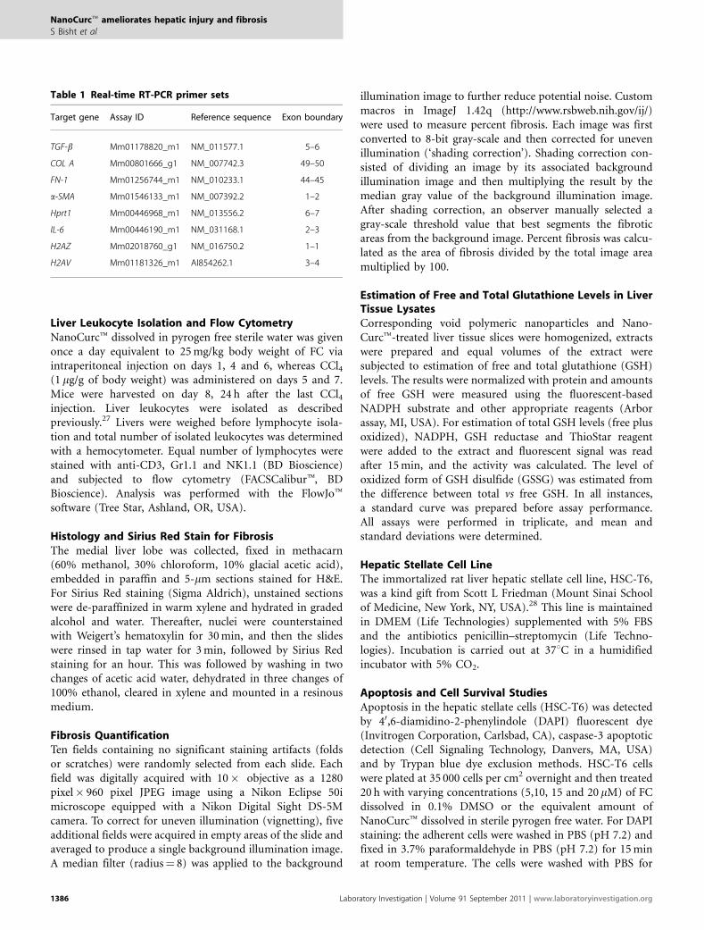
Quantitative real-time RT-PCR amplification wasperformed with TaqMan Fast Universal PCR Master Mixwith (Applied Biosystem, Carlsbad, CA, USA) withoutAmpErase UNG using 250 ng of total RNA in a 96-well plate.The real-time RT-PCR was performed with an AppliedBiosystem Step One Plus system. The assay ID, the referencesequence, and the exon boundaries used to design the probesand primers are shown in Table 1. Genes hprt and H2AX wereused as references for TNF-a and IL-6, whereas gene hprt wasused as reference for COL A, FN-1, TGF-b and PPARG.
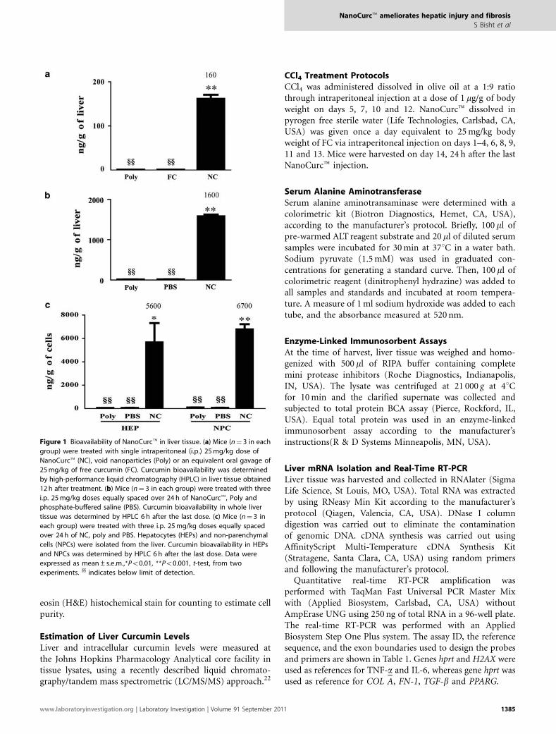
Figure 1 Bioavailability of NanoCurct in liver tissue. (a) Mice (n¼ 3 in each
group) were treated with single intraperitoneal (i.p.) 25mg/kg dose of
NanoCurct (NC), void nanoparticles (Poly) or an equivalent oral gavage of
25mg/kg of free curcumin (FC). Curcumin bioavailability was determined
by high-performance liquid chromatography (HPLC) in liver tissue obtained
12 h after treatment. (b) Mice (n¼ 3 in each group) were treated with three
i.p. 25mg/kg doses equally spaced over 24 h of NanoCurct, Poly and
phosphate-buffered saline (PBS). Curcumin bioavailability in whole liver
tissue was determined by HPLC 6 h after the last dose. (c) Mice (n¼ 3 in
each group) were treated with three i.p. 25mg/kg doses equally spaced
over 24 h of NC, poly and PBS. Hepatocytes (HEPs) and non-parenchymal
cells (NPCs) were isolated from the liver. Curcumin bioavailability in HEPs
and NPCs was determined by HPLC 6 h after the last dose. Data were
expressed as mean±s.e.m.,*Po0.01, **Po0.001, t-test, from two
experiments. yy indicates below limit of detection.
NanoCurct ameliorates hepatic injury and fibrosis
S Bisht et al
www.laboratoryinvestigation.org | Laboratory Investigation | Volume 91 September 2011 1385
Liver Leukocyte Isolation and Flow CytometryNanoCurct dissolved in pyrogen free sterile water was givenonce a day equivalent to 25mg/kg body weight of FC viaintraperitoneal injection on days 1, 4 and 6, whereas CCl4(1 mg/g of body weight) was administered on days 5 and 7.Mice were harvested on day 8, 24 h after the last CCl4injection. Liver leukocytes were isolated as describedpreviously.27 Livers were weighed before lymphocyte isola-tion and total number of isolated leukocytes was determinedwith a hemocytometer. Equal number of lymphocytes werestained with anti-CD3, Gr1.1 and NK1.1 (BD Bioscience)and subjected to flow cytometry (FACSCaliburt, BDBioscience). Analysis was performed with the FlowJotsoftware (Tree Star, Ashland, OR, USA).
Histology and Sirius Red Stain for FibrosisThe medial liver lobe was collected, fixed in methacarn(60% methanol, 30% chloroform, 10% glacial acetic acid),embedded in paraffin and 5-mm sections stained for H&E.For Sirius Red staining (Sigma Aldrich), unstained sectionswere de-paraffinized in warm xylene and hydrated in gradedalcohol and water. Thereafter, nuclei were counterstainedwith Weigert’s hematoxylin for 30min, and then the slideswere rinsed in tap water for 3min, followed by Sirius Redstaining for an hour. This was followed by washing in twochanges of acetic acid water, dehydrated in three changes of100% ethanol, cleared in xylene and mounted in a resinousmedium.
Fibrosis QuantificationTen fields containing no significant staining artifacts (foldsor scratches) were randomly selected from each slide. Eachfield was digitally acquired with 10� objective as a 1280pixel� 960 pixel JPEG image using a Nikon Eclipse 50imicroscope equipped with a Nikon Digital Sight DS-5Mcamera. To correct for uneven illumination (vignetting), fiveadditional fields were acquired in empty areas of the slide andaveraged to produce a single background illumination image.A median filter (radius¼ 8) was applied to the background
illumination image to further reduce potential noise. Custommacros in ImageJ 1.42q (http://www.rsbweb.nih.gov/ij/)were used to measure percent fibrosis. Each image was firstconverted to 8-bit gray-scale and then corrected for unevenillumination (‘shading correction’). Shading correction con-sisted of dividing an image by its associated backgroundillumination image and then multiplying the result by themedian gray value of the background illumination image.After shading correction, an observer manually selected agray-scale threshold value that best segments the fibroticareas from the background image. Percent fibrosis was calcu-lated as the area of fibrosis divided by the total image areamultiplied by 100.
Estimation of Free and Total Glutathione Levels in LiverTissue LysatesCorresponding void polymeric nanoparticles and Nano-Curct-treated liver tissue slices were homogenized, extractswere prepared and equal volumes of the extract weresubjected to estimation of free and total glutathione (GSH)levels. The results were normalized with protein and amountsof free GSH were measured using the fluorescent-basedNADPH substrate and other appropriate reagents (Arborassay, MI, USA). For estimation of total GSH levels (free plusoxidized), NADPH, GSH reductase and ThioStar reagentwere added to the extract and fluorescent signal was readafter 15min, and the activity was calculated. The level ofoxidized form of GSH disulfide (GSSG) was estimated fromthe difference between total vs free GSH. In all instances,a standard curve was prepared before assay performance.All assays were performed in triplicate, and mean andstandard deviations were determined.
Hepatic Stellate Cell LineThe immortalized rat liver hepatic stellate cell line, HSC-T6,was a kind gift from Scott L Friedman (Mount Sinai Schoolof Medicine, New York, NY, USA).28 This line is maintainedin DMEM (Life Technologies) supplemented with 5% FBSand the antibiotics penicillin–streptomycin (Life Techno-logies). Incubation is carried out at 371C in a humidifiedincubator with 5% CO2.
Apoptosis and Cell Survival StudiesApoptosis in the hepatic stellate cells (HSC-T6) was detectedby 40,6-diamidino-2-phenylindole (DAPI) fluorescent dye(Invitrogen Corporation, Carlsbad, CA), caspase-3 apoptoticdetection (Cell Signaling Technology, Danvers, MA, USA)and by Trypan blue dye exclusion methods. HSC-T6 cellswere plated at 35 000 cells per cm2 overnight and then treated20 h with varying concentrations (5,10, 15 and 20 mM) of FCdissolved in 0.1% DMSO or the equivalent amount ofNanoCurct dissolved in sterile pyrogen free water. For DAPIstaining: the adherent cells were washed in PBS (pH 7.2) andfixed in 3.7% paraformaldehyde in PBS (pH 7.2) for 15minat room temperature. The cells were washed with PBS for
Table 1 Real-time RT-PCR primer sets
Target gene Assay ID Reference sequence Exon boundary
TGF-b Mm01178820_m1 NM_011577.1 5–6
COL A Mm00801666_g1 NM_007742.3 49–50
FN-1 Mm01256744_m1 NM_010233.1 44–45
a-SMA Mm01546133_m1 NM_007392.2 1–2
Hprt1 Mm00446968_m1 NM_013556.2 6–7
IL-6 Mm00446190_m1 NM_031168.1 2–3
H2AZ Mm02018760_g1 NM_016750.2 1–1
H2AV Mm01181326_m1 AI854262.1 3–4
NanoCurct ameliorates hepatic injury and fibrosis
S Bisht et al
1386 Laboratory Investigation | Volume 91 September 2011 | www.laboratoryinvestigation.org
5min at room temperature, rinsed in methanol for 5min andafter a final PBS wash the cells were covered with DAPIstaining solution (2 mg/ml) and incubated 5min andobserved under a fluorescence microscope. A small denselystained (pyknotic) nucleus was regarded as apoptotic.Mitotic nuclear forms (metaphase, anaphase, etc) wereregarded as a single non-apoptotic cell. For Trypan blue cellviability counts: cells were plated at 35 000 cells per cm2 incomplete DMEM media overnight, treating 20 h with 20, 40and 80 mM FC dissolved in 0.1% DMSO or equivalentamount of NanoCurct dissolved in sterile pyrogen freewater. After 0.05% trypsinization, cells were collected anddiluted 50:50 into a solution of 0.4% (w/v) Trypan blue.Live cells (exclude dye) and dead cells were counted using ahemocytometer. For caspase-3 detection: cells were seeded ina 24-well plate in complete medium overnight and treated for20 h with varying concentrations (5, 10, 15 and 20 mM) ofFC dissolved in 0.1% DMSO or the equivalent amount ofNanoCurct dissolved in sterile pyrogen free water. Cells werewashed with PBS twice and fixed with 3.7% paraformal-dehyde at room temperature for 10min, washed twice withPBS and denatured in 0.1% Triton X-100 in PBS at roomtemperature for 10min. After a two PBS washes, the cellswere blocked with 5% fetal bovine albumin at room tem-perature for 30min, and then incubated with 1:100 anti-cleaved caspase-3 (Cell Signaling Technology) solution atroom temperature for 1 h. Cells were washed twice with PBSand incubated with 1:300 secondary antibody, Alexa Fluor568 goat anti-rabbit IgG labeled with bright, orange-red—fluorescent Alexa Fluor 568 dye (Invitrogen Corporation) atroom temperature for 30min. Cells were then washed twicewith PBS, counterstained with DAPI and observed undera fluorescent microscope.
RESULTSEnhanced Bioavailability of Systemic NanoCurct in LiverTissueIt has been previously shown that oral intake of up to400mg/kg of curcumin may be required for amelioration ofliver injury in CCl4-treated rodents.10,11 In terms of humanconsumption, these translate into ‘mega’ doses of severalgrams (between 8 and 12 g) of curcumin, which becomesuntenable in a clinical setting.29,30 Furthermore, evenintraperitoneal doses of FC result in only ephemeral (min)intrahepatic curcumin delivery.16–18
A recently published study using NanoCurct in cancermodels shows that a single intraperitoneal dose of thisformulation (containing 25mg/kg of curcumin equivalent)results in peak curcumin plasma levels 4 h after treatment;22
however, the tissue biodistribution, especially in the liver, wasnot studied. Using LC-MS/MS, we found significantly greaterintrahepatic curcumin concentrations (160±9 ng/g of livertissue, Po0.001) 12 h after a single 25mg/kg intraperitonealinjection of NanoCurct compared with control void nano-particles (Figure 1a). As expected, an equivalent dose of FC
delivered via oral gavage did not result in any detectableintrahepatic curcumin. As we planned to use multiple doses,we wanted to see if multi-dosing resulted in higherintrahepatic curcumin concentration than a single doseof NanoCurct. Compared with control injection and voidnanoparticles, we found significantly higher curcuminconcentration (1600±44 ng/g of liver tissue, Po0.001) in theliver 6 h after three equally spaced 25mg/kg intraperitonealdoses (Figure 1b). As the liver is a complex tissue made ofmany cell types, we next wanted to determine if NanoCurctdelivered curcumin to parenchymal (hepatocytes) and/ornon-parenchymal (all other liver cell types) cells. Micereceived three 25mg/kg NanoCurct doses 8 h apart and wereharvested 6 h after the last dose of NanoCurct. The liver wasfractionated into parenchymal cells and NPCs. The purity ofthe NPC fraction (92%) and hepatocyte fraction (96%) wasdetermined by examination of H&E-stained smear prepara-tion of each fraction (Supplementary Figure 1). We detectedsignificantly more curcumin in hepatocytes (5600±1000 ng/gof hepatocytes, Po0.01) and NPCs (6700±440 ng/g NPCs,Po0.001) compared with void polymer- and vehicle-treatedcontrols (Figure 1c). We attributed the increase in con-centration of curcumin in the hepatocytes and NPC-purifiedfractions compared with the intact liver (Figure 1c vs b) toregions, perhaps structural, non-cellular components, thatcontribute to liver weight, but do not absorb curcumin.
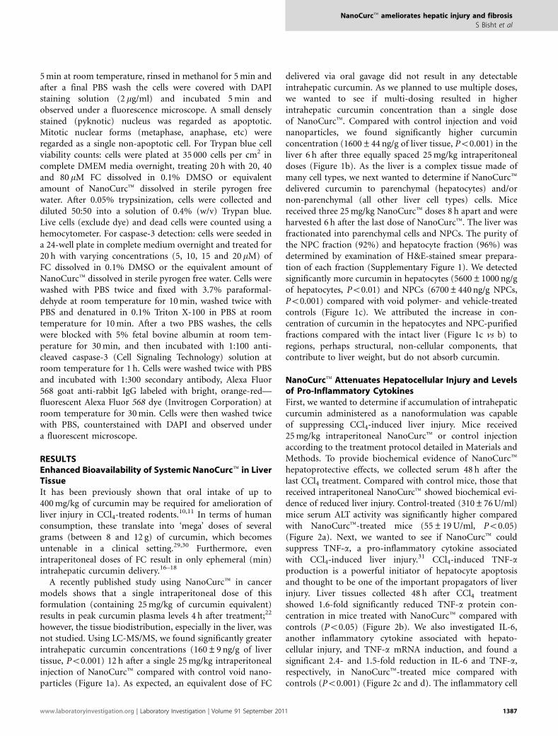
NanoCurct Attenuates Hepatocellular Injury and Levelsof Pro-Inflammatory CytokinesFirst, we wanted to determine if accumulation of intrahepaticcurcumin administered as a nanoformulation was capableof suppressing CCl4-induced liver injury. Mice received25mg/kg intraperitoneal NanoCurct or control injectionaccording to the treatment protocol detailed in Materials andMethods. To provide biochemical evidence of NanoCurcthepatoprotective effects, we collected serum 48 h after thelast CCl4 treatment. Compared with control mice, those thatreceived intraperitoneal NanoCurct showed biochemical evi-dence of reduced liver injury. Control-treated (310±76U/ml)mice serum ALT activity was significantly higher comparedwith NanoCurct-treated mice (55±19U/ml, Po0.05)(Figure 2a). Next, we wanted to see if NanoCurct couldsuppress TNF-a, a pro-inflammatory cytokine associatedwith CCl4-induced liver injury.31 CCl4-induced TNF-aproduction is a powerful initiator of hepatocyte apoptosisand thought to be one of the important propagators of liverinjury. Liver tissues collected 48 h after CCl4 treatmentshowed 1.6-fold significantly reduced TNF-a protein con-centration in mice treated with NanoCurct compared withcontrols (Po0.05) (Figure 2b). We also investigated IL-6,another inflammatory cytokine associated with hepato-cellular injury, and TNF-a mRNA induction, and found asignificant 2.4- and 1.5-fold reduction in IL-6 and TNF-a,respectively, in NanoCurct-treated mice compared withcontrols (Po0.001) (Figure 2c and d). The inflammatory cell
NanoCurct ameliorates hepatic injury and fibrosis
S Bisht et al
www.laboratoryinvestigation.org | Laboratory Investigation | Volume 91 September 2011 1387
milieu after liver injury is complex.32 The innate immunesystem is rapidly activated following injury and has beenimplicated as an important regulator of liver injury andfibrosis.33–35 We examined how NanoCurct treatment mightalter the innate inflammatory response to CCl4. Liversleukocytes were isolated 24 h after the last NanoCurcttreatment. There was a slight increase in the total number ofinflammatory cells in void nanoparticle (1.9±0.3 lympho-cytes per g liver tissue) and NanoCurct (2.2� 106±0.4lymphocytes per g liver tissue) compared with untreatedcontrol mice (1.0� 106±0.5 lymphocytes per g liver tissue);however, this was not statistically significant. Next, we ex-amined CD3þ NK1.1þ natural killer T (NKT) cells whoseabsence makes mice resistant to liver fibrosis, whereas anexcess accelerates fibrosis.36–39 Consistent to what others havereported, we found NKT cells decreased following CCl4treatment. We found 24±1% NKT cells in the liver ofuntreated wild-type mice, which fell to 12±5% in voidnanoparticle and 9±1% in NanoCurct-treated mice thatalso received CCl4
38 (Supplementary Figure 2). There was nosignificant change in the percentage of NK cells (Supple-mentary Figure 2), another innate immune cell implicated inantifibrosis.39–41 Lastly, we looked at neutrophil inflamma-tion using Gr-1high staining.42 Consistent with what othershave reported,38,43 CCl4 enhanced influx of Gr-1high cells intothe liver. Untreated mice had 5%±3 Gr-1high neutrophils in
the liver compared with 13±1% in void nanoparticle-treatedmice that also received CCl4 (Supplementary Figure 2).Interestingly, mice treated with CCl4 and NanoCurct alsohad a significant increase in Gr-1high cells compared withuntreated mice (15±8 vs 5±3%) and was not differentcompared with mice treated with CCl4 and void nano-particles (15±8 vs 13±2%). Taken together, these resultssupport the notion that NanoCurct potently suppresses bothhepatocellular injury and pro-inflammatory markers withrelatively little effect on innate inflammatory cell infiltrateafter CCl4 challenge.
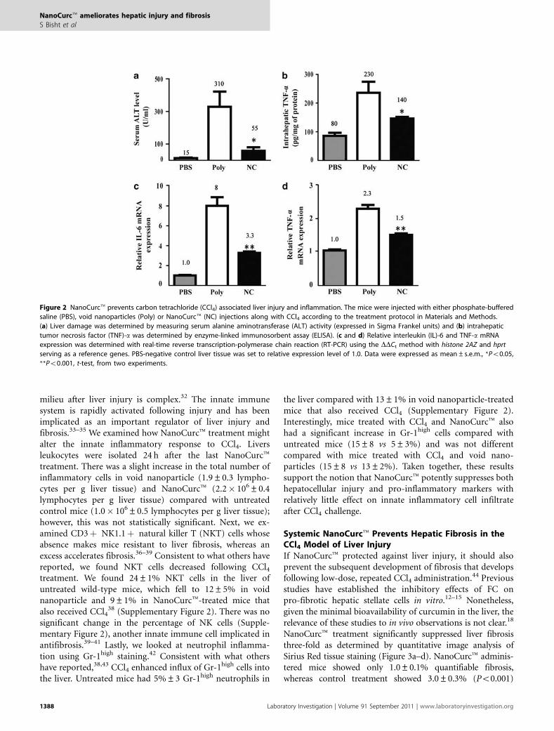
Systemic NanoCurct Prevents Hepatic Fibrosis in theCCl4 Model of Liver InjuryIf NanoCurct protected against liver injury, it should alsoprevent the subsequent development of fibrosis that developsfollowing low-dose, repeated CCl4 administration.44 Previousstudies have established the inhibitory effects of FC onpro-fibrotic hepatic stellate cells in vitro.12–15 Nonetheless,given the minimal bioavailability of curcumin in the liver, therelevance of these studies to in vivo observations is not clear.18
NanoCurct treatment significantly suppressed liver fibrosisthree-fold as determined by quantitative image analysis ofSirius Red tissue staining (Figure 3a–d). NanoCurct adminis-tered mice showed only 1.0±0.1% quantifiable fibrosis,whereas control treatment showed 3.0±0.3% (Po0.001)
Figure 2 NanoCurct prevents carbon tetrachloride (CCl4) associated liver injury and inflammation. The mice were injected with either phosphate-buffered
saline (PBS), void nanoparticles (Poly) or NanoCurct (NC) injections along with CCl4 according to the treatment protocol in Materials and Methods.
(a) Liver damage was determined by measuring serum alanine aminotransferase (ALT) activity (expressed in Sigma Frankel units) and (b) intrahepatic
tumor necrosis factor (TNF)-a was determined by enzyme-linked immunosorbent assay (ELISA). (c and d) Relative interleukin (IL)-6 and TNF-a mRNA
expression was determined with real-time reverse transcription-polymerase chain reaction (RT-PCR) using the DDCt method with histone 2AZ and hprt
serving as a reference genes. PBS-negative control liver tissue was set to relative expression level of 1.0. Data were expressed as mean±s.e.m., *Po0.05,
**Po0.001, t-test, from two experiments.
NanoCurct ameliorates hepatic injury and fibrosis
S Bisht et al
1388 Laboratory Investigation | Volume 91 September 2011 | www.laboratoryinvestigation.org
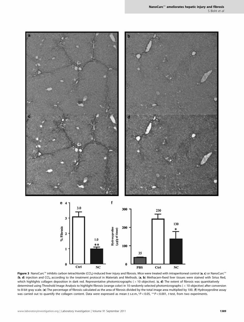
Figure 3 NanoCurct inhibits carbon tetrachloride (CCl4)-induced liver injury and fibrosis. Mice were treated with intraperitoneal control (a, c) or NanoCurct
(b, d) injection and CCl4 according to the treatment protocol in Materials and Methods. (a, b) Methacarn-fixed liver tissues were stained with Sirius Red,
which highlights collagen deposition in dark red. Representative photomicrographs (� 10 objective). (c, d) The extent of fibrosis was quantitatively
determined using Threshold Image Analysis to highlight fibrosis (orange color) in 10 randomly selected photomicrographs (� 10 objective) after conversion
to 8-bit gray scale. (e) The percentage of fibrosis calculated as the area of fibrosis divided by the total image area multiplied by 100. (f) Hydroxyproline assay
was carried out to quantify the collagen content. Data were expressed as mean±s.e.m.,*Po0.05, **Po0.001, t-test, from two experiments.
NanoCurct ameliorates hepatic injury and fibrosis
S Bisht et al
www.laboratoryinvestigation.org | Laboratory Investigation | Volume 91 September 2011 1389
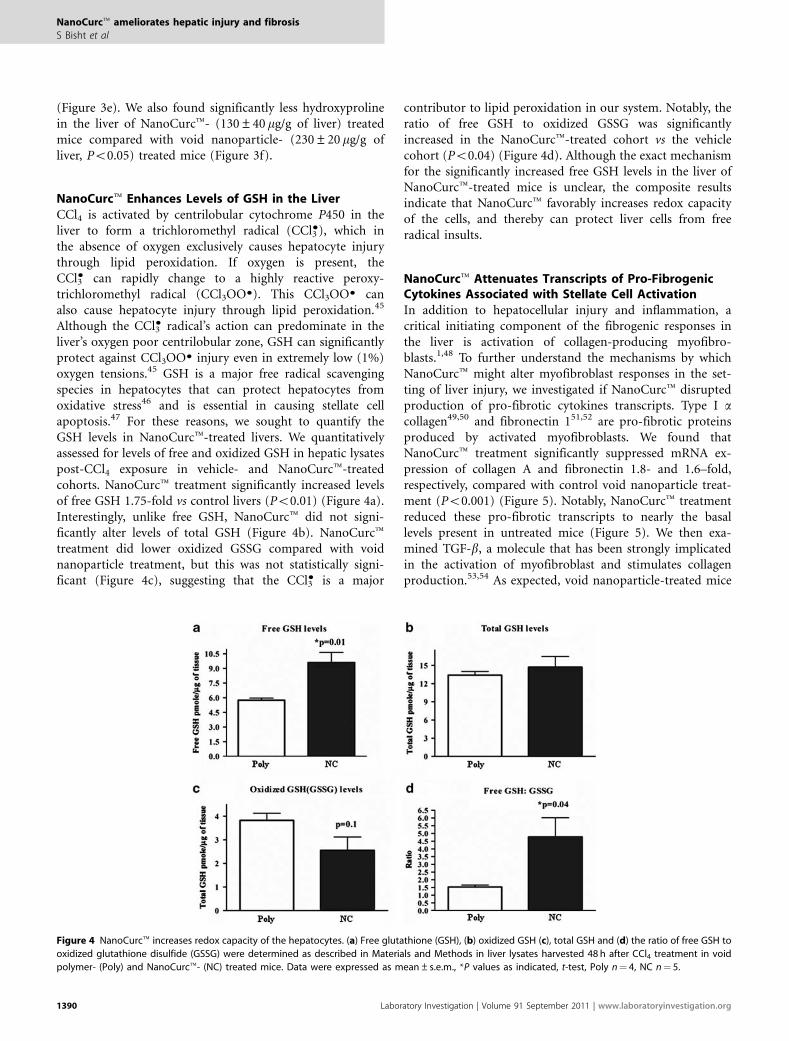
(Figure 3e). We also found significantly less hydroxyprolinein the liver of NanoCurct- (130±40 mg/g of liver) treatedmice compared with void nanoparticle- (230±20 mg/g ofliver, Po0.05) treated mice (Figure 3f).
NanoCurct Enhances Levels of GSH in the LiverCCl4 is activated by centrilobular cytochrome P450 in theliver to form a trichloromethyl radical (CCl3
K), which inthe absence of oxygen exclusively causes hepatocyte injurythrough lipid peroxidation. If oxygen is present, theCCl3
K can rapidly change to a highly reactive peroxy-trichloromethyl radical (CCl3OOK). This CCl3OOK canalso cause hepatocyte injury through lipid peroxidation.45
Although the CCl3K radical’s action can predominate in the
liver’s oxygen poor centrilobular zone, GSH can significantlyprotect against CCl3OOK injury even in extremely low (1%)oxygen tensions.45 GSH is a major free radical scavengingspecies in hepatocytes that can protect hepatocytes fromoxidative stress46 and is essential in causing stellate cellapoptosis.47 For these reasons, we sought to quantify theGSH levels in NanoCurct-treated livers. We quantitativelyassessed for levels of free and oxidized GSH in hepatic lysatespost-CCl4 exposure in vehicle- and NanoCurct-treatedcohorts. NanoCurct treatment significantly increased levelsof free GSH 1.75-fold vs control livers (Po0.01) (Figure 4a).Interestingly, unlike free GSH, NanoCurct did not signi-ficantly alter levels of total GSH (Figure 4b). NanoCurcttreatment did lower oxidized GSSG compared with voidnanoparticle treatment, but this was not statistically signi-ficant (Figure 4c), suggesting that the CCl3
K is a major
contributor to lipid peroxidation in our system. Notably, theratio of free GSH to oxidized GSSG was significantlyincreased in the NanoCurct-treated cohort vs the vehiclecohort (Po0.04) (Figure 4d). Although the exact mechanismfor the significantly increased free GSH levels in the liver ofNanoCurct-treated mice is unclear, the composite resultsindicate that NanoCurct favorably increases redox capacityof the cells, and thereby can protect liver cells from freeradical insults.
NanoCurct Attenuates Transcripts of Pro-FibrogenicCytokines Associated with Stellate Cell ActivationIn addition to hepatocellular injury and inflammation, acritical initiating component of the fibrogenic responses inthe liver is activation of collagen-producing myofibro-blasts.1,48 To further understand the mechanisms by whichNanoCurct might alter myofibroblast responses in the set-ting of liver injury, we investigated if NanoCurct disruptedproduction of pro-fibrotic cytokines transcripts. Type I acollagen49,50 and fibronectin 151,52 are pro-fibrotic proteinsproduced by activated myofibroblasts. We found thatNanoCurct treatment significantly suppressed mRNA ex-pression of collagen A and fibronectin 1.8- and 1.6–fold,respectively, compared with control void nanoparticle treat-ment (Po0.001) (Figure 5). Notably, NanoCurct treatmentreduced these pro-fibrotic transcripts to nearly the basallevels present in untreated mice (Figure 5). We then exa-mined TGF-b, a molecule that has been strongly implicatedin the activation of myofibroblast and stimulates collagenproduction.53,54 As expected, void nanoparticle-treated mice
Figure 4 NanoCurct increases redox capacity of the hepatocytes. (a) Free glutathione (GSH), (b) oxidized GSH (c), total GSH and (d) the ratio of free GSH to
oxidized glutathione disulfide (GSSG) were determined as described in Materials and Methods in liver lysates harvested 48 h after CCl4 treatment in void
polymer- (Poly) and NanoCurct- (NC) treated mice. Data were expressed as mean±s.e.m., *P values as indicated, t-test, Poly n¼ 4, NC n¼ 5.
NanoCurct ameliorates hepatic injury and fibrosis
S Bisht et al
1390 Laboratory Investigation | Volume 91 September 2011 | www.laboratoryinvestigation.org
showed a 2.6-fold increase in TGF-b expression whileNanoCurct treatment significantly and completely abrogatedexpression (Po0.001) (Figure 5). In vitro studies have shownthe importance of the nuclear transcription factor peroxi-some proliferator-activated receptor g (PPAR-g) in control-ling myofibroblast activation and its requirement inpromoting curcumin-induced apoptosis.15,55–57 There wassignificant inhibition of PPAR-g expression in void nano-particle-treated mice, whereas NanoCurct significantlyinduced a 14 -fold expression of PPAR-g compared with voidnanoparticle treatment (Po0.001) (Figure 5). Collectively,these data support NanoCurct ability to inhibit fibrosisthrough suppression of pro-fibrogenic transcripts and sug-gest that NanoCurct might induce myofibroblast apoptosisthrough activation of PPAR-g.
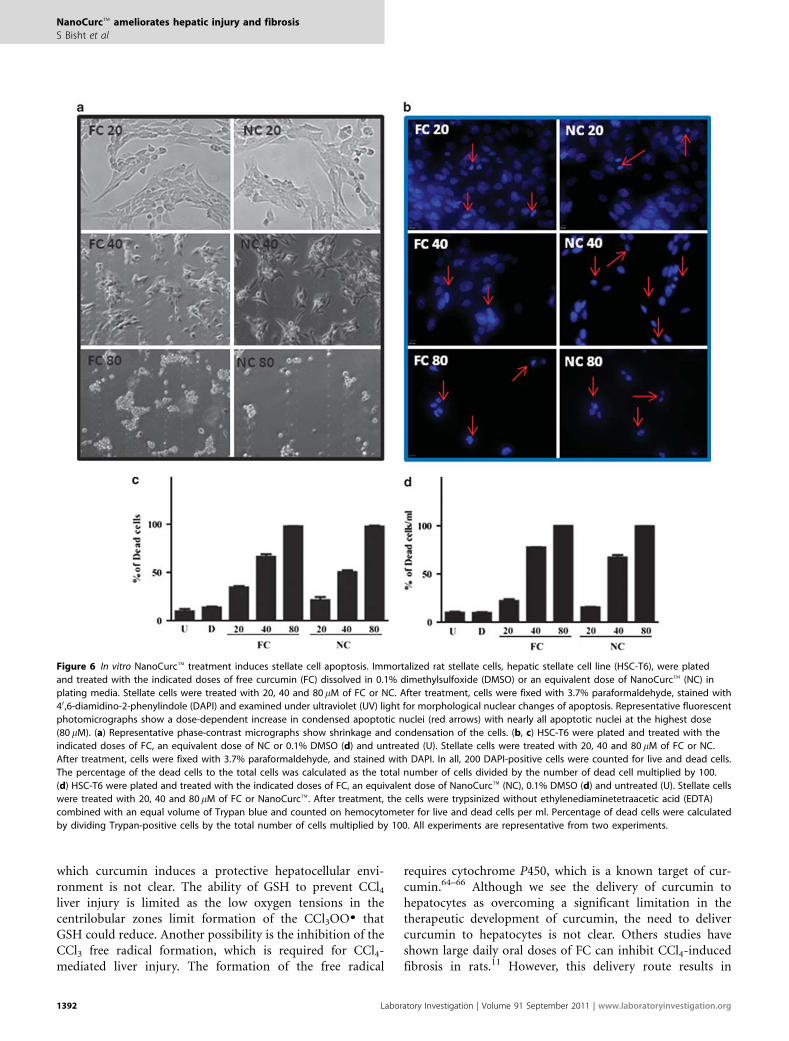
NanoCurct Treatment Induces Hepatic Stellate CellApoptosisAn important, but not the sole source of activated myo-fibroblasts is hepatic stellate cells.1 We wanted to determine ifNanoCurct was capable of inducing stellate cell apoptosis ashas been reported for FC.15,58 In vitro hepatic stellate cellswere treated with equivalent concentrations of curcumin,NanoCurct or vehicle control for 24 h . We found a signi-ficant and dose-dependent decrease in the number of viable
stellate cells in curcumin- and NanoCurct-treated culturescompared with controls (Figure 6a–c). The same trend wasfound whether we assayed with dye exclusion (Figure 6d) orthe apoptosis marker cleaved caspase-3 (SupplementaryFigure 3). As expected, the nanoencapsulation of curcumindid not alter its biological activity in vitro.26
DISCUSSIONHepatic injury and fibrosis develops in response to a widearray of etiologies such as viral infection, toxin exposure,alcohol abuse and metabolic diseases.59 Persistent injury leadsto inflammation, fibrosis and compensatory hepatocytehyperplasia usually cumulating in cirrhosis. Any treatmentthat can prevent hepatocellular injury, inflammation orfibrosis is a potential therapeutic for preventing cirrhosis inthe setting of chronic liver disease. Chronic liver disease is thethird leading cause for hospital visits in the United States,resulting in 5.9 million visits in 2004 and nearly 30 000annual deaths.60 Given this considerable disease burden,there is intense interest in developing therapies aimed atchronic liver disease.
Plant-derived polyphenols are increasingly being recog-nized for their medicinal potential.16 Curcumin is a yellowpolyphenol extracted from the rhizome of turmeric(C. longa), which has been used as a spice, coloring agent andas a therapeutic agent in traditional Indian medicine.3
Curcumin’s promising antineoplastic,4 antioxidant,5 anti-inflammatory6,7 and antidepressant8,9 activities have beenexplored in animals. In vitro and in vivo studies have exploredcurcumin’s antifibrotic potential in the liver.10–15 However,its promise is plagued by the lack of bioavailable curcumin inliver even following massive oral doses.18 Although curcuminformulated with phosphatidylcholine (Merivas) enhancesoral absorption and results in detectable intrahepatic curcu-min, it is only detectable up to 2 h after dosing.17 The lack ofwater solubility is the primary problem limiting curcumin’stherapeutic development.
To address the lack of solubility, we developed a bio-compatible nanomaterial to provide an opportunity forharnessing the full potential of a potent, yet poorly solublecompounds like curcumin.61–63 We have developed poly-meric nanoparticles comprised of NIPAAM, MMA and AAthat are capable of solubilizing a broad range of poorly water-soluble drugs. Curcumin encapsulated in these polymericnanoparticles, NanoCurct, is fully soluble in aqueous media,and shows essentially no toxicity upon daily systemicadministration through the intraperitoneal route in mice.
In this study, we found that NanoCurct delivers a sub-stantial concentration of curcumin to the liver. As early as 6 hafter multiple doses and as long as 12 h after a signal dose,a significant amount of curcumin is detected in the liver.The curcumin is detectable in hepatocytes isolated from theliver. This might explain why we see a significant reduction inCCl4-induced hepatocellular injury and production of pro-inflammatory cytokines (Figure 2). The exact mechanism by
Figure 5 NanoCurct attenuates genes associated with stellate cell
activation and fibrosis while stimulating pro-apoptotic gene expression.
Liver tissue harvested from untreated mice (PBS), void polymer (Poly) or
NanoCurct (NC) injections along with carbon tetrachloride (CCl4) according
to the treatment protocol in Materials and Methods. Gene expression
levels for transcripts encoding for Collagen A (COL A), fibronectin 1 (FN-1),
transforming growth factor-b (TGFB) and peroxisome proliferator-activated
receptor gamma (PPARG) were determined after isolation of RNA with
real-time reverse transcription-polymerase chain reaction (RT-PCR). Gene
expression was normalized with hprt and fold expression was determined
using the DDCt method in which untreated liver tissue was set to
relative expression level of 1.0. Data were expressed as mean±s.e.m.,
**Po0.001, in comparing Ctrl vs NC, t-test; control n¼ 4, NC n¼ 5,
representative of two experiments.
NanoCurct ameliorates hepatic injury and fibrosis
S Bisht et al
www.laboratoryinvestigation.org | Laboratory Investigation | Volume 91 September 2011 1391
which curcumin induces a protective hepatocellular envi-ronment is not clear. The ability of GSH to prevent CCl4liver injury is limited as the low oxygen tensions in thecentrilobular zones limit formation of the CCl3OOK thatGSH could reduce. Another possibility is the inhibition of theCCl3 free radical formation, which is required for CCl4-mediated liver injury. The formation of the free radical
requires cytochrome P450, which is a known target of cur-cumin.64–66 Although we see the delivery of curcumin tohepatocytes as overcoming a significant limitation in thetherapeutic development of curcumin, the need to delivercurcumin to hepatocytes is not clear. Others studies haveshown large daily oral doses of FC can inhibit CCl4-inducedfibrosis in rats.11 However, this delivery route results in
Figure 6 In vitro NanoCurct treatment induces stellate cell apoptosis. Immortalized rat stellate cells, hepatic stellate cell line (HSC-T6), were plated
and treated with the indicated doses of free curcumin (FC) dissolved in 0.1% dimethylsulfoxide (DMSO) or an equivalent dose of NanoCurct (NC) in
plating media. Stellate cells were treated with 20, 40 and 80 mM of FC or NC. After treatment, cells were fixed with 3.7% paraformaldehyde, stained with
40 ,6-diamidino-2-phenylindole (DAPI) and examined under ultraviolet (UV) light for morphological nuclear changes of apoptosis. Representative fluorescent
photomicrographs show a dose-dependent increase in condensed apoptotic nuclei (red arrows) with nearly all apoptotic nuclei at the highest dose
(80 mM). (a) Representative phase-contrast micrographs show shrinkage and condensation of the cells. (b, c) HSC-T6 were plated and treated with the
indicated doses of FC, an equivalent dose of NC or 0.1% DMSO (d) and untreated (U). Stellate cells were treated with 20, 40 and 80 mM of FC or NC.
After treatment, cells were fixed with 3.7% paraformaldehyde, and stained with DAPI. In all, 200 DAPI-positive cells were counted for live and dead cells.
The percentage of the dead cells to the total cells was calculated as the total number of cells divided by the number of dead cell multiplied by 100.
(d) HSC-T6 were plated and treated with the indicated doses of FC, an equivalent dose of NanoCurct (NC), 0.1% DMSO (d) and untreated (U). Stellate cells
were treated with 20, 40 and 80mM of FC or NanoCurct. After treatment, the cells were trypsinized without ethylenediaminetetraacetic acid (EDTA)
combined with an equal volume of Trypan blue and counted on hemocytometer for live and dead cells per ml. Percentage of dead cells were calculated
by dividing Trypan-positive cells by the total number of cells multiplied by 100. All experiments are representative from two experiments.
NanoCurct ameliorates hepatic injury and fibrosis
S Bisht et al
1392 Laboratory Investigation | Volume 91 September 2011 | www.laboratoryinvestigation.org
detectable curcumin in the gastrointestinal tract, but not inthe liver.10 This suggests that curcumin might work throughmultiple mechanisms.
We also found that NanoCurct treatment results in asignificant amount of curcumin in the NPC compartment.Although the cellular make-up of this compartment iscomplex, it does contain pro-fibrotic stellate cells and myo-fibroblasts. Lipid-laden cells, likely stellate cells, were evidentin the NPC fraction (Supplementary Figure 1, green arrows).This is important, as there is considerable in vitro evidencethat curcumin directly induces stellate cell apoptosis andblocks stellate cell activation.10,67 These previous studiesbolster our conclusions as we know that the NanoCurctformulation does not alter its biological activity. NanoCurctis equally effective as the unencapsulated form at inducingstellate cell apoptosis (Figure 6). However, this is not entirelysurprising as NanoCurct bio-effectiveness is comparable tothe free parent compound in a cancer system.26 We alsoprovide in vivo evidence of NanoCurct effectivenessin preventing stellate cell activation as it induces PPAR-gtranscription, an essential mediator of curcumin’s effects oneliminating stellate cells.15
As our data show that NanoCurct protects the liver frominjury, it could be argued that NanoCurct is more hepato-protective than antifibrotic. However, it is likely that Nano-Curct is working at multiple steps in the development offibrosis. First, as already mentioned, NanoCurct preventshepatocellular injury. However, this is not a completesuppression of injury. There is slight but measurable hepa-tocellular injury in NanoCurct-treated mice as evidence bythe increase in serum ALT and intrahepatic TNF-a comparedwith untreated mice (Figure 2). The same is true for theproduction of the potent pro-fibrotic cytokine TGF-b(Figure 5). We also present evidence of effects of curcuminson pro-fibrotic stellate cells through the induction of PPAR-g(Figure 5). Collectively, our findings suggest that NanoCurctworks at multiple steps in the injury and fibrosis pathway.This agent could be particularly useful in preventing theprogression of chronic liver disease in patients with activeon-going liver injury such as in viral or autoimmune hepa-titis as it targets both liver injury and fibrosis mechanisms.
Hepatic inflammation is central to many liver diseases.32
Interestingly, our data show that NanoCurct has little effecton NKT cells and neutrophils in the liver. There is a com-parable decrease in NKT and increase in Gr-1high cells in voidnanoparticle and NanoCurct-treated mice (SupplementaryFigure 2). This is consistent with what has been reported inwild-type CCl4-treated mice.38 This could indicate that a lowlevel of injury is sufficient to affect the immune system orthat another important component of the innate immunesystem, particularly Kupffer cells,68 might be targeted byNanoCurct. More immediate 24 h studies and analysisof chemokines might be more revealing in dissecting Nano-Curct’s potential role in altering the livers inflammatorymilieu.
The results of this study show NanoCurct feasibility intreating chronic liver diseases. Being fully water soluble,NanoCurct overcomes the most important road block thatprevented curcumin’s clinical development, further showingthat NanoCurct prevents liver injury and fibrosis is a signi-ficant developmental step in bringing this natural compoundto clinical trials.
Supplementary Information accompanies the paper on the Laboratory
Investigation website (http://www.laboratoryinvestigation.org)
ACKNOWLEDGEMENTS
This work was financially supported by R01DK080736 (RAA); R01DK081417
(RAA); R01CA113669 (AM); R01CA13767 (AM); P01CA134292 (AM);
P30CA069773 (MAR); S10 RR026824 (MAR); Flight Attendants Medical
Research Institute (FAMRI) (AM and MAR); Johns Hopkins CTSA Institute for
Clinical and Translational Research (UL1RR025005) (MAR); The Sol Goldman
Pancreatic Cancer Research Center (AM); Michael Rolfe Foundation for
Pancreatic Cancer Research (AM and RAA); and SignPath Pharmaceuticals
(AM). This publication was made possible by Grant Number UL1RR025005
from the National Center for Research Resources (NCRR), a component
of the National Institutes of Health (NIH) and NIH Roadmap for Medical
Research. Its contents are solely the responsibility of the authors and do
not necessarily represent the official view of NCRR or NIH.
DISCLOSURE/CONFLICT OF INTEREST
NanoCurc is a registered trademark of SignPath Pharmaceuticals,
Quakertown, PA, USA. AM is a member of the scientific advisory board
of SignPath Pharma, and any conflicts of interest under this arrangement
are handled in accordance with the Johns Hopkins University Office of
Policy Coordination guidelines. SignPath Pharma has provided partial
support for these studies through offsetting the costs of polymer synthesis.
SB, GF and AM have filed a patent application (US 2008/0107749) that is
relevant to the formulation described in this article. A report of invention
to this effect has been filed with Johns Hopkins Technology Transfer and
licensed by SignPath Pharma.
1. Brenner DA. Molecular pathogenesis of liver fibrosis. Trans Am ClinClimatol Assoc 2009;120:361–368.
2. Hellerbrand C, Stefanovic B, Giordano F, et al. The role of TGFbeta1 ininitiating hepatic stellate cell activation in vivo. J Hepatol 1999;30:77–87.
3. Sinha R, Anderson DE, McDonald SS, et al. Cancer risk and diet in India.J Postgrad Med 2003;49:222–228.
4. Huang MT, Smart RC, Wong CQ, et al. Inhibitory effect of curcumin,chlorogenic acid, caffeic acid, and ferulic acid on tumor promotion inmouse skin by 12-O-tetradecanoylphorbol-13-acetate. Cancer Res1988;48:5941–5946.
5. Sharma OP. Antioxidant activity of curcumin and related compounds.Biochem Pharmacol 1976;25:1811–1812.
6. Rao TS, Basu N, Siddiqui HH. Anti-inflammatory activity of curcuminanalogues. Indian J Med Res 1982;75:574–578.
7. Brouet I, Ohshima H. Curcumin, an anti-tumour promoter and anti-inflammatory agent, inhibits induction of nitric oxide synthasein activated macrophages. Biochem Biophys Res Commun 1995;206:533–540.
8. Kulkarni SK, Bhutani MK, Bishnoi M. Antidepressant activity ofcurcumin: involvement of serotonin and dopamine system.Psychopharmacology (Berl) 2008;201:435–442.
9. Xu Y, Ku BS, Yao HY, et al. The effects of curcumin on depressive-likebehaviors in mice. Eur J Pharmacol 2005;518:40–46.
10. Fu Y, Zheng S, Lin J, et al. Curcumin protects the rat liver from CCl4-caused injury and fibrogenesis by attenuating oxidative stress andsuppressing inflammation. Mol Pharmacol 2008;73:399–409.
NanoCurct ameliorates hepatic injury and fibrosis
S Bisht et al
www.laboratoryinvestigation.org | Laboratory Investigation | Volume 91 September 2011 1393
11. Park EJ, Jeon CH, Ko G, et al. Protective effect of curcumin in rat liverinjury induced by carbon tetrachloride. J Pharm Pharmacol 2000;52:437–440.
12. Donatus IA, Sardjoko, Vermeulen NP. Cytotoxic and cytoprotectiveactivities of curcumin. Effects on paracetamol-induced cytotoxicity,lipid peroxidation and glutathione depletion in rat hepatocytes.Biochem Pharmacol 1990;39:1869–1875.
13. Kiso Y, Suzuki Y, Watanabe N, et al. Antihepatotoxic principles ofCurcuma longa rhizomes. Planta Med 1983;49:185–187.
14. Xu J, Fu Y, Chen A. Activation of peroxisome proliferator-activatedreceptor-gamma contributes to the inhibitory effects of curcumin onrat hepatic stellate cell growth. Am J Physiol Gastrointest Liver Physiol2003;285:G20–G30.
15. Zheng S, Chen A. Activation of PPARgamma is required for curcuminto induce apoptosis and to inhibit the expression of extracellularmatrix genes in hepatic stellate cells in vitro. Biochem J 2004;384(Part 1):149–157.
16. Kidd PM. Bioavailability and activity of phytosome complexes frombotanical polyphenols: the silymarin, curcumin, green tea, and grapeseed extracts. Altern Med Rev 2009;14:226–246.
17. Marczylo TH, Verschoyle RD, Cooke DN, et al. Comparison of systemicavailability of curcumin with that of curcumin formulated withphosphatidylcholine. Cancer Chemother Pharmacol 2007;60:171–177.
18. Pan MH, Huang TM, Lin JK. Biotransformation of curcumin throughreduction and glucuronidation in mice. Drug Metab Dispos 1999;27:486–494.
19. Sharma RA, Steward WP, Gescher AJ. Pharmacokinetics and pharmaco-dynamics of curcumin. Adv Exp Med Biol 2007;595:453–470.
20. Anand P, Kunnumakkara AB, Newman RA, et al. Bioavailability ofcurcumin: problems and promises. Mol Pharm 2007;4:807–818.
21. Garcea G, Berry DP, Jones DJ, et al. Consumption of the putativechemopreventive agent curcumin by cancer patients: assessment ofcurcumin levels in the colorectum and their pharmacodynamicconsequences. Cancer Epidemiol Biomarkers Prev 2005;14:120–125.
22. Bisht S, Mizuma M, Feldmann G, et al. Systemic administration ofpolymeric nanoparticle-encapsulated curcumin (NanoCurc) blockstumor growth and metastases in preclinical models of pancreaticcancer. Mol Cancer Ther 2010;9:2255–2264.
23. Garcea G, Jones DJ, Singh R, et al. Detection of curcumin and itsmetabolites in hepatic tissue and portal blood of patients followingoral administration. Br J Cancer 2004;90:1011–1015.
24. Dhillon N, Aggarwal BB, Newman RA, et al. Phase II trial of curcumin inpatients with advanced pancreatic cancer. Clin Cancer Res 2008;14:4491–4499.
25. Bisht S, Feldmann G, Koorstra JB, et al. In vivo characterization ofa polymeric nanoparticle platform with potential oral drug deliverycapabilities. Mol Cancer Ther 2008;7:3878–3888.
26. Bisht S, Feldmann G, Soni S, et al. Polymeric nanoparticle-encapsulatedcurcumin (‘nanocurcumin’): a novel strategy for human cancer therapy.J Nanobiotechnology 2007;5:3.
27. Anders RA, Subudhi SK, Wang J, et al. Contribution of the lymphotoxinbeta receptor to liver regeneration. J Immunol 2005;175:1295–1300.
28. Vogel S, Piantedosi R, Frank J, et al. An immortalized rat liver stellatecell line (HSC-T6): a new cell model for the study of retinoidmetabolism in vitro. J Lipid Res 2000;41:882–893.
29. Aggarwal BB, Sundaram C, Malani N, et al. Curcumin: the Indian solidgold. Adv Exp Med Biol 2007;595:1–75.
30. Bisht S, Maitra A. Systemic delivery of curcumin: 21st century solutionsfor an ancient conundrum. Curr Drug Discov Technol 2009;6:192–199.
31. Sudo K, Yamada Y, Moriwaki H, et al. Lack of tumor necrosis factorreceptor type 1 inhibits liver fibrosis induced by carbon tetrachloridein mice. Cytokine 2005;29:236–244.
32. Adams DH, Ju C, Ramaiah SK, et al. Mechanisms of immune-mediatedliver injury. Toxicol Sci 2010;115:307–321.
33. Li Z, Diehl AM. Innate immunity in the liver. Curr Opin Gastroenterol2003;19:565–571.
34. Szabo G, Mandrekar P, Dolganiuc A. Innate immune response andhepatic inflammation. Semin Liver Dis 2007;27:339–350.
35. Marra F, Aleffi S, Galastri S, et al. Mononuclear cells in liver fibrosis.Semin Immunopathol 2009;31:345–358.
36. Ginsburg I, Koren E, Horani A, et al. Amelioration of hepatic fibrosis viaPadma Hepaten is associated with altered natural killer T lymphocytes.Clin Exp Immunol 2009;157:155–164.
37. Ishikawa S, Ikejima K, Yamagata H, et al. CD1d-restricted natural killerT cells contribute to hepatic inflammation and fibrogenesis in mice.J Hepatol 2010.
38. Park O, Jeong WI, Wang L, et al. Diverse roles of invariant natural killerT cells in liver injury and fibrosis induced by carbon tetrachloride.Hepatology 2009;49:1683–1694.
39. Safadi R, Zigmond E, Pappo O, et al. Amelioration of hepatic fibrosis viabeta-glucosylceramide-mediated immune modulation is associatedwith altered CD8 and NKT lymphocyte distribution. Int Immunol2007;19:1021–1029.
40. Melhem A, Muhanna N, Bishara A, et al. Anti-fibrotic activity of NK cellsin experimental liver injury through killing of activated HSC. J Hepatol2006;45:60–71.
41. Radaeva S, Sun R, Jaruga B, et al. Natural killer cells ameliorate liverfibrosis by killing activated stellate cells in NKG2D-dependent andtumor necrosis factor-related apoptosis-inducing ligand-dependentmanners. Gastroenterology 2006;130:435–452.
42. Daley JM, Thomay AA, Connolly MD, et al. Use of Ly6G-specificmonoclonal antibody to deplete neutrophils in mice. J Leukocyte Biol2008;83:64–70.
43. Karlmark KR, Weiskirchen R, Zimmermann HW, et al. Hepaticrecruitment of the inflammatory Gr1+ monocyte subset upon liverinjury promotes hepatic fibrosis. Hepatology 2009;50:261–274.
44. Weiler-Normann C, Herkel J, Lohse AW. Mouse models of liver fibrosis.Z Gastroenterol 2007;45:43–50.
45. Burk RF, Lane JM, Patel K. Relationship of oxygen and glutathione inprotection against carbon tetrachloride-induced hepatic microsomallipid peroxidation and covalent binding in the rat. Rationale for theuse of hyperbaric oxygen to treat carbon tetrachloride ingestion.J Clin Invest 1984;74:1996–2001.
46. Burk RF, Patel K, Lane JM. Reduced glutathione protection against ratliver microsomal injury by carbon tetrachloride. Dependence on O2.Biochem J 1983;215:441–445.
47. Zheng S, Yumei F, Chen A. De novo synthesis of glutathione is aprerequisite for curcumin to inhibit hepatic stellate cell (HSC)activation. Free Radic Biol Med 2007;43:444–453.
48. Friedman SL. Molecular mechanisms of hepatic fibrosis and principlesof therapy. J Gastroenterol 1997;32:424–430.
49. Friedman SL, Roll FJ, Boyles J, et al. Hepatic lipocytes: the principalcollagen-producing cells of normal rat liver. Proc Natl Acad Sci USA1985;82:8681–8685.
50. Kawase T, Shiratori Y, Sugimoto T. Collagen production by rat liverfat-storing cells in primary culture. Exp Cell Biol 1986;54:183–192.
51. Ramadori G, Knittel T, Odenthal M, et al. Synthesis of cellularfibronectin by rat liver fat-storing (Ito) cells: regulation by cytokines.Gastroenterology 1992;103:1313–1321.
52. Neubauer K, Knittel T, Armbrust T, et al. Accumulation and cellularlocalization of fibrinogen/fibrin during short-term and long-term ratliver injury. Gastroenterology 1995;108:1124–1135.
53. Garcia-Trevijano ER, Iraburu MJ, Fontana L, et al. Transforming growthfactor beta1 induces the expression of alpha1(I) procollagen mRNA bya hydrogen peroxide-C/EBPbeta-dependent mechanism in rat hepaticstellate cells. Hepatology 1999;29:960–970.
54. Gressner AM. Transdifferentiation of hepatic stellate cells (Ito cells) tomyofibroblasts: a key event in hepatic fibrogenesis. Kidney Int Suppl1996;54:S39–S45.
55. Galli A, Crabb D, Price D, et al. Peroxisome proliferator-activatedreceptor gamma transcriptional regulation is involved in platelet-derived growth factor-induced proliferation of human hepatic stellatecells. Hepatology 2000;31:101–108.
56. Marra F, Efsen E, Romanelli RG, et al. Ligands of peroxisomeproliferator-activated receptor gamma modulate profibrogenic andproinflammatory actions in hepatic stellate cells. Gastroenterology2000;119:466–478.
57. Miyahara T, Schrum L, Rippe R, et al. Peroxisome proliferator-activatedreceptors and hepatic stellate cell activation. J Biol Chem 2000;275:35715–35722.
58. Lin J, Zheng S, Chen A. Curcumin attenuates the effects of insulinon stimulating hepatic stellate cell activation by interrupting insulinsignaling and attenuating oxidative stress. Lab Invest 2009;89:1397–1409.
59. Jiao J, Friedman SL, Aloman C. Hepatic fibrosis. Curr Opin Gastro-enterol 2009;25:223–229.
NanoCurct ameliorates hepatic injury and fibrosis
S Bisht et al
1394 Laboratory Investigation | Volume 91 September 2011 | www.laboratoryinvestigation.org
60. Everhart J. The Burden of Digestive Diseases in the United States,Vol. No. 09-6443 US Government: Washington, DC, 2008.
61. van Vlerken LE, Amiji MM. Multi-functional polymeric nanoparticles fortumour-targeted drug delivery. Expert Opin Drug Deliv 2006;3:205–216.
62. Vicent MJ, Duncan R. Polymer conjugates: nanosized medicines fortreating cancer. Trends Biotechnol 2006;24:39–47.
63. Vasir JK, Labhasetwar V. Targeted drug delivery in cancer therapy.Technol Cancer Res Treat 2005;4:363–374.
64. Ganta S, Devalapally H, Amiji M. Curcumin enhances oral bioavailabilityand anti-tumor therapeutic efficacy of paclitaxel upon administrationin nanoemulsion formulation. J Pharm Sci 2010;99:4630–4641.
65. Appiah-Opong R, de Esch I, Commandeur JN, et al. Structure-activity relationships for the inhibition of recombinant human
cytochromes P450 by curcumin analogues. Eur J Med Chem 2008;43:1621–1631.
66. Volak LP, Ghirmai S, Cashman JR, et al. Curcuminoids inhibitmultiple human cytochromes P450, UDP-glucuronosyltransferase,and sulfotransferase enzymes, whereas piperine is a relativelyselective CYP3A4 inhibitor. Drug Metab Dispos 2008;36:1594–1605.
67. Lin J, Chen A. Activation of peroxisome proliferator-activated receptor-gamma by curcumin blocks the signaling pathways for PDGF andEGF in hepatic stellate cells. Lab Invest 2008;88:529–540.
68. Jaeschke H. Reactive oxygen and mechanisms of inflammatoryliver injury: Present concepts. J Gastroenterol Hepatol 2011;26(Suppl 1):173–179.
NanoCurct ameliorates hepatic injury and fibrosis
S Bisht et al
www.laboratoryinvestigation.org | Laboratory Investigation | Volume 91 September 2011 1395