Effectiveness of Storage Conditions to Control the Vinegar ...
Upload
khangminh22Category
view
1download
0
RESEARCH ARTICLE Open Access
Coconut water vinegar amelioratesrecovery of acetaminophen induced liverdamage in miceNurul Elyani Mohamad1, Swee Keong Yeap2, Boon-Kee Beh3,4, Huynh Ky5, Kian Lam Lim6, Wan Yong Ho7,Shaiful Adzni Sharifuddin4, Kamariah Long4* and Noorjahan Banu Alitheen1,3*
Abstract
Background: Coconut water has been commonly consumed as a beverage for its multiple health benefits whilevinegar has been used as common seasoning and a traditional Chinese medicine. The present study investigatesthe potential of coconut water vinegar in promoting recovery on acetaminophen induced liver damage.
Methods: Mice were injected with 250 mg/kg body weight acetaminophen for 7 days and were treated withdistilled water (untreated), Silybin (positive control) and coconut water vinegar (0.08 mL/kg and 2 mL/kg bodyweight). Level of oxidation stress and inflammation among treated and untreated mice were compared.
Results: Untreated mice oral administrated with acetaminophen were observed with elevation of serum liverprofiles, liver histological changes, high level of cytochrome P450 2E1, reduced level of liver antioxidant andincreased level of inflammatory related markers indicating liver damage. On the other hand, acetaminophenchallenged mice treated with 14 days of coconut water vinegar were recorded with reduction of serum liverprofiles, improved liver histology, restored liver antioxidant, reduction of liver inflammation and decreased levelof liver cytochrome P450 2E1 in dosage dependent level.
Conclusion: Coconut water vinegar has helped to attenuate acetaminophen-induced liver damage by restoringantioxidant activity and suppression of inflammation.
Keywords: Cocos nucifera, Paracetamol, Phenolic, Acetification, Inflammation
BackgroundAcetaminophen or more commonly known as paraceta-mol is among the most commonly used mild analgesicdrug worldwide [1]. Although acetaminophen is generallyconsidered safe, unintentional or deliberate overdoses haveresulted in acute liver failure especially in United States[2]. Acetaminophen is metabolized by cytochrome P4502E1 (CYP2E1) in the liver into the reactive metabolite N-acetyl-p-benzoquinone imine (NAPQI). When consumedin safe dosage, NAPQI can be easily detoxified by glutathi-one (GSH) into acetaminophen-glutathione conjugate.
When acetaminophen was consumed in overdose, metab-olism by CYP2E1enzyme produced excessive NAPQI thatdepletes the liver GSH and caused mitochondrial oxidativestress. Subsequently, mitochondrial oxidative stress pro-motes hepatocyte cell death and release of hepatocytecontents such as ALT. Massive release of liver enzymessuch as ALT also results in formation of pro-inflammatorymediators and chronic inflammation [3].Bioactive food ingredients and herbal medicine have
been widely used to alleviate chronic liver disease. Thehepatoprotective effect from these food ingredients andherbal medicine was contributed by the active metabo-lites such as curcumin from turmeric and silymarin frommilk thistle seeds [4]. Although these food or herbal in-gredients have been widely consumed and generally be-lieved as safe, their efficacy and safety will still needfurther validation [4]. Coconut (Cocos nucifera L.) water
* Correspondence: [email protected]; [email protected] Research Centre, Malaysian Agricultural Research andDevelopment Institute (MARDI), 43400 Serdang, Selangor, Malaysia1Department of Cell and Molecular Biology, Faculty of Biotechnology andBiomolecular Science, Universiti Putra Malaysia, 43400 Serdang, Selangor,MalaysiaFull list of author information is available at the end of the article
© The Author(s). 2018 Open Access This article is distributed under the terms of the Creative Commons Attribution 4.0International License (http://creativecommons.org/licenses/by/4.0/), which permits unrestricted use, distribution, andreproduction in any medium, provided you give appropriate credit to the original author(s) and the source, provide a link tothe Creative Commons license, and indicate if changes were made. The Creative Commons Public Domain Dedication waiver(http://creativecommons.org/publicdomain/zero/1.0/) applies to the data made available in this article, unless otherwise stated.
Mohamad et al. BMC Complementary and Alternative Medicine (2018) 18:195 https://doi.org/10.1186/s12906-018-2199-4
is a delicious and refreshing drink in coconut producingcountries. In addition, coconut water is also consumedfor various health benefits. Previous study has reportedthat coconut water is rich in antioxidant and lack ofanti-nutritional factors. Antioxidants in the coconutwater have contributed to prevent lipid peroxidation inthe animal that fed with fish oil diet [5]. In addition, nor-mal animals fed with coconut water were recorded withreduction of liver enzymes and thus proposed as poten-tial hepatoprotective agent [6]. Moreover, coconut waterhas also been reported with hepatoprotection activity oncarbon tetrachloride (CCl4) induced liver damaged [7–9]and alloxan induced diabetic [10] rat models by restor-ing the liver antioxidant level. However, in the coconutindustry, coconut water particularly from the maturecoconut was commonly handled as waste by-productdue to the cost and processing flow [11]. As coconutwater contained substantial amount of sugar [12], thus itis suitable as starting material to be converted as fer-mented end product such as vinegar.Vinegar is widely used as food seasoning and traditional
Chinese medicine [13]. It has been reported with variousbioactivities including anti-diabetic, anti-hypertension,anti-microbe, liver protection and anti-tumor effects [14].However, vinegar produced using different source ofcarbohydrate and strains of microbes may possessdifferent level of bioactivities particularly on the antioxi-dant activity [15]. Previously, nipa water vinegar and ro-selle vinegar were reported with higher antioxidant levelthan the unfermented fruit [16, 17]. As coconut water wasreported to exhibit hepatoprotective effect on CCl4 in-duced liver inflammation, the fermentation of this fruitwater may enhance the effect. Our recent finding on hepa-toprotective effect of nipa vinegar also has demonstrated asignificant restoration of liver inflammation in micetreated with nipa vinegar sample [16]. Nonetheless, acetifi-cation of some fruits was reported with reduction of anti-oxidant activity due to decrease of total phenolics [18].Thus, although coconut water has been reported as poten-tial hepatoprotective agent [7–9] and coconut watervinegar has been commonly consumed to treat variousdisease including liver disorders and inflammation incoconut producing countries, the hepato-recovery effectof coconut water vinegar was still unclear. Thus, this studywas performed to evaluate the effect of coconut water vin-egar in promoting recovery of acetaminophen inducedliver damage.
MethodsOrganic acid and antioxidant level of coconut watervinegarCoconut water vinegar (batch no:2, 9th May 2014) wasobtained from Malaysian Agricultural Research andDevelopment Institute (MARDI) (Selangor, Malaysia) in
year 2013. Details of preparation method were describedin Beh et al. [16]. The coconut water vinegar was stan-dardized to 5% acetic acid and confirmed by reversedphase chromatography with eternal calibration graph(Result not shown). Organic acids in the sample wereseparated on an Extrasil ODS column (250 mm × 4.6 mm, 5 μm) and the detector was set at λ = 210 nm andλ = 245 nm. Determination of acetic acid was carried outat isocratic conditions at 45 °C, using a mobile phase of50 mM phosphate solution (6.8 g potassium dihydrogenphosphate in 900 ml water, pH 2.8). The flow rate of themobile phase was set at 0.7 ml/min. Total phenolic con-tent and total antioxidant capacity were profiled usingFolin-Ciocalteu and FRAP assays [14].
AnimalsThe study was performed according to internationalrules and approved by Universiti Putra Malaysia AnimalCare and Use Committee (IACUC) (UPM/FPV/PS/3.2.1.551/AUP-R168). In brief, a total of 35 BALB/c mice(male, 5–6 weeks old) were purchased from Animal Houseof the Faculty of Veterinary Sciences, Universiti PutraMalaysia and were placed in plastic cages at 22 ± 1 °C with12 h of dark/light cycle and relative humidity approxi-mately 60%.. The mice were maintained on a basal diet(22% crude protein, 5% crude fiber, 3% fat, 13% moisture,8% ash, 0.85–1.2% calcium, 0.6–1% phosphorus and 49%nitrogen free extract) (Mouse pellet 702-P from Gold CoinCo, Limited, Malaysia) and were given distilled water adlibitum. The mice were divided into 5 groups and all micewere pre-treated with acetaminophen (250 mg/kg BW) for7 days via stomach gavage to induce liver inflammation ex-cept for normal control group (N). The post-treatmentwith coconut vinegar begin after 7 days of acetaminopheninduction where distilled water was given to the untreatedgroup (UT), 50 mg/kg BW silybin was given to positivecontrol group (S), 0.08 mL/kg BW coconut vinegar wasgiven to low concentration treatment group (CL) and2 mL/kg BW was given to high concentration treatmentgroup (CH). All samples were prepared freshly prior tousage. At the end of the treatment, the mice were eutha-nized under ketamine-xylazine anesthesia (100 mg keta-mine and 10 mg xylazine per kg body weight). Bloodsamples were collected from their hearts by cardiac punc-ture and liver was harvested for further analysis.
Serum biochemical analysisSerum samples were analyzed for liver marker (ALT, ASTand ALP) and lipid profile (cholesterol, triglyceride, LDLand HDL) using ELISA assay kits (Roche, Germany).
Liver tissue histological analysisHistology of liver tissues was performed as reported pre-viously [14]. The liver was rinsed with PBS and fixed in
Mohamad et al. BMC Complementary and Alternative Medicine (2018) 18:195 Page 2 of 9
buffered formalin for 24 h. Then, the liver was embed-ded in paraffin, sectioned, deparaffinized and rehydratedusing the standard techniques before further stainedwith hematoxylin and eosin. The morphology of the liverwas then observed using bright field optic under a NikonEclipse 90imicroscope (New York, USA) at 40 timesmagnification.
Liver antioxidant levelExcised liver was weighted and meshed in phosphatebuffer saline (PBS) at a ratio of 1 g of liver to 10 mL ofPBS. The supernatant was centrifuged at 10000 rpm for10 min and the upper clear part of the supernatant wascollected and kept in -20 °C prior to the antioxidant ana-lyses. The antioxidant activity in this study was evaluatedthrough superoxide dismutase (SOD) assay, lipid perox-idation (MDA) and glutathione reductase activity(GSH). MDA and SOD was done according to the pre-vious study [14] while GSH was done using Glutathioneassay kit according to the manufacturer’s protocol(Sigma, USA).
Liver cytochrome P450 2E1 levelCytochrome P450 (Abcam, USA) protein expressionlevel was determined using Western blot technique asreported previously [14]. In brief, fresh liver tissue wasweighed and meshed in liquid nitrogen before lysed inRadio Immuno Precipitation Assay (RIPA) buffer(150 mM sodium chloride, 1.0% NP-40 or Triton X-100 0.5% sodium deoxycholate, 0.1% SDS (sodium do-decyl sulphate) and 50 mM Tris, pH 8.0) added withprotease inhibitor cocktail (Pierce, Thermo FisherScientific, USA). Protein was measured using thestandard Bradford protein assay with Bradford reagent(Bio-Rad, USA). Using SDS page, an equal amount ofprotein was separated and transferred to nitrocellulosemembrane (PALL, USA). Then, the membrane wasthen blocked with 5% non-fat milk (Biobasic, USA)overnight. The next day, the membrane was washedwith TBST (10 mM Tris, 140 mM NaCl, 0.1% Tween-20, pH 7.6) and further incubated in primary antibodyfor 1 h at 4 °C followed by washing with TBST beforeincubated with appropriate 2° antibody for another1 h. Then, it was washed again and incubated withHRP substrate for 10 min before viewed using aChemidoc imager (UVP, USA). The density results ob-tained were analyzed using Vision Work LS Analysissoftware, UVP, USA.
Liver iNOS and NF-kB mRNA expression analysisTotal RNA from liver tissue was isolated using theRNeasy kit (Qiagen, Germany). Then, first-strand cDNAwas synthesized using 1 μg of total RNA in a 20 μLreverse transcriptase reaction mixture using Bio-rad
iScript cDNA synthesis kit following the manufacturer’sprotocol. PCR amplification was performed in a 96-wellplate with a 20 μL reaction mixture containing cDNAtemplate and 1 μM of forward and reverse primers.Quantitative real-time PCR assays iNOS and NF-kB werecarried out using iQ5 (Bio-Rad, USA). The differences inCT values and the relative fold change in gene expressionbetween groups of control and treated groups wasanalyzed using Bio-Rad software.
Liver nitric oxide levelThe NO activity was determined using Griess reagentkit protocol given by the manufacturer (Invitrogen,USA). Hundred and fifty μL of liver homogenate wasmixed with 20 μL of Griess Reagent and 130 μL ofdistilled water in a 96-well plate and incubated for30 min at room temperature. The absorbance wasread at 540 nm using an ELISA Reader (Bio-tekInstrument, USA).
Statistical analysisData are reported as mean ± SD and were analyzedusing SPSS 16 software by one-way analysis of vari-ance (ANOVA). K-S (with Lilliefors correction) testsin SPSS was used to test for the normality of the re-sults in this study. Normal distribution was obtainedfor results in all subgroups in all tested assays. Dun-can’s multiple range tests was performed as post-hocanalysis. P values less than 0.05 were consideredsignificant.
ResultsTotal antioxidant and organic acids contentComparing between fresh coconut water (total phenolic acid167.24 ± 0.35 μg GAE/ml; FRAP: 222.87 ± 1.11 μg TE/ml)with coconut water vinegar (total phenolic acid 106.45 ± 0.01 μg GAE/ml; FRAP: 176.65 ± 0.01 μg TE/ml), total phen-olic content (TPC) was recorded with ~ 36% of reduction,which has contributed to ~ 20% reduction of Ferric redu-cing ability of plasma (FRAP) antioxidant content. Aceticacid was not detected in fresh coconut water but theconcentration of acetic acid in coconut water vinegar was4.95% (Result not shown).
Serum liver enzyme and lipid levelsAcetaminophen induced a remarkable increase in serumliver enzyme ALT, ALP and AST comparing to the nor-mal healthy mice. In addition, serum cholesterol and tri-glyceride level were raised while HDL/LDL ratio wasreduced in the untreated acetaminophen challengedmice. Silymarin and coconut water vinegar treatmentwere able to significantly reduced both serum liver en-zymes and lipid profiles. In addition, improvement ofserum liver and lipid profiles by coconut vinegar
Mohamad et al. BMC Complementary and Alternative Medicine (2018) 18:195 Page 3 of 9
treatment in the acetaminophen challenged mice werein dosage dependent manner (Table 1).
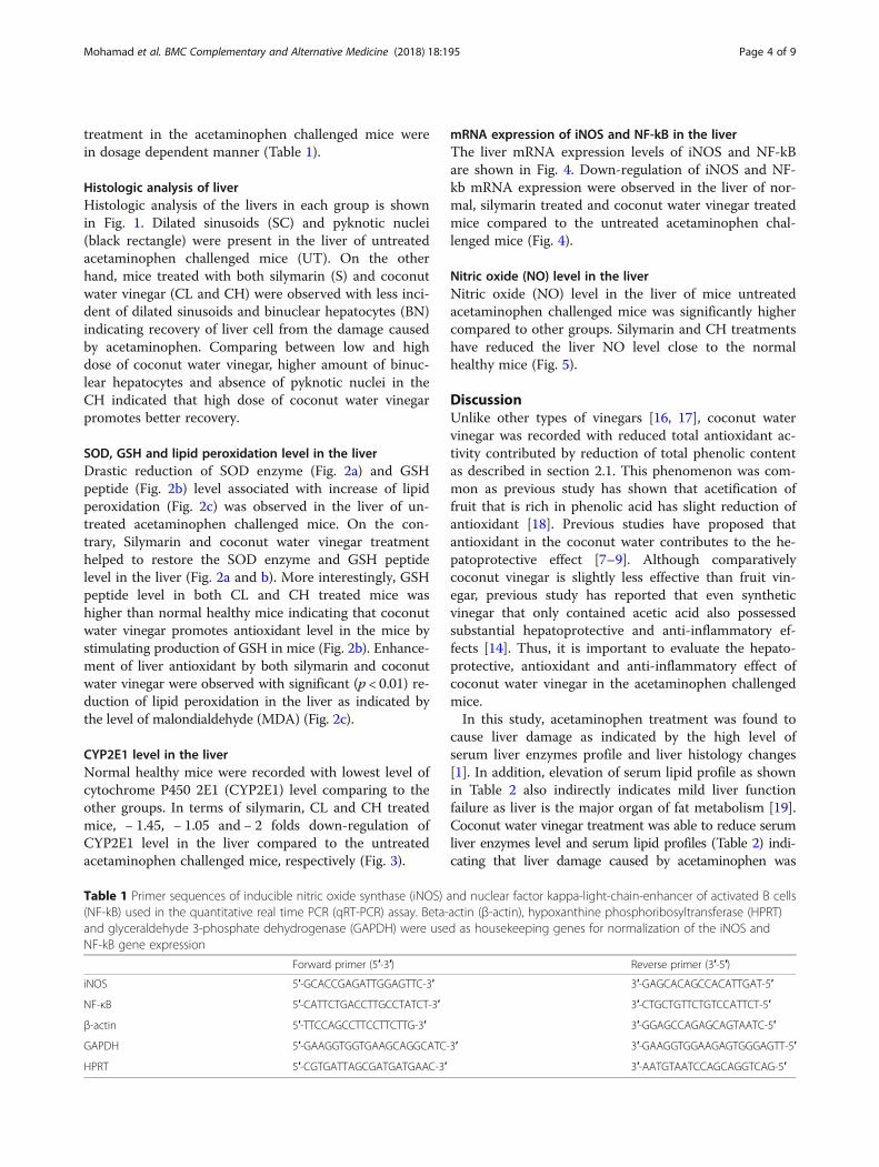
Histologic analysis of liverHistologic analysis of the livers in each group is shownin Fig. 1. Dilated sinusoids (SC) and pyknotic nuclei(black rectangle) were present in the liver of untreatedacetaminophen challenged mice (UT). On the otherhand, mice treated with both silymarin (S) and coconutwater vinegar (CL and CH) were observed with less inci-dent of dilated sinusoids and binuclear hepatocytes (BN)indicating recovery of liver cell from the damage causedby acetaminophen. Comparing between low and highdose of coconut water vinegar, higher amount of binuc-lear hepatocytes and absence of pyknotic nuclei in theCH indicated that high dose of coconut water vinegarpromotes better recovery.
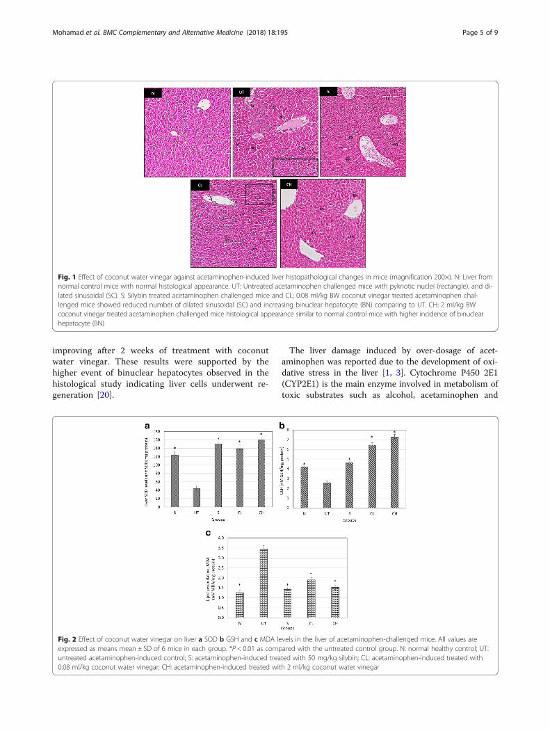
SOD, GSH and lipid peroxidation level in the liverDrastic reduction of SOD enzyme (Fig. 2a) and GSHpeptide (Fig. 2b) level associated with increase of lipidperoxidation (Fig. 2c) was observed in the liver of un-treated acetaminophen challenged mice. On the con-trary, Silymarin and coconut water vinegar treatmenthelped to restore the SOD enzyme and GSH peptidelevel in the liver (Fig. 2a and b). More interestingly, GSHpeptide level in both CL and CH treated mice washigher than normal healthy mice indicating that coconutwater vinegar promotes antioxidant level in the mice bystimulating production of GSH in mice (Fig. 2b). Enhance-ment of liver antioxidant by both silymarin and coconutwater vinegar were observed with significant (p < 0.01) re-duction of lipid peroxidation in the liver as indicated bythe level of malondialdehyde (MDA) (Fig. 2c).
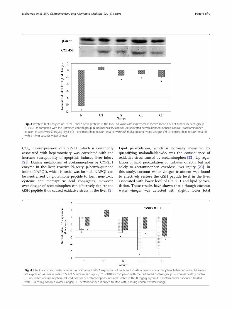
CYP2E1 level in the liverNormal healthy mice were recorded with lowest level ofcytochrome P450 2E1 (CYP2E1) level comparing to theother groups. In terms of silymarin, CL and CH treatedmice, − 1.45, − 1.05 and − 2 folds down-regulation ofCYP2E1 level in the liver compared to the untreatedacetaminophen challenged mice, respectively (Fig. 3).
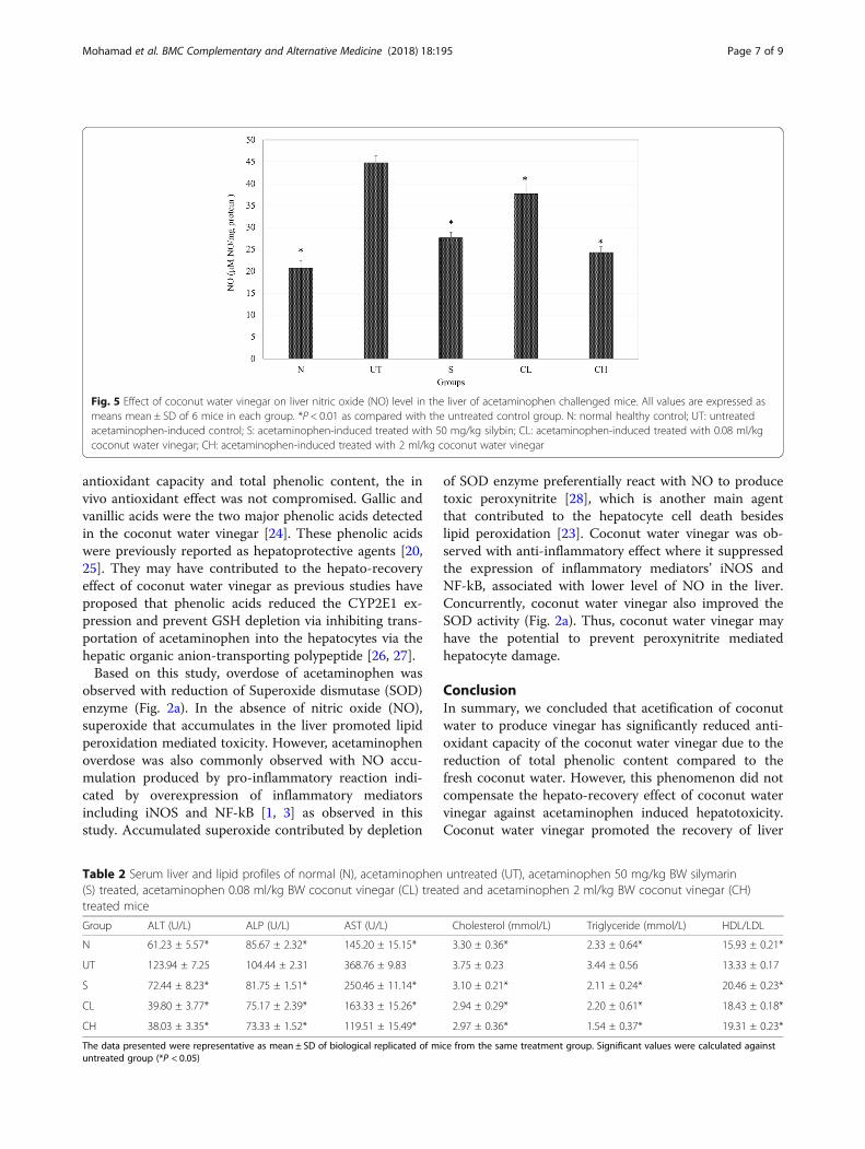
mRNA expression of iNOS and NF-kB in the liverThe liver mRNA expression levels of iNOS and NF-kBare shown in Fig. 4. Down-regulation of iNOS and NF-kb mRNA expression were observed in the liver of nor-mal, silymarin treated and coconut water vinegar treatedmice compared to the untreated acetaminophen chal-lenged mice (Fig. 4).
Nitric oxide (NO) level in the liverNitric oxide (NO) level in the liver of mice untreatedacetaminophen challenged mice was significantly highercompared to other groups. Silymarin and CH treatmentshave reduced the liver NO level close to the normalhealthy mice (Fig. 5).
DiscussionUnlike other types of vinegars [16, 17], coconut watervinegar was recorded with reduced total antioxidant ac-tivity contributed by reduction of total phenolic contentas described in section 2.1. This phenomenon was com-mon as previous study has shown that acetification offruit that is rich in phenolic acid has slight reduction ofantioxidant [18]. Previous studies have proposed thatantioxidant in the coconut water contributes to the he-patoprotective effect [7–9]. Although comparativelycoconut vinegar is slightly less effective than fruit vin-egar, previous study has reported that even syntheticvinegar that only contained acetic acid also possessedsubstantial hepatoprotective and anti-inflammatory ef-fects [14]. Thus, it is important to evaluate the hepato-protective, antioxidant and anti-inflammatory effect ofcoconut water vinegar in the acetaminophen challengedmice.In this study, acetaminophen treatment was found to
cause liver damage as indicated by the high level ofserum liver enzymes profile and liver histology changes[1]. In addition, elevation of serum lipid profile as shownin Table 2 also indirectly indicates mild liver functionfailure as liver is the major organ of fat metabolism [19].Coconut water vinegar treatment was able to reduce serumliver enzymes level and serum lipid profiles (Table 2) indi-cating that liver damage caused by acetaminophen was
Table 1 Primer sequences of inducible nitric oxide synthase (iNOS) and nuclear factor kappa-light-chain-enhancer of activated B cells(NF-kB) used in the quantitative real time PCR (qRT-PCR) assay. Beta-actin (β-actin), hypoxanthine phosphoribosyltransferase (HPRT)and glyceraldehyde 3-phosphate dehydrogenase (GAPDH) were used as housekeeping genes for normalization of the iNOS andNF-kB gene expression
Forward primer (5′-3′) Reverse primer (3′-5′)
iNOS 5′-GCACCGAGATTGGAGTTC-3′ 3′-GAGCACAGCCACATTGAT-5′
NF-κB 5′-CATTCTGACCTTGCCTATCT-3′ 3′-CTGCTGTTCTGTCCATTCT-5′
β-actin 5′-TTCCAGCCTTCCTTCTTG-3′ 3′-GGAGCCAGAGCAGTAATC-5′
GAPDH 5′-GAAGGTGGTGAAGCAGGCATC-3′ 3′-GAAGGTGGAAGAGTGGGAGTT-5′
HPRT 5′-CGTGATTAGCGATGATGAAC-3′ 3′-AATGTAATCCAGCAGGTCAG-5′
Mohamad et al. BMC Complementary and Alternative Medicine (2018) 18:195 Page 4 of 9
improving after 2 weeks of treatment with coconutwater vinegar. These results were supported by thehigher event of binuclear hepatocytes observed in thehistological study indicating liver cells underwent re-generation [20].
The liver damage induced by over-dosage of acet-aminophen was reported due to the development of oxi-dative stress in the liver [1, 3]. Cytochrome P450 2E1(CYP2E1) is the main enzyme involved in metabolism oftoxic substrates such as alcohol, acetaminophen and
Fig. 1 Effect of coconut water vinegar against acetaminophen-induced liver histopathological changes in mice (magnification 200×). N: Liver fromnormal control mice with normal histological appearance. UT: Untreated acetaminophen challenged mice with pyknotic nuclei (rectangle), and di-lated sinusoidal (SC). S: Silybin treated acetaminophen challenged mice and CL: 0.08 ml/kg BW coconut vinegar treated acetaminophen chal-lenged mice showed reduced number of dilated sinusoidal (SC) and increasing binuclear hepatocyte (BN) comparing to UT. CH: 2 ml/kg BWcoconut vinegar treated acetaminophen challenged mice histological appearance similar to normal control mice with higher incidence of binuclearhepatocyte (BN)
Fig. 2 Effect of coconut water vinegar on liver a SOD b GSH and c MDA levels in the liver of acetaminophen-challenged mice. All values areexpressed as means mean ± SD of 6 mice in each group. *P < 0.01 as compared with the untreated control group. N: normal healthy control; UT:untreated acetaminophen-induced control; S: acetaminophen-induced treated with 50 mg/kg silybin; CL: acetaminophen-induced treated with0.08 ml/kg coconut water vinegar; CH: acetaminophen-induced treated with 2 ml/kg coconut water vinegar
Mohamad et al. BMC Complementary and Alternative Medicine (2018) 18:195 Page 5 of 9
CCl4. Overexpression of CYP2E1, which is commonlyassociated with hepatotoxicity was correlated with theincrease susceptibility of apoptosis-induced liver injury[21]. During metabolism of acetaminophen by CYP2E1enzyme in the liver, reactive N-acetyl-p-benzo-quinoneimine (NAPQI), which is toxic, was formed. NAPQI canbe neutralized by glutathione peptide to form non-toxiccysteine and mercapturic acid conjugates. However,over-dosage of acetaminophen can effectively deplete theGSH peptide thus caused oxidative stress in the liver [3].
Lipid peroxidation, which is normally measured byquantifying malondialdehyde, was the consequence ofoxidative stress caused by acetaminophen [22]. Up regu-lation of lipid peroxidation contributes directly but notsolely to acetaminophen overdose liver injury [23]. Inthis study, coconut water vinegar treatment was foundto effectively restore the GSH peptide level in the liverassociated with lower level of CYP2E1 and lipid peroxi-dation. These results have shown that although coconutwater vinegar was detected with slightly lower total
Fig. 4 Effect of coconut water vinegar on normalized mRNA expression of iNOS and NF-kB in liver of acetaminophenchallenged mice. All valuesare expressed as means mean ± SD of 6 mice in each group. *P < 0.01 as compared with the untreated control group. N: normal healthy control;UT: untreated acetaminophen-induced control; S: acetaminophen-induced treated with 50 mg/kg silybin; CL: acetaminophen-induced treatedwith 0.08 ml/kg coconut water vinegar; CH: acetaminophen-induced treated with 2 ml/kg coconut water vinegar
Fig. 3 Western blot analyses of CYP2E1 and β-actin proteins in the liver. All values are expressed as means mean ± SD of 6 mice in each group.*P < 0.01 as compared with the untreated control group. N: normal healthy control; UT: untreated acetaminophen-induced control; S: acetaminophen-induced treated with 50 mg/kg silybin; CL: acetaminophen-induced treated with 0.08 ml/kg coconut water vinegar; CH: acetaminophen-induced treatedwith 2 ml/kg coconut water vinegar
Mohamad et al. BMC Complementary and Alternative Medicine (2018) 18:195 Page 6 of 9
antioxidant capacity and total phenolic content, the invivo antioxidant effect was not compromised. Gallic andvanillic acids were the two major phenolic acids detectedin the coconut water vinegar [24]. These phenolic acidswere previously reported as hepatoprotective agents [20,25]. They may have contributed to the hepato-recoveryeffect of coconut water vinegar as previous studies haveproposed that phenolic acids reduced the CYP2E1 ex-pression and prevent GSH depletion via inhibiting trans-portation of acetaminophen into the hepatocytes via thehepatic organic anion-transporting polypeptide [26, 27].Based on this study, overdose of acetaminophen was
observed with reduction of Superoxide dismutase (SOD)enzyme (Fig. 2a). In the absence of nitric oxide (NO),superoxide that accumulates in the liver promoted lipidperoxidation mediated toxicity. However, acetaminophenoverdose was also commonly observed with NO accu-mulation produced by pro-inflammatory reaction indi-cated by overexpression of inflammatory mediatorsincluding iNOS and NF-kB [1, 3] as observed in thisstudy. Accumulated superoxide contributed by depletion
of SOD enzyme preferentially react with NO to producetoxic peroxynitrite [28], which is another main agentthat contributed to the hepatocyte cell death besideslipid peroxidation [23]. Coconut water vinegar was ob-served with anti-inflammatory effect where it suppressedthe expression of inflammatory mediators’ iNOS andNF-kB, associated with lower level of NO in the liver.Concurrently, coconut water vinegar also improved theSOD activity (Fig. 2a). Thus, coconut water vinegar mayhave the potential to prevent peroxynitrite mediatedhepatocyte damage.
ConclusionIn summary, we concluded that acetification of coconutwater to produce vinegar has significantly reduced anti-oxidant capacity of the coconut water vinegar due to thereduction of total phenolic content compared to thefresh coconut water. However, this phenomenon did notcompensate the hepato-recovery effect of coconut watervinegar against acetaminophen induced hepatotoxicity.Coconut water vinegar promoted the recovery of liver
Fig. 5 Effect of coconut water vinegar on liver nitric oxide (NO) level in the liver of acetaminophen challenged mice. All values are expressed asmeans mean ± SD of 6 mice in each group. *P < 0.01 as compared with the untreated control group. N: normal healthy control; UT: untreatedacetaminophen-induced control; S: acetaminophen-induced treated with 50 mg/kg silybin; CL: acetaminophen-induced treated with 0.08 ml/kgcoconut water vinegar; CH: acetaminophen-induced treated with 2 ml/kg coconut water vinegar
Table 2 Serum liver and lipid profiles of normal (N), acetaminophen untreated (UT), acetaminophen 50 mg/kg BW silymarin(S) treated, acetaminophen 0.08 ml/kg BW coconut vinegar (CL) treated and acetaminophen 2 ml/kg BW coconut vinegar (CH)treated mice
Group ALT (U/L) ALP (U/L) AST (U/L) Cholesterol (mmol/L) Triglyceride (mmol/L) HDL/LDL
N 61.23 ± 5.57* 85.67 ± 2.32* 145.20 ± 15.15* 3.30 ± 0.36* 2.33 ± 0.64* 15.93 ± 0.21*
UT 123.94 ± 7.25 104.44 ± 2.31 368.76 ± 9.83 3.75 ± 0.23 3.44 ± 0.56 13.33 ± 0.17
S 72.44 ± 8.23* 81.75 ± 1.51* 250.46 ± 11.14* 3.10 ± 0.21* 2.11 ± 0.24* 20.46 ± 0.23*
CL 39.80 ± 3.77* 75.17 ± 2.39* 163.33 ± 15.26* 2.94 ± 0.29* 2.20 ± 0.61* 18.43 ± 0.18*
CH 38.03 ± 3.35* 73.33 ± 1.52* 119.51 ± 15.49* 2.97 ± 0.36* 1.54 ± 0.37* 19.31 ± 0.23*
The data presented were representative as mean ± SD of biological replicated of mice from the same treatment group. Significant values were calculated againstuntreated group (*P < 0.05)
Mohamad et al. BMC Complementary and Alternative Medicine (2018) 18:195 Page 7 of 9
damage induced by acetaminophen by improving thehepatic antioxidant level and suppressing the liver in-flammation in dosage dependent manner. Future studiesincluding detailed mechanisms of the coconut water vin-egar hepatoprotective effect and clinical trials shall becarried out to validate the potential of coconut vinegaras a potential food supplement to ameliorate chemicalinduced liver damage.
AbbreviationsALP: Alkaline phosphatase; ALT: Alanine transaminase; ANOVA: one-wayanalysis of variance; AST: Aspartate aminotransferase; BN: Binuclearhepatocytes; CCl4: Carbon tetrachloride; CH: Acetaminophen-induced treatedwith 2 ml/kg coconut water vinegar; CL: Acetaminophen-induced treatedwith 0.08 ml/kg coconut water vinegar; CYP2E1: Cytochrome P450 2E1;FRAP: Ferric reducing ability of plasma; GAPDH: Glyceraldehyde 3-phosphatedehydrogenase; GSH: Glutathione; HPRT: Hypoxanthine phosphoribosyltransferase;iNOS: inducible nitric oxide synthase; MDA: Malondialdehyde; N: healthy normalcontrol group; NAPQI: N-acetyl-p-benzoquinone imine; NF-kB: Nuclear factorkappa-light-chain-enhancer of activated B cells; NO: Nitric oxide; qRT-PCR: quantitative real time PCR; S: acetaminophen-induced treated with50 mg/kg silybin; SC: Dilated sinusoids; SDS: Sodium dodecyl sulphate;SOD: Superoxide dismutase; TPC: Total phenolic content; UT: Untreatedacetaminophen-induced control; β-actin: Beta-actin
FundingThis study was supported by Grant Pembangunan RMK10, Mardi from Ministryof Agriculture, Malaysia. The authors have no conflicts of interest to report.
Availability of data and materialsThe datasets used and/or analysed during the current study available fromthe corresponding author on reasonable request.
Authors’ contributionsSKY, KL and NBA designed the experiment; KL and SAS performed HPLC andprepared coconut water vinegar; NEM, SKY and BKB performed theexperiment; HK and WYH performed qRT-PCR; KLL performed Western blotanalysis; NEM and SKY prepared the manuscript; all authors have gonethrough and approved the manuscript.
Ethics approval and consent to participateAll studies involving animals were conducted in compliance with theUniversiti Putra Malaysia’s ethical guidelines as approved by the AnimalEthics Committee (UPM, Malaysia). The approval number obtained: UPM/FPV/PS/3.2.1.551/AUP-R168.
Competing interestsThe authors declare that they have no competing interests.
Publisher’s NoteSpringer Nature remains neutral with regard to jurisdictional claims inpublished maps and institutional affiliations.
Author details1Department of Cell and Molecular Biology, Faculty of Biotechnology andBiomolecular Science, Universiti Putra Malaysia, 43400 Serdang, Selangor,Malaysia. 2China-ASEAN College of Marine Sciences, Xiamen UniversityMalaysia, Jalan Sunsuria, Bandar Sunsuria, 43900 Sepang, Selangor, Malaysia.3Institute of Bioscience, Universiti Putra Malaysia, Serdang, Selangor, Malaysia.4Biotechnology Research Centre, Malaysian Agricultural Research andDevelopment Institute (MARDI), 43400 Serdang, Selangor, Malaysia.5Department of Genetics and Plant Breeding, College of Agriculture andApplied Biology, Cantho University, 3/2 Street, CanTho City, Vietnam. 6Facultyof Medicine and Health Sciences, Universiti Tunku Abdul Rahman, SungaiLong Campus, Jalan Sungai Long, Bandar Sungai Long, Cheras, 43000 Kajang,Selangor, Malaysia. 7School of Biomedical Sciences, the University ofNottingham Malaysia Campus, Jalan Broga, 43500 Semenyih, Selangor,Malaysia.
Received: 20 October 2017 Accepted: 11 April 2018
References1. Fontana RJ. Acute liver failure including acetaminophen overdose.
Med Clin N Am. 2008;92:761–94.2. Nourjah P, Ahmad SR, Karwoski C, Willy M. Estimates of acetaminophen
(paracetomal)-associated overdoses in the United States.Pharmacoepidemiol Drug Saf. 2006;15:398–405.
3. McGill MR, Sharpe MR, Williams CD, Taha M, Curry SC, Jaeschke H. Themechanism underlying acetaminophen-induced hepatotoxicity in humansand mice involves mitochondrial damage and nuclear DNA fragmentation.J Clin Invest. 2012;122:1574–83.
4. Hong M, Li S, Tan HY, Wang N, Tsao SW, Feng Y. Current status of herbalmedicines in chronic liver disease therapy: the biological effects, moleculartargets and future prospects. Int J Mol Sci. 2015;16:28705–45.
5. Santoso U, Kubo K, Ota T, Tadakoro T, Maekawa A. Antioxidative effect ofcoconut (Cocos nucifera L.) water extract on TBARS value in liver of rats fedfish oil diet. Indo Food Nutri Prog. 1996;3:42–50.
6. Offor C, Adetarami O, Nwali B, Igwenyi I, Afiukwa C. Effect of Cocosnuciferawater on liver enzymes. Middle-East J Sci Res. 2014;21:844–7.
7. Loki AL, Rajamohan T. Hepatoprotective and antioxidant effect of tendercoconut water on carbon tetrachloride induced liver injury in rats. Indian JBiochem Biophys. 2003;40:354–7.
8. Ndubuka GI, Okafor WC, Jervas E, Chidi IS, Oh U, Illiams OI. Protective effectof immature coconut water on hepatocytes against carbontetrachloride-induced liver damage in Wister rats. Int J Sci Res. 2014;4:1427–31.
9. Okafor WC, Ndubuka GI, Jervas E, Chidi IS, Udoka OC, Osuchukwu IW. Effectsof immature coconut water on the hepatocytoarchitecture againstCarbontetrachloride-induced Liver damaging wistar rats. IOSR J Den MedSci. 2014;1:60–5.
10. Preetha P, Girija Devi V, Rajamohan T. Comparative effects of maturecoconut water (Cocos nucifera) and glibenclamide on some biochemicalparameters in alloxan induced diabetic rats. Rev Bras Farmacogn Braz JPharmacogn. 2013;23:481–7.
11. Satheesh N, Prasad N. Production of fermented coconut water beverages.J Food Ag-Ind. 2013;6:281–9.
12. Yong JW, Ge L, Ng YF, Tan SN. The chemical composition and biologicalproperties of coconut (Cocos nucifera L.) water. Molecules. 2009;14:5144–64.
13. Li J, Yu G, Fan J. Alditols and monosaccharides from sorghum vinegar canattenuate platelet aggregation by inhibiting cyclooxygenase-1 andthromboxane-A2 synthase. J Ethnopharmacol. 2014;155:285–92.
14. Mohamad NE, Yeap SK, Lim KL, Yusof HM, Beh BK, Tan SW, Ho WY,Sharifuddin SA, Jamluddin A, Long K, Rahman NMANA, Alitheen NB.Antioxidant effects of pineapple vinegar in reversing of paracetamol-induced liver damage in mice. Chin Med. 2015;10:1–14.
15. Nishidai S, Nakamura Y, Torikai K, Yamamoto M, Ishihara N, Mori H, OhigashiH. Kurosu, a traditional vinegar produced from unpolished rice, suppresseslipid peroxidation in vitro and in mouse skin. Biosci Biotechnol Biochem.2000;64:1909–14.
16. Beh BK, Mohamad NE, Yeap SK, Lim KL, Ho WY, Yusof HM, Sharifuddin SA,Jamaluddin A, Long K, Alitheen NB. Polyphenolic profiles and the in vivoantioxidant effect of nipa vinegar on paracetamol induced liver damage.RSC Adv. 2016;6:63304–13.
17. Kongkiattikajorn J. Antioxidant properties of roselle vinegar production bymixed culture of Acetobacter Acetiand Acetobacter cerevisiae. Kasetsart J(Nat Sci). 2014;48:980–8.
18. Cunha MAA, Lima KP, Santos VAQ, Heinz OL, Schmidt CAP. Blackberry vinegarproduced by successive acetification cycles: Production, characterization andbioactivity parameters. Braz Arch Biol Techn. 2016;59:1–10.
19. Basu S, Haldar N, Bhattacharya Sanji BS, Biswas M. Hepatoprotective activityof Litchi chinensis leaves against paracetamol-induced liver damage in rats.Am-Eur J Sci Res. 2012;7:77–81.
20. Rasool MK, Sabina EP, Ramya SR, Preety P, Patel S, Mandal N, Mishra PP,Samuel J. Hepatoprotective and antioxidant effects of gallic acid inparacetamol-induced liver damage in mice. J Pharm Pharmacol. 2010;62:638–43.
21. Wang X, Lu Y, Cederbaum AI. Induction of cytochrome P450 2E1 increaseshepatotoxicity caused by Fas agonistic Jo2 antibody in mice. Hepatology.2005;42:400–10.
Mohamad et al. BMC Complementary and Alternative Medicine (2018) 18:195 Page 8 of 9
22. Dusan M, Milica N, Danijela V, Miodrag C, Marjan M, Vera T, Milena S,Tatjana R. The effects of ethanol on paracetamol-induced oxidative stress inmice liver. J Serb Chem Soc. 2013;78:179–95.
23. Knight TR, Fariss MW, Farhood A, Jaeschke H. Role of lipid peroxidation as amechanism of liver injury after acetaminophen overdose in mice. ToxicolSci. 2003;76:229–36.
24. Mohamad NE, Yeap SK, Ky H, Ho WY, Boo SY, Chua J, Beh BK, SharifuddinSA, Long K, Alitheen NB. Dietary coconut water vinegar for improvement ofobesity-associated inflammation in high-fat-diet treated mice. Food NutrRes. 2017;61:1368322.
25. Itoh A, Isoda K, Kondoh M, Kawase M, Watari A, Kobayashi M, Tamesada M,Yagi K. Hepatoprotective effect of syringic acid and vanillic acid on CCl4-induced liver injury. Biol Pharm Bull. 2010;33:983–7.
26. Mandery K, Bujok K, Schmidt I, Keiser M, Siegmund W, Balk B, Konig J,Fromm MF, Glaeser H. Influence of the flavonoids apigenin, kaempferol, andquercetin on the function of organic anion transporting polypeptides 1A2and 2B1. Biochem Pharmacol. 2010;80:1746–53.
27. Priyadarsini RV, Nagini S. Quercetin suppresses cytochrome P450 mediatedROS generation and NFκB activation to inhibit the development of 7, 12-dimethylbenz [a] anthracene (DMBA) induced hamster buccal pouchcarcinomas. Free Radic Res. 2012;46:41–9.
28. James LP, Mayeux PR, Hinson JA. Acetaminophen-induced hepatotoxicity.Drug Metab Dispos. 2003;31:1499–506.
Mohamad et al. BMC Complementary and Alternative Medicine (2018) 18:195 Page 9 of 9
Copyright © 2022 FDOKUMEN