Genetic inhibition of JNK3 ameliorates spinal muscular atrophy
19
ORIGINAL ARTICLE Genetic inhibition of JNK3 ameliorates spinal muscular atrophy Naresh K. Genabai 1,2 , Saif Ahmad 1,2 , Zhanying Zhang 1,2 , Xiaoting Jiang 1,2 , Cynthia A. Gabaldon 1,2 and Laxman Gangwani 1,2, * 1 Center of Emphasis in Neurosciences and 2 Department of Biomedical Sciences, Paul L. Foster School of Medicine, Texas Tech University Health Sciences Center, El Paso, TX 79905, USA *To whom correspondence should be addressed. Tel: +1 9152154189; Fax: +1 9157831271; Email: [email protected] Abstract Mutation of the Survival Motor Neuron 1 (SMN1) gene causes spinal muscular atrophy (SMA), an autosomal recessive neurodegenerative disorder that occurs in early childhood. Degeneration of spinal motor neurons caused by SMN deficiency results in progressive muscle atrophyand death in SMA. The molecular mechanism underlying neurodegeneration in SMA is unknown. No treatment is available to prevent neurodegeneration and reduce the burden of illness in SMA. We report that the c-Jun NH 2 -terminal kinase (JNK) signaling pathway mediates neurodegeneration in SMA. The neuron-specific isoform JNK3 is required for neuron degeneration caused by SMN deficiency. JNK3 deficiency reduces degeneration of cultured neurons caused by low levels of SMN. Genetic inhibition of JNK pathway in vivo by Jnk3 knockout results in amelioration of SMA phenotype. JNK3 deficiency preventsthe loss of spinal cord motor neurons, reduces muscle degeneration, improves muscle fiber thickness and muscle growth, improves motor function and overall growth and increases lifespan of micewith SMA that shows a systemic rescue of phenotype by a SMN-independent mechanism. JNK3 represents a potential (non-SMN) therapeutic target for the treatment of SMA. Introduction Spinal muscular atrophy (SMA) is an autosomal recessive neuro- muscular disorder of the early childhood caused by mutation of telomeric copy of the Survival Motor Neuron 1 (SMN1) gene (1). The SMN2, centromeric copy, undergoes alternative splicing and pre- dominately produces truncated product lacking exon 7 (SMNΔ7) (2). Low levels of full-length SMN produced by SMN2 are sufficient for embryonic development and survival, but result in the loss of spinal motor neurons leading to muscle atrophy, respiratory fail- ure and death in SMA patients (3). SMA is a developmental disorder with early onset and stable course, characterized by skeletal muscle atrophy due to the de- generation of spinal motor neurons caused by low levels of SMN (4). Currently, no treatment is available to prevent degener- ation of motor neurons in SMA. The cellular and molecular me- chanisms of motor neuron degeneration caused by SMN deficiency are unknown. Progress has been made towards under- standing biochemical function of SMN but its role in survival and maintenance of motor neurons is unclear (5). The function of SMN is indicated in the development and maintenance of the nervous system, maturation of neuromuscular junctions (NMJs) and growth of skeletal muscle (4,6–8). SMN is shown to play a role in the assembly of spliceosomal small nuclear ribonucleo- proteins (snRNPs) required for pre-mRNA splicing (9). The SMN- dependent splicing defects at pre-, early- and late-symptomatic stages in selective genes (10–12), the SMN-dependent alteration in the levels of different proteins (10,13) and the snRNP biogen- esis-independent functions (14) indicate the complex nature of biochemical alterations. These alterations may affect cellular processes such as axonal growth, pathfinding, NMJs, cytoskel- eton, synaptic maturation and neurotransmitter release in dif- ferent neuronal and muscle cells that might be collectively Received: June 19, 2015. Revised: August 24, 2015. Accepted: September 21, 2015 © The Author 2015. Published by Oxford University Press. All rights reserved. For Permissions, please email: [email protected] Human Molecular Genetics, 2015, Vol. 24, No. 24 6986–7004 doi: 10.1093/hmg/ddv401 Advance Access Publication Date: 30 September 2015 Original Article 6986 Downloaded from https://academic.oup.com/hmg/article/24/24/6986/2384597 by guest on 08 January 2022
-
Upload
khangminh22 -
Category
Documents
-
view
1 -
download
0
Transcript of Genetic inhibition of JNK3 ameliorates spinal muscular atrophy

OR I G INA L ART I C L E
Genetic inhibition of JNK3 ameliorates spinalmuscular atrophyNaresh K. Genabai1,2, Saif Ahmad1,2, Zhanying Zhang1,2, Xiaoting Jiang1,2,Cynthia A. Gabaldon1,2 and Laxman Gangwani1,2,*1Center of Emphasis inNeurosciences and 2Department of Biomedical Sciences, Paul L. Foster School ofMedicine,Texas Tech University Health Sciences Center, El Paso, TX 79905, USA
*To whom correspondence should be addressed. Tel: +1 9152154189; Fax: +1 9157831271; Email: [email protected]
AbstractMutation of the Survival Motor Neuron 1 (SMN1) gene causes spinal muscular atrophy (SMA), an autosomal recessiveneurodegenerative disorder that occurs in early childhood. Degeneration of spinal motor neurons caused by SMN deficiencyresults in progressive muscle atrophy and death in SMA. The molecular mechanism underlying neurodegeneration in SMA isunknown. No treatment is available to prevent neurodegeneration and reduce the burden of illness in SMA. We report that thec-Jun NH2-terminal kinase (JNK) signaling pathway mediates neurodegeneration in SMA. The neuron-specific isoform JNK3 isrequired for neuron degeneration caused by SMN deficiency. JNK3 deficiency reduces degeneration of cultured neurons causedby low levels of SMN. Genetic inhibition of JNK pathway in vivo by Jnk3 knockout results in amelioration of SMAphenotype. JNK3deficiency prevents the loss of spinal cord motor neurons, reduces muscle degeneration, improves muscle fiber thickness andmuscle growth, improves motor function and overall growth and increases lifespan of mice with SMA that shows a systemicrescue of phenotype by a SMN-independent mechanism. JNK3 represents a potential (non-SMN) therapeutic target for thetreatment of SMA.
IntroductionSpinal muscular atrophy (SMA) is an autosomal recessive neuro-muscular disorder of the early childhood caused by mutation oftelomeric copy of the Survival Motor Neuron 1 (SMN1) gene (1). TheSMN2, centromeric copy, undergoes alternative splicing and pre-dominately produces truncated product lacking exon 7 (SMNΔ7)(2). Low levels of full-length SMNproduced by SMN2 are sufficientfor embryonic development and survival, but result in the loss ofspinal motor neurons leading tomuscle atrophy, respiratory fail-ure and death in SMA patients (3).
SMA is a developmental disorder with early onset and stablecourse, characterized by skeletal muscle atrophy due to the de-generation of spinal motor neurons caused by low levels ofSMN (4). Currently, no treatment is available to prevent degener-ation of motor neurons in SMA. The cellular and molecular me-chanisms of motor neuron degeneration caused by SMN
deficiency are unknown. Progress has beenmade towards under-standing biochemical function of SMN but its role in survival andmaintenance of motor neurons is unclear (5). The function ofSMN is indicated in the development and maintenance of thenervous system, maturation of neuromuscular junctions (NMJs)and growth of skeletal muscle (4,6–8). SMN is shown to play arole in the assembly of spliceosomal small nuclear ribonucleo-proteins (snRNPs) required for pre-mRNA splicing (9). The SMN-dependent splicing defects at pre-, early- and late-symptomaticstages in selective genes (10–12), the SMN-dependent alterationin the levels of different proteins (10,13) and the snRNP biogen-esis-independent functions (14) indicate the complex nature ofbiochemical alterations. These alterations may affect cellularprocesses such as axonal growth, pathfinding, NMJs, cytoskel-eton, synaptic maturation and neurotransmitter release in dif-ferent neuronal and muscle cells that might be collectively
Received: June 19, 2015. Revised: August 24, 2015. Accepted: September 21, 2015
© The Author 2015. Published by Oxford University Press. All rights reserved. For Permissions, please email: [email protected]
Human Molecular Genetics, 2015, Vol. 24, No. 24 6986–7004
doi: 10.1093/hmg/ddv401Advance Access Publication Date: 30 September 2015Original Article
6986
Dow
nloaded from https://academ
ic.oup.com/hm
g/article/24/24/6986/2384597 by guest on 08 January 2022

responsible for degeneration of spinal motor neurons (5,7). It ispossible that the two cellular processes in neurons might beaffected simultaneously by the loss of SMN function, (a) reducedefficiency of splicing that may result in low levels of proteins re-quired for survival and maintenance of neurons and (b) activa-tion of intracellular stress signaling pathways that initiateneurodegeneration. The low levels of SMN are the cause of neu-rodegeneration, but it is unclearwhyselectively lower spinal cordmotor neurons degenerate (5,7). Intracellular signaling mechan-isms triggered by the low levels of SMN that might mediate neu-rodegeneration in SMA are also unclear. Recent studies haveindicated that the activation of RhoA/ROCK pathway by SMN de-ficiency might contribute to the disruption of actin cytoskeletonby hyperphosphorylation of profilin and affect neuron integrity(15,16). Inhibition of Rho kinase is shown to increase survival ofSMA mouse model with intermediate severity (17). Alteration inthe β-catenin signaling due to reduced expression of ubiquitin-likemodifier activating enzyme 1 (UBA1) and increased β-cateninlevels might contribute to motor neuron pathology in SMA (13).However, the function of non-SMN targets in fully systemicrescue of SMA pathology without altering levels of SMN (SMN-independent) remains to be examined.
In this study, we investigated the molecular mechanism ofneurodegeneration in SMA.We report that the c-Jun NH2-termin-al kinase (JNK) cascades ASK1/MKK4/7/JNK and MEKK1/MKK4/7/JNK are activated in spinal cords of SMA patients and SMA miceandmediate neurodegeneration in SMA.We identified that JNK3,a neuron-specific isoform, mediates neurodegeneration causedby the low levels of SMN. Deficiency of JNK3 (Jnk3−/−) resultedin reduced degeneration of cultured neurons with low levels ofSMN. To examine the effect of JNK3 deficiency in vivo, we createdSMA carrier mice lacking JNK3 by crossing SMA carrier mice withJnk3−/− knockout mice [Jnk3−/− mice display normal phenotype,including fertility and lifespan (18,19)]. Genetic inhibition ofJNK pathway in vivo by Jnk3 knockout resulted in ameliorationof SMA phenotype. JNK3 deficiency results in systemic rescue ofphenotypewithout altering levels of SMN (SMN-independent) bypreventing degeneration of spinal cord motor neurons, reducingmuscle atrophy, improving overall growth and increasing life-span of mice with SMA. We propose that the JNK3 represents apotential (non-SMN) therapeutic target for the treatment of SMA.
ResultsActivation of the JNK signaling pathway in SMA
The molecular mechanism of neurodegeneration caused by lowlevels of SMN in SMA is unknown. To identify signaling mechan-isms that might mediate neurodegeneration in SMA, we exam-ined the phosphorylation of mitogen activated protein kinases(MAPKs) in the spinal cords (lumbar region, area of primary patho-genesis in SMA) from non-SMA human (Normal, #83, age 69 days)and SMA (type-I) patients (i) SMA4583, age 77 days (ii) SMA4994,age 59 days and (iii) SMA4629, age 169 days and from 12-day oldnon-SMA and mice with SMA-like disease (SMA mice) (20). Weperformed biochemical experiments using individual SMA pa-tient samples and by pooling SMA patient samples to reducethe biological variation. Averaging of data from three individualpatient samples and pooling of three SMA patient samples re-sulted in similar activity.We compared quantification and statis-tical significance (one-way analysis of variance, ANOVA) of datafor SMA mice and SMA patients from two different MAPK arraysand focused our analysis on kinases that were commonly acti-vated in both SMA patient and SMA mice.
The phospho-MAPK array analysis of human spinal cord sam-ples shows an increase in phosphorylation of almost all MAP ki-nases except MKK3 and it was not possible to identify specifickinases activated in SMA (Fig. 1A). The non-specific increase inphosphorylation of kinases might be because of longer post-mortem interval (PMI) for processing human tissues (avg.∼8.5 h). To gain insight into the activation of specificMAP kinasesin SMA, we examined spinal cord samples from normal and SMAmice (PMI, ∼30 min) and compared these with the human sam-ples (Fig. 1B) to identify kinases commonly activated in mouseand human spinal cord. Comparative analysis of data from phos-pho-MAPK antibody array of human and mice spinal cord sam-ples, shows that several MAP kinases (AKT1-3, ERK2, MKK3,MKK6, p38α, p38β, p38γ, p38δ, p70, RSK1-2 and TOR) were notcommonly activated in mice and human samples (Fig. 1A and B),and these kinases were not considered for further analysis. How-ever, three groups ofMAP kinases (i) JNK group (JNK1, JNK2, JNK3),(ii) glycogen synthase kinase 3 (GSK-3α and β) and (iii) extracellu-lar signal-regulated kinase (ERK1) showed increased phosphoryl-ation in both SMA patients and SMAmice comparedwith normal(Fig. 1). Quantitative analyses showedmoderate increase in phos-phorylation of GSK-3α/β (10.10 ± 1.09%, P = 0.086) in SMAmice andSMA patients (39.29 ± 4.48%, P = 0.037), but the increase is notstatistically significant in SMAmice. The increase in phosphoryl-ation of GSK-3β in SMA patients (25.94 ± 14.12%, P = 0.304) andSMA mice (2.45 ± 0.23%, P = 0.10) was not considered significantbecause of P-value > 0.05. Increase in phosphorylation of ERK1(43 ± 2.42%, P = 0.001) in SMA patients and SMA mice (83.12 ±1.13%, P = 0.000) were significant. In contrast, increase in phos-phorylation of ERK2 was not detected in SMAmice and indicatesbetter specificity with reduced PMI (Fig. 1). Interestingly, increasein phosphorylation of all JNK isoforms JNK1, JNK2 and JNK3 wasdetected in both SMA patients and SMAmice (Fig. 1A and B). Thesuppression of Akt activity in SMA mice indicates activation ofJNK-mediated neuronal death (Fig. 1B) (21). The total increasein phosphorylation of JNK (JNK pan) in mouse (39 ± 3.2%, P =0.004) and in human (34 ± 3.1%, P = 0.017) was statistically signifi-cant and indicated possibility of JNK activation in SMA. Further,Bonferroni’s correction for multiple comparisons (mouse)among activation of kinases ERK (P < 0.001), GSK (P < 0.001) andJNK (P < 0.0001) signaling pathways suggest that activation ofthe JNK pathway is most significant and supports JNK activationin SMA.
To further test activation of JNK and to gain insight into up-stream molecular components of the JNK signaling cascade, weemployed second phospho-MAPK antibody array (Full-Moon Bio-Systems). We used standard and custom designed arrays to iden-tify molecular targets that might be activated by intracellularstress caused by low levels of SMN in SMA. Total change in phos-phorylation represents averaging of data of three SMA patients(Fig. 2C). The significance (P-value) of change in the levels ofphosphorylation was determined by one-way ANOVA analysis(mean ± s.e.m., n = 6). Phospho-MAPK analysis did not showmarked change in phosphorylation of GSK-3α/β and p38 MAP ki-nase in SMA mice (Fig. 2A). The small increase in levels of phos-pho-GSK-3α/β (P = 0.193) and phospho-p38 (P = 0.20) in SMApatient samples was not significant (Fig. 2A). The increase inthe levels of p-JNK and p-ERK1 (Fig. 2A–C) in SMA patients andSMAmice is consistentwith the results offirstMAPKarray (Fig. 1).
To testwhether increase in phosphorylation of ERK1 and JNKsis associated with activation of these kinases, we examinedphosphorylation of targets of JNK (c-Jun, JunB, JunD and Elk-1)and ERK1 (ATF-1, Elk-1 and JunD) (22). Comparison of normaland SMAmice did not show significant change (P = 0.94) in levels
Human Molecular Genetics, 2015, Vol. 24, No. 24 | 6987
Dow
nloaded from https://academ
ic.oup.com/hm
g/article/24/24/6986/2384597 by guest on 08 January 2022

of p-JunD (Fig. 2D). Small increase in p-ATF-1 (∼1.4-fold) in mice(P = 0.016) and human (P = 0.022) and p-Elk-1 (∼1.3-fold) in mice(P = 0.012) and human (P = 0.068**, **not significant) suggest mildERK1 activation. Increase in phosphorylation of ERK1 and Elk-1is reported in spinal cords of severe SMAmice and the activationof MEK1/ERK1/Elk-1 cascade may play a role in negative regula-tion of SMN expression (23). However, JNK can also contributeto phosphorylation of Elk-1 (24). Interestingly, comparison ofphospho-c-Jun (p-c-Jun) in normal and SMA spinal cords showssubstantial increase in levels of p-c-Jun in SMA mice (2.47-fold,P= 0.000) and human (3.01-fold, P= 0.003) SMA patients (Fig. 2D–F).These data indicate higher activity of JNK in SMA.
The examination of upstream MAP kinase kinase (MAP2K),MKK4 and MKK7, which are known to activate JNK (25), showsthat both MKK4 and MKK7 were activated in SMA mice and pa-tients (Fig. 2A and B). Comparison of p-MKK4 and p-MKK7 indi-cate that MKK4 activation is higher (4.8-fold, P = 0.007) thanMKK7 (2.6-fold, P = 0.009) in SMA patients (Fig. 2C) and is consist-ent with data from SMA mice (Fig. 2A). To identify upstreamkinases (MAP3Ks) that may activate MKK4/7, we examined
activation of ASK1 (apoptosis signal-regulating kinase-1) andMEKK1 (MAP3K1) that are known to activate MKK4 and MKK7(26). Increase in p-ASK1 in mouse (1.7-fold, P = 0.005) andhuman (2.0-fold, P = 0.005) and increase in p-MEKK1 in SMA pa-tients (1.6-fold, P = 0.015) indicates that ASK1 and MEKK1 maycontribute to JNK activation (Fig. 2A and B). These data indicatethat the two signaling modules, (i) ASK1/MKK4/7/JNK and (ii)MEKK1/MKK4/7/JNK, may be activated in SMA.
To validateMAPK array data and confirm activation of the JNKpathway in SMA, we examined activation of the JNK signalingcomponents in the spinal cords of mice with SMA using immu-noblot (IB) analysis. Quantitative analysis of IBs shows 2.6-foldincrease in the levels of p-JNK (P = 0.003) in SMA mice comparedwith normalmice (Fig. 3A and B). Analysis of JNK activation in thespinal cords during postnatal development at ages of 2-, 6- and12-days from SMA mice show an increase from (1.86 ± 0.07, P =0.023)-folds at 2-days to (2.45 ± 0.41, P = 0.026)-folds at 6-daysand (2.56 ± 0.16, P = 0.026)-folds at 12-days of age compared withnormal mice (Fig. 3C). The analysis of upstream activatorsASK1, MEKK1, MKK4 and MKK7 shows that these kinases are
Figure 1. Activation of JNK group of MAP kinases in SMA. Analysis of activation of protein kinases in spinal cord tissues by phospho-MAPK antibody array (R&D Systems).
(A) Spinal cord protein extracts from age-matched non-SMA (Normal, stripes) and combination of three SMA patients (4583 + 4629 + 4994) (texture) were examined.
Changes in the phosphorylation of kinases are presented as [mean ± s.e.m. Control n = 1, SMA n = 3 with four replicates per sample (n = 4)]. ***Three SMA patient
samples were pooled and examined against single (non-SMA) control. (B) Protein extracts from spinal cords of 12-day old non-SMA (Normal, grey) and with SMA-like
disease (SMA, black) littermates were examined. Changes in the phosphorylation of kinases are presented [mean ± s.e.m., 3/mice per group with four replicates per
group (n = 4)] as bar graphs. Insets show IBs for SMN and tubulin. Statistical significance (P-values) of data was calculated by one-way ANOVA. Total JNK (JNKpan)
activation P-value. Asterisks (*) indicate kinases activated in both mice and human.
6988 | Human Molecular Genetics, 2015, Vol. 24, No. 24
Dow
nloaded from https://academ
ic.oup.com/hm
g/article/24/24/6986/2384597 by guest on 08 January 2022

also phosphorylated and activated in SMA mice at 2-days and6-days of age (Supplementary Material, Fig. S1), and these dataare consistent with 12-days age SMA mice (Fig. 3A). These data
suggest an early activation of JNK in SMA mice that continuesto increase with age and might contribute to increasing severityof SMA disease. We examined JNK downstream targets, ATF-2
Figure 2. Activation of upstream MAP kinases that mediate JNK activation in the spinal cords from SMA mice and SMA patients. Analysis of activation of kinases and
transcription factors in the spinal cord tissues by phospho-MAPK antibody array (Full-Moon Biosystems). (A) Protein extracts from spinal cords of 12-day old non-SMA
(Normal, blue) and SMA mice littermates (red) were examined. Phosphorylation intensities are presented [mean ± s.e.m., 3/mice per group with six replicates per group
(n = 6)] as bar graphs. (B) Spinal cord protein extracts fromage-matchednon-SMA (Normal, blue) and three SMApatients, SMA4583 (red), SMA4629 (green), SMA4994 (violet)
were examined. Insets in (a) and (b) show IBs for SMNand tubulin. (C) Averaged phosphorylation intensities of MAP kinases from three individual SMApatients (SMA4583,
SMA4629, SMA4994) (SMA, orange) andnon-SMA (Normal, blue) presented [mean ± s.e.m., Control n = 1, SMA n = 3with six replicates per sample (n = 6)] as bar graphs. (D–F)Analysis of down stream targets (transcription factors) of activatedMAP kinases using phospho-protein antibodyarray. (D) Protein extracts from the spinal cords of 12-day
old non-SMA (Normal, blue) and SMAmice littermates (red) were examined. (E) Protein extracts from the spinal cords of non-SMA (Normal, blue) and three SMA patients,
SMA4583 (red), SMA4629 (green), SMA4994 (violet) were examined. Phosphorylation intensities of individual human subjects (mean ± s.e.m., n = 6) plotted as bar graph. (F)
Averaged phosphorylation intensities of transcription factors from three individual SMA patients [SMA4583, SMA4629, SMA4994] (SMA, orange) and non-SMA (Normal,
blue) are presented [mean ± s.e.m., Control n = 1, SMA n = 3 with six replicates per sample (n = 6)] as bar graph. Statistical significance (P-values) of data was calculated by
one-way ANOVA. #P-value not significant. Asterisks (*) indicate kinases activated in both mice and human with P-value < 0.05.
Human Molecular Genetics, 2015, Vol. 24, No. 24 | 6989
Dow
nloaded from https://academ
ic.oup.com/hm
g/article/24/24/6986/2384597 by guest on 08 January 2022

and c-Jun. No change in phosphorylation of ATF-2 was detected.Notably, significant increase in the levels of phospho-c-Jun (3.6-fold, P = 0.004) was detected in the spinal cords of SMA comparedwith normal mice (Fig. 3A and B). Increase in phospho-JNK andphospho-c-Jun levels in the spinal cords of SMA mice comparedwith normal mice supports in vivo activation of JNK in SMA. Asmall increase (20 ± 3.0%) in the levels p-MKK7 was detected inSMA compared with normal mice (Fig. 3A and B). Interestingly,a large increase (12.5-fold, P < 0.0001) in the levels of p-MKK4was detected in SMA compared with normal mice (Fig. 3A andB). These data are consistent with the MAPK array and suggestthat both MKK4 and MKK7 might contribute to JNK activation.
Furtherwe examinedMAP3Ks,MLK3 (mixed-lineage kinase3),ASK1 andMEKK1 (MAP3K1) that are known to activateMKK4 and
MKK7 (25). No change in levels of phospho-MLK3 was detected(Fig. 3A and B). Increase in p-MEKK1 (1.6-fold, P = 0.015) levels inthe spinal cords of SMA mice compared with normal mice(Fig. 3A and B) is similar to increase observed in SMA patients(Fig. 2C). Interestingly, 2.0-fold increase (P = 0.001) in the levelsof p-ASK1 (Fig. 3A and B) indicates that both MKK4 and MKK7may also be activated by ASK1. These data supports that two sig-naling modules mediated by ASK1 andMEKK1may contribute toJNK activation.
The examination of SMA patient brain tissues shows activa-tion of the JNK pathway ASK1/MEKK1→MKK4/7→JNK (Fig. 3Dand E) that is similar to MAPK array data of the spinal cordfrom SMA patients (Fig. 2), and is consistent with data of thespinal cords from SMA mice. These data suggests that low levels
Figure 3. Immunoblot analysis of activation of the JNK signaling cascade in SMAmice and SMA patients. (A) Activation of the JNK pathway in SMAmice. Protein extracts
from the spinal cords of 12-day old non-SMA (Normal) and with SMA-like disease (SMA) littermates were prepared and examined by IB analysis using antibodies against
SMN, kinases, phospho-kinases and α-tubulin. Two spinal cords pooled together for each non-SMA (Normal) and SMA-S1 (Set1) littermates and SMA-S2 (Set2) from
another litter. (B) The levels of phospho-proteins (p-c-Jun, p-JNK, p-MKK4, p-MKK7, p-MLK3) in the spinal cord samples from Normal (grey) and SMA (black) mice
were determined by quantitative analysis of IBs, normalized against non-phospho proteins and represented as a bar graph (mean ± s.e.m.; Control n = 2, SMA n = 4). (C)JNK activation during postnatal development of SMAmice. Immunoblot analysis of the spinal cord tissues from 2-, 6- and 12-days old Normal and SMA littermates using
antibodies against JNK and phospho-JNK (p-JNK). The relative levels of p-JNK in Normal and SMA littermates were normalized to total JNK and presented as fold change
(mean ± s.e.m., n = 2) in a bar graph. Asterisks (*) indicate P-value is <0.05. (D) Immunoblot analysis of the brain tissue protein extracts from non-SMA (Normal) andmix of
three SMApatients (4583 + 4629 + 4994) using antibodies against kinases, phospho-kinases and α-tubulin. (E) The levels of phospho-MAPkinases in the brain samples from
Normal (stripes) and SMA patients (texture) mice were determined by quantitative analysis of IBs, normalized against respective non-phospho protein and presented as
bar graph (mean ± s.e.m.; Control n = 1, SMA n = 3 with two replicates n = 2). Statistical significance (P value) of activation of kinases was determined by Student’s t-test
(unpaired) with Welch’s correction. P-values indicated for each kinase on the top of bar graphs.
6990 | Human Molecular Genetics, 2015, Vol. 24, No. 24
Dow
nloaded from https://academ
ic.oup.com/hm
g/article/24/24/6986/2384597 by guest on 08 January 2022
of SMN result in activation of JNK signaling in vivo under physio-logical conditions associated with the pathogenesis of SMA.
JNKactivation in the spinal cordmotor neurons fromSMAmice
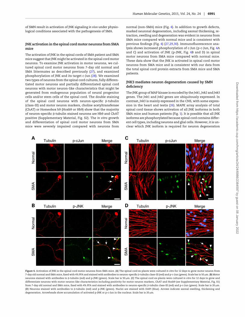
The activation of JNK in the spinal cords of SMA patient and SMAmice suggest that JNKmight be activated in the spinal cordmotorneurons. To examine JNK activation in motor neurons, we cul-tured spinal cord motor neurons from 7-day old normal andSMA littermates as described previously (27), and examinedphosphorylation of JNK and its target c-Jun (28). We examinedtwo types of neurons from the spinal cord cultures, fully differen-tiated motor neurons and partially differentiated spinal cordneurons with motor neuron-like characteristics that might begenerated from endogenous population of neural progenitorcells and/or stem cells of the spinal cord. The double stainingof the spinal cord neurons with neuron-specific β-tubulin(class-III) and motor neuron markers, choline acetyltransferase(ChAT) or Homeobox b9 (Hoxb9 or Hb9) show that the majorityof neuron-specific β-tubulin stained neurons are Hb9 and ChATpositive (Supplementary Material, Fig. S2). The in vitro growthand differentiation of spinal cord motor neurons from SMAmice were severely impaired compared with neurons from
normal (non-SMA) mice (Fig. 4). In addition to growth defects,marked neuronal degeneration, including axonal thickening, re-traction, swelling and degeneration was evident in neurons fromSMA mice compared with normal mice and is consistent withprevious findings (Fig. 4) (27,29,30). Immunofluorescence ana-lysis shows increased phosphorylation of c-Jun (p-c-Jun, Fig. 4Aand C) and activation of JNK (p-JNK, Fig. 4B and D) in spinalmotor neurons from SMA mice compared with normal mice.These data show that the JNK is activated in spinal cord motorneurons from SMA mice and is consistent with our data fromthe total spinal cord protein extracts from SMA mice and SMApatients.
JNK3 mediates neuron degeneration caused by SMNdeficiency
The JNK group ofMAP kinase is encoded by the Jnk1, Jnk2 and Jnk3genes. The Jnk1 and Jnk2 genes are ubiquitously expressed. Incontrast, Jnk3 is mainly expressed in the CNS, with some expres-sion in the heart and testis (26). MAPK array analysis of totalspinal cord tissue shows activation of all JNK isoforms in bothSMA mice and human patients (Fig. 1). It is possible that all JNKisoforms are phosphorylated because spinal cord contains differ-ent cell types, including neurons and glial cells. However, it is un-clear which JNK isoform is required for neuron degeneration
Figure 4. Activation of JNK in the spinal cord motor neurons from SMA mice. (A) The spinal cord ex-plants were cultured in vitro for 12 days to grow motor neuron from
7-day old normal and SMAmice, fixedwith 4% PFA and stainedwith antibodies to neuron-specific β-tubulin class-III (red) and p-c-Jun (green). Scale bar is 50 μm. (B) Motor
neurons stained with antibodies to β-tubulin (red) and p-JNK (green). Scale bar is 50 μm. (C) The spinal cord ex-plants were cultured in vitro for 12 days to grow and
differentiate neurons with motor neuron-like characteristics including positivity for motor neuron markers, ChAT and Hoxb9 (see Supplementary Material, Fig. S1)
from 7-day old normal and SMA mice, fixed with 4% PFA and stained with antibodies to neuron-specific β-tubulin class-III (red) and p-c-Jun (green). Scale bar is 20 μm.
(D) Neurons stained with antibodies to β-tubulin (red) and p-JNK (green). Nuclei are stained with DAPI (blue). Arrows indicate axonal swelling, thickening and
degeneration. Arrowheads show accumulation of activated p-JNK or p-c-Jun in the nucleus. Scale bar is 20 μm.
Human Molecular Genetics, 2015, Vol. 24, No. 24 | 6991
Dow
nloaded from https://academ
ic.oup.com/hm
g/article/24/24/6986/2384597 by guest on 08 January 2022
caused by low levels of SMN. To identify the specific JNK isoformrequired for neurodegeneration, large numbers of purified neu-rons were required for in vitro biochemical experiments. There-fore, we used purified cultures of primary cerebellar granuleneurons (CGNs) from 7-day old mice (31), and examined phos-phorylation of JNK and its target c-Jun to establish that JNK is ac-tivated by SMN deficiency in cultured neurons. First, we testedwhether JNK is activated in cultured CGN from SMA mice. Pri-mary CGN from the normal and SMA mice 7-day old littermateswere cultured and stained with antibodies against phospho-c-Jun and phospho-JNK. The growth of cultured neurons fromSMA mice was severely impaired and gradual degeneration wasnoticed compared with neurons from normal littermates (Sup-plementary Material, Fig. S3). Immunofluorescence analysisshows phosphorylation of c-Jun (Supplementary Material, Fig.S3A) and activation of JNK (Supplementary Material, Fig. S3B) inneurons from SMA mice compared with normal mice. Thesedata show that the JNK is also activated in purified cultures ofCGN from SMA mice, and is consistent with data from culturedspinal cordmotor neurons. These data suggest that the low levelsof SMN associatedwith SMAmight not support normal growth ofneurons from SMA mice under in vitro experimental conditions.
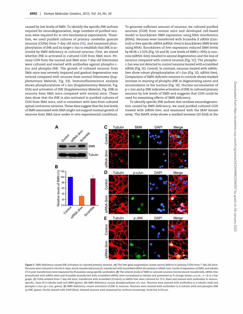
To generate sufficient amount of neurons, we cultured purifiedneurons (CGN) from normal mice and developed cell-basedmodel to knockdown SMN expression using RNA interference(RNAi). Neurons were transfected with Scramble II siRNA (Con-trol) or Smn specific siRNA (siRNA-Smn) to knockdown SMN levelsusing RNAi. Knockdown of Smn expression reduced SMN levelsby 68.06 ± 2.52% (Fig. 5A and B). Low levels of SMN (∼30%) in neu-rons (siRNA-Smn) resulted in axonal degeneration and the loss ofneurons compared with control neurons (Fig. 5C). The phospho-c-Junwas not detected in control neurons treatedwith scrambledsiRNA (Fig. 5D, Control). In contrast, neurons treated with siRNA-Smn show robust phosphorylation of c-Jun (Fig. 5D, siRNA-Smn).Comparison of SMN-deficient neurons to controls showsmarkedincrease in staining of phospho-JNK in degenerating axons andaccumulation in the nucleus (Fig. 5E). Nuclear accumulation ofp-c-Jun and p-JNK indicates activation of JNK in cultured primaryneurons by low levels of SMN and suggests that CGN could beused for examining effects of SMN deficiency.
To identify specific JNK isoform thatmediate neurodegenera-tion caused by SMN deficiency, we used purified cultured CGNtreated with siRNA-Smn, and examined with the MAP kinasearray. The MAPK array shows a marked increase (10-fold) in the
Figure 5. SMN deficiency causes JNK activation in cultured primary neurons. (A) The Smn gene suppression causes axonal defects in primary CGNs from 7-day old mice.
Neuronswere cultured in vitro for 6-days,mock-transfected (control), transfectedwith scrambled siRNA (Scramble) or siRNA-Smn. Levels of expression of SMN, and tubulin
(72 h post transfection) were examined by IB analysis using specific antibodies. (B) The relative levels of SMN in cultured neurons Control (mock-transfected), siRNA-Smn
(transfected with siRNA-Smn) and Scramble (transfected with scrambled siRNA) were normalized to tubulin and presented as % change (mean ± s.e.m., n = 3) in a bar
graph. (C) CGNs isolated from 7-day old mice, transfected with scrambled (Control) or siRNA-Smn were cultured for 72 h, fixed and stained with antibodies to neuron-
specific, class-III β-tubulin (red) and SMN (green). (D) SMN deficiency causes phosphorylation of c-Jun. Neurons were stained with antibodies to β-tubulin (red) and
phospho-c-Jun (p-c-Jun, green). (E) SMN deficiency causes activation of JNK in neurons. Neurons were stained with antibodies to β-tubulin (red) and phospho-JNK
(p-JNK, green). Nuclei stained with DAPI (blue). Stained neurons were examined by confocal microscopy. Scale bar is 8.0 μm.
6992 | Human Molecular Genetics, 2015, Vol. 24, No. 24
Dow
nloaded from https://academ
ic.oup.com/hm
g/article/24/24/6986/2384597 by guest on 08 January 2022

phosphorylation of JNK3, a neuron-specific isoform (19), in de-generating neurons caused by SMN knockdown (Fig. 6A). Thesedata indicate that the reduced levels of SMN cause preferentialactivation of JNK3 in neurons. To test whether JNK3 is requiredfor neurodegeneration,we examined the effect of SMNdeficiencyon neurons lacking JNK3. Neurons from wild-type Jnk3+/+ andknockout Jnk3−/− mice were cultured and transfected withsiRNA. Knockdown of SMN resulted in activation of JNK (p-JNK)and degeneration of Jnk3+/+ neurons (Fig. 6B, Tubulin). Interesting-ly, p-JNK was not detected in Jnk3−/− neurons lacking SMN anddegeneration of Jnk3−/− neurons was reduced compared with
Jnk3+/+ neurons (Fig. 6B). To further test whether JNK3 is requiredfor degeneration of SMN-deficient neurons, we complementedJnk3−/− neurons with recombinant GFP-JNK3 (32) using transienttransfection, and examined the effect of SMN deficiency. Controlexperiments show GFP-JNK3 was distributed in the axons andsoma, but excluded from the nucleus of Jnk3−/− neurons. The ab-sence of GFP-JNK3 in the nucleus of healthy neurons indicatesthat the JNK3 is not activated (Fig. 6C). Interestingly, knockdownof SMN results in degeneration of Jnk3−/− neurons expressingGFP-JNK3. The accumulation of GFP-JNK3 in the nucleus of de-generating neurons indicates phosphorylation and activation ofJNK3 (Fig. 6C). These data suggest that JNK3 contributes to neuro-degeneration caused by SMNdeficiency. Comparison of neurode-generation caused by SMN knockdown in Jnk3+/+and Jnk3−/−
neurons shows JNK3 deficiency provides neuroprotection (57.97± 0.92%, P < 0.0001), and about 80% of the neurons survive(Fig. 6D). Together, these data show that JNK3 is important forneurodegeneration caused by SMN deficiency and genetic elim-ination of JNK3 provides neuroprotection. These findings suggestthat JNK may be a potential target to reduce neurodegenerationand may help rescue SMA phenotype.
Genetic inactivation of Jnk3 rescues SMA phenotypein mice
To testwhether in vivo Jnk3 inactivationwill help reduce neurode-generation and rescue phenotype in mice with SMA, we gener-ated SMNΔ7 carrier mice with Jnk3 mutation (Jnk3−/+) [Smn−/+;SMN2+/+; SMNΔ7+/+; Jnk3−/+] to produce littermates with SMA-like diseasewith Jnk3+/+ (SMA) and Jnk3−/− (SMA-J3) and examinedphenotype. The Jnk3−/− knockout mice were characterized previ-ously and displayed normal phenotype, including fertility andlifespan; however, JNK3 deficiency reduced neurodegenerationinduced by kainic acid-mediated excitotoxicity and ischemia(18,19). Interestingly, JNK3 deficiency resulted in ameliorationof SMA phenotype by improving motor function, overall growthand survival of mice with SMA (Figs 7–9). Mice with SMA lackingJnk3−/− (SMA-J3) were healthy, and able to stand and walk com-pared with SMA mice (SMA) between the ages of 6–10 days(Fig. 7A). Analysis of gross motor functions show JNK3 deficiencyimproves motor functions of mice with SMA. The ability to standon four paws andwalk (time in sec) was improved in SMA-J3mice(22.20 ± 3.74, P = 0.0063) compared with SMA mice (2.50 ± 0.20)(Fig. 7B). Analysis of time-to-right (TTR) shows significant im-provement (P < 0.0001) in the motor function of SMA-J3 micecompared with SMA mice at 7 and 8-days of age (Fig. 7C). Totest whether improvement in the phenotype of SMA-J3 mice isindependent of changes in the SMN levels, we examinedwhetherJNK3 deficiency has any effect on the levels of SMN. Quantitativeanalysis of IBs of the spinal cord, brain andmuscle tissues did notshow change in the levels of SMN protein in SMA-J3 mice com-pared with SMA mice (Fig. 7D and E). These data suggest thatthe improvement in SMA phenotype by JNK3 deficiency may beindependent of SMN.
Deletion of the Jnk3 gene resulted in increase of growth-periodfrom 8-days (SMA) to 14-days (SMA-J3), and shows 1.75-fold (P =0.0023) increase in growth-period of SMA-J3 mice compared withSMAmice. SMA-J3 mice gained higher average peak body weight(g) [4.6 ± 0.30] than SMA [2.88 ± 0.32]. The change in body weight(g) from birth to peak for SMA (1.46 ± 0.25) and SMA-J3 (2.98 ± 0.37)shows a 2.05-fold increase in body growth of SMA-J3 mice(Fig. 8A). It is established that the embryonic growth and weightof SMA newborn babies are slow and lower compared with nor-mal babies. To examine whether JNK3 deficiency also improved
Figure 6. Neuron-specific isoform JNK3 is required for neuron degeneration
caused by SMN deficiency. (A) SMN deficiency causes activation of JNK3, a
neuron-specific isoforms, in cultured primary neurons. Cultured mouse CGNs
were transfected with scrambled siRNA (Control) or siRNA-Smn. The levels of
phosphorylation were examined by phospho-MAPK antibody array (R&D
Systems Inc.). The levels of phosphorylation of individual JNK isoforms, JNK1
JNK2 and JNK3 were quantitated by densitometry and relative intensities were
calculated (mean ± s.e.m., n = 4) to plot fold activation as a bar graph. (B) JNK3 is
required for neuron degeneration caused by SMN deficiency. JNK3 deficiency
reduces neuron degeneration caused by low levels of SMN. Neurons (CGN) from
wild-type (Jnk3+/+) and knockout (Jnk3−/−) 7-day old mice transfected with
scrambled (Control) or siRNA-Smn were cultured for 72 h and stained with
antibodies to β-tubulin (red) and phospho-JNK (green). (C) Neurons from
knockout Jnk3−/− mice were transfected with plasmid expressing GFP-JNK3 and
with either scrambled (Control) or siRNA-Smn, cultured for 72 h and stained
with antibodies to β-tubulin (red). JNK3 was examined by GFP fluorescence
(green) using confocal microscopy. Nuclei stained with DAPI (blue). Scale bar is
20 μm. (D) Quantification of neuron degeneration. Neurons with intact axons
were counted in Control (transfected with scrambled), siRNA-Smn (treated with
SMN specific siRNA) and Jnk3−/− (neurons lacking JNK3). Thirty images, 10 each
from three different experiments for control or treated were examined for
neuron degeneration. The average (mean ± s.e.m.; n = 3) percent of surviving
neurons stained with nuclear DAPI is plotted as a bar graph. The statistical
significance (P-value) of neuroprotection data was determined by one-way
ANOVA and unpaired t-test with Welch’s correction (two tailed). *P < 0.0001.
Human Molecular Genetics, 2015, Vol. 24, No. 24 | 6993
Dow
nloaded from https://academ
ic.oup.com/hm
g/article/24/24/6986/2384597 by guest on 08 January 2022

embryonic growth, we compared weights of Normal (1.76 ±0.031), SMA (1.42 ± 0.047) and SMA-J3 (1.63 ± 0.052) mice at birth.The reduced weight loss in newborn pups of SMA-J3 (0.13 g)than SMA (0.34 g) compared with Normal mice shows a 2.62-fold improvement in embryonic growth of SMA mice lackingJNK3 (ANOVA, P < 0.0001). These data suggest that JNK3 deficiencyimproves embryonic and postnatal growth of mice with SMA.Mutation of Jnk3 also resulted in 2-fold increase in average sur-vival of SMA-J3 mice (14.95 ± 0.79) days comparedwith SMA (7.14± 0.93, Log-rank test, P < 0.0001). Notably, the 7 days increase inmaximum life of SMA-J3 mice (45% increase in lifespan) includes6 days of increase in growth (reduced severity) that shows about86% of the total increase in lifespan is with reduced burden of ill-ness. Importantly, the 4.20-fold increase in initial survival ofSMA-J3mice comparedwith SMAmice suggests that the JNK3 de-ficiencymay reduce the severity of disease andmay contribute tomarked reduction in early mortality compared with SMA mice(Fig. 8B). These data suggest that the JNK3 deficiency improvesthe growth and survival of mice with SMA.
Further, to determine whether improvement in the SMAphenotype is because of reduced neurodegeneration or neuro-protection caused by JNK3 deficiency, we examined motor neu-rons in the spinal cords of Normal, SMA and SMA-J3 mice.Immunohistochemical staining for Choline Acetyletransferase(ChAT) and histochemical staining (H & E) of spinal cord sectionsshow reduced loss of spinal motor neurons in SMA-J3 mice com-pared with SMA mice (Fig. 8C). Immunohistochemical stainingwith antibody to activated (cleaved) caspase 3 shows reducedcell death in spinal cord from SMA-J3 mice compared with SMAmice and suggests decrease in neurodegeneration (Fig. 8C, bottompanel). Comparison of number of motor neurons in the spinalcords (20 sections) of Normal (1462 ± 148), SMA (730 ± 124) andSMA-J3 (1197 ± 101) (mean ± s.d., three mice/group) mice showsthat the JNK3 deficiency reduced loss of motor neurons to ∼20%in SMA-J3 compared with ∼50% loss in SMA mice, and providedneuroprotection by increasing survival of neurons (81.92 ± 3.99%,P = 0.012, t-test) in SMA-J3 mice compared with SMA (49.95 ±4.90%) mice (Fig. 8D). These findings are consistent with the invitro data that the inhibition of JNK3 activity results in reduceddegeneration of SMN-deficient neurons (Fig. 6D).
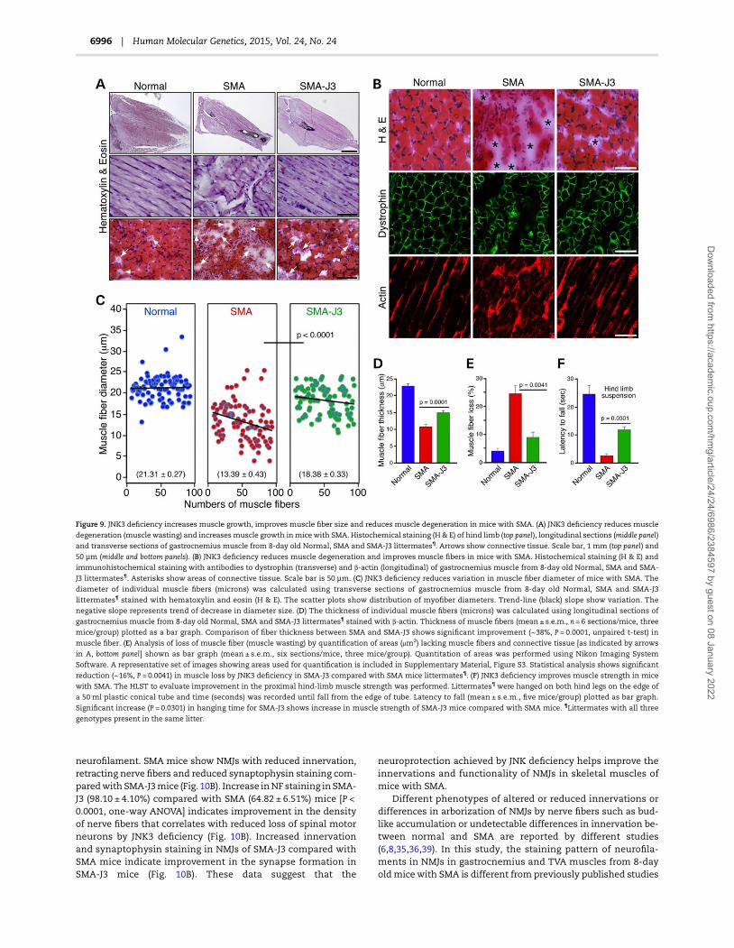
The degeneration of spinal motor neurons leads to progres-sive muscle atrophy (muscle wasting) in SMA. It is possible thatthe reduced loss of motor neurons may improve muscle massand account for increased growth in SMA-J3 mice. Examinationof hind-limb muscle indicates increased muscle mass in SMA-J3 mice that is consistent with 1.75-fold (P = 0.0023) increase inbody weight (Fig. 9A, top panel). Examination of gastrocnemiusskeletal muscle longitudinal and transverse sections shows re-duced muscle atrophy in SMA-J3 mice compared with SMAmice (Fig. 9A, middle and bottom panels). In addition, SMA-J3mice also show reduction in connective tissue and increase inmuscle fibers compared with SMA mice (Fig. 9A, bottom paneland 9B, top panel). Further examination of the muscle integrityand degeneration in skeletal muscle stained with antibodies todystrophin and β-actin (markers formusclemorphology) indicatereduced muscle degeneration in transverse (H & E and Dystroph-in) and longitudinal (Actin) gastrocnemius muscle sections ofSMA-J3 compared with SMA mice (Fig. 9B). Analysis of musclefiber diameter using a scatter plots shows reduced variation inmyofiber size and increased mean diameter (μm) in SMA-J3mice (18.38 ± 0.33) compared with SMA mice (13.39 ± 0.43) with P< 0.0001. Decrease in the negative slope of trend-line for SMA-J3mice compared with SMA mice suggests reduced variation inmyofiber size (Fig. 9C). Comparison of muscle fiber thickness
Figure 7. Genetic inactivation of JNK3 improves gross motor function in mice with
SMAwithout changing levels of the SMN protein. (A) A photograph of 10-day old
littermates generated by breeding of SMA carrier mice SMA-J3Het [Smn−/+; SMN2+/+;
SMNΔ7+/+; Jnk3−/+], non-SMA (Normal) [Smn+/+; SMN2+/+; SMNΔ7+/+; Jnk3+/+], SMA
[Smn−/−; SMN2+/+; SMNΔ7+/+; Jnk3+/+] and SMA mouse with Jnk3 mutation (SMA-J3)
[Smn−/−; SMN2+/+; SMNΔ7+/+; Jnk3−/−]. Genotypes were examined by PCR. (B) JNK3
deficiency improves ability of mice with SMA for motor action. Ability to stand on
four paws and walk was determined by measuring the time to fall off on four
paws in an effort to walk, and was recorded in 8-day old Normal, SMA and SMA-J3
littermates with all three genotypes present in the same litter. Latency to fall (sec)
is presented as (mean± s.e.m, n = 5 mice/group) in a bar graph. Unpaired t-test
(two tailed) between SMA and SMA-J3 shows significant (P = 0.0063) improvement
in the motor ability of SMA-J3 in comparison to SMA mice [dotted line indicates
time limit (30 s) of test]. (C) JNK3 deficiency improves gross motor function. Ability
to right was recorded for 5, 6, 7 and 8-day old Normal, SMA and SMA-J3 littermates
with all three genotypes present in the same litter. TTR with a time limit of 30 s for
test and three recordings per pup is plotted as (mean± s.e.m, n = 5 mice/group).
Improvement in the motor function is indicated by significant increase in the
ability to right for SMA-J3 mice compared with SMA mice. *(ANOVA, P < 0.0001;
t-test P = 0.0001) **(ANOVA, P < 0.0001; t-test P = 0.0038). (D) JNK deficiency did not
alter in vivo levels of SMN protein in SMA-J3 mice. Immunoblot analysis of the
brain, spinal cord and skeletal muscle tissues using antibody against SMN from 8-
day old Normal, SMA and SMA-J3 littermates with all three genotypes present in
the same litter. (E) The relative levels of SMN in the brain, spinal cord and skeletal
muscle tissues from Normal, SMA and SMA-J3 littermates were normalized to α-
tubulin and presented as relative (%) (mean± s.e.m., n= 3) in a bar graph.
6994 | Human Molecular Genetics, 2015, Vol. 24, No. 24
Dow
nloaded from https://academ
ic.oup.com/hm
g/article/24/24/6986/2384597 by guest on 08 January 2022

(μm)betweenNormal (22.73 ± 0.61), SMA (10.73 ± 0.31) and SMA-J3(14.80 ± 0.24) show marked increase (∼38%, P = 0.0001) in musclefiber thickness in SMA-J3 mice (Fig. 9D).
To determine whether neuroprotection provided by JNK defi-ciencywould help reducemuscle degeneration (musclewasting),we examined gastrocnemius muscle areas lacking myofibers asindicated by arrows in Figure 9A, bottom panel and by asterisksin 9B, top panel using Nikon Imaging Software (NIS), Element(v4.2) as described in Supplementary Material, Fig. S4. Analysisof loss of muscle fiber (muscle wasting) by quantification ofareas (μm2) lacking muscle fibers (connective tissue) shows(24.60 ± 1.59)% loss in SMA and only (8.80 ± 1.15)% in SMA-J3.These data suggest that the JNK3 deficiency results in significantreduction (∼16%, P = 0.0041) in muscle degeneration (wasting) ofSMA-J3 mice compared with SMA mice (Fig. 9E). Furthermore,to test whether reduction inmuscle degeneration improvesmus-cle strength, we used hind-limb suspension test (HLST) to evalu-ate improvement in the proximal hind-limbmuscle strength (33).Micewere hanged on both hind legs on the edge of a 50 ml plasticconical tube and time was recorded until fall from edge of thetube (Fig. 9F). Comparison of latency to fall (seconds) betweenSMA-J3 (11.67 ± 1.45) and SMA (2.66 ± 0.67) shows marked (4.39-fold, P = 0.0301) increase in hanging time that suggests increasein hind-limbmuscle strength of SMA-J3mice. These data suggest
that the JNK3 deficiency reduces muscle degeneration, improvesmuscle growth and increases muscle strength of mice with SMA.
Defects in maturation of NMJs in SMA patients (34) and SMAmice are because of poor innervations caused by axonal retrac-tion and neuron degeneration (6,35). To test whether neuropro-tection provided by JNK deficiency will improve NMJs, weexamined hind leg gastrocnemius skeletal muscle stained withantibody to neurofilament (NF-M, 145 kDa) protein and α-Bun-garotoxin (BTX) to visualize nerve and acetylcholine receptors(AChRs), respectively. Comparison of innervated NMJs, indicatedby colocalization (yellow) of NF (green) and AChRs (red), showsthat the muscle from SMA-J3 mice contain higher number of in-nervated NMJs than SMA mice (Fig. 10A, upper panel). Examin-ation of individual NMJs shows improved innervations ofreceptors in SMA-J3 mice compared with SMA mice that showNMJs with partial or full denervation (Fig. 10A, middle and lowerpanels). The denervation of NMJs due to neuron degenerationcauses defects in synapse maintenance and may be responsiblefor NMJ synaptopathy in SMA (6,35). Synaptic defects, such as de-fect in synapsematuration and reduced synapse are shown to bepresent in animalmodels of SMA, including SMAΔ7mousemodel(8,36–38). To test whether JNK deficiency also improved synapticmaturation in SMA-J3mice, we examined transversus abdominis(TVA)muscle stainedwith antibodies against synaptophysin and
Figure 8. The Jnk3 gene knockout in SMAmice reduces degeneration of spinal motor neurons, improves overall growth and increases lifespan of mice with SMA. (A) JNK3
deficiency improves the growth of mice with SMA. Growth (weight) curves of Normal [blue triangles], SMA-J3 [green squares] and SMA [red circles] mice littermates¶. The
results are presented as the mean ± s.e.m., n = 5 mice/group. Bar graphs show increase in number of growth days and increase in average peak weight of SMA-J3 mice
compared with SMA mice. (B) Deletion of Jnk3 gene increases lifespan of mice with SMA. Kaplan–Meier survival analysis of Normal (non-SMA) [blue], SMA-J3 [green]
and SMA [red] mice. Bar graphs show increase in initial and average survival of SMA-J3 mice compared with SMA mice. Arrow indicates initial survival and arrowhead
indicate maximum survival. (C) JNK3 deficiency reduces degeneration of spinal cord neurons in mice with SMA. Histochemical staining with hematoxylin and eosin
(H & E) and immunohistochemical staining of the spinal cords sections from 8-day old Normal, SMA and SMA-J3 littermates¶ with anti-ChAT and cleaved caspase 3
(activated*) antibodies. Insets show ventral horn neurons. Arrows indicate degenerating neurons. Scale bar is 200 µm, top and bottom panels, and 50 µm, middle panel.
(D) JNK3 deficiency provides neuroprotection and decreases the loss of spinal cord motor neurons. Bar Graph shows the loss of motor neurons in the thoracic (T9–T12)
region of spinal cords from SMA and SMA-J3 mice compared with Normal (non-SMA) mice (littermates¶) as the reference point (100%) (mean ± s.e.m.; three mice/group).¶Littermates with all three genotypes present in the same litter.
Human Molecular Genetics, 2015, Vol. 24, No. 24 | 6995
Dow
nloaded from https://academ
ic.oup.com/hm
g/article/24/24/6986/2384597 by guest on 08 January 2022
neurofilament. SMA mice show NMJs with reduced innervation,retracting nerve fibers and reduced synaptophysin staining com-paredwith SMA-J3mice (Fig. 10B). Increase inNF staining in SMA-J3 (98.10 ± 4.10%) compared with SMA (64.82 ± 6.51%) mice [P <0.0001, one-way ANOVA] indicates improvement in the densityof nerve fibers that correlates with reduced loss of spinal motorneurons by JNK3 deficiency (Fig. 10B). Increased innervationand synaptophysin staining in NMJs of SMA-J3 compared withSMA mice indicate improvement in the synapse formation inSMA-J3 mice (Fig. 10B). These data suggest that the
neuroprotection achieved by JNK deficiency helps improve theinnervations and functionality of NMJs in skeletal muscles ofmice with SMA.
Different phenotypes of altered or reduced innervations ordifferences in arborization of NMJs by nerve fibers such as bud-like accumulation or undetectable differences in innervation be-tween normal and SMA are reported by different studies(6,8,35,36,39). In this study, the staining pattern of neurofila-ments in NMJs in gastrocnemius and TVA muscles from 8-dayoldmice with SMA is different from previously published studies
Figure 9. JNK3 deficiency increases muscle growth, improves muscle fiber size and reduces muscle degeneration in mice with SMA. (A) JNK3 deficiency reduces muscle
degeneration (muscle wasting) and increases muscle growth inmicewith SMA. Histochemical staining (H & E) of hind limb (top panel), longitudinal sections (middle panel)
and transverse sections of gastrocnemius muscle from 8-day old Normal, SMA and SMA-J3 littermates¶. Arrows show connective tissue. Scale bar, 1 mm (top panel) and
50 µm (middle and bottom panels). (B) JNK3 deficiency reduces muscle degeneration and improves muscle fibers in mice with SMA. Histochemical staining (H & E) and
immunohistochemical staining with antibodies to dystrophin (transverse) and β-actin (longitudinal) of gastrocnemius muscle from 8-day old Normal, SMA and SMA-
J3 littermates¶. Asterisks show areas of connective tissue. Scale bar is 50 µm. (C) JNK3 deficiency reduces variation in muscle fiber diameter of mice with SMA. The
diameter of individual muscle fibers (microns) was calculated using transverse sections of gastrocnemius muscle from 8-day old Normal, SMA and SMA-J3
littermates¶ stained with hematoxylin and eosin (H & E). The scatter plots show distribution of myofiber diameters. Trend-line (black) slope show variation. The
negative slope represents trend of decrease in diameter size. (D) The thickness of individual muscle fibers (microns) was calculated using longitudinal sections of
gastrocnemius muscle from 8-day old Normal, SMA and SMA-J3 littermates¶ stained with β-actin. Thickness of muscle fibers (mean ± s.e.m., n = 6 sections/mice, three
mice/group) plotted as a bar graph. Comparison of fiber thickness between SMA and SMA-J3 shows significant improvement (∼38%, P = 0.0001, unpaired t-test) in
muscle fiber. (E) Analysis of loss of muscle fiber (muscle wasting) by quantification of areas (μm2) lacking muscle fibers and connective tissue [as indicated by arrows
in A, bottom panel] shown as bar graph (mean ± s.e.m., six sections/mice, three mice/group). Quantitation of areas was performed using Nikon Imaging System
Software. A representative set of images showing areas used for quantification is included in Supplementary Material, Figure S3. Statistical analysis shows significant
reduction (∼16%, P = 0.0041) in muscle loss by JNK3 deficiency in SMA-J3 compared with SMA mice littermates¶. (F) JNK3 deficiency improves muscle strength in mice
with SMA. The HLST to evaluate improvement in the proximal hind-limb muscle strength was performed. Littermates¶ were hanged on both hind legs on the edge of
a 50 ml plastic conical tube and time (seconds) was recorded until fall from the edge of tube. Latency to fall (mean ± s.e.m., five mice/group) plotted as bar graph.
Significant increase (P = 0.0301) in hanging time for SMA-J3 shows increase in muscle strength of SMA-J3 mice compared with SMA mice. ¶Littermates with all three
genotypes present in the same litter.
6996 | Human Molecular Genetics, 2015, Vol. 24, No. 24
Dow
nloaded from https://academ
ic.oup.com/hm
g/article/24/24/6986/2384597 by guest on 08 January 2022

(such as bud-like accumulation) and it might be because of a dif-ferent clone (3H11) of neurofilament antibody (NF-M, 145 kDa)compared with other NF antibody clones such as NN18 (NF-M,160 kDa) or SMI 312 (NF-H) or SMI-31 (phospho-NF-H) antibodiesin comparison to gastrocnemius, TVA/LAL, TA, paraspinal or dia-phragm muscle from mice with varying ages in several studies(6,8,35,36,39). It is possible that the recognition of unique epi-topes on NF protein by individual antibody clones results in dis-tinct patterns of staining. Nevertheless, SMA mice show muscleatrophy, partial and full denervation of NMJs and reduced
synapse formation during post-symptomatic development thatare consistent with SMA pathogenesis (3,20,38,40). Together,these findings suggest that JNK3 deficiency reduces loss ofmotor neurons, decreases muscle degeneration, increases mus-cle fiber size and muscle growth, increases overall growth, im-proves motor function and increases lifespan of mice with SMAthat represents a systemic partial rescue of SMA phenotype bygenetic inhibition of JNK3.
DiscussionSMA is the leading genetic neuromuscular disorder of infantmortality with an incidence of 1:6000–10 000 worldwide. The se-verity of disease is classified into four types [SMA I (severe), II, III,IV] based on the age of onset, motor function ability and copynumber of the SMN2 gene (4,41). The loss of spinalmotor neuronsleading to muscle weakness is the primary pathogenesis ob-served in all forms of SMA. The molecular mechanisms that me-diate neurodegeneration in SMA are unclear. We show that theJNK pathway mediates neurodegeneration in SMA. We identifiedneuron-specific JNK isoforms, JNK3 as a potential target thatmediates degeneration of SMN-deficient neurons. Genetic elim-ination of JNK3 reduces severity of SMA disease and results inpartial but systemic rescue of phenotype in mice with SMA. Wepropose that the JNK signaling pathway mediates neurodegen-eration in SMA and JNK3 represents a potential (non-SMN) thera-peutic target to reduce the burden of illness in SMA.
Activation of the JNK signaling cascade in SMA
The role of JNK has been implicated in neurodegeneration (42).However, the intracellular mechanisms involved in JNK activa-tion under pathological conditions of neurodegenerative disor-ders are unclear. Major challenges to unequivocally identifymolecular events triggered by disease pathogenesis from de-ceased human subject samples include the rare possibility ofmultiple numbers of age-matched (2–6 months) controls (Nor-mal, non-SMA), specific tissue such as spinal cord with specificregion (lumbar region) and the PMI. For example, in one of thecontrols (non-SMA) sample (#82), male, age 137 days, post-mortem interval (37 h), cause of death complications of prema-turity, we found that the tissue and proteins were severelydegraded and this sample could not be used for analysis. Increasein phosphorylation of several kinases by MAPK array in SMA pa-tient tissues suggests some non-specific activation that might bebecause of longer PMI. Comparison of data betweenSMA4583 andSMA4629 shows similar activity and thatmay be because of simi-lar PMI of 4 and 3 h, respectively or similar disease severity. Thesample SMA4994 shows higher activity that could be because ofhigher PMI (19 h) or increased severity. Importantly, Control(#83) that has highest postmortem interval (27 h) showed leastactivity compared with all SMA samples (Fig. 2). Statistical ana-lysis and comparison ofMAPK array data for common kinases ac-tivated in both human and mice helped eliminate non-specificactivation; and suggested that the increase in JNK activitymight be specific to SMA pathogenesis, and provided groundsfor further testing and validation of JNK activation in SMA.
SMA is a developmental neuromuscular disorderwith neurondegeneration during early postnatal development that results inearly onset and stable, non-progressive course with gradual(chronic) increase inmuscleweakness. It is possible that increasein age of patients and/or difference in severity of disease maycontribute to higher kinase activity. Comparison of SMA patientswith significantly different ages 77 days (SMA4583) and 169 days
Figure 10. JNK3 deficiency improves innervation and synapse formation at NMJs
in skeletal muscles of mice with SMA. (A) Immunohistochemical staining of
gastrocnemius skeletal muscle with antibody to neurofilament M protein (NF,
green) and α-Bungarotoxin coupled with Alexa 594 (BTX, red) from 8-day old
Normal, SMA and SMA-J3 littermates¶. Scale bars, 50 µm (upper panel) and
10 µm (middle and lower panels). (B) Immunohistochemical staining TVA skeletal
muscle with antibodies to synaptophysin (red) and neurofilament M protein
(NF, green) from 8-day old Normal, SMA and SMA-J3 littermates¶. Scale bars,
20 µm (upper and middle panels) and 10 µm (lower panel). Arrowheads indicate
partially denervated NMJs. Arrow shows retracting or degenerating nerve fiber.
Asterisk shows non-innervated (or fully denervated) NMJs. ¶Littermates with all
three genotypes present in the same litter.
Human Molecular Genetics, 2015, Vol. 24, No. 24 | 6997
Dow
nloaded from https://academ
ic.oup.com/hm
g/article/24/24/6986/2384597 by guest on 08 January 2022

(SMA4629) show similar amount of JNK activation in spinal cords,and these findings are consistent with data from SMA mice thatshow about 2-fold increase in activated JNK between the ages of2-, 6- and 12-days (Fig. 3B). These findings suggest that agingmaynot cause a large increase in JNK activation during chronic SMAdisease progression; however, difference in severity may affectlevels of JNK activation as noted in SMA patients (SMA4583 andSMA4994).
The low levels of SMN in neurons may result in intracellularstress that might be a cause of activation of intracellular MAPKcascade, leading to JNK-mediated neurodegeneration in SMA.JNK is known to be activated by a variety of extracellular stresssignals, including growth factors, cytokines and UV (26). How-ever, mechanisms of JNK activation by intracellular stresses asa result of disease pathogenesis are relatively under studied. Ac-tivation of two MAP3Ks (ASK1 and MEKK1) and two MAP2Ks(MKK4 and MKK7) suggests higher order of regulation and speci-ficity for JNK activation in SMA. The marked (12-fold) increase inlevels of p-MKK4 compared with p-MKK7 indicates that MKK4might be preferentially activated; however, small activationof MKK7may be required for full in vivo activation of JNK (43). Ac-tivation of ASK1 andMEKK1 suggest the possibility of two signal-ing modules involved in the JNK activation because both MKK4and MKK7 are known to be activated by ASK1 and MEKK1 (25).It has been reported that Gemin5, a part of the SMN complex,interact with ASK1, MKK4 and JNK in 293T cells (44). It is possiblethat Gemin5 might be a scaffold for ASK1/MKK4/JNK signalingmodule in neurons. Low levels of SMN complexes would resultin a free pool of Gemin5 that might increase levels of ASK1/MKK4/JNK module in neurons. MEKK1 may contribute to MKK7activation by another module MEKK1/MKK7/JNK scaffold by neu-ron-specific JNK-interacting protein3 (JIP3) (45). Together, thesefindings suggest that an increase in the levels of ASK1/MKK4/JNK module may be because of low levels of SMN associatedwith SMA pathogenesis. A graphical model of the activation oftwo JNK signaling modules in SMA is presented in Figure 11. In-hibition of the ERK pathway is shown to up regulate SMN2
expression that is mediated by activation of the AKT/CREB path-way in mice with severe SMA [Smn−/−; SMN2+/+] (23). Inhibition ofthe JNKpathway by deletion of Jnk3did not alter the levels of SMNin the spinal cord, brain or muscle tissues of mice with SMA. Thedecrease in AKT phosphorylation in SMA is consistent with sup-pression of AKT pathway during JNK activation (21). These find-ings suggest that JNK3 may not be involved in the regulation ofSMN2 expression. However, the role of the JNK pathway in theregulation of SMN1 expression that generates several alternative-ly spliced transcripts (46), including axonal-specific SMN (a-SMN)isoform (47) that may contribute to SMA pathogenesis, remainsto be studied.
Rescue and potential therapeutic targets of SMA
The lack of knowledge of the molecular mechanisms that medi-ate degeneration of motor neurons has hampered developmentof therapeutics, and no treatment is available to prevent or re-duce neurodegeneration in SMA. Major efforts to develop SMAtherapeutics are focused on increasing levels of SMN proteinfrom the SMN2 gene by correcting splicing defects or enhancingtranscription usingmultiple approaches (48), including antisenseoligonucleotides (ASO) (49) and small cell permeable compoundssuch as histone deacetylase (HDAC) inhibitors (valprioc acid,hydroxyl urea, tricostatin A, suberoylanilidehydroxamic acidand LDN-76070) (50,51), RNA decapping enzyme (DcpS) inhibitors(quinazoline compounds, RG3039) (52), activator of JAK2/STAT5(prolactin) (53) and inhibitor of ERK/ELK-1 (U0126) (23) signalingpathways (48,54). One important question that needs to be ad-dressed is whether restoring levels of SMN in degenerating neu-rons that have reached to a ‘nopoint of return’will allow rescue ofneurons to reintegrate and function normally. To address this, astrategy of combination therapy may be required that could in-clude simultaneous treatments to (a) prevent or reduce degener-ation of neurons (e.g. JNK inhibitors) and (b) increase levels ofSMNusing ASO-based approach or treatment with small cell per-meable therapeutic compounds. Identification of non-SMN orSMN-independent drug targets is required to prevent neurode-generation and development of successful treatments for SMA.Some progress has been made in identifying non-SMN targets,including (i) Plastin-3 (PLS3) (30), and ZPR1 (27) as modifiers ofSMA, and (ii) RhoA/ROCK pathway (17). Overexpression of PLS3in mice with SMA improves NMJ functionality by delaying axonpruning and moderately improves survival of mice (55). Inhib-ition of ROCK using inhibitors (Y-27632 and Fasudil) resulted ina partial rescue with improved NMJ functionality in skeletalmuscle, and increase in survival of mice with intermediateSMAwithout affecting SMN levels (17,56). These findings indicatepossibility of rescue of SMA phenotype by mechanisms inde-pendent of SMN (57). The genetic inhibition of Jnk3 in mice withSMA reduced degeneration of motor neurons without affectingthe levels of SMN in the brain, spinal cord and muscle tissues,suggests that the JNK pathway is independent of SMN and JNK3represents a non-SMN target.
Degeneration of the spinal cordmotor neurons is the primarycause of pathogenesis that leads to progressive muscle atrophy,respiratory failure and death in SMA. Therefore, inhibition ofneurodegeneration in SMA may help reduce severity and rescueSMA phenotype. Deletion of the Jnk3 gene in mice with SMA(SMA-J3) provided protection against neurodegeneration, im-proved NMJ innervations and functionality, improved musclegrowth and motor function that resulted in partial systemic res-cue of SMA phenotype and increased lifespan of mice with SMA.The reasons for a partial rescue are unclear. However, it is
Figure 11. Graphical representation of activation of the JNK signaling pathway in
SMA. Two MAPK signaling modules (MAP3K→MAP2K→MAPK) (A)
ASK1→MKK4→ JNK3 and (B) MEKK1→MKK7→ JNK3 found to be activated in
the spinal cords of SMA mice and patients. In vitro elimination of JNK3 reduces
degeneration of SMN-deficient neurons and provides neuroprotection. In vivo
deletion of Jnk3 in SMA mice results in partial systemic rescue and
improvement in SMA phenotype.
6998 | Human Molecular Genetics, 2015, Vol. 24, No. 24
Dow
nloaded from https://academ
ic.oup.com/hm
g/article/24/24/6986/2384597 by guest on 08 January 2022

possible that the partial rescue is because of (i) partial protectionof neurons that might be due to functional redundancy of JNKisoforms because JNK1 can complement for JNK3 and mediateneurodegeneration (58); (ii) JNK has been shown to be activatedin skeletal muscle cells (59) and cultured primary muscle fromSMA patients (60). Therefore, JNK may also play a role in musclewasting and other JNK isoforms, JNK1 and JNK2,might contributeto muscle degeneration in SMA, but remains to be studied; (iii)JNK3 deficiency may not be able to complement for splicing de-fects caused by SMN deficiency in neurons and skeletal musclecells that result in reduced expression of (a) α-Nrxn2 in culturedneurons from severe SMA mouse model that might alter Ca2+ le-vels (61), (b) mature ryanodine receptor 1 that mediate calciumrelease from sarcoplasmic reticulum in SMA mice (62) and (c)sarcoplasmic reticulum Ca2+ ATPase [SERCA1a isoform predom-inantly express in TA skeletal muscle (63)] pump that removeCa2+ from sarcoplasm in SMA mice (62) and (iv) since JNK3 pre-dominately express in CNS, the function of other non-neuronaltissues/organs such as heart (64–66), pancreas (67), liver (68,69),lung (70), vasculature (71–73), gastrointestinal system (74) anddiaphragm/phrenic nerve (27) that regulate respiration (failureof respiration is the cause of death in SMA) (3) that are affectedin SMA might not be improved by JNK3 deficiency. In additionto CNS, JNK3 has shown to be expressed in the heart, it is possiblethat the JNK3 deficiency might improve heart function, but re-mains to be tested (75).
Genetic inhibition of JNK3 reduces severity of disease, in-creases lifespan and ameliorates SMA phenotype. However, araw comparison of survival of SMNΔ7 mice with other publishedstudies shows reduction in the average survival (∼8 days), com-pared with average survival (11–13 days) reported in differentstudies with SMNΔ7 mouse model (27,50,76). The originalSMNΔ7mice line (Stock #005025) from the Jackson Lab shows sur-vival of (7.6 ± 0.67 days, n = 20) under our laboratory conditions.These mice were crossed with Jnk3−/− mice to generate SMAmice with Jnk3 mutation (SMA-J3) and the survival of SMA pups(7.14 ± 0.93 days, n = 21) from SMA-J3 strain is similar to the sur-vival of original SMA line. The decrease in average survival of ori-ginal SMA Jackson strain might be because of several factors thatcould affect phenotype (survival) of SMA strain in different re-search laboratories, including diet, litter size, local environment,epigenetic modifications and any additional genetic modifica-tions of the parent strain. It is established that the changes inthe genetic content of SMA mouse models causes alteration inphenotype, including survival (77). To eliminate possibility ofgenetic variation, we analyzed and compared data from litter-mates produced by breeding of SMA-J3 carrier strain [Smn−/+;SMN2+/+; SMNΔ7+/+; Jnk3−/+]. Therefore, the marked increase inthe initial (4.20-fold), average (2.03-fold) and max survival by7-days of SMA-J3 mice compared with SMNΔ7 mice suggeststhat the JNK3 deficiency reduces severity of disease and increasessurvival of mice with SMA.
One of the important aspects of an ideal rescue of phenotypeis whether increased survival is accompanied with reduced se-verity of disease. JNK3 deficiency resulted in 2.62-fold increasein embryonic growth of SMA-J3 mice compared with SMA miceindicating beneficial effects of JNK3 inhibition during embryonicdevelopment and suggesting a reduction in the severity of SMA.The increase in postnatal growth-period of SMA-J3mice suggestsdecrease in postnatal severity of disease by elimination of JNK3.Increase in muscle fiber size and decrease in muscle wastingshow improved muscle strength and gross motor function inSMA-J3 mice compared with SMA mice that supports reductionin severity of disease. Together, these findings suggest that
JNK3 deficiency prevented neurodegeneration and reduced theseverity of disease from embryonic to postnatal stages that mayaccount for marked 4-fold increase in initial survival, and mayrepresent an important advancement towards developingalternative (SMN-independent) treatments to ameliorate SMAphenotype.
The role of JNK has been established in neuronal apoptosisand the JNK pathway has been indicated as a potential targetfor treatment of neurodegeneration in Alzheimer’s and Parkin-son’s diseases (78). Our data suggest that the JNK signaling haspotential for therapeutic intervention, and JNK3 may be a viabletarget to prevent neurodegeneration in SMA. A specific JNK3 in-hibitor may be very useful in preventing neurodegeneration;however, functional redundancy of JNK isoforms may comple-ment for JNK3 inhibition, but warrants further studies. It is pos-sible that inhibitors with broad specificity (inhibits all JNKisoforms) may also be very useful in reducing neurodegenerationto rescue SMAphenotype (79). Alternatively, the inhibition of JNKsignaling could also be achieved by targeting upstream kinasessuch as MKK4 and MKK7 (58). However, it remains to be testedwhether pharmacological inhibition of JNK using JNK3 specificor broad specificity inhibitors will be effective in SMA mousemodels. Nonetheless, improvement in SMA phenotype by genet-ic inhibition of JNK3 opens a gateway for exploring new avenuestowards developing therapeutic interventions.
In conclusion, this study provides insight into the role of JNK3(non-SMN target) in the pathogenesis and systemic improvementof SMA phenotype that will have important implications for de-veloping alternative (SMN-independent) strategies for the treat-ment of SMA.
Materials and MethodsMice
The wild-type (C57BL/6J) and Jnk3−/− mice (19) were used to gen-erate 7-day old pups. The SMA carrier mice on FVB background,[Smn−/+; SMN2+/+; SMNΔ7+/+] (20), purchased from the Jackson La-boratory were bred to generate non-SMA (normal) [Smn+/+;SMN2+/+; SMNΔ7+/+] and SMA [Smn−/−; SMN2+/+; SMNΔ7+/+] litter-mates. The Jnk3−/− mice (C57BL/6 background received from DrRoger Davis at the University of Massachusetts Medical School,Worcester) were backcrossed for 10 generations to wild-typeFVB/N mice to create Jnk3−/− (FVB background) mice. The SMAcarrier mice [Smn−/+; SMN2+/+; SMNΔ7+/+] were crossed withJnk3−/− (FVB/N) mice to generate SMA mice with heterozygousJnk3−/+. SMA carrier mice with heterozygous Jnk3−/+ [Smn−/+;SMN2+/+; SMNΔ7+/+; Jnk3−/+] were bred to generate mice withSMA-like disease [Smn−/−; SMN2+/+; SMNΔ7+/+; Jnk3+/+] SMA (simi-lar to SMAΔ7) and [Smn−/−; SMN2+/+; SMNΔ7+/+; Jnk3−/−] SMA-J3(Jnk3-null background) littermates. The homozygous state ofthe two transgenes SMN2+/+ and SMNΔ7+/+ was confirmed bybreeding newly generated carrier mice (SMA-J3) breeder withwild-type mice, and litters were examined by PCR (20) for pres-ence of SMN2 and SMNΔ7 in all the pups (data not shown). Ana-lyses of phenotypes were blinded and genotyping of littermateswere performed after collection of data by PCR using tail DNA.Any combination of two or three pups with genotypes Normal,SMA [Smn−/−; SMN2+/+; SMNΔ7+/+; Jnk3+/+] and SMA-J3 [Smn−/−;SMN2+/+; SMNΔ7+/+; Jnk3−/−] in a litter was considered littermates.Micewere sacrificedat specific timepoints to collect tissues for bio-chemical andhistological analysiswith averagepostmortem inter-val less than 30 minutes. Animal studies were approved by theInstitutional Animal Care and Use Committee (IACUC), TTUHSC.
Human Molecular Genetics, 2015, Vol. 24, No. 24 | 6999
Dow
nloaded from https://academ
ic.oup.com/hm
g/article/24/24/6986/2384597 by guest on 08 January 2022

SMA patient tissues
Human frozen spinal cord and brain tissues of (i) Control (non-SMA) sample-82, male, age 137 days, postmortem interval(37 h), cause of death complications of prematurity (born at 24weeks of gestation), (ii) Control (non-SMA)-83, Age 69 days,Male, postmortem interval 27 h (cause of death Asphyxia), (iii)SMA type I-4994, Age 59 days, Female, postmortem interval19 h (cause of death, complications of the disorder), (iv) SMAtype I-4583, Age 77 days, Male, postmortem interval 4 h (causeof death Pneumonia) and (v) SMA type I-4629, Age 169 days,Male, postmortem interval 3 h (cause of death CardiopulmonaryArrest) were received from the NICHD Brain and Tissue Bank forDevelopmental Disorders, Baltimore, MD 21201, USA. The Insti-tutional Review Board (IRB) of the TTUHSC approved the use ofhuman tissues.
MAPK array analysis
Standard and custom designed phospho-MAPK antibody arraysfrom the R&D Systems and Full-Moon BioSystems were pro-cessed according to manufacturer’s protocol. Protein extractswere prepared from three individual spinal cords from mice(12-day old) or SMA patients either pooled or individually exam-ined according to the manufacturer’s protocols. Array imageswere analyzed using GenePix Pro software. Relative signal inten-sities normalized to β-actin or GAPDH (mean ± sem) were repre-sented as bar graphs. One of the major differences betweenhuman and mouse tissue samples was the PMI. Average PMI formouse was 30 min in contrast to 8.6 h for human. It is plausiblethat certain differences in data could be due to longer PMI ofhuman tissues. We compared data for SMA mice and SMA pa-tients and focused our analysis on kinases that were commonlyactivated in both SMAmice and SMA patients using two differentMAPK arrays. The likely molecular targets were tested and con-firmed by biochemical methods using mouse spinal cord tissuesand cultured primary neurons. To reduce biological variation,protein extracts of spinal cords of three SMA patients or SMAmice were pooled and examined by phospho-MAPK antibodyarrays.
Primary neuron culture and RNAi
Mouse spinal cord explants from 7-day old normal and SMAmice(littermates) were cultured in vitro for 10–12 days in 8-well cham-bers microscope slides, coated with poly-D-lysine/laminin usingneurobasal medium supplemented with B-27, 700 m Glucose,Glutamine, 2.5 m KCl and penicillin/streptomycin with somemodifications as described previously (27). Half of the cultureme-dium was replaced with freshly made neurobasal medium every48 h. The identity and morphology of the spinal cord motor neu-rons and glial cells were established by staining with specificmarkers, including ChAT and Hoxb9 (27). Primary CGNs wereisolated from the cerebellum of 7-day old normal and SMAmice (littermates), WT and Jnk3−/− mice and cultured in vitro for6 days in 35 mm dish or 8-well chambers, coated with poly-D-lysine/laminin in neurobasal medium supplemented with B-27,glutamine, glucose and 2.5 m KCl and penicillin/streptomycin(31). Neurons were transfected with 100 n SMART pool siRNA(Dharmacon) specific for mouse Smn (M-044280-00) or ScrambleII (Control, 5′-GCGCGCTTTGTAGGATTCG-3′) oligos and withpcDNA3/GFP-JNK3 (1 μg) using Lipofectamine 2000 (80). Neuronswere harvested 72 h post transfection and examined by IB andimmunofluorescence (IF) analysis.
Immunoblot analysis
Human SMA patient type-I and non-SMA (Normal) age-matchedfrozen tissues (spinal cord and brain) were used for analysis. Tis-sues from different patients were pooled for extraction to reducebiological variation.Mouse spinal cord tissueswere isolated from12-day old non-SMA (Normal) and with SMA-like disease (SMA)littermates. Two spinal cords for each Normal, SMA set-1 (SMA-S1), set-2 (SMA-S2), set-3 (SMA-S3), set-4 (SMA-S4) were pooledfrom littermates for protein extraction. Protein extracts were pre-pared from culturedneurons,mouse tissues and human SMApa-tient tissues (brain and spinal cord) using Triton lysis buffer (81).Proteins were separated by SDS-PAGE and electrotransferred toPVDF membrane (Millipore). Immunoblot analysis was per-formed using primary antibodies (1:1000) followed by HRP-conju-gated donkey anti-mouse or rabbit IgG (1:5000) secondaryantibodies. The following primary antibodies specific for phosphoand non-phospho proteins were used for analysis: Phospho-ASK1(SAB4504337) and Phospho-MEKK1 (MAP3K1) #SAB4504617(Sigma), ASK1 (D11C9), Phospho-MKK7 (Ser271/Thr275) #4171,MKK7 #4172, Phospho-MLK3 (Thr277/Ser281) #2811, MLK3#2817, Phospho-SEK1/MKK4 (Ser257/Thr261) #9156, SEK1/MKK4#9152, Phospho-SAPK/JNK (Thr183/Tyr185) (81E11) #4668; SAPK/JNK (56G8) #9258, Phospho-c-Jun (Ser63) #9261, c-Jun (60A8)#9165 (Cell Signaling). The other antibodies used for analysis in-clude SMN (Clone 8, Transduction Lab), MEKK1 #ab55653, GFAP(GA5) #36700 (Cell Signaling), Cleaved Caspase-3 (Asp175) #9661.Chemiluminescence and quantitation of IBs were performedusing ImageQuant LAS4000. The relative amounts of proteins(mean ± sem) normalized to either non-phospho protein or tubu-lin are presented as bar graphs.
Immunofluorescence analysis
Cerebellar granule neurons were isolated, purified from the cere-bellum of 7-day old WT and Jnk3−/− mice and cultured in vitro for6 days in 8-well chambers coated with poly-D-lysine/laminin (31).Neuronswerefixedwith4%paraformaldehyde (PFA),washedwithPBS, permeabilized with 0.1% Triton-X100, blocked with 3% BSAand incubated with antibodies (82). Double labeling was per-formed by sequential incubations (1 h) with (a) antibodies againstphospho-JNK #V793B (Promega) or phospho-c-Jun (Ser 63) orChAT #ab6168 (Abcam) or Hoxb9 #92606 (Abcam), (b) Alexa 488-conjugated anti-mouse or rabbit IgG secondary antibody, (c)anti-β-Tubulin class-III neuron-specific, (TUJ1, Covance) and (d)Alexa 546-conjugated anti-rabbit or mouse IgG secondary anti-bodies (81). The GFP-JNK3 fluorescence was detected on excita-tion with 488 nm Laser (32). Cover slips were mounted on slidesusing Vectashield with DAPI (Vector Lab) to stain DNA and IFwas examined using confocal microscopy [Leica SP5-AOBSequipped with 405 nm (UV) laser].
Immunohistochemistry
Mouse tissues, spinal cords and skeletal muscle from Normal(non-SMA), mice with SMA-like disease (SMA) and SMA micewith Jnk3−/− (SMA-J3) littermates 8–12 day old were isolated,fixed in 4% PFA at 4°C for overnight, washed with PBS and thensoaked in 30% sucrose solution for 48 h. Tissues were processedand embedded in paraffin or in OCT for frozen sections. Thin ser-ial sections (10 µm) were cut from the thoracic region (T9–T12) ofthe spinal cords and stained with hematoxylin and eosin or pro-cessed for immunohistochemical staining. Motor neurons werecounted in every fifth section (20 sections) of the thoracic (T9–T12) regions of the spinal cord (27,82). For immunohistochemistry
7000 | Human Molecular Genetics, 2015, Vol. 24, No. 24
Dow
nloaded from https://academ
ic.oup.com/hm
g/article/24/24/6986/2384597 by guest on 08 January 2022

staining, sectionswere processed forantigen retrieval, briefly incu-bated in SDS (1%) solution for 5 min and another 5–10 min in 0.1 Msodium citrate buffermaintained at 95°C. The spinal cord sectionswere rinsed in PBST, blocked in 3% BSAwith 0.1% Triton-X100 foran hour and then incubated overnight with primary antibodies di-luted (1:100) in 3% BSA (see list of antibodies in Immunoblot AnalysisSection). Sections were washed five times in PBST, incubated foran hour with Alexa 488-conjugated anti-mouse or rabbit IgGsecondary antibody diluted to 1:400 in 3%BSA, washed 4× withPBST and mounted with DAPI containing Vectashield (VectorLab). Muscle sections (16 µm) from hind limb were rinsed withPBS to remove OCT compound and then incubated in blockingbuffer (3% BSA with 0.1% Triton-X 100) for 1 h followed by 24 hincubation with primary antibodies to dystrophin (#15277,Abcam), β-actin (Sigma) or neurofilament (NF-M, 145 kDa) protein(#MAB1621, Millipore) diluted to 1:100 in 3% BSA. Next day, sec-tions were washed with PBST (5× for 10 min), incubated for 3 hwith Alexa 488 conjugated goat anti-rabbit or mouse secondaryantibody (diluted to 1:100 in 3% BSA), followed by washing withPBST (5× for 5 min). Double immunolabeling staining was per-formed on sections stained with anti-neurofilament protein.The sections were incubated with 3% BSA for half an hour, fol-lowed by incubation with Alexa 594 conjugated α-Bungarotoxin(Invitrogen) or anti-synaptophysin antibody (Abcam) (diluted to1:100 in 3% BSA) for an additional 3 h and rinsed with PBST (5×for 5 min) and slides were mounted using Vectashield. Immuno-fluorescence was examined by confocal microscopy.
Statistical analysis
The quantitative data are presented as mean ± s.e.m. Statisticalsignificance (P-values) of data was calculated by one-way ana-lysis of variance (ANOVA) analysis using GraphPad Prism or on-line software available at http://www.danielsoper.com/statcalc/calc43.aspx. Kaplan–Meier survival analysis, Log-rank (Mantel–Cox) test, Unpaired t-test with Welch’s correction (two tailed)and Wilcoxon signed rank test (two tailed) were performedusing GraphPad Prism (version 5.0d). The statistical value P = 0.05or less was considered significant.
Supplementary MaterialSupplementary Material is available at HMG online.
AcknowledgementsWe thank Dr Roger J. Davis, University of Massachusetts MedicalSchool, for reagents, including Jnk3 knockout mice. Human tis-sues were obtained from the NICHD Brain and Tissue Bank forDevelopmental Disorders at the University of Maryland, Balti-more, MD, USA. We thank Hafeez Ullah and Marisol Ramirez forexpert administrative assistance.
Conflict of Interest statement. A patent application on the use of JNKinhibitors for the treatment of SMA has been filed by L.G.
FundingThis study was supported in part by research grants to L.G. fromthe Muscular Dystrophy Association (MDA4209); TTUHSC-EPSeed Grant (14029IB) and the National Institutes of Health (R01NS064224).
References1. Lefebvre, S., Burglen, L., Reboullet, S., Clermont, O., Burlet, P.,
Viollet, L., Benichou, B., Cruaud, C., Millasseau, P., Zeviani, M.et al. (1995) Identification and characterization of a spinalmuscular atrophy- determining gene. Cell, 80, 155–165.
2. Lorson, C.L., Hahnen, E., Androphy, E.J. andWirth, B. (1999) Asingle nucleotide in the SMNgene regulates splicing and is re-sponsible for spinal muscular atrophy. Proc. Natl. Acad. Sci.USA, 96, 6307–6311.
3. Crawford, T.O. and Pardo, C.A. (1996) The neurobiology ofchildhood spinal muscular atrophy. Neurobiol. Dis., 3, 97–110.
4. Hamilton, G. and Gillingwater, T.H. (2013) Spinalmuscular at-rophy: going beyond the motor neuron. Trends Mol. Med., 19,40–50.
5. Burghes, A.H. and Beattie, C.E. (2009) Spinal muscular atro-phy: why do low levels of survival motor neuron proteinmake motor neurons sick? Nat. Rev. Neurosci., 10, 597–609.
6. Kariya, S., Park, G.H., Maeno-Hikichi, Y., Leykekhman, O.,Lutz, C., Arkovitz, M.S., Landmesser, L.T. and Monani, U.R.(2008) Reduced SMNprotein impairsmaturation of the neuro-muscular junctions in mouse models of spinal muscular at-rophy. Hum. Mol. Genet., 17, 2552–2569.
7. Monani, U.R. (2005) Spinalmuscular atrophy: a deficiency in aubiquitous protein; a motor neuron-specific disease. Neuron,48, 885–896.
8. Murray, L.M., Comley, L.H., Thomson, D., Parkinson, N., Tal-bot, K. and Gillingwater, T.H. (2008) Selective vulnerability ofmotor neurons and dissociation of pre- and post-synapticpathology at the neuromuscular junction in mouse modelsof spinal muscular atrophy. Hum. Mol. Genet., 17, 949–962.
9. Pellizzoni, L., Kataoka, N., Charroux, B. and Dreyfuss, G.(1998) A novel function for SMN, the spinal muscular atro-phy disease gene product, in pre-mRNA splicing. Cell, 95,615–624.
10. Zhang, Z., Pinto, A.M., Wan, L., Wang, W., Berg, M.G., Oliva, I.,Singh, L.N., Dengler, C., Wei, Z. and Dreyfuss, G. (2013) Dysre-gulation of synaptogenesis genes antecedes motor neuronpathology in spinal muscular atrophy. Proc. Natl. Acad. Sci.USA, 110, 19348–19353.
11. Baumer, D., Lee, S., Nicholson, G., Davies, J.L., Parkinson, N.J.,Murray, L.M., Gillingwater, T.H., Ansorge, O., Davies, K.E. andTalbot, K. (2009) Alternative splicing events are a late featureof pathology in a mouse model of spinal muscular atrophy.PLoS Genet., 5, e1000773.
12. Imlach, W.L., Beck, E.S., Choi, B.J., Lotti, F., Pellizzoni, L. andMcCabe, B.D. (2012) SMN is required for sensory-motor circuitfunction in Drosophila. Cell, 151, 427–439.
13. Wishart, T.M., Mutsaers, C.A., Riessland, M., Reimer, M.M.,Hunter, G., Hannam, M.L., Eaton, S.L., Fuller, H.R., Roche, S.L., Somers, E. et al. (2014) Dysregulation of ubiquitin homeo-stasis and beta-catenin signaling promote spinal muscularatrophy. J. Clin. Invest., 124, 1821–1834.
14. Praveen, K., Wen, Y. and Matera, A.G. (2012) A Drosophilamodel of spinal muscular atrophy uncouples snRNP biogen-esis functions of survivalmotor neuron from locomotion andviability defects. Cell Rep., 1, 624–631.
15. Bowerman, M., Shafey, D. and Kothary, R. (2007) Smn deple-tion alters profilin II expression and leads to upregulation ofthe RhoA/ROCK pathway and defects in neuronal integrity.J. Mol. Neurosci., 32, 120–131.
16. Nolle, A., Zeug, A., van Bergeijk, J., Tonges, L., Gerhard, R.,Brinkmann, H., Al Rayes, S., Hensel, N., Schill, Y., Apkhazava,D. et al. (2011) The spinal muscular atrophy disease protein
Human Molecular Genetics, 2015, Vol. 24, No. 24 | 7001
Dow
nloaded from https://academ
ic.oup.com/hm
g/article/24/24/6986/2384597 by guest on 08 January 2022
SMN is linked to the Rho-kinase pathway via profilin. Hum.Mol. Genet., 20, 4865–4878.
17. Bowerman, M., Beauvais, A., Anderson, C.L. and Kothary, R.(2010) Rho-kinase inactivation prolongs survival of an inter-mediate SMA mouse model. Hum. Mol. Genet., 19, 1468–1478.
18. Kuan, C.Y., Whitmarsh, A.J., Yang, D.D., Liao, G., Schloemer,A.J., Dong, C., Bao, J., Banasiak, K.J., Haddad, G.G., Flavell, R.A., Davis, R.J. and Rakic, P. (2003) A critical role of neural-spe-cific JNK3 for ischemic apoptosis. Proc. Natl. Acad. Sci. USA,100, 15184–15189.
19. Yang, D.D., Kuan, C.Y., Whitmarsh, A.J., Rincon, M., Zheng, T.S., Davis, R.J., Rakic, P. and Flavell, R.A. (1997) Absence of ex-citotoxicity-induced apoptosis in the hippocampus of micelacking the Jnk3 gene. Nature, 389, 865–870.
20. Le, T.T., Pham, L.T., Butchbach, M.E., Zhang, H.L., Monani, U.R., Coovert, D.D., Gavrilina, T.O., Xing, L., Bassell, G.J. andBurghes, A.H. (2005) SMNDelta7, the major product of thecentromeric survival motor neuron (SMN2) gene, extendssurvival inmicewith spinalmuscular atrophy and associateswith full-length SMN. Hum. Mol. Genet., 14, 845–857.
21. Sunayama, J., Tsuruta, F., Masuyama, N. and Gotoh, Y. (2005)JNK antagonizes Akt-mediated survival signals by phosphor-ylating 14-3-3. J. Cell Biol., 170, 295–304.
22. Bogoyevitch, M.A. and Kobe, B. (2006) Uses for JNK: the manyand varied substrates of the c-Jun N-terminal kinases. Micro-biol. Mol. Biol. Rev., 70, 1061–1095.
23. Branchu, J., Biondi, O., Chali, F., Collin, T., Leroy, F., Mam-chaoui, K., Makoukji, J., Pariset, C., Lopes, P., Massaad, C.,Chanoine, C. and Charbonnier, F. (2013) Shift from extracellu-lar signal-regulated kinase to AKT/cAMP response element-binding protein pathway increases survival-motor-neuronexpression in spinal-muscular-atrophy-likemice and patientcells. J. Neurosci., 33, 4280–4294.
24. Whitmarsh, A.J., Shore, P., Sharrocks, A.D. and Davis, R.J.(1995) Integration of MAP kinase signal transduction path-ways at the serum response element. Science, 269, 403–407.
25. Weston, C.R. and Davis, R.J. (2002) The JNK signal transduc-tion pathway. Curr. Opin. Genet. Dev., 12, 14–21.
26. Davis, R.J. (2000) Signal transduction by the JNK group of MAPkinases. Cell, 103, 239–252.
27. Ahmad, S., Wang, Y., Shaik, G.M., Burghes, A.H. and Gangwa-ni, L. (2012) The zinc finger protein ZPR1 is a potential modi-fier of spinal muscular atrophy. Hum. Mol. Genet., 21, 2745–2758.
28. Derijard, B., Hibi, M., Wu, I.H., Barrett, T., Su, B., Deng, T.,Karin, M. and Davis, R.J. (1994) JNK1: a protein kinase stimu-lated by UV light and Ha-Ras that binds and phosphorylatesthe c-Jun activation domain. Cell, 76, 1025–1037.
29. Rossoll, W., Jablonka, S., Andreassi, C., Kroning, A.K., Karle,K., Monani, U.R. and Sendtner,M. (2003) Smn, the spinalmus-cular atrophy-determining gene product, modulates axongrowth and localization of {beta}-actin mRNA in growthcones of motoneurons. J. Cell Biol., 163, 801–812.
30. Oprea, G.E., Krober, S., McWhorter, M.L., Rossoll, W., Muller,S., Krawczak, M., Bassell, G.J., Beattie, C.E. and Wirth, B.(2008) Plastin 3 is a protectivemodifier of autosomal recessivespinal muscular atrophy. Science, 320, 524–527.
31. Watson, A., Eilers, A., Lallemand, D., Kyriakis, J., Rubin, L.L.and Ham, J. (1998) Phosphorylation of c-Jun is necessary forapoptosis induced by survival signal withdrawal in cerebellargranule neurons. J. Neurosci., 18, 751–762.
32. McDonald, P.H., Chow, C.W., Miller, W.E., Laporte, S.A., Field,M.E., Lin, F.T., Davis, R.J. and Lefkowitz, R.J. (2000) Beta-
arrestin 2: a receptor-regulated MAPK scaffold for the activa-tion of JNK3. Science, 290, 1574–1577.
33. El-Khodor, B.F., Edgar, N., Chen, A., Winberg, M.L., Joyce, C.,Brunner, D., Suarez-Farinas, M. and Heyes, M.P. (2008) Identi-fication of a battery of tests for drug candidate evaluation inthe SMNDelta7 neonate model of spinal muscular atrophy.Exp. Neurol., 212, 29–43.
34. Arnold, A.S., Gueye, M., Guettier-Sigrist, S., Courdier-Fruh, I.,Coupin, G., Poindron, P. and Gies, J.P. (2004) Reduced expres-sion of nicotinic AChRs inmyotubes from spinalmuscular at-rophy I patients. Lab. Invest., 84, 1271–1278.
35. Kong, L., Wang, X., Choe, D.W., Polley, M., Burnett, B.G.,Bosch-Marce, M., Griffin, J.W., Rich, M.M. and Sumner, C.J.(2009) Impaired synaptic vesicle release and immaturity ofneuromuscular junctions in spinal muscular atrophy mice.J. Neurosci., 29, 842–851.
36. Ling, K.K., Lin, M.Y., Zingg, B., Feng, Z. and Ko, C.P. (2010) Syn-aptic defects in the spinal and neuromuscular circuitry in amousemodel of spinalmuscular atrophy. PLoS One, 5, e15457.
37. Torres-Benito, L., Ruiz, R. and Tabares, L. (2012) Synaptic de-fects in spinal muscular atrophy animal models. Dev. Neuro-biol., 72, 126–133.
38. Martinez, T.L., Kong, L., Wang, X., Osborne, M.A., Crowder, M.E., VanMeerbeke, J.P., Xu, X., Davis, C., Wooley, J., Goldhamer,D.J. et al. (2012) Survival motor neuron protein in motor neu-rons determines synaptic integrity in spinal muscular atro-phy. J. Neurosci., 32, 8703–8715.
39. Ruiz, R., Casanas, J.J., Torres-Benito, L., Cano, R. and Tabares,L. (2010) Altered intracellular Ca2+ homeostasis in nerveterminals of severe spinalmuscular atrophymice. J. Neurosci.,30, 849–857.
40. McGovern, V.L., Gavrilina, T.O., Beattie, C.E. and Burghes, A.H.(2008) Embryonicmotor axon development in the severe SMAmouse. Hum. Mol. Genet., 17, 2900–2909.
41. D’Amico, A., Mercuri, E., Tiziano, F.D. and Bertini, E. (2011)Spinal muscular atrophy. Orphanet. J. Rare Dis., 6, 71.
42. Coffey, E.T. (2014) Nuclear and cytosolic JNK signalling in neu-rons. Nat. Rev. Neurosci., 15, 285–299.
43. Tournier, C., Dong, C., Turner, T.K., Jones, S.N., Flavell, R.A.and Davis, R.J. (2001) MKK7 is an essential component ofthe JNK signal transduction pathway activated by proinflam-matory cytokines. Genes. Dev., 15, 1419–1426.
44. Kim, E.K., Noh, K.T., Yoon, J.H., Cho, J.H., Yoon, K.W., Drey-fuss, G. and Choi, E.J. (2007) Positive regulation of ASK1-mediated c-Jun NH(2)-terminal kinase signaling pathway bythe WD-repeat protein Gemin5. Cell Death Differ., 14, 1518–1528.
45. Kelkar, N., Gupta, S., Dickens, M. and Davis, R.J. (2000) Inter-action of a mitogen-activated protein kinase signaling mod-ule with the neuronal protein JIP3. Mol. Cell Biol., 20, 1030–1043.
46. Singh, N.N., Seo, J., Rahn, S.J. and Singh, R.N. (2012) A multi-exon-skipping detection assay reveals surprising diversityof splice isoforms of spinal muscular atrophy genes. PLoSOne, 7, e49595.
47. Setola, V., Terao, M., Locatelli, D., Bassanini, S., Garattini, E.and Battaglia, G. (2007) Axonal-SMN (a-SMN), a protein iso-form of the survival motor neuron gene, is specifically in-volved in axonogenesis. Proc. Natl. Acad. Sci. USA, 104, 1959–1964.
48. Howell, M.D., Singh, N.N. and Singh, R.N. (2014) Advances intherapeutic development for spinal muscular atrophy. FutureMed. Chem., 6, 1081–1099.
7002 | Human Molecular Genetics, 2015, Vol. 24, No. 24
Dow
nloaded from https://academ
ic.oup.com/hm
g/article/24/24/6986/2384597 by guest on 08 January 2022

49. Singh, N.N., Shishimorova, M., Cao, L.C., Gangwani, L. andSingh, R.N. (2009) A short antisense oligonucleotide maskinga unique intronicmotif prevents skipping of a critical exon inspinal muscular atrophy. RNA Biol., 6, 341–350.
50. Cherry, J.J., Osman, E.Y., Evans, M.C., Choi, S., Xing, X., Cuny,G.D., Glicksman, M.A., Lorson, C.L. and Androphy, E.J. (2013)Enhancement of SMN protein levels in a mouse model ofspinal muscular atrophy using novel drug-like compounds.EMBO Mol. Med., 5, 1035–1050.
51. Mohseni, J., Zabidi-Hussin, Z.A. and Sasongko, T.H. (2013)Histone deacetylase inhibitors as potential treatment forspinal muscular atrophy. Genet. Mol. Biol., 36, 299–307.
52. Gogliotti, R.G., Cardona, H., Singh, J., Bail, S., Emery, C., Kuntz,N., Jorgensen, M., Durens, M., Xia, B., Barlow, C. et al. (2013)The DcpS inhibitor RG3039 improves survival, function andmotor unit pathologies in two SMA mouse models. Hum.Mol. Genet., 22, 4084–4101.
53. Farooq, F., Molina, F.A., Hadwen, J., MacKenzie, D., Wither-spoon, L., Osmond, M., Holcik, M. and MacKenzie, A. (2011)Prolactin increases SMN expression and survival in amouse model of severe spinal muscular atrophy via theSTAT5 pathway. J. Clin. Invest., 121, 3042–3050.
54. VanMeerbeke, J.P. and Sumner, C.J. (2011) Progress and prom-ise: the current status of spinal muscular atrophy therapeu-tics. Discov. Med., 12, 291–305.
55. Ackermann, B., Krober, S., Torres-Benito, L., Borgmann, A.,Peters, M., Hosseini Barkooie, S.M., Tejero, R., Jakubik, M.,Schreml, J., Milbradt, J. et al. (2013) Plastin 3 ameliorates spinalmuscular atrophy via delayed axon pruning and improvesneuromuscular junction functionality. Hum. Mol. Genet., 22,1328–1347.
56. Bowerman, M., Murray, L.M., Boyer, J.G., Anderson, C.L. andKothary, R. (2012) Fasudil improves survival and promotesskeletal muscle development in a mouse model of spinalmuscular atrophy. BMC Med., 10, 24.
57. Tsai, L.K. (2012) Therapy development for spinalmuscular at-rophy in SMN independent targets. Neural Plast., 2012,456478.
58. Yamasaki, T., Kawasaki, H. and Nishina, H. (2012) DiverseRoles of JNK and MKK Pathways in the Brain. J. Signal Trans-duct., 2012, 459265.
59. Clavel, S., Siffroi-Fernandez, S., Coldefy, A.S., Boulukos, K., Pi-sani, D.F. andDerijard, B. (2010) Regulation of the intracellularlocalization of Foxo3a by stress-activated protein kinase sig-naling pathways in skeletal muscle cells. Mol. Cell. Biol., 30,470–480.
60. Anderson, K., Potter, A., Baban, D. and Davies, K.E. (2003) Pro-tein expression changes in spinal muscular atrophy revealedwith a novel antibody array technology. Brain, 126, 2052–2064.
61. See, K., Yadav, P., Giegerich, M., Cheong, P.S., Graf, M., Vyas,H., Lee, S.G., Mathavan, S., Fischer, U., Sendtner, M. andWinkler, C. (2014) SMN deficiency alters Nrxn2 expressionand splicing in zebrafish andmousemodels of spinalmuscu-lar atrophy. Hum. Mol. Genet., 23, 1754–1770.
62. Boyer, J.G., Murray, L.M., Scott, K., De Repentigny, Y., Renaud,J.M. and Kothary, R. (2013) Early onset muscle weakness anddisruption of muscle proteins in mouse models of spinalmuscular atrophy. Skelet Muscle, 3, 24.
63. Beard, N.A., Laver, D.R. and Dulhunty, A.F. (2004) Calseques-trin and the calcium release channel of skeletal and cardiacmuscle. Prog. Biophys. Mol. Biol., 85, 33–69.
64. Bevan, A.K., Hutchinson, K.R., Foust, K.D., Braun, L., McGo-vern, V.L., Schmelzer, L., Ward, J.G., Petruska, J.C., Lucchesi,P.A., Burghes, A.H. and Kaspar, B.K. (2010) Early heart failure
in the SMNDelta7 model of spinal muscular atrophy and cor-rection by postnatal scAAV9-SMN delivery. Hum. Mol. Genet.,19, 3895–3905.
65. Heier, C.R., Satta, R., Lutz, C. and DiDonato, C.J. (2010)Arrhythmia and cardiac defects are a feature of spinalmuscular atrophy model mice. Hum. Mol. Genet., 19, 3906–3918.
66. Rudnik-Schoneborn, S., Heller, R., Berg, C., Betzler, C.,Grimm, T., Eggermann, T., Eggermann, K., Wirth, R., Wirth,B. and Zerres, K. (2008) Congenital heart disease is a featureof severe infantile spinal muscular atrophy. J. Med. Genet.,45, 635–638.
67. Bowerman, M., Swoboda, K.J., Michalski, J.P., Wang, G.S.,Reeks, C., Beauvais, A., Murphy, K., Woulfe, J., Screaton, R.A., Scott, F.W. and Kothary, R. (2012) Glucose metabolismand pancreatic defects in spinal muscular atrophy. Ann.Neurol., 72, 256–268.
68. Vitte, J.M., Davoult, B., Roblot, N., Mayer, M., Joshi, V., Coura-geot, S., Tronche, F., Vadrot, J., Moreau, M.H., Kemeny, F. andMelki, J. (2004) Deletion ofmurine Smnexon 7directed to liverleads to severe defect of liver development associated withiron overload. Am. J. Pathol., 165, 1731–1741.
69. Hua, Y., Sahashi, K., Rigo, F., Hung, G., Horev, G., Bennett, C.F.and Krainer, A.R. (2011) Peripheral SMN restoration is essen-tial for long-term rescue of a severe spinal muscular atrophymouse model. Nature, 478, 123–126.
70. Schreml, J., Riessland, M., Paterno, M., Garbes, L., Rossbach,K., Ackermann, B., Kramer, J., Somers, E., Parson, S.H., Heller,R. et al. (2013) Severe SMA mice show organ impairment thatcannot be rescued by therapy with the HDACi JNJ-26481585.Eur. J. Hum. Genet., 21, 643–652.
71. Rudnik-Schoneborn, S., Vogelgesang, S., Armbrust, S., Graul-Neumann, L., Fusch, C. and Zerres, K. (2010) Digital necrosesand vascular thrombosis in severe spinal muscular atrophy.Muscle Nerve, 42, 144–147.
72. Araujo, A., Araujo, M. and Swoboda, K.J. (2009) Vascularperfusion abnormalities in infants with spinal muscularatrophy. J. Pediatr., 155, 292–294.
73. Foust, K.D., Wang, X., McGovern, V.L., Braun, L., Bevan, A.K.,Haidet, A.M., Le, T.T., Morales, P.R., Rich, M.M., Burghes, A.H. and Kaspar, B.K. (2010) Rescue of the spinal muscular atro-phy phenotype in a mouse model by early postnatal deliveryof SMN. Nat. Biotechnol., 28, 271–274.
74. Gombash, S.E., Cowley, C.J., Fitzgerald, J.A., Iyer, C.C.,Fried, D., McGovern, V.L., Williams, K.C., Burghes, A.H.,Christofi, F.L., Gulbransen, B.D. and Foust, K.D. (2015)SMN deficiency disrupts gastrointestinal and entericnervous system function in mice. Hum. Mol. Genet., 24,3847–3860.
75. Wang, Y. (2007) Mitogen-activated protein kinases in heartdevelopment and diseases. Circulation, 116, 1413–1423.
76. Butchbach, M.E., Singh, J., Thorsteinsdottir, M., Saieva, L.,Slominski, E., Thurmond, J., Andresson, T., Zhang, J., Edwards,J.D., Simard, L.R. et al. (2010) Effects of 2,4-diaminoquinazolinederivatives on SMN expression and phenotype in a mousemodel for spinal muscular atrophy. Hum. Mol. Genet., 19, 454–467.
77. Monani, U.R., Coovert, D.D. and Burghes, A.H. (2000) Animalmodels of spinal muscular atrophy. Hum. Mol. Genet., 9,2451–2457.
78. Junyent, F., Verdaguer, E., Folch, J., Zarate, C.B., Pallas, M., Au-ladell, C. and Camins, A. (2012) In Torrero, D.M., Haro, D. andValles, J. (eds), Recent Advances in Pharmaceutical SciencesII. Transworld Research Network, pp. 15–28.
Human Molecular Genetics, 2015, Vol. 24, No. 24 | 7003
Dow
nloaded from https://academ
ic.oup.com/hm
g/article/24/24/6986/2384597 by guest on 08 January 2022

79. Borsello, T. and Forloni, G. (2007) JNK signalling: a possibletarget to prevent neurodegeneration. Curr. Pharm. Des., 13,1875–1886.
80. Gangwani, L., Flavell, R.A. and Davis, R.J. (2005) ZPR1 is essen-tial for survival and is required for localization of the survivalmotor neurons (SMN) protein to Cajal bodies. Mol. Cell. Biol.,25, 2744–2756.
81. Gangwani, L., Mikrut, M., Theroux, S., Sharma, M. and Davis,R.J. (2001) Spinalmuscular atrophy disrupts the interaction ofZPR1 with the SMN protein. Nat. Cell. Biol., 3, 376–383.
82. Doran, B., Gherbesi, N., Hendricks, G., Flavell, R.A., Davis, R.J.and Gangwani, L. (2006) Deficiency of the zinc finger proteinZPR1 causes neurodegeneration. Proc. Natl. Acad. Sci. USA,103, 7471–7475.
7004 | Human Molecular Genetics, 2015, Vol. 24, No. 24
Dow
nloaded from https://academ
ic.oup.com/hm
g/article/24/24/6986/2384597 by guest on 08 January 2022