The Nosology ofthe Spinal Muscular Atrophies - NCBI
15
Review Article Journal of Medical Genetics (1971). 8, 481. The Nosology of the Spinal Muscular Atrophies ALAN E. H. EMERY From the University Department of Human Genetics, Western General Hospital, Edinburgh In the last few years there has been an increase in interest among neurologists and geneticists in the spinal muscular atrophies. With the advent of several relatively sophisticated diagnostic procedures many patients once thought to be suffering from muscular dystrophy have in fact been found to have spinal muscular atrophy. The spinal muscular atrophies may be defined as a group of inherited diseases in which there is degeneration of the anterior horn cells (lower motor neurones) of the spinal cord and often the bulbar motor nuclei, but with no evidence of pyramidal tract involvement. When so defined, motor neurone disease (progressive bulbar palsy, progressive muscular atrophy, and amyotrophic lateral sclero- sis) and its variants and the various forms of amyotrophy are excluded as are congenital abnor- malities of the spinal cord, and traumatic, toxic, infective, and neoplastic causes of anterior horn cell degeneration. Early literature on the spinal muscular atrophies has been extensively reviewed by Wiesendanger (1962); Smith and Patel (1965); Hausmanowa- Petrusewicz (1970); and Namba, Aberfeld, and Grob (1970). The frequency of all forms of spinal muscular atrophy is at least 1 in 20,000 (Brandt, 1950a) and possibly very much more. The 'wob- bler' mutant of mouse is a possible analogue (Duchen, Strich, and Falconer, 1968). The main clinical features of spinal muscular atrophy are wasting and weakness of muscles sup- plied by the affected anterior horn cells. Necropsy confirmation of degeneration and loss of anterior horn cells has been reported both in severe (Brandt, 1950a) and more benign (Kohn, 1968; Namba et al, 1970) forms of spinal muscular atrophy. It is essential to recognize the heterogeneity among the spinal muscular atrophies when giving a prognosis and for genetic counselling. Unfor- tunately early attempts to classify them were largely unsatisfactory for two reasons. Firstly, because such classifications were often based on clinical features Received 13 April 1971. alone, insufficient importance being given to genetic differences. Secondly, because of confusion with primary muscle diseases. Only in recent years, with the aid of more refined diagnostic procedures, has it been possible to make a clear distinction between these two groups of disorders. For these reasons in the following discussion evidence will be largely drawn from more recent publications. Some investigators have been attracted by the idea of a spectrum of clinical variation in spinal muscular atrophy ranging from the severe infantile to the benign adult forms. Some have even in- cluded certain neuropathies in this 'spectrum'. This concept however is unhelpful and if anything hinders the investigation of genetic heterogeneity. Aetiology The majority of cases of spinal muscular atrophy are clearly inherited. However, there have been frequent reports of the onset of symptoms following an infection (viral or bacterial) or an inoculation or vaccination (Brandt, 1950a; Wohlfart, Fex, and Eliasson, 1955; Dubowitz, 1964; Gardner-Medwin, Hudgson, and Walton, 1967; Meadows, Marsden, and Harriman, 1969a). Since in several of these reports there were other similarly affected rela- tives, it seems likely that clinical manifestations were precipitated rather than caused by the particular episode in question, but whether this or the subse- quent bed rest was the precipitating factor is not clear. Certainly, following bed rest, symptoms may be made much worse (eg, case 10 in Gardner- Medwin et al, 1967). Case Report. We have studied a patient with scapuloperoneal muscular atrophy in whom muscle weakness was first noticed at age 35 after 10 days bed rest with a swollen and painful leg following a smallpox vaccination. This man has an affected cousin in whom muscle weakness developed insidiously, the onset of which was not clearly associated with any particular exogenous factor. An apparent increase in the incidence of sporadic cases of infantile spinal muscular atrophy (Werdnig- 481
-
Upload
khangminh22 -
Category
Documents
-
view
0 -
download
0
Transcript of The Nosology ofthe Spinal Muscular Atrophies - NCBI

Review ArticleJournal of Medical Genetics (1971). 8, 481.
The Nosology of the Spinal Muscular AtrophiesALAN E. H. EMERY
From the University Department of Human Genetics, Western General Hospital, Edinburgh
In the last few years there has been an increase ininterest among neurologists and geneticists in thespinal muscular atrophies. With the advent ofseveral relatively sophisticated diagnostic proceduresmany patients once thought to be suffering frommuscular dystrophy have in fact been found to havespinal muscular atrophy.The spinal muscular atrophies may be defined as
a group of inherited diseases in which there isdegeneration of the anterior horn cells (lower motorneurones) of the spinal cord and often the bulbarmotor nuclei, but with no evidence of pyramidaltract involvement. When so defined, motorneuronedisease (progressive bulbar palsy, progressivemuscular atrophy, and amyotrophic lateral sclero-sis) and its variants and the various forms ofamyotrophy are excluded as are congenital abnor-malities of the spinal cord, and traumatic, toxic,infective, and neoplastic causes of anterior horn celldegeneration.
Early literature on the spinal muscular atrophieshas been extensively reviewed by Wiesendanger(1962); Smith and Patel (1965); Hausmanowa-Petrusewicz (1970); and Namba, Aberfeld, andGrob (1970). The frequency of all forms of spinalmuscular atrophy is at least 1 in 20,000 (Brandt,1950a) and possibly very much more. The 'wob-bler' mutant of mouse is a possible analogue(Duchen, Strich, and Falconer, 1968).The main clinical features of spinal muscular
atrophy are wasting and weakness of muscles sup-plied by the affected anterior horn cells. Necropsyconfirmation of degeneration and loss of anteriorhorn cells has been reported both in severe (Brandt,1950a) and more benign (Kohn, 1968; Namba et al,1970) forms of spinal muscular atrophy.
It is essential to recognize the heterogeneityamong the spinal muscular atrophies when giving aprognosis and for genetic counselling. Unfor-tunately early attempts to classify them were largelyunsatisfactory for two reasons. Firstly, because suchclassifications were often based on clinical features
Received 13 April 1971.
alone, insufficient importance being given to geneticdifferences. Secondly, because of confusion withprimary muscle diseases. Only in recent years, withthe aid of more refined diagnostic procedures, has itbeen possible to make a clear distinction betweenthese two groups of disorders. For these reasonsin the following discussion evidence will be largelydrawn from more recent publications.Some investigators have been attracted by the
idea of a spectrum of clinical variation in spinalmuscular atrophy ranging from the severe infantileto the benign adult forms. Some have even in-cluded certain neuropathies in this 'spectrum'.This concept however is unhelpful and if anythinghinders the investigation of genetic heterogeneity.
AetiologyThe majority of cases of spinal muscular atrophy
are clearly inherited. However, there have beenfrequent reports of the onset of symptoms followingan infection (viral or bacterial) or an inoculation orvaccination (Brandt, 1950a; Wohlfart, Fex, andEliasson, 1955; Dubowitz, 1964; Gardner-Medwin,Hudgson, and Walton, 1967; Meadows, Marsden,and Harriman, 1969a). Since in several of thesereports there were other similarly affected rela-tives, it seems likely that clinical manifestations wereprecipitated rather than caused by the particularepisode in question, but whether this or the subse-quent bed rest was the precipitating factor is notclear. Certainly, following bed rest, symptoms maybe made much worse (eg, case 10 in Gardner-Medwin et al, 1967).
Case Report. We have studied a patient withscapuloperoneal muscular atrophy in whom muscleweakness was first noticed at age 35 after 10 days bedrest with a swollen and painful leg following a smallpoxvaccination. This man has an affected cousin in whommuscle weakness developed insidiously, the onset ofwhich was not clearly associated with any particularexogenous factor.
An apparent increase in the incidence of sporadiccases of infantile spinal muscular atrophy (Werdnig-
481

Alan E. H. Emery
Hoffman disease) in the region of Freiburg, WestGermany, in recent years has been observed byBeckmann, Manz, and Moser (1970). Theseinvestigators attributed this to the more widespreaduse of oral poliomyelitis vaccine and the possi-bility of infection in utero by poliomyelitis virus.Poliomyelitis was first implicated as a possiblecausative factor in spinal muscular atrophy byMarburg in 1912. However, experimental evi-dence concerning RNA metabolism in degeneratingmotor neurones in infantile spinal muscular atrophy,does not support the idea of infection with an RNAvirus such as poliomyelitis (Hogenhuis, Spaulding,and Engel, 1967). The nature of the underlyinggenetic abnormality is unknown.
DiagnosisClinical Presentation. The distribution of
muscle wasting and weakness is different in differentspinal muscular atrophies, and the weakness is fre-quently associated with muscle fasciculations. Be-nign spontaneous fasciculation, however, is not un-common in normal individuals especially in the calfor small hand muscles. It cannot be distinguishedwith certainty from the pathological type butfasciculation which is seen only after movement orstrong contraction and is not associated with muscleweakness is usually benign (Gardner-Medwin andWalton, 1969). Fasciculations are rarely seen inprimary muscle disease. In spinal muscularatrophy they are indicative of anterior horn-cell de-generation and may be seen in the limb musculatureand/or the tongue.
Electromyography. Electromyographic (EMG)evidence of anterior horn cell degeneration, withloss of corresponding motor units, includes spon-taneous, regular motor unit activity (fibrillation orfasciculation potentials), and 'giant' action potentials(high amplitude and long duration) with reducedinterference pattern on voluntary contraction of themuscle (Lambert, 1963; Amick, Smith, and John-son, 1966; Gardner-Medwin et al, 1967;Hausmanowa-Petrusewicz et al, 1968; Meadowset al, 1969a and b). These large action potentialsmay be partly due to hypertrophy of remainingmuscle fibres. It is more likely, however, that themain factor is either the synchronous firing ofsurviving motor units with summation of motorunit potentials (Buchthal and Olsen, 1970), or thesynchronous firing of the normal motor unit plusthe muscle fibres which, due to collateral re-inner-vation, come to share the same motor nerve (seeBrown and Johns, 1970). In general the EMG
findings in the various spinal muscular atrophiesare very similar, though it has been reported thatthere are differences between the infantile andjuvenile forms (Buchthal and Olsen, 1970). TheEMG has occasionally been found to be normal oreven myopathic (Walton, 1970). Motor nerve con-duction velocity is normal.
Muscle Histology with conventional stainsreveals atrophy of groups of fibres (group atrophy)supplied by the affected anterior horn cells (Pearceand Harriman, 1966; Gardner-Medwin et al, 1967;Hausmanowa-Petrusewicz et al, 1968; Meadows etal, 1969a and b; Munsat et al, 1969; Buchthal andOlsen, 1970; Engel, 1970). Group atrophy ispathognomic of spinal muscular atrophy (Fig. 1).The atrophy may be so profound as to present theappearance of 'nuclear clumps'. The adjacentfibres may appear more-or-less normal or haveundergone changes similar to those seen in myo-pathy (Fig. 2); there may be variations in fibresize-some fibres being hypertrophic, roundingof fibres, central nuclei and even occasionally fibrenecrosis with phagocytosis (Mumenthaler, 1970).In long-standing cases the muscle tissue may belargely replaced by fat and connective tissue. Inthe very early stages of the disease, however, theonly abnormality may be 'scattered atrophy' (Fig.1): scattered small angular fibres wedged betweennormal looking fibres (Engel, 1970). Thus, thehistological appearances in the early stages of thedisease may be easily misdiagnosed as normal or asmyopathic in the later stages of the disease.The apparent myopathic changes in neurogenic
atrophy may be due to reinnervation by neighbour-ing healthy motor neurones (Meadows et al, 1969a).Drachman et al (1967) found that in muscle biopsiesfrom patients who had suffered from poliomyelitis10 to 54 years previously, features classically foundin myopathy were present. They suggested thatwork hypertrophy and partial re-innervation ofatrophic fibres could be responsible for variation infibre size, and that later degenerative changes mightbe due to defective re-innervation. Phagocytosisand endomysial fibrosis are merely reponses tofibre degeneration.
Muscle Histochemistry. Histochemicalstudies, using such enzyme stains as ATPase, NADdiaphorase, and phosphorylase, have shown thatmuscle fibres in human skeletal muscle can bedifferentiated into two types: type I fibres whichpredominate in slow muscles (eg, soleus) are rich inoxidative enzymes but poor in phosphorylaseactivity whereas the reverse is true of type II fibres
482

The Nosology of the Spinal Muscular Atrophies
FIG. 1. Muscle fibre atrophy in spinal muscular atrophy.Above: large groups of atrophic fibres (haematoxylin and eosin; original x 200.)
Below: small group of atrophic fibres (Gomori trichrome; original x 400.)Quadriceps muscle, juvenile spinal muscular atrophy.
483

Alan E. H. Emery
FIG. 2. Myopathic changes in spinal muscular atrophy. Note the variation in fibre size, rounding of muscle fibres, and fibre necrosis.Deltoid muscle, scapuloperoneal spinal muscular atrophy (haematoxylin and eosin; original x 200.)
which predominate in fast muscle (eg, gastrocne-mius). Normal muscle presents a mosaic patternof type I and type II fibres (Fig. 3) but in neurogenicatrophy there is a tendency for 'type grouping' tooccur ie, the grouping of fibres of a particularhistochemical type (I or II). It has been suggestedthat these findings could be due to collateral rein-nervation of affected fibres by normal motorneurones (Engel, 1970).
In long-standing neurogenic atrophy 'targetfibres' (concentric zones of enzyme activity extendingthe length of the fibre) are found in the muscle(Engel, 1961). These resemble the so-called'cores' seen in rabbit muscle during the phase ofreinnervation following denervation (Dubowitz,1967) and are similar to those seen in central coredisease (Dubowitz and Roy, 1970).
Muscle Innervation. Perhaps one of the mostuseful techniques for diagnosing neurogenic atro-phy is the intravital or supravital staining of motornerve filaments and end-plates with methylene blue(Coers and Woolf, 1959). In neurogenic atrophy(Fig. 4) there is branching of subterminal intra-muscular nerve fibres (rarely seen in normalmuscle) with collateral reinnervation, and degenera-tion of end-plates (Amick et al, 1966; Pearce and
Harriman, 1966; Hausmanowa-Petrusewicz et al,1968). Branching of nerve fibres seems to be moreprofuse in the more benign juvenile form of spinalmuscular atrophy than in the infantile form(Hausmanowa-Petrusewicz et al, 1968). A histo-logical diagnosis of denervation is only possiblewhen there is either group atrophy or branching ofsubterminal nerve fibres. The latter is particularlyuseful in cases where routine histology is normal orif 'myopathic' features alone are present (Meadowset al, 1969a).
Other Investigations. The diagnosis of spinalmuscular atrophy can be made on the clinicalfeatures, EMG findings, muscle histology, and thestudy of muscle innervation. Other investigationsadd little further information. The serum level ofcreatine kinase is raised in a proportion of patients,usually with the more benign forms of spinalmuscular atrophy (Hetnarska, Prot, and Sawicka,1968), and is commonly only slightly raised butoccasionally may be up to 20 times the normal value.Since the serum level of creatine kinase may also beelevated in various myopathies (Dreyfus andSchapira, 1962) it is of little diagnostic value.
In the Duchenne type of muscular dystrophy theelectrocardiogram (ECG) is frequently abnormal
484

The Nosology of the Spinal Muscular Atrophies 485
$P .....
L.ts>,,#R,@,5s~~~~~
FIG. 3. Histochemical differentiation into fibre types. Quadriceps muscle.Above: normal muscle (ATPase; original x 400).Below: type grouping (ATPase; original x 400).
41,~~ ~
A'P
41(
._ I1
.A,, .:.
.;e %, ":.'4%."ka..."z
r,
.1

Alan E. H. Emery
FIG. 4. Branching of subterminal intramuscular nerve fibres.Deltoid muscle, scapuloperoneal spinal muscular atrophy (supravitalstaining with methylene blue; original x 800.)
(Schott, Jacobi, and Wald, 1955; Manning andCropp, 1958; Skyring and McKusick, 1961; Gilroyet al, 1963) and characteristically there are tall Rwaves in the right praecordial lead (Perloff et al,1967; Emery, 1969; Emery and Spikesman, 1970).Though in other forms of dystrophy there are nospecific abnormalities, the ECG is frequently ab-normal (Lowenstein, Arbeit, and Rubin, 1962;Perloff, de Leon, and O'Doherty, 1966). In spinalmuscular atrophy, however, apart from a fine tremoroften most marked in the limb leads, the ECG isusually normal (Emery, 1972).Some investigators have reported increased
amounts of amino acids in urine and serum in somepatients with spinal muscular atrophy (Almog andTal, 1968; Gorecka, 1969). However this and theincreased levels of creatine in urine and serum(Namba et al, 1970), probably merely reflectmuscle wasting since they are most marked inpatients with the severe infantile (Werdnig-Hoff-mann) form and are also found in the severer formsof muscular dystrophy. Reported changes in theelectrophoretic patterns of serum proteins in spinalmuscular atrophy (Ionasescu et al, 1965; Askanas,1967) are difficult to evaluate. Comparable ab-normalities have also been described in musculardystrophy by Askanas (1967) but A. Jacobs (per-sonal communication) has been unable to confirm
these findings. Other biochemical findings in thisgroup of disorders have been reviewed by Nambaet al (1970). No specific biochemical abnormalityhas yet been identified in any of this group of dis-orders and there is no detectable chromosome ab-normality (Bonasegla and Montevecchi, 1970).
'Amyotonia Congenita'Amyotonia congenita (myatonia congenita,
Oppenheim's disease) is not a specific disease entitybut refers to the syndrome of generalized muscularhypotonia, feebleness of voluntary movements, anddepressed or absent tendon reflexes which is presentat birth or is manifest during early infancy. Batten(1910) was the first to recognize that the syndromecould be myopathic in origin or result from a lesionin the anterior horn cell of the spinal cord (infantilespinal muscular atrophy). More recent studieshave made it clear that the syndrome may in factresult from many causes (Brandt, 1950a and b;Walton, 1956 and 1957). In a follow-up study of109 cases initially diagnosed as 'amyotonia con-genita' 67 (61 %) were found to have infantile spinalmuscular atrophy, 22 (20%) had a myopathy, andthe remainder had a variety of disorders includingcerebral palsy, mental defect, and scurvy, etc(Walton, 1956). The congenital myopathies are
486

The Nosology of the Spinal Muscular Atrophiesthemselves a heterogeneous group of disorders(Emery and Walton, 1967). For these reasons theterm amyotonia congenita (or myatonia congenita orOppenheim's disease) has no diagnostic signifi-cance.
ClassificationIn 1968, the Research Group on Neuromuscular
Diseases (formerly the World Commission onNeuromuscular Diseases), with the direction ofProfessor J. N. Walton, subdivided the genetically-determined 'infantile and juvenile spinal muscularatrophies' into Werdnig-Hoffmann disease, arthro-gryposis multiplex due to anterior horn cell disease,Kugelberg-Welander disease, and scapuloperoneal
TABLE ICLINICOGENETIC CLASSIFICATION OF THE
SPINAL MUSCULAR ATROPHIES
A. Proximal spinal muscular atrophyI. Infantile (severe)
Autosomal recessiveII. Intermediate
? Autosomal recessiveIII. Juvenile (relatively benign)
1. Autosomal recessive (and 'Ryukyuan'spinal muscular atrophy)
2. Autosomal dominantIV. Adult
1. Autosomal recessive2. Autosomal dominant3. X-linked recessive
B. Distal spinal muscular atrophyI. No sensory loss
1. Autosomal recessive2. Autosomal dominant
II. With sensory loss1. Autosomal recessive2. Autosomal dominant3. X-linked recessive4. X-linked dominant
C. J7uvenile progressive bulbar palsy? Autosomal recessive
D. Scapuloperoneal spinal muscular atrophy1. Autosomal dominant2. ? Autosomal recessive
E. Facioscapulohumeral spinal muscular atrophyAutosomal dominant
spinal muscular atrophy. Subsequent developmentsand experience suggest that further subdivision isjustified.An outline of the proposed classification of the
spinal muscular atrophies based on clinical andgenetic differences is given in Table I. However,until the primary biochemical defect(s) underlyingspinal muscular atrophy is identified such a classi-fication must remain tentative. Nevertheless sincespinal muscular atrophy is clearly more than onegenetic entity (Becker, 1966; McKusick, 1968;Furukawa et al, 1969; Zellweger et al, 1969b) aclassification which is based on both genetic andclinical differences is valid and the best that ispossible with our present knowledge.
Infantile Spinal Muscular Atrophy(Werdnig-Hoffmann Disease)
Werdnig (1891 and 1894) and Hoffmann (1893,1900a and b) were the first to describe a syndromeof progressive muscle weakness beginning in thefirst year of life and terminating in death in earlychildhood, usually before the age of 4. BothWerdnig and Hoffmann had observed extensivedegenerative changes in the motor nuclei of thespinal cord in their patients and therefore this dis-order was a muscular atrophy and not a dystrophy(Fig. 5). The clinical features of infantile spinalmuscular atrophy or Werdnig-Hoffmann diseasehave since been described in detail by several in-vestigators (Hanhart, 1945; Brandt, 1950a and b;Thieffry, Arthuis, and Bargeton, 1955; Fukuyama,1958; Byers and Banker, 1961; Gamstorp, 1967).The largest reported series is that of Brandt
(1950a and b). Of 106 children with infantilespinal muscular atrophy where there was adequateinformation, the onset was in the first year of lifein 101 (95%) while in 3 it commenced in the secondyear of life and in 1 at about 2 years of age. In 94cases with adequate information, 76 (81 %) haddied by age 4. Some cases survived into adoles-cence but these may represent a different disorder(see below).Concordance for infantile spinal muscular atrophyhas been reported in identical twins (see Brandt,
1950a; Marquardt, MacLowry, and Perry, 1962;Zellweger et al, 1969b) and in Brandt's study(Brandt, 1950a) consanguinity was about 8 timesgreater among the parents of affected children thanamong controls. Family studies have shown thatthe condition is inherited as an autosomal recessivetrait. Apparent exceptions in published cases cansometimes be ascribed to faulty diagnosis in affected
487

Alan E. H. Emery
FIG. 5. Infantile spinal muscular atrophy (Werdnig-Hoffmanndisease) in a 2j-year-old boy.
relatives (eg, Brandt, 1950a [families 1 and 16]) or to
quasi-dominance due to inbreeding (eg, Brandt,1950a [family 6]).An association of arthrogryposis multiplex con-
genita and infantile spinal muscular atrophy in sibshas been reported (Frischknecht, Bianchi, andPilleri, 1960; Bargeton et al, 1961) which may re-
present a separate disease entity. Of course the
longer an affected individual survives the morelikely it is that secondary contractures will develop.
Juvenile Spinal Muscular Atrophy(Wohlfart-Kugelberg-Welander Disease)Wohlfart et al (1955) and Kugelberg and Welan-
der (1956) were the first to delineate a form of spinalmuscular atrophy with proximal muscle weakness,more benign than infantile spinal muscular atrophy,with onset usually after the age of 2 and with sur-vival often into adulthood. The clinical presenta-tion in many ways resembles muscular dystrophybut differs in that fasciculations are often presentand the EMG and muscle biopsy findings aretypical of neurogenic atrophy and not dystrophy.Clinical features of this juvenile form (Wohlfart-Kugelberg-Welander disease), have been reviewedby Namba et al (1970). Some investigators havereported that males are more severely affected thantheir female sibs (Dubowitz, 1964; Furukawa et al,1968) but others have not been convinced of this(Kugelberg and Welander, 1956; Gardner-Medwinet al, 1967). The condition may be associated witharthrogryposis (Amick, Johnson, and Smith, 1967[cases 7, 8, and 9]; Zellweger et al, 1969b [cases 13and 15]).Whether the infantile and autosomal recessive
juvenile forms of spinal muscular atrophy can beseparated on clinical grounds has been a matter ofdispute. Some believe the distinction is artificialsince it has been claimed that in a given sibship allgradations of severity can sometimes be seen(Byers and Banker, 1961; Dubowitz, 1964; Dunneand Chutorian, 1966; Rowland et al, 1967;Gardner-Medwin et al, 1967; Kessler, 1968;Meadows et al, 1969a; Munsat et al, 1969). Butmuch of the confusion arises because of overlap inthe age of onset. If other features are also takeninto account, such as the course of the disease andage at death, then it would seem possible that thesetwo disorders can be separated. Features whichmight be used to distinguish not only these twotypes of spinal muscular atrophy but also a possibleintermediate type are summarized in Table II.Ages of onset and at death are approximate as theexact limits for each type have not yet been defined.It is possibly the failure to recognize the existence ofan intermediate type which has made it difficult forinvestigators to separate these types of spinalmuscular atrophy particularly when age at onset wasused as the sole criterion (Fried and Emery, 1971).The frequency of spontaneous activity on electro-
myography (Buchthal and Olsen, 1970) and thedegree of collateral reinnervation (Hausmanowa-Petrusewicz et al, 1968) have also been found to
488

489The Nosology of the Spinal Muscular AtrophiesTABLE II
INFANTILE (I), INTERMEDIATE (II), and JUVENILE (III) AUTOSOMAL RECESSIVEFORMS OF PROXIMAL SPINAL MUSCULAR ATROPHY
Age Ability to Fasciculations Serum CreativeType Sit of Kinase Levelswithout Skeletal MusclesOnset Survival Support*
I. Infantile < 12 mth <4 yr Never + /- NormalII. Intermediate 3-18 mth >4 yr Usually +/- Usually normal
III. Juvenile > 2 yr Adulthood Always + + Often ralsed
* At some time during the course of the illness.
differ in the infantile and juvenile forms. Theinfantile, intermediate, and juvenile forms of proxi-mal spinal muscular atrophy have been referred to astypes I, II, and III respectively.
Autosomal RecessiveJuvenile Spinal Muscu-lar Atrophy. Juvenile spinal muscular atrophyhas been described in identical twins (Hausmanowa-Petrusewicz et al, 1962; Zellweger et al, 1969b),and including those first described by Wohlfart etal (1955) and Kugelberg and Welander (1956) therehave been many reports of families with multipleaffected sibs suggesting autosomal recessive inheri-tance (Byers and Banker, 1961; Levy and Wittig,1962; Castaigne et al, 1963; Dubowitz, 1964;Garvie and Woolf, 1966a; Radu et al, 1966;Gamstorp, 1967; Gardner-Medwin et al, 1967;Rowland et al, 1967; Peters et al, 1968; Meadowset al, 1969a; Hausmanowa-Petrusewicz, 1970).Consanguinity among the parents of affected indi-viduals has also been noted (Hanhart, 1962; Spira,1963; Radu et al, 1966; Almog and Tal, 1968). Ithas been reported that some healthy heterozygotesappear to have hyperaminoaciduria (Almog and Tal,1968) or may be detected by electromyography(Spira, 1967). These findings await confirmation.A possible variant of the juvenile type has been
described in an inbred community in the Ryukyuanislands off Japan (Kondo, Tsubaki, and Sakamoto,1970). This is characterized by the onset in infancyor early childhood of slowly progressive proximalmuscle weakness most marked in the lower extremi-ties and with pes cavus and scoliosis in some affectedindividuals. Muscle fasciculations were observedin several individuals, the serum level of creatinekinase was moderately elevated and there was nospontaneous activity in the electromyograph. Theauthors regarded this as a unique disorder and dif-ferent from the usual form of juvenile spinalmuscular atrophy.
Spiro, Fogelson, and Goldberg (1967) have de-scribed a family in which 3 brothers had a juvenileform of proximal spinal muscular atrophy associated
with microcephaly and mental subnormality whichmay also represent a distinct genetic disorder.
Autosomal Dominant Juvenile Spinal Mus-cular Atrophy. Excluding families with quasi-dominance due to inbreeding (eg, the family ofAlmog and Tal [1968]) a number of families havebeen described where juvenile spinal muscularatrophy was inherited as an autosomal dominanttrait with complete penetrance (Magee and deJong, 1960; Armstrong, Fogelson, and Silberberg,1966; Garvie and Woolf, 1966a; and Gamstorp,1967). In some families where the disorder wasclearly inherited as an autosomal dominant trait,penetrance was incomplete (Wohlfart et al, 1955[family 2]; Kugelberg and Welander, 1956 [family2]; Becker, 1964; Amick et al, 1966; Radu et al,1966; Tsukagoshi et al, 1966).The onset of the autosomal dominant form of
juvenile spinal muscular atrophy is usually in earlychildhood but may be delayed until adolescence oreven later (Kugelberg and Welander, 1956 [case 3];Becker, 1964; Tsukagoshi et al, 1966 [case III-2]).Affected individuals develop proximal muscleweakness which is relatively slowly progressive andseveral patients have been reported in middle ageyet still ambulatory. The condition seems to bemilder than the autosomal recessive form which,from reported familial cases of juvenile spinalmuscular atrophy appears to be at least 10 timescommoner than the autosomal dominant form.
Case Report. We have studied a mother and daughter(HG 243) with this form of spinal muscular atrophy.The mother was bom 4 June 1942 with talipes equino-varus which responded to conservative therapy. Shehas been aware of weakness in her legs from childhoodwhich does not seem to have progressed over the years.She cannot get up from a crouching position and hasdifficulty climbing stairs. On examination there wasweakness of her pelvic girdle musculature, her cranialnerves were intact and no fasciculations were observed.The serum level of creatine kinase was 48-0 Htmoles/min/l (normal less than 50). The diagnosis of spinal

Alan E. H. Emery
muscular atrophy was confirmed by EMG examinationand muscle biopsy. Her daughter (born 6 January1966) was noticed to walk with a waddle from about 2years of age. At present she has difficulty climbingstairs and cannot run as well as other children. Theweakness does not appear to have progressed. Theserum level of creatine kinase was 42-0 i&moles/min/l.No one else in the family has been similarly affected andthere is no consanguinity.
'Intermediate' Type of Spinal MuscularAtrophy
Several authorities have speculated upon thepossibility of a form of spinal muscular atrophyintermediate in severity between the infantile andjuvenile forms (Gamstorp, 1967; Verger et al,1969; Hausmanowa-Petrusewicz, 1970). In factsome of the earliest well documented cases of spinalmuscular atrophy could be considered as belongingto this type (Thomson and Bruce, 1893; Hoffnann,1900a). Some features which might be used todistinguish this type (intermediate or type IIproximal spinal muscular atrophy) are summarizedin Table II. There is some overlap in age of onsetwith infantile spinal muscular atrophy but in theintermediate type it appears that most patients sur-vive beyond 4 years and usually into adolescence orlater (Fried and Emery, 1971). The diagnosis ofintermediate or type II spinal muscular atrophy, if itis to be entertained at all, should therefore rest onseveral criteria. It seems possible that this type ofspinal muscular atrophy is inherited as an autosomalrecessive trait (Fried and Emery, 1971). However,to account for the possibility of mild and severeforms of juvenile spinal muscular atrophy, Becker(1964) has suggested that besides the normalgene (a), being present in about 90% of the popula-tion, there are 3 mutant alleles (a+, a', and a') suchthat a+a produces a normal phenotype, a+a' pro-
duces a milder disease with later onset, and a+a"produces a severer disease with earlier onset ('inter-mediate' type). Such a hypothesis might alsoexplain some of the cases of apparent nonpene-
trance where the only other affected individual in a
family is a second- or third-degree relative of theproband (Zellweger, Schneider, and Schuldt,1969a).
Adult Spinal Muscular AtrophyThis group of disorders present as progressive
muscle weakness with onset usually after the thirddecade of life. Since we are only concerned withdisorders associated with degeneration of theanterior horn cells and not the pyramidal tracts,motor neurone disease (and its variants) and the
various forms of amyotrophy are excluded; thegenetics of these forms have been discussed indetail elsewhere (Pratt, 1967; Kloepfer and Emery,1969).The main clinical feature of adult spinal muscular
atrophy is slowly progressive proximal limb weak-ness, and is therefore easily confused with limbgirdle muscular dystrophy or chronic polymyositisbut the distinction can be made on electromyo-graphic examination or muscle histology. Thebulbar nuclei may also become involved and theserum creatine kinase level may be moderatelyelevated. The prognosis is usually good.
Cases have been described which were sporadic(Hurwitz, Lapresle, and Garcin, 1961; Smith andPatel, 1965; Gross, 1966; Cazzato, 1969) or in-herited as autosomal recessive traits (Funk, 1962;Tsukagoshi et al, 1965 [cases 1, 2, and 3 and pos-sibly 5]; Mapelli and Ramelli, 1970), autosomaldominant traits (Tsukagoshi et al, 1965 [case 4],Peters et al, 1968 [family 13] and possibly the largefamily described by Finkel [1962]), or as X-linkedrecessive traits (Kennedy, Alter, and Foreman,1966; Kennedy, Alter, and Sung, 1968; Shoji et al,1970). An X-linked form of adult spinal muscularatrophy with a slightly earlier age of onset has alsobeen described (Tsukagoshi, Shoji, and Furukawa,1970). The 3 affected brothers reported byMagee (1960) might also belong to this categorythough the muscle weakness appeared to be slightlymore marked distally. The association of adultspinal muscular atrophy with type II hyperlipo-proteinaemia reported by Quarfordt et al (1970) wasprobably coincidental.At present there appears to be no clinical means
of distinguishing between patients affected with theautosomal dominant, autosomal recessive, or X-linked recessive forms of adult spinal muscularatrophy.
Distal Spinal Muscular AtrophyWithout Sensory Loss. A number of cases
have been described of a relatively benign form ofspinal muscular atrophy with onset usually in earlychildhood in which muscle weakness is predomi-nantly distal and there is no sensory loss. Affectedindividuals often survive at least to middle age.Charcot-Marie-Tooth disease is somewhat similarto this disorder, but as Meadows et al (1969b) havepointed out, in the former condition the legs aretypically more severely affected than the arms, peri-pheral sensory abnormalities can usually be de-tected if carefully sought for and motor nerve con-duction velocities are usually reduced, features not
490

The Nosology of the Spinal Muscular Atrophies
found in distal spinal muscular atrophy. Thegroup of patients described by Dyck and Lambert(1968b) as having 'progressive spinal muscularatrophy of Charcot-Marie-Tooth type' might belogically included under the heading of distalspinal muscular atrophy.
In some families this disorder has been sporadic(Gardner-Medwin et al, 1967 [case 17]; Ricker,Mertens, and Schimrigk, 1968 [case 3]) but inothers, sibs have been similarly affected (Martin-Sneessens, 1962; Meadows and Marsden, 1969),suggesting autosomal recessive inheritance. How-ever Lugaresi, Gambetti, and Rossi (1966) havedescribed a father and son with what could be con-sidered distal spinal muscular atrophy. A familywith an autosomal dominant form of distal spinalmuscular atrophy with onset in early adult life hasbeen described by Nelson and Amick (1966).
With Sensory Loss. This group of disorders isoften referred to as peroneal muscular atrophy orCharcot-Marie-Tooth disease. Weakness usuallystarts in the feet and legs and later affects the hands.Not only are the anterior horn cells of the spinalcord involved but also the peripheral nerves withsome posterior column degeneration resulting insensory loss over the areas of muscle weakness andthere is slowing of nerve conduction, features notfound in true spinal muscular atrophy. This groupof disorders is included for the sake of completeness.Their clinical features have been discussed in detailby Becker (1966), Pratt (1967), and Dyck and Lam-bert (1968a and b). The mode of inheritance maybe as an autosomal recessive or autosomal dominanttrait (Dyck and Lambert, 1968a and b), as an X-linked recessive trait (see Pratt, 1967), or rarely as anX-linked dominant trait (Woratz, 1964). Ingeneral the recessive disorder is more severe thanthe dominant and the X-linked forms are inter-mediate (Allen, 1939).
Juvenile Progressive Bulbar Palsy(Fazio-Londe Disease)
Sometimes referred to as Fazio-Londe disease,this disorder is characterized by progressive cranialnerve paralyses (ophthalmoplegia, facial paralysis,laryngeal palsy, etc) dating from early childhood.The disorder has been reviewed by Gomez,Clermont, and Bernstein (1962). The mode ofinheritance has usually been consistent with thatof an autosomal recessive trait though the mother ofthe patient originally described by Fazio was alsoaffected.
Chronic progressive bulbar palsy with onset inchildhood or adolescence and associated with
bilateral perceptive deafness (Van Laere's syn-drome) may be sporadic (Trillet et al, 1970) but canapparently be inherited as an autosomal recessivetrait (Van Laere, 1966).
In juvenile spinal muscular atrophy there isusually only minimal clinical involvement of thecranial nerves but progressive ophthalmoplegia anddysphagia were the main features in a 15-year-oldboy with this disorder described by Aberfeld andNamba (1969). However, involvement of thecranial nerves was a late manifestation in thecourse of the disease whereas in juvenile progressivebulbar palsy it is the main feature from the onset.
Scapuloperoneal Spinal Muscular AtrophyThe remaining two types of spinal muscular
atrophy, namely scapuloperoneal and facioscapulo-humeral, are classified separately because of the dis-tinctive patterns of muscle weakness in these twodisorders both of which have counterparts amongthe muscular dystrophies.
In scaculoperoneal spinal muscular atrophyweakness is mainly localized to the pectoral girdlemusculature and the peroneal muscles and is veryslowly progressive (Fig. 6). There may or may notbe any facial weakness, and there is no sensory loss.The onset is usually in the second or third decadebut at least in some of the sporadic cases which havebeen described onset has been in childhood (Emery,Fenichel, and Eng, 1968; Munsat, 1968; Zellwegerand McCormick, 1968; Schuchmann, 1970).When inherited it usually behaves as an auto-
somal dominant trait (Kaeser, 1965; Ricker et al,1968 [case 1]; Tsukagoshi et al, 1969). However,we have had an opportunity to study a family wherethe disorder appears to be inherited as an autosomalrecessive trait.
Case Report. The propositus (HG 388) now aged19, was perfectly well until 6 years ago when he firstbecame aware of weakness in his shoulders and arms.Four years ago weakness developed in his peronealmuscles and more recently there has been some weaknessin the extensors of his right wrist. He also has someslight facial weakness. The serum creatine kinase levelwas moderately raised (134 ,tmoles/min/l). The diag-nosis of spinal muscular atrophy was confirmed byEMG examination, muscle histology, and motor end-plate biopsy. He has 3 sibs who are healthy and a sister(aged 30) who is similarly affected. The parents arehealthy and unrelated. There is no family history ofanyone else being similarly affected.
Facioscapulohumeral Spinal MuscularAtrophy
In this disorder weakness is mainly confined to the
491
Alan E. H. Emery
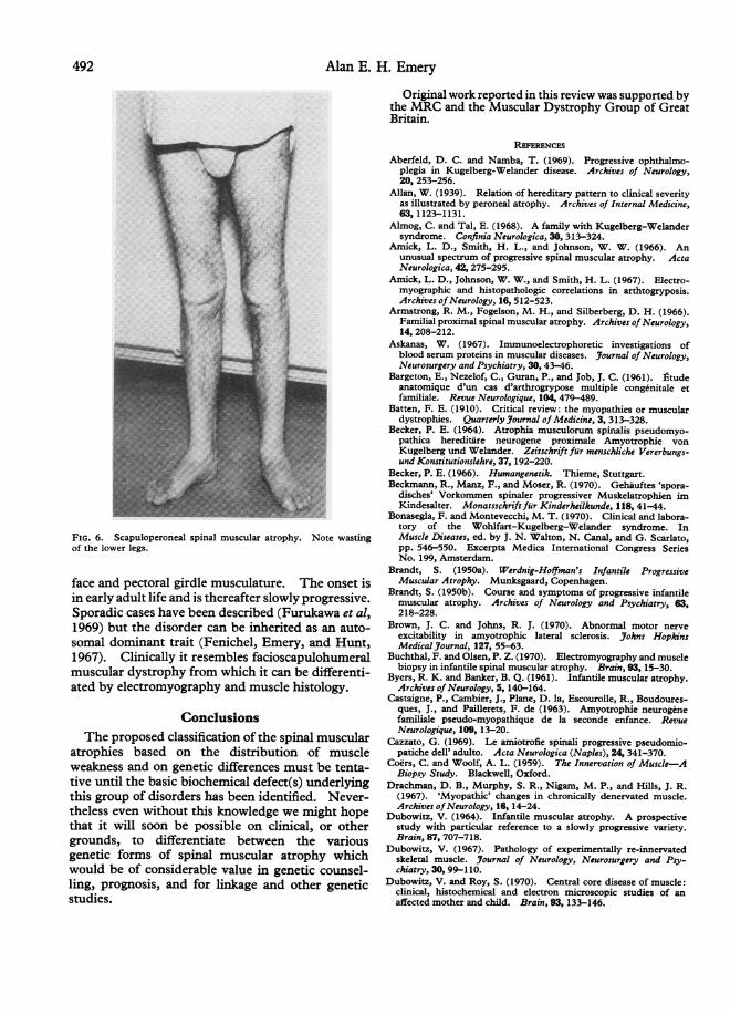
FIG. 6. Scapuloperoneal spinal muscular atrophy. Note wastingof the lower legs.
face and pectoral girdle musculature. The onset isin early adult life and is thereafter slowly progressive.Sporadic cases have been described (Furukawa et al,1969) but the disorder can be inherited as an auto-somal dominant trait (Fenichel, Emery, and Hunt,1967). Clinically it resembles facioscapulohumeralmuscular dystrophy from which it can be differenti-ated by electromyography and muscle histology.
Conclusions
The proposed classification of the spinal muscularatrophies based on the distribution of muscleweakness and on genetic differences must be tenta-tive until the basic biochemical defect(s) underlyingthis group of disorders has been identified. Never-theless even without this knowledge we might hopethat it will soon be possible on clinical, or othergrounds, to differentiate between the variousgenetic forms of spinal muscular atrophy whichwould be of considerable value in genetic counsel-ling, prognosis, and for linkage and other geneticstudies.
Original work reported in this review was supported bythe MRC and the Muscular Dystrophy Group of GreatBritain.
REFERENCES
Aberfeld, D. C. and Namba, T. (1969). Progressive ophthalmo-plegia in Kugelberg-Welander disease. Archives of Neurology,20, 253-256.
Allan, W. (1939). Relation of hereditary pattern to clinical severityas illustrated by peroneal atrophy. Archives of Internal Medicine,63, 1123-1131.
Almog, C. and Tal, E. (1968). A family with Kugelberg-Welandersyndrome. Confinia Neurologica, 30, 313-324.
Amick, L. D., Smith, H. L., and Johnson, W. W. (1966). Anunusual spectrum of progressive spinal muscular atrophy. ActaNeurologica, 42, 275-295.
Amick, L. D., Johnson, W. W., and Smith, H. L. (1967). Electro-myographic and histopathologic correlations in arthtogryposis.Archives ofNeurology, 16, 512-523.
Armstrong, R. M., Fogelson, M. H., and Silberberg, D. H. (1966).Familial proximal spinal muscular atrophy. Archives of Neurology,14, 208-212.
Askanas, W. (1967). Immunoelectrophoretic investigations ofblood serum proteins in muscular diseases. Journal of Neurology,Neurosurgery and Psychiatry, 30, 43-46.
Bargeton, E., Nezelof, C., Guran, P., and Job, J. C. (1961). l-tudeanatomique d'un cas d'arthrogrypose multiple congenitale etfamiliale. Revue Neurologique, 104, 479-489.
Batten, F. E. (1910). Critical review: the myopathies or musculardystrophies. Quarterly Journal of Medicine, 3, 313-328.
Becker, P. E. (1964). Atrophia musculorum spinalis pseudomyo-pathica hereditire neurogene proximale Amyotrophie vonKugelberg und Welander. Zeitschrift fur menschliche Vererbungs-und Konstitutionslehre, 37, 192-220.
Becker, P. E. (1966). Humangenetik. Thieme, Stuttgart.Beckmann, R., Manz, F., and Moser, R. (1970). Gehauftes 'spora-
disches' Vorkommen spinaler progressiver Muskelatrophien imKindesalter. MonatsschriftfUr Kinderheilkunde, 118, 41-44.
Bonasegla, F. and Montevecchi, M. T. (1970). Clinical and labora-tory of the Wohlfart-Kugelberg-Welander syndrome. InMuscle Diseases, ed. by J. N. Walton, N. Canal, and G. Scarlato,pp. 546-550. Excerpta Medica International Congress SeriesNo. 199, Amsterdam.
Brandt, S. (1950a). Werdnig-Hoi5man's Infantile ProgressiveMuscular Atrophy. Munksgaard, Copenhagen.
Brandt, S. (1950b). Course and symptoms of progressive infantilemuscular atrophy. Archives of Neurology and Psychiatry, 63,218-228.
Brown, J. C. and Johns, R. J. (1970). Abnormal motor nerveexcitability in amyotrophic lateral sclerosis. Johns HopkinsMedical Journal, 127, 55-63.
Buchthal, F. and Olsen, P. Z. (1970). Electromyography and musclebiopsy in infantile spinal muscular atrophy. Brain, 93, 15-30.
Byers, R. K. and Banker, B. Q. (1961). Infantile muscular atrophy.Archives of Neurology, 5, 140-164.
Castaigne, P., Cambier, J., Plane, D. la, Escourolle, R., Boudoures-ques, J., and Paillerets, F. de (1963). Amyotrophie neurogenefamiliale pseudo-myopathique de la seconde enfance. RevueNeurologique, 109, 13-20.
Cazzato, G. (1969). Le amiotrofie spinali progressive pseudomio-patiche dell' adulto. Acta Neurologica (Naples), 24,341-370.
Coers, C. and Woolf, A. L. (1959). The Innervation of Muscle-ABiopsy Study. Blackwell, Oxford.
Drachman, D. B., Murphy, S. R., Nigam, M. P., and Hills, J. R.(1967). 'Myopathic' changes in chronically denervated muscle.Archives ofNeurology, 16, 14-24.
Dubowitz, V. (1964). Infantile muscular atrophy. A prospectivestudy with particular reference to a slowly progressive variety.Brain, 87, 707-718.
Dubowitz, V. (1967). Pathology of experimentally re-innervatedskeletal muscle. Journal of Neurology, Neurosurgery and Psy-chiatry, 30, 99-1 10.
Dubowitz, V. and Roy, S. (1970). Central core disease of muscle:clinical, histochemical and electron microscopic studies of anaffected mother and child. Brain, 93, 133-146.
492

The Nosology of the Spinal Muscular Atrophies
Duchen, L. W., Strich, S. J., and Falconer, D. S. (1968). Anhereditary motor neurone disease with progressive denervation ofmuscle in the mouse: the mutant 'wobbler'. Journal of Neurology,Neurosurgery and Psychiatry, 31,535-542.
Dunne, P. B. and Chutorian, A. M. (1966). The relationship be-tween infantile and juvenile spinal muscular atrophy. Neurology,16, 306.
Dyck, P. J. and Lambert, E. H. (1968a). Lower motor and primarysensory neuron diseases with peroneal muscular atrophy. I.Neurologic, genetic and electrophysiologic findings in hereditarypolyneuropathies. Archives of Neurology, 18, 603-618.
Dyck, P. J. and Lambert, E. H. (1968b). Lower motor and primarysensory neuron diseases with peroneal muscular atrophy. II.Neurologic, genetic and electrophysiologic findings in variousneuronal degenerations. Archives of Neurology, 18, 619-625.
Emery, A. E. H. (1969). Abnormalities of the electrocardiogramin female carriers of Duchenne muscular dystrophy. BritishMedical Journal, 2, 418-420.
Emery, A. E. H. (1972). Abnormalities of the electrocardiogram inhereditary myopathies. Journal of Medical Genetics, 9.(In press.)
Emery, A. E. H. and Spikesman, A. (1970). Evidence against theexistence of a subclinical form of X-linked Duchenne musculardystrophy. Journal of the Neurological Science, 10, 523-533.
Emery, A. E. H. and Walton, J. N. (1967). The genetics of musculardystrophy. In Progress in Medical Genetics, ed. by A. G.Steinberg and A. G. Bearn, vol. 5, pp. 116-145. Grune andStratton, New York.
Emery, E. S., Fenichel, G. M., and Eng, G. (1968). A spinalmuscular atrophy with scapuloperoneal distribution. Archives ofNeurology, 18, 129-133.
Engel, W. K. (1961). Muscle target fibres, a newly recognised signof denervation. Nature (London), 191, 389-390.
Engel, W. K. (1970). Selective and non-selective susceptibility ofmuscle fibre types. Archives of Neurology, 22,97-117.
Fenichel, G. M., Emery, E. S., and Hunt, P. (1967). Neurogenicatrophy simulating facioscapulohumeral dystrophy. Archives ofNeurology, 17, 257-260.
Finkel, N. (1962). A forma pseudomiopatica tardia da atrofiamuscular progressiva heredo-familial. Arquivos de Neuro-Psiquiatria, 20, 307-322.
Fried, K. and Emery, A. E. H. (1971). Spinal muscular atrophy,type II. Clinical Genetics, 2, 203-209.
Frischknecht, W., Bianchi, L., and Pilleri, G. (1960). FamiliaireArthrogryposis multiplex congenita Neuro-arthro-myodyplasiacongenita. Helvetica Paediatrica Acta, 15, 259-279.
Fukuyama, Y. (1958). Infantile progressive muscular atrophy(Werdnig-Hoffmann) in relation to myatonia congenita (Oppen-heim). Six cases including two autopsy cases. PaediatriaUniversitatis (Tokyo), 2, 39-44.
Funk, F. (1962). Zur familiaren Form der progressiven spinalenMuskelatrophie. Fortschritte der Neurologie, Psychiatrie undihrer Grenzgebiete, 30, 324-330.
Furukawa, T., Nakao, K., Sugita, H., and Tsukagoshi, H. (1968).Kugelberg-Welander disease, with particular reference to sex-
influenced manifestations. Archives of Neurology, 19, 156-162.Furukawa, T., Tsukagoshi, H., Sugita, H., and Toyokura, Y. (1969).
Neurogenic muscular atrophy simulating facioscapulohumeralmuscular dystrophy. Journal of the Neurological Sciences, 9,389-397.
Gamstorp, I. (1967). Progressive spinal muscular atrophy withonset in infancy or early childhood. Acta Paediatrica, 56, 408-
423.Gardner-Medwin, D., Hudgson, P., and Walton, J. N. (1967).
Benign spinal muscular atrophy arising in childhood and adoles-
cence. Journal of the Neurological Sciences, 5, 121-158.Gardner-Medwin, D. and Walton, J. N. (1969). A classification of
the neuromuscular disorders and a note on the clinical examination
of the voluntary muscles. In Disorders of Voluntary Musck, ed.by J. N. Walton, pp.411-453. Churchill. London.
Giarvie, J. M. and Woolf, A. L. (1966a). Kugelberg-Welandersyndrome (hereditary proximal spinal muscular atrophy). British
MedicalJournal, 1, 1458-1461.Gomez, M. R., Clermont, V., and Bernstein, J. (1962). Progressive
bulbar paralysis in childhood (Fazio-Londe's disease). Report of
a case with pathologic evidence of nuclear atrophy. Archives of
Neurology, 6, 317-323.
Gorecka, A. (1969). Aminoacyduria i aminoacydemia w chorobachmiesni pochodzenia rdzeniowego. Neurologia Neurochirurgia(Polska), 3, 607-612.
Gross, M. (1966). Proximal spinal muscular atrophy. Journal ofNeurology, Neurosurgery and Psychiatry, 29, 29-34.
Hanhart, E. (1945). Die infantile progressive spinale Muskel-atrophie (Werdnig-Hoffmann) als einfach rezessive, subletaleMutation auf Grund von 29 Fallen in 14 Sippen. HelveticaPaediatrica Acta, 1, 110-133.
Hanhart, E. (1962). Die Genealogie der 6 sicheren und 4wahrscheinlichen Falle von neurogener, proximaler Amyotrophie(Kugelberg-Welander) in einer Sippe aus dem Isolat I (KantonSchwyz). Archiv der Julius Klaus-Stiftung fur Vererbungs-forschung, Sozialanthropologie, und Rassenhygiene, 37, 175-193.
Hausmanowa-Petrusewicz, I. (1970). Infantile and juvenile spinalmuscular atrophy. In Muscle Diseases, ed. by J. N. Walton, N.Canal, and G. Scarlato, pp. 558-567. Excerpta Medica Inter-national Congress Series No. 199, Amsterdam.
Hausmanowa-Petrusewicz, I., Sobkowicz, H., Zielinska, S., andDobosz, I. (1962). A propos d'atrophies musculaires juvenilesheredo-familiales. Schweizer Archiv fur Neurologie, Neuro-chirurgie und Psychiatrie, 90,255-267.
Hausmanowa-Petrusewicz, I., Askana, W., Badurska, B., Emeryk, B.,Fidzianiska, A., Garbalinska, W., Hetnarska, L.,Jodrzejowska, H.,Kamieniecka, Z., Niebr6t-Dobosz, I., Prot, J., and Sawicka, E.(1968). Infantile and juvenile spinal muscular atrophy. journalof the Neurological Sciences, 6, 269-287.
Hetnarska, L., Prot, J., and Sawicka, E. (1968). Creatine phospho-kinase activity in spinal muscular atrophy. Journal of the Neuro-logical Sciences, 6, 261-267.
Hoffmann, J. (1893). Ueber chronische spinale Muskelatrophie imKindesalter auf familiirer Basis. Deutsche Zeitschrift fur Nerven-heilkunde, 3,427-470.
Hoffmann, J. (1900a). Ueber die hereditire progressive spinaleMuskelatrophie im Kindesalter. Munchener Medizinische Wochen-schrift, 47, 1649-1651.
Hoffmann, J. (1900b). Dritter Beitrag zur Lehre von der heredi-tarer progressiven spinalen Muskelatrophie im Kindesalter.Deutsche Zeitschrift fur Nervenheilkunde, 18,217-224.
Hogenhuis, L. A. H., Spaulding, S. W., and Engel, W. K. (1967).Neuronal RNA metabolism in infantile spinal muscular atrophy(Werdnig-Hoffmann's disease) studied by radioautography. Anew technique in the investigation of neurological disease.
Journal of Neuropathology and Experimental Neurology, 26, 335-341.
Hurwitz, L., Lapresle, J., and Garcin, R. (1961). Atrophie muscu-
laire neurogene de topographie proximale et symetrique simulantune myopathie: etude de deux observations. Revue Neurologique,104, 97-107.
Ionasescu, V., Luca, N. Petrescu, A., and Calcaianu, G. (1965).Hereditary proximal spinal muscular atrophy. Biochemical in-vestigations. Confinia Neurologica, 25, 79-86.
Kaeser, H. E. (1965). Scapuloperoneal muscular atrophy. Brain,
88,407-418.Kennedy, W. R., Alter, M., and Foreman, R. T. (1966). Hereditary
proximal spinal muscular atrophy of late onset. Neurology, 16,306-307.
Kennedy, W. R., Alter, M., and Sung, J. H. (1968). Progressiveproximal spinal and bulbar muscular atrophy of late onset. A
sex-linked recessive trait. Neurology, 18, 671-680.Kessler, G. B. (1968). Non-progressive proximal and generalized
spinal muscular atrophy in siblings. Bulletin of the Los AngelesNeurological Societies, 33, 21-25.
Kloepfer, H. W. and Emery, A. E. H. (1969). Genetic aspects of
neuromuscular disease. In Disorders of Voluntary Muscle, ed. by
J. N. Walton, pp. 683-712. Churchill, London.Kohn, R. (1968). Postmortem findings in a case of Wohlfart-Kugel-berg-Welander disease. Confinia Neurologica, 30, 253-260.
Kondo, K., Tsubaki, T., and Sakamoto, F. (1970). The Ryukyuanmuscular atrophy. An obscure heritable neuromuscular diseasefound in the islands of Southern Japan. Journal of the Neuro-logical Sciences, 11, 359-382.
Kugelberg, E. and Welander, L. (1956). Heredofamilial juvenile
muscular atrophy simulating muscular dystrophy. Archives of
Neurology and Psychiatry, 75, 500-509.Lambert, E. H. (1963). Electromyography and electric stimulation
of peripheral nerves and muscles. In Clinical Examinations in
493

Alan E. H. Emery
Neurology, 2nd ed., pp. 311-341. Saunders, London andPhiladelphia.
Levy, J. A. and Wittig, E. 0. (1962). Atrofia muscular proximalfamiliar. Arquivos de Neuro-Psiquiatria, 20, 233-237.
Lowenstein, A. S., Arbeit, S. R., and Rubin, I. L. (1962). Cardiacinvolvement in progressive muscular dystrophy. An electro-cardiographic and ballistocardiographic study. American Journalof Cardiology, 9, 528-533.
Lugaresi, E., Gambetti, P., and Rossi, P. G. (1966). ChronicNeurogenic muscle atrophies of infancy. Their nosological re-
lationship with Werdnig-Hoffmann's disease. Journal of theNeurological Sciences, 3, 399-409.
McKusick, V. A. (1968). Mendelian Inheritance in Man, 2nd ed.Johns Hopkins, Baltimore.
Magee, K. R. (1960). Familial progressive bulbarspinal muscularatrophy. Neurology, 10, 295-305.
Magee, K. R. and Jong, R. N. de (1960). Neurogenic muscularatrophy simulating muscular dystrophy. Archives of Neurology.2, 677-682.
Manning, G. W. and Cropp, G. J. (1958). The electrocardiogramin progressive muscular dystrophy. British Heart Journal, 20,416-420.
Mapelli, G. and Ramelli, E. (1970). Familial progressive spinalamyotrophy with limb root distribution and onset in adult life(neurogenic pseudomyopathy of Wohlfart-Kugelberg-Welander).In Muscle Diseases, ed. by J. N. Walton, N. Canal and G. Scarlato,pp. 551-554. Excerpta Medica International Congress SeriesNo. 199, Amsterdam.
Marburg, 0. (1912). Zur Klinik und Pathologie der Myatoniacongenita (Appenheim). Arbeiten aus der Neurologischen Institutan der Wiener Universitdt, 19, 133-154. Quoted by Brandt(1950a).
Marquardt, J. E., MacLowry, J., and Perry, R. E. (1962). Infantileprogressive spinal muscular atrophy in identical Negro twins.New England Journal of Medicine, 267, 386-388.
Martin-Sneessens, L. (1962). Formes a evolution tres prolingee del'amyotrophie spinale de Werdnig-Hoffmann. Journal de GenetiqueHumaine, 11, 251-269.
Meadows, J. C. and Marsden, C. D. (1969). A distal form ofchronic spinal muscular atrophy. Neurology, 19, 53-58.
Meadows, J. C., Marsden, C. D., and Harriman, D. G. F. (1969a).Chronic spinal muscular atrophy in adults. Part I. TheKugelberg-Welander syndrome. Journal of the NeurologicalSciences, 9, 527-550.
Meadows, J. C., Marsden, C. D., and Harriman, D. G. F. (1969b).Chronic spinal muscular atrophy in adults. Part 2. Other forms.Journal of the Neurological Sciences, 9, 551-566.
Mumenthaler, M. (1970). Myopathy in neuropathy In MuscleDiseases, ed. by J. N. Walton, N. Canal, and G. Scarlato, pp. 585-598. Excerpta Medica International Congress Series No. 199,Amsterdam.
Munsat, T. L. (1968). Quoted by Zellweger and McCormick(1968).
Munsat, T. L., Woods, R., Fowler, W., and Pearson, C. M. (1969).Neurogenic muscular atrophy of infancy with prolonged survival.The variable course of Werdnig-Hoffmann disease. Brain, 92,9-24.
Namba, L., Aberfeld, D. C., and Grob, D. (1970). Chronicproximal spinal muscular atrophy. Journal of the NeurologicalSciences, 11, 401-423.
Nelson, J. W. and Amick, L. D. (1966). Heredofamilial progres-sive spinal muscular atrophy: a clinical and electromyographicstudy of a kinship. Neurology, 16, 306.
Pearce, J. and Harriman, D. G. F. (1966). Chronic spinal muscularatrophy. Journal of Neurology Neurosurgery and Psychiatry, 29,509-520.
Perloff, J. K., Leon, A. C. de, and O'Doherty, D. (1966). Thecardiomyopathy of progressive muscular dystrophy. Circulation,33,625-648.
Perloff, J. K., Roberts, W. C., Leon, A. C. de, and O'Doherty, D.(1967). The distinctive electrocardiogram of Duchenne's pro-gressive muscular dystrophy. American Journal of Medicine,42, 179-188.
Peters, H. A., Opitz, J. M., Goto, I., and Reese, H. H. (1968). Thebenign proximal spinal progressive muscular atrophies. A clini-cal and genetical study. Acta Neurologica, 44,542-560.
Pratt, R. T. C. (1967). The Genetics of Neurological Disorders.Oxford University Press, London.
Quarfordt, S. H., Vivo, D. C. de, Engel, W. K., Levy, R. I., andFredrickson, D. S. (1970). Familial adult-onset proximal spinalmuscular atrophy. Archives of Neurology, 22, 541-549.
Radu, H., Seceleanu, A., Migea, S., Torok, Z., Bordeianu, L., andSeceleanu, S. (1966). La pseudomyopathie neurogene deKugelberg-Welander. Acta Neurologica Psychiatrica, 66, 409-427.
Research Group on Neuromuscular Diseases (1968). Classificationof the neuromuscular disorders (Appendix A). Journal of theNeurological Sciences, 6, 165-177.
Ricker, K., Mertens, H. G., and Schimrigk, K. (1968). The neuro-genic scapulo-peroneal syndrome. European Neurology, 1, 257-274.
Rowland, L. P., Schotland, D. L., Lovelace, R. E., and Layzer,R. B. (1967). Neurogenic muscular atrophies. In ExploratoryConcepts in Muscular Dystrophy and Related Disorders, ed. by A. T.Milhorat, pp. 41-45. Excerpta Medica International CongressSeries No. 147, Amsterdam.
Schott, J., Jacobi, M., and Wald, M. A. (1955). Electrocardio-graphic patterns in the differential diagnosis of progressive muscu-lar dystrophy. American Journal of the Medical Sciences, 229,517-524.
Schuchmann, L. (1970). Spinal muscular atrophy of the scapulo-peroneal-type. Zeitschrift fur Kinderheilkunde, 109, 118-123.
Shoji, H., Sugita, K., Furukawa, T., Igata, A., and Tsukagoshi, H.(1970). Proximal spinal muscular atrophy (Kugelberg-Welanderdisease and its variant) with sex-linked recessive inheritance.Clinical Neurology (Tokyo), 10, 256-262.
Skyring, A. and McKusick, V. A. (1961). Clinical, genetic andelectrocardiographic studies in childhood muscular dystrophy.American Journal of the Medical Sciences, 242, 534-547.
Smith, J. B. and Patel, A. (1965). The Wohlfart-Kugelberg-Welander disease. Neurology, 15, 469-473.
Spira, R. (1963). Neurogenic, familial, girdle type muscularatrophy. Confinia Neurologica, 23, 245-255.
Spira, R. (1967). A family with Kugelberg-Welander disease:electromyographic findings in subclinical cases. ConfiniaNeurologica, 28, 423-431.
Spiro, A. J., Fogelson, M. H., and Goldberg, A. C. (1967). Micro-cephaly and mental subnormality in chronic progressive spinalmuscular atrophy of childhood. Developmnental Medicine andChild Neurology, 9, 594-601.
Thieffry, S., Arthuis, M., and Bargeton, E. (1955). Quarante cas demaladie de Werdnig-Hoffmann avec onze examens anatomiques.Revue Neurologique, 93, 621-644.
Thomson, J. and Bruce, A. (1893). A case of progressive muscularatrophy in a child with a spinal lesion. Edinburgh HospitalReports, 1, 361-383.
Trillet, M., Girard, P. F., Schott, B., Ramel, P., and Woehrle, R.(1970). La paralysie bulbo-pontine chronique progressive avecsurdite. A propos d'une observation clinique. Presse Medicale,78, 129-130.
Tsukagoshi, H., Nakanishi, L., Kondo, K., and Tsubaki, T. (1965).Hereditary proximal neurogenic muscular atrophy in adult.Archives of Neurology, 12, 597-603.
Tsukagoshi, H., Sugita, H., Furukawa, T., Tsubaki, T., and Ono, E.(1966). Kugelberg-Welander syndrome with dominant inheri-tance. Archives of Neurology, 14,378-381.
Tsukagoshi, H., Shoji, H., and Furukawa, T. (1970). Proximalneurogenic muscular atrophy in adolescence and adulthood withX-linked recessive inheritance. Neurology, 20, 1188-1193.
Tsukagoshi, H., Takasu, T., Yoshida, M., and Toyokura, Y. (1969).A family with scapuloperoneal muscular atrophy. Clinical Neuro-logy (Tokyo), 9, 511-517.
Van Laere, J. (1966). Paralysie bulbo-pontine chronique pro-gressive familiale avec surdite. Un cas de syndrome de Klippel-Trenaunay dans la mame fratrie. Problemes diagnostiques etgenetiques. Revue Neurologique, 115, 289-295.
Verger, P., Vital, C., Guillard, J. M., Eschapasse, P., and Le Pennec,J. J. (1969). Les formes 'intermediaires' de l'amyotrophiespinale infantile (a propos de six observations nouvelles).Pediatrie, 24, 131-143.
Walton, J. N. (1956). Amyotonia congenita, a follow-up study.Lancet, 1, 1023-1028.
Walton, J. N. (1957). The limp child. Journal of Neurology,Neurosurgery and Psychiatry, 20, 144-154.
494

The Nosology of the Spinal Muscular AtrophiesWalton, J. N. (1970). Some pitfalls in the diagnosis of muscular spinal muscular atrophy-a
disorders. In Proceedings of the 2nd International Congress of muscular dystrophy. ActaNeuro-Muscular Pathology, Marseille. (In press.) Woratz, G. (1964). Neural
Werdnig, G. (1891). Zwei frihinfantile hereditare Falle von X-chromosomalem Erbgangprogressiver Muskelatrophie unter dem Bilde der Dystrophie, aber demie der Wissenschaften zuauf neurotischer Grundlage. Archiv fur Psychiatrie und Nerven- Quoted by Becker (1966) anckrankheiten, 22, 437-481. Zellweger, H. and McCormi
Werdnig, G. (1894). Die friihinfantile progressive spinale Amyo- dystrophy and scapuloperotrophie. Archiv fur Psychiatrie und Nervenkrankheiren, 26, 706- Acta, 23, 643-649.744. Zellweger, H., Schneider, H.,
Wiesendanger, M. (1962). t)ber die hereditare nuerogene, proxi- genetic variant of spinal mumale Amyotrophie (Kugelberg-Welander). Archiv der Julius 869.Klaus-Stiftung fur Vererbungsforschung, Soziolanthropologie, und Zellweger, H., Schneider, H.Rassenhygiene, 37, 147-174. (1969b). Heritable spinal
Wohlfart, G., Fex, J., and Eliasson, S. (1955). Hereditary proximal diatrica Acta, 24, 92-105.
495
clinical entity simulating progressivePsychiatrica Neurologica, 30, 395-406.le Muskelatrophie mit dominantemg. Abhandlungen der deutschen Aka-Berlin, Klasse fur Medizin, 2, 1-99.
, Pratt (1967).ick, W. F. (1968). ScapuloperonealDneal atrophy. Helvetica Paediatrica
,and Schuldt, D. R. (1969a). A newscular atrophy. Neurology, 19, 865-
J., Schuldt, D. R., and Mergner, W.muscular atrophies. Helvetica Pae-