introduction to gross anatomy of the muscular system
29
ANATOMY & PHYSIOLOGY BIO 181 INSTRUCTOR: FRED WENDLER INTRODUCTION TO GROSS ANATOMY OF THE MUSCULAR SYSTEM BASED ON THE BOOKS: HUMAN ANATOMY & PHYSIOLOGY, NINTH EDITION BY ELAINE N. MARIEB AND KATJA HOEHN; AND ANATOMY AND PHYSIOLOGY FROM OPENSTAX COLLEGE, ISBN 1-938168-13-5 WITH ILLUSTRATIONS FROM HUMAN ANATOMY & PHYSIOLOGY, NINTH EDITION BY ELAINE N. MARIEB AND KATJA HOEHN; AND ANATOMY AND PHYSIOLOGY FROM OPENSTAX COLLEGE, ISBN 1-938168-13-5
-
Upload
khangminh22 -
Category
Documents
-
view
1 -
download
0
Transcript of introduction to gross anatomy of the muscular system
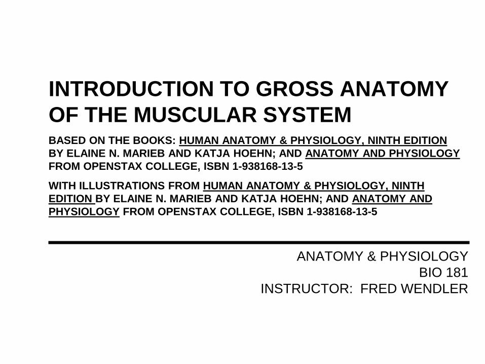
ANATOMY & PHYSIOLOGY
BIO 181
INSTRUCTOR: FRED WENDLER
INTRODUCTION TO GROSS ANATOMY
OF THE MUSCULAR SYSTEM BASED ON THE BOOKS: HUMAN ANATOMY & PHYSIOLOGY, NINTH EDITION
BY ELAINE N. MARIEB AND KATJA HOEHN; AND ANATOMY AND PHYSIOLOGY
FROM OPENSTAX COLLEGE, ISBN 1-938168-13-5
WITH ILLUSTRATIONS FROM HUMAN ANATOMY & PHYSIOLOGY, NINTH
EDITION BY ELAINE N. MARIEB AND KATJA HOEHN; AND ANATOMY AND
PHYSIOLOGY FROM OPENSTAX COLLEGE, ISBN 1-938168-13-5
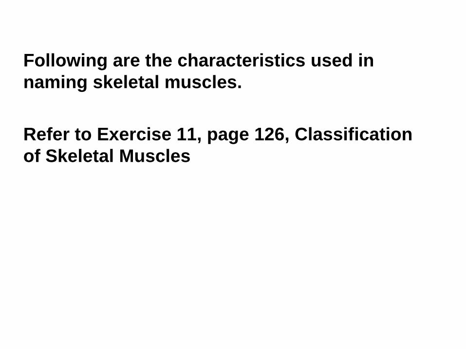
Following are the characteristics used in
naming skeletal muscles.
Refer to Exercise 11, page 126, Classification
of Skeletal Muscles
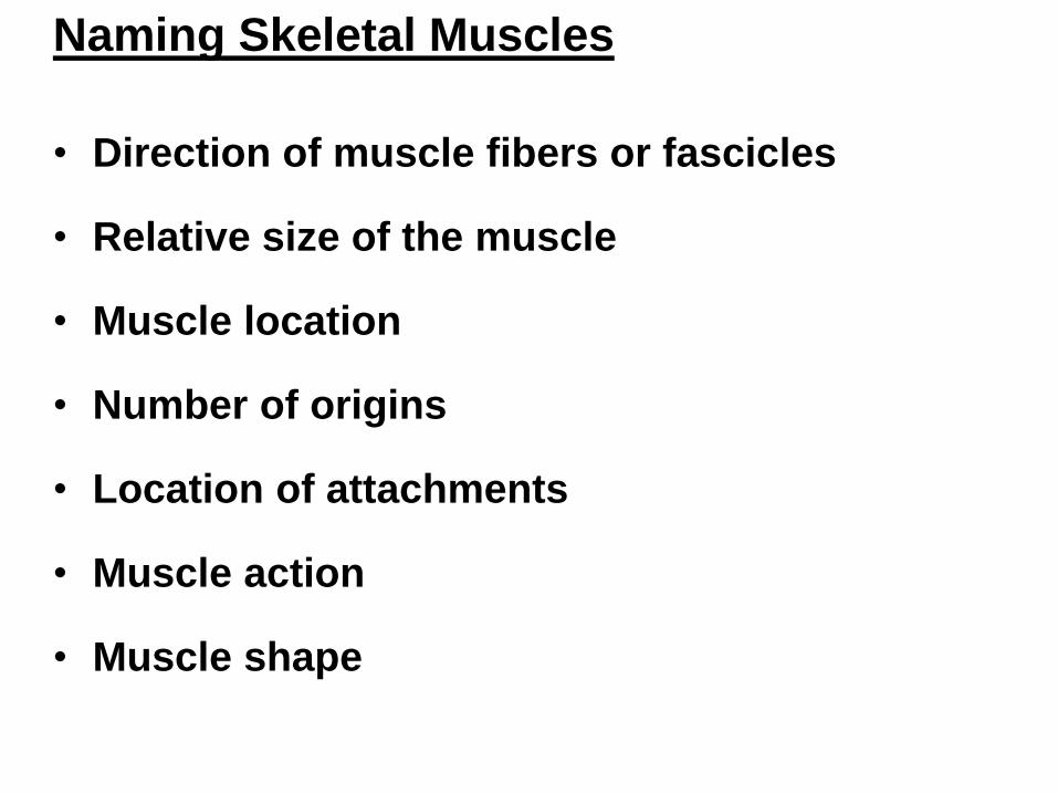
Naming Skeletal Muscles
• Direction of muscle fibers or fascicles
• Relative size of the muscle
• Muscle location
• Number of origins
• Location of attachments
• Muscle action
• Muscle shape
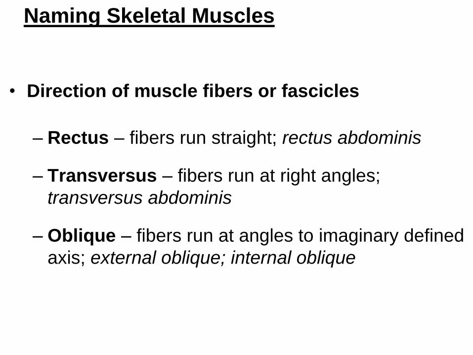
Naming Skeletal Muscles
• Direction of muscle fibers or fascicles
– Rectus – fibers run straight; rectus abdominis
– Transversus – fibers run at right angles;
transversus abdominis
– Oblique – fibers run at angles to imaginary defined
axis; external oblique; internal oblique
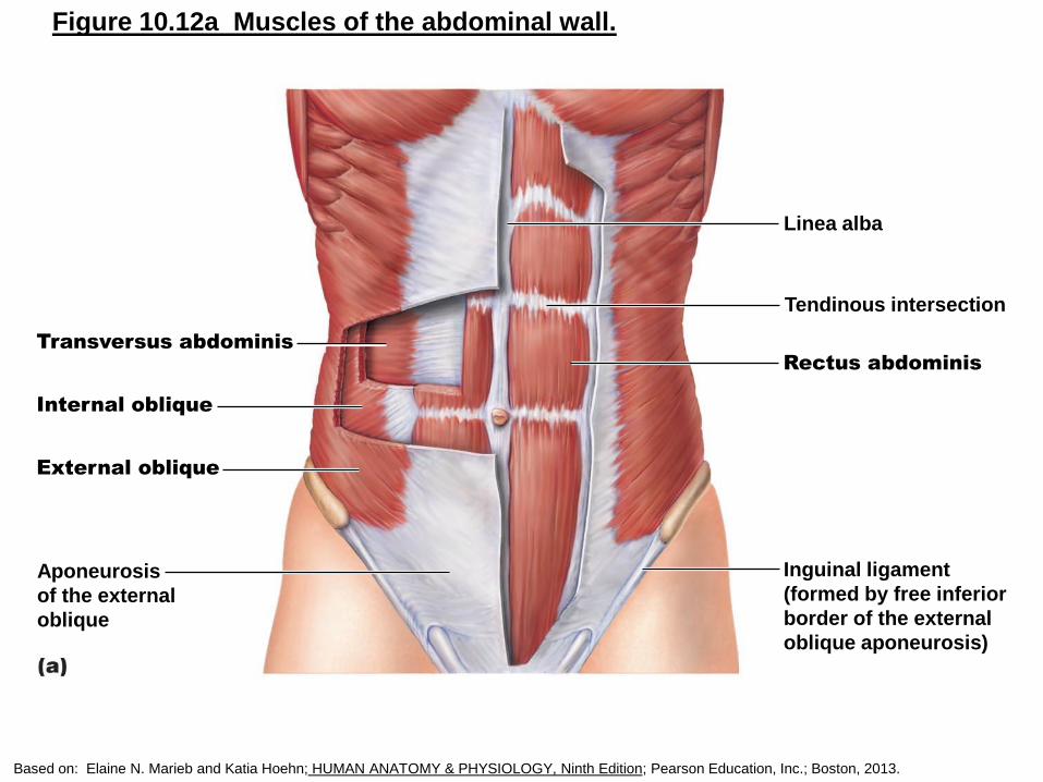
Figure 10.12a Muscles of the abdominal wall.
Linea alba
Tendinous intersection
Rectus abdominis
Inguinal ligament
(formed by free inferior
border of the external
oblique aponeurosis)
Aponeurosis
of the external
oblique
Transversus abdominis
Internal oblique
External oblique
Based on: Elaine N. Marieb and Katia Hoehn; HUMAN ANATOMY & PHYSIOLOGY, Ninth Edition; Pearson Education, Inc.; Boston, 2013.
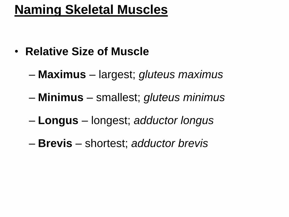
Naming Skeletal Muscles
• Relative Size of Muscle
– Maximus – largest; gluteus maximus
– Minimus – smallest; gluteus minimus
– Longus – longest; adductor longus
– Brevis – shortest; adductor brevis

Naming Skeletal Muscles
• Muscle location
– bone or body region with which muscle
associated
– The temporalis lie over the temporal bone
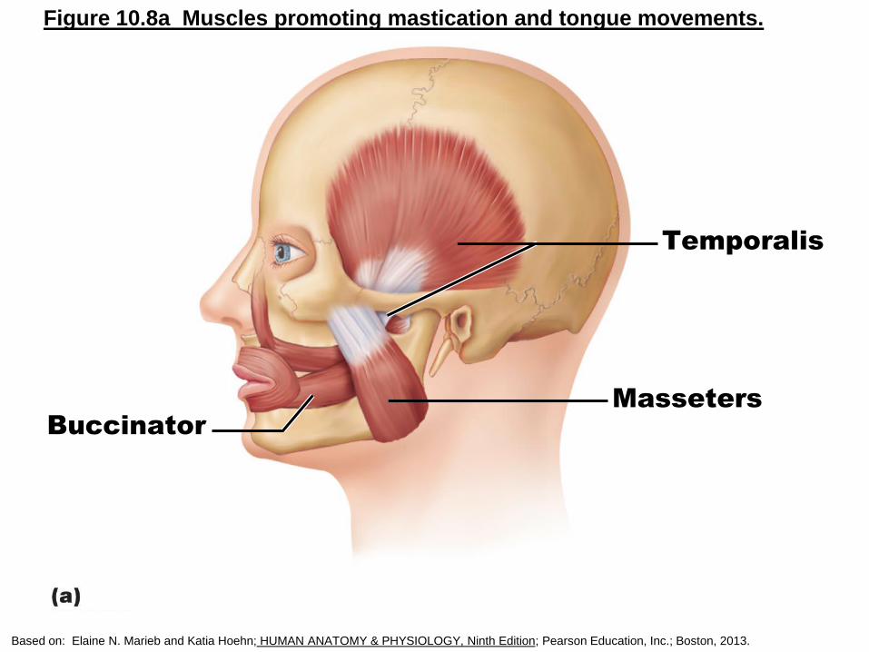
Figure 10.8a Muscles promoting mastication and tongue movements.
Buccinator
Temporalis
Masseters
Based on: Elaine N. Marieb and Katia Hoehn; HUMAN ANATOMY & PHYSIOLOGY, Ninth Edition; Pearson Education, Inc.; Boston, 2013.
Naming Skeletal Muscles
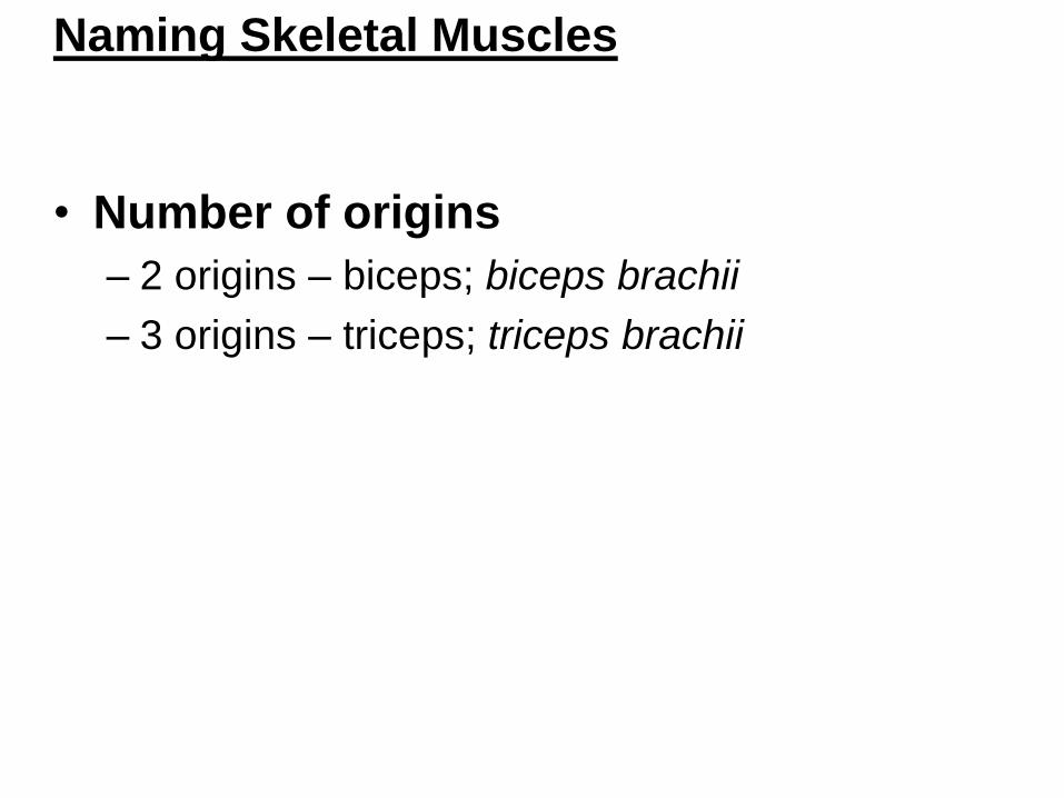
• Number of origins
– 2 origins – biceps; biceps brachii
– 3 origins – triceps; triceps brachii
Naming Skeletal Muscles
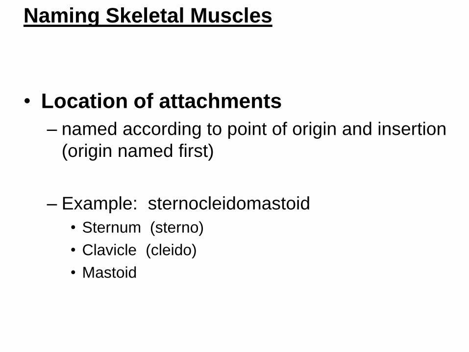
• Location of attachments
– named according to point of origin and insertion
(origin named first)
– Example: sternocleidomastoid
• Sternum (sterno)
• Clavicle (cleido)
• Mastoid
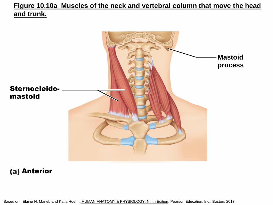
Figure 10.10a Muscles of the neck and vertebral column that move the head
and trunk.
Sternocleido-mastoid
Mastoidprocess
Anterior
Based on: Elaine N. Marieb and Katia Hoehn; HUMAN ANATOMY & PHYSIOLOGY, Ninth Edition; Pearson Education, Inc.; Boston, 2013.
Naming Skeletal Muscles
• Muscle action
– named for action they produce
• Flexion - flexor digitorum
• Extension – extensor digitorum
Naming Skeletal Muscles
• Muscle shape
– deltoid muscle is shaped like a triangle
– trapezius is shaped like a trapezoid
**Several criteria can be combined, e.g.,
extensor carpi radialis longus
Muscle Shape and Arrangement of
Fascicles/Fiber Alignment
Most common patterns:
• Circular
• Convergent
• Parallel
• Fusiform
• Pennate
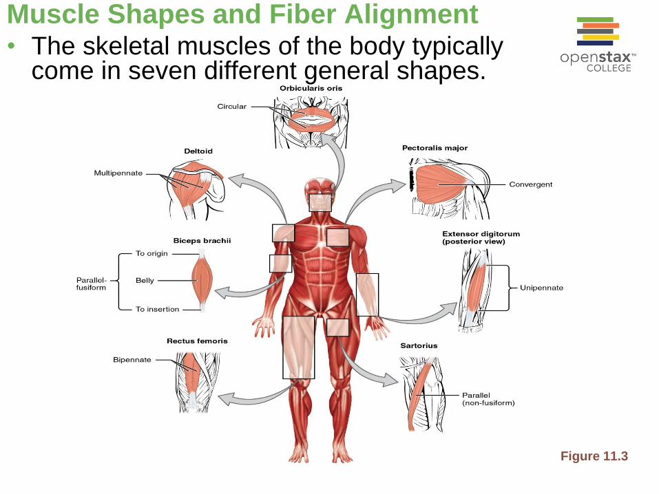
Muscle Shapes and Fiber Alignment• The skeletal muscles of the body typically
come in seven different general shapes.
Figure 11.3
Arrangement of Fascicles
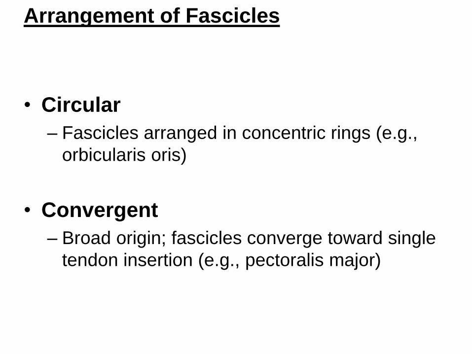
• Circular
– Fascicles arranged in concentric rings (e.g.,
orbicularis oris)
• Convergent
– Broad origin; fascicles converge toward single
tendon insertion (e.g., pectoralis major)

Figure 10.2 Patterns of fascicle arrangement in muscles.
Convergent(pectoralis major)
Circular
(orbicularis oris)
Fusiform(biceps brachii)
Parallel (sartorius)
Multipennate
(deltoid)
Bipennate(rectus femoris)
Unipennate(extensor digitorum longus)
(a)
(b)(c)
(d) (f)
(g)
(e)
Elaine N. Marieb and Katia Hoehn; HUMAN ANATOMY & PHYSIOLOGY, Ninth Edition; Pearson Education, Inc.; Boston, 2013.
Muscle Mechanics: Arrangement of
Fascicles
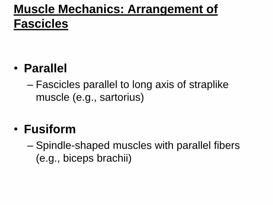
• Parallel
– Fascicles parallel to long axis of straplike
muscle (e.g., sartorius)
• Fusiform
– Spindle-shaped muscles with parallel fibers
(e.g., biceps brachii)
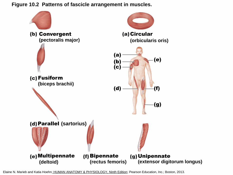
Figure 10.2 Patterns of fascicle arrangement in muscles.
Convergent(pectoralis major)
Circular
(orbicularis oris)
Fusiform(biceps brachii)
Parallel (sartorius)
Multipennate
(deltoid)
Bipennate(rectus femoris)
Unipennate(extensor digitorum longus)
(a)
(b)(c)
(d) (f)
(g)
(e)
Elaine N. Marieb and Katia Hoehn; HUMAN ANATOMY & PHYSIOLOGY, Ninth Edition; Pearson Education, Inc.; Boston, 2013.
Muscle Mechanics: Arrangement of
Fascicles
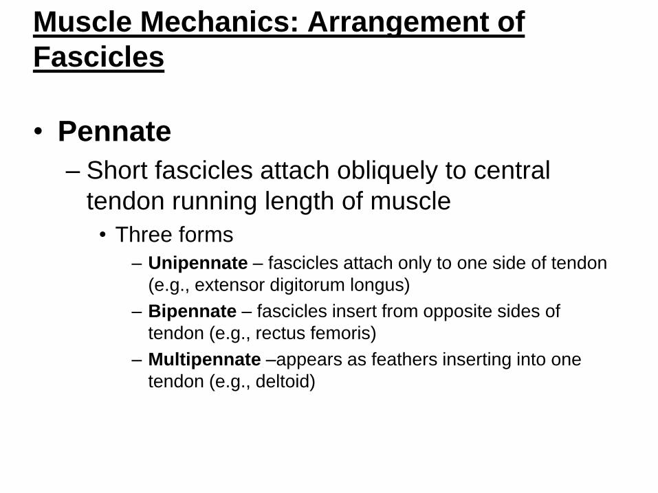
• Pennate
– Short fascicles attach obliquely to central
tendon running length of muscle
• Three forms
– Unipennate – fascicles attach only to one side of tendon
(e.g., extensor digitorum longus)
– Bipennate – fascicles insert from opposite sides of
tendon (e.g., rectus femoris)
– Multipennate –appears as feathers inserting into one
tendon (e.g., deltoid)
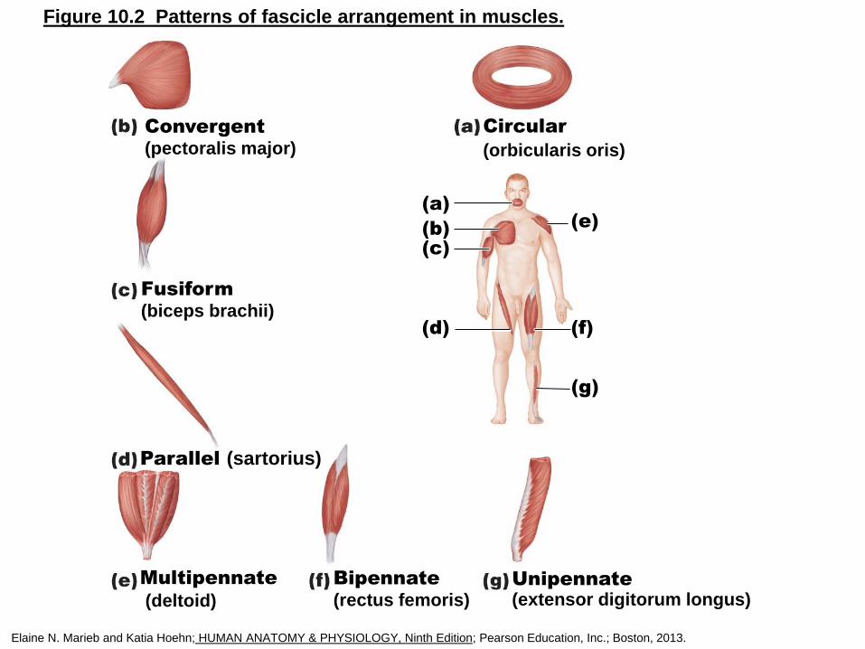
Figure 10.2 Patterns of fascicle arrangement in muscles.
Convergent(pectoralis major)
Circular
(orbicularis oris)
Fusiform(biceps brachii)
Parallel (sartorius)
Multipennate
(deltoid)
Bipennate(rectus femoris)
Unipennate(extensor digitorum longus)
(a)
(b)(c)
(d) (f)
(g)
(e)
Elaine N. Marieb and Katia Hoehn; HUMAN ANATOMY & PHYSIOLOGY, Ninth Edition; Pearson Education, Inc.; Boston, 2013.
Arrangement of Fascicles
• Determines muscle's range of motion
• Determines muscle’s power
Types of Skeletal Muscle
• Muscles can only pull; never push
• What one muscle group "does", another
"undoes"
Types of Skeletal Muscle
• Functional Groups
– Prime mover (agonist)
• Major responsibility for producing specific
movement
– Antagonist
• Opposes or reverses particular movement
– Prime mover and antagonist on opposite
sides of joint across which they act
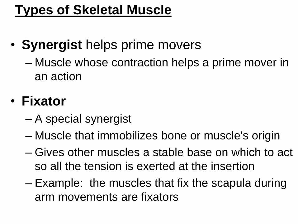
Types of Skeletal Muscle
• Synergist helps prime movers
– Muscle whose contraction helps a prime mover in
an action
• Fixator
– A special synergist
– Muscle that immobilizes bone or muscle's origin
– Gives other muscles a stable base on which to act
so all the tension is exerted at the insertion
– Example: the muscles that fix the scapula during
arm movements are fixators
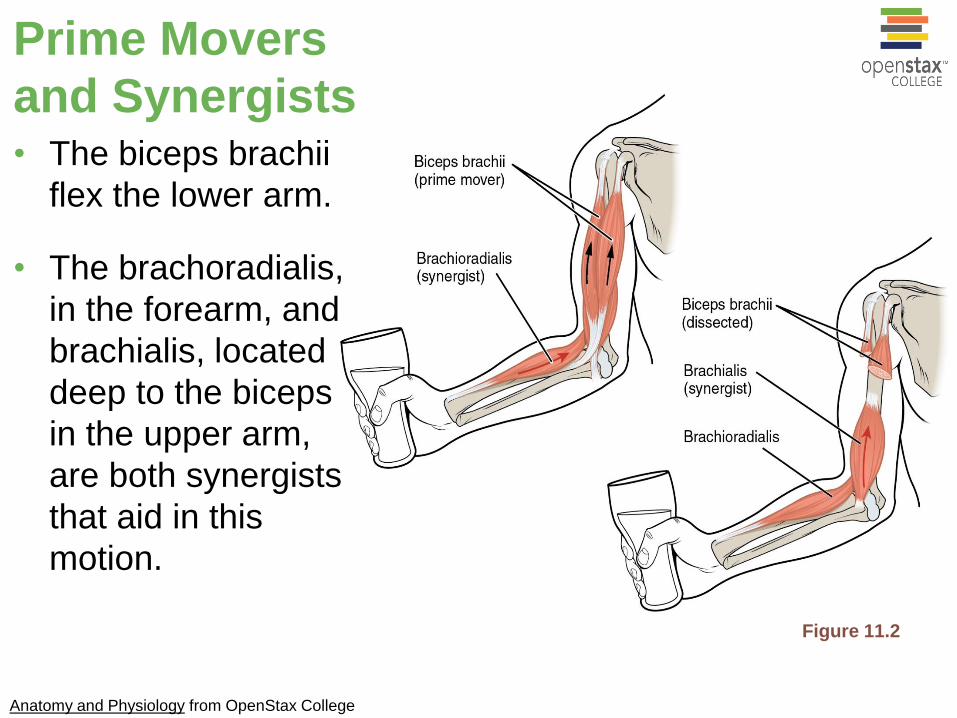
Figure 11.2
Prime Movers
and Synergists• The biceps brachii
flex the lower arm.
• The brachoradialis,
in the forearm, and
brachialis, located
deep to the biceps
in the upper arm,
are both synergists
that aid in this
motion.
Anatomy and Physiology from OpenStax College

Major Skeletal Muscles of the Body
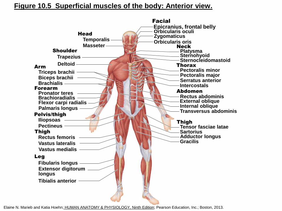
Figure 10.5 Superficial muscles of the body: Anterior view.
Head
Neck
Epicranius, frontal bellyOrbicularis oculiZygomaticusOrbicularis orisTemporalis
MasseterPlatysmaSternohyoidSternocleidomastoid
ThoraxPectoralis minorPectoralis majorSerratus anteriorIntercostals
AbdomenRectus abdominisExternal obliqueInternal obliqueTransversus abdominis
ThighTensor fasciae lataeSartoriusAdductor longusGracilis
Leg
Fibularis longusExtensor digitorumlongus
Tibialis anterior
ThighRectus femoris
Vastus lateralis
Vastus medialis
Pelvis/thighIliopsoas
Pectineus
ForearmPronator teresBrachioradialisFlexor carpi radialisPalmaris longus
ArmTriceps brachiiBiceps brachiiBrachialis
Shoulder
Trapezius
Deltoid
Facial
Elaine N. Marieb and Katia Hoehn; HUMAN ANATOMY & PHYSIOLOGY, Ninth Edition; Pearson Education, Inc.; Boston, 2013.
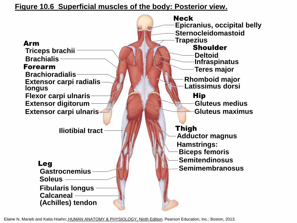
Figure 10.6 Superficial muscles of the body: Posterior view.
NeckEpicranius, occipital bellySternocleidomastoidTrapezius
ShoulderDeltoidInfraspinatusTeres major
Rhomboid majorLatissimus dorsi
HipGluteus mediusGluteus maximus
Adductor magnus
Biceps femorisHamstrings:
SemitendinosusSemimembranosus
LegGastrocnemiusSoleus
Fibularis longusCalcaneal(Achilles) tendon
Iliotibial tract
ArmTriceps brachii
BrachialisForearmBrachioradialisExtensor carpi radialislongusFlexor carpi ulnaris
Extensor carpi ulnarisExtensor digitorum
Thigh
Elaine N. Marieb and Katia Hoehn; HUMAN ANATOMY & PHYSIOLOGY, Ninth Edition; Pearson Education, Inc.; Boston, 2013.