Classification and Treatment of Facial Tissue Atrophy in Parry–Romberg Disease
Upload
independentCategory
view
3download
0
ARTICLE
Received 12 Feb 2014 | Accepted 3 Jun 2014 | Published 3 Jul 2014
EXOSC8 mutations alter mRNA metabolism andcause hypomyelination with spinal muscularatrophy and cerebellar hypoplasiaVeronika Boczonadi1,*, Juliane S. Muller1,*, Angela Pyle1,*, Jennifer Munkley1,*, Talya Dor2, Jade Quartararo3,
Ileana Ferrero3, Veronika Karcagi4, Michele Giunta1, Tuomo Polvikoski5, Daniel Birchall6, Agota Princzinger7,
Yuval Cinnamon2,8, Susanne Lutzkendorf9, Henriett Piko4, Mojgan Reza1, Laura Florez10,
Mauro Santibanez-Koref1, Helen Griffin1, Markus Schuelke9, Orly Elpeleg2, Luba Kalaydjieva10,
Hanns Lochmuller1, David J. Elliott1, Patrick F. Chinnery1, Shimon Edvardson2 & Rita Horvath1
The exosome is a multi-protein complex, required for the degradation of AU-rich element
(ARE) containing messenger RNAs (mRNAs). EXOSC8 is an essential protein of the exosome
core, as its depletion causes a severe growth defect in yeast. Here we show that homozygous
missense mutations in EXOSC8 cause progressive and lethal neurological disease in 22 infants
from three independent pedigrees. Affected individuals have cerebellar and corpus callosum
hypoplasia, abnormal myelination of the central nervous system or spinal motor neuron
disease. Experimental downregulation of EXOSC8 in human oligodendroglia cells and in
zebrafish induce a specific increase in ARE mRNAs encoding myelin proteins, showing that
the imbalanced supply of myelin proteins causes the disruption of myelin, and explaining
the clinical presentation. These findings show the central role of the exosomal pathway in
neurodegenerative disease.
DOI: 10.1038/ncomms5287 OPEN
1 Institute of Genetic Medicine, Wellcome Trust Centre for Mitochondrial Research, Newcastle University, Central Parkway, Newcastle upon Tyne NE1 3BZ,UK. 2 The Monique and Jacques Roboh Department of Genetic Research, Hadassah– Hebrew University Medical Center, Jerusalem 91120, Israel.3 Department of Life Sciences, University of Parma, Parco Area delle Scienze 11A, Parma 43124, Italy. 4 Department of Molecular Genetics and Diagnostics,NIEH, Albert Florian ut 2-6, Budapest 1097, Hungary. 5 Department of Pathology, Institute for Ageing and Health, Newcastle University, Campus for Ageingand Vitality, Newcastle upon Tyne NE4 5PL, UK. 6 Neuroradiology Department, Regional Neurosciences Centre, Queen Victoria Road, Newcastle upon TyneNE1 4PL, UK. 7 Department of Paediatrics, Josa Andras Hospital, Szent Istvan utca 6, Nyiregyhaza 4400, Hungary. 8 Department of Poultry and AquacultureSciences, Institute of Animal Science, Agricultural Research Organization, The Volcani Center, P.O.Box 6, Bet Dagan 50250, Israel. 9 Department ofNeuropediatrics and NeuroCure Clinical Research Center, Charite-Universitatsmedizin, Charite-Platz 1, 10117 Berlin, Germany. 10 Western Australian Institutefor Medical Research/Centre for Medical Research, The University of Western Australia, 35 Stirling Highway Crawley, Western Australia 6009 Perth,Australia. * These authors contributed equally to this work. Correspondence and requests for materials should be addressed to R.H. (email:[email protected]) or to S.E. (email: [email protected]).
NATURE COMMUNICATIONS | 5:4287 | DOI: 10.1038/ncomms5287 | www.nature.com/naturecommunications 1
& 2014 Macmillan Publishers Limited. All rights reserved.
The degradation of messenger RNAs (mRNAs) isan important regulatory step, which controls geneexpression1,2. Unstable mammalian mRNAs contain AU-
rich elements (AREs) within their 30-untranslated regions. Rapiddegradation of ARE-containing RNAs is performed by a multi-protein complex, the exosome3,4. The versatility and specificity ofthe exosome regulate the activity and maintain the fidelity of geneexpression5. Both in yeast and humans, nine proteins organizethe ‘exosome core’ in a two-layered ring. The central hexamerchannel is composed by six subunits (Rrp41p/EXOSC4, Rrp46p/EXOSC5, Rrp45p/PM/Scl-75/EXOSC9, Rrp42p/EXOSC7, Mtr3/EXOSC6 and Rrp43p/Oip2/EXOSC8), while the cap consists ofthree proteins (Rrp4/EXOSC2, Rrp40/EXOSC3 and Csl4/SOCS4)4,6.The exosome degrades RNA starting at the 30-end by anexoribonucleolytic function and it also has an endoribonucleolyticfunction7,8. The catalytic activity of the exosome core is providedthrough the association with other proteins (RRP44/DIS3, RRP6/PM/Scl-100/EXOSC10 ribonucleases)1. ARE recognition requiresARE-binding proteins that interact with the exosome forrecruitment, thereby promoting the rapid degradation of targetRNAs9.
The human exosome also regulates gene expression via diverseRNA processing reactions10. Many cellular RNAs that play keyroles in important cellular processes such as translation (ribosomalRNAs, transfer RNAs and small nucleolar RNAs) and mRNAsplicing (small nuclear RNAs) are produced as precursor moleculesthat are trimmed from their 30-ends by the human exosome11.
This complex organization of the exosome provides the versatilityneeded to cope with the huge variety of RNA substratesin the cell12. However, detailed in vivo analyses are technicallychallenging and many questions remain unresolved.
Here we report that deficiency of a core component of thehuman exosome leads to severe infantile overlap phenotype ofpsychomotor deficit, cerebellar and corpus callosum hypoplasia,hypomyelination and spinal muscular atrophy (SMA).
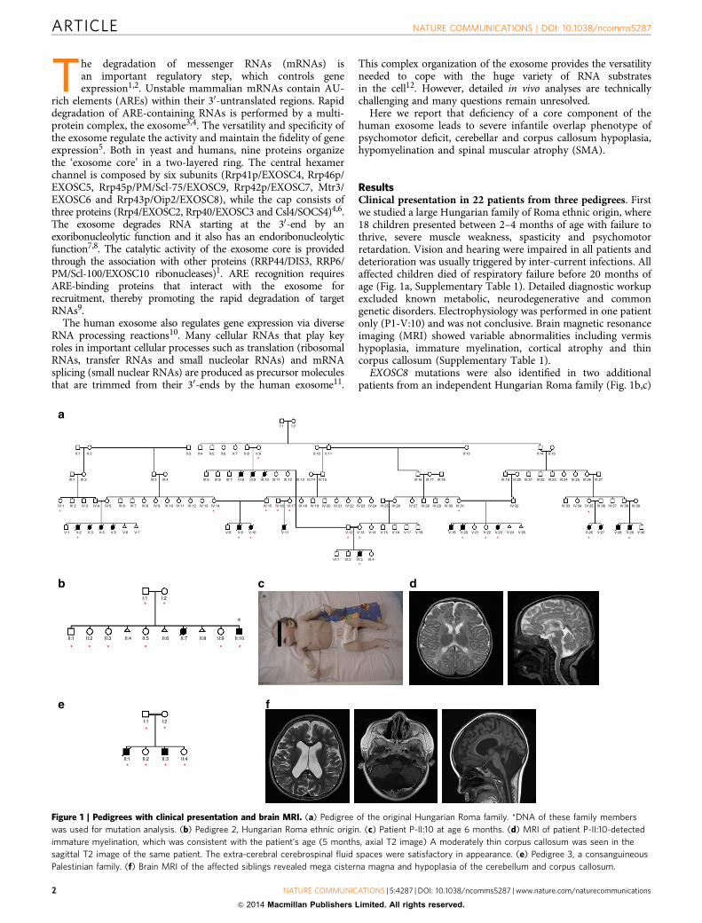
ResultsClinical presentation in 22 patients from three pedigrees. Firstwe studied a large Hungarian family of Roma ethnic origin, where18 children presented between 2–4 months of age with failure tothrive, severe muscle weakness, spasticity and psychomotorretardation. Vision and hearing were impaired in all patients anddeterioration was usually triggered by inter-current infections. Allaffected children died of respiratory failure before 20 months ofage (Fig. 1a, Supplementary Table 1). Detailed diagnostic workupexcluded known metabolic, neurodegenerative and commongenetic disorders. Electrophysiology was performed in one patientonly (P1-V:10) and was not conclusive. Brain magnetic resonanceimaging (MRI) showed variable abnormalities including vermishypoplasia, immature myelination, cortical atrophy and thincorpus callosum (Supplementary Table 1).
EXOSC8 mutations were also identified in two additionalpatients from an independent Hungarian Roma family (Fig. 1b,c)
I:1
V:1 V:2
*
* *
* * * * * * * *
**
*
*
*
*
* *
V:3 V:4 V:5 V:6 V:7 V:8 V:9 V:10 V:11 V:12
VI:1 VI:2 VI:3 VI:4
V:13 V:14 V:15 V:16 V:17 V:18 V:19 V:20 V:21 V:22 V:23 V:24 V:25 V:26 V:27 V:28 V:29 V:30
IV:1 IV:2
III:1
II:1 II:2
a
b c d
f
II:3 II:4 II:5 II:6 II:7 II:8 II:9
I:1 I:2
II:10 II:11 II:12 II:14 II:15
III:2 III:3 III:4 III:5 III:6 III:7 III:8 III:9 III:10 III:11 III:12 III:13 III:14 III:15 III:16 III:17 III:18 III:19 III:20 III:21 III:22 III:23 III:24 III:25 III:26 III:27
IV:3 IV:4 IV:5 IV:6 IV:7 IV:8 IV:9 IV:10 IV:11 IV:12 IV:13 IV:14 IV:15 IV:16 IV:17 IV:18 IV:19 IV:20 IV:21 IV:22 IV:23 IV:24 IV:25 IV:26 IV:27 IV:28 IV:29 IV:30 IV:31 IV:32 IV:33 IV:34 IV:35 IV:36 IV:37 IV:38 IV:39
II:1 II:2 II:3 II:4 II:5 II:6 II:7 II:8 II:9 II:10
∗
∗
**
eI:1
II:1 II:2 II:3 II:4
I:2
**
* * * *
* * **
I:2
**
Figure 1 | Pedigrees with clinical presentation and brain MRI. (a) Pedigree of the original Hungarian Roma family. *DNA of these family members
was used for mutation analysis. (b) Pedigree 2, Hungarian Roma ethnic origin. (c) Patient P-II:10 at age 6 months. (d) MRI of patient P-II:10-detected
immature myelination, which was consistent with the patient’s age (5 months, axial T2 image) A moderately thin corpus callosum was seen in the
sagittal T2 image of the same patient. The extra-cerebral cerebrospinal fluid spaces were satisfactory in appearance. (e) Pedigree 3, a consanguineous
Palestinian family. (f) Brain MRI of the affected siblings revealed mega cisterna magna and hypoplasia of the cerebellum and corpus callosum.
ARTICLE NATURE COMMUNICATIONS | DOI: 10.1038/ncomms5287
2 NATURE COMMUNICATIONS | 5:4287 | DOI: 10.1038/ncomms5287 | www.nature.com/naturecommunications
& 2014 Macmillan Publishers Limited. All rights reserved.
and in two affected siblings from a consanguineous Arab–Palestinian family (Fig. 1e). The clinical presentation of theadditional patients was compatible with a progressive, infantileonset neurological disease, presenting with severe muscleweakness, respiratory problems, developmental delay and earlydeath (Supplementary Table 1). Vermis hypoplasia was moreprominent in the third pedigree (Fig. 1f), while immaturemyelination was reported in pedigree 2 (Fig. 1d). Weakness inP3 was proximal more than distal with attendant tonguefasciculations. Motor neuronopathy was noted on electrophysio-logical examination in P3-II:1.
Muscle biopsy in patient P1-V:10 at 5 months of age detectedvariations in fibre size and increased subsarcolemmal nuclei, butno signs of SMA. Cytochrome c oxidase (COX) negativefibres were noted on histochemical staining and activities ofrespiratory chain complexes I and IV were moderately decreased(Supplementary Note 1). We excluded mitochondrial DNA(mtDNA)-mediated mechanisms for multiple respiratory chainenzyme deficiency (mtDNA deletion/depletion, point mutations).Muscle biopsy of another patient (P3-II:1) at 2 years of age showedgroups of atrophic and hypertrophic fibres compatible with SMA.
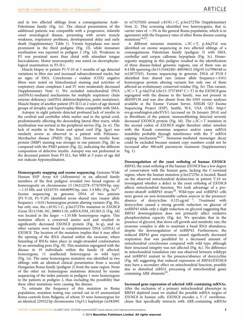
Autopsy in eight patients detected profound lack of myelin inthe cerebral and cerebellar white matter and in the spinal cord,predominantly affecting the descending lateral fibre tracts, whilemyelination was normal in the peripheral nerves (Fig. 2a–n). Thelack of myelin in the brain and spinal cord (Fig. 2g,n) wassimilarly severe as observed in a patient with Pelizaeus–Merzbacher disease (PMD) (Fig. 2m). However myelin basicprotein (MBP) staining was stronger in our patients (Fig. 2k) ascompared with the PMD patient (Fig. 2j), indicating the differentcomposition of defective myelin. Autopsy was not performed inthe deceased patient from P3-II:1, but MRI at 5 years of age didnot indicate hypomyelination.
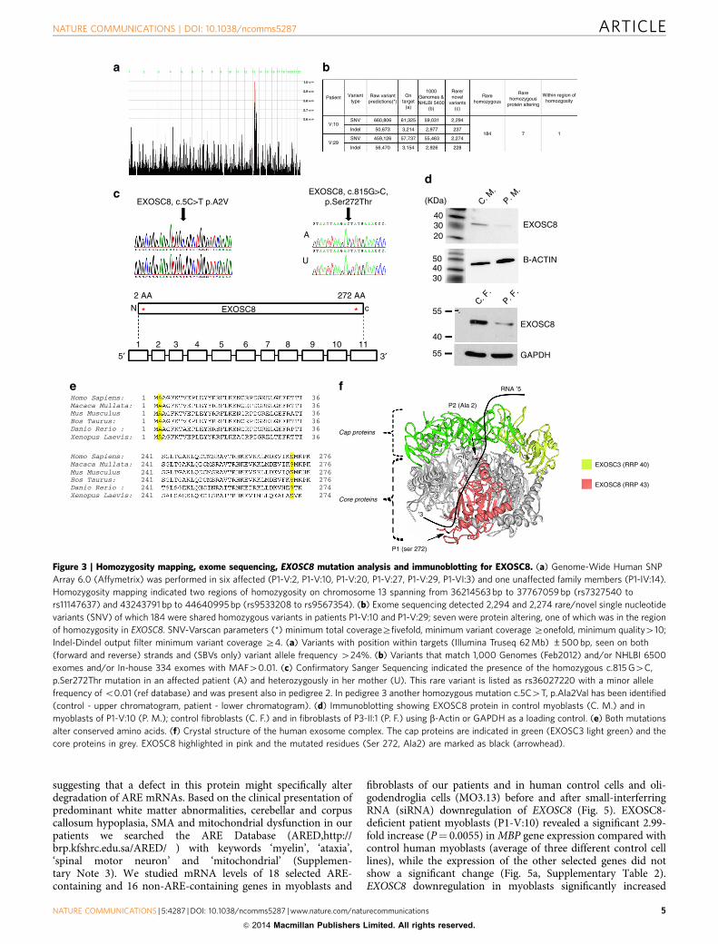
Homozygosity mapping and exome sequencing. Genome-WideHuman SNP Array 6.0 (Affymetrix) in six affected familymembers of the first pedigree revealed two broad regions ofhomozygosity on chromosome 13 (36212278–37767059 bp, size:B1.55 Mb; and 43243791-44640995 bp, size: 1.4 Mb) (Fig. 3a)13.Exome sequencing in genomic DNA of two patients(P1-V:10, P1-V:29) identified seven shared rare (major allelefrequency o0.01) homozygous protein altering variants (Fig. 3b),but only one, the c.815G4C, p.Ser272Thr mutation in EXOSC8(Gene ID:11340, NC_000013.10, mRNA:NM_181503.2) (Fig. 3d)was located in the larger B1.55 Mb homozygous region. Thismutation affects a conserved amino acid and resulted insignificantly decreased EXOSC8 protein (Fig. 3e–g), and noother variants were found in complementary DNA (cDNA) ofEXOSC8. The location of the mutation implies that it may affectthe opening of the RNA channel within the exosome, wherefunneling of RNAs takes place in single-stranded conformationby an unwinding pore (Fig. 3f). This mutation segregated with thedisease in 19 individuals within the family (8 affected:homozygous; 11 unaffected: heterozygous or wild type)(Fig. 1a). The same homozygous mutation was identified in twosiblings with an identical clinical presentation from a secondHungarian Roma family (pedigree 2) from the same region. Noneof the other six homozygous mutations detected by exomesequencing of the index patients in pedigree 1 were homozygousin the patients in pedigree 2, thus excluding the possibility thatthese other mutations were causing the disease.
To estimate the frequency of this mutation in Romapopulation, mutation testing was carried out in 63 anonymizedRoma controls from Bulgaria, of whom 33 were homozygous foran identical 229522 bp chromosome 13q13.1 haplotype (rs582091
to rs7327020) around c.815G4C, p.Ser272Thr (SupplementaryNote 2). This screening identified two heterozygotes, that is,carrier rates of B3% in the general Roma population, which is inagreement with the frequency rates of other Roma disease causingmutations14,15.
A different missense mutation, c.5C4T, p.Ala2Val wasidentified on exome sequencing in two affected siblings of aconsanguineous Palestinian family (pedigree 3) with SMA,cerebellar and corpus callosum hypoplasia (Fig. 3c). Homo-zygosity mapping in this pedigree resulted in the identificationof three disease-linked genomic regions, one of them was an8.4 Mb spanning chr13:31642481-40039652 (Hg19) (rs7996548 tors12873765). Exome sequencing in genomic DNA of P3:II-3identified four shared rare (minor allele frequencyo0.01)homozygous protein altering variants but only one of themaffected an evolutionary conserved residue (Fig. 3e). This variant,c.5C4T, p.Ala2Val (chr13: 37574947 C4T) in the EXOSC8 genesegregated with the disease in the family, was not present indbSNP138 and was also absent from the 6503 exome analysesavailable at the Exome Variant Server, NHLBI GO ExomeSequencing Project (ESP), Seattle, WA, USA (URL: http://evs.gs.washington.edu/EVS/) (accessed on 26 January 2014) andin fibroblasts of the patient, immunoblotting detected severelydecreased EXOSC8 protein (Fig. 3d). The c.5C4T mutation inthe second codon of EXOSC8 might therefore either interferewith the Kozak consensus sequence and/or cause mRNAinstability probably through interference with the 50 mRNAcapping mechanism16,17. Nonsense-mediated messenger decaycould be excluded because mutant copy numbers could not beincreased after 300 mM puromycin treatment (SupplementaryFig. 1).
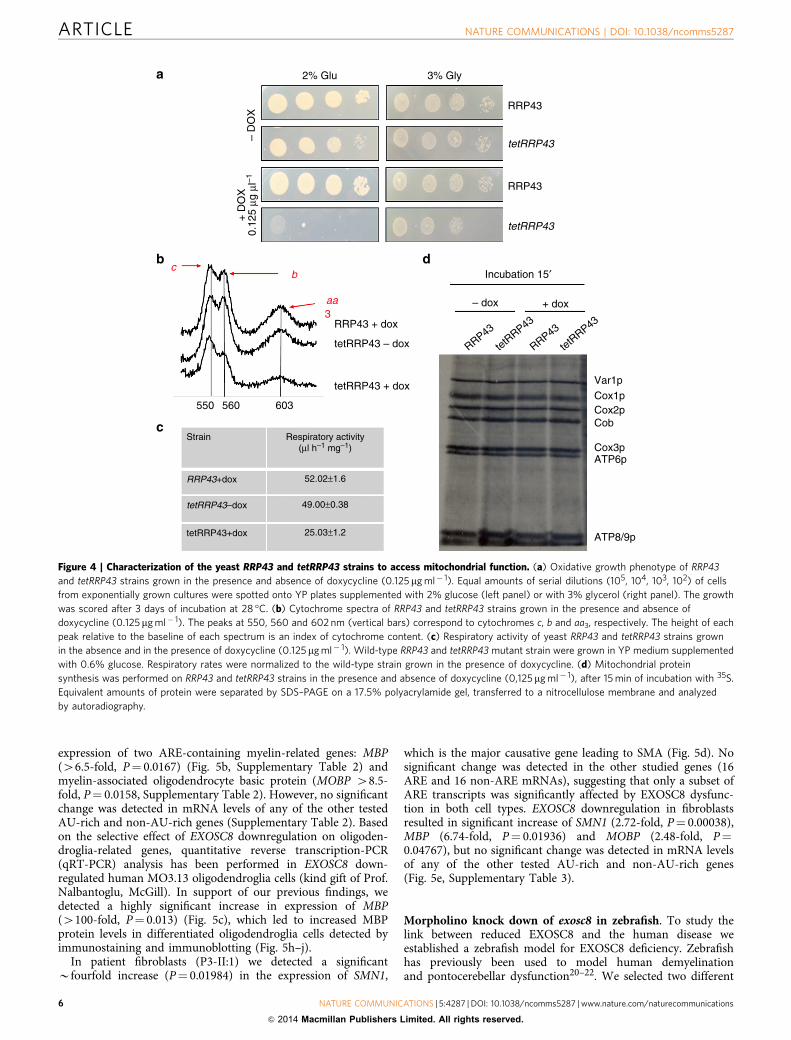
Downregulation of the yeast ortholog of human EXOSC8.RRP43, the yeast ortholog of the human EXOSC8 has a low degreeof conservation with the human gene, lacking the C-terminalregion, where the human mutation p.Ser272Thr is located. Basedon the observed mitochondrial dysfunction in patient V:10 weinvestigated whether a defect in the yeast ortholog of EXOSC8affects mitochondrial function. We took advantage of a pro-moter-shutoff tetRRP43 strain18. Wild-type and tetRRP43 cellswere grown on non-fermentable carbon sources in the presence/absence of doxycycline 0.125 mg ml� 1. Treatment withdoxycycline caused a strong growth reduction on glucose oftetRRP43 while only a slight reduction on glycerol suggesting thatRRP43 downregulation does not primarily affect oxidativephosphorylation capacity (Fig. 4a). We speculate that in thepresence of glycerol, that slows cell growth and metabolic rate, theexosome complex is able to maintain a basal RNA abundance,despite the downregulation of tetRRP43. Furthermore, thereduced RRP43 gene expression caused significantly decreasedrespiration that was paralleled by a decreased amount ofmitochondrial cytochromes compared with wild type, althoughtheir structural integrity was not affected (Fig. 4c). No differencein mitochondrial translation rate was observed between wildtypeand tetRRP43 mutant in the presence/absence of doxycycline(Fig. 4d) suggesting that reduced expression of RRP43/EXOSC8may have a secondary effect on mitochondrial function, possiblydue to disturbed mRNA processing of mitochondrial genescontaining ARE elements19.
Increased gene expression of selected ARE-containing mRNAs.After the exclusion of a primary mitochondrial phenotype inRRP43 depleted yeast we studied the possible mRNA targets ofEXOSC8 in human cells. EXOSC8 encodes a 30–50 exoribonu-clease that specifically interacts with ARE-containing mRNAs
NATURE COMMUNICATIONS | DOI: 10.1038/ncomms5287 ARTICLE
NATURE COMMUNICATIONS | 5:4287 | DOI: 10.1038/ncomms5287 | www.nature.com/naturecommunications 3
& 2014 Macmillan Publishers Limited. All rights reserved.
Cor
tex
a b c
d
g h
e f
*
2 mm 200 μm 2 mmWhi
te m
atte
r
200 μm 200 μm 200 μm
200 μm 200 μm
500 μm 500 μm 500 μm
i j k
**
l m n
500 μm 500 μm 500 μm
Figure 2 | Autopsy findings. (a–h) Cerebral cortex and white matter in autopsy of patient V:3 (pedigree 1). (a,b) Thickness of the white matter is
significantly reduced, while the cortex is relatively well preserved. Black arrow: thickness of the cortex from the pia to the cortex/white matter borderzone;
grey arrow: thickness of the white matter from the borderzone to the ependymal lining (Hematoxylin and eosin (H&E) staining); extensive astrogliosis
(pale nuclei; white arrowhead) and reduced frequency of oligodendrocytes (dark round nuclei; black arrowhead). (c) Cerebellar cortex is relatively well
preserved, but there is a microvacuolation of the underlying white matter within the cerebellar folia (H&E staining). (d) Reactive astrocytes (arrows) within
the cerebellar white matter (vimentin immunohistochemistry). (e,f) Purkinje cell axonal torpedos (modified Bielschowsky silver method) (e) and
neurofilament antibodies (f) indicating the loss of axonal connections. (g) Loss of myelin within the cerebellar white matter (Luxol Fast Blue). (h) Relative
preservation of the myelin staining on myelin basic protein immunohistochemistry. (i–n) Spinal cord: normal control (i,l), Pelizaeus–Merzbacher
disease (j,m) and EXOSC8 deficiency (patient V:20) (k,n). In EXOSC8 deficiency, myelin basic protein is present–apart from the longitudinal descending
fibre tracts (k, *)–while it is completely absent in Pelizaeus–Merzbacher disease (j). Myelin is well preserved within the peripheral nerve roots
(m,n, arrowheads), while indicates severe loss of myelin in both conditions within the spinal cord (m,n, Luxol Fast Blue).
ARTICLE NATURE COMMUNICATIONS | DOI: 10.1038/ncomms5287
4 NATURE COMMUNICATIONS | 5:4287 | DOI: 10.1038/ncomms5287 | www.nature.com/naturecommunications
& 2014 Macmillan Publishers Limited. All rights reserved.
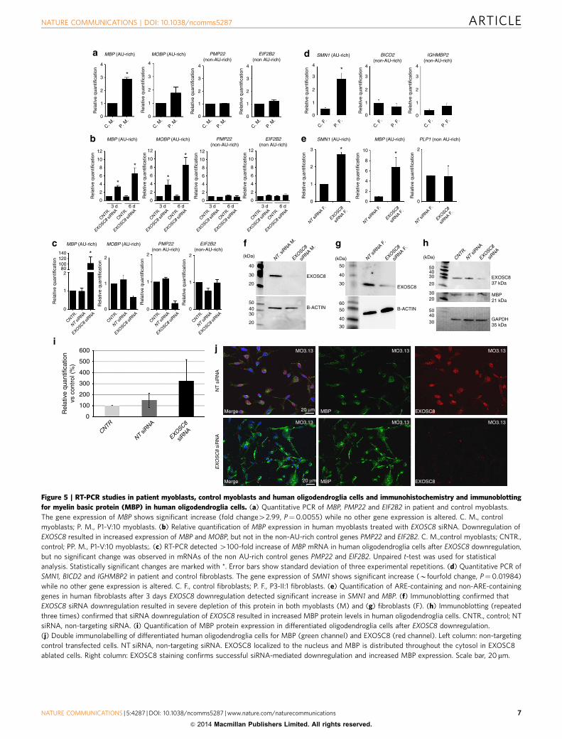
suggesting that a defect in this protein might specifically alterdegradation of ARE mRNAs. Based on the clinical presentation ofpredominant white matter abnormalities, cerebellar and corpuscallosum hypoplasia, SMA and mitochondrial dysfunction in ourpatients we searched the ARE Database (ARED,http://brp.kfshrc.edu.sa/ARED/ ) with keywords ‘myelin’, ‘ataxia’,‘spinal motor neuron’ and ‘mitochondrial’ (Supplemen-tary Note 3). We studied mRNA levels of 18 selected ARE-containing and 16 non-ARE-containing genes in myoblasts and
fibroblasts of our patients and in human control cells and oli-godendroglia cells (MO3.13) before and after small-interferringRNA (siRNA) downregulation of EXOSC8 (Fig. 5). EXOSC8-deficient patient myoblasts (P1-V:10) revealed a significant 2.99-fold increase (P¼ 0.0055) in MBP gene expression compared withcontrol human myoblasts (average of three different control celllines), while the expression of the other selected genes did notshow a significant change (Fig. 5a, Supplementary Table 2).EXOSC8 downregulation in myoblasts significantly increased
Within region ofhomozgosity
21 3 4 5 6 7 8 9 10 11 12 13 14 15 16 17 18 19 20 21 22
1.0 ×
0.9 ×
0.8 ×
0.7 ×
0.6 ×
Patient
b
d
a
V:10SNV 660,806 61,325 59,031
2,977
55,463
2,926
(KDa) C. M.
C. F.
P. M.
P. F.
403020
EXOSC8
EXOSC8
B-ACTIN
GAPDH55
40
55
f
EXOSC3 (RRP 40)
EXOSC8 (RRP 43)
P1 (ser 272)
′3
Core proteins
Cap proteins
RNA ′5
P2 (Ala 2)
eHomo Sapiens: 1
11111
241241241241241
241 276
363636363636
276276276274274
Homo Sapiens:
Macaca Mullata:
Macaca Mullata:
Mus Musculus
Mus Musculus
Bos Taurus:
Bos Taurus:
Danio Rerio :
Danio Rerio :
Xenopus Laevis:
Xenopus Laevis:
c
272 AA
U
A
2 AA
* *EXOSC8
EXOSC8, c.815G>C,p.Ser272ThrEXOSC8, c.5C>T p.A2V
cN
3′5′1110987654321
504030
228
2,274
237184 7 1
2,294
3,214
57,737
3,154
50,673
459,126
56,470
Indel
SNV
IndelV:29
Varianttype
Raw variantpredictions(*)
Ontarget
(a)
1000Genomes &NHLBI 5400
(b)
Rare/novel
variants(c)
Rarehomozygous
Rarehomozygous
protein altering
Figure 3 | Homozygosity mapping, exome sequencing, EXOSC8 mutation analysis and immunoblotting for EXOSC8. (a) Genome-Wide Human SNP
Array 6.0 (Affymetrix) was performed in six affected (P1-V:2, P1-V:10, P1-V:20, P1-V:27, P1-V:29, P1-VI:3) and one unaffected family members (P1-IV:14).
Homozygosity mapping indicated two regions of homozygosity on chromosome 13 spanning from 36214563 bp to 37767059 bp (rs7327540 to
rs11147637) and 43243791 bp to 44640995 bp (rs9533208 to rs9567354). (b) Exome sequencing detected 2,294 and 2,274 rare/novel single nucleotide
variants (SNV) of which 184 were shared homozygous variants in patients P1-V:10 and P1-V:29; seven were protein altering, one of which was in the region
of homozygosity in EXOSC8. SNV-Varscan parameters (*) minimum total coverageZfivefold, minimum variant coverage Zonefold, minimum quality410;
Indel-Dindel output filter minimum variant coverage Z4. (a) Variants with position within targets (Illumina Truseq 62 Mb) ±500 bp, seen on both
(forward and reverse) strands and (SBVs only) variant allele frequency 424%. (b) Variants that match 1,000 Genomes (Feb2012) and/or NHLBI 6500
exomes and/or In-house 334 exomes with MAF40.01. (c) Confirmatory Sanger Sequencing indicated the presence of the homozygous c.815 G4C,
p.Ser272Thr mutation in an affected patient (A) and heterozygously in her mother (U). This rare variant is listed as rs36027220 with a minor allele
frequency of o0.01 (ref database) and was present also in pedigree 2. In pedigree 3 another homozygous mutation c.5C4T, p.Ala2Val has been identified
(control - upper chromatogram, patient - lower chromatogram). (d) Immunoblotting showing EXOSC8 protein in control myoblasts (C. M.) and in
myoblasts of P1-V:10 (P. M.); control fibroblasts (C. F.) and in fibroblasts of P3-II:1 (P. F.) using b-Actin or GAPDH as a loading control. (e) Both mutations
alter conserved amino acids. (f) Crystal structure of the human exosome complex. The cap proteins are indicated in green (EXOSC3 light green) and the
core proteins in grey. EXOSC8 highlighted in pink and the mutated residues (Ser 272, Ala2) are marked as black (arrowhead).
NATURE COMMUNICATIONS | DOI: 10.1038/ncomms5287 ARTICLE
NATURE COMMUNICATIONS | 5:4287 | DOI: 10.1038/ncomms5287 | www.nature.com/naturecommunications 5
& 2014 Macmillan Publishers Limited. All rights reserved.
expression of two ARE-containing myelin-related genes: MBP(46.5-fold, P¼ 0.0167) (Fig. 5b, Supplementary Table 2) andmyelin-associated oligodendrocyte basic protein (MOBP 48.5-fold, P¼ 0.0158, Supplementary Table 2). However, no significantchange was detected in mRNA levels of any of the other testedAU-rich and non-AU-rich genes (Supplementary Table 2). Basedon the selective effect of EXOSC8 downregulation on oligoden-droglia-related genes, quantitative reverse transcription-PCR(qRT-PCR) analysis has been performed in EXOSC8 down-regulated human MO3.13 oligodendroglia cells (kind gift of Prof.Nalbantoglu, McGill). In support of our previous findings, wedetected a highly significant increase in expression of MBP(4100-fold, P¼ 0.013) (Fig. 5c), which led to increased MBPprotein levels in differentiated oligodendroglia cells detected byimmunostaining and immunoblotting (Fig. 5h–j).
In patient fibroblasts (P3-II:1) we detected a significantBfourfold increase (P¼ 0.01984) in the expression of SMN1,
which is the major causative gene leading to SMA (Fig. 5d). Nosignificant change was detected in the other studied genes (16ARE and 16 non-ARE mRNAs), suggesting that only a subset ofARE transcripts was significantly affected by EXOSC8 dysfunc-tion in both cell types. EXOSC8 downregulation in fibroblastsresulted in significant increase of SMN1 (2.72-fold, P¼ 0.00038),MBP (6.74-fold, P¼ 0.01936) and MOBP (2.48-fold, P¼0.04767), but no significant change was detected in mRNA levelsof any of the other tested AU-rich and non-AU-rich genes(Fig. 5e, Supplementary Table 3).
Morpholino knock down of exosc8 in zebrafish. To study thelink between reduced EXOSC8 and the human disease weestablished a zebrafish model for EXOSC8 deficiency. Zebrafishhas previously been used to model human demyelinationand pontocerebellar dysfunction20–22. We selected two different
Respiratory activity(μl h–1 mg–1)
Strain
RRP43+dox
tetRRP43–dox
tetRRP43+dox
52.02±1.6
49.00±0.38
25.03±1.2
b
aa3
c
RRP43 + dox
tetRRP43 – dox
tetRRP43 + dox
550 560 603
b
c
Incubation 15′
– dox
RRP43
RRP43
tetRRP43
tetRRP43
+ dox
Var1p
Cox1pCox2pCob
Cox3pATP6p
ATP8/9p
d
a 2% Glu
– D
OX
+ D
OX
0.12
5 μg
μl–1
3% Gly
RRP43
tetRRP43
tetRRP43
RRP43
Figure 4 | Characterization of the yeast RRP43 and tetRRP43 strains to access mitochondrial function. (a) Oxidative growth phenotype of RRP43
and tetRRP43 strains grown in the presence and absence of doxycycline (0.125 mg ml� 1). Equal amounts of serial dilutions (105, 104, 103, 102) of cells
from exponentially grown cultures were spotted onto YP plates supplemented with 2% glucose (left panel) or with 3% glycerol (right panel). The growth
was scored after 3 days of incubation at 28 �C. (b) Cytochrome spectra of RRP43 and tetRRP43 strains grown in the presence and absence of
doxycycline (0.125mg ml� 1). The peaks at 550, 560 and 602 nm (vertical bars) correspond to cytochromes c, b and aa3, respectively. The height of each
peak relative to the baseline of each spectrum is an index of cytochrome content. (c) Respiratory activity of yeast RRP43 and tetRRP43 strains grown
in the absence and in the presence of doxycycline (0.125 mg ml� 1). Wild-type RRP43 and tetRRP43 mutant strain were grown in YP medium supplemented
with 0.6% glucose. Respiratory rates were normalized to the wild-type strain grown in the presence of doxycycline. (d) Mitochondrial protein
synthesis was performed on RRP43 and tetRRP43 strains in the presence and absence of doxycycline (0,125mg ml� 1), after 15 min of incubation with 35S.
Equivalent amounts of protein were separated by SDS–PAGE on a 17.5% polyacrylamide gel, transferred to a nitrocellulose membrane and analyzed
by autoradiography.
ARTICLE NATURE COMMUNICATIONS | DOI: 10.1038/ncomms5287
6 NATURE COMMUNICATIONS | 5:4287 | DOI: 10.1038/ncomms5287 | www.nature.com/naturecommunications
& 2014 Macmillan Publishers Limited. All rights reserved.
MBP (AU-rich) MOBP (AU-rich) PMP22(non-AU-rich)
EIF2B2(non AU-rich)
MBP (AU-rich) MOBP (AU-rich) PMP22 (non-AU-rich)
EIF2B2(non AU-rich)
SMN1 (AU-rich) BICD2(non-AU-rich)
IGHMBP2(non-AU-rich)
SMN1 (AU-rich) MBP (AU-rich) PLP1 (non AU-rich)
*
4
3
2
1
Rel
ativ
e qu
antif
icat
ion
Rel
ativ
e qu
antif
icat
ion
Rel
ativ
e qu
antif
icat
ion
Rel
ativ
e qu
antif
icat
ion
0
4
3
2
1
Rel
ativ
e qu
antif
icat
ion
0
4
3
2
1
Rel
ativ
e qu
antif
icat
ion
0
4
3
2
1
Rel
ativ
e qu
antif
icat
ion
0
4*
3
2
1
Rel
ativ
e qu
antif
icat
ion
0
4
3
2
1
Rel
ativ
e qu
antif
icat
ion
0
4
3
2
1
Rel
ativ
e qu
antif
icat
ion
0
C. M.
P. M.
C. M.
P. M.
C. M.
P. M.
C. M.
P. M.
P. F.
C. F.
P. F.
C. F.
P. F.
C. F.
a d
b12
10
8
6
4 *2
0
Rel
ativ
e qu
antif
icat
ion
12
10
8
6
4
2
0R
elat
ive
quan
tific
atio
n 12
10
8
6
4
2
0
Rel
ativ
e qu
antif
icat
ion
Rel
ativ
e qu
antif
icat
ion
Rel
ativ
e qu
antif
icat
ion
Rel
ativ
e qu
antif
icat
ion
12 3 2
0
10
8
6
4
2
0
2
1
0
NT siRNA F
.
EXOSC8
siRNA F
.
NT siRNA F
.
EXOSC8
siRNA F
.
NT siRNA F
.
EXOSC8
siRNA F
.
10
8
6
4
2
03 d
CNTR.
EXOSC8 siR
NA
CNTR.
EXOSC8 siR
NA6 d 3 d
CNTR.
EXOSC8 siR
NA
CNTR.
EXOSC8 siR
NA6 d 3 d
CNTR.
EXOSC8 siR
NA
CNTR.
EXOSC8 siR
NA6 d 3 d
CNTR.
EXOSC8 siR
NA
CNTR.
EXOSC8 siR
NA6 d
*
*
**
*
MBP (AU-rich)
*
MOBP (AU-rich) PMP22(non AU-rich)
EIF2B2 (non-AU-rich)
1402
1
0
Rel
ativ
e qu
antif
icat
ion
2
1
0
Rel
ativ
e qu
antif
icat
ion
2
1
0
120100802
1
0
CNTR.
NT siRNA
EXOSC8 siR
NA
CNTR.
NT siRNA
EXOSC8 siR
NA
CNTR.
NT siRNA
EXOSC8 siR
NA
CNTR.
NT siRNA
EXOSC8 siR
NA
e
c
i600
500
400
300
200
Rel
ativ
e qu
antif
icat
ion
vs c
ontr
ol (
%)
100
0
CNTR
NT siRNA
EXOSC8
siRNA
h
CNTR.
NT siRNA
EXOSC8
s
iRNA
(kDa)
EXOSC837 kDa
MBP21 kDa
GAPDH35 kDa
504030
20
3020
504030
MO3.13 MO3.13 MO3.13
MO3.13 MO3.13 MO3.13
NT siRNA F
.
EXOSC8
si
RNA F.
NT. siR
NA M.
EXOSC8
si
RNA M.
(kDa) (kDa)
EXOSC8
B-ACTIN
40
30
20
504030
20
50
40
30
6050
40
30
f g
EXOSC8
B-ACTIN
j
NT
siR
NA
EX
OS
C8
siR
NA
Merge MBP20 μm EXOSC8
Merge MBP EXOSC820 μm
Figure 5 | RT-PCR studies in patient myoblasts, control myoblasts and human oligodendroglia cells and immunohistochemistry and immunoblotting
for myelin basic protein (MBP) in human oligodendroglia cells. (a) Quantitative PCR of MBP, PMP22 and EIF2B2 in patient and control myoblasts.
The gene expression of MBP shows significant increase (fold change42.99, P¼0.0055) while no other gene expression is altered. C. M., control
myoblasts; P. M., P1-V:10 myoblasts. (b) Relative quantification of MBP expression in human myoblasts treated with EXOSC8 siRNA. Downregulation of
EXOSC8 resulted in increased expression of MBP and MOBP, but not in the non-AU-rich control genes PMP22 and EIF2B2. C. M.,control myoblasts; CNTR.,
control; PP. M., P1-V:10 myoblasts;. (c) RT-PCR detected 4100-fold increase of MBP mRNA in human oligodendroglia cells after EXOSC8 downregulation,
but no significant change was observed in mRNAs of the non AU-rich control genes PMP22 and EIF2B2. Unpaired t-test was used for statistical
analysis. Statistically significant changes are marked with *. Error bars show standard deviation of three experimental repetitions. (d) Quantitative PCR of
SMN1, BICD2 and IGHMBP2 in patient and control fibroblasts. The gene expression of SMN1 shows significant increase (Bfourfold change, P¼0.01984)
while no other gene expression is altered. C. F., control fibroblasts; P. F., P3-II:1 fibroblasts. (e) Quantification of ARE-containing and non-ARE-containing
genes in human fibroblasts after 3 days EXOSC8 downregulation detected significant increase in SMN1 and MBP. (f) Immunoblotting confirmed that
EXOSC8 siRNA downregulation resulted in severe depletion of this protein in both myoblasts (M) and (g) fibroblasts (F). (h) Immunoblotting (repeated
three times) confirmed that siRNA downregulation of EXOSC8 resulted in increased MBP protein levels in human oligodendroglia cells. CNTR., control; NT
siRNA, non-targeting siRNA. (i) Quantification of MBP protein expression in differentiated oligodendroglia cells after EXOSC8 downregulation.
(j) Double immunolabelling of differentiated human oligodendroglia cells for MBP (green channel) and EXOSC8 (red channel). Left column: non-targeting
control transfected cells. NT siRNA, non-targeting siRNA. EXOSC8 localized to the nucleus and MBP is distributed throughout the cytosol in EXOSC8
ablated cells. Right column: EXOSC8 staining confirms successful siRNA-mediated downregulation and increased MBP expression. Scale bar, 20 mm.
NATURE COMMUNICATIONS | DOI: 10.1038/ncomms5287 ARTICLE
NATURE COMMUNICATIONS | 5:4287 | DOI: 10.1038/ncomms5287 | www.nature.com/naturecommunications 7
& 2014 Macmillan Publishers Limited. All rights reserved.
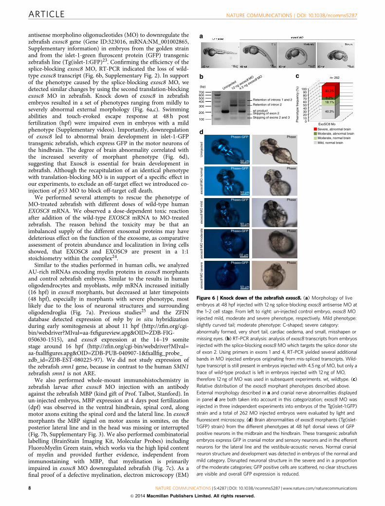
antisense morpholino oligonucleotides (MO) to downregulate thezebrafish exosc8 gene (Gene ID:323016, mRNA:NM_001002865,Supplementary information) in embryos from the golden strainand from the islet-1-green fluroscent protein (GFP) transgeniczebrafish line (Tg(islet-1:GFP)23. Confirming the efficiency of thesplice-blocking exosc8 MO, RT-PCR indicated the loss of wild-type exosc8 transcript (Fig. 6b, Supplementary Fig. 2). In supportof the phenotype caused by the splice-blocking exosc8 MO, wedetected similar changes by using the second translation-blockingexosc8 MO in zebrafish. Knock down of exosc8 in zebrafishembryos resulted in a set of phenotypes ranging from mildly toseverely abnormal external morphology (Fig. 6a,c). Swimmingabilities and touch-evoked escape response at 48 h postfertilization (hpf) were impaired even in embryos with a mildphenotype (Supplementary videos). Importantly, downregulationof exosc8 led to abnormal brain development in islet-1-GFPtransgenic zebrafish, which express GFP in the motor neurons ofthe hindbrain. The degree of brain abnormality correlated withthe increased severity of morphant phenotype (Fig. 6d),suggesting that Exosc8 is essential for brain development inzebrafish. Although the recapitulation of an identical phenotypewith translation-blocking MO is in support of a specific effect inour experiments, to exclude an off-target effect we introduced co-injection of p53 MO to block off-target cell death.
We performed several attempts to rescue the phenotype ofMO-treated zebrafish with different doses of wild-type humanEXOSC8 mRNA. We observed a dose-dependent toxic reactionafter addition of the wild-type EXOSC8 mRNA to MO-treatedzebrafish. The reason behind the toxicity may be that animbalanced supply of the different exosomal proteins may havedeleterious effect on the function of the exosome, as comparativeassessment of protein abundance and localization in living cellsshowed, that EXOSC8 and EXOSC9 are present in a 1:1stoichiometry within the complex24.
Similar to the studies performed in human cells, we analyzedAU-rich mRNAs encoding myelin proteins in exosc8 morphantsand control zebrafish embryos. Similar to the results in humanoligodendrocytes and myoblasts, mbp mRNA increased initially(16 hpf) in exosc8 morphants, but decreased at later timepoints(48 hpf), especially in morphants with severe phenotype, mostlikely due to the loss of neuronal structures and surroundingoligodendroglia (Fig. 7a). Previous studies25 and the ZFINdatabase detected expression of mbp by in situ hybridizationduring early somitogenesis at about 11 hpf (http://zfin.org/cgi-bin/webdriver?MIval=aa-fxfigureview.apg&OID=ZDB-FIG-050630-1515), and exosc8 expression at the 14–19 somitestage around 16 hpf (http://zfin.org/cgi-bin/webdriver?MIval=aa-fxallfigures.apg&OID=ZDB-PUB-040907-1&fxallfig_probe_zdb_id=ZDB-EST-080225-97). We did not study expression ofthe zebrafish smn1 gene, because in contrast to the human SMN1zebrafish smn1 is not ARE.
We also performed whole-mount immunohistochemistry inzebrafish larvae after exosc8 MO injection with an antibodyagainst the zebrafish MBP (kind gift of Prof. Talbot, Stanford). Inun-injected embryos, MBP expression at 4 days post fertilization(dpf) was observed in the ventral hindbrain, spinal cord, alongmotor axons exiting the spinal cord and the lateral line. In exosc8morphants the MBP signal on motor axons in somites, on theposterior lateral line and in the head was missing or interrupted(Fig. 7b, Supplementary Fig. 3). We also performed combinatoriallabelling (BrainStain Imaging Kit, Molecular Probes) includingFluoroMyelin Green stain, which works via the high lipid contentof myelin and provided further evidence, independent fromimmunostaining with MBP, that myelination is primarilyimpaired in exosc8 MO downregulated zebrafish (Fig. 7c). As afinal proof of a defective myelination, electron microscopy (EM)
b c
1009080706050403020100
ExoSC8 Mo
Phe
noty
pe fr
eque
ncy
(%)
40.3%
18.1%8.2%
33.3%
n= 262
Severe, abnormal brainModerate, abnormal brainModerate, normal brain
Mild, normal brain
(bp)
700
Retention of introns 1 and 2
Retention of intron 2
wt productSkipping of exon 2Skipping of exons 2 and 3
Uninjected
12 ng exosc8 MO
4.5 ng exosc8 MO
600500400
300
200
100
dPhase
Phase
Phase
Phase
Phase
Uni
njec
ted
exos
c8 M
O n
orm
alex
osc8
MO
mild
exos
c8 M
O m
oder
ate
exos
c8 M
O s
ever
e
Phase+GFP
Phase+GFP
Phase+GFP
Phase+GFP
50 μm
50 μm
50 μm
50 μm
50 μm
Phase+GFP
a
Figure 6 | Knock down of the zebrafish exosc8. (a) Morphology of live
embryos at 48 hpf injected with 12 ng splice-blocking exosc8 antisense MO at
the 1–2 cell stage. From left to right: un-injected control embryo, exosc8 MO
injected mild, moderate and severe phenotype, respectively. Mild phenotype:
slightly curved tail; moderate phenotype: C-shaped; severe category:
abnormally formed, very short tail, cardiac oedema, and small, misshapen or
missing eyes. (b) RT–PCR analysis: analysis of exosc8 transcripts from embryos
injected with the splice-blocking exosc8 MO which targets the splice donor site
of exon 2. Using primers in exons 1 and 4, RT–PCR yielded several additional
bands in MO injected embryos originating from mis-spliced transcripts. Wild-
type transcript is still present in embryos injected with 4.5 ng of MO, but only a
trace of wild-type product is left in embryos injected with 12 ng of MO,
therefore 12 ng of MO was used in subsequent experiments. wt, wildtype. (c)
Relative distribution of the exosc8 morphant phenotypes described above.
External morphology described in a and cranial nerve abnormalities displayed
in panel d are both taken into account in this categorization; exosc8 MO was
injected in three independent experiments into embryos of the Tg(islet-1:GFP)
strain and a total of 262 MO injected embryos were evaluated by light and
fluorescent microscopy. (d) Brain abnormalities of exosc8 morphants (Tg(islet-
1:GFP) strain) from the different phenotypes at 48 hpf: dorsal views of GFP
positive neurons in the midbrain and the hindbrain. These transgenic zebrafish
embryos express GFP in cranial motor and sensory neurons and in the efferent
neurons for the lateral line and the vestibule-acoustic nerves. Normal cranial
neuron structure and development was detected in embryos of the normal and
mild category. Disrupted neuronal structure in the severe and in a proportion
of the moderate categories; GFP positive cells are scattered, no clear structures
are visible and overall GFP expression is reduced.
ARTICLE NATURE COMMUNICATIONS | DOI: 10.1038/ncomms5287
8 NATURE COMMUNICATIONS | 5:4287 | DOI: 10.1038/ncomms5287 | www.nature.com/naturecommunications
& 2014 Macmillan Publishers Limited. All rights reserved.
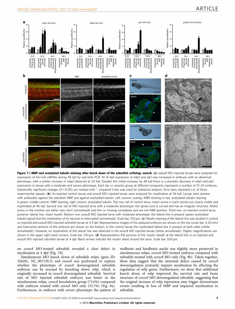
on exosc8 MO-treated zebrafish revealed a clear defect inmyelination at 4 dpf (Fig. 7d).
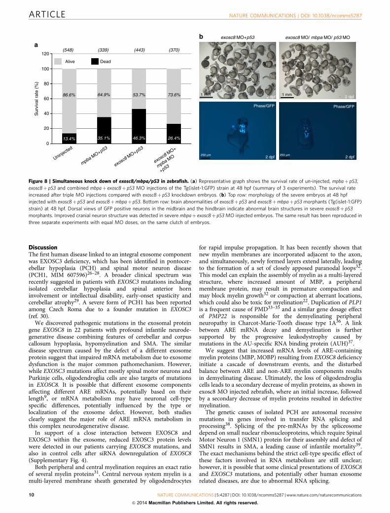
Simultaneous MO knock down of zebrafish mbpa (gene ID:326281, NC_007130.5) and exosc8 was performed to explorewhether the phenotype of exosc8 downregulated zebrafishembryos can be rescued by knocking down mbp, which isoriginally increased in exosc8 downregulated zebrafish. Survivalrate of MO injected zebrafish embryos was better in thesimultaneous mbpa, exosc8 knockdown group (73.6%) comparedwith embryos treated with exosc8 MO only (53.7%) (Fig. 8a).Furthermore, in embryos with severe phenotype the pattern of
midbrain and hindbrain nuclei was slightly more preserved insimultaneous mbpa, exosc8 MO-treated embryos compared withzebrafish treated with exosc8 MO only (Fig. 8b). Taken together,these data suggest that the neuronal defect caused by exosc8downregulation primarily impairs myelination by affecting theregulation of mbp genes. Furthermore, we show that additionalknock down of mbp improved the survival rate and brainstructure of exosc8 MO downregulated zebrafish, suggesting thatthe original increase of mbp expression may trigger downstreamevents resulting in loss of MBP and impaired myelination inzebrafish.
b dMyelin
Myelin exosc8 MO
Acetylated tubulinMBP
Uni
njec
ted
Uni
njec
ted
exos
c8 M
Pse
vere
phe
noty
peex
osc8
MO
mod
erat
e ph
enot
ype
Uninjected
∗
∗
∗ ∗
a mbp1 (AU-rich)
∗ ∗
∗ ∗ ∗
∗
mbp2 (AU-rich)R
elat
ive
quan
tific
atio
n8
6
4
2
0
Rel
ativ
e qu
antif
icat
ion
8
6
4
2
0
12 h 16 h 24 h 48 h 12 h 16 h 24 h 48 h
plp1 (AU-rich) pmp22 (non AU-rich)
Rel
ativ
e qu
antif
icat
ion
8
6
4
2
0
Rel
ativ
e qu
antif
icat
ion
8
6
4
2
0
12 h 16 h 24 h 48 h 12 h 16 h 24 h 48 h
Uni
njec
ted
Phe
nol r
edex
osc8
MO
Uni
njec
ted
Phe
nol r
ed
Uni
njec
ted
Uni
njec
ted
Phe
nol r
ed
Nor
mal
Mild
Mod
erat
eS
ever
e
Nor
mal
Nor
mal
/mild
Mod
erat
e/se
vere
Abn
orm
al
Uni
njec
ted
Phe
nol r
edex
osc8
MO
Uni
njec
ted
Phe
nol r
ed
Uni
njec
ted
Uni
njec
ted
Phe
nol r
ed
Nor
mal
Mild
Mod
erat
eS
ever
e
Nor
mal
Nor
mal
/mild
Mod
erat
e/se
vere
Abn
orm
al
Uni
njec
ted
Phe
nol r
edex
osc8
MO
Uni
njec
ted
Phe
nol r
ed
Uni
njec
ted
Uni
njec
ted
Phe
nol r
ed
Nor
mal
Mild
Mod
erat
eS
ever
e
Nor
mal
Nor
mal
/mild
Mod
erat
e/se
vere
Abn
orm
al
Uni
njec
ted
Phe
nol r
edex
osc8
MO
Uni
njec
ted
Phe
nol r
ed
Uni
njec
ted
Uni
njec
ted
Phe
nol r
ed
Nor
mal
Mild
Mod
erat
eS
ever
e
Nor
mal
Nor
mal
/mild
Mod
erat
e/se
vere
Abn
orm
al
cControl
4.5 dpf
Myelin
Myelin
4.5 dpf
exosc8 MO
Figure 7 | MBP and acetylated tubulin staining after knock down of the zebrafish ortholog: exosc8. (a) exosc8 MO injected larvae were analyzed for
expression of AU-rich mRNAs during 48 hpf by real-time PCR. At 16 hpf expression of mbp1 and plp1 was increased in embryos with an abnormal
phenotype, with a similar increase in mbp2 observed at 24 hpf. Despite this initial increase, by 48 hpf there is a dramatic decrease in mbp1 and plp1
expression in larvae with a moderate and severe phenotype. Each bar or severity group at different timepoints represents a number of 15–20 embryos.
Statistically significant changes (Po0.05) are marked with *. Unpaired t-test was used for statistical analysis. Error bars represent s.d. of three
experimental repeats. (b) Un-injected control larvae and exosc8 MO injected larvae were analyzed for myelination at 96 hpf. Larvae were stained
with antibodies against the zebrafish MBP and against acetylated tubulin. Left column: overlay, MBP staining in red, acetylated tubulin staining
in green; middle column: MBP staining; right column: acetylated tubulin. Top row: tail of control larva: motor axons in each somite are clearly visible and
myelinated at 96 hpf. Second row: tail of MO injected larva with a moderate phenotype: the spinal cord is curved and has an irregular structure. Motor
axons in the somites are either very short (arrowhead) and thin or missing completely and are not MBP-positive. Third row: un-injected control larva,
posterior lateral line, intact myelin. Bottom row: exosc8 MO injected larva with moderate phenotype: the lateral line is present (green acetylated
tubulin signal) but the myelination of its neurons is interrupted (arrowhead). Scale bar, 100mm. (c) Myelin staining of the lateral line was studied in control
un-injected and exosc8 MO injected zebrafish larvae at 4.5 dpf. Representative images of the analyzed embryos are shown on the top (scale bar, 0.25 mm)
and transverse sections of the embryos are shown on the bottom. In the control larvae the myelinated lateral line is present at both sides (white
arrowheads). However, no myelination of the lateral line was detected in the exosc8 MO injected larvae (white arrowheads). Higher magnifications are
shown in the upper right hand corners. Scale bar, 100mm. (d) Representative EM pictures of the myelin sheath at the lateral line in un-injected and
exosc8 MO injected zebrafish larvae at 4 dpf. Black arrows indicate the myelin sheet around the axon. Scale bar, 500mm.
NATURE COMMUNICATIONS | DOI: 10.1038/ncomms5287 ARTICLE
NATURE COMMUNICATIONS | 5:4287 | DOI: 10.1038/ncomms5287 | www.nature.com/naturecommunications 9
& 2014 Macmillan Publishers Limited. All rights reserved.
DiscussionThe first human disease linked to an integral exosome componentwas EXOSC3 deficiency, which has been identified in pontocer-ebellar hypoplasia (PCH) and spinal motor neuron disease(PCH1, MIM 607596)26–28. A broader clinical spectrum wasrecently suggested in patients with EXOSC3 mutations includingisolated cerebellar hypoplasia and spinal anterior horninvolvement or intellectual disability, early-onset spasticity andcerebellar atrophy29. A severe form of PCH1 has been reportedamong Czech Roma due to a founder mutation in EXOSC3(ref. 30).
We discovered pathogenic mutations in the exosomal proteingene EXOSC8 in 22 patients with profound infantile neurode-generative disease combining features of cerebellar and corpuscallosum hypoplasia, hypomyelination and SMA. The similardisease spectrum caused by the defect of a different exosomeprotein suggest that impaired mRNA metabolism due to exosomedysfunction is the major common pathomechanism. However,while EXOSC3 mutations affect mostly spinal motor neurons andPurkinje cells, oligodendroglia cells are also targets of mutationsin EXOSC8. It is possible that different exosome componentsaffecting different ARE mRNAs, potentially based on theirlength9, or mRNA metabolism may have neuronal cell-typespecific differences, potentially influenced by the type orlocalization of the exosome defect. However, both studiesclearly suggest the major role of ARE mRNA metabolism inthis complex neurodegenerative disease.
In support of a close interaction between EXOSC8 andEXOSC3 within the exosome, reduced EXOSC3 protein levelswere detected in our patients carrying EXOSC8 mutations, andalso in control cells after siRNA downregulation of EXOSC8(Supplementary Fig. 4).
Both peripheral and central myelination requires an exact ratioof several myelin proteins31. Central nervous system myelin is amulti-layered membrane sheath generated by oligodendrocytes
for rapid impulse propagation. It has been recently shown thatnew myelin membranes are incorporated adjacent to the axon,and simultaneously, newly formed layers extend laterally, leadingto the formation of a set of closely apposed paranodal loops32.This model can explain the assembly of myelin as a multi-layeredstructure, where increased amount of MBP, a peripheralmembrane protein, may result in premature compaction andmay block myelin growth32 or compaction at aberrant locations,which could also be toxic for myelination22. Duplication of PLP1is a frequent cause of PMD33–35 and a similar gene dosage effectof PMP22 is responsible for the demyelinating peripheralneuropathy in Charcot-Marie-Tooth disease type 1A36. A linkbetween ARE mRNA decay and demyelination is furthersupported by the progressive leukodystrophy caused bymutations in the AU-specific RNA binding protein (AUH)37.
We suggest that increased mRNA levels of ARE-containingmyelin proteins (MBP, MOBP) resulting from EXOSC8 deficiencyinitiate a cascade of downstream events, and the disturbedbalance between ARE and non-ARE myelin components resultsin demyelinating disease. Ultimately, the loss of oligodendrogliacells leads to a secondary decrease of myelin proteins, as shown inexosc8 MO injected zebrafish, where an initial increase, followedby a secondary decrease of myelin proteins resulted in defectivemyelination.
The genetic causes of isolated PCH are autosomal recessivemutations in genes involved in transfer RNA splicing andprocessing38. Splicing of the pre-mRNAs by the spliceosomedepend on small nuclear ribonucleoproteins, which require SpinalMotor Neuron 1 (SMN1) protein for their assembly and defect ofSMN1 results in SMA, a leading cause of infantile mortality39.The exact mechanisms behind the strict cell-type specific effect ofthese factors involved in RNA metabolism are still unclear;however, it is possible that some clinical presentations of EXOSC8and EXOSC3 mutations, and potentially other human exosomerelated diseases, are due to abnormal RNA splicing.
b exosc8 MO+p53 exosc8 MO/ mbpa MO/ p53 MO
2 dpf
2 dpf 2 dpf
1 mm1 mm
250 μm 250 μm
Phase/GFP Phase/GFP
2 dpf
(548)120
a
100
80
86.6%
13.4% 35.1% 46.3% 26.4%
64.9% 53.7% 73.6%
Alive Dead
60
Sur
viva
l rat
e (%
)
40
20
0
Uninjected
mpba MO+p53
exosc8 MO+p53
exosc8 MO+
mpba MO
+p53
(339) (443) (370)
Figure 8 | Simultaneous knock down of exosc8/mbpa/p53 in zebrafish. (a) Representative graph shows the survival rate of un-injected, mpbaþ p53,
exosc8þ p53 and combined mbpaþ exosc8þ p53 MO injections of the Tg(islet-1:GFP) strain at 48 hpf (summary of 3 experiments). The survival rate
increased after triple MO injections compared with exosc8þ p53 knockdown embryos. (b) Top row: morphology of the severe embryos at 48 hpf
injected with exosc8þ p53 and exosc8þmbpaþ p53. Bottom row: brain abnormalities of exosc8þ p53 and exosc8þmbpaþ p53 morphants (Tg(islet-1:GFP)
strain) at 48 hpf. Dorsal views of GFP positive neurons in the midbrain and the hindbrain indicate abnormal brain structures in severe exosc8þ p53
morphants. Improved cranial neuron structure was detected in severe mbpaþ exosc8þ p53 MO injected embryos. The same result has been reproduced in
three separate experiments with equal MO doses, on the same clutch of embryos.
ARTICLE NATURE COMMUNICATIONS | DOI: 10.1038/ncomms5287
10 NATURE COMMUNICATIONS | 5:4287 | DOI: 10.1038/ncomms5287 | www.nature.com/naturecommunications
& 2014 Macmillan Publishers Limited. All rights reserved.
In summary, patients with EXOSC8 mutations present with acharacteristic spectrum of overlapping phenotypes of infantileonset hypomyelination, cerebellar hypoplasia and SMA. Theclinical presentation may depend on the type and localization ofmutations and provides a clue to unravel the complex molecularpathways caused by the defective exosome function.
MethodsPatients. We have received informed consent from the patients (parents) includedin this study. Ethical approval was obtained from the County Durham and TeesValley 2 Research Ethics Committee (08/H0905/106) and from the Ethics com-mittee of the Hebrew University Hadassah Medical School (0485-09). Writtenconsent to publish the photo of a patient was obtained.
Histological and biochemical analyses of skeletal muscle. Histological andbiochemical analyses of skeletal muscle were performed by standard methods40.
Autopsy staining methods. The brain and spinal cord samples were fixed in 10%phosphate-buffered formal saline before embedding in paraffin wax. For Luxol FastBlue staining 10-mm sections were dehydrated and stained with Luxol Fast Blueovernight at room temperature, and then rinsed in alcohol and water to removeexcess blue colour. The sections were differentiated with a weak solution of lithiumcarbonate, followed by 70% alcohol solution. Immunohistochemistry was donefollowing citrate (pH 6) and EDTA (pH 8) pre-treatment for 10 min. Immuno-histochemistry was carried out on 5-mm sections with mouse monoclonal anti-bodies that recognize MBP (1:2000 SM194R, Covance, NJ, USA) and vimentin(1:5600 clone V9 Dako; Copenhagen, Denmark). Rabbit monoclonal antibody wasused for p62 1:150 SQTM1 (Santa Cruz Biotechnology, TX, USA). Sections werealso stained with haematoxylin. Stainings were performed on at least three differentsections.
Genetic analysis. Genome-Wide Human SNP Array 6.0 (Affymetrix) was per-formed in pedigree 1 on six affected (P1-V:2, P1-V:10, P1-V:19, P1-V:22, P1-V:26,P1-V:28) and one unaffected family member (P1-IV:14), and in pedigree 3 and in 2it was performed on affected family members (P3-II:1, P3-II:3) Single nucleotidepolymorphisms were analyzed using HomozygosityMapper13. Excesshomozygosity was defined using [max_block_length¼ 1,000] and an excesshomozygosity threshold of 0.8 (80%)13. For exome sequencing genomic DNA frompatients P1-V:10, P1-V:29 from pedigree 1 and P3-II:1 from pedigree 3 wasfragmented to 150–200 bp with the use of Adaptive Focused Acoustics (Covaris);then end-repaired, adenylated, and ligated to adaptors (Illumina Paired-EndSample Preparation kit). Ligated libraries were hybridized with whole-exome baitsthat covered 27,184 genes with modifications for the SureSelect Human All ExonKit Version 2 (Agilent), Illumina Paired-end Sequencing Library Version 2.0.1. Thecaptured fragments were purified and clonally amplified, and then sequenced ontwo lanes of an Illumina Genome Analyzer IIx with the use of 75 bp paired-endreads. Sequence was aligned to the human reference genome (UCSC hg19) with theBurrows Wheeler Aligner41,42, then reformatted with the use of SAMtools v0.1.18(ref. 43). Exon target sequence (93.4%) was covered by 410 reads with a meantarget depth of 121. Single base variants were identified with Varscan v2.2 (ref. 44)and Indels were identified with Dindel v1.01 (ref. 45). Variants were annotatedusing wAnnovar46. Lists of on-target variants were filtered against data fromNHLBI-6500-ESP, the 1,000 Genomes project, and the exome sequences of 334unrelated in-house control exomes to identify rare homozygous variants with aMAFo0.01.
Cell culture and siRNA transfection. Cultured myoblasts of patient V:10 (pedi-gree 1) and controls were grown in skeletal muscle cell growth medium andsupplement mix (PromoCell) supplemented with 10% (v/v) foetal bovine serum(FBS, Sigma Aldrich) and 4-mM L-glutamine (Invitrogen) and cultured asrecommended by the supplier. Fibroblasts of patient P3-II:1 were grown in highglucose Dulbecco’s modified Eagle’s medium (DMEM, Sigma, Poole, UK) sup-plemented with 10% FBS. Myoblasts of patient V:10 and a control cell line wereimmortalized by transduction with a retroviral vector expressing the catalyticcomponent of human telomerase47. Silencer EXOSC8 RNA (s22370 siRNA,Ambion–Life Technologies) was transiently transfected in control myoblasts,fibroblasts and in M03.13 human oligodendroglia cells at a final concentration of5 nm using Lipofectamine RNAiMAX (Invitrogen), according to the manufacturer’sspecifications. Transfections were repeated on day 3 and cells were harvested on day6. A non-targeting SilencerSelect Negative Control (#1) was used as a control.
Human oligodendrocytic cells (MO3.13) were grown in DMEM supplementedwith 10% FBS. The human oligodendroglia cell line was received from the McGillUniversity, where it was originally characterized. Medium was changed every 2–3days. MO3.13 cells were differentiated in oligodendrocyte differentiation mediumfor 7 days. Each experiment was repeated at least three times.
Immunoblotting. Aliquots of total protein were loaded on 14% SDS–polyacrylamide gel electrophoresis (SDS–PAGE), transferred to polyvinylidenefluoride membranes and subsequently probed with a polyclonal antibody recog-nizing EXOSC8 (Proteintech Group Inc., 11979-1-AP, 1:200) or Pierce Antibodies(PA5-12378, 1:100, Fig. 3f) and monoclonal antibodies targeting mitochondrialCOXI (Mitosciences MS404, 0.5 mg ml� 1), COXII (Abcam ab110258, 1 mg ml� 1),NDUFB8 (Abcam ab110242, 0.5 mg ml� 1), MBP (Covance SMI-94R, 1:1,000),EXOSC3 (Proteintech Group Inc., 15062-1-AP, 1:200), GAPDH (Santa Cruzsc25778, 1:500), porin (Abcam ab14734, 1 mg ml� 1) and b-actin (Sigma A1978,0.5 mg ml� 1), anti-acetylated tubulin for zebrafish (Sigma, T 6793, 1:500) accordingto the instructions of the suppliers. MBP for Zebrafish was a kind gift fromProf. William Talbot. Each experiment has been repeated at least three times.
Blue native PAGE (BN–PAGE). BN–PAGE was performed as described48.
Yeast strains and culture condition. Saccharomyces cerevisiae strains used in thisstudy are R1158 (deletion consortium wild-type MATa his3D1 leu2D0 met15D0ura3D0 BY4741 with TTA integrated at URA3) and RRP43 Tet-O7-promotermutant (referred as tetRRP43), kindly provided by T. Hughes18. The promoter-replacement system approach allows modulating the downregulation of essentialgenes by replacing the native promoter with one tetracycline-regulatable promoterthat can be repressed by addition of doxycycline to the growth medium.
Cells were cultured in yeast peptone (YP) medium (1% Bacto–yeast extract and2% Bacto–peptone (ForMediumTM, UK))49. Various carbon sources were added at2% (w/v) (Carlo Erba Reagents, Italy). Media were solidified with 20 g l� 1 agar(ForMediumTM, UK). For respiration and mitochondria extraction, cells weregrown to late-log phase in the YP medium supplemented with 0.6% glucose.Oxidative growth, respiration and cytochrome spectra in yeast were performed.Oxidative growth phenotype was performed by spotting decreasing concentrationsof yeast cells on YP medium supplemented either with 2% glucose or with 2%glycerol. Differential spectra between reduced and oxidized cells of a suspension ofcells at 500 mg ml� 1 (wet weight) were recorded at room temperature, using aCary 300 Scan spectrophotometer (Varian, Palo Alto, CA, USA). Oxygen uptakerate was measured at 30 �C using a Clark electrode in a reaction vessel of 3 ml ofair-saturated respiration buffer (0.1 M phthalate–NaOH, pH 5.0), 10 mM glucose,starting the reaction with the addition of 10 mg of wet weight of cells50.Mitochondrial protein synthesis in yeast was performed by standard methods51.
Gene expression studies. cDNA was generated by reverse transcription of 500 ngof total RNA using the Superscript VILO cDNA synthesis kit (Invitrogen, 11754-050) according to manufacturers’ instructions. qPCR (Applied Biosystems7900HT) was performed in triplicate on cDNA using SYBR Green PCR MasterMix (Invitrogen, 4309155). For human cells samples were normalized using theaverage of three reference genes, GAPDH, b–tubulin and actin; and for zebrafishsamples were normalized to actin, EF1a and Rpl13a. Primers are shown onSupplementary Table 4.
Measurement of EXOSC8 mRNA copy numbers. Patient and three controlfibroblast lines were grown in DMEM supplemented with 15% FBS and 1%Penicillin/Streptomycin to semi-confluence. To half of the cultures 300 mM pur-omycin was added 6 h prior to harvesting the cells. mRNA was prepared with theTrizole method and reverse transcribed into cDNA with random hexamers usingthe Superscript II kit (Life Technologies). For qPCR detection we used SYBR Green(Life Technologies) and a primer pair that generated a 198-bp product thatcomprised all known splice isoforms of EXOSC8: forward, 50-GCATCCGTGTCGAAAGTGC-30 ; reverse, 50-CTGGGTTCAAAACCGTGGAACC-30. The qPCR wasrun on an ABI 7700 machine using the –DDCt method with HPRT: forward,50-ACAATGCAGACTTTGCTTTCC-30 ; reverse, 50-TCAAGGGCATATCCTACAACAA-30) as the reference gene.
Zebrafish strains and maintenance. Zebrafish strains used in this study were thegolden strain and the islet-1-GFP transgenic line (Tg(islet-1:GFP), expressing GFPdriven by the islet-1 promoter)23. All zebrafish procedures were performed underHome Office UK licence regulations. Zebrafish embryos were collected and raisedat 28.5 �C according to standard procedures52 and staged in hpf or dpf according tostandard criteria53 PTU (0.003%; Sigma) was used to suppress pigmentation whennecessary. The p53 morpholino was purchased from Gene Tools (Pilomath, OR).
Antisense MO injections. The antisense MO to downregulate exosc8, mbpa andmbpb expression in zebrafish was purchased from Gene Tools. The MO wasdesigned using the mRNA of the zebrafish exosc8 orthologue; (Gene ID: 323016,mRNA accession number: NM_001002865) and the corresponding genomicsequence on zebrafish chromosome 10. We designed a splice-blocking MO directedagainst the splice donor site of exon 2: 50-AGATTAACTCTCACCAGAAAGCTCC-30 and a second translation-blocking MO 50-TTTAAAACCAGCCGCCATGATGTTT-30 . Embryos were injected with 10–12 ng exosc8 MO. We designed atranslation-blocking MO directed against mbpa (50-GGCCATTCTAGGTGTTGATCTGTTC-30). Embryos were simultaneously injected with 1 ng MO, with the
NATURE COMMUNICATIONS | DOI: 10.1038/ncomms5287 ARTICLE
NATURE COMMUNICATIONS | 5:4287 | DOI: 10.1038/ncomms5287 | www.nature.com/naturecommunications 11
& 2014 Macmillan Publishers Limited. All rights reserved.
addition of 5-ng p53 MO. We also simultaneously injected mbpa (1 ng) and exosc8(10–12 ng) MOs with 5 ng p53 in separate experiments with equal MO doses, onthe same clutch of embryos.
Three independent MO injection experiments were performed and over 300injected embryos were evaluated in total to establish morphant phenotypes. MOinjection into embryos of the Tg(islet-1:GFP) strain were used to study brainabnormalities, whereas embryos of the golden strain were used for whole-mountantibody staining procedures. Zebrafish were anesthetized with Tricaine solutionand phenotyped at 48 hpf to assess brain and tail morphology. Images werecaptured using a MZ16F fluorescent stereomicroscope (Leica).
RNA isolation and RT-PCR in zebrafish. RNA from zebrafish embryos and larvaewas isolated with Trizol reagent (Invitrogen) following the manufacturer’sinstructions. For RT–PCR analysis in developing zebrafish embryos and followingMO injection, RNA from B30 embryos was extracted and after cDNA synthesisfrom three different MO injections. Primers used for zebrafish RT–PCR are shownon Supplementary Table 5.
Whole-mount antibody immunofluorescence. For whole-mount immuno-fluorescence staining, zebrafish embryos and larvae were fixed in 4% para-formaldehyde in phosphate-buffered saline overnight at 4 �C and thenpermeabilized in cold acetone at � 20 �C. In addition, 4-dpf-old larvae were per-meabilized with collagenase A (Roche Diagnostics, 1 mg ml� 1) for 90 min.Embryos/larvae were blocked in 5% horse serum in phosphate-buffered salinecontaining 0.1% Tween-20 (PBT). Embryos/larvae were incubated in blockingsolution containing primary antibody overnight at 4 �C followed by washing sev-eral times with PBT and incubation with secondary antibody (anti-rabbit AlexaFluor 594 and anti-mouse Alexa Fluor 488, Invitrogen). The primary antibodiesused in this study were a polyclonal rabbit anti-MBP antibody (1:50 dilution, a kindgift from Prof. William Talbot, Stanford University) and a mouse monoclonal anti-acetylated tubulin antibody (1:500 dilution, Sigma) to label axon tracts. Combi-national staining of myelin, neurons and nuclei in zebrafish embryos was per-formed using BrainStain Imaging Kit (Molecular Probes, B34650). Stainedembryos/larvae were imaged with a Zeiss Axio Imager microscope. Immuno-fluorescence images were collected using a Zeiss Axio Imager Z1 fluorescencemicroscope equipped with Zeiss Apotome 2 (Zeiss, Germany) in AxioVision Rel4.9 software.
Electron microscopy. Zebrafish at 4 dpf were fixed in 2% glutaraldehyde insodium cacodylate buffer at 4 �C overnight and suddenly washed three times(15 min each) in cacodylate buffer, and then stained with 1% osmium tetroxide(Agar Scientific) in dH2O for 1 h. Fish were dehydrated using graded acetone (25,50 and 75% and twice in 100%). Fish were impregnated through increasing con-centration of resin in acetone (25, 50, 75 and 100%) and then embedded in 100%resin at 60 �C for 24 h (TAAB Lab. Equip). Ultra-thin transverse sections ofB70 nm were cut using a diamond knife on a Reichert Ultracut E ultramicrotome.The sections were stretched with chloroform to eliminate compression andmounted on pioloform-filmed copper grids. The grids were then stained with 2%aqueous uranyl acetate lead citrate and subsequently examined using a Philips CM100 Compustage (FEI) Transmission Electron Microscope and digital images werecollected using an AMT CCD camera (Deben) at the Electron MicroscopyResearch Services, Newcastle University.
Statistical analysis. For the statistical analysis of qRT-PCR we used unpairedt-test.
References1. Lehner, B. & Sanderson, C. M. A protein interaction framework for human
mRNA degradation. Genome Res. 14, 1315–1323 (2004).2. Estevez, A. M., Lehner, B., Sanderson, C. M., Ruppert, T. & Clayton, C. The
roles of intersubunit interactions in exosome stability. J. Biol. Chem. 12,34943–34951 (2003).
3. Chen, C. Y. et al. AU binding proteins recruit the exosome to degradeARE-containing mRNAs. Cell 16, 451–464 (2001).
4. Makino, D. L., Baumgartner, M. & Conti, E. Crystal structure of an RNA-bound 11-subunit eukaryotic exosome complex. Nature 7, 70–75 (2013).
5. Evguenieva-Hackenberg, E., Roppelt, V., Lassek, C. & Klug, G. Subcellularlocalization of RNA degrading proteins and protein complexes in prokaryotes.RNA Biol. 8, 49–54 (2011).
6. Staals, R. H. & Pruijn, G. J. The human exosome and disease. Adv. Exp. Med.Biol. 702, 132–142 (2010).
7. Evguenieva-Hackenberg, E., Roppelt, V., Finsterseifer, P. & Klug, G. Rrp4 andCsl4 are needed for efficient degradation but not for polyadenylation ofsynthetic and natural RNA by the archaeal exosome. Biochemistry 16,13158–13168 (2008).
8. Hartung, S., Niederberger, T., Hartung, M., Tresch, A. & Hopfner, K. P.Quantitative analysis of processive RNA degradation by the archaeal RNAexosome. Nucleic Acids Res. 38, 5166–5176 (2010).
9. Niederberger, T., Hartung, S., Hopfner, K. P. & Tresch, A. Processive RNAdecay by the exosome: merits of a quantitative Bayesian sampling approach.RNA Biol. 8, 55–60 (2011).
10. Coy, S., Volanakis, A., Shah, S. & Vasiljeva, L. The Sm complex is required for theprocessing of non-coding RNAs by the exosome. PLoS ONE 8, e65606 (2103).
11. Milligan, L., Torchet, C., Allmang, C., Shipman, T. & Tollervey, D. A nuclearsurveillance pathway for mRNAs with defective polyadenylation. Mol. Cell Biol.25, 9996–10004 (2005).
12. Schneider, C. & Tollervey, D. Threading the barrel of the RNA exosome. TrendsBiochem. Sci. 38, 485–493 (2013).
13. Seelow, D., Schuelke, M., Hildebrandt, F. & Nurnberg, P. HomozygosityMapper–an interactive approach to homozygosity mapping. Nucleic Acids Res.37, W593–W599 (2009).
14. Morar, B. et al. Mutation history of the roma/gypsies. Am. J. Hum. Genet. 75,596–609 (2004).
15. Kalaydjieva, L., Morar, B., Chaix, R. & Tang, H. A newly discovered founderpopulation: the Roma/Gypsies. Bioessays 27, 1084–1094 (2005).
16. Tucker, M. & Parker, R. Mechanisms and control of mRNA decappingSaccharomyces cerevisiae. Annu. Rev. Biochem. 69, 571–595 (2000).
17. Muhlrad, D. & Parker, R. Premature translational termination triggers mRNAdecapping. Nature 370, 578–581 1994.
18. Mnaimneh, S. et al. Exploration of essential gene functions via titratablepromoter alleles. Cell 118, 31–44 (2004).
19. Sanchez, M. I. et al. RNA processing in human mitochondria. Cell Cycle 1,2904–2916 (2011).
20. Kasher, P. R. et al. Impairment of the tRNA-splicing endonuclease subunit 54(tsen54) gene causes neurological abnormalities and larval death in zebrafishmodels of pontocerebellar hypoplasia. Hum. Mol. Genet. 20, 1574–1584 (2011).
21. Buckley, C. E., Goldsmith, P. & Franklin, R. J. Zebrafish myelination: atransparent model for remyelination? Dis. Model. Mech. 1, 221–228 (2008).
22. Lyons, D. A., Naylor, S. G., Scholze, A. & Talbot, W. S. Kif1b is essential formRNA localization in oligodendrocytes and development of myelinated axons.Nat. Genet. 41, 854–858 (2009).
23. Higashijima, S., Hotta, Y. & Okamoto, H. Visualization of cranial motorneurons in live transgenic zebrafish expressing green fluorescent protein underthe control of the islet-1 promoter/enhancer. J. Neurosci. 20, 206–218 (2000).
24. Orlowska, K. P. et al. A new strategy for gene targeting and functionalproteomics using the DT40 cell line. Nucleic Acid Res. 41, e167 (2013).
25. Nawaz, S., Schweitzer, J., Jahn, O. & Werner, H. B. Molecular evolutionof myelin basic protein, an abundant structural myelin component. Glia 61,1364–1377 (2013).
26. Wan, J. et al. Mutations in the RNA exosome component gene EXOSC3 causepontocerebellar hypoplasia and spinal motor neuron degeneration. Nat. Genet.44, 704–708 (2012).
27. Biancheri, R. et al. EXOSC3 mutations in isolated cerebellar hypoplasia andspinal anterior horn involvement. J. Neurol. 260, 1866–1870 (2013).
28. Rudnik-Schoneborn, S. et al. Pontocerebellar hypoplasia type 1: clinicalspectrum and relevance of EXOSC3 mutations. Neurology 80, 438–446 (2013).
29. Zanni, G. et al. Exome sequencing in a family with intellectual disability, earlyonset spasticity, and cerebellar atrophy detects a novel mutation in EXOSC3.Neurogenetics 14, 247–250 (2013).
30. Schwabova, J. et al. Homozygous EXOSC3 Mutation c.92G-C, p.G31A is afounder mutation causing severe pontocerebellar hypoplasia Type 1 among theCzech Roma. J. Neurogenet. 27, 163–169 (2013).
31. de Monasterio-Schrader, P. et al. Systematic approaches to central nervoussystem myelin. Cell. Mol. Life Sci. 69, 2879–2894 (2012).
32. Snaidero, N. et al. Myelin membrane wrapping of CNS axons by PI(3,4,5)P3-dependent polarized growth at the inner tongue. Cell 156, 277–290 (2014).
33. Hobson, G. M. & Garbern, J. Y. Pelizaeus-Merzbacher disease, Pelizaeus-Merzbacher-like disease 1, and related hypomyelinating disorders. Semin.Neurol. 32, 62–67 (2012).
34. Karim, S. A. et al. PLP overexpression perturbs myelin protein compositionand myelination in a mouse model of Pelizaeus-Merzbacher disease. Glia 55,341–351 (2007).
35. Karim, S. A. et al. PLP/DM20 expression and turnover in a transgenic mousemodel of Pelizaeus-Merzbacher disease. Glia 58, 1727–1738 (2010).
36. Meyer zu Horste, G., Prukop, T., Nave, K. A. & Sereda, M. W. Myelin disorders:causes and perspectives of Charcot-Marie-Tooth neuropathy. J. Mol. Neurosci.28, 77–88 (2006).
37. IJlst, L. et al. 3-Methylglutaconic aciduria type I is caused by mutations inAUH. Am. J. Hum. Genet. 71, 1463–1466 (2002).
38. Namavar, Y., Barth, P. G., Poll-The, B. T. & Baas, F. Classification, diagnosisand potential mechanisms in pontocerebellar hypoplasia. Orphanet J. Rare Dis.6, 50 (2011).
39. Sleeman, J. Small nuclear RNAs and mRNAs: linking RNA processing andtransport to spinal muscular atrophy. Biochem. Soc. Trans. 41, 871–875 (2013).
ARTICLE NATURE COMMUNICATIONS | DOI: 10.1038/ncomms5287
12 NATURE COMMUNICATIONS | 5:4287 | DOI: 10.1038/ncomms5287 | www.nature.com/naturecommunications
& 2014 Macmillan Publishers Limited. All rights reserved.
40. Gempel, K. et al. The myopathic form of coenzyme Q10 deficiency is caused bymutations in the electron-transferring-flavoprotein dehydrogenase (ETFDH)gene. Brain 130, 2037–2044 (2007).
41. Langmead, B., Trapnell, C., Pop, M. & Salzberg, S. L. Ultrafast and memory-efficient alignment of short DNA sequences to the human genome. GenomeBiol. 10, R25 (2009).
42. Li, H. & Durbin, R. Fast and accurate short read alignment with Burrows-Wheeler transform. Bioinformatics 25, 1754–1760 (2009).
43. Li, H. et al. 1000 genome project data processing subgroup. Bioinformatics 25,2078–2079 (2009).
44. Koboldt, D. C. et al. VarScan: variant detection in massively parallel sequencingof individual and pooled samples. Bioinformatics 25, 2283–2285 (2009).
45. Albers, C. A. et al. Accurate indel calls from short-read data. Genome Res. 21,961–973 (2011).
46. Chang, X. & Wang, K. wANNOVAR: annotating genetic variants for personalgenomes vie the web. J. Med. Genet. 49, 433–436 (2012).
47. Lochmuller, H., Johns, T. & Shoubridge, E. A. Expression of the E6 and E7genes of human papillomavirus (HPV16) extends the life span of humanmyoblasts. Exp. Cell. Res. 248, 186–193 (1999).
48. Leary, S. C. & Sasarman, F. Oxidative phosphorylation: synthesis ofmitochondrially encoded proteins and assembly of individual structuralsubunits into functional holoenzyme complexes. Methods Mol. Biol. 554,143–162 (2009).
49. Baruffini, E., Ferrero, I. & Foury, F. In vivo analysis of mtDNA replicationdefects in yeast. Methods 51, 426–436 (2010).
50. Goffrini, P. et al. Functional study in a yeast model of a novel succinatedehydrogenase subunit B gene germline missense mutation (C191Y) diagnosedin a patient affected by a glomus tumor. Hum. Mol. Genet. 18, 1860–1868(2009).
51. Barrientos, A., Korr, D. & Tzagoloff, A. Shy1p is necessary for full expression ofmitochondrial COX1 in the yeast model of Leigh’s syndrome. EMBO J. 21,43–52 (2002).
52. Whitlock, K. E. & Westerfield, M. The olfactory placodes of the zebrafish formby convergence of cellular fields at the edge of the neural plate. Development127, 3645–3653 (2000).
53. Kimmel, C. B., Ballard, W. W., Kimmel, S. R., Ullmann, B. & Schilling, T. F. Stagesof embryonic development of the zebrafish. Dev. Dyn. 203, 253–310 (1995).
AcknowledgementsR.H. is supported by the Medical Research Council (UK) (G1000848) and the EuropeanResearch Council (309548). J.S.M. and H.L. are supported by a grant from the MedicalResearch Council UK (reference G1002274, grant ID 98482). H.L. receives funding fromthe European Union Seventh Framework Programme (FP7/2007-2013) under grantagreement No. 305444 (RD-Connect) and 305121 (Neuromics). J.M. was supported byProstate Cancer UK (grant PG12-34). I.F. was supported by Fondazione Telethon (Italy)(grant GGP11011). M.G. is supported by the Mitochondrial European EducationalTraining (MEET), ITN MARIE CURIE PEOPLE, (317433). P.F.C. is a Wellcome TrustSenior Fellow in Clinical Science and an NIHR Senior Investigator who also receives
funding from the Medical Research Council (UK), the UK Parkinson’s Disease Society,and the UK NIHR Biomedical Research Centre for Ageing and Age-related disease awardto the Newcastle upon Tyne Foundation Hospitals NHS Trust. S.E. and M.S. weresupported by the Einstein Stiftung Berlin, Germany (A-2011-63). We thank Dr DimitarAzmanov for providing exome data from control Roma population. We are very thankfulfor the clinical workup of some of the patients for Dr Rozalia Kalmanchey, who sadlydied recently. We are grateful for Dr Francz Monika for providing the autopsy material.We thank Dr David Lyons for useful discussions about the zebrafish studies. We aregrateful to the Medical Research Council (MRC) Centre for Neuromuscular DiseasesBiobank Newcastle and for the EuroBioBank for supporting this project and forproviding primary human cells.
Author contributionsV.B., J.S.M., A.Py. and J.M. were involved in the data acquisition, study design and thedrafting of the manuscript. V.B. performed studies in oligodendrocytes and imaging inzebrafish. J.S.M. was responsible for the majority of the zebrafish work. A.Py. performedand analyzed the genetic studies (homozygosity mapping, exome sequencing, Sangersequencing). J.M. did the real-time PCR work to study ARE mRNAs in humans andzebrafish. J.Q. and I.F. designed and performed the experiments in yeast. T.D., V.K.,A.Pr., H.P. and S.E. provided the pedigrees and patient information. M.G. performed apart of the zebrafish work. T.P. has done the autopsy analysis. D.B. reported the MRIs.M.R. performed the immortalization of patient cells. M.S., S.L. and Y.C. performedcDNA studies and immunoblotting of patient fibroblasts. L.F. and L.K. provided theRoma haplotype data. M.S.K. and H.G. did the bioinformatics analyses. H.L., P.F.C,D.J.E., O.E. and S.E. were involved in the study design and in drafting the manuscript.R.H. initiated and organized the study and drafted and finalized the manuscript.
Additional informationAccession codes: Patient exome sequence data have been deposited in the EuropeanGenome-phenome Archive (EGA) under the accession code EGAS00001000856.
Supplementary Information accompanies this paper at http://www.nature.com/naturecommunications
Competing financial interests: The authors declare no competing financial interests.
Reprints and permission information is available online at http://npg.nature.com/reprintsandpermissions/
How to cite this article: Boczonadi, V. et al. EXOSC8 mutations alter mRNAmetabolism and cause hypomyelination with spinal muscular atrophy and cerebellarhypoplasia. Nat. Commun. 5:4287 doi: 10.1038/ncomms5287 (2014).
This work is licensed under a Creative Commons Attribution 4.0International License. The images or other third party material in this
article are included in the article’s Creative Commons license, unless indicated otherwisein the credit line; if the material is not included under the Creative Commons license,users will need to obtain permission from the license holder to reproduce the material.To view a copy of this license, visit http://creativecommons.org/licenses/by/4.0/
NATURE COMMUNICATIONS | DOI: 10.1038/ncomms5287 ARTICLE
NATURE COMMUNICATIONS | 5:4287 | DOI: 10.1038/ncomms5287 | www.nature.com/naturecommunications 13
& 2014 Macmillan Publishers Limited. All rights reserved.
Copyright © 2022 FDOKUMEN