Presymptomatic hippocampal atrophy in Alzheimer's disease: A longitudinal MRI study
8
Brain (1996), 119, 2001-2007 Presymptomatic hippocampal atrophy in Alzheimer's disease A longitudinal MRI study N. C. Fox, 12 E. K. Warrington, ' P. A. Freeborough, 1 - 2 P. Hartikainen, 1 - 2 A. M. Kennedy, 12 J. M. Stevens 1 - 2 and M. N. Rossor 12 1 The National Hospital for Neurology and Neurosurgery and 2 St Mary's Hospital, London, UK Correspondence to: Dr Martin Rossor, Dementia Research Group, The National Hospital for Neurology and Neurosurgery, Queen Square, London WC1N 3BG, UK Summary The hippocampal formation (HF) is known from pathological and MRI studies to be severely atrophied in established Alzheimer's disease. However, it is unclear when the earliest changes in the HF occur. We performed a longitudinal study of asymptomatic individuals at risk of autosomai dominant familial A liheimer 's disease in order to assess presymptomatic changes in the HF. Seven at risk members of a familial Alzheimer's disease pedigree associated with the amyloid precursor protein 717 valine to glycine mutation underwent serial MR scanning and neuropsychological assessments over 3 years. These assessments were compared with results from 38 normal controls. During the study three at risk subjects became clinically affected. Volumetric measurement of the HF showed that asymmetrical atrophy developed in these subjects before the appearance of symptoms. Verbal and visual memory measures declined in parallel with hippocampal loss. A loss of up to 8% per annum of the volume of the HF occurred in the 2 years over which symptoms first appeared. These findings may have implications for early diagnosis ofAIzlieimer's disease. Keywords: Alzheimer's disease; MRI; hippocampus; memory; amyloid precursor protein Abbreviations: CDR = Clinical Dementia Rating; HF = hippocampal formation; MMSE = Mini Mental State Examination; nHF = hippocampal formation normalized for intracranial volume Introduction The HF is invariably involved in severe cases of Alzheimer's disease, so much so that Alzheimer's disease has been called a 'hippocampal dementia' (Hyman el al., 1984; Ball et al., 1985). The histopathological hallmarks of the disease, neurofibrillary tangles and amyloid plaques, are found in large numbers in the hippocampus. Macroscopically these are accompanied by gross atrophy of the HF and concomitant enlargement of surrounding CSF spaces (West et al., 1994). Post-mortem studies of elderly mildly affected individuals have suggested that these pathological changes start in the entorhinal cortex and hippocampus before progressing to the association cortices (Braak et al., 1993). The hippocampus is known to be closely involved in memory function (Squire, 1986). These findings accord with the clinical observation that memory impairment is one of the earliest manifestations of Alzheimer's disease (Newman et al., 1994; Soininen etai, 1994; Hodges and Patterson, 1995). © Oxford University Press 1996 Support in vivo for the involvement of the HF in Alzheimer's disease comes from MRI. Reproducible MRI- based volumetric measurement of the HF is well established with contiguous thin slice acquisition allowing accurate quantification of atrophy (Jack et al., 1992). Hippocampal volumes have consistently been shown to be reduced by as much as 40% in patients with clinically diagnosed Alzheimer's disease of moderate severity (Seab et al., 1988; Kesslak et al., 1991; Jack et al., 1992). The extent of hippocampal atrophy correlates with overall severity and memory impairment (Kesslak et al., 1991; Deweer et al., 1995; Laakso et al., 1995). Studies of patients described as mildly affected, e.g. Mini Mental State Examination (MMSE) score >21, have shown that on average the HF has already lost 25% or more of its volume at this stage of Alzheimer's disease when compared with normal controls (Killiany et al., 1993; Lehericy et al., 1994; Laakso et al., 1995). However, by guest on July 18, 2011 brain.oxfordjournals.org Downloaded from
-
Upload
independent -
Category
Documents
-
view
2 -
download
0
Transcript of Presymptomatic hippocampal atrophy in Alzheimer's disease: A longitudinal MRI study

Brain (1996), 119, 2001-2007
Presymptomatic hippocampal atrophy inAlzheimer's diseaseA longitudinal MRI study
N. C. Fox,12 E. K. Warrington, ' P. A. Freeborough,1-2 P. Hartikainen,1-2 A. M. Kennedy,12
J. M. Stevens1-2 and M. N. Rossor12
1 The National Hospital for Neurology and Neurosurgeryand 2St Mary's Hospital, London, UK
Correspondence to: Dr Martin Rossor, Dementia ResearchGroup, The National Hospital for Neurology andNeurosurgery, Queen Square, London WC1N 3BG, UK
SummaryThe hippocampal formation (HF) is known from pathologicaland MRI studies to be severely atrophied in establishedAlzheimer's disease. However, it is unclear when the earliestchanges in the HF occur. We performed a longitudinal studyof asymptomatic individuals at risk of autosomai dominantfamilial A liheimer 's disease in order to assess presymptomaticchanges in the HF. Seven at risk members of a familialAlzheimer's disease pedigree associated with the amyloidprecursor protein 717 valine to glycine mutation underwentserial MR scanning and neuropsychological assessments over
3 years. These assessments were compared with results from38 normal controls. During the study three at risk subjectsbecame clinically affected. Volumetric measurement of theHF showed that asymmetrical atrophy developed in thesesubjects before the appearance of symptoms. Verbal andvisual memory measures declined in parallel withhippocampal loss. A loss of up to 8% per annum of thevolume of the HF occurred in the 2 years over which symptomsfirst appeared. These findings may have implications forearly diagnosis ofAIzlieimer's disease.
Keywords: Alzheimer's disease; MRI; hippocampus; memory; amyloid precursor protein
Abbreviations: CDR = Clinical Dementia Rating; HF = hippocampal formation; MMSE = Mini Mental State Examination;nHF = hippocampal formation normalized for intracranial volume
IntroductionThe HF is invariably involved in severe cases of Alzheimer'sdisease, so much so that Alzheimer's disease has been calleda 'hippocampal dementia' (Hyman el al., 1984; Ball et al.,1985). The histopathological hallmarks of the disease,neurofibrillary tangles and amyloid plaques, are found inlarge numbers in the hippocampus. Macroscopically theseare accompanied by gross atrophy of the HF and concomitantenlargement of surrounding CSF spaces (West et al., 1994).Post-mortem studies of elderly mildly affected individualshave suggested that these pathological changes start in theentorhinal cortex and hippocampus before progressing to theassociation cortices (Braak et al., 1993). The hippocampusis known to be closely involved in memory function (Squire,1986). These findings accord with the clinical observationthat memory impairment is one of the earliest manifestationsof Alzheimer's disease (Newman et al., 1994; Soininen etai,1994; Hodges and Patterson, 1995).
© Oxford University Press 1996
Support in vivo for the involvement of the HF inAlzheimer's disease comes from MRI. Reproducible MRI-based volumetric measurement of the HF is well establishedwith contiguous thin slice acquisition allowing accuratequantification of atrophy (Jack et al., 1992). Hippocampalvolumes have consistently been shown to be reduced by asmuch as 40% in patients with clinically diagnosedAlzheimer's disease of moderate severity (Seab et al., 1988;Kesslak et al., 1991; Jack et al., 1992). The extent ofhippocampal atrophy correlates with overall severity andmemory impairment (Kesslak et al., 1991; Deweer et al.,1995; Laakso et al., 1995). Studies of patients described asmildly affected, e.g. Mini Mental State Examination (MMSE)score >21, have shown that on average the HF has alreadylost 25% or more of its volume at this stage of Alzheimer'sdisease when compared with normal controls (Killiany et al.,1993; Lehericy et al., 1994; Laakso et al., 1995). However,
by guest on July 18, 2011brain.oxfordjournals.org
Dow
nloaded from

2002 N. C. Fox et al.
4 3 4 5 49 410 4 12 4 13
Fig. 1 Family tree. Squares = males; circles = females; filled symbols = affected; diagonal line =deceased subject. At risk individuals are shown with a diamond to preserve anonymity; those who tookpart in the study have subject numbers underneath the symbol.
even such mildly affected patients have had symptoms forsome months or years. Studies of patients with clinicallydiagnosed Alzheimer's disease will inevitably miss any pre-symptomatic changes.
To test the hypothesis that structural changes in the HFare measurable by MRI before the development of overtsymptoms, we conducted a prospective study of subjects atrisk of familial Alzheimer's disease. Individuals carrying amutation in the amyloid precursor protein gene developfamilial Alzheimer's disease with an early and relativelypredictable age at onset (Rossor et al., 1993). This studyrecruited subjects who were within 5 years of the historicalage at onset for that family. In this way early symptomaticor pre-symptomatic cerebral changes may be detected inindividuals who later become clinically affected. Longitudinalscanning allows small volume changes to be detected as theinitial scan for each subject can be used as a within-subjectcontrol. Over a 3-year period serial MRI-based volumetricmeasurement of the HF and detailed neuropsychologicalassessments of seven initially asymptomatic, at riskindividuals from a familial Alzheimer's disease pedigreeassociated with the amyloid precursor protein 717 valine toglycine mutation were performed (Chartier-Harlin et al.,1991). The results in the three subsequently symptomaticcases and in the four asymptomatic siblings were eachcompared with neuropsychological and HF measurements of38 normal controls.
MethodsSubjectsSeven (six male and one female) at risk members of alarge familial Alzheimer's disease amyloid precursor protein717vai-Giy pedigree who were within 5 years of the historicalage at onset (45-55 years) of the disease within the familywere recruited to a longitudinal study. The family tree isshown in Fig. 1; subject numbers refer to individuals whoparticipated in the study. Details of this pedigree havepreviously been described (Kennedy et al., 1993). Thirty-
eight (19 male and 19 female) normal control subjects withouta family history of dementia were recruited from volunteersand from the spouses of individuals with familial Alzheimer'sdisease. Informed written consent was obtained from allsubjects. The study received ethical approval fromparticipating centres.
Each assessment included clinical examination, neuro-psychometry and an MRI volumetric scan. On each visit acareful history for the presence of symptoms was taken fromthe subject and from the subject's spouse or other closefamily member. All subjects were assessed with the MMSEand the Clinical Dementia Rating (CDR) (Folstein et al,1975; Hughes et al., 1982).
NeuropsychologyComprehensive neuropsychological assessments were carriedout at approximately the same time as the MRI scan. Thetest battery included measures of current intelligence, verbaland visual memory, naming, perception and arithmetic. Thetests administered were the Wechsler Adult Inventory Scale—Revised, short version of four verbal subtests and threeperformance subtests (Warrington et al., 1986); theRecognition Memory Test (Warrington, 1984); the GradedNaming Test (McKenna and Warrington, 1983); the VisualObject and Spatial Perception Test (Warrington and James,1991); Psychomotor Speed Tests (Willison and Warrington,1992) and the Graded Difficulty Arithmetic Test (Jacksonand Warrington, 1986). The National Adult Reading Test(Nelson, 1991) was administered to obtain a measure (readingIQ equivalent) of optimum level of intellectual function.Controls underwent the memory tests described above aswell as the National Adult Reading Test.
ImagingMRI was performed on a General Electric 1.5 Tesla Signaunit. All scans included a routine sagittal (Trweighted) scoutsequence and an axial dual-echo sequence (T2- and proton
by guest on July 18, 2011brain.oxfordjournals.org
Dow
nloaded from
Hippocampal atrophy in Alzheimer's disease 2003
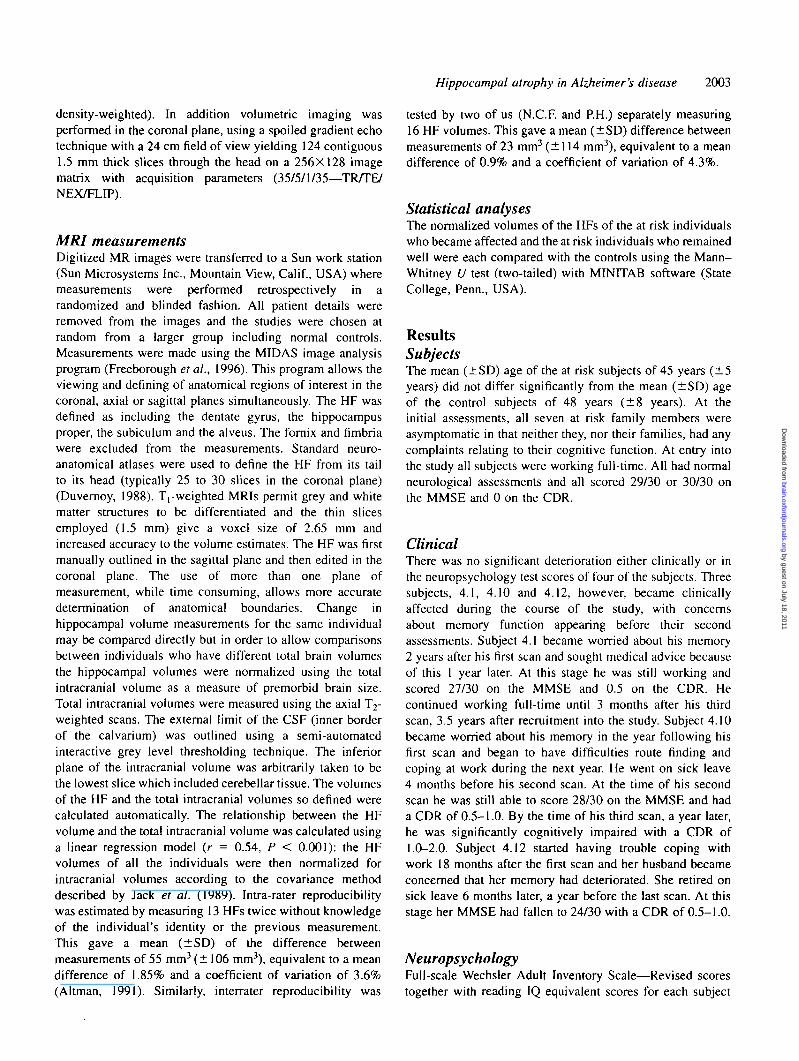
density-weighted). In addition volumetric imaging wasperformed in the coronal plane, using a spoiled gradient echotechnique with a 24 cm field of view yielding 124 contiguous1.5 mm thick slices through the head on a 256X128 imagematrix with acquisition parameters (35/5/1/35—TR/TE/NEX/FLIP).
MRI measurementsDigitized MR images were transferred to a Sun work station(Sun Microsystems Inc., Mountain View, Calif., USA) wheremeasurements were performed retrospectively in arandomized and blinded fashion. All patient details wereremoved from the images and the studies were chosen atrandom from a larger group including normal controls.Measurements were made using the MIDAS image analysisprogram (Freeborough et al., 1996). This program allows theviewing and defining of anatomical regions of interest in thecoronal, axial or sagittal planes simultaneously. The HF wasdefined as including the dentate gyrus, the hippocampusproper, the subiculum and the alveus. The fornix and fimbriawere excluded from the measurements. Standard neuro-anatomical atlases were used to define the HF from its tailto its head (typically 25 to 30 slices in the coronal plane)(Duvernoy, 1988). Trweighted MRIs permit grey and whitematter structures to be differentiated and the thin slicesemployed (1.5 mm) give a voxel size of 2.65 mm andincreased accuracy to the volume estimates. The HF was firstmanually outlined in the sagittal plane and then edited in thecoronal plane. The use of more than one plane ofmeasurement, while time consuming, allows more accuratedetermination of anatomical boundaries. Change inhippocampal volume measurements for the same individualmay be compared directly but in order to allow comparisonsbetween individuals who have different total brain volumesthe hippocampal volumes were normalized using the totalintracranial volume as a measure of premorbid brain size.Total intracranial volumes were measured using the axial T2-weighted scans. The external limit of the CSF (inner borderof the calvarium) was outlined using a semi-automatedinteractive grey level thresholding technique. The inferiorplane of the intracranial volume was arbitrarily taken to bethe lowest slice which included cerebellar tissue. The volumesof the HF and the total intracranial volumes so defined werecalculated automatically. The relationship between the HFvolume and the total intracranial volume was calculated usinga linear regression model (r = 0.54, P < 0.001): the HFvolumes of all the individuals were then normalized forintracranial volumes according to the covariance methoddescribed by Jack et al. (1989). Intra-rater reproducibilitywas estimated by measuring 13 HFs twice without knowledgeof the individual's identity or the previous measurement.This gave a mean (±SD) of the difference betweenmeasurements of 55 mm3 (±106 mm3), equivalent to a meandifference of 1.85% and a coefficient of variation of 3.6%(Altman, 1991). Similarly, interrater reproducibility was
tested by two of us (N.C.F. and P.H.) separately measuring16 HF volumes. This gave a mean (±SD) difference betweenmeasurements of 23 mm3 (± 114 mm3), equivalent to a meandifference of 0.9% and a coefficient of variation of 4.3%.
Statistical analysesThe normalized volumes of the HFs of the at risk individualswho became affected and the at risk individuals who remainedwell were each compared with the controls using the Mann-Whitney U test (two-tailed) with MINITAB software (StateCollege, Penn., USA).
ResultsSubjectsThe mean (±SD) age of the at risk subjects of 45 years (±5years) did not differ significantly from the mean (±SD) ageof the control subjects of 48 years (±8 years). At theinitial assessments, all seven at risk family members wereasymptomatic in that neither they, nor their families, had anycomplaints relating to their cognitive function. At entry intothe study all subjects were working full-time. All had normalneurological assessments and all scored 29/30 or 30/30 onthe MMSE and 0 on the CDR.
ClinicalThere was no significant deterioration either clinically or inthe neuropsychology test scores of four of the subjects. Threesubjects, 4.1, 4.10 and 4.12, however, became clinicallyaffected during the course of the study, with concernsabout memory function appearing before their secondassessments. Subject 4.1 became worried about his memory2 years after his first scan and sought medical advice becauseof this 1 year later. At this stage he was still working andscored 27/30 on the MMSE and 0.5 on the CDR. Hecontinued working full-time until 3 months after his thirdscan, 3.5 years after recruitment into the study. Subject 4.10became worried about his memory in the year following hisfirst scan and began to have difficulties route finding andcoping at work during the next year. He went on sick leave4 months before his second scan. At the time of his secondscan he was still able to score 28/30 on the MMSE and hada CDR of 0.5-1.0. By the time of his third scan, a year later,he was significantly cognitively impaired with a CDR of1.0-2.0. Subject 4.12 started having trouble coping withwork 18 months after the first scan and her husband becameconcerned that her memory had deteriorated. She retired onsick leave 6 months later, a year before the last scan. At thisstage her MMSE had fallen to 24/30 with a CDR of 0.5-1.0.
NeuropsychologyFull-scale Wechsler Adult Inventory Scale—Revised scorestogether with reading IQ equivalent scores for each subject
by guest on July 18, 2011brain.oxfordjournals.org
Dow
nloaded from
2004 N. C. Fox et al.
Table 1A Reading and intelligence tests
4.34.134.54.9
4.14.124.10
Reading IQequivalent
Session/scan
1
10594
110111
96116108
Full scaleWAIS IQ
Session/scan
1
10910198
117
10193*95*
2
118100101107
10184+
83+
3
109
109
95
78f
Discrepancy score: *<5%; f<\%.
Table IB Recognition memory tests
4.34.134.54.9
4.14.124.10
Words (max. 50)
Session/scan
1
49464937*
40*40*33*
2
48454948
28*27*25*
3
45
4747
38*
1 3 t 5
Faces (max. 50)
Session/scan
1
48464547
494233*
2
46454746
31*3735*
3
48
4544
36+
l , t . §
Discrepancy score: *<25%; +<5%; *<1%. 5Maximum 25.
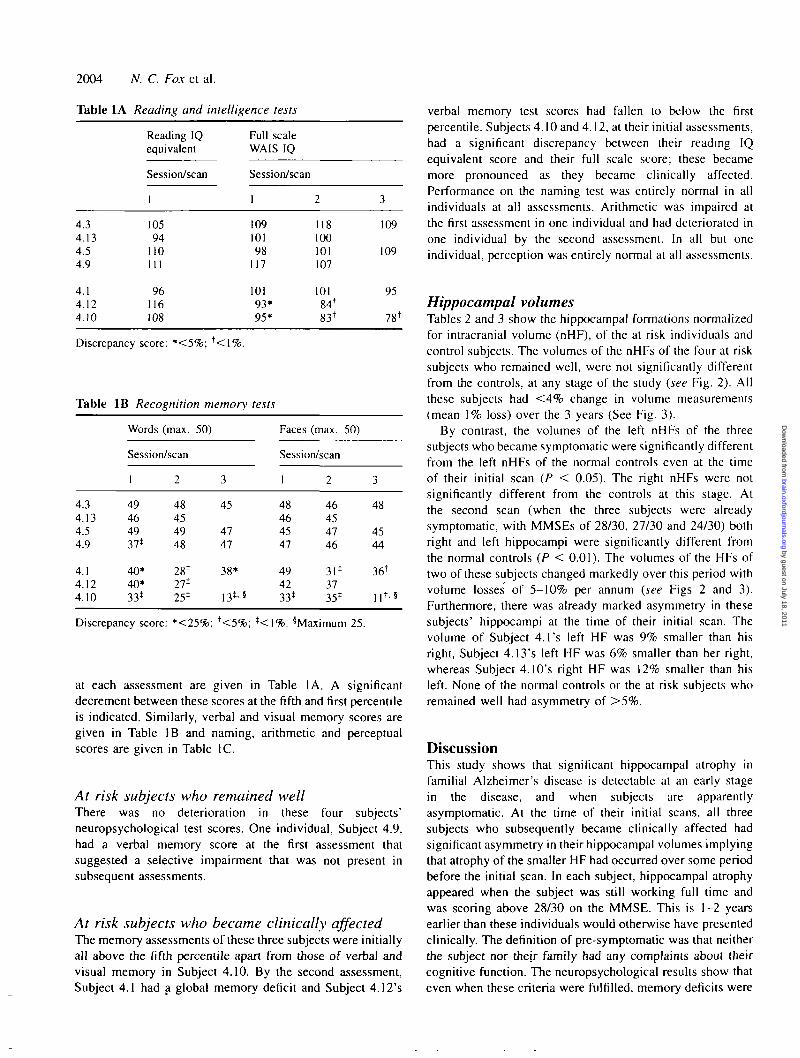
at each assessment are given in Table 1A. A significantdecrement between these scores at the fifth and first percentileis indicated. Similarly, verbal and visual memory scores aregiven in Table IB and naming, arithmetic and perceptualscores are given in Table 1C.
At risk subjects who remained wellThere was no deterioration in these four subjects'neuropsychological test scores. One individual, Subject 4.9,had a verbal memory score at the first assessment thatsuggested a selective impairment that was not present insubsequent assessments.
At risk subjects who became clinically affectedThe memory assessments of these three subjects were initiallyall above the fifth percentile apart from those of verbal andvisual memory in Subject 4.10. By the second assessment,Subject 4.1 had a global memory deficit and Subject 4.12's
verbal memory test scores had fallen to below the firstpercentile. Subjects 4.10 and 4.12, at their initial assessments,had a significant discrepancy between their reading IQequivalent score and their full scale score; these becamemore pronounced as they became clinically affected.Performance on the naming test was entirely normal in allindividuals at all assessments. Arithmetic was impaired atthe first assessment in one individual and had deteriorated inone individual by the second assessment. In all but oneindividual, perception was entirely normal at all assessments.
Hippocampal volumesTables 2 and 3 show the hippocampal formations normalizedfor intracranial volume (nHF), of the at risk individuals andcontrol subjects. The volumes of the nHFs of the four at risksubjects who remained well, were not significantly differentfrom the controls, at any stage of the study (see Fig. 2). Allthese subjects had <4% change in volume measurements(mean 1% loss) over the 3 years (See Fig. 3).
By contrast, the volumes of the left nHFs of the threesubjects who became symptomatic were significantly differentfrom the left nHFs of the normal controls even at the timeof their initial scan (P < 0.05). The right nHFs were notsignificantly different from the controls at this stage. Atthe second scan (when the three subjects were alreadysymptomatic, with MMSEs of 28/30, 27/30 and 24/30) bothright and left hippocampi were significantly different fromthe normal controls (P < 0.01). The volumes of the HFs oftwo of these subjects changed markedly over this period withvolume losses of 5-10% per annum (see Figs 2 and 3).Furthermore, there was already marked asymmetry in thesesubjects' hippocampi at the time of their initial scan. Thevolume of Subject 4.l's left HF was 9% smaller than hisright, Subject 4.13's left HF was 6% smaller than her right,whereas Subject 4.10's right HF was 12% smaller than hisleft. None of the normal controls or the at risk subjects whoremained well had asymmetry of >5%.
DiscussionThis study shows that significant hippocampal atrophy infamilial Alzheimer's disease is detectable at an early stagein the disease, and when subjects are apparentlyasymptomatic. At the time of their initial scans, all threesubjects who subsequently became clinically affected hadsignificant asymmetry in their hippocampal volumes implyingthat atrophy of the smaller HF had occurred over some periodbefore the initial scan. In each subject, hippocampal atrophyappeared when the subject was still working full time andwas scoring above 28/30 on the MMSE. This is 1-2 yearsearlier than these individuals would otherwise have presentedclinically. The definition of pre-symptomatic was that neitherthe subject nor their family had any complaints about theircognitive function. The neuropsychological results show thateven when these criteria were fulfilled, memory deficits were
by guest on July 18, 2011brain.oxfordjournals.org
Dow
nloaded from

Hippocampal atrophy in Alzheimer's disease 2005
Table 1C Naming, arithmetic and perception
4.34.134.54.9
4.14.124.10
Naming (max.
Session/scan
1
25222028
192626
30)
2
28222127
182026
3
2222
20
28
Arithmetic (max
Session/scan
1
13112011
12115*
.24)
2
15112112
156*2*
3
1223
14
Perception (max
Session/scan
1
29282027
2116*26
. 30)
2
29272127
2713*23
3
2921
26
24
Discrepancy score: *<25%; ^<5%; *<1%. The results of the neuropsychological tests conducted at the time of each scan (see Fig. 1)are shown for the at risk individuals; subjects who became symptomatic are shown in the lower half.
Table 2 Normalized hippocampal volumes for the at riskindividuals
Those at risk individuals who became symptomatic are shown inthe lower half of the table.
Table
LeftRight
3 nHF volumes of the
Median/mm3
30013025
controls
Mean/mm3
30213038
SD/mm3
280267
already measurable, with one individual having a globalmemory deficit and two other individuals having a selectivelyweak verbal memory.
This study of familial Alzheimer's disease, extendsprevious MRI findings in sporadic Alzheimer's disease whichhave shown that even mildly affected individuals havehippocampal volumes that are, on average, 25% smaller thannormal controls (Killiany et al., 1993; Lehericy et ai, 1994).Individuals classified as mildly demented on the basis of anMMSE score may have had symptoms for some considerableperiod, usually for at least 2 years (Laakso et al., 1995), butin some cases the duration of symptoms was 5=6 years
(A)
4.3
4.13
4.5
4.9
4.1
4.12
4.10
LRLRLRLR
LRLRLR
Scan 1(nHF/mm3)
29753021315131993170332828802991
274530182176230524052113
Scan 2(nHF/mm3)
30413153306331513203318729623056
264825742084216022491865
Scan 3(nHF/mm3)
29222971
3194334729563012
25292475
18881699
Change(%/year)
-0.6-0.6-0.9-0.5
0.20.21.10.3
-2.7-6.2-1.4-2.1-8.6-7.8
1~Z 2500E
755 2000-
1500-
3500
S- 3000E
^ 2 5 0 0E
o> 2000-
i<vm - -
M F
Controls
1 2 3
Time (years)(B)
- •43
- 4 1 3
- 4 5
- 4 9
- 4 12
- 4 1
-410
-43
-4 13
-45
-49
-412
-4 1
M F
Controls
1 2 3
Time (years)
Fig. 2 Left (A) and right (B) hippocampal formation volumes,normalized for intracranial volume, showing atrophy in the threesubjects who became affected (open symbols) while thehippocampal volumes of the four at risk individuals whoremained well (closed symbols) stayed within the normal range ofthe controls. Values for the male (M) and female (F) controls areshown on the left of the graphs.
(Murphy et al., 1993). Rates of atrophy in familialAlzheimer's disease at a presymptomatic or very earlysymptomatic stage have not been reported previously. Ratesof atrophy in the hippocampus may vary widely betweenindividuals and with disease progression (Jobst et al., 1994).If the onset of hippocampal atrophy were to coincide withthe onset of symptoms, then in order to have lost 25% ofthe volume of the hippocampus by the time the individualswere mildly affected, the mean rate of atrophy must havebeen between 5 and 10% per year if symptoms are assumedto have been present for between 2 and 5 years. The rate ofhippocampal atrophy in this study was considerable and
by guest on July 18, 2011brain.oxfordjournals.org
Dow
nloaded from
2006 N. C. Fox et al.
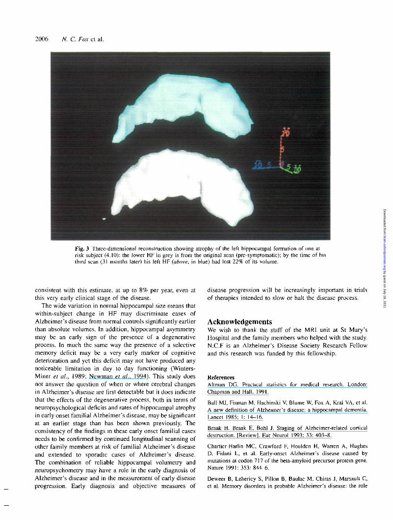
Fig. 3 Three-dimensional reconstruction showing atrophy of the left hippocampal formation of one atrisk subject (4.10): the lower HF in grey is from the original scan (pre-symptomatic); by the time of histhird scan (31 months later) his left HF (above, in blue) had lost 22% of its volume.
consistent with this estimate, at up to 8% per year, even atthis very early clinical stage of the disease.
The wide variation in normal hippocampal size means thatwithin-subject change in HF may discriminate cases ofAlzheimer's disease from normal controls significantly earlierthan absolute volumes. In addition, hippocampal asymmetrymay be an early sign of the presence of a degenerativeprocess. In much the same way the presence of a selectivememory deficit may be a very early marker of cognitivedeterioration and yet this deficit may not have produced anynoticeable limitation in day to day functioning (Winters-Miner et al., 1989; Newman et al., 1994). This study doesnot answer the question of when or where cerebral changesin Alzheimer's disease are first detectable but it does indicatethat the effects of the degenerative process, both in terms ofneuropsychological deficits and rates of hippocampal atrophyin early onset familial Alzheimer's disease, may be significantat an earlier stage than has been shown previously. Theconsistency of the findings in these early onset familial casesneeds to be confirmed by continued longitudinal scanning ofother family members at risk of familial Alzheimer's diseaseand extended to sporadic cases of Alzheimer's disease.The combination of reliable hippocampal volumetry andneuropsychometry may have a role in the early diagnosis ofAlzheimer's disease and in the measurement of early diseaseprogression. Early diagnosis and objective measures of
disease progression will be increasingly important in trialsof therapies intended to slow or halt the disease process.
AcknowledgementsWe wish to thank the staff of the MRI unit at St Mary'sHospital and the family members who helped with the study.N.C.F is an Alzheimer's Disease Society Research Fellowand this research was funded by this fellowship.
ReferencesAltman DG. Practical statistics for medical research. London:Chapman and Hall, 1991.
Ball MJ, Fisman M, Hachinski V, Blume W, Fox A, Krai VA, et al.A new definition of Alzheimer's disease: a hippocampal dementia.Lancet 1985; I: 14-16.
Braak H. Braak E, Bohl J. Staging of Alzheimer-related corticaldestruction. [Review]. Eur Neurol 1993; 33: 403-8.
Chartier-Harlin MC, Crawford F, Houlden H, Warren A, HughesD. Fidani L, et al. Early-onset Alzheimer's disease caused bymutations at codon 717 of the beta-amyloid precursor protein gene.Nature 1991: 353: 844-6.
Deweer B, Lehericy S, Pillon B, Baulac M. Chiras J, Marsault C,et al. Memory disorders in probable Alzheimer's disease: the role
by guest on July 18, 2011brain.oxfordjournals.org
Dow
nloaded from

Hippocampal atrophy in Alzheimer's disease 2007
of hippocampal atrophy as shown with MRI. J Neurol NeurosurgPsychiatry 1995; 58: 590-7.
Duvernoy HM. The human hippocampus: an atlas of appliedanatomy. MUnchen: Bergmann, 1988.
Folstein M, Folstein SE, McHugh PR. 'Mini-mental state": apractical method for grading the cognitive state of patients for theclinician. J Psychiatr Res 1975; 12: 189-98.
Freeborough PA, Fox NC, Kitney RI. Accurate segmentation of 3Dbrain scans: interactive software and algorithms. Proc EurographicsUKConf 1996; 2: 261-71.
Hodges JR, Patterson K. Is semantic memory consistently impairedearly in the course of Alzheimer's disease? Neuroanatomical anddiagnostic implications. Neuropsychologia 1995; 33: 441-59.
Hughes CP, Berg L, Danziger WL, Coben LA, Martin RL. A newclinical scale for the staging of dementia. Br J Psychiatry 1982;140: 566-72.
Hyman BT, Van Horsen GW, Damasio AR, Barnes CL. Alzheimer'sdisease: cell-specific pathology isolates the hippocampal formation.Science 1984; 225: 1168-70.
Jack CR Jr, Twomey CK, Zinsmeister AR, Sharbrough FW, PetersenRC, Cascino GD. Anterior temporal lobes and hippocampalformations: normative volumetric measurements from MR imagesin young adults. Radiology 1989; 172: 549-54.
Jack CR Jr, Petersen RC, O'Brien PC, Tangalos EG. MR-basedhippocampal volumetry in the diagnosis of Alzheimer's disease.Neurology 1992; 42: 183-8.
Jackson M, Warrington EKW. Arithmetic skills in patients withunilateral cerebral lesions. Cortex 1986; 22: 611-20.
Jobst KA, Smith AD, Szatmari M, Esiri MM, Jaskowski A, HindleyN, et al. Rapidly progressing atrophy of medial temporal lobe inAlzheimer's disease. Lancet 1994; 343: 829-30.
Kennedy AM, Newman S, McCaddon A, Ball J, Roques P, MullanM, et al Familial Alzheimer's disease. A pedigree with a mis-sensemutation in the amyloid precursor protein gene (amyloid precursorprotein 717 valine -> glycine). Brain 1993; 116: 309-24.
Kesslak JP, Nalcioglu O, Cotman CW. Quantification of magneticresonance scans for hippocampal and parahippocampal atrophy inAlzheimer's disease [see comments]. Neurology 1991; 41: 51-4.Comment in: Neurology 1991, 41: 954-5.
Kilhany RJ, Moss MB, Albert MS, Sandor T, Tieman J, Jolesz F.Temporal lobe regions on magnetic resonance imaging identifypatients with early Alzheimer's disease. Arch Neurol 1993; 50:949-54.
Laakso MP, Soininen H, Partanen K, Helkala EL, Hartikainen P,Vainio P, et al. Volumes of hippocampus, amygdala and frontallobes in the MRI-based diagnosis of early Alzheimer's disease:correlation with memory functions. J Neural Transm Park DisDement Sect 1995; 9: 73-86.
Lehericy S, Baulac M, Chiras J, Pierot L, Martin N, Pillon B, et al.Amygdalohippocampal MR volume measurements in the earlystages of Alzheimer disease. AJNR Am J Neuroradiol 1994; 15:927-37.
McKenna P, Warrington EK. The Graded Naming Test. Windsor:NFER-Nelson, 1983.
Murphy DG, DeCarli CD, Daly E, Gillette JA, Mclntosh AR, HaxbyJV, et al. Volumetric magnetic resonance imaging in men withdementia of the Alzheimer type: correlations with disease severity.Biol Psychiatry 1993; 34: 612-21.
Nelson HE. The National Adult Reading Test (NART). Manual.2nd ed. Windsor: NFER-Nelson, 1991.
Newman SK, Warrington EK, Kennedy AM, Rossor MN. Theearliest cognitive change in a person with familial Alzheimer'sdisease: presymptomatic neuropsychological features in a pedigreewith familial Alzheimer's disease confirmed at necropsy. J NeurolNeurosurg Psychiatry 1994; 57: 967-72.
Rossor MN, Newman S, Frackowiak RS, Lantos P, KennedyAM. Alzheimer's disease families with amyloid precursor proteinmutations. [Review]. Ann NY Acad Sci 1993; 695: 198-202.
Seab JB, Jagust WJ, Wong STS, Roos MS, Reed BR, BudingerTF. Quantitative NMR measurements of hippocampal atrophy inAlzheimer's disease. Magn Reson Med 1988; 8: 200-8.
Soininen HS, Partanen K, Pitkanen A, Vainio P, Hanninen T,Hallikainen M, et al. Volumetric MRI analysis of the amygdalaand the hippocampus in subjects with age-associated memoryimpairment: correlation to visual and verbal memory. Neurology1994; 44: 1660-8.
Squire LR. Mechanisms of memory. Science 1986; 232: 1612-19.
Warrington EK. Manual for the Recognition Memory Test for wordsand faces. Windsor: NFER-Nelson, 1984.
Warrington EK, James M. The visual object and space perceptionbattery. Bury St. Edmunds: Thames Valley Test Company, 1991.
Warrington EK, James M, Maciejewski C. The WAIS as a lateralizingand localizing diagnostic instrument: a study of 656 patients withunilateral cerebral lesions. Neuropsychologia 1986; 24: 223-39.
West MJ, Coleman PD, Flood DG, Troncoso JC. Differences inthe pattern of hippocampal neuronal loss in normal ageing andAlzheimer's disease. Lancet 1994; 344: 769-72.
Willison JR, Warrington EK. Cognitive retardation in a patient withpreservation of psychomotor speed. Behav Neurol 1992; 5: 113-16.
Winters-Miner LA, Stryker D, Burgus RC, Miner GD. The searchfor possible neuropsychological associations and other early markersin familial Alzheimer's disease. In: Miner GD, Richter RW, BlassJP, Valentine JL, Winters-Miner LA, editors. Familial Alzheimer'sdisease: molecular genetics and clinical perspectives. New York:Marcel Dekker, 1989: 203-22.
Received June 3, 1996. Accepted July 19, 1996.
by guest on July 18, 2011brain.oxfordjournals.org
Dow
nloaded from