Functional organization of the hippocampal memory system
8
Proc. Natl. Acad. Sci. USA Vol. 93, pp. 13500–13507, November 1996 Colloquium Paper This paper was presented at a colloquium entitled ‘‘Memory: Recording Experience in Cells and Circuits,’’ organized by Patricia S. Goldman-Rakic, held February 17–20, 1996, at the National Academy of Sciences in Irvine, CA. Functional organization of the hippocampal memory system (hippocampusycerebral cortexymemoryyolfactionyrats) HOWARD EICHENBAUM*, GEOFFREY SCHOENBAUM ² ,BRIAN YOUNG ‡ , AND MICHAEL BUNSEY § *Center for Behavioral Neuroscience, State University of New York, Stony Brook, NY 11794-2575; ² Department of Psychology, University of North Carolina, Chapel Hill, NC 27599-3270; ‡ Department of Psychology, University of Otago, P.O. Box 56, Dunedin, New Zealand; and § Department of Psychology, Kent State University, Kent, OH 44240 ABSTRACT In humans declarative or explicit memory is supported by the hippocampus and related structures of the medial temporal lobe working in concert with the cerebral cortex. This paper reviews our progress in developing an animal model for studies of cortical–hippocampal interac- tions in memory processing. Our findings support the view that the cortex maintains various forms of memory represen- tation and that hippocampal structures extend the persistence and mediate the organization of these codings. Specifically, the parahippocampal region, through direct and reciprocal interconnections with the cortex, is sufficient to support the convergence and extended persistence of cortical codings. The hippocampus itself is critical to the organization cortical representations in terms of relationships among items in memory and in the flexible memory expression that is the hallmark of declarative memory. It is widely accepted that the hippocampus and closely related structures of the medial temporal lobe interact with the cerebral cortex in support of the persistence and organization of memories (1–6). Here we review our own recent develop- ments of a model system for exploring the nature of cortical and hippocampal memory representation and the specific roles played by hippocampal structures in memory formation. This research is guided by a simple conception of cortical– hippocampal pathways in which there are three main serially and bidirectionally connected components: the cortex, the parahippocampal region, and the hippocampus itself (Fig. 1A). The beginning and end point of this system involves several tertiary or ‘‘association’’ cortical areas whose outputs converge on the parahippocampal region. This region, composed of the interconnected perirhinal, parahippocampal (or postrhinal), and entorhinal cortices, surround the hippocampus in both rats (7, 8) and monkeys (9, 10) and merge different sources of cortical input bound for the hippocampus. Within the hip- pocampus itself there are several stages of serial and parallel processing, and the outcome of this integration is sent back to the parahippocampal region, which in turn sends its main outputs to the same tertiary cortical areas that provided the source of input. Our working hypothesis about this system is that memory representations are established and maintained in the cortex, and that the parahippocampal region and the hippocampus contribute to memory processing by modifying the persistence and organization of those cortical representations. Not all modifications of cortical representations depend on the hip- pocampal system. In particular hippocampal processing is not viewed as required for the ‘‘tuning’’ or ‘‘biasing’’ of cortical encodings, that is, an enhanced or diminished responsiveness to particular stimuli resulting from repeated exposure or association with reward or punishment. However, hippocam- pal structures receive all this information and are in position to compare and combine distinct cortical representations, and thus are viewed to play a critical role in extending the persistence of cortical memory representations and in creating and updating their organization. More specifically, we will argue that the two main components of the hippocampal region have distinct functions. First, the parahippocampal region, through its direct and reciprocal cortical connections, may be sufficient to extend the persistence of cortical memory representations. Second, the hippocampus itself plays a special role in mediating the systematic organization of cortical cod- ings. Olfactory Memory in Rodents as a Model System for Studies of Cortical–Hippocampal Processing Our explorations focus on the set of interconnected olfactory– cortical and hippocampal structures that are fully evolved in rodents (Fig. 1B). Odor information initially processed by the olfactory bulb is sent directly to the piriform cortex and closely interconnected orbital prefrontal cortex (11, 12). Both of these cortical areas, as well as the olfactory bulb, project heavily to the perirhinal and entorhinal components of the parahip- The publication costs of this article were defrayed in part by page charge payment. This article must therefore be hereby marked ‘‘advertisement’’ in accordance with 18 U.S.C. §1734 solely to indicate this fact. Abbreviation: DNMS, delayed nonmatch to sample. FIG. 1. (A) Simple schematic diagram of cortical–hippocampal connections. (B) Outline of a horizontal rat brain section illustrating the locations and flow of information between components of the hippocampus, parahippocampal region, and adjacent cortical areas. DG, dentate gyrus; EC, entorhinal cortex; FF, fimbria–fornix; Hipp, hippocampus proper; OF, orbitofrontal cortex; Pir, perirhinal cortex; PR, perirhinal cortex; Sub, subiculum. 13500
Transcript of Functional organization of the hippocampal memory system

Proc. Natl. Acad. Sci. USAVol. 93, pp. 13500–13507, November 1996Colloquium Paper
This paper was presented at a colloquium entitled ‘‘Memory: Recording Experience in Cells and Circuits,’’ organized byPatricia S. Goldman-Rakic, held February 17–20, 1996, at the National Academy of Sciences in Irvine, CA.
Functional organization of the hippocampal memory system(hippocampusycerebral cortexymemoryyolfactionyrats)
HOWARD EICHENBAUM*, GEOFFREY SCHOENBAUM†, BRIAN YOUNG‡, AND MICHAEL BUNSEY§
*Center for Behavioral Neuroscience, State University of New York, Stony Brook, NY 11794-2575; †Department of Psychology, University of North Carolina,Chapel Hill, NC 27599-3270; ‡Department of Psychology, University of Otago, P.O. Box 56, Dunedin, New Zealand; and §Department of Psychology,Kent State University, Kent, OH 44240
ABSTRACT In humans declarative or explicit memory issupported by the hippocampus and related structures of themedial temporal lobe working in concert with the cerebralcortex. This paper reviews our progress in developing ananimal model for studies of cortical–hippocampal interac-tions in memory processing. Our findings support the viewthat the cortex maintains various forms of memory represen-tation and that hippocampal structures extend the persistenceand mediate the organization of these codings. Specifically,the parahippocampal region, through direct and reciprocalinterconnections with the cortex, is sufficient to support theconvergence and extended persistence of cortical codings. Thehippocampus itself is critical to the organization corticalrepresentations in terms of relationships among items inmemory and in the f lexible memory expression that is thehallmark of declarative memory.
It is widely accepted that the hippocampus and closely relatedstructures of the medial temporal lobe interact with thecerebral cortex in support of the persistence and organizationof memories (1–6). Here we review our own recent develop-ments of a model system for exploring the nature of corticaland hippocampal memory representation and the specific rolesplayed by hippocampal structures in memory formation.This research is guided by a simple conception of cortical–
hippocampal pathways in which there are three main seriallyand bidirectionally connected components: the cortex, theparahippocampal region, and the hippocampus itself (Fig. 1A).The beginning and end point of this system involves severaltertiary or ‘‘association’’ cortical areas whose outputs convergeon the parahippocampal region. This region, composed of theinterconnected perirhinal, parahippocampal (or postrhinal),and entorhinal cortices, surround the hippocampus in both rats(7, 8) and monkeys (9, 10) and merge different sources ofcortical input bound for the hippocampus. Within the hip-pocampus itself there are several stages of serial and parallelprocessing, and the outcome of this integration is sent back tothe parahippocampal region, which in turn sends its mainoutputs to the same tertiary cortical areas that provided thesource of input.Our working hypothesis about this system is that memory
representations are established and maintained in the cortex,and that the parahippocampal region and the hippocampuscontribute to memory processing by modifying the persistenceand organization of those cortical representations. Not allmodifications of cortical representations depend on the hip-pocampal system. In particular hippocampal processing is notviewed as required for the ‘‘tuning’’ or ‘‘biasing’’ of cortical
encodings, that is, an enhanced or diminished responsivenessto particular stimuli resulting from repeated exposure orassociation with reward or punishment. However, hippocam-pal structures receive all this information and are in positionto compare and combine distinct cortical representations, andthus are viewed to play a critical role in extending thepersistence of cortical memory representations and in creatingand updating their organization. More specifically, we willargue that the two main components of the hippocampalregion have distinct functions. First, the parahippocampalregion, through its direct and reciprocal cortical connections,may be sufficient to extend the persistence of cortical memoryrepresentations. Second, the hippocampus itself plays a specialrole in mediating the systematic organization of cortical cod-ings.
Olfactory Memory in Rodents as a Model System forStudies of Cortical–Hippocampal Processing
Our explorations focus on the set of interconnected olfactory–cortical and hippocampal structures that are fully evolved inrodents (Fig. 1B). Odor information initially processed by theolfactory bulb is sent directly to the piriform cortex and closelyinterconnected orbital prefrontal cortex (11, 12). Both of thesecortical areas, as well as the olfactory bulb, project heavily tothe perirhinal and entorhinal components of the parahip-The publication costs of this article were defrayed in part by page charge
payment. This article must therefore be hereby marked ‘‘advertisement’’ inaccordance with 18 U.S.C. §1734 solely to indicate this fact. Abbreviation: DNMS, delayed nonmatch to sample.
FIG. 1. (A) Simple schematic diagram of cortical–hippocampalconnections. (B) Outline of a horizontal rat brain section illustratingthe locations and flow of information between components of thehippocampus, parahippocampal region, and adjacent cortical areas.DG, dentate gyrus; EC, entorhinal cortex; FF, fimbria–fornix; Hipp,hippocampus proper; OF, orbitofrontal cortex; Pir, perirhinal cortex;PR, perirhinal cortex; Sub, subiculum.
13500

pocampal region, which then provides the primary source ofolfactory sensory information to the hippocampus itself. In thereturn pathway, the outputs of hippocampal processing involvedirect projections from the parahippocampal region to boththe piriform and orbital prefrontal cortices.Physiological findings complement the anatomical data in-
dicating that information processing in the olfactory and limbicsystems are closely integrated during odor-guided learning andmemory. During olfactory learning rats typically investigate anodor cue with about of 3–6 sniffs synchronized to the ongoingprominent theta rhythm recorded in the hippocampus (13).The reliability of this relationship is maximal just before theanimal reaches accurate performance in the discriminationand just after a reversal of odor valences, two training stageswhen the rat is maximally attentive. In addition, hippocampalcellular activity includes firing bursts synchronized with thesniff and theta cycles, a pattern of neural activation that isconsistent with optimal conditions for the induction of long-term potentiation during odor sampling (14).In sum, olfactory–hippocampal pathways provide a straight-
forward and relatively simple instantiation of the generalmodel of cortical–hippocampal interconnections. The olfac-tory and hippocampal systems are closely and reciprocallyinterconnected, and the neural activity patterns in the hip-pocampus reflect strong temporal integration with olfactoryprocessing. Given these close connections, it is not surprisingthat rats have superb olfactory learning and memory capaci-ties, as will be described below. Our efforts in exploringcortical–hippocampal interactions involve the development ofbehavioral tasks that assess different aspects of olfactorylearning and memory ability, and identify specific functionalcontributions at each level of this system. This work involves acombination of neuropsychological studies on the effects ofselective ablation of these areas, and electrophysiologicalstudies that characterize the coding properties of single neu-rons in each area during memory performance.
Multiple Forms of Odor Memory RepresentationWithin the Olfactory Cortex
Our starting point in describing the operations of this systeminvolves a characterization of neural coding in the olfactorycortical areas that constitute the relevant perceptual repre-sentations for odor-guided learning. Based on our findings thatrats with damage to the orbitofrontal cortex are severelyimpaired in olfactory learning (15, 16) and on the modeldescribed above, we expected that encodings of odor identityand the distinct significance of individual odors would beobserved in olfactory cortical areas. In addition, we expectedto observe firing patterns in olfactory cortical neurons thatreflect both the extended persistence of odor representationsand the organization of odor memories, properties that mightdepend on hippocampal processing. We sought neurophysio-logical correlates of all these aspects of memory processing inrats performing a specially designed odor discrimination task(17, 18).Odor Coding and the Tuning and Biasing of Odor Repre-
sentations. The basic discrimination task involved a sequenceof trials in which the rat was signaled by a light that it couldinitiate an odor presentation by poking its nose into a stimulussampling port. When the odor was an assigned as a rewarded(S1) stimulus an additional nose poke into a separate waterport resulted in water delivery, and no reward was givenregardless of the response for other (S2) odors. On each trialone of eight different odor stimuli (identified here by numberand assigned reward values) was presented, half of which wereassociated with a sweetened water reward and the other halfnot rewarded. We examined the coding of odor identity bycomparing neural activity during odor sampling across theeight odors. In addition we assessed whether cortical odor
codings could be ‘‘biased’’ or ‘‘tuned’’ in accordance with thesignificance of items by comparing responses across the groupsof S1 and S2 stimuli.We found that many neurons in the orbitofrontal cortex and
piriform cortex encode both odor identity and odor valence(Fig. 2). Although a few cells fired only to one odor, most werecoarsely tuned as reflected by different levels of activationacross the eight stimuli (Fig. 2A). In addition, most cells firedmore strongly either to all the S1 odors or to all the S2 odors.For example, in Fig. 2B the response was stronger to all S1odors, and strongest of all to odors 11 and 51. An equivalentproportion of cells showed the opposite pattern, firing morestrongly to all S2 odors. The example shown in Fig. 2C showslarger responses to all S2 odors, with the greatest response toodor 42. This pattern of findings indicates, not surprisingly,that olfactory cortical areas encode odor identity. In addition,assigned stimulus valences are also very prominently repre-sented in the cortex, indicating that cortical odor representa-tions are indeed tuned or biased in association with rewardcontingencies.Persistent Odor Representations in the Olfactory Cortex.
The odors were presented in a pseudorandom order (withspecific exceptions described below) so that we could evaluatethe extent to which particular odor responses were influencedby neural representations established on the preceding trial. Inthese analyses we controlled for the valence of the stimulus byseparately analyzing data for preceding S1 and S2 cues, andmeasured responses during odor sampling on subsequenttrials. We found numerous cases of particular stimuli control-ling subsequent odor responses. For example, in Fig. 3 theneural response during odor sampling was weak when thepreceding trial involved odor 11 and strong for other preced-
FIG. 2. Odor responses of olfactory cortex neurons. The dark barsrepresent neural responses to S1 odors and striped bars representresponses to S2 odors. OF, orbitofrontal.
Colloquium Paper: Eichenbaum et al. Proc. Natl. Acad. Sci. USA 93 (1996) 13501

ing S1 odors. This pattern of results indicates that a persistentrepresentation of specific odors is carried over into latersensory processing. This capacity might subserve short-termmemory required for comparisons between sequentially pre-sented stimuli.Organized Odor Representations in Olfactory Cortex.How
might one detect an organization of odor representations inthe cerebral cortex? Our efforts were directed at a simple kindof learned stimulus organization that might be found insensory cortex, an acquired association between two odorssuch that presentation of one of them predicts the subsequentoccurrence of the other (19). Assuming rats can learn apredictable relationship between odor stimuli, a neural reflec-tion of the acquired associationmight be observed in the abilityof presentation of the first odor to evoke or modify the neuralrepresentation of the second. We trained rats to associatesequentially presented odors by including specific exceptionsto the otherwise random stimulus presentation order. Forexample, odors 51 and 62 always were followed by odor 71;as a control, odor 71 also sometimes followed other odorsunpredicted. Rats learned these incidental predictabilities inthe odor sequence, as reflected in more rapid trial initiationwhen an S1 was predicted and slowed initiation of trials whereS2 odors were predicted.We found two kinds of neural correlates of the learned odor
associations. First we observed cells that fired differently inresponse to an odor depending on whether the preceding odorpredicted its occurrence. For example, in Fig. 4A the responseto odor 11 was much reduced when it was predicted than whenit was unexpected. Second, we identified olfactory cortex cellsthat fired in anticipation of a predicted odor. In the exampleshown in Fig. 4B the cell fired maximally prior to as well asduring the presentation of odor 11 when it was predicted. Bycontrast there was almost no response when the odor was notpredicted by its associate. This pattern of results indicates thatlearned stimulus associations are indeed established within theolfactory cortex.In sum, our characterization of olfactory cortical neurons
indicated that the piriform and orbitofrontal cortical areasencode aspects of stimulus memories that should occur inde-pendent of hippocampal function, specifically the tuning andbiasing of odor representations, as well as aspects of memorythat may depend on hippocampal function, such as the per-sistence of odor memories and odor–odor associations.
Memory Processing Within the Hippocampal System:Which Structures Extend the Persistenceof Odor Memories?
The earliest reports of amnesia in humans following damage tothe hippocampal region emphasized the critical role of thehippocampus in bridging between immediate memory andpermanent memory formation (20). In amnesic patients andanimals with hippocampal region damage, deficits in memorypersistence are characteristically observed as a dissociationbetween intact immediate memory versus subsequent abnor-mally rapid forgetting. Initially it was thought that the hip-pocampus itself played the primary role in extending retentionbeyond immediate memory, but recent data have brought thisview into question (21, 22). Our studies have focused on theissue of which components of the hippocampal system arecritical to the persistence of odor memory representations.Efforts to delineate the anatomical structures involved in
maintaining a memory trace have focused on the delayednonmatch to sample task (DNMS). In most versions of thistask the subject is initially presented with a sample memorycue. This is followed by a delay phase during which thememoryfor that cue must be maintained. Finally, in the choice phase,the subject is presented with the sample and a novel stimulusand the unfamiliar ‘‘nonmatch’’ cue must be selected. The loadon memory can be increased by lengthening the delay phase orby presenting a ‘‘list’’ of sample cues prior to a series of choicerecognition tests. This paradigm was first developed for mon-keys by using three-dimensional junk objects that provide richand salient cues for this species (23, 24). Damage to thehippocampal system results in a performance deficit that isdependent on the duration of the delay or the length of thesample ‘‘list,’’ revealing a delay-dependent deficit in newlearning similar to that observed in human amnesics (25, 26).To assess which components of the hippocampal system are
critical to the maintenance of odor memory representations we
FIG. 3. Short-term memory correlates of the activity of olfactorycortical neurons. Each panel illustrates the neural response of a cell tovarious current odors depending upon the stimulus on the precedingtrial. OF, orbitofrontal.
FIG. 4. Neural correlates of odor–odor associations in the olfac-tory cortex. See text for description.
13502 Colloquium Paper: Eichenbaum et al. Proc. Natl. Acad. Sci. USA 93 (1996)
developed a variant of the DNMS task that used odor cues andinvolved a stimulus presentation protocol that could be used toassess neural responses to single stimuli as well as behavioralresponses in accordance with the nonmatch memory contin-gency. In this version of the task, a continuous series of singleodor cues is presented with the contingency that an appropri-ate response to each odor is rewarded only if that odor wasdifferent from (that is, a nonmatch with) the preceding odor.Rats were trained initially with a short (3-sec) interval betweenodor presentations. Subsequently the interval between odorpresentations was manipulated to vary the retention delay,allowing an assessment of forgetting rate as in the studies onmonkeys.In an initial experiment we compared the effects of selective
ablation of the parahippocampal region versus that of damageto the fornix, a major fiber bundle that connects the hippocam-pus itself with subcortical structures (ref. 27; see Fig. 1B).Normal rats acquire this task within '150 trials and neitherlesion affected the acquisition rate. In subsequent tests acrossmemory delays, intact rats showed 90% or better retention atthe shortest (3-sec) delay and their memory performancegradually declined as the delay was increased. Rats withdamage to the parahippocampal region also showed goodretention at the shortest delay, but their performance declinedabnormally rapidly, showing a severe deficit within 1 min. Bycontrast rats with fornix lesions were unimpaired across delays,showing the same gradual memory decay as intact rats. Theseresults indicate that neither hippocampal component is criticalto odor perception, acquisition of the nonmatch rule, orimmediate memory. However, the parahippocampal region iscritical to extending the persistence of cortical representationsbeyond immediate memory in rats, as it is in monkeys (28–30).Furthermore, through its direct and reciprocal connectionswith the cortex, the parahippocampal region is sufficient tomediate this memory function independent of hippocampalprocessing. The results of this processing may be reflected inthe persistent effects of odor representations we observed inolfactory cortex, as described above.In complementary electrophysiological studies we have also
examined the response properties of neurons in the parahip-pocampal region and the hippocampus in rats performing theodor-guided DNMS task (31). Cells in the perirhinal cortex, inthe lateral entorhinal cortex, and in the subiculum showedselective or coarse tuning to odor stimuli, indicating that theparahippocampal region encodes specific odor representa-tions. In addition, odor representations in each of these areaswas observed to persist through the memory delay. Some cellsshowed striking odor-specific activity during odor samplingand throughout the delay period, such as the example shownin Fig. 5. To detect persistent memory representations acrossall cells, we focused on the trial period immediately precedingthe initiation of trials. At this time the overt behavior of theanimal is consistent (it is approaching the stimulus port),allowing us to determine if neural activity at this time varieswith the identity of the odor that must be remembered duringthat period. A substantial number of cells throughout theparahippocampal region fired differentially associated withthe preceding sample odor at this time. Some cells maintaineddifferential activity for both short and long memory delays,whereas others lost the representation at the longer delay.To assess odor memory representations in the hippocampus
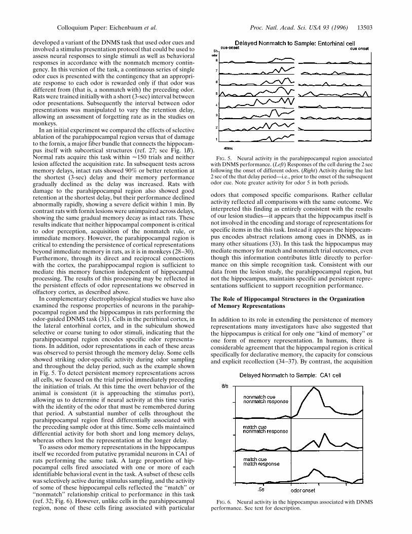
itself we recorded from putative pyramidal neurons in CA1 ofrats performing the same task. A large proportion of hip-pocampal cells fired associated with one or more of eachidentifiable behavioral event in the task. A subset of these cellswas selectively active during stimulus sampling, and the activityof some of these hippocampal cells reflected the ‘‘match’’ or‘‘nonmatch’’ relationship critical to performance in this task(ref. 32; Fig. 6). However, unlike cells in the parahippocampalregion, none of these cells firing associated with particular
odors that composed specific comparisons. Rather cellularactivity reflected all comparisons with the same outcome. Weinterpreted this finding as entirely consistent with the resultsof our lesion studies—it appears that the hippocampus itself isnot involved in the encoding and storage of representations forspecific items in the this task. Instead it appears the hippocam-pus encodes abstract relations among cues in DNMS, as inmany other situations (33). In this task the hippocampus maymediate memory for match and nonmatch trial outcomes, eventhough this information contributes little directly to perfor-mance on this simple recognition task. Consistent with ourdata from the lesion study, the parahippocampal region, butnot the hippocampus, maintains specific and persistent repre-sentations sufficient to support recognition performance.
The Role of Hippocampal Structures in the Organizationof Memory Representations
In addition to its role in extending the persistence of memoryrepresentations many investigators have also suggested thatthe hippocampus is critical for only one ‘‘kind of memory’’ orone form of memory representation. In humans, there isconsiderable agreement that the hippocampal region is criticalspecifically for declarative memory, the capacity for consciousand explicit recollection (34–37). By contrast, the acquisition
FIG. 6. Neural activity in the hippocampus associated with DNMSperformance. See text for description.
FIG. 5. Neural activity in the parahippocampal region associatedwith DNMS performance. (Left) Responses of the cell during the 2 secfollowing the onset of different odors. (Right) Activity during the last2 sec of the that delay period—i.e., prior to the onset of the subsequentodor cue. Note greater activity for odor 5 in both periods.
Colloquium Paper: Eichenbaum et al. Proc. Natl. Acad. Sci. USA 93 (1996) 13503

of biases or adaptations to individual items, engaged throughrepetition of the learning event and revealed typically byimplicit measures of memory, is intact following hippocampaldamage. To study the representational features of hippocam-pal-dependent memory in animals we have focused on twocharacteristic performance capacities associated with declar-ative memory: the ability to store and remember relationshipsamong perceptually distinct items and the ability to expressthese memories flexibly in novel situations. Furthermore, as inour studies on the olfactory cortex, this work involves perfor-mance in learning and remembering relationships betweenodor stimuli as a prototypical example of declarative memoryprocessing.In attempting to understand the neural mechanisms that
underlie learning-stimulus relations it is important to considertwo general ways by which stimulus representations couldbecome bound to one another. In his classic considerations ofthe ‘‘binding problem’’ in perception and memory, WilliamJames (38) suggested that stimuli may either be conceived asnot distinct from one another and consequently might bebound by a conceptual fusion or, alternatively, might bediscriminated as separate and then bound by association inmemory. Indeed these two forms of stimulus binding can bedistinguished in the performance of human amnesics. Amne-sics are typically impaired in learning new associations, butwith extra effort they do sometimes succeed. In these cases theassociations appear to be too well bound such that amnesicsfind it abnormally difficult to express memory for the originalelements of a successfully acquired association when theelements are subsequently separated. Such associations arecharacterized as ‘‘hyperspecific’’ in that they can be expressedonly in highly constrained conditions that imitate the condi-tions of original learning. For example, in one experiment thatinvolved learning baseball facts in a question-and-answerformat, a densely amnesic subject could correctly recall an-swers only if the test procedure included precise repetitions ofthe original questions used during learning (35).Hyperspecificity of associations has also been observed in
animals with damage to the hippocampal system. For example,in our own previous work we found that rats with damage tothe hippocampal system were abnormally inclined to bindtogether the representations of stimuli that were closely jux-taposed in olfactory or spatial learning (39). These rats wereable to perform odor-discrimination problems when they hadto choose between two discriminative cues presented in fre-quently experienced pairings but, unlike normal rats, theycould not recognize the same stimuli in probe trials thatinvolved novel pairings of familiar odor cues taken fromdifferent discrimination problems. Similarly, we found that ratswith hippocampal system damage could learn to use distalspatial cues to locate an escape platform in the Morris watermaze when they were allowed to begin trials from a consistentstarting point but, unlike normal rats, they could not use thesesame cues to navigate to the escape locus in probe trials wherethey had to view those familiar stimuli from novel startingpoints in the maze. Our interpretation of this data is thatamnesia associated with damage to the hippocampal systemdistinguishes between James’ two forms of binding; amnesicsare abnormally inclined to fuse rather than distinguish andassociate items.These considerations led us to examine the role of the
hippocampal system in a classic form of stimulus–stimulusassociation, paired-associate learning. The verbal paired-associate task has been exceedingly useful in understandingcognitive aspects of associative learning in humans and is oftenapplied in the assessment of amnesia. It seemed to us that apaired-associate task adapted for animals was the simplestparadigm that we could exploit for neurobiological studies onthe learning of new associations between distinct and neutralstimulus events. The paired-associate task as typically used for
humans involves presentation of a list of arbitrarily pairedwords followed by testing in which the subject is cued with thefirst item of each pair andmust recall the second item. For rats,we designed an analogous task using odor stimuli and arecognition format that required subjects to distinguish ap-propriate odor pairs from a large number of foils (40).Rats were trained to perform a nose poke into a sniff port
when a signal light was illuminated. They sniffed two odorspresented in rapid succession, separated by a period whenairf low was reversed to prevent stimulus blending. Four re-warded odor sequences (paired associates) were composed outof eight different odors (A-B, C-D, E-F, G-H). When the ratsmelled a rewarded pair (in either order, e.g., A-B or B-A), itcould obtain a sweetened water reward from the water port.There were two kinds of unrewarded ‘‘foil’’ odor sequences.One kind (mispairings) was composed of the same odors used
FIG. 7. (A) Training and testing on a ‘‘naturalistic’’ odor–odorassociation. In phase I a ‘‘demonstrator’’ rat is given a distinctivelyscented food. Then, in phase II, the demonstrator is presented to anexperimental subject for a brief period of social interaction. Duringthis experience subjects associate two odors carried on the demon-strator’s breath, the distinctive food odor and carbon disulfide (CS2),a natural constituent of rats’ breath (ref. 46; phase II). In phase III, totest memory for the food odor–CS2 association, subjects are presentedwith the same food or another distinctively scented food, eitherimmediately or after a 24-hr delay. After this training normal rats showa strong selection preference for the trained food odor. (B) The effectsof hippocampal lesions on performance in the retention test. Reten-tion was intact immediately after learning but severely impaired duringa 24-hr retention test.
13504 Colloquium Paper: Eichenbaum et al. Proc. Natl. Acad. Sci. USA 93 (1996)

to form the paired associates but presented in differentcombinations—e.g., A-C. There were 48 of these mispairsequences. To distinguish a mispairing from a paired associate,the rat had to learn the arbitrarily assigned association betweenthe odors. The other type of foil (nonrelational sequences)involved one of the odors A through H combined with one offour other odors that was never associated with reward (Wthrough Z). There were 64 of these nonrelational sequences.To distinguish a nonrelational sequence from a paired asso-ciate the rat was required only to recognize the never-rewardedodor in the sequence. The inclusion of both types of foilsallowed us to examine in the same subjects the effects ofhippocampal system damage on associative and nonassociativelearning.We began our experiments on paired-associate learning by
examining the performance of rats in which the parahippocam-pal region had been removed, effectively eliminating thecontributions of both that area and the hippocampus itself.Intact rats and rats with parahippocampal area lesions learnedto distinguish nonrelational pairs from paired associates at thesame rate. In addition, normal rats gradually learned todistinguish paired associates from odor mispairings. By con-trast, rats with parahippocampal lesions could not learn todistinguish paired associates from mispairings, even whengiven nearly twice as many training trials as normal rats.Similar findings of impaired stimulus–stimulus associationhave been made in monkeys (41). In a subsequent study (42)we evaluated the role of the hippocampus itself in paired-associate learning using the identical task and testing proce-dures. Selective neurotoxic lesions of the hippocampus alsoaffected paired-associate learning and had no effect on learn-ing nonrelational sequences. However, by contrast to thesevere impairment observed after parahippocampal regionlesions, hippocampal lesions resulted in a striking facilitation indistinguishing paired associates from mispairings. This com-bination of findings indicates that both areas normally con-tribute to paired-associate learning, and suggests their func-tions are different and perhaps antagonistic. The results led usto speculate that stimulus representations involved in a pairedassociate could be encoded in two fundamentally different andopposing ways, one subserved by the parahippocampal regionand another mediated by the hippocampus (43).One form of encoding could involve the fusion of the two
odor representations just as James (38) described occurs whenthe items are not conceptually distinct. More recently and todiffering ends, Schacter (36) characterized this type of repre-sentation as a ‘‘unitized structure,’’ and others (44, 45) havereferred to such an encoding as a ‘‘configural’’ representation.Extending our results showing that the parahippocampal re-gion can maintain persistent stimulus representations, we havesuggested this area can combine items that occur sequentiallyas well as simultaneously (22, 43). In this way the parahip-pocampal region could mediate the encoding of the elements
FIG. 8. Inferential expression of odor–odor associations. (A) Sche-matic diagram of paired associate training and probe testing. Letters
represent odor stimulus items; arrows without question marks indicatetrained pairings, whereas arrows with question marks indicate ex-pected transitive and symmetrical choices. Rats are first trained on twooverlapping sets of paired associates (Left). Then (Right) they aretested for inferential expression in two ways. In the test for transitivity,they are presented with one of two sample cues from the first trainingset and the required to select between the choice cues from the secondset, based on the shared associates of these items. In the test forsymmetry or ‘‘reversibility’’ of the associations, they are presented withone of two choice cues from the second set and required to select theappropriate sample cue from that set. (B) Errors to criterion onacquisition of the two sets of paired associates for sham-operated andhippocampal subjects. (C) Preference indices on the test for transitiveinference. For these probe trials a preference score was calculated as(X2 Y)y(X1 Y), whereX and Ywere the digging times in the transitiveand alternate choices, respectively. (D) Preference indices on the testfor symmetrical expression.
Colloquium Paper: Eichenbaum et al. Proc. Natl. Acad. Sci. USA 93 (1996) 13505

of paired associates as fused, unitized, or configural represen-tations. Alternatively, stimulus elements in paired associatescould be separately encoded and then have their representa-tions associated in memory. An ‘‘association’’ of this typediffers from a fused representation in that it maintains thecompositionality of the elemental representations and orga-nizes them according to the relevant relationships among theitems. We have previously argued that such relational repre-sentations aremediated by the hippocampal system (39); basedon the findings on paired-associate learning, our current viewis that relational memory processing is mediated specifically bythe hippocampus itself (43).To distinguish these two types of stimulus–stimulus repre-
sentation we developed two other variants of the paired-associate paradigm. To speed the rate of learning pairedassociates, we also adopted new testing methods that involvedmore ‘‘naturalistic’’ behaviors for memory testing. A centralfeature of our new tasks was that rats were required to expressthe memories of odor–odor associations in novel situationswhere the learned odor elements were separated and one ofthem had to be used to guide behavioral responses thatdiffered from those involved in the initial learning. Becausethese demands of memory expression require a compositionalrepresentation, our expectation was that the hippocampusitself would be required for performance in such tests ofpaired-associate learning.One of these experiments involved a ‘‘natural’’ form of
paired associate learning by rats developed by Galef (46) tostudy the social transmission of odor selection. He has shownthat rats learn from conspecifics which foods are preferable byexperiencing the pairing of a distinctive (not necessarily novel)food odor with an odorous constituent of rat’s breath (carbondisulfide). Retention of learning in Galef’s task required ratsto employ the learned association of the distinctive food odorto guide subsequent food selection during an explicit choicebetween multiple foods. We (ref. 47; also see ref. 48) haverecently found that long-term memory for this form of paired-associate learning is blocked by selective neurotoxic lesions ofthe entire hippocampus, indicating that the memory for pairedassociates does depend on the hippocampus itself in a situationwhere the relevant stimulus relationships are set in a ‘‘natural’’context and memory expression differs from repetition of thelearning event (Fig. 7).In addition, to more explicitly examine the flexibility of
memory dependent on the hippocampus itself, we developedanother paired-associate task that was used to assess ananimal’s ability to infer relations among associated elementspresented in novel configurations (49). As described above,animals with selective hippocampal damage can acquire odor–odor representations in learning responses to representationsof specific odor pairings. However, we believe this learning issupported by fused stimulus representations that are ‘‘hyper-specific,’’ rendering the animals unable to make flexible andinferential judgments about the same items when presented inunusual ways. Exploiting rodents’ natural foraging strategiesthat employ olfactory cues, animals were trained with stimulithat consisted of distinctive odors added to amixture of groundrat chow and sand through which they dug to obtain buriedcereal rewards. On each paired-associate trial one of twosample odors initially presented was followed by two choiceodors, each assigned as the ‘‘associate’’ of one of the samplesand baited only when preceded by that sample (Fig. 8A).Following training on two sets of overlapping odor–odorassociations, subsequent probe tests were used to characterizethe extent to which learned representations supported twoforms of flexible memory expression, transitivity, the ability tojudge inferentially across stimulus pairs that share a commonelement, and symmetry, the ability to associate paired elementspresented in the reverse of training order.
Intact rats learned paired associates rapidly and hippocam-pal damage did not affect acquisition rate on either of the twotraining sets, consistent with recent reports on stimulus–stimulus association learning in rats and monkeys (refs. 41 and42; Fig. 8B). Intact rats also showed strong transitivity acrossthe sets with a preference of '2:1 in favor of choice itemsindirectly associated with the presented sample (Fig. 8C). Bycontrast rats with selective hippocampal lesions were severelyimpaired in that they showed no evidence of transitivity. In thesymmetry test, intact rats again showed the appropriate pref-erence of '3:1 in the direction of the symmetrical association(Fig. 8D). By contrast, rats with hippocampal lesions againwere severely impaired, showing no significant capacity forsymmetry.These findings provide compelling evidence that some form
of stimulus–stimulus representations can be acquired indepen-dent of the hippocampus itself (see refs. 50 and 51), althoughthis form of representation is hyperspecific. Only a hippocam-pally mediated representation can support the flexible expres-sion of associations among items within a larger organization.Collectively, the findings from our studies on paired-associatelearning in animals provide an extension of classic views onhuman memory, such as William James’ (38) description of‘‘memory’’ as involving an elaborated network of associationsthat can be applied across a broad range of situations, asdistinct from ‘‘habits’’ that depend on rigid associative se-quences. These findings are also entirely consistent withpresent day characterizations of human declarative memory,such as Cohen’s (33) description of declarative memory as‘‘promiscuous’’ in its accessibility by novel routes of expression.Our experiments using a rodent model of declarative memoryshow this capacity is dependent on the circuitry within thehippocampus itself.
Conclusions
In everyday life surely the distinct aspects of cortical andhippocampal memory processing are intertwined. The prom-inence of bidirectional connections between cortical and hip-pocampal structures would make it difficult to have parallelcoexisting short-lived and persistent, or fused and associatedrepresentations at distinct levels of the system. Rather, asindicated in our characterization of neuronal firing propertiesin the olfactory cortex, interactions among these structureslikely results in a unified persistence and form of representa-tion throughout the system in intact animals. How theseinteractions unfold among components of the cortical–hippocampal system should become a main target of interestin studies on the operation of this system.
This work was supported by grants from the National Institute ofMental Health, the National Institute on Aging, the Office of NavalResearch, and the Human Frontiers Science Program.
1. Gray, J. A. (1982) The Neuropsychology of Anxiety: An Enquiryinto the Functions of the Septo-Hippocampal System (Oxford Univ.Press, New York).
2. Halgren, E. (1984) in Neuropsychology of Memory, eds. Butters,N. & Squire, L. R. (Guilford, New York), pp. 165–182.
3. Rolls, E. (1989) in Neural Models of Plasticity: Theoretical andEmpirical Approaches, eds. Byrne, J. H. & Berry, W. O. (Aca-demic, New York), pp. 240–265.
4. Squire, L. R., Cohen, N. J. & Nadel, L. (1984) in MemoryConsolidation, eds. Weingartner, H. & Parker, E. (Erlbaum,Hillsdale, NJ), pp. 185–210.
5. Teyler, T. J. & DiScenna, P. (1986) Behav. Neurosci. 100, 147–154.
6. Wickelgren, W. A. (1979) Psychol. Rev. 86, 44–60.7. Burwell, R. D., Witter, M. P. & Amaral, D. G. (1995)Hippocam-
pus 5, 390–408.8. Deacon, T. W., Eichenbaum, H., Rosenberg, P. & Eckmann,
K. W. (1983) J. Comp. Neurol. 220, 168–190.
13506 Colloquium Paper: Eichenbaum et al. Proc. Natl. Acad. Sci. USA 93 (1996)

9. Suzuki,W. A. &Amaral, D. G. (1994) J. Neurosci. 14, 1856–1877.10. Witter, M. P., Groenewegen, H. J., Lopes da Silva, F. H. &
Lohman, A. H. M. (1989) Progr. Neurobiol. 33,161–254.11. Kosel, K. C., Van Hoesen, G. W. & West, J. R. (1981) J. Comp.
Neurol. 198, 467–482.12. Price, J. L., Carmichael, S. T., Carnes, K., Clugnet, M.-C., Ku-
roda, M. & Ray, J. P. (1991) in Olfaction: A Model System forComputational Neuroscience, eds. Davis, J. L. & Eichenbaum, H.(MIT Press, Cambridge, MA), pp 101–120.
13. Macrides, F., Eichenbaum, H. & Forbes,W. B. (1982) J. Neurosci.2, 1705–1717.
14. Otto, T., Eichenbaum, H., Wiener, S. I. & Wible, C. G. (1991)Hippocampus 1, 181–192.
15. Eichenbaum, H., Eckmann, K. W. & Shedlack, K. J. (1980) BrainBehav. Evol. 17, 255–275.
16. Eichenbaum, H., Clegg, R. A. & Feeley, A. (1983) Exp. Neurol.79, 434–45l.
17. Schoenbaum, G. & Eichenbaum, H. (1995) J. Neurophysiol. 74,733–750.
18. Schoenbaum, G. & Eichenbaum, H. (1995) J. Neurophysiol. 74,751–762.
19. Sakai, K. & Miyashita, Y. (1991) Nature (London) 354, 152–155.20. Scoville, W. B. & Milner, B. (1957) J. Neurol. Neurosurg. Psychi-
atry 20, 11–21.21. Brown, M. W., Wilson, F. A. W. & Riches, I. P. (1987) Brain Res.
409, 158–162.22. Eichenbaum, H., Otto, T. & Cohen, N. J. (1994) Brain Behav. Sci.
17, 449–518.23. Gaffan, D. (1974) J. Comp. Physiol. Psychol. 86, 1100–1109.24. Mishkin, M. & Delacour, J. (1975) J. Exp. Psychol. Ann. Behav.
Proc. 1, 326–334.25. Mishkin, M. (1978) Nature (London) 273, 297–298.26. Zola-Morgan, S. & Squire L. R. (1985) Behav. Neurosci. 99,
22–34.27. Otto, T. & Eichenbaum, H. (1992) Behav. Neurosci. 106, 1763–
1776.28. Meunier, M., Bachevalier, J., Mishkin, M. & Murray, E. A.
(1993) J. Neurosci. 13, 5418–5432.29. Murray, E. A. & Mishkin, M. (1986) J. Neurosci. 6, 1991–2003.
30. Zola-Morgan, S., Squire, L. R., Amaral, D. G. & Suzuki, W. A.(1989) J. Neurosci. 9, 4355–4370.
31. Young, B. J., Otto, T., Fox, G. D. & Eichenbaum, H. (1995) Soc.Neurosci. Abstr. 21, 943.
32. Otto, T. & Eichenbaum, H. (1992) Hippocampus 2, 323–334.33. Eichenbaum, H. (1996) Curr. Opin. Neurobiol. 6, 187–195.34. Cohen, N. J. & Eichenbaum, H. (1993)Memory, Amnesia, and the
Hippocampal System (MIT Press, Cambridge, MA).35. Cohen, N. J. & Squire, L. R. (1980) Science 210, 207–210.36. Schacter, D. L. (1985) in Memory Systems of the Brain, eds.
Weinberger, N. M., McGaugh, J. L. & Lynch, G. (Gilford, NewYork), pp. 351–380.
37. Squire, L. R. (1992) Psychol. Rev. 99, 195–231.38. James, W. (1890) The Principles of Psychology (Holt, New York),
1918 Ed.39. Eichenbaum, H., Otto, T. & Cohen, N. J. (1992) Behav. Neural
Biol. 57, 2–36.40. Bunsey, M. & Eichenbaum, H. (1993) Behav. Neurosci. 107,
740–747.41. Murray, E. A., Gaffan, D. & Mishkin, M. (1993) J. Neurosci. 13,
4549–4561.42. Bunsey, M. & Eichenbaum, H. (1993) Soc. Neurosci. Abstr. 19,
358.43. Eichenbaum, H. & Bunsey, M. (1995) Curr. Dir. Psychol. Sci. 4,
19–23.44. Rescorla, R. A. (1972) J. Comp. Physiol. Psychol. 79, 307–317.45. Sutherland, R. J. & Rudy, J. W. (1989) Psychobiology 17, 129–
144.46. Galef, B. G. (1990) in Contemporary Issues in Comparative Psy-
chology, ed. Dewsbury, D. A. (Sinauer, Sunderland, MA), pp.55–79.
47. Bunsey, M. & Eichenbaum, H. (1995) Hippocampus 5, 546–556.48. Winocur, G. (1990) Behav. Brain Res. 38, 145–154.49. Bunsey, M. & Eichenbaum, H. (1996) Nature (London) 379,
255–257.50. Moscovitch, M. (1994) in Memory Systems 1994, eds. Schacter,
D. L. & Tulving, E. (MIT Press, Cambridge, MA), pp. 269–310.51. Musen, G. & Squire, L. R. (1993) Neuropsychology 7, 119–135.
Colloquium Paper: Eichenbaum et al. Proc. Natl. Acad. Sci. USA 93 (1996) 13507