Hippocampal spatial representations require vestibular input
13
Hippocampal Spatial Representations Require Vestibular Input Robert W. Stackman, Ann S. Clark, and Jeffrey S. Taube * Department of Psychological and Brain Sciences, Dartmouth College, Hanover, New Hampshire ABSTRACT: The hippocampal formation is essential for forming declar- ative representations of the relationships among multiple stimuli. The rodent hippocampal formation, including the entorhinal cortex and sub- icular complex, is critical for spatial memory. Two classes of hippocampal neurons fire in relation to spatial features. Place cells collectively map spatial locations, with each cell firing only when the animal occupies that cell’s “place field,” a particular subregion of the larger environment. Head direction (HD) cells encode directional heading, with each HD cell firing when the rat’s head is oriented in that cell’s particular “preferred firing direction.” Both landmarks and internal cues (e.g., vestibular, motor efference copy) influence place and HD cell activity. However, as is the case for navigation, landmarks are believed to exert greater influence over place and HD cell activity. Here we show that temporary inactivation of the vestibular system led to the disruption of location-specific firing in hippocampal place cells and direction-specific discharge of postsubicular HD cells, without altering motor function. Place and HD cell activity recovered over a time course similar to that of the restoration of vestib- ular function. These results indicate that vestibular signals provide an important influence over the expression of hippocampal spatial represen- tations, and may explain the navigational deficits of humans with vestib- ular dysfunction. Hippocampus 2002;12:291–303. © 2002 Wiley-Liss, Inc. KEY WORDS: place cell; head direction cell; theta cell; spatial mem- ory; vestibular system; navigation INTRODUCTION The hippocampal formation is an essential brain substrate for declarative memories of multiple stimuli and the relationships among them (Eichen- baum, 1999; Zola et al., 2000). Considerable empirical data indicate the importance of the rodent hippocampal formation for spatial memory (Mor- ris et al., 1982; Jarrard, 1993). Two types of neurons with firing patterns related to spatial behavior have been found within the hippocampal formation. The discharge of a place cell is best associated with the spatial location of the animal. The place cell exhibits its peak discharge when the animal occupies that cell’s “place field,” a particular subregion of the larger environment (O’Keefe and Nadel, 1978). Head direction (HD) cells (Taube, 1998) encode directional heading, with each HD cell firing when the rat’s head is oriented in a particular “preferred firing di- rection.” Hippocampal place cells and HD cells may be interactive components of a brain circuit that guides nav- igation (McNaughton et al., 1996; Taube, 1998). Both landmarks and internal cues (e.g., vestibular, mo- tor efference copy) influence spatial memory and naviga- tion (Landeau et al., 1984; Etienne et al., 1996; Mc- Naughton et al., 1996; Whishaw and Gorny, 1999). In general, when familiar landmarks are not available, such as in the dark or in a novel location, navigation (or path integration) depends on any available cues. For example, a rodent can return directly to its home nest after a cir- cuitous outward journey in complete darkness, by inte- grating vestibular and other self-motion cues of the out- bound trip (Mittelstaedt and Mittelstaedt, 1980; Etienne et al., 1985). Repeated “disorientation” of rats disrupted spatial learning in an appetitive radial-arm maze task (Dudchenko et al., 1997; Martin et al., 1997), but did not impair learning in the Morris water maze task. Repeated disorientation may have disrupted the rats’ ability to form a representation of the goal location with respect to extramaze cues (Dudchenko et al., 1997; Martin et al., 1997). Lesions of the vestibular system impair: 1) a rat’s ability to return to a goal location following passive trans- port (Miller et al., 1983); 2) spontaneous alternation (Po- tegal et al., 1977); 3) navigation in the absence of a visual landmark (Stackman and Herbert, 2002); and 4) spatial learning in a radial-arm maze task (Ossenkopp and Har- greaves, 1993). The results from studies of vestibular- lesioned animals indicate that the lesion causes deficits that are not simply a consequence of motor impairment or due to a lack of attention or motivation. Collectively, these results indicate an important contribution of the Grant sponsor: National Institute on Deafness and Other Communication Disorders; Grant number: DC00236; Grant sponsor: National Institute of Mental Health; Grant numbers: MH48924, MH01286. A portion of this research was presented in preliminary form at the 26th annual meeting of the Society for Neuroscience in Washington, DC. Present address for R.W.S. is the Department of Behavioral Neuroscience, L470, Oregon Health Sciences University, 3181 SW Sam Jackson Park Road, Portland, OR 97201-3098. E-mail: [email protected] *Correspondence to: Jeffrey S. Taube, Ph.D., 6207 Moore Hall, Dartmouth College, Hanover, NH 03755-3549. E-mail: [email protected] Accepted for publication 16 July 2001 DOI 10.1002/hipo.1112 Published online 00 Month 2002 in Wiley InterScience (www.interscience. wiley.com). HIPPOCAMPUS 12:291–303 (2002) © 2002 WILEY-LISS, INC.
-
Upload
independent -
Category
Documents
-
view
2 -
download
0
Transcript of Hippocampal spatial representations require vestibular input
Hippocampal Spatial Representations RequireVestibular Input
Robert W. Stackman, Ann S. Clark, andJeffrey S. Taube*
Department of Psychological and Brain Sciences,Dartmouth College, Hanover, New Hampshire
ABSTRACT: The hippocampal formation is essential for forming declar-ative representations of the relationships among multiple stimuli. Therodent hippocampal formation, including the entorhinal cortex and sub-icular complex, is critical for spatial memory. Two classes of hippocampalneurons fire in relation to spatial features. Place cells collectively mapspatial locations, with each cell firing only when the animal occupies thatcell’s “place field,” a particular subregion of the larger environment.Head direction (HD) cells encode directional heading, with each HD cellfiring when the rat’s head is oriented in that cell’s particular “preferredfiring direction.” Both landmarks and internal cues (e.g., vestibular, motorefference copy) influence place and HD cell activity. However, as is thecase for navigation, landmarks are believed to exert greater influence overplace and HD cell activity. Here we show that temporary inactivation ofthe vestibular system led to the disruption of location-specific firing inhippocampal place cells and direction-specific discharge of postsubicularHD cells, without altering motor function. Place and HD cell activityrecovered over a time course similar to that of the restoration of vestib-ular function. These results indicate that vestibular signals provide animportant influence over the expression of hippocampal spatial represen-tations, and may explain the navigational deficits of humans with vestib-ular dysfunction. Hippocampus 2002;12:291–303.© 2002 Wiley-Liss, Inc.
KEY WORDS: place cell; head direction cell; theta cell; spatial mem-ory; vestibular system; navigation
INTRODUCTION
The hippocampal formation is an essential brain substrate for declarativememories of multiple stimuli and the relationships among them (Eichen-baum, 1999; Zola et al., 2000). Considerable empirical data indicate theimportance of the rodent hippocampal formation for spatial memory (Mor-
ris et al., 1982; Jarrard, 1993). Two types of neurons withfiring patterns related to spatial behavior have been foundwithin the hippocampal formation. The discharge of aplace cell is best associated with the spatial location of theanimal. The place cell exhibits its peak discharge whenthe animal occupies that cell’s “place field,” a particularsubregion of the larger environment (O’Keefe and Nadel,1978). Head direction (HD) cells (Taube, 1998) encodedirectional heading, with each HD cell firing when therat’s head is oriented in a particular “preferred firing di-rection.” Hippocampal place cells and HD cells may beinteractive components of a brain circuit that guides nav-igation (McNaughton et al., 1996; Taube, 1998).
Both landmarks and internal cues (e.g., vestibular, mo-tor efference copy) influence spatial memory and naviga-tion (Landeau et al., 1984; Etienne et al., 1996; Mc-Naughton et al., 1996; Whishaw and Gorny, 1999). Ingeneral, when familiar landmarks are not available, suchas in the dark or in a novel location, navigation (or pathintegration) depends on any available cues. For example,a rodent can return directly to its home nest after a cir-cuitous outward journey in complete darkness, by inte-grating vestibular and other self-motion cues of the out-bound trip (Mittelstaedt and Mittelstaedt, 1980; Etienneet al., 1985). Repeated “disorientation” of rats disruptedspatial learning in an appetitive radial-arm maze task(Dudchenko et al., 1997; Martin et al., 1997), but did notimpair learning in the Morris water maze task. Repeateddisorientation may have disrupted the rats’ ability toform a representation of the goal location with respect toextramaze cues (Dudchenko et al., 1997; Martin et al.,1997). Lesions of the vestibular system impair: 1) a rat’sability to return to a goal location following passive trans-port (Miller et al., 1983); 2) spontaneous alternation (Po-tegal et al., 1977); 3) navigation in the absence of a visuallandmark (Stackman and Herbert, 2002); and 4) spatiallearning in a radial-arm maze task (Ossenkopp and Har-greaves, 1993). The results from studies of vestibular-lesioned animals indicate that the lesion causes deficitsthat are not simply a consequence of motor impairmentor due to a lack of attention or motivation. Collectively,these results indicate an important contribution of the
Grant sponsor: National Institute on Deafness and Other CommunicationDisorders; Grant number: DC00236; Grant sponsor: National Institute ofMental Health; Grant numbers: MH48924, MH01286.A portion of this research was presented in preliminary form at the 26thannual meeting of the Society for Neuroscience in Washington, DC.Present address for R.W.S. is the Department of Behavioral Neuroscience,L470, Oregon Health Sciences University, 3181 SW Sam Jackson ParkRoad, Portland, OR 97201-3098. E-mail: [email protected]*Correspondence to: Jeffrey S. Taube, Ph.D., 6207 Moore Hall, DartmouthCollege, Hanover, NH 03755-3549. E-mail: [email protected] for publication 16 July 2001DOI 10.1002/hipo.1112Published online 00 Month 2002 in Wiley InterScience (www.interscience.wiley.com).
HIPPOCAMPUS 12:291–303 (2002)
© 2002 WILEY-LISS, INC.

vestibular system to hippocampal-dependent spatial memory andnavigation.
Place and HD cell activity are also influenced by both externaland internal cues, although landmarks are believed to exert greaterinfluence over the spatially tuned activity (Quirk et al., 1990;Goodridge and Taube, 1995). Place and HD cell activity is pre-served in the absence of external cues, even despite blindness ordeafness (Hill and Best, 1981; Goodridge et al., 1998). Therefore,internal self-motion cues also play an important role in influencingthe spatial firing properties of hippocampal neurons (Foster et al.,1989; Taube and Burton, 1995; Knierim et al., 1998). Indeed,vestibular stimulation activates the hippocampal formation (Horiiet al., 1994; Vitte et al., 1996), influences hippocampal place cellactivity (Sharp et al., 1995), and influences navigation in humans(Telford et al., 1995). Finally, lesions of the vestibular apparatusabolish the directional firing of anterior thalamic neurons (Stack-man and Taube, 1997) and impair spatial memory, as outlined above.Therefore, vestibular input to the hippocampus may be critical forspatial navigation and for updating brain representations of spatialinformation (McNaughton et al., 1996; Smith, 1997). To test thisassumption, we examined the spatial firing of hippocampal neuronsbefore and after reversible inactivation of the vestibular system.
MATERIALS AND METHODS
Subjects and Training
Subjects were 13 female Long-Evans rats, weighing 250–300 gat the beginning of the experiment. Rats were maintained on afood-restricted diet (15–20 g/day) and housed separately in sus-pended wire mesh cages. Tap water was available ad libitum. Alltraining, unit screening, and recording occurred during sessions inwhich the rats foraged for food pellets in a cylindrical apparatus (76cm diameter, 51 cm high). A black floor-to-ceiling curtain enclo-sure (2 m diameter) surrounded the cylinder, and four uniformlyarranged overhead DC lamps provided illumination. A color videocamera (Sony XC-711) was centered above the cylinder 3 m fromthe floor surface. The cylinder was placed on a sheet of gray pho-tographic backdrop paper. A white cue card attached to the insidewall of the cylinder, occupying approximately 100° of arc, served asa visual landmark. Rats received at least 5 training trials (1 trial/day), during which food pellets (20 mg, P.J. Noyes, Lancaster,NH) were thrown randomly into the cylinder. By the completionof training, rats engaged in nearly continuous food pellet searchbehavior over the entire floor of the cylinder. All procedures in-volving the rats were performed in compliance with institutionalstandards as set forth by the National Institutes of Health’s Guidefor the Care and Use of Laboratory Animals and the Society forNeuroscience.
Electrode Implantation
Rats were stereotaxically implanted with a single 10-wire ad-vanceable microelectrode array directed at area CA1 of hippocam-
pus (n � 10), or the postsubiculum (n � 3). Electrode construc-tion and implantation techniques used were similar to thatdescribed previously (Taube et al., 1990). Briefly, each electrodearray consisted of a bundle of 10 25-�m diameter nichrome wires(California Fine Wire Co., Grover City, CA), insulated except atthe tips. The wire bundle was passed through a 26-gauge stainlesssteel cannula, and each wire attached to a modified 11-pin Augatconnector. The electrode array could be advanced in the dorsoven-tral plane, with three screws attached to the electrode’s acrylic base(Kubie et al., 1990). Upon habituation to the cylindrical apparatusand adequate foraging behavior, each rat was anaesthetized with aketamine-xylazine mixture (2 ml/kg, i.m.) and stereotaxically im-planted with an electrode array. Electrode coordinates, with re-spect to bregma, were: CA1, anterior/posterior, 3.0 mm; medial/lateral, �2.8 mm; ventral, 1.7 mm from the cortical surface;Postsubiculum, anterior/posterior �6.6 mm; medial/lateral, �2.8mm; ventral, 1.6 mm from the cortical surface (Paxinos andWatson, 1998). Jeweler’s screws placed in the skull plates over thecerebellar cortex, parietal cortex, and frontal cortex, and dentalcement, anchored the electrode assembly in place. All procedureswere conducted according to an institutionally approved animalcare protocol. All surgical procedures were conducted under asep-tic conditions, and the rats were allowed a 1-week postoperativerecovery interval before single-unit screening commenced.
Single-Unit Recording
Electrophysiological methods were identical to those describedpreviously (Stackman and Taube, 1997). All recording procedureswere conducted while the rats freely moved about a wooden cylin-drical apparatus described previously (Taube et al., 1990; Stack-man and Taube, 1997). The cylinder contained a single polarizingwhite cue card covering �100° of the cylinder wall and was presentin a fixed position (3 o’clock) at all times. Food pellets (20 mg, P.J.Noyes) were dropped randomly into the cylinder to encouragemovement of rats. The recording cable included a red LED thatwas positioned over the rat’s snout, a green LED positioned overthe rat’s back, and operational amplifiers connected in source-follower configuration. Single units were isolated using windowdiscriminators (BAK Electronics, Inc., Germantown, MD), mon-itored on an oscilloscope (Tektronix, Beaverton, OR), and time-stamped. Spike data were integrated with data regarding the posi-tion of the rat derived from the X and Y coordinates of therecording cable LEDs generated from a dual-spot video trackingunit (Ebtronics, Brooklyn, NY). Complex-spike and theta cellwaveforms were verified by several criteria, as defined by Ranck(1973). After isolating a place cell, theta cell, or HD cell, the cell’sactivity was recorded over at least two 16-min (8 min for HD cells)sessions separated by 24 h, to verify stability of the signal. Duringbaseline recording sessions, most place and HD cells were recordedduring sessions in which we examined the stimulus control exertedby the polarizing cue card of the cylinder. The amount of controlthe cue card exerted was assessed by determining the extent towhich rotation of the cue card shifted the location of place fields orthe preferred firing direction of HD cells. If stable (i.e., 1) pre-
292 STACKMAN ET AL.

served place field or preferred firing direction, 2) consistent unitwaveform characteristics, and 3) similar peak firing rates acrossrecording sessions), the rat was prepared for vestibular inactivation.Once begun, if the isolation of a unit was ever lost, or in question,that experiment was terminated. For one rat, hippocampal EEGrecords were acquired simultaneous with single-unit recordings.Hippocampal EEG was differentially recorded against the record-ing system ground. EEG signals were bandpass-filtered (1–30 Hz)and acquired on the audio channel of videotapes for off-line anal-yses. EEG traces were acquired by passing the audio channel datainto an oscilloscope (Hameg, Oceanside, CA). Representativetraces were stored on a Pentium computer interfaced with theoscilloscope. Placement of electrodes within the CA1 hippocam-pus or postsubiculum was verified by histological analysis.
Inactivation of the Vestibular Apparatus
Blockade of neuronal activity within the vestibular apparatuswas produced by bilateral transtympanic injection of tetrodotoxin(TTX) (12.5 �l of 0.6 mM, dissolved in phosphate-buffered sa-line; Sigma, St. Louis, MO), under Brevital anesthesia (50 mg/kg,i.p.). Injections were made using a 25-�l Hamilton syringe. TTXproduces a near immediate, but transient, abolition of neural ac-tivity within cranial nerve VIII (Beitz et al., 1995), and behavioralchanges commensurate with those observed following bilateral lab-yrinthectomies or transtympanic injection of the vestibular toxin,sodium arsanilate (i.e., head dorsiflexion, a failure of contact-right-ing, flattened posture with forelimbs and hindlimbs abducted, in-creased tendency to locomote backwards, and hyperreactivity tohandling; Horn et al., 1981; Hunt et al., 1987; Kaufman et al.,1992). The transtympanic TTX injection procedure was also usedto examine the anatomical and electrophysiological effects in thecochlear nucleus following VIIIth cranial nerve blockade in thechick (Canady and Rubel, 1992) and gerbil (Pasic and Rubel,1989). We found the transtympanic injection of TTX in rats to bean efficient and reliable method of inactivating the vestibular sys-tem. Following transtympanic TTX, it is possible to determinewhether the toxin caused only unilateral inactivation rather thanbilateral, as the animal will roll its head, and move in a circularpattern, in the direction of the inactivated side. In case of a unilat-eral TTX inactivation, the opposite side is injected again to pro-duce a complete bilateral vestibular inactivation. In all cases, afterinitial bilateral injections of TTX, we observed vestibular dysfunc-tion that was indistinguishable from that of injections of sodiumarsanilate.
During recovery from Brevital anesthesia (20–50 min postin-jection), most rats engaged in vigorous motoric behaviors, includ-ing forward and backward circling, writhing or rolling of theirbodies along the anterior-posterior axis, rapid horizontal angularhead movements, falling onto their lateral flanks when attemptingto walk, and “wet dog shakes.” By 1 h postinjection, all rats hadrecovered from the Brevital anesthesia and walked around the cyl-inder floor in an upright position without falling. During the 1-hpostinjection recording session, TTX-treated rats were mildlyataxic (see section on Locomotor Activity below). This ataxia and
other abnormal motor movements had subsided by 12 h postin-jection. We assessed vestibular function before each recording ses-sion by the contact-righting test. The contact-righting test is sen-sitive to vestibular dysfunction induced both surgically and bytranstympanic injection of sodium arsanilate (Chen et al., 1986;Ossenkopp et al., 1990; Shoham et al., 1989; Stackman andTaube, 1997). For our purposes, it was important to employ amethod to assess labyrinthine righting that limited the possibilityof disturbing the hippocampal microdrive/electrode arrays. Thecontact-righting test requires placing the rat supine on a tabletopsurface and bringing a Plexiglas surface into gentle contact with theventral surface of the rat’s feet. Intact rats will rapidly right them-selves upon making contact with the second surface. In contrast,rats with lesions or inactivation of the vestibular system will notright themselves, but will “walk” about under the Plexiglas surfacewhile in the supine position. Following recovery from the anes-thetic, cell activity and the rat’s vestibular function were monitoredat 1, 4, and 12 h postinjection, and then at 12-h intervals until therat exhibited complete recovery of vestibular function, as assessedby the contact-righting test.
Analysis of Electrophysiological Data
To determine whether a given hippocampal unit exhibitedlocation-specific activity, color-coded place by firing rate mapswere constructed for each recording session (Muller et al.,1987). The place field of a place cell was defined as a conglom-erate of at least nine contiguous pixels with a firing rate abovethe mean firing rate. Quantitative measures of place cell firingwere overall mean firing rate and mean firing rate within thefiring field. Measures of location-specificity for cell firing were:1) spatial coherence (Kubie et al., 1990), which measures theorderliness of discharge within and outside the place field, cal-culated by pairing firing rate of a given pixel with the meanfiring rate of its eight contiguous neighboring pixels, with a finalcorrelation value computed from all pairs; and 2) informationcontent (Skaggs et al., 1993), a quantitative measure of theamount of information conveyed by each spike. Informationcontent is thought of as a measure of the degree to which cellfiring can predict the animal’s location in the cylinder. Aninformation content value of 0 reflects no correlation betweenthe rat’s location and cell firing; a value approaching or greaterthan 1 indicates a strong positive correlation between locationand firing rate. Autocorrelation histograms provide an indica-tion of whether neuronal firing is modulated in a rhythmicmanner, as is typical for hippocampal theta cells. Autocorrela-tion histograms were constructed by summing the number oftimes a spike occurred within each 1-ms interval from 0 –350ms, given the occurrence of a spike at time 0. The ordinate ofeach graph represents the number of spikes that occurred ineach 1-ms interval during a 16-min recording session, and isexpressed in Hertz. All HD cells were recorded during 8-minsessions, and firing rates by HD tuning functions were con-structed for each cell, using procedures described previously(Taube et al., 1990; Stackman and Taube, 1997).
____________________________________ HIPPOCAMPAL SPATIAL FIRING AND VESTIBULAR INPUT 293

RESULTS
Vestibular Function
In all 10 cases, transtympanic injection of TTX produced ves-tibular dysfunction, as measured by the contact-righting test(Chen et al., 1986; Shoham et al., 1989; Stackman and Taube,1997). Vestibular dysfunction was judged to be bilateral if the ratfailed to contact-right, showed bilateral circling behavior, andshowed no obvious torsion or roll of the head towards one side ofthe body (as observed following unilateral vestibular lesion). Allrats failed to contact-right at 1 h postinjection, and did not recoverthis vestibular behavior before 48 h postinjection. The time ofrecovery of contact-righting varied in each case (range, 48–72 hpostinjection). During the 1-h postinjection session, TTX-treatedrats tended to locomote in a forward circular pattern (in bothdirections), and were more apt to walk close to the cylinder walls(thigmotaxis) than in the center of the cylinder. However, themaps of Figure 1 clearly show that locomotor behavior was notconfined to the walls; for the most part, all rats moved over theentire floor surface during the post-1-h sessions.
All treated rats also displayed postural changes consistent withthose reported following bilateral surgical labyrinthectomies, orsodium arsanilate treatment. These behavioral changes includedhead dorsiflexion, rapid oscillatory head movements in the yawplane, flattened posture with abducted limbs, hyperreactivity tohandling, increased circling behavior (both ipsi- and contralateral),and increased tendency to locomote backwards. Further, whenlifted by the base of the tail from a horizontal surface, normal,vestibular-intact rats extend their forelimbs towards the horizontalsurface. Exhibition of such a “landing” posture is considered de-pendent on the vestibular system (Selye, 1957; Hunt et al., 1987).Vestibular-lesioned rats often exhibit a deficient “landing” re-sponse, where they will curl their body ventrally around and to-wards their tail. We noted a similar failure of this “landing” posturein TTX-treated rats during assessments made when transferringrats to and from their home cages. The profile of behavioralchanges we observed following bilateral transtympanic injectionTTX was consistent with that reported following sodium arsanilateadministration or surgical labyrinthectomy (T’Ang and Wu, 1936,1937; Horn et al., 1981; Chen et al., 1986; Hunt et al., 1987;Shoham et al., 1989; Stackman and Taube, 1997). Finally, all ratsappeared to fully recover vestibular function, as assessed by thecontact-righting test and from our observations of their behavior.We defined “recovery” as the postlesion time at which rats exhib-ited a recovery of contact-righting, the lack of postural abnormal-ities associated with vestibular dysfunction, and the recovery of the“landing” posture when held by the tail, as described above. Therecovery interval varied across rats from 48–72 h postinjection.
Place Cells
Individual hippocampal CA1 neurons were recorded from ratsmoving freely about a cylindrical arena, foraging for food pellets.After recording baseline activity, rats received bilateral transtym-panic injections of TTX (Beitz et al., 1995) under Brevital anes-
thesia. At 1 h postinactivation, all rats displayed behavioral se-quelae characteristic of bilateral lesion of the vestibular apparatus.Two rats received bilateral transtympanic injections of 12.5 �l of0.9% sterile saline as a control for nonspecific effects of inactiva-tion. During recovery from anesthesia, the isolation of two placecells was lost, probably due to the vigorous motor responses elicitedduring the first 30 min after injection, while under the influence ofvestibular inactivation. The data shown below were collected fromcells for which unit isolation was maintained over the entire exper-iment, from baseline through recovery of vestibular function.
Vestibular inactivation disrupted the location-specific firing ofhippocampal place cells. The place fields of all ten hippocampalcells (Fig. a–j) were severely degraded at 1 h postinjection, as com-pared to each respective baseline session. Place cell activity re-mained disrupted for a period of 36–72 h postinjection. Despitedisruption of baseline place fields (in particular, Fig. 1b–h), somehippocampal cells exhibited nonrandom firing patterns during ves-tibular inactivation. For example at 24 h postinjection, the cells inFigure 1a,i exhibited elevated firing in two locations, and cells inFig. 1c,d,f,h,i had circular or crescent-shaped firing fields. Al-though many of the hippocampal neurons exhibited nonrandompatterns of firing after vestibular inactivation, these firing patternswere dramatically different from place fields in normal intact ratsand could not be defined as place fields according to our workingdefinition.
Given the consistency and stability of cell waveforms (e.g., Fig.1k), we are confident that electrical isolation of each cell was main-tained over the course of inactivation. It is relevant to note thewaveform traces of Figure 1k were acquired during the recording ofthe cell depicted in Figure 1c. In spite of its consistent waveformsover the course of the experiment, the place field of this cell exhib-ited a �120° shift in position upon recovery, as compared to thebaseline session (see below for recovery results). Complex-spikefiring patterns, characteristic of hippocampal CA1 neurons, werepresent during vestibular inactivation (see Fig. 1l), suggesting thathippocampal network properties were preserved in the absence ofvestibular input. Place cell activity was not disrupted by transtym-panic injections of saline: spatial coherence, information content,and peak firing rate measures did not change appreciably followingsaline injections, and place fields remained stable in these animalsfor several days postinjection (data not shown). Vestibular inacti-vation significantly decreased place cell spatial coherence (Fig. 2a)(F(5,45) � 18.19, P � 0.0001), and spatial information content(F(5,45) � 4.89, P � 0.01), but did not affect place cell peak firingrate (F(5,45) � 1.21, n.s.) or average firing rate (F(5,45) � 0.90,n.s.). Although auditory function was disrupted by tympanicmembrane perforation and blockade of auditory hair cell activity,hearing loss is unlikely to have influenced place fields, becauselocation-specific firing remained intact in saline-injected controlrats and in deaf rats (Hill and Best, 1981).
Locomotor Activity
Vestibular inactivation did not disrupt rats’ ability to moveabout the cylinder and retrieve food pellets (Fig. 2b). All TTX-injected rats continued to forage for and consume food pellets
294 STACKMAN ET AL.

FIGURE 1. Vestibular inactivation disrupts location-specific fir-ing in hippocampal place cells: examples of firing fields of all 10 cellsrecorded, before and after inactivation of the vestibular apparatus.a–j: For each map, increasing rates of discharge are coded from yellow,orange, red, green, blue, and purple, with yellow pixels depictinglocations visited where no spikes were fired. Pixels that were nevervisited during the recording session are coded white. Each map wasautoscaled such that the number of pixels in the next higher firing ratecategory was equal to 0.8 times the number of pixels in the lowerfiring rate category (Muller et al., 1987). For each example, Pre de-picts activity recorded during the baseline session; postinjection ac-tivity is depicted in the remaining plots under the headings 1 hr, 24
hr, 48 hr, and Recovery. In each case, Recovery represents that activityacquired during the recording session at which vestibular functionwas judged as restored. Respective recovery time points for each cellwere as follows: a, 60 h; b, 72 h; c, 60 h; d, 72 h; e, 48 h; f, 72 h; g, 60 h;h, 96 h; i, 72 h; and j, 72 h. k: Unit waveform traces acquired duringrecording the cell depicted in c, at each of the time points before andafter vestibular inactivation (i–v: Pre; Post 1 hr; 24 hr; 48 hr; andRecovery). The calibration scale represents 50 �V/200 �s. l: Repre-sentative spike trace records depicting complex spike activity of thecell depicted in c, acquired at each of the time points before and aftervestibular inactivation (i–v: Pre; Post 1 hr; 24 hr; 48 hr; and Recov-ery). Calibration scale represents 50 �V/10 ms.
____________________________________ HIPPOCAMPAL SPATIAL FIRING AND VESTIBULAR INPUT 295
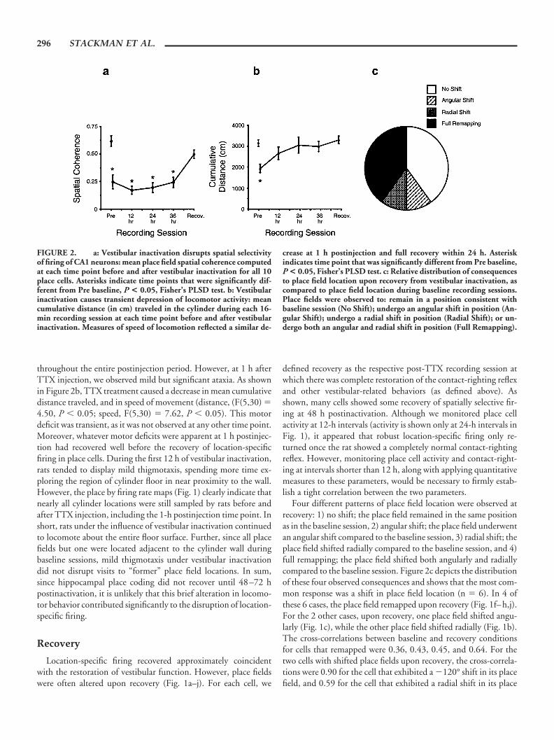
throughout the entire postinjection period. However, at 1 h afterTTX injection, we observed mild but significant ataxia. As shownin Figure 2b, TTX treatment caused a decrease in mean cumulativedistance traveled, and in speed of movement (distance, (F(5,30) �4.50, P � 0.05; speed, F(5,30) � 7.62, P � 0.05). This motordeficit was transient, as it was not observed at any other time point.Moreover, whatever motor deficits were apparent at 1 h postinjec-tion had recovered well before the recovery of location-specificfiring in place cells. During the first 12 h of vestibular inactivation,rats tended to display mild thigmotaxis, spending more time ex-ploring the region of cylinder floor in near proximity to the wall.However, the place by firing rate maps (Fig. 1) clearly indicate thatnearly all cylinder locations were still sampled by rats before andafter TTX injection, including the 1-h postinjection time point. Inshort, rats under the influence of vestibular inactivation continuedto locomote about the entire floor surface. Further, since all placefields but one were located adjacent to the cylinder wall duringbaseline sessions, mild thigmotaxis under vestibular inactivationdid not disrupt visits to “former” place field locations. In sum,since hippocampal place coding did not recover until 48–72 hpostinactivation, it is unlikely that this brief alteration in locomo-tor behavior contributed significantly to the disruption of location-specific firing.
Recovery
Location-specific firing recovered approximately coincidentwith the restoration of vestibular function. However, place fieldswere often altered upon recovery (Fig. 1a–j). For each cell, we
defined recovery as the respective post-TTX recording session atwhich there was complete restoration of the contact-righting reflexand other vestibular-related behaviors (as defined above). Asshown, many cells showed some recovery of spatially selective fir-ing at 48 h postinactivation. Although we monitored place cellactivity at 12-h intervals (activity is shown only at 24-h intervals inFig. 1), it appeared that robust location-specific firing only re-turned once the rat showed a completely normal contact-rightingreflex. However, monitoring place cell activity and contact-right-ing at intervals shorter than 12 h, along with applying quantitativemeasures to these parameters, would be necessary to firmly estab-lish a tight correlation between the two parameters.
Four different patterns of place field location were observed atrecovery: 1) no shift; the place field remained in the same positionas in the baseline session, 2) angular shift; the place field underwentan angular shift compared to the baseline session, 3) radial shift; theplace field shifted radially compared to the baseline session, and 4)full remapping; the place field shifted both angularly and radiallycompared to the baseline session. Figure 2c depicts the distributionof these four observed consequences and shows that the most com-mon response was a shift in place field location (n � 6). In 4 ofthese 6 cases, the place field remapped upon recovery (Fig. 1f–h,j).For the 2 other cases, upon recovery, one place field shifted angu-larly (Fig. 1c), while the other place field shifted radially (Fig. 1b).The cross-correlations between baseline and recovery conditionsfor cells that remapped were 0.36, 0.43, 0.45, and 0.64. For thetwo cells with shifted place fields upon recovery, the cross-correla-tions were 0.90 for the cell that exhibited a �120° shift in its placefield, and 0.59 for the cell that exhibited a radial shift in its place
FIGURE 2. a: Vestibular inactivation disrupts spatial selectivityof firing of CA1 neurons: mean place field spatial coherence computedat each time point before and after vestibular inactivation for all 10place cells. Asterisks indicate time points that were significantly dif-ferent from Pre baseline, P < 0.05, Fisher’s PLSD test. b: Vestibularinactivation causes transient depression of locomotor activity: meancumulative distance (in cm) traveled in the cylinder during each 16-min recording session at each time point before and after vestibularinactivation. Measures of speed of locomotion reflected a similar de-
crease at 1 h postinjection and full recovery within 24 h. Asteriskindicates time point that was significantly different from Pre baseline,P < 0.05, Fisher’s PLSD test. c: Relative distribution of consequencesto place field location upon recovery from vestibular inactivation, ascompared to place field location during baseline recording sessions.Place fields were observed to: remain in a position consistent withbaseline session (No Shift); undergo an angular shift in position (An-gular Shift); undergo a radial shift in position (Radial Shift); or un-dergo both an angular and radial shift in position (Full Remapping).
296 STACKMAN ET AL.

field. In contrast, upon recovery, the remaining four cells had placefields that matched those observed during baseline (Fig. 1a,d,e,i),with a mean cross-correlation of 0.88 � 0.02 (range, 0.84–0.93).These cells exhibited shifts in their fields of 0°, 12°, �18°, and�6°, respectively, between baseline and recovery sessions.
In two cases, two place cells were simultaneously recorded priorto, and over the course of, TTX-induced vestibular inactivationfrom the same rat. The place fields of the first pair (Fig. 1a,b)underwent a similar degree of TTX-induced disruption, and theplace fields appeared to recover to within 12° of their baselinelocations. The plane fields of the second pair of cells (Fig. 1c,g)were equally disrupted during the vestibular inactivation, and bothfields exhibited a rotational shift upon recovery (cell c � 120° shift;cell g � 174° shift). These data suggest that rather than TTXinducing an individual cell to remap or angularly shift its placefield, the vestibular inactivation produced a more generalized dis-ruption of hippocampal spatial mapping.
Two rats underwent a second vestibular inactivation after newplace cells were isolated 1 month later. We did not find any com-monality in the degree to which place fields were altered uponrecovery of vestibular inactivation between the first and secondvestibular inactivation. Specifically, for one animal, no shift inplace field location was observed after the first inactivation, but anew place cell exhibited a radial shift after the second inactivation.For the other animal, an angular shift was observed after the firstinactivation, while no shift in place field location was observedafter the second inactivation. Therefore, it is unlikely that themanner is which place fields respond to vestibular inactivation(e.g., angular shift, remapping) is common to individual animals.
Theta Cells
We also investigated the influence of vestibular inactivation onthe activity of 14 hippocampal theta cells. Theta cells of the hip-pocampus 1) are putative interneurons located within the stratumoriens; 2) discharge at a characteristic 4–12-Hz firing pattern; and3) exhibit firing rates that are correlated with locomotor behaviors(Ranck, 1973) and minimally influenced by spatial location (Ku-bie et al., 1990). The observed mean spatial coherence of theta cellsduring baseline sessions, 0.22 � 0.03 (range, 0.03–0.47), wasconsiderably lower than that for place cells (0.62 � 0.05), althoughsignificantly different from zero (t(13) � 7.74, P � 0.001). Ves-tibular inactivation caused little change in the spatial properties oftheta cell firing (Fig. 3a,c), and theta cell spatial coherence was notaltered over the course of inactivation, (F(5,65) � 1.38, n.s.).Vestibular inactivation also did not influence theta cell peak firingrates (F(5,65) � 2.12, n.s.) or average firing rates (F(5,65) � 0.97,n.s.). Autocorrelograms (i.e., Fig. 3b,d), constructed to reveal thetemporal characteristics of theta cell activity, indicated that rhyth-mic discharge was disrupted during inactivation for eight cells (Fig.3d), and maintained for six cells (Fig. 3b). These data suggest thepresence of two distinct populations of theta cells, one sensitive,and one resistant, to vestibular inactivation. In two cases, bothtypes of theta cells were recorded simultaneously from the sameanimal. Hippocampal theta cells have been classified as theta Onand theta Off, depending on their sensitivity or resistance to cho-
linergic drugs and medial septal inactivation (Smythe et al., 1991).It is not known whether the different sensitivities of theta cells tovestibular inactivation correspond to these two cell classes. Rhyth-mic firing properties were restored consummate with recovery ofvestibular function.
Representative EEG traces were acquired during each recordingsession for one theta cell (Fig. 3e). Baseline (Pre) traces revealed anincreased rhythmic theta activity during episodes of walking ascompared to episodes of immobility (Fig. 3e). This pattern of EEGactivity is consistent with previous reports of hippocampal thetacells (Ranck, 1973; Kubie et al., 1990), and was preserved, for themost part, during (24 h) and following vestibular inactivation (Re-covery; Fig. 3e). Theta rhythm was also evident in the EEG recordof a second rat, at 12 and 24 h following vestibular inactivation.The preservation of movement-related hippocampal theta duringvestibular inactivation is consistent with previous reports of intactmovement-related hippocampal theta activity in vestibular-defi-cient rodents (Frederickson et al., 1982; Shoham et al., 1989).
Head Direction Cells
Transtympanic TTX also disrupted the directional firing ofthree postsubicular cells. Head direction (HD) cell activity wasdisrupted 1 h after inactivation, and recovered with the restorationof vestibular function (48–72 h postinjection). The preferred fir-ing direction of two HD cells shifted upon recovery (30° and 96°;Fig. 4a–c), suggesting that vestibular inactivation caused someretuning among HD cells. The disruption of directional activityduring vestibular inactivation is consistent with our previous re-port that permanent vestibular lesion abolished anterior thalamicHD cell activity (Stackman and Taube, 1997). We did not attemptto record both hippocampal place cells and HD cells simulta-neously; therefore, we do not know whether place and HD cellsundergo coincident remapping/reorientation.
DISCUSSION
Many studies have demonstrated that the hippocampus plays acritical role in spatial memory (O’Keefe and Nadel, 1978; Jarrard,1993; Whishaw and Maaswinkel, 1998; O’Keefe, 1999). Rodentand primate hippocampal neurons discharge with respect to spatialinformation (location and head direction; Rolls and O’Mara,1995; Robertson et al., 1998), as well as nonspatial information(e.g., cue match/nonmatch; Colombo and Gross, 1994; Dead-wyler et al., 1996; Wood et al., 1999). As outlined above, substan-tial research indicates that external cues or landmarks influenceplace cells, HD cells, and navigation. Recent data support thenotion that in the absence of landmarks, internal (self-motion)cues also influence place cell and HD cell activity, suggesting ahierarchical involvement of external over internal cues. The preciseinvolvement of self-motion cues in the firing of hippocampal placecells is less certain. Tight restraint of a rat within the place field ofa hippocampal neuron causes a marked decline in location-specificfiring, however if the rat is loosely restrained, location-specific
____________________________________ HIPPOCAMPAL SPATIAL FIRING AND VESTIBULAR INPUT 297

FIGURE 3
FIGURE 4
298 STACKMAN ET AL.

firing recovers (Foster et al., 1989). These data suggest that hip-pocampal spatial representations require a “preparedness for move-ment signal.” However, Gavrilov et al. (1998) recently showed thatlocation-specific hippocampal neuronal firing is intact in rats thatare gently restrained and passively transported about an enclosure.Taken together, these data indicate that multiple sources of move-ment-associated cues influence place cells, yet active motion maynot be essential for the firing of hippocampal place cells. Impor-tantly, the present data demonstrate that the capacity of the hip-pocampal formation to encode location-specific firing requires ves-tibular input.
A consideration to note is that Hill and Best (1981) adminis-tered neomycin sulfate to rats daily for 40–60 days in order toproduce deafness and reported that location-specific firing per-sisted in hippocampal place cells. Neomycin sulfate is known to betoxic to cochlear hair cells (Leake and Hradek, 1988; Lowenheimet al., 1999), and because of their similarity and close proximity tovestibular hair cells, the drug could also have been toxic to them. Ifthere was a loss of vestibular hair cells following neomycin sulfatetreatment, then to be consistent with the results reported here, one
would expect a disruption of location-specific firing following neo-mycin sulfate treatment. Unfortunately, Hill and Best (1981) didnot assess vestibular function, although they reported that drug-treated rats performed well on a working memory task conductedon an elevated radial-arm maze. This finding suggests that vestib-ular functions may have been intact, since vestibular-lesioned ratsexhibit difficulties traversing elevated radial mazes that have open(nonenclosed) arms (R.W. Stackman, unpublished results), andbecause other studies involving vestibular lesions reported im-paired performance on a similar task (Ossenkopp and Hargreaves,1993). In sum, without confirmation that vestibular function wascompromised by neomycin sulfate treatment, the findings of Hilland Best (1981) can be considered consistent with our results.
Vestibular inactivation disrupted place and HD cell activitydespite the presence of the cylinder cue card, suggesting that ves-tibular input is a requirement for hippocampal spatial representa-tions. Thus, in contrast to the notion that internal cues supportlocation-specific firing only in the absence of external landmarks, ourdata suggest that hippocampal neurons may be continually moni-toring vestibular input to maintain a current representation ofspatial orientation. It is important to note that the principal locusof effect of vestibular inactivation is considerably distant from thehippocampus. Transtympanic TTX causes a neuronal blockade ofperipheral vestibular activity. The consequence of inactivation ofthe vestibular end organs, and the resulting alteration in brain-stemvestibular nuclei activity, likely influence the tone of vestibularsignals conveyed to the hippocampus. A consequence of the depri-vation of vestibular input to the hippocampus may be impairedprocessing of spatial information.
We previously reported that permanent lesions of the vestibularapparatus abolish HD cell activity in anterior thalamic nuclei(Stackman and Taube, 1997). It is possible that the loss of HD cellactivity in postsubiculum contributes to the impaired location-specific firing of place cells during vestibular inactivation. How-ever, this possibility is unlikely, since bilateral lesions of either theanterior dorsal thalamus or postsubiculum do not abolish location-specific firing of hippocampal place cells (Dudchenko et al., 1995;Archey et al., 1997). It is also unlikely that the loss of location-specific firing is attributed to changes in locomotor behavior or intheta cell activity. First, normal motor behavior returned well be-fore (at least 24 h) the recovery of location-specific firing. Second,although theta cell activity was disrupted in about half the thetacells recorded, other studies have shown that suppression of thetacell activity by either medial septal inactivation (Mizumori et al.,1989; Brazhnik et al., 1995), or intraventricular injection of acholinergic antagonist (scopolamine; Brazhnik et al., 1994), doesnot abolish location-specific firing in place cells, although applica-tion of scopolamine will reduce the place cell’s in-field firing rate.
It is possible that the inactivation of the vestibular apparatuscauses dizziness and motion sickness in treated rats, which leads todisorientation and, in turn, disrupts the location-specific firing ofhippocampal neurons. Manipulation of the vestibular system hasbeen demonstrated to cause aversive symptoms in animals. Forexample, bilateral TTX-induced inactivation of brain-stem vestib-ular nuclei is an effective substitute unconditioned stimulus fortaste aversion learning in rats (Ballesteros and Gallo, 2000). In
FIGURE 3. Representative examples of hippocampal theta cellactivity recorded before and after vestibular inactivation. a, c: Place byfiring rate plots reveal minimal alteration in location-specific firingover the course of vestibular inactivation. Spatial coherence values forthese cells at baseline and recovery were as follows: a, 0.243 and 0.487;c, 0.200 and 0.205, respectively. b, d: Respective autocorrelationfunctions observed for the cells plotted in a and c. The autocorrelationfunction represents the measure of cell firing at each 1 msec interval(from 0–350 msec), given a spike discharge at time 0. Autocorrelationfunctions were constructed by normalizing the spike count for eachinterval with respect to its peak value. The plot of the theta cell shownin b illustrates a preservation of rhythmic discharge over the course ofinactivation, while the plot of the cell shown in d indicates a diminu-tion of rhythmic discharge at 24 h and 48 h postinjection. e: Repre-sentative 1-s hippocampal EEG traces are depicted for the cell plottedin a, taken at each of the three time points indicated. Upper traces(Immobile) illustrate the EEG when the rat was stationary. In thiscase, voltage amplitude was decreased and theta rhythm was not evi-dent. Lower traces (Walking) illustrate the EEG during episodes oflocomotion when voltage amplitude was increased and theta rhyth-micity was observed. Similar patterns of activity were reflected in EEGtraces taken during and following vestibular inactivation; however,traces taken at 24 h suggest a slight irregularity in theta frequency overthe course of the 1-sec trace. The calibration scale represents 2 mV/100 ms.
FIGURE 4. Examples of firing rate by HD tuning curves of threepostsubicular HD cells recorded from three different rats, before andafter inactivation of the vestibular apparatus. For each cell, the base-line activity, or Pre, is represented by a heavy black line. The remain-ing lines illustrate the postinjection time course of inactivation: red,1 h; green, 24 h; and blue, vestibular recovery, namely, a, 48 h; b, 72 h;and c, 72 h. For all three cells, directional discharge was abolished by1 h postinjection. Upon recovery of vestibular function, two HD cellsexhibited a marked shift in preferred firing direction as compared tothe baseline session. Cross-correlation analyses of baseline vs. recov-ery session activity determined that the preferred firing direction ofthe cell in a shifted by 30°, the preferred firing direction of the cell inb shifted by 96°, and the preferred firing direction of the cell in c didnot shift upon recovery.
____________________________________ HIPPOCAMPAL SPATIAL FIRING AND VESTIBULAR INPUT 299

causing similar aversive distress and disorientation symptoms,transtympanic TTX may have prevented the normal expression ofplace fields in the familiar cylinder environment. Although we didnot test whether our TTX-treated rats were disoriented by havingthem perform a spatial task, other studies have shown that vestib-ular-lesioned rats are impaired on two different spatial tasks (Po-tegal et al., 1977; Ossenkopp and Hargreaves, 1993). Of course, ananimal can be impaired on a spatial task without being disoriented,so these behavioral data do not provide information on the extentto which these animals were disoriented.
It is possible that a general illness experienced by rats after ves-tibular inactivation caused the rats to fail to attend to the familiarcue card, or experience it as an unstable landmark. If the ratsperceived the cue card as unstable, one might have expected adeterioration of location-specific firing, or place fields that contin-ually drifted around the cylinder. We examined the latter possibil-ity by dividing 16-min recording sessions acquired during vestib-ular inactivation into four nonoverlapping 4-min sessions, andplotting firing rate by location maps for each. If a place field werepresent but perpetually drifting, we would expect to see evidence ofthe field in the 4-min session maps. We did not find any evidencefor drifting fields using this procedure (data not shown). Further,all rats demonstrated that they used the cue card as a landmarkprior to vestibular inactivation, because in cue card rotation exper-iments conducted prior to TTX-injections, we found that placefields shifted an amount similar to that of the rotation of the cuecard. Thus, before inactivation, all rats demonstrated the use of thecue card as a polarizing stimulus for orientation. Clearly, this ca-pacity was not intact during inactivation of the vestibular system.We cannot rule out, however, that the ill effects of vestibular inac-tivation either distracted the animals from using the cue as a land-mark, or distorted their perception of the cue card so that it pre-vented them from using it as a landmark. Although illness remainsa possible explanation for our data, we have no direct evidence thatthe rats were nauseous, distressed, or even disoriented because ofthe vestibular inactivation. Furthermore, TTX-treated rats did notexhibit appreciable loss of body weight during vestibular inactiva-tion, suggesting that any consequence of the inactivation did notinterrupt their normal feeding patterns.
It is possible that with permanent loss of vestibular function,compensatory mechanisms would eventually enable the rats toperceive the cue as stable, thus allowing some recovery of place cellactivity. However, we previously reported that permanent vestib-ular lesions caused a lasting disruption of HD cell firing (Stackmanand Taube, 1997). The rats in that study had had considerable pre-and postlesion experience with the same cylinder and cue card;however, HD cell firing did not show any sign of recovery over theseveral months of postlesion evaluation.
The possibility that compensatory mechanisms following long-term vestibular dysfunction might lead to a restoration of location-specific firing cannot be ruled out in the present study. Humanswith bilateral loss of vestibular function generally exhibit somedegree of recovery of spatial function postlesion and do not reportbeing disoriented under well-lit conditions in the presence of fa-miliar landmarks. This recovery is often attributed to a compensa-tory reliance upon visual cues, as the deficits return if the subjects
are tested in the dark or in the absence of familiar landmarks(Beritoff, 1966). Ossenkopp and Hargreaves (1993) reported thatspatial learning is impaired in rats tested 1 week after vestibularlesioning. We recently examined the long-term effects of vestibularlesions on spatial learning and memory. The results of our studies(Stackman and Herbert, 2002) demonstrated that lesions of thevestibular system did not impair acquisition of a spatial navigationtask when a visible landmark was present. However, vestibular-lesioned rats were markedly impaired during probe trials when thepolarizing visual cue was removed. In contrast, the performance ofcontrol rats was spared during these no cue card probe trials. Thesedata argue that despite the disorienting effects of the lesion, vestib-ular-deficient rats retain the ability to use a visual cue as a landmarkto guide spatial behavior. It is interesting to note that impairmentof navigation in the absence of a visual cue is consistent withclinical reports of humans with bilateral vestibular dysfunction(Beritoff, 1966; Pozzo et al., 1991; Brookes et al., 1993). Together,our data and those of others (Potegal et al., 1977) indicate thatvestibular-lesioned rats exhibit persistent cognitive impairmentson spatial navigational tasks, which are not a consequence of a lackof attention or of locomotor deficits.
Circular or Crescent-Shaped Firing FieldsDuring Vestibular Inactivation
It is noteworthy that several hippocampal cells exhibited a firingrate distribution that could be characterized as a circular firingpattern during vestibular inactivation (Fig. 1a,c,d,f,h,i). This pat-tern of firing during the 24-h and 48-h recording sessions corre-sponded with a propensity for thigmotaxic behavior, as deter-mined from location by time plots generated for each cell (data notshown), however, the rats clearly moved about the entire floorsurface, as indicated by the absence of significant nonsampled re-gions in the firing rate plots (Fig. 1, white pixels). Interestingly, atthe 1-h time point, when none of the cells exhibited crescent- orcircular-shaped fields, location by time plots revealed thigmotaxicbehavior for 5 of the 10 cells. Therefore, it is not clear whetherthigmotaxic behavior relates directly to the observed crescent- orcircular-shaped firing fields, since not all place cells fired at theperiphery during thigmotaxic behavior. One possibility is that thisfiring pattern reflects the disorienting consequence of a loss ofvestibular function. It is interesting to consider that vestibularinactivation may have caused the animals to become disoriented,which in turn led to the disruption of hippocampal place fields.Alternatively, vestibular inactivation may have led to the disrup-tion of place fields, which in turn caused the animals to becomedisoriented. Of course, neither issue can be resolved from our data;nor is the neural substrate for the sensation of disorientationknown. Furthermore, we have no evidence that the rats were trulydisoriented after vestibular inactivation. It is conceivable that theinactivation of the vestibular system impaired the rat’s ability to usethe landmark cue to determine its location and bearing. Such adeficit might have caused decay in the precision of spatial firing tosuch a degree that the once-concise place fields became smearedinto weakly firing, elongated crescent-shaped fields. Interestingly,McNaughton et al. (1995) theorized that hippocampal place cells
300 STACKMAN ET AL.

would adopt circular firing fields if directional input to the hip-pocampus were disrupted (McNaughton et al., 1995). Thus, onemight conclude from our results that the disruption of place fieldsafter vestibular inactivation is a secondary consequence of impairedHD cell activity. Again, as mentioned above, this conclusion isunlikely, since lesions to the postsubiculum or anterior dorsal tha-lamic nuclei (Dudchenko et al., 1995; Archey et al., 1997) fail toabolish hippocampal place cell activity.
Remapping
Place cells are considered to have undergone remapping when 1)most of the recorded place fields undergo a substantial shift in bothangular and radial dimensions, 2) new place fields appear in loca-tions where they were previously absent, or 3) a previously activeplace cell becomes quiescent or silent (Bostock et al., 1991;Knierim et al., 1995; Muller, 1996). The finding that several placefields exhibited complete remapping, or an angular shift, uponrecovery from vestibular inactivation was surprising, given that cellisolation remained consistent across each experiment (see Fig. 1k).In other words, the change in place field location from baseline torecovery cannot be explained as a loss of the original cell isolation.One possibility is that vestibular inactivation may have served as acue for place cells to remap upon recovery. Alternatively, repeatedexposure to the cylinder environment under the influence of ves-tibular inactivation may have encouraged place fields to remap.During vestibular inactivation, rats may have perceived the envi-ronment as distinct from that of the baseline session, which in turnmay have influenced the degree of remapping, or place field shiftobserved upon recovery. In short, vestibular inactivation may havedisrupted the rat’s use of familiar cylinder cues. When such con-ditions were experienced repeatedly during inactivation and as theanimal recovered, a distinct set of stimuli may have driven hip-pocampal activity, causing the eventual emergence of a novel spa-tial representation. In fact, a similar explanation was offered for thecomplete remapping of hippocampal place cells when rats repeat-edly experienced conflicting visuo-spatial cue information (Sha-piro et al., 1997).
Vestibular-Hippocampal Interaction
There is considerable neuroanatomical and neurophysiologicalsupport for vestibular-hippocampal interaction (reviewed inSmith, 1997). Our present data suggest that such an interactionplays a functionally significant role in hippocampal processing ofspatial information. Vestibular input to the hippocampus mayupdate spatial representations as the animal moves, so that therepresentations reflect current body position (McNaughton et al.,1996; Smith, 1997). Vestibular stimulation activates the hip-pocampal formation, parietal cortex, and retrosplenial cortex inhumans (Vitte et al., 1996) and rats (Horii et al., 1994), andmodulates primate hippocampal neuronal activity (O’Mara et al.,1994). Vestibular stimulation modulates rat hippocampal placecell activity (Sharp et al., 1995; Wiener et al., 1995; Bures et al.,1997), hippocampal theta rhythm (Gavrilov et al., 1995), and HD
cell activity (Knierim et al., 1995; Blair and Sharp, 1996). Record-ings of place and HD cells following brief episodes of detectableand undetectable rotational stimulation indicate that vestibularinformation alone provides sufficient evidence to the animal thatits position has changed. Thus, the hippocampus uses vestibularinformation (among other cues) to update representations of theanimal’s spatial orientation.
Neural network models of the place and HD cell systems in-clude an angular velocity code that is necessary for the network toaccurately track direction or location as the organism moves angu-larly or linearly. Several models propose that vestibular input isnecessary for anterior thalamic HD cells to exhibit anticipatoryfiring, angular velocity modulation of HD cell firing, and the sus-tained firing of HD cells in the absence of visual input (Redish etal., 1996; Touretzky and Redish, 1996). A model of hippocampalspatial representations (Samsonovich and McNaughton, 1997) ar-gues that the place cell (P) layer is influenced by a layer that inte-grates the output of the HD cell layer. Interestingly, their HD cellcomponent, which is consistent with earlier models from the samelaboratory (Skaggs et al., 1995; McNaughton et al., 1996), is di-rectly influenced by rotation cells that integrate the output of an-gular velocity cells signals. Taken together, these neural modelsutilize a vestibular mechanism in order to enable the models toaccurately represent an organism’s moment-to-moment spatial lo-cation and directional heading.
Humans with bilateral vestibular deficits exhibit spatial impair-ments (Pozzo et al., 1991; Brookes et al., 1993; Grasso et al., 1996;Israel et al., 1997; Peruch et al., 1999) that may be a consequenceof disturbed hippocampal coding of spatial information. Spatialnavigation requires a perpetual integration of self-motion andlandmark information in order to keep track of one’s current loca-tion and directional heading. The present data indicate that ves-tibular-hippocampal interaction provides an important influenceon hippocampal spatial representations, and support the view thataccurate navigation is a consequence of the monitoring of internalself-motion cues and landmarks (McNaughton et al., 1996;O’Keefe, 1999).
Acknowledgment
The authors thank Ms. Siobhan Robinson for surgical assis-tance.
NOTE ADDED IN PROOF
Saxon DW et al. (2001) recently reported that transtympanictetrodotoxin produces a transient spontaneous nystagmus, de-creases the vestibular-ocular reflex, and decreases brainstem vestib-ular neural activity. Taken together with the present findings, thetranstympanic administration of tetrodotoxin appears to be a use-ful method for producing a temporary disruption of central vestib-ular function.
____________________________________ HIPPOCAMPAL SPATIAL FIRING AND VESTIBULAR INPUT 301

REFERENCES
Archey WB, Stackman RW, Goodridge JP, Dudchenko PA, Taube JS.1997. Place cells show directionality in an open field following lesionsof the head direction cell system. Soc Neurosci Abstr 23:504.
Ballesteros MA, Gallo M. 2000. Bilateral tetrodotoxin blockade of the ratvestibular nuclei substitutes the natural unconditioned stimulus intaste aversion learning. Neurosci Lett 279:161–164.
Beitz AJ, Saxon DW, Anderson JH. 1995. Development of a novel, re-versible labyrinthectomy model: behavioral and anatomical correlates.Soc Neurosci Abstr 21:919.
Beritoff JS. 1966. Neural mechanisms of higher vertebrate behavior. Bos-ton: Little, Brown and Co.
Blair HT, Sharp PE. 1996. Visual and vestibular influences on head-direction cells in the anterior thalamus of the rat. Behav Neurosci110:643–660.
Bostock E, Muller RU, Kubie JL. 1991. Experience-dependent modifica-tions of hippocampal place cell firing. Hippocampus 1:193–206.
Brazhnik ES, Fox SE, Muller RU. 1994. Either blockade or enhancementof cholinergic transmission affects the location-specific firing of hip-pocampal pyramidal cells. Soc Neurosci Abstr 20:343.
Brazhnik ES, Muller RU, Fox SE. 1995. Temporary suppression of medialseptal activity greatly reduces the activity of ca1 place cells. Soc Neu-rosci Abstr 21:1439.
Brookes GB, Gresty MA, Nakamura T, Metcalfe T. 1993. Sensing andcontrolling rotational orientation in normal subjects and patients withloss of labyrinthine function. Am J Otol 14:349–351.
Bures J, Fenton AA, Kaminsky Y, Rossier J, Sacchetti B, Zinyuk L. 1997.Dissociation of exteroceptive and idiothetic orientation cues: effect onhippocampal place cells and place navigation. Philos Trans R Soc Lond[Biol] 352:1515–1524.
Canady KS, Rubel EW. 1992. Rapid and reversible astrocytic reaction toafferent activity blockade in chick cochlear nucleus. J Neurosci 12:1001–1009.
Chen YC, Pellis SM, Sirkin DW, Potegal M, Teitelbaum P. 1986. Ban-dage backfall: labyrinthine and non-labyrinthine components. PhysiolBehav 37:805–814.
Colombo M, Gross CG. 1994. Responses of inferior temporal cortex andhippocampal neurons during delayed matching to sample in monkeys(Macaca fascicularis). Behav Neurosci 108:443–455.
Deadwyler SA, Bunn T, Hampson RE. 1996. Hippocampal ensembleactivity during spatial delayed-nonmatch-to-sample performance inrats. J Neurosci 16:354–372.
Dudchenko P, Goodridge JP, Taube JS. 1995. The effects of lesions of thepostsubiculum on hippocampal place cell activity. Soc Neurosci Abstr21:945.
Dudchenko PA, Goodridge JG, Seiterle DA, Taube JS. 1997. Effects ofrepeated disorientation on the acquisition of two spatial referencememory tasks in rats: dissociation between the radial arm maze and theMorris water maze. J Exp Psychol [Anim Behav] 23:194–210.
Eichenbaum HB. 1999. The hippocampus and the mechanisms of declar-ative memory. Behav Brain Res 103:123–133.
Etienne AS, Teroni E, Maurer R, Portenier V, Saucy F. 1985. Short-distance homing in a small mammal: the role of exteroceptive cues andpath integration. Experientia 41:122–125.
Etienne AS, Maurer R, Seguinot V. 1996. Path integration in mammalsand its interaction with visual landmarks. J Exp Biol 199:201–209.
Foster TC, Castro CA, McNaughton BL. 1989. Spatial selectivity of rathippocampal neurons: dependence on preparedness for movement.Science 244:1580–1582.
Frederickson CJ, Frederickson MH, Lewis C, Howell GA, Smylie C,Wright CG. 1982. Hippocampal EEG in normal mice and in micewith congenital vestibular defects. Behav Neural Biol 34:121–131.
Gavrilov VV, Wiener SI, Berthoz A. 1995. Enhanced hippocampal thetaEEG during whole body rotations in awake restrained rats. NeurosciLett 197:239–241.
Gavrilov VV, Wiener SI, Berthoz A. 1998. Discharge correlates of hip-pocampal complex spike neurons in behaving rats passively displacedon a mobile robot. Hippocampus 8:475–490.
Goodridge JP, Taube JS. 1995. Preferential use of the landmark naviga-tional system by head direction cells in rats. Behav Neurosci 109:49–61.
Goodridge JP, Dudchenko PA, Worboys KA, Golob EJ, Taube JS. 1998.Cue control and head direction cells. Behav Neurosci 112:749–761.
Grasso R, Ivanenko Y, Israel I, Berthoz A. 1996. Vestibular spatial mem-ory: perception of horizontal angular displacements in two-dimen-sional trajectories. J Vestib Res 6:16.
Hill AJ, Best PJ. 1981. Effects of deafness and blindness on the spatialcorrelates of hippocampal unit activity in the rat. Exp Neurol 74:204–217.
Horii A, Takeda N, Mochizuki T, Okakura-Mochizuki K, Yamamoto Y,Yamatodani A. 1994. Effects of vestibular stimulation on acetylcholinerelease from rat hippocampus: an in vivo microdialysis study. J Neu-rophysiol 72:605–611.
Horn KM, DeWitt JR, Nielson HC. 1981. Behavioral assessment ofsodium arsanilate induced vestibular dysfunction in rats. Physiol Psy-chol 9:371–378.
Hunt MA, Miller SW, Nielson HC, Horn KM. 1987. Intratympanicinjection of sodium arsanilate (Atoxyl) solution results in posturalchanges consistent with changes described for labyrinthectomized rats.Behav Neurosci 101:427–428.
Israel I, Grasso R, Georges-Francois P, Tsuzuku T, Berthoz A. 1997.Spatial memory and path integration studied by self-driven passivelinear displacement. I. Basic properties. J Neurophysiol 77:3180–3192.
Jarrard LE. 1993. Review: on the role of the hippocampus in learning andmemory in the rat. Behav Neural Biol 60:9–26.
Kaufman GD, Anderson JH, Beitz AJ. 1992. Fos-defined activity in ratbrainstem following centripetal acceleration. J Neurosci 12:4489–4500.
Knierim JJ, Kudrimoti HS, McNaughton BL. 1995. Place cells, headdirection cells, and the learning of landmark stability. J Neurosci 15:1648–1659.
Knierim JJ, Kudrimoti HS, McNaughton BL. 1998. Interactions betweenidiothetic cues and external landmarks in the control of place cells andhead direction cells. J Neurophysiol 80:425–446.
Kubie JL, Muller RU, Bostock E. 1990. Spatial firing properties of hip-pocampal theta cells. J Neurosci 10:1110–1123.
Landeau B, Spelke E, Gleitman H. 1984. Spatial knowledge in a youngblind child. Cognition 16:225–260.
Leake PA, Hradek GT. 1988. Cochlear pathology of long term neomycininduced deafness in cats. Hear Res 33:11–34.
Lowenheim J, Kil J, Gultig K, Zenner HP. 1999. Determination of haircell degeneration and hair cell death in neomycin treated cultures ofthe neonatal rat cochlea. Hear Res 128:16–26.
Martin GM, Harley CW, Smith AR, Hoyles ES, Hynes CA. 1997. Spatialdisorientation blocks reliable goal location on a plus maze but does notprevent goal location in the Morris maze. J Exp Psychol [Anim Behav]23:183–193.
McNaughton BL, Knierim JJ, Wilson MA. 1995. Vector encoding andthe vestibular foundations of spatial cognition: Neurophysiologicaland computational mechanisms. In: Gazzaniga M, editor. The cogni-tive neurosciences. Cambridge, MA: MIT Press. p 585–595.
McNaughton BL, Barnes CA, Gerrard JL, Gothard K, Jung M, KnierimJJ, Kudrimoti HS, Qin Y, Skaggs WE, Suster M, Weaver KL. 1996.Deciphering the hippocampal polyglot: the hippocampus as a pathintegration system. J Exp Biol 199:173–185.
Miller S, Potegal M, Abraham L. 1983. Vestibular involvement in a pas-sive transport and return task. Physiol Psychol 11:1–10.
302 STACKMAN ET AL.

Mittelstaedt ML, Mittelstaedt H. 1980. Homing by path integration inthe mammal. Naturwissenschaften 67:566–567.
Mizumori SJ, Barnes CA, McNaughton BL. 1989. Reversible inactivationof the medial septum: selective effects on the spontaneous unit activityof different hippocampal cell types. Brain Res 500:99–106.
Morris RGM, Garrud P, Rawlins JNP, O’Keefe J. 1982. Place navigationimpaired in rats with hippocampal lesions. Nature 297:681–683.
Muller RU. 1996. A quarter of a century of place cells. Neuron 17:813–822.
Muller RU, Kubie JL, Ranck JB. 1987. Spatial firing patterns of hip-pocampal complex-spike cells in a fixed environment. J Neurosci7:1935–1950.
O’Keefe J. 1999. Do hippocampal pyramidal cells signal nonspatial as wellas spatial information? Hippocampus 9:352–364.
O’Keefe J, Nadel L. 1978. The hippocampus as a cognitive map. Oxford:Clarendon Press.
O’Mara SM, Rolls ET, Berthoz A, Kesner RP. 1994. Neurons respondingto whole-body motion in the primate hippocampus. J Neurosci 14:6511–6523.
Ossenkopp KP, Hargreaves EL. 1993. Spatial learning in an enclosedeight-arm radial maze in rats with sodium arsanilate-induced laby-rinthectomies. Behav Neural Biol 59:253–257.
Ossenkopp KP, Prkacin A, Hargreaves EL. 1990. Sodium arsanilate-in-duced vestibular dysfunction in rats: effects on open-field behavior andspontaneous activity in the automated Digiscan monitoring system.Pharmacol Biochem Behav 36:875–881.
Pasic TR, Rubel EW. 1989. Rapid changes in cochlear nucleus cell sizefollowing blockade of auditory nerve electrical activity in gerbils.J Comp Neurol 283:474–480.
Paxinos G, Watson C. 1998. The rat brain in stereotaxic coordinates. 3rded. San Diego: Academic Press.
Peruch P, Borel L, Gaunet F, Thinus-Blanc G, Magnan J, Lacour M.1999. Spatial performance of unilateral vestibular defective patients innonvisual versus visual navigation. J Vestib Res 9:37–47.
Potegal M, Day MJ, Abraham L. 1977. Maze orientation, visual andvestibular cues in two-maze spontaneous alternation of rats. PhysiolPsychol 5:414–420.
Pozzo T, Berthoz A, LeFort L, Vitte E. 1991. Head stabilization duringvarious locomotor tasks in humans. II. Patients with bilateral periph-eral vestibular deficits. Exp Brain Res 85:208–217.
Quirk GJ, Muller RU, Kubie JL. 1990. The firing of hippocampal placecells in the dark depends on the rat’s recent experience. J Neurosci10:2008–2017.
Ranck JB Jr. 1973. Studies on single neurons in dorsal hippocampalformation and septum in unrestrained rats. I. Behavioral correlates andfiring repertoires. Exp Neurol 41:461–531.
Redish AD, Elga AN, Touretzky DS. 1996. A coupled attractor model ofthe rodent head direction system. Netw Comput Neural Syst 7:671–685.
Robertson RG, Rolls ET, Georges-Francois P. 1998. Spatial view cells inthe primate hippocampus: effects of removal of view details. J Neuro-physiol 79:1145–1156.
Rolls ET, O’Mara SM. 1995. View-responsive neurons in the primatehippocampal complex. Hippocampus 5:409–424.
Samsonovich A, McNaughton BL. 1997. Path integration and cognitivemapping in a continuous attractor neural network model. J Neurosci17:5900–5920.
Saxon DW, Anderson JH, Beitz AJ. 2001. Transtympanic tetrodotoxinalters the VOR and Fos labeling in the vestibular complex. Neurore-port 12:3051–3055.
Selye H. 1957. Lathyrism. Rev Can Biol 16:1–82.Shapiro ML, Tanila H, Eichenbaum H. 1997. Cues that hippocampal
place cells encode: dynamic and hierarchical representation of localand distal stimuli. Hippocampus 7:624–642.
Sharp PE, Blair HT, Tzanetos DB. 1995. Influences of vestibular andvisual motion information on the spatial firing patterns of hippocam-pal place cells. J Neurosci 15:173–189.
Shoham S, Chen YC, DeVietti TL, Teitelbaum P. 1989. Deafferentationof the vestibular organ: effects on atropine-resistant EEG in rats. Psy-chobiology 17:307–314.
Skaggs WE, McNaughton BL, Gothard KM, Markus EJ. 1993. An infor-mation-theoretic approach to deciphering the hippocampal code. In:Hanson SJ, Cowan JD, Giles CL, editors. Advances in neural infor-mation processing systems. San Mateo, CA: Morgan Kaufmann. p1030–1037.
Skaggs WE, Knierim JJ, Kudrimoti HS, McNaughton BL. 1995. A modelof the neural basis of the rat’s sense of direction. In: Tesauro G,Touretzky DS, Leen T, editors. Advances in neural information pro-cessing systems. Cambridge, MA: MIT Press. p 173–180.
Smith PF. 1997. Vestibular-hippocampal interactions. Hippocampus7:465–471.
Smythe JW, Cristie BR, Colom LV, Lawson VH, Bland BH. 1991. Hip-pocampal theta field activity and theta-on/theta-off cell discharges arecontrolled by an ascending hypothalamo-septal pathway. J Neurosci11:2241–2248.
Stackman RW, Herbert AM. 2002. Rats with vestibular lesions require avisual landmark for spatial navigation. Behav Brain Res 128:27–40.
Stackman RW, Taube JS. 1997. Firing properties of head direction cells inthe rat anterior thalamic neurons: dependence on vestibular input.J Neurosci 17:4349–4358.
T’Ang Y, Wu C-F. 1936. The effects of unilateral labyrinthectomy in thealbino rat. Chin J Physiol 10:571–598.
T’Ang Y, Wu C-F. 1937. The effects of central compensation in laby-rinthectomized rats. Chin J Physiol 12:117–124.
Taube JS. 1998. Head direction cells and the neurophysiological basis fora sense of direction. Prog Neurobiol 55:225–256.
Taube JS, Burton HL. 1995. Head direction cell activity monitored in anovel environment and during a cue conflict situation. J Neurophysiol74:1953–1971.
Taube JS, Muller RU, Ranck JB Jr. 1990. Head-direction cells recordedfrom the postsubiculum in freely moving rats. I. Description andquantitative analysis. J Neurosci 10:420–435.
Telford L, Howard IP, Ohmi M. 1995. Heading judgments during activeand passive self-motion. Exp Brain Res 104:502–510.
Touretzky DS, Redish AD. 1996. Theory of rodent navigation based oninteracting representations of space. Hippocampus 6:247–270.
Vitte E, Derosier C, Caritu Y, Berthoz A, Hasboun D, Soulie D. 1996.Activation of the hippocampal formation by vestibular stimulation: afunctional magnetic resonance imaging study. Exp Brain Res 112:523–526.
Whishaw IQ, Gorny B. 1999. Path integration absent in scent-trackingfimbria-fornix rats: evidence for hippocampal involvement in “sense ofdirection” and “sense of distance” using self-movement cues. J Neuro-sci 19:4662–4673.
Whishaw IQ, Maaswinkel H. 1998. Rats with fimbria-fornix lesions areimpaired in path integration: a role for the hippocampus in “sense ofdirection.” J Neurosci 18:3050–3058.
Wiener SI, Korshunov VA, Garcia R, Berthoz A. 1995. Inertial, substrataland landmark cue control of hippocampal ca1 place cell activity. EurJ Neurosci 7:2206–2219.
Wood ER, Dudchenko PA, Eichenbaum H. 1999. The global record ofmemory in hippocampal neuronal activity. Nature 397:613–616.
Zola SM, Squire LR, Teng E, Stefanacci L, Buffalo EA, Clark RE. 2000.Impaired recognition memory in monkeys after damage limited to thehippocampal region. J Neurosci 20:451–63.
____________________________________ HIPPOCAMPAL SPATIAL FIRING AND VESTIBULAR INPUT 303