On the lateralization of emotional prosody: An event-related functional MR investigation
Upload
independentCategory
view
4download
0
A Comparison of Two FMRI Methods for Predicting Verbal MemoryDecline After Left Temporal Lobectomy: Language Lateralizationvs. Hippocampal Activation Asymmetry
Jeffrey R. Binder, MD1, Sara J. Swanson, PhD1, David S. Sabsevitz, PhD1, Thomas A.Hammeke, PhD1, Manoj Raghavan, MD, PhD1, and Wade M. Mueller, MD21 Comprehensive Epilepsy Center, Department of Neurology, Medical College of Wisconsin,Milwaukee, Wisconsin2 Comprehensive Epilepsy Center, Department of Neurosurgery, Medical College of Wisconsin,Milwaukee, Wisconsin
AbstractPurpose—Language lateralization measured by preoperative fMRI was shown recently to bepredictive of verbal memory outcome in patients undergoing left anterior temporal lobe (L-ATL)resection. The aim of this study was to determine whether language lateralization or hippocampalactivation asymmetry is a better predictor of memory outcome in this setting.
Methods—Thirty L-ATL patients underwent preoperative language fMRI, preoperativehippocampal fMRI using a scene encoding task, and pre- and postoperative neuropsychologicaltesting. A group of 37 right ATL surgery patients who underwent the same testing procedures wasincluded for comparison.
Results—Verbal memory decline occurred in roughly half of the L-ATL patients. Preoperativelanguage lateralization was correlated with postoperative verbal memory change. Hippocampalactivation asymmetry was strongly related to side of seizure focus and to Wada memory asymmetrybut was unrelated to verbal memory outcome.
Discussion—Preoperative hippocampal activation asymmetry elicited by a scene encoding task isnot predictive of verbal memory outcome. Risk of verbal memory decline is likely to be related tolateralization of material-specific verbal memory networks, which are more closely correlated withlanguage lateralization than with overall asymmetry of episodic memory processes.
Keywordstemporal lobectomy; fMRI; memory; hippocampus; language dominance
IntroductionAnterior temporal lobe (ATL) resection is an effective treatment for individuals with medicallyintractable epilepsy (Wiebe et al., 2001; Tellez-Zenteno et al., 2005). The undeniable benefitof this treatment must be balanced against the risk of memory decline, particularly in patientsundergoing left ATL resection. Though the incidence of postoperative morbidity varies fromstudy to study depending on the specific assessment methods used, approximately 30–60% of
Corresponding Author: Jeffrey R. Binder, M.D., Department of Neurology, Medical College of Wisconsin, 9200 W. Wisconsin Ave.,Milwaukee, WI 53226, Phone: 414-456-4662, Fax: 414-456-6562, [email protected].
NIH Public AccessAuthor ManuscriptEpilepsia. Author manuscript; available in PMC 2010 October 1.
Published in final edited form as:Epilepsia. 2010 April ; 51(4): 618–626. doi:10.1111/j.1528-1167.2009.02340.x.
NIH
-PA Author Manuscript
NIH
-PA Author Manuscript
NIH
-PA Author Manuscript
patients undergoing left ATL resection show significant declines in verbal memory (Cheluneet al., 1993; Helmstaedter & Elger, 1996; Martin et al., 1998; Sabsevitz et al., 2001; Lee et al.,2002; Stroup et al., 2003; Gleissner et al., 2004; Baxendale et al., 2006; Lineweaver et al.,2006; Binder et al., 2008). One principal goal of the preoperative evaluation in ATL surgerycandidates is, therefore, to estimate the risk of verbal memory decline.
Functional magnetic resonance imaging (fMRI) is a safe, non-invasive brain mapping tool thatmay provide information about preoperative lateralization of memory networks. Severalpreliminary studies showed correlations between preoperative medial temporal lobe (MTL)activation and postoperative verbal memory decline (Richardson et al., 2004; Richardson etal., 2006; Frings et al., 2008; Powell et al., 2008). These studies focused on activation in thehippocampus and other MTL areas. In a recent study of 60 patients undergoing left ATLresection, we showed that postoperative verbal memory change was predicted by preoperativelanguage-related activation measured throughout the cerebral hemispheres (Binder et al.,2008). In the present study we compare this language lateralization index with an fMRI methodbased on hippocampal activation asymmetry to determine which is a better predictor ofpostoperative verbal memory outcome.
MethodsParticipants
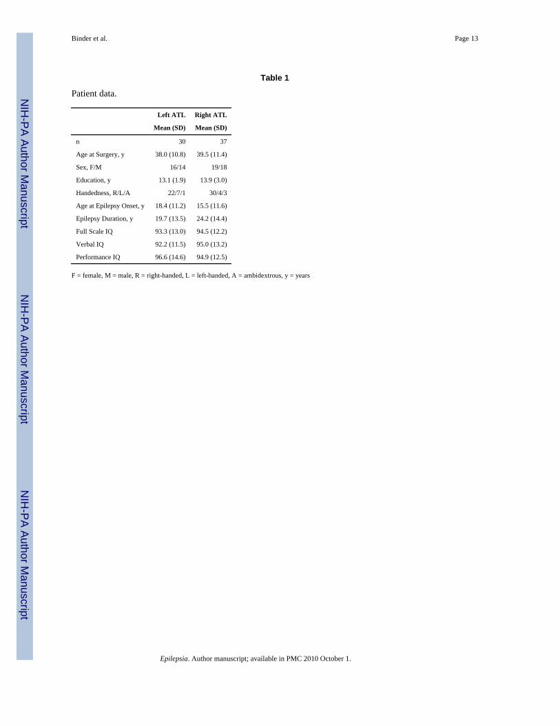
Participants were 30 patients with intractable left temporal lobe epilepsy (L-ATL group),encountered consecutively at the Medical College of Wisconsin Comprehensive EpilepsyProgram between 1995 and 2006, who met the following criteria: (a) left ATL resection, (b)preoperative fMRI language mapping performed, (c) preoperative fMRI hippocampal mappingperformed; (d) preoperative and 6-month-postoperative neuropsychological evaluationperformed, and (e) full scale IQ > 70. These patients are a subset from our previous study(Binder et al., 2008) who also underwent hippocampal fMRI mapping. One other L-ATLpatient who otherwise met criteria was excluded because fMRI showed no significantactivation in the hippocampus on either side. A control group of 37 consecutively encounteredpatients who had right ATL resection (R-ATL group) and preoperative hippocampal fMRIserved as a comparison group.
All patients underwent a standard ATL resection removing the anterior 3 to 4 cm of the temporallobe, including lateral (middle temporal gyrus, inferior temporal gyrus, polar superior temporalgyrus) and medial (fusiform gyrus, parahippocampus, amygdala, anterior hippocampus)structures. The posterior extent of resection was tailored using electrocorticography in allpatients and intra- or extraoperative stimulation mapping of language cortex when thelanguage-dominant temporal lobe was resected. The same epilepsy surgeon performed alloperations. FMRI data were collected for research purposes only and were not available to thesurgeon before or after surgery. Informed consent was obtained from all patients prior to fMRIusing a protocol approved by the Institutional Review Board of the Medical College ofWisconsin.
Patient demographic information is summarized in Table 1. The L-ATL and R-ATL groupsdid not differ on age, sex, years of education, handedness, age at onset of epilepsy (defined asthe age at onset of recurring seizures), epilepsy duration, or measures of intelligence (all Pvalues > 0.1).
Neuropsychological TestingNeuropsychological testing was performed before and 6 months following ATL surgery.Verbal episodic memory was assessed using the 6-trial Selective Reminding Test (SRT)
Binder et al. Page 2
Epilepsia. Author manuscript; available in PMC 2010 October 1.
NIH
-PA Author Manuscript
NIH
-PA Author Manuscript
NIH
-PA Author Manuscript
(Buschke & Fuld, 1974). The SRT includes several measures of word list learning as well asdelayed recall and recognition tests. Pre- to post-operative change scores on each test werecalculated by subtracting the preoperative score from the postoperative score. Positive valuesthus indicate improvement, whereas negative values indicate a decline in performance. Othermemory tests given before and after surgery included the Logical Memory and VisualReproduction subtests of the Wechsler Memory Scale (WMS-R or WMS-III) and the 7/24Spatial Recall Test (Rao et al., 1984). We focus here on the Consistent Long-Term Recall(CLTR) and Delayed Recall measures of the SRT, which were among the most sensitive testsfor detecting verbal memory decline in our prior study (Binder et al., 2008). CLTR indexes therate at which a list of words is learned, whereas Delayed Recall indexes the ability to recall thelist after a 30 minute delay.
Patients were nominally classified as either showing or not showing decline on each test,defined as a negative change score of 1.5 standard deviations or more from the mean changescore in the R-ATL group. The resulting cut-off values are somewhat conservative andrepresent substantial declines, generally in the range of 40–60% relative to pre-operative rawscores.
Wada TestingWada memory data were available for 28 of the 30 L-ATL patients and all of the R-ATLpatients. Wada language and memory testing followed procedures described previously(Loring et al., 1992; Binder et al., 1996; Binder et al., 2008). Immediately after recovery fromthe anesthetic, a 24-item object memory recognition test was administered. Eight target objectsthat had been presented in the visual field ipsilateral to the injection and named aloud by theexaminer during the period of anesthesia and 16 novel foil objects were presented in a randomorder, and patients were required to indicate whether or not each object had been presentedpreviously. A recognition memory score for each hemisphere, corrected for guessing, wascalculated as the number of items correctly recognized minus half the number of false positiveresponses. These raw scores, which can range from 0 to 8, were divided by 8 to yield valuesranging from 0 to 1. Wada memory asymmetry (WMA) was calculated by taking the differencebetween the recognition memory performances of the left hemisphere (inject right) and theright hemisphere (inject left), yielding values ranging from −1 (all memory capacity in the righthemisphere) to +1 (all memory capacity in the left hemisphere). In a previous study, thisasymmetry measure correctly classified 89% of temporal lobe epilepsy patients as to side ofseizure focus (Binder et al., 2008).
MRI AcquisitionImaging was conducted on a 1.5 T GE Signa scanner. High-resolution, T1-weighted anatomicalreference images of the entire brain were acquired with a spoiled-gradient-echo ("SPGR")sequence. Whole brain functional imaging used a T2*-weighted gradient-echo, echoplanarsequence with the following parameters: TE = 40 ms, TR = 3000 ms, field of view = 240 mm,pixel matrix = 64 × 64, 19 sagittal slices, voxel size = 3.75 × 3.75 × 7 mm.
FMRI Activation TasksBoth fMRI protocols used a block design with alternation between two active task conditions.The language protocol contrasted a semantic decision task with a tone decision task. In thesemantic decision task, patients heard animal names and decided whether each animal is “foundin the United States” and “used by humans.” In the tone decision task, patients heard brieftrains of high and low tones and decided whether the train contained two high tones. Thesetasks, their rationale, and the typical patterns of activation and lateralization observed in normalparticipants were described previously (Binder et al., 1997; Frost et al., 1999; Springer et al.,1999; Szaflarski et al., 2002). The contrast between semantic decision and tone decision tasks
Binder et al. Page 3
Epilepsia. Author manuscript; available in PMC 2010 October 1.
NIH
-PA Author Manuscript
NIH
-PA Author Manuscript
NIH
-PA Author Manuscript
highlights speech perception and lexical-semantic processes engaged during the semanticdecision task while controlling for early auditory, attention, executive, and motor responseprocesses. This contrast produces reliable and strongly left-lateralized activation in areaspreviously implicated in language processing, including frontal, temporal, and parietalassociation cortices (Binder et al., 1997; Frost et al., 1999), and in the left hippocampus andsurrounding medial temporal lobe (Binder et al., 1997; Bellgowan et al., 1998).
The hippocampal protocol contrasted a visual scene encoding task with a visual noise encodingtask (Binder et al., 2005). In the scene encoding task, patients saw color photographs of naturalscenes and decided whether each scene was indoor or outdoor. In the noise encoding task,patients saw spatially scrambled versions of the natural scenes and decided whether the leftand right halves of each image were identical. These tasks, their rationale, and the typicalpatterns of activation observed in normal participants were described previously (Binder et al.,2005). Control participants show significantly better post-scan recognition of the scenes thanof the scrambled scenes (Binder et al., 2005), reflecting the more effective activation of episodicmemory encoding processes during the scene condition. The contrast between scene and noiseencoding tasks highlights visual object recognition and episodic memory encoding processesengaged during the scene encoding task while controlling for early visual, attention, executive,and motor response processes. This contrast produces reliable, symmetric activation of theanterior hippocampus, posterior parahippocampus, and medial fusiform gyrus in healthyparticipants (Binder et al., 2005).
FMRI Data AnalysisImage processing and statistical analyses were conducted with AFNI(http://afni.nimh.nih.gov/afni). All analyses were performed at the individual subject level.Image alignment was used to reduce the effects of head movement. Task-related changes inMRI signal were identified using multiple regression.This method compares the time series ofMRI signal values in each image voxel with an idealized hemodynamic response to the taskalternation. The idealized response was modeled by convolving a gamma function with a timeseries of impulses representing each task trial. The regression model also included sixmovement vectors (computed during image registration) to further reduce any effects of headmovement on estimation of the task response, and first- and second-order covariates to modelany linear or quadratic baseline shifts.
Language lateralization was measured for three brain regions: frontal lobe, temporal lobe, anda lateral hemisphere region including frontal, temporal, and parietal lobes. The volumesdefining these regions of interest (ROIs) were based on an average left hemisphere activationmap from 80 healthy, right-handed participants (Frost et al., 1999; Szaflarski et al., 2002).Significantly activated voxels (uncorrected p < .001) in the left and right hemisphere werecounted in each ROI for each patient. Laterality indexes (LI), reflecting the interhemisphericdifference between voxel counts in left and right homologous ROIs, were calculated for eachregion using the formula: LI = (L−R)/(L+R) (Binder et al., 1996; Springer et al., 1999;Sabsevitz et al., 2003). LIs can range from −1 (all active voxels in the right hemisphere) to +1(all active voxels in the left hemisphere).
For the hippocampal fMRI protocol, hippocampus LIs were computed for three regions:anterior hippocampus, posterior hippocampus, and whole (anterior + posterior) hippocampus.These ROIs were created by manually tracing the hippocampus in 32 healthy participants(Binder et al., 2005). These tracings were aligned in stereotaxic space and averaged to createvalues for the probability of overlap at each voxel. Voxels included in the hippocampus ROIswere those with at least 90% overlap across the individual tracings. The anterior and posteriorROIs were delineated by a coronal plane through the posterior edge of the interpeduncular
Binder et al. Page 4
Epilepsia. Author manuscript; available in PMC 2010 October 1.
NIH
-PA Author Manuscript
NIH
-PA Author Manuscript
NIH
-PA Author Manuscript
cistern. Given the small size of these ROIs, an uncorrected threshold of p <.05 was used todefine activated voxels for the LI computations.
ResultsPre- and Post-Operative Memory Performance
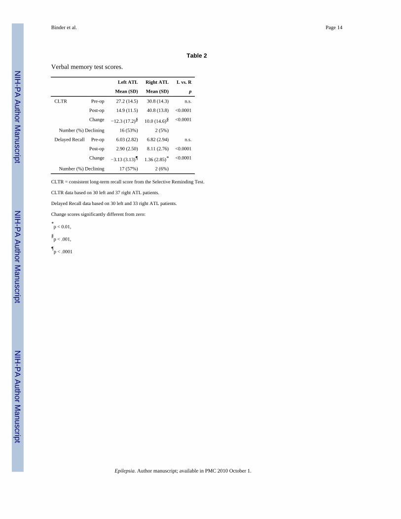
Table 2 shows the average pre-operative, post-operative, and pre-post change scores for eachATL group on the two verbal memory measures. The percentage of patients who declined isshown for each test. The L-ATL group showed significant decline in verbal memory, whereasthe R-ATL group significantly improved. Roughly half of the L-ATL patients declined on eachmeasure. See Binder et al. (2008) for more details on verbal and nonverbal memory outcomein the larger sample from which this subgroup was taken.
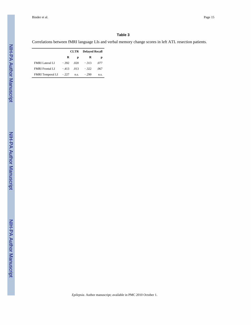
Language LI as a Predictor of Memory OutcomeTable 3 shows correlations between verbal memory change scores and the fMRI language LIscalculated over lateral hemisphere, frontal, and temporal regions. Change scores on CLTR, thelist learning measure, were significantly correlated with preoperative language lateralizationin the lateral hemisphere and frontal lobe ROIs. Change scores on Delayed Recall showedstrong trends for correlation with language lateralization in these ROIs. These correlationsreached significance in the larger patient sample studied previously (Binder et al., 2008).
Hippocampal Functional Asymmetry as a Predictor of Memory OutcomeValidity of the hippocampal fMRI measures was assessed by comparing hippocampal LIs inthe left and right ATL groups and by correlating the hippocampal LIs with memory asymmetryscores on the Wada test. As shown in Figure 1, all three hippocampal fMRI LIs differed betweenthe left and right ATL groups. The anterior LI was the most strongly related to seizure focus(L-ATL: mean(sd) = −.283(.620); R-ATL: mean(sd) = .175(.622); p = .004). The posteriorhippocampus LI differed only marginally between groups (L-ATL: mean(sd) = .120(.652);RATL: mean(sd) = .390(.553); p = .071). The whole hippocampus LI differed between thegroups (L-ATL: mean(sd) = −.049(.520); R-ATL: mean(sd) = .270(.541); p = .017) but not asmuch as the anterior LI.
Similarly, the anterior hippocampus LI (r = .307, p = .009) and whole hippocampus LI (r = .310, p = .007) were significantly correlated with Wada memory asymmetry across all patients.The posterior hippocampus LI was only weakly correlated with Wada memory asymmetry (r= .225, p = .061). These validation measures demonstrate that the fMRI hippocampal LIs,particularly the anterior and whole hippocampus LIs, are sensitive to structural and functionalasymmetries in the MTL memory system related to chronic temporal lobe epilepsy.
In contrast to the language LIs, none of the three hippocampal LIs was significantly correlatedwith either of the verbal memory change scores. Correlation values ranged from −.134(posterior hippocampus LI vs. Delayed Recall change) to .149 (anterior hippocampus LI vs.Delayed Recall change); none of these approached statistical significance. Correlation betweenthe anterior hippocampus LI and CLTR change was nil (r = −.040). Figure 2 illustrates thesignificant relationship between CLTR outcome and language LI and the lack of correlationbetween CLTR outcome and anterior hippocampus LI.
Several previous studies reported significant relationships between unilateral left hippocampalactivation and verbal memory outcome (Richardson et al., 2006; Powell et al., 2008). In oneof these studies (Richardson et al., 2006), change scores were negatively correlated withactivation in both left and right hippocampus, suggesting that unilateral activation on the sideof surgery may be a more reliable predictor of outcome than activation asymmetry. We found
Binder et al. Page 5
Epilepsia. Author manuscript; available in PMC 2010 October 1.
NIH
-PA Author Manuscript
NIH
-PA Author Manuscript
NIH
-PA Author Manuscript

no statistically significant correlations between unilateral activation in any of the hippocampalROIs (mean beta coefficient averaged across voxels in the ROI) and verbal memory changeon either of the outcome measures. Correlation values ranged from −.158 (posterior lefthippocampus activation vs. CLTR change) to −.099 (anterior left hippocampus activation vs.CLTR change); none of these correlations approached statistical significance.
DiscussionRecent studies have explored the possibility of using preoperative fMRI to predict episodicmemory impairment after ATL resection. Given the prominent role played by the hippocampusand other MTL structures in episodic memory, most of this research has focused onpreoperative activation in MTL structures. In the first study to examine verbal memoryoutcome, Richardson and colleagues identified a focus in the anterior hippocampus whereasymmetry of activation on fMRI predicted verbal memory outcome on a standardized wordlist learning test after left ATL resection (Richardson et al., 2004). Greater activation in thisregion on the left side relative to the right side predicted greater decline. Subsequent studiesby the same authors showed correlations between verbal memory change and preoperativeactivation in either the left hippocampus (Powell et al., 2008) or both the left and righthippocampus (Richardson et al., 2006). Frings et al. showed a weak correlation (1-tailed p = .077) between postoperative verbal memory change and preoperative fMRI activationasymmetry in the hippocampus (Frings et al., 2008). While encouraging, these studies usedvery small sample sizes (7–12 left ATL surgery patients) and should therefore be viewed aspreliminary. Moreover, the results were not entirely consistent across studies, with tworeporting activation asymmetry as a significant predictor (Richardson et al., 2004; Frings et al.,2008) and two reporting unilateral activation on the left side as the main predictor (Richardsonet al., 2006; Powell et al., 2008). In the studies by Richardson and colleagues, the exact locationof the hippocampal ROI that best predicts outcome has also varied from study to study. Finally,a study by Rabin et al. failed to find any relationship between preoperative MTL activation (oractivation asymmetry) and postoperative verbal memory outcome (Rabin et al., 2004).
One difficulty in using hippocampal activation as a prognostic marker is the relatively smallsize of this structure, which contains only around a hundred fMRI voxels even when scanningat relatively high resolution. Compared to the entire cerebral hemisphere, which contains manythousands of voxels, measuring activation in the hippocampus thus depends on a much smallersample size and is consequently more susceptible to random noise, subject motion, andmeasurement error arising from imperfect identification of hippocampal boundaries. Loss ofMRI signal due to macroscopic field inhomogeneity ("signal dropout") often affects theamygdala and occasionally the anterior hippocampus (Constable et al., 2000; Fransson et al.,2001; Morawetz et al., 2008) and represents another technical difficulty for fMRI of thehippocampus. For these reasons, we explored the use of language lateralization as a prognosticmarker for verbal memory outcome. The obvious advantage of such an approach is that theROI over which lateralization is measured can be much larger, potentially offering a morestable and reliable measurement. In addition to this technical advantage, however, there aretheoretical reasons to consider language lateralization as a prognostic marker. Evidencesuggests that verbal and nonverbal information is encoded in somewhat distinct episodicmemory networks, and that these typically have different patterns of hemispheric lateralization(Glosser et al., 1995; Kelley et al., 1998; Chiaravalloti & Glosser, 2001; Golby et al., 2001;Reber et al., 2002; Powell et al., 2005). Furthermore, it seems likely that this specialization forverbal or nonverbal material is not a property of the MTL itself, but is rather a function of thetype of information received by the MTL from the ipsilateral hemisphere. If this model iscorrect, then the MTL in the language-dominant hemisphere is likely to be more critical forsupporting verbal episodic memory, and language lateralization should be correlated withverbal memory lateralization.
Binder et al. Page 6
Epilepsia. Author manuscript; available in PMC 2010 October 1.
NIH
-PA Author Manuscript
NIH
-PA Author Manuscript
NIH
-PA Author Manuscript
Analysis of the current patient subgroup is consistent with our previous larger study (Binderet al., 2008) in showing a correlation between verbal memory change and preoperativelanguage LI. Effect sizes were similar in the two analyses, though the correlations with DelayedRecall outcome did not reach standard significance levels in the subgroup analysis due to thesmaller sample size. In the previous study, both fMRI language LI (r = −.432, p <.001) andWada language asymmetry (r = −.398, p <.01) were significant predictors of verbal memorychange, lending strong support to the hypothesis that verbal memory lateralization is correlatedwith language lateralization. In contrast, a material-nonspecific Wada memory asymmetryscore (recognition of visually-encoded real objects) was only weakly related to verbal memoryoutcome (r = −.331, p <.05).
In contrast to the correlations with language LI, we found no evidence of a correlation betweenverbal memory outcome and preoperative hippocampal activation. There were no significantcorrelations with either activation asymmetry or unilateral activation in the left hippocampus.These null findings held for ROIs targeting the anterior hippocampus, posterior hippocampus,and whole hippocampus.
Notably, these null results cannot be explained by poor data quality or lack of functionalactivation. All patients in the study had significantly activated voxels detected in thehippocampus. More importantly, the hippocampal LIs, especially the anterior hippocampusLI, showed clear evidence of functional lateralization away from the side of the seizure focus,as expected in chronic temporal lobe epilepsy. Finally, the anterior and whole hippocampusLIs were significantly correlated with Wada memory asymmetry. Thus, this fMRI protocolbased on a visual scene encoding task detected hippocampal functional asymmetrypreoperatively, but failed to predict postoperative memory outcome.
We believe this apparent paradox can be explained by activation of both verbal and nonverbalepisodic memory networks during the scene encoding task. The resulting activation reflectsoverall asymmetry of the (verbal + nonverbal) episodic memory system and is therefore a goodindicator of lateralized MTL damage. However, because the activation represents a mix ofverbal and nonverbal encoding processes, it is not a specific indicator of verbal memorylateralization and thus cannot accurately predict verbal memory outcome. In some patients, forexample, overall episodic memory is relatively lateralized to the right side while verbal memoryremains exclusively on the left, resulting in unexpected verbal memory decline despitepreoperative lateralization of activation to the right (Figure 2). In contrast, languagelateralization is predictive of verbal memory outcome because it is closely tied to lateralizationof verbal episodic memory processes.
According to this model, predicting verbal memory outcome requires an fMRI contrast thatdistinguishes verbal from nonverbal memory processes. The work by Richardson andcolleagues, cited above, has followed this approach (Richardson et al., 2004; Richardson et al.,2006; Powell et al., 2008). Their patients performed a semantic decision task (e.g., "Is it livingor nonliving?") on a series of words during fMRI and were later given a recognition testincluding the same words. Activation related specifically to verbal encoding was identified bycontrasting the subsequently recognized (and therefore successfully encoded) words againstwords that were not subsequently recognized. The authors then searched for voxels in the MTLwhere activation or activation asymmetry was correlated with verbal memory change acrossthe patient sample. A limitation of this approach is the initial uncertainty about the locationsof these correlated voxels, which have varied somewhat from study to study. In order for thetest to be useful in a newly encountered individual, some way of identifying these critical voxelsa priori would seem to be necessary.
Binder et al. Page 7
Epilepsia. Author manuscript; available in PMC 2010 October 1.
NIH
-PA Author Manuscript
NIH
-PA Author Manuscript
NIH
-PA Author Manuscript
The language activation protocol used in the current study also uses a semantic decision taskthat engages verbal episodic memory encoding. Although the contrast does not focus onsuccessful vs. unsuccessful encoding, the comparison between a semantic word task and asensory tone task constitutes a "depth of processing" manipulation that is known to modulateverbal episodic memory encoding processes (Craik & Lockhart, 1972; Otten et al., 2001; Barthaet al., 2003). Thus, the critical difference between our approach and the one adopted byRichardson and colleagues is the region of interest over which activation is analyzed. Giventhe assumption that lateralization of verbal episodic memory processes is tightly coupled tolanguage lateralization, using the larger language network to compute a lateralization indexprovides a more stable measurement than a small hippocampal ROI and obviates the need tofind the set of hippocampus voxels that are predictive of outcome.
In summary, this study shows that preoperative hippocampal activation asymmetry elicited bya scene encoding task is not predictive of verbal memory outcome. Risk of verbal memorydecline is likely to be related to lateralization of material-specific verbal memory networks,which are more closely correlated with language lateralization than with overall episodicmemory asymmetry.
AcknowledgmentsThanks to Linda Allen, Patrick Bellgowan, Julie Frost, George Morris, Edward Possing, and Jane Springer forassistance with patient recruitment and collecting and coding the data. Supported by National Institute of NeurologicalDiseases and Stroke grant R01 NS35929, National Institutes of Health General Clinical Research Center grant M01RR00058, National Research Service Award Fellowship F32 MH11921, and the Charles A. Dana Foundation. Weconfirm that we have read the Journal’s position on issues involved in ethical publication and affirm that this reportis consistent with those guidelines. None of the authors has any conflicts of interest to disclose.
ReferencesBartha L, Brenneis C, Schocke M, Trinka E, Koylu B, Trieb T, Kremser C, Jaschke W, Bauer G, Poewe
W, Benke T. Medial temporal lobe activation during semantic language processing: fMRI findings inhealthy left- and right-handers. Cognitive Brain Res 2003;17:339–346.
Baxendale S, Thompson P, Harkness W, Duncan J. Predicting memory decline following epilepsysurgery: A multivariate approach. Epilepsia 2006;47:1887–1894. [PubMed: 17116029]
Bellgowan PSF, Binder JR, Swanson SJ, Hammeke TA, Springer JA, Frost JA, Mueller WM, MorrisGL. Side of seizure focus predicts left medial temporal lobe activation during verbal encoding.Neurology 1998;51:479–484. [PubMed: 9710022]
Binder JR, Swanson SJ, Hammeke TA, Morris GL, Mueller WM, Fischer M, Benbadis S, Frost JA, RaoSM, Haughton VM. Determination of language dominance using functional MRI: A comparison withthe Wada test. Neurology 1996;46:978–984. [PubMed: 8780076]
Binder JR, Frost JA, Hammeke TA, Cox RW, Rao SM, Prieto T. Human brain language areas identifiedby functional MRI. J Neurosci 1997;17:353–362. [PubMed: 8987760]
Binder JR, Bellgowan PSF, Hammeke TA, Possing ET, Frost JA. A comparison of two fMRI protocolsfor eliciting hippocampal activation. Epilepsia 2005;46:1061–1070. [PubMed: 16026558]
Binder JR, Sabsevitz DS, Swanson SJ, Hammeke TA, Raghavan M, Mueller WM. Use of preoperativefunctional MRI to predict verbal memory decline after temporal lobe epilepsy surgery. Epilepsia2008;49:1377–1394. [PubMed: 18435753]
Buschke H, Fuld PA. Evaluating storage, retention, and retrieval in disordered memory and learning.Neurology 1974;24:1019–1025. [PubMed: 4473151]
Chelune GJ, Naugle RI, Lüders H, Sedlak J, Awad IA. Individual change after epilepsy surgery: Practiceeffects and base-rate information. Neuropsychology 1993;7:41–52.
Chiaravalloti ND, Glosser G. Material-specific memory changes after anterior temporal lobectomy aspredicted by the intracarotid amobarbital test. Epilepsia 2001;42:902–911. [PubMed: 11488891]
Binder et al. Page 8
Epilepsia. Author manuscript; available in PMC 2010 October 1.
NIH
-PA Author Manuscript
NIH
-PA Author Manuscript
NIH
-PA Author Manuscript
Constable RT, Carpentier A, Pugh K, Westerveld M, Oszunar Y, Spencer DD. Investigation of thehippocampal formation using a randomized event-related paradigm and z-shimmed functional MRI.Neuroimage 2000;12:55–62. [PubMed: 10875902]
Craik FIM, Lockhart RS. Levels of processing: a framework for memory research. J Verb Learn VerbBehav 1972;11:671–684.
Fransson P, Merboldt KD, Ingvar M, Petersson KM, Frahm J. Functional MRI with reduced susceptibilityartifact: high-resolution mapping of episodic memory encoding. Neuroreport 2001;12:1415–1420.[PubMed: 11388421]
Frings L, Wagner K, Halsband U, Schwarzwald R, Zentner J, Schulze-Bonhage A. Lateralization ofhippocampal activation differs between left and right temporal lobe epilepsy patients and correlateswith postsurgical verbal learning decrement. Epilepsy Res 2008;78:161–170. [PubMed: 18180141]
Frost JA, Binder JR, Springer JA, Hammeke TA, Bellgowan PSF, Rao SM, Cox RW. Languageprocessing is strongly left lateralized in both sexes: Evidence from FMRI. Brain 1999;122:199–208.[PubMed: 10071049]
Gleissner U, Helmstaedter C, Schramm J, Elger CE. Memory outcome after selectiveamygdalohippocampectomy in patients with temporal lobe epilepsy: One-year follow-up. Epilepsia2004;45:960–962. [PubMed: 15270763]
Glosser G, Saykin AJ, Deutch GK, O'Connor MJ, Sperling MR. Neural organization of material-specificmemory functions in temporal lobe epilepsy patients as assessed by the intracarotid amobarbital test.Neuropsychology 1995;9:449–456.
Golby AJ, Poldrack RA, Brewer JB, Spencer D, Desmond JE, Aron AP, Gabrieli JD. Material-specificlateralization in the medial temporal lobe and prefrontal cortex during memory encoding. Brain2001;124:1841–1854. [PubMed: 11522586]
Helmstaedter C, Elger CE. Cognitive consequences of two-thirds anterior temporal lobectomy on verbalmemory in 144 patients: a three-month follow-up study. Epilepsia 1996;37:171–180. [PubMed:8635428]
Kelley WM, Miezin FM, McDermott KB, Buckner RL, Raichle ME, Cohen NJ, Ollinger JM, AkbudakE, Conturo TE, Snyder AZ, Petersen SE. Hemispheric specialization in human dorsal frontal cortexand medial temporal lobe for verbal and nonverbal memory encoding. Neuron 1998;20:927–936.[PubMed: 9620697]
Lee TMC, Yip JTH, Jones-Gotman M. Memory deficits after resection of left or right anterior temporallobe in humans: A meta-analytic review. Epilepsia 2002;43:283–291. [PubMed: 11906514]
Lineweaver TT, Morris HH, Naugle RI, Najm IM, Diehl B, Bingaman W. Evaluating the contributionsof state-of-the-art assessment techniques to predicting memory outcome after unilateral anteriortemporal lobectomy. Epilepsia 2006;47:1895–1903. [PubMed: 17116030]
Loring, DW.; Meador, KJ.; Lee, GP.; King, DW. Amobarbital Effects and Lateralized Brain Function:The Wada Test. Springer-Verlag; New York: 1992.
Martin RC, Sawrie SM, Roth DL, Giliam FG, Faught E, Morawetz RB, Kuzniecky R. Individual memorychange after anterior temporal lobectomy: a base rate analysis using regression-based outcomemethodology. Epilepsia 1998;39:1075–1082. [PubMed: 9776328]
Morawetz C, Holz P, Lange C, Baudewig J, Weniger G, Irle E, Dechent P. Improved functional mappingof the human amygdala using a standard functional magnetic resonance imaging sequence withsimple modifications. Magn Reson Imaging 2008;26:45–53. [PubMed: 17574366]
Otten LJ, Henson RNA, Rugg MD. Depth of processing effects on neural correlates of memory encoding.Relationship between findings from across- and within-task comparisons. Brain 2001;124:399–412.[PubMed: 11157567]
Powell HW, Koepp MJ, Symms MR, Boulby PA, Salek-Haddadi A, Thompson PJ, Duncan JS,Richardson MP. Material-specific lateralization of memory encoding in the medial temporal lobe:Blocked versus event-related design. Neuroimage 2005;48:1512–1525.
Powell HWR, Richardson MP, Symms MR, Boulby PA, Thompson PJ, Duncan JS, Koepp MJ.Preoperative fMRI predicts memory decline following anterior temporal lobe resection. J NeurolNeurosurg Psychiatry 2008;79:686–693. [PubMed: 17898035]
Binder et al. Page 9
Epilepsia. Author manuscript; available in PMC 2010 October 1.
NIH
-PA Author Manuscript
NIH
-PA Author Manuscript
NIH
-PA Author Manuscript
Rabin ML, Narayan VM, Kimberg DY, Casasanto DJ, Glosser G, Tracy JI, French JA, Sperling MR,Detre JA. Functional MRI predicts post-surgical memory following temporal lobectomy. Brain2004;127:2286–2298. [PubMed: 15329352]
Rao SM, Hammeke TA, McQuillen MP, Khatri BO, Lloyd D. Memory disturbance in chronic progressivemultiple sclerosis. Arch Neurol 1984;41:625–631. [PubMed: 6721737]
Reber PJ, Wong EC, Buxton RB. Encoding activity in the medial temporal lobe examined withanatomically constrained fMRI analysis. Hippocampus 2002;12:363–376. [PubMed: 12099487]
Richardson MP, Strange BA, Thompson PJ, Baxendale SA, Duncan JS, Dolan RJ. Pre-operative verbalmemory fMRI predicts post-operative memory decline after left anterior temporal lobe resection.Brain 2004;127:2419–2426. [PubMed: 15459025]
Richardson MP, Strange BA, Duncan JS, Dolan RJ. Memory fMRI in left hippocampal sclerosis.Optimizing the approach to predicting postsurgical memory. Neurology 2006;66:699–705. [PubMed:16534106]
Sabsevitz DS, Swanson SJ, Morris GL, Mueller WM, Seidenberg M. Memory outcome after left anteriortemporal lobectomy in patients with expected and reversed Wada memory asymmetry scores.Epilepsia 2001;42:1408–1415. [PubMed: 11879343]
Sabsevitz DS, Swanson SJ, Hammeke TA, Spanaki MV, Possing ET, Morris GL, Mueller WM, BinderJR. Use of preoperative functional neuroimaging to predict language deficits from epilepsy surgery.Neurology 2003;60:1788–1792. [PubMed: 12796532]
Springer JA, Binder JR, Hammeke TA, Swanson SJ, Frost JA, Bellgowan PSF, Brewer CC, Perry HM,Morris GL, Mueller WM. Language dominance in neurologically normal and epilepsy subjects: afunctional MRI study. Brain 1999;122:2033–2045. [PubMed: 10545389]
Stroup E, Langfitt JT, Berg M, McDrmott M, Pilcher W, Como P. Predicting verbal memory declinefollowing anterior temporal lobectomy (ATL). Neurology 2003;60:1266–1273. [PubMed:12707428]
Szaflarski JP, Binder JR, Possing ET, McKiernan KA, Ward DB, Hammeke TA. Language lateralizationin left-handed and ambidextrous people: fMRI data. Neurology 2002;59:238–244. [PubMed:12136064]
Tellez-Zenteno JF, Dhar R, Wiebe S. Long-term seizure outcomes following epilepsy surgery: asystematic review and meta-analysis. Brain 2005;128:1188–1198. [PubMed: 15758038]
Wiebe S, Blume W, Girvin JP, Eliasziw M. A randomized, controlled trial of surgery for temporal-lobeepilepsy. NEJM 2001;345:311–318. [PubMed: 11484687]
Binder et al. Page 10
Epilepsia. Author manuscript; available in PMC 2010 October 1.
NIH
-PA Author Manuscript
NIH
-PA Author Manuscript
NIH
-PA Author Manuscript
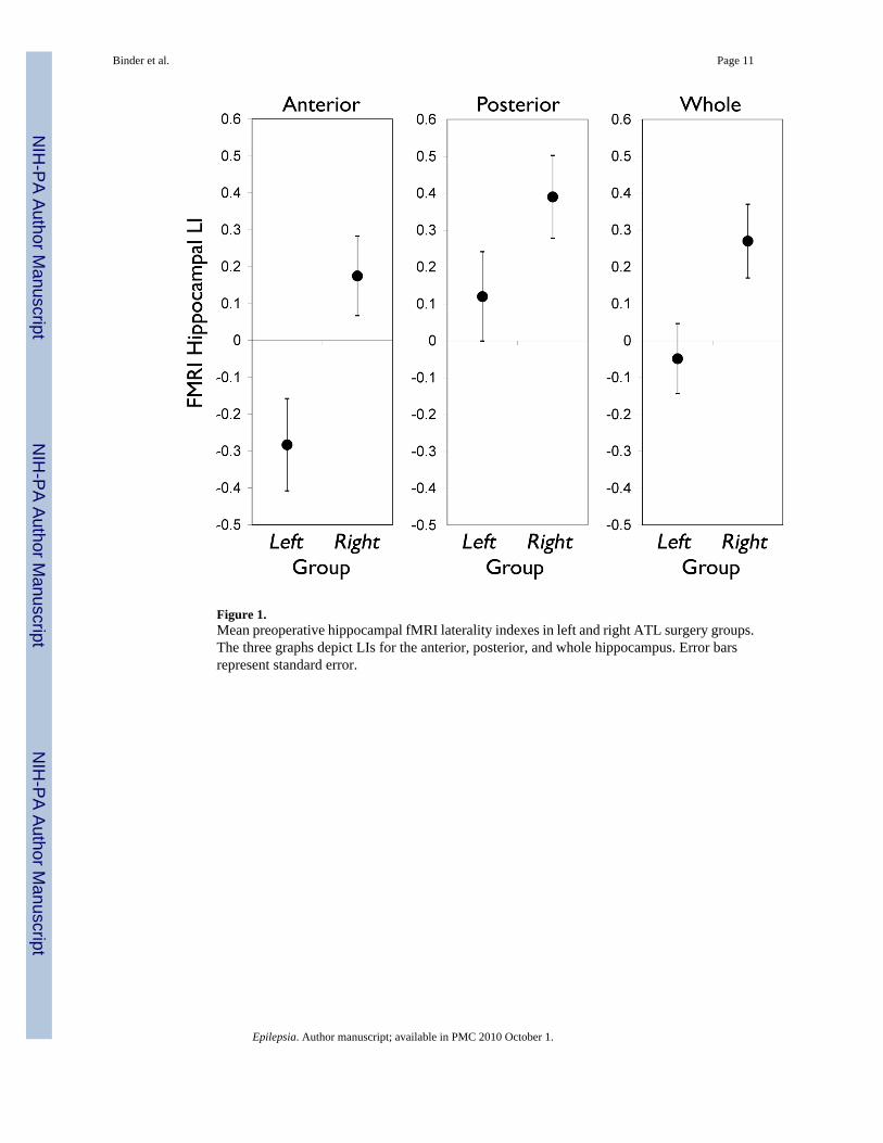
Figure 1.Mean preoperative hippocampal fMRI laterality indexes in left and right ATL surgery groups.The three graphs depict LIs for the anterior, posterior, and whole hippocampus. Error barsrepresent standard error.
Binder et al. Page 11
Epilepsia. Author manuscript; available in PMC 2010 October 1.
NIH
-PA Author Manuscript
NIH
-PA Author Manuscript
NIH
-PA Author Manuscript
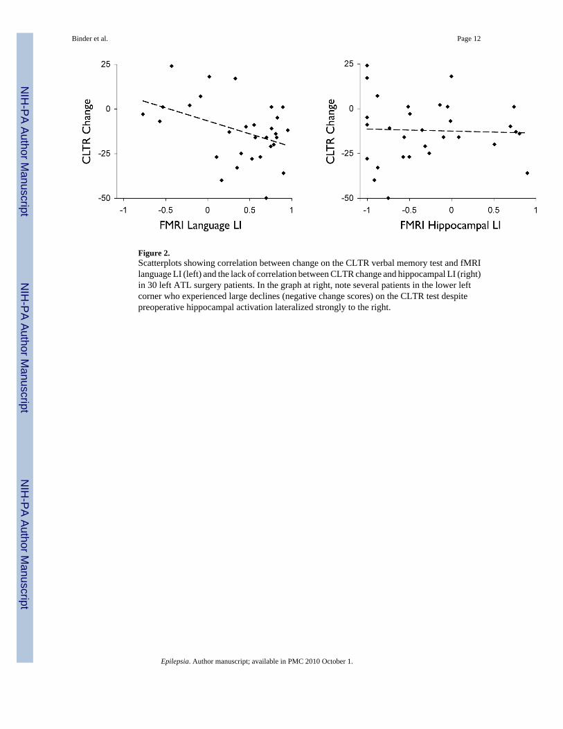
Figure 2.Scatterplots showing correlation between change on the CLTR verbal memory test and fMRIlanguage LI (left) and the lack of correlation between CLTR change and hippocampal LI (right)in 30 left ATL surgery patients. In the graph at right, note several patients in the lower leftcorner who experienced large declines (negative change scores) on the CLTR test despitepreoperative hippocampal activation lateralized strongly to the right.
Binder et al. Page 12
Epilepsia. Author manuscript; available in PMC 2010 October 1.
NIH
-PA Author Manuscript
NIH
-PA Author Manuscript
NIH
-PA Author Manuscript
NIH
-PA Author Manuscript
NIH
-PA Author Manuscript
NIH
-PA Author Manuscript
Binder et al. Page 13
Table 1
Patient data.
Left ATL Right ATL
Mean (SD) Mean (SD)
n 30 37
Age at Surgery, y 38.0 (10.8) 39.5 (11.4)
Sex, F/M 16/14 19/18
Education, y 13.1 (1.9) 13.9 (3.0)
Handedness, R/L/A 22/7/1 30/4/3
Age at Epilepsy Onset, y 18.4 (11.2) 15.5 (11.6)
Epilepsy Duration, y 19.7 (13.5) 24.2 (14.4)
Full Scale IQ 93.3 (13.0) 94.5 (12.2)
Verbal IQ 92.2 (11.5) 95.0 (13.2)
Performance IQ 96.6 (14.6) 94.9 (12.5)
F = female, M = male, R = right-handed, L = left-handed, A = ambidextrous, y = years
Epilepsia. Author manuscript; available in PMC 2010 October 1.
NIH
-PA Author Manuscript
NIH
-PA Author Manuscript
NIH
-PA Author Manuscript
Binder et al. Page 14
Table 2
Verbal memory test scores.
Left ATL Right ATL L vs. R
Mean (SD) Mean (SD) p
CLTR Pre-op 27.2 (14.5) 30.8 (14.3) n.s.
Post-op 14.9 (11.5) 40.8 (13.8) <0.0001
Change −12.3 (17.2)§ 10.0 (14.6)§ <0.0001
Number (%) Declining 16 (53%) 2 (5%)
Delayed Recall Pre-op 6.03 (2.82) 6.82 (2.94) n.s.
Post-op 2.90 (2.50) 8.11 (2.76) <0.0001
Change −3.13 (3.13)¶ 1.36 (2.85)* <0.0001
Number (%) Declining 17 (57%) 2 (6%)
CLTR = consistent long-term recall score from the Selective Reminding Test.
CLTR data based on 30 left and 37 right ATL patients.
Delayed Recall data based on 30 left and 33 right ATL patients.
Change scores significantly different from zero:
*p < 0.01,
§p < .001,
¶p < .0001
Epilepsia. Author manuscript; available in PMC 2010 October 1.
NIH
-PA Author Manuscript
NIH
-PA Author Manuscript
NIH
-PA Author Manuscript
Binder et al. Page 15
Table 3
Correlations between fMRI language LIs and verbal memory change scores in left ATL resection patients.
CLTR Delayed Recall
R p R p
FMRI Lateral LI −.392 .020 −.313 .077
FMRI Frontal LI −.413 .013 −.322 .067
FMRI Temporal LI −.227 n.s. −.290 n.s.
Epilepsia. Author manuscript; available in PMC 2010 October 1.
Copyright © 2022 FDOKUMEN