Minicraniotomy for Standard Temporal Lobectomy: A Minimally Invasive Surgical Approach
8
Clinical Study Minicraniotomy for Standard Temporal Lobectomy: A Minimally Invasive Surgical Approach Faisal Al-Otaibi, 1 Monirah Albloushi, 2 and Saleh Baeesa 3 1 Division of Neurosurgery, Neuroscience Department, King Faisal Specialist Hospital and Research Center, P.O. Box 3354, Riyadh 11211, Saudi Arabia 2 College of Nursing, King Saud University, Riyadh, Saudi Arabia 3 Division of Neurosurgery, King Abdulaziz University, Jeddah, Saudi Arabia Correspondence should be addressed to Faisal Al-Otaibi; [email protected] Received 5 November 2013; Accepted 29 December 2013; Published 6 February 2014 Academic Editors: C.-Y. Hsu and N. Rainov Copyright © 2014 Faisal Al-Otaibi et al. is is an open access article distributed under the Creative Commons Attribution License, which permits unrestricted use, distribution, and reproduction in any medium, provided the original work is properly cited. Introduction. e common surgical approach for standard temporal lobectomy is a question-mark skin incision and a fron- totemporal craniotomy. Herein, we describe minicraniotomy approach through a linear skin incision for standard temporal lobectomy. Methods. A retrospective observational cohort study was conducted for a group of consecutive 21 adult patients (group I) who underwent minicraniotomy for standard temporal lobectomy utilizing a linear skin incision. is group was compared to a consecutive 17 adult patients (group II) who previously underwent a reverse question-mark skin incision and standard frontotemporal craniotomy. Results. e mean age was 29 and 23 for groups I and II, respectively. e mean estimated blood loss was 190 mL and 280 mL in groups I and II, respectively ( = 0.019). ree patients in group II developed chronic postcraniotomy headache compared to none in group I. Cosmetic outcome was excellent in group I while 4 patients in group II developed disfiguring depression at lateral sphenoid wing and anterior temple. In group I 17 out of 21 became seizure-free at one-year followup. Conclusion. Minicraniotomy through a linear skin incision is a sufficient surgical approach for effective standard temporal lobectomy and it has an excellent cosmetic outcome. 1. Introduction Several modifications have been made to the surgical tech- niques and methods used to treat temporal lobe epilepsy over the last 50 years [1–4]. Performing a standard anterior temporal lobectomy consists of resecting the lateral temporal and mesial temporal structures, either en bloc or separately as popularized by Penfield [1]. e anteromedial temporal resection technique was developed by Spencer to preserve lateral temporal cortex function and to access the mesial tem- poral structures through the temporal pole corridor [5]. Both procedures are done traditionally through a question-mark skin incision and frontotemporal craniotomy [6]. Conversely, selective transcortical amygdalohippocampectomy is done mainly through a smaller question-mark or vertical temporal skin incision and temporal minicraniotomy [7]. A minimal- ly invasive neurosurgical approach has shown a benefit in reducing morbidity and producing better cosmetic results [8– 10]. Several modifications to temporal lobe resective surgery have been based either on resection of the epileptogenic zone, assisted by the use of electrocorticography and cortical map- ping to avoid functional deficits, or on resection of the seizure onset zone, as with selective amygdalohippocampectomy. In this cohort study, we report a minimally invasive surgical technique for standard temporal lobectomy and its outcome. 2. Methods 2.1. Study Design. A total of 21 consecutive patients (group I) who had undergone a modified minimally invasive surgical approach for standard temporal lobectomy (from March 2011 till December 2012) were retrospectively analyzed. is group of patients was compared to a group of 17 consecutive Hindawi Publishing Corporation ISRN Neurology Volume 2014, Article ID 532523, 7 pages http://dx.doi.org/10.1155/2014/532523
Transcript of Minicraniotomy for Standard Temporal Lobectomy: A Minimally Invasive Surgical Approach

Clinical StudyMinicraniotomy for Standard Temporal Lobectomy:A Minimally Invasive Surgical Approach
Faisal Al-Otaibi,1 Monirah Albloushi,2 and Saleh Baeesa3
1 Division of Neurosurgery, Neuroscience Department, King Faisal Specialist Hospital and Research Center,P.O. Box 3354, Riyadh 11211, Saudi Arabia
2 College of Nursing, King Saud University, Riyadh, Saudi Arabia3 Division of Neurosurgery, King Abdulaziz University, Jeddah, Saudi Arabia
Correspondence should be addressed to Faisal Al-Otaibi; [email protected]
Received 5 November 2013; Accepted 29 December 2013; Published 6 February 2014
Academic Editors: C.-Y. Hsu and N. Rainov
Copyright © 2014 Faisal Al-Otaibi et al.This is an open access article distributed under the Creative CommonsAttribution License,which permits unrestricted use, distribution, and reproduction in any medium, provided the original work is properly cited.
Introduction. The common surgical approach for standard temporal lobectomy is a question-mark skin incision and a fron-totemporal craniotomy. Herein, we describe minicraniotomy approach through a linear skin incision for standard temporallobectomy.Methods. A retrospective observational cohort study was conducted for a group of consecutive 21 adult patients (groupI) who underwent minicraniotomy for standard temporal lobectomy utilizing a linear skin incision. This group was comparedto a consecutive 17 adult patients (group II) who previously underwent a reverse question-mark skin incision and standardfrontotemporal craniotomy. Results.The mean age was 29 and 23 for groups I and II, respectively. The mean estimated blood losswas 190mL and 280mL in groups I and II, respectively (𝑃 = 0.019). Three patients in group II developed chronic postcraniotomyheadache compared to none in group I. Cosmetic outcomewas excellent in group Iwhile 4 patients in group II developed disfiguringdepression at lateral sphenoidwing and anterior temple. In group I 17 out of 21 became seizure-free at one-year followup.Conclusion.Minicraniotomy through a linear skin incision is a sufficient surgical approach for effective standard temporal lobectomy and it hasan excellent cosmetic outcome.
1. Introduction
Several modifications have been made to the surgical tech-niques and methods used to treat temporal lobe epilepsyover the last 50 years [1–4]. Performing a standard anteriortemporal lobectomy consists of resecting the lateral temporaland mesial temporal structures, either en bloc or separatelyas popularized by Penfield [1]. The anteromedial temporalresection technique was developed by Spencer to preservelateral temporal cortex function and to access themesial tem-poral structures through the temporal pole corridor [5]. Bothprocedures are done traditionally through a question-markskin incision and frontotemporal craniotomy [6]. Conversely,selective transcortical amygdalohippocampectomy is donemainly through a smaller question-mark or vertical temporalskin incision and temporal minicraniotomy [7]. A minimal-ly invasive neurosurgical approach has shown a benefit in
reducingmorbidity and producing better cosmetic results [8–10]. Several modifications to temporal lobe resective surgeryhave been based either on resection of the epileptogenic zone,assisted by the use of electrocorticography and cortical map-ping to avoid functional deficits, or on resection of the seizureonset zone, as with selective amygdalohippocampectomy. Inthis cohort study, we report a minimally invasive surgicaltechnique for standard temporal lobectomy and its outcome.
2. Methods
2.1. Study Design. A total of 21 consecutive patients (group I)who had undergone a modified minimally invasive surgicalapproach for standard temporal lobectomy (from March2011 till December 2012) were retrospectively analyzed. Thisgroup of patients was compared to a group of 17 consecutive
Hindawi Publishing CorporationISRN NeurologyVolume 2014, Article ID 532523, 7 pageshttp://dx.doi.org/10.1155/2014/532523

2 ISRN Neurology
(a)
(b) (c)
Figure 1: Intraoperative photographs depicting the linear temporal skin incision andminicraniotomy that was utilized in group I (a) and (b).Postoperative computerized tomography of the brain showing the size of minicraniotomy (c).
patients (group II) who had undergone conventional reversequestion-mark skin incision and standard frontotemporalcraniotomy for temporal lobectomy. Institutional reviewboard approval was obtained for the retrospective reviewof consecutive patient records pertinent to the study. Thefollowing variables were evaluated in both groups: surgerytime, estimated blood loss, extent of resection, chronicpostcraniotomy pain (persistent headache at one-year fol-lowup), cosmetic effect, seizure outcome, and surgical com-plications. Since this study is a descriptive of a surgicalapproach technique, the adverse effect of surgery such asthe effect on memory and visual field functions was notincluded in this study.The extent of resection of the temporalneocortex and mesial temporal structures was measured byfollow-up magnetic resonance imaging (MRI). In group I,the follow-up MRI result was compared to the intraoperativeneuronavigation image guidance estimate of resection. Thefollow-up period was a minimum of one year (range 1–7years).
2.2. Minicraniotomy Surgical Technique for Standard Tempo-ral Lobectomy. In this section, we describe the linear skinincision and minicraniotomy surgical approach for standardtemporal lobectomy, Figure 1. The procedure is usually per-formed with the patient in the supine position, elevatingthe ipsilateral shoulder with a roll and rotating the head tothe contralateral side. The head is tilted slightly laterally toplace the zygoma at an approximately 10-degree angle from
the horizontal plane of the surgical floor. Neuronavigationis applied for image guidance throughout the procedure. Toavoid injury to the frontalis branch of the facial nerve, thelinear skin incision is begun 1 cm above the zygoma and1 cm anterior to the tragus and extended vertically 6–8 cm toapproximately the level of superior temporal line. The super-ficial temporal artery is dissected and preserved as much aspossible. Sharp dissection is used to open temporalis muscleand electrocautery is avoided asmuch as possible tominimizethe subsequent atrophy of the muscle. Minicraniotomy atthe temporal area is carried out with size of 3 cm using afast drill and one or two burr holes, Figure 1. The upperedge of the minicraniotomy is at the sylvian fissure levelguided by neuronavigation. A U-shaped durotomy incisionis performed with the base reflected anteriorly. The posteriorextent of neocortical resection is guided by neuronavigationand direct measurement from the temporal tip at the levelof the middle temporal gyrus. The posterior resection line isplaced at 4 cm on the nondominant temporal lobe and 3 cmon the dominant temporal lobe at the level of the middletemporal gyrus. The posterior resection is slanted anteriorlyacross the superior temporal gyrus to avoid the primaryauditory cortex. More posterior resection is usually done atthe inferior temporal gyrus and less resection at the superiortemporal gyrus.
The microscope is then introduced to the surgical field,and a subpial dissection is done at the superior margin ofthe superior temporal gyrus using an ultrasonic aspirator

ISRN Neurology 3
exposing the sylvian fissure down to the insular cortex.The insula is exposed, and dissection extending to thelateral uncus is performed. The temporal pole is reflectedlaterally after the coagulation and division of the anteriorleptomeninges. The posterior resection line is extended fromthe superior gyrus through the middle gyrus and into theinferior temporal gyrus. This line is then extended mediallythrough the fusiform gyrus to the collateral sulcus. Thetemporal horn is entered through the white matter abovethe fusiform gyrus. The wall of the temporal horn can beidentified by the bluish ependyma. Subsequently, openingof the ventricle anteriorly exposes the hippocampal head.The temporal stem is resected at the inferior circular sulcus.The temporal neocortex is removed by dividing the basalleptomeninges lateral to the temporal horn exposure.
The mesial temporal structures are resected using anultrasonic aspirator from anterior to posterior. No retractionis used during this step. In our practice, the posterior portionof the hippocampus is removed using an ultrasonic aspiratorto the level of the midbrain tectum, as identified by imageguidance, Figure 2. Next, homeostasis is secured, and woundclosure is performed in a standard manner, Figure 3.
2.3. Statistical Analysis. Data were entered and analyzedusing the SPSS 17th edition (Chicago, IL, USA).The nonpara-metric Mann-Whitney test was used to compare differencesbetween estimated blood loss in both groups. Statistic signif-icance was determined if P value <0.05.
3. Results
Themean agewas 29.2 years in group I and 23.6 years in groupII. The surgical opening average time from skin to dura was20 minutes, and the closure average time from dura to skinwas about 30 minutes in group I, Table 1. The opening andclosure times were not calculated for group II. The overalloperative time from skin to skin was on average 3 hours and20 minutes in group I and 3 hours and 40 minutes in groupII. The mean estimated blood loss was 190mL and 280mL ingroups I and II, respectively (𝑃 value = 0.019). The averagelength of hospital stay was 4 days for group I and 4.5 days forgroup II.The cosmetic result was excellent in group I with nopresence of wide scar formation. In group II, four patientshad a disfiguring depression at the anterior temporal area.Chronic postcraniotomy pain at the surgical site occurred inthree patients in group II and none of the patients in group I.Out of those three patients, one had continuous local pain atthe surgical site and twohad chronic intermittent hemicranialheadaches.
The extent of resection of the temporal neocortex andmesial structures measured on follow-up MRI was almostsimilar in both groups. The extent of posterior hippocampusresection was found to be at the level of the quadrigeminalplate or posterior to it, Figure 2. Intraoperative neuronavi-gation image guidance was found to overestimate the extentof hippocampus posterior resection by an average of 16mm,Figure 4. In terms of seizure freedom, therewas no significantdifference between the groups. In group I, 17 out of 21 patients
(a)
(b)
Figure 2: Postoperative MRI brain (axial and coronal T2 weightedimages) showing the extent of neocortex (a) and posterior hip-pocampus resection (b). Note: the posterior resection beyond thelevel of brainstem folliculi (quadrigeminal plate level) (see redarrow).
(80.9%) became seizure-free at followup of one year or more.In group II, 12 out of 17 patients (70.6%) became seizure-free,taking into consideration that bitemporal epilepsy was foundin three patients in group II and only two patients in group I.Complications included one patient with a superficial woundinfection and one with transient third nerve partial palsy ingroup II. In group I, one patient developed transient limita-tion of mouth opening ability associated with intermittentpain at the temporomandibular joint area, and one patientdeveloped a small focal hair loss posterior to the wound,which was related to skin retraction that subsided after six-month followup. Table 2 summarizes different variables in thetwo patient groups. Figure 5 demonstrates a case illustrationof a redo temporal lobectomy and lesionectomy utilizingsmall access craniotomy and comparing that to the previouslyused craniotomy access.
4. Discussion
Minimally invasive surgical approaches are widely used byneurosurgeons. The main advantages are shorter operativetimes, less surgical trauma, shorter hospitalization times,less chance of postoperative pain, and achieving of excel-lent cosmetic results [11]. Surgical approaches for tempo-ral lobectomy to treat medically intractable epilepsy haveseveral technical variations. These include standard tem-poral lobectomy, anteromedial temporal lobectomy, selec-tive amygdalohippocampectomy, and stereotactic approaches[2, 5, 12, 13]. Penfield popularized standard temporal lo-bectomy using a large reverse question-mark skin inci-sion and frontotemporal craniotomy and bone removal of

4 ISRN Neurology
(a) (b)
Figure 3: Intraoperative photos demonstrating neocortex specimen (a) and temporalis muscle fascia closure (b).
Table 1: Summary of group I patient data.
No. AgeSex Pathology Surgery
side
Extent of neocorticalresection fromtemporal pole]
Extent of posteriorhippocampusresection
Cosmeticresult
Seizure outcome.(Engel classification)
Surgicalcomplications
1 27 F MTS𝜙 Rt 4 cm QP levelΔ + Class I None2 31 M Normal Rt 4 cm QP level + Class II None3 26 M MTS Rt 4.5 cm Post. to QP + Class I None4 42 M Cavernoma Rt 4 cm Post. to QP + Class I None5 17 F MTS Lt 3.5 cm QP level + Class I None6 20 F MTS Lt 3 cm QP level + Class I None
7 38 M MTS Rt 4 cm QP level + Class I Mouth openinglimitation
8 22 F Ganglioglioma Lt 4 cm Post. to QP + Class I None9 18 F MTS Rt 4 cm Post. to QP + Class I None10 32 F MTS Rt 4.5 cm QP level + Class I None11 24 F MTS Lt 3.7 cm Post. to QP + Class I None12 44 M MTS Lt 3.5 cm QP level + Class I None13 21 M MTS Rt 3.8 cm Post. to QP + Class II None14 28 F MTS Rt 4 cm QP level + Class I None15 18 F MTS Lt 3.5 cm QP level + Class I None16 29 M Normal Rt 3.8 cm QP level + Class I Focal alopecia
17 31 F Gangliogliomaand MTS Lt 3.6 cm Post. to QP + Class I None
18 27 F Neocorticalastrogliosis Lt 3 cm Post. to QP + Class II None
19 19 M Normal Rt 4.5 cm QP level + Class I None
20 24 M Low gradeglioma Rt 4 cm Post. to QP + Class II None
21 36 M MTS Rt 4 cm QP level + Class I None]Measurement is based on postoperative MRI brain from temporal pole at the level of middle temporal gyrus.ΔQP: quadrigeminal plate.+: no postoperative disfiguring features.𝜙MTS: Mesial temporal sclerosis.
ISRN Neurology 5
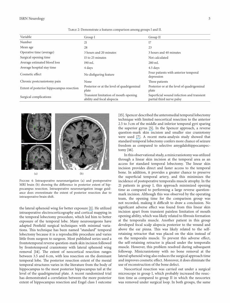
Table 2: Demonstrate a features comparison among groups I and II.
Variable Group I Group IINumber 21 17Mean age 28 23Operative time (average) 3 hours and 20 minutes 3 hours and 40 minutesSurgical opening time 15 to 25 minutes Not calculatedAverage estimated blood loss 190mL 280mLAverage hospital stay time 4 days 4.5 days
Cosmetic effect No disfiguring feature Four patients with anterior temporaldepression
Chronic postcraniotomy pain None Three patients
Extent of posterior hippocampus resection Posterior or at the level of quadrigeminalplate
Posterior or at the level of quadrigeminalplate
Surgical complications Transient limitation of mouth openingability and focal alopecia
Superficial wound infection and transientpartial third nerve palsy
(a) (b)
Figure 4: Intraoperative neuronavigation (a) and postoperativeMRI brain (b) showing the difference in posterior extent of hip-pocampus resection. Intraoperative neuronavigation image guid-ance does overestimate the extent of posterior resection due tointraoperative brain shift.
the lateral sphenoid wing for better exposure [1]. He utilizedintraoperative electrocorticography and cortical mapping inthe temporal lobectomy procedure, which led him to betterexposure of the temporal lobe. Many neurosurgeons haveadapted Penfield surgical techniques with minimal varia-tions. This technique has been named “standard” temporallobectomy because it is a reproducible procedure and varieslittle from surgeon to surgeon. Most published series used afrontotemporal reverse question-mark skin incision followedby frontotemporal craniotomy with lateral sphenoid wingremoval [14]. The extent of neocortical resection rangesbetween 3.5 and 6 cm, with less resection on the dominanttemporal lobe. The posterior resection extent of the mesialtemporal structures varies in the literature from the body ofhippocampus to the most posterior hippocampus tail at thelevel of the quadrigmeinal plate. A recent randomized trialhas demonstrated a correlation between the more posteriorextent of hippocampus resection and Engel class I outcome
[15]. Spencer described the anteromedial temporal lobectomytechnique with limited neocortical resection to the anterior2.5 to 3 cm of the middle and inferior temporal gyri sparingthe superior gyrus [5]. In the Spencer approach, a reversequestion-mark skin incision and smaller size craniotomywere used [7]. A recent meta-analysis study showed thatstandard temporal lobectomy confers more chance of seizurefreedom as compared to selective amygdalohippocampec-tomy [16].
In this observational study, aminicraniotomywas utilizedthrough a linear skin incision at the temporal area as anaccess for standard temporal lobectomy. The linear skinincision provides direct and faster access to the temporalbone. In addition, it provides a greater chance to preservethe superficial temporal artery, and this minimizes theincidence of postoperative temporalis muscle atrophy. In the21 patients in group I, this approach minimized openingtime as compared to performing a large reverse question-mark incision. Although this was observed by the operatingteam, the opening time for the comparison group wasnot recorded, making it difficult to draw a conclusion. Nosignificant adverse effect was found from this linear skinincision apart from transient painless limitation of mouthopening ability, which was likely related to fibrosis formationat the temporalis muscle. Another patient in this groupdeveloped focal scalp alopecia posterior to the wound andabove the ear pinna. This was likely related to the self-retaining retractor that was placed on the skin instead ofon the temporalis muscle. To prevent this adverse effect,the self-retaining retractor is placed under the temporalismuscle. However, this problem resolved during subsequentfollowup. Minicraniotomy with no bone removal at thelateral sphenoid wing also reduces the surgical approach timeand improves cosmetic effect. Moreover, it does eliminate theuse of reconstruction of the bone defect.
Neocortical resection was carried out under a surgicalmicroscope in group I, which probably increased the resec-tion time as compared to group II in which the neocortexwas removed under surgical loop. In both groups, the same
6 ISRN Neurology
(a) (b) (c)
(d) (e)
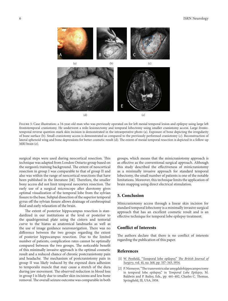
Figure 5: Case illustration: a 34-year-old man who was previously operated on for left mesial temporal lesion and epilepsy using large leftfrontotemporal craniotomy. He underwent a redo lesionectomy and temporal lobectomy using smaller craniotomy access. Large fronto-temporal reverse question-mark skin incision is demonstrated in the intraoperative photo (a). Exposure of bone depicting the irregularityof bone surface (b). Small craniotomy access is demonstrated as compared to the previously performed craniotomy (c). Reconstruction oflateral sphenoid wing and bone depressions for better cosmetic result (d). The extent of mesial temporal resection is depicted in a follow-upMRI brain (e).
surgical steps were used during neocortical resection. Thistechnique was adapted from LondonOntario group based onthe surgeon’s training background. The extent of neocorticalresection in group I was comparable to that of group II andalso was within the range of neocortical resections that havebeen published in the literature [14]. Therefore, the smallerbony access did not limit temporal neocortex resection. Theearly use of a surgical microscope after durotomy givesoptimal visualization of the temporal lobe from the sylvianfissure to the base. Subpial dissection of the superior temporalgyrus off the sylvian fissure allows drainage of cerebrospinalfluid and early relaxation of the brain.
The extent of posterior hippocampus resection is stan-dardized in our institutions at the level or posterior tothe quadrigeminal plate using the cistern and tentorialcurve to the hiatus as anatomical landmarks as well asthe use of image guidance neuronavigation. There was nodifference between the two groups regarding the extentof posterior hippocampus resection. Due to the limitednumber of patients, complication rates cannot be optimallycompared between the two groups. The noticeable benefitof this minimally invasive approach is the optimal cosmeticresult and a reduced chance of chronic postcraniotomy painand headache. The mechanism of postcraniotomy pain ingroup II was likely induced by the exposed dura adhesionto temporalis muscle that may cause a stretch of the duraduring jaw movement. The observed reduction in blood lossin group I is likely due to smaller skin incisions and less boneremoval.The overall seizure outcomewas comparable in both
groups, which means that the minicraniotomy approach isas effective as the conventional surgical approach. Althoughthis study described the effectiveness of minicraniotomyas a minimally invasive approach for standard temporallobectomy, the small number of patients is one of the notablelimitations. Moreover, this technique limits the application ofbrain mapping using direct electrical stimulation.
5. Conclusion
Minicraniotomy access through a linear skin incision forstandard temporal lobectomy is aminimally invasive surgicalapproach that has an excellent cosmetic result and is aneffective technique for temporal lobe epilepsy treatment.
Conflict of Interests
The authors declare that there is no conflict of interestsregarding the publication of this paper.
References
[1] W. Penfield, “Temporal lobe epilepsy,” The British Journal ofSurgery, vol. 41, no. 168, pp. 337–343, 1954.
[2] P.Niemeyer, “The transventricular amygdalohippocampectomyin temporal lobe epilepsy,” in Temporal Lobe Epilepsy, M.Baldwin and P. Bailey, Eds., pp. 461–482, Charles C. Thomas,Springfield, Ill, USA, 1958.
ISRN Neurology 7
[3] M. R. Sperling, M. J. O’Connor, A. J. Saykin, and C. Plummer,“Temporal lobectomy for refractory epilepsy,” Journal of theAmericanMedical Association, vol. 276, no. 6, pp. 470–475, 1996.
[4] A. Olivier, “Surgical techniques in temporal lobe epilepsy,”Clin-ical Neurosurgery, vol. 44, pp. 211–241, 1997.
[5] D. D. Spencer, S. S. Spencer, and R. H. Mattson, “Access to theposterior medial temporal lobe structures in the surgical treat-ment of temporal lobe epilepsy,”Neurosurgery, vol. 15, no. 5, pp.667–671, 1984.
[6] F. Al-Otaibi, S. S. Baeesa, A. G. Parrent, J. P. Girvin, and D.Steven, “Surgical techniques for the treatment of temporal lobeepilepsy,” Epilepsy Research and Treatment, vol. 2012, Article ID374848, 13 pages, 2012.
[7] D. Spencer and K. Burchiel, “Selective amygdalohippocampec-tomy,” Epilepsy Research and Treatment, vol. 2012, Article ID382095, 8 pages, 2012.
[8] J.-Z. Zhao, “Minimally invasive neurosurgery: the technicalplatform for translational medicine,” Chinese Medical Journal,vol. 122, no. 20, pp. 2403–2404, 2009.
[9] K. Mursch, T. Gotthardt, R. Kroger, M. Bublat, and J. Behnke-Mursch, “Minimally invasive neurosurgery within a 0.5 teslaintraoperative magnetic resonance scanner using an off-lineneuronavigation system,”Minimally Invasive Neurosurgery, vol.48, no. 4, pp. 213–217, 2005.
[10] E. Ferrer, “Minimally invasive neurosurgery,” in Risk Controland Quality Management in Neurosurgery, vol. 78 of ActaNeurochirurgica Supplements, pp. 107–110, Springer, Vienna,Austria, 2001.
[11] R. Reisch and A. Perneczky, “Ten-year experience with thesupraorbital subfrontal approach through an eyebrow skinincision,” Neurosurgery, vol. 57, no. 4, supplement, pp. 242–253,2005.
[12] A. Olivier, “Transcortical selective amygdalohippocampectomyin temporal lobe epilepsy,” Canadian Journal of NeurologicalSciences, vol. 27, supplement 1, pp. S68–S76, 2000.
[13] H.G.Wieser andM.G. Yasargil, “Selective amygdalohippocam-pectomy as a surgical treatment of mesiobasal limbic epilepsy,”Surgical Neurology, vol. 17, no. 6, pp. 445–457, 1982.
[14] W. Bingaman and I. Najm, “Standard temporal lobectomy,”in Youmans Neurological Surgery, R. Winn, Ed., pp. 767–773,Elsevier Saunders, Philadelphia, Pa, USA, 2011.
[15] J. Schramm, T. N. Lehmann, J. Zentner et al., “Random-ized controlled trial of 2.5-cm versus 3.5-cm mesial temporalresection—part 2: volumetric resection extent and subgroupanalyses,”ActaNeurochirurgica, vol. 153, no. 2, pp. 221–228, 2011.
[16] C. B. Josephson, J. Dykeman, K. M. Fiest et al., “Systematicreview and meta-analysis of standard versus selective temporallobe epilepsy surgery,” Neurology, vol. 80, no. 18, pp. 1669–1676,2013.

Submit your manuscripts athttp://www.hindawi.com
Stem CellsInternational
Hindawi Publishing Corporationhttp://www.hindawi.com Volume 2014
Hindawi Publishing Corporationhttp://www.hindawi.com Volume 2014
MEDIATORSINFLAMMATION
of
Hindawi Publishing Corporationhttp://www.hindawi.com Volume 2014
Behavioural Neurology
International Journal of
EndocrinologyHindawi Publishing Corporationhttp://www.hindawi.com
Volume 2014
Hindawi Publishing Corporationhttp://www.hindawi.com Volume 2014
Disease Markers
BioMed Research International
Hindawi Publishing Corporationhttp://www.hindawi.com Volume 2014
OncologyJournal of
Hindawi Publishing Corporationhttp://www.hindawi.com Volume 2014
Hindawi Publishing Corporationhttp://www.hindawi.com Volume 2014
Oxidative Medicine and Cellular Longevity
PPARRe sea rch
Hindawi Publishing Corporationhttp://www.hindawi.com Volume 2014
The Scientific World JournalHindawi Publishing Corporation http://www.hindawi.com Volume 2014
Immunology ResearchHindawi Publishing Corporationhttp://www.hindawi.com Volume 2014
Journal of
ObesityJournal of
Hindawi Publishing Corporationhttp://www.hindawi.com Volume 2014
Hindawi Publishing Corporationhttp://www.hindawi.com Volume 2014
Computational and Mathematical Methods in Medicine
OphthalmologyJournal of
Hindawi Publishing Corporationhttp://www.hindawi.com Volume 2014
Diabetes ResearchJournal of
Hindawi Publishing Corporationhttp://www.hindawi.com Volume 2014
Hindawi Publishing Corporationhttp://www.hindawi.com Volume 2014
Research and TreatmentAIDS
Hindawi Publishing Corporationhttp://www.hindawi.com Volume 2014
Gastroenterology Research and Practice
Parkinson’s DiseaseHindawi Publishing Corporationhttp://www.hindawi.com Volume 2014
Evidence-Based Complementary and Alternative Medicine
Volume 2014Hindawi Publishing Corporationhttp://www.hindawi.com