Comparison of MRI Visualization Following Minimally Invasive ...
Upload
khangminh22Category
view
0download
0
Citation: Melo, D.N.; Lima, G.R.P.;
Fernandes, C.G.; Teixeira, A.C.;
Filho, J.B.; Araújo, F.M.C.; Araújo,
L.C.; Siqueira, A.M.; Farias, L.A.B.G.;
Monteiro, R.A.A.; et al. Post-Mortem
Diagnosis of Pediatric Dengue Using
Minimally Invasive Autopsy during
the COVID-19 Pandemic in Brazil.
Trop. Med. Infect. Dis. 2022, 7, 123.
https://doi.org/10.3390/
tropicalmed7070123
Academic Editor: Yannick Simonin
Received: 17 May 2022
Accepted: 24 June 2022
Published: 30 June 2022
Publisher’s Note: MDPI stays neutral
with regard to jurisdictional claims in
published maps and institutional affil-
iations.
Copyright: © 2022 by the authors.
Licensee MDPI, Basel, Switzerland.
This article is an open access article
distributed under the terms and
conditions of the Creative Commons
Attribution (CC BY) license (https://
creativecommons.org/licenses/by/
4.0/).
Tropical Medicine and
Infectious Disease
Case Report
Post-Mortem Diagnosis of Pediatric Dengue Using MinimallyInvasive Autopsy during the COVID-19 Pandemic in BrazilDeborah N. Melo 1,2, Giovanna R. P. Lima 3 , Carolina G. Fernandes 3, André C. Teixeira 1,3,4, Joel B. Filho 1,Fernanda M. C. Araújo 5 , Lia C. Araújo 6, André M. Siqueira 7, Luís A. B. G. Farias 8, Renata A. A. Monteiro 9,Jaume Ordi 10,11 , Miguel J. Martinez 11 , Paulo H. N. Saldiva 9 and Luciano P. G. Cavalcanti 2,3,12,*
1 Serviço de Verificação de Óbitos Dr Rocha Furtado, Fortaleza 60842-395, Brazil;[email protected] (D.N.M.); [email protected] (A.C.T.); [email protected] (J.B.F.)
2 Programa de Pós-graduação em Patologia, Universidade Federal do Ceará, Fortaleza 60020-181, Brazil3 Faculdade de Medicina, Centro Universitário Christus, Fortaleza 60190-180, Brazil;
[email protected] (G.R.P.L.); [email protected] (C.G.F.)4 Argos Laboratory, Fortaleza 60175-047, Brazil5 Fundação Oswaldo Cruz, Eusebio 61760-000, Brazil; [email protected] Programa de Residencia Medica em Patologia pela Universidade Federal do Ceará, 60,
Fortaleza 60020-181, Brazil; [email protected] Fundação Oswaldo Cruz, Rio de Janeiro 21040-360, Brazil; [email protected] Hospital São José de Doenças Infecciosas, Fortaleza 60455-610, Brazil; [email protected] Departamento de Patologia, Faculdade de Medicina da Universidade de São Paulo,
São Paulo 01246-903, Brazil; [email protected] (R.A.A.M.); [email protected] (P.H.N.S.)10 ISGlobal, Barcelona Institute for Global Health, Hospital Clínic, Universitat de Barcelona,
08036 Barcelona, Spain; [email protected] ISGlobal, Hospital Clínic, University of Barcelona, 08007 Barcelona, Spain; [email protected] Programa de Pós-graduação em Saúde Coletiva, Universidade Federal do Ceará, Fortaleza 60020-181, Brazil* Correspondence: [email protected]; Tel.: +55-85-999878969
Abstract: We report the first pediatric disease in which the use of minimally invasive autopsy (MIA)confirmed severe dengue as the cause of death. During the COVID-19 pandemic, a previously healthy10-year-old girl living in north-eastern Brazil presented fever, headache, diffuse abdominal pain,diarrhoea, and vomiting. On the fourth day, the clinical symptoms worsened and the patient died. AnMIA was performed, and cores of brain, lungs, heart, liver, kidneys, and spleen were collected with14G biopsy needles. Microscopic examination showed diffuse oedema and congestion, pulmonaryintra-alveolar haemorrhage, small foci of midzonal necrosis in the liver, and tubular cell necrosis inthe kidneys. Dengue virus RNA and NS1 antigen were detected in blood and cerebrospinal fluidsamples. Clinical, pathological, and laboratory findings, in combination with the absence of otherlesions and microorganisms, allowed concluding that the patient had died from complications ofsevere dengue.
Keywords: severe dengue; autopsy; minimally invasive autopsy; arbovirus; COVID-19
1. Introduction
Dengue is the most important arbovirus worldwide, causing epidemics with a high hu-man health and economic impact. Severe symptoms mainly affect the pediatric populationfrom endemic low- and middle-income countries [1,2].
In Brazil, dengue remains the most widespread disease caused by arbovirus, even afterthe introduction of Zika and chikungunya. In north-eastern Brazil, deaths from dengue arefrequent, even in non-epidemic years, especially in socially vulnerable populations [1–3].Clinically, most dengue infections are either asymptomatic or produce mild disease [1–3].However, given the high number of infections, severe cases are often reported duringepidemics and represent a challenge for diagnosis and clinical management. In fatal
Trop. Med. Infect. Dis. 2022, 7, 123. https://doi.org/10.3390/tropicalmed7070123 https://www.mdpi.com/journal/tropicalmed
Trop. Med. Infect. Dis. 2022, 7, 123 2 of 7
cases, most organs and systems are affected, particularly the heart, central nervous system,gastrointestinal tract, and kidneys.
After the establishment of Death Verification Services (DVS) in Brazil, the use ofconventional autopsy (CA), the gold standard technique for the diagnosis of deaths causedby dengue, has contributed to the detection of patients clinically not diagnosed [4] andhas significantly reduced neglected and underreported cases. However, the existence offew DVS in the cities, together with the low acceptability of CA among the relatives ofthe deceased and the lack of financial resources and specialised personnel, has resulted inlimited implementation of this procedure [5]. Thus, the application of new strategies forpost-mortem tissue collection is necessary, particularly for pediatric deaths, as rejection ofCA by the relatives is very high in this population [5–10].
Minimally invasive autopsy (MIA) has been used as an alternative to CA with promis-ing results [8,10–14]. This technique allows obtaining core biopsies of key organs bypercutaneous puncture, with or without guidance with an imaging technique. MIA hasbeen widely used in the context of the COVID-19 pandemic as a fast and non-disfiguringmethod with minimal biological risk for the personnel performing the procedure [8,9,15–17].However, current knowledge of the performance of this technique for arboviral diseases inthe paediatric population is very limited.
We report the first case of fatal dengue infection, which occurred in a previouslyhealthy 10-year-old girl living in north-eastern Brazil during the COVID-19 pandemic.MIA sampling allowed correct diagnosis and showed complete agreement with the CA.We show that this acceptable, simplified, and non-disfiguring post-mortem technique canreliably diagnose death from severe dengue.
2. Case Report
A 10-year-old girl presented with fever, headache, diffuse abdominal pain, diarrhoea,and vomiting at the end of June 2021. She was previously healthy and had no comorbidities.A previously healthy 10-year-old girl with no comorbidities presented with fever, headache,diffuse abdominal pain, diarrhoea, and vomiting at the end of June 2021. The patient wasinitially treated with dipyrone. After 24 h, the patient presented dark stools. Two dayslater her clinical condition worsened and she was admitted to an emergency care unit(ECU), in which a blood count revealed thrombocytopenia (57,000/mm3). Intravenoushydration, antipyretics, and antiemetics were administered. After 3 days, the abdominalpain worsened, and the patient developed cutaneous pallor, arterial hypotension, anddrowsiness, and was transferred to a paediatric hospital, where she arrived pale, withcold skin, thin pulse, gasping, dehydrated, and with tense abdomen. Myocarditis wasconsidered by the physician. A femoral central venous access allowed expansion withalbumin. The blood count revealed mild anaemia (haemoglobin 11.9 g/dL, haematocrit35.9%), lymphopenia (92/mm3), and thrombocytopenia (57,000/mm3 on admission, whichdropped to 20,000/mm3 within a few hours). Liver enzymes were above reference levelsduring hospitalisation (aspartate aminotransferase 741.1 U/L; alanine aminotransferase248.9 U/L). She also had altered renal function, hyperkalaemia (10 mmol/L), and severemetabolic acidosis (pH 6.7). Activated partial thromboplastin time and prothrombin timewere prolonged (Table 1). The next day, the patient suffered cardiorespiratory arrest, unre-sponsive to resuscitation measures. There was profuse bleeding through the oropharynx,trachea, and stomach. The clinical diagnoses were severe acute hepatitis of unexplainedcause, acute renal dysfunction, and shock.
The mother of the patient reported the presence of several neighbors with similarsymptoms and the recent admission of an aunt who lived with her, who had been diagnosedwith severe dengue. Neither respiratory symptoms nor recent contact with suspected orconfirmed cases of COVID-19 were described. Remarkably, co-circulation of SARS-CoV-2and dengue has recently been reported in the Americas [18].
Trop. Med. Infect. Dis. 2022, 7, 123 3 of 7
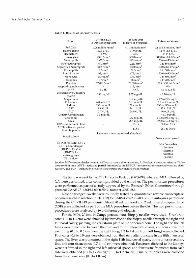
Table 1. Results of laboratory tests.
Exam 27 June 2021(3 Days of Symptom)
28 June 2021(4 Days of Symptom) Reference Values
Red Cells 4.29 million/mm3 4.11 million/mm3 4.1 to 5.3 million/mm3
Haemoglobin 12.3 g/dL 11.9 g/dL 12 to 14.5 g/dLHaematocrit 35.9% 35% 36 to 43%Leukocytes 2300/mm3 5600/mm3 3400 to 10,800/mm3
Neutrophils 1955/mm3 4424/mm3 1500 to 8500/mm3
Rod Neutrophils 69/mm3 224/mm3 0 to 860/mm3
Segmented Neutrophils 1886/mm3 4200/mm3 1500 to 8500/mm3
Eosinophils 0/mm3 56/mm3 0 to 500/mm3
Lymphocytes 92/mm3 672/mm3 1500 to 6500/mm3
Monocytes 253/mm3 336/mm3 0 to 800/mm3
Basophils 0/mm3 0/mm3 0 to 200/mm3
Platelets 57,000/mm3 20,000/mm3 150 to 450 mil/mm3
Atypical Lymphocytes - 112/mm3 0%MPV 8.3 fL 7.5 fL 9.2 to 12.6 fL
Ultrasensitive C-reactiveprotein 2.96 mg/dL 2.37 mg/dL <0.10 mg/dL
Magnesium - 2.02 mg/dL 2.02 to 2.75 mg/dLPotassium 4.0 mmol/L 6.8 mmol/L 3.5 to 5.1 mmol/L
Sodium 136 mmol/L 139 mmol/L 136 to 145 mmol/LAST 83.3 U/L 741.1 U/L 17 to 33 U/LALT 27.3 U/L 248.9 U/L 9 to 23 U/L
Urinary Urobilinogen 3.0 mg/dL - < 1.0 mg/dLCreatinine - 0.82 mg/dL 0.32 to 0.61 mg/dL
Urea - 28.9 mg/dL 19.2 to 46.2 mg/dLTAP—prothrombin time - 16.8 s 9.4 to 12.5 sAPTT- activated partial
thromboplastin - 49.8 s 25.1 to 36.5 s
Laboratory tests performed after deathBlood culture
RT-PCR for SARS-CoV-2qRT-PCR for dengue
qRT-PCR for ZikaqRT-PCR forchikungunyaNS1 antigen
No microbial growth
Not DetectablePositive
NegativeNegativePositive
Subtitle: MPV—mean platelet volume, AST—aspartate aminotransferase, ALT—alanine aminotransferase, TAP—prothrombin time, APTT—activated partial thromboplastin, RT-PCR—reverse transcriptase-polymerase chainreaction, qRT-PCR—quantitative reverse transcriptase-polymerase chain reaction.
The body was sent to the DVS Dr Rocha Furtado (DVS-RF), where an MIA followed byCA were performed, after consent provided by the mother. The post-mortem procedureswere performed as part of a study approved by the Research Ethics Committee throughprotocol CAAE 27162619.1.0000.5049, number 3,851,684.
Nasopharyngeal swabs were routinely tested by quantitative reverse transcriptase-polymerase chain reaction (qRT-PCR) for SARS-CoV-2 in all DVS-RF autopsies performedduring the COVID-19 pandemic. About 20 mL of blood and 2 mL of cerebrospinal fluid(CSF) were collected as part of the MIA procedure before the CA. The two post-mortemprocedures were analysed by two different pathologists.
For the MIA, 20 cm, 14 Gauge percutaneous biopsy needles were used. Four braincores (1.2 to 1.3 cm) were obtained by introducing the biopsy needle through the right andleft nasal cavity, piercing the cribriform plate of the sphenoid bone. The right and the leftlungs were punctured between the third and fourth intercostal spaces, and four cores fromeach lung (0.5 to 0.6 cm from the right lung; 1.2 to 1.4 cm from left lung) were collected.Four cores (0.8 to 0.9 cm) were obtained from the heart, after puncture in the fifth intercostalspace. The liver was punctured in the right 11th intercostal space, in the anterior axillaryline, and four tissue cores (0.7 to 1.0 cm) were obtained. Punctures directed to the kidneyswere performed in the right and left subcostal spaces and four tissue fragments from eachside were obtained (1.5 to 1.7 cm right, 1.0 to 1.2 cm left). Finally, four cores were collectedfrom the splenic area (0.8 to 1.0 cm).
Trop. Med. Infect. Dis. 2022, 7, 123 4 of 7
CA was performed following the DVS-RF protocol (4), after opening all cavities. Thebrain was swollen (weight 1310 g). Bilateral pleural effusion and ascites were observed.The lungs were oedematous and showed areas of haemorrhage (weight: 365 g left and375 g right). The liver and the spleen showed congestion and weighed 1200 g and 415 g,respectively. One-hundred-and-fifty milliliters of fresh blood were identified in the stomach.The kidneys were pale and oedematous (weight: 110 g right and 100 g left). The adrenalsshowed no abnormalities.
Microscopic examination showed oedema and congestion in all organs, foci of intra-alveolar haemorrhage in the lungs, and foci of midzonal necrosis in the liver. Hypoplasiaof the white pulp of the spleen was associated with abundant macrophages with large clearnuclei. There was extensive coagulative necrosis of the cortical tubules of the right kidney.
Samples of the left kidney and spleen of the MIA showed only skeletal muscle, con-nective tissue, vessels, and nerves under microscopy, with no cores of parenchyma.
The nasopharyngeal swab, blood, and CSF were sent to the Central Laboratory ofPublic Health of Ceará (LACEN-CE) for laboratory tests: qRT-PCR for respiratory viruses,arboviruses (dengue, Zika, and chikungunya), and detection of dengue NS1 antigen inblood and CSF [19]. A blood culture for bacterial research was also performed.
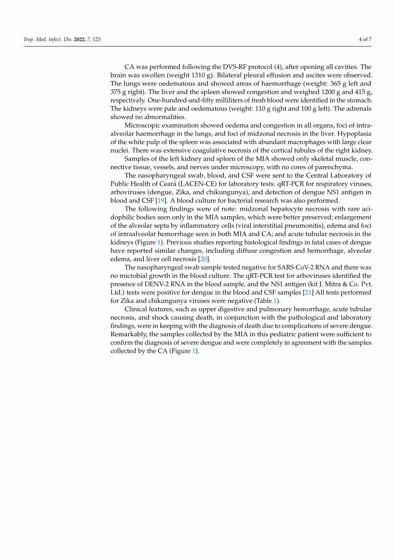
The following findings were of note: midzonal hepatocyte necrosis with rare aci-dophilic bodies seen only in the MIA samples, which were better preserved; enlargementof the alveolar septa by inflammatory cells (viral interstitial pneumonitis), edema and fociof intraalveolar hemorrhage seen in both MIA and CA; and acute tubular necrosis in thekidneys (Figure 1). Previous studies reporting histological findings in fatal cases of denguehave reported similar changes, including diffuse congestion and hemorrhage, alveolaredema, and liver cell necrosis [20].
The nasopharyngeal swab sample tested negative for SARS-CoV-2 RNA and there wasno microbial growth in the blood culture. The qRT-PCR test for arboviruses identified thepresence of DENV-2 RNA in the blood sample, and the NS1 antigen (kit J. Mitra & Co. Pvt.Ltd.) tests were positive for dengue in the blood and CSF samples [21] All tests performedfor Zika and chikungunya viruses were negative (Table 1).
Clinical features, such as upper digestive and pulmonary hemorrhage, acute tubularnecrosis, and shock causing death, in conjunction with the pathological and laboratoryfindings, were in keeping with the diagnosis of death due to complications of severe dengue.Remarkably, the samples collected by the MIA in this pediatric patient were sufficient toconfirm the diagnosis of severe dengue and were completely in agreement with the samplescollected by the CA (Figure 1).
Trop. Med. Infect. Dis. 2022, 7, 123 5 of 7
Trop. Med. Infect. Dis. 2022, 7, x FOR PEER REVIEW 5 of 7
clear nuclei. There was extensive coagulative necrosis of the cortical tubules of the right kidney.
Samples of the left kidney and spleen of the MIA showed only skeletal muscle, con-nective tissue, vessels, and nerves under microscopy, with no cores of parenchyma.
The nasopharyngeal swab, blood, and CSF were sent to the Central Laboratory of Public Health of Ceará (LACEN-CE) for laboratory tests: qRT-PCR for respiratory viruses, arboviruses (dengue, Zika, and chikungunya), and detection of dengue NS1 antigen in blood and CSF [19]. A blood culture for bacterial research was also performed.
The following findings were of note: midzonal hepatocyte necrosis with rare aci-dophilic bodies seen only in the MIA samples, which were better preserved; enlargement of the alveolar septa by inflammatory cells (viral interstitial pneumonitis), edema and foci of intraalveolar hemorrhage seen in both MIA and CA; and acute tubular necrosis in the kidneys (Figure 1). Previous studies reporting histological findings in fatal cases of den-gue have reported similar changes, including diffuse congestion and hemorrhage, alveo-lar edema, and liver cell necrosis [20].
iv
iii
ii
i
viii
vi
v
vii
x
ix
xi
xii
Figure 1. Images of MIA and CA samples of patient. (i)—Heart. Cardiac muscle fibres. (HE,100X—MIA). (ii)—Heart. Cardiac muscle fibres. (HE, 100X—CA). (iii)—Liver. Mild microvesicularsteatosis. (HE, 50X—MIA). (iv)—Liver. Midzonal necrosis of hepatocytes. (HE, 100X—MIA). (v)—Liver. Mononuclear portal infiltrate. Edema and congestion. (HE, 50X)—CA). (vi)—Lung. Interstitialpneumonitis. (HE, 50X—MIA). (vii)—Lung. Interstitial pneumonitis. (HE, 100X—CA). (viii)—Lung.Interstitial pneumonitis. Alveolar edema. (HE, 100X—CA). (ix)—Kidney R. Necrosis of renal tubules.(HE, 100X—MIA). (x)—Kidney Necrosis of renal tubules. (HE, 100X—CA). (xi)—Brain. Brain edema.(HE, 50X—MIA). (xii)—Cerebellum. Edema. (HE, 100X—CA). Legend: CA = conventional autopsy;MIA = minimally invasive autopsy.
3. Conclusions
Disease surveillance and patient healthcare requires adequate ascertainment of thecause of death, especially in the current context of circulation of multiple arbovirusesand other pathogens with the potential of causing epidemics. In a scenario of reducedacceptability of CA, MIA is a promising tool, which has proven to be successful even duringthe COVID-19 pandemic, for diagnosing arboviral-related deaths.
Trop. Med. Infect. Dis. 2022, 7, 123 6 of 7
Author Contributions: D.N.M., G.R.P.L., C.G.F., J.O., M.J.M., P.H.N.S. and L.P.G.C.—conceptualization;D.N.M., A.C.T., J.O., M.J.M. and P.H.N.S.—methodology; D.N.M., A.C.T., J.B.F. and L.P.G.C.—formalanalysis; D.N.M., G.R.P.L., C.G.F., F.M.C.A., L.C.A. and L.P.G.C.—writing—original draft prepa-ration; D.N.M., G.R.P.L., C.G.F., A.C.T., J.B.F., F.M.C.A., L.C.A., A.M.S., L.A.B.G.F., R.A.A.M., J.O.,M.J.M., P.H.N.S. and L.P.G.C.—writing: review and editing. All authors have read and agreed to thepublished version of the manuscript.
Funding: This research was funded by REPLICK/DECIT/MS (process number 421724/2017-0 CNPQ)and SVS/MS (process number 707272/19-002).
Institutional Review Board Statement: The study was conducted in accordance with the Declarationof Helsinki and approved by the Institutional Review Board of Centro Universitario Christus, protocolcode CAAE 27162619.1.0000.5049, number 3,851,684.
Informed Consent Statement: Informed consent was obtained from the family.
Data Availability Statement: Not applicable.
Conflicts of Interest: The authors declare no conflict of interest.
References1. Yang, X.; Quam, M.B.M.; Zhang, T.; Sang, S. Global burden for dengue and the evolving pattern in the past 30 years. J. Travel Med.
2021, 28, taab146. [CrossRef] [PubMed]2. Du, M.; Jing, W.; Liu, M.; Liu, J. The Global Trends and Regional Differences in Incidence of Dengue Infection from 1990 to 2019:
An Analysis from the Global Burden of Disease Study 2019. Infect. Dis. Ther. 2021, 10, 1625–1643. [CrossRef] [PubMed]3. Martins, A.B.S.; Alencar, C.H. Ecoepidemiology of dengue in Brazil: From the virus to the environment. One Health Implement.
Res. 2022, 2, 1–14. [CrossRef]4. Cavalcanti, L.P.D.G.; Castiglioni, M.; da Silva, L.M.A.; Malta, D.L.; Pereira, R.A.D.C.; Silva-Junior, J.U.; Aguiar, M.G.; Pompeu,
M.M.D.L.; Araújo, F.M.D.C.; Braga, D.N.D.M. Postmortem Diagnosis of Dengue as an Epidemiological Surveillance Tool. Am. J.Trop. Med. Hyg. 2016, 94, 187–192. [CrossRef]
5. Maixenchs, M.; Anselmo, R.; Sanz, A.; Castillo, P.; Macete, E.; Carrilho, C.; Ordi, J.; Menéndez, C.; Bassat, Q.; Munguambe, K.Healthcare providers’ views and perceptions on post-mortem procedures for cause of death determination in Southern Mozam-bique. PLoS ONE 2018, 13, e0200058. [CrossRef]
6. Maixenchs, M.; Anselmo, R.; Zielinski-Gutiérrez, E.; Odhiambo, F.O.; Akello, C.; Ondire, M.; Zaidi, S.S.H.; Soofi, S.B.; Bhutta, Z.A.;Diarra, K.; et al. Willingness to Know the Cause of Death and Hypothetical Acceptability of the Minimally Invasive Autopsy inSix Diverse African and Asian Settings: A Mixed Methods Socio-Behavioural Study. PLoS Med. 2016, 13, e1002172. [CrossRef]
7. Castillo, P.; Ussene, E.; Ismail, M.R.; Jordao, D.; Lovane, L.; Carrilho, C.; Lorenzoni, C.; Lacerda, M.V.; Palhares, A.E.M.; Rodríguez-Carunchio, L.; et al. Pathological Methods Applied to the Investigation of Causes of Death in Developing Countries: MinimallyInvasive Autopsy Approach. PLoS ONE 2015, 10, e0132057. [CrossRef]
8. Bassat, Q.; Castillo, P.; Alonso, P.L.; Ordi, J.; Menéndez, C. Resuscitating the Dying Autopsy. PLoS Med. 2016, 13, e1001927.[CrossRef]
9. Bassat, Q.; Castillo, P.; Martinez, M.J.; Jordao, D.; Lovane, L.; Hurtado, J.C.; Nhampossa, T.; Ritchie, P.S.; Bandeira, S.;Sambo, C.; et al. Validity of a minimally invasive autopsy tool for cause of death determination in pediatric deaths in Mozambique:An observational study. PLoS Med. 2017, 14, e1002317. [CrossRef]
10. Menéndez, C.; Castillo, P.; Martinez, M.J.; Jordao, D.; Lovane, L.; Ismail, M.R.; Carrilho, C.; Lorenzoni, C.; Fernandes, F.;Nhampossa, T.; et al. Validity of a minimally invasive autopsy for cause of death determination in stillborn babies and neonatesin Mozambique: An observational study. PLoS Med. 2017, 14, e1002318. [CrossRef]
11. Schwartz, D.A. Autopsy and Postmortem Studies Are Concordant: Pathology of Zika Virus Infection Is Neurotropic in Fetusesand Infants With Microcephaly Following Transplacental Transmission. Arch. Pathol. Lab. Med. 2016, 141, 68–72. [CrossRef][PubMed]
12. Duarte-Neto, A.N.; Monteiro, R.; Johnsson, J.; Cunha, M.D.P.; POUR, S.Z.; Saraiva, A.C.; Ho, Y.-L.; Da Silva, L.F.F.; Mauad, T.;Zanotto, P.M.D.A.; et al. Ultrasound-guided minimally invasive autopsy as a tool for rapid post-mortem diagnosis in the 2018Sao Paulo yellow fever epidemic: Correlation with conventional autopsy. PLoS Negl. Trop. Dis. 2019, 13, e0007625. [CrossRef][PubMed]
13. Argueta, V. La importancia de la autopsia en epidemias. Rev Me´d (Col Me´d Cir Guatem) 2020, 159, 2–3. [CrossRef]14. Martínez, M.J.; Massora, S.; Mandomando, I.; Ussene, E.; Jordao, D.; Lovane, L.; Muñoz-Almagro, C.; Castillo, P.; Mayor, A.;
Rodriguez, C.; et al. Infectious cause of death determination using minimally invasive autopsies in developing countries. Diagn.Microbiol. Infect. Dis. 2016, 84, 80–86. [CrossRef]
15. Melo, D.N.; Coelho, T.M.; Lima, G.R.P.; Fernandes, C.G.; Alves, B.C.F.D.B.; Araújo, F.M.D.C.; Monteiro, R.A.D.A.; Ordi, J.;Saldiva, P.H.D.N.; Cavalcanti, L.P.D.G. Use of minimally invasive autopsy during the COVID-19 pandemic and its possibilities inthe context of developing countries. PLoS Negl. Trop. Dis. 2021, 15, e0009629. [CrossRef]
Trop. Med. Infect. Dis. 2022, 7, 123 7 of 7
16. Saldiva, P.H.N. Minimally invasive autopsies: A promise to revive the procedure. Autops. Case Rep. 2014, 4, 1–3. [CrossRef]17. Rakislova, N.; Marimon, L.; Ismail, M.; Carrilho, C.; Fernandes, F.; Ferrando, M.; Castillo, P.; Rodrigo-Calvo, M.; Guerrero, J.;
Ortiz, E.; et al. Minimally Invasive Autopsy Practice in COVID-19 Cases: Biosafety and Findings. Pathogens 2021, 10, 412.[CrossRef]
18. Reyes-Ruiz, J.M.; Campuzano-Vences, R.; Osuna-Ramos, J.F.; De Jesús-González, L.A.; Pérez-Méndez, M.J.; González-González, C.;Farfan-Morales, C.N.; Rivas-Tovar, L.; Dávila-González, E.; del Ángel, R.M.; et al. Case Report: Extrapulmonary Manifestationsof COVID-19 and Dengue Coinfection. Am. J. Trop. Med. Hyg. 2021, 105, 363–367. [CrossRef]
19. Araújo, F.; Brilhante, R.; Cavalcanti, L.; Rocha, M.; Cordeiro, R.; Perdigão, A.; Miralles, I.; Araújo, L.; Lima, E.; Sidrim, J.Detection of the dengue non-structural 1 antigen in cerebral spinal fluid samples using a commercially available enzyme-linkedimmunosorbent assay. J. Virol. Methods 2011, 177, 128–131. [CrossRef]
20. Póvoa, T.F.; Alves, A.M.B.; Oliveira, C.A.B.; Nuovo, G.J.; Chagas, V.L.A.; Paes, M. The Pathology of Severe Dengue in MultipleOrgans of Human Fatal Cases: Histopathology, Ultrastructure and Virus Replication. PLoS ONE 2014, 9, e83386. [CrossRef]
21. Araujo, F.M.C.; Araujo, M.S.; Nogueira, R.M.R.; Brilhante, R.S.N.; Oliveira, D.N.; Rocha, M.F.G.; Cordeiro, R.A.; Sidrim, J.J.C.Central nervous system involvement in dengue: A study in fatal cases from a dengue endemic area. Neurology 2012, 78, 736–742.[CrossRef] [PubMed]
Copyright © 2022 FDOKUMEN