The application of scrotoscope-assisted minimally invasive ...
13
Page 1/13 The application of scrotoscope-assisted minimally invasive excision for epididymal mass: an initial report Chuying Qin Second Xiangya Hospital of Central South University Jinrui Yang Second Xiangya Hospital of Central South University Ruochen Zhang Fujian Provincial Hospital Yaojing Yang Fujian Provincial Hospital Wanghai Cai Fujian Provincial Hospital Tao Li Fujian Provincial Hospital Qingguo Zhu Fujian Provincial Hospital Liefu Ye Fujian Provincial Hospital Yunliang Gao ( [email protected] ) Second Xiangya Hospital of Central South University Yongbao Wei Fujian Provincial Hospital Research Article Keywords: Epididymal mass, minimally invasive, open excision, scrotoscope Posted Date: September 7th, 2021 DOI: https://doi.org/10.21203/rs.3.rs-858240/v1 License: This work is licensed under a Creative Commons Attribution 4.0 International License. Read Full License
-
Upload
khangminh22 -
Category
Documents
-
view
2 -
download
0
Transcript of The application of scrotoscope-assisted minimally invasive ...

Page 1/13
The application of scrotoscope-assisted minimallyinvasive excision for epididymal mass: an initialreportChuying Qin
Second Xiangya Hospital of Central South UniversityJinrui Yang
Second Xiangya Hospital of Central South UniversityRuochen Zhang
Fujian Provincial HospitalYaojing Yang
Fujian Provincial HospitalWanghai Cai
Fujian Provincial HospitalTao Li
Fujian Provincial HospitalQingguo Zhu
Fujian Provincial HospitalLiefu Ye
Fujian Provincial HospitalYunliang Gao ( [email protected] )
Second Xiangya Hospital of Central South UniversityYongbao Wei
Fujian Provincial Hospital
Research Article
Keywords: Epididymal mass, minimally invasive, open excision, scrotoscope
Posted Date: September 7th, 2021
DOI: https://doi.org/10.21203/rs.3.rs-858240/v1
License: This work is licensed under a Creative Commons Attribution 4.0 International License. Read Full License

Page 2/13
Version of Record: A version of this preprint was published at Frontiers in Surgery on February 24th, 2022.See the published version at https://doi.org/10.3389/fsurg.2022.804803.

Page 3/13
Abstract
BackgroundTraditional open excision of epididymal mass is a non-minimal invasive treatment and brings relativelymore postoperative discomfort and complications. To solve this problem, we apply scrotoscope to treatepididymal mass and compare the middle-term e�cacy and safety results between scrotoscope-assisted(SA) minimally invasive excision and traditional open excision (OE) for the treatment of epididymal mass.
MethodsA total of 253 males with surgery excision of epididymal mass from 2012 to 2018 were included in thisretrospective study. The primary outcomes included general information, intraoperative data andpostoperative data.
Results174 patients underwent SA and other 79 underwent OE. Demographic data was similar between the twogroups. Compared with OE surgery, SA could signi�cantly shorten operating time (19.4 ± 4.1 vs 53.8 ± 12.9 minutes), reduce blood loss (5.3 ± 1.5 vs 21.3 ± 5.6 mL) and downsize the operative incision (1.5 ± 0.3 cm vs 4.5 ± 0.8 cm). Additionally, postoperative complications were signi�cantly less occurred in SAgroup than those in OE (15.5 % vs 21.5%). Patients in SA group had a signi�cant higher overallsatisfaction score (94.8 ± 3.7 vs 91.7 ± 4.9) than that in OE group.
ConclusionSA is emerging as a novel and effective option with promising perspectives for epididymal mass therapy.
IntroductionEpididymal mass is recognized as a common disorder in male population. It appears to be a diagnosticdilemma and the most prominent types are mass-forming epididymitis,1,2 epididymal cyst,3 epididymalsperm granuloma,4,5 epididymal tuberculosis and so on. Primary tumors of the epididymis origin arerarely occurred, accounting for about 2.5% of male genital tumors6 and at most 0.03% of all malecancers.7 Adenomatoid tumor is the most common type of epididymal tumors. Epididymal masses arealmost always benign without speci�c treatment. However, parts of patients are admitted to the hospitaldue to different degrees of scrotal symptoms such as scrotal distention, chronic pain, and tenderness.When an epididymal mass does not bene�t from conservative treatment, surgical intervention appears tobe considered.

Page 4/13
For the surgical treatment of epididymal mass, the traditional open excision of mass (OE) is one of themain choices. However, OE offers a non-minimal invasive treatment to scrotum and brings relatively morepostoperative discomfort and complications (hematomas, infection, etc.).8,9 Firstly described by Gerrisand Sha�k,10,11 the scrotoscope has been found to be a minimally invasive and less complicatedoperation for the diagnosis and treatment of scrotal diseases. As described previously, we havesuccessfully applied scrotoscope to manage different scrotal diseases including epididymal cyst, adulttesticular hydrocele, testicular rupture, testicular torsion, and all achieved satisfactory results.12–16 Inorder to further improve surgical outcomes, this study was carried out for the evaluation of the feasibilityand e�cacy of the scrotoscope-assisted (SA) excision of epididymal masses.
ResultsGeneral Information
A total of 253 patients with epididymal masses were enrolled in this retrospective study, 174 underwentSA and 79 underwent OE. The mean age was 47±12.8 (22-80) years in SA and 48±14.9 (22-80) years inOE, respectively. The mean follow-up time was 20.8±8.2 months in SA vs 19.4±8.6 months in OE. Table 1presents a summary of patients’ characteristics. Two groups showed no signi�cant differences in termsof age, time since onset, follow-up period, mass size (maximal diameter) on ultrasound and the side oftreatment.
Table 1. Demographic Characteristics
There was no signi�cant difference found in demographic characteristics of both groups, including theage, duration of disease, mass size, and follow-up times, preoperative ultrasound result, and mass side(all p >0.05). Abbreviations: OE, open excision; SA, scrotoscope-assisted excision.
Intraoperative Data
All patients underwent surgery successfully. The mean operating time in SA was signi�cant shorter thanOE (19.4±4.1 vs 53.8±12.9 minutes). The blood loss in SA was signi�cantly less than OE (5.3±1.5 vs21.3±5.6 mL). The mean incision size in SA was signi�cantly shorter than that in OE (1.5±0.3 vs 4.5±0.8cm).
Postoperative Data
Patients in SA group had a signi�cant less frequency of dressing changes (2.9±1.3 vs 4.4±1.7 times) anda signi�cant shorter length of postoperative hospital stay (4.1±0.9 vs 5.0±1.5 days) when compared with
Page 5/13
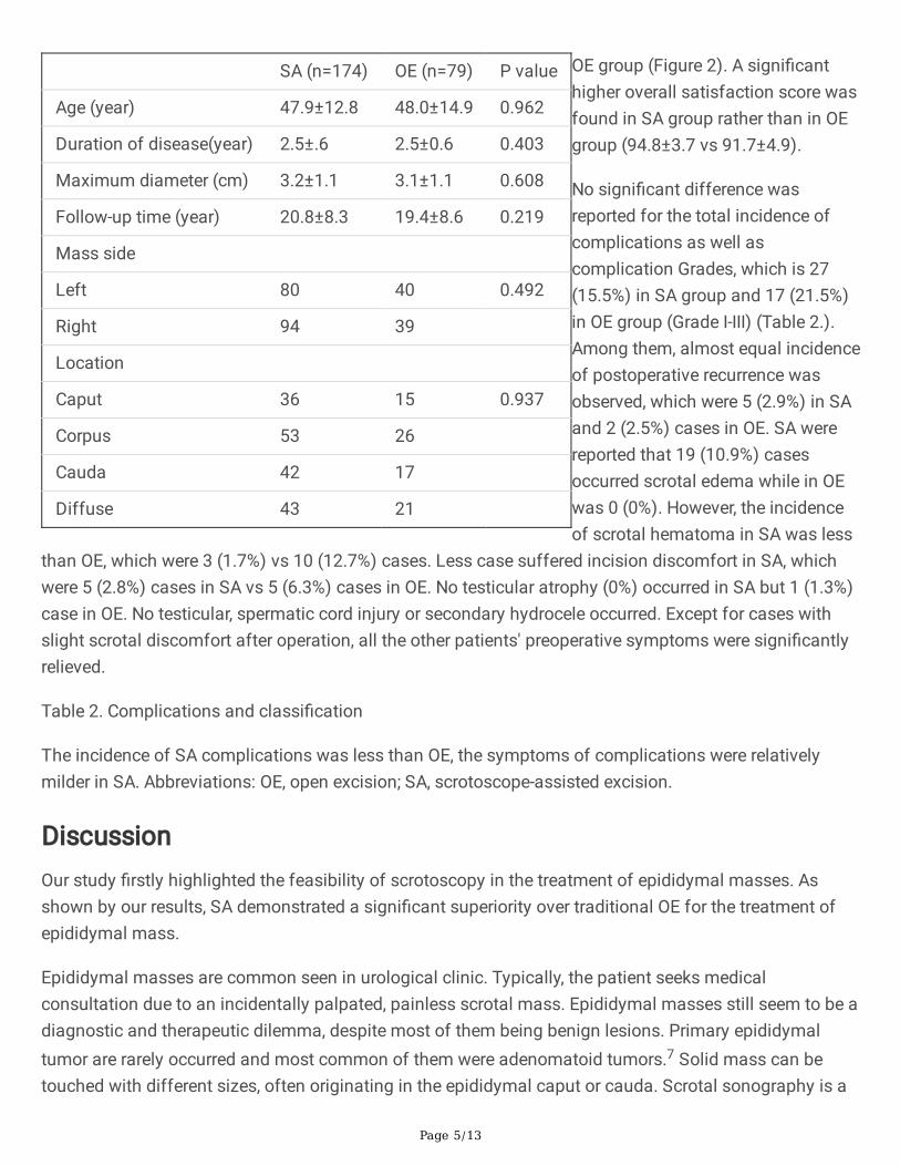
SA (n=174) OE (n=79) P value
Age (year) 47.9±12.8 48.0±14.9 0.962
Duration of disease(year) 2.5±.6 2.5±0.6 0.403
Maximum diameter (cm) 3.2±1.1 3.1±1.1 0.608
Follow-up time (year) 20.8±8.3 19.4±8.6 0.219
Mass side
Left 80 40 0.492
Right 94 39
Location
Caput 36 15 0.937
Corpus 53 26
Cauda 42 17
Diffuse 43 21
OE group (Figure 2). A signi�canthigher overall satisfaction score wasfound in SA group rather than in OEgroup (94.8±3.7 vs 91.7±4.9).
No signi�cant difference wasreported for the total incidence ofcomplications as well ascomplication Grades, which is 27(15.5%) in SA group and 17 (21.5%)in OE group (Grade I-III) (Table 2.).Among them, almost equal incidenceof postoperative recurrence wasobserved, which were 5 (2.9%) in SAand 2 (2.5%) cases in OE. SA werereported that 19 (10.9%) casesoccurred scrotal edema while in OEwas 0 (0%). However, the incidenceof scrotal hematoma in SA was less
than OE, which were 3 (1.7%) vs 10 (12.7%) cases. Less case suffered incision discomfort in SA, whichwere 5 (2.8%) cases in SA vs 5 (6.3%) cases in OE. No testicular atrophy (0%) occurred in SA but 1 (1.3%)case in OE. No testicular, spermatic cord injury or secondary hydrocele occurred. Except for cases withslight scrotal discomfort after operation, all the other patients' preoperative symptoms were signi�cantlyrelieved.
Table 2. Complications and classi�cation
The incidence of SA complications was less than OE, the symptoms of complications were relativelymilder in SA. Abbreviations: OE, open excision; SA, scrotoscope-assisted excision.
DiscussionOur study �rstly highlighted the feasibility of scrotoscopy in the treatment of epididymal masses. Asshown by our results, SA demonstrated a signi�cant superiority over traditional OE for the treatment ofepididymal mass.
Epididymal masses are common seen in urological clinic. Typically, the patient seeks medicalconsultation due to an incidentally palpated, painless scrotal mass. Epididymal masses still seem to be adiagnostic and therapeutic dilemma, despite most of them being benign lesions. Primary epididymaltumor are rarely occurred and most common of them were adenomatoid tumors.7 Solid mass can betouched with different sizes, often originating in the epididymal caput or cauda. Scrotal sonography is a

Page 6/13
SA (%) OE (%) P value
Total complications No 147 (84.5) 62 (78.5) 0.243
Yes 27 (15.5) 17 (21.5)
Complication classi�cation I 22 (12.6) 12 (15.2) 0.099
II 5 (2.9) 4 (5.1)
III 0 (0) 1 (1.3)
Relief of symptoms Complete 155 (89.1) 66 (83.5) 0.134
Partial 18 (10.3) 10 (12.7)
None 1 (0.6) 3 (3.8)
Recurrence No 169 (97.1) 77 (97.5) 0.878
Yes 5 (2.9) 2 (2.5)
Scrotal edema No 155 (89.1) 79 (100) 0.009
Slight 15 (8.6) 0 (0)
Severe 4 (2.3) 0 (0)
Scrotal hematoma No 171 (98.3) 69 (87.3) 0.000
Yes 3 (1.7) 10 (12.7)
Incision discomfort No 169 (97.1) 74 (93.7) 0.191
Yes 5 (2.9) 5 (6.3)
Testicular atrophy No 174 (100) 78 (98.7) 0.137
Yes 0 (0) 1 (1.3)
recommended imagingtechnique to analyze thescrotal abnormality, andMRI is usually applied inselected patients.Combining with clinicalhistory, sonographic�ndings could aid toestablish a shortdifferential diagnosisand subsequent plan ofcare. Currently,treatment forepididymal mass is stillnot established andconservative treatmentis the main choice.However, the patientsmay be indicated forsurgical excision ofepididymal mass underthe followingconditions: failedmedicine treatmentwith/without obvioussymptoms, orsuspended malignanttumor.19–21 In the past,open resection of
epididymal mass or open partial epididymectomy was most commonly chosen for epididymal masssurgical treatment.22 However, open surgery had a relatively larger trauma and a higher postoperativemorbidity-in particular hematoma and infection, which was not conducive to the rapid recovery ofpatients.23–25 It is a necessity to develop a minimally invasive approach in surgical treatment of scrotaldiseases.
Firstly described by Gerris and Sha�k,10,11 the scrotoscopy has been found to be a minimally invasiveand less complicated operation for the diagnosis and treatment of intrascrotal diseases. In 1992,scrotoscopy was �rst performed by our group in Chinese population. We then expanded the applicationrange of scrotoscopy for the management of other scrotal diseases, including testicular torsion,26
testicular rupture,13,15 testicular hydrocele,14,17,27 and some rare benign diseases of the scrotum.13–15 All

Page 7/13
of them have achieved favorable results. Compared with traditional open approach, scrotoscope-aidedone has obvious advantages such as a shorter operation time, a smaller incision, and a faster recoveryafter surgery of fast, minimally invasive, rapid recovery and fewer complications.12
Consistently, we applied scrotoscopy for the treatment of epididymal masses and achieved superiorresults over traditional approach. In present study, SA showed signi�cantly less operating time, less bloodloss, and shorter length of incision than that in OE. Possible reasonings for these results are given below.Mean operating time in SA was less than OE, because more time were need to achieve haemostasis inOE. The endoscope can easily insert into the cavity of tunica vaginalis and can easily observe the space-limited cavity, so the incision does not need to be as long as the OE. With the application of electriccutting technology, no obvious bleeding was observed during the SA process. These above supports thatscrotoscope could serve as a feasible and e�cient tool for the treatment of epididymal mass.
In terms of surgical safety, SA group showed less postoperative complications and a faster recoverywhen compared with OE group. According to the Clavien-Dindo grading system, all the postoperativecomplications in two groups were Grade I-III. One Grade III case was reported in OE group but none in SAgroup. Scrotal edema was the most common complication in SA, this may be related to the damage ofperididymis or extra �uid in�ltration into the interlayer of the scrotal wall through the incision. Generally,avoiding wall sheath damage and reducing perfusion pressure and time can effectively avoid edema.12,17
Based on our own experience, a maintained 60-80-centimeter hydraulic pressure is preferred. Scrotalhematoma, however, was the most common complication in OE rather than in SA, possibly due to therelative larger surgical trauma in open procedure. For SA patients, the mass could be minimally excisedunder scrotoscope and the bleeding site could be electrocoagulated without extruding the testicle fromthe scrotum, minimizing the risk of bleeding. Of note, no case of testicular atrophy occurred in SA groupbut one in OE group, potentially due to no extrusion of testicle and spermatic cord from the scrotalincision. Besides, SA was performed under clear direct vision to avoid severe damage. As shown inTable 2, patients with SA also lead to a signi�cantly higher rate of symptom relief and higher score insatisfaction, suggesting that patients were highly satis�ed with SA. These may be attributed by multipleadvantages of SA, including less postoperative complications, less incision discomfort, less trauma, lessdressing changes but a more rapid recovery. When it comes to postoperative recurrence, there was nosigni�cant difference between the two groups. SA had less frequency of dressing changes, probably dueto the shorter incisions and better bleeding control.
However, certain limitations should be addressed in this research, including the retrospective nature, thelimited size of the total population and lack of long-term follow-up data. Future studies with largersample sizes should be conducted to further validate its diagnostic and therapeutic value.
In conclusion, our present study con�rmed that SA was a safe and effective minimally invasivetherapeutic option for epididymal mass. It has the advantages of small incision, rapid recovery and lowrisk of complications. It is worthy of further clinical application.

Page 8/13
MethodsStudy Population
A retrospective analysis was performed at the Second Xiangya Hospital and Fujian Provincial Hospitalfrom January 2002 to 2018. Study approval was obtained from the Ethics Committee of Fujian ProvincialHospital (K-2019-10-03). We con�rmed that all methods were carried out in accordance with relevantguidelines and regulations. And the informed consent was obtained from all subjects and/or their legalguardian. As it is retrospective study and we are unable to provide informed consent for the same, theinformed consent was waived by ethical committee of both Fujian Provincial Hospital and SecondXiangya Hospital. Epididymal mass was diagnosed by scrotum ultrasound. The patient was includedunder the following conditions: aged 18 to 60; diagnosed with epididymal mass; conservative treatmentsfailed with or without obvious symptoms. All patients underwent a clinical assessment including vitalsigns, electrocardiography, laboratory examination (hemostasis parameters, routine hematology, liver andrenal function, etc.). We developed C-reactive protein, puri�ed protein derivative and chest radiography,urinary ultrasound or KUB+IVP, CT and/or scrotum MRI to exclude epididymis tuberculosis or malignanttumor. Cases with severe cardiopulmonary diseases or coagulation disorders were excluded.
Main Surgical Procedures
All patients received general anesthesia and operated in bladder lithotomy position with routine skinpreparation. Disposable plastic incise drape was pasted in the operation area after sterilized. A 10Fpediatric cystoscope was employed as scrotoscope. Isotonic crystalloid solution was suspended at aheight of 60-80mm as perfusate.
The SA procedure was performed as previous described.12,14,17 (Figure. 1) Brie�y, a 1.0-cm incision wasestablished on the affected side of the scrotum, then dissecting through the scrotal layer into the tunicasac. Two Allis forceps hold the whole scrotal wall. The scrotoscope was put into the tunica sac withcontinuous sterile saline infusion. The scrotal contents, including the testicle, epididymis, and tunicavaginalis were inspected sequentially. Concurrently, the location, appearance, size, margin of theepididymal mass were mainly observed. Then electrosurgical excision of epididymal mass wasconducted in a systematic fashion, taking down gradually from the caput side to cauda side andreaching deeply to the plane between epididymis and testicle. Testicular injury should be carefullyavoided. After excision, the wound got electrocoagulation to stop bleeding. The resected fragments ofmass were retrieved using Ellik evacuator and sent for pathological examination. A scrotoscopy was re-performed to examine the scrotal contents to exclude any active bleeding or neglected lesion. Theincision was sutured with absorbable stitches. A drainage strip was placed into the scrotum and removedafter 24-48 hours.
For the patients treated by OE, an about 4-cm anterior scrotal incision was made in the ill side. Theepididymis and testicle were taken out of the incision and the epididymal mass was inspected and

Page 9/13
excised. Like SA, the incision was then closed, and a drainage strip was also used and removed within 24-48 hours.
Outcome Measures
A descriptive analysis of patient demographics was performed, including age, time since onset, follow-upperiod and the characteristics of epididymal mass. Surgical details mainly included intraoperative(operating time, incision size, blood loss = (gauze weight after wiping all blood loss-dry gauze weight)g/1.05g/mL) and postoperative (frequency of dressing changes, complications, hospital stay, pathology�nding) results. Postoperative complications were graded by Clavien-Dindo system. All patientscompleted at least one follow-up visit within 6 months after surgery. During the follow-up period, thepatients completed the survey of the overall satisfaction of surgical treatment (ranged from 0 to 100score).18
Statistical Analysis
Data were expressed respectively in terms of means ± standard deviation (SD) or percentages. Wecompared continuous variables with t tests and used a p-value of less than 0.05 as a cutoff for statisticalsigni�cance. All data entry and analysis were carried out in SPSS 24.0 statistical analysis software(SPSS, Chicago, IL).
DeclarationsAcknowledgements: None.
Data availability: The datasets generated for this study are available on request to the correspondingauthor.
Funding: Startup Fund for scienti�c research, Fujian Medical University Grant number: 2019QH1154 andhigh-level hospital foster grants from Fujian Provincial Hospital, Fujian province, China (2019HSJJ29).
Author's contributions: Yunliang Gao and Yongbao Wei conceived of the presented idea. Yongbao Weideveloped the theory and performed the computations. Chuying Qin and Jinrui Yang veri�ed theanalytical methods. Yunliang Gao encouraged Chuying Qin to develop the manuscript and supervised the�ndings of this work. All authors discussed the results and contributed to the �nal manuscript
Disclosures Drs. Chuying Qin, Jinrui Yang, Ruochen Zhang, Yaojing Yang, Wanghai Cai, Tao Li, QingguoZhu, Liefu Ye, Yunliang Gao, and Yongbao Wei have no con�icts of interest or �nancial ties to disclose.
References1 Redshaw, J. D., Tran, T. L., Wallis, M. C. & deVries, C. R. Epididymitis: a 21-year retrospective reviewof presentations to an outpatient urology clinic. The Journal of urology 192, 1203-1207,

Page 10/13
doi:10.1016/j.juro.2014.04.002 (2014).
2 Calleary, J. G., Masood, J. & Hill, J. T. Chronic epididymitis: is epididymectomy a valid surgicaltreatment? International journal of andrology 32, 468-472, doi:10.1111/j.1365-2605.2008.00880.x (2009).
3 Karaman, A., Afsarlar, C. E. & Arda, N. Epididymal cyst: not always a benign condition. Internationaljournal of urology : o�cial journal of the Japanese Urological Association 20, 457-458,doi:10.1111/j.1442-2042.2012.03152.x (2013).
4 Tani, Y. et al. Epididymal sperm granuloma induced by chronic administration of 2-methylimidazole in B6C3F1 mice. Toxicologic pathology 33, 313-319, doi:10.1080/01926230590922866(2005).
5 Boorjian, S., Lipkin, M. & Goldstein, M. The impact of obstructive interval and sperm granuloma onoutcome of vasectomy reversal. The Journal of urology 171, 304-306,doi:10.1097/01.ju.0000098652.35575.85 (2004).
6 Hamm, B. Sonography of the testis and epididymis. Andrologia 26, 193-210, doi:10.1111/j.1439-0272.1994.tb00789.x (1994).
7 Yeung, C., Wang, K. & Cooper, T. Why are epididymal tumours so rare? Asian journal of andrology14, 465-475, doi:10.1038/aja.2012.20 (2012).
8 Kiddoo, D., Wollin, T. & Mador, D. A population based assessment of complications followingoutpatient hydrocelectomy and spermatocelectomy. The Journal of urology 171, 746-748,doi:10.1097/01.ju.0000103636.61790.43 (2004).
9 Swartz, M., Morgan, T. & Krieger, J. Complications of scrotal surgery for benign conditions. Urology69, 616-619, doi:10.1016/j.urology.2007.01.004 (2007).
10 Sha�k, A. The scrotoscope. A new instrument for examining the scrotal contents. British journal ofurology 65, 209-210, doi:10.1111/j.1464-410x.1990.tb14702.x (1990).
11 Gerris, J., Van Camp, C., Van Neuten, J., Gentens, P. & Van Camp, K. Scrotal endoscopy in maleinfertility. Lancet (London, England) 1, 1102, doi:10.1016/s0140-6736(88)91915-0 (1988).
12 Yang, J. R. et al. Comparison between Open Epididymal Cystectomy and Minimal Resection ofEpididymal Cysts Using a Scrotoscope: A Clinical Trial for the Evaluation of a New Surgical Technique.Urology 85, 1510-1514, doi:10.1016/j.urology.2015.03.003 (2015).
13 Wei, Y. et al. Scrotoscopy exploration of testicular rupture: A pilot study. Medicine 98, e17389,doi:10.1097/md.0000000000017389 (2019).

Page 11/13
14 Lin, L. et al. Individualized minimally invasive treatment for adult testicular hydrocele: A pilotstudy. World journal of clinical cases 7, 727-733, doi:10.12998/wjcc.v7.i6.727 (2019).
15 Wang, Z. et al. Diagnosis and management of testicular rupture after blunt scrotal trauma: aliterature review. International urology and nephrology 48, 1967-1976, doi:10.1007/s11255-016-1402-0(2016).
16 Hong, H., Cai, W., Wu, J., Wu, X. & Yang, J. Scrotoscopy and traditional open surgery shows a highdegree of consistency in the diagnosis of testicular torsion: An initial report. Medicine 99, e21545 (2020).
17 Bin, Y., Yong-Bao, W., Zhuo, Y. & Jin-Rui, Y. Minimal hydrocelectomy with the aid of scrotoscope: aten-year experience. International braz j urol : o�cial journal of the Brazilian Society of Urology 40, 384-389, doi:10.1590/s1677-5538.Ibju.2014.03.13 (2014).
18 Yossepowitch, O., Aviv, D., Wainchwaig, L. & Baniel, J. Testicular prostheses for testis cancersurvivors: patient perspectives and predictors of long-term satisfaction. The Journal of urology 186,2249-2252, doi:10.1016/j.juro.2011.07.075 (2011).
19 Jones, M. A., Young, R. H. & Scully, R. E. Adenocarcinoma of the epididymis: a report of four casesand review of the literature. The American journal of surgical pathology 21, 1474-1480,doi:10.1097/00000478-199712000-00010 (1997).
20 Ganem, J. P., Jhaveri, F. M. & Marroum, M. C. Primary adenocarcinoma of the epididymis: casereport and review of the literature. Urology 52, 904-908, doi:10.1016/s0090-4295(98)00275-1 (1998).
21 Erikci, V. et al. Management of epididymal cysts in childhood. Journal of pediatric surgery 48,2153-2156, doi:10.1016/j.jpedsurg.2013.01.058 (2013).
22 Vavallo, A. et al. Capillary hemangioma of the scrotum mimicking an epididymal tumor: casereport. Archivio italiano di urologia, andrologia : organo u�ciale [di] Societa italiana di ecogra�a urologicae nefrologica 86, 395-396, doi:10.4081/aiua.2014.4.395 (2014).
23 Hicks, N. & Gupta, S. Complications and risk factors in elective benign scrotal surgery.Scandinavian journal of urology 50, 468-471, doi:10.1080/21681805.2016.1204622 (2016).
24 Chalasani, V. & Woo, H. H. Why not use a small incision to treat large hydroceles? ANZ journal ofsurgery 72, 594-595, doi:10.1046/j.1445-2197.2002.02469.x (2002).
25 Onol, S. Y. et al. A novel pull-through technique for the surgical management of idiopathichydrocele. The Journal of urology 181, 1201-1205, doi:10.1016/j.juro.2008.10.166 (2009).
26 Ye, H. et al. A Minimally Invasive Method in Diagnosing Testicular Torsion: The Initial Experienceof Scrotoscope. Journal of endourology 30, 704-708, doi:10.1089/end.2015.0724 (2016).

Page 12/13
27 Khaniya, S., Agrawal, C. S., Koirala, R., Regmi, R. & Adhikary, S. Comparison of aspiration-sclerotherapy with hydrocelectomy in the management of hydrocele: a prospective randomized study.International journal of surgery (London, England) 7, 392-395, doi:10.1016/j.ijsu.2009.07.002 (2009).
Figures
Figure 1
Main Surgical Procedures of Scrotoscope-assisted Epididymal Mass Excision. (A) A 1.0-cm incision wasestablished on the affected side of the scrotum, then two Allis forceps hold the whole scrotal wall. (B)The scrotoscope was put into the tunica sac and the scrotal contents were inspected sequentially. (C) Thelocation, appearance, size, margin of the epididymal mass were mainly observed. (D) Electrosurgicalexcision of the epididymal mass by plasma eletrosection was performed. (E) The wound gotelectrocoagulation to stop bleeding. (F) The resected fragments of mass were retrieved. (G) A drainagestrip was placed into the scrotum. (H) The resected fragments of mass were sent for pathologicalexamination. EM, epididymal mass.

Page 13/13
Figure 2
Intraoperative and Postoperative data. Scrotoscope-assisted excision (SA) showed less operating time,less blood loss, shorter length of incision, and less frequency of dressing changes. SA presented nosigni�cant advantage in the number of hospital stay. Besides, compared to open excision, SA also lead toa higher score in satisfaction (all P Value <.05).