Determining asymmetry of roll-over shapes in ... - CiteSeerX
White Matter Pathway AsymmetryUnderlies Functional Lateralization
Thomas R. Barrick1, I. Nigel Lawes2, Clare E. Mackay3 and
Chris A. Clark1
1Centre for Clinical Neuroscience, Division of Cardiac and
Vascular Sciences and 2Department of Basic Medical Sciences,
St George’s University of London, Cranmer Terrace, London
SW17 0RE, UK and 3University Department of Psychiatry and
Prince of Wales International Centre, Warneford Hospital,
Headington, Oxford OX3 7JX, UK
Structural and functional asymmetry of the human brain has beenwell documented using techniques such as magnetic resonanceimaging (MRI). However, asymmetry of underlying white matterconnections is less well understood. We applied an MRI techniqueknown as diffusion tensor tractography to reveal the morphology ofthe white matter in vivo by mapping directions of maximum waterdiffusion in brain tissue. White matter pathway asymmetry wasinvestigated in a normalized image data set of 30 right-handedyoung healthy individuals. We identified, for the first time, a right-wardly asymmetric pathway connecting the posterior temporal lobeto the superior parietal lobule. This pathway may be related toauditory spatial attention and working memory for which there isevidence for a rightward laterality from functional imaging studies.Additional leftward asymmetries connecting the parietal and frontallobes to the temporal lobe may be more closely related to lateralityof language.
Keywords: asymmetry, brain, diffusion tensor tractography,functional lateralization, MRI
Introduction
Asymmetrical structure--function relationships have been stud-
ied in the human brain since Broca (1861) identified a major
functional asymmetry, whereby speech production is linked
preferentially to the left hemisphere for the majority of people.
Since Broca (1861), lesion studies and, more recently, functional
neuroimaging studies have established hemispheric specificity
for a range of language, motor, and spatial tasks (Davidson and
Hugdahl 1995), but their relationship to underlying gray and
white matter anatomy remains unclear. A frequently cited
structure--function relationship in the human brain is between
leftward (i.e., left greater than right) planum temporale asym-
metry and hemispheric dominance for language function
(Geschwind and Levitsky 1968). The planum temporale is
a large region of auditory association cortex located on the
superior temporal plane posterior to Heschl’s gyrus involved in
verbal and nonverbal stimuli (Griffiths and Warren 2002). It has
been reported to have leftward asymmetry in postmortem
(Geschwind and Levitsky 1991), magnetic resonance imaging
(MRI) volumetry (for review, see Shapleske and others 1999),
and voxel-based MRI asymmetry studies (Good and others 2001;
Watkins and others 2001; Barrick and others 2005). This
structure--function relationship is further supported by reports
of reduced morphological asymmetry in patients with language
disorders such as dyslexia (e.g., Rumsey and others 1986).
Recently, the temporal lobes have been implicated in auditory
dual-pathway (what and where) networks that involve the
frontal and parietal lobes (Rauscheker 1998; Romanski and
others 1999; Clarke and others 2000, 2003; Zatorre and others
2002; Arnott and others 2004; Hickok and Poeppel 2004). In
addition to language laterality, lateralized function of auditory
spatial processing and attention in the inferior parietal lobule
are reported in functional imaging studies (Griffiths and others
1998; Weeks and others 1999; Zatorre and others 1999, 2002;
Griffiths and others 2000; Itoh and others 2000; Alain and others
2001; Brunetti and others 2005). Furthermore, functional
laterality in the superior parietal lobule is also reported
(Griffiths and others 1998, 2000; Zatorre and others 1999;
Macaluso and others 2004).
This extensive literature describing asymmetry of brain
morphology and function implicates the temporal, frontal, and
parietal lobes. However, the anatomy of white matter pathways
related to these asymmetries remains to be determined. In
particular, investigations that provide greater anatomical speci-
ficity of the underlying white matter will contribute to an
understanding of cerebral asymmetry at the cortical and
functional level. In the present study, we examine the cerebral
asymmetry of white matter architecture imaged by mapping
water diffusion characteristics from diffusion-weighted MRI.
This technique, known as diffusion tensor tractography, is the
only noninvasive method currently available for investigation of
white matter organization and hence, the underlying connec-
tivity of the brain. Although connectivity of individual axons
cannot be inferred, tractography provides information on the
order of magnitude of a millimeter and has been applied to
reconstruct anatomically plausible commissural (Catani and
others 2002; Mori and others 2002; Hagmann and others
2003), association (Catani and others 2002; Mori and others
2002; Hagmann and others 2003), and projection fibers (Beh-
rens and others 2003; Johansen-Berg and others 2005) as well as
white matter in the midbrain (Stieltjes and others 2001), fornix
(Catani and others 2002; Hagmann and others 2003), and
cingulum (Catani and others 2002; Hagmann and others 2003).
Diffusion tensor tractography has recently been applied to
describe left hemisphere language-related white matter path-
ways (Catani and others 2005). Moreover, Parker and others
(2005) reported that 2 of the 3 language pathways described by
Catani and others (2005) are leftwardly asymmetric. Catani and
others (2005) and Parker and others (2005) reconstructed
white matter structure by extracting pathways passing through,
or originating from, user-defined regions of interest. Because
the gross gray and white matter anatomy of the temporal lobe is
asymmetric (Barrick and others 2004, 2005), manually defining
regions of interest can lead to anatomical bias and potentially
erroneous assignment of asymmetry to white matter pathways.
In the present study, we remove any bias that may occur from
application of user-defined regions of interest by reconstructing
white matter pathways that pass between entire cerebral lobes
Cerebral Cortex March 2007;17:591--598
doi:10.1093/cercor/bhk004
Advance Access publication April 20, 2006
� The Author 2006. Published by Oxford University Press. All rights reserved.
For permissions, please e-mail: [email protected]
by guest on September 15, 2013
http://cercor.oxfordjournals.org/D
ownloaded from

segmented in standardized space (Evans and others 1993) using
a map of segmented cerebral structures known as the Talairach
Daemon (Lancaster and others 2000). We apply this fully
automated lobe-based tractography technique to investigate
the underlying morphology and asymmetry of all white matter
pathways that connect the temporal lobe to the parietal and
frontal lobes of 30 young healthy right-handed subjects. Novel
visualization techniques are introduced to define the anatomy
of white matter pathway asymmetry in terms of symmetrical
streamline color (SSC) and intersubject variability maps. For the
first time, we reveal a significant asymmetry of white matter
pathways, connecting the superior and middle temporal gyri to
the superior parietal lobule that may be related to laterality of
auditory spatial function and auditory working memory. Signif-
icant white matter pathway asymmetries associated with
language function are also found, in agreement with previous
studies.
Materials and Methods
Data AcquisitionDiffusion-weighted images were acquired for 30 right-handed healthy
volunteers (15 males, 15 females, ages 20--39 years, mean age 27.2 ± 5.2
years) from whom informed written consent had been obtained to
participate in the study. Diffusion-weighted images were obtained using
a diffusion-sensitized spin echo--planar imaging sequence using a 1.5-T
General Electric Signa MRI system equipped with magnetic field
gradients of up to 22 mTm–1 (GE Electric, Milwaukee, WI) and a
proprietary head coil (50 3 2.8--mm thick interleaved slices, field
of view 240 3 240 mm2, matrix 96 3 96 were reconstructed
on a 256 3 256--matrix giving a final resolution of 0.9375 3 0.9375 3
2.8 mm3). Following an acquisition without diffusion sensitization
(b = 0), images were acquired with diffusion gradients applied
(b = 1000 s mm–2) in 12 directions to eliminate diffusion-imaging
gradient cross terms (Barrick and Clark 2004). The acquisitions were
repeated 4 times in order to improve signal to noise ratio.
Image AnalysisData were analyzed on an independent workstation (Sun Blade 1500;
Sun Microsystems, Mountain View, CA). Images were realigned to
remove eddy current distortions (Woods and others 1993). Diffusion
tensor elements were computed at each voxel as described by Basser
and others (1994) and diagonalized to determine the eigenvalues and
eigenvectors. Normalization of diffusion tensor imaging to the stan-
dardized space of Evans and others (1993) was achieved using pre-
viously reported methods (Barrick and Clark 2004). All normalized DTIs
were skull stripped using brain extraction tool (Smith 2002), part of the
Oxford Centre for Functional Magnetic Resonance Imaging of the Brain
software library (FSL) (Oxford University, Oxford, UK, http://www.fmri-
b.ox.ac.uk/). From the 30 normalized skull-stripped DTI images,
a group-averaged mean DTI was computed (Jones and others 2002).
For each normalized DTI and the mean DTI, fractional anisotropy (FA)
maps were computed (Pierpaoli and Basser 1996).
Diffusion Tensor TractographyTractography was performed to determine the white matter pathways
that pass from the temporal lobe to the frontal and parietal lobes in the
left and right cerebral hemispheres. Streamline tractography was seeded
from the center of every voxel throughout the entire brain of the 30
normalized DTIs and the mean DTI by application of a previously
reported tractography algorithm (Barrick and Clark 2004). Streamline
tracts were initiated in orthograde and retrograde directions from each
image voxel and were iteratively followed by moving 1 mm in the
direction of the principal eigenvector. Tracts were terminated in
regions of low FA to allow tracts to pass into gray matter (FA > 0.08).
No angular threshold was applied on coincident principal eigenvector
orientations.
Lobe SegmentationTermination points for each individual tract (x, y, z coordinates in
millimeters within standard space) were determined, and tempor-
oparietal and temporofrontal tracts were retained in the left and
right hemispheres if their terminations lay within the temporal lobe
and either the parietal or the frontal lobes. Segmentations of the left
and right temporal, frontal, and parietal lobes in standard space were
obtained using an image found within mri3dX (http://www.aston.
ac.uk/lhs/staff/singhkd/mri3dX/mri3dX.jsp), which utilizes a stored
representation of the Talairach Daemon Database (Lancaster and
others 2000). Using this functionality, any brain voxel can be labeled
with one or more of the 5 levels within the database (i.e., hemi-
sphere, lobe [which also includes the limbic system and deep white
matter], neuroanatomical structure, tissue type, and Brodmann area
[BA]). To allow for differences in morphology between single
subjects and the Talairach Daemon, the termination coordinates
were labeled by hemisphere and lobe by determining the shortest 3-
dimensional Euclidean distance to the Talairach Daemon labels in
standard space.
Computation of White Matter Pathway Intersubject Variabilityand Asymmetry MapsMaps representing the pathway intersubject variability and asymmetry
of temporofrontal and temporoparietal white matter pathways in the 30
normalized DTIs were computed. For each subject, voxelwise binary
images containing 0 or 1 at each voxel were generated for the tem-
porofrontal and temporoparietal tracts in the left and right hemi-
spheres by determining whether tracts passed through image voxels
(0 = outside pathway, 1 = inside pathway). A map representing inter-
subject variability in pathway morphology across the 30 subjects was
generated by calculating the mean of these binary images for the
temporofrontal and temporoparietal pathways in the left and right
cerebral hemispheres. Pathway variability maps represent the normal-
ized frequency at which tracts pass through each image voxel in
standard space (i.e., 0 = no tracts pass through the voxel in any subject,
1 = tracts pass through the voxel in every subject). Pathway asymmetry
maps were generated by reflecting the pathway variability maps for the
left hemisphere in the midsagittal plane of standard space (i.e., the plane
x = 0 mm) and subtracting the reflected left hemisphere map from the
unreflected right hemisphere map. Pathway asymmetry maps represent
the difference in normalized frequency between the left and right
hemispheres (i.e., –1 = tracts pass through the voxel in each subject for
the left hemisphere, but none pass through the homologous right
hemisphere voxel, 0 = no tracts pass through the voxel in any subject, 1
= tracts pass through the voxel in each subject for the right hemisphere,
but none pass through the homologous left hemisphere voxel).
Nonparametric Statistical Analysis of Pathway AsymmetryParametric statistical tests are unsuitable for the analysis of the
voxelwise binary images representing temporoparietal and temporo-
frontal pathways because the assumption of a normal distribution of data
at each image voxel across the 30 subjects is violated. Consequently,
a nonparametric statistical test was applied to investigate the anatomy of
cerebral white matter pathway asymmetry. McNemar’s test (Siegel
1956) was applied on a voxel-by-voxel basis to investigate the null
hypothesis of no significant difference between the numbers of subjects
with leftward or rightward asymmetry. In particular, McNemar’s test
assesses the significance between 2 dependent samples when the
variable of interest is a dichotomy. In the present study, the 2 dependent
samples are homologous voxels in the left and right cerebral hemi-
spheres, and the variable of interest is a binary number, representing
whether white matter pathways are absent (=0) or present (=1) in the
voxel. Temporoparietal and temporofrontal left and right hemisphere
binary pathway images of the 30 subjects were statistically analyzed
(N.B. all left hemisphere binary images were reflected in the plane x = 0
of standard space), and statistical results were transformed to the z
statistic. Multiple comparisons correction was performed at the cluster
level using FSL (Oxford University, Oxford, UK, http://www.fmrib.ox.
ac.uk/). The z statistic was thresholded at z > 2.3, and multiple
comparisons correction applied at P < 0.001 over the analyzed volumes.
592 Asymmetry Underlying Lateralization d Barrick and others
by guest on September 15, 2013
http://cercor.oxfordjournals.org/D
ownloaded from
Tract Representation for the Mean DTI3-dimensional white matter pathway reconstructions illustrating aver-
age morphology and asymmetry were computed from the mean DTI in
standard space. Individual tract termination coordinates in standard
space were used to generate tract-specific RGB colors such that if
symmetrical pathways are present, they are illustrated using the same
color in both hemispheres, whereas if pathways are asymmetrical, they
are illustrated using different colors in the left and right hemispheres.
This new color scheme is referred to as the SSC map. Temporofrontal
and temporoparietal tract termination x, y, and z coordinates in standard
space were scaled such that all coordinates lay between 0 and 255.
x termination coordinates were scaled from 0 at the left of the brain to
255 at the midsagittal plane (x = 0 of standard space) and back to 0 at the
right. y (and z) coordinates were scaled from 0 at the posterior (inferior)
of the brain to 255 at the anterior (superior) of standard space. These
scaled termination coordinates were separated into start and end
coordinates for each tract. Start coordinates were defined as lying
more laterally than end coordinates. For each tract, the scaled start
and end coordinates were divided by 16 and rounded to 4-bit binary
numbers between 0000 (i.e., 0) and 1111 (i.e., 15). Eight-bit binary
numbers representing the 3 components of the 24-bit RGB SSC were
constructed for each of the x (red RGB component), y (green RGB
component), and z (blue RGB component) termination coordinates by
placing the start and end 4-bit numbers side-by-side and computing the
8-bit components of the RGB color, the merged 4-bit numbers
represent. For example, if xstart = 0101 = 5 and xend = 1101 = 13 for
a single streamline, then the red component of the RGB color is
01011101 = 93. By performing this operation for each tract, the SSC was
generated and applied to the white matter pathway reconstructions
illustrated in the present study.
Results
Temporoparietal Pathway Morphology and Asymmetry
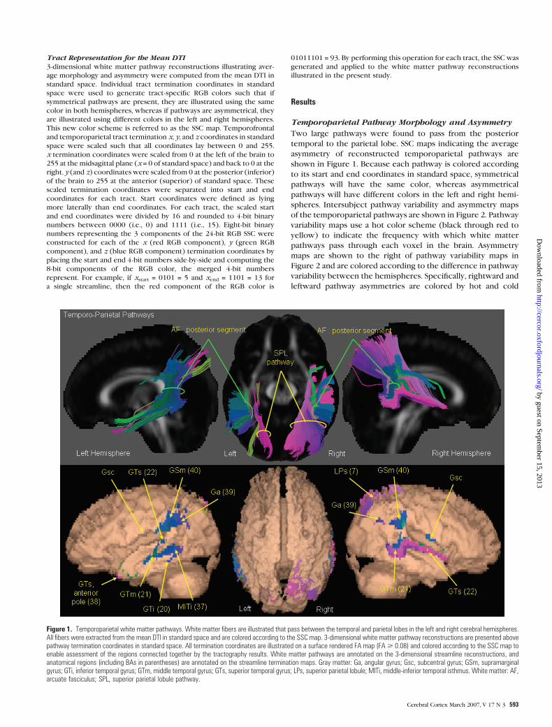
Two large pathways were found to pass from the posterior
temporal to the parietal lobe. SSC maps indicating the average
asymmetry of reconstructed temporoparietal pathways are
shown in Figure 1. Because each pathway is colored according
to its start and end coordinates in standard space, symmetrical
pathways will have the same color, whereas asymmetrical
pathways will have different colors in the left and right hemi-
spheres. Intersubject pathway variability and asymmetry maps
of the temporoparietal pathways are shown in Figure 2. Pathway
variability maps use a hot color scheme (black through red to
yellow) to indicate the frequency with which white matter
pathways pass through each voxel in the brain. Asymmetry
maps are shown to the right of pathway variability maps in
Figure 2 and are colored according to the difference in pathway
variability between the hemispheres. Specifically, rightward and
leftward pathway asymmetries are colored by hot and cold
Figure 1. Temporoparietal white matter pathways. White matter fibers are illustrated that pass between the temporal and parietal lobes in the left and right cerebral hemispheres.All fibers were extracted from the mean DTI in standard space and are colored according to the SSC map. 3-dimensional white matter pathway reconstructions are presented abovepathway termination coordinates in standard space. All termination coordinates are illustrated on a surface rendered FA map (FA> 0.08) and colored according to the SSC map toenable assessment of the regions connected together by the tractography results. White matter pathways are annotated on the 3-dimensional streamline reconstructions, andanatomical regions (including BAs in parentheses) are annotated on the streamline termination maps. Gray matter: Ga, angular gyrus; Gsc, subcentral gyrus; GSm, supramarginalgyrus; GTi, inferior temporal gyrus; GTm, middle temporal gyrus; GTs, superior temporal gyrus; LPs, superior parietal lobule; MITi, middle-inferior temporal isthmus. White matter: AF,arcuate fasciculus; SPL, superior parietal lobule pathway.
Cerebral Cortex March 2007, V 17 N 3 593
by guest on September 15, 2013
http://cercor.oxfordjournals.org/D
ownloaded from
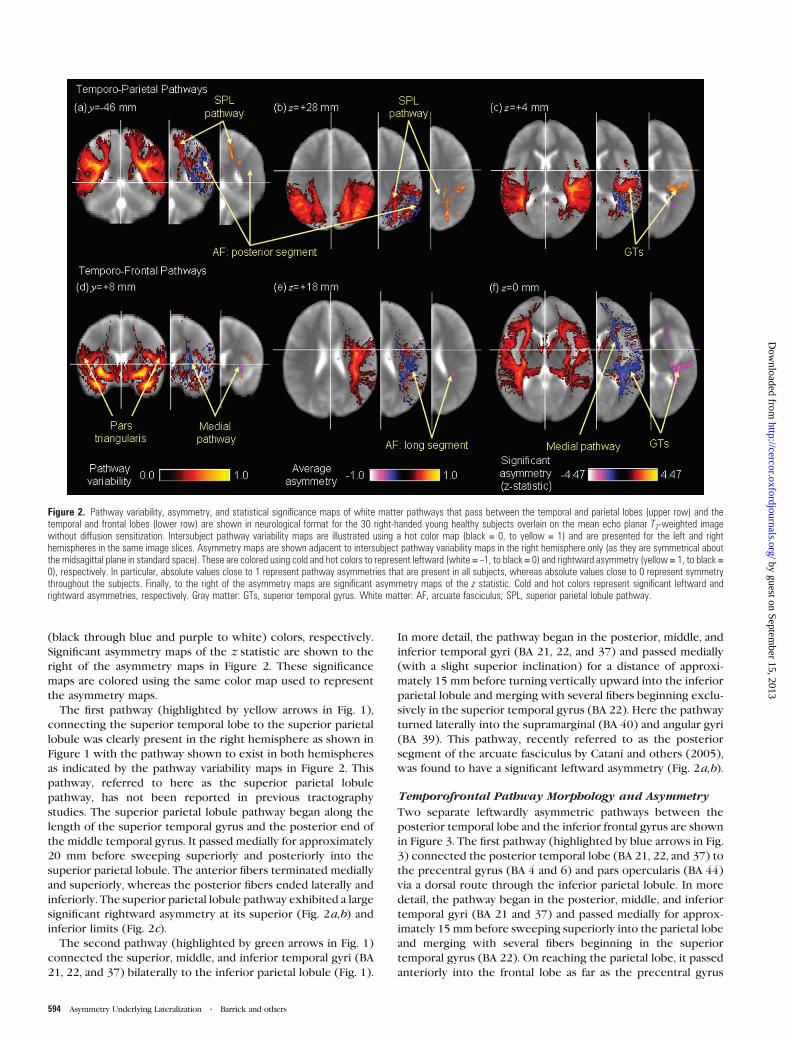
(black through blue and purple to white) colors, respectively.
Significant asymmetry maps of the z statistic are shown to the
right of the asymmetry maps in Figure 2. These significance
maps are colored using the same color map used to represent
the asymmetry maps.
The first pathway (highlighted by yellow arrows in Fig. 1),
connecting the superior temporal lobe to the superior parietal
lobule was clearly present in the right hemisphere as shown in
Figure 1 with the pathway shown to exist in both hemispheres
as indicated by the pathway variability maps in Figure 2. This
pathway, referred to here as the superior parietal lobule
pathway, has not been reported in previous tractography
studies. The superior parietal lobule pathway began along the
length of the superior temporal gyrus and the posterior end of
the middle temporal gyrus. It passed medially for approximately
20 mm before sweeping superiorly and posteriorly into the
superior parietal lobule. The anterior fibers terminated medially
and superiorly, whereas the posterior fibers ended laterally and
inferiorly. The superior parietal lobule pathway exhibited a large
significant rightward asymmetry at its superior (Fig. 2a,b) and
inferior limits (Fig. 2c).
The second pathway (highlighted by green arrows in Fig. 1)
connected the superior, middle, and inferior temporal gyri (BA
21, 22, and 37) bilaterally to the inferior parietal lobule (Fig. 1).
In more detail, the pathway began in the posterior, middle, and
inferior temporal gyri (BA 21, 22, and 37) and passed medially
(with a slight superior inclination) for a distance of approxi-
mately 15 mm before turning vertically upward into the inferior
parietal lobule and merging with several fibers beginning exclu-
sively in the superior temporal gyrus (BA 22). Here the pathway
turned laterally into the supramarginal (BA 40) and angular gyri
(BA 39). This pathway, recently referred to as the posterior
segment of the arcuate fasciculus by Catani and others (2005),
was found to have a significant leftward asymmetry (Fig. 2a,b).
Temporofrontal Pathway Morphology and Asymmetry
Two separate leftwardly asymmetric pathways between the
posterior temporal lobe and the inferior frontal gyrus are shown
in Figure 3. The first pathway (highlighted by blue arrows in Fig.
3) connected the posterior temporal lobe (BA 21, 22, and 37) to
the precentral gyrus (BA 4 and 6) and pars opercularis (BA 44)
via a dorsal route through the inferior parietal lobule. In more
detail, the pathway began in the posterior, middle, and inferior
temporal gyri (BA 21 and 37) and passed medially for approx-
imately 15 mm before sweeping superiorly into the parietal lobe
and merging with several fibers beginning in the superior
temporal gyrus (BA 22). On reaching the parietal lobe, it passed
anteriorly into the frontal lobe as far as the precentral gyrus
Figure 2. Pathway variability, asymmetry, and statistical significance maps of white matter pathways that pass between the temporal and parietal lobes (upper row) and thetemporal and frontal lobes (lower row) are shown in neurological format for the 30 right-handed young healthy subjects overlain on the mean echo planar T2-weighted imagewithout diffusion sensitization. Intersubject pathway variability maps are illustrated using a hot color map (black = 0, to yellow = 1) and are presented for the left and righthemispheres in the same image slices. Asymmetry maps are shown adjacent to intersubject pathway variability maps in the right hemisphere only (as they are symmetrical aboutthe midsagittal plane in standard space). These are colored using cold and hot colors to represent leftward (white = –1, to black = 0) and rightward asymmetry (yellow = 1, to black =0), respectively. In particular, absolute values close to 1 represent pathway asymmetries that are present in all subjects, whereas absolute values close to 0 represent symmetrythroughout the subjects. Finally, to the right of the asymmetry maps are significant asymmetry maps of the z statistic. Cold and hot colors represent significant leftward andrightward asymmetries, respectively. Gray matter: GTs, superior temporal gyrus. White matter: AF, arcuate fasciculus; SPL, superior parietal lobule pathway.
594 Asymmetry Underlying Lateralization d Barrick and others
by guest on September 15, 2013
http://cercor.oxfordjournals.org/D
ownloaded from
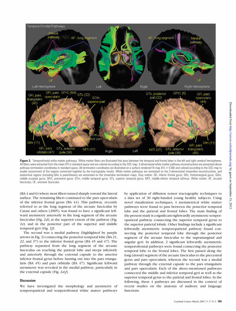
(BA 4 and 6) where most fibers turned sharply toward the lateral
surface. The remaining fibers continued to the pars opercularis
of the inferior frontal gyrus (BA 44). This pathway, recently
referred to as the long segment of the arcuate fasciculus by
Catani and others (2005), was found to have a significant left-
ward asymmetry anteriorly in the long segment of the arcuate
fasciculus (Fig. 2d), at the superior extent of the pathway (Fig.
2e), and in the posterior part of the superior and middle
temporal gyri (Fig. 2f).
The second was a medial pathway (highlighted by purple
arrows in Fig. 3) connecting the posterior temporal lobe (BA 21,
22, and 37) to the inferior frontal gyrus (BA 45 and 47). The
pathway separated from the long segment of the arcuate
fasciculus on reaching the parietal lobe and swept inferiorly
and anteriorly through the external capsule to the anterior
inferior frontal gyrus before fanning out into the pars triangu-
laris (BA 45) and pars orbitalis (BA 47). Significant leftward
asymmetry was revealed in the medial pathway, particularly in
the external capsule (Fig. 2d,f).
Discussion
We have investigated the morphology and asymmetry of
temporoparietal and temporofrontal white matter pathways
by application of diffusion tensor tractography techniques to
a data set of 30 right-handed young healthy subjects. Using
novel visualization techniques, 4 asymmetrical white matter
pathways were found to pass between the posterior temporal
lobe and the parietal and frontal lobes. The main finding of
thepresent study is a significant rightwardly asymmetric tempor-
oparietal pathway connecting the superior temporal gyrus to
the superior parietal lobule. Other findings include a significant
leftwardly asymmetric temporoparietal pathway found con-
necting the posterior temporal lobe through the posterior
segment of the arcuate fasciculus to the supramarginal and
angular gyri. In addition, 2 significant leftwardly asymmetric
temporofrontal pathways were found connecting the posterior
temporal lobe to the frontal lobes. The first passed along the
long (dorsal) segment of the arcuate fasciculus to the precentral
gyrus and pars opercularis, whereas the second was a medial
pathway through the external capsule to the pars triangularis
and pars opercularis. Each of the above-mentioned pathways
connected the middle and inferior temporal gyri as well as the
superior temporal gyrus to the parietal and frontal lobes. In the
following, these 4 pathways are discussed in the context of
recent studies on the anatomy of auditory and language
functions.
Figure 3. Temporofrontal white matter pathways. White matter fibers are illustrated that pass between the temporal and frontal lobes in the left and right cerebral hemispheres.All fibers were extracted from the mean DTI in standard space and are colored according to the SSC map. 3-dimensional white matter pathway reconstructions are presented abovepathway termination coordinates in standard space. All termination coordinates are illustrated on a surface rendered FA map (FA> 0.08) and colored according to the SSC map toenable assessment of the regions connected together by the tractography results. White matter pathways are annotated on the 3-dimensional streamline reconstructions, andanatomical regions (including BAs in parentheses) are annotated on the streamline termination maps. Gray matter: GFi, inferior frontal gyrus; Gfm, frontomarginal gyrus; GOm,middle occipital gyrus; GPrC, precentral gyrus; GTm, middle temporal gyrus; GTs, superior temporal gyrus; MITi, middle-inferior temporal isthmus. White matter: AF, arcuatefasciculus; UF, uncinate fasciculus.
Cerebral Cortex March 2007, V 17 N 3 595
by guest on September 15, 2013
http://cercor.oxfordjournals.org/D
ownloaded from

Our finding of a large rightward asymmetry in white matter
pathways connecting the superior and middle temporal gyri to
the superior parietal lobule has not been previously reported
to our knowledge. Culham and Kanwisher (2001) report the
human parietal lobe to be a functionally heterogeneous region,
subserving a wide range of functions related to attention. In
particular, the superior parietal lobule is a cortical region that
integrates multimodal sensory information and provides guid-
ance to motor operations through connections with the
premotor cortex (Friedman and Goldman-Rakic 1994; Bushara
and others 1999). Functional imaging studies have shown that
the superior parietal lobule is part of a larger network involved
in working memory of verbal (Gurd and others 2002; Crottaz-
Herbette and others 2004), auditory spatial (Zatorre and others
1999; Arnott and others 2005), and auditory pitch tasks (Zatorre
and others 1999; Gaab and others 2003). Several studies report
lateralized right hemisphere processing of auditory spatial
information in the superior parietal lobule (Griffiths and others
1998, 2000). The superior parietal lobule pathway has been
implicated in combined aspects of auditory visual processing
such as temporal and spatial aspects of audiovisual speech that
have been shown to activate the superior temporal sulcus and
an internal region of the right parietal lobe when auditory and
visual sources are presented in opposite hemifields (Macaluso
and others 2004).
Connections between the middle and superior temporal gyri
and the supramarginal (BA 40) and angular (BA 39) gyri through
the posterior segment of the arcuate fasciculus exhibited
leftward asymmetry. This is consistent with tractography results
reported by Parker and others (2005). As suggested by Catani
and others (2005), this pathway may be involved in ideational
speech; however, it is unlikely that these pathways are solely
implicated in language processing. The angular gyrus and, in
particular, the supramarginal gyrus have been shown to
represent part of a larger network involved in auditory spatial
and attentional functions (e.g., Griffiths and others 1998, 2000;
Bushara and others 1999; Weeks and others 1999; Zatorre and
others 1999, 2002; Alain and others 2001; Maeder and others
2001). This is further supported by a meta-analysis of the
auditory dual-pathway (what--where) model (Arnott and others
2004). Arnott and others (2004) reported the bilateral activa-
tion of the supramarginal gyrus in 50% of studies. However,
there are several studies supporting greater functional activity
in the right inferior parietal lobule than the left for auditory
spatial and attentional processing (Griffiths and others 1998;
Weeks and others 1999; Zatorre and others 1999, 2002; Griffiths
and others 2000; Itoh and others 2000; Alain and others 2001;
Brunetti and others 2005). In the context of the leftward
asymmetry of the posterior segment of the arcuate fasciculus, it
seems that overall the leftward asymmetry in right-handed
subjects is related to lateralization of language, rather than
auditory spatial function.
Our finding of dorsal white matter pathway connections to
the frontal lobe via the long segment of the arcuate fasciculus is
consistent with tractography results obtained by Catani and
others (2005) for the left cerebral hemisphere and Parker and
others (2005) for both cerebral hemispheres. These results are
consistent with a recently emerging functional model of
language perception hypothesized from lesion, positron emission
tomography, functional magnetic resonance imaging, electro-
cortical mapping by cortical stimulation, and hemispheric
anesthesia by intracorotid amobarbitol studies (Boatman 2004;
Hickok and Poeppel 2004; Scott and Wise 2004). In particular,
Hickok and Poeppel suggest the existence of bilateral (but
leftwardly asymmetrical) dorsal functional pathways between
the superior temporal gyrus and a posterior inferior frontal region
including various parts of Broca’s area, the frontal operculae, the
motor face area, and dorsal premotor cortex. These pathways
(apart from those to the dorsal premotor cortex) are identified by
our analysis and are shown to be leftwardly asymmetric. In
addition, we found a leftwardly asymmetric medial pathway
connecting the superior temporal gyrus through the external
capsule to the pars triangularis and pars orbitalis. Parker and
others (2005) also found this pathway but referred to it as ventral
rather than medial. As to the potential functional processing of
this pathway, the pathway connects the posterior temporal lobe
to BA 46 and BA 47, and these regions have been recently
implicated in verbal working memory functions (Cabeza and
Nyberg 2000; Dronkers and others 2004).
Dorsal and medial temporofrontal pathways were found to
have extensive connections with the middle and inferior
temporal gyri, particularly in the left hemisphere. These regions
primarily correspond with the location of the posterior and
anterior sections of BA 21 and BA 37, respectively. It seems
reasonable that white matter pathways should emanate from
this region, particularly in the left hemisphere, as the middle
temporal gyrus has been shown to be important in speech
production (Indefrey and Levelt 2004). Furthermore, the mid-
dle temporal gyrus (posterior BA 21 and anterior BA 37) has
been implicated in word-level processing and generation
(Cabeza and Nyberg 2000; Dronkers and others 2004).
There are several limitations of the applied techniques that
are now discussed. First, the Talairach Daemon was used to
segment the temporal, parietal, and frontal lobes from the
normalized DTI data. Necessarily, the Talairach Daemon seg-
mentation will be imprecise at lobe boundaries due to inaccur-
acies accrued by image registration to standard space. However,
such registration errors are limited to lobe boundaries, and
because the white matter pathways of interest are not located
exclusively at lobe boundaries, these errors will have little effect
on our findings. Second, the applied tractography technique
does not account for regions of white matter fiber crossing in
the human brain. Recently, high angular resolution diffusion--
imaging and associated analysis techniques, such as q-ball
imaging (Tuch and others 2002), persistent angular structure
MRI (Jansons and Alexander 2003), and spherical deconvolution
(Tournier and others 2004), have been proposed to resolve this
problem but have not yet been validated anatomically. In
addition, application of tractography methods to crossing data
is not straightforward due to noise propagation into the diffu-
sion profiles and the detection of false-positive crossings. How-
ever, white matter pathways of the brain have been faithfully
reconstructed using diffusion tensor tractography without in-
voking methodology to resolve fiber crossing (e.g., Catani and
others 2002, 2003, 2005). Furthermore, the pathways of interest
segmented in the present study do not pass through fiber-
crossing regions reported in previous tractography studies.
In conclusion, we have applied a new fully automated lobe-
based tractography technique to investigate the underlying
morphology and asymmetry of white matter pathways associ-
ated with the posterior temporal lobe and its connections to the
parietal and frontal lobes. For the first time, a significant right-
wardly asymmetric pathway connecting the temporal lobe to
the superior parietal lobule was identified. This pathway may be
596 Asymmetry Underlying Lateralization d Barrick and others
by guest on September 15, 2013
http://cercor.oxfordjournals.org/D
ownloaded from

related to auditory spatial attention and working memory for
which there is evidence for a rightward lateralization from
functional imaging studies. Additional significant leftward asym-
metries connecting the parietal and frontal lobes to the
temporal lobe may be more closely related to laterality of
language. This study highlights the potential of DTI and
tractography techniques to discern white matter pathways of
functional relevance in the human brain in vivo, indicating
a close association between asymmetric white matter organi-
zation and lateralized brain function.
Notes
This research was funded by Research into Aging fellowship #259. All 3-
dimensional white matter pathway reconstructions were displayed
using Geomview, http://www.geomview.org. All medical image slices
and white matter surface renderings were displayed using mri3dX,
http://www-users.aston.ac.uk/~singhkd/mri3dX/. Thanks also to Re-
becca Charlton and Arani Nitkunan for their assistance in MRI data
acquisition. Conflict of Interest : None declared.
Address correspondence to Dr Thomas Barrick, Centre for Clinical
Neuroscience, Division of Cardiac and Vascular Sciences, St George’s
University of London, Cranmer Terrace, London SW17 0RE, UK. Email:
References
Alain C, Arnott SR, Hevenor S, Graham S, Grady CL. 2001. ‘‘What’’ and
‘‘where’’ in the human auditory system. Proc Natl Acad Sci USA
98(21):12301--12306.
Arnott SR, Binns MA, Grady CL, Alain C. 2004. Assessing the auditory dual
pathway model in humans. Neuroimage 22:401--408.
Arnott SR, Grady CL, Hevenor SJ, Graham S, Alain C. 2005. The functional
organization of auditory working memory as revealed by fMRI.
J Cogn Neurosci 17(5):819--831.
Barrick TR, Clark CA. 2004. Singularities in diffusion tensor fields and
their relevance in white matter fiber tractography. Neuroimage
22(2):481--491.
Barrick TR, Lawes INC, Clark CA. 2004. White matter pathway asym-
metry corresponds to auditory-spatial and language lateralisation.
Proceedings of the 12thMeeting of the International Society for Mag-
netic Resonance in Medicine; Kyoto, Japan; 2004 May 15--16. 314 p.
Barrick TR, Mackay CE, Prima S, Maes F, Vandermeulen D, Crow TJ,
Roberts N. 2005. Automatic analysis of cerebral asymmetry: an
exploratory study of the relationship between brain torque and
planum temporale asymmetry. Neuroimage 24:678--691.
Basser PJ, Mattiello J, LeBihan D. 1994. Estimation of the effective self-
diffusion tensor from the NMR spin echo. J Magn Reson B
103:247--254.
Behrens TE, Johansen-Berg H, Woolrich MW, Smith SM, Wheeler-
Kingshott CA, Boulby PA, Barker GJ, Sillery EL, Sheehen K, Ciccarelli
O, Thompson AJ, Brady JM, Matthews PE. 2003. Non-invasive
mapping of connections between human thalamus and cortex using
diffusion imaging. Nat Neurosci 6(7):750--757.
Boatman D. 2004. Cortical bases of speech perception: evidence from
functional lesion studies. Cognition 92(1--2):47--65.
Broca P. 1861. Remarques sur la siege de la faculte du langue. Bull Soc
Anat Paris 6:330--357.
Brunetti M, Belardinelli P, Caulo M, Del Gratta C, Della Penna S, Ferretti
A, Lucci G, Moretti A, Pizella V, Tartaro A, Torquati K, Olivetti
Belardinelli M, Romani GL. 2005. Human brain activation during
passive listening to sounds from different location: an fMRI and MEG
study. Hum Brain Mapp 26(4):251--261.
Bushara KO, Weeks RA, Ishii K, Catalan MJ, Tian B, Rauscheeker JP,
Hallett M. 1999. Modality-specific frontal and parietal areas for
auditory and visual spatial localization in humans. Nat Neurosci
2(8):759--766.
Cabeza R, Nyberg L. 2000. Imaging cognition II: an empirical review
of 275 PET and fMRI studies. J Cogn Neurosci 12(1):1--47.
Catani M, Howard RJ, Pajevic S, Jones DK. 2002. Virtual in vivo dissection
of white matter fasiculi in the human brain. Neuroimage
17(1):77--94.
Catani M, Jones DK, Donato R, Ffytche DH. 2003. Occipito-temporal
connections in the human brain. Brain 126(9):2093--2107.
Catani M, Jones DK, Ffytche DH. 2005. Perisylvian language networks of
the human brain. Ann Neurol 57:8--16.
Clarke S, Bellmann A, Meuli RA, Assal G, Steck AJ. 2000. Auditory agnosia
and auditory spatial deficits following left hemispheric lesions:
evidence for distinct processing pathways. Neuropsychologia
38:797--807.
Clarke S, Thiran Bellmann A, Maeder P, Adriana M, Vernet O, Regli L,
Cuisenaire O, Thiran J.-P. 2003. What and where in human audition:
selective deficits following focal hemispheric lesions. Exp Brain Res
147:8--15.
Crottaz-Herbette S, Anagnoson RT, Menon V. 2004. Modality effects in
verbal working memory: differential prefrontal and parietal re-
sponses in auditory and visual stimuli. Neuroimage 21:340--351.
Culham JC, Kanwisher NG. 2001. Neuroimaging of cognitive functions
in human parietal cortex. Curr Opin Neurobiol 11(2):157--163.
Davidson RJ, Hugdahl K, editors. 1995. Brain asymmetry. Cambridge, MA:
MIT Press.
Dronkers NF, Wilkins DP, Van Valin RD Jr, Redfern BB, Jaeger JJ. 2004.
Lesion analysis of the brain areas involved in language comprehen-
sion. Cognition 92(1--2):145--177.
Evans AC, Collins DL, Mills SR, Brown ED, Kelly RL, Peters TM. 1993. 3D
statistical neuroanatomical models from 305 MRI volumes. Proceed-
ings of IEEE-Nuclear Science Symposium and Medical Imaging
Conference; Piscataway, New York: IEEE press. p 1813--1817.
Friedman HR, Goldman-Rakic PS. 1994. Coactivation of prefrontal
cortex and inferior parietal cortex in working memory tasks
revealed by 2DG functional mapping in the rhesus monkey.
J Neurosci 14(5):2775--2788.
Gaab N, Gaser C, Zaehle T, Jancke L, Schlaug G. 2003. Functional
anatomy of pitch memory—an fMRI study with sparse sampling.
Neuroimage 19:1417--1426.
Geschwind N, Levitsky W. 1968. Human brain left-right asymmetries in
temporal speech region. Science 161:186--187.
Geschwind N, Levitsky W. 1991. Left-right asymmetry in temporal
speech region. Biol Psychiatry 29:159--175.
Good CD, Johnsrude I, Ashburner J, Henson RNA, Friston KJ, Frackowiak
RSJ. 2001. Cerebral asymmetry and the effects of sex and handedness
on brain structure: a voxel-based morphometric analysis of 465
normal adult human brains. Neuroimage 14:685--700.
Griffiths TD, Green GGR, Rees A, Rees G. 2000. Human brain areas
involved in the analysis of movement. Hum Brain Mapp 9:72--80.
Griffiths TD, Rees G, Rees A, Green GGR, Witton C, Rowe D, Buchel C,
Turner R, Frackowiak RSJ. 1998. Right parietal cortex is involved in
the perception of sound movement in humans. Nature 1(1):74--79.
Griffiths TD,Warren JD. 2002. The planum temporale as a computational
hub. Trends Neurosci 25(7):348--353.
Gurd JM, Amunts K, Weiss PH, Zafiris O, Zilles K, Marshall JC, Fink GR.
2002. Posterior parietal cortex is implicated in continuous switching
between verbal fluency tasks: an fMRI study with clinical implica-
tions. Brain 125:1024--1038.
Hagmann P, Thiran JP, Jonasson L, Vandergheynst P, Clarke S, Maeder P,
Meuli R. 2003. DTI mapping of human brain connectivity: statistical
fibre tracking and virtual dissection. Neuroimage 19(3):545--554.
Hickok G, Poeppel D. 2004. Dorsal and ventral streams: a framework for
understanding aspects of the functional anatomy of language.
Cognition 92(1--2):67--99.
Indefrey P, Levelt WJ. 2004. The spatial and temporal signatures of word
production components. Cognition 92(1--2):101--144.
Itoh K, Yumoto M, Uno A, Kurauchi T, Kaga K. 2000. Temporal stream of
cortical representation for auditory spatial localization in human
hemispheres. Neurosci Lett 292:215--219.
Jansons KM, Alexander DC. 2003. Persistent angular structure: new
insights from diffusion magnetic resonance imaging data. Inverse
Probl 19:1031--1046.
Johansen-Berg H, Behrens TE, Sillery E, Ciccarelli O, Thompson AJ, Smith
SM, Matthews PM. 2005. Functional-anatomical validation and in-
Cerebral Cortex March 2007, V 17 N 3 597
by guest on September 15, 2013
http://cercor.oxfordjournals.org/D
ownloaded from
dividual variation of diffusion tractography-based segmentation of
the human thalamus. Cereb Cortex 15(1):31--39.
Jones DK, Griffin LD, Alexander AC, Catani M, Horsfield MA, Howard R,
Williams SCR. 2002. Spatial normalization and averaging of diffusion
tensor MRI datasets. Neuroimage 17:592--617.
Lancaster JL, Woldorff MG, Parsons LM, Liotti M, Freitas CS, Rainey L,
Kochunov PV, Nickerson D, Mikiten SA, Fox PT. 2000. Automated
Talairach atlas labels for functional brain mapping. Hum Brain Mapp
10:120--131.
Macaluso E, George N, Dolan R, Spence C, Driver J. 2004. Spatial and
temporal factors during processing of audio-visual speech: a PET
study. Neuroimage 21:725--732.
Maeder PP, Meuli R, Adriani M, Bellmann A, Fornari E, Thiran J-P, Pittet A,
Clarke S. 2001. Distinct pathways involved in sound recognition and
localisation: a human fMRI study. Neuroimage 14:802--816.
Mori S, Kaufmann WE, Davatzikos C, Stieltjes B, Amodei L, Fredericksen
K, Pearlson GlD, Melhem ER, Solaiyappan M, Raymond GV, Moser
HW, van Zijl PCM. 2002. Imaging cortical association tracts in the
human brain using diffusion-tensor-based axonal tracking. Magn
Reson Med 47:215--223.
Parker GJM, Luzzi S, Alexander DC, Wheeler-Kingshott CAM, Ciccarelli
O, Ralph MAL. 2005. Lateralisation of ventral and dorsal auditory-
language pathways in the human brain. Neuroimage 24:656--666.
Pierpaoli C, Basser PJ. 1996. Toward quantitative assessment of diffusion
anisotropy. Magn Reson Med 36:893--906.
Rauscheker JP. 1998. Parallel processing in the auditory cortex of
primates. Audiol Neurootol 3:86--103.
Romanski LM, Tian B, Fritz J, Mishkin M, Goldman-Rakic PS, Rauscheker
JP. 1999. Dual streams of auditory afferents target multiple domains
in the primate prefrontal cortex. Nat Neurosci 2(12):1131--1136.
Rumsey JM, Dorwast R, Verness M, Denkla MB, Kruesi JP, Rapport JL.
1986. Magnetic resonance imaging of the brain anatomy in severe
developmental dyslexia. Arch Neurol 43:1045--1046.
Scott SK, Wise RJS. 2004. The functional neuroanatomy of prelexical
processing in speech perception. Cognition 92(1--2):13--45.
Shapleske J, Rossell SL, Woodruff PW, David AS. 1999. The planum
temporale: a systematic, quantitative review of its structural,
functional and clinical significance. Brain Res Rev 29(1):26--49.
Siegel S. 1956. Nonparametric statistics for the behavioral sciences. New
York: McGraw-Hill.
Smith SM. 2002. Fast robust automated brain extraction. Hum Brain
Mapp 17(3):143--155.
Stieltjes B, Kaufmann WE, van Zijl PCM, Fredericksen K, Pearlson GD,
Solaiyappan M, Mori S. 2001. Diffusion tensor imaging and axonal
tracking in the human brainstem. Neuroimage 14(3):723--735.
Tournier JD, Calamante F, Gadian DG, Connelly A. 2004. Direct
estimation of the fiber orientation density function from diffusion-
weighted MRI data using spherical deconvolution. Neuroimage
23(3):1176--1185.
Tuch DS, Reese TG, Wiegell MR, Makris N, Belliveau JW, Wedeen VJ.
2002. High angular resolution diffusion imaging reveals intravoxel
white matter fiber heterogeneity. Magn Reson Med 48(4):
577--582.
Watkins KE, Paus T, Lerch JP, Zijdenbos A, Collins DL, Neelin P, Taylor J,
Worsley KJ, Evans AC. 2001. Structural asymmetries in the human
brain: a voxel-based statistical analysis of 142 MRI scans. Cereb
Cortex 11:868--877.
Weeks RA, Aziz-Sultan A, Bushara KO, Tian B, Wessiger CM, Dang N,
Rauscheker JP, Hallett M. 1999. A PET study of human auditory
spatial processing. Neurosci Lett 262:155--159.
Woods RP, Maziotta JC, Cherry SR. 1993. MRI-PET registration
with automated algorithm. J Comput Assisted Tomogr 17(4):
536--546.
Zatorre RJ, Bouffard M, Ahad P, Belin P. 2002. Where is ‘where’ in the
human auditory cortex? Nat Neurosci 5(9):905--909.
Zatorre RJ, Mondor TA, Evans AC. 1999. Auditory attention to space and
frequency activates similar cerebral systems. Neuroimage 10:
544--554.
598 Asymmetry Underlying Lateralization d Barrick and others
by guest on September 15, 2013
http://cercor.oxfordjournals.org/D
ownloaded from
Copyright © 2022 FDOKUMEN