Anti-tau antibody reduces insoluble tau and decreases brain atrophy
11
RESEARCH ARTICLE Anti-tau antibody reduces insoluble tau and decreases brain atrophy Kiran Yanamandra 1,2,3 , Hong Jiang 1,2,3 , Thomas E. Mahan 1,2,3 , Susan E. Maloney 4,5 , David F. Wozniak 4,6 , Marc I. Diamond 7 & David M. Holtzman 1,2,3 1 Department of Neurology, Washington University School of Medicine, St. Louis, Missouri, 63110 2 Hope Center for Neurological Disorders, Washington University School of Medicine, St. Louis, Missouri, 63110 3 Charles F. and Joanne Knight Alzheimer’s Disease Research Center, Washington University School of Medicine, St. Louis, Missouri, 63110 4 Department of Psychiatry, Washington University School of Medicine, St. Louis, Missouri, 63110 5 Department of Genetics, Washington University School of Medicine, St. Louis, Missouri, 63110 6 The Taylor Family Institute for Innovative Psychiatric Research, Washington University School of Medicine, St. Louis, Missouri, 63110 7 Department of Neurology and Neurotherapeutics, University of Texas, Southwestern Medical Center, Dallas, Texas, 75390 Correspondence David M. Holtzman, Department of Neurology, Washington University, 660 S. Euclid Ave, St. Louis, MO 63110. Tel: 314-362-9872; Fax: 314-362-1771; E-mail: [email protected] Funding Information This work was funded by grants from the Tau Consortium (D. M. H. and M. I. D.), C2N Diagnostics (D. M. H. and M. I. D.), Cure Alzheimer’s Fund (D. M. H.), and the JPB Foundation (D. M. H.). Received: 30 December 2014; Accepted: 1 January 2015 doi: 10.1002/acn3.176 Abstract Objective: We previously found a strong reduction in tau pathology and insoluble tau in P301S tau transgenic mice following intracerebroventricular infusion of the anti-tau antibody HJ8.5. We sought to determine the effects of HJ8.5 in the same model following peripheral administration. Methods: The primary objective was to determine if HJ8.5 administered at a dose of 50 mg kg 1 week 1 by intraperitoneal (IP) injection to 6-month-old P301S mice for 3 months would influence phospho-tau (p-tau) accumulation, tau insolubility, and neurodegeneration. Results: Treatment with HJ8.5 at 50 mg/kg showed a very strong decrease in detergent-insoluble tau. Importantly, HJ8.5 significantly reduced the loss of cortical and hippocampal tissue volumes com- pared to control treated mice. HJ8.5 treatment reduced hippocampal CA1 cellu- lar layer staining with the p-tau antibody AT8 and thio-S-positive tau aggregates in piriform cortex and amygdala. Moreover, mice treated with HJ8.5 at 50 mg/kg showed a decrease in motor/sensorimotor deficits compared to vehicle-treated mice. Some effects of HJ8.5, including reduction in brain atrophy, and p-tau immunostaining were also seen with a dose of 10 mg kg 1 week 1 . In BV2-microglial cells, we observed significantly higher uptake of P301S tau aggregates in the presence of HJ8.5. HJ8.5 treatment also resulted in a large dose-dependent increase of tau in the plasma. Interpreta- tion: Our results indicate that systemically administered anti-tau antibody HJ8.5 significantly decreases insoluble tau, decreases brain atrophy, and improves motor/sensorimotor function in a mouse model of tauopathy. These data further support the idea that anti-tau antibodies should be further assessed as a potential treatment for tauopathies. Introduction The accumulation of hyperphosphorylated, aggregated forms of the tau protein defines many neurodegenerative diseases termed tauopathies including Alzheimer’s disease, progressive supranuclear palsy (PSP), corticobasal degener- ation (CBD), and certain forms of frontotemporal lobar degeneration (FTLD). 1 The accumulation of tau aggregates correlates well with neurodegeneration in these disorders. Mutations in the microtubule-associated protein tau gene, MAPT, causes an autosomal dominant FTLD, a primary tauopathy. 2,3 Certain changes in MAPT are also recognized as a risk factor for PSP and Parkinson’s disease and other disorders. 4 Under normal conditions, tau is an abundant, soluble, intracellular protein and its main function is to bind to microtubules and promote microtubule stability. 5,6 Under pathological conditions, tau aggregates, becomes hyperphosphorylated, and forms neurofibrillary tangles ª 2015 The Authors. Annals of Clinical and Translational Neurology published by Wiley Periodicals, Inc on behalf of American Neurological Association. This is an open access article under the terms of the Creative Commons Attribution-NonCommercial-NoDerivs License, which permits use and distribution in any medium, provided the original work is properly cited, the use is non-commercial and no modifications or adaptations are made. 1
-
Upload
independent -
Category
Documents
-
view
0 -
download
0
Transcript of Anti-tau antibody reduces insoluble tau and decreases brain atrophy

RESEARCH ARTICLE
Anti-tau antibody reduces insoluble tau and decreases brainatrophyKiran Yanamandra1,2,3, Hong Jiang1,2,3, Thomas E. Mahan1,2,3, Susan E. Maloney4,5, David F.Wozniak4,6, Marc I. Diamond7 & David M. Holtzman1,2,3
1Department of Neurology, Washington University School of Medicine, St. Louis, Missouri, 631102Hope Center for Neurological Disorders, Washington University School of Medicine, St. Louis, Missouri, 631103Charles F. and Joanne Knight Alzheimer’s Disease Research Center, Washington University School of Medicine, St. Louis, Missouri, 631104Department of Psychiatry, Washington University School of Medicine, St. Louis, Missouri, 631105Department of Genetics, Washington University School of Medicine, St. Louis, Missouri, 631106The Taylor Family Institute for Innovative Psychiatric Research, Washington University School of Medicine, St. Louis, Missouri, 631107Department of Neurology and Neurotherapeutics, University of Texas, Southwestern Medical Center, Dallas, Texas, 75390
Correspondence
David M. Holtzman, Department of
Neurology, Washington University, 660 S.
Euclid Ave, St. Louis, MO 63110.
Tel: 314-362-9872; Fax: 314-362-1771;
E-mail: [email protected]
Funding Information
This work was funded by grants from the
Tau Consortium (D. M. H. and M. I. D.), C2N
Diagnostics (D. M. H. and M. I. D.), Cure
Alzheimer’s Fund (D. M. H.), and the JPB
Foundation (D. M. H.).
Received: 30 December 2014; Accepted: 1
January 2015
doi: 10.1002/acn3.176
Abstract
Objective: We previously found a strong reduction in tau pathology and
insoluble tau in P301S tau transgenic mice following intracerebroventricular
infusion of the anti-tau antibody HJ8.5. We sought to determine the effects of
HJ8.5 in the same model following peripheral administration. Methods: The
primary objective was to determine if HJ8.5 administered at a dose of
50 mg kg�1 week�1 by intraperitoneal (IP) injection to 6-month-old P301S
mice for 3 months would influence phospho-tau (p-tau) accumulation, tau
insolubility, and neurodegeneration. Results: Treatment with HJ8.5 at 50 mg/kg
showed a very strong decrease in detergent-insoluble tau. Importantly, HJ8.5
significantly reduced the loss of cortical and hippocampal tissue volumes com-
pared to control treated mice. HJ8.5 treatment reduced hippocampal CA1 cellu-
lar layer staining with the p-tau antibody AT8 and thio-S-positive tau
aggregates in piriform cortex and amygdala. Moreover, mice treated with HJ8.5
at 50 mg/kg showed a decrease in motor/sensorimotor deficits compared to
vehicle-treated mice. Some effects of HJ8.5, including reduction in brain
atrophy, and p-tau immunostaining were also seen with a dose of
10 mg kg�1 week�1. In BV2-microglial cells, we observed significantly higher
uptake of P301S tau aggregates in the presence of HJ8.5. HJ8.5 treatment also
resulted in a large dose-dependent increase of tau in the plasma. Interpreta-
tion: Our results indicate that systemically administered anti-tau antibody
HJ8.5 significantly decreases insoluble tau, decreases brain atrophy, and
improves motor/sensorimotor function in a mouse model of tauopathy. These
data further support the idea that anti-tau antibodies should be further assessed
as a potential treatment for tauopathies.
Introduction
The accumulation of hyperphosphorylated, aggregated
forms of the tau protein defines many neurodegenerative
diseases termed tauopathies including Alzheimer’s disease,
progressive supranuclear palsy (PSP), corticobasal degener-
ation (CBD), and certain forms of frontotemporal lobar
degeneration (FTLD).1 The accumulation of tau aggregates
correlates well with neurodegeneration in these disorders.
Mutations in the microtubule-associated protein tau gene,
MAPT, causes an autosomal dominant FTLD, a primary
tauopathy.2,3 Certain changes in MAPT are also recognized
as a risk factor for PSP and Parkinson’s disease and other
disorders.4 Under normal conditions, tau is an abundant,
soluble, intracellular protein and its main function is to
bind to microtubules and promote microtubule stability.5,6
Under pathological conditions, tau aggregates, becomes
hyperphosphorylated, and forms neurofibrillary tangles
ª 2015 The Authors. Annals of Clinical and Translational Neurology published by Wiley Periodicals, Inc on behalf of American Neurological Association.
This is an open access article under the terms of the Creative Commons Attribution-NonCommercial-NoDerivs License, which permits use and
distribution in any medium, provided the original work is properly cited, the use is non-commercial and no modifications or adaptations are made.
1
inside neurons and dystrophic neurites.1 The amount of
tau pathology correlates well with synaptic dysfunction,
neuronal loss, and functional decline in human and
transgenic mouse models.7–9 In addition to its location in
the cytoplasm, monomeric tau is also normally present in
the extracellular space of the central nervous system
(CNS) in both the cerebrospinal fluid (CSF) and intersti-
tial fluid (ISF).10 Under disease conditions, there is
mounting evidence that oligomeric or fibrillar forms of
tau can escape cells where it can be taken up by nearby
and synaptically connected cells. It can then lead to seed-
ing of monomeric tau and further tau aggregation and
spread of pathology.11–13
Active vaccination studies in tau transgenic mice using
different phospho-tau peptides has been shown to reduce
tau pathology14,15 and some studies have shown improve-
ment in behavioral deficits.16–18 Passive immunization
studies in tauopathy mice with anti-phospho tau and
other types of anti-tau antibodies have reported reduced
tau pathology19 and improvement in behavioral defi-
cits.20–22
In our recent study, we initially hypothesized that if
oligomeric/aggregated forms of tau reach the extracellular
space and promote tau seeding and spreading of tau
pathology and that anti-tau antibodies which block the
propagation of tau aggregates from cell to cell would be
most effective in treating tauopathies in vivo.23 We first
screened anti-tau antibodies that block seeding of tau in
cell culture, and then selected three anti-tau antibodies
and tested them by assessing their effects following intra-
cerebroventricular (ICV) infusion in P301S tau transgenic
mice, a mouse model of tauopathy.24 All three antibodies,
which bound to different nonphosphorylated epitopes on
tau, reduced p-tau staining, insoluble tau accumulation,
or both to varying degrees. The antibody with the most
potent effects was HJ8.5. As peripheral administration of
humanized antibodies used commonly is more practical
than ICV administration, we set out to assess the effects
of peripheral treatment with HJ8.5. Herein, we peripher-
ally administered HJ8.5 to assess its effect on tau pathol-
ogy, biochemistry, and behavior in P301S tau transgenic
mice. Our results suggest that this treatment reduces tau
pathology, improves certain motor functions, and reduces
brain atrophy.
Methods
Antibodies
HJ8.5 monoclonal antibody recognize only human tau at
the N-terminal region (epitope at residues 25–30 aa). The
antibody is of the IgG2b isotype.23 Mouse monoclonal BT2
antibody, biotinylated mouse monoclonal anti-tau BT-2
antibody, mouse monoclonal anti-human tau-specific
biotinylated HT7 antibody, and biotinylated AT8 antibody
were purchased from Thermo Scientific (Asheville, NC).
Rat anti-mouse CD68 antibody was purchased from AbD
Sero Tec (Raleigh, NC).
Animals
P301S male transgenic mice (Jackson Laboratories, Bar
Harbor, ME) expressing P301S human tau T34 isoform
(1N4R) under the control of PrP promoter were used.24
These mice are on the B6C3 background. Animal proce-
dure and experimental protocols were approved by the
Animal Studies Committee at Washington University
School of Medicine.
Administration of anti-tau antibodies
At 6 months of age, male P301S mice were peripherally
(IP) injected with two different dose of HJ8.5 (10 and
50 mg/kg) or vehicle (PBS – phosphate-buffered saline).
All mice received single injection weekly for a period of
3 months. At 9 months of age, all the mice were sacri-
ficed. Behavioral analysis was performed during the last
4 weeks of treatment.
Tau enzyme-linked immunosorbent assay
Total tau enzyme-linked immunosorbent assay (ELISA)
was performed as described previously.23 Briefly, HJ8.7
antibody in carbonate buffer pH 9.6 was used to coat
plates and incubated at 4°C, overnight on a shaker. ELISA
plates were washed five times (BioTek ELx405 plate
washer, BioTek, Winooski, VT) with PBS and blocked
with 4% BSA (bovine serum albumin) in PBS for 1 h at
37°C. Sequentially extracted brain samples in reassembly
buffer (RAB), radio immunoprecipitation assay buffer
(RIPA), or 70% formic acid (FA) soluble tau brain tissue
fractions were diluted in sample buffer (0.25% BSA in
PBS, 300 nmol/L Tris pH 7.4 supplemented by protease
inhibitor) at 4°C overnight on shaker. FA (70%) fractions
were neutralized with 1 mol/L Tris pH 11.0 by 1:20 dilu-
tion and further dilutions were made in sample buffer.
ELISA plates were washed eight times with PBS, followed
by incubating the plates with biotinylated mouse mono-
clonal anti-tau antibody BT-2 antibody (0.3 lg/mL,
Thermo Scientific) in 0.5% BSA in PBS for 1.5 h at 37°C;by addition of streptavidin-poly-horseradish peroxidase-
40 (1:4000, Fitzgerald, Acton, MA) for 1.5 h in the dark
at room temperature (RT); and then developed with
Super Slow ELISA TMB (Sigma, St. Louis, MO) and
absorbance read at 650 nm (BioTek Synergy 2 plate
reader, BioTek, Winooski, VT). Recombinant human tau
2 ª 2015 The Authors. Annals of Clinical and Translational Neurology published by Wiley Periodicals, Inc on behalf of American Neurological Association.
Anti-Tau Antibody Reduces Tau Pathology K. Yanamandra et al.

was used to develop standard curve. Negative control
wells included omission of primary antibody in each
plate. To determine the levels of human tau in 70% FA
fractions, mouse monoclonal antibody Tau5 (20 lg/mL,
gift from L. Binder) was used as coating antibody and
mouse monoclonal anti-human tau-specific biotinylated
HT7 antibody (0.2 lg/mL, Thermo Scientific) was used
as detection antibody.
To measure the plasma tau, we utilized an ELISA in
which plates were first coated with 1 lg/mL of BT2
(Thermo Scientific) antibody in PBS supplemented with
20% glycerol. The next day, plates were blocked with 2%
BSA in PBS supplemented with Tween-20 0.05% and
20% glycerol for 1.5 h at RT. Plasma samples were
diluted in sample buffer (0.25% BSA in PBS, 300 nmol/L
Tris pH 7.4 supplemented by protease inhibitor) at 4°Covernight on a shaker. The next day, plates were
washed 4 9 with PBS followed by incubation with bioti-
nylated mouse monoclonal anti-tau antibody HJ8.7
(1 lg/mL) in 1% BSA in PBS for 1.5 h at RT. Plates
were washed 4 9 with PBS followed by addition of
streptavidin-poly-horseradish peroxidase-40 (1:6000, Fitz-
gerald). Plates were washed 4 9 with PBS and then
developed with Super Slow ELISA TMB (Sigma) and
absorbance read at 650 nm. Recombinant human tau
1N4R (Tau-412, rPeptide, Bogart, GA) was used to
develop the standard curve. Plasma from tau knockout
mice was used to spike the standard curve to avoid
matrix interference.
Biochemical extraction of brain tissue
Brain tissue extraction was performed as described previ-
ously.10,23
Histology
Preparation of tissue and staining was performed as
described previously.23 Briefly, after 12 weeks of the treat-
ment, P301S mice were anesthetized with IP pentobarbital
200 mg/kg, followed by perfusion with 3 U/mL heparin in
cold Dulbecco’s PBS. Brains were cut into two hemispheres,
the left hemisphere was fixed in 4% paraformaldehyde for
24 h and then transferred to 30% sucrose in PBS. These
fixed half brains were cut coronally into 50 lm sections
with a freezing sliding microtome from the front to the
back of the brain. All sections were stored in 24-well plates
with cryoprotectant (0.2 mol/L PBS, 30% sucrose, 30%
ethylene glycol) at �20°C until use. The cortex and hippo-
campus were dissected from the right hemisphere of freshly
perfused brains and stored at �80°C until use. The stained
tissues were scanned using a NanoZoomer digital pathol-
ogy system (Hamamatsu Photonics, Middlesex, NJ).
Volumetric analysis of P301S brain sections
Every sixth coronal brain section beginning rostrally at the
crossing of the corpus callosum to the dorsal end of the
hippocampus was mounted (300 lm between sections).
These sections corresponding to bregma coordinates 1.5 to
�3.9 mm in the mouse brain atlas25 were used for volu-
metric analysis. All these mounted tissue sections were
stained with PHF1 antibody (gift from Peter Davies). The
stained tissues were scanned using a NanoZoomer digital
pathology system (Hamamatsu Photonics).
Immunohistochemistry
Immunohistochemistry was performed as described in Ya-
namandra et al.23 Briefly, to assess abnormally phosphor-
ylated tau in the brain, we utilized three 50-lm thick
coronal brain sections spaced 300 lm apart in all treated
mice. The brain sections were washed three times with
Tris-buffered saline (TBS) followed by incubation in 0.3%
hydrogen peroxide in TBS for 10 min at RT. Sections
from all treatment groups (vehicle [n = 15], HJ8.5 –10 mg/kg [n = 13], and HJ8.5 – 50 mg/kg [n = 12]) were
blocked with 3% milk in TBS with 0.25% (vol/vol)
Triton-X followed by incubation at 4°C overnight with
the biotinylated AT8 antibody (Thermo Scientific, 1:500).
AT8 antibody recognizes tau phosphorylated at Ser202 and
Thr205.26 Three brain sections from each mouse separated
by 300 lm, approximately corresponding to sections at
bregma coordinates �1.4, �1.7, and �2.0 mm in the
mouse brain atlas25 were used for AT8 staining. Stained
sections were scanned by using NanoZoomer digital
pathology system.
Thioflavin-S staining
Thioflavin-S (Thio-S) staining was performed as described
previously.23 To assess thio-S staining, three brain
sections from each mouse separated by 300 lm from all
the treated groups were stained in thio-S in 50% ethanol
(0.25 mg/mL) for 3 min, followed by washing in 50%
ethanol and distilled water. Thio-S staining was quantified
in the piriform cortex and amygdala by a blinded rater
who gave a score from 1 (no staining) to 5 (maximum
staining) in all control and anti-tau antibody-treated
mice. Three sections at Bregma coordinates �2.3, �2.6,
and �2.9 mm were utilized.
Immunoprecipitation of P301S tauaggregates and labeling
Nine-month-old male P301S mice brain tissues were
homogenized in TBS supplemented with protease and
ª 2015 The Authors. Annals of Clinical and Translational Neurology published by Wiley Periodicals, Inc on behalf of American Neurological Association. 3
K. Yanamandra et al. Anti-Tau Antibody Reduces Tau Pathology

phosphatase inhibitors (Roche, Indianapolis, IN) as
described previously.23 Protein A/G agarose beads
(150 lL) were coupled with 100 lg of HJ8.5 antibody
incubated at 4°C for 24 h. HJ8.5 was then cross-linked to
the beads using 0.05 mmol/L DSS (disuccinimidyl sube-
rate) in coupling buffer. The cross-linking reaction lasted
for 60 min at RT. Brain-homogenized TBS soluble frac-
tions (ca. 300 lL) were then incubated in HJ8.5 cross-
linked to protein-G-agarose beads (as per kit recommen-
dations, Pierce Crosslink Immunoprecipitation kit,
Thermo Scientific, Rockford, IL) at 4°C with end-over-
end rotation for 24 h. Then the column was incubated
with 50 lg of Alexa-Fluor-647 dye (Life Technologies)
overnight at 4°C. The column was then washed three
times with 1 9 TBS buffer to remove the unbound dye.
The column was then eluted to collect the labeled P301S
tau-647 aggregates. For the Alexa-Fluor-647 dye-only
sample condition, the antibody cross-linked column was
incubated with 1 9 TBS buffer, which was then followed
by column incubation with Alexa-Fluor-647 dye overnight
at 4°�C.
Uptake of P301S tau aggregates and flowcytometry
Microglial BV2 cells were utilized at 90,000 cells per well
in a 24-well plate. Primary cortical neurons were made
from 18-day timed pregnant C57BL/6J mouse (E18) and
isolated in filtered sterilized 2 mg/mL papain (Worthing-
ton Biochemicals, Lakewood, NJ) in hibernate E-calcium/
B27 medium supplemented with 0.1% DNase I (Invitro-
gen, Grand Island, NY). Neurons in neurobasal media
(Gibco, Invitrogen, Grand Island, NY) containing serum-
free B-27 (Invitrogen) and supplemented with 0.5 mmol/
L Glutamax (Invitrogen) were plated in 24-well plates
precoated with 10 lg/mL poly-D-lysine (Sigma Aldrich,
St. Louis, MO) followed by washing (4 9 ) with sterile
distilled water.
To determine whether anti-tau antibody HJ8.5 will
promote the uptake of P301S tau aggregates, we preincu-
bated 13 nmol/L of HJ8.5 and HJ3.4 (control antibody)
with labeled P301S tau-A647 aggregates (10 nmol/L)
which was then added to the BV2 cells or primary cortical
neurons. Seven days (DIV7) after plating 100,000 cells/
well cortical neurons in 24-well plates, cells were treated
with preincubated P301S tau aggregates with or without
antibodies. Cells were harvested at various time points
(BV2 cells at 1, 2, 4, and 7 h; cortical neurons at 2, 4,
and 11 h) with 0.25% trypsin for 3 min and fixed with
4% paraformaldehyde for 10 min, then resuspended in
Hank’s balanced salt solution with 1% FBS and 1 mmol/
L EDTA buffer before running on a flow cytometer
MACSQuant VYB (Miltenyi Biotec, San Diego, CA).
Immunoprecipitation of antibodies fromplasma
Nine-month-old P310S mice were injected IP with vehicle
(PBS) or HJ8.5 of 10 or 50 mg/kg (n = 3/group). Forty-
eight hours later, mice were sacrificed and plasma collected.
A column was packed with 220 lL of protein A/G agarose
resin (Thermo Scientific) and 50 lL plasma samples were
1:1 diluted with PBS and incubated on the column for
2.5 h at 4°C. The columns were then spun at 1000g for
1 min in a refrigerated microfuge 20R centrifuge (Beckman
Coulter, Indianapolis, IN). The flow through contained free
tau unbound to antibodies. The column was washed four
times with PBS, then eluted with 0.1 mol/L glycine
(pH 2.7), and eluates were neutralized with 1 mol/L Tris
pH 9.0. Following elution, samples contained tau bound to
antibodies. All the samples were stored at �80°C until the
tau levels were measured by ELISA.
Ledge and inverted screen tests
Ledge test
Each mouse was timed for how long it could maintain its
balance on an elevated 0.75 cm wide Plexiglas “ledge” (a
rectangular piece of Plexiglas placed perpendicular to a
table top) without falling (60 sec maximum). A score of
60 sec was assigned if the mouse traversed the entire
length (51 cm) of the apparatus and returned to the start-
ing place in less than 60 sec without falling.
Inverted screen test
Each mouse was placed in the middle of a screen (wire
mesh grid - 16 squares per 10 cm) that was inclined at
60° with its head oriented downward. When the mouse
was judged to be stable in gripping the screen, it was
gently inverted to 180° and the mouse was timed for how
long it could remain hanging upside down on the screen
using a 60 sec maximum trial time. The apparatuses for
both tests were elevated 47 cm above the testing surface.
Greater details for the tests may be found in previous
publications, for example, Wozniak et al.27
Conditioned fear test
Conditioned fear test was performed as described previ-
ously.23
Statistical analysis
Statistical analysis of behavior data was performed as
described previously.23 All the data were presented as
4 ª 2015 The Authors. Annals of Clinical and Translational Neurology published by Wiley Periodicals, Inc on behalf of American Neurological Association.
Anti-Tau Antibody Reduces Tau Pathology K. Yanamandra et al.
mean � SEM. Statistics were performed using GraphPad
Prism 6.05 for Windows (GraphPad Software Inc.). As
the prespecified primary outcome of these studies was to
compare HJ8.5 at 50 mg/kg to PBS-treated mice, an
unpaired t-test was used to compare these two groups.
One-way ANOVA followed by Tukey’s post hoc test was
used to compare all three groups of P301S mice (PBS,
HJ8.5 10 mg/kg and HJ8.5 50 mg/kg).
Results
Anti-tau antibody treatment reducesdetergent/insoluble tau
We previously reported that continuous ICV infusion of
anti-tau antibody HJ8.5 into the lateral ventricle of P301S
transgenic mice significantly reduces insoluble tau.23 To
determine the effect of HJ8.5 on soluble and insoluble tau
after IP administration, we decided to administer weekly
IP injections with PBS or with HJ8.5. Before beginning
this study, we decided that our prespecified primary out-
come was to compare the effect of HJ8.5, given IP at
50 mg kg�1 week�1 on detergent insoluble tau, p-tau
immunostaining, and specific behavioral tests. This was
based on the fact that 2 days after IP administration of
50 mg/kg of HJ8.5, we were able to obtain CSF concen-
trations of HJ8.5 of ~1 lg/mL, similar to that obtained in
our previous ICV injection study where HJ8.5 was effec-
tive at decreasing tau pathology. We also assessed a sec-
ond dose of HJ8.5 at 10 mg kg�1 week�1. Treatment
began in 6-month-old P301S tau transgenic mice, an age
when tau accumulation becomes apparent by histopathol-
ogy. After 3 months of treatment, the cortex was sequen-
tially extracted with RAB (aqueous buffer), RIPA
(detergent buffer), and finally 70% FA to solubilize the
detergent insoluble final pellet. As the anti-tau antibody
HJ8.5 binds only to soluble and aggregated forms of
human but not mouse tau,23 we assessed the human tau
in the brain lysates. P301S mice over express human tau
under the control of the prion promoter at levels 5 9
higher than mouse tau.24 Utilizing a quantitative human
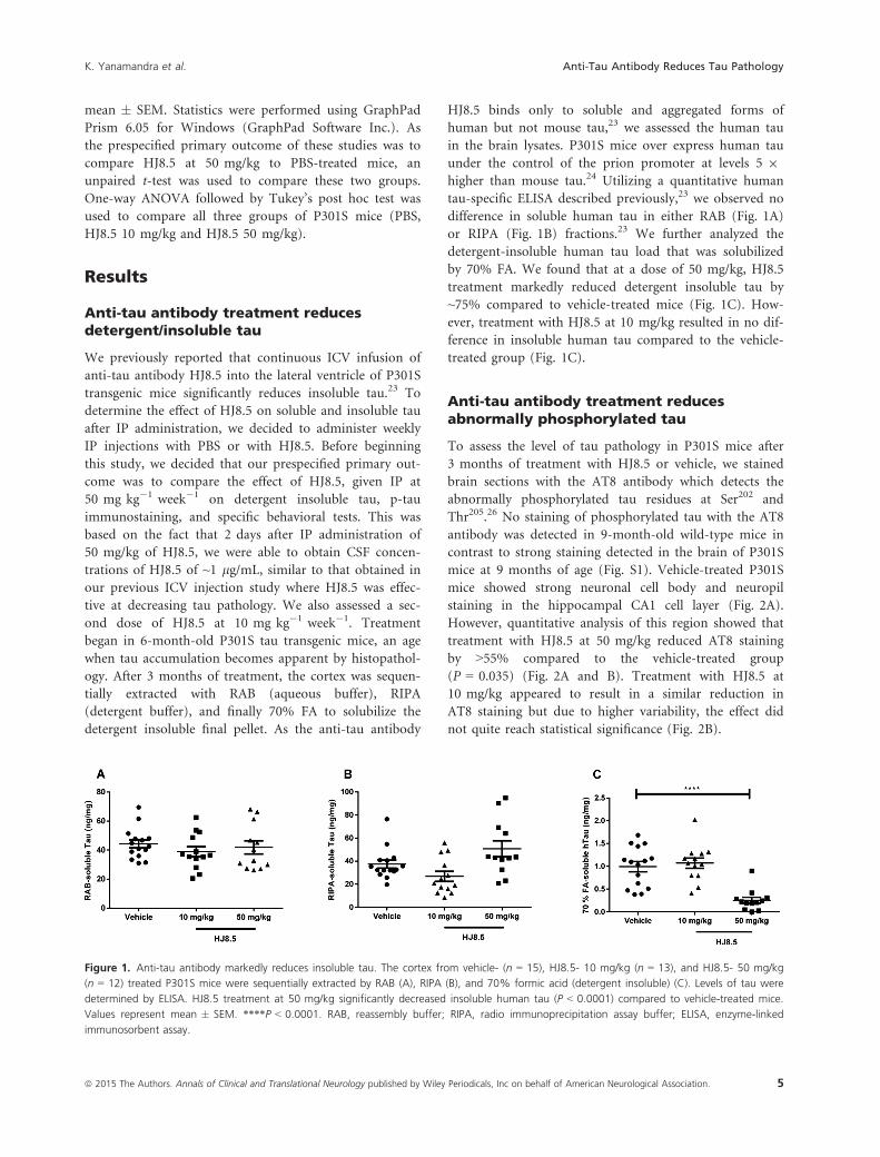
tau-specific ELISA described previously,23 we observed no
difference in soluble human tau in either RAB (Fig. 1A)
or RIPA (Fig. 1B) fractions.23 We further analyzed the
detergent-insoluble human tau load that was solubilized
by 70% FA. We found that at a dose of 50 mg/kg, HJ8.5
treatment markedly reduced detergent insoluble tau by
~75% compared to vehicle-treated mice (Fig. 1C). How-
ever, treatment with HJ8.5 at 10 mg/kg resulted in no dif-
ference in insoluble human tau compared to the vehicle-
treated group (Fig. 1C).
Anti-tau antibody treatment reducesabnormally phosphorylated tau
To assess the level of tau pathology in P301S mice after
3 months of treatment with HJ8.5 or vehicle, we stained
brain sections with the AT8 antibody which detects the
abnormally phosphorylated tau residues at Ser202 and
Thr205.26 No staining of phosphorylated tau with the AT8
antibody was detected in 9-month-old wild-type mice in
contrast to strong staining detected in the brain of P301S
mice at 9 months of age (Fig. S1). Vehicle-treated P301S
mice showed strong neuronal cell body and neuropil
staining in the hippocampal CA1 cell layer (Fig. 2A).
However, quantitative analysis of this region showed that
treatment with HJ8.5 at 50 mg/kg reduced AT8 staining
by >55% compared to the vehicle-treated group
(P = 0.035) (Fig. 2A and B). Treatment with HJ8.5 at
10 mg/kg appeared to result in a similar reduction in
AT8 staining but due to higher variability, the effect did
not quite reach statistical significance (Fig. 2B).
Figure 1. Anti-tau antibody markedly reduces insoluble tau. The cortex from vehicle- (n = 15), HJ8.5- 10 mg/kg (n = 13), and HJ8.5- 50 mg/kg
(n = 12) treated P301S mice were sequentially extracted by RAB (A), RIPA (B), and 70% formic acid (detergent insoluble) (C). Levels of tau were
determined by ELISA. HJ8.5 treatment at 50 mg/kg significantly decreased insoluble human tau (P < 0.0001) compared to vehicle-treated mice.
Values represent mean � SEM. ****P < 0.0001. RAB, reassembly buffer; RIPA, radio immunoprecipitation assay buffer; ELISA, enzyme-linked
immunosorbent assay.
ª 2015 The Authors. Annals of Clinical and Translational Neurology published by Wiley Periodicals, Inc on behalf of American Neurological Association. 5
K. Yanamandra et al. Anti-Tau Antibody Reduces Tau Pathology

Anti-tau antibody treatment reduced thio-Sstaining
To test the effects of HJ8.5 on tau present in an amyloid
conformation, we stained all the treated mice with thio-S.
We semiquantitatively analyzed thio-S staining in two
regions of brain of P301S mice where we could easily
detect such deposits, in the piriform cortex and amygdala.
Assessment of thio-S-positive staining was done in all
treated animals by a blinded rater who gave a score from
1 (minimal staining) to 5 (maximal staining) as described
previously.23 We found that HJ8.5-treated animals
showed less thio-S-positive staining compared to vehicle-
treated mice (Fig. 3A). By semiquantitative analysis of all
treated mice, we observed there was a significant decrease
in thio-S-positive staining in mice treated with HJ8.5 at
both 10 and 50 mg/kg compared to vehicle-treated mice
(Fig. 3B).
Anti-tau antibody reduces brain atrophy inP301S mice
P301S mice develop not only tau aggregation but also
prominent neurodegeneration. In prior characterization,
it has been shown that there is significant and progressive
loss of brain volume in these mice in both the hippocam-
pus and neocortex at both 9 and 12 months of age. How-
ever, no significant difference in brain atrophy or
neuronal loss was observed in these mice at 6 months of
age compared to wild-type mice.24 This gave us an
opportunity to determine effects of HJ8.5 not only on tau
pathology but also to determine its effects on the brain
atrophy observed in this model. To determine whether
HJ8.5 treatment had any effect on brain volume loss, we
performed a blinded stereological volumetric analysis of
the cortex and hippocampus in P301S mice at 9 months
of age following 3 months of treatment with HJ8.5 or
PBS. In mice treated with HJ8.5 at 50 mg/kg, both the
cortex and hippocampus were significantly larger than in
Figure 2. Anti-tau antibody decreased phospho-tau staining in the
hippocampal CA1 cell layer. (A) Representative coronal sections of
biotinylated AT8 antibody staining of phosphorylated tau in the
hippocampal CA1 cellular region of 9-month-old P301S mice treated
for 3 months with vehicle and HJ8.5 at 50 mg/kg. The lower
images are higher power views of the CA1 region in the uppers
panels. Red arrows indicate the area magnified in the lower image.
Black arrows indicate the hippocampal CA1 cell layer. (B)
Quantification of biotinylated AT8 antibody staining of abnormally
phosphorylated tau revealed a significant decrease in AT8 staining in
mice treated with HJ8.5 at 50 mg/kg in the hippocampal CA1
cellular layer compared to vehicle-treated mice (P = 0.035). HJ8.5 at
10 mg/kg treated mice also showed decreased AT8 staining
compared to the vehicle-treated group, but this was not statistically
significant (P > 0.05). Values represent mean � SEM. *P < 0.05.
Scale bar represents 300 lm.
Figure 3. Thio-S staining is decreased in anti-tau antibody-treated
P301S mice. (A) Representative coronal sections of thio-S staining of
tau aggregates in piriform cortex and amygdala in 9-month-old P301S
mice treated for 3 months with 10 and 50 mg kg�1 week�1 of HJ8.5
as well as from vehicle-treated mice. (B) Semiquantitative analysis of
thio-S staining was rated on a scale of 1 (no staining) to 5 (maximum
staining) in all treatment groups. Both doses of HJ8.5 antibody
treatment showed significantly lower thio-S staining compared to
vehicle-treated mice. Values represent mean � SEM. *P < 0.05. Scale
bar represents 25 lm.
6 ª 2015 The Authors. Annals of Clinical and Translational Neurology published by Wiley Periodicals, Inc on behalf of American Neurological Association.
Anti-Tau Antibody Reduces Tau Pathology K. Yanamandra et al.

PBS-treated animals (Fig. 4). The cortex was ~5% larger
(Fig. 4B) and the hippocampus ~10% larger (Fig. 4C) in
HJ8.5 versus PBS-treated mice. There was a strong trend
of decreased atrophy with 10 mg/kg but it was not quite
significant. To determine whether HJ8.5 treatment was
preventing versus decreasing brain atrophy, we assessed
the total hippocampal and cortical volumes in a separate
cohort of 6-month-old P301S mice. Relative to 6-month-
old P301S mice, the hippocampus was 33% and the
cortex was 17% smaller in PBS-treated P301S mice at
9 months (Fig. 4). In contrast, the HJ8.5 treatment
groups exhibited a ~23% smaller hippocampal and ~12%smaller cortical volume. This suggests that peripheral
HJ8.5 treatment significantly attenuated but did not block
brain atrophy.
Anti-tau antibody improved motor/sensorimotor deficits in P301S mice
To determine whether HJ8.5 administration had any
effect on function in P301S mice, we assessed motor
coordination, balance, and strength since in some studies,
motor/sensorimotor function has been shown to be
impaired in these mice.24,28 To evaluate these functions,
we quantified the performance of all mice on both the
inverted screen and ledge tests. Results from the inverted
screen test, which requires both strength and coordina-
tion, showed that the mice treated with HJ8.5 at 50 mg/
kg exhibited improved performance relative to vehicle-
treated controls as evidenced by the HJ8.5-treated mice
being able to hold onto the inverted screen for
significantly longer periods of time (Fig. 5A). We also
assessed performance of all mice on the ledge test, which
requires balance, grip strength, and coordination. This
test involves quantifying the amount of time a mouse can
remain on a narrow Plexiglas “ledge.” Analysis of the
ledge test data showed again that the mice treated with
HJ8.5 at 50 mg/kg demonstrated significantly improved
performance compared to the PBS-treated group
(Fig. 5B). The ability of the anti-tau antibody treatments
to rescue cognitive deficits in P301S mice was evaluated
by assessing the performance of the mice on the condi-
tioned fear procedure. All three treatment groups of mice
exhibited similar levels of baseline freezing during the first
2 min in the conditioning chamber and there were no
significant group comparisons found regarding the freez-
ing levels observed during the tone-shock training which
involved three pairings of the conditioned stimulus–unconditioned stimulus (CS-US) on day 1 (data not
shown). On day 2, mice were evaluated on the contextual
fear test. On this test, mice treated with HJ8.5 at both 10
and 50 mg/kg displayed increased freezing time levels
compared to vehicle controls, but the amount of
increased freezing was not significant (Fig. 5C).
Anti-tau antibody promotes tau aggregatesuptake in a microglial cell line as well asincreasing tau levels in plasma
Following peripheral administration of IgG antibodies
such as HJ8.5, ~0.1–0.2% of the concentration of the
antibody in the plasma is present in the extracellular
space of the brain (i.e., CSF).23 Once present in the extra-
cellular space of the CNS, such an antibody can bind to
and sequester both monomeric and aggregated forms of
tau which might be present. Once HJ8.5 is bound to tau,
it could potentially neutralize any potential toxicity of
extracellular tau that is present. It could also lead to local
clearance by cells or transport out of the brain. We
wanted to test potential mechanisms of how such an anti-
body might be promoting the clearance of different forms
of tau. Previous studies with anti-Ab and anti-a-synucleinantibodies have shown that certain antibodies are able to
trigger microglial cells to clear aggregates through Fc
receptor-mediated phagocytosis and subsequent peptide
degradation.29–32 To examine the potential ability of anti-
tau antibody HJ8.5 to initiate cellular clearance of tau
Figure 4. HJ8.5 treatment decreased brain atrophy. (A) Repre-
sentative coronal sections of 9-month-old P301S mice treated with
vehicle or HJ8.5 (50 mg/kg). Quantification of cortical (B) and
hippocampal (C) volumetric analysis were performed among 6-month-
old P301S mice (n = 10) and 9-month-old P301S mice treated for
3 months with vehicle (n = 15), HJ8.5 – 10 mg/kg (n = 13), and
HJ8.5 – 50 mg/kg (n = 12). HJ8.5-treated mice at 50 mg/kg had
increased cortical and hippocampal volumes (*P < 0.05) compared to
vehicle-treated mice. Values represent mean � SEM.
ª 2015 The Authors. Annals of Clinical and Translational Neurology published by Wiley Periodicals, Inc on behalf of American Neurological Association. 7
K. Yanamandra et al. Anti-Tau Antibody Reduces Tau Pathology

aggregates, we labeled P301S tau aggregates with Alexa-
Fluor-647. Labeled tau aggregates were preincubated with
HJ8.5 or control antibody (HJ3.4, anti-Ab antibody).
Preincubated tau aggregates with or without antibodies
were applied to primary cortical neurons as well as the
microglial cell line, BV2. We observed no significant
difference in tau aggregate uptake in the presence or
absence of HJ8.5 versus control antibody HJ3.4 using
primary cortical neurons (Fig. 6A). However, in the pres-
ence of microglial-like BV2 cells, there was an increase in
tau aggregate uptake over time under all conditions with
HJ8.5, resulting in significant increased uptake compared
to control conditions (Fig. 6B). These results suggest that
HJ8.5 can promote the clearance of extracellular tau
aggregates into microglial-like cells via an Fcc-mediated
clearance mechanism.
Previous studies using anti-Ab antibodies have shown
that following peripheral administration of certain antibod-
ies, there is a large increase in plasma Ab.33–35 To test
whether an anti-tau antibody elevates plasma tau, we mea-
sured the levels of plasma tau in all treated mice by quanti-
tative ELISA. We observed a strong, dose-dependent
increase in plasma tau levels in HJ8.5-treated mice
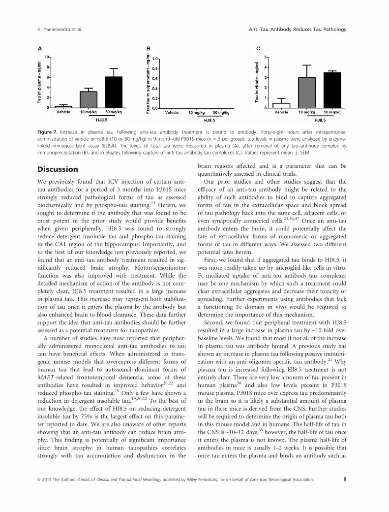
(Fig. 6C). To determine whether this increase in plasma
tau is free or bound to antibody, we injected HJ8.5 or vehi-
cle in a separate group of P301S mice. Forty-eight hours
following a single peripheral IP injection of HJ8.5 (10 or
50 mg/kg) or vehicle, mice were sacrificed and their plasma
was collected. We observed that the large increase in plasma
tau following HJ8.5 administration is antibody bound
(Fig. 7A and C). Free tau was not detected in the plasma of
HJ8.5-treated mice (Fig. 7B).
Figure 5. Anti-tau antibody treatment improves sensorimotor function in P301S mice. (A) Mice treated with HJ8.5 at 50 mg/kg exhibited
improved performance on the inverted screen test compared to the phosphate-buffered saline (PBS)-treated control group by being able to
remain on inverted screen for a significantly longer time compared to the controls. (B) Likewise, the mice treated with HJ8.5 at 50 mg/kg were
able to remain on a narrow Plexiglas “ledge” for a significantly longer time relative to the vehicle-treated group. (C) A trend toward improved
performance was observed in the mice treated with HJ8.5 compared to controls in terms of freezing levels averaged across the 8-min session
during the contextual fear test conducted on day 2, although these differences were not statistically significant. *P < 0.05. Values represent
mean � SEM.
Figure 6. HJ8.5 treatment increased the uptake of aggregated tau in BV2 cells and tau levels in plasma. Alexa-Fluor-647-labeled P301S
aggregates (P301S-agg A647) were preincubated with HJ8.5 (P301S-agg A647 + HJ8.5) or control antibody HJ3.4 (P301S-agg A647 + HJ3.4)
overnight. Then Alexa-Fluor-647 dye alone (A647) or preincubated labeled P301S aggregates were added to primary cortical neurons (A) or
microglial BV2 cells (B). Uptake of labeled P301S tau aggregates were measured by flow cytometry. A significant increase in P301S tau aggregate
uptake in microglial BV2 cells was observed in the presence of HJ8.5 compared to either no antibody or control antibody (P < 0.0001, two-way
analysis of variance [ANOVA] followed by Tukey’s post hoc test). However, there was no difference in uptake of tau aggregates in primary
cortical neurons in the presence or absence of HJ8.5. (C) After 3 month administration of two different doses of HJ8.5 (10 and 50 mg/kg) from
6 months till 9 months in P301S mice, tau levels in plasma were analyzed by ELISA. A dose-dependent increase of tau in plasma compared to
controls was observed (P < 0.0001). Values represent mean � SEM.
8 ª 2015 The Authors. Annals of Clinical and Translational Neurology published by Wiley Periodicals, Inc on behalf of American Neurological Association.
Anti-Tau Antibody Reduces Tau Pathology K. Yanamandra et al.
Discussion
We previously found that ICV injection of certain anti-
tau antibodies for a period of 3 months into P301S mice
strongly reduced pathological forms of tau as assessed
biochemically and by phospho-tau staining.23 Herein, we
sought to determine if the antibody that was found to be
most potent in the prior study would provide benefits
when given peripherally. HJ8.5 was found to strongly
reduce detergent insoluble tau and phospho-tau staining
in the CA1 region of the hippocampus. Importantly, and
to the best of our knowledge not previously reported, we
found that an anti-tau antibody treatment resulted in sig-
nificantly reduced brain atrophy. Motor/sensorimotor
function was also improved with treatment. While the
detailed mechanism of action of the antibody is not com-
pletely clear, HJ8.5 treatment resulted in a large increase
in plasma tau. This increase may represent both stabiliza-
tion of tau once it enters the plasma by the antibody but
also enhanced brain to blood clearance. These data further
support the idea that anti-tau antibodies should be further
assessed as a potential treatment for tauopathies.
A number of studies have now reported that peripher-
ally administered monoclonal anti-tau antibodies to tau
can have beneficial effects. When administered to trans-
genic mouse models that overexpress different forms of
human tau that lead to autosomal dominant forms of
MAPT-related frontotemporal dementia, some of these
antibodies have resulted in improved behavior20,22 and
reduced phospho-tau staining.19 Only a few have shown a
reduction in detergent insoluble tau.19,20,22 To the best of
our knowledge, the effect of HJ8.5 on reducing detergent
insoluble tau by 75% is the largest effect on this parame-
ter reported to date. We are also unaware of other reports
showing that an anti-tau antibody can reduce brain atro-
phy. This finding is potentially of significant importance
since brain atrophy in human tauopathies correlates
strongly with tau accumulation and dysfunction in the
brain regions affected and is a parameter that can be
quantitatively assessed in clinical trials.
Our prior studies and other studies suggest that the
efficacy of an anti-tau antibody might be related to the
ability of such antibodies to bind to capture aggregated
forms of tau in the extracellular space and block spread
of tau pathology back into the same cell, adjacent cells, or
even synaptically connected cells.23,36,37 Once an anti-tau
antibody enters the brain, it could potentially affect the
fate of extracellular forms of monomeric or aggregated
forms of tau in different ways. We assessed two different
potential fates herein.
First, we found that if aggregated tau binds to HJ8.5, it
was more readily taken up by microglial-like cells in vitro.
Fc-mediated uptake of anti-tau antibody-tau complexes
may be one mechanism by which such a treatment could
clear extracellular aggregates and decrease their toxicity or
spreading. Further experiments using antibodies that lack
a functioning Fc domain in vivo would be required to
determine the importance of this mechanism.
Second, we found that peripheral treatment with HJ8.5
resulted in a large increase in plasma tau by ~10-fold over
baseline levels. We found that most if not all of the increase
in plasma tau was antibody bound. A previous study has
shown an increase in plasma tau following passive immuni-
zation with an anti-oligomer-specific tau antibody.21 Why
plasma tau is increased following HJ8.5 treatment is not
entirely clear. There are very low amounts of tau present in
human plasma38 and also low levels present in P301S
mouse plasma. P301S mice over express tau predominantly
in the brain so it is likely a substantial amount of plasma
tau in these mice is derived from the CNS. Further studies
will be required to determine the origin of plasma tau both
in this mouse model and in humans. The half-life of tau in
the CNS is ~10–12 days,39 however, the half-life of tau once
it enters the plasma is not known. The plasma half-life of
antibodies in mice is usually 1–2 weeks. It is possible that
once tau enters the plasma and binds an antibody such as
Figure 7. Increase in plasma tau following anti-tau antibody treatment is bound to antibody. Forty-eight hours after intraperitoneal
administration of vehicle or HJ8.5 (10 or 50 mg/kg) in 9-month-old P301S mice (n = 3 per group), tau levels in plasma were analyzed by enzyme-
linked immunosorbent assay (ELISA). The levels of total tau were measured in plasma (A), after removal of any tau–antibody complex by
immunoprecipitation (B), and in eluates following capture of anti-tau antibody-tau complexes (C). Values represent mean � SEM.
ª 2015 The Authors. Annals of Clinical and Translational Neurology published by Wiley Periodicals, Inc on behalf of American Neurological Association. 9
K. Yanamandra et al. Anti-Tau Antibody Reduces Tau Pathology

HJ8.5, plasma tau levels increase due to the antibody pre-
venting rapid tau degradation as the half-life of the HJ8.5-
tau antibody complex is long. Even if this is one explana-
tion for the increase in plasma tau, it seems likely that part
of this increase is due to the HJ8.5 that enters the brain,
captures tau, and then exits the CNS. In fact, following
injection of HJ8.5 into the CNS, we found a large amount
of tau bound to HJ8.5 in the periphery.23 Since the concen-
tration of an IgG in the CNS extracellular space is 0.1–0.2%of the concentration of IgG in plasma and IgG is constantly
moving in and out of the brain, it is likely that once HJ8.5
enters the brain extracellular space, it binds to whatever tau
is present (monomers and aggregates) and these complexes
take on the fate of the antibody, a component of which is
cleared into blood via glymphatic flow.40 It will be interest-
ing to determine whether the concentration of plasma tau
following peripheral anti-tau antibody in some way reflects
CNS tau concentrations as well as the amount of tau
pathology. Clearly, further experiments need to be done to
sort out the origins of tau in plasma in the presence and
absence of anti-tau antibodies.
While the greatest effects of HJ8.5, including effects on
detergent insoluble tau, phospho-tau staining, atrophy,
and behavior were seen at a dose of 50 mg/kg, some
effects were also seen at 10 mg/kg. The fact that the
10 mg/kg dose had these effects without decreasing deter-
gent insoluble tau is interesting and suggests the possibil-
ity that at somewhat lower concentrations, the antibody is
able to bind to some sort of toxic extracellular species of
tau and neutralize its negative functional effects. This type
of mechanism was suggested by a recent study.21 While
there is as yet no direct evidence for this possibility, if
future methods allow for detection of extracellular species
of tau oligomers in vivo, this hypothesis may be testable.
Acknowledgment
This work was funded by grants from the Tau Consor-
tium (D. M. H. and M. I. D.), C2N Diagnostics (D. M.
H. and M. I. D.), Cure Alzheimer’s Fund (D. M. H.), and
the JPB Foundation (D. M. H.).
Conflict of Interest
D. M. H. cofounded and is on the scientific advisory
board of C2N Diagnostics. D. M. H., M. I. D., and H. J.
are inventors on a submitted patent “Antibodies to Tau”
that is licensed by Washington University. D. M. H.
consults for Genentech, AstraZeneca, Neurophage, and
Eli Lilly. Washington University and UTSW receive
grants that support laboratory research of D. M. H. and
M. I. D. from Janssen. M. I. D. is a cofounder of ARTA
Biosciences.
References
1. Mandelkow EM, Mandelkow E. Biochemistry and cell
biology of tau protein in neurofibrillary degeneration. Cold
Spring Harb Perspect Med 2012;2:a006247.
2. Hutton M, Lendon CL, Rizzu P, et al. Association of
missense and 50-splice-site mutations in tau with the
inherited dementia FTDP-17. Nature 1998;393:702–705.3. Poorkaj P, Bird TD, Wijsman E, et al. Tau is a candidate
gene for chromosome 17 frontotemporal dementia. Ann
Neurol 1998;43:815–825.
4. Hardy J, Singleton A. The HapMap: charting a course for
genetic discovery in neurological diseases. Arch Neurol
2008;65:319–321.5. Drechsel DN, Hyman AA, Cobb MH, Kirschner MW.
Modulation of the dynamic instability of tubulin assembly
by the microtubule-associated protein tau. Mol Biol Cell
1992;3:1141–1154.6. Trinczek B, Biernat J, Baumann K, et al. Domains of tau
protein, differential phosphorylation, and dynamic
instability of microtubules. Mol Biol Cell 1995;6:1887–1902.7. Arriagada PV, Growdon JH, Hedley-Whyte ET, Hyman
BT. Neurofibrillary tangles but not senile plaques parallel
duration and severity of Alzheimer’s disease. Neurology
1992;42:631–639.8. Polydoro M, Acker CM, Duff K, et al. Age-dependent
impairment of cognitive and synaptic function in the htau
mouse model of tau pathology. J Neurosci 2009;29:10741–
10749.
9. Small SA, Duff K. Linking Abeta and tau in late-onset
Alzheimer’s disease: a dual pathway hypothesis. Neuron
2008;60:534–542.
10. Yamada K, Cirrito JR, Stewart FR, et al. In vivo
microdialysis reveals age-dependent decrease of brain
interstitial fluid tau levels in P301S human tau transgenic
mice. J Neurosci 2011;31:13110–13117.
11. Clavaguera F, Bolmont T, Crowther RA, et al.
Transmission and spreading of tauopathy in transgenic
mouse brain. Nat Cell Biol 2009;11:909–913.12. Frost B, Diamond MI. Prion-like mechanisms in
neurodegenerative diseases. Nat Rev Neurosci 2010;11:155–159.
13. Frost B, Jacks RL, Diamond MI. Propagation of tau
misfolding from the outside to the inside of a cell. J Biol
Chem 2009;284:12845–12852.14. Bi M, Ittner A, Ke YD, et al. Tau-targeted immunization
impedes progression of neurofibrillary histopathology in
aged P301L tau transgenic mice. PLoS One 2011;6:e26860.
15. Boimel M, Grigoriadis N, Lourbopoulos A, et al. Efficacy
and safety of immunization with phosphorylated tau
against neurofibrillary tangles in mice. Exp Neurol
2010;224:472–485.
16. Asuni AA, Boutajangout A, Quartermain D, Sigurdsson
EM. Immunotherapy targeting pathological tau conformers
10 ª 2015 The Authors. Annals of Clinical and Translational Neurology published by Wiley Periodicals, Inc on behalf of American Neurological Association.
Anti-Tau Antibody Reduces Tau Pathology K. Yanamandra et al.
in a tangle mouse model reduces brain pathology with
associated functional improvements. J Neurosci
2007;27:9115–9129.17. Boutajangout A, Quartermain D, Sigurdsson EM.
Immunotherapy targeting pathological tau prevents
cognitive decline in a new tangle mouse model. J Neurosci
2010;30:16559–16566.
18. Troquier L, Caillierez R, Burnouf S, et al. Targeting
phospho-Ser422 by active Tau immunotherapy in the
THYTau22 mouse model: a suitable therapeutic approach.
Curr Alzheimer Res 2012;9:397–405.
19. d’Abramo C, Acker CM, Jimenez HT, Davies P. Tau
passive immunotherapy in mutant P301L mice: antibody
affinity versus specificity. PLoS One 2013;8:e62402.
20. Boutajangout A, Ingadottir J, Davies P, Sigurdsson EM.
Passive immunization targeting pathological phospho-tau
protein in a mouse model reduces functional decline and
clears tau aggregates from the brain. J Neurochem
2011;118:658–667.
21. Castillo-Carranza DL, Sengupta U, Guerrero-Munoz MJ,
et al. Passive immunization with Tau oligomer
monoclonal antibody reverses tauopathy phenotypes
without affecting hyperphosphorylated neurofibrillary
tangles. J Neurosci 2014;34:4260–4272.22. Chai X, Wu S, Murray TK, et al. Passive immunization
with anti-Tau antibodies in two transgenic models:
reduction of Tau pathology and delay of disease
progression. J Biol Chem 2011;286:34457–34467.23. Yanamandra K, Kfoury N, Jiang H, et al. Anti-tau
antibodies that block tau aggregate seeding in vitro
markedly decrease pathology and improve cognition in
vivo. Neuron 2013;80:402–414.24. Yoshiyama Y, Higuchi M, Zhang B, et al. Synapse loss and
microglial activation precede tangles in a P301S tauopathy
mouse model. Neuron 2007;53:337–351.
25. Paxinos G, Franklin KBJ. The mouse brain in stereotactic
coordinates. New York: Academic Press, 2008.
26. Goedert M, Jakes R, Vanmechelen E. Monoclonal antibody
AT8 recognises tau protein phosphorylated at both serine
202 and threonine 205. Neurosci Lett 1995;189:167–169.
27. Wozniak DF, Hartman RE, Boyle MP, et al. Apoptotic
neurodegeneration induced by ethanol in neonatal mice is
associated with profound learning/memory deficits in
juveniles followed by progressive functional recovery in
adults. Neurobiol Dis 2004;17:403–414.28. Iba M, Guo JL, McBride JD, et al. Synthetic tau fibrils
mediate transmission of neurofibrillary tangles in a
transgenic mouse model of Alzheimer’s-like tauopathy.
J Neurosci 2013;33:1024–1037.29. Bacskai BJ, Kajdasz ST, Christie RH, et al. Imaging of
amyloid-beta deposits in brains of living mice permits
direct observation of clearance of plaques with
immunotherapy. Nat Med 2001;7:369–372.
30. Bae EJ, Lee HJ, Rockenstein E, et al. Antibody-aided
clearance of extracellular alpha-synuclein prevents cell-to-cell
aggregate transmission. J Neurosci 2012;32:13454–13469.31. Bard F, Cannon C, Barbour R, et al. Peripherally
administered antibodies against amyloid beta-peptide enter
the central nervous system and reduce pathology in a mouse
model of Alzheimer disease. Nat Med 2000;6:916–919.
32. Lee HJ, Suk JE, Bae EJ, Lee SJ. Clearance and deposition
of extracellular alpha-synuclein aggregates in microglia.
Biochem Biophys Res Commun 2008;372:423–428.33. DeMattos RB, Bales KR, Cummins DJ, et al. Peripheral
anti-A beta antibody alters CNS and plasma A beta
clearance and decreases brain A beta burden in a mouse
model of Alzheimer’s disease. Proc Natl Acad Sci USA
2001;98:8850–8855.
34. Lemere CA, Spooner ET, LaFrancois J, et al. Evidence for
peripheral clearance of cerebral Abeta protein following
chronic, active Abeta immunization in PSAPP mice.
Neurobiol Dis 2003;14:10–18.
35. Winkler DT, Abramowski D, Danner S, et al. Rapid
cerebral amyloid binding by Abeta antibodies infused into
beta-amyloid precursor protein transgenic mice. Biol
Psychiatry 2010;68:971–974.
36. de Calignon A, Polydoro M, Suarez-Calvet M, et al.
Propagation of tau pathology in a model of early
Alzheimer’s disease. Neuron 2012;73:685–697.37. Liu L, Drouet V, Wu JW, et al. Trans-synaptic spread of
tau pathology in vivo. PLoS One 2012;7:e31302.
38. Zetterberg H, Wilson D, Andreasson U, et al. Plasma tau
levels in Alzheimer’s disease. Alzheimers Res Ther
2013;5:9.
39. Yamada K, Holth JK, Liao F, et al. Neuronal activity
regulates extracellular tau in vivo. J Exp Med
2014;211:387–393.40. Iliff JJ, Wang M, Zeppenfeld DM, et al. Cerebral arterial
pulsation drives paravascular CSF-interstitial fluid exchange
in the murine brain. J Neurosci 2013;33:18190–18199.
Supporting Information
Additional Supporting Information may be found in the
online version of this article:
Figure S1. Phospho-tau staining of wild-type and P301S
tau transgenic mouse brains with AT8 antibody. Repre-
sentative coronal sections of phosphorylated tau staining
with AT8 antibody in 9-month-old wild-type (A) and
P301S mouse brain (B). Higher magnification views of
coronal sections of hippocampus from 9-month-old wild-
type (C) and P301S mouse hippocampus (D) stained with
AT8 antibody. Scale bar in B (for A and B) and C (for C
and D) represents 500 lm.
ª 2015 The Authors. Annals of Clinical and Translational Neurology published by Wiley Periodicals, Inc on behalf of American Neurological Association. 11
K. Yanamandra et al. Anti-Tau Antibody Reduces Tau Pathology
![[Posterior cortical atrophy]](https://static.fdokumen.com/doc/165x107/6331b9d14e01430403005392/posterior-cortical-atrophy.jpg)