The Economic Efficiency of Telecommunications in a Deregulated Market: The Case of New Zealand
Upload
khangminh22Category
view
0download
0
OR I G INA L ART I C L E
NH2-truncated human tau induces deregulatedmitophagy in neurons by aberrant recruitment of Parkinand UCHL-1: implications in Alzheimer’s diseaseV. Corsetti1, F. Florenzano2, A. Atlante3, A. Bobba3, M.T. Ciotti4, F. Natale4,F. Della Valle4, A. Borreca4, A. Manca2, G. Meli2, C. Ferraina2, M. Feligioni2,S. D’Aguanno4, R. Bussani5, M. Ammassari-Teule4, V. Nicolin6, P. Calissano2,†
and G. Amadoro1,2,†,*1Institute of Translational Pharmacology (IFT) – National Research Council (CNR), Via Fosso del Cavaliere100-00133, Rome, Italy, 2European Brain Research Institute (EBRI), Via del Fosso di Fiorano 64-65, 00143 Rome,Italy, 3Institute of Biomembranes and Bioenergetics (IBBE)-CNR, Via Amendola 165/A, 70126 Bari, Italy, 4Instituteof Cellular Biology and Neuroscience (IBCN)-CNR, IRCSS Santa Lucia Foundation Via del Fosso di Fiorano 64-65,00143 Rome, Italy, 5UCO Pathological Anatomy and Histopathology Unit, Cattinara Hospital Strada di Fiume 447,34149 Trieste, Italy and 6Department of Medical, Surgical and Health Science, University of Trieste, Strada diFiume 449, 34149 Trieste, Italy
*Towhom correspondence should be addressed at: Institute of Translational Pharmacology (IFT) – National Research Council (CNR), Via Fosso del Cavaliere,100-00133 Rome, Italy. Tel: +39 06501703269; Fax: +39 06501703313; Email: [email protected]
AbstractDisarrangement in functions and quality control of mitochondria at synapses are early events in Alzheimer’s disease (AD)pathobiology.We reported that a 20–22 kDaNH2-tau fragmentmapping between 26 and 230 amino acids of the longest human tauisoform (aka NH2htau): (i) is detectable in cellular and animal AD models, as well in synaptic mitochondria and cerebrospinalfluids (CSF) from human AD subjects; (ii) is neurotoxic in primary hippocampal neurons; (iii) compromises the mitochondrialbiology both directly, by inhibiting the ANT-1-dependent ADP/ATP exchange, and indirectly, by impairing their selectiveautophagic clearance (mitophagy). Here, we show that the extensive Parkin-dependent turnover of mitochondria occurring inNH2htau-expressing post-mitotic neurons plays a pro-death role and that UCHL-1, the cytosolic Ubiquitin-C-terminal hydrolaseL1 which directs the physiological remodeling of synapses by controlling ubiquitin homeostasis, critically contributes tomitochondrial and synaptic failure in this in vitro AD model. Pharmacological or genetic suppression of improper mitophagy,either by inhibition of mitochondrial targeting to autophagosomes or by shRNA-mediated silencing of Parkin or UCHL-1 geneexpression, restores synaptic andmitochondrial content providing partial but significant protection against theNH2htau-inducedneuronal death. Moreover, in mitochondria from human AD synapses, the endogenous NH2htau is stably associated with Parkinand with UCHL-1. Taken together, our studies show a causative link between the excessive mitochondrial turnover and theNH2htau-induced in vitro neuronal death, suggesting that pathogenetic tau truncationmay contribute to synaptic deterioration inADbyaberrant recruitment of Parkin andUCHL-1 tomitochondriamaking themmore prone todetrimental autophagic clearance.
† These authors equally contributed to this work.Received: November 12, 2014. Revised: January 29, 2015. Accepted: February 10, 2015
© The Author 2015. Published by Oxford University Press. All rights reserved. For Permissions, please email: [email protected]
Human Molecular Genetics, 2015, Vol. 24, No. 11 3058–3081
doi: 10.1093/hmg/ddv059Advance Access Publication Date: 15 February 2015Original Article
3058
Dow
nloaded from https://academ
ic.oup.com/hm
g/article/24/11/3058/719750 by guest on 01 April 2022
IntroductionN-terminal truncation of tau is an early event in Alzheimer’sdisease (AD) and not-AD tauopathies (1–4). Mitochondrial dys-function (5–9) as well as unbalance in protein ubiquitylation/de-ubiquitylation (10–13) are chronic and precipitating mechan-isms underlying the tau-induced synaptic degeneration whichhas been shown to be closely correlated to impaired cognitivefunction and memory loss in AD pathology (14,15). Cellular andtransgenic AD models have shown that Aβ and tau can in inde-pendent and/or synergic way(s) adversely affect metabolism ofmitochondria-including alteration in their trafficking, dynamicsand clearance (16–23)-favoring bioenergetic failure and synapsesloss (24–26). In the last few years a deregulated turnover of mito-chondria (27–29), which leads to a reduction in their biomass andmetabolic capacities, has been described to take place into vul-nerable neurons and synapses from AD brains (30–33) as well asfromAD transgenicmice (34), thus supporting strategies aimed atpreserving a proper quality control of these organelles as noveland promising disease-modifying interventions in therapy ofthis devastating disease (35,36). However, the exact and causalrole of unbalancedmitophagy in neurodegeneration is still largelydebated. In fact, on the one hand, autophagic elimination ofmitochondria plays a neuroprotective role under conditions ofacute and/or mild stress or at the beginning of insult, when elim-ination of damaged mitochondria can be properly counterba-lanced by an equivalent functional reserve of these organellesthrough a homeostatic induction of their biogenesis (37,38). Onthe other hand, under exposure to lethal and/or chronic injuries,the overactivated removal of mitochondria exceeds the normalregenerative capacity of mitochondrial biogenesis and, conse-quently, massive mitophagy turns out to be neurotoxic leadingto an insufficient amount of intact and respiration-competentmitochondria which, eventually, causes ‘mitophagic’ death par-ticularly in suffering or aged neuronal populations (39–43).
Compelling in vitro and in vivo evidence has also indicated thatthe free pool of ubiquitin represents an unifying cellular hub link-ing the two major intracellular proteolytic systems, the ubiqui-tin-proteasome system (UPS) and the selective autophagy(44,45), and that a tight control of ubiquitin steady levels is ofgreat importance for synaptic homeostasis andneuronal survival(12). Actually the Parkin-driven mitophagic pathway, a selectiveform of autophagy which is crucial in maintaining an healthyand energetic-competent mitochondrial network in post-mitoticneuronsmainly at ATP-consuming synapses (38,46–48), relies notonly on the ubiquitylation of several mitochondrial outer mem-brane proteins (49–53) but also on direct recruitment and activa-tion of UPS components onto mitochondria (54–57). Amongmembers belonging to family of neuron-specific deubiquitinat-ing enzymes (DUBs), Ubiquitin-C-terminal hydrolase L1 (UCHL-1,PGP9.5) is the most abundant (1–2% of soluble brain proteins)and multifunctional one, being endowed with two different ac-tivities: it can act both as hydrolase, by removing and recyclingubiquitin molecules from target proteins, and as ubiquityl ligase,by generating polyubiquitin chains which tag them for disposal.Moreover, UCHL-1 is also able to associate with ubiquitin to in-hibit its degradation in neurons (58) and therefore its primaryrole at synapses (59,60) is to critically control their structureand/or functions by locally maintaining ubiquitin homeostasisin vitro as well as in vivo (61,62). An impaired activity of UCHL-1has been proved to be linked to synaptic failure in AD neuro-degeneration (63–66) and its immunoreactivity is prominentlydetected in tau-laden, tangles-bearing neurons which arelocated in selectively vulnerable and affected regions (65,67,68).
Interestingly, the ubiquitin E3 ligase Parkin and other proteins in-volved in mitochondrial energy metabolism and/or glycolysishave been recently identified as UCHL-1 interacting proteins(69,70), suggesting that this enzyme may serve in neurons as apossible modifier in Parkin-dependent mitochondrial turnover.However, whether dysregulation in UCHL-1 activity can actuallypredispose to AD neurodegeneration by adversely affectingmito-chondrial dynamics at synapses is still unknown.
We have previously reported that a NH2-26-230 tau fragment(s) (aka NH2htau), mapping between 26 and 230 amino acids ofthe longest human isoform (htau40): (i) is neurotoxic in primarycultured neurons (71,72); (ii) is detected in cellular and animal ADmodels (73), as shown by other authors (74–76); (iii) is largely en-riched in humanmitochondria fromAD synaptosomes, in correl-ation with synaptic disarrangement, with load of Aβ oligomersand with organelle functional impairment (77). The 20–22 kDaNH2htau, but not the physiological full-length protein, preferen-tially interacts with Aβ peptide oligomers at human AD synapsesand cooperates with them in inhibiting the adenine nucleotidetranslocator-1 (ANT-1)-dependent ADP/ATP exchange at mito-chondria (17). Moreover, the NH2htau fragment is also presentoutside neurons in peripheral CSF from living patients affectedby tau-dependentneurodegenerative diseases associatedwith de-mentia, providing a novel biomarker for AD and not-AD humantauopathies (78). Interestingly, in vivo expression of NH2htaufragment activates a non-apoptotic cell death pathway causingin transgenic mice severe defects of hippocampal neurogenesisin connection with an increased anxiety-behavior and impairedepisodic-likememory (79). Finally, an extensive Parkin-dependentautophagic clearance of mitochondria (mitophagy) occurs inNH2htau, expressing post-mitotic neurons in concomitance tobioenergetic deficits and synaptic damages (80).
In order to better clarify the molecular mechanisms under-lying the NH2htau-induced perturbations in quality control ofneuronal mitochondria and to understand how this deregulatedmitophagy could be related to AD neurodegeneration, here weshow, by cell-based and in vivo approaches, that: (i) the Parkin-dependent mitophagy plays a pro-death role in NH2htau-expressing post-mitotic neurons and (ii) UCHL-1 is a criticalmediator of mitochondrial decline and synaptic failure.
ResultsPharmacological and genetic inhibition of mitochondrialquality control preserves the loss of mitochondrialproteins, significantly attenuating the NH2htau-inducedin vitro neuronal death
We reported that NH2htau early affects the quality control ofmitochondria in post-mitotic neurons leading to: (i) net reductionin their mass in correlation with a general Parkin-mediated re-modeling of membrane proteome; (ii) their extensive associationwith LC3 and LAMP1 autophagic markers; (iii) bioenergetic defi-cits and (iv) in vitro synaptic pathology (80). However, whetherthe NH2htau-enhanced autophagic turnover of mitochondria ex-acerbates neuronal death because of depletion of functionalmito-chondria or, alternatively, is a compensatory attempt to mitigateneuronal damage, still remains to be clarified. Therefore, withthe intent of evaluating whether the increased mitochondrialclearance occurring in NH2htau-expressing neuronsmay be bene-ficial or rather toxic, we interfered with mitochondria loss in twoalternative but complementary ways: (1) by blocking the autopha-gic machinery directly; (2) by up-regulating the intracellular levelsof Mitofusin 2 (Mfn2) and OPtic Atrophy-1 (OPA-1), two proteins
Human Molecular Genetics, 2015, Vol. 24, No. 11 | 3059
Dow
nloaded from https://academ
ic.oup.com/hm
g/article/24/11/3058/719750 by guest on 01 April 2022
which directly control the mitochondrial dynamics and whosereduction in expression has proved to facilitate the mitophagy(81–83), as detectable in NH2htau-expressing neurons (80).
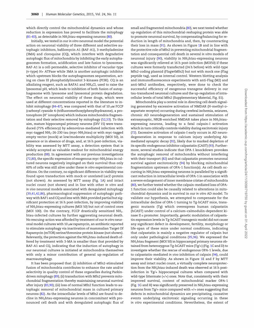
Initially, we tested in our in vitro neuronal model the potentialaction on neuronal viability of three different and selective au-tophagic inhibitors, bafilomycin A1 (BAF-A1), 3-methyladenine(3MA) and cloroquine (CQ), which interfere with degradativeautophagic flux of mitochondria by inhibiting the early autopha-gosomes formation, acidification and late fusion to lysosomes.BAF-A1 is a cell-permeable, selective inhibitor of vacuolar-type(v-type) H+ ATPase while 3MA is another autophagic inhibitorwhich upstream blocks the autophagosomes sequestration, act-ing on class III phosphatidylinositol 3-kinases (PI3K). CQ is analkalizing reagent, such as BAFA1 and NH4Cl, used to raise thelysosomal pH, which leads to inhibition of both fusion of autop-hagosome with lysosome and lysosomal protein degradation.The effect on neuronal viability of these drugs, which wereused at different concentrations reported in the literature to in-hibit mitophagy (84–87), was compared with that of 10 μ FCCP(carbonyl cyanide 4-(trifluoromethoxy)phenylhydrazone), a pro-tonophore (H+ ionophore) which inducesmitochondria fragmen-tation and their selective removal by mitophagy (52,53). To thisaim, mature hippocampal primary neurons (DIV 15) were trans-duced (75% efficiency) by adenovirus-mediated infection withmyc-tagged NH2-26-230 tau (myc-NH2htau) or with myc-taggedempty vector (mock) at low/moderate multiplicity (MOI 50), inpresence or in absence of drugs, and, at 16 h post-infection, via-bility was assessed by MTT assay, a detection system that iswidely accepted as valuable readout for mitochondrial energyproduction (88). In agreement with our previous investigations(72,80), the specific expression of exogenousmyc-NH2htau in cul-tured neurons negatively impinged on their survival thus only60% of cells was still alive under these in vitro experimental con-ditions. On the contrary, no significant difference in viability wasfound upon transduction with mock or unrelated LacZ-protein(not shown). As assessed by MTT assay (Fig. 1A) and intactnuclei count (not shown) and in line with other in vitro andin vivo neuronal models associated with deregulated mitophagy(39,41,42,86), pharmacological suppression of autophagic path-waywith BAF1 andCQand lesswith 3MAprovidedpartial but sig-nificant protection at 16 h post-infection, by improving viabilityof NH2htau-expressing cultures (MOI 50) but not of control ones(MOI 100). On the contrary, FCCP drastically sensitized NH2-
htau-infected cultures by further aggravating neuronal death.No rescuing-actionwas afforded by treatment of our in vitroneur-onalmodel cultureswith 10 μ rapamycin, an antibiotic reportedto stimulate autophagy via inactivation of mammalian Target OfRapamycin (mTOR) serine/threonine protein kinase (not shown).Relevantly, the protection against theNH2htau-induced death of-fered by treatment with 3-MA is smaller than that provided byBAF-A1 and CQ, indicating that the induction of autophagy inour neuronal cultures is initiated at mitochondria themselveswith only a minor contribution of general up-regulation ofmacroautophagy.
It has been proposed that: (i) inhibition of Mfn2-stimulatedfusion of mitochondria contributes to enhance the rate and/orselectivity in quality control of these organelles during Parkin-drivenmitophagy (89); (ii) transductionwithMfn2 preventsmito-chondrial fragmentation thereby maintaining neuronal survivalafter injury (83,90); (iii) loss of normal Mfn2 function leads to au-tophagic removal of mitochondrial mass in cultured primaryneurons (82). As the intracellular levels of Mfn2 are found to de-cline in NH2htau-expressing neurons in concomitant with pro-nounced cell death and with deregulated autophagic flux of
small and fragmentedmitochondria (80), we next testedwhetherup-regulation of this mitochondrial-reshaping protein was ableto promote neuronal survival, by compensating/balancing for re-duction in length of mitochondria and, then, by counteractingtheir loss in mass (91). As shown in Figure 1B and in line withthe protective role of Mfn2 in preventingmitochondrial fragmen-tation and consequential cell death in several in vitro models ofneuronal injury (90), viability in NH2htau-expressing neuronswas significantly relieved at 16 h post-infection (MOI50) if thesecultures were formerly transfected (24 h before) with wild-typeMfn2-coding plasmid (FlagwtMfn2) but not with mock one (Flagpeptide tag), used as internal control. Western blotting analysisand immunofluorescence experiments with anti-Flag (M2) andanti-Mfn2 antibodies, respectively, were done to check thesuccessful efficiency of exogenous transgene delivery in ourtau-transduced neuronal cultures and the up-regulation of intra-cellular levels of total Mfn2 (Supplementary Material, Fig. S1).
Mitochondria play a central role in directing cell death signal-ing generated by excessive activation of NMDAR (N-methyl--aspartate receptor) occurring during cerebral ischemia, seizures,chronic AD neurodegeneration and sustained stimulation ofextrasynaptic, NR2B-enriched NMDAR takes place in NH2htau-expressing neurons, leading to a fatal calpain-I activationwhich in turn critically controls viability during excitotoxic injury(72). Excessive activation of calpain-I early occurs in AD neuro-degeneration in response to calcium injury underlying Aβ-mediated synaptic decay (92), likely due to marked depletion ofits specific endogenous inhibitor calpastatin (CAST) (93). Further-more, several studies indicate that OPA-1 knockdown provokesthe autophagic removal of mitochondria without interferingwith their transport (82) and that calpastatin promotes neuronalsurvival against excitotoxicity (94) by blocking mitochondrialfragmentation upstream of OPA-1 function(s). As cell death oc-curring in NH2htau-expressing neurons is paralleled by a signifi-cant reduction in intracellular levels of OPA-1 in association witha severe enlargement of innermembrane cristae ofmitochondria(80), we further testedwhether the calpain-mediated loss of OPA-1 function could also be causally related to alterations in mito-chondrial dynamics and in survival in our in vitro AD model. Tovalidate our hypothesis, we attempted to compensate for theintracellular decline of OPA-1 turning to Tg-hCAST mice, trans-genic mutants (Tg) which overexpress human calpastatin(hCAST) under control of a calcium–calmodulin-dependent ki-nase II α promoter. Importantly, genetic modulation of calpasta-tin expression levels in Tg-hCAST transgenicmodel did not causeany significant defect in development, fertility, morphology orlife-span of these mice under normal conditions, indicatingthat calpastatin is mainly a negative regulator of calpain butonly under pathological conditions (95,96). We expressed theNH2htau fragment (MOI 50) in hippocampal primary neurons ob-tained fromheterozygous Tg-hCASTmice (Tg/+) (Fig. 1C andD) toinvestigate whether the rescue of endogenous OPA-1 levels, dueto calpastatin-mediated in vivo inhibition of calpain (94), couldimprove their viability. As shown in Figure 1E and F by MTTassay and intact nuclei count, a virtually complete neuroprotec-tion from the NH2htau-induced death was observed at 16 h post-infection in Tg/+ hippocampal cultures when compared withwild-type littermate (+/+) ones. Note that, consistently with theirimproved survival, content of mitochondrial marker OPA-1(Fig. 1G and H) was significantly preserved in NH2htau-expressingneurons from Tg/+ mice compared with +/+ ones suggesting thatdefects in mitochondrial dynamics are precipitating, causativeevents underlying excitotoxic signaling occurring in thesein vitro experimental conditions. Nevertheless, the extent of
3060 | Human Molecular Genetics, 2015, Vol. 24, No. 11
Dow
nloaded from https://academ
ic.oup.com/hm
g/article/24/11/3058/719750 by guest on 01 April 2022
protection afforded by the calpastatin overexpression in neuronswas much greater than that observed by wtMfn2 overexpressionimplicating that, as we and others previously reported (72,97),
other mechanisms acting on direct calpain-inhibition canpromote survival of injured NH2htau-expressing cultures. Alter-natively, neuroprotection provided by wtMfn2 overexpression
Figure 1. Genetic modulation of mitochondrial dynamics provides moderate but significant neuroprotection from NH2htau-induced death. (A) Graph shows neuronal
viability, evaluated by mitochondria-based MTT assay on hippocampal primary neurons (DIV 15) adenovirally transduced with myc-NH2htau (MOI 50) or with mock
control (MOI 100) and examined at 16 h post-infection, in absence or in presence of different autophagic inhibitors used at the indicated concentrations. Values are
means of nine independent experiments and statistically significant differences were calculated by unpaired two-tailed Student’s t-test (*P < 0.05; ***P < 0.0001).
(B) Hippocampal primary neurons were transfected by Nucleofector kit (AMAXA) for 24 h with plasmid expressing Flag-tag alone, as internal control, or Flag-tagged
wtMfn2 and then infected with myc-NH2htau (MOI 50) and with mock control (MOI 100) for additional 16 h. Neuronal viability was evaluated by MTT assay. Values are
means of three independent experiments and statistically significant differences were calculated by unpaired two-tailed Student’s t-test (*P < 0.05; ***P < 0.0001). (C andD)
Representative genotype test by PCR analysis (C) showing the specific 350 bp band of hCAST in heterozygous transgenicmice (Tg) that overexpress the human calpastatin
(hCAST) construct (Tg/+). Only the 100 bp band of GADPH internal control was present inWT littermates (+/+). Immunoblot analysis using an antibody specific for hCAST
(200) confirms the expression of human calpastatin protein in cultures from Tg/+ transgenic animals, with no detectable levels in +/+ littermates (D). (E and F) Primary
hippocampal neurons (DIV 15), obtained fromheterozygous transgenicmice (Tg) that overexpress the human calpastatin (hCAST) construct (Tg/+) or fromWT littermates
(+/+), were infected with myc-NH2htau (MOI 50) and with mock control (MOI 100) for 16 h. Neuronal viability was evaluated by MTT assay (E) and intact nuclei count (F).
Values aremeans of three independent experiments and statistically significant differenceswere calculated by unpaired two-tailed Student’s t-test (*P < 0.05; ***P < 0.0001).
(G and H) Representative blot of western blotting analysis (n = 3) on total protein extracts from hippocampal primary neurons (DIV 15) that overexpress the human
calpastatin (hCAST) construct (Tg/+) or from WT littermates (+/+), infected at MOI 50 for 16 h with myc-NH2htau (MOI 50) and with mock control (MOI 100) to assess
the content of the mitochondria marker OPA-1. β-III tubulin was used as loading control (G) and densitometric quantification was performed (H). Values are means of
three independent experiments and statistically significant differences were calculated by unpaired two-tailed Student’s t-test (*P < 0.05; ***P < 0.0001 versus mock;
**P < 0.01 versus NH2htau/+/+).
Human Molecular Genetics, 2015, Vol. 24, No. 11 | 3061
Dow
nloaded from https://academ
ic.oup.com/hm
g/article/24/11/3058/719750 by guest on 01 April 2022

was significant but not complete owing to residual toxicity ofmitochondria-targeted NH2htau on ANT-1-mediated ADP/ATPexchange.
Altogether these data demonstrate that pharmacological andgenetic suppression ofmitophagy provides partial but significantprotection against the NH2htau-induced neuronal death, point-ing out that neuronal demise induced in vitro by the enhancedintracellular levels of NH2htau, as we detected in vivo in humanAD brains (18,77), is at least in part due to an excessive inductionof signaling pathway(s) for quality control of mitochondria.
Parkin plays a critical role in mitophagyin NH2htau-dying neurons
Recruitment of the endogenous Parkin onto depolarized mito-chondria—which is early detected in our NH2htau expressingcultured neurons undergoing increased mitophagy, synapticchanges and cell death (80)—is critically required in mature neu-rons for selective elimination of mitochondria through the au-tophagy-lysosomal pathway (50,52,53,57,98,99). Furthermore,hexokinase activity triggers mitophagy in post-mitotic neuronsas it upstream controls relocalization of Parkin to defectivemito-chondria and supports glycolysis, allowing for ATP synthesis(98,100). For these reasons, we first tested whether the moderate(MOI 50) and early in vitro expression of NH2htau fragment posi-tively elevated hexokinase activity in our 16 h-infected primaryneurons. As shown in Figure 2A, which reports values assessedin total cell homogenates (for detail see methods) from mock-and NH2htau-tranduced group, a significant increase in hexoki-nase activity (normalized to total homogenate proteins content)was detected in NH2htau-infected cultures when compared withcontrols, consistently with finding that a robust Parkin-mediatedmitophagy in neurons is strictly dependent on their bioenergeticstatus (98,100). Importantly, variations in hexokinase activitytook place in NH2htau-expressing neurons regardless of equiva-lent abundance of protein, as checked by western blottinganalysis on whole-cell extracts (Fig. 2A), proving thus that itsup-regulation under our in vitro conditions was not attributableto differences in intracellular expression levels but rather to atruly positive modulation in enzymatic activity.
In view of the fact that our pharmacological and genetic datademonstrated that deregulated mitophagic pathway was causal-ly involved in our in vitro AD model (Fig. 1), we next analyzedwhether Parkin was actually associated with NH2htau-inducedcell death by accelerating lysosomal-mediated clearance ofmito-chondria. To address this possibility, mitochondrial dyshomeos-tasis as well as loss in synaptic content and cell viability weremonitored in our tau-transduced cultures after specific target-gene silencing with small hairpin (sh)RNA plasmid for Parkinexpression. Consistently with the protective role of Parkin sup-pression in another cellular paradigm of neurodegenerationcharacterized by excessive mitophagy following the in vitro over-expression of mutant A53T α-synuclein (41), we found that neur-onal survival evaluated at 16 post-infection by MTT assay wassignificantly improved in NH2htau-expressing neurons (MOI 50)by shRNAknock-downing of Parkin (48 h before). On the contrary,no rescue in viability was afforded by non-target shRNA (shRNAscrambled), used as negative internal control (Fig. 2B). High-effi-ciency of Parkin silencing was checked by western blotting ana-lysis followed by densitometric quantification (Fig. 2C and D)and no change in protein levels of myc-NH2htau was detectedafter shRNA treatment of neuronal cultures up to 48 h (Fig. 2E).More importantly, specific inhibition of Parkin expression,which is per se relatively nontoxic in primary neurons (41), not
only partially reverted cell death in NH2htau-infected culturesbut also suppressed the drop inmitochondrial as well in synapticprotein content, as shownbywestern blottingwithOPA-1 and sy-napsin I antibodies respectively (Fig. 2E).
We have previously reported that neither clearance of defect-ive mitochondria and, consequently, nor significant cell deathare detected upon NH2htau overexpression in HeLa (80), a cellline which has little or no endogenous expression of Parkin(87,101,102). In order to definitively validate the pro-death roleof Parkin under these experimental in vitro conditions, we testedwhether ectopic expression of a specific Parkin-coding plasmid inHeLa cell type could restore susceptibility to NH2htau-inducedmitochondrial injury. As shown in Figure 2F, viability assessedby MTT assay was significantly affected at 16 h post-infection(MOI 50) if HeLa cells were exogenously endowed (24 h before)with expression vector for Parkin protein. Confocal microscopy(Fig. 2G) was carried out to check the efficiency (∼75%) of suc-cessful delivery of exogenous mCherry Parkin transgene in thiscell line.
Altogether these findings indicate the hexokinase-dependent, Parkin-overdriven mitophagy crucially affectssynapses stability and, then, cell viability in in vitro NH2htau-expressing neurons.
UCHL1 relocalizes to mitochondria and criticallycontributes to their deregulated autophagic eliminationin NH2htau-expressing neurons
Accumulating evidence reveals that the efficient removal ofdysfunctional mitochondria can be achieved by the synergisticand/or parallel actions of UPS and autophagic-lysosomal path-way (54–56) and that the UPS components are directly recruitedand activated on mitochondria during Parkin-dependent mito-phagy (55). Intriguingly, activation, translocation and enzymaticfunctions of Parkin during mitochondrial quality control arecontrolled by ubiquitylation (103–105) and reversible monoubi-quitination negatively regulates the activity of UCHL-1 (106).Besides, proteomic approaches have recently identified UCHL-1as a novel Parkin-binding partner in SH-SY5Y neuronal cell lineundergoing FCCP-induced mitophagy (69,70), consistently withits ubiquitin-stabilizing activity in neurons (58).
In view of the above-mentioned considerations, we set out toestablish whether UCHL-1 actually served as modifier in the Par-kin-dependentmitophagyoccurring inNH2htau-expressing neu-rons. To this aim, we first investigated whether clearance ofmitochondria following the moderate expression of NH2htaufragment was associated with recruitment of UCHL-1 to mito-chondria, in a similar way to that we reported to occur in ourin vitromodel for Parkin which targets these organelles triggeringthus their autophagic elimination (80). As shown by biochemicalseparation of crude cytosolic/mitochondrial proteins obtainedfrom 16 h-infected neurons (Fig. 3A), we found that a stableproportion of UCHL-1 was distributed at mitochondria in ourtau-transduced neurons because it was partially recovered inmitochondria-enriched fractions. Notably, the mitochondrialrelocalization of UCHL-1 occurred in connection with that ofboth overexpressed myc-NH2htau (MOI 50) and endogenous Par-kin which, in agreement with our previous investigations (80),appeared nearly excluded from corresponding cytosolic com-partments. Consistently with our biochemical results, confocalanalysis (Fig. 3B and B′) performed on 16 h-infected hippocampalcultures (MOI 50) following double immunofluorescence withUCHL-1 (green) and Tomm20 or Grp75 (red), which are two differ-ent mitochondrial markers used routinely to detect activation of
3062 | Human Molecular Genetics, 2015, Vol. 24, No. 11
Dow
nloaded from https://academ
ic.oup.com/hm
g/article/24/11/3058/719750 by guest on 01 April 2022

Figure 2. Parkin is involved in the NH2htau-induced mitochondrial removal (A) Spectrophotometric determination of activity (expressed as % of control) of hexokinase
(HK1), was assayed through a coupled reaction with glucose-6-phosphate dehydrogenase (G6PDH), following the NADP reduction at 340 nm (ε340 nm = 6.22 m−1 cm−1).
Absorbance was recorded with a Jasco doublebeam/double-wavelength spectrophotometer UV-550 and was normalized to the total proteins content of whole-cell
homogenates from mock- or NH2htau-tranduced neurons (MOI50) at 16 h post-infection (for detail see Materials and Methods). Values are means of five independent
experiments and statistically significant differences were calculated by unpaired two-tailed Student’s t-test (***P < 0.0001 versus mock). Western blotting analysis on
total protein extracts from hippocampal primary neurons (DIV 15) infected with myc-NH2htau (MOI 50) or with mock control (MOI 100) for 16 h shows no difference in
cellular HK1 amount. β-III tubulin wasmonitored as loading control. (B–E) In (B) Hippocampal neurons were transfected by Nucleofector kit (AMAXA) with target-specific
Parkin shRNA plasmid (shRNA Parkin) or with scrambled plasmid (shRNAscrambled), used as negative control. After 48 h, cultureswere infectedwithmyc-NH2htau (MOI
50) andwithmock control (MOI 100) and viability was assessed by MTT assay at 16 h post-infection. The histogram reports the neuronal survival calculated as the ratio of
respective myc-NH2htau/mock values in shRNA Parkin-treated samples over the corresponding ones in shRNAscrambled-treated controls. Values are means of three
independent experiments and statistically significant differences were calculated by unpaired two-tailed Student’s t-test (***P < 0.0001 versus shRNAscrambled). In
(C and D) Suppression of Parkin expression in neuronal cultures was validated at 48 h by western blotting analysis on total proteins extracts (C) and down-regulation
in expression levels of protein was quantified by densitometry normalizing on β-III tubulin signal (D). Values are means of three independent experiments and
statistically significant differences were calculated by unpaired two-tailed Student’s t-test (**P < 0.01 versus shRNAscrambled). In (E) western blotting analysis (n = 3) on
total protein extracts from hippocampal primary neurons transfected for 48 h with shRNAParkin plasmid or with shRNAscambled one and then infected with myc-
NH2htau (MOI 50) and with mock control (MOI 100) for additional 16 h. Immunoblots were probed with specific antibodies against mitochondrial and synaptic
markers, such as OPA-1 and synapsin I, respectively. The exogenous expression of myc-NH2htau was unchanged by treatments and densitometric quantification of
immunoreactivity levels were calculated by normalizing the values on the β-III tubulin intensity. Values are means of three independent experiments and statistically
significant differences were calculated by unpaired-two tailed Student’s t-test (***P < 0.0001 versusmock; **P < 0.01 versus shRNAscrambled). Note that Parkin knockdown
attenuated the loss of mitochondrial and synaptic content in NH2htau-expressing group. (F and G) HeLa cells were 48 h transfected with plasmid expressing mCherry
alone, as internal control, or with mChrerryParkin and then infected with myc-NH2htau (MOI 50) and with mock control (MOI 100). Viability was examined at 16 h
post-infection by MTT assay (F). Values are means of three independent experiments and statistically significant differences were calculated by unpaired two-tailed
Student’s t-test (***P < 0.0001). Note that ectopic introduction of Parkin-coding expression vector into HeLa cell-type restored mitochondrial vulnerability to NH2htau-
induced injury. Confocal microscopy analysis (G) on HeLacells transfected for 48 h with mCherry Parkin (red channel) was performed to check the successful delivery
of exogenous transgene. Nuclei (blue channel) were stained with DAPI. A DIC (differential interference contrast) light transmitted channel was acquired to visualize
the whole-cell morphology. Scale bar: 10 μm.
Human Molecular Genetics, 2015, Vol. 24, No. 11 | 3063
Dow
nloaded from https://academ
ic.oup.com/hm
g/article/24/11/3058/719750 by guest on 01 April 2022

mitophagy in neurons (52,53,98,107), also displayed that aconsiderable amount of UCHL-1 was enriched in mitochon-dria from NH2htau-expressing neurons. Remarkably, althoughUCHL-1 staining appeared in control neurons essentially cyto-plasmatic and distributed to different subcellular domains, sev-eral UCHL-1-positive puncta colocalized with fragmented andcircle-shapedmitochondria accumulating into the somatoneuri-tic compartment (mito-aggresomes) inNH2htau-expressing neu-rons (Fig. 3B and B′ arrows). However, this distribution patternranged within different intensity degrees in number and size ofcolabeled dots (yellow) in clear association with the healthystate of neurons, suggesting that UCHL-1mitochondrial localiza-tion was directly related to cellular damage. In detail, viable neu-rons showed neither fragmented mitochondria nor dystrophicneuritis and UCHL-1 homogeneously compartmentalized bothin processes and cell body, in line with previous findings (61).Conversely, in dying neurons, dense clusters of round-shapedstructures, which were both positive for UCHL-1 and mitochon-drialmarkers (arrows), segregated into the somatic compartmentmainly in perinuclear region, in a way which is similar to that isevident during canonical mitophagy in primary neurons (52,53).On the contrary, mock-infected neurons displayed a healthy anduniformly distributed Tomm20-positive mitochondrial networkalong with a more disperse and not-compartmentalized UCHL-1 staining in the cytoplasm, consistently with its physiologicalregular expression at synapses, soma and neuritis in culturedhippocampal neurons (60,61,108).
We subsequently tested our hypothesis that UCHL-1 and Par-kin might colocalize at mitochondria in NH2htau-expressingneurons, suggesting a potential functional interaction betweenthese two proteins in vitro. Consistently with our biochemicaland morphological data (Fig. 3A and B) and with tandem affinitypurification/mass spectrometry (TAP)/MS) results by others(69,70), confocal analysis performed on 16 h-infected hippocam-pal cultures (MOI 50) following double immunofluorescencewithUCHL-1 (green) and Parkin antibodies (red) revealed that an in-creased staining in UCHL-1-positive dots, which were also Par-kin-labeled (yellow) and mainly distributed into the somaticperinuclear area (Fig. 3C′, arrows), was strongly noticeable afterNH2htau overexpression, again resembling the distribution ofroundish and damaged mitochondria detectable during theexplicit induction of selective mitophagy in neurons (52,53).Remarkably, this co-distribution pattern was also strictly remin-iscent of the one described above (UCHL-1/mitochondrialmarkers, Fig. 3B and B′), suggesting that at least part of thoseUCHL-1/Parkin positive spots were mitochondria. Conversely, adiffuse and essentially not-overlapping cytoplasmatic pattern ofUCHL-1 and Parkin was detected in mock-infected neurons. Inter-estingly, a comparable dotted co-staining pattern was alsoobserved under the same experimental conditions in NH2htau-dying neurons undergoing mitophagy but not in mock-infectedcultures after double immunofluorescence staining for exogenousmyc-tau (green) and Parkin (red) antibodies (Fig. 3C, arrows), sug-gesting that those Parkin-labeled punctate structures were alsopositive for truncated tau (yellow) and that at least part of themmight be also positive for UCHL-1. Taken together these morpho-logical data strongly suggest thatUCHL-1, Parkinandmyc-NH2htaucoexist at compromised spherical mitochondria in degeneratingneurons, in line with our in vitro results from biochemical cytosol/mitochondria fractionation (Fig. 3A, 80) and with in vivo observa-tions showing that NH2htau specifically interacts with rupturedanddisorganizedmitochondria in humanADconditions (18,74,77).
Next, in order to confirm the effective role of UCHL-1 in re-moval of dysfunctionalmitochondria inNH2htau-dying neurons,
we tested whether partial loss of UCHL-1 was per se able to rescuethe detrimental tau-induced mitophagy and, then, cell viabilityin our 16 h-infected hippocampal cultures. As shown in Figure 3DbyMTT assay at 16 h post-infection (MOI 50), neuronal death wassignificantly but partially relieved in NH2htau-expressing cul-tures if neurons were formerly transduced with specific shRNAplasmid knock-downing the UCHL-1 expression (48 h before)but not with non-target shRNA (shRNAscrambled) one (Fig. 3Eand F), in line with previous evidence showing that the suppres-sion of UCHL-1 activity can provide different outcomes undernormal versus pathological conditions in the context of α-synu-clein-induced neurodegeneration (108). Remarkably, as shownby western blotting with OPA-1 and synapsin I (Fig. 3G), thedownregulation of UCHL-1, which is moderately harmful in con-trol group (108), restored the mitochondrial as well as synapticprotein content in tau-transduced neurons, just resembling theprotective role afforded in these in vitromodel by partial suppres-sion of Parkin (Fig. 2B–E). Finally, no change in the protein levelsofmyc-NH2htauwas detected after shRNA treatment of neuronalcultures up to 48 h, ruling out the possibility of off-targets effectsresponsible of increased viability.
Remarkably, the physiological full-length human myc-tau1–441 (htau40), which we reported does not change neither theviability (71), nor the estimation ofmtDNA content or the expres-sion levels of proteins involved in the mitochondrial autophagyas well as in the synapses stability (80), when overexpressed inneuronal cultures for 16 h and at similar low levels (MOI 50) oftoxic NH2htau fragment, was also unable to alter the cytoplas-matic distribution of both Parkin and UCHL-1 (SupplementaryMaterial, Fig. S2), confirming again that the in vitro effects ofNH2-truncated tau are specific.
Taken together and in line with other papers reporting thatabnormal and deleterious binding of UCHL-1 to several subcellu-lar sites such as lysosomes (109,110) and endoplasmic reticulum(ER) (111) promotes the α-synuclein- induced neurotoxicity, ourfindings show that: (i) in NH2htau-expressing neurons undergo-ingmitophagy, a stable proportion of UCHL-1 relocalizes tomito-chondria and likely physically interacts with Parkin andexogenous myc-NH2htau fragment; (ii) the deficiency of UCHL-1, as well as of Parkin, acts as a protective modifier against theNH2htau-dependent in vitro neuronal death, by relieving autop-hagic mitochondrial loss and synapses demise.
Recruitment of UCHL-1 to mitochondria is also detectedin human AD brains as well as in transgenic AD micemodel
Increasing evidence has demonstrated that synaptic mitoc-hondria of hippocampal and cortical regions from human ADspecimens and presymptomatic (6-month-old) Tg2576 mice, awell-established AD animal model carrying the Swedish mutantform of Aβ precursor protein (AβPP), are more impaired thannon-synaptic ones in their morphology, metabolic functionsand quality control (8,32,112–115). In view of these considera-tions,we investigatedwhether themitochondrial targeting of en-dogenous NH2htau fragment, which we previously described totake place in vivo at synapses from human AD brains (17,77), isaccompanied by a parallel redistribution of Parkin and UCHL1to these organelles, inAD subjects aswell as in Tg2576 transgenicmice.
To this aim, purified synaptosomal preparations, which re-present ‘pinched-off nerve endings’ containing complete pre-synaptic terminals, mitochondria and synaptic vesicles alongwith postsynaptic membrane and postsynaptic density (PSD)
3064 | Human Molecular Genetics, 2015, Vol. 24, No. 11
Dow
nloaded from https://academ
ic.oup.com/hm
g/article/24/11/3058/719750 by guest on 01 April 2022
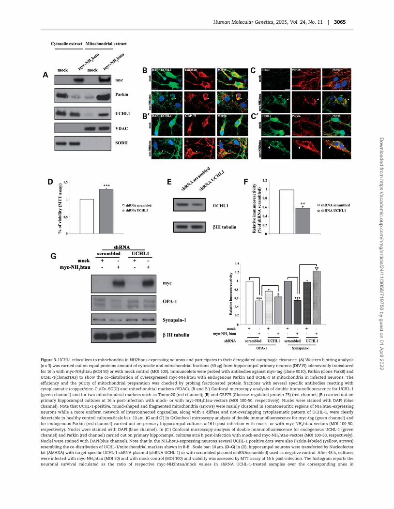
Figure 3. UCHL1 relocalizes to mitochondria in NH2htau-expressing neurons and participates to their deregulated autophagic clearance. (A) Western blotting analysis
(n = 3) was carried out on equal proteins amount of cytosolic and mitochondrial fractions (40 μg) from hippocampal primary neurons (DIV15) adenovirally transduced
for 16 h with myc-NH2htau (MOI 50) or with mock control (MOI 100). Immunoblots were probed with antibodies against myc-tag (clone 9E10), Parkin (clone Park8) and
UCHL-1(clone31A3) to show the co-distribution of overexpressed myc-NH2htau with endogenous Parkin and UCHL-1 at mitochondria in infected neurons. The
efficiency and the purity of mitochondrial preparation was checked by probing fractionated protein fractions with several specific antibodies reacting with
cytoplasmatic (copper/zinc-Cu/Zn-SODI) and mitochondrial markers (VDAC). (B and B′) Confocal microscopy analysis of double immunofluorescence for UCHL-1
(green channel) and for two mitochondrial markers such as Tomm20 (red channel); (B) and GRP75 (Glucose-regulated protein 75) (red channel; (B′) carried out on
primary hippocampal cultures at 16 h post-infection with mock- or with myc-NH2htau-vectors (MOI 100-50, respectively). Nuclei were stained with DAPI (blue
channel). Note that UCHL-1-positive, round-shaped and fragmented mitochondria (arrows) were mainly clustered in somatoneuritic regions of NH2htau-expressing
neurons while a more uniform network of interconnected organelles, along with a diffuse and not-overlapping cytoplasmatic pattern of UCHL-1, were clearly
detectable in healthy control cultures.Scale bar: 10 μm. (C and C′) In C:Confocal microscopy analysis of double immunofluorescence for myc-tag (green channel) and
for endogenous Parkin (red channel) carried out on primary hippocampal cultures at16 h post-infection with mock- or with myc-NH2htau-vectors (MOI 100-50,
respectively). Nuclei were stained with DAPI (blue channel). In (C′) Confocal microscopy analysis of double immunofluorescence for endogenous UCHL-1 (green
channel) and Parkin (red channel) carried out on primary hippocampal cultures at16 h post-infection with mock-and myc-NH2htau-vectors (MOI 100-50, respectively).
Nuclei were stained with DAPI(blue channel). Note that in the NH2htau-expressing neurons several UCHL-1 positive dots were also Parkin-labeled (yellow, arrows)
resembling the co-distribution of UCHL-1/mitochondrial markers shown in B-B′. Scale bar: 10 μm. (D–G) In (D), hippocampal neurons were transfected by Nucleofector
kit (AMAXA) with target-specific UCHL-1 shRNA plasmid (shRNA UCHL-1) or with scrambled plasmid (shRNAscrambled) used as negative control. After 48 h, cultures
were infected with myc-NH2htau (MOI 50) and with mock control (MOI 100) and viability was assessed by MTT assay at 16 h post-infection. The histogram reports the
neuronal survival calculated as the ratio of respective myc-NH2htau/mock values in shRNA UCHL-1-treated samples over the corresponding ones in
Human Molecular Genetics, 2015, Vol. 24, No. 11 | 3065
Dow
nloaded from https://academ
ic.oup.com/hm
g/article/24/11/3058/719750 by guest on 01 April 2022

(116), were first obtained from hippocampal regions of AD andage-matched cognitively intact human controls (ND). Humanisolated nerve terminals were then analyzed by western blottingwith CCP-NH2 tau antiserum (Caspase-Cleaved Protein-NH2tauantibody 26–36 residues), recognizing the endogenous 20–22 kDa NH2h fragment without any cross-reaction with intacthuman full-length tau (17,74), with Parkin and UCHL-1 anti-bodies. Consistently with our in vitro results of ectopic overex-pression of neurotoxic myc-NH2htau (Fig. 3A), biochemicalpartition of cytosolic/mitochondrial proteins carried out onsynapses-enriched fractions revealed that the elevated levels ofendogenous 20–22 kDa NH2htau distributed in AD mitochondriain correlation with a prominent immunoreactivity of Parkin(Fig. 4A, Supplementary Material, Fig. S3). Interestingly and inline with our previous investigations (77), we confirmed thatthe NH2htau fragment ran on SDS–PAGE as a doublet with anadditional faster band which stood constantly excluded frommitochondria beingmainly recovered in corresponding cytoplas-mic fraction (Fig. 4A, Supplementary Material, Fig. S3), likely dueto its unknown post-translational modification such as monou-biquitylation which has been proved to critically govern the sub-cellular localization of Parkin and control its activation and/ortranslocation during induction of canonical mitophagy (103–105). Moreover, a sizeable amount of UCHL-1 (Fig. 4A, Supple-mentary Material, Fig. S3) was also recovered in mitochondrialfraction from synapses-enriched diseased samples in differentlyanalyzed human brains, consistently with proportionally higherexpression levels of NH2htau associatedwith AD specimens thanthose derived from control ones.
Next the potential co-expression of NH2htau, Parkin andUCHL1 at synapses was also assessed in cortical regions ofTg2576 mice (6-month-old) in comparison with their wt litter-mates. In order to detect early pathogenic changes in mito-chondrial functions, which precede in this AD mice modelthe cognitive deterioration and Aβ aggregation starting atapproximately six months of age (117), our synaptosomal pre-parations were also stained for red-fluorescent dye MitotrackerCMxRos (membrane potential-dependent) before performingthe standard immunofluorescence procedures. Relevantly, ac-cumulation of this functional probe into the lipid environmentof mitochondria is driven in live cells by their membrane po-tential thus its fluorescence intensity indirectly indicates theenergetic state of these organelles, allowing us to selectivelyprobe synaptosomes endowed with still metabolically activemitochondria prior to overt neurodegeneration (7). Confocalmicroscopy of triple fluorescence for NH2htau/Mitotracker/Par-kin or NH2htau/Mitotracker/UCHL-1 (Fig. 4B, lower and upperpanels, respectively, of each experimental group) revealedthat in presymptomatic Tg2576 mice a large proportion ofstained synaptosomes co-expressed high levels of NH2htauand Parkin or UCHL-1 mainly at mitochondria (Fig. 4B, arrows).At variance, in wt mice, NH2htau exhibited low/very low basal
staining and never colocalized with Parkin which displayed anot-preferential both cytoplasmic and mitochodrial distribution.Likewise, a significant colocalization of UCHL-1 and NH2htau insynaptosomal mitochondria was not clearly detectable in not-transgenic healthy mice.
In order to better charactherize the actual sublocalization ofthese three proteins into synaptic compatments, we performedan additional extraction procedure on our purified synaptoneuro-somes, by separating the Triton X-soluble fraction and PSD pelletwhich still retain synaptophysin and PDS95 markers, respective-ly (Fig. 4C). At our surprise but consistentlywith recent reports in-dicating that pathogenetic soluble tau species also exhibit apresynaptic localization (118) causing thus defective changes inprobability of neurotransmitter release in P301L mutant humantau mice (119), we found that pathogenetic NH2htau was distrib-utedmainly into presynaptic fraction of both Tg and not-Tgmiceshowing an immunoreactivity which is much greater in un-healthy animals than in their control littermates. Besides, theimmunoreactivity of NH2htau fragment appeared as a doubletonly in presynaptic but not in postsynaptic samples, suggestingthat the unknown disease-associated transition from cytospla-matic to mitochondrial pathogenetic truncated tau startedand/or took place for the most part in this specific subsynapticregions, in line with the in vivo trans-synaptically spreading oftau pathology (120). Besides, UCHL-1 remained largely in pre-synaptic fraction, in agreement with the fact that only a slightsubpopulationof active protein isdistributed to spines andpostsy-naptic densities inneurons (61),while theproportionof Parkindis-played no significant difference by comparing the two synapticsubfractions in both blot profiles from Tg2576 and not-Tg mice.
On the whole, our results show that a significant co-enrichment and colocalization of endogenous NH2htaufragment, UCHL-1 and Parkin, occur at mitochondria in vivo inboth transgenic AD mice and AD brains, as we found in in vitroconditions (Fig. 3), and provide a hint that the pathogenetic accu-mulation of truncated tau may serve as docking site by stablysequestering UCHL-1 and Parkin on these organelles throughphysical anomalous protein–protein interaction.
Endogenous NH2htau interacts with Parkin and UCHL-1in human synaptic mitochondria from AD but not fromage-matched cognitively intact controls
Having established that a proportion of UCHL-1 relocalized tomitochondria and plays a critical role in Parkin-dependent mito-phagy inNH2htau-dying cultured neurons, we addressedwhetherits anomalous interactionand/orco-distributionwith endogenousmitochondria-targeted NH2htruncated tau (17,74,77) and Parkinalso tookplace in vivo, inhumanADtissues. To this aim, co-immu-noprecipitation (coIP) experiments on mitochondria-enrichedfractions from hippocampal synaptosomes of AD and age-matched, not-demented (ND) specimens were performed using
shRNAscrambled-treated controls. Values are means of three independent experiments and statistically significant differences were calculated by unpaired two-tailed
Student’s t-test (***P < 0.0001 versus shRNAscrambled). In (E and F) knockdown of UCHL-1 in neuronal cultures wasmonitored at 48 h bywestern blotting analysis on total
proteins extracts to assess the expression levels of target protein (E) and efficiency of gene silencingwas quantified by densitometry normalizing on β-III tubulin intensity
(F). Values are means of three independent experiments and statistically significant differences were calculated by unpaired two-tailed Student’s t-test (**P < 0.01 versus
shRNAscrambled). In (G) representative immunoblot (n = 3) of whole-cell lysates from hippocampal primary neurons transfected for 48 h with shRNAUCHL-1 plasmid or
with shRNAscambled one and then infected with myc-NH2htau (MOI 50) and with mock control (MOI 100) for additional 16 h by probing with specific antibodies against
mitochondrial and synaptic markers, such as OPA-1 and synapsin I, respectively. The exogenous expression of myc-NH2htau was unchanged by treatments and
densitometric quantification of immunoreactivity levels were calculated by normalizing the values on the β-III tubulin intensity, used as loading control. Values are
means of three independent experiments and statistically significant differences were calculated by unpaired two-tailed Student’s t-test (***P < 0.0001 versus mock;
**P < 0.01 versus shRNAscrambled; *P < 0.05 versus shRNAscrambled). Note that in vitro inhibition of UCHL-1 expression protects against NH2htau-induced
mitochondrial removal and synapses loss.
3066 | Human Molecular Genetics, 2015, Vol. 24, No. 11
Dow
nloaded from https://academ
ic.oup.com/hm
g/article/24/11/3058/719750 by guest on 01 April 2022

Figure 4. The endogenous pathogenetic NH2-derived 20–22 kDa tau fragment, Parkin and UCHL-1 co-segregate in synaptic mitochondria from human AD cases as well as
from Tg2576 ADmice (6-month-old). (A) The co-distribution of endogenous NH2-derived 20–22 kDa tau fragment, Parkin and UCHL-1 inmitochondriawas ascertained by
western blotting analysis comparing equal proteins amount of cytosolic and mitochondrial fractions (25 μg) from purified synaptosomes of AD (AD) and age-matched,
cognitively intact, not-demented (ND) human subjects. Immunoblots were probed with CCP-NH2 tau (NH2 aa2636 of human tau) antiserum recognizing the
endogenous 20–22 kDa NH2h fragment without any cross-reaction with intact human full-length tau (17,74), or with Parkin (clone Park8) or with UCHL-1 (PGP9.5 clone
31A3) antibodies. The efficiency and the purity of mitochondrial preparation was checked by probing fractionated protein fractions with several specific antibodies
reacting with cytoplasmatic (copper/zinc-Cu/Zn-SODI) (D) and mitochondrial markers (VDAC). (B) Confocal analysis of triple colocalization in synaptosomes from
cortical regions of Tg2576 mice (6-month-old) labeled with CCP-NH2 tau antiserum (green channel), MitoTraker Red (red channel) and with Parkin (blu channel) or
with UCHL-1 (blu channel). Panel shows fluoresecence single channels and fluorescence merge, DIC channel, fluorescence and DIC channel merge. Note that in
synaptic mitochondria from Tg2576 mice, the endogenous NH2htau fragment shows high expression levels and colocalization degree with either Parkin or UCHL-1
(arrows pointing to white dots which result from the merge of the three colours). Moreover, a proportion of NH2htau fragment targets mitochondria without Parkin of
UCHL-1 (yellow dots which result from the merge of the two colours, green and red) whereas its extra-mitochondrial staining (see above in (A) is also detectable
(green dots), in line with our previous investigantions (77). Conversely, in litteramate not-Tg mice, the lower basal expression levels of NH2htau expression do not
show any appreciable colocalization pattern with either Parkin or UCHL-1. Scale bar: 3 μm. (C) Biochemical distribution of endogenous NH2-derived 20–22 kDa tau
fragment, UCHL-1 and Parkin in synaptosomal subfractions from cortical regions of Tg2576 (6-month-old) and littermate WT mice. Purified synatoneurosomes were
further separated into the Triton X-extractable fraction and the crude PSD pellet, as shown by corresponding expression of synaptophysin and PSD95 markers.
Western blotting analysis was carried out on equal amount of synaptosomal subfractions and Ponceau staining was used as loading control. Immunoblots were
probed with CCP-NH2 tau, mAb Parkin and UCHL-1. The high expression levels of NH2htau in diseased AD mice are largely recovered in presynaptic fractions and co-
segregate with Parkin and UCHL-1 in the same subcellular compartment.
Human Molecular Genetics, 2015, Vol. 24, No. 11 | 3067
Dow
nloaded from https://academ
ic.oup.com/hm
g/article/24/11/3058/719750 by guest on 01 April 2022

as bait-antibody the CCP-NH2 tau antiserum (Caspase-CleavedProtein-NH2tau antibody 26–36 residues) recognizing the en-dogenous 20–22 kDa NH2h fragment without any cross-reactionwith intact human full-length tau (17). As shown (Fig. 5B) by im-munoblotting with a monoclonal detection-antibody againstParkin (clone Park8), a detectable proportionof endogenous Parkinco-imunoprecipitated with the NH2htau fragment from AD mito-chondria suggesting that an intact Parkin/NH2htau complex wasstably present in diseased neurons. Consistently with the un-detectable or low/very low intracellular levels of NH2htau frag-ment in physiological conditions, no comparable Parkin signalwas co-immunoprecipitated by ND synaptic mitochondria. Con-trol experiments demonstrated that NH2htau fragment was effi-ciently enriched from mitochondria by IP with the respectiveantibody (Fig. 5A) and no signalwas foundwhen the bait-antibodyCCP NH2tau was replaced by preimmune IgG (not shown). Like-wise, a small fraction of UCHL-1 was also co-immunoprecipitatedwith the NH2htau fragment from AD mitochondria but not fromcontrol ones (Fig. 5C), showing that a trimeric NH2htau/Parkin/UCHL-1 may exist only in pathological conditions whereas it ap-peared to be virtually excluded from not-diseased cases.
Reciprocal IP, carried out with the UCHL-1 capture-antibodyfollowed by western blotting detection with CCP-NH2 tau anti-serum, in vivo confirmed a stable UCHL-1/NH2htau physical inter-action in ADmitochondria (Fig. 5F). Likewise and in line with ourresults of biochemical subcellular fractionation (Fig. 4A), Parkinco-immunoprecipitated together with UCHL-1 but only in AD-af-fectedmitochondria-enriched fractions (Fig. 5E), confirming thusthat the pathological high levels of mitochondria-targeted NH2-
htau fragment (17) was able to intercept and trap both UCHL-1and Parkin on these organelles. Control experiments were per-formed to check that endogenous UCHL-1 protein was specifical-ly and successfully immunoprecipitated by the respectiveantibody (Fig. 5D).
Next, as we carried out on tau-infected cultured neurons(Fig. 3B, B′, C and C′), we addressed morphological coexpressionbetween endogenous NH2htau/UCHL-1 and NH2htau/Parkin byimmunofluorescence experiments performed on brain tissuesfrom AD and ND subjects. Confocal microscopy analysis on ADcerebral sections double-labeled with CCP-NH2htau (green) andUCHL-1 (red) antibodies revealed a net increase in fluorescenceintensity and number of co-labeled spots (yellow), akin to puta-tive fragmented mitochondrial clusters surrounding degenerat-ing and dysmorphic nuclei (Fig. 5L). Relevantly, in AD tissues(Fig. 5L, upper panel), the NH2htau-positive puncta which werealso strongly UCHL-1-labeled selectively concentrated into cellbody whereas, in age-matched ND brains and in correlationwith their faint expression levels of truncated tau (Fig. 5I upperpanel), a more disperse and regular localization of UCHL-1 wasdetectable. Specific staining was lost by omission of CCP-NH2
tau and UCHL-1 antibodies or by replacement of antibodiespre-adsorbed with their respective antigens or by preimmuneIgG (not shown).
Quantitative analysis of the fluorescence intensity confirmedand extended our qualitative morphological observations(Fig. 5I–L, lower panel). In AD cases, the immunofluorescenceintensity of NH2htau and UCHL-1 significantly increased if com-pared with ND ones, reaching a percentage increase of 24 and38%, respectively. For co-localization analysis, we took advantageof two different indexes, such as Pearson’s and Manders’ coeffi-cients. The Pearson’s coefficient in ND specimens was 0.34 ± 0.06and in AD ones was 0.77 ± 0.03, accounting for a 94% significativeincrease (Student’s t-test *P < 0.05). In the ND conditions, the Man-ders’ coefficients were 0.56 ± 0.06 for NH2htau/UCHL-1 ratio and
0.72 ± 0.18 for UCHL-1/NH2htau ratio. On the contrary, in AD, theManders’ coefficients were 0.67 ± 0.07 for NH2htau/UCHL-1 ratioand 0.75 ± 0.05 for UCHL-1/NH2htau ratio, accounting for only amodest and not significative 15% in the NH2htau/UCHL-1 ratiobut a 3% increase in the UCHL-1/NH2htau ratio. The analysis ofthese two colocalization indexes pointed to amajor and significa-tive increase in the correlation (Pearson’s) between thefluorescentsignals, although a not significative increase in the fluorescenceassociation ratio (Manders) was also detected.
Likewise, confocal microscopy analysis on human hippocam-pal sections double-labeled with CCP-NH2 tau (green) and Parkin(red) antibodies displayed that NH2htau-positive mitochondriawere also decorated with endogenous Parkin in AD specimens(Fig. 5H, upper panel). Interestingly, both Parkin and NH2htau ap-peared to be selectively concentrated into perinuclear and som-atic compartments and co-localized only in fragmented andspherical dotted structures resembling mitochondria similar tothose that we and others have previously described to be clearlydetectable in primary neurons after frank mitophagy induction(17,52,53,98). Note that a tiny amount of NH2htau was also pre-sent away from mitochondria into dystrophic neuritis (arrows),in line with previous immunohistochemistry studies referringan additional but less prominent subsynaptic, extra-mitochon-drial distribution of the N-derived D25-cleaved human tau frag-ment(s) in AD brains (74,77). Conversely, a non-overlappingdistribution was found in age-matched ND tissues which re-vealed a low intensity and amore diffuse immunostaining of Par-kin being mainly localized in the cytosol or only occasionallydotted in line with an almost undetectable intracellular expres-sion of NH2htau (Fig. 5G, upper panel). Specific staining waslost by omission of CCP-NH2 tau and Parkin antibodies or by re-placement of antibodies pre-adsorbed with their respective anti-gens or by preimmune IgG (not shown).
Quantitative analysis (Fig. 5G and H, lower panel) showedthat, in the NH2htau/Parkin double-labeled sections, the NH2-
htau immunofluorescence intensity significantly increased inAD specimens of 18% in respect of ND ones, whereas the Parkinintensity significantly increased of 26%. The Pearson’s coefficientin ND cases was 0.17 ± 0.18 and for AD ones was 0.70 ± 0.16,accounting for a significative 283% increase (Student’s t-test**P < 0.05). The Manders’ coefficients were 0.73 ± 0.02 for NH2-
htau/Parkin ratio and 0.68 ± 0.07 for Parkin/NH2htau ratio in NDconditions. In AD, the Manders’ coefficients were 0.76 ± 0.12 forNH2htau//Parkin ratio and 0.88 ± 0.17 for Parkin/NH2htau/ ratio,accounting for a modest and not significative 2% in the NH2-
htau/Parkin ratio and a 4% increase in the Parkin/NH2htau ratio.Taken together, these qualitative and quantitative morpho-
logical data suggested that in age-matchedND cases the basal ex-pression levels of NH2htau were localized with either Parkin orUCHL-1 to the same subcellular domains (high Mander’s coeffi-cients) but these proteins were not highly compartmentalizedand significantly correlated (low Pearson’s coefficient; see alsothe Plot Profile). On the contrary, in the AD cases, the high expres-sion levels of NH2htau, UCHL-1 and Parkin were still localized tothe same subcellular compartments (high Mander’s coefficients)but, importantly, they were also highly compartmentalized andsignificantly correlated (high Pearson’s coefficient; see also thePlot Profile).
Altogether our results indicate that: (i) an aberrant bindingbetween the endogenous NH2htau fragment, UCHL-1 and Parkintakes place only at diseased synaptic mitochondria in degenerat-ing AD human neurons, as we detected in synaptosomes fromTg2576 AD animal model (Fig. 4B) and in vitro after ectopic ex-pression of neurotoxic myc-NH2htau (Fig. 3B and C); (ii) the
3068 | Human Molecular Genetics, 2015, Vol. 24, No. 11
Dow
nloaded from https://academ
ic.oup.com/hm
g/article/24/11/3058/719750 by guest on 01 April 2022

Figure 5. Endogenous NH2htau stably interacts and colocalizes with Parkin and UCHL-1 in synaptic mitochondria fromAD patients but not fromage-matched cognitively
intact controls (A–C). In (B and C) representative western blotting analysis (n = 3) comparing mitochondria-enriched extracts (500 μg) from ND and AD human
synaptosomes immunoprecipitated (IP) with CCP-NH2 tau (NH2 aa26-36 of human tau) antiserum or IgG, used as negative control. Immunoblots were probed (WB)
with Parkin (Park8 clone) (B) and UCHL-1 (PGP9.5 clone 31A3) (C) antibodies. Total proteins from ND and AD cases (20–25 μg INPUT) were also loaded for lane and
included as positive internal control of electrophoretic mobility and IP enrichment. Asterisk indicates the signal of immunoglobulin heavy chains. No signal was
found when the bait CCP-NH2 tau antiserum was replaced by preimmune IgG. In (A) control experiments demonstrated the NH2htau fragment around 20–22 kDa was
efficiently immunoprecipitated with CCP-NH2tau antiserum. Asterisk indicates the signal of immunoglobulin heavy chains. (D–F) In (E and F) mitochondrial fractions
were purified from ND and AD synaptosomes and proteins (1 mg) were subjected to reciprocal IP with IP-suitable UCHL-1 antibody or IgG, used as negative control.
Western blots shown were representative of at least three separate experiments and immunoblots were probed with Parkin (aa304-322 of Parkin) (E) and with CCP-
NH2 tau (NH2 aa26-36 of human tau) antisera (F). No specific band corresponding to the signal of immunoglobulin heavy chains (asterisk) was found when the UCHL-1
bait-antibodywas replaced by preimmune IgG. In (D) control experimentswas carried out to ascertain the efficiencyof IP performedusing UCHL-1 bait-antibody. (G andH)
Upper panel: confocal images of double immunofluorescencewith polyclonal CCP-NH2 tau (NH2 aa26-36 of human tau, green channel) andmouse Parkin (Park8 clone, red
channel) antibodies on hippocampal CA1 region from AD (H) and age-matched ND control case (G). Nuclear counterstaining was with DAPI (blue channel). In the control
specimens, the Parkin immunoreactivity was largely diffuse in the cytoplasmatic domain—or occasionally dotted—while the CCP-NH2tau staining appeared barely
detectable on the tissue background, with the exception of several intense puncta, and did not show appreciable colocalization with Parkin. In the diseased subjects,
the CCP-NH2 tau as well as the Parkin staining intensities were more intense, highly colocalized, confined to putative fragmented and highly clustered mitochondria.
Glial cells (asterisk)—which are easily distinguished by their compact nuclear area displaying an intense DAPI fluorescence—also exhibited low levels of Parkin
immunoreactivity in ND cases compared with a more intense and clustered staining detectable in AD specimens. Note that there are several CCP-NH2tau
immunopositive structures resembling dystrophic neuritis (arrows) which are not Parkin immunoreactive. Lower left panel: inset boxes are high magnifications from
AD and ND cells showing in detail the cytoplasmatic texture. Note the more diffuse appearance of both Parkin and CCP-NH2tau immunoreactivity in ND cases, and
the densely clustered immunoreactive nature of the cytoplasmatic texture in AD specimens. Lower right panel: image analysis performed on the cytoplasmatic
texture. Top plots illustrate the intensity-distance (pixel/μm) profiles derived from inset boxes white line displaying for ND cases a rather homogeneous pixel
intensities distribution endowed with a low-medium matching trend between the two analyzed channels which indicates the lack of signal colocalization. On the
contrary, in AD specimens the intensities distribution displays alternating peaks and valleys suggestive of a patchy distribution endowed with a high- similarity trend
between the two analyzed channels. Bottom histograms show the fluorescence intensities for CCP-NH2 tau (green column) and for Parkin (red column).
Immunofluorescence studies shown were representative of at least two separate experiments. Scale bars: upper panel = 10 μm and lower panel = 1 μm. (I–L) Upper
panel: confocal images of double immunofluorescence with polyclonal CCP-NH2 tau (NH2 aa26-36 of human tau, green channel) and mouse UCHL-1 (PGP9.5 clone
31A3, red channel) antibodies on hippocampal CA1 region from AD (L) and age-matched ND control cases (I). Nuclear counterstaining was with DAPI (blue channel).
Human Molecular Genetics, 2015, Vol. 24, No. 11 | 3069
Dow
nloaded from https://academ
ic.oup.com/hm
g/article/24/11/3058/719750 by guest on 01 April 2022

NH2htau-dependent deleterious action on mitochondrial turn-over can be per se a by-product of the overactivation of intracellu-lar degradationmachinery, likely due to improper engagement ofParkin and UCHL-1 tomitochondria which force these organellesto inappropriate or excessive elimination via selective autophagy(17,80).
DiscussionIn the present paper, we extend our previous observations con-cerning the deregulated autophagic clearance of mitochondriatriggered in post-mitotic neurons by the increased intracellularlevels of toxic NH2htau fragment (80), which in vivo prominentlytargets and affectsmitochondria at AD synapses (17,77). Here, weshow that: (i) the imbalanced Parkin-mediatedmitophagy plays apro-death role in our in vitro neuronal ADmodel; (ii) the NH2htaufragment alters the quality control of neuronal mitochondria byfacilitating subcellular trafficking and/or recruitment of both Par-kin and UCHL-1 to these organelles making them more prone toindiscriminate and detrimental autophagic removal.
Unbalanced mitophagy is harmful in post-mitoticneurons
Several lines of evidence have confirmed a pathogenetic role ofderegulated mitophagy in neurodegeneration, as a maladaptiveand imbalanced response to pathological stressors (121). Inpost-mitotic neurons, which critically relies onmitochondria-de-rived ATP for their energetic demands, the ability of compensat-ing an increased mitochondrial clearance with an appropriatebiogenesis of these organelles plays a critical role in dictatingthe final outcome of adaptation/recovery from mitophagy-indu-cing injuries and cell survival (38). In fact, although the clearanceof dysfunctional or damaged mitochondria is neuroprotective bypreventing release of deleterious ROS and pro-apoptogenic fac-tors or accumulation of toxic oxidative proteins, lipids andDNA, an excessive, inadequate or incomplete mitophagy,which is not properly balanced by coupled up-regulation in bio-genesis of these organelles, can provoke in neurons a significantreduction in aerobic respiration due to net loss of functional andintact mitochondria (41) as well as degeneration of dendrites(122) leading, eventually, to cell death (39,40,86,123,124). To thisregard, an increased autophagic turnover of mitochondria hasbeen detected in injured AD neurons (25,28–31) and pathologicaltau per se can affect the mitochondrial quality control (18–20,80)as well as the autophagic pathway (125,126). In linewith previousfindings reporting that unbalanced mitophagy can be extremelydeleterious in post-mitotic neurons (41,86,121,123), by comple-mentary pharmacological and genetic approaches (Figs 1 and2), we show a selective pro-death role of Parkin-dependent mito-phagy in mature hippocampal neurons that ectopically expressthe toxic NH2htau fragment, extending thus our previous
observations which demonstrate that early changes in mito-chondrial dynamics as well as mitochondrial recruitment of en-dogenous Parkin, bioenergetic deficits and synaptic damagesstrongly correlate with the extent of cell death in this in vitro ADmodel (80). Moreover, inhibition of excessive mitophagy in ourNH2htau-transduced neurons, although partially restores themitochondrial content (Figs 2E and 3G), does not completely pre-vent the mitochondrial damage resulting in amodest but signifi-cant protection against the cell death assessed at 16 h post-infection (Fig. 1A). Consistently with our results, knockingdown of Parkin by shRNA-encoding plasmids or overexpressionof wtMfn2 are sufficient per se to suppress the excessive mito-phagy occurring in A53T α-synuclein-expressing dopaminergicneurons, providing thus a partial but significant protectionagainst cell death in this in vitro PD model (41). Interestinglyand just resembling the deleterious role of pathogenetic NH2htaufragment in in vivoAD conditions (17,77), mutant A53T α-syn alsophysically localizes to dopaminergicmitochondria in PD transgen-ic mice and impacts on their function and/or integrity, leading torespiratory complex I inhibition along with an increased mito-phagy (127). Moreover, in vivo Parkin deficiency delays the pro-gression of neurodegeneration in mice overexpressing mutantA30P α-synuclein (128,129), as well as Parkin-null cortical neur-onal/glial cultures are more resistant than wild-type ones totoxicity of Aβ oligomers (130) that also target and impair ADmito-chondria in cooperation with the pathogenetic tau (16,17,77).Taken together, our findings further strengthen the causalrelationship between uncoordinated mitophagy and neurode-generation (38), by identifying the deregulated clearance of mito-chondria as one of the pathological changes underlying synapsesdegeneration associated to N-terminal truncation of tau whichearly occurs in diverse human age-related tauopathies includingAD (4).
The protective versus toxic role of UCHL-1 and Parkinin AD neurodegeneration
Concerning the pathogenetic role of aberrant physical interactionbetween NH2htau, UCHL-1 and Parkin in promoting extensivemitophagy, as we detected both in vitro and in vivo in synapticmitochondria from human AD brains and pre-symptomaticTg2576 transgenic mice (Figs 4 and 5), we wish to stress thatthese two enzymes can function in the context of neurodegen-eration in opposite ways and change their physiological role intight relation to cellular conditions, interactionwith other regula-tory proteins and/or post-translational modifications which con-trol activity and substrate selectivity (131).
In fact, environmental and/or intrinsic factors can irreversiblycompromise the well-known broad-spectrum protective func-tions ascribed to both UCHL-1 and Parkin by predisposing or ac-celerating vulnerability of neuronal populations to degeneration,in particular when they are challenged by chronic exposure to
In ND, the UCHL-1 immunoreactivity appeared uniformly expressed in both cell bodies and processes and did notmergewith the CCP-NH2 tau staining whichwas hardly
detectable on the tissue background. Conversely, the increased signal intensity of CCP-NH2 tau labelingwas clearly detectable inAD samples and robustly colocalizedwith
UCHL-1 immunoreactivity. Note that both stainings appeared dot-shaped and restricted to putative, compromised and spherical mitochondria accumulating in
perinuclear sites. Lower left panel: inset boxes are high magnifications from AD and ND cells illustrating in detail the cytoplasmatic texture. In ND cases, the
homogeneous and diffuse cytoplasmatic texture was only occasionally endowed with dots showing immunoreactivity for both UCHL-1 and CCP-NH2 tau. On the
contrary, in AD conditions, both UCHL-1 and CCP-NH2 tau stainings display a cytoplasmatic texture characterized by a more densely clustered immunoreactivity and
a high colocalization degree. Lower right panel: image analysis performed on the cytoplasmatic texture. Top plots illustrate the intensity-distance (pixel/μm) profiles
derived from inset boxes white lines showing for ND cases a heterogeneous pixel intensities distribution endowed with a low-medium similarity trend between the
two analyzed channels which indicates the essential lack of signal colocalization, with the exception of some rare dotted structures. In AD specimens, the intensities
distribution exhibits alternating peaks and valleys suggestive of a patchy distribution characterized by a highly significative matching trend between the two channel
curves. Bottom histograms display the fluorescence intensities for CCP-NH2 tau (green column) and UCHL-1 (red column). Immunofluorescence studies shown were
representative of at least two separate experiments. Scale bars: upper panel = 10 μm and lower panel = 1 μm.
3070 | Human Molecular Genetics, 2015, Vol. 24, No. 11
Dow
nloaded from https://academ
ic.oup.com/hm
g/article/24/11/3058/719750 by guest on 01 April 2022
cumulative insults such as those experienced by brain in slowand progressive age-dependent neurodegenerative diseases. Tothis regard, although a marked reduction in soluble form aswell as in overall activity of UCHL-1 (65–68) has been speculatedto contribute to familiar and sporadic AD pathogenesis, its in vivogenetic ablation in mice produces gad (gracile axonal dystrophy)phenotype which is characterized by ‘dying-back’-type axonaldegeneration with accumulation of APP, Aβ peptide(s) but devoidof any obvious neuronal loss and, more importantly, by a robustresistance to ischemic injury (132) or to accumulation of α-synu-clein (133) which both are insults occurring during normal agingand mainly associated with induction of mitophagic signaling(127,134). Compelling in vivo evidence has also demonstratedthat transduction of UCHL-1 as fusion protein into the hippocam-pus of APP/PS1 transgenicmice is beneficial, by restoring the nor-mal activity of UPSwhich, in turn, stimulates in neurons the PKA/CREB (cAMP-dependent protein kinase (PKA)-cAMP responseelement binding protein) pro-survival pathway (63,64). Neverthe-less, recent findings indicate that UCHL-1 per se does not signifi-cantly influence the activity of proteasome machinery becauseno detectable change in ubiquiting hydrolysis has been foundin vivo after its genetic ablation (135,136). On the contrary, an al-ternative non-degradative role of UCHL-1 in the lysosomal path-way has been proposed, consistently with the findings that:(i) thismultifunction enzyme also exhibits an ubiquitin ligase ac-tivity and, similarly to Parkin, is able to generate lysine (K)-63linked side chains involved in proteasomal-independent signal-ing pathway (60); (ii) its pharmacological or genetic functional de-pletion leads to significant alterations in levels of cathepsin Dand L lysosomal proteases, in vitro as well as in vivo (65,135). Onthe other hand, an unbalanced ubiquitin hydrolase and/or ligaseactivity acquired by ‘gain-of-function’ variants of UCHL-1 havealso been suggested to critically participate to AD synaptic failureand neurodegeneration, as a consequence of its age-dependentoxidative post-translational modifications (67,137–140), its in-creased interactionwithmembranes (141) or its anomalous bind-ing with other cellular partners such as Amyloid PrecursorProtein (APP) (142). Actually, in AD brains, most of UCHL-1 var-iants are irreversibly and harmfully oxidized (68,138,140), displayan increased insolubility and/or an elevated anomalous interac-tions with a variety of proteins resulting in deleterious effects oncellular physiology. For instance, an aberrant binding betweenUCHL-1 and tubulin and several membrane-bound lysosomalproteins, such as lysosome-associated membrane protein type2A (LAMP-2A), heat shock cognate protein 70 (Hsc70) and heatshock protein 90 (Hsp90), has proved to adversely impinge onmicrotubule dynamics and on Chaperone-Mediated Autophagy(CMA) which is another form of selective but ubiquitin-independent autophagy, respectively (109,110). The identi-fication of S18Y polymorphism in UCHL-1 gene, which ingenetic-epidemiological studies appears to reduce susceptibilityto AD (143), further supports the notion that an actual dysregual-tion of UCHL-1 plays an important role in AD neurodegenerationand that its functional lossmay also have neuroprotective effectsunder several pathogenic conditions, providing thus beneficialeffects on neuronal survival (60,108,133).
Likewise, although Parkin overexpression is generally consid-ered neuroprotective in several tauopathiesmainly by promotingthe proteasome/ubiquitin-dependent degradation of misfoldedaggregates of β-amyloid and phospho-tau (144–146), stressor-induced post-translational modifications, interactions with not-canonical binding partners and oxidation/S-nitrosylation havebeen proved to adversely change its biochemical and functionalproperties, harming thus neuronal survival by means of
alterations in subcellular localization, solubility and/or enzymaticactivity (147–154). Furthermore, emerging roles of ubiquitin andubiquitin-binding proteins in selective Parkin-driven mitophagyhave been recently highlighted (45) and UCHL-1 has been identi-fied as a novel candidate Parkin-associated protein by TAP/MSproteomic approaches on human SH-SY5Y neuronal cells un-dergoing frank mitophagy after FCCP-provoked dissipation ofΔψm (69,70). In this context and in line with intrinsic ability ofUCHL-1 to control in in vitro and in vivo neurons the steady-state of ubiquitin pool by direct association with free monomer(58), we wish to point out that our results reveal a novel andcausal autophagy-modifying role of this enzyme in promotingextensive turnover of mitochondria occurring in NH2htau-expressing cultured neurons, as demonstrated by the fact thatmitochondrial loss and cell death are partially but significantlyrelieved after in vitro genetic suppression of its expression byshRNA-encoding plasmid. Finally, our data showing a toxicgain-of-function of UCHL-1 in affecting Parkin-dependent mito-phagy in NH2htau-expressing neurons (Fig. 3) are also supportedby recent results, demonstrating that the genetic and pharmaco-logical suppression of UCHL-1 activity provoke opposite effects onsynapses stability and autophagic pathway in not-transgenic ver-sus α-synuclein overespressing transgenic mice since this enzymecan function in different ways under pathogenetic versus physio-logical conditions (108).
Concluding remarks
In conclusion, we provide new insights into gain-of-functionmechanism(s) underlying NH2htau-induced neurotoxicity atsynapses, showing that this pathogenetic truncated form of tau,whose levels are increased in affected mitochondria from humanAD brains (17,77), improperly sequesters Parkin as well as UCHL-1,compelling these organelles to inappropriate/excessive and dele-terious elimination via selective autophagy. We propose thatboth Parkin and UCHL-1may have a dual participation in the con-text of ADmitochondrial impariment and neurodegeneration andin relation to the expression threshold of toxic NH2htau fragment(Fig. 6): (i) a protective role (i.e. in normal conditions when onlylow/very levels of NH2htau fragment are present at mitochondria)by promoting in neurons the ubiquitin-dependent degradation ofmisfolded proteins and/or dysfunctional mitochondria as well asby controlling the general stability of intracellular pool of ubiqutin;(ii) alternativelya noxious role, by dictating the outcome for the ac-tivation of deleterious mitophagic pathway due to their anomal-ous interaction at mitochondria with the higher pathologicallevels of NH2htau fragment .
Finally, our findings will contribute to better understand theactual role of mitochondrial dynamics and mitophagy in AD-associated, tau-dependent synaptic dysfunctions in order to pro-ceed developing therapeutic strategies to treat this devastatingdisease.
Materials and MethodsReagents and antibodies
Bafilomycin A1 (BAF-A1) B1793, 3-methyladenine (3MA) M9281,cloroquine (CQ) C-6628, Carbonyl cyanide 4-(trifluoromethoxy)phenylhydrazone (FCCP) C2759 were from Sigma Aldrich. Rapa-mycin, 553210 was from Calbiochem.
The following antibodies were used: β–III tubulin antibodyrabbit ab18207 Abcam; Mfn2 antibody mouse ab56889 Abcam;anti-Flag (cloe M2) mouse F3165 Sigma-Aldrich; Myc(9E10)
Human Molecular Genetics, 2015, Vol. 24, No. 11 | 3071
Dow
nloaded from https://academ
ic.oup.com/hm
g/article/24/11/3058/719750 by guest on 01 April 2022
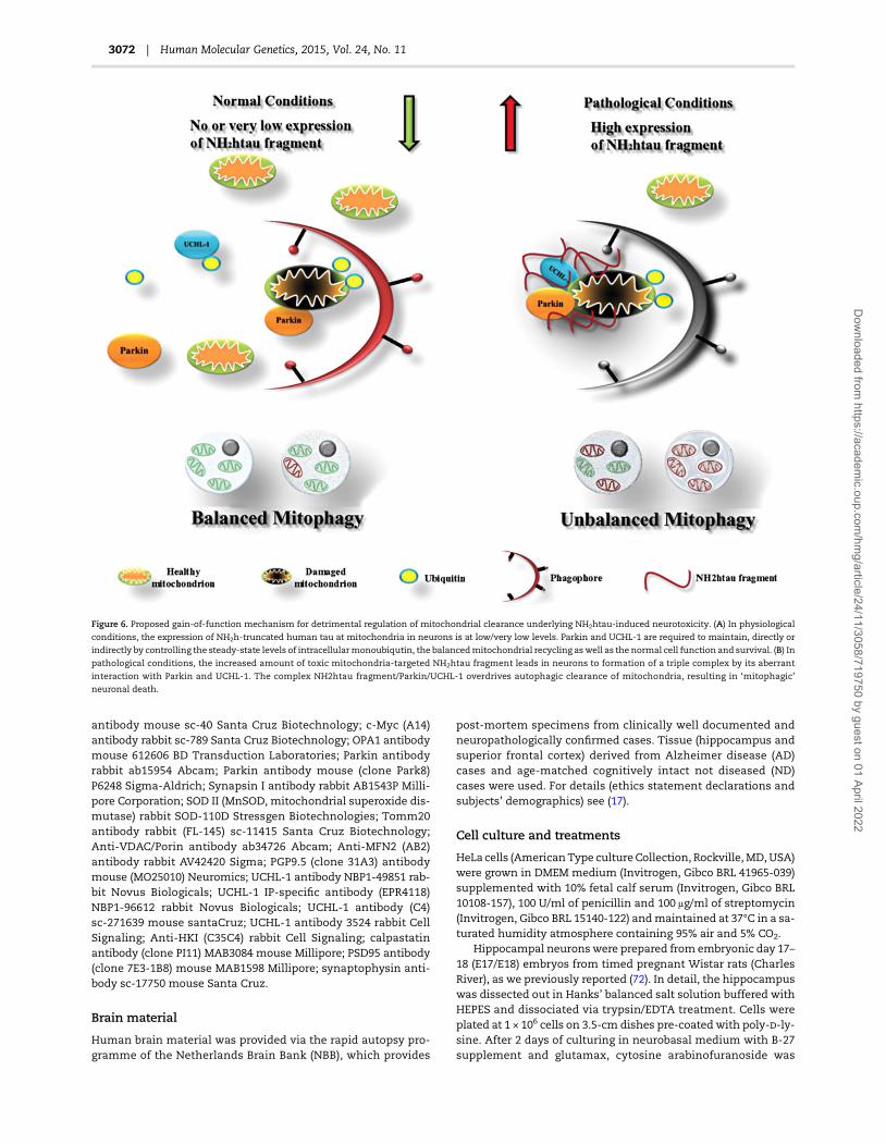
antibody mouse sc-40 Santa Cruz Biotechnology; c-Myc (A14)antibody rabbit sc-789 Santa Cruz Biotechnology; OPA1 antibodymouse 612606 BD Transduction Laboratories; Parkin antibodyrabbit ab15954 Abcam; Parkin antibody mouse (clone Park8)P6248 Sigma-Aldrich; Synapsin I antibody rabbit AB1543P Milli-pore Corporation; SOD II (MnSOD, mitochondrial superoxide dis-mutase) rabbit SOD-110D Stressgen Biotechnologies; Tomm20antibody rabbit (FL-145) sc-11415 Santa Cruz Biotechnology;Anti-VDAC/Porin antibody ab34726 Abcam; Anti-MFN2 (AB2)antibody rabbit AV42420 Sigma; PGP9.5 (clone 31A3) antibodymouse (MO25010) Neuromics; UCHL-1 antibody NBP1-49851 rab-bit Novus Biologicals; UCHL-1 IP-specific antibody (EPR4118)NBP1-96612 rabbit Novus Biologicals; UCHL-1 antibody (C4)sc-271639 mouse santaCruz; UCHL-1 antibody 3524 rabbit CellSignaling; Anti-HKI (C35C4) rabbit Cell Signaling; calpastatinantibody (clone PI11) MAB3084 mouse Millipore; PSD95 antibody(clone 7E3-1B8) mouse MAB1598 Millipore; synaptophysin anti-body sc-17750 mouse Santa Cruz.
Brain material
Human brain material was provided via the rapid autopsy pro-gramme of the Netherlands Brain Bank (NBB), which provides
post-mortem specimens from clinically well documented andneuropathologically confirmed cases. Tissue (hippocampus andsuperior frontal cortex) derived from Alzheimer disease (AD)cases and age-matched cognitively intact not diseased (ND)cases were used. For details (ethics statement declarations andsubjects’ demographics) see (17).
Cell culture and treatments
HeLa cells (AmericanType cultureCollection, Rockville, MD, USA)were grown in DMEM medium (Invitrogen, Gibco BRL 41965-039)supplemented with 10% fetal calf serum (Invitrogen, Gibco BRL10108-157), 100 U/ml of penicillin and 100 μg/ml of streptomycin(Invitrogen, Gibco BRL 15140-122) andmaintained at 37°C in a sa-turated humidity atmosphere containing 95% air and 5% CO2.
Hippocampal neuronswere prepared from embryonic day 17–18 (E17/E18) embryos from timed pregnant Wistar rats (CharlesRiver), as we previously reported (72). In detail, the hippocampuswas dissected out in Hanks’ balanced salt solution buffered withHEPES and dissociated via trypsin/EDTA treatment. Cells wereplated at 1 × 10⁶ cells on 3.5-cm dishes pre-coated with poly--ly-sine. After 2 days of culturing in neurobasal medium with B-27supplement and glutamax, cytosine arabinofuranoside was
Figure 6. Proposed gain-of-function mechanism for detrimental regulation of mitochondrial clearance underlying NH2htau-induced neurotoxicity. (A) In physiological
conditions, the expression of NH2h-truncated human tau at mitochondria in neurons is at low/very low levels. Parkin and UCHL-1 are required to maintain, directly or
indirectly by controlling the steady-state levels of intracellularmonoubiqutin, the balancedmitochondrial recycling aswell as the normal cell function and survival. (B) Inpathological conditions, the increased amount of toxic mitochondria-targeted NH2htau fragment leads in neurons to formation of a triple complex by its aberrant
interaction with Parkin and UCHL-1. The complex NH2htau fragment/Parkin/UCHL-1 overdrives autophagic clearance of mitochondria, resulting in ‘mitophagic’
neuronal death.
3072 | Human Molecular Genetics, 2015, Vol. 24, No. 11
Dow
nloaded from https://academ
ic.oup.com/hm
g/article/24/11/3058/719750 by guest on 01 April 2022

added to reduce glial proliferation. Half of themediumwas chan-ged every 3–4 days andmature neuronal cultureswere infected at15 days in vitro, as reported in (72). All experimental details con-cerning the handling with recombinant adenoviral vectors (con-struction, packaging, purification, titration), the used infectionprotocol, the time course of overexpression levels and the neur-onal viability assays of both tau-coding and control transgeneshave been previously reported in (71,72).
All animal experiments were approved by the CNR AnimalCare and Use Committee and were conducted in full accordancewith the Guide for laboratory animal practicewith care tominim-ize the number of animals and their suffering.
Assessment of neuronal viability
Cell viability was quantified by counting the number of intact nu-clei, after lysing in detergent-containing solution (155,156) and bythe MTT tetrazolium salt assay (157).
DNA transfection
Hippocampal primary neurons were transfected at high effi-ciency and very low cytotoxicity (%50) with plasmid expressingFlag-tagged wtMfn2 or Flag-tag alone by Amaxa rat neuron Nu-cleofector kit (VGP-1003), according to the manufacturer’s in-structions. The expression plasmid (p3xFLAG-CMV-10) codingfor Flag-tagged wtMfn2 was gently provided by Prof. NobuhiroNakamura Department of Biological Sciences, Tokyo Instituteof Technology, 4259-B19 Nagatsuta-cho, Midori-ku, Yokohama,226-8501, Japan.
HeLa cells were transfected using Xfect™ Clontech reagents(Takara Clontech 631318) according to the manufacturer’sinstructions
Short hairpin RNA (shRNA) interference for Parkinand UCHL-1
Gene silencing of primary hippocampal neurons was carried outby Amaxa rat neuron Nucleofector kit (VGP-1003) according tothe manufacturer’s instructions. Forty picomoles of shRNAscrambled Parkin or 40 pmol or shRNAParkin (Santa Cruz, sc-270243-SH); 5 μg of shRNA scrambled UCHL1 or 5 μg of shRNAUCHL1 (Origene, TF710038) were used to transfect 5 × 10⁶ cellsper sample with the selected program O-003. After 48 h of cultur-ing, the medium was again replaced with fresh culture medium(Neurobasal + 2%B27) and the cells were infected according to (72).
Hexokinase activity
The activity of Hexokinase (HK), which catalyses the reactionATP + glucose → glucose-6-phosphate (G6P) + ADP, was assayedthrough a coupled reaction with glucose-6-phosphate dehydro-genase (G6PDH), following the NADP reduction at 340 nm (ε340-nm = 6.22 m−1 cm−1), using a Jasco double-beam/double-wavelength spectrophotometer UV-550, under Vmax conditions.The assay was performed at 25°C and pH 7.2 to mimic intracellu-lar pH. The assay mix contained: 5 m MgCl2, 8 m ATP, 1 m
NADP, 0.1 U/ml of G6PDH and 0.5 mg cell homogenate. After10 m glucose addition to cell homogenate (0.2 mg protein), theactivity of HK was determined by following the conversion ofNADP to NADPH of the coupled reaction in which G6P is furtheroxidized to 6-phosphogluconate via G6PDH (158). Enzyme activ-ity was obtained as the tangent to the initial part of the progresscurves and expressed, on the basis of protein present in the cellhomogenate, as nmol/min ( =mU) per mg of protein, where 1
unit of enzyme activity (U) is defined as μmol/min and thespecific activity as U/mg protein.
Immunofluorescence
Following treatment, neuronal cultures were washed twice withPBS and fixed in 4% (w/v) paraformaldehyde for 15 min at roomtemperature. Cells were permeabilized with 0.1% (v/v) TritonX-100/PBS pH 7.4 for 4 min at room temperature. Coverslips weresaturated with 2% BSA and 10% normal goat serum (NGS) for 3 hfollowed by incubation overnight at 4°C in a humidified chamberwith primary antibodies. Unbound antibody was removed bythree washes and bound antibody was detected by incubationwith donkey anti-rabbit Alexa 488 (1:300) and donkey anti-mouserhodamine-conjugated (1:300) secondary antibodies (Invitrogen)at room temperature for 30 min. Nuclei were stained with nuclearmarker 4,6-diamidino-2-phenylindole dihydrochloride (DAPI;Sigma, St Louis, MO, USA) 1:1000 in PBS for 5 min. Controls wereperformed either by omitting the primary antibody.
Tissue slabs derived from CA1 hippocampal region of AD andND caseswere placed in a solution of 4% paraformaldehyde at +4°for 3 days. After threewashes 10 min each, slabs were placed in asolution of 30% (w/v) sucrose in PBS at +4° until they sank. Thecryoprotected slabs were cut on a freezing microtome into40 μm thick sections and collected in a culture well for freefloating immunofluorescence procedures. For double immu-nofluorescence the following primary antibodies were used:anti-Parkin (Park8), mouse 1:100; anti: UCHL-1, mouse 1:100;anti-CCP NH2 tau rabbit 1:300. After incubation in blocking solu-tion (25% FBS in 0.3% Triton X-100 in TBS) for 2 h at room tem-perature, sections were incubated with primary antibodiesdiluted in 20% FBS in 0.3% Triton X-100 in TBS for 48 h at +4°Cand, then, for additional 30 min at room temperature. Afterthree washing, sections were incubated with a mix solution ofdonkey anti-rabbit Alexa 488 (1:100) and donkey anti-mouserhodamine-conjugated (1:100) secondary antibodies (Invitrogen)diluted in 10%FBS in 0.3%TritonX-100 inTBS for 2 h at room tem-perature. The sections were next washed three times in TBS andthen stained with 0.3% Sudan Black B—to reduce autofluores-cence—for 1 min at room temperature, followed by washing inwater for three times. Sections were then incubated with the nu-clear marker, DNA-fluorochrome 4′,6-diamidino-2-phenylindole(DAPI, 1:1000, Sigma-Aldrich) for 10 min, for nuclei visualization,followed by a further rinse. Sections were mounted on slides, airdried and coverslipped using gel mount (Biomeda Corp., FosterCity, CA, USA).
Isolation of mitochondria
Isolation of mitochondria was performed using mitochondrialextraction kit (Qiagen 37 612), as previously described (17). Inbrief samples were homogenized in 0.5 ml of homogenizationbuffer using a tissue ruptor rotor-stator and incubated on anend-over-end shaker for 10 min at 4°C. The homogenatewas cen-trifuged at 1000g for 10 min at 4°C. The resulting pellet was resus-pended in disruption buffer, repeatedly passed through anarrow-gauge needle (26 or 21 gauge) and re-centrifuged at1000g for 10 min; the pellet contains nuclei, cell debris and un-broken cells. The supernatant, which contains mitochondria,was re-centrifuged at 6000g for 10 min. The mitochondria pelletwas resuspended in mitochondria purification buffer and slowlythe disruption buffer was pipetted under the purification buffer.The mitochondrial suspension was pipetted on top of layers andcentrifuged at 14 000g for 15 min at 4°C. The Mitochondria pellet
Human Molecular Genetics, 2015, Vol. 24, No. 11 | 3073
Dow
nloaded from https://academ
ic.oup.com/hm
g/article/24/11/3058/719750 by guest on 01 April 2022
was resuspended in mitochondria storage buffer. The purity ofmitochondrial preparation was checked by probing fractionatedprotein extracts with several specific antibodies reacting withmitochondrial markers, as previously described (17).
Isolation of purified synaptosomes and PSD fraction
Purified synaptosomes were prepared as previously described(159,160). Briefly, the tissue was homogenized in 10 volumes of0.32 sucrose, buffered to pH 7.4 with Tris-(hydroxymethyl)-amino methane [Tris, final concentration (f.c.) 0.01 ] using aglass/Teflon tissue grinder (clearance 0.25 mm); the homogenatewas centrifuged at 1000g for 5 min to remove nuclei and debrisand the supernatant was gently stratified on a discontinuousPercoll (Sigma-Aldrich, Italy) gradient (2, 6, 10 and 20% v/v inTris-buffered sucrose) and centrifuged at 33 500g for 5 min. Thelayer between 10 and 20% Percoll (synaptosomal fraction) wascollected and washed by centrifugation. The synaptosomalpellets were resuspended in a physiological solution (StandardMedium) with the following composition (m): NaCl, 140; KCl,3; MgSO4, 1.2; CaCl2, 1.2; NaH2PO4, 1.2; NaHCO3, 5; HEPES, 10;glucose, 10; pH 7.2–7.4. Immediately before fixation procedures,synaptosomes were incubated with MitoTracker red CMxRos(M7512 Molecular Probes, Eugene, OR, USA) at a concentrationof 500 for 20 min. After three washing, synaptosomes wereseeded for 30min on coverslip coatedwith poly--lysine (50 μg/ml)at 4°C, washed twice with PBS and fixed in 2% (w/v) freshly pre-pared paraformaldehyde in PBS for 10 min at room temperature.After four washes synaptosomes were permeabilized with 0.05%(v/v) Triton X-100/PBS for 5 min,washed and incubated overnightin a mix solution of primary antibodies each diluted 1:200 in0.05% (v/v) Triton X-100/PBS at 4 °C. Following three washes, sy-naptosomes were incubated with a mix solution of donkeyanti-rabbit Alexa 488 (1:500) and donkey anti-mouse Alexa 647(1:500) secondary antibodies (Invitrogen) for 2 h at 4°C, thencoverslip were washed, air dried and mounted on slide usinggel mount. Controls were performed by omitting the primaryantibodies.
PSD-enriched fraction was prepared according procedure de-scribed in (161). Briefly, the brain was rapidly dissected and thehippocampus was homogenized in homogenization buffer(320 m sucrose, 10 m Tris–HCl (pH 7.4), 1 m EDTA, 1 m
NaHCO3, 1 m PMSF, 1 m Na3VO4, 20 m β-glycerophosphate,5 m NaF, with protease inhibitors; Sigma) with 10 strokes of atight-fitting glass Dounce tissue grinder (7 ml, Wheaton). Thehomogenate was centrifuged at 1000g for 10 min and the result-ing supernatant was centrifuged at 10 000g for 15 min. The pelletwas resuspended in homogenization buffer with 0.5% TritonX-100 containing protease inhibitors and centrifuged at 32 000gfor 20 min. The supernatant was referred as extra synaptic mem-brane enriched fraction while the pellet containing the PSD frac-tion (Triton X-100 insoluble fraction) was then resuspended inRIPA buffer (50 m Tris–HCl (pH 7.4), 1% Triton X-100, 0.25% so-dium deoxycholate, (wt/vol) 150 m NaCl, 5 m MgCl2, 1 m
EDTA, 0.1%SDS (wt/vol), protease inhibitors), sonicated and incu-bated on ice for 20 min. The samples were centrifuged at 11 500gfor 10 min and the protein concentration of resulting super-natant was determined.
Protein cellular lysates preparation
Total proteins were extracted by scraping the cells in an SDS-reducing sample buffer or lysis in ice-cold RIPA buffer (50 m
Tris–HCl, pH 8, 150 m NaCl, 1% Triton, 2 m EDTA, 0.1% SDS
plus proteases inhibitor cocktail (SigmaAldrich, P8340) and phos-phatase inhibitor cocktail (Sigma-Aldrich, P5726/P2850) andcentrifuged at 4°C for 15 min at 1000g. The supernantant wasthen collected and boiled for 5 min.
Western blot analysis and densitometry
Equal amounts of protein were subjected to SDS-PAGE 7.5–15%linear gradient or Bis–Tris gel 4–12% (NuPage, Invitrogen). Afterelectroblotting onto a nitrocellulose membrane (Hybond-CAmersham Biosciences, Piscataway, NJ, USA) the filters wereblocked in TBS containing 10% non-fat dried milk for 1 h atroom temperature or overnight at 4°C. Proteins were visualizedusing appropriate primary antibodies. All primary antibodieswere diluted in TBS and incubated with the nitrocellulose blotovernight at 4°C. Incubation with secondary peroxidase coupledanti-mouse, anti-rabbit or anti-goat antibodieswas performed byusing the ECL system (Amersham, Arlington Heights, IL, USA)Final figures were assembled by using Adobe Photoshop 6 andAdobe Illustrator 10 and quantitative analysis of acquired imageswas performed by using ImageJ (http://imagej.nih.gov/ij/).
Confocal microscopy and image analysis
Slides were examined under a confocal laser scanning micro-scope (Leica SP5, Leica Microsystems, Wetzlar, Germany)equipped with four laser lines: violet diode emitting at 405 nm,argon emitting at 488 nm, helium/neon emitting at 543 nm,helium/neon emitting at 633 nm and a transmitted light detectorfor differential interference contrast (DIC; Nomarski) imaging.Bright field images were corrected for inhomogeneities of thebrightness across the image by using Adobe Photoshop high-pass filter, and edges contrast was enhanced with ImageJ CLAHE(Contrast Limited Adaptive Histogram Equalization; http://rsbweb.nih.gov/ij/plugins/clahe/index.html) filter plugin (blocksize, 127; histogram bins, 256; maximum slope, 3). Confocal ac-quisition settingwas identical between control and experimentalcases. For production of figures, brightness and contrast ofimages were globally enhanced by using linear histogram correc-tion and slightly over-saturated. The over-saturation enables abetter qualitative visual evaluation of both morphological fea-tures and colocalization domains. Quantitative image analysiswas performed by using Imaris Suite 7.4® (Bitplane A.G., Zurich,Switzerland) or ImageJ 1.4 (imagej.nih.gov/ij/) softwares on fivedifferent images derived from each experimental group. Imageanalysis was performed under visual control to determinethresholds that subtracts background noise and takes into ac-count neuronal structures. During image processing, the imageswere compared with the original raw data to make sure that nostructures were introduced that were not seen in the originaldata series or that structures present in the original data serieswere not removed. To determine fluorescent signal colocaliza-tion between different channels the ImarisColoc module wasused. Before colocalization analysis, a median filter was appliedto the images to reduce the background noise and source chan-nels threshold were established by automatic thresholding ofthe P-value. The degree of channels colocalization was analyzedby considering the Pearson’s coefficient and thresholded Man-ders coefficient A and B in colocalized volume. To compare theintensity relations between the fluorescence channels acrossthe morphological features in colocalizion studies the ImageJtool plot profile was used. To evaluate the fluorescence intensityseveralmaskswere drawnon the putative cell bodies, afterwhichthe DAPI channel was deleted and measures were performed.
3074 | Human Molecular Genetics, 2015, Vol. 24, No. 11
Dow
nloaded from https://academ
ic.oup.com/hm
g/article/24/11/3058/719750 by guest on 01 April 2022

Images of synaptosomes were acquired as described above anddeconvolved through a blind deconvolution algorithm by usingAutoQuant X3 (Media Cybernetics, Rockville, USA).
Coimmunoprecipitation
The mitochondrial extraction from human purified synapto-somes was performed according to (17) using mitochondrial ex-traction kit (Qiagen 37612) according to the manufacturer’sinstructions (see the Isolation of mitochondria section). The syn-aptic mitochondrial pellet was resuspended in mitochondriastorage buffer, the proteins concentration was determined byBCA assay (Dc Protein Assay, BioRad, 500-0116) and then sampleswere subjected to IP, as previously described (53). In brief, 500 μgmitochondrial proteins extract was lysed using immunoprecipi-tation (IP) buffer (50 m Tris, pH 8.0, 150 m NaCl, 10% glycerol,0.5 m EDTA, 0.5 m EGTA, 50 m NaF, 1 m NaOV4, 1% NP40)supplemented with proteases inhibitor cocktail (Sigma-Aldrich,P8340) and phosphatase inhibitor cocktail (Sigma-Aldrich,P5726/P2850). Lysates were then centrifuged at 4°C for 10 min at1000 g and the supernatants were precleared using 10 μl of pro-tein G/A (Invitrogen) for 1 h at 4°C. The pre-cleared protein ex-tracts were immunoprecipitated by magnetic Dynabeads(Invitrogen) according to the manufacturer’s instructions using:4 μg of antiserumCCPNH2 tau for 20 μl of proteinA. The immuno-complexeswere elutedwith Laemmli buffer 1× plus DTT (Invitro-gen) and were next analyzed by immunoblotting with mouseParkin antibody (Park8clone) and mouse UCHL-1 (clone31A3)
Reciprocal IP was performed on the precleared proteins ex-tractswith rabbit UCHL-1 (EPR4118) 1:10 for 1.5 mgprotein extractfor 40 μl of protein A. The immunocomplexes were next elutedwith Laemmli buffer 1× plus DTT (Invitrogen) and analyzed byimmunoblotting with mouse Parkin antibody (Park8clone) andrabbit CCP NH2 tau antibody.
In several experiments, to eliminate the detection-interference from both heavy-chain (∼50 kDa) and light-chain(25 kDa) IgG-fragments, an HRP-conjugated detection secondaryantibody, which specifically binds to functional primary anti-bodies (whole IgG), has been used.
Calpastatin transgenic mice
Transgenic mice expressing human calpastatin (hCAST), undercontrol of the neuron-specific calcium–calmodulin-dependentkinase II α subunit promoter, were gently provided by Prof. Ta-kaomi Saido Riken Brain Science Institute, Saitamo 351-0198,Japan. hCAST mice were created on a C57Bl/6 background andcharacterized as previously described (95,96) and these mice ex-hibit a 3-fold increase in calpastatin activity and display no ad-verse phenotype (96). A colony was established at CNR-EBRIconsortium from which hCAST transgenic (Tg) and WT litter-mates were bred for experimental studies. All mice were housedin controlled conditions under a 14:10 light:dark photoperiod andallowed food and water ad libitum. Animal husbandry and surgi-cal procedures were performed in accordance with EEC guide-lines (Directive 86/609/CEE). The genotype was determined byPCR with specific primers 5′-CATGAACCACAGACAGCTTGGTTGAC-3′ and 5′-GGAGGATTTGATATTCACCTGGCCCG-3′ that gen-erate a 350-bp product. GADPH forward 5′-ACCACAGTCCATGCCATCAC-3′ and reverse 5′-TCCACCACCCTGTTGCTG-3′ primerswere also included as internal control. We finally confirmedthat the transgene-derived human calpastatin was robustly ex-pressed in hippocampus and neocortex by western blotting
analysis with human-specific calpastatin antibody (clone PI11Millipore MAB3084), as described in (162).
Statistical analysis
Experiments were carried out in triplicates and repeated at leastthree times. Data were expressed as means ± SD (n = 4). Statistic-ally significant differences were calculated by unpaired two-tailed Student’s t-test (*P < 0.05; **P < 0.01; ***P < 0.0001) as indictedin the figure legends. Data from western blot analysis and im-munofluorescence studies were representative of at least threeseparate experiments.
Supplementary MaterialSupplementary Material is available at HMG online.
AcknowledgementsWe wish to thank Prof. Nobuhiro Nakamura Department of Bio-logical Sciences, Tokyo Institute of Technology, 4259-B19 Nagat-suta-cho, Midori-ku, Yokohama, 226–8501, Japan for providingexpression plasmid (p3xFLAG-CMV-10) coding for Flag-taggedwtMfn2. We are also very grateful to Prof. Takaomi Saido RikenBrain Science Institute, Saitamo 351-0198, Japan for providinghCAST mice.
Conflict of Interest statement. None declared.
FundingThis researchwas supported by grant from FIRB B81J07000070001and Fondazione Roma to P.C and by grant from PRIN 2010–2011(prot. 2010M2JARJ-003) to G.A. The funders had no role in studydesign, data collection and analysis, decision to publish or prep-aration of manuscript.
References1. Guillozet-Bongaarts, A.L., Glajch, K.E., Libson, E.G., Cahill, M.
E., Bigio, E., Berry, R.W. and Binder, L.I. (2007) Phosphoryl-ation and cleavage of tau in non-AD tauopathies.Acta Neuro-pathol., 113, 513–520.
2. García-Sierra, F., Mondragón-Rodríguez, S. and Basurto-Islas, G. (2008) Truncation of tau protein and its pathologicalsignificance in Alzheimer’s disease. J. Alzheimers Dis., 14,401–409.
3. Horowitz, P.M., Patterson, K.R., Guillozet-Bongaarts, A.L.,Reynolds, M.R., Carroll, C.A., Weintraub, S.T., Bennett, D.A.,Cryns, V.L., Berry, R.W. andBinder, L.I. (2004) EarlyN-termin-al changes and caspase-6 cleavage of tau in Alzheimer’s dis-ease. J. Neurosci., 24, 7895–7902.
4. Wang, Y., Garg, S., Mandelkow, E.M. and Mandelkow, E.(2010) Proteolytic processing of tau. Biochem. Soc. Trans., 38,955–961.
5. Reddy, P.H. (2011) Abnormal tau,mitochondrial dysfunction,impaired axonal transport of mitochondria, and synapticdeprivation in Alzheimer’s disease. Brain Res., 1415, 136–148.
6. Manczak, M. and Reddy, P.H. (2012) Abnormal interaction ofVDAC1 with amyloid beta and phosphorylated tau causesmitochondrial dysfunction in Alzheimer’s disease. Hum.Mol. Genet., 21, 5131–5146.
7. Du, H., Guo, L., Yan, S., Sosunov, A.A., McKhann, G.M. andYan, S.S. (2010) Early deficits in synaptic mitochondria in
Human Molecular Genetics, 2015, Vol. 24, No. 11 | 3075
Dow
nloaded from https://academ
ic.oup.com/hm
g/article/24/11/3058/719750 by guest on 01 April 2022

an Alzheimer’s disease mouse model. Proc. Natl. Acad. Sci.USA, 107, 18670–18675.
8. Du, H., Guo, L. and Yan, S.S. (2012) Synaptic mitochondrialpathology in Alzheimer’s disease. Antioxid. Redox. Signal.,16, 1467–1475.
9. Pooler, A.M., Noble, W. and Hanger, D.P. (2014) A role for tauat the synapse in Alzheimer’s disease pathogenesis. Neuro-pharmacology, 76(Pt A), 1–8.
10. Tai, H.C., Serrano-Pozo, A., Hashimoto, T., Frosch, M.P.,Spires-Jones, T.L. and Hyman, B.T. (2012) The synaptic accu-mulation of hyperphosphorylated tau oligomers in Alzhei-mer disease is associated with dysfunction of theubiquitin-proteasome system. Am. J. Pathol., 181, 1426–1435.
11. Upadhya, S.C. and Hegde, A.N. (2007) Role of the ubiquitinproteasome system in Alzheimer’s disease. BMC Biochem.,8(Suppl 1), S12.
12. Hegde, A.N. and DiAntonio, A. (2002) Ubiquitin and the syn-apse. Nat. Rev. Neurosci., 3, 854–861.
13. Keck, S., Nitsch, R., Grune, T. and Ullrich, O. (2003) Prote-asome inhibition by paired helical filament-tau in brainsof patients with Alzheimer’s disease. J. Neurochem., 85,115–122.
14. Terry, R.D., Masliah, E., Salmon, D.P., Butters, N., DeTeresa,R., Hill, R., Hansen, L.A. and Katzman, R. (1991) Physicalbasis of cognitive alterations in Alzheimer’s disease: syn-apse loss is the major correlate of cognitive impairment.Ann. Neurol., 30, 572–580.
15. DeKosky, S.T. and Scheff, S.W. (1990) Synapse loss in frontalcortex biopsies in Alzheimer’s disease: correlation with cog-nitive severity. Ann. Neurol., 27, 457–464.
16. Rhein, V., Song, X., Wiesner, A., Ittner, L.M., Baysang, G.,Meier, F., Ozmen, L., Bluethmann, H., Dröse, S., Brandt, U.et al. (2009) Amyloid-beta and tau synergistically impairthe oxidative phosphorylation system in triple transgenicAlzheimer’s disease mice. Proc. Natl Acad. Sci. USA, 106,20057–20062.
17. Amadoro, G., Corsetti, V., Atlante, A., Florenzano, F., Capso-ni, S., Bussani, R., Mercanti, D. and Calissano, P. (2012) Inter-action between NH(2)-tau fragment and Aβ in Alzheimer’sdisease mitochondria contributes to the synaptic deterior-ation. Neurobiol. Aging., 33, 833.e1–833S25.
18. Manczak, M. and Reddy, P.H. (2012) Abnormal interactionbetween the mitochondrial fission protein Drp1 andhyperphosphorylated tau in Alzheimer’s disease neurons:implications for mitochondrial dysfunction and neuronaldamage. Hum. Mol. Genet., 21, 2538–2547.
19. Quintanilla, R.A., Matthews-Roberson, T.A., Dolan, P.J. andJohnson, G.V. (2009) Caspase-cleaved tau expression in-duces mitochondrial dysfunction in immortalized corticalneurons: implications for the pathogenesis of Alzheimerdisease. J. Biol. Chem., 284, 18754–18766.
20. Quintanilla, R.A., Dolan, P.J., Jin, Y.N. and Johnson, G.V.(2012) Truncated tau andAβ cooperatively impairmitochon-dria in primary neurons. Neurobiol. Aging., 33, 619.e25–35.
21. Eckert, A., Schmitt, K. and Götz, J. (2011) Mitochondrialdysfunction—the beginning of the end in Alzheimer’sdisease? Separate and synergistic modes of tau andamyloid-β toxicity. Alzheimers Res. Ther., 3, 15. doi:10.1186/alzrt74.
22. Wang, X., Su, B., Siedlak, S.L., Moreira, P.I., Fujioka, H., Wang,Y., Casadesus, G. and Zhu, X. (2008) Amyloid-β overproduc-tion causes abnormal mitochondrial dynamics via differen-tial modulation of mitochondrial fission/fusion proteins.Proc. Natl Acad. Sci. USA, 105, 19318–19323.
23. Wang, X., Su, B., Lee, H.G., Li, X., Perry, G., Smith, M.A. andZhu, X. (2009) Impaired balance of mitochondrial fissionand fusion inAlzheimer’s disease. J. Neurosci., 29, 9090–9103.
24. Karbowski, M. and Neutzner, A. (2012) Neurodegenerationas a consequence of failed mitochondrial maintenance.Acta Neuropathol., 123, 157–171.
25. DuBoff, B., Feany, M. and Götz, J. (2013) Why size matters—balancing mitochondrial dynamics in Alzheimer’s disease.Trends Neurosci., 36, 325–335.
26. Zhu, X., Perry, G., Smith, M.A. andWang, X. (2013) Abnormalmitochondrial dynamics in the pathogenesis of Alzheimer’sdisease. J. Alzheimers Dis., 33(Suppl 1), S253–S262.
27. Wang, X., Su, B., Zheng, L., Perry, G., Smith, M.A. and Zhu, X.(2009) The role of abnormal mitochondrial dynamics in thepathogenesis of Alzheimer’s disease. J. Neurochem., 109(Suppl 1), 153–159.
28. Moreira, P.I., Siedlak, S.L., Wang, X., Santos, M.S., Oliveira, C.R., Tabaton, M., Nunomura, A., Szweda, L.I., Aliev, G., Smith,M.A., Zhu, X. and Perry, G. (2007) Increased autophagic deg-radation of mitochondria in Alzheimer disease. Autophagy,3, 614–615.
29. Moreira, P.I., Siedlak, S.L., Wang, X., Santos, M.S., Oliveira, C.R., Tabaton, M., Nunomura, A., Szweda, L.I., Aliev, G., Smith,M.A., Zhu, X. and Perry, G. (2007) Autophagocytosis of mito-chondria is prominent in Alzheimer disease. J. Neuropathol.Exp. Neurol., 66, 525–532.
30. Young-Collier, K.J., McArdle, M. and Bennett, J.P. (2012) Thedying of the light: mitochondrial failure in Alzheimer’s dis-ease. J. Alzheimers Dis., 28, 771–781.
31. Baloyannis, S.J. (2006) Mitochondrial alterations in Alzhei-mer’s disease. J. Alzheimers Dis., 9, 119–126.
32. Baloyannis, S.J. (2011) Mitochondria are related to synapticpathology in Alzheimer’s disease. Int. J. Alzheimers Dis.,2011, 305395.
33. de la Monte, S.M., Luong, T., Neely, T.R., Robinson, D. andWands, J.R. (2000) Mitochondrial DNA damage as amechan-ism of cell loss in Alzheimer’s disease. Lab. Invest., 80, 1323–1335.
34. Xie, H., Guan, J., Borrelli, L.A., Xu, J., Serrano-Pozo, A. and Bacs-kai, B.J. (2013)Mitochondrial alterations near amyloid plaquesin an Alzheimer’s disease mouse model. J. Neurosci., 33,17042–17051.
35. Bonda, D.J., Wang, X., Perry, G., Smith, M.A. and Zhu, X.(2010) Mitochondrial dynamics in Alzheimer’s disease:opportunities for future treatment strategies. Drugs Aging,27, 181–192.
36. Kamat, P.K., Kalani, A., Kyles, P., Tyagi, S.C. and Tyagi, N.(2014) Autophagy of mitochondria: a promising therapeutictarget for neurodegenerative disease. Cell Biochem Biophys.,70, 707–719.
37. Palikaras, K. and Tavernarakis, N. (2014) Mitochondrialhomeostasis: the interplay between mitophagy and mito-chondrial biogenesis. Exp. Gerontol., 56, 182–188.
38. Zhu, J., Wang, K.Z. and Chu, C.T. (2013) After the banquet:mitochondrial biogenesis, mitophagy, and cell survival. Au-tophagy, 9, 1663–1676.
39. Hung, K.C., Huang, H.J., Lin, M.W., Lei, Y.P. and Lin, A.M.(2014) .Roles of autophagy in MPP+-induced neurotoxicityin vivo: the involvement of mitochondria and α-synucleinaggregation. PLoS One, 9, e91074.
40. Zhu, J.H., Horbinski, C., Guo, F.,Watkins, S., Uchiyama, Y. andChu, C.T. (2000) Regulation of autophagy by extracellularsignal-regulated protein kinases during 1-methyl-4-phenylpyridinium -induced cell death. Am. J. Pathol., 170, 75–86.
3076 | Human Molecular Genetics, 2015, Vol. 24, No. 11
Dow
nloaded from https://academ
ic.oup.com/hm
g/article/24/11/3058/719750 by guest on 01 April 2022

41. Choubey, V., Safiulina, D., Vaarmann, A., Cagalinec, M.,Wareski, P., Kuum, M., Zharkovsky, A. and Kaasik, A. (2011)Mutant A53 T alpha-synuclein induces neuronal death byincreasing mitochondrial autophagy. J. Biol. Chem., 286,10814–10824.
42. Chang, C.F., Huang, H.J., Lee, H.C., Hung, K.C., Wu, R.T. andLin, A.M. (2012) Melatonin attenuates kainic acid-inducedneurotoxicity in mouse hippocampus via inhibition of au-tophagy and α-synuclein aggregation. J. Pineal. Res., 52,312–321.
43. Tzeng, Y.W., Lee, L.Y., Chao, P.L., Lee, H.C., Wu, R.T. and Lin,A.M. (2010) Role of autophagy in protection afforded by hyp-oxic preconditioning againstMPP+-induced neurotoxicity inSH-SY5Y cells. Free Radic. Biol. Med., 49, 839–846.
44. Escobar-Henriques, M. and Langer, T. (2014) Dynamic surveyof mitochondria by ubiquitin. EMBO Rep., 15, 231–243.
45. Kirkin, V., McEwan, D.G., Novak, I. and Dikic, I. (2009) A rolefor ubiquitin in selective autophagy. Mol. Cell, 34, 259–269.
46. Amadoro, G., Corsetti, V., Florenzano, F., Atlante, A., Bobba,A., Nicolin, V., Nori, S.L. and Calissano, P. (2014) Morpho-logical and bioenergetic demands underlying the mito-phagy in post-mitotic neurons: the pink-parkin pathway.Front. Aging Neurosci., 6, 18.
47. Reddy, P.H., Tripathi, R., Troung, Q., Tirumala, K., Reddy, T.P.,Anekonda, V., Shirendeb, U.P., Calkins, M.J., Reddy, A.P.,Mao, P. andManczak, M. (2012) Abnormalmitochondrial dy-namics and synaptic degeneration as early events in Alzhei-mer’s disease: implications to mitochondria-targetedantioxidant therapeutics. Biochim. Biophys. Acta, 1822,639–649.
48. Grenier, K., McLelland, G.L. and Fon, E.A. (2013) Parkin- andPINK1-dependent mitophagy in neurons: will the real path-way please stand up? Front. Neurol., 4, 100.
49. Taylor, E.B. and Rutter, J. (2011) Mitochondrial quality con-trol by the ubiquitin-proteasome system. Biochem. Soc.Trans., 39, 1509–1513.
50. Joselin, A.P., Hewitt, S.J., Callaghan, S.M., Kim, R.H., Chung,Y.H., Mak, T.W., Shen, J., Slack, R.S. and Park, D.S. (2012)ROS-dependent regulation of Parkin and DJ-1 localizationduring oxidative stress in neurons. Hum. Mol. Genet., 21,4888–4903.
51. Koyano, F., Okatsu, K., Ishigaki, S., Fujioka, Y., Kimura, M.,Sobue, G., Tanaka, K. and Matsuda, N. (2013) The principalPINK1 and Parkin cellular events triggered in response todissipation of mitochondrial membrane potential occur inprimary neurons. Genes Cells., 18, 672–681.
52. Cai, Q., Zakaria, H.M., Simone, A. and Sheng, Z.H. (2012) Spa-tial parkin translocation and degradation of damaged mito-chondria via mitophagy in live cortical neurons. Curr. Biol.,22, 545–552.
53. Cai, Q., Zakaria, H.M. and Sheng, Z.H. (2012) Long time-lapseimaging reveals unique features of PARK2/Parkin-mediatedmitophagy in mature cortical neurons. Autophagy, 8, 976–978.
54. Tanaka, A., Cleland, M.M., Xu, S., Narendra, D.P., Suen, D.F.,Karbowski, M. andYoule, R.J. (2010) Proteasome and p97me-diate mitophagy and degradation of mitofusins induced byParkin. J. Cell Biol., 191, 1367–1380.
55. Chan, N.C., Salazar, A.M., Pham, A.H., Sweredoski, M.J., Kola-wa, N.J., Graham, R.L., Hess, S. and Chan, D.C. (2011) Broadactivation of the ubiquitin-proteasome system by Parkin iscritical for mitophagy. Hum. Mol. Genet., 20, 1726–1737.
56. Yoshii, S.R., Kishi, C., Ishihara, N. and Mizushima, N.(2011) Parkin mediates proteasome-dependent protein
degradation and rupture of the outer mitochondrial mem-brane. J. Biol. Chem., 286, 19630–19640.
57. Bingol, B., Tea, J.S., Phu, L., Reichelt, M., Bakalarski, C.E.,Song, Q., Foreman, O., Kirkpatrick, D.S. and Sheng, M.(2014) The mitochondrial deubiquitinase USP30 opposesparkin-mediated mitophagy. Nature, 510, 370–375.
58. Osaka, H.,Wang, Y.L., Takada, K., Takizawa, S., Setsuie, R., Li,H., Sato, Y., Nishikawa, K., Sun, Y.J., Sakurai, M. et al. (2003)Ubiquitin carboxy-terminal hydrolase L1 binds to and stabi-lizes monoubiquitin in neuron. Hum. Mol. Genet., 12, 1945–1958.
59. Bizzi, A., Schaetzle, B., Patton, A., Gambetti, P. and Autilio-Gambetti, L. (1991) Axonal transport of two major compo-nents of the ubiquitin system: free ubiquitin and ubiquitincarboxyl-terminal hydrolase PGP 9.5. Brain Res., 548, 292–299.
60. Liu, Y., Fallon, L., Lashuel, H.A., Liu, Z. and Lansbury, P.T. Jr.(2002) The UCH-L1 gene encodes two opposing enzymaticactivities that affect alpha-synuclein degradation and Par-kinson’s disease susceptibility. Cell, 111, 209–218.
61. Cartier, A.E., Djakovic, S.N., Salehi, A., Wilson, S.M., Masliah,E. and Patrick, G.N. (2009) Regulation of synaptic structure byubiquitin C-terminal hydrolase L1. J. Neurosci., 29, 7857–7868.
62. Chen, F., Sugiura, Y., Myers, K.G., Liu, Y. and Lin, W. (2010)Ubiquitin carboxyl-terminal hydrolase L1 is required formaintaining the structure and function of the neuromuscu-lar junction. Proc. Natl Acad. Sci. USA, 107, 1636–1641.
63. Gong, B., Cao, Z., Zheng, P., Vitolo, O.V., Liu, S., Staniszewski,A., Moolman, D., Zhang, H., Shelanski, M. and Arancio, O.(2006) Ubiquitin hydrolase Uch-L1 rescues beta-amyloid-in-duced decreases in synaptic function and contextual mem-ory. Cell, 126, 775–788.
64. Smith, D.L., Pozueta, J., Gong, B., Arancio, O. and Shelanski,M. (2009) Reversal of long-term dendritic spine alterations inAlzheimer disease models. Proc. Natl Acad. Sci. USA, 106,16877–16882.
65. Guglielmotto, M., Monteleone, D., Boido, M., Piras, A., Gili-berto, L., Borghi, R., Vercelli, A., Fornaro, M., Tabaton, M.and Tamagno, E. (2012) Aβ1–42-mediated down-regulationof Uch-L1 is dependent on NF-κB activation and impairedBACE1 lysosomal degradation. Aging Cell, 11, 834–844.
66. Poon, W.W., Carlos, A.J., Aguilar, B.L., Berchtold, N.C., Kawa-no, C.K., Zograbyan, V., Yaopruke, T., Shelanski, M. andCotman, C.W. (2013) β-Amyloid (Aβ) oligomers impairbrain-derived neurotrophic factor retrograde trafficking bydown-regulating ubiquitin C-terminal hydrolase, UCH-L1.J. Biol. Chem., 288, 16937–16948.
67. Choi, J., Levey, A.I., Weintraub, S.T., Rees, H.D., Gearing, M.,Chin, L.S. and Li, L. (2004) Oxidative modifications anddown-regulation of ubiquitincarboxyl-terminal hydrolaseL1 associated with idiopathic Parkinson’sand Alzheimer’sdisease. J. Biol. Chem., 279, 13256–13264.
68. Pasinetti, G.M. (2001) Use of cDNA microarray in the searchfor molecular markers involved in the onset of Alzheimer’sdisease dementia. J. Neurosci. Res., 65, 471–476.
69. Davison, E.J., Pennington, K., Hung, C.C., Peng, J., Rafiq, R.,Ostareck-Lederer, A., Ostareck, D.H., Ardley, H.C., Banks, R.E. and Robinson, P.A. (2009) Proteomic analysis of increasedParkin expression and its interactants provides evidence fora role inmodulation ofmitochondrial function. Proteomics, 9,4284–4297.
70. Zanon, A., Rakovic, A., Blankenburg, H., Doncheva, N.T.,Schwienbacher, C., Serafin, A., Alexa, A., Weichenberger, C.X., Albrecht, M., Klein, C. et al. (2013) Profiling of Parkin-
Human Molecular Genetics, 2015, Vol. 24, No. 11 | 3077
Dow
nloaded from https://academ
ic.oup.com/hm
g/article/24/11/3058/719750 by guest on 01 April 2022
binding partners using tandem affinity purification. PLoSOne, 8, e78648.
71. Amadoro, G., Serafino, A.L., Barbato, C., Ciotti, M.T., Sacco,A., Calissano, P. and Canu, N. (2004) Role of N-terminal taudomain integrity on the survival of cerebellar granule neu-rons. Cell Death Differ., 11, 217–230.
72. Amadoro, G., Ciotti, M.T., Costanzi, M., Cestari, V., Calissano,P. and Canu, N. (2006) NMDA receptor mediates tau-inducedneurotoxicity by calpain and ERK/MAPK activation. Proc. NatlAcad. Sci. USA, 103, 2892–2897.
73. Corsetti, V., Amadoro, G., Gentile, A., Capsoni, S., Ciotti, M.T.,Cencioni, M.T., Atlante, A., Canu, N., Rohn, T.T., Cattaneo, A.and Calissano, P. (2008) Identification of a caspase-derivedN-terminal tau fragment in cellular and animal Alzheimer’sdisease models. Mol. Cell Neurosci., 38, 381–392.
74. Rohn, T.T., Rissman, R.A., Davis, M.C., Kim, Y.E., Cotman, C.W. and Head, E. (2002) Caspase-9 activation and caspasecleavage of tau in the Alzheimer’s disease brain. Neurobiol.Dis., 11, 341–354.
75. Reifert, J., Hartung-Cranston, D. and Feinstein, S.C. (2011)Amyloid beta-mediated cell death of cultured hippocampalneurons reveals extensive Tau fragmentation without in-creased full-length tau phosphorylation. J. Biol. Chem., 286,20797–20811.
76. Park, S.Y. and Ferreira, A. (2005) The generation of a 17 kDaneurotoxic fragment: an alternative mechanism by whichtau mediates beta-amyloid-induced neurodegeneration.J. Neurosci., 25, 5365–5375.
77. Amadoro, G., Corsetti, V., Stringaro, A., Colone, M., D’Aguan-no, S., Meli, G., Ciotti, M., Sancesario, G., Cattaneo, A., Bussa-ni, R., Mercanti, D. and Calissano, P. (2010) A NH2 taufragment targets neuronal mitochondria at AD synapses:possible implications for neurodegeneration. J. AlzheimersDis., 21, 445–470.
78. Amadoro, G., Corsetti, V., Sancesario, G.M., Lubrano, A., Mel-chiorri, G., Bernardini, S., Calissano, P. and Sancesario, G.(2014c) Cerebrospinal fluid levels of a 20–22 kDa NH2 frag-ment of human tau provide a novel neuronal injury bio-marker in Alzheimer’s disease and other dementias.J. Alzheimers Dis., 42, 211–226.
79. Pristerà, A., Saraulli, D., Farioli-Vecchioli, S., Strimpakos, G.,Costanzi, M., di Certo, M.G., Cannas, S., Ciotti, M.T., Tirone,F., Mattei, E., Cestari, V. and Canu, N. (2013) Impact of N-tau on adult hippocampal neurogenesis, anxiety, andmem-ory. Neurobiol. Aging, 34, 2551–2563.
80. Amadoro, G., Corsetti, V., Florenzano, F., Atlante, A., Ciotti,M.T.,Mongiardi,M.P., Bussani, R., Nicolin, V., Nori, S.L., Cam-panella, M. and Calissano, P. (2014) AD-linked, toxic NH2human tau affects the quality control of mitochondria inneurons. Neurobiol. Dis., 62, 489–507.
81. Yu, W., Sun, Y., Guo, S. and Lu, B. (2011) The PINK1/Parkinpathway regulates mitochondrial dynamics and functionin mammalian hippocampal and dopaminergic neurons.Hum. Mol. Genet., 20, 3227–3240.
82. Misko, A.L., Sasaki, Y., Tuck, E., Milbrandt, J. and Baloh, R.H.(2012) Mitofusin2 mutations disrupt axonal mitochondrialpositioning and promote axon degeneration. J. Neurosci.,32, 4145–4155.
83. Lee, S., Sterky, F.H., Mourier, A., Terzioglu, M., Cullheim, S.,Olson, L. and Larsson, N.G. (2012) Mitofusin 2 is necessaryfor striatal axonal projections of midbrain dopamine neu-rons. Hum. Mol. Genet., 21, 4827–4835.
84. Myeku, N. and Figueiredo-Pereira, M.E. (2011) Dynamics ofthe degradation of ubiquitinated proteins by proteasomes
and autophagy: association with sequestosome 1/p62.J. Biol. Chem., 286, 22426–22440.
85. Burbulla, L.F., Fitzgerald, J.C., Stegen, K., Westermeier, J.,Thost, A.K., Kato, H., Mokranjac, D., Sauerwald, J., Martins,L.M., Woitalla, D. et al. (2014) Mitochondrial proteolyticstress induced by loss ofmortalin function is rescued by Par-kin and PINK1. Cell Death Dis., 5, e1180.
86. Zhu, J.H., Gusdon, A.M., Cimen, H., Van Houten, B., Koc, E.andChu, C.T. (2012) Impairedmitochondrial biogenesis con-tributes to depletion of functional mitochondria in chronicMPP(+) toxicity: dual roles for ERK1/2. Cell Death Dis., 3, e312.
87. Van Humbeeck, C., Cornelissen, T., Hofkens, H., Mande-makers, W., Gevaert, K., De Strooper, B. and Vandenberghe,W. (2011) Parkin interacts with Ambra1 to induce mito-phagy. J. Neurosci., 31, 10249–10261.
88. Zeiger, S.L., Stankowski, J.N. and McLaughlin, B. (2011) As-sessing neuronal bioenergetic status. Methods Mol. Biol.,758, 215–235.
89. Ziviani, E. and Whitworth, A.J. (2010) How could Parkin-mediated ubiquitination of mitofusin promote mitophagy?Autophagy, 6, 660–662.
90. Jahani-Asl, A., Cheung, E.C., Neuspiel, M., MacLaurin, J.G.,Fortin, A., Park, D.S., McBride, H.M. and Slack, R.S. (2007) Mi-tofusin 2 protects cerebellar granule neurons against injury-induced cell death. J. Biol Chem., 282, 23788–23798.
91. Misko, A., Jiang, S., Wegorzewska, I., Milbrandt, J. and Baloh,R.H. (2010) Mitofusin 2 is necessary for transport of axonalmitochondria and interacts with the Miro/Milton complex.J. Neurosci., 30, 4232–4240.
92. Trinchese, F., Fa’, M., Liu, S., Zhang, H., Hidalgo, A., Schmidt,S.D., Yamaguchi, H., Yoshii, N., Mathews, P.M., Nixon, R.A.and Arancio, O. (2008) Inhibition of calpains improvesmem-ory and synaptic transmission in a mouse model of Alzhei-mer disease. J. Clin. Invest., 118, 2796–2807.
93. Rao, M.V., Mohan, P.S., Peterhoff, C.M., Yang, D.S., Schmidt,S.D., Stavrides, P.H., Campbell, J., Chen, Y., Jiang, Y., Paske-vich, P.A. et al. (2008) Marked calpastatin (CAST) depletionin Alzheimer’s disease accelerates cytoskeleton disruptionand neurodegeneration: neuroprotection by CAST overex-pression. J. Neurosci., 28, 12241–12254.
94. Jahani-Asl, A., Pilon-Larose, K., Xu, W., MacLaurin, J.G., Park,D.S., McBride, H.M. and Slack, R.S. (2011) The mitochondrialinner membrane GTPase, optic atrophy 1 (Opa1), restoresmitochondrial morphology and promotes neuronal survivalfollowing excitotoxicity. J. Biol. Chem., 286, 4772–4782.
95. Takano, J., Tomioka, M., Tsubuki, S., Higuchi, M., Iwata, N.,Itohara, S., Maki, M. and Saido, T.C. (2005) Calpain mediatesexcitotoxic DNA fragmentation viamitochondrial pathwaysin adult brains: evidence from calpastatin mutant mice.J. Biol. Chem., 280, 16175–16184.
96. Higuchi, M., Tomioka, M., Takano, J., Shirotani, K., Iwata, N.,Masumoto, H., Maki, M., Itohara, S. and Saido, T.C. (2005)Distinct mechanistic roles of calpain and caspase activationin neurodegeneration as revealed in mice overexpressingtheir specific inhibitors. J. Biol. Chem., 280, 15229–15237.
97. Rao, M.V., McBrayer, M.K., Campbell, J., Kumar, A., Hashim,A., Sershen, H., Stavrides, P.H., Ohno, M., Hutton, M. andNixon, R.A. (2014) Specific calpain inhibition by calpastatinprevents tauopathy and neurodegeneration and restoresnormal lifespan in tau P301Lmice. J. Neurosci., 34, 9222–9234.
98. McCoy, M.K., Kaganovich, A., Rudenko, I.N., Ding, J. andCookson, M.R. (2014) Hexokinase activity is required for re-cruitment of parkin to depolarized mitochondria. Hum.Mol. Genet., 23, 145–156.
3078 | Human Molecular Genetics, 2015, Vol. 24, No. 11
Dow
nloaded from https://academ
ic.oup.com/hm
g/article/24/11/3058/719750 by guest on 01 April 2022

99. Wang, X.,Winter, D., Ashrafi, G., Schlehe, J.,Wong, Y.L., Selk-oe, D., Rice, S., Steen, J., LaVoie, M.J. and Schwarz, T.L. (2011)PINK1 and Parkin target Miro for phosphorylation anddegradation to arrest mitochondrial motility. Cell, 147,893–906.
100. Van Laar, V.S. and Berman, S.B. (2013) The interplay of neur-onal mitochondrial dynamics and bioenergetics: implica-tions for Parkinson’s disease. Neurobiol. Dis., 51, 43–55.
101. Denison, S.R., Wang, F., Becker, N.A., Schüle, B., Kock, N.,Phillips, L.A., Klein, C. and Smith, D.I. (2003) Alterations inthe common fragile site gene Parkin in ovarian and othercancers. Oncogene, 22, 8370–8378.
102. Pawlyk, A.C., Giasson, B.I., Sampathu, D.M., Perez, F.A., Lim,K.L., Dawson, V.L., Dawson, T.M., Palmiter, R.D., Trojanows-ki, J.Q. and Lee, V.M. (2003) Novel monoclonal antibodiesdemonstrate biochemical variation of brain parkin withage. J. Biol. Chem., 278, 48120–48128.
103. Chaugule, V.K., Burchell, L., Barber, K.R., Sidhu, A., Leslie, S.J., Shaw, G.S. andWalden, H. (2011) Autoregulation of Parkinactivity through its ubiquitin-like domain. EMBO J., 30, 2853–2867.
104. Zheng, X. and Hunter, T. (2013) Parkin mitochondrial trans-location is achieved through a novel catalytic activitycoupled mechanism. Cell Res., 23, 886–897.
105. Koyano, F., Okatsu, K., Kosako, H., Tamura, Y., Go, E., Kimura,M., Kimura, Y., Tsuchiya, H., Yoshihara, H., Hirokawa, T. et al.(2014) Ubiquitin is phosphorylated by PINK1 to activate par-kin. Nature, 510, 162–166.
106. Meray, R.K. and Lansbury, P.T. Jr. (2007) Reversiblemonoubi-quitination regulates the Parkinson disease-associated ubi-quitin hydrolase UCH-L1. J. Biol. Chem., 282, 10567–10575.
107. Rakovic, A., Shurkewitsch, K., Seibler, P., Grünewald, A.,Zanon, A., Hagenah, J., Krainc, D. and Klein, C. (2013)PTEN-induced putative kinase 1 (PINK1)-dependent ubiqui-tination of endogenous Parkin attenuatesmitophagy: studyin human primary fibroblasts and induced pluripotent stem(iPS) cell-derived neurons. J. Biol. Chem., 288, 2223–2237.
108. Cartier, A.E., Ubhi, K., Spencer, B., Vazquez-Roque, R.A., Kos-berg, K.A., Fourgeaud, L., Kanayson, P., Patrick, C., Rocken-stein, E., Patrick, G.N. and Masliah, E. (2012) Differentialeffects of UCHL1 modulation on alpha-synuclein in PD-likemodels of alpha-synucleinopathy. PLoS One, 7, e34713.
109. Kabuta, T. and Wada, K. (2008) Insights into links betweenfamilial and sporadic Parkinson’s disease: physical relation-ship betweenUCH-L1 variants and chaperone-mediated au-tophagy. Autophagy, 4, 827–829.
110. Kabuta, T., Furuta, A., Aoki, S., Furuta, K. andWada, K. (2008)Aberrant interaction between Parkinson disease-associatedmutant UCH-L1 and the lysosomal receptor for chaperone-mediated autophagy. J. Biol. Chem., 283, 23731–23738.
111. Liu, Z., Meray, R.K., Grammatopoulos, T.N., Fredenburg, R.A.,Cookson, M.R., Liu, Y., Logan, T. and Lansbury, P.T. Jr. (2009)Membrane-associated farnesylated UCH-L1 promotesalpha-synuclein neurotoxicity and is a therapeutic targetfor Parkinson’s disease. Proc. Natl Acad. Sci. USA, 106, 4635–4640.
112. Cuadrado-Tejedor, M., Cabodevilla, J.F., Zamarbide, M.,Gómez-Isla, T., Franco, R. and Perez-Mediavilla, A. (2013)Age-related mitochondrial alterations without neuronalloss in the hippocampus of a transgenic model of Alzhei-mer’s disease. Curr. Alzheimer Res., 10, 390–405.
113. Calkins, M.J., Manczak, M., Mao, P., Shirendeb, U. and Reddy,P.H. (2011) Impaired mitochondrial biogenesis, defectiveaxonal transport of mitochondria, abnormal mitochondrial
dynamics and synaptic degeneration in a mouse model ofAlzheimer’s disease. Hum. Mol. Genet., 20, 4515–4529.
114. Varghese, M., Zhao, W., Wang, J., Cheng, A., Qian, X.,Chaudhry, A., Ho, L. and Pasinetti, G.M. (2011) Mitochondrialbioenergetics is defective in presymptomatic Tg2576 ADmice. Transl Neurosci., 2, doi:10.2478/s13380-011-0011-8.
115. Trushina, E., Nemutlu, E., Zhang, S., Christensen, T., Camp,J., Mesa, J., Siddiqui, A., Tamura, Y., Sesaki, H.,Wengenack, T.M., Dzeja, P.P. and Poduslo, J.F. (2012) Defects in mitochon-drial dynamics and metabolomic signatures of evolving en-ergetic stress in mouse models of familial Alzheimer’sdisease. PLoS One, 7, e32737.
116. Bai, F. and Witzmann, F. (2007) Synaptosome proteomics.Subcell Biochem., 43, 77–98.
117. Gillardon, F., Rist, W., Kussmaul, L., Vogel, J., Berg, M., Dan-zer, K., Kraut, N. andHengerer, B. (2007) Proteomic and func-tional alterations in brain mitochondria from Tg2576 miceoccur before amyloid plaque deposition. Proteomics, 7, 605–616.
118. Harris, J.A., Koyama, A., Maeda, S., Ho, K., Devidze, N., Dubal,D.B., Yu, G.Q., Masliah, E. andMucke, L. (2012) HumanP301L-mutant tau expression in mouse entorhinal-hippocampalnetwork causes tau aggregation and presynaptic pathologybut no cognitive deficits. PLoS One, 7, e45881.
119. Polydoro, M., Dzhala, V.I., Pooler, A.M., Nicholls, S.B., McKin-ney, A.P., Sanchez, L., Pitstick, R., Carlson, G.A., Staley, K.J.,Spires-Jones, T.L. andHyman, B.T. (2014) Soluble pathologic-al tau in the entorhinal cortex leads to presynaptic deficitsin an early Alzheimer’s disease model. Acta Neuropathol.,127, 257–270.
120. Mohamed, N.V., Herrou, T., Plouffe, V., Piperno, N. and Le-clerc, N. (2013) Spreading of tau pathology in Alzheimer’sdisease by cell-to-cell transmission. Eur. J .Neurosci., 37,1939–1948.
121. Cherra, S.J. III and Chu, C.T. (2008) Autophagy in neuropro-tection and neurodegeneration: a question of balance. Fu-ture Neurol., 3, 309–323.
122. Cherra, S.J. III, Steer, E., Gusdon, A.M., Kiselyov, K. and Chu,C.T. (2013) Mutant LRRK2 elicits calcium imbalance and de-pletion of dendritic mitochondria in neurons. Am. J. Pathol.,182, 474–484.
123. Wong, A.S., Lee, R.H., Cheung, A.Y., Yeung, P.K., Chung, S.K.,Cheung, Z.H. and Ip, N.Y. (2011) Cdk5-mediated phosphoryl-ation of endophilin B1 is required for induced autophagy inmodels of Parkinson’s disease. Nat Cell Biol., 13, 568–579. Er-ratum in Nat Cell Biol., 2011;13:734.
124. Zhu, J.H., Horbinski, C., Guo, F., Watkins, S., Uchiyama, Y.and Chu, C.T. (2007) Regulation of autophagy by extracellu-lar signal-regulated protein kinases during 1-methyl-4-phe-nylpyridinium-induced cell death. Am. J. Pathol., 170, 75–86.
125. Inoue, K., Rispoli, J., Kaphzan, H., Klann, E., Chen, E.I., Kim, J.,Komatsu, M. and Abeliovich, A. (2012) Macroautophagydeficiency mediates age-dependent neurodegenerationthrough a phospho-tau pathway. Mol. Neurodegener., 7, 48.
126. Pacheco, C.D., Elrick, M.J. and Lieberman, A.P. (2009) Tau de-letion exacerbates the phenotype of Niemann-Pick type Cmice and implicates autophagy in pathogenesis. Hum. Mol.Genet., 18, 956–965.
127. Chinta, S.J., Mallajosyula, J.K., Rane, A. and Andersen, J.K.(2010) Mitochondrial α-synuclein accumulation impairscomplex I function in dopaminergic neurons and resultsin increased mitophagy in vivo. Neurosci. Lett., 486, 235–239.
128. Fournier, M., Vitte, J., Garrigue, J., Langui, D., Dullin, J.P.,Saurini, F., Hanoun, N., Perez-Diaz, F., Cornilleau, F., Joubert,
Human Molecular Genetics, 2015, Vol. 24, No. 11 | 3079
Dow
nloaded from https://academ
ic.oup.com/hm
g/article/24/11/3058/719750 by guest on 01 April 2022

C. et al. (2009) Parkin deficiency delays motor decline anddisease manifestation in a mouse model of synucleinopa-thy. PLoS One, 4, e6629.
129. Fournier, M., Roux, A., Garrigue, J., Muriel, M.P., Blanche, P.,Lashuel, H.A., Anderson, J.P., Barbour, R., Huang, J., du Mon-tcel, S.T., Brice, A. andCorti, O. (2013) Parkin depletion delaysmotor decline dose-dependently without overtly affectingneuropathology in α-synuclein transgenic mice. BMC Neu-rosci., 14, 135. doi:10.1186/1471-2202-14-135.
130. Solano, R.M., Casarejos, M.J., Gómez, A., Perucho, J., de Yé-benes, J.G. and Mena, M.A. (2012) Parkin Null Cortical Neur-onal/glial cultures are resistant to amyloid-β1-42 toxicity: arole for autophagy? J. Alzheimers Dis., 32, 57–76.
131. Campello, S., Strappazzon, F. and Cecconi, F. (2014) Mito-chondrial dismissal in mammals, from protein degradationto mitophagy. Biochim. Biophys. Acta, 1837, 451–460.
132. Harada, T., Harada, C.,Wang, Y.L., Osaka, H., Amanai, K., Ta-naka, K., Takizawa, S., Setsuie, R., Sakurai,M., Sato, Y., Noda,M. and Wada, K. (2004) Role of ubiquitin carboxy terminalhydrolase-L1 in neural cell apoptosis induced by ischemicretinal injury in vivo. Am. J. Pathol., 164, 59–64.
133. Yasuda, T., Nihira, T., Ren, Y.R., Cao, X.Q., Wada, K., Setsuie,R., Kabuta, T.,Wada, K., Hattori, N., Mizuno, Y. andMochizu-ki, H. (2009) Effects of UCH-L1 on alpha-synuclein over-expression mouse model of Parkinson’s disease.J. Neurochem., 108, 932–944.
134. Wei, K., Wang, P. and Miao, C.Y. (2012) A double-edgedsword with therapeutic potential: an updated role of autop-hagy in ischemic cerebral injury. CNS Neurosci. Therap., 18,879–886.
135. Walters, B.J., Campbell, S.L., Chen, P.C., Taylor, A.P., Schroe-der, D.G., Dobrunz, L.E., Artavanis-Tsakonas, K., Ploegh, H.L.,Wilson, J.A., Cox, G.A. andWilson, S.M. (2008) Differential ef-fects of Usp14 and Uch-L1 on the ubiquitin proteasome sys-tem and synaptic activity. Mol. Cell Neurosci., 39, 539–548.
136. Kyratzi, E., Pavlaki, M. and Stefanis, L. (2008) The S18Y poly-morphic variant of UCH-L1 confers an antioxidant functionto neuronal cells. Hum. Mol. Genet., 17, 2160–2171.
137. Di Domenico, F., Coccia, R., Cocciolo, A., Murphy, M.P., Cen-ini, G., Head, E., Butterfield, D.A., Giorgi, A., Schinina, M.E.,Mancuso, C., Cini, C. and Perluigi, M. (2013) Impairment ofproteostasis network in Down syndrome prior to the devel-opment of Alzheimer’s disease neuropathology: redox pro-teomics analysis of human brain. Biochim. Biophys. Acta,1832, 1249–1259.
138. Butterfield, D.A., Gnjec, A., Poon, H.F., Castegna, A., Pierce,W.M., Klein, J.B. and Martins, R.N. (2006) Redox proteomicsidentification of oxidatively modified brain proteins ininherited Alzheimer’s disease: an initial assessment.J. Alzheimers Dis., 10, 391–397.
139. Sultana, R., Boyd-Kimball, D., Cai, J., Pierce, W.M., Klein, J.B.,Merchant, M. and Butterfield, D.A. (2007) Proteomics ana-lysis of the Alzheimer’s disease hippocampal proteome.J. Alzheimers Dis., 11, 153–164.
140. Castegna, A., Aksenov, M., Aksenova, M., Thongboonkerd,V., Klein, J.B., Pierce, W.M., Booze, R., Markesbery, W.R. andButterfield, D.A. (2002) Proteomic identification of oxida-tively modified proteins in Alzheimer’s disease brain. PartI: creatine kinase BB, glutamine synthase, and ubiquitincarboxy-terminal hydrolase L-1. Free Radic. Biol. Med., 33,562–571.
141. Donovan, L.E., Higginbotham, L., Dammer, E.B., Gearing, M.,Rees, H.D., Xia, Q., Duong, D.M., Seyfried, N.T., Lah, J.J. andLevey, A.I. (2012) Analysis of a membrane-enriched
proteome from postmortem human brain tissue in Alzhei-mer’s disease. Proteomics Clin. Appl., 6, 201–211.
142. Cottrell, B.A., Galvan, V., Banwait, S., Gorostiza, O., Lombar-do, C.R., Williams, T., Schilling, B., Peel, A., Gibson, B., Koo, E.H., Link, C.D. and Bredesen, D.E. (2005) A pilot proteomicstudy of amyloid precursor interactors in Alzheimer’s dis-ease. Ann. Neurol., 58, 277–289.
143. Xue, S. and Jia, J. (2006) Genetic association between Ubiqui-tin Carboxy-terminal Hydrolase-L1 gene S18Y polymorph-ism and sporadic Alzheimer’s disease in a Chinese Hanpopulation. Brain Res., 1087, 28–32.
144. Kumar, P., Pradhan, K., Karunya, R., Ambasta, R.K. andQuer-furth, H.W. (2012) Cross-functional E3 ligases Parkin and C-terminus Hsp70-interacting protein in neurodegenerativedisorders. J. Neurochem., 120, 350–370.
145. Burns, M.P., Zhang, L., Rebeck, G.W., Querfurth, H.W. andMoussa, C.E. (2009) Parkin promotes intracellular Abeta1-42 clearance. Hum. Mol. Genet., 18, 3206–3216.
146. Rodríguez-Navarro, J.A., Gómez, A., Rodal, I., Perucho, J.,Martinez, A., Furió, V., Ampuero, I., Casarejos, M.J., Solano,R.M., de Yébenes, J.G. and Mena, M.A. (2008) Parkin deletioncauses cerebral and systemic amyloidosis inhumanmutatedtau over-expressing mice. Hum. Mol. Genet., 17, 3128–3143.
147. Chung, K.K., Thomas, B., Li, X., Pletnikova, O., Troncoso, J.C.,Marsh, L., Dawson, V.L. and Dawson, T.M. (2004) S-nitrosyla-tion of parkin regulates ubiquitination and compromisesparkin’s protective function. Science, 304, 1328–1331.
148. Ko, H.S., Lee, Y., Shin, J.H., Karuppagounder, S.S., Gadad, B.S.,Koleske, A.J., Pletnikova, O., Troncoso, J.C., Dawson, V.L. andDawson, T.M. (2010) Phosphorylation by the c-Abl proteintyrosine kinase inhibits parkin’s ubiquitination and protect-ive function. Proc. Natl Acad. Sci. USA, 107, 16691–16696.
149. LaVoie, M.J., Ostaszewski, B.L., Weihofen, A., Schlossma-cher, M.G. and Selkoe, D.J. (2005) Dopamine covalentlymodifies and functionally inactivates parkin. Nat. Med., 11,1214–1221.
150. Wang, C., Ko, H.S., Thomas, B., Tsang, F., Chew, K.C., Tay, S.P., Ho,M.W., Lim, T.M., Soong, T.W., Pletnikova, O. et al. (2005)Stress-induced alterations in parkin solubility promote par-kin aggregation and compromise parkin’s protective func-tion. Hum. Mol. Genet., 14, 3885–3897.
151. Lonskaya, I., Shekoyan, A.R., Hebron, M.L., Desforges, N., Al-garzae, N.K. and Moussa, C.E. (2013) Diminished parkinsolubility and co-localization with intraneuronal amyloid-β are associated with autophagic defects in Alzheimer’sdisease. J. Alzheimers Dis., 33, 231–247.
152. Kalia, S.K., Lee, S., Smith, P.D., Liu, L., Crocker, S.J., Thorar-insdottir, T.E., Glover, J.R., Fon, E.A., Park, D.S. and Lozano,A.M. (2004) BAG5 inhibits parkin and enhances dopamin-ergic neuron degeneration. Neuron, 44, 931–945.
153. Avraham, E., Rott, R., Liani, E., Szargel, R. and Engelender, S.(2007) Phosphorylation of Parkin by the cyclin-dependentkinase 5 at the linker region modulates its ubiquitin-ligaseactivity and aggregation. J. Biol. Chem., 282, 12842–12850.
154. Choo, Y.S., Vogler, G., Wang, D., Kalvakuri, S., Iliuk, A.,Tao, W.A., Bodmer, R. and Zhang, Z. (2012) Regulation ofparkin and PINK1 by neddylation. Hum. Mol. Genet., 21,2514–2523.
155. Soto, A.M. and Sonnenschein, C. (1985) The role of estrogenon the proliferation of human breast tumor cells (MCF-7).J. Steroid. Biochem., 23, 87–94.
156. Volontè, C., Ciotti, M.T. and Battistini, L. (1994) Developmentof a method for measuring cell number: application to CNSprimary neuronal culture. Cytometry, 17, 274–276.
3080 | Human Molecular Genetics, 2015, Vol. 24, No. 11
Dow
nloaded from https://academ
ic.oup.com/hm
g/article/24/11/3058/719750 by guest on 01 April 2022

157. Manthorpe, M., Fagnani, R., Skaper, S.D. and Varon, S. (1986)An automated colorimetric microassay for neuronotrophicfactors. Brain Res., 390, 191–198.
158. Santiago, A.P., Chaves, E.A., Oliveira, M.F. and Galina, A.(2008) Reactive oxygen species generation is modulated bymitochondrial kinases: correlation with mitochondrialantioxidant peroxidases in rat tissues. Biochimie, 90, 1566–1577.
159. Pittaluga, A., Segantini, D., Feligioni, M. and Raiteri, M.(2005) Extracellular protons differentially potentiatethe responses of native AMPA receptor subtypes regu-lating neurotransmitter release. Br. J. Pharmacol., 144,293–299.
160. Pittaluga, A., Feligioni, M., Longordo, F., Luccini, E. and Rai-teri, M. (2006) Trafficking of presynaptic AMPA receptorsme-diating neurotransmitter release: neuronal selectivity andrelationships with sensitivity to cyclothiazide. Neuro-pharmacology, 50, 286–296.
161. Davies, K.D., Alvestad, R.M., Coultrap, S.J. and Browning, M.D. (2007) αCAMKII autophosphorylation levels differ de-pending on subcellular localization. Brain Res., 1158, 39–49.
162. Schoch, K.M., Evans, H.N., Brelsfoard, J.M., Madathil, S.K.,Takano, J., Saido, T.C. and Saatman, K.E. (2012) Calpastatinoverexpression limits calpain-mediated proteolysis andbehavioral deficits following traumatic brain injury. Exp.Neurol., 236, 371–282.
Human Molecular Genetics, 2015, Vol. 24, No. 11 | 3081
Dow
nloaded from https://academ
ic.oup.com/hm
g/article/24/11/3058/719750 by guest on 01 April 2022
Copyright © 2022 FDOKUMEN