A time to reap, a time to sow: Mitophagy and biogenesis in cardiac pathophysiology
11
Review article A time to reap, a time to sow: Mitophagy and biogenesis in cardiac pathophysiology Allen M. Andres, Aleksandr Stotland, Bruno B. Queliconi, Roberta A. Gottlieb ⁎ Cedars-Sinai Heart Institute and Barbra Streisand Women's Heart Center abstract article info Article history: Received 3 September 2014 Received in revised form 6 October 2014 Accepted 7 October 2014 Available online 16 October 2014 Keywords: Mitochondria Autophagy Mitophagy Mitochondrial biogenesis Cardiac Pathogenesis Balancing mitophagy and mitochondrial biogenesis is essential for maintaining a healthy population of mito- chondria and cellular homeostasis. Coordinated interplay between these two forces that govern mitochondrial turnover plays an important role as an adaptive response against various cellular stresses that can compromise cell survival. Failure to maintain the critical balance between mitophagy and mitochondrial biogenesis or homeo- static turnover of mitochondria results in a population of dysfunctional mitochondria that contribute to various disease processes. In this review we outline the mechanics and relationships between mitophagy and mitochon- drial biogenesis, and discuss the implications of a disrupted balance between these two forces, with an emphasis on cardiac physiology. This article is part of a Special Issue entitled "Mitochondria: From Basic Mitochondrial Biology to Cardiovascular Disease". © 2014 The Authors. Published by Elsevier Ltd. This is an open access article under the CC BY-NC-ND license (http://creativecommons.org/licenses/by-nc-nd/3.0/). Contents 1. Introduction . . . . . . . . . . . . . . . . . . . . . . . . . . . . . . . . . . . . . . . . . . . . . . . . . . . . . . . . . . . . . . . 62 2. Mechanics of mitophagy and mitochondrial biogenesis . . . . . . . . . . . . . . . . . . . . . . . . . . . . . . . . . . . . . . . . . . . . 63 2.1. Mechanics of mitophagy . . . . . . . . . . . . . . . . . . . . . . . . . . . . . . . . . . . . . . . . . . . . . . . . . . . . . . 63 2.2. Overview of mitochondrial biogenesis . . . . . . . . . . . . . . . . . . . . . . . . . . . . . . . . . . . . . . . . . . . . . . . . 65 2.3. Interplay between mitophagy and mitochondrial biogenesis . . . . . . . . . . . . . . . . . . . . . . . . . . . . . . . . . . . . . . 66 3. Mitochondrial turnover in the context of the organ and the organism . . . . . . . . . . . . . . . . . . . . . . . . . . . . . . . . . . . . . 67 3.1. Circadian rhythms and metabolic status . . . . . . . . . . . . . . . . . . . . . . . . . . . . . . . . . . . . . . . . . . . . . . . 67 3.2. Cardiac development, cardioprotection and cardiac pathology. . . . . . . . . . . . . . . . . . . . . . . . . . . . . . . . . . . . . . 67 4. Prospects and challenges for the future . . . . . . . . . . . . . . . . . . . . . . . . . . . . . . . . . . . . . . . . . . . . . . . . . . . 68 Conflict of interest . . . . . . . . . . . . . . . . . . . . . . . . . . . . . . . . . . . . . . . . . . . . . . . . . . . . . . . . . . . . . . . 69 Acknowledgments . . . . . . . . . . . . . . . . . . . . . . . . . . . . . . . . . . . . . . . . . . . . . . . . . . . . . . . . . . . . . . . 69 References . . . . . . . . . . . . . . . . . . . . . . . . . . . . . . . . . . . . . . . . . . . . . . . . . . . . . . . . . . . . . . . . . . . 69 1. Introduction Mitochondria function as cellular power plants essential for meeting the energetic demands of eukaryotic cells. Their role extends to reg- ulating fuel utilization, calcium stores, intracellular signaling and cell death. Because of the broad range of cellular functions they are involved in, mitochondria inherently occupy an important position as mediators of cellular homeostasis. Consequently, this crucial position associates the dysfunction of mitochondria to the development of various human diseases. Notably, studies to dissect the etiology of Parkinson Disease (PD) were among the first to highlight the physiological consequence of having poor mitochondrial quality control. Genetic models strongly implicate mitochondrial dysfunction as a common feature in the develop- ment of this neurodegenerative disease that leads to the loss of dopami- nergic neurons (reviewed in [1–5]). In support of this is the fact that aging increases the risk of developing PD, which correlates with higher inci- dence of mitochondrial DNA mutations in dopaminergic neurons [6]. Moreover, agents that induce mitochondrial toxicity have been shown to lead to PD-like symptoms in animal models [7]. The major chronic diseases we face today such as neurodegenerative diseases, cancer, aging, diabetes, and heart failure are accompanied by mitochondrial dysfunction, and in fact, many elements of these chronic Journal of Molecular and Cellular Cardiology 78 (2015) 62–72 ⁎ Corresponding author at: Cedars-Sinai Heart Institute, 127 S. San Vicente Blvd. AHSP9105, Los Angeles, CA 90048, USA. Tel.: +1 424 315 2556. E-mail address: [email protected] (R.A. Gottlieb). http://dx.doi.org/10.1016/j.yjmcc.2014.10.003 0022-2828/© 2014 The Authors. Published by Elsevier Ltd. This is an open access article under the CC BY-NC-ND license (http://creativecommons.org/licenses/by-nc-nd/3.0/). Contents lists available at ScienceDirect Journal of Molecular and Cellular Cardiology journal homepage: www.elsevier.com/locate/yjmcc
Transcript of A time to reap, a time to sow: Mitophagy and biogenesis in cardiac pathophysiology
Journal of Molecular and Cellular Cardiology 78 (2015) 62–72
Contents lists available at ScienceDirect
Journal of Molecular and Cellular Cardiology
j ourna l homepage: www.e lsev ie r .com/ locate /y jmcc
Review article
A time to reap, a time to sow: Mitophagy and biogenesis incardiac pathophysiology
Allen M. Andres, Aleksandr Stotland, Bruno B. Queliconi, Roberta A. Gottlieb ⁎Cedars-Sinai Heart Institute and Barbra Streisand Women's Heart Center
⁎ Corresponding author at: Cedars-Sinai Heart InstiAHSP9105, Los Angeles, CA 90048, USA. Tel.: +1 424 315
E-mail address: [email protected] (R.A. Gottli
http://dx.doi.org/10.1016/j.yjmcc.2014.10.0030022-2828/© 2014 The Authors. Published by Elsevier Ltd
a b s t r a c t
a r t i c l e i n f oArticle history:Received 3 September 2014Received in revised form 6 October 2014Accepted 7 October 2014Available online 16 October 2014
Keywords:MitochondriaAutophagyMitophagyMitochondrial biogenesisCardiacPathogenesis
Balancing mitophagy and mitochondrial biogenesis is essential for maintaining a healthy population of mito-chondria and cellular homeostasis. Coordinated interplay between these two forces that govern mitochondrialturnover plays an important role as an adaptive response against various cellular stresses that can compromisecell survival. Failure tomaintain the critical balance betweenmitophagy andmitochondrial biogenesis or homeo-static turnover of mitochondria results in a population of dysfunctional mitochondria that contribute to variousdisease processes. In this reviewwe outline themechanics and relationships betweenmitophagy andmitochon-drial biogenesis, and discuss the implications of a disrupted balance between these two forces, with an emphasison cardiac physiology. This article is part of a Special Issue entitled "Mitochondria: From Basic MitochondrialBiology to Cardiovascular Disease".
© 2014 The Authors. Published by Elsevier Ltd. This is an open access article under the CC BY-NC-ND license(http://creativecommons.org/licenses/by-nc-nd/3.0/).
Contents
1. Introduction . . . . . . . . . . . . . . . . . . . . . . . . . . . . . . . . . . . . . . . . . . . . . . . . . . . . . . . . . . . . . . . 622. Mechanics of mitophagy and mitochondrial biogenesis . . . . . . . . . . . . . . . . . . . . . . . . . . . . . . . . . . . . . . . . . . . . 63
2.1. Mechanics of mitophagy . . . . . . . . . . . . . . . . . . . . . . . . . . . . . . . . . . . . . . . . . . . . . . . . . . . . . . 632.2. Overview of mitochondrial biogenesis . . . . . . . . . . . . . . . . . . . . . . . . . . . . . . . . . . . . . . . . . . . . . . . . 652.3. Interplay between mitophagy and mitochondrial biogenesis . . . . . . . . . . . . . . . . . . . . . . . . . . . . . . . . . . . . . . 66
3. Mitochondrial turnover in the context of the organ and the organism . . . . . . . . . . . . . . . . . . . . . . . . . . . . . . . . . . . . . 673.1. Circadian rhythms and metabolic status . . . . . . . . . . . . . . . . . . . . . . . . . . . . . . . . . . . . . . . . . . . . . . . 673.2. Cardiac development, cardioprotection and cardiac pathology. . . . . . . . . . . . . . . . . . . . . . . . . . . . . . . . . . . . . . 67
4. Prospects and challenges for the future . . . . . . . . . . . . . . . . . . . . . . . . . . . . . . . . . . . . . . . . . . . . . . . . . . . 68Conflict of interest . . . . . . . . . . . . . . . . . . . . . . . . . . . . . . . . . . . . . . . . . . . . . . . . . . . . . . . . . . . . . . . 69Acknowledgments . . . . . . . . . . . . . . . . . . . . . . . . . . . . . . . . . . . . . . . . . . . . . . . . . . . . . . . . . . . . . . . 69References. . . . . . . . . . . . . . . . . . . . . . . . . . . . . . . . . . . . . . . . . . . . . . . . . . . . . . . . . . . . . . . . . . . 69
1. Introduction
Mitochondria function as cellular power plants essential for meetingthe energetic demands of eukaryotic cells. Their role extends to reg-ulating fuel utilization, calcium stores, intracellular signaling and celldeath. Because of the broad range of cellular functions they are involvedin,mitochondria inherently occupy an important position asmediators ofcellular homeostasis. Consequently, this crucial position associates thedysfunction of mitochondria to the development of various human
tute, 127 S. San Vicente Blvd.2556.eb).
. This is an open access article under
diseases. Notably, studies to dissect the etiology of Parkinson Disease(PD) were among the first to highlight the physiological consequenceof having poor mitochondrial quality control. Genetic models stronglyimplicatemitochondrial dysfunction as a common feature in the develop-ment of this neurodegenerative disease that leads to the loss of dopami-nergic neurons (reviewed in [1–5]). In support of this is the fact that agingincreases the risk of developing PD, which correlates with higher inci-dence of mitochondrial DNA mutations in dopaminergic neurons [6].Moreover, agents that induce mitochondrial toxicity have been shownto lead to PD-like symptoms in animal models [7].
Themajor chronic diseaseswe face today such as neurodegenerativediseases, cancer, aging, diabetes, and heart failure are accompanied bymitochondrial dysfunction, and in fact, many elements of these chronic
the CC BY-NC-ND license (http://creativecommons.org/licenses/by-nc-nd/3.0/).
63A.M. Andres et al. / Journal of Molecular and Cellular Cardiology 78 (2015) 62–72
diseases may be directly attributed to mitochondrial pathology [8].Mitochondrial disorders may be inherited either through maternaltransmission of an abnormal mitochondrial genome or through autoso-mal transmission of mutations in the nuclear-encoded mitochondrialgenes. However, far more commonly, mitochondrial dysfunction is aconsequence of derangements in the ordinarily robust systems that or-chestrate andmaintain the health and function of these vital organelles.
Mitochondrial quality control collectively describes the cellular sys-tems used tomaintain a population of optimally-functioningmitochon-dria. Mitochondria possess an internal protein quality control system torefold or eliminate misfolded proteins, comprising chaperones (Hps10,Hsp60 and others) and proteases (Lon and other AAA proteases).Import of nuclear-encoded proteins must be coordinated with the ex-pression of mitochondrial subunits for proper assembly of oxidativephosphorylation (OXPHOS) complexes. Homeostatic control of this ismediated through the mitochondrial unfolded protein response(UPRmt), which is activated by an imbalance of nuclear vs. mitochon-drial OXPHOS subunits [9]. Mitochondrial turnover is another integralaspect of quality control in which dysfunctional mitochondria are se-lectively eliminated through autophagy (mitophagy) and replacedthrough the expansion of preexisting mitochondria (biogenesis). Im-paired mitochondrial quality control results in the accumulation of dam-aged mitochondria that may generate more reactive oxygen species(ROS), produce ATP less efficiently, have a lower threshold for cyto-chrome c release (apoptosis) or mitochondrial permeability transitionpore (MPTP) opening (necrosis), or may release mitochondrial compo-nents (mtHSP60, oxidized mitochondrial DNA) into cytosol where itsrecognition by receptors for damage-associated molecular patterns(DAMP) activates inflammation. In this way, impaired mitochondrialquality control gives rise to a myriad of disease states. Mitochondrialquality control is critically dependent on autophagy; factors that impairautophagy, such as advanced age or the metabolic syndrome (MetS),will impact mitochondrial quality control and accelerate the develop-ment of chronic disease phenotypes. In this review, we focus on themechanics of mitophagy and mitochondrial biogenesis, and discuss theinterplay between these two forces. We then discuss the pathophysio-logical consequences with an emphasis on the heart.
2. Mechanics of mitophagy and mitochondrial biogenesis
2.1. Mechanics of mitophagy
Autophagy is a lysosome-dependent cellular degradation system ineukaryotic cells that allows for the bulk recycling of unwanted cytoplas-mic aggregate proteins or dysfunctional organelles [10]. Along with theubiquitin proteasome system (UPS), autophagy is important for main-taining proteostasis in the heart [11]. Mitophagy is the selective targetingand removal of mitochondria through autophagy. While some authorsrefer to the general process as mitochondrial autophagy and use thetermmitophagy tomean Parkin-dependent elimination of mitochondria,in this review we will use ‘mitophagy’ to indicate autophagic removal ofmitochondria, and where appropriate, will specify Parkin-dependentmitophagy. Mitophagy plays a critical role in protecting the heart duringischemia/reperfusion injury [12–14]. Depolarization of mitochondria is aprerequisite for Parkin-dependent mitophagy, but mitophagy mediatedby Bnip3 and NIX may be triggered through other pathways includingreactive oxygen species (ROS) [15], which promote dimerization ofBnip3 (and potentially NIX) on the mitochondrial outer membrane[16]. Nutrient stress (fasting) activates AMPK and general autophagy,which is associated with production of ROS frommitochondrial complexI [17]; however, fasting-induced mitophagy is impaired in cyclophilinD-deficient mice [18], which have hyperpolarized mitochondria. Thusthere are hints that mitophagy initiated by nutrient stress may beinitiated by mitochondrial depolarization and Parkin translocation, buta role for ROS and Bnip3 is not excluded.
Parkin-dependent (macro)mitophagy has been commonly studiedusing chemical uncouplers of mitochondria such as carbonyl cyanide4-(trifluoromethoxy) phenylhydrazone (FCCP) or carbonyl cyanidem-chlorophenyl hydrazone (CCCP). Cellular stresses such as ischemiaalso triggermitochondrial depolarization [13], resulting in the stabiliza-tion of the serine/threonine kinase phosphatase and tensin homologue(PTEN)-induced kinase 1 (PINK1) on the outermitochondrial membrane(OMM) and recruitment of the E3 ubiquitin ligase Parkin, key factors formitophagy [19–22]. PINK1 and Parkin function as critical partners tomediate the clearance of dysfunctional mitochondria [23,24]. AnotherParkin-dependent mechanism for degrading mitochondrial componentsis through mitochondria-derived vesicles (MDV), which transit tomultivesicular bodies and eventually the lysosome, or to the peroxisome[25].
Mitochondrial dynamics (fusion and fission) also play a critical rolein mitochondrial quality control, and the process is closely linked tomitophagy, where fission is favored and fusion is suppressed, enablingengulfment by autophagosomes. Fission of reticulate mitochondriainto smaller fragments is essential for mitophagy to occur [26,27]. Keyto this process is the dynamin-related protein 1 (Drp1), a GTPase in thedynamin super-family of proteins, which is recruited to themitochondriaand facilitates the process of mitochondrial fragmentation [28]. Fission 1(Fis1) is another key player in mitochondrial dynamics that interactswith Drp1 to facilitate mitochondria fragmentation [29]. Mfn1 and 2,which promote OMM fusion, are ubiquitinated and targeted for elimina-tion by the UPS. Optic atrophy protein 1 (OPA1), important for fusion ofthe inner mitochondrial membrane, is degraded during mitophagy bythe inner membrane zincmetalloprotease OMA1, which has overlappingactivity with matrix AAA proteases [30–32].
PINK1 is constitutively made and continuously degraded by themitochondria-specific proteases presenilin-associated rhomboid-likeprotein (PARL) and mitochondrial processing peptidase (MPP). Loss ofmembrane potential across the inner mitochondrial membrane inacti-vates PARL and MPP through an uncharacterized mechanism and per-mits the accumulation of PINK1 on the OMM. The kinase domain ofPINK1 faces the cytosol and phosphorylates OMM proteins facilitatingthe recruitment of the E3-ubiquitin ligase Parkin [33–35]. PINK1 hasbeen reported to phosphorylate a number of targets including Parkin it-self [36,37], mitofusin 2 (Mfn2) [15], and mitochondrial rho 1 (MIRO)[38], a component of the microtubule-associated motor complex thatanchors kinesin to mitochondria. Mfn2, which functions in mitochon-drial fusion events and links endoplasmic reticulum to mitochondria,functions as a Parkin receptor after phosphorylation by PINK1, therebyrecruiting Parkin to the mitochondria, where it ubiquitinates a numberof OMM targets. Voltage-dependent anion channel 1 (VDAC1) has beenshown to be a Parkin target essential for mitophagy [19], although thisfinding has been contested [39]. Ubiquitination and proteasomal degra-dation ofMIRO,Mfn2, andMfn1 serve to immobilize themitochondrionand prevent it from rejoining themitochondrial network through fusion[15,38,40–42]. Ubiquitination of OMM proteins facilitates recruitmentof autophagy adapter proteins such as neighbor of BRCA1 (NBR1) orsequestosome-1 (p62/SQSTM1). These bifunctional adaptor proteinshave an ubiquitin binding domain (UBA) and microtubule-associatedprotein 1 light chain 3 (LC3) interacting region (LIR) to bring the develop-ing autophagosomal membrane in proximity to the tagged mitochondri-on in a zipper-like process [43,44]. SMAD-specific E3 ubiquitin ligase 1(SMURF1) has also been linked to Parkin-dependent mitophagy [45].Surprisingly, its ability to facilitate mitophagy has been found to be inde-pendent of its E3 ubiquitin ligase function. Another Parkin-interacting au-tophagy promoter, activating molecule in Beclin 1-regulated autophagy(Ambra1) dissociates frommitochondrial Bcl-2 to bind Beclin1 to initiateautophagy [46,47]. Ambra1 interacts with Parkin to promote mitophagy,but is not a substrate of Parkin [48].
Mitophagy that is independent of PINK1/Parkin/ubiquitin can beinitiated through atypical members of the Bcl-2 homology domain 3(BH3) family members such as BCL2/adenovirus E1B 19 kDa protein-
64 A.M. Andres et al. / Journal of Molecular and Cellular Cardiology 78 (2015) 62–72
interacting protein 3 (BNIP3) and BCL2/adenovirus E1B 19 kDa protein-interacting protein 3-like protein (BNIP3L aka NIX). These proteinsinsert into the OMM and facilitate engulfment by the autophagosomethrough a LIR domain that can interact with LC3 isoforms includinggamma-aminobutyric acid receptor-associated protein (GABARAP)and GABARAP-like 1 (GABARAPL1) [49,50]. One study demonstratedin cardiomyocytes that Bnip3 recruited Parkin and Drp1 to the mito-chondria to promote fission and mitophagy [51]. In hypoxic conditionsmitophagy has been reported to be mediated by the OMM proteinFUN14 domain containing 1 (FUNDC1) which contains a LIR [52].
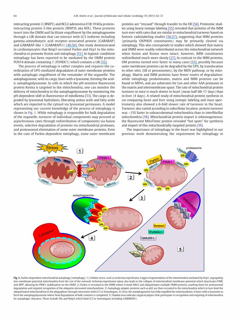
The process of mitophagy is rather complex and requires the co-ordination of UPS-mediated degradation of outer membrane proteinswith autophagic engulfment of the remainder of the organelle. Theautophagosome,with its cargo, fuseswith a lysosome, forming the acid-ic autophagolysosome. In cells in which the pH-sensitive fluorescentprotein Keima is targeted to the mitochondria, one can monitor thedelivery of mitochondria to the autophagolysosome by monitoring thepH-dependent shift in fluorescence of mitoKeima [53]. The cargo is de-graded by lysosomal hydrolases, liberating amino acids and fatty acidswhich are exported to the cytosol via lysosomal permeases. A modelrepresenting our current knowledge of the process of mitophagy isshown in Fig. 1. While mitophagy is responsible for bulk degradationof the organelle, turnover of individual components may proceed atasynchronous rates through redistribution of components via fusionevents, selective degradation of proteins via mitochondrial proteases,and proteasomal elimination of some outer membrane proteins. Evenin the case of Parkin-dependent mitophagy, some outer membrane
Fig. 1.Parkin-dependentmitochondrial autophagy (mitophagy). 1) Cellular stress, such as ischelow-membrane potential mitochondria from the rest of the network. Ischemia/reperfusion injuand MPP, allowing for PINK1 stabilization on the OMM. 2) Parkin is recruited to the OMMwhedegradation and targeted recognition of the ubiquitin-decoratedmitochondrion. 3) Autophagyubiquitinatedmitochondrion to thephagophore through interactionwith LC3 or homologues. 4)form the autophagolysosomewhere final degradation of bulk contents is completed. 5) Shadedfor autophagic clearance. These include Nix and Bnip3 which bind LC3 or homologues includin
proteins are “rescued” through transfer to the ER [54]. Proteomic stud-ies using heavy isotope labeling [55] revealed that proteins of the IMMturn overwith rates that are similar tomitochondrial turnover based onhistoric radiolabeling studies [56,57], suggesting that IMM proteins(primarily OXPHOS constituents) may be primarily cleared viamitophagy. This also corresponds to studies which showed that matrixand OMMwere readily redistributed across themitochondrial networkwhen fusion and fission were intact; however, IMM constituentsredistributed much more slowly [27]. In contrast to the IMM proteins,OM proteins turned over faster in many cases [55], possibly becauseouter membrane proteins can be degraded by the UPS, by translocationto other sites (ER or peroxisomes), by the MDV pathway, or by mito-phagy. Matrix and IMM proteins have fewer routes of degradation:while mitophagy predominates, matrix and IMM proteins can befound in MDVs, and are substrates for Lon and other AAA proteases inthematrix and intermembrane space. The rate of mitochondrial proteinturnover in mice is much slower in heart (mean half-life 17 days) thanin liver (4 days). A related study of mitochondrial protein synthesis inrat comparing heart and liver using isotopic labeling and mass spec-trometry also showed a 6-fold slower rate of turnover in the heart.Turnover also varied according to subcellular location: protein turnoverwas ~15% faster in subsarcolemmal mitochondria than in interfibrillarmitochondria [58]. Mitochondrial protein import is inhomogeneous:the fluorescent MitoTimer protein revealed “hot spots” for synthesisand import of this mitochondrially-targeted protein [59].
The importance of mitophagy in the heart was highlighted in ourprevious work demonstrating the requirement for mitophagy in
mia/reperfusion, triggers fragmentation of themitochondriamediatedbyDrp1, segregatingry also leads to the collapse of mitochondrial membrane potential which deactivates PARLre it binds Mfn2 and ubiquitinates multiple OMM proteins, marking them for proteasomaladapter proteins such as p62 are then recruited to themitochondria which in turn bind theOnce the autophagosomehas fully engulfed themitochondrion, it fuseswith a lysosome toarea indicates atypical players that participate in recognition and targeting of mitochondriag GABARAPL1.

65A.M. Andres et al. / Journal of Molecular and Cellular Cardiology 78 (2015) 62–72
cardioprotection conferred by ischemic preconditioning [13] and acutestatin administration [12]. These results suggest that mitophagy is partof the final common pathway for various cardioprotective interventions,and indeed, may be the ultimate effector. Beyond cardioprotection,Parkin-dependent mitophagy plays a role in ischemia tolerance [60],myocardial aging [61], and pathologic remodeling in response to pressureoverload [62]. The cardiac effects of Parkin deficiency are phenocopied byPINK1 and Atg5 deletions [63,64]. Similarly, deletion of Mfn2, consideredan essential mitochondrial docking partner for Parkin, leads tomitochon-drial dysfunction and heart failure [65]. These findings highlight theimportance of autophagy and mitophagy in cardiac function.
2.2. Overview of mitochondrial biogenesis
Mitochondrial biogenesis, which acts in concert with mitophagy tomaintain homeostasis in cells, depends on the coordination of nuclearand mitochondrial-encoded gene expression. The nuclear-encodedgenes are primarily controlled by the transcription cofactor peroxisomeproliferator-activated receptor gamma coactivator 1-alpha (PGC-1α)[66]. First identified as a binding partner of peroxisome proliferator-activated receptor γ (PPARγ) that increased its transcriptional activityduring thermogenesis, PGC-1α is a member of the nuclear receptorsuperfamily of proteins that are responsible for assembling functionalmacrocomplexes of transcriptional machinery at specific DNA se-quences [67,68]. PGC-1α controls the expression of nuclear respiratoryfactors 1 and 2 (NRF-1 and NRF-2), which in turn control the expressionof mitochondrial transcription factor A (Tfam) [69]. Tfam plays a keyrole in mitochondrial biogenesis by regulating the expression of mito-chondrial genes (tRNAs, rRNAs and 13 subunits of the respiratorychain) from the heavy and light strands of the mitochondrial genome;it is also essential for replication of mitochondrial DNA (mtDNA) [70].
Several studies have demonstrated the physiological importance ofPGC-1α in mitochondrial biogenesis in response to cold and exercise.These external stimuli increase expression of PGC-1α leading to in-creased expression of mitochondrial enzymes such as ATP synthase(β-subunit), COX (cytochrome c oxidase) subunits (COX II and COX IV)and δ-aminolevulinate synthase (δ-ALAS) [71,72]. Increased mitochon-dria content allows for more efficient thermogenesis in response to coldand enhanced capacity to generate ATP to sustain exercise bouts. An iso-form of PGC-1α, PGC-1β has also been identified, and although overex-pression of this protein increased mitochondrial biogenesis and basaloxygen consumptionmuch like PGC-1α, it was not induced by cold or ex-ercise, suggesting alternate pathways for the induction of mitochondrialbiogenesis [73].
Aside from influencing the transcription of key players in the respira-tory chain, PGC-1α also interacts with and upregulates the expression ofother transcription factors such as peroxisome proliferator-activated re-ceptors (PPARs) [74], hormone receptors for estrogen and thyroxine, aswell as estrogen-related receptors (ERRs)α and γ [75]. ERRs are a partic-ularly interesting set of nuclear receptors known as orphan receptors dueto a lack of an associated ligand [76]. These proteins are involved inPGC-1α-dependent regulation of mitochondrial biogenesis. For ex-ample, Schreiber et al. have demonstrated that over-expression ofPGC-1α results in upregulation of 151 genes that encodemitochondrialproteins involved in manymetabolic functions of mitochondria such asfatty acid β-oxidation (FAO), tricarboxylic acid cycle and oxidativephosphorylation, as well as mitochondrial ribosomal machinery andmitochondrial membrane transport proteins. This effect is inhibited bysiRNA targeted to ERRα, and conversely mimicked by overexpressionof ERRα [77]. Endonuclease G is regulated by ERRα and PGC-1α, andits deletion results in cardiac hypertrophy and mitochondrial dysfunc-tion [78]. Additional factors implicated in mitochondrial biogenesis in-clude Lon protease and Hsp78 [79].
The expression of PGC-1α is controlled primarily through signalingcascades. Calcineurin A-dependent (CnA) and Ca2+/calmodulin-dependent protein kinase IV (CaMKIV) regulation of PGC-1α has been
well characterized. CnA interacts with and activates myocyte enhancerfactors 2C and 2D (MEF2C and MEF2D), which regulate the transcriptionof PGC-1α directly [80,81]. Furthermore, activation of PGC-1α results inthe upregulation of MEF2C and 2D, creating a feed forward loop thatallows PGC-1α to increase its own expression [82]. CaMKIV activatesPGC-1α by phosphorylating the transcription factor cAMP response ele-ment (CRE)-binding protein (CREB). Phosphorylated CREB binds to pro-moter elements of the PGC-1α gene to drive its expression [80]. Otherplayers controlling PGC-1α expression include p38 mitogen-activatedprotein kinase (p38 MAPK) and AMP-activated protein kinase (AMPK).p38MAPKactivity is increased following exercise, and this leads to the ac-tivation of MEF2 and activating transcription factor 2 (ATF2), both ofwhich drive the expression of PGC-1α [83,84]. The activation of AMPKin response to glucose depletion results in direct phosphorylation ofPGC-1α on threonine-177 and serine-538, which is crucial for the acti-vation of PGC-1PGC-1α-dependent transcription from the PGC-1α pro-moter [85].
Post-translational modifications such as phosphorylation and acety-lation regulate PGC-1α activity. Kinases that have been implicated incontrolling PGC-1α activity include: AMPK and Akt during caloric re-striction and p38 MAPK after endurance exercise [85–87]. Likewise,p38MAPK increases the activity of PGC-1α by directly phosphorylatingthreonine-262, serine-265, and threonine-268,which stabilizes the pro-tein and disrupts the interaction between PGC-1α and its inhibitorp160MBP [86,88]. Conversely, insulin inhibits the activity of PGC-1αthroughAkt, directly through phosphorylation of the serine-570 residueon PGC-1α, and indirectly through phosphorylation of the Clk2 kinasewhich in turn phosphorylates the C-terminal serine and threonine-richregions of PGC-1α, thereby decreasing its co-transcriptional activity [89,90]. In an even more indirect manner, glycogen synthase kinase 3beta(GSK3β) has also been shown to inhibit PGC-1α activity in response toacute oxidative stress by increasing its proteasomal degradation andinhibiting the activity of Sirt1, an NAD-dependent deacetylase thoughtto activate PGC-1α [91]. This deacetylation event is crucial for the activa-tion of PGC-1α, as the protein is very heavily acetylated by the acetyl-transferase GCN5, inhibiting its activity and sequestering it in nuclearfoci distant from promoter regions of its target genes [92]. Sirt1 activityis dependent upon the coenzyme nicotinamide adenine dinucleotide(NAD+), and it is therefore highly sensitive to the changes in the ener-getic state of the cell. Increased NAD+/NADH ratio—whichmay occur inresponse to fasting, exercise or redox stress—activates Sirt1, leading toPGC-1α deacetylation [93,94]. The result of this deacetylation is an in-crease in the transcription of PGC-1α targets, leading to mitochondrialbiogenesis [95–97]. Interestingly, AMPK may once again play a role inactivating PGC-1α, not only by directly phosphorylating the protein,but also indirectly by increasing NAD+ levels in the cell by fatty acidoxidation, thereby increasing the activity of Sirt1 [97]. Other posttrans-lational modifications such as ubiquitination or methylation also play arole in modulating the activity of PGC-1α in response to energydemands and oxidative stress, states that requiremitochondrial biogen-esis [98].
Other proteins that play amajor part inmitochondrial biogenesis arevascular endothelial growth factor (VEGF) and hypoxia-inducible factor1 (HIF-1). VEGF is a key regulator of angiogenesis which involves sub-stantial cell proliferation and production and remodeling of extracellularmatrix, processes which utilize large amounts of ATP. A study byWrightet al. demonstrated that VEGF stimulates mitochondrial biogenesisby coordinated upregulation of OMM protein Tom70 and activationof PGC-1α [99]. Interestingly, PGC-1α can also activate VEGF, bycoactivating ERR-α on conserved binding sites found in the promoterand in a cluster within the first intron of the VEGF gene, driving angio-genesis in response to oxygen deprivation independently of HIF-1[100]. HIF-1 is a master regulator of the adaptive response to hypoxia,and as such is intimately linked to inducing mitochondrial biogenesis.Several studies have linked PGC-1α and PGC-1β with HIF-1 activity.O'Hagan et al. reported that mitochondrial biogenesis driven by the

66 A.M. Andres et al. / Journal of Molecular and Cellular Cardiology 78 (2015) 62–72
expression of PGC-1α results in increased oxygen consumption and de-creased intracellular oxygen tension, permitting stabilization of HIF-1and activation of a gene expression program to increase oxygen deliveryto the tissue [101]. Zhang et al. demonstrated that HIF-1 repressesmito-chondrial biogenesis by controlling the transcription of PGC-1β throughdownregulation of c-MYC transcription factor [102]. Fig. 2 highlights thecentral role of PGC-1α in regulating mitochondrial biogenesis.
2.3. Interplay between mitophagy and mitochondrial biogenesis
Mitophagy and mitochondrial biogenesis are opposing forces thatgovern the rate of mitochondrial turnover. This dynamic tension allowsfor a readily adjustable population of mitochondria to match cellulardemands. Here we discuss several players that participate in this regu-latory cross-talk.
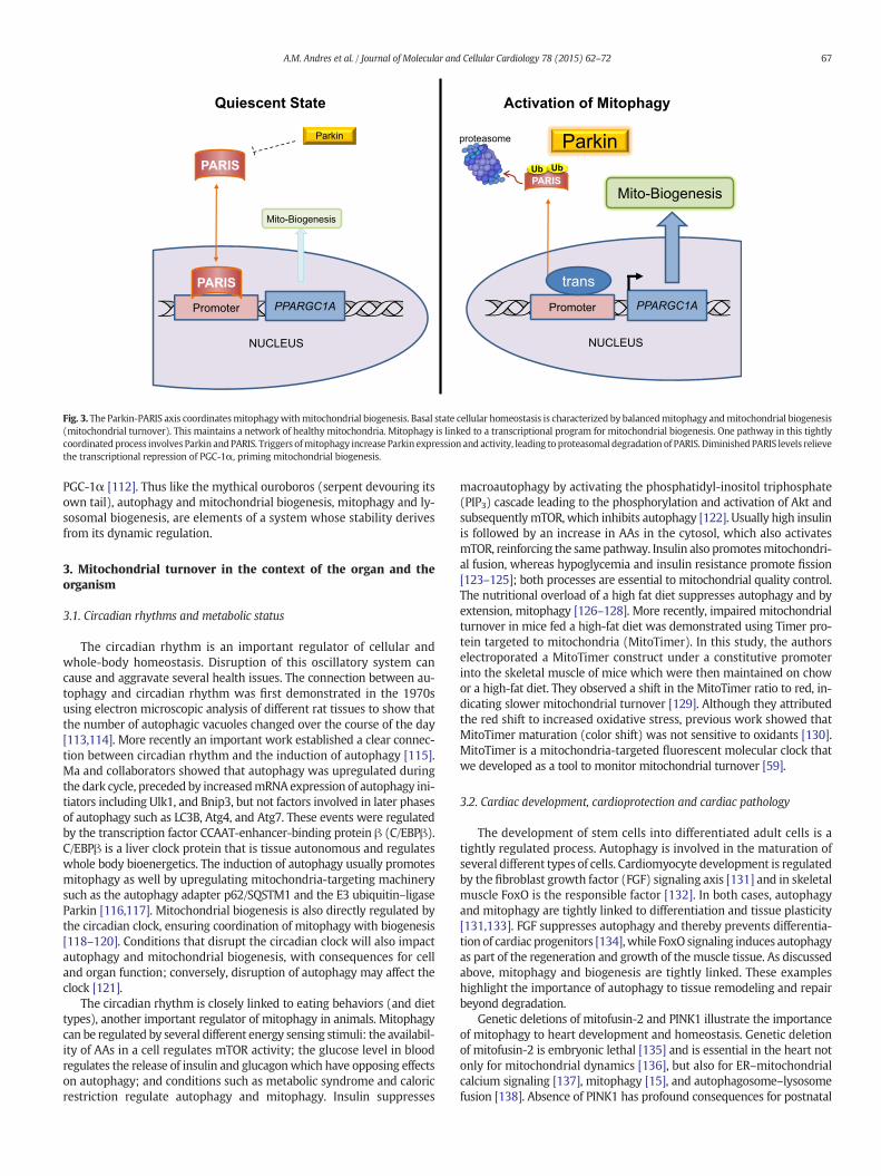
Sirt1, an NAD-dependent lysine deacetylase, stimulates autophagydirectly by deacetylating various autophagy proteins including Atg7,Atg5, and Atg8 (LC3) [103]. Sirt1 also deacetylates and activates PGC-1α[104], thus positively regulating both mitophagy and biogenesis. Activa-tion of PGC-1α is a key event in initiating mitochondrial biogenesis, butno less important are the repressors of the process. Parkin-interactingsubstrate (PARIS or ZNF746) has recently been identified as a direct tran-scriptional repressor of PGC-1α [105]. The accumulation of PARIS in thenucleus leads to direct inactivation of PGC-1α transcription and inhibitionof expression of PGC-1α-dependent genes. Aside from its role in facilitat-ingmitophagy, Parkinwas shown to directly target PARIS for degradationthrough the UPS. Events leading to the upregulation of mitophagy alsoincrease Parkin activitywhich then degrades PARIS, relieving repressionof mitochondrial biogenesis. This relationship establishes an intricate
Cold Exercise
cAMP
CREBP
PG
P38 MAPK Akt3
NCORMEF2C/D
CaMKIV
CnA
VEGF
P
PKACa++
Ca++
PPARGC1A
ESRRA NFE2L1 PPARANFE2L2
Promoter
Fig. 2. PGC-1α regulation ofmitochondrial biogenesis. PGC-1α is considered amaster regulatorto environmental cues of fuel availability, fuel type, and cellular energy requirements. PGC-1αMEF2, C/EBP, FoxO, CREB/CRTC, ERRγ, and MyoD/E2A. These factors in turn are activated by spand PKA, and suppressed by other signals including GCN5, AKT and SHP. In addition to transcfactors illustrated here. Ultimately, PGC-1α increases mitochondrial biogenesis and the capaci
system that links mitophagy with mitochondrial biogenesis. A modelillustrating this mechanism linking mitophagy with mitochondrial bio-genesis is shown in Fig. 3. As the relationship between Parkin and PARIShas thus far been identified only in neuronal models, an important andexciting question remains as to whether this system exists in the heart.Moreover, what are its implications in the setting of ischemia/reperfu-sion injury?
Other repressors of mitochondrial biogenesis operate in less directmanner. Nuclear co-repressor 1 (Ncor1) acts as a repressor of PPARγ,PPARδ and ERR activity by interacting with histone deacetylases suchas HDAC3 and SIRT1 to maintain tonic repression of MEF2, PPARδ, andERR, thereby suppressing their participation in transcription programsinvolving mitochondrial oxidative metabolism [106]. mTOR is a serine/threonine kinase involved in numerous cell functions and can directly ac-tivate PGC-1α to control mitochondrial biogenesis. During fasting, mTORis inhibited and autophagy/mitophagy is active. However, as lysosomaldegradation releases amino acids, mTOR is reactivated, suppressingautophagy and supporting lysosomal and mitochondrial biogenesis[107]. Eukaryotic translation initiation factor 4E (eIF4E)-binding proteins(4E-BP) prevent translation of targets including nuclear encoded mito-chondrial protein mRNAs including TFAM (transcription factor A, mito-chondrial) and subunits of complex V and complex I. This inhibition islifted by the action of mTORC1 which inhibits 4E-BP proteins from bind-ing their targets [108,109].
Autophagy and mitochondrial biogenesis are linked in both direc-tions: PGC-1α induces the expression of transcription factor EB (TFEB)[110], the master regulator of lysosome biogenesis and the autophagypathway [111]. TFEB and PGC-1α regulate one another's expression,and the nutrient sensing regulator GCN5L1 suppresses both TFEB and
Nutrient Deprivation
AMP/ATP NAD+/NADH
C-1α
Ac
GCN5
AMPK SIRT1
mtDNA
TFAM
ofmitochondrial biogenesis. Transcriptional control of PGC-1α expression is closely linkedtranscription is governed by multiple transcription factors (trans) including PPAR/RXR,ecific signal pathways including free fatty acids, AMPK, calcineurin, p38 MAPK, CaMK IV,riptional control, PGC-1α activity is regulated by acetylation and phosphorylation by thety to perform OXPHOS, in particular, fatty acid oxidation.
Parkin
NUCLEUS
PPARGC1APromoter
NUCLEUS
PPARGC1APromoter
Mito-Biogenesis
Parkin
Mito-Biogenesis
Quiescent State Activation of Mitophagy
PARIS
PARISPARIS
trans
proteasome
Ub Ub
Fig. 3. The Parkin-PARIS axis coordinatesmitophagywithmitochondrial biogenesis. Basal state cellular homeostasis is characterized by balancedmitophagy andmitochondrial biogenesis(mitochondrial turnover). This maintains a network of healthy mitochondria. Mitophagy is linked to a transcriptional program for mitochondrial biogenesis. One pathway in this tightlycoordinatedprocess involves Parkin and PARIS. Triggers ofmitophagy increase Parkin expression and activity, leading to proteasomal degradation of PARIS. Diminished PARIS levels relievethe transcriptional repression of PGC-1α, priming mitochondrial biogenesis.
67A.M. Andres et al. / Journal of Molecular and Cellular Cardiology 78 (2015) 62–72
PGC-1α [112]. Thus like the mythical ouroboros (serpent devouring itsown tail), autophagy and mitochondrial biogenesis, mitophagy and ly-sosomal biogenesis, are elements of a system whose stability derivesfrom its dynamic regulation.
3. Mitochondrial turnover in the context of the organ and theorganism
3.1. Circadian rhythms and metabolic status
The circadian rhythm is an important regulator of cellular andwhole-body homeostasis. Disruption of this oscillatory system cancause and aggravate several health issues. The connection between au-tophagy and circadian rhythm was first demonstrated in the 1970susing electron microscopic analysis of different rat tissues to show thatthe number of autophagic vacuoles changed over the course of the day[113,114]. More recently an important work established a clear connec-tion between circadian rhythm and the induction of autophagy [115].Ma and collaborators showed that autophagy was upregulated duringthe dark cycle, preceded by increasedmRNAexpression of autophagy ini-tiators including Ulk1, and Bnip3, but not factors involved in later phasesof autophagy such as LC3B, Atg4, and Atg7. These events were regulatedby the transcription factor CCAAT-enhancer-binding protein β (C/EBPβ).C/EBPβ is a liver clock protein that is tissue autonomous and regulateswhole body bioenergetics. The induction of autophagy usually promotesmitophagy as well by upregulating mitochondria-targeting machinerysuch as the autophagy adapter p62/SQSTM1 and the E3 ubiquitin–ligaseParkin [116,117]. Mitochondrial biogenesis is also directly regulated bythe circadian clock, ensuring coordination of mitophagy with biogenesis[118–120]. Conditions that disrupt the circadian clock will also impactautophagy and mitochondrial biogenesis, with consequences for celland organ function; conversely, disruption of autophagy may affect theclock [121].
The circadian rhythm is closely linked to eating behaviors (and diettypes), another important regulator of mitophagy in animals. Mitophagycan be regulated by several different energy sensing stimuli: the availabil-ity of AAs in a cell regulates mTOR activity; the glucose level in bloodregulates the release of insulin and glucagonwhich have opposing effectson autophagy; and conditions such as metabolic syndrome and caloricrestriction regulate autophagy and mitophagy. Insulin suppresses
macroautophagy by activating the phosphatidyl-inositol triphosphate(PIP3) cascade leading to the phosphorylation and activation of Akt andsubsequentlymTOR,which inhibits autophagy [122]. Usually high insulinis followed by an increase in AAs in the cytosol, which also activatesmTOR, reinforcing the samepathway. Insulin also promotesmitochondri-al fusion, whereas hypoglycemia and insulin resistance promote fission[123–125]; both processes are essential to mitochondrial quality control.The nutritional overload of a high fat diet suppresses autophagy and byextension, mitophagy [126–128]. More recently, impaired mitochondrialturnover in mice fed a high-fat diet was demonstrated using Timer pro-tein targeted to mitochondria (MitoTimer). In this study, the authorselectroporated a MitoTimer construct under a constitutive promoterinto the skeletal muscle of mice which were then maintained on chowor a high-fat diet. They observed a shift in the MitoTimer ratio to red, in-dicating slower mitochondrial turnover [129]. Although they attributedthe red shift to increased oxidative stress, previous work showed thatMitoTimer maturation (color shift) was not sensitive to oxidants [130].MitoTimer is a mitochondria-targeted fluorescent molecular clock thatwe developed as a tool to monitor mitochondrial turnover [59].
3.2. Cardiac development, cardioprotection and cardiac pathology
The development of stem cells into differentiated adult cells is atightly regulated process. Autophagy is involved in the maturation ofseveral different types of cells. Cardiomyocyte development is regulatedby the fibroblast growth factor (FGF) signaling axis [131] and in skeletalmuscle FoxO is the responsible factor [132]. In both cases, autophagyand mitophagy are tightly linked to differentiation and tissue plasticity[131,133]. FGF suppresses autophagy and thereby prevents differentia-tion of cardiac progenitors [134],while FoxO signaling induces autophagyas part of the regeneration and growth of the muscle tissue. As discussedabove, mitophagy and biogenesis are tightly linked. These exampleshighlight the importance of autophagy to tissue remodeling and repairbeyond degradation.
Genetic deletions of mitofusin-2 and PINK1 illustrate the importanceof mitophagy to heart development and homeostasis. Genetic deletionof mitofusin-2 is embryonic lethal [135] and is essential in the heart notonly for mitochondrial dynamics [136], but also for ER–mitochondrialcalcium signaling [137], mitophagy [15], and autophagosome–lysosomefusion [138]. Absence of PINK1 has profound consequences for postnatal

68 A.M. Andres et al. / Journal of Molecular and Cellular Cardiology 78 (2015) 62–72
heart development [63] and exacerbates ischemia/reperfusion injury[139].
Among themost potent interventions to protect the heart from ische-mia and reperfusion injury are ischemic pre- and post-conditioning [140,141]. Pre and post conditioning require autophagy to deliver the protec-tion [142–144], although this is controversial in the brain [145,146].Mitophagy is part of the autophagy response that is specifically requiredfor protection [13]. Other interventions that protect the heart againstischemic injury, including chloramphenicol [147], caloric restriction[148] simvastatin [12], and SAHA [149] all act through the autophagy/mitophagy pathway, thus establishing autophagy/mitophagy as a hubfor cardiac protection. There are few direct inducers of autophagy;rapamycin is anmTOR inhibitorwidely used as a drug to induce autoph-agy. Rapamycin administration also decreased ischemia/reperfusion in-jury [149,150] while upregulating autophagy. In chronic models ofheart failure, rapamycin also helps to ameliorate the phenotype [151].Taken together, these facts support the beneficial effects of inducingautophagy.
In conditions where autophagy/mitophagy is impaired the opposite istrue: there is increased cardiac dysfunction and exacerbation of ischemia/reperfusion injury in the setting of Parkin and PINK1 deletion [60,139],deletion of macrophage migration inhibitor factor, an inducer of homeo-static autophagy [152], and obesity [153]. Obesity [154] and diabetes[155] disrupt normal energy metabolism, changing basal activation ofmTOR and other nutrient signaling cascades that regulate autophagy.High fat diets are known to increase ischemia/reperfusion damage inhearts [156,157] and there is now a significant amount of work linkingthe high fat diet to impaired autophagy [154] and accumulation ofdysfunctional mitochondria [158,159], highlighting the importance ofmitophagy to cardiac ischemia tolerance. The regulation of cardiacmitophagy and physiological importance of this process is illustrated inFig. 4.
Autophagy may not always be a good thing in the heart. Infection ofjuvenilemicewith Coxsackievirus B virus can exacerbate stress-inducedmyocardial injury in adulthood [160]. Autophagy is needed for thespread and reproduction of the virus [161], and virus-induced autophagytriggers premature differentiation of cardiac-resident progenitor cells,
DiabetesObesityAging
ChloramphenicolStatins
Caloric Restriction
C/EBPb
Clock Proteins
Regulation Cardiac Importance
p62/SQSTM1
High FatsHigh Insulin
Increased
Food Intake HFD
mTOR
Parkin
PINK1
Decreased
• Cardiomyocyte Maturation
• Cardioprotection
• Increased Mitochondrial
Turnover
• Loss of Preconditioning
• Less Ischemia Tolerance
• Accumulation of Damaged
Mitochondria
AutophagyRapamycin
Mitophagy
Fig. 4. Cardiac mitophagy regulation and significance. Mitophagy is essential duringcardiomyocyte differentiation and for homeostatic mitochondrial turnover to maintain ahealthy population of mitochondria. During cardiac stress such as ischemia/reperfusion,mitophagy functions to eliminate damaged mitochondria and reduce injury. Mitophagyis also critical for ischemic preconditioning. Circadian rhythm regulates basal levels ofcardiac mitophagy. Nutrient overload, type 2 diabetes, obesity, and advanced age maycompromise cardiac autophagy and mitophagy, disrupting this adaptive physiologicalresponse to stress.
contributing to heart failure later in life [160]. Excessive autophagy hasbeen implicated in doxorubicin-mediated cardiac injury [162,163],although other studies have reported a beneficial role for autophagy[164–167]. Interestingly, deletion of Nrf2, a transcriptional regulator ofautophagy andmitochondrial biogenesis, exacerbated doxorubicin tox-icity, but this was reversed by overexpression of Atg5 [168]. They didnot examine whether restoring autophagy resulted in mitochondrialbiogenesis independent of Nrf2. It seems likely that unless mitophagyis balanced by biogenesis, problems will ensue.
Mitochondrial biogenesis in the heart is tightly responsive to oxygentension. This is manifested at the transition from the fetal hypoxic stateto the postnatal aerobic environment, when HIF signaling is lost, therebyfavoring mitochondrial fusion and mitochondrial biogenesis [169].During cardiac hypertrophy in response to aortic banding,mitochondrialdysfunction and decreased biogenesis were noted [170]. Downregula-tion of PGC-1α is observed in animal models of heart failure, butattempts to restore mitochondrial biogenesis by overexpression ofPGC-1α did not improve cardiac function despite a modest increase inmitochondrial content [171]. In fact, inducible overexpression ofPGC-1α in the heart resulted in abnormal mitochondrial morphologyand cardiomyopathy which was reversible upon the normalization ofPGC-1α levels [172]. These studies did not examine mitochondrialautophagy. However, in a porcine study of renovascular hypertension,hypertrophy was accompanied by the upregulation of mTOR, increasedabundance of markers of autophagy and mitophagy, and decreasedmitochondrial protein content, all of whichwere reversed by the angio-tensin II receptor blocker valsartan [173]. The authors concluded thathypertension increased autophagic clearance of mitochondria andvalsartan suppressed autophagy and restoredmitochondrial biogenesis.However, because they did not measure autophagic flux or p62 (asurrogate marker of flux), the data lend themselves to the oppositeinterpretation: that hypertension impaired autophagic flux, therebylimiting mitochondrial biogenesis possibly through the Parkin/PARIS/PGC-1α network. Evidence in support of the latter interpretationis the finding that mTOR was strongly upregulated in the hypertensivehearts, which would suppress autophagy. The observed increase inBeclin 1 and LC3-II could reflect increased autophagy or impaired flux.Beclin 1 is known to interfere with autophagic flux [174,175]. Theperinuclear accumulation of Parkin-decorated mitochondria is also in-dicative of impaired lysosomal clearance of autophagosomes. This alsoillustrates the importance of determining autophagic flux and mito-chondrial turnover before reaching a conclusion.
4. Prospects and challenges for the future
Mitochondrial quality control depends upon mitophagy, biogenesis,fusion, and fission, as well as selective protein quality control via AAAproteases and chaperones. To date, most studies have explored mito-chondrial dynamics (fusion/fission) and mitophagy (Parkin-dependentand Parkin-independent mitochondrial autophagy). Mitophagy is in-creasingly recognized to play a significant role in the heart, yet inorder to maintain homeostasis, biogenesis must keep pace. Therefore,approaches to monitoring mitochondrial turnover (the integrated out-come of these four processes) are needed. Recent advances include theanalysis of the half-lives of mitochondrial proteins using mass spectrom-etry analysis and deuterium labeling [55], mito-Keima, which can reporton mitochondria delivered to the lysosome [53], and MitoTimer, a fluo-rescent protein that can be used to monitor mitochondrial turnover [59,129,130]. What lies ahead is the application of these tools to study phys-iologic and pathologic processes in the heart.
A major challenge to overcome is imaging autophagy (or mitophagy)in humans. Relatively few studies have examined autophagy in thehuman heart, largely because of the challenges of accessing tissue, andnone have examined mitophagy, although animal studies indicate thatmitochondria are a frequent target of autophagy. There is a significant
69A.M. Andres et al. / Journal of Molecular and Cellular Cardiology 78 (2015) 62–72
need to develop better tools for in vivo imaging of autophagy andmitophagy.
Still lacking is a thorough understanding of mitochondrial bio-genesis: are all mitochondria equally capable of expanding and un-dergoing fission to give rise to daughter mitochondria enriched fornewly-imported proteins and highly functional OXPHOS assem-blies, or is there a subset of mitochondria that are specialized formitochondrial regeneration? Studies of MitoTimer suggest thatprotein import preferentially takes place in mitochondria closestto the nucleus [59,130]. This could be a trivial consequence ofmRNA proximity, and import of MitoTimer may not necessarily re-flect sites of biogenesis. It is exciting, however, to speculate that thesubpopulation of mitochondria most actively engaged in importingnewly-synthesized protein is indeed unique. Future studies mayshed light on this.
Mitochondrial protein import is essential for biogenesis, but is alsoimplicated in the regulation of mitophagy because PINK1 must transitthrough the intermembrane space in order to be degraded by PARL.Few studies have considered whether defective protein import is thered flag that signifies a mitochondrion due for autophagic elimination.It has not been demonstrated whether pre-amyloid oligomers mightdisrupt mitochondrial protein import, yet this might explain the deteri-oration of mitochondrial function (78) and impaired biogenesis (79)that often accompanies Alzheimer's disease and potentially other pro-tein folding disorders.We can expect that in the coming years investiga-tors will integrate information exchange between mitochondria andcytosol/nucleus, for which the TOM/TIM complex and VDAC serve asimportant carriers.
A key to mitochondrial homeostasis is the ability to remove andreplace components throughout the network: not only proteins, butalso lipids and mtDNA copies. In the heart, where mitochondrial fusionand fission events seem to occur with a frequency approximately equalto the rate of turnover of the entire organelle, intra-mitochondrial deg-radation and protein import generalized across the network may play alarger role than regionally restricted biogenesis followed by redistribu-tion via fusion events. The intriguing observation that proteins insubsarcolemmal mitochondria turn over faster than in interfibrillar mi-tochondria suggests that different turnover mechanisms may operatewithin the same cell. The thought-provoking discovery that individualmitochondrial proteins have widely differing half-lives raises questionsabout the mechanisms governing this process; regulation of these dif-ferent mechanisms for degrading mitochondrial proteins may be quitecomplex, and their contribution to disease phenotypes will be equallyso. The importance of mitochondrial protein import is emerging:recently it was reported that redirecting a mutant form of alanine:glyoxalate aminotransferase from mitochondria to peroxisomes cor-rects primary hyperoxaluria 1 (PH1), a lethal metabolic disease [176].Enzymes that may traffic either to mitochondria or peroxisomes canhave radically different consequences depending on their location; thepotential significance of this process for heart disease is unknown atpresent.
Yet another emerging area is the role of miRNAs in regulatingautophagy and mitochondrial biogenesis. miRNA-149 inhibitspoly(ADP-ribose) polymerase-2 (PARP-2), thereby allowing an in-crease in cellular NAD+ and activation of sirtuin-1, leading to mito-chondrial biogenesis [177]. miR-27a and miR-27b impairmitochondrial biogenesis [178]. miRNAs also regulate the Nrf2pathway [179] and autophagy [180–184]. Elucidating the contribu-tion of miRNAs to the dynamic regulation of mitophagy and biogen-esis will require a systems biology approach.
Many open questions remain to be resolved, but technical advancescontinue to make new discoveries possible. The advent of novel genetherapy approaches, cell permeable proteins, and small moleculetherapeutics targeting mitochondrial quality control mechanisms holdpromise for treating a variety of diseases from the perspective of the un-derlying mitochondrial dysfunction. It is not too farfetched to envision
mitochondrial medicine becoming a medical specialty as much as sur-gery, cardiology, or genetics.
Conflict of interest
RAG is a consultant for Takeda Pharmaceuticals and is a cofounder ofTissueNetix, Inc. The other authors have no potential conflicts of interestto disclose.
Acknowledgments
RAG holds the Dorothy and E. Phillip Lyon Chair in Molecular Cardi-ology in honor of Clarence M. Agress, MD. This work was funded in partby NIHP01 HL112730 (RAG).
Mitochondrial MysteriesRoberta A. GottliebWe know so much yet understand so littleAbout mitochondrial ox-phos and fusion and fission,Mitochondrial autophagy and biogenesis.MitoTimer and lenses have given us celluvision.Though heart cells live years it's quite different within:Mitochondrial life is counted in weeks.Outer and inner membrane proteins vary yet moreIn their lifespans revealed by mass spectrum peaks.Protein import must match what's inside,Lest proteins unfold and fall prey to Lon.The peptides escape to the cytosolTo trigger transcription of chaperones.Try we must to describe and defineThe complex nature of the proteome,As mitochondria expand and divide,Fragment and fall into autophagosomes.Yet for all we know and all we learnThe mysteries grow and questions expandLike Mandelbrot sets of fractal images,We see the work of divinity's hand.
References
[1] Bogaerts V, Theuns J, van Broeckhoven C. Genetic findings in Parkinson's diseaseand translation into treatment: a leading role for mitochondria? Genes BrainBehav 2008;7:129–51.
[2] Bueler H. Impaired mitochondrial dynamics and function in the pathogenesis ofParkinson's disease. Exp Neurol 2009;218:235–46.
[3] Park J, Kim Y, Chung J. Mitochondrial dysfunction and Parkinson's disease genes:insights from Drosophila. Dis Model Mech 2009;2:336–40.
[4] Schapira AH. Mitochondria in the aetiology and pathogenesis of Parkinson'sdisease. Lancet Neurol 2008;7:97–109.
[5] Trancikova A, Tsika E, Moore DJ. Mitochondrial dysfunction in genetic animalmodels of Parkinson's disease. Antioxid Redox Signal 2012;16:896–919.
[6] Biskup S, Moore DJ. Detrimental deletions: mitochondria, aging and Parkinson'sdisease. Bioessays 2006;28:963–7.
[7] Kotake Y, Ohta S. MPP + analogs acting on mitochondria and inducing neuro-degeneration. Curr Med Chem 2003;10:2507–16.
[8] DuchenMR, Szabadkai G. Roles of mitochondria in human disease. Essays Biochem2010;47:115–37.
[9] Pellegrino MW, Nargund AM, Haynes CM. Signaling the mitochondrial unfoldedprotein response. Biochim Biophys Acta 1833;2013:410–6.
[10] Mizushima N, Levine B, Cuervo AM, Klionsky DJ. Autophagy fights disease throughcellular self-digestion. Nature 2008;451:1069–75.
[11] Wang C,Wang X. The interplay between autophagy and the ubiquitin–proteasomesystem in cardiac proteotoxicity. Biochim Biophys Acta 2014. http://dx.doi.org/10.1016/j.bbadis.2014.07.028 (epub ahead of print).
[12] Andres AM, Hernandez G, Lee P, Huang C, Ratliff EP, Sin J, et al. Mitophagy is re-quired for acute cardioprotection by simvastatin. Antioxid Redox Signal 2013.http://dx.doi.org/10.1089/ars.2013.5416.
[13] Huang C, Andres AM, Ratliff EP, Hernandez G, Lee P, Gottlieb RA. Preconditioninginvolves selective mitophagy mediated by Parkin and p62/SQSTM1. PLoS One2011;6:e20975.
70 A.M. Andres et al. / Journal of Molecular and Cellular Cardiology 78 (2015) 62–72
[14] Piquereau J, Godin R, Deschenes S, Bessi VL, Mofarrahi M, Hussain SN, Burelle Y.Protective role of park2/parkin in sepsis-induced cardiac contractile andmitochon-drial dysfunction. Autophagy 2013;9:1837–51.
[15] Chen Y, Dorn 2nd GW. PINK1-phosphorylated mitofusin 2 is a Parkin receptor forculling damaged mitochondria. Science 2013;340:471–5.
[16] Kubli DA, Quinsay MN, Huang C, Lee Y, Gustafsson AB. Bnip3 functions as a mito-chondrial sensor of oxidative stress during myocardial ischemia and reperfusion.Am J Physiol Heart Circ Physiol 2008;295:H2025–31.
[17] Scherz-Shouval R, Shvets E, Fass E, ShorerH, Gil L, Elazar Z. Reactive oxygen species areessential for autophagy and specifically regulate the activity of Atg4. EMBO J 2007;26:1749–60.
[18] Carreira RS, Lee Y, GhochaniM,GustafssonAB, Gottlieb RA. Cyclophilin D is requiredfor mitochondrial removal by autophagy in cardiac cells. Autophagy 2010;6:462–72.
[19] Geisler S, Holmstrom KM, Skujat D, Fiesel FC, Rothfuss OC, Kahle PJ, et al. PINK1/Parkin-mediated mitophagy is dependent on VDAC1 and p62/SQSTM1. Nat CellBiol 2010;12:119–31.
[20] Matsuda N, Sato S, Shiba K, Okatsu K, Saisho K, Gautier CA, et al. PINK1 stabilized bymitochondrial depolarization recruits Parkin to damaged mitochondria and acti-vates latent Parkin for mitophagy. J Cell Biol 2010;189:211–21.
[21] Narendra D, Tanaka A, Suen DF, Youle RJ. Parkin is recruited selectively to impairedmitochondria and promotes their autophagy. J Cell Biol 2008;183:795–803.
[22] Narendra DP, Jin SM, Tanaka A, SuenDF, Gautier CA, Shen J, et al. PINK1 is selectivelystabilized on impairedmitochondria to activate Parkin. PLoS Biol 2010;8:e1000298.
[23] Clark IE, Dodson MW, Jiang C, Cao JH, Huh JR, Seol JH, et al. Drosophila pink1 is re-quired for mitochondrial function and interacts genetically with Parkin. Nature2006;441:1162–6.
[24] Park J, Lee SB, Lee S, Kim Y, Song S, Kim S, et al. Mitochondrial dysfunction inDrosophila PINK1 mutants is complemented by Parkin. Nature 2006;441:1157–61.
[25] Sugiura A, McLelland GL, Fon EA, McBride HM. A new pathway for mitochondrialquality control: mitochondrial-derived vesicles. EMBO J 2014;33:2142–56.
[26] Tanaka A, ClelandMM, Xu S, Narendra DP, Suen DF, Karbowski M, et al. Proteasomeand p97 mediate mitophagy and degradation of mitofusins induced by Parkin. JCell Biol 2010;191:1367–80.
[27] Twig G, Elorza A, Molina AJ, Mohamed H, Wikstrom JD, Walzer G, et al. Fission andselective fusion govern mitochondrial segregation and elimination by autophagy.EMBO J 2008;27:433–46.
[28] Smirnova E, Griparic L, Shurland DL, van der Bliek AM. Dynamin-related proteinDrp1 is required for mitochondrial division in mammalian cells. Mol Biol Cell2001;12:2245–56.
[29] Yoon Y, Krueger EW, Oswald BJ, McNivenMA. Themitochondrial protein hFis1 reg-ulates mitochondrial fission in mammalian cells through an interaction with thedynamin-like protein DLP1. Mol Cell Biol 2003;23:5409–20.
[30] MacVicar TD, Lane JD. Impaired OMA1-dependent cleavage of OPA1 and reducedDRP1 fission activity combine to prevent mitophagy in cells that are dependenton oxidative phosphorylation. J Cell Sci 2014;127:2313–25.
[31] Kaser M, Kambacheld M, Kisters-Woike B, Langer T. Oma1, a novel membrane-bound metallopeptidase in mitochondria with activities overlapping with the m-AAA protease. J Biol Chem 2003;278:46414–23.
[32] McBride H, Soubannier V. Mitochondrial function: OMA1 and OPA1, thegrandmasters of mitochondrial health. Curr Biol 2010;20:R274–6.
[33] Greene AW, Grenier K, Aguileta MA, Muise S, Farazifard R, Haque ME, et al. Mito-chondrial processing peptidase regulates PINK1 processing, import and Parkinrecruitment. EMBO Rep 2012;13:378–85.
[34] Jin SM, Lazarou M, Wang C, Kane LA, Narendra DP, Youle RJ. Mitochondrial mem-brane potential regulates PINK1 import and proteolytic destabilization by PARL. JCell Biol 2010;191:933–42.
[35] Zhou C, Huang Y, Shao Y, May J, Prou D, Perier C, et al. The kinase domain ofmitochondrial PINK1 faces the cytoplasm. Proc Natl Acad Sci U S A 2008;105:12022–7.
[36] IguchiM, Kujuro Y, Okatsu K, Koyano F, Kosako H, KimuraM, et al. Parkin-catalyzedubiquitin-ester transfer is triggered by PINK1-dependent phosphorylation. J BiolChem 2013;288:22019–32.
[37] Lazarou M, Jin SM, Kane LA, Youle RJ. Role of PINK1 binding to the TOM complexand alternate intracellular membranes in recruitment and activation of the E3ligase Parkin. Dev Cell 2012;22:320–33.
[38] Wang X, Winter D, Ashrafi G, Schlehe J, Wong YL, Selkoe D, et al. PINK1 and Parkintarget Miro for phosphorylation and degradation to arrest mitochondrial motility.Cell 2011;147:893–906.
[39] Narendra D, Kane LA, Hauser DN, Fearnley IM, Youle RJ. p62/SQSTM1 is required forParkin-induced mitochondrial clustering but not mitophagy; VDAC1 is dispensablefor both. Autophagy 2010;6:1090–106.
[40] Gegg ME, Cooper JM, Chau KY, Rojo M, Schapira AH, Taanman JW. Mitofusin 1 andmitofusin 2 are ubiquitinated in a PINK1/parkin-dependent manner upon induc-tion of mitophagy. Hum Mol Genet 2010;19:4861–70.
[41] Glauser L, Sonnay S, Stafa K, Moore DJ. Parkin promotes the ubiquitination and degra-dation of themitochondrial fusion factor mitofusin 1. J Neurochem 2011;118:636–45.
[42] Poole AC, Thomas RE, Yu S, Vincow ES, Pallanck L. The mitochondrial fusion-promoting factor mitofusin is a substrate of the PINK1/Parkin pathway. PLoS One2010;5:e10054.
[43] Kirkin V, Lamark T, Sou YS, Bjorkoy G, Nunn JL, Bruun JA, et al. A role for NBR1 inautophagosomal degradation of ubiquitinated substrates. Mol Cell 2009;33:505–16.
[44] Pankiv S, Clausen TH, Lamark T, Brech A, Bruun JA, Outzen H, et al. p62/SQSTM1binds directly to Atg8/LC3 to facilitate degradation of ubiquitinated protein aggre-gates by autophagy. J Biol Chem 2007;282:24131–45.
[45] Orvedahl A, Sumpter Jr R, Xiao G, Ng A, Zou Z, Tang Y, et al. Image-basedgenome-wide siRNA screen identifies selective autophagy factors. Nature2011;480:113–7.
[46] Fimia GM, Stoykova A, Romagnoli A, Giunta L, Di Bartolomeo S, Nardacci R, et al.Ambra1 regulates autophagy and development of the nervous system. Nature2007;447:1121–5.
[47] Strappazzon F, Vietri-Rudan M, Campello S, Nazio F, Florenzano F, Fimia GM, et al.Mitochondrial BCL-2 inhibits AMBRA1-induced autophagy. EMBO J 2011;30:1195–208.
[48] Van Humbeeck C, Cornelissen T, Hofkens H, Mandemakers W, Gevaert K, DeStrooper B, et al. Parkin interacts with Ambra1 to induce mitophagy. J Neurosci2011;31:10249–61.
[49] Chakrama FZ, Seguin-Py S, Le Grand JN, Fraichard A, Delage-Mourroux R, DespouyG, et al. GABARAPL1 (GEC1) associates with autophagic vesicles. Autophagy 2010;6:495–505.
[50] Novak I, Kirkin V,McEwan DG, Zhang J,Wild P, Rozenknop A, et al. Nix is a selectiveautophagy receptor for mitochondrial clearance. EMBO Rep 2010;11:45–51.
[51] Lee Y, Lee HY, Hanna RA, Gustafsson AB. Mitochondrial autophagy by Bnip3 in-volves Drp1-mediated mitochondrial fission and recruitment of Parkin in cardiacmyocytes. Am J Physiol Heart Circ Physiol 2011;301:H1924–31.
[52] Liu L, Feng D, Chen G, Chen M, Zheng Q, Song P, et al. Mitochondrial outer-membrane protein FUNDC1 mediates hypoxia-induced mitophagy in mammaliancells. Nat Cell Biol 2012;14:177–85.
[53] Katayama H, Kogure T, Mizushima N, Yoshimori T, Miyawaki A. A sensitive andquantitative technique for detecting autophagic events based on lysosomal delivery.Chem Biol 2011;18:1042–52.
[54] Saita S, ShiraneM, NakayamaKI. Selective escape of proteins from themitochondriaduring mitophagy. Nat Commun 2013;4:1410.
[55] Kim TY, Wang D, Kim AK, Lau E, Lin AJ, Liem DA, et al. Metabolic labeling revealsproteome dynamics of mouse mitochondria. Mol Cell Proteomics 2012;11:1586–94.
[56] Aschenbrenner B, Druyan R, Albin R, RabinowitzM. Haem a, cytochrome c and totalprotein turnover in mitochondria from rat heart and liver. Biochem J 1970;119:157–60.
[57] Lipsky NG, Pedersen PL. Mitochondrial turnover in animal cells. Half-lives of mito-chondria and mitochondrial subfractions of rat liver based on [14C]bicarbonate in-corporation. J Biol Chem 1981;256:8652–7.
[58] Kasumov T, Dabkowski ER, Shekar KC, Li L, Ribeiro Jr RF, Walsh K, et al. Assessmentof cardiac proteome dynamics with heavy water: slower protein synthesis rates ininterfibrillar than subsarcolemmal mitochondria. Am J Physiol Heart Circ Physiol2013;304:H1201–14.
[59] Hernandez G, Thornton C, Stotland A, Lui D, Sin J, Ramil J, et al. MitoTimer: a noveltool for monitoring mitochondrial turnover. Autophagy 2013;9:1852–61.
[60] Kubli DA, Zhang X, Lee Y, Hanna RA, Quinsay MN, Nguyen CK, et al. Parkin proteindeficiency exacerbates cardiac injury and reduces survival followingmyocardial in-farction. J Biol Chem 2013;288:915–26.
[61] Kubli DA, Quinsay MN, Gustafsson AB. Parkin deficiency results in accumulation ofabnormal mitochondria in aging myocytes. Commun Integr Biol 2013;6:e24511.
[62] Oka T, Hikoso S, Yamaguchi O, Taneike M, Takeda T, Tamai T, et al. MitochondrialDNA that escapes from autophagy causes inflammation and heart failure. Nature2012;485:251–5.
[63] Billia F, Hauck L, Konecny F, Rao V, Shen J, Mak TW. PTEN-inducible kinase 1(PINK1)/Park6 is indispensable for normal heart function. Proc Natl Acad Sci U S A2011;108:9572–7.
[64] Nakai A, Yamaguchi O, Takeda T, Higuchi Y, Hikoso S, Taniike M, et al. The role ofautophagy in cardiomyocytes in the basal state and in response to hemodynamicstress. Nat Med 2007;13:619–24.
[65] Chen Y, Liu Y, Dorn 2nd GW. Mitochondrial fusion is essential for organelle func-tion and cardiac homeostasis. Circ Res 2011;109:1327–31.
[66] Wu Z, Puigserver P, Spiegelman BM. Transcriptional activation of adipogenesis.Curr Opin Cell Biol 1999;11:689–94.
[67] Puigserver P,Wu Z, Park CW, Graves R, Wright M, Spiegelman BM. A cold-induciblecoactivator of nuclear receptors linked to adaptive thermogenesis. Cell 1998;92:829–39.
[68] Xiao X, Wang P, Chou KC. Recent progresses in identifying nuclear receptors andtheir families. Curr Top Med Chem 2013;13:1192–200.
[69] Virbasius JV, Scarpulla RC. Activation of the humanmitochondrial transcription factorA gene by nuclear respiratory factors: a potential regulatory link between nuclear andmitochondrial gene expression in organelle biogenesis. Proc Natl Acad Sci U S A 1994;91:1309–13.
[70] Fisher RP, Clayton DA. A transcription factor required for promoter recognition byhumanmitochondrial RNA polymerase. Accurate initiation at the heavy- and light-strand promoters dissected and reconstituted in vitro. J Biol Chem 1985;260:11330–8.
[71] Wu Z, Puigserver P, Andersson U, Zhang C, Adelmant G, Mootha V, et al. Mecha-nisms controlling mitochondrial biogenesis and respiration through the thermo-genic coactivator PGC-1. Cell 1999;98:115–24.
[72] Baar K, Wende AR, Jones TE, Marison M, Nolte LA, Chen M, et al. Adaptations ofskeletal muscle to exercise: rapid increase in the transcriptional coactivator PGC-1.FASEB J 2002;16:1879–86.
[73] Meirhaeghe A, Crowley V, Lenaghan C, Lelliott C, Green K, Stewart A, et al. Character-ization of the human, mouse and rat PGC1 beta (peroxisome-proliferator-activatedreceptor-gamma co-activator 1 beta) gene in vitro and in vivo. Biochem J 2003;373:155–65.
[74] Madrazo JA, Kelly DP. The PPAR trio: regulators of myocardial energy metabolismin health and disease. J Mol Cell Cardiol 2008;44:968–75.

71A.M. Andres et al. / Journal of Molecular and Cellular Cardiology 78 (2015) 62–72
[75] Ventura-Clapier R, Garnier A, Veksler V. Transcriptional control of mitochondrialbiogenesis: the central role of PGC-1alpha. Cardiovasc Res 2008;79:208–17.
[76] Giguere V. Transcriptional control of energy homeostasis by the estrogen-relatedreceptors. Endocr Rev 2008;29:677–96.
[77] Schreiber SN, Emter R, Hock MB, Knutti D, Cardenas J, Podvinec M, et al. Theestrogen-related receptor alpha (ERRalpha) functions in PPARgamma coactivator1alpha (PGC-1alpha)-induced mitochondrial biogenesis. Proc Natl Acad Sci U S A2004;101:6472–7.
[78] McDermott-Roe C, Ye J, Ahmed R, Sun XM, Serafin A, Ware J, et al. Endonuclease Gis a novel determinant of cardiac hypertrophy and mitochondrial function. Nature2011;478:114–8.
[79] Marton O, Koltai E, Takeda M, Koch LG, Britton SL, Davies KJ, et al. Mitochondrialbiogenesis-associated factors underlie the magnitude of response to aerobic endur-ance training in rats. Pflugers Arch European Journal of Physiology 2014:1–10.
[80] Herzig S, Long F, Jhala US, Hedrick S, Quinn R, Bauer A, et al. CREB regulates hepaticgluconeogenesis through the coactivator PGC-1. Nature 2001;413:179–83.
[81] Handschin C, Rhee J, Lin J, Tarr PT, Spiegelman BM. An autoregulatory loop controlsperoxisome proliferator-activated receptor gamma coactivator 1alpha expressionin muscle. Proc Natl Acad Sci U S A 2003;100:7111–6.
[82] Lin J,Wu H, Tarr PT, Zhang CY,Wu Z, Boss O, et al. Transcriptional co-activator PGC-1 alpha drives the formation of slow-twitch muscle fibres. Nature 2002;418:797–801.
[83] Akimoto T, Pohnert SC, Li P, Zhang M, Gumbs C, Rosenberg PB, et al. Exercise stim-ulates Pgc-1alpha transcription in skeletal muscle through activation of the p38MAPK pathway. J Biol Chem 2005;280:19587–93.
[84] ZhaoM, New L, Kravchenko VV, Kato Y, GramH, di Padova F, et al. Regulation of theMEF2 family of transcription factors by p38. Mol Cell Biol 1999;19:21–30.
[85] Jager S, Handschin C, St-Pierre J, Spiegelman BM. AMP-activated protein kinase(AMPK) action in skeletal muscle via direct phosphorylation of PGC-1alpha. ProcNatl Acad Sci U S A 2007;104:12017–22.
[86] Puigserver P, Rhee J, Lin J,Wu Z, Yoon JC, ZhangCY, et al. Cytokine stimulation of en-ergy expenditure through p38MAP kinase activation of PPARgamma coactivator-1.Mol Cell 2001;8:971–82.
[87] Zong H, Ren JM, Young LH, Pypaert M, Mu J, Birnbaum MJ, et al. AMP kinase isrequired for mitochondrial biogenesis in skeletal muscle in response to chronicenergy deprivation. Proc Natl Acad Sci U S A 2002;99:15983–7.
[88] Sano M, Tokudome S, Shimizu N, Yoshikawa N, Ogawa C, Shirakawa K, et al. Intra-molecular control of protein stability, subnuclear compartmentalization, and coac-tivator function of peroxisome proliferator-activated receptor gamma coactivator1alpha. J Biol Chem 2007;282:25970–80.
[89] Li X,Monks B, GeQ, BirnbaumMJ. Akt/PKB regulates hepaticmetabolism bydirectlyinhibiting PGC-1alpha transcription coactivator. Nature 2007;447:1012–6.
[90] Rodgers JT, HaasW, Gygi SP, Puigserver P. Cdc2-like kinase 2 is an insulin-regulatedsuppressor of hepatic gluconeogenesis. Cell Metab 2010;11:23–34.
[91] Anderson RM, Barger JL, Edwards MG, Braun KH, O'Connor CE, Prolla TA, et al.Dynamic regulation of PGC-1alpha localization and turnover implicates mitochon-drial adaptation in calorie restriction and the stress response. Aging Cell 2008;7:101–11.
[92] Lerin C, Rodgers JT, Kalume DE, Kim SH, Pandey A, Puigserver P. GCN5 acetyltrans-ferase complex controls glucose metabolism through transcriptional repression ofPGC-1alpha. Cell Metab 2006;3:429–38.
[93] Hayashida S, Arimoto A, Kuramoto Y, Kozako T, Honda S, Shimeno H, et al. Fastingpromotes the expression of SIRT1, an NAD+-dependent protein deacetylase, viaactivation of PPARalpha in mice. Mol Cell Biochem 2010;339:285–92.
[94] White AT, Schenk S.NAD(+)/NADHand skeletalmusclemitochondrial adaptationsto exercise. Am J Physiol Endocrinol Metab 2012;303:E308–21.
[95] Houtkooper RH, Canto C, Wanders RJ, Auwerx J. The secret life of NAD+: an oldmetabolite controlling new metabolic signaling pathways. Endocr Rev 2010;31:194–223.
[96] Gerhart-Hines Z, Rodgers JT, BareO, Lerin C, Kim SH,Mostoslavsky R, et al.Metaboliccontrol of muscle mitochondrial function and fatty acid oxidation through SIRT1/PGC-1alpha. EMBO J 2007;26:1913–23.
[97] Canto C, Gerhart-Hines Z, Feige JN, LagougeM, Noriega L, Milne JC, et al. AMPK reg-ulates energy expenditure by modulating NAD + metabolism and SIRT1 activity.Nature 2009;458:1056–60.
[98] Fernandez-Marcos PJ, Auwerx J. Regulation of PGC-1alpha, a nodal regulator ofmitochondrial biogenesis. Am J Clin Nutr 2011;93:884S–90S.
[99] Wright GL, Maroulakou IG, Eldridge J, Liby TL, Sridharan V, Tsichlis PN, et al. VEGFstimulation of mitochondrial biogenesis: requirement of AKT3 kinase. FASEB J2008;22:3264–75.
[100] Arany Z, Foo SY, Ma Y, Ruas JL, Bommi-Reddy A, Girnun G, et al. HIF-independentregulation of VEGF and angiogenesis by the transcriptional coactivator PGC-1alpha.Nature 2008;451:1008–12.
[101] O'Hagan KA, Cocchiglia S, Zhdanov AV, Tambuwala MM, Cummins EP, Monfared M,et al. PGC-1alpha is coupled to HIF-1alpha-dependent gene expression by increasingmitochondrial oxygen consumption in skeletal muscle cells. Proc Natl Acad Sci U S A2009;106:2188–93.
[102] Zhang H, Gao P, Fukuda R, Kumar G, Krishnamachary B, Zeller KI, et al. HIF-1 in-hibits mitochondrial biogenesis and cellular respiration in VHL-deficient renalcell carcinoma by repression of C-MYC activity. Cancer Cell 2007;11:407–20.
[103] Lee IH, Cao L, Mostoslavsky R, Lombard DB, Liu J, Bruns NE, et al. A role for the NAD-dependent deacetylase Sirt1 in the regulation of autophagy. Proc Natl Acad Sci U S A2008;105:3374–9.
[104] Nemoto S, Fergusson MM, Finkel T. SIRT1 functionally interacts with the metabolicregulator and transcriptional coactivator PGC-1{alpha}. J Biol Chem 2005;280:16456–60.
[105] Shin JH, Ko HS, Kang H, Lee Y, Lee YI, Pletinkova O, et al. PARIS (ZNF746) repressionof PGC-1alpha contributes to neurodegeneration in Parkinson's disease. Cell 2011;144:689–702.
[106] Yamamoto H, Williams EG, Mouchiroud L, Canto C, FanW, Downes M, et al. NCoR1is a conserved physiological modulator of muscle mass and oxidative function. Cell2011;147:827–39.
[107] Yu L, McPhee CK, Zheng L, Mardones GA, Rong Y, Peng J, et al. Termination of autoph-agy and reformation of lysosomes regulated by mTOR. Nature 2010;465:942–6.
[108] Morita M, Gravel SP, Chenard V, Sikstrom K, Zheng L, Alain T, et al. mTORC1 con-trols mitochondrial activity and biogenesis through 4E-BP-dependent translationalregulation. Cell Metab 2013;18:698–711.
[109] Cunningham JT, Rodgers JT, Arlow DH, Vazquez F, Mootha VK, Puigserver P. mTORcontrols mitochondrial oxidative function through a YY1-PGC-1alpha transcrip-tional complex. Nature 2007;450:736–40.
[110] Tsunemi T, Ashe TD,MorrisonBE, SorianoKR, Au J, RoqueRA, et al. PGC-1alpha rescuesHuntington's disease proteotoxicity by preventing oxidative stress and promotingTFEB function. Sci Transl Med 2012;4:142ra97.
[111] Settembre C, Di Malta C, Polito VA, Garcia Arencibia M, Vetrini F, Erdin S, et al. TFEBlinks autophagy to lysosomal biogenesis. Science 2011;332:1429–33.
[112] Scott I, Webster BR, Chan CK, Okonkwo JU, Han K, Sack MN. GCN5-like protein 1(GCN5L1) controls mitochondrial content through coordinated regulation of mito-chondrial biogenesis and mitophagy. J Biol Chem 2014;289:2864–72.
[113] Pfeifer U, Scheller H. A morphometric study of cellular autophagy including diurnalvariations in kidney tubules of normal rats. J Cell Biol 1975;64:608–21.
[114] Reme CE, Sulser M. Diurnal variation of autophagy in rod visual cells in the rat.Albrecht von Graefes Archiv fur klinische und experimentelle OphthalmologieAlbrecht von Graefe's archive for clinical and experimental ophthalmology, 203;1977 261–70.
[115] Ma D, Panda S, Lin JD. Temporal orchestration of circadian autophagy rhythmby C/EBPbeta. EMBO J 2011;30:4642–51.
[116] Cali T, Ottolini D, Negro A, Brini M. Enhanced parkin levels favor ER–mitochondriacrosstalk and guarantee Ca(2+) transfer to sustain cell bioenergetics. BiochimBiophys Acta 1832;2013:495–508.
[117] Suen DF, Narendra DP, Tanaka A, Manfredi G, Youle RJ. Parkin overexpressionselects against a deleterious mtDNA mutation in heteroplasmic cybrid cells. ProcNatl Acad Sci U S A 2010;107:11835–40.
[118] Bray MS, Young ME. Diurnal variations in myocardial metabolism. Cardiovasc Res2008;79:228–37.
[119] BrayMS, ShawCA,MooreMW, Garcia RA, ZanquettaMM, DurganDJ, et al. Disruptionof the circadian clock within the cardiomyocyte influences myocardial contractilefunction, metabolism, and gene expression. Am J Physiol Heart Circ Physiol 2008;294:H1036–47.
[120] Young ME, Razeghi P, Cedars AM, Guthrie PH, Taegtmeyer H. Intrinsic diurnalvariations in cardiac metabolism and contractile function. Circ Res 2001;89:1199–208.
[121] Ma D, Li S, Molusky MM, Lin JD. Circadian autophagy rhythm: a link between clockand metabolism? Trends Endocrinol Metab 2012;23:319–25.
[122] Troncoso R, Diaz-Elizondo J, Espinoza SP, Navarro-Marquez MF, Oyarzun AP,Riquelme JA, et al. Regulation of cardiac autophagy by insulin-like growth factor1. IUBMB Life 2013;65:593–601.
[123] Liesa M, Shirihai OS. Mitochondrial dynamics in the regulation of nutrient utiliza-tion and energy expenditure. Cell Metab 2013;17:491–506.
[124] Jhun BS, Lee H, Jin ZG, Yoon Y. Glucose stimulation induces dynamic change of mi-tochondrial morphology to promote insulin secretion in the insulinoma cell lineINS-1E. PLoS One 2013;8:e60810.
[125] Holmstrom MH, Iglesias-Gutierrez E, Zierath JR, Garcia-Roves PM. Tissue-specificcontrol ofmitochondrial respiration in obesity-related insulin resistance and diabetes.Am J Physiol Endocrinol Metab 2012;302:E731–9.
[126] Giricz Z, Mentzer Jr RM, Gottlieb RA. Autophagy, myocardial protection, and themetabolic syndrome. J Cardiovasc Pharmacol 2012;60:125–32.
[127] Xu X, Ren J. Unmasking the janus faces of autophagy in obesity-associated insulinresistance and cardiac dysfunction. Clin Exp Pharmacol Physiol 2012;39:200–8.
[128] Sciarretta S, Zhai P, Shao D, Maejima Y, Robbins J, Volpe M, et al. Rheb is a criticalregulator of autophagy during myocardial ischemia: pathophysiological implica-tions in obesity and metabolic syndrome. Circulation 2012;125:1134–46.
[129] Laker RC, Xu P, Ryall KA, Sujkowski A, Kenwood BM, Chain KH, et al. A novelMitoTimer reporter gene for mitochondrial content, structure, stress, and damagein vivo. J Biol Chem 2014;289:12005–15.
[130] Ferree AW, Trudeau K, Zik E, Benador IY, Twig G, Gottlieb RA, et al. MitoTimerprobe reveals the impact of autophagy, fusion, and motility on subcellular distribu-tion of young and old mitochondrial protein and on relative mitochondrial proteinage. Autophagy 2013;9:1887–96.
[131] Zhang J, Liu J, Liu L, McKeehan WL, Wang F. The fibroblast growth factor signalingaxis controls cardiac stem cell differentiation through regulating autophagy.Autophagy 2012;8:690–1.
[132] Sanchez AM, Candau RB, Bernardi H. FoxO transcription factors: their roles in themaintenance of skeletal muscle homeostasis. Cell Mol Life Sci 2014;71:1657–71.
[133] Sanchez AM, Bernardi H, Py G, Candau R. Autophagy is essential to support skeletalmuscle plasticity in response to endurance exercise. Am J Physiol Regul IntegrComp Physiol 2014;307:R956–9.
[134] Zhang J, Liu J, Huang Y, Chang JY, Liu L, McKeehan WL, et al. FRS2alpha-mediatedFGF signals suppress premature differentiation of cardiac stem cells through regu-lating autophagy activity. Circ Res 2012;110:e29–39.
[135] Chen H, Detmer SA, Ewald AJ, Griffin EE, Fraser SE, Chan DC. Mitofusins Mfn1 andMfn2 coordinately regulate mitochondrial fusion and are essential for embryonicdevelopment. J Cell Biol 2003;160:189–200.
72 A.M. Andres et al. / Journal of Molecular and Cellular Cardiology 78 (2015) 62–72
[136] Eura Y, Ishihara N, Yokota S, Mihara K. Twomitofusin proteins, mammalian homo-logues of FZO, with distinct functions are both required for mitochondrial fusion. JBiochem 2003;134:333–44.
[137] de Brito OM, Scorrano L. Mitofusin 2 tethers endoplasmic reticulum to mitochondria.Nature 2008;456:605–10.
[138] Zhao T, Huang X, Han L, Wang X, Cheng H, Zhao Y, et al. Central role of mitofusin2 in autophagosome–lysosome fusion in cardiomyocytes. J Biol Chem 2012;287:23615–25.
[139] Siddall HK, Yellon DM, Ong SB, Mukherjee UA, Burke N, Hall AR, et al. Loss of PINK1increases the heart's vulnerability to ischemia-reperfusion injury. PLoS One 2013;8:e62400.
[140] Murry CE, Jennings RB, Reimer KA. Preconditioning with ischemia: a delay of lethalcell injury in ischemic myocardium. Circulation 1986;74:1124–36.
[141] Zhao ZQ, Corvera JS, Halkos ME, Kerendi F, Wang NP, Guyton RA, et al. Inhibi-tion of myocardial injury by ischemic postconditioning during reperfusion:comparison with ischemic preconditioning. Am J Physiol Heart Circ Physiol 2003;285:H579–88.
[142] Han Z, Cao J, Song D, Tian L, Chen K, Wang Y, et al. Autophagy is involved in thecardioprotection effect of remote limb ischemic postconditioning onmyocardial is-chemia/reperfusion injury in normalmice, but not diabetic mice. PLoS One 2014;9:e86838.
[143] Huang C, Yitzhaki S, Perry CN, Liu W, Giricz Z, Mentzer Jr RM, et al. Autophagy in-duced by ischemic preconditioning is essential for cardioprotection. J CardiovascTransl Res 2010;3:365–73.
[144] Wei C, Li H, Han L, Zhang L, Yang X. Activation of autophagy in ischemicpostconditioning contributes to cardioprotective effects against ischemia/reperfu-sion injury in rat hearts. J Cardiovasc Pharmacol 2013;61:416–22.
[145] Gao L, Jiang T, Guo J, Liu Y, Cui G, Gu L, et al. Inhibition of autophagy contributes toischemic postconditioning-induced neuroprotection against focal cerebral ische-mia in rats. PLoS One 2012;7:e46092.
[146] Qi ZF, Luo YM, Liu XR, Wang RL, Zhao HP, Yan F, et al. AKT/GSK3beta-dependentautophagy contributes to the neuroprotection of limb remote ischemicpostconditioning in the transient cerebral ischemic rat model. CNS Neurosci Ther2012;965–73.
[147] Sala-Mercado JA, Wider J, Undyala VV, Jahania S, Yoo W, Mentzer Jr RM, et al. Pro-found cardioprotection with chloramphenicol succinate in the swine model ofmyocardial ischemia–reperfusion injury. Circulation 2010;122:S179–84.
[148] Shinmura K, Tamaki K, Sano M, Murata M, Yamakawa H, Ishida H, et al. Impact oflong-term caloric restriction on cardiac senescence: caloric restriction amelioratescardiac diastolic dysfunction associated with aging. J Mol Cell Cardiol 2011;50:117–27.
[149] Xie M, Kong Y, Tan W, May H, Battiprolu PK, Pedrozo Z, et al. Histone deacetylaseinhibition blunts ischemia/reperfusion injury by inducing cardiomyocyte autopha-gy. Circulation 2014;129:1139–51.
[150] Dutta D, Xu J, Kim JS, Dunn Jr WA, Leeuwenburgh C. Upregulated autophagy pro-tects cardiomyocytes from oxidative stress-induced toxicity. Autophagy 2013;9:328–44.
[151] Sciarretta S, Volpe M, Sadoshima J. Mammalian target of rapamycin signaling incardiac physiology and disease. Circ Res 2014;114:549–64.
[152] XuX,Hua Y, Nair S, Bucala R, Ren J.Macrophagemigration inhibitory factor deletionexacerbates pressure overload-induced cardiac hypertrophy through mitigatingautophagy. Hypertension 2014;63:490–9.
[153] Giricz Z, Mentzer Jr RM, Gottlieb RA. Autophagy, myocardial protection and themetabolic syndrome. J Cardiovasc Pharmacol 2012;60:125–32.
[154] Ren SY, Xu X. Role of autophagy in metabolic syndrome-associated heart disease.Biochim Biophys Acta 2014. http://dx.doi.org/10.1016/j.bbadis.2014.04.029 (epubahead of print).
[155] Kobayashi S, Liang Q. Autophagy and mitophagy in diabetic cardiomyopathy.Biochim Biophys Acta 2014. http://dx.doi.org/10.1016/j.bbadis.2014.05.020 (Epubahead of print).
[156] Chess DJ, Stanley WC. Role of diet and fuel overabundance in the development andprogression of heart failure. Cardiovasc Res 2008;79:269–78.
[157] Mozaffari MS, Schaffer SW. Myocardial ischemic–reperfusion injury in a rat modelof metabolic syndrome. Obesity 2008;16:2253–8 [Silver Spring, Md].
[158] EssopMF, Anna ChanWY, Valle A, Garcia-Palmer FJ, Du Toit EF. Impaired contractilefunction and mitochondrial respiratory capacity in response to oxygen deprivationin a rat model of pre-diabetes. Acta Physiol 2009;197:289–96.
[159] Yu L, Fink BD, Herlein JA, Oltman CL, Lamping KG, Sivitz WI. Dietary fat, fattyacid saturation and mitochondrial bioenergetics. J Bioenerg Biomembr 2014;46:33–44.
[160] Sin J, Puccini JM, Huang C, Konstandin MH, Gilbert PE, Sussman MA, et al. The im-pact of juvenile coxsackievirus infection on cardiac progenitor cells and postnatalheart development. PLoS Pathog 2014;10:e1004249.
[161] Robinson SM, TsuengG, Sin J,Mangale V, Rahawi S,McIntyre LL, et al. Coxsackievirus Bexits the host cell in shed microvesicles displaying autophagosomal markers. PLoSPathog 2014;10:e1004045.
[162] Dirks-Naylor AJ. The role of autophagy in doxorubicin-induced cardiotoxicity. LifeSci 2013;93:913–6.
[163] Wang X, Wang XL, Chen HL, Wu D, Chen JX, Wang XX, et al. Ghrelin inhibitsdoxorubicin cardiotoxicity by inhibiting excessive autophagy through AMPKand p38-MAPK. Biochem Pharmacol 2014;88:334–50.
[164] Kawaguchi T, Takemura G, Kanamori H, Takeyama T, Watanabe T, Morishita K,et al. Prior starvation mitigates acute doxorubicin cardiotoxicity through restora-tion of autophagy in affected cardiomyocytes. Cardiovasc Res 2012;96:456–65.
[165] Hoshino A, Mita Y, Okawa Y, Ariyoshi M, Iwai-Kanai E, Ueyama T, et al. Cytosolicp53 inhibits Parkin-mediated mitophagy and promotes mitochondrial dysfunctionin the mouse heart. Nat Commun 2013;4:2308.
[166] Sishi BJ, Loos B, van Rooyen J, Engelbrecht AM. Autophagy upregulation promotessurvival and attenuates doxorubicin-induced cardiotoxicity. Biochem Pharmacol2013;85:124–34.
[167] Xu X, Bucala R, Ren J. Macrophage migration inhibitory factor deficiency augmentsdoxorubicin-induced cardiomyopathy. J Am Heart Assoc 2013;2:e000439.
[168] Li S, Wang W, Niu T, Wang H, Li B, Shao L, et al. Nrf2 deficiency exaggeratesdoxorubicin-induced cardiotoxicity and cardiac dysfunction. OxidMed Cell Longev2014;2014:748524.
[169] Neary MT, Ng KE, Ludtmann MH, Hall AR, Piotrowska I, Ong SB, et al. Hypoxiasignaling controls postnatal changes in cardiac mitochondrial morphology andfunction. J Mol Cell Cardiol 2014;74:340–52.
[170] Mei Z,WangX, LiuW, Gong J, Gao X, Zhang T, et al. Mitochondrial adaptations duringmyocardial hypertrophy induced by abdominal aortic constriction. Cardiovasc Pathol2014;23:283–8.
[171] Karamanlidis G, Garcia Menendez L, Kolwicz Jr SC, Lee CF, Tian R. PromotingPGC1alpha-driven mitochondrial biogenesis is detrimental in pressure overloadedmouse hearts. Am J Physiol Heart Circ Physiol 2014. http://dx.doi.org/10.1152/ajpheart.00280.2014 (epub ahead of print).
[172] Russell LK, Mansfield CM, Lehman JJ, Kovacs A, Courtois M, Saffitz JE, et al. Cardiac-specific induction of the transcriptional coactivator peroxisome proliferator-activated receptor gamma coactivator-1alpha promotes mitochondrial biogenesisand reversible cardiomyopathy in a developmental stage-dependent manner. CircRes 2004;94:525–33.
[173] Zhang X, Li ZL, Crane JA, Jordan KL, Pawar AS, Textor SC, et al. Valsartan regulatesmyocardial autophagy and mitochondrial turnover in experimental hypertension.Hypertension 2014;64:87–93.
[174] Ma X, Liu H, Foyil SR, Godar RJ, Weinheimer CJ, Hill JA, et al. Impairedautophagosome clearance contributes to cardiomyocyte death in ischemia–reperfusion injury. Circulation 2012;125:3170–81.
[175] Zhong Y,Wang QJ, Li X, Yan Y, Backer JM, Chait BT, et al. Distinct regulation of autoph-agic activity by Atg14L and Rubicon associated with Beclin 1-phosphatidylinositol-3-kinase complex. Nat Cell Biol 2009;11:468–76.
[176] Miyata N, Steffen J, Johnson ME, Fargue S, Danpure CJ, Koehler CM. Pharmacologicrescue of an enzyme-trafficking defect in primary hyperoxaluria 1. Proc Natl AcadSci U S A 2014;111:14406–11.
[177] Mohamed JS, Hajira A, Pardo PS, Boriek AM. MicroRNA-149 inhibits PARP-2 andpromotes mitochondrial biogenesis via SIRT-1/PGC-1alpha network in skeletalmuscle. Diabetes 2014;63:1546–59.
[178] Kang T, LuW, XuW, Anderson L, BacanamwoM, ThompsonW, et al. MicroRNA-27(miR-27) targets prohibitin and impairs adipocyte differentiation and mitochon-drial function in human adipose-derived stem cells. J Biol Chem 2013;288:34394–402.
[179] Cheng X, Ku CH, Siow RC. Regulation of the Nrf2 antioxidant pathway bymicroRNAs: new players in micromanaging redox homeostasis. Free Radic BiolMed 2013;64:4–11.
[180] Wang Z, Wang N, Liu P, Chen Q, Situ H, Xie T, et al. MicroRNA-25 regulateschemoresistance-associated autophagy in breast cancer cells, a process modulatedby the natural autophagy inducer isoliquiritigenin. Oncotarget 2014;5:7013–26.
[181] Song L, Su M, Wang S, Zou Y, Wang X, Wang Y, et al. MiR-451 is decreased in hy-pertrophic cardiomyopathy and regulates autophagy by targeting TSC1. J Cell MolMed 2014. http://dx.doi.org/10.1111/jcmm.12380 (epub ahead of print).
[182] Seca H, Lima RT, Lopes-Rodrigues V, Guimaraes JE, Almeida GM, Vasconcelos MH.Targeting miR-21 induces autophagy and chemosensitivity of leukemia cells. CurrDrug Targets 2013;14:1135–43.
[183] PanW, ZhongY, Cheng C, Liu B,Wang L, Li A, et al.MiR-30-regulated autophagyme-diates angiotensin II-induced myocardial hypertrophy. PLoS One 2013;8:e53950.
[184] Yu Y, Yang L, Zhao M, Zhu S, Kang R, Vernon P, et al. Targeting microRNA-30a-mediated autophagy enhances imatinib activity against human chronic myeloidleukemia cells. Leukemia 2012;26:1752–60.