Molecular mechanisms and physiological functions of mitophagy
27
Review Molecular mechanisms and physiological functions of mitophagy Mashun Onishi 1,† , Koji Yamano 2,† , Miyuki Sato 3,* , Noriyuki Matsuda 2,** & Koji Okamoto 1,*** Abstract Degradation of mitochondria via a selective form of autophagy, named mitophagy, is a fundamental mechanism conserved from yeast to humans that regulates mitochondrial quality and quantity control. Mitophagy is promoted via specific mitochondrial outer membrane receptors, or ubiquitin molecules conjugated to proteins on the mitochondrial surface leading to the formation of autophagosomes surrounding mitochondria. Mitophagy-mediated elimination of mitochondria plays an important role in many processes including early embryonic development, cell differentia- tion, inflammation, and apoptosis. Recent advances in analyzing mitophagy in vivo also reveal high rates of steady-state mitochon- drial turnover in diverse cell types, highlighting the intracellular housekeeping role of mitophagy. Defects in mitophagy are associ- ated with various pathological conditions such as neurodegenera- tion, heart failure, cancer, and aging, further underscoring the biological relevance. Here, we review our current molecular under- standing of mitophagy, and its physiological implications, and discuss how multiple mitophagy pathways coordinately modulate mitochondrial fitness and populations. Keywords autophagy; mitochondria; phosphorylation; quality and quantity control; ubiquitin Subject Category Autophagy & Cell Death DOI 10.15252/embj.2020104705 | Received 14 February 2020 | Revised 3 August 2020 | Accepted 2 October 2020 | Published online 13 January 2021 The EMBO Journal (2021) 40:e104705 See the Glossary for abbreviations used in this article. Introduction Mitochondria are double-membrane-bound subcellular compart- ments that function in fundamental processes such as ATP produc- tion, phospholipid biosynthesis/transport, calcium signaling, and iron homeostasis (Raffaello et al, 2016; Tamura & Endo, 2017; Spinelli & Haigis, 2018). These organelles act as platforms for vari- ous events including apoptosis, innate immune response, and cell differentiation (Mehta et al, 2017; Kalkavan & Green, 2018; Lisowski et al, 2018). Since mitochondria generate reactive oxygen species (ROS) from the electron transport chain, they are constantly chal- lenged with oxidative stress that ultimately may lead to their struc- tural and functional failure (Wong et al, 2017). Therefore, cells need sophisticated systems for maintaining mitochondrial fitness. Mito- chondrial quality control relies on diverse pathways: ROS scaveng- ing, DNA repair, and protein refolding/degradation (Scheibye- Knudsen et al, 2015). In addition to these processes, mitochondrial fusion and fission play key roles in mitochondrial quality control (Eisner et al, 2018). While fusion promotes content mixing between healthy and partially dysfunctional mitochondria, fission separates damaged mitochondrial components from the mitochondrial pool. The autophagic system targets impaired mitochondria and deliv- ers them to lysosomes for degradation. This catabolic process, called mitophagy, contributes to maintaining mitochondrial quality control (Pickles et al, 2018) and mitochondrial quantity in multiple cell types. In tissues consuming a large amount of ATP such as brain, skeletal muscle, heart, liver, and kidney, mitochondria are highly developed in order to maintain the proper balance between energy demand and supply. When these cells are shifted from normoxia to hypoxia, mitophagy is induced to decrease mitochondrial quantity, thereby adapting cellular metabolism to anaerobic conditions (Wu & Chen, 2015). Thus, mitochondrial biogenesis and degradation are two opposing processes that determine mitochondrial quantity (Ploumi et al, 2017). In addition, mitochondria are almost comple- tely eliminated during erythrocyte maturation (Ney, 2015). Further- more, accumulating evidence reveals that maternal inheritance of mitochondrial DNA (mtDNA) depends on selective clearance of sperm-derived paternal mitochondria during early embryogenesis (Sato & Sato, 2017). Although autophagy is generally recognized as a bulk degrada- tion process that non-selectively transports cytoplasmic components such as nucleic acids, proteins, and organelles to lysosomes (Nakatogawa, 2020), it also acts as a selective system to mediate clearance of particular organelles (Gatica et al, 2018). Mitophagy is 1 Laboratory of Mitochondrial Dynamics, Graduate School of Frontier Biosciences, Osaka University, Suita, Japan 2 The Ubiquitin Project, Tokyo Metropolitan Institute of Medical Science, Tokyo, Japan 3 Laboratory of Molecular Membrane Biology, Institute for Molecular and Cellular Regulation, Gunma University, Maebashi, Japan *Corresponding author. Tel: +81 27 220 8842; E-mail: [email protected] **Corresponding author. Tel: +81 3 5316 3244; E-mail: [email protected] ***Corresponding author. Tel: +81 6 6879 7970; E-mail: [email protected] † These authors contributed equally to this work ª 2021 The Authors. Published under the terms of the CC BY 4.0 license The EMBO Journal 40:e104705 | 2021 1 of 27
-
Upload
khangminh22 -
Category
Documents
-
view
2 -
download
0
Transcript of Molecular mechanisms and physiological functions of mitophagy

Review
Molecular mechanisms and physiological functionsof mitophagyMashun Onishi1,† , Koji Yamano2,† , Miyuki Sato3,* , Noriyuki Matsuda2,** & Koji Okamoto1,***
Abstract
Degradation of mitochondria via a selective form of autophagy,named mitophagy, is a fundamental mechanism conserved fromyeast to humans that regulates mitochondrial quality and quantitycontrol. Mitophagy is promoted via specific mitochondrial outermembrane receptors, or ubiquitin molecules conjugated toproteins on the mitochondrial surface leading to the formation ofautophagosomes surrounding mitochondria. Mitophagy-mediatedelimination of mitochondria plays an important role in manyprocesses including early embryonic development, cell differentia-tion, inflammation, and apoptosis. Recent advances in analyzingmitophagy in vivo also reveal high rates of steady-state mitochon-drial turnover in diverse cell types, highlighting the intracellularhousekeeping role of mitophagy. Defects in mitophagy are associ-ated with various pathological conditions such as neurodegenera-tion, heart failure, cancer, and aging, further underscoring thebiological relevance. Here, we review our current molecular under-standing of mitophagy, and its physiological implications, anddiscuss how multiple mitophagy pathways coordinately modulatemitochondrial fitness and populations.
Keywords autophagy; mitochondria; phosphorylation; quality and quantity
control; ubiquitin
Subject Category Autophagy & Cell Death
DOI 10.15252/embj.2020104705 | Received 14 February 2020 | Revised 3
August 2020 | Accepted 2 October 2020 | Published online 13 January 2021
The EMBO Journal (2021) 40: e104705
See the Glossary for abbreviations used in this article.
Introduction
Mitochondria are double-membrane-bound subcellular compart-
ments that function in fundamental processes such as ATP produc-
tion, phospholipid biosynthesis/transport, calcium signaling, and
iron homeostasis (Raffaello et al, 2016; Tamura & Endo, 2017;
Spinelli & Haigis, 2018). These organelles act as platforms for vari-
ous events including apoptosis, innate immune response, and cell
differentiation (Mehta et al, 2017; Kalkavan & Green, 2018; Lisowski
et al, 2018). Since mitochondria generate reactive oxygen species
(ROS) from the electron transport chain, they are constantly chal-
lenged with oxidative stress that ultimately may lead to their struc-
tural and functional failure (Wong et al, 2017). Therefore, cells need
sophisticated systems for maintaining mitochondrial fitness. Mito-
chondrial quality control relies on diverse pathways: ROS scaveng-
ing, DNA repair, and protein refolding/degradation (Scheibye-
Knudsen et al, 2015). In addition to these processes, mitochondrial
fusion and fission play key roles in mitochondrial quality control
(Eisner et al, 2018). While fusion promotes content mixing between
healthy and partially dysfunctional mitochondria, fission separates
damaged mitochondrial components from the mitochondrial pool.
The autophagic system targets impaired mitochondria and deliv-
ers them to lysosomes for degradation. This catabolic process, called
mitophagy, contributes to maintaining mitochondrial quality control
(Pickles et al, 2018) and mitochondrial quantity in multiple cell
types. In tissues consuming a large amount of ATP such as brain,
skeletal muscle, heart, liver, and kidney, mitochondria are highly
developed in order to maintain the proper balance between energy
demand and supply. When these cells are shifted from normoxia to
hypoxia, mitophagy is induced to decrease mitochondrial quantity,
thereby adapting cellular metabolism to anaerobic conditions (Wu &
Chen, 2015). Thus, mitochondrial biogenesis and degradation are
two opposing processes that determine mitochondrial quantity
(Ploumi et al, 2017). In addition, mitochondria are almost comple-
tely eliminated during erythrocyte maturation (Ney, 2015). Further-
more, accumulating evidence reveals that maternal inheritance of
mitochondrial DNA (mtDNA) depends on selective clearance of
sperm-derived paternal mitochondria during early embryogenesis
(Sato & Sato, 2017).
Although autophagy is generally recognized as a bulk degrada-
tion process that non-selectively transports cytoplasmic components
such as nucleic acids, proteins, and organelles to lysosomes
(Nakatogawa, 2020), it also acts as a selective system to mediate
clearance of particular organelles (Gatica et al, 2018). Mitophagy is
1 Laboratory of Mitochondrial Dynamics, Graduate School of Frontier Biosciences, Osaka University, Suita, Japan2 The Ubiquitin Project, Tokyo Metropolitan Institute of Medical Science, Tokyo, Japan3 Laboratory of Molecular Membrane Biology, Institute for Molecular and Cellular Regulation, Gunma University, Maebashi, Japan
*Corresponding author. Tel: +81 27 220 8842; E-mail: [email protected]**Corresponding author. Tel: +81 3 5316 3244; E-mail: [email protected]***Corresponding author. Tel: +81 6 6879 7970; E-mail: [email protected]†These authors contributed equally to this work
ª 2021 The Authors. Published under the terms of the CC BY 4.0 license The EMBO Journal 40: e104705 | 2021 1 of 27
one of the organelle-specific autophagy pathways that serves to
maintain cell structure and function (Okamoto, 2014) (Fig 1). The
term “mitophagy” was first coined in 2005 (Lemasters, 2005; Priault
et al, 2005), and within a few years, major breakthroughs led to the
discovery of key proteins that selectively mediate mitochondrial
degradation in yeast (Okamoto et al, 2009; Kanki et al, 2009b) and
mammalian cells (Schweers et al, 2007; Narendra et al, 2008;
Sandoval et al, 2008). In this review, we will describe the molecular
mechanisms underlying mitophagy in yeast, worms, Drosophila,
and mammalian cells and cover its physiological and pathophysio-
logical functions.
Receptor-mediated mitophagy in yeast
Regulation of mitophagy by Atg32Mitophagy in the budding yeast Saccharomyces cerevisiae is mostly
mediated by Atg32, a single-pass transmembrane protein in the
outer mitochondrial membrane (OMM) (Okamoto et al, 2009; Kanki
et al, 2009) (Fig 2A). In this unicellular eukaryote, mitophagy is
induced when cells are grown to stationary phase or upon nitrogen
starvation (Tal et al, 2007; Kanki & Klionsky, 2008; Okamoto et al,
2009). Under such conditions, Atg32 expression is induced at the
transcriptional level and accumulates on the OMM, forming a
complex with Atg8 and Atg11 on the surface of mitochondria. Atg8
is localized to autophagosomes, and Atg11 acts as a scaffold for
other Atg proteins to promote autophagosome formation. Loss of
Atg32 almost completely abolishes mitophagy while its overexpres-
sion increases mitophagy activity, suggesting that this molecule is a
rate-limiting factor for regulating the number of mitochondria to be
degraded. Atg32 is specifically important to degrade mitochondria
and is dispensable for other types of autophagy-related processes
including bulk autophagy, the cytoplasm-to-vacuole targeting path-
way, ER-phagy, and pexophagy.
Several lines of evidence reveal that phosphorylation is a key
event for Atg32-mediated mitophagy (Fig 2B). During respiration or
upon a shift from respiration to starvation, Atg32 is phosphorylated
in a manner dependent on its Atg11-interacting motif containing
Ser114 and Ser119 (Aoki et al, 2011; Kondo-Okamoto et al, 2012).
Importantly, this post-translational modification is mediated by
CK2, an evolutionarily conserved serine/threonine kinase that regu-
lates a variety of cellular processes (Kanki et al, 2013). CK2 interacts
with Atg32 in vivo and directly phosphorylates Atg32 in vitro (Kanki
et al, 2013). Mutagenesis of Atg32 Ser114, Ser119, and other
Glossary
ALLO-1 Allophagy-1ATG Autophagy-related proteinBCL2L1/BCL-XL BCL2 like 1BCL2L13 B-cell lymphoma 2-like 13BNIP3 BCL2 and adenovirus E1B 19-kDa-interacting
protein 3BNIP3L Nip3-like protein X (NIX)/BNIP3-like proteinCCCP Carbonyl cyanide m-chlorophenylhydrazonecGAS Cyclic GMP-AMP synthaseCK2 Casein kinase 2CPS-6 Mitochondrial endonuclease GDFCP1/ZFYVE1 DFCP1/zinc finger FYVE-type containing 1FIP200/RB1CC1 FIP200/RB1-inducible coiled-coil protein 1Fis1 Fission, mitochondrial 1FKBP8/FKBP38 FK506-binding protein 8FOXO1 Forkhead box O1FUNDC1 FUN14 domain-containing protein 1GABARAP GABA type A receptor-associated proteinGABARAPL1/2 GABA type A receptor-associated protein-like 1/2GFP Green fluorescent proteinHOPS Homotypic fusion and vacuole protein sortingIGF-1 Insulin-like growth factor 1Keap1 Kelch-like ECH-associated protein 1LC3A/B/C Microtubule-associated protein 1 light chain 3
alpha/beta/gammaLIR LC3-interacting regionMARCH5/MITOL Membrane-associated ring-CH-type finger 5MBP Maltose-binding proteinMiro Mitochondrial RhomTORC1 Mechanistic target of rapamycin complex 1MUL1 mitochondrial E3 ubiquitin protein ligase 1NBR1 NBR1 autophagy cargo receptorNDP52/CALCOCO2 NDP52/calcium binding and coiled-coil domain 2NLRP3 NLR family pyrin domain-containing 3NOD Nucleotide-binding oligomerization domainNRF2 Nuclear factor, erythroid 2-like 2
OPTN Optineurinp62/SQSTM1 p62/Sequestosome 1PARL Presenilin-associated rhomboid-like proteinPC PhosphatidylcholinePE PhosphatidylethanolaminePGAM5 PGAM family member 5, mitochondrial serine/
threonine protein phosphatasePI PhosphatidylinositolPI3K Phosphatidylinositol 3-kinasePI3P Phosphatidylinositol-3-phosphatePINK1 PTEN induced kinase 1PLEKHM1 Pleckstrin homology and RUN domain-containing
M1RABGEF1 RAB guanine nucleotide exchange factor 1Rheb Ras homolog, mTORC1 bindingSNARE Soluble N-ethylmaleimide-sensitive factor
attachment protein receptorSrc SRC proto-oncogene, non-receptor tyrosine kinaseSTING Stimulator of interferon genesTAX1BP1 Tax1 binding protein 1TBC1D15 TBC1 domain family member 15TBC1D17 TBC1 domain family member 17TBK1 TANK-binding kinase 1TOMM/TOM Translocase of the outer mitochondrial
membraneTORC1 Target of rapamycin complex 1UBAN Ubiquitin-binding domain in ABIN proteins and
NEMOULK1 Unc-51-like autophagy activating kinase 1USP Ubiquitin specific proteaseVDAC Voltage-dependent anion channelVPS Vacuolar protein sortingWIPI WD repeat domain, phosphoinositide interacting
2 of 27 The EMBO Journal 40: e104705 | 2021 ª 2021 The Authors
The EMBO Journal Mashun Onishi et al

conserved residues in the Atg11-interacting motif or impairment of
CK2 function destabilizes Atg32-Atg11 interactions and strongly
suppresses mitophagy (Aoki et al, 2011; Kondo-Okamoto et al,
2012; Kanki et al, 2013), suggesting that CK2-dependent phosphory-
lation could act as a regulatory step to activate Atg32 for recruiting
Atg11 to mitochondria.
A recent study has demonstrated that the protein phosphatase
2A (PP2A)-like protein Ppg1 is critical for dephosphorylation of
Atg32 and negatively regulates mitophagy (Furukawa et al, 2018)
(Fig 2B). In cells lacking Ppg1, Atg32 is phosphorylated even at the
respiratory log phase (stage prior to mitophagy induction), likely
resulting in increased Atg32-Atg11 interactions that accelerate mito-
chondrial degradation (Furukawa et al, 2018). Ppg1-dependent
mitophagy suppression also requires its binding partners Far
proteins that have previously been suggested to form a complex crit-
ical for pheromone-induced cell cycle arrest (Pracheil & Liu, 2013).
These findings raise the possibility that the Ppg1-Far complex
dephosphorylates Atg32, competing against CK2-mediated phospho-
rylation under mitophagy non-inducing conditions.
Atg32 has been known to be proteolytically cleaved by Yme1, a
catalytic subunit of metalloprotease in the inner mitochondrial
membrane (IMM) that belongs to the ATPases associated with
diverse cellular activities (AAA) protein family (Leonhard et al,
1996). Upon mitophagy, Atg32 is proteolytically processed at its C-
terminal portion in a Yme1-dependent manner (Wang et al, 2013)
(Fig 2B). Loss of Yme1 leads to a strong decrease in Atg32-Atg11
interactions and mitophagy under nitrogen starvation (Wang et al,
2013). These findings support the idea that Yme1-mediated proteol-
ysis is required for efficient mitophagy. However, other studies
suggest minor or no mitophagy deficiencies in cells lacking Yme1
(Welter et al, 2013; Gaspard & McMaster, 2015), raising the possibil-
ity that Yme1-dependent processing may be relevant to Atg32-medi-
ated mitophagy in some specific strains and/or under some specific
conditions.
Regulation of mitophagy via ER factorsIn yeast, mitochondria and the ER are connected at contact sites via
the ER–mitochondria encounter structure (ERMES) complex that
facilitates phospholipid transfer between these two organelles
(Kornmann et al, 2009). The ERMES complex is localized at discrete
foci where the ER and mitochondria are closely positioned, and loss
of ERMES leads to severe defects in starvation-induced mitophagy
(Bockler & Westermann, 2014). Under starvation conditions, the
ERMES component Mmm1 forms foci that partially co-localize with
Atg8 dot-like structures, suggesting that autophagosomes are associ-
ated with the ER–mitochondria contact sites (Bockler & Wester-
mann, 2014). Ubiquitylation of the ERMES component Mdm12/34
by the E3 ligase Rsp5 has also been linked to mitophagy (Belgareh-
Touze et al, 2017).
Atg32-mediated mitophagy is also regulated via Get1/2 complex
and Opi3, two factors associated with the ER (Sakakibara et al,
2015; Onishi et al, 2018). The Get1/2 complex is important for inser-
tion of tail-anchored proteins into the ER membrane (Schuldiner
et al, 2005; Schuldiner et al, 2008; Wang et al, 2014). Loss of Get1/2
causes defects in mitophagy under respiratory conditions, while
other types of autophagy-related pathways are slightly or hardly
affected (Onishi et al, 2018). How Get1/2 acts in trans to promote
mitochondrial clearance remains unclear. Surprisingly, loss of Opi3,
1 2 4
5
6
Mitochondria
Isolation membrane/Phagophore
Autophagosome Lysosome(Vacuole in yeast)
1 Isolation of excess or damaged mitochondria
2 Activation of mitophagy receptors or Recruitment of ubiquitin-autophagy adaptors
3 Recognition by autophagy proteins
4 Sequestration
5 Fusion with lysosome (or vacuole)
6 Degradation and recycle
3
• Toxic chemicals• mtDNA mutations• ROS• …
Mitophagy receptorsor
Ubiquitin-autophagyadaptors
© E
MB
O
Figure 1. Overview of mitophagy.
(1) Intra- and extracellular cues promote isolation of excess or damaged mitochondria via fragmentation of tubular networks. (2) Mitophagy receptors or ubiquitin–autophagy adaptors that confer selectivity for degradation are recruited and/or activated on the surface of mitochondria. (3) Core autophagy-related proteins target tomitochondria and generate the isolation membrane/phagophore surrounding mitochondria. (4) Targeted mitochondria are enclosed and sequestrated byautophagosomes. (5) Autophagosomes are transported and fused with lytic compartments such as vacuoles in yeast or lysosomes in mammals. (6) Lysosomal or vacuolaracidic hydrolases flow into autophagosomes to degrade mitochondria, and the contents will be recycled.
ª 2021 The Authors The EMBO Journal 40: e104705 | 2021 3 of 27
Mashun Onishi et al The EMBO Journal
IMS
Isolation membrane
FUNDC1
P
P
P
CK2
PGAM5Src
CK2
ULK1
Src
FUNDC1
PP
P
P
FUNDC1
P
P
OMM
NIX NIX
PP PPS13
Y18 S17S34
S35
IMS
Isolation membrane
BCL2L13
OMM
BNIP3
P
P BNIP3
P
P
S17
S24
IMS
FKBP8 FKBP8
P
BCL2L13
PS272
NIX219
BH3 TM
FUNDC1155
TM TMTM
BNIP3194
TMBH3
BCL2L13434
BH4 BH3 BH1 BH2 TM
AIM/LIR
Atg321 529
TM
412TPRPPIase TPR TPR TMFKBP8 CaM
Atg32
Yme1
Ppg1
Atg32P
P
CK2
S119S114
Atg8Atg11
Atg32P
P
ATG8ATG8ATG8
ATG8ATG8
A B
C
D
Isolation membrane
OMM
© E
MB
O
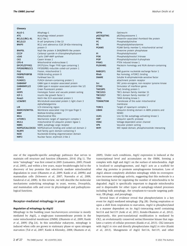
Figure 2. Receptor-mediated mitophagy.
(A) Schematic representation of the domain structures of mitophagy receptors in yeast and mammals. AIM/LIR, Atg8-family protein-interacting motif/LC3-interactingregion (pink); TM, transmembrane domain (light blue); BH1-4, Bcl-2 homology 1-4 domain (green and light green); PPlase, peptidyl-prolyl cis-trans isomerase domain(orange); TPR, tetratricopeptide repeat domain (purple); CaM, calmodulin-binding domain (dark red). The protein size is indicated as the number of amino acids. (B-D)Models for mitophagy receptor activation and protein recruitment on the mitochondrial surface. The yeast mitophagy receptor Atg32 (B), and the mammalian mitophagyreceptors BNIP3, BCL2L13, FKBP8 (C), FUNDC1, and NIX (D) bind to ATG8 family proteins and then target the autophagy machinery to mitochondria. Phosphorylation anddephosphorylation serve as regulatory mechanisms to modulate the activity of mitophagy receptors. For details, see text.
4 of 27 The EMBO Journal 40: e104705 | 2021 ª 2021 The Authors
The EMBO Journal Mashun Onishi et al

a phospholipid methyltransferase localized in the ER, leads to
suppression of Atg32 induction during respiration (Sakakibara et al,
2015). Opi3 acts in the phospholipid biosynthesis pathway for
conversion of PE into PC. Depletion of Opi3 causes aberrant eleva-
tion of glutathione levels that reduces cellular oxidative stress and
thus negatively affects induction of Atg32 and mitophagy (Deffieu
et al, 2009; Okamoto et al, 2009; Sakakibara et al, 2015). These
findings raise the possibility that respiring yeast cells coordinate
phospholipid methylation and mitophagy through unknown
mechanisms.
Receptor-mediated mitophagy in mammals
In mammals, mitophagy is mechanistically more complex than in
yeast and is induced by different cellular stress signals and develop-
mental changes. Disruption of mitochondrial membrane potential is
a potent trigger of mitophagy (Elmore et al, 2001). CCCP, a proton-
selective ionophore, and antimycin A (an inhibitor of the respiratory
complex III) are commonly used to impair mitochondria and acti-
vate mitophagy. Because CCCP is highly toxic and induces non-
physiological levels of mitochondrial damage especially in neurons,
antimycin A is often used to induce mitophagy in neuronal cells
(Cai et al, 2012; Ashrafi et al, 2014). Both reagents trigger mitochon-
drial depolarization and promote accumulation of mitophagy recep-
tors on the OMM. These receptors are integral membrane proteins
that promote specific binding to mammalian Atg8 family members
(LC3A/B/C, GABARAP, GABARAP-L1/2) through a conserved LC3-
interacting regions (LIRs) and regulate the formation of isolation
membranes enclosing mitochondria.
Two major types of receptors have been suggested to mediate
elimination of mitochondria under physiological and pathological
conditions in mammals (Fig 2A). One group includes BNIP3 and
BNIP3L (also known as NIX) (Boyd et al, 1994; Matsushima et al,
1998; Chen et al, 1999; Vande Velde et al, 2000; Regula et al, 2002;
Kubli et al, 2007; Schweers et al, 2007; Sandoval et al, 2008; Hanna
et al, 2012), and the other group includes FUNDC1 (Liu et al, 2012).
In addition, BCL2L13 is the mammalian functional counterpart of
yeast receptor Atg32 (Murakawa et al, 2015) (Fig 2A). In the follow-
ing part, we will discuss the molecular functions of mitophagy
receptors in mammalian cells and the role of a family of receptors,
namely FKBP proteins (Bhujabal et al, 2017).
BNIP3 and NIXBNIP3 is required for efficient turnover of mitochondria under
hypoxic conditions (Zhang et al, 2008). In response to hypoxia,
BNIP3 is upregulated and anchored to the OMM via its C-terminal
transmembrane (TM) domain, exposing the N-terminal domain to
the cytosol (Hanna et al, 2012). BNIP3 is usually expressed as an
inactive monomer in the cytosol, but following stress signals, it
forms a stable homodimer via its C-terminal TM domain and is
integrated into the OMM (Chen et al, 1997; Ray et al, 2000; Kubli
et al, 2008). BNIP3 mutations, which disrupt homodimerization
but do not affect mitochondrial localization, cause a mitophagy
defect, supporting the idea that homodimerization of BNIP3 is
important for efficient degradation of mitochondria (Hanna et al,
2012). Similar to other mitophagy receptors, BNIP3 has a LIR
motif at its N-terminal region (Fig 2A) and mutations in this
region block the interaction with LC3, leading to mitophagy
defects. Phosphorylation of BNIP3 at Ser17 and Ser24 near the LIR
motif is important for BNIP3-LC3 interactions (Zhu et al, 2013)
(Fig 2C).
NIX shows homology to BNIP3 (53–56% amino acid sequence
identity) (Matsushima et al, 1998; Chen et al, 1999) and promotes
selective degradation of mitochondria during reticulocyte matura-
tion (Schweers et al, 2007; Sandoval et al, 2008). During erythroid
differentiation, cell nucleus, mitochondria, and other intracellular
organelles are eliminated, so that erythrocytes can keep maximum
space for hemoglobin that delivers oxygen (Koury et al, 2005;
Yoshida et al, 2005; Fader & Colombo, 2006). With the high
sequence similarity between these two proteins, expression of
BNIP3 can restore mitochondrial clearance in reticulocytes lacking
NIX (Zhang et al, 2012). NIX contains an LIR motif that promotes
binding to LC3A, LC3B, GABARAP, GABARAP-L1, and GABARAP-
L2 (Novak et al, 2010) (Fig 2A). In CCCP-treated cells, NIX recruits
GABARAP-L1 to damaged mitochondria and promotes mitophagy in
a manner dependent on its LIR motif (Novak et al, 2010). Phospho-
rylation of Ser34 and Ser35, two tandem serine residues near the
LIR motif, stabilizes NIX-LC3 interactions and promotes mitophagy
(Rogov et al, 2017) (Fig 2D). Similar to BNIP3, dimerization of NIX,
which is regulated by phosphorylation of its C-terminal region, is
important for efficient recruitment of the autophagic machinery to
mitochondria (Marinkovic et al, 2020).
Accumulation of ROS (triggered by oxidative phosphorylation)
promotes NIX-mediated mitophagy via a recruitment of LC3 to mito-
chondria (Melser et al, 2013). Under conditions of oxidative phos-
phorylation, Rheb, a small GTPase of the Ras superfamily,
translocates to mitochondria and forms a complex with NIX and
LC3 to promote mitophagosome formation (Melser et al, 2013).
Expression of Rheb in HeLa cells increases mitochondrial respira-
tion, and loss of Rheb decreases the oxygen consumption capacity
(Melser et al, 2013). Whether these phenotypes depend on Rheb-
induced mitophagy remains to be addressed. BNIP3 has also been
shown to bind and inhibit Rheb, which is crucial for mTORC1 acti-
vation (Li et al, 2007). As mTORC1 negatively regulates bulk autop-
hagy and mitophagy (Bartolome et al, 2017), BNIP3-dependent
mTORC1 inhibition might facilitate mitophagy induction or take part
in a positive feedback loop to amplify the initiation signal of mito-
phagy.
Several studies have reported that BNIP3 and NIX act in
PINK1/Parkin-mediated mitophagy. NIX is ubiquitylated by
Parkin, which in turn promotes targeting of the selective autop-
hagy adaptor NBR1 that binds both ubiquitin and LC3/GABARAP
to promote formation of autophagosomes surrounding mitochon-
dria (Gao et al, 2015). In addition, BNIP3 interacts with PINK1
and facilitates accumulation of PINK1 on the OMM, resulting in
Parkin translocation to mitochondria (Zhang et al, 2016a). NIX
also contributes to CCCP-induced mitochondrial depolarization,
and accumulation of Parkin on damaged mitochondria (Ding
et al, 2010b). Pathophysiological relevance of BNIP3 and NIX in
Parkinson’s disease remains unknown.
FUNDC1
FUNDC1 is an integral OMM protein that functions as a receptor for
hypoxia-induced mitophagy. It contains a typical LIR motif near the
N-terminal region and three TM domains (Liu et al, 2012) (Fig 2A).
ª 2021 The Authors The EMBO Journal 40: e104705 | 2021 5 of 27
Mashun Onishi et al The EMBO Journal
Mutations in the LIR motif disrupt FUNDC1-LC3 interactions and
mitophagy induction (Liu et al, 2012). FUNDC1 protein levels are
regulated in part by OMM-anchored MARCH5/MITOL (Chen et al,
2017), an E3 ubiquitin ligase that is known to ubiquitylate several
proteins acting in mitochondrial dynamics (Yonashiro et al, 2006;
Sugiura et al, 2013; Park et al, 2014). FUNDC1 expression is
decreased during hypoxia in a ubiquitin–proteasome-dependent
manner due to MARCH5-mediated ubiquitylation of FUNDC1 at
Lys119 (Chen et al, 2017). Knockdown of endogenous MARCH5 or
overexpression of a MARCH5 catalytic mutant impairs ubiquityla-
tion and degradation of FUNDC1, thereby enhancing hypoxia-
induced mitophagy (Chen et al, 2017). Similar to Atg32 in yeast
cells, FUNDC1 is regulated via phosphorylation and dephosphoryla-
tion during mitophagy on residues Ser13 and Tyr18 that are located
near the LIR motif. Under normoxia conditions, Ser13 is phosphory-
lated by CK2, while the Src tyrosine kinase mediates phosphoryla-
tion of Tyr18 to negatively regulate FUNDC1-LC3 interactions (Liu
et al, 2012; Chen et al, 2014) (Fig 2D). Upon hypoxia, Src becomes
inactivated, causing decreased phosphorylation of Tyr18, stabiliza-
tion of the interaction between FUNDC1 and LC3, and promotion of
mitophagosome formation (Liu et al, 2012). The mitochondrial
serine/threonine phosphatase PGAM5 dephosphorylates Ser13 and
enhances FUNDC1-LC3 interactions to promote mitophagy (Chen
et al, 2014).
Hypoxia or mitochondrial depolarization induces ULK1 expres-
sion and its targeting to mitochondria, leading to FUNDC1 phospho-
rylation at Ser17 (near the LIR motif) and stabilization of its
interaction with LC3 (Wu et al, 2014b). Expression of a FUNDC1
variant defective in ULK1 binding inhibits targeting of ULK1 to mito-
chondria and mitophagy, suggesting that FUNDC1 also acts as a
receptor for ULK1 (Wu et al, 2014b). Under normoxic conditions,
BCL2L1/Bcl-xL, an antiapoptotic BH3 domain-containing molecule,
binds PGAM5 and inhibits PGAM5-FUNDC1 interactions to prevent
dephosphorylation of FUNDC1 Ser13 and mitophagy (Wu et al,
2014a).
BCL2L13
Atg32 homologs have so far not been identified in mammalian
cells, but findings from yeast reveal that BCL2L13 can induce
mitophagy in cells lacking Atg32, raising the possibility that
BCL2L13 acts as a mammalian Atg32 functional counterpart
(Murakawa et al, 2015). BCL2L13 is an OMM-anchored single-
pass membrane protein containing two LIR motifs (Fig 2A).
BCL2L13 also regulates mitochondrial morphology and its over-
expression induces mitochondrial fragmentation, while its silenc-
ing causes mitochondrial elongation (Murakawa et al, 2015).
BCL2L13-dependent mitophagy in yeast cells lacking Atg32 is
likely mediated via the conventional autophagy machinery as it
requires Atg7, a core protein essential for Atg8 lipidation (Mura-
kawa et al, 2015). In addition, mutations in the second LIR
motif reduce BCL2L13-dependent mitochondrial degradation in
the absence of Atg32, supporting the notion that BCL2L13
promotes mitophagy via Atg8 in yeast (Murakawa et al, 2015).
BCL2L13 phosphorylation also seems to contribute to regulation
of BCL2L13-LC3 interactions as the mutation at Ser272 near the
second LIR motif reduces mitophagy (Murakawa et al, 2015)
(Fig 2C). BCL2L13 also interacts with ULK1 to localize the
autophagy initiation complex to mitochondria (Murakawa et al,
2019). However, under which physiological conditions BCL2L13
is induced and activated remains to be elucidated.
FKBP8
The immunosuppressant drug FK506 (also known as tacrolimus)
binds to a conserved family of proteins called FKBP that functions
in different cellular processes including transcription, protein fold-
ing/trafficking, signaling, and apoptosis (Bonner & Boulianne,
2017). Co-overexpression of FKBP8 and LC3A promotes degrada-
tion of depolarized mitochondria in CCCP-treated, Parkin-depleted
HeLa cells (Bhujabal et al, 2017). FKBP8 is an integral OMM
protein containing a canonical LIR motif near the N-terminus and a
TM domain at the C-terminus (Fig 2A). FKBP8 preferentially inter-
acts with LC3A over other Atg8 family proteins in vivo, and this is
critical for its mitophagy activity (Bhujabal et al, 2017) (Fig 2C).
Moreover, FKBP8 can escape from degradation-prone mitochon-
dria and localizes to the ER via unknown mechanisms (Saita et al,
2013; Bhujabal et al, 2017). Given the complexity due to its versa-
tile functions (Bonner & Boulianne, 2017), further studies are
needed to clarify whether endogenous FKBP8 is directly involved
in mitophagy.
Ubiquitin-mediated mitophagy
PINK1 and ParkinParkinson’s disease (PD) is a major neurodegenerative disease char-
acterized by cell death of dopaminergic neurons (Lotharius &
Brundin, 2002). PD occurs sporadically in 1–2% of people above
65 years of age but can also arise earlier mostly due to genetic muta-
tions. Common disease phenotypes observed in PD patients are
motor symptoms (tremor, bradykinesia, rigidity, and postural insta-
bility) that result from dopaminergic neuronal loss in substantia
nigra. Non-motor symptoms such as autonomic dysfunction,
neuropsychiatric problems, and sleep difficulties are also frequently
observed. The relationship between sporadic PD and mitochondrial
abnormality has been suggested since 1980s (Corti et al, 2011). The
serine–threonine kinase PINK1 and the E3 ubiquitin ligase PARKIN
were identified as causal genes for hereditary recessive PD with
young onset (Kitada et al, 1998; Valente et al, 2004).
Parkin activationIn 2008, a key study revealed that loss of the mitochondrial
membrane potential triggers recruitment of Parkin to mitochondria
and that Parkin promotes degradation of damaged mitochondria
through autophagy (Narendra et al, 2008). PINK1 has subse-
quently been reported to regulate Parkin E3 activity upon mito-
chondrial depolarization (Matsuda et al, 2010; Narendra et al,
2010). Since conversion of Parkin from inactive to active form
requires PINK1, PINK1-mediated phosphorylation should play an
important role in Parkin activation. PINK1 directly phosphorylates
and activates Parkin on Ser65 in its ubiquitin-like (Ubl) domain,
and this phosphorylation is important for Parkin function (Konda-
palli et al, 2012; Shiba-Fukushima et al, 2012; Iguchi et al, 2013).
However, phosphomimetic mutation did not cause autoubiquityla-
tion of GFP-tagged phosphomimetic Parkin, suggesting that Parkin
phosphorylation itself is insufficient for its activation. Three
groups independently found another PINK1 target that is key for
6 of 27 The EMBO Journal 40: e104705 | 2021 ª 2021 The Authors
The EMBO Journal Mashun Onishi et al
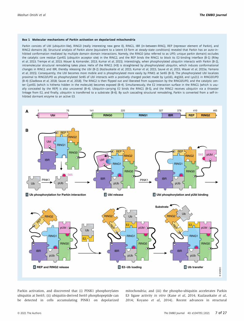
Parkin activation, and discovered that (i) PINK1 phosphorylates
ubiquitin at Ser65; (ii) ubiquitin-derived Ser65 phosphopeptide can
be detected in cells accumulating PINK1 on depolarized
mitochondria; and (iii) the phospho-ubiquitin accelerates Parkin
E3 ligase activity in vitro (Kane et al, 2014; Kazlauskaite et al,
2014; Koyano et al, 2014). Recent advances in structural
Box 1 Molecular mechanisms of Parkin activation on depolarized mitochondria
Parkin consists of Ubl (ubiquitin-like), RING0 (really interesting new gene 0), RING1, IBR (in-between-RING), REP (repressor element of Parkin), andRING2 domains (A). Structural analysis of Parkin alone (equivalent to a latent E3 form at steady-state conditions) revealed that Parkin has an auto-in-hibited conformation mediated by multiple domain–domain interactions. Namely, the RING0 (also referred to as UPD; unique parkin domain) occludesthe catalytic core residue Cys431 (ubiquitin acceptor site) in the RING2, and the REP binds the RING1 to block its E2-binding interface (B-1) (Rileyet al, 2013; Trempe et al, 2013; Wauer & Komander, 2013; Kumar et al, 2015). Interestingly, when phosphorylated ubiquitin interacts with Parkin (B-2),intramolecular structural remodeling takes place. Helix of the RING1 (H3) is straightened by phosphorylated ubiquitin, which induces conformationalchanges in RING1 and IBR, thereby releasing the Ubl (B-2) (Kazlauskaite et al, 2015; Kumar et al, 2015; Sauve et al, 2015; Wauer et al, 2015a; Yamanoet al, 2015). Consequently, the Ubl becomes more mobile and is phosphorylated more easily by PINK1 at Ser65 (B-3). The phosphorylated Ubl localizesproximal to RING0/UPD as phosphorylated Ser65 of Ubl interacts with a positively charged pocket made by Lys161, Arg163, and Lys211 in RING0/UPD(B-4) (Gladkova et al, 2018; Sauve et al, 2018). The RING2 is then flipped out and liberated from suppression by the RING0/UPD, and the catalytic cen-ter Cys431 (which is hitherto hidden in the molecule) becomes exposed (B-4). Simultaneously, the E2 interaction surface in the RING1 (which is usu-ally concealed by the REP) is also uncovered (B-4). Ubiquitin-carrying E2 binds the RING1 (B-5), and the RING2 receives ubiquitin via a thioesterlinkage from E2, and finally, ubiquitin is transferred to a substrate (B-6). By such cascading structural remodeling, Parkin is converted from a self-in-hibited dormant enzyme to an active E3.
PINK1
E2
Ub
RING1
RING2
Ubl
IBR
RING0
REP
Ub pUbPINK1
1 Ub phosphorylation for Parkin interaction 2 Ubl release 3 Ubl phosphorylation and pUbl binding
4 REP and RING2 release 5 E2~Ub loading 6 Ub transfer
UblRING2
IBR
RING0
REP
pUb
RING1
RING2
IBR
RING0
REP
pUb
pUbl
RING1
IBR
RING0
REP
pUb
pUbl
RING2
RING1
IBR
RING0
pUb
pUbl
REP
RING2
E2
Ub
RING1
IBR
RING0
pUb
pUbl
REP
RING2Ub
E2
Substrate
RING1
RING2REPRING0 RING1 IBRUbl
1 76 141 225 327 378 410 465A
B
S65
S65
© E
MB
O
ª 2021 The Authors The EMBO Journal 40: e104705 | 2021 7 of 27
Mashun Onishi et al The EMBO Journal
A P S228
S228
ATPATP
N-lobe C-lo
be
P
ATPATPATP
N-lobe C-lo
be
P Ub
ATPATPADP
N-lobe C-lo
be
P pUb
ATPATPATP
N-lobe C-lo
be
PP
ATPATP
S228P
ATPATPATP
N-lobe C-lo
be
P
ATPATPATPATP
ATP
N-lobe C-lo
be
CTR
Ub
1 PINK1 accumulation as a dimer 2 Auto-phosphorylation 3 i3 stabilization
4 Ub recognition 5 Ub phosphorylation 6 pUb release
ATP
ADP
ATP
i3
i3
1 34 11085 156 320 511 581
CTRTM N-lobe C-lobei1 i2 i3
MTS
OMM
S228
IMS
pUb
FIP200
ULKcomplex
ATG9
ATG9vesicle
Isolation membrane
OPTN
B
OMM
IMS
P
TBK1P
TBK1P
PS177 P S473
S513
S172
S172S403
pUb
Ub
RAB5
CCZ1MON1
RAB7
RAB5
FIS1
RAB7
GDP
GTP GTP
GDP
ATG9
OMM
IMS
Isolation membrane
pUbUb
ATG8 ATG8
p62
NDP52
TAX1BP1
NBR1P
P
ATG8
TBC1D17
TBC1D15
ATG8 ATG8
RABGEF1
C
D
ATG9vesicle
© E
MB
O
Figure 3.
8 of 27 The EMBO Journal 40: e104705 | 2021 ª 2021 The Authors
The EMBO Journal Mashun Onishi et al
information and molecular mechanisms underlying Parkin activa-
tion are described in detail in Box 1.
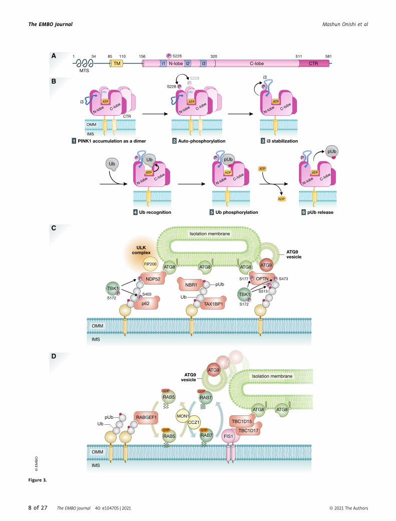
Autophosphorylation of PINK1 is essential forubiquitin recognitionPINK1 Ser228 and Ser402 residues are autophosphorylated upon
decreased mitochondrial membrane potential, and this autophos-
phorylation is essential for Parkin recruitment onto damaged mito-
chondria (Okatsu et al, 2012). The significance of
autophosphorylation at Ser402 is still unknown, and this phospho-
rylation site does not exist in insect PINK1. Autophosphorylation of
Ser228 has been shown in both mammalian cells and insects
(Woodroof et al, 2011). In the kinase domain, PINK1 has three
unique insert regions called Insert 1, Insert 2, and Insert 3 (Fig 3A).
Insert 1 varies in length from 35 to only 5 amino acids in human
and in insect PINK1, respectively, and Insert 2 is not well-
conserved. By contrast, Insert 3 is highly conserved from insects to
humans. The structures of the kinase and C-terminal region (CTR)
domains of insect PINKs—TcPINK (small beetle Tribolium casta-
neum) and PhPINK1 (Pediculus humanus corporis)—have been
solved (Kumar et al, 2017; Schubert et al, 2017; Okatsu et al, 2018).
The kinase domain consists of an N-lobe containing five b-sheetsand a C-lobe containing a-helices that are connected by a hinge
region. The ATP binding site and enzymatic catalytic center localize
in groove between N-lobe and C-lobe. These features are basically
common to other kinases. As a characteristic structure of PINK1, the
CTR domain consists of four a-helices that support the C-lobe struc-
ture from backside. The structural analysis of the PhPINK1–ubiqui-
tin complex revealed that Insert 3 is a key motif for PINK1 to
recognize ubiquitin (Schubert et al, 2017). Phosphorylated PhPINK1
Ser202 (corresponding to human HsPINK1 Ser228) interacts with
Insert 3 Arg282/Asn283 to proper position Insert 3 for ubiquitin
recognition and subsequent phosphorylation (Fig 3B). As Ser202
locates on the upper side of N-lobe and far from the enzymatic
active center of PhPINK1, this seems not to involve intramolecular
autophosphorylation but rather autophosphorylated in trans via
intermolecular phosphorylation. Indeed, dimerization of HsPINK1
on depolarized mitochondria is thought to be important for
autophosphorylation (Okatsu et al, 2013; Rasool et al, 2018).
Parkin’s substrates and Ubiquitin chain amplificationPINK1-mediated phosphorylation leads to Parkin activation and
ubiquitination of substrates on damaged mitochondria that func-
tion as autophagy-mediated degradation signals (Pickrell & Youle,
2015; Khaminets et al, 2016; Yamano et al, 2016). Upon
mitophagy, several OMM proteins such as mitofusin, Miro, and
VDAC have been identified as Parkin substrates (Gegg et al, 2010;
Poole et al, 2010; Tanaka et al, 2010; Ziviani et al, 2010; Geisler
et al, 2010a; Rakovic et al, 2011; Wang et al, 2011). Other OMM
proteins that undergo Parkin-mediated ubiquitylation have later
been identified by mass spectrometry (Chan et al, 2011; Sarraf
et al, 2013), suggesting that Parkin can ubiquitylate a large
number of proteins on the surface of mitochondria. Although
general E3 ligases have stringent substrate selectivity that prevents
cross-reaction among other E3s to ensure correct substrate ubiqui-
tylation, Parkin seems to have rather low substrate selectivity.
Instead, Parkin has evolved to have spatial selectivity for depolar-
ized mitochondria rather than substrate selectivity. Artificial mito-
chondria-targeted exogenous proteins such as GFP and MBP can
be ubiquitylated by Parkin (Koyano et al, 2019). Such a unique
specificity seems optimal for Parkin to achieve efficient and quick
ubiquitylation of dysfunctional mitochondria. Even under steady-
state conditions, a small amount of ubiquitin is attached to
proteins on the surface of mitochondria. When PINK1 phosphory-
lates such ubiquitin, the resultant phospho-ubiquitin recruits
Parkin from the cytosol and activates it on depolarized mitochon-
dria to generate more ubiquitin chains. This Parkin-catalyzed ubiq-
uitylation then further drives PINK1-catalyzed ubiquitin
phosphorylation, leading to formation of a positive feedback loop
for PINK1- and Parkin-catalyzed ubiquitylation (Ordureau et al,
2014; Okatsu et al, 2015). Low substrate specificity of Parkin
might facilitate this positive feedback cycle as only a small amount
of PINK1 on the OMM is needed to recruit quite a few amount of
Parkin to dysfunctional mitochondria (Matsuda, 2016; Matsuda &
Yamano, 2020).
Autophagosome formation in PINK1/Parkin-mediated mitophagyIn order to detach damaged mitochondria from a healthy network
and to eliminate them, proper and selective encapsulation of
damaged mitochondria by autophagosomes is required. In addition,
autophagosomes containing damaged mitochondria must rapidly
fuse with lysosomes to facilitate their degradation. To complete
these processes, many molecules involved in autophagosome/
autolysosome formation work cooperatively with PINK1 and Parkin.
In starvation-induced autophagy, formation of phagophore begins at
a particular region of the ER (Hayashi-Nishino et al, 2009), or at the
contact sites between the ER and mitochondria (Hamasaki et al,
2013). Several autophagy-related proteins are recruited to the
autophagosome formation site in a hierarchical order (Itakura &
Mizushima, 2010).
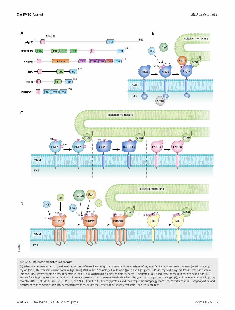
◀ Figure 3. PINK1 and ubiquitin-mediated mitophagy.
(A) Schematic depiction of the domain structures of PINK1. The protein and domain sizes are indicated as the number of amino acids. MTS, mitochondrial targetingsignal; TM, transmembrane segment; N-lobe and C-lobe, N-terminal and C-terminal lobes found in a typical kinase, respectively. i1, i2, and i3, the insert regions uniqueto PINK1; CTR, C-terminal region conserved among PINK1 homologs. (B) Molecular mechanisms underlying ubiquitin phosphorylation by PINK1 on depolarizedmitochondria. (1) PINK1 forms a dimer on damaged mitochondria. (2) Ser228 is phosphorylated via intermolecular autophosphorylation in dimerized PINK1. (3) Ser228phosphorylation stabilizes and underpins “insert 3 (i3)” at the correct position. (4) Ubiquitin (Ub) is recognized by PINK1 as a genuine substrate, (5) Ub Ser65 residue isphosphorylated via ATP hydrolysis, and (6) phosphorylated Ub (pUb) is released. (C) Recruitment of the core autophagy proteins and isolation membranes tomitochondria during PINK1/Parkin-mediated mitophagy. Poly-ubiquitin chains on damaged mitochondria are recognized directly by various autophagy adaptors. Theyare phosphorylated by TBK1 kinase, and the phosphorylation enhances the binding affinity to ubiquitin chains and ATG8 family proteins. NDP52 and OPTN specificallyrecruit ULK complex via FIP200 and ATG9 vesicles, respectively. (D) RABGEF1 recruited to mitochondria by poly-ubiquitin chains triggers endosomal Rab cycles includingRAB5 and the MON1/CCZ1 complex. MON1/CCZ1 directs RAB7 to mitochondria, and RAB7 facilitates the assembly of ATG9 vesicles to the autophagosome formation site.Mitochondrial Rab-GAPs, TBC1D15 and TBC1D17, assist to complete RAB7 cycles and interact with ATG8 family proteins to recruit the isolation membrane.
ª 2021 The Authors The EMBO Journal 40: e104705 | 2021 9 of 27
Mashun Onishi et al The EMBO Journal
Autophagy adaptors in PINK1/Parkin-mediated mitophagyIn selective autophagy-related processes including mitophagy, a
series of autophagy adaptors (p62/SQSTM1, NBR1, NDP52/
CALCOCO2, TAX1BP1, and OPTN) play important roles in selec-
tive uptake of cargoes (Johansen & Lamark, 2011; Mizushima &
Komatsu, 2011; Stolz et al, 2014; Zaffagnini & Martens, 2016).
These autophagy adaptors contain both a ubiquitin-binding
domain that recognizes ubiquitin chains conjugated to the
cargoes and an LC3-interacting region that acts to recruit phago-
phore membranes coated with LC3. During mitophagy, all
known autophagy adaptors are recruited to damaged mitochon-
dria in a Parkin/PINK1-dependent manner (Lazarou et al, 2015).
Compared to the autophagic events under starvation, different
cascading reactions occur during PINK1/Parkin-mediated mito-
phagy. Upon mitochondrial membrane potential dissipation, the
ULK1 complex and ATG9 vesicles are recruited near damaged
mitochondria even in the absence of membrane-bound LC3
(Itakura et al, 2012). Loss of autophagy adaptors impairs not
only recruitment of the LC3-labeled membrane to damaged mito-
chondria, but also recruitment of upstream autophagy-related
proteins such as ULK1 and WIPI1 during PINK1/Parkin-mediated
mitophagy. Among five autophagy adaptors, only NDP52 and
OPTN can grow isolation membrane through an ATG8-dependent
positive feedback loop (Padman et al, 2019). In addition, NDP52
directly binds to FIP200/RB1CC1, a ULK1 complex subunit (Var-
gas et al, 2019), and OPTN can form a complex with ATG9
vesicles (Yamano et al, 2020). Therefore, NDP52 and OPTN bind
to multiple core autophagy proteins. As compared to the hierar-
chy of autophagy under starvation conditions (mTORC1?ULK1?LC3), Parkin-mediated mitophagy uses the following cascading
reaction: ubiquitylation?NDP52?ULK1/LC3, and ubiquityla-
tion?OPTN?ATG9/LC3.
TBK1 kinase in PINK1/Parkin-mediated mitophagyDuring PINK1/Parkin-mediated mitophagy, TBK1 directly or indi-
rectly mediates phosphorylation of all known autophagy receptors
(Richter et al, 2016). TBK1 activity is required for efficient recruit-
ment of OPTN and NDP52 to the ubiquitinated mitochondria (Heo
et al, 2015) where TBK1 phosphorylates OPTN at Ser177 to increase
LC3 binding affinity (Wild et al, 2011) and at Ser473 and Ser513 to
further increase the binding of OPTN to ubiquitin chains (Heo et al,
2015) (Fig 3C). Thus, in addition to Parkin–PINK1–ubiquitin-posi-
tive feedback loop, another feedback loop (ubiquitin–OPTN–TBK1)
constitutes more landing sites for autophagy adaptors on damaged
mitochondria. In addition, TBK1 during mitophagy blocks mitosis
due to the sequestration of TBK1 from its physiological role at
centrosomes (Sarraf et al, 2019).
Elongation of phagophore membranes during mitophagyUnlike starvation-induced autophagy by which cytoplasmic compo-
nents are randomly encapsulated, mitophagy requires elongation
of the phagophore membrane specifically surrounding damaged
mitochondria. The LIR-containing proteins TBC1D15 and TBC1D17
are important for expansion of the phagophore membrane during
mitophagy (Yamano et al, 2014). TBC1D15 and TBC1D17 function
as GTPase-activating proteins (GAPs) for Rab-type GTPases regu-
lating membrane fusion processes in vesicular trafficking (Barr &
Lambright, 2010). TBC1D15 and TBC1D17 target the OMM via
their receptor Fis1 (Onoue et al, 2013). Abnormal LC3-labeled
tubular phagophore structures are formed upon loss of TBC1D15
or Fis1 during mitophagy, but not during starvation-induced autop-
hagy, in mammalian cultured cells (Yamano et al, 2014). In addi-
tion, loss of Fis1 in Caenorhabditis elegans causes a PINK1-
dependent accumulation of LC3 aggregates (Shen et al, 2014). Both
Fis1 and TBC1D15 are required for efficient OXPHOS-induced mito-
phagy and for elimination of paternal mitochondria in fertilized
eggs (Rojansky et al, 2016). Fis1-TBC1D15/17-Rab may be addi-
tionally required for proper formation of autophagosomes during
mitophagy. RABGEF1, an upstream factor of the endosomal Rab
GTPase cascade, is recruited to damaged mitochondria via ubiqui-
tin binding downstream of Parkin. RABGEF1 directs the Rab
proteins RAB5 and RAB7 to damaged mitochondria. Furthermore,
depletion of RAB7 or loss of TBK1-mediated RAB7 phosphorylation
inhibits ATG9 vesicle assembly and subsequent encapsulation of
mitochondria by autophagic membranes (Yamano et al, 2014; Heo
et al, 2018). These results suggest that the endosomal Rab cycle
on damaged mitochondria acts as a crucial regulator of mitophagy
via assembling ATG9 vesicles (Fig 3D). Furthermore, other Rab-
GAPs such as TBC1D5 target ATG9A vesicles around damaged
mitochondria by regulating Rab7 activity during mitophagy
(Jimenez-Orgaz et al, 2018).
Autophagosome closure and autophagosome–lysosome fusionThe final step to eliminate damaged mitochondria requires fusion
of autophagosomes with lysosomes. Although it has been thought
that Atg8 family proteins and their conjugation systems are
required for autophagosome formation, autophagosome-like struc-
tures are formed in the absence of lipidated Atg8 family proteins
(Tsuboyama et al, 2016). In mammals, Atg8 family consists of six
different proteins divided into the LC3 (LC3A, LC3B, and LC3C)
and GABARAP (GABARAP, GABARAP-L1, and GABARAP-L2)
subfamilies. All six proteins are covalently linked to the PE via
two ubiquitin-like conjugation systems. PE-conjugated Atg8 family
proteins associate with both elongating isolation membranes and
mature autophagosomes, and LC3B is widely used as an autop-
hagic membrane marker (Kabeya et al, 2000; Kabeya et al, 2004).
Atg8 family proteins are not essential for encapsulation of
damaged mitochondria by autophagosomes, but required for
autophagosome–lysosome fusion (Nguyen et al, 2016) or efficient
degradation of the inner autophagosomal membrane in lysosomes
(Tsuboyama et al, 2016). Although damaged mitochondria are
properly sequestered by autophagosomal membranes in cells lack-
ing all Atg8 family proteins, the size of autophagosomes is much
smaller than that in wild-type cells (Nakatogawa et al, 2007; Weid-
berg et al, 2010).
Unlike starvation-induced autophagy, PINK1/Parkin-mediated
mitophagy may need PLEKHM1 rather than STX17, an
autophagosome-specific SNARE, for autophagosome–lysosome
fusion (McEwan et al, 2015). PLEKHM1 contains multiple func-
tional domains that directly bind Rab7, the HOPS complex, and
Atg8 family proteins, and is required for selective and nonselec-
tive autophagy (McEwan et al, 2015). GABARAP subfamily
proteins localize on mature autophagosome and associate with
PLEKHM1 at the lysosome to facilitate autophagosome–lysosome
fusion during PINK1/Parkin-mediated mitophagy (Nguyen et al,
2016).
10 of 27 The EMBO Journal 40: e104705 | 2021 ª 2021 The Authors
The EMBO Journal Mashun Onishi et al

Deubiquitylating enzymes in PINK1/Parkin-mediated mitophagyUbiquitylation is a reversible process as deubiquitylating enzymes can
remove ubiquitin from ubiquitylated substrates. USP8, USP15, and
USP30 regulate PINK1/Parkin-mediated mitophagy positively and
negatively (Bingol et al, 2014; Cornelissen et al, 2014; Durcan et al,
2014; Cunningham et al, 2015; Liang et al, 2015). USP15 and USP30
deubiquitylate mitochondrial substrates to counteract Parkin-medi-
ated ubiquitylation and subsequent mitophagy (Bingol et al, 2014;
Cornelissen et al, 2014; Cunningham et al, 2015; Liang et al, 2015). In
contrast, USP8 detaches ubiquitin from autoubiquitylated Parkin,
acting as a positive regulator that promotes Parkin mitochondrial
targeting and accelerates mitophagy (Durcan et al, 2014). Although
USP8 can digest ubiquitin chains of any linkage in vitro (Faesen et al,
2011), it selectively removes K6-linked ubiquitin chains from Parkin
in mammalian cultured cells (Durcan et al, 2014). USP30 is thought to
specifically digest K6-linked ubiquitin chains through unique ubiquitin
recognition mechanisms (Gersch et al, 2017; Sato et al, 2017). It
remains unclear how K6-linked ubiquitin chains of Parkin and OMM
Isolation membrane
CA
B
OMM
IMS
OMM
IMS
CED-9DCT-1
PINK-1PDR-1
Mitochondrial stresses
K26
Fertilization
Relocation of CPS-6
Membranedisorganization
IKKE-1
Isolation membrane
ALLO-1
PP
Protein X ?
Ub
Ub T74
LGG-1 LGG-1
LGG-1 LGG-1
• Paraquat• CCCP• Heat shock• Aging
Low IGF-1 signaling Damaged mitochondria
Mitochondrial biogenesisMitophagy
DCT-1
DAF-16 SKN-1
Quality control of mitochondria
Longevity
© E
MB
O
Figure 4. Regulation of mitophagy in C. elegans.
(A) PINK-1/PDR-1-mediated mitophagy in somatic cells. DCT-1 functions as an autophagy adaptor in association with CED-9. DCT-1 Lys26 (K26) is ubiquitinated in aPINK-1/PDR-1-dependent manner. (B) Mechanism of allophagy in embryos. Fertilization triggers relocation of CPS-6, the mitochondrial endonuclease, leading tomembrane disorganization and ubiquitylation of paternal mitochondria. Ubiquitin (Ub) molecules on paternal mitochondria are recognized directly or indirectly by theautophagy adaptor ALLO-1. IKKE-1-dependent phosphorylation of ALLO-1 is also important for allophagy. (C) Transcriptional regulation of DCT-1 contributes tomitochondrial homeostasis and longevity.
ª 2021 The Authors The EMBO Journal 40: e104705 | 2021 11 of 27
Mashun Onishi et al The EMBO Journal
proteins are removed selectively by USP8 and USP30, respectively.
Although USP15 has been suggested to trim K48- and K63-linked ubiq-
uitin chains on depolarized mitochondria (Cornelissen et al, 2014),
the effect of USP15 on K6-linked ubiquitin chains has not been exam-
ined. Moreover, PINK1-mediated ubiquitin phosphorylation impedes
the enzyme activities of USP8, USP15, and USP30 (Wauer et al,
2015b), adding a new layer of complexity to the deubiquitylation reac-
tions. Although early studies suggested USP30 counteracts Parkin-
mediated ubiquitylation as described, recent two papers showed that
ubiquitylation of the vast majority of Parkin targets is rather unaf-
fected in USP30 knockout cells (Ordureau et al, 2020; Phu et al, 2020).
Instead, elevated ubiquitylation is observed in components of the
mitochondrial translocator and intramitochondrial substrates in
USP30 knockout cells. It is possible that USP30 removes ubiquitin
from import substrates and components of the mitochondrial translo-
cator, and these processes are required for efficient translocation
through the import channels. Future studies on the actions of PINK1,
Parkin, and USP8/15/30 will shed light on the functions of deubiquity-
lating enzymes and the significance of K6-linked ubiquitylation in
PINK1/Parkin-mediated mitophagy.
Parkin alternatives in mitophagyMany papers reported that PINK1/Parkin-catalyzed ubiquitylation
induces mitophagy of damaged mitochondria. However, most of
these data were obtained from experiments using cultured cells (e.g.,
Parkin-expressing HeLa cells), and there is much less evidence for
PINK1/Parkin-mediated mitophagy in vivo. In the case of genetic
studies using Drosophila, it is controversial whether PINK1/Parkin-
catalyzed ubiquitylation induces mitophagy or not. One study
reported that age-dependent rise in mitophagy activity is abrogated
in PINK1- or Parkin-deficient flies (Cornelissen et al, 2018), whereas
another work showed that any substantial impact on basal mito-
phagy was not observed in pink1 or parkin-null flies (Lee et al,
2018). Transgenic mice to monitor mitophagy have already been
established, and loss of PINK1 did not influence basal mitophagy
activities in such mice (McWilliams et al, 2018). This finding seem-
ingly suggests that PINK1 and Parkin are not involved in mitophagy
in vivo; however, the results can be interpreted in several ways.
Unlike human, whose dysfunction of PINK1 or Parkin causes early-
onset Parkinsonism, disease-relevant phenotypes have not been
observed in pink1 or parkin knockout mice. It might not be surpris-
ing even if mitophagy activity is normal in pink1 KO mice without
an obvious phenotype. To reconcile these conflicting findings, we
have to consider functional redundancy of other mitochondrial E3
ligases. Indeed, ARIH1/HHARI (Villa et al, 2017), March5 (Chen
et al, 2017), MAPL/MULAN/GIDE/MUL1 (Ambivero et al, 2014; Yun
et al, 2014; Li et al, 2015; Igarashi et al, 2020), p62-keap1-Rbx1 axis
(Yamada et al, 2018), and HUWE1 (Di Rita et al, 2018) have been
reported to mediate Parkin-independent mitophagy. These E3s could
compensate for PINK1/Parkin-mediated mitophagy and conceal the
output when the PINK1/Parkin function is inhibited.
Mitophagy in worms and flies
PINK-1/PDR-1-mediated mitophagy in somatic cellsIn the nematode Caenorhabditis elegans, stress-induced mitophagy
is regulated by PINK-1 and PDR-1 (a worm Parkin homolog),
supporting that the PINK1/Parkin-dependent pathway has been
conserved during evolution (Palikaras et al, 2015) (Fig 4A). The
NIX and BNIP3 homolog DCT-1 functions as an autophagy receptor
for PINK-1/PDR-1-mediated mitophagy (Palikaras et al, 2015). DCT-
1 is ubiquitylated on its Lys26 residue, and this modification is
enhanced under mitophagy-inducing conditions in a PINK-1-depen-
dent manner (Palikaras et al, 2015). In addition to DCT-1, the Bcl-2
homolog CED-9 interacts with DCT-1 and may act in the same
genetic pathway to control mitophagy (Palikaras et al, 2015).
Clearance of paternal mitochondriaBesides mitophagy in somatic cells, paternal mitochondria provided
by sperm are selectively degraded via autophagy in C. elegans fertil-
ized embryos (Al Rawi et al, 2011; Sato & Sato, 2011). This type of
mitophagy is a developmentally programmed process and does not
require any artificial stimuli to be induced and is referred to as allo-
geneic (non-self) organelle autophagy (allophagy) (Al Rawi et al,
2012; Sato & Sato, 2012) since the paternal organelle, so-called
membranous organelles (MOs), are also degraded in this process
(Al Rawi et al, 2011; Sato & Sato, 2011). mtDNA is maternally inher-
ited in many organisms including humans (Ankel-Simons &
Cummins, 1996; Sato & Sato, 2013; Sato & Sato, 2017). In worm
mutants of core Atg genes, paternal mitochondria and their mtDNA
persist in late-stage embryos or even in F1 larvae, suggesting that
allophagy is required to prevent transmission of paternal mtDNA to
the progeny (Al Rawi et al, 2011; Sato & Sato, 2011). Autophagy-
dependent degradation of paternal mitochondria also occurs in
Drosophila and mouse embryos (Politi et al, 2014; Rojansky et al,
2016).
Caenorhabditis elegans has two Atg8 family members, LGG-1
and LGG-2 that are both recruited to allophagosomes (autophago-
somes containing paternal mitochondria and/or MOs). In lgg-2
mutant embryos, LGG-1-positive allophagosomes are formed, but
their turnover is delayed (Manil-Segalen et al, 2014; Djeddi et al,
2015). LGG-2 directly binds to VPS-39, a subunit of the HOPS
complex, and enhances fusion of autophagosomes with lysosomes
(Manil-Segalen et al, 2014). LGG-2 is also required for microtubule-
dependent migration of autophagosomes toward the pericentroso-
mal region where lysosomes are concentrated (Djeddi et al, 2015).
In addition to autophagy-related genes, degradation of paternal
mtDNA is delayed by knockdown of proteasome subunit genes,
suggesting that the ubiquitin–proteasome system is involved in this
process (Zhou et al, 2011).
Electron tomography has revealed that the inner membrane
structure of paternal mitochondria starts to be disorganized quickly
after fertilization (Zhou et al, 2016). The OMM rapture and reduced
membrane potential were also observed (Zhou et al, 2016). These
changes in paternal mitochondrial structure are initiated before
autophagosome formation and lysosomal degradation. Such qualita-
tive alteration of paternal mitochondria could be a trigger to
promote their selective autophagic clearance. CPS-6, a mitochon-
drial endonuclease G, is also linked to clearance of paternal mito-
chondria (Zhou et al, 2016). CPS-6 was originally identified as an
apoptotic factor that redistributes from mitochondria to the nucleus
and mediates chromosome fragmentation during apoptosis (Parrish
et al, 2001). When paternal cps-6 is mutated, clearance of paternal
mitochondria is delayed (Zhou et al, 2016). Since CPS-6 in paternal
mitochondria relocates from the mitochondrial intermembrane
12 of 27 The EMBO Journal 40: e104705 | 2021 ª 2021 The Authors
The EMBO Journal Mashun Onishi et al

PINK1 Parkin
Bnip3 Parkin
NIX
PINK1 Parkin
FFUNDC1
PINK1 Parkin
PINK1 Parkin
PINK1 Parkin
Bnip3 Parkin
NIX
PINK1 Parkin
FUNDC1
PINK1 Parkin
PINK1 Parkin
Neuroprotection
Tumorsuppression
Protection fromischemia/repurfusion injury
Restraining innate immunity
Longevity
Neurodegeneration
Tumorigenesis
Tissue injury
Excess inflammation
Aging
MA
MM
AL
SF
LIE
S/
WO
RM
SM
AM
MA
LS
WO
RM
S
AnemiaDecreased RGC differentiation
Insufficient beige-to-whiteadipocyte transition
Retained paternalmitochondria Alloo-11
PINNK1 PParkin
MULL1
NIXX
Parkkinn
NIX
Allo-1
PINK1 Parkin
MUL1
Parkin
Erythrocyte maturation RGC differentiation
Adipocyte cell fate decision
Degradation of paternal mitochondria
Degradation of paternalmitochondria
Retained paternalmitochondria
STRESSES
MITOPHAGYACTIVE
MITOPHAGYINACTIVE
MITOPHAGYACTIVE
MITOPHAGYINACTIVE
Accumulation of damaged mitochondria Retention of healthy mitochondria
DEVELOPMENTAL SIGNALS
Retained mitochondria Appropriate elimination of mitochondria
A
B
© E
MB
O
Figure 5.
ª 2021 The Authors The EMBO Journal 40: e104705 | 2021 13 of 27
Mashun Onishi et al The EMBO Journal

space to the matrix after fertilization, mtDNA digestion by matrix-
localized CPS-6 might initiate degeneration of mitochondrial
membranes (Zhou et al, 2016). The IMM protein prohibitin 2 also
plays a role in the selective engulfment of paternal mitochondria by
autophagosomes and functions as an autophagy receptor for
damaged mitochondria in mammals and paternal mitochondria in
C. elegans (Wei et al, 2017).
More recently, a novel LIR-containing protein named ALLO-1 has
been identified as an autophagy adaptor for degradation of paternal
mitochondria and MOs (Sato et al, 2018) (Fig 4B). In allo-1 mutant
embryos, allophagosomes are not formed, and paternal organelles
and paternal mtDNA remain in the late embryos or larvae (Sato
et al, 2018). ALLO-1 is conserved only in nematode species;
however, its function is very similar to that of known autophagy
adaptors. Several lines of evidence suggest that ubiquitylation of
targets is involved in ALLO-1 localization (Al Rawi et al, 2011; Sato
& Sato, 2011). It is also reported that simultaneous knockdown of
ubc-16 and ubc-18 impairs allophagy (Molina et al, 2019). Since
mutations in the pink-1 or pdr-1 gene do not significantly affect allo-
phagy, it remains unknown how this ubiquitylation is regulated
(Sato et al, 2018). In addition to ALLO-1, the worm homolog of
mammalian TBK1/IKKe kinases IKKE-1 is essential for allophagy
(Sato et al, 2018) (Fig 4B). IKKE-1 phosphorylates ALLO-1 on Thr74
although additional phosphorylation targets are likely to exist (Sato
et al, 2018). This is reminiscent of TBK1 function in mitophagy and
xenophagy, and phosphorylation of adaptor molecules could be a
conserved mechanism regulating selective autophagy pathways
(Fig 4B).
In Drosophila, paternal mitochondria form a very long shape
parallel to the axoneme that is degraded by multiple-step mecha-
nisms (Politi et al, 2014). After fertilization, paternal mitochondria
are dissociated from the axoneme and fragmented into small mito-
chondria, which are then engulfed by autophagosomes. Their degra-
dation partly depends on p62, and accumulation of K63-linked
ubiquitin chains on paternal mitochondria has been observed.
Although the precise mechanism remains unclear, this might also
involve autophagy regulators during early dissociation or fragmen-
tation steps (Politi et al, 2014).
In mouse embryos, ubiquitin and autophagy regulators such as
LC3, GABARAP, and p62 are detected on paternal mitochondria
(Sutovsky et al, 1999; Al Rawi et al, 2011). Knockdown of p62 or
PINK1 in embryos impairs degradation of paternal mitochondria,
supporting autophagy-dependent degradation of ubiquitylated pater-
nal mitochondria (Rojansky et al, 2016). Degradation of paternal
mitochondria is also impaired by simultaneous knockdown of
Parkin and MUL1, a mitochondria-localized E3 ubiquitin ligase,
suggesting that these E3 ligases may function redundantly (Rojansky
et al, 2016). The fly and worm Parkin mutants exhibit slight or
minor defects in degradation of paternal mitochondria (Politi et al,
2014; Sato et al, 2018), but possible redundancies with other E3
ligases function cannot be excluded. Similar to C. elegans, loss of
inner membrane potential has been observed in paternal mitochon-
dria in mouse embryos (Rojansky et al, 2016). Notably, Fis1 and
TBC1D15 act in degradation of paternal mitochondria (Rojansky
et al, 2016). These observations suggest a significant overlap
between paternal mitochondria degradation and mitophagy of
damaged mitochondria in somatic cells. However, in contrast to
these studies, Luo et al argued that paternal mitochondria are not
actively removed and persist in embryos at least until the morula
stage (Luo et al, 2013). Further studies are needed to resolve when
and how paternal mitochondria are removed.
Physiology and pathophysiology of mitophagy
A growing body of research has explored the pathophysiological
functions of mitophagy mainly by using mammalian cells or mice
lacking key mitophagy-related factors. These studies also provide a
framework for physiological functions of mitophagy and unveil
previously unappreciated links to diverse biological processes
(Fig 5).
Physiological functions of mitophagy in yeastSince Atg32 is an essential protein for mitophagy in yeast, atg32-null
yeast cells have been used to explore the physiological significance
of mitophagy. Under longevity-extending conditions, loss of Atg32
causes accumulation of dysfunctional mitochondria and impaired
mitochondrial network, leading to a shortened lifespan (Richard
et al, 2013). Mitophagy also contributes to the maintenance of
mtDNA (Kurihara et al, 2011; Karavaeva et al, 2017). In heteroplas-
mic zygotes containing wild-type and mutant mtDNA molecules,
mitophagy is activated and further accelerated by the treatment of
mitochondrial uncouplers, suggesting that enhanced mitophagy in
zygotes could prevent clonal expansion of mutant mtDNA (Kar-
avaeva et al, 2017). Furthermore, during prolonged nitrogen starva-
tion, cells lacking Atg32 exhibit mitochondrial ROS accumulation
and mtDNA instability, indicating that mitophagy contributes to
mitochondrial fitness under stress conditions (Kurihara et al, 2011).
Mitophagy in development, differentiation, and tissue protectionOne example that illustrates the physiological function of mitophagy
in mammals is NIX-mediated mitochondrial elimination during
erythrocyte maturation (Schweers et al, 2007; Sandoval et al, 2008).
Intracellular organelles including mitochondria are removed when
reticulocytes differentiate into mature erythrocytes. Electron micro-
scopic analysis revealed that during early stage of erythrocyte dif-
ferentiation, autophagic bodies accumulated in human peripheral
blood cells, rat erythroblasts, and reticulocytes (Takano-Ohmuro
et al, 2000). Mice lacking the autophagy gene Atg7 in the
hematopoietic system suffer from severe anemia, and Atg7-deficient
erythrocytes accumulate damaged or dysfunctional mitochondria
with altered membrane potential (Mortensen et al, 2010). Consistent
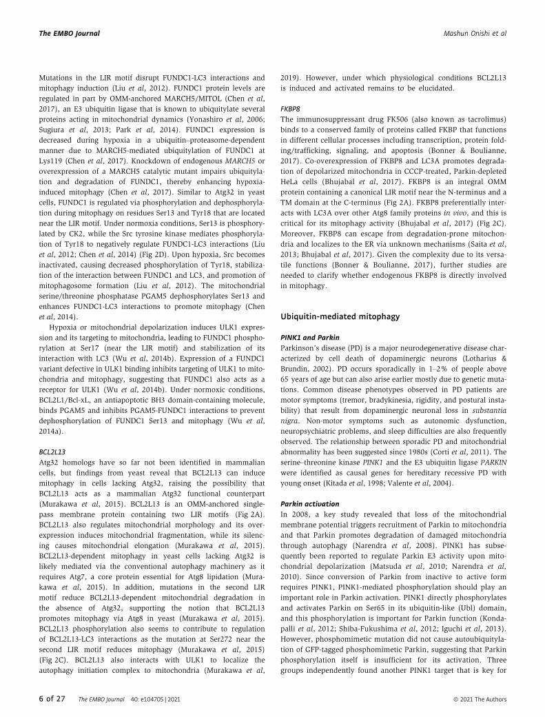
◀ Figure 5. Physiological functions of mitophagy against mitochondrial stresses.
(A) Mitochondria are constantly challenged by a subset of mitochondrial stresses such as oxidative stress. Mitophagy contributes to mitochondrial quality control andprevention of pathologies including neurodegeneration, tumorigenesis, tissue injury, excess inflammation, and aging. (B) Physiological functions of mitophagy duringdevelopment and differentiation. Mitochondrial elimination in response to developmental cues is crucial for maturation of cells and tissues.
14 of 27 The EMBO Journal 40: e104705 | 2021 ª 2021 The Authors
The EMBO Journal Mashun Onishi et al

with these observations, loss of NIX in mice causes defects in mito-
chondrial clearance and anemia (Schweers et al, 2007; Sandoval
et al, 2008). NIX is also involved in mouse retinal ganglion cell
(RGC) differentiation (Esteban-Martinez et al, 2017). During RGC
differentiation, a shift from oxidative phosphorylation to glycolysis
is needed in order to meet the metabolic demands of RGCs (Galvan-
Pena & O’Neill, 2014; Ng et al, 2015; Chandel et al, 2016). Retinas
from NIX-deficient mice show increased mitochondrial mass,
reduced expression of glycolytic enzymes, and inefficient neuronal
differentiation (Esteban-Martinez et al, 2017).
Mitophagy has also been linked to the maturation of muscle
tissue. During myogenesis and muscle regeneration, mitochondrial
activity is drastically increased (Duguez et al, 2002; Sin et al, 2016),
likely due to a shift in metabolism from glycolysis to oxidative phos-
phorylation which eventually increases mitochondrial oxidative
stress. Suppression of the essential autophagy gene Atg5 leads to
accumulation of abnormal mitochondria and inefficient differentia-
tion into mature muscle tissue (Sin et al, 2016).
Parkin-dependent degradation of mitochondria has also been
linked to cell fate decision of adipocytes. Mice lacking Parkin retain
mitochondrial abundance in beige adipocytes and show defects in
beige-to-white adipocyte transition (Lu et al, 2018). While white
adipocytes containing a small quantity of mitochondria serve as fat
tissues to store energy, beige adipocytes contain a large quantity of
mitochondria and act in thermogenesis by uncoupling mitochon-
drial proton gradient in response to various cues such as chronic
cold exposure and exercise (Harms & Seale, 2013; Kajimura et al,
2015). After withdrawal of such stimuli, beige adipocytes acquire
white adipocyte-like characteristics in a manner dependent on
autophagic mitochondrial turnover (Altshuler-Keylin et al, 2016).
In addition to its roles in development and differentiation, mito-
phagy is also involved in tissue protection against several types of
injuries. Studies in mice demonstrated autophagy- and mitophagy-
dependent protection against ischemia/reperfusion (I/R) injury in
several tissues. While reperfusion to restore blood flow after
ischemia is necessary to salvage the injured tissues, this can para-
doxically lead to an excess ROS production from mitochondria
(Pulsinelli & Duffy, 1983; Aronowski et al, 1997). Genetic suppres-
sion of core autophagy-related genes aggravates neuronal injury and
cellular death after I/R injury, mainly by increasing cytochrome c
release from mitochondria (Zhang et al, 2013). NIX expression in
Nix-deficient neurons restored cell viability after I/R-induced injury
and knockout of NIX in mice exacerbated I/R brain injury as indi-
cated by increased cerebral infarct volume (Yuan et al, 2017).
FUNDC1 seems to function in cardioprotection through modulating
the platelet activity (Zhang et al, 2016b). I/R-induced platelet activa-
tion and release of platelet-derived mediators aggravate tissue injury
in the heart (Gawaz, 2004). Hypoxia induces FUNDC1-mediated
mitophagy in platelets, thereby promoting turnover of mitochondria
and suppressing platelet activation (Zhang et al, 2016b). Parkin
plays a protective role in heart against myocardial infarction, as
Parkin-deficient mice exhibit accumulation of dysfunctional mito-
chondria, a broader zone of the infarction, and reduced survival
rates (Kubli et al, 2013). PINK1 KO mice also exhibit dysregulated
mitochondrial functions and excess cardiomyocyte cell death (Billia
et al, 2011).
In addition, mitophagy contributes to kidney homeostasis and
protection against acute kidney injury (AKI). PINK1-, Parkin-, and
double-KO mice show increased mitochondrial damage, ROS gener-
ation, inflammatory response, and serum creatinine (an index of
renal dysfunction), raising the possibility that PINK1/Parkin-medi-
ated mitophagy prevents cell death and maintains renal function
against I/R-induced AKI (Tang et al, 2018). The autophagy receptor
optineurin has also been suggested to act in renal tissue protection
(Chen et al, 2018). Deletion of optineurin drastically decreases
mitophagosome formation during high glucose treatment and exac-
erbates RTEC (renal tubular epithelial cells) senescence (Chen et al,
2018), one of the factors contributing to renal injury in diabetic
kidney.
Alcoholic liver disease (ALD) is caused by excess alcohol intake
(Rehm et al, 2013). Several studies suggest that autophagy-related
processes, especially mitophagy, act in protection against alcohol-
induced liver injury (Ding et al, 2010a; Ding et al, 2011; Williams &
Ding, 2015). Since ALD pathology is associated with ROS accumula-
tion and mtDNA damage, it is conceivable that mitophagy is
induced in the liver tissue of ALD patients. Consistent with this idea,
Parkin KO mice display increased liver injury, oxidative stress, and
steatosis after alcohol treatment, highlighting a protective role of
mitophagy against tissue injury in the liver (Williams et al, 2015).
Nonalcoholic fatty liver disease (NAFLD) is the most common
cause of liver disease and has also been linked with mitochondrial
dynamics and Parkin-independent mitophagy (Loomba & Sanyal,
2013; Masuoka & Chalasani, 2013; Yamada et al, 2018). In the
NAFLD mice model, megamitochondria, which are extremely
enlarged mitochondria, are observed (Wakabayashi, 2002; Neuman
et al, 2014; Targher et al, 2018; Younossi et al, 2018). Deletion of
Opa1 (an IMM protein required for mitochondrial fusion) in NAFLD
model mice decreases mitochondrial size and ameliorates liver
tissue damage, suggesting that restoring mitochondrial size can be a
potent therapeutic treatment (Yamada et al, 2018). In hepatocytes,
the autophagy adaptor p62 recruits Keap1, a component of the
cullin–RING ubiquitin ligase complex containing the E3 enzyme
Rbx1, to mitochondria and promotes ubiquitylation of OMM
proteins and mitophagy in a manner independent of Parkin
(Yamada et al, 2018).
Mitophagy and cancerSince accumulation of dysfunctional mitochondria is involved in
tumorigenesis, it is conceivable that mitophagy seems to be impor-
tant as a tumor-suppressive system (Gogvadze et al, 2008; Vara-
Perez et al, 2019). Expression of the mitophagy receptor BNIP3
declines in several types of cancer and is associated with cancer
metastasis and chemoresistance (Erkan et al, 2005; Manka et al,
2005; Koop et al, 2009). Mice lacking Bnip3 display more rapid
tumor growth than wild-type mice, which can be caused by excess
accumulation of dysfunctional mitochondria and elevated ROS
production (Chourasia et al, 2015).
Expression of Parkin is also lost in many types of cancer (Bernar-
dini et al, 2017). Overexpression of Parkin in breast and glioma cells
retarded cellular proliferation (Tay et al, 2010). However, as Parkin
is involved in proteasomal degradation of cyclins, which is funda-
mentally important for cell cycle control and tumor growth suppres-
sion (Staropoli et al, 2003; Veeriah et al, 2010; Gong et al, 2014),
Parkin might act via multiple mechanisms to suppress tumor
growth. Similar to Parkin, PINK1 overexpression is suggested to
attenuate in vivo glioblastoma growth (Agnihotri et al, 2016).
ª 2021 The Authors The EMBO Journal 40: e104705 | 2021 15 of 27
Mashun Onishi et al The EMBO Journal

Although mitophagy factors described above are shown to be
dysregulated in cancer patients, it should be noted that whether they
act as a tumor suppressor or promoter depends on cellular subtypes
and cancer stages (e.g., BNIP3 is also suggested to support mela-
noma migration (Maes et al, 2014)). Further studies are needed to
clarify the precise roles of mitophagy in tumorigenesis.
Mitophagy and neurodegenerationMitophagy may contribute to the prevention of neurodegeneration.
As discussed above, PD is a major neurodegenerative disease char-
acterized by loss of dopaminergic neurons in the substantia nigra
(Lotharius & Brundin, 2002). Mutations in the Pink1 gene have
been associated with PD pathogenesis (Valente et al, 2001; Valente
et al, 2002; Geisler et al, 2010b). Dopaminergic neurons expressing
Pink1 mutants show enlarged mitochondria and undergo cell death
(Park et al, 2006). Overexpression of Parkin can eliminate mito-
chondria containing mtDNA mutations in heteroplasmic cybrid
cells, ultimately leading to an increase in mitochondria containing
wild-type mtDNA (Suen et al, 2010). Loss of Parkin, which by
itself does not cause obvious PD pathogenesis in mice (Palacino
et al, 2004; Stichel et al, 2007), synergistically promotes dopamin-
ergic neuron degeneration in mouse models that also contain
mtDNA mutations. These mice accumulate dysfunctional mito-
chondria, supporting the idea that Parkin acts in mitochondrial
quality control and neuroprotection (Pickrell et al, 2015; Song
et al, 2017).
Mitophagy may also play a protective role against Alzheimer’s
disease (AD), a progressive neuronal disorder characterized by a
severe loss of memories and cognitive functions (Querfurth &
LaFerla, 2010). Accumulation of insoluble b-amyloid plaques and
formation of neurofibrillary tangles (aggregates of hyperphosphory-
lated tau proteins) in brain are the major pathological hallmarks of
AD (Small et al, 2006; Querfurth & LaFerla, 2010). PINK1 protein
levels are decreased, and the number of mitochondria is increased
in AD model mouse hippocampal neurons (Manczak et al, 2018).
Overexpression of Parkin in AD model mice decreases b-amyloid
plaques and amyloid-induced inflammation in hippocampus and
cortex, contributing to amelioration of behavioral abnormalities
(Hong et al, 2014). Pharmacological or genetic stimulation of mito-
phagy mitigates b-amyloid plaque formation and tau hyperphospho-
rylation, and reverses memory impairment (Sorrentino et al, 2017;
Fang et al, 2019).
Similarly, mitophagy may abate progression of amyotrophic
lateral sclerosis (ALS), a disease characterized by degeneration of
motor neurons, which leads to muscle weakness and paralysis
(Evans & Holzbaur, 2018). Mutations in several autophagy-related
proteins including optineurin, which acts downstream of PINK1/
Parkin-mediated mitophagy, have been linked to ALS (Maruyama
et al, 2010; Cirulli et al, 2015). Loss of optineurin decreases LC3
recruitment to damaged mitochondria and impairs subsequent
autophagosome formation. One of the ALS-associated mutations,
E478G, is located in the UBAN domain for ubiquitin binding,
disturbs targeting of optineurin to damaged mitochondria, and
suppresses mitophagosome formation (Wong & Holzbaur, 2014).
TBK1, which phosphorylates optineurin and enhances its binding to
ubiquitin chains, is also mutated in ALS patients (Freischmidt et al,
2015; Oakes et al, 2017; Pozzi et al, 2017). A disease-associated
mutation in the TBK1 C-terminal coiled-coil domain disrupts its
interaction with optineurin and possibly affects mitophagy and ALS
pathogenesis (Freischmidt et al, 2015). It should be noted that opti-
neurin and TBK1 are also crucial to eliminate cytosolic protein
aggregates via autophagy and whether the phenotypes are due to
impaired elimination of toxic protein aggregates, or mitophagy, or a
combination of both is not currently clear.
Mitophagy and immune responseAutophagy and mitophagy have been linked to the immune
response, suppressing overactivation of the NLRP3 inflammasome
and subsequent immune response. Inflammasomes are multisubunit
protein complexes consisting of NOD-like receptor (NLR) that
induce downstream immune signaling against microbial infection
and intracellular damage (Schroder & Tschopp, 2010). As the NLRP3
inflammasome is activated by mitochondrial ROS and mtDNA
(Nakahira et al, 2011), autophagy-dependent clearance of mitochon-
dria suppresses overactivation of NLRP3 inflammasome. Sestrin2, a
conserved stress-inducible metabolic protein, protects cells and
tissues against excess activation of the NLRP3 inflammasomes (Kim
et al, 2016). Macrophages isolated from Sestrin2-deficient mice
show hyperactivation of caspase-1, leading to enhanced secretion of
IL-1b and IL-18. Sestrin2 localizes to mitochondria upon
lipopolysaccharide stimulation and promotes targeting of p62 to
damaged mitochondria, thereby contributing to mitophagy during
the immune response and preventing prolonged NLRP3 inflamma-
some activation (Kim et al, 2016).
PINK1 and Parkin KO mice have also been shown to be more sensi-
tive to polymicrobial sepsis-induced multiple organ failure and death
(Kang et al, 2016). The enhanced sensitivity of these KO mice to lethal
sepsis is alleviated by simultaneous depletion of Nlrp3 (Kang et al,
2016). PINK1/Parkin-mediated mitophagy is also linked to the STING
pathway, a major intracellular signaling pathway of the type I IFN in
response to cytosolic DNA (Sliter et al, 2018). Disruption of mitochon-
dria triggers inflammatory responses via the NLRP3 inflammasome
and also via the cGAS-STING pathway (Rongvaux et al, 2014). After
exhaustive exercise, PINK1- and Parkin-deficient mice show increased
STING activation (Sliter et al, 2018). Importantly, inflammatory
responses in these mutant mice are abolished by concurrent depletion
of STING, supporting the idea that PINK1 and Parkin may prevent
release of mtDNA from dysfunctional mitochondria, thereby inhibiting
an excess inflammatory response via the STING pathway (Sliter et al,
2018). However, loss of Sting does not suppress mitochondrial
dysfunctions in Drosophila Pink1/parkin mutants, raising the possibil-
ity that the Pink1/parkin-mediated processes are not linked to the
STING pathway in flies (Lee et al, 2020).
A recent study reveals that hepatocyte-specific FUNDC1 knock-
out promotes initiation of hepatocarcinogenesis (HCG), whereas
FUNDC1 overexpression in hepatocytes suppresses it, suggesting
that FUNDC1 acts in prevention of HCG (Li et al, 2019). Loss of
FUNDC1 causes accumulation of damaged mitochondria and
induces release of mtDNA into the cytosol, leading to aggravated
activation of inflammasomes. Thus, dysregulated immune response
in FUNDC1-depleted hepatocytes seems to excessively promote
hepatocellular proliferation.
Mitophagy and agingHealth issues associated with aging are of great concern, especially
in aging societies. Autophagy has been suggested to be a convergent
16 of 27 The EMBO Journal 40: e104705 | 2021 ª 2021 The Authors
The EMBO Journal Mashun Onishi et al
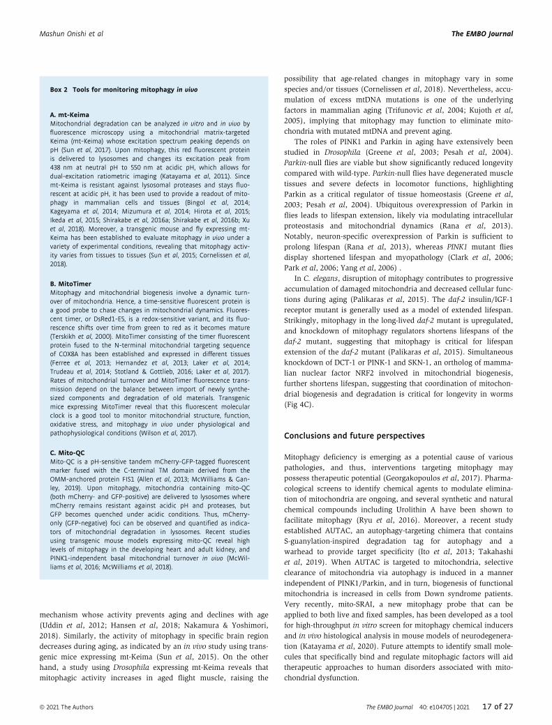
mechanism whose activity prevents aging and declines with age
(Uddin et al, 2012; Hansen et al, 2018; Nakamura & Yoshimori,
2018). Similarly, the activity of mitophagy in specific brain region
decreases during aging, as indicated by an in vivo study using trans-
genic mice expressing mt-Keima (Sun et al, 2015). On the other
hand, a study using Drosophila expressing mt-Keima reveals that
mitophagic activity increases in aged flight muscle, raising the
possibility that age-related changes in mitophagy vary in some
species and/or tissues (Cornelissen et al, 2018). Nevertheless, accu-
mulation of excess mtDNA mutations is one of the underlying
factors in mammalian aging (Trifunovic et al, 2004; Kujoth et al,
2005), implying that mitophagy may function to eliminate mito-
chondria with mutated mtDNA and prevent aging.
The roles of PINK1 and Parkin in aging have extensively been
studied in Drosophila (Greene et al, 2003; Pesah et al, 2004).
Parkin-null flies are viable but show significantly reduced longevity
compared with wild-type. Parkin-null flies have degenerated muscle
tissues and severe defects in locomotor functions, highlighting
Parkin as a critical regulator of tissue homeostasis (Greene et al,
2003; Pesah et al, 2004). Ubiquitous overexpression of Parkin in
flies leads to lifespan extension, likely via modulating intracellular
proteostasis and mitochondrial dynamics (Rana et al, 2013).
Notably, neuron-specific overexpression of Parkin is sufficient to
prolong lifespan (Rana et al, 2013), whereas PINK1 mutant flies
display shortened lifespan and myopathology (Clark et al, 2006;
Park et al, 2006; Yang et al, 2006) .
In C. elegans, disruption of mitophagy contributes to progressive
accumulation of damaged mitochondria and decreased cellular func-
tions during aging (Palikaras et al, 2015). The daf-2 insulin/IGF-1
receptor mutant is generally used as a model of extended lifespan.
Strikingly, mitophagy in the long-lived daf-2 mutant is upregulated,
and knockdown of mitophagy regulators shortens lifespans of the
daf-2 mutant, suggesting that mitophagy is critical for lifespan
extension of the daf-2 mutant (Palikaras et al, 2015). Simultaneous
knockdown of DCT-1 or PINK-1 and SKN-1, an ortholog of mamma-
lian nuclear factor NRF2 involved in mitochondrial biogenesis,
further shortens lifespan, suggesting that coordination of mitochon-
drial biogenesis and degradation is critical for longevity in worms
(Fig 4C).
Conclusions and future perspectives
Mitophagy deficiency is emerging as a potential cause of various
pathologies, and thus, interventions targeting mitophagy may
possess therapeutic potential (Georgakopoulos et al, 2017). Pharma-
cological screens to identify chemical agents to modulate elimina-
tion of mitochondria are ongoing, and several synthetic and natural
chemical compounds including Urolithin A have been shown to
facilitate mitophagy (Ryu et al, 2016). Moreover, a recent study
established AUTAC, an autophagy-targeting chimera that contains
S-guanylation-inspired degradation tag for autophagy and a
warhead to provide target specificity (Ito et al, 2013; Takahashi
et al, 2019). When AUTAC is targeted to mitochondria, selective
clearance of mitochondria via autophagy is induced in a manner
independent of PINK1/Parkin, and in turn, biogenesis of functional
mitochondria is increased in cells from Down syndrome patients.
Very recently, mito-SRAI, a new mitophagy probe that can be
applied to both live and fixed samples, has been developed as a tool
for high-throughput in vitro screen for mitophagy chemical inducers
and in vivo histological analysis in mouse models of neurodegenera-
tion (Katayama et al, 2020). Future attempts to identify small mole-
cules that specifically bind and regulate mitophagic factors will aid
therapeutic approaches to human disorders associated with mito-
chondrial dysfunction.
Box 2 Tools for monitoring mitophagy in vivo
A. mt-KeimaMitochondrial degradation can be analyzed in vitro and in vivo byfluorescence microscopy using a mitochondrial matrix-targetedKeima (mt-Keima) whose excitation spectrum peaking depends onpH (Sun et al, 2017). Upon mitophagy, this red fluorescent proteinis delivered to lysosomes and changes its excitation peak from438 nm at neutral pH to 550 nm at acidic pH, which allows fordual-excitation ratiometric imaging (Katayama et al, 2011). Sincemt-Keima is resistant against lysosomal proteases and stays fluo-rescent at acidic pH, it has been used to provide a readout of mito-phagy in mammalian cells and tissues (Bingol et al, 2014;Kageyama et al, 2014; Mizumura et al, 2014; Hirota et al, 2015;Ikeda et al, 2015; Shirakabe et al, 2016a; Shirakabe et al, 2016b; Xuet al, 2018). Moreover, a transgenic mouse and fly expressing mt-Keima has been established to evaluate mitophagy in vivo under avariety of experimental conditions, revealing that mitophagy activ-ity varies from tissues to tissues (Sun et al, 2015; Cornelissen et al,2018).
B. MitoTimerMitophagy and mitochondrial biogenesis involve a dynamic turn-over of mitochondria. Hence, a time-sensitive fluorescent protein isa good probe to chase changes in mitochondrial dynamics. Fluores-cent timer, or DsRed1-E5, is a redox-sensitive variant, and its fluo-rescence shifts over time from green to red as it becomes mature(Terskikh et al, 2000). MitoTimer consisting of the timer fluorescentprotein fused to the N-terminal mitochondrial targeting sequenceof COX8A has been established and expressed in different tissues(Ferree et al, 2013; Hernandez et al, 2013; Laker et al, 2014;Trudeau et al, 2014; Stotland & Gottlieb, 2016; Laker et al, 2017).Rates of mitochondrial turnover and MitoTimer fluorescence trans-mission depend on the balance between import of newly synthe-sized components and degradation of old materials. Transgenicmice expressing MitoTimer reveal that this fluorescent molecularclock is a good tool to monitor mitochondrial structure, function,oxidative stress, and mitophagy in vivo under physiological andpathophysiological conditions (Wilson et al, 2017).
C. Mito-QCMito-QC is a pH-sensitive tandem mCherry-GFP-tagged fluorescentmarker fused with the C-terminal TM domain derived from theOMM-anchored protein FIS1 (Allen et al, 2013; McWilliams & Gan-ley, 2019). Upon mitophagy, mitochondria containing mito-QC(both mCherry- and GFP-positive) are delivered to lysosomes wheremCherry remains resistant against acidic pH and proteases, butGFP becomes quenched under acidic conditions. Thus, mCherry-only (GFP-negative) foci can be observed and quantified as indica-tors of mitochondrial degradation in lysosomes. Recent studiesusing transgenic mouse models expressing mito-QC reveal highlevels of mitophagy in the developing heart and adult kidney, andPINK1-independent basal mitochondrial turnover in vivo (McWil-liams et al, 2016; McWilliams et al, 2018).
ª 2021 The Authors The EMBO Journal 40: e104705 | 2021 17 of 27
Mashun Onishi et al The EMBO Journal

In yeast, Atg32-mediated mitophagy seems to be the sole
pathway that confers selectivity toward mitochondria versus other
cellular constituents, acting as a quantity adaptation to the low-
energy demand in non-dividing cells. In mammals, multiple mito-
phagy receptors/adaptors promote mitochondrial degradation in
certain specific cell types and under particular conditions, but they
may also function redundantly in reducing or completely eliminat-
ing mitochondria. Since mammalian cells contain a large number of
mitochondria that are heterogeneous (e.g., membrane potential,
respiratory activity, and oxidative damage), they may have needed
to additionally evolve diverse ubiquitin-mediated pathways that
establish selectivity toward dysfunctional mitochondria versus
healthy mitochondria, acting as a quality management system.
These mitophagy-dependent mitochondrial quantity and quality
control mechanisms are not mutually exclusive, as the former can
help improve mitochondrial fitness in cooperation with mitochon-
drial biogenesis that provides fresh mitochondria, and the latter
can help decrease mitochondrial populations without wasting
healthy mitochondria.
Over the last decade, numerous studies have contributed to
establish the paradigm that mitophagy serves as a system to modu-
late mitochondrial fitness and populations in response to changes in
intra- and extracellular environments. Studies using in vivo models
have provided new insights into the physiological and pathological
implications of mitophagy (see Box 2). Although loss of mitophagy
is detrimental to mitochondrial homeostasis, it seems conceivable
that aberrantly hyperactivated mitophagy could also be deleterious
and may ultimately lead to cell death. Therefore, mitophagy must
be tightly regulated by both accelerators and brakes. Several
outstanding questions remain to be addressed: What are those pro-
and anti-mitophagic factors/mechanisms? How is basal mitochon-
drial turnover controlled? Are there additional mitophagy receptors
that are ubiquitous or limited to specific tissues and cell types?
How do cells coordinate mitochondrial biogenesis and degradation?
Do other organelles promote and/or suppress mitophagy? Undoubt-
edly, many exciting discoveries and translational innovations are
yet to come.
AcknowledgementsThe authors apologize to the many colleagues whose work could not be cited
owing to space limitations. We thank Soichiro Kakuta (Juntendo University,
Tokyo, Japan) and Shotaro Saita (Osaka University, Suita, Japan) for helpful
comments on this manuscript. This work was supported by the Japan Society
for the Promotion of Science (JSPS) KAKENHI Grants JP18K06237 and
JP18H05500 (to KY), JP18H02435 and JP19H05712 (to MS), JP18H02443 and
JP19H05712 (to NM), and JP16H04784 and JP19H03222 (to KO); CREST, Japan
Agency for Medical Research and Development (to NM) and Takeda Science
Foundation (to MS and NM). MO was supported by Aid for JSPS Fellows (MEXT
| JSPS) Grant JP19J10384.
Conflict of interestThe authors declare that they have no conflict of interest.
References
Agnihotri S, Golbourn B, Huang X, Remke M, Younger S, Cairns RA, Chalil A,
Smith CA, Krumholtz SL, Mackenzie D et al (2016) PINK1 is a negative
regulator of growth and the Warburg effect in glioblastoma. Cancer Res
76: 4708 – 4719
Al Rawi S, Louvet-Vallee S, Djeddi A, Sachse M, Culetto E, Hajjar C, Boyd L,
Legouis R, Galy V (2011) Postfertilization autophagy of sperm organelles
prevents paternal mitochondrial DNA transmission. Science 334:
1144 – 1147
Al Rawi S, Louvet-Vallee S, Djeddi A, Sachse M, Culetto E, Hajjar C, Boyd L,
Legouis R, Galy V (2012) Allophagy: a macroautophagic process degrading
spermatozoid-inherited organelles. Autophagy 8: 421 – 423
Allen GF, Toth R, James J, Ganley IG (2013) Loss of iron triggers PINK1/Parkin-
independent mitophagy. EMBO Rep 14: 1127 – 1135
Altshuler-Keylin S, Shinoda K, Hasegawa Y, Ikeda K, Hong H, Kang Q, Yang Y,
Perera RM, Debnath J, Kajimura S (2016) Beige adipocyte maintenance is
regulated by autophagy-induced mitochondrial clearance. Cell Metab 24:
402 –419
Ambivero CT, Cilenti L, Main S, Zervos AS (2014) Mulan E3 ubiquitin ligase
interacts with multiple E2 conjugating enzymes and participates in
mitophagy by recruiting GABARAP. Cell Signal 26: 2921 – 2929
Ankel-Simons F, Cummins JM (1996) Misconceptions about mitochondria and
mammalian fertilization- Implications for theories on human evolution.
Proc Natl Acad Sci USA 93: 13859 – 13863
Aoki Y, Kanki T, Hirota Y, Kurihara Y, Saigusa T, Uchiumi T, Kang D (2011)
Phosphorylation of Serine 114 on Atg32 mediates mitophagy. Mol Biol Cell
22: 3206 – 3217
Aronowski J, Strong R, Grotta JC (1997) Reperfusion injury: demonstration of
brain damage produced by reperfusion after transient focal ischemia in
rats. J Cereb Blood Flow Metab 17: 1048 – 1056
Ashrafi G, Schlehe JS, LaVoie MJ, Schwarz TL (2014) Mitophagy of damaged
mitochondria occurs locally in distal neuronal axons and requires PINK1
and Parkin. J Cell Biol 206: 655 – 670
Barr F, Lambright DG (2010) Rab GEFs and GAPs. Curr Opin Cell Biol 22:
461 – 470
Bartolome A, Garcia-Aguilar A, Asahara SI, Kido Y, Guillen C, Pajvani UB,
Benito M (2017) MTORC1 regulates both general autophagy and
mitophagy induction after oxidative phosphorylation uncoupling. Mol Cell
Biol 37: e00441 – 17
Belgareh-Touze N, Cavellini L, Cohen MM (2017) Ubiquitination of ERMES
components by the E3 ligase Rsp5 is involved in mitophagy. Autophagy 13:
114 – 132
Bernardini JP, Lazarou M, Dewson G (2017) Parkin and mitophagy in cancer.
Oncogene 36: 1315 – 1327
Bhujabal Z, Birgisdottir AB, Sjottem E, Brenne HB, Overvatn A, Habisov S,
Kirkin V, Lamark T, Johansen T (2017) FKBP8 recruits LC3A to mediate
Parkin-independent mitophagy. EMBO Rep 18: 947 – 961
Billia F, Hauck L, Konecny F, Rao V, Shen J, Mak TW (2011) PTEN-inducible
kinase 1 (PINK1)/Park6 is indispensable for normal heart function. Proc
Natl Acad Sci USA 108: 9572 – 9577
Bingol B, Tea JS, Phu L, Reichelt M, Bakalarski CE, Song Q, Foreman O,
Kirkpatrick DS, Sheng M (2014) The mitochondrial deubiquitinase USP30
opposes parkin-mediated mitophagy. Nature 510: 370 – 375
Bockler S, Westermann B (2014) Mitochondrial ER contacts are crucial for
mitophagy in yeast. Dev Cell 28: 450 – 458
Bonner JM, Boulianne GL (2017) Diverse structures, functions and uses of
FK506 binding proteins. Cell Signal 38: 97 – 105
Boyd JM, Malstrom S, Subramanian T, Venkatesh LK, Schaeper U, Elangovan
B, D’Sa-Eipper C, Chinnadurai G (1994) Adenovirus E1B 19 kDa and Bcl-2
proteins interact with a common set of cellular proteins. Cell 79:
341 – 351
18 of 27 The EMBO Journal 40: e104705 | 2021 ª 2021 The Authors
The EMBO Journal Mashun Onishi et al
Cai Q, Zakaria HM, Simone A, Sheng ZH (2012) Spatial parkin translocation
and degradation of damaged mitochondria via mitophagy in live cortical
neurons. Curr Biol 22: 545 – 552
Chan NC, Salazar AM, Pham AH, Sweredoski MJ, Kolawa NJ, Graham RL, Hess
S, Chan DC (2011) Broad activation of the ubiquitin-proteasome system
by Parkin is critical for mitophagy. Hum Mol Genet 20: 1726 – 1737
Chandel NS, Jasper H, Ho TT, Passegue E (2016) Metabolic regulation of stem
cell function in tissue homeostasis and organismal ageing. Nat Cell Biol
18: 823 – 832
Chen G, Cizeau J, Vande Velde C, Park JH, Bozek G, Bolton J, Shi L, Dubik D,
Greenberg A (1999) Nix and Nip3 form a subfamily of pro-apoptotic
mitochondrial proteins. J Biol Chem 274: 7 – 10
Chen G, Han Z, Feng D, Chen Y, Chen L, Wu H, Huang L, Zhou C, Cai X, Fu C
et al (2014) A regulatory signaling loop comprising the PGAM5 phosphatase
and CK2 controls receptor-mediated mitophagy. Mol Cell 54: 362 – 377
Chen G, Ray R, Dubik D, Shi L, Cizeau J, Bleackley RC, Saxena S, Gietz RD,
Greenberg AH (1997) The E1B 19K:Bcl-2–binding protein Nip3 is a dimeric
mitochondrial protein that activates apoptosis. J Exp Med 186: 1975 – 1983
Chen K, Dai H, Yuan J, Chen J, Lin L, Zhang W, Wang L, Zhang J, Li K, He Y
(2018) Optineurin-mediated mitophagy protects renal tubular epithelial
cells against accelerated senescence in diabetic nephropathy. Cell Death
Dis 9: 105
Chen Z, Liu L, Cheng Q, Li Y, Wu H, Zhang W, Wang Y, Sehgal SA, Siraj S,
Wang X et al (2017) Mitochondrial E3 ligase MARCH5 regulates FUNDC1
to fine-tune hypoxic mitophagy. EMBO Rep 18: 495 – 509
Chourasia AH, Tracy K, Frankenberger C, Boland ML, Sharifi MN, Drake LE,
Sachleben JR, Asara JM, Locasale JW, Karczmar GS et al (2015) Mitophagy
defects arising from BNip3 loss promote mammary tumor progression to
metastasis. EMBO Rep 16: 1145 – 1163
Cirulli ET, Lasseigne BN, Petrovski S, Sapp PC, Dion PA, Leblond CS, Couthouis
J, Lu YF, Wang Q, Krueger BJ et al (2015) Exome sequencing in
amyotrophic lateral sclerosis identifies risk genes and pathways. Science
347: 1436 – 1441
Clark IE, Dodson MW, Jiang C, Cao JH, Huh JR, Seol JH, Yoo SJ, Hay BA, Guo M
(2006) Drosophila pink1 is required for mitochondrial function and
interacts genetically with parkin. Nature 441: 1162 – 1166
Cornelissen T, Haddad D, Wauters F, Van Humbeeck C, Mandemakers W,
Koentjoro B, Sue C, Gevaert K, De Strooper B, Verstreken P et al (2014) The
deubiquitinase USP15 antagonizes Parkin-mediated mitochondrial
ubiquitination and mitophagy. Hum Mol Genet 23: 5227 – 5242
Cornelissen T, Vilain S, Vints K, Gounko N, Verstreken P, Vandenberghe W
(2018) Deficiency of parkin and PINK1 impairs age-dependent mitophagy
in Drosophila. Elife 7: e35878
Corti O, Lesage S, Brice A (2011) What genetics tells us about the causes and
mechanisms of Parkinson’s disease. Physiol Rev 91: 1161 – 1218
Cunningham CN, Baughman JM, Phu L, Tea JS, Yu C, Coons M, Kirkpatrick DS,
Bingol B, Corn JE (2015) USP30 and parkin homeostatically regulate
atypical ubiquitin chains on mitochondria. Nat Cell Biol 17: 160 – 169
Deffieu M, Bhatia-Kissova I, Salin B, Galinier A, Manon S, Camougrand N
(2009) Glutathione participates in the regulation of mitophagy in yeast. J
Biol Chem 284: 14828 – 14837
Di Rita A, Peschiaroli A, D’Acunzo P, Strobbe D, Hu Z, Gruber J, Nygaard M,
Lambrughi M, Melino G, Papaleo E et al (2018) HUWE1 E3 ligase
promotes PINK1/PARKIN-independent mitophagy by regulating AMBRA1
activation via IKKalpha. Nat Commun 9: 3755
Ding WX, Li M, Chen X, Ni HM, Lin CW, Gao W, Lu B, Stolz DB, Clemens DL,
Yin XM (2010a) Autophagy reduces acute ethanol-induced hepatotoxicity
and steatosis in mice. Gastroenterology 139: 1740 – 1752
Ding WX, Li M, Yin XM (2011) Selective taste of ethanol-induced autophagy
for mitochondria and lipid droplets. Autophagy 7: 248 – 249
Ding WX, Ni HM, Li M, Liao Y, Chen X, Stolz DB, Dorn GW, Yin XM (2010b)
Nix is critical to two distinct phases of mitophagy, reactive oxygen
species-mediated autophagy induction and Parkin-ubiquitin-p62-mediated
mitochondrial priming. J Biol Chem 285: 27879 – 27890
Djeddi A, Al Rawi S, Deuve JL, Perrois C, Liu YY, Russeau M, Sachse M, Galy V
(2015) Sperm-inherited organelle clearance in C. elegans relies on LC3-
dependent autophagosome targeting to the pericentrosomal area.
Development 142: 1705 – 1716
Duguez S, Feasson L, Denis C, Freyssenet D (2002) Mitochondrial biogenesis
during skeletal muscle regeneration. Am J Physiol Endocrinol Metab 282:
E802 – 809
Durcan TM, Tang MY, Perusse JR, Dashti EA, Aguileta MA, McLelland GL, Gros
P, Shaler TA, Faubert D, Coulombe B et al (2014) USP8 regulates
mitophagy by removing K6-linked ubiquitin conjugates from parkin. EMBO
J 33: 2473 – 2491
Eisner V, Picard M, Hajnoczky G (2018) Mitochondrial dynamics in adaptive
and maladaptive cellular stress responses. Nat Cell Biol 20: 755 – 765
Elmore SP, Qian T, Grissom SF, Lemasters JJ (2001) The mitochondrial
permeability transition initiates autophagy in rat hepatocytes. FASEB J 15:
2286 – 2287
Erkan M, Kleeff J, Esposito I, Giese T, Ketterer K, Buchler MW, Giese NA, Friess
H (2005) Loss of BNIP3 expression is a late event in pancreatic cancer
contributing to chemoresistance and worsened prognosis. Oncogene 24:
4421 – 4432
Esteban-Martinez L, Sierra-Filardi E, McGreal RS, Salazar-Roa M, Marino G,
Seco E, Durand S, Enot D, Grana O, Malumbres M et al (2017)
Programmed mitophagy is essential for the glycolytic switch during cell
differentiation. EMBO J 36: 1688 – 1706
Evans CS, Holzbaur ELF (2018) Autophagy and mitophagy in ALS. Neurobiol
Dis 122: 35 – 40
Fader CM, Colombo MI (2006) Multivesicular bodies and autophagy in
erythrocyte maturation. Autophagy 2: 122 – 125
Faesen AC, Luna-Vargas MP, Geurink PP, Clerici M, Merkx R, van Dijk WJ,
Hameed DS, El Oualid F, Ovaa H, Sixma TK (2011) The differential
modulation of USP activity by internal regulatory domains, interactors
and eight ubiquitin chain types. Chem Biol 18: 1550 – 1561
Fang EF, Hou Y, Palikaras K, Adriaanse BA, Kerr JS, Yang B, Lautrup S, Hasan-
Olive MM, Caponio D, Dan X et al (2019) Mitophagy inhibits amyloid-beta
and tau pathology and reverses cognitive deficits in models of Alzheimer’s
disease. Nat Neurosci 22: 401 – 412
Ferree AW, Trudeau K, Zik E, Benador IY, Twig G, Gottlieb RA, Shirihai OS
(2013) MitoTimer probe reveals the impact of autophagy, fusion, and
motility on subcellular distribution of young and old mitochondrial
protein and on relative mitochondrial protein age. Autophagy 9:
1887 – 1896
Freischmidt A, Wieland T, Richter B, Ruf W, Schaeffer V, Muller K, Marroquin
N, Nordin F, Hubers A, Weydt P et al (2015) Haploinsufficiency of TBK1
causes familial ALS and fronto-temporal dementia. Nat Neurosci 18:
631 – 636
Furukawa K, Fukuda T, Yamashita SI, Saigusa T, Kurihara Y, Yoshida Y,
Kirisako H, Nakatogawa H, Kanki T (2018) The PP2A-like protein
phosphatase Ppg1 and the Far complex cooperatively counteract CK2-
mediated phosphorylation of Atg32 to inhibit mitophagy. Cell Rep 23:
3579 – 3590
Galvan-Pena S, O’Neill LA (2014) Metabolic reprogramming in macrophage
polarization. Front Immunol 5: 420
ª 2021 The Authors The EMBO Journal 40: e104705 | 2021 19 of 27
Mashun Onishi et al The EMBO Journal

Gao F, Chen D, Si J, Hu Q, Qin Z, Fang M, Wang G (2015) The mitochondrial
protein BNIP3L is the substrate of PARK2 and mediates mitophagy in
PINK1/PARK2 pathway. Hum Mol Genet 24: 2528 – 2538
Gaspard GJ, McMaster CR (2015) The mitochondrial quality control protein
Yme1 is necessary to prevent defective mitophagy in a yeast model of
Barth syndrome. J Biol Chem 290: 9284 – 9298
Gatica D, Lahiri V, Klionsky DJ (2018) Cargo recognition and degradation by
selective autophagy. Nat Cell Biol 20: 233 – 242
Gawaz M (2004) Role of platelets in coronary thrombosis and reperfusion of
ischemic myocardium. Cardiovasc Res 61: 498 – 511
Gegg ME, Cooper JM, Chau KY, Rojo M, Schapira AH, Taanman JW (2010)
Mitofusin 1 and mitofusin 2 are ubiquitinated in a PINK1/parkin-
dependent manner upon induction of mitophagy. Hum Mol Genet 19:
4861 – 4870
Geisler S, Holmstrom KM, Skujat D, Fiesel FC, Rothfuss OC, Kahle PJ, Springer
W (2010a) PINK1/Parkin-mediated mitophagy is dependent on VDAC1 and
p62/SQSTM1. Nat Cell Biol 12: 119 – 131
Geisler S, Holmstrom KM, Treis A, Skujat D, Weber SS, Fiesel FC, Kahle PJ,
Springer W (2010b) The PINK1/Parkin-mediated mitophagy is
compromised by PD-associated mutations. Autophagy 6: 871 – 878
Georgakopoulos ND, Wells G, Campanella M (2017) The pharmacological
regulation of cellular mitophagy. Nat Chem Biol 13: 136 – 146
Gersch M, Gladkova C, Schubert AF, Michel MA, Maslen S, Komander D (2017)
Mechanism and regulation of the Lys6-selective deubiquitinase USP30.
Nat Struct Mol Biol 24: 920 – 930
Gladkova C, Maslen S, Skehel J, Komander D (2018) Mechanism of parkin
activation by PINK1. Nature 559: 410 – 414
Gogvadze V, Orrenius S, Zhivotovsky B (2008) Mitochondria in cancer cells:
what is so special about them? Trends Cell Biol 18: 165 – 173
Gong Y, Zack TI, Morris LG, Lin K, Hukkelhoven E, Raheja R, Tan IL,
Turcan S, Veeriah S, Meng S et al (2014) Pan-cancer genetic analysis
identifies PARK2 as a master regulator of G1/S cyclins. Nat Genet 46:
588 – 594
Greene JC, Whitworth AJ, Kuo I, Andrews LA, Feany MB, Pallanck LJ (2003)
Mitochondrial pathology and apoptotic muscle degeneration in Drosophila
parkin mutants. Proc Natl Acad Sci USA 100: 4078 – 4083
Hamasaki M, Furuta N, Matsuda A, Nezu A, Yamamoto A, Fujita N, Oomori H,
Noda T, Haraguchi T, Hiraoka Y et al (2013) Autophagosomes form at ER-
mitochondria contact sites. Nature 495: 389 – 393
Hanna RA, Quinsay MN, Orogo AM, Giang K, Rikka S, Gustafsson AB (2012)
Microtubule-associated protein 1 light chain 3 (LC3) interacts with Bnip3
protein to selectively remove endoplasmic reticulum and mitochondria via
autophagy. J Biol Chem 287: 19094 – 19104
Hansen M, Rubinsztein DC, Walker DW (2018) Autophagy as a promoter of
longevity: insights from model organisms. Nat Rev Mol Cell Biol 19:
579 – 593
Harms M, Seale P (2013) Brown and beige fat: development, function and
therapeutic potential. Nat Med 19: 1252 – 1263
Hayashi-Nishino M, Fujita N, Noda T, Yamaguchi A, Yoshimori T, Yamamoto
A (2009) A subdomain of the endoplasmic reticulum forms a cradle for
autophagosome formation. Nat Cell Biol 11: 1433 – 1437
Heo JM, Ordureau A, Paulo JA, Rinehart J, Harper JW (2015) The PINK1-
PARKIN mitochondrial ubiquitylation pathway drives a program of OPTN/
NDP52 recruitment and TBK1 activation to promote mitophagy. Mol Cell
60: 7 – 20
Heo JM, Ordureau A, Swarup S, Paulo JA, Shen K, Sabatini DM, Harper JW
(2018) RAB7A phosphorylation by TBK1 promotes mitophagy via the PINK-
PARKIN pathway. Sci Adv 4: eaav0443
Hernandez G, Thornton C, Stotland A, Lui D, Sin J, Ramil J, Magee N, Andres
A, Quarato G, Carreira RS et al (2013) MitoTimer: a novel tool for
monitoring mitochondrial turnover. Autophagy 9: 1852 – 1861
Hirota Y, Yamashita S, Kurihara Y, Jin X, Aihara M, Saigusa T, Kang D, Kanki T
(2015) Mitophagy is primarily due to alternative autophagy and requires
the MAPK1 and MAPK14 signaling pathways. Autophagy 11: 332 – 343
Hong X, Liu J, Zhu G, Zhuang Y, Suo H, Wang P, Huang D, Xu J, Huang Y, Yu
M et al (2014) Parkin overexpression ameliorates hippocampal long-term
potentiation and beta-amyloid load in an Alzheimer’s disease mouse
model. Hum Mol Genet 23: 1056 – 1072
Igarashi R, Yamashita SI, Yamashita T, Inoue K, Fukuda T, Fukuchi T, Kanki T
(2020) Gemcitabine induces Parkin-independent mitophagy through
mitochondrial-resident E3 ligase MUL1-mediated stabilization of PINK1.
Sci Rep 10: 1465
Iguchi M, Kujuro Y, Okatsu K, Koyano F, Kosako H, Kimura M, Suzuki N,
Uchiyama S, Tanaka K, Matsuda N (2013) Parkin-catalyzed ubiquitin-ester
transfer is triggered by PINK1-dependent phosphorylation. J Biol Chem
288: 22019 – 22032
Ikeda Y, Shirakabe A, Maejima Y, Zhai P, Sciarretta S, Toli J, Nomura M,
Mihara K, Egashira K, Ohishi M et al (2015) Endogenous Drp1 mediates
mitochondrial autophagy and protects the heart against energy stress.
Circ Res 116: 264 – 278
Itakura E, Kishi-Itakura C, Koyama-Honda I, Mizushima N (2012) Structures
containing Atg9A and the ULK1 complex independently target depolarized
mitochondria at initial stages of Parkin-mediated mitophagy. J Cell Sci
125: 1488 – 1499
Itakura E, Mizushima N (2010) Characterization of autophagosome formation
site by a hierarchical analysis of mammalian Atg proteins. Autophagy 6:
764 – 776
Ito C, Saito Y, Nozawa T, Fujii S, Sawa T, Inoue H, Matsunaga T, Khan S,
Akashi S, Hashimoto R et al (2013) Endogenous nitrated nucleotide is a
key mediator of autophagy and innate defense against bacteria. Mol Cell
52: 794 – 804
Jimenez-Orgaz A, Kvainickas A, Nagele H, Denner J, Eimer S, Dengjel J,
Steinberg F (2018) Control of RAB7 activity and localization through the
retromer-TBC1D5 complex enables RAB7-dependent mitophagy. EMBO J 37:
235 – 254
Johansen T, Lamark T (2011) Selective autophagy mediated by autophagic
adapter proteins. Autophagy 7: 279 – 296
Kabeya Y, Mizushima N, Ueno T, Yamamoto A, Kirisako T, Noda T, Kominami
E, Ohsumi Y, Yoshimori T (2000) LC3, a mammalian homologue of yeast
Apg8p, is localized in autophagosome membranes after processing. EMBO
J 19: 5720 – 5728
Kabeya Y, Mizushima N, Yamamoto A, Oshitani-Okamoto S, Ohsumi Y,
Yoshimori T (2004) LC3, GABARAP and GATE16 localize to
autophagosomal membrane depending on form-II formation. J Cell Sci
117: 2805 – 2812
Kageyama Y, Hoshijima M, Seo K, Bedja D, Sysa-Shah P, Andrabi SA, Chen W,
Hoke A, Dawson VL, Dawson TM et al (2014) Parkin-independent
mitophagy requires Drp1 and maintains the integrity of mammalian heart
and brain. EMBO J 33: 2798 – 2813
Kajimura S, Spiegelman BM, Seale P (2015) Brown and beige fat:
physiological roles beyond heat generation. Cell Metab 22: 546 – 559
Kalkavan H, Green DR (2018) MOMP, cell suicide as a BCL-2 family business.
Cell Death Differ 25: 46 – 55
Kane LA, Lazarou M, Fogel AI, Li Y, Yamano K, Sarraf SA, Banerjee S, Youle RJ
(2014) PINK1 phosphorylates ubiquitin to activate Parkin E3 ubiquitin
ligase activity. J Cell Biol 205: 143 – 153
20 of 27 The EMBO Journal 40: e104705 | 2021 ª 2021 The Authors
The EMBO Journal Mashun Onishi et al
Kang R, Zeng L, Xie Y, Yan Z, Zhou B, Cao L, Klionsky DJ, Tracey KJ, Li J, Wang
H et al (2016) A novel PINK1- and PARK2-dependent protective
neuroimmune pathway in lethal sepsis. Autophagy 12: 2374 – 2385
Kanki T, Klionsky DJ (2008) Mitophagy in yeast occurs through a selective
mechanism. J Biol Chem 283: 32386 – 32393
Kanki T, Kurihara Y, Jin X, Goda T, Ono Y, Aihara M, Hirota Y, Saigusa T, Aoki
Y, Uchiumi T et al (2013) Casein kinase 2 is essential for mitophagy. EMBO
Rep 14: 788 – 794
Kanki T, Wang K, Cao Y, Baba M, Klionsky DJ (2009) Atg32 is a mitochondrial
protein that confers selectivity during mitophagy. Dev Cell 17: 98 – 109
Karavaeva IE, Golyshev SA, Smirnova EA, Sokolov SS, Severin FF, Knorre DA
(2017) Mitochondrial depolarization in yeast zygotes inhibits clonal
expansion of selfish mtDNA. J Cell Sci 130: 1274 – 1284
Katayama H, Hama H, Nagasawa K, Kurokawa H, Sugiyama M, Ando R,
Funata M, Yoshida N, Homma M, Nishimura T et al (2020) Visualizing and
modulating mitophagy for therapeutic studies of neurodegeneration. Cell
Cell 181: 1176 – 1187.e16
Katayama H, Kogure T, Mizushima N, Yoshimori T, Miyawaki A (2011) A
sensitive and quantitative technique for detecting autophagic events
based on lysosomal delivery. Chem Biol 18: 1042 – 1052
Kazlauskaite A, Martinez-Torres R, Wilkie S, Kumar A, Peltier J, Gonzalez A,
Johnson C, Zhang J, Hope AG, Peggie M et al (2015) Binding to serine 65-
phosphorylated ubiquitin primes Parkin for optimal PINK1-dependent
phosphorylation and activation. EMBO Rep 16: 939 – 954
Kazlauskaite A, Kondapalli C, Gourlay R, Campbell DG, Ritorto MS, Hofmann
K, Alessi DR, Knebel A, Trost M, Muqit MM (2014) Parkin is activated by
PINK1-dependent phosphorylation of ubiquitin at Ser65. Biochem J 460:
127 – 139
Khaminets A, Behl C, Dikic I (2016) Ubiquitin-dependent and independent
signals in selective autophagy. Trends Cell Biol 26: 6 – 16
Kim MJ, Bae SH, Ryu JC, Kwon Y, Oh JH, Kwon J, Moon JS, Kim K, Miyawaki A,
Lee MG et al (2016) SESN2/sestrin2 suppresses sepsis by inducing
mitophagy and inhibiting NLRP3 activation in macrophages. Autophagy
12: 1272 – 1291
Kitada T, Asakawa S, Hattori N, Matsumine H, Yamamura Y, Minoshima S,
Yokochi M, Mizuno Y, Shimizu N (1998) Mutations in the parkin gene
cause autosomal recessive juvenile parkinsonism. Nature 392: 605 – 608
Kondapalli C, Kazlauskaite A, Zhang N, Woodroof HI, Campbell DG, Gourlay R,
Burchell L, Walden H, Macartney TJ, Deak M et al (2012) PINK1 is
activated by mitochondrial membrane potential depolarization and
stimulates Parkin E3 ligase activity by phosphorylating Serine 65. Open
Biol 2: 120080
Kondo-Okamoto N, Noda NN, Suzuki SW, Nakatogawa H, Takahashi I,
Matsunami M, Hashimoto A, Inagaki F, Ohsumi Y, Okamoto K (2012)
Autophagy-related protein 32 acts as an autophagic degron and directly
initiates mitophagy. J Biol Chem 287: 10631 – 10638
Koop EA, van Laar T, van Wichen DF, de Weger RA, Wall E, van Diest PJ
(2009) Expression of BNIP3 in invasive breast cancer: correlations with
the hypoxic response and clinicopathological features. BMC Cancer 9:
175
Kornmann B, Currie E, Collins SR, Schuldiner M, Nunnari J, Weissman JS,
Walter P (2009) An ER-mitochondria tethering complex revealed by a
synthetic biology screen. Science 325: 477 – 481
Koury MJ, Koury ST, Kopsombut P, Bondurant MC (2005) In vitro maturation
of nascent reticulocytes to erythrocytes. Blood 105: 2168 – 2174
Koyano F, Okatsu K, Kosako H, Tamura Y, Go E, Kimura M, Kimura Y, Tsuchiya
H, Yoshihara H, Hirokawa T et al (2014) Ubiquitin is phosphorylated by
PINK1 to activate parkin. Nature 510: 162 – 166
Koyano F, Yamano K, Kosako H, Tanaka K, Matsuda N (2019) Parkin
recruitment to impaired mitochondria for nonselective ubiquitylation is
facilitated by MITOL. J Biol Chem 294: 10300 – 10314
Kubli DA, Quinsay MN, Huang C, Lee Y, Gustafsson AB (2008) Bnip3 functions
as a mitochondrial sensor of oxidative stress during myocardial ischemia
and reperfusion. Am J Physiol Heart Circ Physiol 295: H2025 – 2031
Kubli DA, Ycaza JE, Gustafsson AB (2007) Bnip3 mediates mitochondrial
dysfunction and cell death through Bax and Bak. Biochem J 405: 407 –415
Kubli DA, Zhang X, Lee Y, Hanna RA, Quinsay MN, Nguyen CK, Jimenez R,
Petrosyan S, Murphy AN, Gustafsson AB (2013) Parkin protein deficiency
exacerbates cardiac injury and reduces survival following myocardial
infarction. J Biol Chem 288: 915 – 926
Kujoth GC, Hiona A, Pugh TD, Someya S, Panzer K, Wohlgemuth SE, Hofer T,
Seo AY, Sullivan R, Jobling WA et al (2005) Mitochondrial DNA mutations,
oxidative stress, and apoptosis in mammalian aging. Science 309:
481 – 484
Kumar A, Aguirre J, Condos T, Martinez-Torres R, Chaugule V, Toth R,
Sundaramoorthy R, Mercier P, Knebel A, Spratt D et al (2015) Disruption of
the autoinhibited state primes the E3 ligase parkin for activation and
catalysis. EMBO J 34: 2506 – 2521
Kumar A, Tamjar J, Waddell AD, Woodroof HI, Raimi OG, Shaw AM, Peggie M,
Muqit MM, van Aalten DM (2017) Structure of PINK1 and mechanisms of
Parkinson’s disease-associated mutations. Elife 6: e29985
Kurihara Y, Kanki T, Aoki Y, Hirota Y, Saigusa T, Uchiumi T, Kang D (2011)
Mitophagy plays an essential role in reducing mitochondrial production of
reactive oxygen species and mutation of mitochondrial DNA by
maintaining mitochondrial quantity and quality in yeast. J Biol Chem 287:
3265 – 3272
Laker RC, Drake JC, Wilson RJ, Lira VA, Lewellen BM, Ryall KA, Fisher CC,
Zhang M, Saucerman JJ, Goodyear LJ et al (2017) Ampk phosphorylation of
Ulk1 is required for targeting of mitochondria to lysosomes in exercise-
induced mitophagy. Nat Commun 8: 548
Laker RC, Xu P, Ryall KA, Sujkowski A, Kenwood BM, Chain KH, Zhang M,
Royal MA, Hoehn KL, Driscoll M et al (2014) A novel MitoTimer reporter
gene for mitochondrial content, structure, stress, and damage in vivo. J
Biol Chem 289: 12005 – 12015
Lazarou M, Sliter DA, Kane LA, Sarraf SA, Wang C, Burman JL, Sideris DP,
Fogel AI, Youle RJ (2015) The ubiquitin kinase PINK1 recruits autophagy
receptors to induce mitophagy. Nature 524: 309 – 314
Lee JJ, Andreazza S, Whitworth AJ (2020) The STING pathway does not
contribute to behavioural or mitochondrial phenotypes in Drosophila
Pink1/parkin or mtDNA mutator models. Sci Rep 10: 2693
Lee JJ, Sanchez-Martinez A, Zarate AM, Beninca C, Mayor U, Clague MJ,
Whitworth AJ (2018) Basal mitophagy is widespread in Drosophila but
minimally affected by loss of Pink1 or parkin. J Cell Biol 217: 1613 – 1622
Lemasters JJ (2005) Selective mitochondrial autophagy, or mitophagy, as a
targeted defense against oxidative stress, mitochondrial dysfunction, and
aging. Rejuvenation Res 8: 3 – 5
Leonhard K, Herrmann J, Stuart R, Mannhaupt G, Neupert W, Langer T (1996)
AAA proteases with catalytic sites on opposite membrane surfaces
comprise a proteolytic system for the ATP-dependent degradation of inner
membrane proteins in mitochondria. EMBO J 15: 4218 – 4229
Li J, Qi W, Chen G, Feng D, Liu J, Ma B, Zhou C, Mu C, Zhang W, Chen Q
et al (2015) Mitochondrial outer-membrane E3 ligase MUL1 ubiquitinates
ULK1 and regulates selenite-induced mitophagy. Autophagy 11:
1216 – 1229
Li W, Li Y, Siraj S, Jin H, Fan Y, Yang X, Huang X, Wang X, Wang J, Liu L et al
(2019) FUN14 domain-containing 1-mediated mitophagy suppresses
ª 2021 The Authors The EMBO Journal 40: e104705 | 2021 21 of 27
Mashun Onishi et al The EMBO Journal

hepatocarcinogenesis by inhibition of inflammasome activation in mice.
Hepatology 69: 604 – 621
Li Y, Wang Y, Kim E, Beemiller P, Wang CY, Swanson J, You M, Guan KL
(2007) Bnip3 mediates the hypoxia-induced inhibition on mammalian
target of rapamycin by interacting with Rheb. J Biol Chem 282:
35803 – 35813
Liang JR, Martinez A, Lane JD, Mayor U, Clague MJ, Urbe S (2015) USP30
deubiquitylates mitochondrial Parkin substrates and restricts apoptotic
cell death. EMBO Rep 16: 618 – 627
Lisowski P, Kannan P, Mlody B, Prigione A (2018) Mitochondria and the
dynamic control of stem cell homeostasis. EMBO Rep 19: e45432
Liu L, Feng D, Chen G, Chen M, Zheng Q, Song P, Ma Q, Zhu C, Wang R, Qi W
et al (2012) Mitochondrial outer-membrane protein FUNDC1 mediates
hypoxia-induced mitophagy in mammalian cells. Nat Cell Biol 14: 177 – 185
Loomba R, Sanyal AJ (2013) The global NAFLD epidemic. Nat Rev
Gastroenterol Hepatol 10: 686 – 690
Lotharius J, Brundin P (2002) Pathogenesis of Parkinson’s disease: dopamine,
vesicles and alpha-synuclein. Nat Rev Neurosci 3: 932 – 942
Lu X, Altshuler-Keylin S, Wang Q, Chen Y, Henrique Sponton C, Ikeda K,
Maretich P, Yoneshiro T, Kajimura S (2018) Mitophagy controls beige
adipocyte maintenance through a Parkin-dependent and UCP1-
independent mechanism. Sci Signal 11: eaap8526
Luo SM, Ge ZJ, Wang ZW, Jiang ZZ, Wang ZB, Ouyang YC, Hou Y, Schatten H,
Sun QY (2013) Unique insights into maternal mitochondrial inheritance in
mice. Proc Natl Acad Sci USA 110: 13038 – 13043
Maes H, Van Eygen S, Krysko DV, Vandenabeele P, Nys K, Rillaerts K, Garg AD,
Verfaillie T, Agostinis P (2014) BNIP3 supports melanoma cell migration
and vasculogenic mimicry by orchestrating the actin cytoskeleton. Cell
Death Dis 5: e1127
Manczak M, Kandimalla R, Yin X, Reddy PH (2018) Hippocampal mutant APP
and amyloid beta-induced cognitive decline, dendritic spine loss, defective
autophagy, mitophagy and mitochondrial abnormalities in a mouse model
of Alzheimer’s disease. Hum Mol Genet 27: 1332 – 1342
Manil-Segalen M, Lefebvre C, Jenzer C, Trichet M, Boulogne C, Satiat-
Jeunemaitre B, Legouis R (2014) The C. elegans LC3 acts downstream of
GABARAP to degrade autophagosomes by interacting with the HOPS
subunit VPS39. Dev Cell 28: 43 – 55
Manka D, Spicer Z, Millhorn DE (2005) Bcl-2/adenovirus E1B 19 kDa
interacting protein-3 knockdown enables growth of breast cancer
metastases in the lung, liver, and bone. Cancer Res 65: 11689 – 11693
Marinkovic M, Sprung M, Novak I (2020) Dimerization of mitophagy receptor
BNIP3L/NIX is essential for recruitment of autophagic machinery.
Autophagy 1–12
Maruyama H, Morino H, Ito H, Izumi Y, Kato H, Watanabe Y, Kinoshita Y,
Kamada M, Nodera H, Suzuki H et al (2010) Mutations of optineurin in
amyotrophic lateral sclerosis. Nature 465: 223 – 226
Masuoka HC, Chalasani N (2013) Nonalcoholic fatty liver disease: an
emerging threat to obese and diabetic individuals. Ann N Y Acad Sci 1281:
106 – 122
Matsuda N (2016) Phospho-ubiquitin: upending the PINK-Parkin-ubiquitin
cascade. J Biochem 159: 379 – 385
Matsuda N, Sato S, Shiba K, Okatsu K, Saisho K, Gautier CA, Sou YS, Saiki S,
Kawajiri S, Sato F et al (2010) PINK1 stabilized by mitochondrial
depolarization recruits Parkin to damaged mitochondria and activates
latent Parkin for mitophagy. J Cell Biol 189: 211 – 221
Matsuda N, Yamano K (2020) Two sides of a coin: Physiological significance
and molecular mechanisms for damage-induced mitochondrial
localization of PINK1 and Parkin. Neurosci Res 19: 30171
Matsushima M, Fujiwara T, Takahashi E, Minaguchi T, Eguchi Y, Tsujimoto Y,
Suzumori K, Nakamura Y (1998) Isolation, mapping, and functional
analysis of a novel human cDNA (BNIP3L) encoding a protein homologous
to human NIP3. Genes Chromosomes Cancer 21: 230 – 235
McEwan DG, Popovic D, Gubas A, Terawaki S, Suzuki H, Stadel D, Coxon FP,
Miranda de Stegmann D, Bhogaraju S, Maddi K et al (2015) PLEKHM1
regulates autophagosome-lysosome fusion through HOPS complex and
LC3/GABARAP proteins. Mol Cell 57: 39 – 54
McWilliams TG, Ganley IG (2019) Investigating mitophagy and mitochondrial
morphology in vivo using mito-QC: a comprehensive guide. Methods Mol
Biol 1880: 621 – 642
McWilliams TG, Prescott AR, Allen GF, Tamjar J, Munson MJ, Thomson C,
Muqit MM, Ganley IG (2016) mito-QC illuminates mitophagy and
mitochondrial architecture in vivo. J Cell Biol 214: 333 – 345
McWilliams TG, Prescott AR, Montava-Garriga L, Ball G, Singh F, Barini E,
Muqit MMK, Brooks SP, Ganley IG (2018) Basal mitophagy occurs
independently of PINK1 in mouse tissues of high metabolic demand. Cell
Metab 27: 439 – 449.e5
Mehta MM, Weinberg SE, Chandel NS (2017) Mitochondrial control of
immunity: beyond ATP. Nat Rev Immunol 17: 608 – 620
Melser S, Chatelain EH, Lavie J, Mahfouf W, Jose C, Obre E, Goorden S, Priault
M, Elgersma Y, Rezvani HR et al (2013) Rheb regulates mitophagy induced
by mitochondrial energetic status. Cell Metab 17: 719 – 730
Mizumura K, Cloonan SM, Nakahira K, Bhashyam AR, Cervo M, Kitada T,
Glass K, Owen CA, Mahmood A, Washko GR et al (2014) Mitophagy-
dependent necroptosis contributes to the pathogenesis of COPD. J Clin
Invest 124: 3987 – 4003
Mizushima N, Komatsu M (2011) Autophagy: renovation of cells and tissues.
Cell 147: 728 – 741
Molina P, Lim Y, Boyd L (2019) Ubiquitination is required for the
initial removal of paternal organelles in C. elegans. Dev Biol 453:
168 – 179
Mortensen M, Ferguson DJ, Edelmann M, Kessler B, Morten KJ, Komatsu M,
Simon AK (2010) Loss of autophagy in erythroid cells leads to defective
removal of mitochondria and severe anemia in vivo. Proc Natl Acad Sci
USA 107: 832 – 837
Murakawa T, Okamoto K, Omiya S, Taneike M, Yamaguchi O, Otsu K (2019) A
mammalian mitophagy receptor, Bcl2-L-13, recruits the ULK1 complex to
induce mitophagy. Cell Rep 26: 338 – 345.e336
Murakawa T, Yamaguchi O, Hashimoto A, Hikoso S, Takeda T, Oka T, Yasui H,
Ueda H, Akazawa Y, Nakayama H et al (2015) Bcl-2-like protein 13 is a
mammalian Atg32 homologue that mediates mitophagy and
mitochondrial fragmentation. Nat Commun 6: 7527
Nakahira K, Haspel JA, Rathinam VA, Lee SJ, Dolinay T, Lam HC, Englert JA,
Rabinovitch M, Cernadas M, Kim HP et al (2011) Autophagy proteins
regulate innate immune responses by inhibiting the release of
mitochondrial DNA mediated by the NALP3 inflammasome. Nat Immunol
12: 222 – 230
Nakamura S, Yoshimori T (2018) Autophagy and longevity. Mol Cells 41:
65 – 72
Nakatogawa H (2020) Mechanisms governing autophagosome biogenesis. Nat
Rev Mol Cell Biol 21: 439 –458
Nakatogawa H, Ichimura Y, Ohsumi Y (2007) Atg8, a ubiquitin-like protein
required for autophagosome formation, mediates membrane tethering
and hemifusion. Cell 130: 165 – 178
Narendra D, Tanaka A, Suen DF, Youle RJ (2008) Parkin is recruited selectively
to impaired mitochondria and promotes their autophagy. J Cell Biol 183:
795 – 803
22 of 27 The EMBO Journal 40: e104705 | 2021 ª 2021 The Authors
The EMBO Journal Mashun Onishi et al

Narendra DP, Jin SM, Tanaka A, Suen DF, Gautier CA, Shen J, Cookson MR,
Youle RJ (2010) PINK1 is selectively stabilized on impaired mitochondria
to activate Parkin. PLoS Biol 8: e1000298
Neuman MG, French SW, French BA, Seitz HK, Cohen LB, Mueller S, Osna NA,
Kharbanda KK, Seth D, Bautista A et al (2014) Alcoholic and non-alcoholic
steatohepatitis. Exp Mol Pathol 97: 492 – 510
Ney PA (2015) Mitochondrial autophagy: Origins, significance, and role of
BNIP3 and NIX. Biochim Biophys Acta 1853: 2775 – 2783
Ng SK, Wood JP, Chidlow G, Han G, Kittipassorn T, Peet DJ, Casson RJ (2015)
Cancer-like metabolism of the mammalian retina. Clin Exp Ophthalmol 43:
367 – 376
Nguyen TN, Padman BS, Usher J, Oorschot V, Ramm G, Lazarou M (2016)
Atg8 family LC3/GABARAP proteins are crucial for autophagosome-
lysosome fusion but not autophagosome formation during PINK1/Parkin
mitophagy and starvation. J Cell Biol 215: 857 – 874
Novak I, Kirkin V, McEwan DG, Zhang J, Wild P, Rozenknop A, Rogov V, Lohr
F, Popovic D, Occhipinti A et al (2010) Nix is a selective autophagy
receptor for mitochondrial clearance. EMBO Rep 11: 45 – 51
Oakes JA, Davies MC, Collins MO (2017) TBK1: a new player in ALS linking
autophagy and neuroinflammation. Mol Brain 10: 5
Okamoto K (2014) Organellophagy: eliminating cellular building blocks via
selective autophagy. J Cell Biol 205: 435 – 445
Okamoto K, Kondo-Okamoto N, Ohsumi Y (2009) Mitochondria-anchored
receptor Atg32 mediates degradation of mitochondria via selective
autophagy. Dev Cell 17: 87 – 97
Okatsu K, Koyano F, Kimura M, Kosako H, Saeki Y, Tanaka K, Matsuda N
(2015) Phosphorylated ubiquitin chain is the genuine Parkin receptor. J
Cell Biol 209: 111 – 128
Okatsu K, Oka T, Iguchi M, Imamura K, Kosako H, Tani N, Kimura M, Go E,
Koyano F, Funayama M et al (2012) PINK1 autophosphorylation upon
membrane potential dissipation is essential for Parkin recruitment to
damaged mitochondria. Nat Commun 3: 1016
Okatsu K, Sato Y, Yamano K, Matsuda N, Negishi L, Takahashi A, Yamagata A,
Goto-Ito S, Mishima M, Ito Y et al (2018) Structural insights into ubiquitin
phosphorylation by PINK1. Sci Rep 8: 10382
Okatsu K, Uno M, Koyano F, Go E, Kimura M, Oka T, Tanaka K, Matsuda N
(2013) A dimeric PINK1-containing complex on depolarized mitochondria
stimulates Parkin recruitment. J Biol Chem 288: 36372 – 36384
Onishi M, Nagumo S, Iwashita S, Okamoto K (2018) The ER membrane
insertase Get1/2 is required for efficient mitophagy in yeast. Biochem
Biophys Res Commun 503: 14 – 20
Onoue K, Jofuku A, Ban-Ishihara R, Ishihara T, Maeda M, Koshiba T, Itoh T,
Fukuda M, Otera H, Oka T et al (2013) Fis1 acts as a mitochondrial
recruitment factor for TBC1D15 that is involved in regulation of
mitochondrial morphology. J Cell Sci 126: 176 – 185
Ordureau A, Paulo JA, Zhang J, An H, Swatek KN, Cannon JR, Wan Q,
Komander D, Harper JW (2020) Global landscape and dynamics of Parkin
and USP30-dependent ubiquitylomes in iNeurons during mitophagic
signaling. Mol Cell 77: 1124 – 1142.e10
Ordureau A, Sarraf SA, Duda DM, Heo JM, Jedrychowski MP, Sviderskiy
VO, Olszewski JL, Koerber JT, Xie T, Beausoleil SA et al (2014)
Quantitative proteomics reveal a feedforward mechanism for
mitochondrial PARKIN translocation and ubiquitin chain synthesis. Mol
Cell 56: 360 – 375
Padman BS, Nguyen TN, Uoselis L, Skulsuppaisarn M, Nguyen LK,
Lazarou M (2019) LC3/GABARAPs drive ubiquitin-independent
recruitment of Optineurin and NDP52 to amplify mitophagy. Nat
Commun 10: 408
Palacino JJ, Sagi D, Goldberg MS, Krauss S, Motz C, Wacker M, Klose J, Shen J
(2004) Mitochondrial dysfunction and oxidative damage in parkin-
deficient mice. J Biol Chem 279: 18614 – 18622
Palikaras K, Lionaki E, Tavernarakis N (2015) Coordination of mitophagy and
mitochondrial biogenesis during ageing in C. elegans. Nature 521:
525 – 528
Park J, Lee SB, Lee S, Kim Y, Song S, Kim S, Bae E, Kim J, Shong M, Kim JM
et al (2006) Mitochondrial dysfunction in Drosophila PINK1 mutants is
complemented by parkin. Nature 441: 1157 – 1161
Park YY, Nguyen OT, Kang H, Cho H (2014) MARCH5-mediated quality control
on acetylated Mfn1 facilitates mitochondrial homeostasis and cell
survival. Cell Death Dis 5: e1172
Parrish J, Li L, Klotz K, Ledwich D, Wang X, Xue D (2001) Mitochondrial
endonuclease G is important for apoptosis in C. elegans. Nature 412:
90 – 94
Pesah Y, Pham T, Burgess H, Middlebrooks B, Verstreken P, Zhou Y, Harding
M, Bellen H, Mardon G (2004) Drosophila parkin mutants have decreased
mass and cell size and increased sensitivity to oxygen radical stress.
Development 131: 2183 – 2194
Phu L, Rose CM, Tea JS, Wall CE, Verschueren E, Cheung TK, Kirkpatrick DS,
Bingol B (2020) Dynamic regulation of mitochondrial import by the
ubiquitin system. Mol Cell 77: 1107 – 1123.e10
Pickles S, Vigie P, Youle RJ (2018) Mitophagy and quality control mechanisms
in mitochondrial maintenance. Curr Biol 28: R170 –R185
Pickrell AM, Huang CH, Kennedy SR, Ordureau A, Sideris DP, Hoekstra JG,
Harper JW, Youle RJ (2015) Endogenous Parkin preserves dopaminergic
substantia nigral neurons following mitochondrial DNA mutagenic stress.
Neuron 87: 371 – 381
Pickrell AM, Youle RJ (2015) The roles of PINK1, parkin, and mitochondrial
fidelity in Parkinson’s disease. Neuron 85: 257 – 273
Ploumi C, Daskalaki I, Tavernarakis N (2017) Mitochondrial biogenesis and
clearance: a balancing act. FEBS J 284: 183 – 195
Politi Y, Gal L, Kalifa Y, Ravid L, Elazar Z, Arama E (2014) Paternal
mitochondrial destruction after fertilization is mediated by a common
endocytic and autophagic pathway in Drosophila. Dev Cell 29: 305 – 320
Poole AC, Thomas RE, Yu S, Vincow ES, Pallanck L (2010) The mitochondrial
fusion-promoting factor mitofusin is a substrate of the PINK1/parkin
pathway. PLoS One 5: e10054
Pozzi L, Valenza F, Mosca L, Dal Mas A, Domi T, Romano A, Tarlarini C,
Falzone YM, Tremolizzo L, Soraru G et al (2017) TBK1 mutations in Italian
patients with amyotrophic lateral sclerosis: genetic and functional
characterisation. J Neurol Neurosurg Psychiatry 88: 869 – 875
Pracheil T, Liu Z (2013) Tiered assembly of the yeast Far3-7-8-9-10-11
complex at the endoplasmic reticulum. J Biol Chem 288:
16986 – 16997
Priault M, Salin B, Schaeffer J, Vallette FM, di Rago JP, Martinou JC (2005)
Impairing the bioenergetic status and the biogenesis of mitochondria
triggers mitophagy in yeast. Cell Death Differ 12: 1613 – 1621
Pulsinelli WA, Duffy TE (1983) Regional energy balance in rat brain after
transient forebrain ischemia. J Neurochem 40: 1500 – 1503
Querfurth HW, LaFerla FM (2010) Alzheimer’s disease. N Engl J Med 362:
329 – 344
Raffaello A, Mammucari C, Gherardi G, Rizzuto R (2016) Calcium at the
center of cell signaling: interplay between endoplasmic reticulum,
mitochondria, and lysosomes. Trends Biochem Sci 41: 1035 – 1049
Rakovic A, Grunewald A, Kottwitz J, Bruggemann N, Pramstaller PP, Lohmann
K, Klein C (2011) Mutations in PINK1 and Parkin impair ubiquitination of
Mitofusins in human fibroblasts. PLoS One 6: e16746
ª 2021 The Authors The EMBO Journal 40: e104705 | 2021 23 of 27
Mashun Onishi et al The EMBO Journal
Rana A, Rera M, Walker DW (2013) Parkin overexpression during aging
reduces proteotoxicity, alters mitochondrial dynamics, and extends
lifespan. Proc Natl Acad Sci USA 110: 8638 – 8643
Rasool S, Soya N, Truong L, Croteau N, Lukacs GL, Trempe JF (2018) PINK1
autophosphorylation is required for ubiquitin recognition. EMBO Rep 19:
e44981
Ray R, Chen G, Velde CV, Cizeau J, Park JH, Reed JC, Gietz RD, Greenberg AH
(2000) BNIP3 heterodimerizes with Bcl-2:Bcl-XL and induces cell death
independent of a Bcl-2 homology 3 (BH3) domain at both mitochondrial
and nonmitochondrial sites. J Biol Chem 275: 1439 – 1448
Regula KM, Ens K, Kirshenbaum LA (2002) Inducible expression of BNIP3
provokes mitochondrial defects and hypoxia-mediated cell death of
ventricular myocytes. Circ Res 91: 226 – 231
Rehm J, Samokhvalov AV, Shield KD (2013) Global burden of alcoholic liver
diseases. J Hepatol 59: 160 – 168
Richard VR, Leonov A, Beach A, Burstein MT, Koupaki O, Gomez-Perez A, Levy
S, Pluska L, Mattie S, Rafesh R et al (2013) Macromitophagy is a longevity
assurance process that in chronologically aging yeast limited in calorie
supply sustains functional mitochondria and maintains cellular lipid
homeostasis. Aging 5: 234 – 269
Richter B, Sliter DA, Herhaus L, Stolz A, Wang C, Beli P, Zaffagnini G, Wild P,
Martens S, Wagner SA et al (2016) Phosphorylation of OPTN by TBK1
enhances its binding to Ub chains and promotes selective autophagy of
damaged mitochondria. Proc Natl Acad Sci USA 113: 4039 – 4044
Riley B, Lougheed J, Callaway K, Velasquez M, Brecht E, Nguyen L, Shaler T,
Walker D, Yang Y, Regnstrom K et al (2013) Structure and function of
Parkin E3 ubiquitin ligase reveals aspects of RING and HECT ligases. Nat
Commun 4: 1982
Rogov VV, Suzuki H, Marinkovic M, Lang V, Kato R, Kawasaki M, Buljubasic M,
Sprung M, Rogova N, Wakatsuki S et al (2017) Phosphorylation of the
mitochondrial autophagy receptor Nix enhances its interaction with LC3
proteins. Sci Rep 7: 1131
Rojansky R, Cha MY, Chan DC (2016) Elimination of paternal mitochondria in
mouse embryos occurs through autophagic degradation dependent on
PARKIN and MUL1. Elife 5: e17896
Rongvaux A, Jackson R, Harman CC, Li T, West AP, de Zoete MR, Wu Y, Yordy
B, Lakhani SA, Kuan CY et al (2014) Apoptotic caspases prevent the
induction of type I interferons by mitochondrial DNA. Cell 159: 1563 – 1577
Ryu D, Mouchiroud L, Andreux PA, Katsyuba E, Moullan N, Nicolet-Dit-Felix
AA, Williams EG, Jha P, Lo Sasso G, Huzard D et al (2016) Urolithin A
induces mitophagy and prolongs lifespan in C. elegans and increases
muscle function in rodents. Nat Med 22: 879 – 888
Saita S, Shirane M, Nakayama KI (2013) Selective escape of proteins from the
mitochondria during mitophagy. Nat Commun 4: 1410
Sakakibara K, Eiyama A, Suzuki SW, Sakoh-Nakatogawa M, Okumura N, Tani
M, Hashimoto A, Nagumo S, Kondo-Okamoto N, Kondo-Kakuta C et al
(2015) Phospholipid methylation controls Atg32-mediated mitophagy and
Atg8 recycling. EMBO J 34: 2703 – 2719
Sandoval H, Thiagarajan P, Dasgupta SK, Schumacher A, Prchal JT, Chen M,
Wang J (2008) Essential role for Nix in autophagic maturation of erythroid
cells. Nature 454: 232 – 235
Sarraf SA, Raman M, Guarani-Pereira V, Sowa ME, Huttlin EL, Gygi SP, Harper
JW (2013) Landscape of the PARKIN-dependent ubiquitylome in response
to mitochondrial depolarization. Nature 496: 372 – 376
Sarraf SA, Sideris DP, Giagtzoglou N, Ni L, Kankel MW, Sen A, Bochicchio LE,
Huang CH, Nussenzweig SC, Worley SH et al (2019) PINK1/Parkin
influences cell cycle by sequestering TBK1 at damaged mitochondria,
inhibiting mitosis. Cell Rep 29: 225 – 235.e5
Sato K, Sato M (2017) Multiple ways to prevent transmission of paternal
mitochondrial DNA for maternal inheritance in animals. J Biochem 162:
247 – 253
Sato M, Sato K (2011) Degradation of paternal mitochondria by fertilization-
triggered autophagy in C. elegans embryos. Science 334: 1141 – 1144
Sato M, Sato K (2012) Maternal inheritance of mitochondrial DNA:
degradation of paternal mitochondria by allogeneic organelle autophagy,
allophagy. Autophagy 8: 424 –425
Sato M, Sato K (2013) Maternal inheritance of mitochondrial DNA by diverse
mechanisms to eliminate paternal mitochondrial DNA. Biochim Biophys
Acta 1833: 1979 – 1984
Sato M, Sato K, Tomura K, Kosako H, Sato K (2018) The autophagy receptor
ALLO-1 and the IKKE-1 kinase control clearance of paternal mitochondria
in Caenorhabditis elegans. Nat Cell Biol 20: 81 – 91
Sato Y, Okatsu K, Saeki Y, Yamano K, Matsuda N, Kaiho A, Yamagata A, Goto-
Ito S, Ishikawa M, Hashimoto Y et al (2017) Structural basis for specific
cleavage of Lys6-linked polyubiquitin chains by USP30. Nat Struct Mol Biol
24: 911 – 919
Sauve V, Lilov A, Seirafi M, Vranas M, Rasool S, Kozlov G, Sprules T, Wang J,
Trempe J, Gehring K (2015) A Ubl/ubiquitin switch in the activation of
Parkin. EMBO J 34: 2492 – 2505
Sauve V, Sung G, Soya N, Kozlov G, Blaimschein N, Miotto L, Trempe J, Lukacs
G, Gehring K (2018) Mechanism of parkin activation by phosphorylation.
Nat Struct Mol Biol 25: 623 – 630
Scheibye-Knudsen M, Fang EF, Croteau DL, Wilson 3rd DM, Bohr VA (2015)
Protecting the mitochondrial powerhouse. Trends Cell Biol 25: 158 – 170
Schroder K, Tschopp J (2010) The inflammasomes. Cell 140: 821 – 832
Schubert AF, Gladkova C, Pardon E, Wagstaff JL, Freund SMV, Steyaert J,
Maslen SL, Komander D (2017) Structure of PINK1 in complex with its
substrate ubiquitin. Nature 552: 51 – 56
Schuldiner M, Collins SR, Thompson NJ, Denic V, Bhamidipati A, Punna T,
Ihmels J, Andrews B, Boone C, Greenblatt JF et al (2005) Exploration of the
function and organization of the yeast early secretory pathway through
an epistatic miniarray profile. Cell 123: 507 – 519
Schuldiner M, Metz J, Schmid V, Denic V, Rakwalska M, Schmitt HD,
Schwappach B, Weissman JS (2008) The GET complex mediates insertion
of tail-anchored proteins into the ER membrane. Cell 134: 634 – 645
Schweers RL, Zhang J, Randall MS, Loyd MR, Li W, Dorsey FC, Kundu M,
Opferman JT, Cleveland JL, Miller JL et al (2007) NIX is required for
programmed mitochondrial clearance during reticulocyte maturation. Proc
Natl Acad Sci USA 104: 19500 – 19505
Shen Q, Yamano K, Head BP, Kawajiri S, Cheung JT, Wang C, Cho JH, Hattori
N, Youle RJ, van der Bliek AM (2014) Mutations in Fis1 disrupt orderly
disposal of defective mitochondria. Mol Biol Cell 25: 145 – 159
Shiba-Fukushima K, Imai Y, Yoshida S, Ishihama Y, Kanao T, Sato S, Hattori N
(2012) PINK1-mediated phosphorylation of the Parkin ubiquitin-like
domain primes mitochondrial translocation of Parkin and regulates
mitophagy. Sci Rep 2: 1002
Shirakabe A, Fritzky L, Saito T, Zhai P, Miyamoto S, Gustafsson AB, Kitsis RN,
Sadoshima J (2016a) Evaluating mitochondrial autophagy in the mouse
heart. J Mol Cell Cardiol 92: 134 – 139
Shirakabe A, Zhai P, Ikeda Y, Saito T, Maejima Y, Hsu CP, Nomura M,
Egashira K, Levine B, Sadoshima J (2016b) Drp1-dependent
mitochondrial autophagy plays a protective role against pressure
overload-induced mitochondrial dysfunction and heart failure.
Circulation 133: 1249 – 1263
Sin J, Andres AM, Taylor DJ, Weston T, Hiraumi Y, Stotland A, Kim BJ, Huang
C, Doran KS, Gottlieb RA (2016) Mitophagy is required for mitochondrial
24 of 27 The EMBO Journal 40: e104705 | 2021 ª 2021 The Authors
The EMBO Journal Mashun Onishi et al

biogenesis and myogenic differentiation of C2C12 myoblasts. Autophagy
12: 369 – 380
Sliter DA, Martinez J, Hao L, Chen X, Sun N, Fischer TD, Burman JL, Li Y,
Zhang Z, Narendra DP et al (2018) Parkin and PINK1 mitigate STING-
induced inflammation. Nature 561: 258 – 262
Small GW, Kepe V, Ercoli LM, Siddarth P, Bookheimer SY, Miller KJ, Lavretsky
H, Burggren AC, Cole GM, Vinters HV et al (2006) PET of brain amyloid
and tau in mild cognitive impairment. N Engl J Med 355: 2652 – 2663
Song L, McMackin M, Nguyen A, Cortopassi G (2017) Parkin deficiency
accelerates consequences of mitochondrial DNA deletions and
Parkinsonism. Neurobiol Dis 100: 30 – 38
Sorrentino V, Romani M, Mouchiroud L, Beck JS, Zhang H, D’Amico D,
Moullan N, Potenza F, Schmid AW, Rietsch S et al (2017) Enhancing
mitochondrial proteostasis reduces amyloid-beta proteotoxicity. Nature
552: 187 – 193
Spinelli JB, Haigis MC (2018) The multifaceted contributions of mitochondria
to cellular metabolism. Nat Cell Biol 20: 745 – 754
Staropoli JF, McDermott C, Martinat C, Schulman B, Demireva E, Abeliovich A
(2003) Parkin is a component of an SCF-like ubiquitin ligase complex and
protects postmitotic neurons from kainate excitotoxicity. Neuron 37:
735 – 749
Stichel CC, Zhu XR, Bader V, Linnartz B, Schmidt S, Lubbert H (2007) Mono-
and double-mutant mouse models of Parkinson’s disease display severe
mitochondrial damage. Hum Mol Genet 16: 2377 – 2393
Stolz A, Ernst A, Dikic I (2014) Cargo recognition and trafficking in selective
autophagy. Nat Cell Biol 16: 495 – 501
Stotland A, Gottlieb RA (2016) alpha-MHC MitoTimer mouse: in vivo
mitochondrial turnover model reveals remarkable mitochondrial
heterogeneity in the heart. J Mol Cell Cardiol 90: 53 – 58
Suen DF, Narendra DP, Tanaka A, Manfredi G, Youle RJ (2010) Parkin
overexpression selects against a deleterious mtDNA mutation in
heteroplasmic cybrid cells. Proc Natl Acad Sci USA 107:
11835 – 11840
Sugiura A, Nagashima S, Tokuyama T, Amo T, Matsuki Y, Ishido S, Kudo Y,
McBride HM, Fukuda T, Matsushita N et al (2013) MITOL regulates
endoplasmic reticulum-mitochondria contacts via Mitofusin2. Mol Cell 51:
20 – 34
Sun N, Malide D, Liu J, Rovira II, Combs CA, Finkel T (2017) A fluorescence-
based imaging method to measure in vitro and in vivo mitophagy using
mt-Keima. Nat Protoc 12: 1576 – 1587
Sun N, Yun J, Liu J, Malide D, Liu C, Rovira II, Holmstrom KM, Fergusson MM,
Yoo YH, Combs CA et al (2015) Measuring in vivo mitophagy. Mol Cell 60:
685 – 696
Sutovsky P, Moreno RD, Ramalho-Santos J, Dominko T, Simerly C, Schatten G
(1999) Ubiquitin tag for sperm mitochondria. Nature 402: 371 – 372
Takahashi D, Moriyama J, Nakamura T, Miki E, Takahashi E, Sato A, Akaike T,
Itto-Nakama K, Arimoto H (2019) AUTACs: cargo-specific degraders using
selective autophagy. Mol Cell 76: 797 – 810.e10
Takano-Ohmuro H, Mukaida M, Kominami E, Morioka K (2000) Autophagy in
embryonic erythroid cells: its role in maturation. Eur J Cell Biol 79:
759 – 764
Tal R, Winter G, Ecker N, Klionsky DJ, Abeliovich H (2007) Aup1p, a yeast
mitochondrial protein phosphatase homolog, is required for efficient
stationary phase mitophagy and cell survival. J Biol Chem 282:
5617 – 5624
Tamura Y, Endo T (2017) Role of intra- and inter-mitochondrial membrane
contact sites in yeast phospholipid biogenesis. Adv Exp Med Biol 997:
121 – 133
Tanaka A, Cleland MM, Xu S, Narendra DP, Suen DF, Karbowski M, Youle RJ
(2010) Proteasome and p97 mediate mitophagy and degradation of
mitofusins induced by Parkin. J Cell Biol 191: 1367 – 1380
Tang C, Han H, Yan M, Zhu S, Liu J, Liu Z, He L, Tan J, Liu Y, Liu H et al
(2018) PINK1-PRKN/PARK2 pathway of mitophagy is activated to protect
against renal ischemia-reperfusion injury. Autophagy 14: 880 – 897
Targher G, Lonardo A, Byrne CD (2018) Nonalcoholic fatty liver disease and
chronic vascular complications of diabetes mellitus. Nat Rev Endocrinol 14:
99 – 114
Tay SP, Yeo CW, Chai C, Chua PJ, Tan HM, Ang AX, Yip DL, Sung JX, Tan PH,
Bay BH et al (2010) Parkin enhances the expression of cyclin-dependent
kinase 6 and negatively regulates the proliferation of breast cancer cells. J
Biol Chem 285: 29231 – 29238
Terskikh A, Fradkov A, Ermakova G, Zaraisky A, Tan P, Kajava AV, Zhao X,
Lukyanov S, Matz M, Kim S et al (2000) "Fluorescent timer": protein that
changes color with time. Science 290: 1585 – 1588
Trempe J, Sauve V, Grenier K, Seirafi M, Tang M, Menade M, Al-Abdul-
Wahid S, Krett J, Wong K, Kozlov G et al (2013) Structure of parkin
reveals mechanisms for ubiquitin ligase activation. Science 340:
1451 – 1455
Trifunovic A, Wredenberg A, Falkenberg M, Spelbrink JN, Rovio AT, Bruder CE,
Bohlooly YM, Gidlof S, Oldfors A, Wibom R et al (2004) Premature ageing
in mice expressing defective mitochondrial DNA polymerase. Nature 429:
417 – 423
Trudeau KM, Gottlieb RA, Shirihai OS (2014) Measurement of mitochondrial
turnover and life cycle using MitoTimer. Methods Enzymol 547: 21 – 38
Tsuboyama K, Koyama-Honda I, Sakamaki Y, Koike M, Morishita H,
Mizushima N (2016) The ATG conjugation systems are important for
degradation of the inner autophagosomal membrane. Science 354:
1036 – 1041
Uddin MN, Nishio N, Ito S, Suzuki H, Isobe K (2012) Autophagic activity in
thymus and liver during aging. Age34: 75 – 85
Valente EM, Abou-Sleiman PM, Caputo V, Muqit MM, Harvey K, Gispert S, Ali
Z, Del Turco D, Bentivoglio AR, Healy DG et al (2004) Hereditary early-
onset Parkinson’s disease caused by mutations in PINK1. Science 304:
1158 – 1160
Valente EM, Bentivoglio AR, Dixon PH, Ferraris A, Ialongo T, Frontali M,
Albanese A, Wood NW (2001) Localization of a novel locus for autosomal
recessive early-onset parkinsonism, PARK6, on human chromosome 1p35-
p36. Am J Hum Genet 68: 895 – 900
Valente EM, Brancati F, Caputo V, Graham EA, Davis MB, Ferraris A, Breteler
MM, Gasser T, Bonifati V, Bentivoglio AR et al (2002) PARK6 is a common
cause of familial parkinsonism. Neurol Sci 23(Suppl 2): S117 – 118
Vande Velde C, Cizeau J, Dubik D, Alimonti J, Brown T, Israels S, Hakem R,
Greenberg AH (2000) BNIP3 and genetic control of necrosis-like cell death
through the mitochondrial permeability transition pore. Mol Cell Biol 20:
5454 – 5468
Vara-Perez M, Felipe-Abrio B, Agostinis P (2019) Mitophagy in cancer: a tale
of adaptation. Cells 8: 493
Vargas JNS, Wang C, Bunker E, Hao L, Maric D, Schiavo G, Randow F, Youle RJ
(2019) Spatiotemporal control of ULK1 activation by NDP52 and TBK1
during selective autophagy. Mol Cell 74: 347 – 362.e6
Veeriah S, Taylor BS, Meng S, Fang F, Yilmaz E, Vivanco I, Janakiraman M,
Schultz N, Hanrahan AJ, Pao W et al (2010) Somatic mutations of the
Parkinson’s disease-associated gene PARK2 in glioblastoma and other
human malignancies. Nat Genet 42: 77 – 82
Villa E, Proics E, Rubio-Patino C, Obba S, Zunino B, Bossowski JP, Rozier RM,
Chiche J, Mondragon L, Riley JS et al (2017) Parkin-independent
ª 2021 The Authors The EMBO Journal 40: e104705 | 2021 25 of 27
Mashun Onishi et al The EMBO Journal

mitophagy controls chemotherapeutic response in cancer cells. Cell Rep
20: 2846 – 2859
Wakabayashi T (2002) Megamitochondria formation - physiology and
pathology. J Cell Mol Med 6: 497 – 538
Wang F, Chan C, Weir NR, Denic V (2014) The Get1/2 transmembrane
complex is an endoplasmic-reticulum membrane protein insertase. Nature
512: 441 – 444
Wang K, Jin M, Liu X, Klionsky DJ (2013) Proteolytic processing of Atg32 by
the mitochondrial i-AAA protease Yme1 regulates mitophagy. Autophagy 9:
1828 – 1836
Wang X, Winter D, Ashrafi G, Schlehe J, Wong YL, Selkoe D, Rice S, Steen J,
LaVoie MJ, Schwarz TL (2011) PINK1 and Parkin target Miro for
phosphorylation and degradation to arrest mitochondrial motility. Cell
147: 893 – 906
Wauer T, Komander D (2013) Structure of the human Parkin ligase domain in
an autoinhibited state. EMBO J 32: 2099 – 2112
Wauer T, Simicek M, Schubert A, Komander D (2015) Mechanism of phospho-
ubiquitin-induced PARKIN activation. Nature 524: 370 – 374
Wauer T, Swatek KN, Wagstaff JL, Gladkova C, Pruneda JN, Michel MA, Gersch
M, Johnson CM, Freund SM, Komander D (2015) Ubiquitin Ser65
phosphorylation affects ubiquitin structure, chain assembly and
hydrolysis. EMBO J 34: 307 – 325
Wei Y, Chiang WC, Sumpter Jr R, Mishra P, Levine B (2017) Prohibitin 2 is an
inner mitochondrial membrane mitophagy receptor. Cell 168:
224 – 238.e10
Weidberg H, Shvets E, Shpilka T, Shimron F, Shinder V, Elazar Z (2010) LC3
and GATE-16/GABARAP subfamilies are both essential yet act differently in
autophagosome biogenesis. EMBO J 29: 1792 – 1802
Welter E, Montino M, Reinhold R, Schlotterhose P, Krick R, Dudek J, Rehling P,
Thumm M (2013) Uth1 is a mitochondrial inner membrane protein
dispensable for post-log-phase and rapamycin-induced mitophagy. FEBS J
280: 4970 – 4982
Wild P, Farhan H, McEwan DG, Wagner S, Rogov VV, Brady NR, Richter B,
Korac J, Waidmann O, Choudhary C et al (2011) Phosphorylation of the
autophagy receptor optineurin restricts Salmonella growth. Science 333:
228 – 233
Williams JA, Ding WX (2015) A mechanistic review of mitophagy and its role
in protection against alcoholic liver disease. Biomolecules 5: 2619 – 2642
Williams JA, Ni HM, Ding Y, Ding WX (2015) Parkin regulates mitophagy and
mitochondrial function to protect against alcohol-induced liver injury and
steatosis in mice. Am J Physiol Gastrointest Liver Physiol 309: G324 – 340
Wilson RJ, Drake JC, Cui D, Zhang M, Perry HM, Kashatus JA, Kusminski CM,
Scherer PE, Kashatus DF, Okusa MD et al (2017) Conditional MitoTimer
reporter mice for assessment of mitochondrial structure, oxidative stress,
and mitophagy. Mitochondrion 44: 20 – 26
Wong HS, Dighe PA, Mezera V, Monternier PA, Brand MD (2017) Production
of superoxide and hydrogen peroxide from specific mitochondrial sites
under different bioenergetic conditions. J Biol Chem 292: 16804 – 16809
Wong YC, Holzbaur EL (2014) Optineurin is an autophagy receptor for
damaged mitochondria in parkin-mediated mitophagy that is disrupted
by an ALS-linked mutation. Proc Natl Acad Sci USA 111: E4439 – 4448
Woodroof HI, Pogson JH, Begley M, Cantley LC, Deak M, Campbell DG, van
Aalten DM, Whitworth AJ, Alessi DR, Muqit MM (2011) Discovery of
catalytically active orthologues of the Parkinson’s disease kinase PINK1:
analysis of substrate specificity and impact of mutations. Open biology 1:
110012
Wu H, Chen Q (2015) Hypoxia activation of mitophagy and its role in disease
pathogenesis. Antioxid Redox Signal 22: 1032 – 1046
Wu H, Xue D, Chen G, Han Z, Huang L, Zhu C, Wang X, Jin H, Wang J, Zhu Y
et al (2014a) The BCL2L1 and PGAM5 axis defines hypoxia-induced
receptor-mediated mitophagy. Autophagy 10: 1712 – 1725
Wu W, Tian W, Hu Z, Chen G, Huang L, Li W, Zhang X, Xue P, Zhou C, Liu L
et al (2014b) ULK1 translocates to mitochondria and phosphorylates
FUNDC1 to regulate mitophagy. EMBO Rep 15: 566 – 575
Xu G, Li T, Chen J, Li C, Zhao H, Yao C, Dong H, Wen K, Wang K, Zhao J et al
(2018) Autosomal dominant retinitis pigmentosa-associated gene PRPF8 is
essential for hypoxia-induced mitophagy through regulating ULK1 mRNA
splicing. Autophagy 14: 1818 – 1830
Yamada T, Murata D, Adachi Y, Itoh K, Kameoka S, Igarashi A, Kato T, Araki Y,
Huganir RL, Dawson TM et al (2018) Mitochondrial stasis reveals p62-
mediated ubiquitination in Parkin-independent mitophagy and mitigates
nonalcoholic fatty liver disease. Cell Metab 28: 588 – 604.e5
Yamano K, Fogel AI, Wang C, van der Bliek AM, Youle RJ (2014) Mitochondrial
Rab GAPs govern autophagosome biogenesis during mitophagy. Elife 3:
e01612
Yamano K, Kikuchi R, Kojima W, Hayashida R, Koyano F, Kawawaki J, Shoda T,
Demizu Y, Naito M, Tanaka K et al (2020) Critical role of mitochondrial
ubiquitination and the OPTN-ATG9A axis in mitophagy. J Cell Biol 219:
e201912144
Yamano K, Matsuda N, Tanaka K (2016) The ubiquitin signal and autophagy:
an orchestrated dance leading to mitochondrial degradation. EMBO Rep
17: 300 – 316
Yamano K, Queliconi B, Koyano F, Saeki Y, Hirokawa T, Tanaka K, Matsuda N
(2015) Site-specific interaction mapping of phosphorylated ubiquitin to
uncover Parkin activation. J Biol Chem 290: 25199 – 25211
Yang Y, Gehrke S, Imai Y, Huang Z, Ouyang Y, Wang JW, Yang L, Beal MF,
Vogel H, Lu B (2006) Mitochondrial pathology and muscle and
dopaminergic neuron degeneration caused by inactivation of Drosophila
Pink1 is rescued by Parkin. Proc Natl Acad Sci USA 103: 10793 – 10798
Yonashiro R, Ishido S, Kyo S, Fukuda T, Goto E, Matsuki Y, Ohmura-Hoshino
M, Sada K, Hotta H, Yamamura H et al (2006) A novel mitochondrial
ubiquitin ligase plays a critical role in mitochondrial dynamics. EMBO J 25:
3618 – 3626
Yoshida H, Kawane K, Koike M, Mori Y, Uchiyama Y, Nagata S (2005)
Phosphatidylserine-dependent engulfment by macrophages of nuclei from
erythroid precursor cells. Nature 437: 754 – 758
Younossi Z, Anstee QM, Marietti M, Hardy T, Henry L, Eslam M, George J,
Bugianesi E (2018) Global burden of NAFLD and NASH: trends, predictions,
risk factors and prevention. Nat Rev Gastroenterol Hepatol 15: 11 – 20
Yuan Y, Zheng Y, Zhang X, Chen Y, Wu X, Wu J, Shen Z, Jiang L, Wang L, Yang
W et al (2017) BNIP3L/NIX-mediated mitophagy protects against ischemic
brain injury independent of PARK2. Autophagy 13: 1754 – 1766
Yun J, Puri R, Yang H, Lizzio MA, Wu C, Sheng ZH, Guo M (2014) MUL1 acts
in parallel to the PINK1/parkin pathway in regulating mitofusin and
compensates for loss of PINK1/parkin. Elife 3: e01958
Zaffagnini G, Martens S (2016) Mechanisms of selective autophagy. J Mol Biol
428: 1714 – 1724
Zhang H, Bosch-Marce M, Shimoda LA, Tan YS, Baek JH, Wesley JB, Gonzalez
FJ, Semenza GL (2008) Mitochondrial autophagy is an HIF-1-dependent
adaptive metabolic response to hypoxia. J Biol Chem 283: 10892 – 10903
Zhang J, Loyd MR, Randall MS, Waddell MB, Kriwacki RW, Ney PA (2012) A
short linear motif in BNIP3L (NIX) mediates mitochondrial clearance in
reticulocytes. Autophagy 8: 1325 – 1332
Zhang T, Xue L, Li L, Tang C, Wan Z, Wang R, Tan J, Tan Y, Han H, Tian R
et al (2016a) BNIP3 protein suppresses PINK1 kinase proteolytic cleavage
to promote mitophagy. J Biol Chem 291: 21616 – 21629
26 of 27 The EMBO Journal 40: e104705 | 2021 ª 2021 The Authors
The EMBO Journal Mashun Onishi et al

Zhang W, Ren H, Xu C, Zhu C, Wu H, Liu D, Wang J, Liu L, Li W, Ma Qet al
(2016b) Hypoxic mitophagy regulates mitochondrial quality and platelet
activation and determines severity of I/R heart injury. Elife 5: e21407
Zhang X, Yan H, Yuan Y, Gao J, Shen Z, Cheng Y, Shen Y, Wang RR, Wang X,
Hu WW et al (2013) Cerebral ischemia-reperfusion-induced autophagy
protects against neuronal injury by mitochondrial clearance. Autophagy 9:
1321 – 1333
Zhou Q, Li H, Li H, Nakagawa A, Lin JL, Lee ES, Harry BL, Skeen-Gaar RR, Suehiro
Y, William D et al (2016) Mitochondrial endonuclease G mediates breakdown
of paternal mitochondria upon fertilization. Science 353: 394 – 399
Zhou Q, Li H, Xue D (2011) Elimination of paternal mitochondria through
the lysosomal degradation pathway in C. elegans. Cell Res 21:
1662 – 1669
Zhu Y, Massen S, Terenzio M, Lang V, Chen-Lindner S, Eils R, Novak I, Dikic I,
Hamacher-Brady A, Brady NR (2013) Modulation of serines 17 and 24 in
the LC3-interacting region of Bnip3 determines pro-survival mitophagy
versus apoptosis. J Biol Chem 288: 1099 – 1113
Ziviani E, Tao RN, Whitworth AJ (2010) Drosophila parkin requires PINK1 for
mitochondrial translocation and ubiquitinates mitofusin. Proc Natl Acad
Sci USA 107: 5018 – 5023
License: This is an open access article under the
terms of the Creative Commons Attribution License,
which permits use, distribution and reproduction in
any medium, provided the original work is properly
cited.
ª 2021 The Authors The EMBO Journal 40: e104705 | 2021 27 of 27
Mashun Onishi et al The EMBO Journal