ANATOMICAL AND PHYSIOLOGICAL RESPONSES OF ...
136
JOÃO PAULO RODRIGUES MARTINS ANATOMICAL AND PHYSIOLOGICAL RESPONSES OF Billbergia zebrina (Bromeliaceae) UNDER DIFFERENT IN VITRO CONDITIONS LAVRAS- MG 2015
-
Upload
khangminh22 -
Category
Documents
-
view
0 -
download
0
Transcript of ANATOMICAL AND PHYSIOLOGICAL RESPONSES OF ...
JOÃO PAULO RODRIGUES MARTINS
ANATOMICAL AND PHYSIOLOGICAL
RESPONSES OF Billbergia zebrina
(Bromeliaceae) UNDER DIFFERENT IN VITRO
CONDITIONS
LAVRAS- MG
2015

JOÃO PAULO RODRIGUES MARTINS
ANATOMICAL AND PHYSIOLOGICAL RESPONSES OF Billbergia
zebrina (BROMELIACEAE) UNDER DIFFERENT IN VITRO
CONDITIONS
This thesis is being submitted in a
partial fulfilment of the requirements
for degree of Doctor in Applied
Botanic of Universidade Federal de
Lavras.
Supervisor
Dr. Moacir Pasqual
Co-supervisor
Dr. Maurice De Proft
LAVRAS- MG
2015

Ficha catalográfica elaborada pelo Sistema de Geração de Ficha Catalográfica da Biblioteca
Universitária da UFLA, com dados informados pelo(a) próprio(a) autor(a).
Martins, João Paulo Rodrigues.
Anatomical and physiological responses of Billbergia zebrina
(Bromeliaceae) under different in vitro conditions / João Paulo
Rodrigues Martins. – Lavras : UFLA, 2015.
136 p. : il.
Tese(doutorado)–Universidade Federal de Lavras, 2015.
Orientador(a): Moacir Pasqual.
Bibliografia.
1. Bromeliad. 2. In vitro culture. 3. Photoautotrophic growth. 4.
Plant anatomy. 5. Plant physiology. I. Universidade Federal de
Lavras. II. Título.
JOÃO PAULO RODRIGUES MARTINS
ANATOMICAL AND PHYSIOLOGICAL RESPONSES OF Billbergia
zebrina (BROMELIACEAE) UNDER DIFFERENT IN VITRO
CONDITIONS
This thesis is being submitted in a
partial fulfilment of the requirements
for degree of Doctor in Applied
Botanic of Universidade Federal de
Lavras.
APPROVED 09th of June, 2015
Dr Diogo Pedrosa Corrêa da Silva UFLA
Dra Leila Aparecida Salles Pio UFLA
Dr Thiago Corrêa de Souza UNIFAL-MG
Dra Vânia Helena Techio UFLA
Dra Cynthia de Oliveira UFLA
Supervisor
Dr. Moacir Pasqual
Co-supervisor
Dr. Maurice De Proft
LAVRAS- MG
2015
ACKNOWLEDGEMENTS
God for having guided my path.
My wonderful family (Including Capivara), I could not ask for better
people. Thanks for supporting me and I apologise for my absence during my
academic life.
My grandmother Maria Ana (in memoriam). She pushed me to study
and she will always be the most important person in my life. Thanks!
CAPES (Coordenação de Aperfeiçoamento de Pessoal de Nivel
Superior) for granting me a scholarship.
Eliana and Fernando, I will always be thankful for your help during
my PhD.
Italo, Claret and Vantuil, thanks you all support in the lab, I could
not finish my thesis without your help.
My promoter in Brazil, Dr. Moacir Pasqual, for all confidence in the
development of the thesis project.
My colleagues of NECULT, especially Adalvan, for all help in our
lab. Thanks for that!
All master and PhD students of Applied Botanic Post-graduation
Program. You are great friends and I will never forget the farewell party you
have done to me. I hope to keep our friendship for a very long time. Thanks
for sharing your time with me.
My most sincere thanks to Alexandra Nunes, Carol, Cíntia, Diego,
Dorfo, Flávia, Gabriel, Jaiane, Jaque, Jean, Katixa, Laiane, Larissa, Luiz,
Mairy, Marcio, Marinês, Mayara, Nayara, Suelen, and Thais. I could not
spend my time at many bars with better people. A special thanks to Marinês,
who pushed me to PhD, I would not be here without her.
Pedro, you are a loyal friend, thanks for your help and time at Meu
Cantinho and other places. I really appreciate it.

Martin and his family, they gave me one of the nicest Christmas of
my life. Thanks Martin, you pushed me to Europe, I will always be in debt to
you for it.
My friends from Alfenas, Kellen, Lucas, Marina, Pitol, Samara and
Vanessa. We have been friends for years and part of this thesis came from
you. You are my inspiration to keep trying to be a better person each day.
Maurice, you were more than a promoter during my PhD sandwich
in Belgium, you were a father. And if I had a father, I cannot complain, I had
a Belgian mother as well. Thanks Veerle! I had my sister and brothers too,
Ruth, Willem and Bart. Thanks you all for being so kind to me.
I also thank my good friends from Europe. Ana, Anže, Bart Panis,
Bie, Carlo, Clara, Deby, Delfina, Dieter, Ewaut, Francesco, Jassmine, Jelena,
Kelvin, Lisa, Martin Nolf, Mehdi, Nadia, Nina, Nele, Nick, Oreste, Stijn,
Wouter and all people who I have met in Belgium. You guys made my time
in Belgium even better. I really hope to see you again.
My Brazilian friends from Belgium, Ariane, Gabriela, Guido,
Hianne, Hygor, Leandro, Pacheco, Pedro, Matheus, Radd, Samuel, Thales,
Tiago and Tuane. Thanks for everything in Leuven, I could not have best
flatmates.
Thank Natália and Pascal for all support in Leuven. I wish you two
all the best!
Silvia, my favourite Italian in the world. You are my little sister and
I really hope to see you and Riccardo again.
K.U. Leuven for giving me a chance to study abroad and all
technical support.
My friends from Couchsurfing, thanks for being so welcoming and
for sharing your time with me. I learnt a lot from you, specially my crazy
friend Mati. One day you will win a Nobel Prize, but a really good one,
Nobel Prize of Peace. Don’t forget me when it comes!
Thank you guys and everyone else who made my academic life
happier and enjoyable, I want to register here my deep gratitude.
ABSTRACT
The thesis was divided in four articles, in which three are related to
in vitro propagation and how the microenvironmental conditions play on
physiology and anatomy of B. zebrina. The last article is related to
anatomical and physiological changes of B. zebrina under copper (Cu)
excess stress. For all studies, B. zebrina plants were previously in vitro-
established in MS medium. Plants were transferred to media at
concentrations of 0%, 50%, 100%, 150% or 200% of the original salt
concentration of MS medium. The media were prepared in two different
consistencies, stationary liquid and 6 g L-1 agar. For in vitro rooting studies,
the shoots grew in a medium supplemented with different sucrose
concentrations. Soluble carbohydrates contents were assessed after the
rooting. The in vitro multiplication of B. zebrina shoots is enhanced by using
200% of MS-salts concentration and liquid medium. The use of 15 g L-1
sucrose increased endogenous carbohydrate stocks and induced a good
formation of the root systems on in vitro shoots. From these results, a second
experiment was designed. B. zebrina side shoots were transferred to culture
media containing 0.0, 15.0, 30.0, 45.0 or 60.0 g L-1 sucrose. Two different
culture container sealing systems were tested: lids with a filter and a filter
covered with PVC. At 45 days in vitro growth, B. zebrina plants were
transplanted onto suitable soil mix and evaluated at 80 days growth in
greenhouse. At 45 days in vitro and 80 days of acclimatization in the
greenhouse, the biomass of plants was evaluated. Anatomical and
physiological analysis were also performed on plants grown in vitro. Limited
air exchange resulted in plantlets with anatomical and physiological
disorders at the end of the in vitro period. The highest growth rate in the
greenhouse was observed in plants previously propagated in unlimited gas
exchange system and sugar-free medium. An environmental approach was
proposed in the last study, in which copper was used. Anatomical and
growth analysis were measured. Plants did not show any visible disturb, like
necrosis on the leaves and all plants survived. Plants grown under 200 µM
Cu showed anatomical changes that can help tolerating this metal, like high
stomatal index and thicker cell wall in exodermis. Cu affected the leaf and
root anatomy as well as on growth. B. zebrina tolerates high amounts of Cu.
From the results it was possible to verify that microenvironmental conditions
can change the growth, physiology and anatomy of B. zebrina during in vitro
culture. In vitro technique showed a great potential on plant propagation of
B. zebrina as well as it also presented an important tool for studies on plant
physiology and anatomy.
Keywords: Bromeliad. In vitro culture. Photoautotrophic growth. Plant
anatomy. Plant physiology.

RESUMO
A tese foi dividida em quatro artigos, nos quais três estão
relacionados a propagação in vitro e como as condições microambientas
afetam a fisiologia e anatomia de B. zebrina. O último artigo está
relacionado as modificações fisiologicas e anatômicas de B. zebrina sob
estresse ao excesso de cobre (Cu). Para todos os estudos, plantas de B.
zebrina foram previamente estabelecidas in vitro em meio MS. Plantas
foram transferidas para meios nas concentrações de 0%, 50%, 100%, 150% e
200% da concentração original dos sais do meio MS. Os meios foram
preparados em duas concentrações diferentes, líquido estacionário e com 6 g
L-1 de ágar. Para os estudos de enraizamento in vitro, brotos foram cultivados
em meios suplementado com diferentes concentrações de sacarose.
Carboidratos solúveis foram analisados após o enraizamento. A
multiplicação in vitro de B. zebrina é melhor com o uso de 200% dos sais
MS e em meio líquido. O uso de 15 g L-1 de sacarose aumentou os estoques
de carboidratos endógenos e induziram uma boa formação do sistema
radicular dos brotos in vitro. A partir desses resultados, um segundo
experimento foi delineado. Brotos laterais de B. zebrina foram transferidos
para meios de cultura contendo 0,0; 15,0; 30,0; 45,0 ou 60,0 g L-1 de
sacarose. Dois diferentes tipos de vedação dos frascos foram testados:
tampas com um filtro e filtro coberto com plástico PVC. Aos 45 dias de
cultivo in vitro, as plantas de B. zebrina foram transplantadas para uma
mistura de solo e avaliadas aos 80 dias de cultivo em casa de vegetação. Aos
45 dias in vitro e 80 dias de aclimatização em casa de vegetação, a biomassa
das plantas foi avaliada. Análises fisiológicas e anatômicas foram feitas nas
plantas cultivadas in vitro. A troca de ar limitada resultou em plantas com
disordens anatômicas e fisiológicas ao final do período in vitro. A maior taxa
de crescimento na casa de vegetação foi observado em plantas previamente
propagadas no sistema que permitia troca gasosa e sem açucar no meio. Uma
abordagem ambiental foi proposta no último capítulo, no qual o cobre (Cu)
foi usado. Análises anatômicas e crescimento foram mensuradas. As plantas
não apresentaram qualquer distúrbio, como necrose nas folhas e todas as
plantas sobreviveram. Plantas cultivadas com 200 µM de Cu apresentaram
modificações anatômicas que ajudam na tolerânica desse metal, como alto
índice estomático e parede celular mais espessa na exoderme. Cu afetou a
anatomia foliar e radicular, bem como o crescimento. B. zebrina tolera altas
quantidade de Cu. A partir dos resultados obtidos foi possível verificar que
as condições microambientais podem modificar o crescimento, fisiologia e
anatomia de B. zebrina durante o cultivo in vitro. As técnicas in vitro
mostraram um bom potencial na propagação vegetal de B. zebrina bem
como também apresentou ser uma importante ferramenta para estudos sobre
fisiologia e anatomia.
Palavras-chave: Bromélia. Cultivo in vitro. Cultivo fotoautotrófico.
Anatomia vegetal. Fisiologia vegetal.
SUMMARY
1 INTRODUCTION ............................................................................... 10
1 BACKGROUND ................................................................................. 12
2.1 General botanical aspects of bromeliads .............................................. 12
2.2 General botanical aspects of Billbergia zebrina .................................. 14
2.3 Plant tissue culture ............................................................................... 16
2.4 Plant tissue culture applied to an environmental approach .................. 18
3 GERAL CONSIDERATION ............................................................... 21
REFERENCES .................................................................................... 22
ARTICLE 1.......................................................................................... 31
Effects of salts and sucrose concentrations on in vitro propagation
of Billbergia zebrina (Herbert) Lindley (Bromeliaceae) ..................... 31
ARTICLE 2 ......................................................................................... 54
Impacts of photoautotrophic and photomixotrophic condition on in
vitro propagated Billbergia zebrina (Bromeliaceae) ........................... 54
ARTICLE 3 ......................................................................................... 84
Physiological responses of Billbergia zebrina (Bromeliaceae) in
controlled microenvironmental conditions .......................................... 84
ARTICLE 4 ....................................................................................... 104
Anatomical and physiological responses of Billbergia zebrina
(Bromeliaceae) under copper excess ................................................. 104

10
1 INTRODUCTION
Many bromeliad species have been reduced in number or even
eradicated due to habitat destruction as a result of anthropic action, such as
increasing deforesting, and the occurrence of selective extraction
(ROCHA et al., 2004). The attributes of colourful bracts and flowers which
can last for several months, and leaves of high visual appeal, confer elements
of a highly appreciated value to bromeliads as an ornamental plant
(PEDROSO et al., 2010). Among them is Billbergia zebrina (Herbert)
Lindley, an epiphytic tank bromeliad native to Atlantic Rainforest. As an
effect of habitat loss and species exploitation, this species is considered a
vulnerable species in the Rio Grande do Sul State, in Southern Brazil
(VESCO et al., 2011).
In vitro propagation techniques have been widely used for rapid
multiplication of several economically important or endangered plant
species, such as those in the Bromeliaceae family (GUERRA; VESCO,
2010). Several studies related to in vitro culture of bromeliads have been
published, such as Chu et al. (2010), Huang et al. (2011), Martins et al.
(2013), Martins et al. (2014), Martins et al. (2015) and Silva et al. (2012).
Plant tissue culture techniques have been extensively used for the
rapid multiplication of many plant species. During in vitro culture many
factors can interfere on the in vitro morphogenetic responses. Studies have
already reported the use of plant growth regulators (CHU et al., 2010;
MARTINS et al., 2013; MARTINS et al., 2014), mineral composition of the
culture medium (KURITA; TAMAKI, 2014) and carbon source (MARTINS
et al., 2015; MENGESHA; AYENEW; TADESSE, 2013) as modulators of
in vitro morphogenetic responses of bromeliads.
11
Mineral compounds in the culture medium play an important role
during in vitro propagation of bromeliads. Mineral concentration in the
medium may be related to growth and multiplication rate or even to
physiological disorders (ARANHA-PERES et al., 2009; GIAMPAOLI et al.,
2012; KANASHIRO et al., 2009; KURITA et al., 2012; KURITA;
TAMAKI, 2014; MARTINS et al., 2015).
Carbon source (e.g. sucrose) as well as gas exchange also play a role
on in vitro morphogenesis and they may induce plants with limited
photosynthetic ability, physiological and anatomical disorders (IAREMA et
al., 2012; MOHAMED; ALSADON, 2010; SHIN; PARK; PAEK, 2013) and
it might interfere on plant survival and growth rate during later
acclimatization (MOHAMED; ALSADON, 2010; SHIN; PARK; PAEK,
2014). Nevertheless, sugars in the medium act positively on carbohydrate
stocks of in vitro propagated plants (FERREIRA et al., 2011; MARTINS et
al., 2015).
In vitro techniques can also be used in plant physiological analysis
as already verified by (GIAMPAOLI et al., 2012; KHATUN et al., 2008).
This technique is advantageous because it allows to isolate a certain effect
on the metabolism of plants from the effects of other stress types
(GIAMPAOLI et al., 2012).
The aim was to verify the effects of the microenvironmental
conditions under growth, physiology and anatomy of B. zebrina during the in
vitro propagation.

12
1 BACKGROUND
2.1 General botanical aspects of bromeliads
Bromeliaceae is among the greatest in diversity and richness plant
family in the Brazilian Atlantic Forest, comprising 30 genera and 904
species (FORZZA et al., 2013; RIBEIRO et al., 2009). Atlantic Forest is one
of the three most threatened regions on the planet (MYERS et al., 2000) due
to its species richness, extremely high levels of endemism and the small
fraction of the original forest remaining (RIBEIRO et al., 2009).
Bromeliaceae family is traditionally divided in threes subfamilies:
Pitcairnioideae with terrestrial species and winged seeds adapted for wind
dispersal; Tillandsioideae with predominantly epiphytic species with
plumose seeds, dispersed by wind; and Bromelioideae terrestrial and
epiphytic species, with fleshy fruits and seeds dispersed by animals
(BENZING, 2000). Molecular analyses have already confirmed
Bromelioideae and Tillandsioideae as monophyletic groups, but
Pitcairnioideae may be a polyphyletic group (BARFUSS et al., 2005;
HORRES et al., 2000). Currently, the phylogenetic relationships and
circumscriptions of the subfamilies are clearer, with eight subfamilies now
established: Brocchinioideae, Lindmanioideae, Hechtioideae, Navioideae,
Tillandsioideae, Pitcairnioideae, Puyoideae and Bromelioideae (GIVNISH et
al., 2007; GIVNISH et al., 2011)
Bromeliads in their general structure consists of a usually short stem
covered with leaves arranged spirally around forming a rosette. The leaves
present trichomes in both sides of epidermis. This structure is important for
being responsible for water and nutrients absorption, besides contributing to
the light excess reflection, which can cause photoinhibition (BENZING,

13
2000; MARTIN; RUX; HERPPICH, 2013). The trichomes are connected to
the epidermis by one or more cells placed in a uniseries structure
(MANTOVANI et al., 2012). In bromeliad’s leaves there is a water-storage
parenchyma (hydrenchyma) formed by non-chlorophyll cells with thin walls
(MARTINS et al., 2014). This parenchyma is responsible for the hydric
maintenance of bromeliads and it protects the chlorophyll region from
intense light, besides favouring the photosynthetic process (BRIGHIGNA;
CECCHI-FIORDI; PALANDRI, 1984).
Approximately half of all bromeliads are epiphytes, and the success
of this family in the epiphytic niche is frequently associated with the
development of strategies to intercept, absorb (trichomes) and store
rainwater more efficiently (BENZING, 2000). In the tank-forming
bromeliads, e.g., the rainwater accumulates in external tanks formed by the
overlapping of the leaf bases, allowing the plant to store water for periods of
drought (SCHMIDT; ZOTZ, 2001).
In addition to morphological and anatomical specializations, a large
number of epiphytic bromeliads display Crassulacean Acid Metabolism
(CAM), which is a specialized photosynthetic pathway that minimizes water
loss demand by opening its stomata mainly throughout the night when
atmospheric vapour pressure deficits are lower (LÜTTGE, 2004). Besides
water-use efficiency, another key feature is the modulation of CAM
pathway, depending on the level of water supply provided by this mode of
photosynthesis, which can perform either C3 or CAM depending on the
environmental conditions (PEREIRA; PURGATTO; MERCIER, 2013).
Many bromeliad species have commercial value as an ornamental
plant, due to the beauty of their leaves and flowers (VESCO et al., 2011).
For this reason, illegal gathering has been carried out in natural
14
environments aiming income supplement. It is threatening some species with
extinction (NEGRELLE; MITCHELL; ANACLETO, 2012).
Therefore, the mass multiplication of bromeliads rises as an
economical and ecological viable option. Bromeliads are frequently
propagated by seeds (sexual propagation) or side shoots (asexual
propagation). Nevertheless, these propagation techniques are not efficient to
mass propagation required to ornamental plant species (CARNEIRO et al.,
1999). Asexual propagation of bromeliads may be too low, since low
number of side shoots are produced per plant (PAIVA et al., 2006). In the
sexual propagation, disadvantages are related to limited production of
plantlets (DAQUITA et al., 1999), because the germination rate of bromeliad
seeds is low (MERCIER; KERBAUY, 1995).
In vitro propagation techniques are used to rapid multiplication of
plants and it has been widely employed in bromeliads species (GUERRA;
VESCO, 2010) as already verified by Huang et al. (2011) with Guzmania
‘Hilda’ and Martins et al. (2014) with Neoregelia concentrica (Vellozo) L.B.
Smith.
2.2 General botanical aspects of Billbergia zebrina
Billbergia Thunb. is an ornamental genus consisting of about 64
species and 26 varieties (LUTHER, 2008) belonging to the subfamily
Bromelioideae. It is distributed from Central America to southern South
America (SMITH; DOWNS, 1979) and Atlantic Rain Forest is the diversity
centre of this genus (BARROS; COSTA, 2008).
B. zebrina is an epiphytic species with tubular rosette (Figure 1). The
flowering ranges from 50 to 80 cm. The floral bracts are flabby, pink or
white, with persistent coloured with the base partially surrounding the

15
escape. The inflorescence consisting in 22-40 flowers and the fruits are rigid
berries (BARROS; COSTA, 2008).
Figure 1 B. zebrina plants with pink (A) and white (B) flowering. Plants
collected in Lavras-MG, Brazil. João Martins’ pictures.
The distribution of B. zebrina occurs in the states of Bahia, Minas
Gerais, Rio de Janeiro, Sao Paulo, Parana, Santa Catarina and Rio Grande do
Sul, in Atlantic Rain Forest, Restinga forests, Cerrado and rock fields, up to
1.100m altitude. Its distribution is internalized to the south to Paraguay and
Argentina (FONTOURA; COSTA; WENDT, 1991; MOURA et al., 2007;
SMITH; DOWNS, 1979; VERSIEUX; WENDT, 2006).
B. zebrina has an important commercial value as ornamental plant,
due to the beauty of its leaves and flowers. As an effect of habitat loss and
species exploitation, it is considered a vulnerable species in the Rio Grande
do Sul State, in Southern Brazil (VESCO et al., 2011).
A B

16
This bromeliad has already been in vitro propagated by Vesco et al.
(2011). These authors propagated B. zebrina by induction of nodular
cultures. However, physiological and anatomical analyses to verify the
quality of micropropagated plants have not been done so far, as well as in
others in vitro propagated bromeliad species.
2.3 Plant tissue culture
Plant tissue culture may be defined as the aseptic culture of cells,
tissues, organs or even whole plants under controlled nutritional and
environmental conditions (THORPE, 2007). The first reports regarding plant
tissue culture date back to the beginning of the 20th century (GARCÍA-
GONZÁLES et al., 2010). Currently, this technique is a well-established
technology, like many other technologies. It is known as mass clonal plant
propagation system or just micropropagation (AKIN-IDOWU; IBITOYE;
ADEMOYEGUN, 2009).
Many factors can modulate the in vitro morphogenetic responses, for
instance plant growth regulators (CHU et al., 2010; MARTINS et al., 2013;
2014), mineral composition of the culture medium (KURITA; TAMAKI,
2014) and carbon source (MARTINS et al., 2015; MENGESHA; AYENEW;
TADESSE, 2013).
Plant growth regulators are synthetic compounds added exogenously
to the medium. These compounds are classified in different groups: auxins,
cytokinins, gibberellins, abscisic acid and ethylene and they may interfere on
in vitro morphogenetic responses (JIMÉNEZ, 2005). The in vitro
multiplication may occur via side-shoots induction with aid of synthetic
cytokinins and the organogenic responses may be influenced by cytokinin
type added to the medium (MARTINS et al., 2014). Auxins are frequently

17
used to adventitious rooting induction (MARTINS et al., 2013) and ethylene
may be produced by tissue, callus and plantlets. It influences on in vitro
morphogenesis like inducing chlorophyll breakdown leading to senescence
and leaf abscission (HAZARIKA, 2006).
The massive propagation of plants has traditionally been carried out
in solid medium (DEBNATH, 2007). Nevertheless during the last few years,
cultures in liquid medium with the objective of massive plant propagation
have appeared as an alternative (AKIN-IDOWU; IBITOYE;
ADEMOYEGUN, 2009), but it may induce hyperhydricity in in vitro plants
(CASANOVA et al., 2008).
Mineral composition of the culture medium also plays an important
role during in vitro propagation. The culture medium nutrient requirements
may vary and function differently based on the species or the used technique.
The mineral concentration may interfere on growth or even physiological
disorders (ARANHA-PERES et al., 2009; GIAMPAOLI et al., 2012;
KURITA; TAMAKI, 2014). It has led to modifications to existing media or
even new formulations (GREENWAY et al., 2012).
Conventional in vitro propagation is carried out using closed
containers and sucrose added to the medium as carbon source (XIAO; NIU;
KOZAI, 2011). Sucrose supplementation in culture medium meets the
energy demands for growth and physiological function (HAZARIKA, 2003).
However, sugars added in the medium may induce plants with limited
photosynthetic ability (SHIN; PARK; PAEK, 2013), moreover plants grown
in vitro may show specific characteristics such as low differentiation of leaf
tissues due to high humidity and low gas exchange with external
environment (MAJADA et al., 2000).

18
Anatomical features such as reduced development mesophyll,
especially chlorenchyma and vascular bundles may occur (GONÇALVES et
al., 2000; ROMANO; MARTINS-LOUÇÃO, 2003). In addition, a little or
limited functionality of the stomata of plants cultivated in vitro has already
been reported (MARTINS et al., 2014) and this leads to the downregulation
to water loss after transfer to ex vitro conditions (LAMHAMEDI et al.,
2003).
The knowledge of the anatomical changes that occur during the in
vitro culture may help on in vitro propagation, producing plants with a high
quality and maximizing the establishment of these plants to ex vitro
conditions (APOSTOLO; LLORENTE, 2000; CALVETE et al., 2002).
Anatomical changes during in vitro culture have been reported in
some bromeliads species, such as Ananas erectifolius (PEREIRA et al.,
2007), A. comosus (BARBOZA et al., 2006) and Neoregelia concentrica
(MARTINS et al., 2014). However, these authors have not checked how the
in vitro conditions may influence on survivel and growth rate of bromeliads
after transfer to greenhouse conditions.
2.4 Plant tissue culture applied to an environmental approach
Plant tissue culture is an important tool for some agricultural sectors,
such as ornamental plant propagation (CARNEIRO et al., 1999).
Nevertheless, the applications of this technique go well beyond agriculture
and horticulture (AKIN-IDOWU; IBITOYE; ADEMOYEGUN, 2009).
Some researchers have used this technique to check the changes in the plant
physiology and plant cell biochemistry in response to different stress
conditions (GIAMPAOLI et al., 2012; RIBEIRO et al., 2014), e.g. copper
(Cu) excess conditions (AHMAD et al., 2015; GIAMPAOLI et al., 2012;

19
KHATUN et al., 2008). According to Giampaoli et al. (2012), the in vitro
culture technique is advantageous because it allows isolation of the effects of
certain metal on plant metabolism from the effects of other stress types.
Essential metals, such as Cu, play biochemical and physiological
functions in plants and animals. Cu plays an essential role in signaling of
transcription and protein trafficking machinery, oxidative phosphorylation
and iron mobilization. Thus, plants require Cu as an essential micronutrient
for normal growth and development (YRUELA, 2005). However, at high
concentrations, this metal can become extremely toxic causing symptoms
such as chlorosis and necrosis, stunting, leaf discoloration and inhibition of
root growth (VAN ASSCHE; CLIJSTERS, 1990).
Cu is a non-degradable heavy metal and it can accumulate in soil or
leach into water sources. Its accumulation in topsoil has impacted micro and
macro organisms (MACKIE; MÜLLER; KANDELER, 2012). The
environmental pollution may lead this element reach toxic or poisonous
concentrations, causing severe damage to living beings (NAGAJYOTI et al.,
2010).
Currently, polluted environments are observed in large urban and
industrial centers as well as in remote locations as a result of intense
agricultural activities, mining, volcanic emissions, among others
(DRAGUNSKI et al., 2009). Incineration of municipal waste has generated
significant concentrations of many kinds of heavy metals, like Cu
(NAGAJYOTI et al., 2010). In agricultural areas, fungicidal spraying has
contributed in the Cu accumulation in enviroment (MACKIE; MÜLLER;
KANDELER, 2012). This heavy metal may also be associated with dust
particles and thus be carried by the wind (FIGUEIREDO et al., 2007).

20
An alternative way to promote the monitoring of pollution is the use
of species that assimilate nutrients dispersed in the atmosphere or soil
(FIGUEIREDO et al., 2007). This methodology is quite attractive because it
has some advantages over conventional techniques such as low cost, the
possibility of interaction with pollutants over long time intervals and
simultaneous monitoring of multiple locations (DRAGUNSKI et al., 2009).
The use of bromeliad species in biomonitoring or bioindication of
polluted environmentals is a viable alternative, in tropical conditions, in
order to maintain life quality of the organisms (LUOMA; RAINBOW,
2005). Some bromeliads, like Tillandsia, have been used with this approch
(AMADO et al., 2002; FIGUEIREDO et al., 2007). However, how the heavy
metals, like Cu, interfer on plant growth, physiology and anatomy of
bromeliads is not well clear.

21
3 GERAL CONSIDERATION
In vitro technique showed a great potential on plant propagation of
B. zebrina as well as it also presented an important tool for studies on plant
physiology and anatomy.

22
REFERENCES
AHMAD, N.; ALATAR, A. A.; FAISAL, M.; KHAN, M. I.; FATIMA, N.;
ANIS, M.; HEGAZY A. K. Effect of copper and zinc on the in vitro
regeneration of Rauvolfia serpentine. Biologia Plantarum, Prague, v. 59, n.
1, p. 11-17, 2015.
AKIN-IDOWU, P. E.; IBITOYE, D. O.; ADEMOYEGUN, O. T. Tissue
culture as a plant production technique for horticultural crops. African
Journal of Biotechnology, Abraka, v. 8, n. 16, p. 3782-3788, 2009.
AMADO, F. G. M.; ANDRADE, L. R.; FARINA, M.; MALM, O. Hg
localisation in Tillandsia usneoides L. (Bromeliaceae), an atmospheric
biomonitor. Atmospheric environment, Kidlington, v. 36, n. 5, p. 881-887,
2002.
APOSTOLO, N. M.; LLORENTE, B. E. Anatomy of normal and
hyperhydric leaves and shoots in vitro grown Simmondsia chinensis (Link)
Schn. In Vitro Cellular & Developmental Biology - Plant, New Yourk, v.
36, n. 4, p. 243–249, 2000.
ARANHA-PERES, A. N.; PERES, L. E. P.; HIGASHI, E. N.;
MARTINELLI, A. P. Adjustment of mineral elements in the culture medium
for the micropropagation of three Vriesea bromeliads from the Brazilian
Atlantic forest: the importance of calcium. HortScience, Alexandria, v. 44,
n. 1, p. 106-112, 2009.
BARBOZA, S. B. S. C.; GRACIANO-RIBEIRO, D.; TEIXEIRA, J. B.;
PORTES, T. A.; SOUZA,L.A.C. Anatomia foliar de plantas
micropropagadas de abacaxi. Pesquisa Agropecuária Brasileira, Brasília,
v. 41, n. 2, p. 185-194, 2006.
BARFUSS, M. H. J.; SAMUEL, R.; TILL, W.; STUESSY, T. F.
Phylogenetic relationships in subfamily Tillandsioideae (Bromeliaceae)
based on DNA sequence data from seven plastid regions. American Journal
of Botany, St Loius, v. 92, n. 2, p. 337-351, 2005.

23
BARROS J. V.; COSTA A. F. O gênero Billbergia Thunb. (Bromeliaceae)
no Estado do Rio de Janeiro, Brasil. Acta Botanica Brasilica, São Paulo, v.
22, n. 4, p. 1172–1192, 2008.
BENZING, D. H. Vegetative structure. In: Bromeliaceae: profile of an
adaptive radiation. (Ed.): Benzing, D.H. Cambridge: Cambridge University
Press, p. 19-77, 2000.
BRIGHIGNA, L.; CECCHI-FIORDI, A.; PALANDRI, M. R. Structural
characteristics of mesophyll in some Tillandsia species. Phytomorphology,
India, v. 34, p. 191-200, 1984.
CALVETE, E. O.; AZEVEDO, M.; BORDIGNON, M. H.; SUZIN, M.
Análises anatômicas e da biomassa em plantas de morangueiro cultivadas in
vitro e ex vitro. Horticultura Brasileira, Campinas, v. 20, n. 4, p. 649-653,
2002.
CARNEIRO, L. A.; ARAÚJO, R. F. G.; BRITO, G. J. M.; FONSECA, M.
H. P. B.; COSTA, A.; CROCOMO, O. J.; MANSUR, E. In vitro
regeneration from leaf explants of Neoregelia cruenta (R. Graham) L. B.
Smith, an endemic bromeliad from Eastern Brazil. Plant Cell, Tissue and
Organ Culture, Dordrecht, v. 55, n. 2, p. 79-83, 1999.
CASANOVA, E.; MOYSSET, L.; TRILLAS, M. I. Effects of agar
concentration and vessel closure on the organogenesis and hyperhydricity of
adventitious carnation shoots. Bioliologia Plantarum, Prague, v. 52, n. 1, p.
1-8, 2008.
CHU, E. P.; TAVARES, A. R.; KANASHIRO, S.; GIAMPAOLI, P.;
YOKOTA, E. S. Effects of auxins on soluble carbohydrates, starch and
soluble protein content in Aechmea blanchetiana (Bromeliaceae) cultured in
vitro. Scientia Horticulturae, Amsterdam, v. 125, n. 3, p. 451–455, 2010.
DAQUINTA, M. M.; ALMEIDA, A.; GUERRA, M. P. In vitro
morphogenesis of immature flower and buds of flower stalk in Dyckia
distachya. Journal of Bromeliad Society, Florida, v. 49, n. 2, p. 72-76,
1999.
DEBNATH, S. C. A two-step procedure for in vitro multiplication of
cloudberry (Rubus chamaemorus L.) shoots using bioreactor. Plant Cell
Tissue and Organ Culture, Dordrecht, v. 88, n. 2, p. 185–191, 2007.
24
DRAGUNSKI, D. C.; CAIADO, J.; FERREIRA, E. F.; DELAPORTE, R.
H.; LAVERDE Jr. A. Uso de bromeliáceas em biomonitoramento
atmosférico. Arquivos Ciência Saúde, São José do Rio Preto, v. 13, n. 3, p.
205-209, 2009.
FERREIRA, W. M.; SUZUKI, R. M.; PESCADOR, R.; FIGUEIREDO-
RIBEIRO, R. C. L.; KERBAUY, G. B. Propagation, growth, and
carbohydrates of Dendrobium Second Love (Orchidaceae) in vitro as
affected by sucrose, light, and dark. In Vitro Cellular & Developmental
Biology – Plant, New Yourk, v. 47, n. 3, p. 420–427, 2011.
FIGUEIREDO, A. M. G.; NOGUEIRA, C. A.; SAIKI, M; MILIAN, F.M.;
DOMINGOS, M. Assessment of atmospheric metallic pollution in the
metropolitan region of São Paulo, Brazil, employing Tillandsia usneoides L.
as biomonitor. Environmental Pollution, Kidlington, v. 145, n. 1, p. 279–
292, 2007.
FONTOURA, T.; COSTA, A.; WENDT, T. 1991. Preliminary checklist of
Bromeliaceae of Rio de Janeiro State, Brazil. Selbyana, Florida, v. 12, p. 1-
45, 1991.
FORZZA, R. C.; COSTA, A. F.; LEME, E. M. C.; VERSIEUX, L. M.;
WANDERLEY, M. G. L.; LOUZADA, R. B.; MONTEIRO, R. F.; JUDICE,
D. M.; FERNANDEZ, E. P.; BORGES, R. A. X.; PENEDO, T. S. A.;
MONTEIRO, N. P.; MORAES, M. A. Bromeliaceae. In: MARTINELLI,
G.; MORAES, M. A. Eds. Livro vermelho da flora Brasileira. Rio de
Janeiro: Instituto de Pesquisas Jardim Botânico do Rio de Janeiro, p. 315–
396, 2013.
GARCÍA-GONZÁLES, R.; QUIROZ, K.; CARRASCO, B.; CALIGARI, P.
Plant tissue culture: Current status, opportunities and challenges. Ciencia e
Investigación Agraria, Santiago, v. 37, n. 3, p. 5-30, 2010.
GIAMPAOLI, P.; TRESMONDI, F.; LIMA, G. P. P.; KANASHIRO, S.;
ALVES, E. S.; DOMINGOS, M.; TAVARES, A. R. Analysis of tolerance to
copper and zinc in Aechmea blanchetiana grown in vitro. Biologia
Plantarum, Prague, v. 56, n. 1, p. 83-88, 2012.
GIVNISH, T. J.; MILLAM, K. C.; BERRY, P. E.; SYSTMA, K. J.
Phylogeny, adaptive radiation and historical biogeography of Bromeliaceae
25
inferred from ndhF sequence data. Aliso, Claremont, v. 23, n. 1, p. 3–26,
2007.
GIVNISH, T. J.; BARFUSS, M. H. J.; VAN, E. E. B.; RIINA, R.;
SCHULTE, K.; HORRES, R.; GONSISKA, P. A.; JABAILY, R. S.;
CRAYN, D. M.; SMITH, J. A. C.; WINTER, K.; BROWN, G. K.; EVANS,
T. M.; HOLST, B. K.; LUTHER, H.; TILL, W.; ZIZKA, G.; BERRY, P. E.;
SYTSMA, K. Phylogeny, adaptive radiation, and historical biogeography in
Bromeliaceae: Insights from an eightlocus plastid phylogeny. American
Journal of Botany, St Louis, v. 98, n. 5, p. 872–895, 2011.
GONÇALVES, J. C.; DIOGO, G.; COELHO, M. T.; AMÂNCIO, S.
Changes in leaf morphology and anatomy of in vitro-cultured chestnut
plantlets during acclimatization. Acta Horticulturae, Leuven, n. 520, p.
183-193, 2000.
GREENWAY, M. B.; PHILLIPS, I. C.; LLOYD, M. N.;
HUBSTENBERGER, J. F.; PHILLIPS, G. C. A nutrient medium for diverse
applications and tissue growth of plant species in vitro. In Vitro Cellular &
Developmental Biology – Plant, New Yourk, v. 48, n. 4, p. 403–410, 2012.
GUERRA, M. P.; VESCO, L. L. D. Strategies for the micropropagation
of bromeliads. In: JAIN, S. M.; OCHATT, S. J. (Eds.), Protocols for in vitro
propagation of ornamental plants: methods in molecular biology, Humana
Press, New York, p. 47–66, 2010.
HAZARIKA, B. N. Acclimatization of tissue-cultured plants. Current
Science, Bangalore, v. 85, n. 12, p. 1704–1712, 2003.
HAZARIKA, B. N. Morpho-physiological disorders in in vitro culture of
plants. Scientia Horticulturae, Amsterdam, v. 108, n. 2, p. 105-120, 2006.
HORRES, R.; ZIZKA, G.; KAHL, G.; WEISING, K. Molecular
phylogenetics of Bromeliaceae: evidence from trnL (UAA) intron sequences
of the chloroplast genome. Plant Biology, Hoboken, v. 2, n. 3, p. 306-315,
2000.
HUANG, P. L.; LIU, Z. H.; CHANG, M.L.; LIAO, L. J. Micropropagation
of the bromeliad Guzmania ‘Hilda’ via organogenesis and the effect of α-
naphthaleneacetic acid on plantlet elongation. Scientia Horticulturae,
Amsterdam, v. 130, n. 4, p. 894-898, 2011.

26
IAREMA, L.; CRUZ, A. C. F.; SALDANHA, C. W.; DIAS, L. L.
C.; VIEIRA, R. F.; OLIVEIRA, E. J.; OTONI, W. C. Photoautotrophic
propagation of Brazilian ginseng [Pfaffia glomerata (Spreng.) Pedersen].
Plant Cell, Tissue and Organ Culture, Dordrecht, v. 110, n. 2, p. 227-238,
2012.
JIMÉNEZ, V. M. Involvement of plant hormones and plant growth
regulators on in vitro somatic embryogenesis. Plant Growth Regulation,
Dordrecht, v. 47, n. 2-3, p. 91–110, 2005.
KANASHIRO, S.; RIBEIRO, R. C. S.; GONÇALVES, A. N.; DEMÉTRIO,
V. A.; JOCYS, T.; TAVARES, A. R. Effect of Calcium on the in vitro
Growth of Aechmea blanchetiana (Baker) L. B. Smith Plantlets. Journal of
Plant Nutrition, Philadelphia, v. 32, n. 5, p. 867-877, 2009
KHATUN, S.; ALI, M. B.; HAHNA, E. J.; PAEK, K. Y. Copper toxicity in
Withania somnifera: Growth and antioxidant enzymes responses of in vitro
grown plants. Environmental and Experimental Botany, Kidlington, v.
64, n. 3, p. 279–285, 2008.
KURITA, F. M. K.; SILVA, P. P. A.; DE ANDRADE, S. V.; TAMAKI, V.
Photosynthetic pigments of three species of bromeliads cultured in vitro with
different concentrations of nitrogen. Communications in Plant Science,
Florianópolis, v. 2, n. 3-4, p. 63-65, 2012.
KURITA, F. M. K.; TAMAKI, V. In vitro growth of the bromeliad
Alcantarea imperialis (Carrière) Harms with different concentrations of
nitrogen. Acta Scientiarum. Biological Sciences, Maringá, v. 36, n. 3, p.
279-285, 2014.
LAMHAMEDI, M.; CHAMBERLAND, H.; TREMBLAY, F. M. Epidermal
transpiration, ultrastuctural characteristics and net photosynthesis of white
spruce somatic seedlings in response to in vitro acclimatization. Physiologia
Plantarum, Hoboken, v. 118, n. 4, p. 554-561, 2003.
LUOMA, S. N.; RAINBOW, P. S. Why is metal bioaccumulation so
variable? Biodynamics as a unifying concept. Environmental Science &
Technology, Washington, v. 39, n. 7, p. 1921-1931, 2005.

27
LUTHER, H. E. An alphabetical list of bromeliad binomials, 11th edn.
The Bromeliad Society International, Sarasota, 2008.
LÜTTGE, U. Ecophysiology of crassulacean acid metabolism (CAM).
Annals of Botany, London, v. 93, n. 6, p. 629–652, 2004.
MACKIE K. A.; MÜLLER, T.; KANDELER, E. Remediation of copper in
vineyards – A mini review. Environmental Pollution, Kidlington, v. 167, p.
16–26, 2012.
MAJADA, J. P.; TADEO, F.; FAL, M. A.; SANCHEZ-TAMES, R. Impact
of culture vessel ventilation on the anatomy and morphology of
micropropagated carnation. Plant Cell, Tissue and Organ Culture,
Dordrecht, v. 63, n. 3, p. 207–214, 2000.
MANTOVANI, A., VENDA, A.K.L.; ALMEIDA, V. R.; COSTA, A. F.;
FORZZA, R. C. Leaf anatomy of Quesnelia (Bromeliaceae): implications
for the systematics of core bromelioids. Plant Systematics and Evolution,
Wien, v. 298, n. 4, p. 787-800, 2012.
MARTIN, C. E.; RUX, G.; HERPPICH, W. B. Responses of epidermal cell
turgor pressure and photosynthetic activity of leaves of the atmospheric
epiphyte Tillandsia usneoides (Bromeliaceae) after exposure to high
humidity. Journal of Plant Physiology, Jena, v. 170, n. 1, p. 70-73, 2013.
MARTINS, J. P. R.; PASQUAL, M.; MARTINS, A. D.; RIBEIRA, S. F.
Effects of salts and sucrose concentrations on in vitro propagation of
Billbergia zebrina (Herbert) Lindley (Bromeliaceae). Australian Journal of
Crop Science, Sidney, v. 9, n. 1, p. 85-91, 2015.
MARTINS, J. P. R.; SCHIMILDT, E. R.; ALEXANDRE, R. S.; CASTRO,
E. M.; NANI, T. F.; PIRES, M. F.; PASQUAL, M. Direct organogenesis and
leaf-anatomy modifications in vitro of Neoregelia concentrica (Vellozo)
L.B. Smith (Bromeliaceae). Pakistan Journal of Botany, Karachi, v. 46, n.
6, p. 2179-2187, 2014.
MARTINS, J. P. R.; SCHIMILDT, E. R.; ALEXANDRE, R. S.; SANTOS,
B. R.; MAGEVSKI, G. C. Effect of synthetic auxins on in vitro and ex vitro
bromeliad rooting. Pesquisa Agropecuária Tropical, Goiânia, v. 43, n. 2, p.
138–146, 2013.
28
MENGESHA, A.; AYENEW, B.; TADESSE, T. Energy sources affect in
vitro propagation and subsequent acclimatization of Ananas comosus, var.
smooth cayenne plants. Journal of Microbiology, Biotechnology and Food
Sciences, Nitra, v. 2, p. 2372-2376, 2013.
MERCIER, H.; KERBAUY, G. B. The importance of tissue culture
technique for conservation of endangered Brazilian bromeliads from Atlantic
rain forest canopy. Selbyana, Florida, v. 16, p. 147-149, 1995.
MOHAMED, M.A.; ALSADON, -H. A. A. Influence of ventilation and
sucrose on growth and leaf anatomy of micropropagated potato plantlets.
Scientia Horticulturae, Amsterdam, v. 123, n. 3, p. 295–300, 2010.
MOURA, R. L.; COSTA, A. F.; ARAUJO, D. S. D. Bromeliaceae das
Restingas Fluminenses: Florística e Fitogeografia. Arquivos do Museu
Nacional, Rio de Janeiro, v. 65, p. 139-168, 2007.
MYERS, N.; MITTERMEIER, R. A.; FONSECA, G. A. B.; KENT, J.
Biodiversity hotspots for conservation priorities. Nature, London, v. 403, n.
6772, p. 853–858, 2000.
NAGAJYOTI, P. C.; LEE, K. D.; SREEKANTH, T. V. M. Heavy metals,
occurrence and toxicity for plants: a review. Environmental Chemistry
Letters, Heidelberg, v. 8, n. 3, p. 199–216, 2010.
NEGRELLE, R. R. B.; MITCHELL D.; ANACLETO, A. Bromeliad
ornamental species: conservation issues and challenges related to
commercialization. Acta Scientiarum. Biological Sciences, Maringá, v. 34,
n. 1, p. 91-100, 2012.
PAIVA, P. D.; NAVES, V. C.; PAIVA, R.; PASQUAL, M. Avaliação de
diferentes formulações de sais minerais para a micropropagação de
Nidularium fulgens LAM. Plant Cell Culture & Micropropagation,
Lavras, v. 2, n. 1, p. 9-14, 2006.
PEDROSO, A. N. V.; LAZARINI, R. A. M.; TAMAKI, V.; NIEVOLA, C.
C. In vitro culture at low temperature and ex vitro acclimatization of Vriesea
inflata an ornamental bromeliad. Revista Brasileira de Botânica, São
Paulo, v. 33, n. 3, p. 407-414, 2010.
29
PEREIRA, F.D.; PINT O, J. E. B. P.; ROSADO, L. D. S.; CASTRO, D. M.;
RODRIGUES, H. C. A.; BEIJO. L. A.; LAMEIRA, O. A. Caracteres
anatômicos de fibras foliares de brotações de curauá propagadas in vitro.
Acta Scientiarum. Biological Sciences, Maringá, v. 29, n. 1, p. 23-28,
2007.
PEREIRA, P. N.; PURGATTO, E.; MERCIER, H. Spatial division of
phosphoenolpyruvate carboxylase and nitrate reductase activity and its
regulation by cytokinins in CAM-induced leaves of Guzmania monostachia
(Bromeliaceae). Journal of Plant Physiology, Jena, v. 170, n. 12, p. 1067–
1074, 2013.
RIBEIRO, K. M.; BARRETO, B.; PASQUAL, M.; WHITE, P. J.; BRAGA,
R. A.; DUPUY, L. X. Continuous, high-resolution biospeckle imaging
reveals a discrete zone of activity at the root apex that responds to contact
with obstacles. Annals of Botany, London, v. 113, p. 555-563, 2014.
RIBEIRO, M. C.; METZGER, J. P.; MARTENSEN, A. C.; PONZONI, F.
J.; HIROTA, M. M. The Brazilian Atlantic Forest: how much is left, and
how is the remaining forest distributed? Implications for
conservation. Biological Conservation, Kidlington, v. 142, p. 1141–1153,
2009.
ROCHA, C. F. D.; COGLIATTI-CARVALHO, L.; NUNES-FREITAS,
A.F.; ROCHA-PESSÔA, T.C.; DIAS, A. S.; ARIANI, C. V.; MORGADO,
L. N. Conservando uma larga porção da diversidade biológica através da
conservação de Bromeliaceae. Vidália, Viçosa, v. 2, p. 52-68, 2004.
ROMANO, A.; MARTINS-LOUÇÃO, M.A. Water loss and morphological
modifications in leaves during acclimatization of cork oak micropropagated
plantlets. Acta Horticulturae, Leuven, v. 616, p. 439-442, 2003.
SCHMIDT, G.; ZOTZ, G. Ecophysiological consequences of differences in
plant size: in situ carbon gain and water relations of the epiphytic bromeliad,
Vriesea sanguinolenta. Plant, Cell & Environment, Malden, v. 24, n. 1, p.
101–111, 2001.
SILVA, A. L. L.; COSTA, J. L.; ALCANTARA, G. B.; CARVALHO, D.
C.; SCHUCK, M. R.; BIASI, L. A.; SCHEIDT L. N.; SOCCOL, C. R.
Micropropagation of Nidularium innocentii Lem. and Nidularium procerum

30
Lindm (Bromeliaceae). Pakistan Journal of Botany, Karachi, v. 44, n. 3, p.
1095-1101, 2012.
SHIN, K. S.; PARK, S. Y.; PAEK, K. Y. Sugar metabolism, photosynthesis,
and growth of in vitro plantlets of Doritaenopsis under controlled
microenvironmental conditions. In Vitro Cellular & Developmental
Biology – Plant, New York, v. 49, n. 4, p. 445–454, 2013.
SHIN, K. S.; PARK, S. Y.; PAEK, K. Y. Physiological and biochemical
changes during acclimatization in a Doritaenopsis hybrid cultivated in
different microenvironments in vitro. Environmental and Experimental
Botany, Kidlington, v. 100, p. 26– 33, 2014.
SMITH, L. B.; DOWNS, R. J. Bromeliaceae, subfamily
Bromelioideae. Flora Neotropica, p. 1493-2142, 1979.
THORPE, T. History of plant tissue culture. Molecular Biotechnology,
Totowa, v. 37, n. 2, p. 169-180, 2007.
VAN ASSCHE F.; CLIJSTERS, H. Effects of metals on enzyme activity in
plants. Plant, Cell & Environment, Malden, v. 13, n. 3, p. 195-206, 1990.
VESCO, L. L. D.; STEFENON, V. M.; WELTER, L. J.; SCHERER, R. F.;
GUERRA M. P. Induction and scale-up of Billbergia zebrina nodule cluster
cultures: implications for mass propagation, improvement and conservation.
Scientia Horticulturae, Amsterdam, v. 28, n. 4, p. 515-522, 2011.
VERSIERUX, L. M.; WENDT, T. Checklist of Bromeliaceae of Minas
Gerais, Brazil, with notes on taxonomy and endemism. Selbyana, Florida, v.
27, p. 107-146, 2006.
XIAO, Y.; NIU, G.; KOZAI, T. Development and application of
photoautotrophic micropropagation plant system. Plant Cell, Tissue and
Organ Culture, Dordrecht, v. 105, n. 2, p. 149-158, 2011.
YRUELA, I. Copper in plants. Brazilian Journal of Plant Physiology,
Campos dos Goytacazes, v. 17, n. 1, p. 145-156, 2005

31
ARTICLE 1
Effects of salts and sucrose concentrations on in vitro
propagation of Billbergia zebrina (Herbert) Lindley
(Bromeliaceae)
(Prepared in accordance with Australian Journal of Crop Science's
standards)

32
Abstract Tissue culture can contribute to the multiplication of several species with
commercial interest, such as bromeliads. This study aimed to evaluate the
effects of MS-salts (Murashige and Skoog) and sucrose concentrations on
the in vitro multiplication and rooting of Billbergia zebrina, respectively.
The in vitro-established B. zebrina plants were inoculated on MS-salts at
concentrations of 0%, 50%, 100%, 150% or 200% of the original salt
concentration of the medium. The media were prepared in two different
consistencies, stationary liquid and 6 g L-1 agar. For in vitro rooting studies,
the shoots grew in a medium supplemented with 0, 15, 30, 45 or 60 g L-1
sucrose. Soluble carbohydrates and photosynthetic pigment contents were
assessed after the rooting. Significant differences were verified observed in
the evaluated characteristics due to the treatments. The use of liquid medium
and the 200% concentration of MS-salts induced the highest shoot number
per explant (23.94 shoots) and 100% budding. The explants cultivated in
liquid and solid medium without MS-salts did not display shoot formation.
High sucrose concentrations (45 and 60 g L-1) induced greater root numbers
and higher carbohydrate stocks, but shorter plants with a reduction of
photosynthetic pigments content compared to plants grown on medium
without sucrose. The in vitro multiplication of B. zebrina shoots is enhanced
by using 200% of MS-salts concentration and liquid medium. The use of 15
g L-1 sucrose increased endogenous carbohydrate stocks and induced a good
formation of the root systems in in vitro shoots.
Keywords: bromeliad; ornamental plant; plant propagation; in vitro rooting;
tissue culture.
Abbreviations: ANOVA_ analysis of variance; BA_ 6-benzilaminopurine;
MS_ Murashige and Skoog medium.

33
Introduction
The Bromeliaceae family is highly diverse, with terrestrial, rock and
epiphytic species (Benzing, 2000). Many bromeliads have commercial value
as ornamental plants, due to the beauty of their leaves and flowers (Vesco et
al., 2011). For this reason, illegal gathering has been carried out in natural
environments for the purpose of supplementing income. Illegal gathering is
threatening some species with extinction (Negrelle et al., 2012). Among
them is Billbergia zebrina (Herbert) Lindley, which is classified as
endangered according to the list of threatened species (Martinelli et al.,
2008; Vesco et al., 2011).
In vitro propagation techniques have been widely used for rapid
multiplication of various economically important plant species or
endangered species, such as those in the Bromeliaceae family (Guerra and
Vesco, 2010). Several studies related to the in vitro culture of bromeliads
have been published, such as Chu et al. (2010), Huang et al. (2011), Silva et
al. (2012) and Martins et al. (2013). However, most of these previous studies
relate to the use of plant growth regulator as the main modulator of in vitro
morphogenetic responses of bromeliads.
Mineral compounds in the culture medium play an important role in
regeneration processes (Ramage and Williams, 2002). The culture medium
nutrient requirements may vary and function differently based on the species
or the technique used. These differences have led to modifications to
existing media or even new formulations (Greenway et al., 2012). The
formulation created by Murashige and Skoog (1962) is used most commonly
for in vitro culture of bromeliads. It has already been employed during the
direct organogenesis of Neoglaziovia variegata (Arr. Cam.) Mez (Silveira et
al., 2009), Neoregelia cruenta (R.Graham) L.B.Sm., Tillandsia stricta Sol.,

34
Vriesea gigantea Gaudich., V. guttata Linden & André, V. incurvata
Gaudich. (Mengarda et al., 2009), Nidularium procerum Lindm., N.
innocentii Lem. (Silva et al., 2012), Aechmea blanchetiana (Baker) L.B.Sm.
and A. distichantha Lem. (Santa-Rosa et al., 2013).
The consistency of the medium may also influence the in vitro
organogenesis of bromeliads (Mengarda et al., 2009; Silva et al., 2012).
Agar is a gelling medium commonly employed in plant tissue culture and its
use in low concentrations may promote vigorous shoot induction in some
plant species (Mengarda et al., 2009; Suthar et al., 2011). Nevertheless, it
also may induce the growth of hyperhydric plants (Casanova et al., 2008).
Another essential factor for in vitro propagation is the inclusion of
carbohydrates in the growth medium. Carbohydrates are required by plant
cells as carbon resources, act as energy for growth and biosynthetic
processes, and may influence in vitro rooting (Ferreira et al., 2011). The
most common carbohydrate used is sucrose, at a concentration of 3% as
recommended by Murashige and Skoog (1962). The supplemental sugar in
the in vitro medium may assist in water conservation and maintaining the
osmotic potential of cells (Hazarika, 2003). Sucrose is also closely related to
stomatal density and photosynthetic pigment content, as well as development
induction in some plant tissues, such as vascular and support tissues,
(Mohamed and Alsadon, 2010; Iarema et al., 2012). The exogenous sucrose
supply may increase the endogenous content of carbohydrate stocks such as
starch, sucrose, fructose and glucose in micropropagated plants. It may favor
acclimatization and accelerate physiological adaptations (Jo et al., 2009).
However, high sucrose concentrations in the medium may decrease the
photosynthetic ability of in vitro plants (Fuentes et al., 2005a, b).

35
The aim of this study was to analyse the effects of MS-salts
concentration and sucrose on direct organogenesis and in vitro rooting in B.
zebrina plants.
Material and Methods
In vitro establishment
Billbergia zebrina fruits were collected from adult plants grown in a
greenhouse (voucher specimen 27.329 – ESAL herbarium). They were
submitted to disinfestation in 70% ethanol for one minute and sodium
hypochlorite (30% commercial solution and 2.5% activated chlorine) for 20
minutes. Subsequently, the seeds were washed tree times in autoclaved
distilled water to remove excess disinfesting solution and were then
inoculated in test tubes containing 10 mL of MS medium (Murashige and
Skoog, 1962) at half the original concentration, supplemented with 30 g L-1
of sucrose and solidified with 7 g L-1 of agar. The medium pH was adjusted
to 5.8 before autoclaving at 120ºC for 20 minutes. After inoculation in a
horizontal laminar flow cabinet, the plant material was kept in a growth
room at 27±2 oC with a 16-hour photoperiod under fluorescent lamps
providing 25.2 μmol m-2 s-1 of photosynthetic photon flux.
MS-salt concentrations effects in vitro multiplication
Plants that were 60 days old were inoculated in glass vessels
containing 50 mL MS medium (Murashige and Skoog, 1962) at 0%, 50%,
100%, 150% or 200% of the original saline concentration, 30 g L-1 sucrose
and 15 μM 6-benzilaminopurine (BA). The media were prepared in two
different consistencies, stationary liquid (no gelling agent) and 6 g L-1 agar.
The pH was adjusted to 5.8 before autoclaving at 120ºC for 20 minutes.

36
After inoculation in a horizontal laminar flow cabinet, the plant material was
kept in a growth room at 27±2 oC with a 16-hour photoperiod under
fluorescent lamps providing 25.2 μmol m-2 s-1 of photosynthetic photon flux.
The evaluation was performed after 45 days of cultivation, with 15
plants per treatment divided into five parcels. The analyzed phytotechnical
features were as follows: budding (%), shoot number, and fresh- and dry-
weight (g) of the buds. To obtain dry mass, the plant material was kept in a
forced ventilation oven at 65 ºC until stabilization.
Sucrose concentrations in vitro rooting and growth
B. zebrina shoots were maintained in MS liquid medium with 100%
of the original saline concentration, 15 μM BAP, 30 g L-1 sucrose and no
gelling agent for 45 days for shoot induction. The newly formed shoots were
transferred to 250 mL vessels with 50 mL of MS stationary liquid medium
without plant growth regulators until they were 30 days old. Then, the shoots
were identified with the aid of a scalpel and inoculated in 50 mL vessels
with MS medium at 0, 15, 30, 45 or 60 g L-1 sucrose and solidified with 7 g
L-1 agar. The pH was adjusted to 5.8 before autoclaving at 120ºC for 20
minutes. After inoculation in a horizontal laminar flow cabinet, the plant
material was kept in a growth room at 27±2 oC with a 16-hour photoperiod
under fluorescent lamps providing 25.2 μmol m-2 s-1 of photosynthetic
photon flux.
The evaluation was performed after 45 days of cultivation, with 18
plants per treatment divided into six parcels. The analyzed phytotechnical
features were as follows: rooting (%), number of roots, longest root length
(cm), total fresh weight (g), and fresh weight of aerial parts (g) and roots (g).

37
Photosynthetic pigment content analysis
For pigment content analysis, the aerial parts of 15 plants per
treatment were used and were divided into three parcels. The plant material
was weighed and macerated in liquid nitrogen and placed in 80% acetone.
The material was then centrifuged at 8,000 g for 15 minutes, the supernatant
was collected and diluted to 25 ml. The instrument estimates the pigment
content on the basis of the absorbance at 470(A470), 647 (A647) and 663
nm (A663), for chlorophyll a, chlorophyll b (Engel and Poggiane, 1991) and
carotenoids (Higby, 1962), respectively. The equations used were according
to Li et al. (2013).
Total soluble carbohydrates analysis
To analyze the total soluble carbohydrates, 600 mg of dry material
from aerial parts in each treatment were used and were divided into three
parcels. The total soluble carbohydrates were extracted and determined using
the anthrone method (Dische, 1962).
Statistical analysis
For the analysis of in vitro multiplication, a completely randomized
design in a factorial arrangement (five MS concentrations x two medium
consistencies) was adopted. The obtained data were submitted to analysis of
variance (ANOVA), the averages of the factor medium consistencies were
compared using Tukey’s test at 5% probability, and the MS-salts
concentrations were subjected to regression analysis. For the analysis of in
vitro rooting, a completely randomized design was adopted. The data were
subjected to analysis of variance and regression analyses. All statistical
analyses were performed using the Genes software (Cruz, 2013).

38
Results
In vitro multiplication under different MS-salt concentrations
The MS-salts concentration and medium consistency directly
influenced in vitro morphogenic responses in B. zebrina shoots (Fig 1). The
explants cultivated on growth medium without mineral salts did not display
shoot formation, even when exogenous cytokinin was used in the medium.
Explants cultivated on stationary liquid medium had higher budding
percentages and shoot numbers per explant than explants cultivated on
medium solidified with agar. Shoot formation was stimulated when MS-salts
concentrations were raised (Table 1).
Table 1. Budding (%) and shoot number under different MS-salts
concentrations.
* For each phytotechnical characteristic analyzed, averages followed by the same
letter in the line do not differ according to the Tukey test, at 5%. (1) ŷ = 0.0511 +
0.1252x, R2 = 0.975; (2) ŷ = 0.3143 + 0.0282x – 0.0001x2, R2 = 0.519
MS medium
(%)
Budding (%) Shoot number
Liquid Solid Liquid Solid
0 0 a* 0 a 0 a (1) 0 a (2)
50 100 a 66.67 b 6.60 a 1.94 b
100 100 a 60.00 b 11.00 a 2.47 b
150 100 a 33.33 b 21.31 a 1.00 b
200 100 a 53.33 b 23.94 a 2.00 b
39
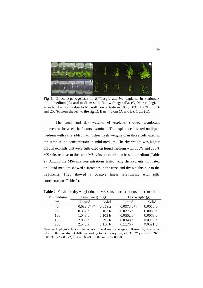
Fig 1. Direct organogenesis in Billbergia zebrina explants in stationary
liquid medium (A) and medium solidified with agar (B). (C) Morphological
aspects of explants due to MS-salt concentrations (0%; 50%; 100%; 150%
and 200%, from the left to the right). Barr = 3 cm (A and B); 1 cm (C).
The fresh and dry weights of explants showed significant
interactions between the factors examined. The explants cultivated on liquid
medium with salts added had higher fresh weights than those cultivated in
the same saline concentration in solid medium. The dry weight was higher
only in explants that were cultivated on liquid medium with 150% and 200%
MS salts relative to the same MS-salts concentration in solid medium (Table
2). Among the MS-salts concentrations tested, only the explants cultivated
on liquid medium showed differences in the fresh and dry weights due to the
treatments. They showed a positive linear relationship with salts
concentration (Table 2).
Table 2. Fresh and dry weight due to MS-salts concentrations in the medium
*For each phytotechnical characteristic analyzed, averages followed by the same
letter in the line do not differ according to the Tukey test, at 5%. (1) ŷ = - 0.1026 +
0.0133x, R² = 0.972; (2) ŷ = 0.0029 + 0.0006x, R² = 0.988.
MS medium
(%)
Fresh weight (g) Dry weight (g)
Liquid Solid Liquid Solid
0 0.083 a* (1) 0.059 a 0.0073 a (2) 0.0050 a
50 0.382 a 0.103 b 0.0276 a 0.0089 a
100 1.048 a 0.103 b 0.0552 a 0.0078 a
150 2.060 a 0.093 b 0.0948 a 0.0082 b
200 2.575 a 0.110 b 0.1176 a 0.0091 b

40
In vitro rooting under different sucrose concentration
The induction of roots occurred in all treatments, with an average
rooting percentage higher than 97%. The root number was confirmed to be
influenced by sucrose. Root number followed a positive quadratic
relationship with sucrose concentration in the medium (Fig 2).
Fig 2. Root number of Billbergia zebrina shoots due to sucrose
concentration n the medium.
The longest root lengths and aerial parts presented quadratic
relationships with increasing sucrose concentrations (Fig 3). The root growth
was best at 43.27 g L-1 sucrose (Fig 3A). Plants cultivated on sucrose
concentrations higher than 24.63 g L-1 showed shorter aerial parts (Fig 3B).
When the accumulation of matter in the plants was analyzed, we verified that
the fresh weight accumulation of the aerial parts and roots presented a
positive quadratic relationship with sucrose (Fig 4).

41
Fig 3. Longest root length and aerial part length of B. zebrina shoots due to
sucrose concentration in the medium.
Fig 4. Fresh weight of aerial parts and roots of B. zebrina shoots due to
sucrose concentration in the medium.
The chlorophyll a, b and carotenoids contents were affected by
sucrose addition in the medium. An increase in sucrose had a negative effect,
and we verified a decreasing linear model for all of the pigments analyzed
(Fig 5). Conversely, the content of total soluble sugars in B. zebrina shoots
increased with increasing sucrose concentrations (Fig 6).

42
Fig 5. Photosynthetic pigment content of B. zebrina shoots due to sucrose
concentration in the medium. (A) Chlrophlly a; (B) Chlorophlly b; (C)
Chlorophlly a+b; (D) carotenoids.
Fig 6. Total soluble sugars content of B. zebrina shoots due to sucrose
concentration in the medium.

43
Discussion
There are several studies that consider the use of plant growth
regulators as the primary direct organogenesis modulators of plant species
(Silveira et al., 2009; Santa-Rosa et al., 2013; Sangeetha and
Venkatachalam, 2014). In this study we examined whether the agar and
minerals in the growth medium play fundamental roles in the direct
organogenesis of B. zebrina shoots.
The use of agar may interfere with organogenesis response in some
plant species. It may reduce the formation of lateral shoots (Ivanova and
Staden, 2011). The employment of a gelling agent in the medium may
decrease water availability, mineral salts and plant growth regulators
(Debergh, 1983) and may also decrease endogenous cytokinin (Ivanova et
al., 2006). Mengarda et al. (2009) worked with different bromeliad species
and obtained higher multiplication rates with stationary liquid medium
relative to medium solidified with agar. Nevertheless, the findings of Silva et
al. (2012) with Nidularium procerum and N. innocentii do not apply to all
bromeliad species, which do not display differences in multiplication rate
between liquid and solid media.
The employment of a gelling agent is important for some plant
species, and it may assist with plant formation without physiological
disturbances such as hyperhydricity (Ivanova and Staden, 2011).
Hyperhydricity during plant formation on liquid medium or on medium with
low agar concentrations has already been noted in some in vitro plant species
(Casanova et al., 2008). Hyperhydricity is more common in plants grown on
liquid medium, most likely because the tissues are kept submerged and
undergo marked oxidative stress, in addition to high concentrations of
reactive oxygen species associated with changes in the activities of

44
antioxidant enzymes (Ziv, 2005). However, hyperhydricity symptoms were
not observed in B. zebrina shoots, which did not show morphological
changes such as vitrification even when explants were kept submerged in
medium for 45 days (Fig 1).
Increased MS-salts concentrations stimulated shoot formation in B.
zebrina explants (Table 1). Mineral components in the growth medium are
vitally important for the in vitro regeneration process in plants (Williams,
1993). Some mineral compounds are related to endogenous cytokinin
biosynthesis. An increase in nitrate resources may induce the expression of
genes responsible for the biosynthesis of cytokinins, resulting in
accumulation of these hormones in plants (Takei et al., 2004; Wang et al.,
2004). Cytokinins are primarily responsible for breaking apical dominance
and consequent lateral shoot induction. The breaking apical dominance is
fundamental in the first cell division (Pasternak et al., 2000). However, as
this study verified in B. zebrina explants, the addition of only a cytokinin
resource in the medium could not ensure an organogenesis response,
indicating that the concentration of salts is very important to ensure and
enhance cell division (Table 1).
Low water availability in media solidified with agar has a direct
effect on fresh and dry weights, which decrease with increasing
concentrations of gelling agent (Suthar et al., 2011). Agar may also adsorb
some nutrients from the medium. Nutrients may become unavailable to the
explants (Romberger and Tabor, 1971), restricting their growth and
consequently the increase in explant mass. Fresh and dry mass accumulation
relate to nutrition (Huang et al., 2010). Aranda-Peres et al. (2009) worked
with mineral nutrition of different bromeliad species and have verified that
calcium plays an important role during in vitro growth. Furthermore, it

45
exerts a positive influence on the absorption of other nutrients in the
medium. They have also verified that increasing calcium concentration is a
promising method for increasing fresh and dry weights of in vitro bromeliad
explants.
The explants of B. zebrina showed high rooting frequencies. The
same behavior has already been verified in other plant species, in which
rooting occurred in sugar-free medium (Cha-um et al., 2011; Iarema et al.,
2012). Adventitious root induction is more related to the concentration and
endogenous balance of plant hormones (Dong et al., 2012). Auxins are the
major plant hormones responsible for initiation of adventitious roots. The
most abundant endogenous auxin in plants is 3-indoleacetic acid (IAA) (Li et
al., 2009). IAA concentration in shoots may ensure the in vitro rhizogenic
response (Martins et al., 2013). It may also explain the in vitro rooting rate
of B. zebrina shoots on medium without sucrose and exogenous auxin.
The rhizogenic process requires available energy in the explant,
given that the root number typically responds to sucrose (Jo et al., 2009;
Rocha et al., 2013). Kumar et al. (1999) reported that carbohydrates used as
carbon sources in the medium act on root frequency and quality, determining
plant success after the transfer to an ex vitro environment. Carbohydrates are
transported to meristematic cells, regulating rhizogenesis by modulating
gene expression and enzyme activity (Pawlicki et al., 1995). The
carbohydrate source is also related to the growth hormone production
(auxins) involved in the rooting process (Rolland et al., 2002).
Sucrose contributes to the growth of roots because it acts on cell
expansion and proliferation (Wang and Ruan, 2013). Nevertheless, high
sugar concentrations can inhibit the growth of aerial plant parts (Al-Khateeb,
2008), as verified on B. zebrina shoots. Inhibition is due to osmotic stress in

46
the medium with a high sucrose concentration (Nowak et al., 2004; Jo et al.,
2009). Osmotic potential may interfere with nutrient abortion by the cells,
which is essential to growth and cell division in the aerial parts (Siwach et
al., 2011).
Sucrose cleavage in the medium results in glucose and fructose
production. It may accelerate cell division and consequently increase the
explant weight and volume (Gurel and Gulsen, 1998). However, high
sucrose concentrations may limit growth, due to an increase in the osmotic
potential in the medium caused by sucrose (Rejšková et al., 2007). This
supports our observations on B. zebrina shoots, which have reduced weights
with sucrose concentrations higher than 41 g L-1 (Fig 4A).
The reduction in chlorophyll content in in vitro plants may reduce
photosynthetic ability by decreasing light absorption (Sivanesan et al.,
2008). The decrease in photosynthetic pigments due to carbohydrate addition
in the medium has already noted in previous literature, such as Mohamed
and Alsadon (2010) and Swamy et al. (2010). These authors concluded that
lower sucrose concentrations may stimulate the chlorophyll production in in
vitro plants.
A higher total soluble sugars content in B. zebrina shoots on media
with high sucrose concentrations was verified (Fig 6). Similar results have
been reported in Dendrobium Second Love shoots. The soluble carbohydrate
increased concomitantly with growth medium sucrose concentrations in
shoots of this orchid (Ferreira et al., 2011). High stock-carbohydrate
concentrations in in vitro-formed plant tissues may improve the performance
of plants during the acclimatization phase (Chu et al., 2010). Plants
cultivated on medium without sugar may have better photosynthetic rates in
vitro when they are compared with plants grown on medium with sucrose.
47
Nevertheless, the absence of stock substances may result in low survival
rates after transfer to ex vitro conditions, and carbon source additions to the
medium in low concentrations are recommended (Fuentes et al., 2005b).
This supports the use of 15 g L-1 sucrose during the rooting phase. Since B.
zebrina shoots did not have a high decreasing photosynthetic pigments
content and increased carbohydrate stocks in the shoots.
Conclusion
The concentrations of MS-salts and sucrose have direct effects on
morphological responses in B. zebrina shoots during in vitro growth. The in
vitro multiplication of B. zebrina shoots is enhanced by doubling the original
concentration of MS-salts (200%) and liquid media. While the use of agar
during the multiplication phase limits the organogenesis response in B.
zebrina shoots. Sucrose does not determine root induction in B. zebrina
shoots but it induces better growth of roots. The use of 15 g L-1 sucrose
could be an interesting method to increase the endogenous carbohydrate
stock with few negative effects on photosynthetic pigments and maintaining
a good growth.
Acknowledgements
The authors would like to thank the CAPES, CNPq and FAPEMIG
(Brazil) for their financial support.
References
Al-Khateeb AA (2008) Regulation of in vitro bud formation of date palm
(Phoenix dactylifera L.) cv. Khanezi by different carbon sources. Bioresour
Technol. 99(14):6550–6555

48
Aranda-Peres AN, Peres LEP, Higashi EN, Martinelli AP (2009) Adjustment
of mineral elements in the culture medium for the micropropagation of three
Vriesea bromeliads from the Brazilian Atlantic Forest: The importance of
calcium. HortScience. 44(1):106-112
Benzing DH (2000) Vegetative structure. In: Benzing DH
(ed), Bromeliaceae: profile of an adaptive radiation. Cambridge: Cambridge
University Press, p19-77
Casanova E, Moysset L, Trillas MI (2008) Effects of agar concentration and
vessel closure on the organogenesis and hyperhydricity of adventitious
carnation shoots. Biol Plantarum. 52(1):1-8
Cha-um S, Chanseetis S, Chintakovid C, Pichakum A, Supaibulwatana K
(2011) Promoting root induction and growth of in vitro macadamia
(Macadamia tetraphylla L. ‘Keaau’) plantlets using CO2-enriched
photoautotrophic conditions. Plant Cell Tiss Org. 106 (3):435–444
Chu EP, Tavares AR, Kanashiro S, Giampaoli P, Yokota ES (2010) Effects
of auxins on soluble carbohydrates, starch and soluble protein content in
Aechmea blanchetiana (Bromeliaceae) cultured in vitro. Sci Hort.
125(3):451–455
Cruz CD (2013) Genes: a software package for analysis in experimental
statistics and quantitative genetics. Acta Sci Agron. 35(3):271–276
Debergh PC (1983) Effects of agar brand and concentration on the tissue
culture medium. Physiol Plant. 59(2):270–276
Dische Z (1962) General color reactions. In: Whistler, R.L.; Wolfram, M.L.
(Ed.) Carbohydrate chemistry, New York: Academic, p477-520
Dong N, Pei D, Yin W (2012) Tissue-specific localization and dynamic
changes of endogenous IAA during poplar leaf rhizogenesis revealed by in
situ immunohistochemistry. Plant Biotechnol Rep. 6(2):165–174
Engel VL, Poggiane F (1991) Estudo da concentração de clorofila nas folhas
e seu espectro de absorção de luz em função do sombreamento em mudas de
quatro espécies florestais nativas. Braz J Plant Physiol. 3(1):39-45.

49
Ferreira WM, Suzuki RM, Pescador R, Figueiredo-Ribeiro RCL, Kerbauy
GB (2011) Propagation, growth, and carbohydrates of Dendrobium Second
Love (Orchidaceae) in vitro as affected by sucrose, light, and dark. In Vitro
Cell Dev Biol-Plant. 47(3):420–427
Fuentes G, Talavera C, Desjardins Y, Santamaria JM (2005a) High
irradiance can minimize the negative effect of exogenous sucrose on the
photosynthetic capacity of in vitro grown coconut plantets. Biol Plantarum.
49(1):7-15
Fuentes G, Talavera C, Opereza C, Desjardins Y, Santamaria J (2005b)
Exogenus sucrose can decrease in vitro photosynthesis but improve Field
survival and growth of coconut (Cocos nucifera L.) in vitro plantlets. In vitro
Cell Dev Biol-Plant. 41(1):69-76
Greenway MB, Phillips IC, Lloyd MN, Hubstenberger JF, Phillips GC
(2012) A nutrient medium for diverse applications and tissue growth of plant
species in vitro. In Vitro Cell. Dev Biol-Plant. 48(4):403–410
Guerra MP, Vesco LLD (2010) Strategies for the micropropagation of
bromeliads. In: Jain SM, Ochatt SJ (eds.) Protocols for in vitro propagation
of ornamental plants: methods in molecular biology, v.589. Humana Press,
New York, p47–66
Gurel S, Gulsen Y (1998) The effects of different sucrose, agar and pH
levels on in vitro shoot production of almond (Amygdalus communis L.).
Turkish J Bot. 22(6):363–373
Hazarika BN (2003) Acclimatization of tissue-cultured plants. Curr Sci.
85(12):1704–1712
Higby WK (1962) A simplified method for determination of some the
carotenoid distribution in natural and carotene-fortified orange juice. J Food
Sci. 27(1):42-49
Huang Z C, Zeng FH, Lu XY (2010) Efficient regeneration of Eucalyptus
urophylla from seedling-derived hypocotyls. Biol Plantarum. 54(1):131-134
Huang PL, Liu ZH, Chang ML, Liao LJ (2011) Micropropagation of the
bromeliad Guzmania “Hilda” via organogenesis and the effect of α-
naphthaleneacetic acid on plantlet elongation. Sci Hortic. 130(4):894–898

50
Iarema L, Cruz ACF, Saldanha CW, Dias LLC, Vieira RF, Oliveira
EJ, Otoni WC (2012) Photoautotrophic propagation of Brazilian ginseng
[Pfaffia glomerata (Spreng.) Pedersen]. Plant Cell Tiss Org. 110(2):227-238
Ivanova M, Staden JV (2011) Influence of gelling agent and cytokinins on
the control of hyperhydricity in Aloe polyphylla. Plant Cell Tiss Org.
104(1):13–21
Ivanova M, Nova´k O, Strnad M, Van Staden J (2006) Endogenous
cytokinins in shoots of Aloe polyphylla cultured in vitro in relation to
hyperhydricity, exogenous cytokinins and gelling agents. Plant Growth
Regul. 50(2-3):219–230
Jo EA, Tewari RK, Hahn EJ, Paek KY (2009) In vitro sucrose concentration
affects growth and acclimatization of Alocasia amazonica plantlets. Plant
Cell Tiss Org. 96(3):307–315
Kumar A, Sood A, Palni LMS, Gupta AK (1999) In vitro propagation of
Gladiolus hybridus Hort.: synergistic effect of heat shock and sucrose on
morphogenesis. Plant Cell Tiss Org. 57(2):105–112
Li SW, Xue L, Xu S, Feng H, An L (2009) Mediators, Genes and Signaling
in Adventitious Rooting. Bot Rev. 75(2):230–247
Li H, Tang C, Xu Z (2013) The effects of different light qualities on
rapeseed (Brassica napus L.) plantlet growth and morphogenesis in vitro.
Sci Hort. 150:117–124
Martinelli G, Vieira CM, Gonzalez M, Leitman P, Piratininga A, Costa AF,
Forzza RC (2008) Bromeliaceae da Mata Atlântica brasileira: Lista de
espécies, distribuição e conservação. Rodriguésia. 59(1):209-258
Martins JPR, Schimildt ER, Alexandre RS, Santos BR, Magevski GC (2013)
Effect of synthetic auxins on in vitro and ex vitro bromeliad rooting. Pesqui
Agropec Trop. 43(2):138-146
Mengarda LHG, Povoas L, Debiasi C, Pescador R (2009) Estado físico do
meio de cultura na propagação in vitro de Bromeliaceae. Scientia Agraria.
10(6):469-474

51
Mohamed MAH, Alsadon AA (2010) Influence of ventilation and sucrose on
growth and leaf anatomy of micropropagated potato plantlets. Sci Hort.
123(3):295–300
Murashige T, Skoog F (1962) A revised medium for rapid growth and
bioassays with tobacco tissue cultures. Physiol Plantarum. 15:473-497
Negrelle RRB, Mitchell D, Anacleto A (2012) Bromeliad ornamental
species: conservation issues and challenges related to commercialization.
Acta Sci Biol Sci. 34(1):91-100
Nowak B, Miczynski K, Hudy L (2004) Sugar uptake and utilization during
adventitious bud differentiation on in vitro leaf explant of Wegierka zwykla
plum (Prunus domestica). Plant Cell Tiss Org. 76(3):255-260
Pasternak T, Miskolczi P, Ayaydin F, Mészáros T, Dudits D, Fehér A (2000)
Exogenous auxin and cytokinin dependent activation of CDKs and cell
division in leaf protoplast-derived cells of alfalfa. Plant Growth Regul. 32(2-
3):129–141
Pawlicki N, Welander M (1995) Influence of carbohydrate source, auxin
concentration and time of exposure on adventitious rooting of the apple
rootstock Jork 9. Plant Sci. 106(2):167–176
Ramage CM, Williams RR (2002) Mineral nutrition and plant
morphogenesis. In Vitro Cell Dev Biol-Plant. 38(2):116–124
Rejšková A, Patková L, Stodůlková E, Lipavská H (2007) The effect of
abiotic stresses on carbohydrate status of olive shoots (Olea europaea L.)
under in vitro conditions. J Plant Physiol. 164(2):174–184
Rocha PSG, Oliveira RP, Scivittaro WB (2013) Sugarcane micropropagation
using light emitting diodes and adjustment in growth-medium sucrose
concentration. Cienc Rural. 43(7):1168-1173
Rolland F, Moore B, Sheen J (2002) Sugar sensing and signaling in plants.
Plant Cell. 14(suppl 1):185–205
Romberger JA, Tabor CA (1971) The Picea abies shoot apical meristem in
culture: agar and autoclaving effects. Am J Bot. 58(2):131–140

52
Sangeetha P, Venkatachalam P (2014) Induction of direct shoot
organogenesis and in vitro flowering from shoot tip explants of cucumber
(Cucumis sativus L. cv. ‘Green long’). In Vitro Cell Dev Biol-Plant.
50(2):242–248
Santa-Rosa SI, Souza FVD, Vidal AM, Ledo CAS, Santana JRF (2013)
Micropropagation of the ornamental vulnerable bromeliads Aechmea
blanchetiana and Aechmea distichantha. Hortic Bras. 31(1):112-118
Silva ALL, Costa JL, Alcantara GB, Carvalho DC, Schuck MR, Biasi LA,
Scheidt LN, Soccol CR (2012) Micropropagation of Nidularium
innocentii Lem. and Nidularium procerum Lindm (Bromeliaceae). Pak J
Bot. 44(3):1095-1101
Silveira DG, Souza FVD, Pelacani CR, Souza AS, Ledo CAS Santana JRF
(2009) Micropropagation and in vitro conservation of Neoglaziovia
variegata (Arr. Cam.) Mez, a fiber producing bromeliad from Brazil. Braz
Arch Biol Techn. 52(4):923-932
Sivanesan L, Hwang SL, Jeong BR (2008) Influence of plant growth
regulators on axillary shoot multiplication and iron sourse on growth of
Scrophularia takesimensis Nakal – a rare endemic medicinal plant. Afr J
Biotechnol. 7(24):4484-4490
Siwach P, Grover K, Gill AR (2011) The influence of plant growth
regulators, explant nature and sucrose concentration on in vitro callus growth
of Thevetia peruviana Schum. Asian J Biotechnol. 3(3):280-292
Swamy MK, Sudipta KM, Balasubramanya S, Anuradha M (2010) Effect of
Different carbon sources on in vitro morphogenetic response of patchouli
(Pogostemon cablin Benth.). J Phytol. 2(8):11-17
Suthar RK, Habibi N, Purohit SD (2011) Influence of agar concentration and
liquid medium on in vitro propagation of Boswellia serrata Roxb. Indian J
Biotechnol. 10:224-227
Takei K, Ueda N, Aoki K, Kuromori T, Hirayama T, Shinozaki K et al
(2004) AtIPT3 is a key determinant of nitrate-dependent cytokinin
biosynthesis in Arabidopsis. Plant Cell Physiol. 45(8):1053–1062

53
Vesco LLD, Stefenon VM, Welter LJ, Scherer RF, Guerra MP (2011)
Induction and scale-up of Billbergia zebrinanodule cluster cultures:
implications for mass propagation, improvement and conservation. Sci Hort.
28(4):515-522
Wang L, Ruan YL (2013) Regulation of cell division and expansion by sugar
and auxin signaling. Front Plant Sci. 4:1-9
Wang R, Tischner R, Gutie´rrez RA, Hoffman M, Xing X, Chen M et al
(2004) Genomic analysis of the nitrate response using a nitrate reductase-
null mutant of Arabidopsis. Plant Physiol. 136(1):2512–2522
Williams RR (1993) Mineral nutrition in vitro – a mechanistic approach.
Aust J Bot. 41(2):237–251
Ziv M (2005) Simple bioreactors for mass propagation plants. In Vitro Cell
Dev Biol-Plant. 81:277-285

54
ARTICLE 2
Impacts of photoautotrophic and photomixotrophic
condition on in vitro propagated Billbergia zebrina
(Bromeliaceae)
(Prepared in accordance with Plant Cell, Tissue and Organ
Culture's standards)
55
Abstract
Micro-propagation techniques contribute to the multiplication of several
bromeliad species. However, micropropagated plantlets often present low
survival rate due to anatomical and physiological disorders induced by in
vitro conditions. This study aimed to evaluate the sucrose and gas exchange
impact on in vitro propagated Billbergia zebrina plants and to check if there
is any residual effect of the in vitro conditions on micropropagated plants
after acclimatization. Previously in vitro-established B. zebrina plants were
transferred to culture media containing 0.0, 15.0, 30.0, 45.0 or 60.0 g L-1
sucrose. Two different culture container sealing systems were tested: lids
with a filter (permitting an excellent gas exchange) and lids with no filter
(blocking fluent gas exchange). At 45 days in vitro growth, B. zebrina
plantlets were transplanted onto suitable soil mix and evaluated at 80 days
growth in greenhouse. At 45 days in vitro and 80 days of acclimatization in
the greenhouse, the plants were evaluated. High sucrose levels in the in vitro
media resulted in reduced growth. Plantlets exposed to aerated containers
presented better rooting, being the sugar-free medium the best in vitro
condition (photoautotrophic condition). Limited air exchange resulted in
plantlets with anatomical and physiological disorders at the end of the in
vitro period. The highest growth rate in the greenhouse was observed in
plants previously propagated in unlimited gas exchange system and sugar-
free medium. The use of photoautotrophic conditions induces B. zebrina
plantlets without anatomical and physiological disorders and it interfere
positively on ex vitro growth.
Keywords: bromeliad, in vitro plant, plant anatomy, photoautotrophic
growth, tissue culture
56
Introduction
In vitro propagation techniques have been widely used for rapid
multiplication of various economically important or endangered plant
species, such as many species from the Bromeliaceae family (Guerra and
Vesco 2010). Micro-propagation procedures for bromeliads have frequently
been published (Martins et al. 2013, 2014; Mengesha et al. 2013; Kurita and
Tamaki 2014). Most of these studies are related to the use of plant growth
regulators, temperature, carbon source or mineral composition of the culture
medium being the main modulators of in vitro morphogenetic responses.
Conventional in vitro propagation techniques are carried out using
closed containers and sugars as carbon source added to the medium (Xiao et
al. 2011). The internal atmosphere of these conventional in vitro containers
consists of high relative humidity, variable CO2 concentrations, and potential
accumulation of ethylene and other gases (De Proft et al. 1985; Kozai 2010).
Moreover, depending on the culture condition in the growth room, low
irradiance, variable air and temperature may occur.
Conventional in vitro conditions may influence on anatomical,
physiological and morphological plant characteristics, and cause great losses
during later acclimatization treatment (Kitaya et al. 2005). Plants cultivated
in vitro often present peculiar characteristics, such as poorly developed
shoots, tissues with low mechanical strength, higher water content, non-
functional stomata and small, thin leaves with fewer trichomes and low
photoautotrophic activity (Kozai and Kubota 2001; Cha-um et al. 2011; Xiao
et al. 2011). These anomalies become negative features during transfer of in
vitro plants into the greenhouse or field conditions, causing slow
establishment and low survival rate (Pospíšilová et al. 2000; Hazarika 2006;
Saez et al. 2012). Only few works have been documented about anatomical

57
disorders on in vitro propagated bromeliad species (Carvalho et al. 2014;
Martins et al. 2014), but how it might interfere on survival and growth rate
of bromeliads after transfer to greenhouse is still unclear.
According to Fuentes et al. (2005b) the use of sucrose during in vitro
propagation induces some physiological disorders in micropropagated
plants, but sucrose increases the survival rate after greenhouse transfer.
Carbohydrates are required by plant cells as carbon resources, act as energy
supporting growth and biosynthetic processes, and influence in a positive
way in vitro rooting (Ferreira et al. 2011).
Supplemental sugar in the in vitro medium assists in water
conservation and in maintaining the osmotic potential of cells (Hazarika
2003). Sucrose is closely related to stomatal density and chlorophyll content,
as well as morphogenetic behavior in some plant tissues, such as vascular
and support tissues (Mohamed and Alsadon 2010; Iarema et al. 2012). The
exogenous sucrose supply increases the endogenous content of carbohydrate
stocks (starch, sucrose, fructose and glucose) in micropropagated plantlets. It
favors acclimatization and accelerates physiological adaptations (Jo et al.
2009). However, high sucrose concentrations in the medium potentially
decrease photosynthetic ability of in vitro plants (Fuentes et al. 2005a, b).
Container closure types ensuring improved ventilation act positively
on quality of in vitro plantlets. Strong changes of CO2 and ethylene during
the in vitro magnolia cultivation have been noticed by De Proft et al. (1985)
causing losses of plants when gas exchange was prohibited. The presence of
sugar in the medium and accumulated ethylene in sealed vessels may let to
the development of abnormal stomata which subsequently reduced the
survival rate upon transfer to ex vitro conditions (Zobayed 2005). Permeable
closures help in vitro plantlets to grow photomixotrophically or

58
photoautotrophically resulting in higher plant functionality and less
propagule loss during acclimatization process (Zobayed 2000; Iarema et al.
2012).
Studies indicate that the photoautotrophic growth in vitro of many
plant species can be significantly promoted by increasing the CO2
concentration in the culture vessel and reducing relative humidity (Xiao et
al. 2011). Photoautotrophic propagation has recently been successfully used
for some plant species (Cha-um et al. 2011; Iarema et al. 2012; Shin et al.
2013; Saldanha et al. 2014).
The aim of this study was to analyze the impact of culture container
gas exchange and sucrose concentration in the culture media on in vitro
propagated B. zebrina plantlets and if there is any residual effect of these in
vitro conditions on micropropagated plants after acclimatization.
Material and Methods
Plant material and culture conditions
Billbergia zebrina plantlets were previously established in vitro from
seeds and cultivated for 60 days. The obtained plantlets were transferred to
250.0 mL glass containers offering 50.0 mL stationary liquid MS medium
(Murashige and Skoog 1962), supplemented with 30.0 g L-1 sucrose and 13.0
µM 6-benzylaminopurine (BAP) and set at pH 5.8 (Martins et al. 2015).
After 30 days of growth, side shoots were subcultivated, for 45 days, in
250.0 mL glass containers containing 50.0 mL stationary liquid MS medium
with no plant growth regulator and supplemented with 30.0 g L-1 sucrose.
Media were autoclaved at 120ºC for 20 minutes. The multiplication
procedures were carried out three times on obtained side shoots in order to
obtain the necessary number of side shoots with a similar morphology. In
59
vitro plant material was kept in a growth room, at 26 ± 2ºC and a 16-hour
photoperiod (8:00 to 00:00 h), under fluorescent tube lamps Philips Master
TL5 HO, 49W/840 providing 90 μmol m-2 s-1 of PAR light.
Sucrose and gas exchange during the in vitro propagation
B. zebrina side shoots with approximately 4.0 cm in length (obtained
from previous in vitro subculture – third generation of induced side shoots),
were individualized (5-8 side shoots per seedling) with the aid of a scalpel,
after mixing of the explants five side shoots were taken and transferred to
280.0 mL polypropylene containers (ECO2 NV®, Geraardsbergen,
Belgium) containing 50.0 mL MS medium solidified with 7.0 g L-1 agar
(Difco - Agar No 1 - MC002) and 0, 15.0, 30.0, 45.0 or 60.0 g L-1 sucrose.
This phase was carried out with five shoots per container. pH was adjusted
to 5.8, before autoclaving at 120ºC, for 20 minutes. Two different sealing
systems were tested: lids with a XXL filter (permitted gas exchange system -
62.83 gas replacements per day) and lids with a XXL filter covered with two
layers of polyvinylchloride (PVC) transparent film (blocking fluent gas
exchange – 4.19 gas replacements per day). After sterile inoculation the
material was kept, for 45 days, in a growth room, at 26 ± 2ºC and a 16-hour
photoperiod (8:00 to 00:00 h), under fluorescent lamps Philips Master TL5
HO, 49W/840 providing 230 μmol m-2 s-1 of PAR light.
Growth parameters of in vitro plantlets
To evaluate growth, twenty plantlets from each treatment were
sampled randomly at 45-day growth and they were divided into four parcels.
The aerial part and roots were separated to determine fresh and dry weight.

60
CO2 content in the in vitro containers
The CO2 content was analyzed at 45-day of in vitro culture at four
times over a 24 hour period (6:30, 15:30, 00:30 and 6:30 h) using gas
chromatography (Compact GC, Interscience, Louvain-La-Neuve, Belgium,
equipped with a thermal conductivity detector). The measurements were
performed on three containers per treatment each time.
Anatomical analysis
Anatomical characterization was performed on three plantlets from
each treatment. Samples were randomly collected at 45-day growth and
fixed in FAA (formaldehyde, acetic acid and ethanol 50%, 0.5/0.5/9, v/v) for
48 hours, followed by storage in 70% ethanol (Johansen 1940).
Paradermal and cross sections were performed in the median region
of the first completely expanded leaf in the rosette central region with aid of
a double edge razor. Sections were cleared with sodium hypochlorite 10%
(v/v) followed by staining with safranin. Finally glycerin 50% was used to
assemble the slides.
The sections were investigated using a light microscope
(OLYMPUS) coupled to a digital camera. The photomicrographs were used
to measure the anatomy characteristics using the software UTHSCSA-
Imagetool® calibrated with a microscopy ruler.
The thickness of adaxial and abaxial epidermis (µm), hydrenchyma
(µm) and chlorenchyma (µm), sclerenchyma area (µm2) and xylem diameter
(µm) were determined.
61
Acclimatization of the in vitro plantlets
At 45 days in vitro culture using different sucrose concentrations and
sealing systems, plantlets of B. zebrina were transplanted to plastic pots
containing peat as a potting medium and kept covered with transparent
polyethylene plastic lids in order to maintain a high relative humidity for one
week. After this period lids were removed. The substrate remained at a
constant moisture level at full field capacity. The plant material was kept, for
80 days, in a greenhouse, at 18ºC (minimal night temperature) and a 16-hour
photoperiod.
Growth of acclimatized plants
To evaluate growth, fifteen plants from each treatment were sampled
randomly and they were divided into five parcels. The dry weight was
measured at 80 days of ex vitro culture.
Statistical analysis
The experiment was performed in ten containers per treatment and a
completely randomized design in a factorial arrangement (five sucrose
concentrations x two different sealing systems) was adopted. The obtained
data were submitted to analysis of variance (ANOVA), the averages of the
factor sealing systems were compared using Tukey’s test and the sucrose
concentrations were subjected to regression analysis.
Results
Growth and development of in vitro plantlets
The gas exchange rate of the in vitro containers and the sucrose
concentrations of the media directly influenced the morphologic responses of
62
B. zebrina plantlets (Fig. 1). Fresh and dry weights of the explants’ aerial
part showed significant interactions between the two factors examined. Fresh
and dry weight of plantlets cultivated in containers with filter (aerated
containers permitting unrestricted gas exchange) presented a negative linear
relationships with increasing sucrose concentrations (Fig. 2A-B). When
restricting the gas exchange the plantlets’ dry weight showed positive
relationships with increasing sucrose concentrations. Sucrose levels in the
media bring up a kind of optimum around 30 g L-1 for the plantlets fresh
weight. B. zebrina plantlets cultivated on sugar-free medium in aerated in
vitro container had the highest fresh and dry weight build up (Fig. 2A-B),
resulting in the most developed plantlets (Fig. 1A).

63
Fig. 1 B. zebrina plantlets at 45 days of in vitro growth, exposed to different
sucrose concentrations in the medium and sealing systems of the containers.
Aerial part and roots of plantlets grown in filter (A-J) and no filter containers
(K-T) in function of sucrose concentration (0.0, 15.0, 30.0, 45.0, 60 g L-1
sucrose, always from top to bottom).

64
Fig. 2 Fresh (A) and dry (B) weight (mg/plant) of B. zebrina plantlets (aerial
parts) as a function of sucrose (g L-1) in the in vitro medium and sealing
system of the culture containers. For each sucrose concentration (g L-1)
analyzed, averages followed by asterisk differ from each other according to
the Tukey’s test, at 5%.
Rooting occurred in all treatments studied (Fig.1). The roots fresh
and dry weights were similar for all plantlets grown with a limited gas
exchange (filter covered with PVC) (average of 60 mg/plant). When explants
were cultivated with unlimited gas exchange (filter) the roots fresh and dry
weight presented a negative linear correlation with increasing sucrose
concentrations. Accumulation of root dry matter was lower in limited gas
exchange containers (Fig. 3A-B).

65
Fig. 3 Fresh (A) and dry (B) weight (mg/plant) roots from B. zebrina
plantlets as a function of sucrose (g L-1) in the medium and gas sealing of the
culture containers. For each sucrose concentration (g L-1) analyzed, averages
followed by asterisk differ each other according to the Tukey’s test, at 5%.
CO2 concentration of the containers
The CO2 level inside the in vitro culture containers was clearly
influenced by the gas exchange capacity. With unrestricted gas exchange,
CO2 levels were balancing around 250 ppm (dark and light phase).
Restricting the gas exchange resulted in an increased CO2 level. The highest
CO2 values were reached at the end of the dark period (6:30h) getting up to
600 ppm. During the light period (15:30h) lower CO2 levels were detected (3
times lower than during the dark period) (Fig. 4).

66
Fig. 4 CO2 concentration (%) at different times during the day in in vitro
containers holding B. zebrina plantlets. For each time analyzed, averages
followed by different letters differ from each other according to Tukey’s test,
at 5%. The dark period is indicated by the black bar at the bottom of the
figure.
Anatomical analysis
In B. zebrina plantlets, the surface of leaves shows tetracytic
stomata with hypostomatic distribution. Transversal leaf sections present a
one cell layer epidermis both sides of the leaf. The mesophyll has a
dorsiventral organization. At the adaxial leaf side there is the water-storage
parenchyma (hydrenchyma) containing non-chlorophyll with thin walled
cells. Chlorophyll parenchyma (chlorenchyma) with isodiametric cells is
located at abaxial leaf side. The vascular bundles are collateral, usually
larger bundles alternating with smaller bundles and surrounded by
sclerenchyma fibers (Fig. 5). This leaf structure is frequently observed and
described in many bromeliads (Mantovani et al. 2012; Pereira et al. 2013;
Martins et al. 2014).
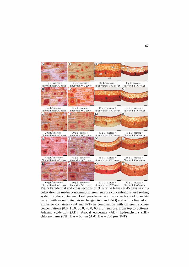
67
Fig. 5 Paradermal and cross sections of B. zebrina leaves at 45 days in vitro
cultivation on media containing different sucrose concentrations and sealing
system of the containers. Leaf paradermal and cross sections of plantlets
grown with an unlimited air exchange (A-E and K-O) and with a limited air
exchange containers (F-J and P-T) in combination with different sucrose
concentrations (0.0, 15.0, 30.0, 45.0, 60 g L-1 sucrose, from top to bottom).
Adaxial epidermis (AD), abaxial epidermis (AB), hydrenchyma (HD)
chlorenchyma (CH). Bar = 50 µm (A-J); Bar = 200 µm (K-T).

68
Sucrose concentrations of the culture media influenced the stomatal
shape. A larger stomatal porous was noted in treatments with limited gas
exchange and low sucrose levels. Increasing sucrose in the media induced
more elliptical shaped stomata and this is directly correlated to higher
stomatal functionality (Fig. 5).
Adaxial epidermis thickness increased with increasing sucrose
concentrations in containers with limited air exchange. Nevertheless, when
the plantlets were cultivated without air exchange restriction, the adaxial
epidermis did not show any difference between sucrose treatments, being as
thick as those grown in high sucrose concentration and limited air exchange
(Fig. 6A). Among the treatments, only the sucrose concentrations influenced
the abaxial epidermis thickness regardless the sealing type of the containers.
The lower epidermis, being thinner than the upper one, presented an
increasing linearity as the sucrose concentrations raised (Fig. 6B).
Fig. 6 Adaxial (A) and abaxial (B) epidermis thickness (µm) of B. zebrina
leaves influenced by sucrose (g L-1) in the in vitro medium and sealing type
of the in vitro container at 45 days in in vitro culture. For each sucrose

69
concentration (g L-1) analyzed, averages followed by asterisk differ from
each other according to the Tukey’s test, at 5%.
The thickest hydrenchyma was observed in plantlets grown in sugar-
free medium and a free gas exchanging container. Increasing sucrose
concentrations (60 g L-1) resulted in a 50 % decrease of this hydrenchyma
layer thickness. No differences between the sucrose treatments were noted
in containers with restricted air exchange (Table 1).
The chlorochyma thickness presented a linear relation with
increasing sucrose concentrations in both container sealing systems.
However thickening was observed as plantlets grown under limited air
exchange and sucrose levels induced a negative relationship when compared
in those plantlets grown in more aerated containers (Table 1).
The sclerenchyma area and xylem vessel diameter were analyzed in
central vascular bundles of B. zebrina leaves (Fig. 7). High sucrose
concentrations had a positive effect on the sclerenchyma area size.
Ventilated containers act in a positive way on sclerenchyma increment
(Table 1). On the other hand, sucrose and non-ventilated containers had a
negative effect on xylem vessel diameter making these water conducting
vessels smaller in size (Table 1).
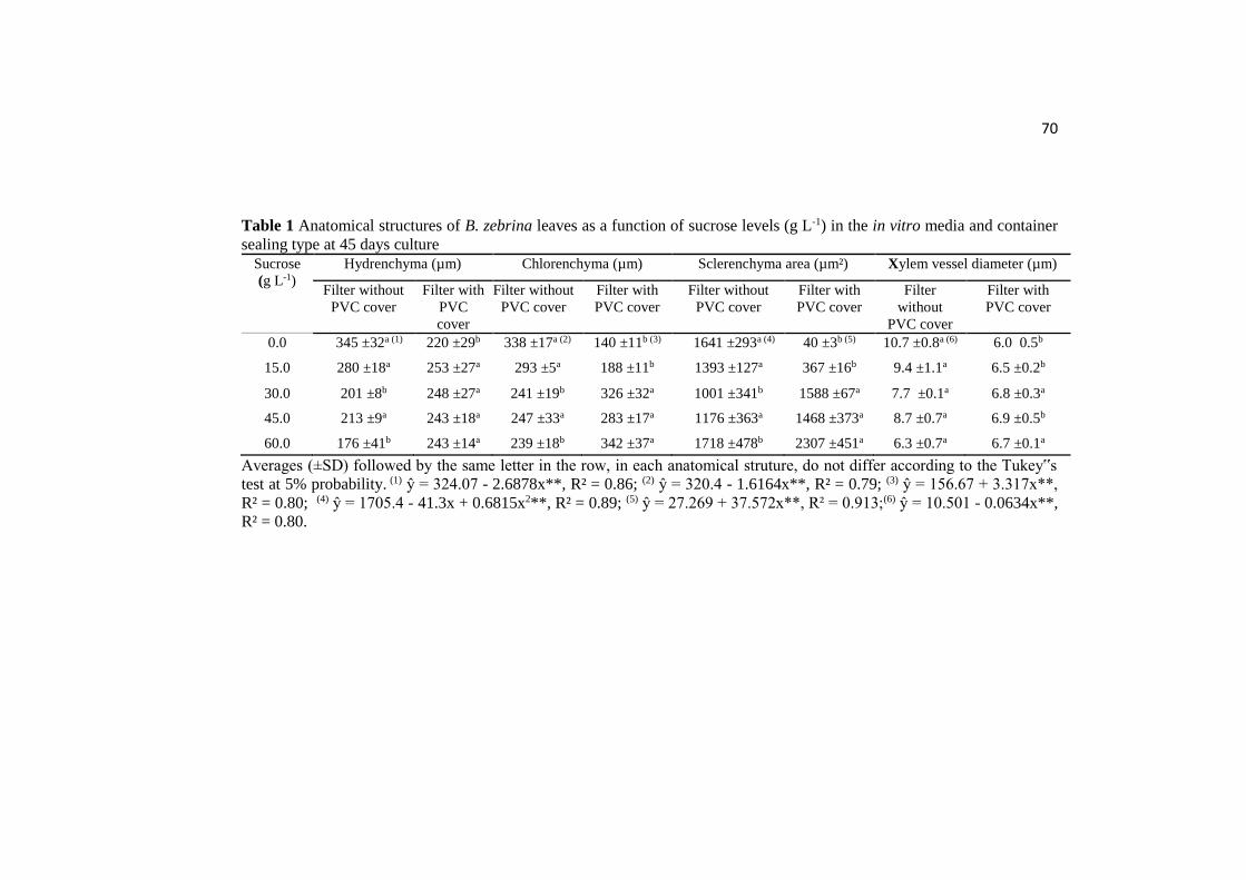
70
Table 1 Anatomical structures of B. zebrina leaves as a function of sucrose levels (g L-1) in the in vitro media and container
sealing type at 45 days culture Sucrose
(g L-1)
Hydrenchyma (µm) Chlorenchyma (µm) Sclerenchyma area (µm²) Xylem vessel diameter (µm)
Filter without
PVC cover
Filter with
PVC
cover
Filter without
PVC cover
Filter with
PVC cover
Filter without
PVC cover
Filter with
PVC cover
Filter
without
PVC cover
Filter with
PVC cover
0.0 345 ±32a (1) 220 ±29b 338 ±17a (2) 140 ±11b (3) 1641 ±293a (4) 40 ±3b (5) 10.7 ±0.8a (6) 6.0 0.5b
15.0 280 ±18a 253 ±27a 293 ±5a 188 ±11b 1393 ±127a 367 ±16b 9.4 ±1.1a 6.5 ±0.2b
30.0 201 ±8b 248 ±27a 241 ±19b 326 ±32a 1001 ±341b 1588 ±67a 7.7 ±0.1a 6.8 ±0.3a
45.0 213 ±9a 243 ±18a 247 ±33a 283 ±17a 1176 ±363a 1468 ±373a 8.7 ±0.7a 6.9 ±0.5b
60.0 176 ±41b 243 ±14a 239 ±18b 342 ±37a 1718 ±478b 2307 ±451a 6.3 ±0.7a 6.7 ±0.1a
Averages (±SD) followed by the same letter in the row, in each anatomical struture, do not differ according to the Tukey‟s
test at 5% probability. (1) ŷ = 324.07 - 2.6878x**, R² = 0.86; (2) ŷ = 320.4 - 1.6164x**, R² = 0.79; (3) ŷ = 156.67 + 3.317x**,
R² = 0.80; (4) ŷ = 1705.4 - 41.3x + 0.6815x2**, R² = 0.89; (5) ŷ = 27.269 + 37.572x**, R² = 0.913;(6) ŷ = 10.501 - 0.0634x**,
R² = 0.80.
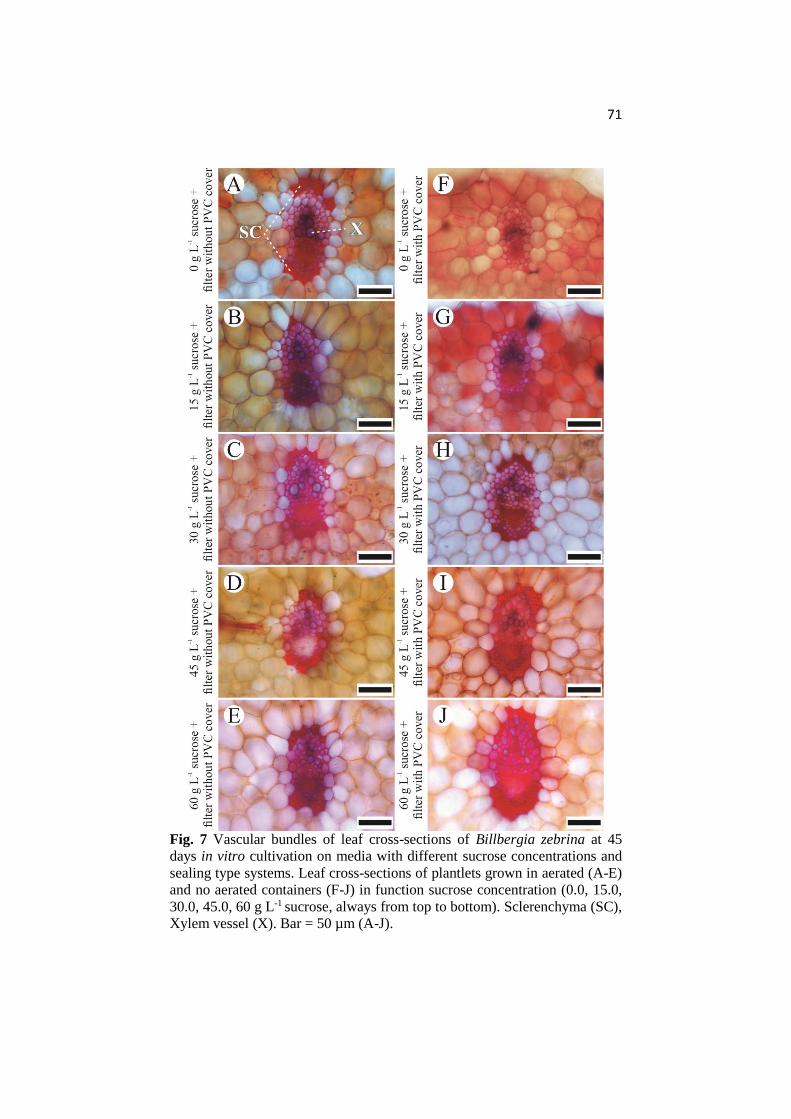
71
Fig. 7 Vascular bundles of leaf cross-sections of Billbergia zebrina at 45
days in vitro cultivation on media with different sucrose concentrations and
sealing type systems. Leaf cross-sections of plantlets grown in aerated (A-E)
and no aerated containers (F-J) in function sucrose concentration (0.0, 15.0,
30.0, 45.0, 60 g L-1 sucrose, always from top to bottom). Sclerenchyma (SC),
Xylem vessel (X). Bar = 50 µm (A-J).

72
Growth and development of acclimatized plants
At 80 days ex vitro conditions, B. zebrina plants presented a survival
rate of 100% in all previously examined in vitro treatments, but these plants
showed different growth rates. Plantlets cultivated in photoautotrophic
conditions in vitro (sugar-free in the medium and aerated containers) had
aerial parts with the highest dry weight. Aerial part’s dry weight of plantlets
previously grown with limited air exchange showed a generally stable level
with increasing sucrose concentrations after the transfer to greenhouse (Fig.
8).
Fig. 8 Aerial part dry weight (mg/plant) of acclimatized B. zebrina plants in
relation to the sucrose concentrations (g L-1) in the medium and container
sealing type during in vitro culture. For each sucrose concentration (g L-1)
analyzed, averages followed by asterisk differ from each other according to
the Tukey’s test, at 5%.
Discussion
This research reports for the first time the impact of sucrose and gas
exchange effects on bromeliads grown under tissue culture conditions.
Under photoautotrophic conditions (sugar-free media and no gas exchange
restrictions) a higher growth rate and no anatomical disorders were found
compared to bromeliad plantlets kept under conventional in vitro culture

73
conditions. These conditions may determine the explant survival rate after
greenhouse transfer (Park et al. 2011).
B. zebrina plantlets cultivated in conventional in vitro containers
respond clearly on the supply of sucrose (Fig. 1, 2). Plantlets cultivated in
containers with filter covered with PVC, require exogenous sucrose for
growth and rooting (Iarema et al. 2012; Eckstein et al. 2012). Sucrose
contributes to the induction and growth of roots by acting on cell expansion
and proliferation (Wang and Ruan 2013). Nevertheless, the supplementation
of sucrose was not a limiting factor for root induction in B. zebrina. Plantlets
cultivated in ventilated containers with no sucrose in de medium performed
excellent root formation (Fig. 1, 3). The positive effect of container aeration
is mainly due to the improved CO2 exchange with the open air and the
prevention of ethylene accumulation (Trevisan and Mendes 2005). Using
gas-permeable container types allow a continuous supply of CO2 during the
light period and lower relative humidity (Xiao et al. 2011). CO2 increase in
photoautotrophic culture conditions may play a key role as carbon and
energy support for root initiation and development (Hazarika 2003; Chan-um
et al. 2011). However, the improved ventilation of containers enhances
evapotranspiration which may alter the physical properties of culture media
attributed to water loss (Ivanova and Staden 2010). Evaporation of artificial
media decreases the osmotic potential due to an increased concentration of
the dissolved compounds resulting into osmotic stress of the in vitro plants
(Cui et al. 2010).
When subjected to environmental stress, plants actively reduce their
vegetative growth to conserve and redistribute resources and increasing their
chance of survival if stress continuous (Skirycz and Inzé 2010). This was
verified on B. zebrina plantlets grown on media with sucrose in ventilated
containers (Fig. 1, 2). Media in these aerated containers had substantial
water loss (around 34% of water loss) at the same time reduced plant growth
was noticed probably due to low osmotic potential of the media in
combination with a reduced air humidity.
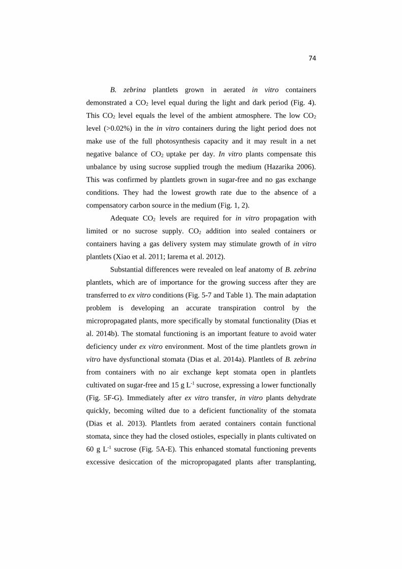
74
B. zebrina plantlets grown in aerated in vitro containers
demonstrated a CO2 level equal during the light and dark period (Fig. 4).
This CO2 level equals the level of the ambient atmosphere. The low CO2
level (>0.02%) in the in vitro containers during the light period does not
make use of the full photosynthesis capacity and it may result in a net
negative balance of CO2 uptake per day. In vitro plants compensate this
unbalance by using sucrose supplied trough the medium (Hazarika 2006).
This was confirmed by plantlets grown in sugar-free and no gas exchange
conditions. They had the lowest growth rate due to the absence of a
compensatory carbon source in the medium (Fig. 1, 2).
Adequate CO2 levels are required for in vitro propagation with
limited or no sucrose supply. CO2 addition into sealed containers or
containers having a gas delivery system may stimulate growth of in vitro
plantlets (Xiao et al. 2011; Iarema et al. 2012).
Substantial differences were revealed on leaf anatomy of B. zebrina
plantlets, which are of importance for the growing success after they are
transferred to ex vitro conditions (Fig. 5-7 and Table 1). The main adaptation
problem is developing an accurate transpiration control by the
micropropagated plants, more specifically by stomatal functionality (Dias et
al. 2014b). The stomatal functioning is an important feature to avoid water
deficiency under ex vitro environment. Most of the time plantlets grown in
vitro have dysfunctional stomata (Dias et al. 2014a). Plantlets of B. zebrina
from containers with no air exchange kept stomata open in plantlets
cultivated on sugar-free and 15 g L-1 sucrose, expressing a lower functionally
(Fig. 5F-G). Immediately after ex vitro transfer, in vitro plants dehydrate
quickly, becoming wilted due to a deficient functionality of the stomata
(Dias et al. 2013). Plantlets from aerated containers contain functional
stomata, since they had the closed ostioles, especially in plants cultivated on
60 g L-1 sucrose (Fig. 5A-E). This enhanced stomatal functioning prevents
excessive desiccation of the micropropagated plants after transplanting,

75
increasing the chances of survival in ex vitro environment (Deccetti et al.
2008).
Well-developed dermal tissue is crucial for checking undue water
loss under low availability of water, particularly under osmotic stress
(Hameed et al. 2013). This supports the observed increase of epidermis
thickness with increasing sucrose concentrations (Fig. 5H-Q and Fig. 6A-B).
In addition, ventilated container also acts on epidermis thickness (Mohamed
and Alsadon 2010), as verified on adaxial epidermis of B. zebrina plantlets.
Larger epidermal cells developed in plantlets grown in ventilated containers
might support the mechanisms preventing water loss, favouring the
acclimatization during adaptation ex vitro.
Besides avoiding water loss, the hydric maintenance is essential for
the ex vitro growth after transfer from in vitro. Hydrenchyma cells are
specialized in storing water. Increased volume of this tissue results in a
higher water storage capacity (Pedroso et al. 2010; Martins et al. 2014). This
water-storage parenchyma has a water buffering action, avoiding
dehydration of in vitro propagated plantlets during the first days after
transplantation, contributing to a higher survival rate (Barboza et al. 2006).
B. zebrina plantlets cultivated in sugar-free media in a ventilated container
do have a thicker hydrenchyma (Fig. 5 and Table 1), bringing up a 100%
survival of the in vitro plantlets during the acclimatization.
Possessing a good photosynthesis rate is very relevant for plant
growth during acclimatization. Higher photosynthetic capacity may be
correlated with a larger mesophyll volume (Freschi et al. 2010). Thus,
stimulation of the photoautotrophic behaviour of plantlets grown in vitro is
potential to improve quality of the plants. B. zebrina plantlets cultivated
under these conditions had the highest chlorophyll containing parenchyma
thickness (Fig. 5 and Table 1). Moderate hydric stress may increase the
mesophyll thickness, as already demonstrated by Aragón et al. (2012) for
pineapple plants. Leaf plasticity is important to maintain the control of water
use in plants and it may reduce leaf area (Shao et al. 2008), as verified in B.

76
zebrina plantlets cultivated in high sucrose. Plantlets cultivated under high
air humidity (filter covered with PVC) and osmotic stress due to high
sucrose concentrations had smaller and thicker leaves (Fig. 1, 5 and Table 1).
Proper development of sclerenchyma cells is important for plant
shape and functioning. Sclerenchyma containing tissue provides critical
mechanical support for plants. Being part of vascular bundles sclerenchyma
containing tissues support transport of water, nutrients and signalling
molecules throughout the plants (Bao et al. 2012). However, plants grown in
vitro have considerably less sclerenchyma than plants grown in open air
(Hazarika 2006). Sclerenchyma in leaves provide rigidity and offers
protection of specific structures like vascular bundles (Ola et al. 2012). B.
zebrina plantlets demonstrate good development of sclerenchyma tissue
under sucrose concentration higher than 30 g L-1 (Fig. 5, 7 and Table 1). This
may be a consequence of the osmotic stress caused by the in vitro medium.
Plants under osmotic stress present smaller leaves and well-developed
sclerenchyma in these leaves (Salih et al. 1999), supporting our observations.
Ventilation of the culture container also resulted in an increasing
sclerenchyma area around the vascular bundles. Plantlets cultivated under
photoautotrophic conditions showed clearly more sclerenchyma (Fig. 7 and
Table 1).
The xylem vessel diameter was larger in plantlets grown in aerated
containers. The vascular system of leaves under the ventilated conditions
offered a good development xylem as demonstrated in potato plants by
Mohamed and Alsadon (2010). The largest xylem vessel diameter was
observed in plantlets cultivated in sugar-free and ventilated containers. (Fig.
7 and Table 1). Larger xylem vessels are related to a higher hydraulic
conductance (Rodríguez-Gamir et al. 2010). Efficient xylem is characterized
by wide conduits and long vessels (Bresta et al. 2011).
The in vitro culture history had a high impact on plant growth of B.
zebrina still at 80 days greenhouse growth. Plants cultivated in
photoautotrophic conditions (sugar-free and aerated containers) presented
77
the best growth (Fig. 8). This was supported by leaf anatomic features,
which provide a good functionality of the physiologic apparatus. In vitro
culture conditions may contribute to a plant phenotype that cannot survive in
ex vitro environmental conditions (Hazarika 2006). A low survival rate side
by side with a slow growth rate of tissue culture plants are critical problems
facing the success of tissue culture as a commercial technique (Mohamed
2008).
Conclusion
Culture container gas exchange and sucrose concentration of the
media during in vitro growth have an influence on in vitro and ex vitro plant
growth, anatomical and physiological functioning. Photoautotrophic
conditions induce B. zebrina plantlets to grow and develop without
anatomical and physiological disorders. This induced a behaviour that
interfered positively on ex vitro growth. Conventional in vitro culture of
plantlets showed a slower growth after transfer to ex vitro greenhouse
conditions.
Acknowledgements
The authors would like to thank the CAPES (Brazil) for the financial
support as a scholarship granted to João P. R. Martins during his sandwich
PhD. They also thank to Katholieke Universiteit Leuven for the technical
support.
References
Aragón C, Carvalho L, González J, Escalona M, Amancio S (2012) The
physiology of ex vitro pineapple (Ananas comosus L. Merr. var MD-2) as
CAM or C3 is regulated by the environmental conditions. Plant Cell Rep
31:757–769. doi: 10.1007/s00299-011-1195-7
Bao C, Wang J, Zhang R, Zhang B, Zhang H, Zhou Y, Huang S (2012)
Arabidopsis VILLIN2 and VILLIN3 act redundantly in sclerenchyma
development via bundling of actin filaments. Plant J 71:962–975. doi:
10.1111/j.1365-313X.2012.05044.x
78
Barboza SBSC, Graciano-Ribeiro D, Teixeira JB, Portes TA, Souza LAC
(2006) Anatomia foliar de plantas micropropagadas de abacaxi. Pesqui
Agropecu Bras 41:185-194. doi: 10.1590/S0100-204X2006000200002
Bresta P, Nikolopoulos D, Economou G, Vahamidis P, Lyra D, Karamanos
A, Karabourniotis G (2011) Modification of water entry (xylem vessels) and
water exit (stomata) orchestrates long term drought acclimation of wheat
leaves. Plant Soil 347:179–193. doi: 10.1007/s11104-011-0837-4
Carvalho V, Santos DS, Nievola CC (2014) In vitro storage under slow
growth and ex vitro acclimatization of the ornamental bromeliad
Acanthostachys strobilacea. S Afr J Bot 92:39-43. doi:
10.1016/j.sajb.2014.01.011
Cha-um S, Chanseetis C, Chintakovid W, Pichakum A, Supaibulwatana K
(2011) Promoting root induction and growth of in vitro macadamia
(Macadamia tetraphylla L. ‘Keaau’) plantlets using CO2-enriched
photoautotrophic conditions. Plant Cell Tiss Org 106:435–444. doi:
10.1007/s11240-011-9940-8
Cui XH, Murthy HN, Wu CH, Paek KY (2010) Sucrose-induced osmotic
stress affects biomass, metabolite, and antioxidant levels in root suspension
cultures of Hypericum perforatum L. Plant Cell Tiss Org 103:7–14. doi:
10.1007/s11240-010-9747-z
Deccetti SFC, Soares AM, Paiva R, Castro EM (2008) Effect of the culture
environment on stomatal features, epidermal cells and water loss of
micropropagated Annona glabra L. plants. Sci Hortic 117:341–344. doi:
10.1016/j.scienta.2008.05.020
De Proft MP, Maenen LJ, Debergh PC (1985) Carbon dioxide and ethylene
evolution in the culture atmosphere of Magnolia cultured in vitro. Physiol
Plantarum 65:373-379. doi: 10.1111/j.1399-3054.1985.tb08660.x
Dias GMG, Soares JDR, Pasqual M, Silva RAL, Rodrigues LCA, Pereira FJ,
Castro EM (2014a) Photosynthesis and leaf anatomy of Anthurium cv. Rubi
plantlets cultured in vitro under different silicon (Si) concentrations. Aust J
Crop Sci 8:1160-1167
Dias MC, Correia C, Moutinho-Pereira J, Oliveira H, Santos C (2014b)
Study of the effects of foliar application of ABA during acclimatization.
Plant Cell Tiss Org 117:213-224. doi: 10.1007/s11240-014-0434-3
Dias MC, Pinto G, Guerra C, Jesus C, Amaral J, Santos C (2013) Effect of
irradiance during acclimatization on content of proline and phytohormones
79
in micropropagated Ulmus minor. Biol Plantarum 57:769-772. doi:
10.1007/s10535-013-0341-1
Eckstein A, Zieba P, Gabrys H (2012) Sugar and light effects on the
condition of the photosynthetic apparatus of Arabidopsis thaliana cultured in
vitro. J Plant Growth Regul 31:90–101. doi: 10.1007/s00344-011-9222-z
Ferreira WM, Suzuki RM, Pescador R, Figueiredo-Ribeiro RCL, Kerbauy
GB (2011) Propagation, growth, and carbohydrates of Dendrobium Second
Love (Orchidaceae) in vitro as affected by sucrose, light, and dark. In Vitro
Cell Dev-Pl 47:420–427. doi: 10.1007/s11627-010-9311-x
Freschi L, Takahashi CA, Cambui CA, Semprebom TR, Cruz AB, Mioto PT,
Versieux LM, Calvente A, Latansio-Aidar SR, Aidar MPM, Mercier H
(2010) Specific leaf areas of the tank bromeliad Guzmania monostachia
perform distinct functions in response to water shortage. J Plant Physiol
167:526-533. doi: 10.1016/j.jplph.2009.10.011
Fuentes G, Talavera C, Desjardins Y, Santamaria JM (2005a) High
irradiance can minimize the negative effect of exogenous sucrose on the
photosynthetic capacity of in vitro grown coconut plantlets. Biol Plantarum
49:7-15. doi: 10.1007/s10535-005-7015-6
Fuentes G, Talavera C, Opereza C, Desjardins Y, Santamaria J (2005b)
Exogenus sucrose can decrease in vitro photosynthesis but improve Field
survival and growth of coconut (Cocos nucifera L.) in vitro plantlets. In
Vitro Cell Dev-Pl 41:69-76. doi: 10.1079/IVP2004597
Guerra MP, Vesco LLD (2010) Strategies for the micropropagation of
bromeliads. In: Jain SM, Ochatt SJ (eds.) Protocols for in vitro propagation
of ornamental plants: methods in molecular biology, v.589. Humana Press,
New York, pp 47-66. doi: 10.1007/978-1-60327-114-1_6
Hameed M, Ashraf M, Naz N, Nawaz T, Batool R, Ahmad MSA, Ahmad F,
Hussain M (2013) Anatomical adaptations of Cynodon dactylon (l.) Pers.
from the salt range (Pakistan) to salinity stress. II. Leaf anatomy Pakistan J
Bot 45:133-142
Hazarika BN (2003) Acclimatization of tissue-cultured plants. Curr Sci
85:1704–1712
Hazarika BN (2006) Morpho-physiological disorders in in vitro culture of
plants. Sci Hortic 108:105-120. doi: 10.1016/j.scienta.2006.01.038
Iarema L, Cruz ACF, Saldanha CW, Dias LLC, Vieira RF, Oliveira
EJ, Otoni WC (2012) Photoautotrophic propagation of Brazilian ginseng

80
[Pfaffia glomerata (Spreng.) Pedersen]. Plant Cell Tiss Org 110:227-238.
doi: 10.1007/s11240-012-0145-6
Ivanova M, Staden JV (2010) Natural ventilation effectively reduces
hyperhydricity in shoot cultures of Aloe polyphylla Scho¨nland ex Pillans.
Plant Growth Regul 60:143-150. doi: 10.1007/s10725-009-9430-8
Jo EA, Tewari RK, Hahn EJ, Paek KY (2009) In vitro sucrose concentration
affects growth and acclimatization of Alocasia amazonica plantlets. Plant
Cell Tiss Org 96:307-315. doi: 10.1007/s11240-008-9488-4
Johansen DA (1940) Plant microtechnique. Mc Graw-Hill (2ª Ed.), New
York, pp 523.
Kitaya Y, Ohmura Y, Kubota C, Kozai T (2005) Manipulation of the culture
environment on in vitro air movement and its impact on plantlets
photosynthesis. Plant Cell Tiss Org 83:251–257. doi: 10.1007/s11240-005-
6839-2
Kozai T (2010) Photoautotrophic micropropagation - environmental control
for promoting photosynthesis. Prop Ornam Plants 10:188-204
Kozai T, Kubota C (2001) Developing a photoautotrophic micropropagation
system for woody plants. J Plant Res 114:525-537. doi:
10.1007/PL00014020
Kurita FMK, Tamaki V (2014) In vitro growth of the bromeliad Alcantarea
imperialis (Carrière) Harms with different concentrations of nitrogen. Acta
Sci Biol Sci 36:279-285. doi: 10.4025/actascibiolsci.v36i3.22933
Mantovani A, Venda AKL, Almeida VR, Costa AF, Forzza RC (2012) Leaf
anatomy of Quesnelia (Bromeliaceae): implications for the systematics of
core bromelioids. Plant Syst Evol 298:787-800. doi: 10.1007/s00606-012-
0590-z
Martins J PR, Pasqual M, Martins AD, Ribeira SF (2015) Effects of salts and
sucrose concentrations on in vitro propagation of Billbergia zebrina
(Herbert) Lindley (Bromeliaceae). Aust J Crop Sci 9:85-91
Martins JPR, Schimildt ER, Alexandre RS, Santos BR, Magevski GC (2013)
Effect of synthetic auxins on in vitro and ex vitro bromeliad rooting. Pesqui
Agropec Trop 43:138–146. doi: 10.1590/S1983-40632013000200009
Martins JPR, Schimildt ER, Alexandre RS, Castro EM, Nani TF, Pires MF,
Pasqual M (2014) Direct organogenesis and leaf-anatomy modifications in
81
vitro of Neoregelia concentrica (Vellozo) L.B. Smith (Bromeliaceae).
Pakistan J Bot 46:2179-2187
Mengesha A, Ayenew B, Tadesse T (2013) Energy sources affect in vitro
propagation and subsequent acclimatization of Ananas comosus, var. smooth
cayenne plants. Journal of Microbiology, Biotechnology and Food Sciences
2:2372-2376
Mohamed AA (2008) Promotive effects of a 5-aminolevulinic acid-based
fertilizer on growth of tissue culture-derived date palm plants (Phoenix
dactylifera L.) during acclimatization. Sci Hortic 118:48-52. doi:
10.1016/j.scienta.2008.05.034
Mohamed MA, Alsadon HAA (2010) Influence of ventilation and sucrose on
growth and leaf anatomy of micropropagated potato plantlets. Sci Hortic
123:295-300. doi: 10.1016/j.scienta.2009.09.014
Murashige T, Skoog F (1962) A revised medium for rapid growth and
bioassays with tobacco tissue cultures. Physiol Plantarum 15:473-497. doi:
10.1111/j.1399-3054.1962.tb08052.x
Ola H, Reham AE, Farag E, Eisa SS, Habib SA (2012) Morpho-anatomical
changes in salt stressed kallar grass (Leptochloa fusca L. Kunth). Research
Journal of Agriculture and Biological Sciences 8:158-166
Park SY, Moon HK, Murthy HN, Kim YW (2011) Improved growth and
acclimatization of somatic embryo-derived Oplopanax elatus plantlets by
ventilated photoautotrophic culture. Biol Plantarum 55:559–562. doi:
10.1007/s10535-011-0125-4
Pedroso ANV, Lazarini RAM, Tamaki V, Nievola C (2010) In vitro culture
at low temperature and ex vitro acclimatization of Vriesea inflata an
ornamental bromeliad. Rev Bras Bot 33:407-414. doi: 10.1590/S0100-
84042010000300004
Pereira TAR, Silva LC, Azevedo AA, Francino DMT, Coser TS, Pereira JD
(2013) Leaf morpho-anatomical variations in Billbergia elegans and
Neoregelia mucugensis (Bromeliaceae) exposed to low and high solar
radiation. Botany 91:327-334. doi: 10.1139/cjb-2012-0276
Pospíšilová J, Haisel D, Synková H, Čatský J, Wilhelmová N, Plzáková S,
Procházková D, Šrámek F (2000) Photosynthetic pigments and gas exchange
during ex vitro acclimation of tobacco plants as affected by CO2 supply and
abscisic acid. Plant Cell Tiss Org 61:125-133. doi:
10.1023/A:1006402719458

82
Rodríguez-Gamir J, Intrigliolo DS, Primo-Millo E, Forner-Giner MA (2010)
Relationships between xylem anatomy, root hydraulic conductivity, leaf/root
ratio and transpiration in citrus trees on different rootstocks. Physiol
Plantarum 139:159–169. doi: 10.1111/j.1399-3054.2010.01351.x
Sáez PL, Bravo LA, Sáez KL, Sánchez-Olate M, Latsague MI, Ríos DG
(2012) Photosynthetic and leaf anatomical characteristics of Castanea
sativa: a comparison between in vitro and nursery plants. Biol Plantarum
56:15-24. doi: 10.1007/s10535-012-0010-9
Salih AA, Ali IA, Lux A, Luxova M, Cohen Y, Sugimoto Y, Inanaga S
(1999) Rooting, water uptake and xylem structure adaptation to drought of
two sorghum cultivars. Crop Sci 39:168–173. doi:
10.2135/cropsci1999.0011183X003900010027x
Saldanha CW, Otoni CG, Rocha DI, Cavatte PC, Detmann KSC, Tanaka
FKO, Dias LLC, DaMatta FM, Otoni WC (2014) CO2-enriched atmosphere
and supporting material impact the growth, morphophysiology and
ultrastructure of in vitro Brazilian-ginseng [Pfaffia glomerata (Spreng.)
Pedersen] plantlets. Plant Cell Tiss Org 118:87-99. doi: 10.1007/s11240-
014-0464-x
Shao HB, Chud LY, Jaleelc CH, Zhao CX (2008) Water-deficit stress-
induced anatomical changes in higher plants. C R Biol 331:215-225. doi:
10.1016/j.crvi.2008.01.002
Shin KS, Park SY, Paek KY (2013) Sugar metabolism, photosynthesis, and
growth of in vitro plantlets of Doritaenopsis under controlled
microenvironmental conditions. In Vitro Cell Dev-Pl 49:445-454. doi:
10.1007/s11627-013-9524-x
Skirycz A, Inzé D (2010). More from less: Plant growth under limited water.
Curr Opin Biotech 21: 197–203. doi: 10.1016/j.copbio.2010.03.002
Trevisan F, Mendes BMJ (2005) Optimization of in vitro organogenesis in
passion fruit (Passiflora edulis f. flavicarpa). Sci Agric 62: 346-350. doi:
10.1590/S0103-90162005000400007
Xiao Y, Niu G, Kozai T (2011) Development and application of
photoautotrophic micropropagation plant system. Plant Cell Tiss Org
105:149-158. doi: 10.1007/s11240-010-9863-9
Wang L, Ruan YL (2013) Regulation of cell division and expansion by sugar
and auxin signaling. Front Plant Sci 4:1-9. doi: 10.3389/fpls.2013.00163

83
Zobayed SMA (2005) Ventilation in micropropagation. In: Photoautotrophic
(sugar-free medium) Micropropagation as a New Micropropagation and
Transplant Production System. Springer, Netherlands, pp 147-186. doi:
10.1007/1-4020-3126-2_9
Zobayed SMA (2000) In vitro propagation of Lagerstroemia spp. from nodal
explants and gaseous composition in the culture headspace. Environ Control
Biol 38:1-11

84
ARTICLE 3
Physiological responses of Billbergia zebrina
(Bromeliaceae) in controlled microenvironmental
conditions
(Prepared in accordance with Biologia Plantarum's standards)

85
Abstract
Microenvironmental conditions may act on growth of in vitro plants.
Sucrose, being the most common used carbon source in conventional in vitro
culture, and limited air exchange by the culture containers are conditions
acting on growth and development of tissue cultured plants. However, it may
induce physiological disorders and decrease survival rate of these plants
after transfer to ex vitro conditions. The aim was to analyze the effects of gas
exchange and sucrose concentration on B. zebrina plantlets during in vitro
propagation. Previously in vitro-established B. zebrina plants were
transferred to culture media containing 0, 15, 30, 45 or 60 g dm-3 sucrose.
Two different culture container sealing systems were compared: lids with a
filter (permitting gas exchange) and lids with no filter (blocking fluent gas
exchange). Carbohydrate and chlorophyll (Chl a+b) contents were analysed
in B. zebrina plantlets at 45-d of culture. Added sucrose in the medium
reduced the Chl a+b content of the plantlets. On the other hand, supplied
sucrose acted positively on carbohydrate stock formation of in vitro
propagated B. zebrina plantlets. Photoautotrophic conditions (air exchange
and sugar-free) induced plantlets with normal levels of photoassimilates. It
became clear that a photoautotrophic system offers good prospects for the in
vitro propagation of B. zebrina without creating physiological disorders.
Additional key words: bromeliad, in vitro plant, photoautotrophic growth,
physiological disorders, sucrose, tissue culture
Abbreviations: Chl a+b - chlorophyll a+b; MS - Murashige and Skoog
medium.
Acknowledgements: The authors would like to thank CAPES (Brazil) for the
financial support as a scholarship granted to João P. R. Martins during his
sandwich PhD. They also thank Katholieke Universiteit Leuven for the
technical support and structure made available to them.

86
Introduction
Micropropagation is by far the most used method for large-scale
cloning of several high value crops, such as bulbous plants, fruit trees and
ornamentals. Bromeliads being grown as flowering potted ornamentals with
high commercial value represent a large part of the flower industry
worldwide (Zhang et al. 2012). Billbergia zebrina (Herbert) Lindley, is an
epiphytic tank bromeliad native to Atlantic Rainforest of Brazil. This plant
has commercial value as an ornamental due to the beauty of its leaves and
inflorescence (Vesco et al. 2011).
Application of plant tissue culture techniques for the in vitro
propagation of bromeliads has already been reported (Chu et al. 2010,
Huang et al. 2011, Martins et al. 2013, Martins et al. 2014). Most of these
studies are related to the use of plant growth regulators as major modulator
of in vitro morphogenesis. In vitro culture conditions are considered to be
stressful for plants (Desjardins et al. 2009), as such, influencing plant
development (Shi et al. 2013). Conventional in vitro propagation has been
proven to induce anatomical and physiological disorders on plantlets
(Mohamed and Alsadon 2010, Iarema et al. 2012) and may interfere on
growth and survival rates of plants after transfer to ex vitro conditions
(Fuentes et al. 2005, Shi et al. 2014).
Disorders observed on in vitro plants are directly or indirectly
related to the heterotrophic conditions in a conventional micropropagation
system (Hazarika 2006). Conventional in vitro propagation is mostly carried
out using small closed glass culture containers and composed nutrient media
containing sucrose as the major carbon source (Xiao et al. 2011). This
external sugar supply is adequate for growth and organogenesis support (Jo
et al. 2009). In the case of bromeliads 3% sucrose in the medium is
recommended (Chu et al. 2010, Martins et al. 2013). Sucrose effects on the
physiology of in vitro plants has been documented (Mohamed and Alsadon
2010, Iarema et al. 2012, Shi et al. 2013, Shi et al. 2014). Reduced
photosynthetic ability (Shi et al. 2013) and plant survival and growth rate
87
during later acclimatization arise from previous sucrose treatments (Shi et al.
2014). However, supplementing sugars acts positively on carbohydrate stock
formation of in vitro propagated plants (Ferreira et al. 2011). A high
carbohydrate stock improves plant performance during the acclimatization
phase (Fuentes et al. 2005, Chu et al. 2010).
Limited gas exchange, caused by the type of culture containers used,
acts on the development of in vitro grown plants. In vitro environment is
characterized by high relative humidity, a potential of ethylene build up,
stagnant air, and fluctuating CO2 concentration caused by day and night
cycles (Fujiwara and Kozai 1995). CO2 concentrations have a high impact
on photosynthesis and growth of in vitro plants and it affects the metabolism
of these plants (Iarema et al. 2012, Shi et al. 2013). Gas exchange improves
in ventilated culture containers helping in vitro plantlets to growth
photomixotrophically or even photoautotrophically (sugar-free and gas
exchange potentials) resulting in higher plant quality and less propagule
losses during the acclimatization process (Zobayed et al. 2000, Shi et al.
2014). The aim of this study was to analyze how the gas exchange and
sucrose concentration can act on the physiology of B. zebrina plantlets
during the in vitro propagation.
Material and Methods
Plant material and culture conditions
Billbergia zebrina plantlets previously established in vitro through
seeds were transferred to 250 mL glass containers containing 50 mL
stationary liquid Murashige and Skoog medium (1962; MS), supplemented
with 30 g dm-3 sucrose and 13 µM 6-benzylaminopurine. After 30-d of
growth, side shoots were subcultivated, for 45-d, in 250 mL glass containers
containing 50 mL stationary liquid MS medium with no plant growth
regulator and supplemented with 30 g dm-3 sucrose. Medium pH was set at
5.8, before autoclaving at 120ºC for 20 min. After sterile inoculation , plant
cultures were kept in a growth room, at 26 ± 2ºC and a 16-h photoperiod

88
(8:00 to 00:00 h), under fluorescent lamps Philips Master TL5 HO, 49W/840
providing 90 μmol m-2 s-1 of PAR light.
Sucrose and gas exchange during the in vitro propagation
B. zebrina side shoots approximately 4.0 cm in length, from previous
in vitro stage, were individualized with the aid of a scalpel and transferred to
280 mL polypropylene containers (ECO2 NV®, Geraardsbergen, Belgium)
containing 50 mL MS medium solidified with 7 g dm-3 agar and
supplemented with 0, 15, 30, 45 or 60 g dm-3 sucrose. Each container
received five shoots. The pH was adjusted to 5.8, before autoclaving at
120ºC, for 20 min. Two different sealing systems were used : container lids
with a XXL filter (permitted gas exchange system – minimal 63 air
exchanges per day) and the same container lids covered with 2 layers of
polyvinyl chloride (PVC) transparent film (blocking gas exchange ofthe
XXL filter). After sterile inoculation the cultured plants were kept, for 45-d,
in a growth room, at 26 ± 2ºC and a 16-h photoperiod (8:00 to 00:00 h),
under fluorescent lamps Philips Master TL5 HO, 49W/840 providing 230
μmol m-2 s-1 of PAR light.
Extraction and analyses of chlorophyll
For chlorophyll analysis, the aerial part of five plants per treatment
were used and divided into five parcels. After weighing, chlorophyll was
extracted from the plant material using dimethyl-formamide by a 72 hours
dark incubation. The absorbance at 647 and 663 nm was measured by a
spectrophotometer (UV-1800, Shimadzu). The final determination of the
chlorophyll content was done based on the work of Wellburn (1994).
Extraction and analyses of metabolites
To determine plant metabolites, fifteen plants per treatment were
collected at 6:00 h, after mixing the plants three parcels were taken. Leaves
and roots were collected and individualized with the aid of a scalpel. All

89
samples were immediately frozen in liquid nitrogen. Plant material was
lyophilized and crushed before taking a known amount of plant material on
which metabolite analyses were carried out. For determination of malic acid,
glucose, fructose and sucrose contents we used the ENZYTEC system
(Enzytec, Scil Diagnostics GmbH, Germany) using a spectrophotometer set
at 340 nm (DU-65; Beckman, Fullerton, CA) according to the protocol
described by Ceusters at al. (2008). Starch content was determined as
glucose equivalents following digestion with amyloglucosidase according
the instructions of Enzytec (Enzytec, Scil Diagnostics GmbH, Germany).
Analyses were performed on three independent biological samples. To
quantify glucose, fructose and sucrose contents in the culture media, three
independent samples per treatment were collected in different containers and
performed as mentioned above.
Water loss of containers
To evaluate water loss of the containers used in this study, five
containers from each treatment were sampled randomly. They were weighed
at 0 and 45-day and the differences of weights were used to determine water
loss (%).
Statistical analysis
The experiments were performed in a completely randomized design
in a factorial arrangement (five sucrose concentrations x two different
sealing systems). The obtained data were submitted to analysis of variance
(ANOVA), the averages of the factor sealing systems were compared using
Tukey’s test, and the sucrose concentrations were subjected to regression
analysis.
Results
Chlorophyll a+b (Chl a+b) levels decreased with increasing sucrose
concentrations, independent of the sealing system (Fig. 1A). However, when
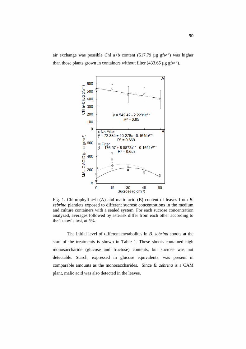
90
air exchange was possible Chl a+b content (517.79 µg gfw-1) was higher
than those plants grown in containers without filter (433.65 µg gfw-1).
Fig. 1. Chlorophyll a+b (A) and malic acid (B) content of leaves from B.
zebrina plantlets exposed to different sucrose concentrations in the medium
and culture containers with a sealed system. For each sucrose concentration
analyzed, averages followed by asterisk differ from each other according to
the Tukey’s test, at 5%.
The initial level of different metabolites in B. zebrina shoots at the
start of the treatments is shown in Table 1. These shoots contained high
monosaccharide (glucose and fructose) contents, but sucrose was not
detectable. Starch, expressed in glucose equivalents, was present in
comparable amounts as the monosaccharides. Since B. zebrina is a CAM
plant, malic acid was also detected in the leaves.

91
Table 1. Carbon metabolite contents (µmol gdw-1) in B. zebrina shoots at the
start of incubation on different sucrose concentrations and different culture
container aeration Samples were taken at 6:00 am. *not detectable.
Metabolites Content (µmol gdw-1)
Malic acid 112 ± 13
Glucose 890 ± 14
Fructose 386 ± 34
Sucrose nd*
Starch 573 ± 223
Among the findings, malic acid content presented a positive
quadratic relationship with increasing sucrose concentrations in both
container sealing systems after 45-d of growth. Plantlets cultivated on sugar-
free or 15 g dm-3 sucrose in the medium and filter containers had higher
malic acid levels than plantlets kept in the same sucrose concentrations and
no filter containers (Fig. 1B).
Glucose and fructose content of the leaves presented positive linear
and negative quadratic models with increasing sucrose concentrations when
they were cultivated in no filter and filter containers, respectively. Plants
grown on medium with 60 g dm-3 sucrose and no aeration of the container
had the highest glucose content (Fig. 2A). Conversely, the fructose content
was the highest in plants cultivated on sugar-free medium and in an aerated
container (Fig. 2B). The monosaccharide contents were in general lower in
roots than in leaves. Glucose content in roots showed a positive linear model
independent of the container sealing system (Fig. 2E). However, fructose
content was higher in plants grown in aerated containers and there was an
increment of the carbohydrate content relationships with increasing sucrose
concentrations (Fig. 2F).

92
Fig. 2. Carbohydrate content in leaves (A-D) and roots (E-H) determined at
the end of the dark period (6:00am) of B. zebrina plants in relation with the
sucrose concentrations in the medium and container sealing system. For each
sucrose concentration analyzed, averages followed by asterisk differ from
each other according to the Tukey’s test, at 5%.
93
Sucrose content was higher in roots than in leaves of plants
cultivated on different sucrose levels in the in vitro media. Sucrose levels for
both plant organs presented linear relationships with increasing sucrose
concentration in the medium (Fig. 2C, G). Starch contents were higher in
leaves than roots at 45-d of in vitro growth. The increment of starch was
positively related with increasing of sucrose concentration in the medium.
The highest starch content was verified in leaves of plants grown in
containers with limited air exchange and a high sucrose content of the
medium. However, the content of starch in roots was rather low for all plants
grown in containers with no aeration (Fig. 2D, H).
The carbohydrate content of all nutrient media was analysed. When
compared the two sealing systems, a higher glucose and fructose content was
verified in the media with prior addition of 15 and 30 g dm-3 sucrose and
filter (Table 2). In containers without gas exchange, supplemented sucrose
was consumed by the plants or converted into glucose or fructose. A slight
increasing of carbohydrate accumulation in the media with previous addition
of 45 and 60 g dm-3 sucrose when the air exchange was observed (Table 2).
All treatments were performed in containers without plants and the
carbohydrate contents verified at 45-d in the same growth conditions. As
shown in the Table 2, there were no differences in the sucrose contents
between container sealing systems, as expected (Table 2).

94
Table 2. Carbohydrate contents in the culture media at 45-days in containers with and without plants. Averages followed by the same
letter in the row for each carbohydrate content, do not differ according to the Tukey’s test, at 5%.
Sucrose (g dm-3)
Carbohydrate contents in the culture media with plants
Glucose (µmol gfw-1) Fructose (µmol gfw-1) Sucrose (µmol gfw-1)
No Filter Filter No Filter Filter No Filter Filter
0 0.00 ± 0.00a 0.00 ± 0.00a 0.00 ± 0.00a 0.00 ± 0.00a 0.00 ± 0.00a 0.00 ± 0.00a
15 0.18 ± 0.07b 0.33 ± 0.09a 0.18 ± 0.07b 0.33 ± 0.10a 0.27 ± 0.12a 0.49 ± 0.20a
30 0.16 ± 0.03b 0.34 ± 0.16a 0.16 ± 0.03b 0.34 ± 0.15a 1.06 ± 0.11a 0.89 ± 0.11a
45 0.23 ± 0.02a 0.23 ± 0.02a 0.22 ± 0.02a 0.23 ± 0.02a 1.68 ± 0.10b 2.38 ± 0.29a
60 0.25 ± 0.07a 0.29 ± 0.05a 0.29 ± 0.16a 0.30 ± 0.05a 2.46 ± 0.08b 3.42 ± 0.62a
Sucrose (g dm-3) Carbohydrate contents in the culture media without plants
Glucose (µmol gfw-1) Fructose (µmol gfw-1) Sucrose (µmol gfw-1)
No Filter Filter No Filter Filter No Filter Filter
0 0.00 ± 0.00a 0.00 ± 0.00a 0.00 ± 0.00a 0.00 ± 0.00a 0.00 ± 0.00a 0.00 ± 0.00a
15 0.01 ± 0.00a 0.02 ± 0.00a 0.01 ± 0.00a 0.02 ± 0.00a 0.83 ± 0.13a 0.79 ± 0.21a
30 0.02 ± 0.01b 0.04 ± 0.01a 0.02 ± 0.01b 0.04 ± 0.01a 1.36 ± 0.13a 1.77 ± 0.30a
45 0.02 ± 0.00b 0.05 ± 0.01a 0.03 ± 0.00b 0.05 ± 0.01a 2.41 ± 0.23a 2.27 ± 0.61a
60 0.06 ± 0.00a 0.07 ± 0.01a 0.06 ± 0.00a 0.07 ± 0.01a 2.78 ± 0.04a 2.95 ± 0.54a

95
Water loss was observed in both sealing sytem, independent of
sucrose concentration added in the medium. However, a higher dehydration
of medium was verified in containers with filter (Fig. 3).
Fig. 3. Water loss at 45 days in containers with plants and without plants due
to sealing system. For each plant culture system in the containers analyzed,
averages followed by different letters differ from each other according to
Tukey’s test, at 5%.
Discussion
B. zebrina plants show different physiologic responses due to sealing
systems and sucrose concentrations. In vitro plantlets may present poor
chlorophyll content, as verified in B. zebrina plants (Fig. 1A). This is
because of the exogenous supply of sucrose, which does not necessitate the
normal development of photosynthetic apparatus. Although such plantlets
may appear normal, their photosynthetic apparatus may be not actively
functional (Hazarika 2006). Reduction of this pigment results in a limitation
of light absorption and therefore provides less ATP and NADPH for the dark
reactions (Sivanesan et al. 2008). In this respect chlorophyll is a good first
and easy to measure indicator of the photosynthetic potential and apparatus
status (Alvarez et al. 2012, Sáez et al. 2012). Less energy input correlated
well to biochemical processes related to both quantity and activity of
ribulose-1,5-bisphosphate carboxylase/oxygenase (Koch 1996). The

96
insufficient supply of ribulose-1,5-biphosphate may also be related to an
increased plant susceptibility to feedback inhibition, which is possibly
associated with an excessive accumulation of hexoses and starch (Le et al.
2001). This agrees with was observed in B. zebrina, presenting low
chlorophyll content (Fig. 1A), but high carbohydrate content (Fig. 2).
B. zebrina plants presented malic acid in all treatments, including
shoots cultivated in liquid medium (Fig. 1B and Table 1). Modulation of the
CAM pathway occurs in the bromeliad Guzmania monostachia, depending
on the level of water supply (Pereira et al. 2013). The decrease of malic acid
content observed in plants cultivated in sucrose concentrations higher than
30 g dm-3 probably was due to osmotic stress created by the in vitro media.
Sucrose concentrations higher than 30 g dm-3 in the medium may induce
osmotic stress on in vitro plants (Cui et al. 2010). Photosynthesis is one of
the most sensitive processes for any kind of stress (Walters 2005). The malic
acid content decreases in the leaves under a long period of water stress, as
proven for in the CAM plant Aechmea ‘Maya’ by Ceusters et al. (2009).
These authors mentioned that possibly roots are involved in the metabolic
response to water limitation. On the other hand, lowest malic acid content
(39.35 µmol gdw-1) in plants grown in sugar-free medium (no osmotic
stress) may be related to insufficient availability of CO2. An increased CO2
concentration increases the rate of malic acid formation by PEPc and its
concentration in the cytoplasm (Zobayed et al. 2000). The increment in
malic acid content in Doritaenopsis occurred with additional CO2 during the
in vitro photoautotrophic culture (Shin et al. 2013). During CAM action,
glycolytic breakdown of storage polysaccharides (e.g., starch) or soluble
sugars (e.g., glucose, fructose and sucrose) previously formed are also
fundamental as substrates for the production of the CO2 acceptor
phosphoenolpyruvate (PEP) in the dark period (Ceusters and Borland 2011).
It supports the results verified in plants grown in sugar-free medium and no
filter containers, since those plants did not have enough carbohydrate stock
to regenerate PEPc enzyme.
97
Sucrose concentrations in the in vitro media created higher
monosaccharide and sucrose stocks in leaves and roots (Fig. 2). Sucrose
supplement in plant tissue culture decrease the water potential of media and
increase glucose, fructose, sucrose and starch contents in leaf tissue in a dose
dependent manner. Monosaccharides are effective osmotic agents in plants.
Osmotic stress in roots may increase monosaccharide accumulation (Xie et
al. 2009). Sugars, especially glucose, fructose and sucrose, play an important
role on stress alleviation through regulation of plant osmotic potential
(Tuteja and Sopory 2008, Polanco et al. 2014). In photoautotrophic
conditions, the plants showed high monosaccharide contents in the leaves
(Fig. 2) and accumulation of different carbohydrates, evidencing good
performance of photosynthetic apparatus. The presence of organic reserves
and a functional photosynthetic apparatus in the in vitro plants should
improve acclimatization performance (Shi et al. 2013, Shi et al. 2014).
Sugars are crucial for building substances for plants as well as a key
source of energy necessary for inciting biochemical processes (Piotrowska et
al. 2010). Plants cultivated in sugar-free medium and no ventilated
containers showed the lowest carbohydrate stocks (Fig. 2) due to limited
photosynthesis rate. Carbohydrates are required by plant cells as carbon
resources, act as energy for growth and biosynthetic processes (Ferreira et al.
2011). Increasing sucrose in the media induced plants with higher starch
content (Fig. 2D, H). Plants cultured in high sucrose concentrations during in
vitro growth may present bigger and higher number of starch granules in the
chloroplasts (Capellades et al. 1991). The main reserves of carbon and
therefore energy are sucrose and starch. When the export rate of sucrose is
lower than the sucrose synthesis rate, an accumulation of hexoses and triose-
phosphates occurs, and as a result, the synthesis of starch begins in the
chloroplasts during the light period (Dennis and Blakely 2000).
The use of containers with unlimited air exchange induced plants
with lower starch content (Fig. 2D). High osmotic stress induced by
dissolved sugars in the media can limit the starch formation. In general,

98
under osmotic and drought stress soluble sugars tend to increase, while
starch content decreases (Chaves 1991). Starch does not play directly as an
osmoprotectant, since it is not soluble in water. B. zebrina plants decreased
the water osmotic potential of roots by increasing the soluble sugars content.
Lower starch content and higher soluble sugars content could facilitated
water flux from the medium into the plants.
At 45 days of growth, B. zebrina plants did not have consumed all
added sucrose in the media, even in those without air exchange (Table 2).
Some plant species have also been presented a low consume in sugar
reserves in the culture medium (Palonen and Junttila 1999). After transfer of
plants to the culture medium, they may hydrolyse sucrose into glucose and
fructose by the activity of invertase released to the culture medium (Karhu
1997). This extracellular enzymatic system associated with the sucrose
hydrolysis is initiated by presence of plant tissues and keeping activated
even without plants, increasing fructose and glucose contents in the medium
(Tremblay and Tremblay 1995). Sucrose hydrolysed to glucose and fructose,
nearly doubling the osmolality of the medium (Bishnoi et al. 2000). In our
study, sucrose hydrolysis increased the osmolality of the medium. This
effect was probably even higher when combined with water loss (Fig. 3)
from the media in containers with filter, increasing the effects by low solvent
content.
Plants under environmental stress, e.g. osmotic stress, may present a
membrane lipid peroxidation, which induces increase of cell membrane
permeability and extravasation of cell-soluble substances (Zeng et al. 2006).
It agrees with our results, since plants grown under ventilated conditions and
in medium with 45 and 60 g dm-3 sucrose showed a slight increment in
sugars into the medium. It was probably due to extravasation of cell content
that includes glucose, fructose and sucrose. Under those conditions the
plants did not growth (data did not show), showing toxic effect of the
osmotic stress, as chlorosis and necrosis.
99
Conclusion
In vitro culture condition influences the physiology of B. zebrina
plantlets during micropropagation time. Sucrose in the culture medium leads
to reduced chlorophyll content and increased starch concentrations.
Supplemented sucrose acts as an osmotic stress factor on B. zebrina.
Ventilating the culture containers increases these stress responses evoking
water loss and increasing sugar content in the media. Plantlets grown in
photoautotrophic conditions (air exchange and sugar-free) did not show any
physiological disorders and showed a normal production of photoassimilates.
References
Alvarez, C., Sáez, P., Sáez, K., Sánchez-Olate, M., Ríos, D.: Effects of light
and ventilation on physiological parameters during in vitro acclimatization
of Gevuina avellana mol. Plant Cell. Tiss. Org. 110: 93-101, 2012.
Bishnoi, U., Jain, R.K., Rohilla, J.S., Chowdhury, V.K., Gupta, K.R.,
Chowdhury, J.B.: Anther culture of recalcitrant indica x Basmati rice
hybrids. Euphytica 114: 93–101, 2000.
Capellades, M., Lemeur, R., Debergh, P.: Effects of sucrose on starch
accumulation and rate of photosynthesis in Rosa cultured in vitro. Plant Cell.
Tiss. Org. 25: 21-26, 1991.
Ceusters, J., Borland, A.M.: Impacts of elevated CO2 on the growth and
physiology of plants with crassulacean acid metabolism In: Lüttge UE,
Beyschlag W, Büdel B, Francis D (eds.) Progress in Botany, v.72. Springer,
Berlin Heidelberg, p163-181, 2011.
Ceusters, J., Borland, A.M., Londers, E., Verdoodt, V., Godts, C., Proft,
M.P.: Differential usage of storage carbohydrates in the CAM bromeliad
Aechmea ‘Maya’ during acclimation to drought and recovery from
dehydration. Physiol. Plantarum 135: 174–184, 2009.
Chaves, M.M.: Effects of water deficits on carbon assimilation. J. Exp. Bot.
42: 1-16, 1991.
Chu, E.P., Tavares, A.R., Kanashiro, S., Giampaoli, P., Yokota, E.S.: Effects
of auxins on soluble carbohydrates, starch and soluble protein content in

100
Aechmea blanchetiana (Bromeliaceae) cultured in vitro. Sci. Hortic. 125:
451–455, 2010.
Cui, X.H., Murthy, H.N., Wu, C.H., Paek, K.Y.: Sucrose-induced osmotic
stress affects biomass, metabolite, and antioxidant levels in root suspension
cultures of Hypericum perforatum L. Plant Cell. Tiss. Org. 103: 7–14, 2010.
Desjardins, Y., Dubuc, J-F., Badr, A.: In vitro culture of plants: a stressful
activity. Acta Hort. 812: 29-50, 2009.
Dennis, D.T., Blakely, S.D.: Carbohydrate metabolism. In: Buchaman B,
Gruissen W, Jones R. (Eds.) Biochemistry and Molecular Biology of Plants,
American Society of Plant Physiology, Rockville p630-675, 2000.
Ferreira, W.M., Suzuki, R.M., Pescador, R., Figueiredo-Ribeiro, R.C.L.,
Kerbauy, G.B.: Propagation, growth, and carbohydrates of Dendrobium
Second Love (Orchidaceae) in vitro as affected by sucrose, light, and dark.
In Vitro Cell. Dev-Pl. 47: 420-427, 2011.
Fuentes, G., Talavera, C., Opereza, C., Desjardins, Y., Santamaria, J.:
Exogenus sucrose can decrease in vitro photosynthesis but improve Field
survival and growth of coconut (Cocos nucifera L.) in vitro plantlets. In
Vitro Cell. Dev-Pl. 41: 69-76, 2005.
Fujiwara, K., Kozai, T.: Physical microenvironment and its effects. In:
Aitken-Christie J, Kozai T, Smith MAL (Eds.) Automation and
environmental control in plant tissue culture. Kluwer Academic Publishers,
Dordrecht, p319-369, 1995.
Hazarika, B.N.: Morpho-physiological disorders in in vitro culture of plants.
Sci. Hortic. 108: 105-120, 2006.
Huang, P.L., Liu, Z.H., Chang, M.L., Liao, L.J.: Micropropagation of the
bromeliad Guzmania ‘Hilda’ via organogenesis and the effect of α-
naphthaleneacetic acid on plantlet elongation. Sci. Hort. 130: 894-898, 2011.
Iarema, L., Cruz, A.C.F., Saldanha, C.W., Dias, L.L.C., Vieira, R.F.,
Oliveira, E.J., Otoni, W.C.: Photoautotrophic propagation of Brazilian
ginseng [Pfaffia glomerata (Spreng.) Pedersen]. Plant Cell. Tiss. Org. 110:
227-238, 2012.
Jo, E.A., Tewari, R.K., Hahn, E.J., Paek, K.Y.: In vitro sucrose
concentration affects growth and acclimatization of Alocasia amazonica
plantlets. Plant Cell. Tiss. Org. 96: 307-315, 2009.
101
Karhu, S.: Sugar use in relation to shoot induction by sorbitol and cytokinin
in apple. J. Am. Soc. Hortic. Sci. 122: 476-480, 1997.
Koch, K.E.: Carbohydrate-modulated gene expression in plants. Annu. Rev.
Plant Phys. 47: 509-540, 1996.
Le, V.Q., Samson, G., Desjardins, Y.: Opposite effects of exogenous sucrose
on growth, photosynthesis and carbon metabolism of in vitro plantlets of
tomato (L. esculentum Mill.) grown under two levels of irradiances and CO2
concentrations. J. Plant Physiol. 158: 599-605, 2001.
Martins, J.P.R., Schimildt, E.R., Alexandre, R.S., Santos, B.R., Magevski,
G.C.: Effect of synthetic auxins on in vitro and ex vitro bromeliad rooting.
Pesqui. Agropec. Trop. 43: 138–146, 2013.
Martins, J.P.R., Schimildt, E.R., Alexandre, R.S., Castro, E.M., Nani, T.F.,
Pires, M.F., Pasqual, M.: Direct organogenesis and leaf-anatomy
modifications in vitro of Neoregelia concentrica (Vellozo) L.B. Smith
(Bromeliaceae). Pakistan J. Bot. 46: 2179-2187, 2014.
Mohamed, M.A., Alsadon, H.A.A.: Influence of ventilation and sucrose on
growth and leaf anatomy of micropropagated potato plantlets. Sci. Hortic.
123: 295-300, 2010.
Murashige, T., Skoog, F.: A revised medium for rapid growth and bioassays
with tobacco tissue cultures. Physiol. Plantarum 15: 473-497, 1962.
Palonen, P., Junttila, O.: Cold hardening of raspberry plants in vitro is
enhanced by increasing sucrose in the culture medium. Physiol. Plantarum
106: 386–392. 1999
Pereira, P.N., Purgatto, E., Mercier, H.: Spatial division of
phosphoenolpyruvate carboxylase and nitrate reductase activity and its
regulation by cytokinins in CAM-induced leaves of Guzmania monostachia
(Bromeliaceae). J. Plant Physiol. 170: 1067-1074, 2013.
Piotrowska, A., Bajguz, A., Zyłkiewicz, B.G., Zambrzycka, E.: Changes in
growth, biochemical components, and antioxidant activity in aquatic plant
Wolffia arrhiza (Lemnaceae) exposed to cadmium and lead. Arch. Environ.
Contam. Toxicol. 58: 594-604, 2010.
Polanco, M.C., Romera, B.S., Aroca, R.: Mild salt stress conditions induce
different responses in root hydraulic conductivity of Phaseolus vulgaris
over-time. PLoS ONE 9: e90631, 2014.
102
Sáez, P.L., Bravo, L.A., Sáez, K.L., Sánchez-Olate, M., Latsague, M.I.,
Ríos, D.G.: Photosynthetic and leaf anatomical characteristics of Castanea
sativa: a comparison between in vitro and nursery plants. Biol. Plantarum
56: 15-24, 2012.
Sivanesan, I., Hwang, S.J., Jeong, B.R.: Influence of plant growth regulators
on axillary shoot multiplication and iron source on growth of Scrophularia
takesimensis Nakai - a rare endemic medicinal plant. Afr. J. Biotechnol. 7:
4484-4490, 2008.
Shin, K.S., Park, S.Y., Paek, K.Y.: Sugar metabolism, photosynthesis, and
growth of in vitro plantlets of Doritaenopsis under controlled
microenvironmental conditions. In Vitro Cell. Dev-Pl. 49: 445-454, 2013.
Shin, K.S., Park, S.Y., Paek, K.Y.: Physiological and biochemical changes
during acclimatization in a Doritaenopsis hybrid cultivated in different
microenvironments in vitro. Environ. Exp. Bot. 100: 26-33, 2014.
Tremblay, L., Tremblay, F.M.: Maturation of black spruce somatic embryos:
Sucrose hydrolysis and resulting osmotic pressure of the medium. Plant Cell.
Tiss. Org. 42: 39-46, 1995.
Tuteja, N., Sopory, S.K.: Chemical signaling under abiotic stress
environment in plants. Plant Signal Behav. 3: 525-536, 2008.
Vesco, L.L.D., Stefenon, V.M., Welter, L.J., Scherer, R.F., Guerra, M.P.:
Induction and scale-up of Billbergia zebrina nodule cluster cultures:
implications for mass propagation, improvement and conservation. Sci. Hort.
28: 515-522, 2011.
Xiao, Y., Niu, G., Kozai, T.: Development and application of
photoautotrophic micropropagation plant system. Plant Cell. Tiss. Org. 105:
149-158, 2011.
Xie, Z.S., Li, B., Forney, C.F., Xu, W.P., Wang, S.P.: Changes in sugar
content and relative enzyme activity in grape berry in response to root
restriction. Sci. Hort. 123: 39-45, 2009.
Walters, R.: Towards an understanding of photosynthetic acclimation. J.
Exp. Bot. 56: 435-447, 2005.
Wellburn, A.R.: The spectral determination of chlorophylls a and b, as well
as total carotenoids, using various solvents with spectrophotometers of
different resolution. J. Plant Physiol. 144: 307-313, 1994.

103
Zeng, Q., Zhu, J.G., Cheng, H.L., Xie, Z.B., Chu, H.Y.: Phytotoxicity of
lanthanum in rice in haplic acrisols and cambisols. Ecotox. Environ. Safe.
64: 226–233, 2006.
Zhang, F., Wang, W., Ge, Y., Shen, X., Tian, D., Liu, J., Liu, X., Yu, X.,
Zhang, Z.: Genetic relatedness among Aechmea species and hybrids inferred
from AFLP markers and pedigree data. Sci. Hort. 139: 39-45, 2012.
Zobayed, S.M.A., Afreen, F., Kubota, C., Kozai, T.: Water control and
survival of Ipomoea batatas grown photoautotrophically under forced
ventilation and photomixotrophically under natural ventilation. Ann. Bot. 86:
60-610, 2000.
104
ARTICLE 4
Anatomical and physiological responses of Billbergia
zebrina (Bromeliaceae) under copper excess
(Prepared in accordance with Environmental and Experimental
Botany's standards)

105
Abstract
Copper (Cu) plays biochemical and physiological functions in plants.
However, at high concentrations, Cu can become extremely toxic, inducing
several physiological changes and symptoms of toxicity such as necrosis.
The aim was to analyze anatomical and physiological changes of Billbergia
zebrina induced by Cu excess in controlled conditions. Previously in vitro-
established B. zebrina plants were transferred to culture media containing
0.0, 2.0, 20.0 or 200.0 µM Cu. Growth (number of leaves and roots) and
enzyme activity (superoxide dismutase and ascorbate peroxidase) analysis
were measured at 20, 40, 60 and 80 days in vitro growth. To verify the
biological activity of roots grown under Cu concentrations, a Biospeckle
laser was used at 20 and 80 days. Biomass accumulation and anatomical
analyses were performed at 80 days. Plants did not show any visible disturb,
as necrosis on the leaves, and all plants survived. However, anatomical and
physiological changes were verified. Plants grown under 200 µM Cu showed
anatomical changes that may help tolerating the metal, like high stomatal
index and thicker cell wall in exodermis. High biological activity was
verified only at 20 days, when the analysed root area was still young. This
activity was linked to high increment in cell wall at exodermis. Enzymatic
antioxidant system was functional in all treatments. Cu affected the leaf and
root anatomy as well as its growth, but this metal induced low changes on
enzyme activity. B. zebrina tolerates high amounts of Cu.
Keywords: biological activity, bromeliad, in vitro culture, plant anatomy,
plant physiology
106
Introduction
Essential metals, such as copper (Cu), play biochemical and
physiological functions in plants and animals. Cu is found naturally in the
environment in low concentrations and it is essential for the metabolism of
many plant species (Nagajyoti et al. 2010). This metal plays an important
role in signaling of transcription and protein trafficking machinery, oxidative
phosphorylation and iron mobilization. Cu is also necessary in others
physiological processes, including photosynthesis, respiration, antioxidant
activity and cell wall lignification metabolism (Himelblau and Amasino
2000, Pilon et al. 2006, Burkhead et al. 2009, Sharma et al. 2012).
At high concentrations, Cu can become extremely toxic causing
symptoms such as chlorosis and necrosis, stunting, leaf discoloration and
inhibition of root growth (Van Assche and Clijsters 1990). Excess Cu may
also induce nutritional deficiencies in plants. It accumulates in plant tissues
and induces many physiological problems and biochemical changes,
reducing growth, especially after chronic exposure (Wannaz et al. 2003,
Khatun et al. 2008).
Cu is a non-degradable heavy metal and it can accumulate in soil or
leach into water sources. Its accumulation in topsoil has impacted micro and
macro organisms (Mackie et al. 2012). The environmental pollution may
lead Cu reach toxic or poisonous concentrations, causing severe damage to
living beings. Incineration of municipal waste has generated significant
concentrations of many kinds of heavy metals, like Cu (Nagajyoti et al.
2010). In agricultural areas, fungicidal spraying has contributed in the Cu
accumulation in enviroment (Mackie et al. 2012). In addition, this heavy
metal may also be associated with dust particles and thus be carried by the
wind (Figueiredo et al. 2007).
The pollution monitoring may be performed by plant species that
assimilate nutrients dispersed in the environment. These plants can be called
bioindicators and they are defined as organisms which are able to provide
information on the quality of their environment (Figueiredo et al. 2007, Elis

107
et al. 2008). This methodology is quite attractive because it has some
advantages over conventional techniques such as low cost, the possibility of
interaction with pollutants over a long period and simultaneously monitoring
of multiple locations. The use of bromeliads as a pollution indicator has been
published (Figueiredo et al. 2007, Alves et al. 2008, Bermudez and Pignata
2011). However, most of these studies are related to the use of Tillandsia
species.
Anatomical changes of plants may indicate the quality of the
environment. Phenotypic plasticity is the key to adaptive process of plants to
environmental conditions, being each species having distinctive
characteristics (Gomes et al. 2012). Studies on anatomical changes of
bromeliads under heavy metal exposure have been focused only on leaves,
especially about changes on trichomes and stomata (Amando-Filho et al.
2002, Alves et al. 2008, Kováčik et al. 2012). Although the trichomes on
leaves are responsible for absorbing water and nutrients, bromeliads may
also absorb nutrients from the roots (Proença and Sajo 2008). Absorbent
tank roots contribute in mineral uptake and they constitute a highly
specialized root system that penetrates the spaces between of the basal parts
of tank-forming leaves (Lüttge 2010). The toxic effect of metals on the root
system is of particular importance for the whole plant. Since the root is an
entry site for water and nutrients into the plant, any defect or malformation
of the root may create problems for plant growth and development (Panou-
Filotheou et al. 2001).
Physiological mechanisms are also involved to tolerate heavy metal
stress. Plants have been stimulated antioxidant system to combat the
oxidative injury induced by the heavy metals (Zabalza et al. 2008). These
include several reactive oxygen species (ROS) removing enzymes such as
superoxide dismutase (SOD), catalases (CAT), ascorbate peroxidase (APX),
among others. ROS are damaging to essential cellular components and it
may lead to oxidative stress affecting plant growth (Giampaoli et al. 2012).

108
Effects of Cu on physiology of Aechmea blanchetiana
(Bromeliaceae) have been studied by Giampaoli et al. (2012). Nevertheless,
this study was realized on in vitro conditions. Physiological changes under
Cu exposure in controlled microenvironment conditions have also been
verified by Khatun et al. (2008) and Son et al. (2014). In vitro technique is
advantageous because it enables to isolate the effects of a certain metal on
plant metabolism from the effects from other stresses (Giampaoli et al.
2012).
The aim of this work was analyse anatomical and physiological
changes induced by Cu in controlled microenviroment conditions of
Billbergia zebrina (Herbert) Lindley, a bromeliad native to Brazil and
considered an endangered species.
Material and Methods
Plant material and culture conditions
Billbergia zebrina fruits were collected from adult plants grown in a
greenhouse and the seeds were used as initial explant. B. zebrina plantlets
previously established in vitro through seeds were transferred to 250.0 mL
glass containers containing 50.0 mL stationary liquid MS medium
(Murashige and Skoog 1962), supplemented with 30.0 g L-1 sucrose and
13.0 µM 6-benzylaminopurine (Martins et al. 2015). After 30 days growth,
side shoots were subcultivated, for 45 days, in 250.0 mL glass containers
with 50.0 mL stationary liquid MS medium with no plant growth regulator
and supplemented with 30.0 g L-1 sucrose. Medium pH was set at 5.8, before
autoclaving at 120ºC for 20 minutes. After inoculation in a laminar flow
cabinet, plant cultures were kept in a growth room, at 27 ± 2ºC and 16-hour
photoperiod under fluorescent lamps providing 25.2 μmol m-2 s-1 of
photosynthetic photon flux.

109
Copper during the in vitro growth
B. zebrina side shoots with approximately 4.0 cm in length, from
previous in vitro stage, were individualized with the aid of a scalpel and
transferred to test tubes containing 10.0 mL MS medium solidified with 6.0
g L-1 agar and supplemented with 0, 2, 20 or 200 µM Cu. Each test tube
received one side shoot. Medium pH was adjusted to 5.8, before autoclaving
at 120ºC, for 20 minutes. After sterile inoculation the cultured plants were
kept in a growth room for until 80 days, at 27 ± 2ºC and 16-hour photoperiod
under fluorescent lamps providing 25.2 μmol m-2 s-1 of photosynthetic
photon flux.
Plant anatomy analysis
Leaf and root anatomy characterization was carried out on three
plants from each treatment. Samples were randomly collected at 80-day
growth and fixed in FAA (formaldehyde, acetic acid and ethanol 50%,
0.5/0.5/9, v/v) for 72 hours, followed by storage in 50% ethanol (Johansen
1940).
Paradermal and cross sections were performed in the median region
of the first completely expanded leaf in the rosette central region with aid of
a double edge razor. Cross sections were also performed at the root basis
(0.5 cm from shoot) with aid of a double edge razor. Sections were cleared
with sodium hypochlorite 10% (v/v) followed by staining with safranin and
astra-blue solution. Finally glycerin 50% was used to assemble the slides.
The sections were investigated using a light microscope (Zeiss
MicroImaging GmbH Scope.A1, Göttingen, Germany) coupled to a digital
camera (Canon A630). The photomicrographs were used to measure the
anatomy characteristics using the software UTHSCSA-Imagetool®.
To determine the lignin distribution in root tissues, the second
staining procedure on root cross-sections was carried out with berberine
hemi-sulphate solution 0.1% (w/v) for 1 hour and aniline blue 0.5% (w/v)
for 30 min. Glycerin 50% (v/v) containing 0.1% (w/v) of FeC13 was used
110
assemble the slides. This entire procedure was performed according to
Brundrett et al. (1988). The sections were observed using a fluorescence
microscope Olympus BX60 equipped with cooled monochrome camera
(Olympus Optical CO., LTD., Tokyo, Japan). Images were captured at
ultraviolet wavelengths with excitation/emission waves of 358-461 nm
(Brundrett et al. 1988).
The stomatal density (cm2) and index (%), thickness of adaxial and
abaxial epidermis (µm), hydrenchyma (µm) and chlorenchyma (µm) and
xylem diameter (µm) were determined in leaves.
The total area of roots (mm2) and central cylinder (mm2), thickness
of cell walls of cortex (µm), endodermis (µm), diameter of xylem vessel
(µm) and number of xylem vessels were determine in roots.
Biological activity of roots
Biological activity of B. zebrina roots (between 0.5 to 1.0 cm from
shoot) was measured at 20 and 80 days of growth with the aid of Biospeckle
laser (or Dynamic Laser Speckle). Four test tubes from each treatment
containing one plant were illuminated by a coherent light provided by a
diode laser (635 nm, pumped by a source offering 66 mA). The experimental
configuration was the back-scattering with a 90o angle between the expanded
laser and the camera (Ribeiro et al. 2014). The camera was a digitalmini-
microscope (Dino-Lite brand AM 413zt). The interference patterns formed
by the interaction of the light with the material studied was collected by the
portable digital microscope, assembling a collection of 128 images in gray
levels, with a resolution of 1,280 x 1,024 pixels and 0.08 s of interval
between frames. The analysis and interpretation of the data were performed
using graphical and numerical analysis, Standard Deviation (Nothdurft and
Yao 2005) and Absolute Value of the Differences (Braga et al. 2011).

111
Enzyme activity analysis
To determine the antioxidant enzyme activities, plants were
collected at 20, 40, 60 and 80 days growth. Each time, nine plants per
treatment were collected and after mixing the plants three parcels were
taken. All samples were immediately frozen in liquid nitrogen. Plant
material was stored in freezer -80 oC until the analysis. The whole plant was
used as plant material. The samples were crushed before taking a known
amount of plant material on which extractions were carried out with a mix of
100 mM potassium phosphate buffer (pH = 7.8), 0.1 mM EDTA and 10 mM
ascorbate acid. The samples were centrifuged at 13,000 g for 10 min at 4 °C
and the supernatants were used for the assay of enzyme activities of
superoxide dismutase (SOD) and ascorbate peroxidase (APX).
SOD activity was assayed as described by Giannopolitis and Ries
(1977) by determining its ability to inhibit the photochemical reduction of
nitroblue tetrazolium (NBT). APX activity was analyzed according to
Nakano and Asada (1981) by monitoring the decrease of ascorbate at 290
nm for 1 min. Enzyme activity was expressed as Unit mg-1 fw (SOD) and
µM H2O2 min-1 mg-1 fw (APX).
Growth trait analysis
To evaluate growth traits, thirty plantlets from each treatment were
randomly collected at 20, 40, 60 and 80-day growth and they were divided
over three parcels. The number of leaves and roots, root length (cm) and
fresh weight were determined.
Statistical analysis
For the analysis of the anatomical structures, a completely
randomized design was adopted. For the analysis of the biological activity of
roots (two time of analysis x four Cu concentrations), enzyme activity and
growth traits (four times of analysis x four Cu concentrations) a completely
randomized design in a factorial arrangement was performed. The obtained

112
data were submitted to analysis of variance (ANOVA), the averages were
compared using Tukey’s test or regression analysis at 5% probability.
Results
Leaf anatomy analysis
In B. zebrina plants, the surface of leaves showed tetracytic stomata
with hypostomatic distribution. Transversal leaf sections presented one cell
layer epidermis both sides of the leaf, water-storage parenchyma
(hydrenchyma) containing non-chlorophyll with thin walled cells at the
adaxial leaf side, chlorophyll parenchyma (chlorenchyma) with isodiametric
cells, collateral vascular bundles, usually larger bundles alternating with
smaller bundles and surrounded by sclerenchyma fibers. The mesophyll has
a dorsiventral organization (Figure 1). This leaf structuring is frequently
observed and described in many bromeliads (Pereira et al. 2013, Martins et
al. 2014).
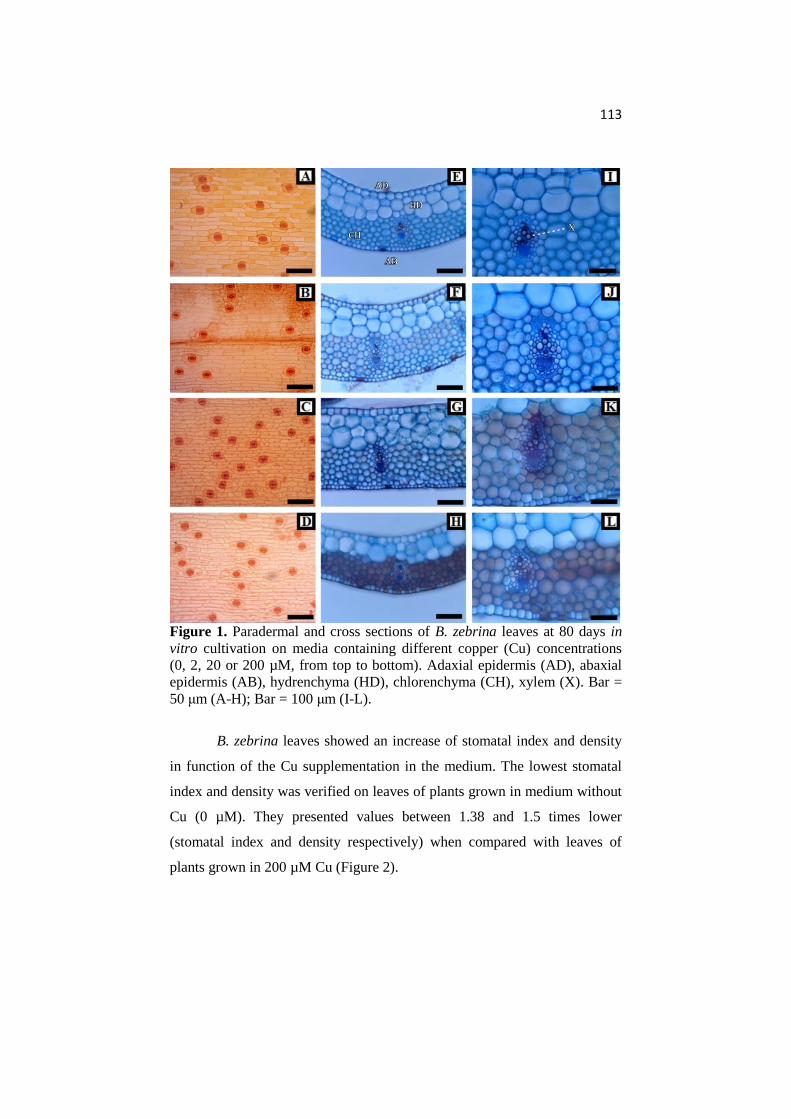
113
Figure 1. Paradermal and cross sections of B. zebrina leaves at 80 days in
vitro cultivation on media containing different copper (Cu) concentrations
(0, 2, 20 or 200 µM, from top to bottom). Adaxial epidermis (AD), abaxial
epidermis (AB), hydrenchyma (HD), chlorenchyma (CH), xylem (X). Bar =
50 μm (A-H); Bar = 100 μm (I-L).
B. zebrina leaves showed an increase of stomatal index and density
in function of the Cu supplementation in the medium. The lowest stomatal
index and density was verified on leaves of plants grown in medium without
Cu (0 µM). They presented values between 1.38 and 1.5 times lower
(stomatal index and density respectively) when compared with leaves of
plants grown in 200 µM Cu (Figure 2).

114
Figure 2. Stomatal index (%) and density (mm2) of B. zebrina leaves as a
function of Cu (µM). For each anatomical trait, averages followed by the
same letter do not differ according to Tukey’s test at 5% probability.
The epidermis thickness did not show any difference throughout all
treatments. The averages of adaxial and abaxial side of the epidermis were
24.67 µm and 15.25 µm respectively (Figure 3). Nevertheless, the other
plant tissues showed significant differences between the Cu concentrations.
Plants grown on media with 2 and 20 µM Cu presented the thickest
hydrenchyma and chlorenchyma as well as the largest xylem diameters. On
the other hand, plants cultured on medium with 200 µM showed the lowest
thickness of those leaf tissues, besides a reduced xylem diameter (Figure 3).
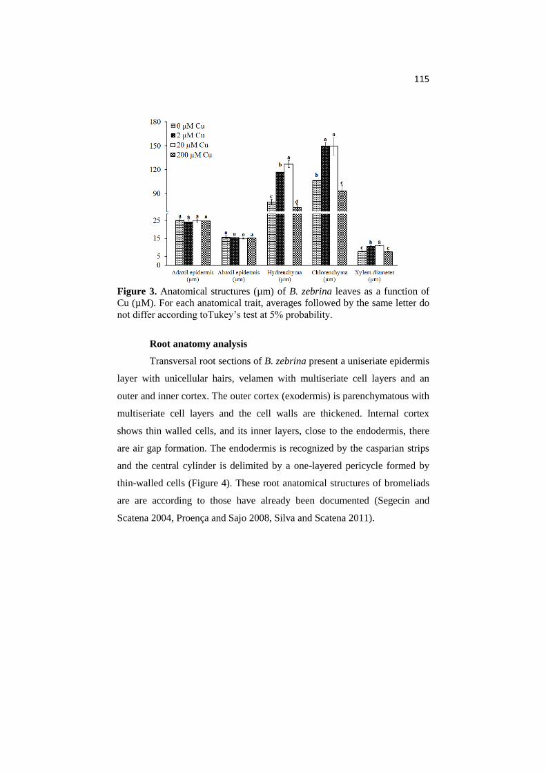
115
Figure 3. Anatomical structures (µm) of B. zebrina leaves as a function of
Cu (µM). For each anatomical trait, averages followed by the same letter do
not differ according toTukey’s test at 5% probability.
Root anatomy analysis
Transversal root sections of B. zebrina present a uniseriate epidermis
layer with unicellular hairs, velamen with multiseriate cell layers and an
outer and inner cortex. The outer cortex (exodermis) is parenchymatous with
multiseriate cell layers and the cell walls are thickened. Internal cortex
shows thin walled cells, and its inner layers, close to the endodermis, there
are air gap formation. The endodermis is recognized by the casparian strips
and the central cylinder is delimited by a one-layered pericycle formed by
thin-walled cells (Figure 4). These root anatomical structures of bromeliads
are are according to those have already been documented (Segecin and
Scatena 2004, Proença and Sajo 2008, Silva and Scatena 2011).
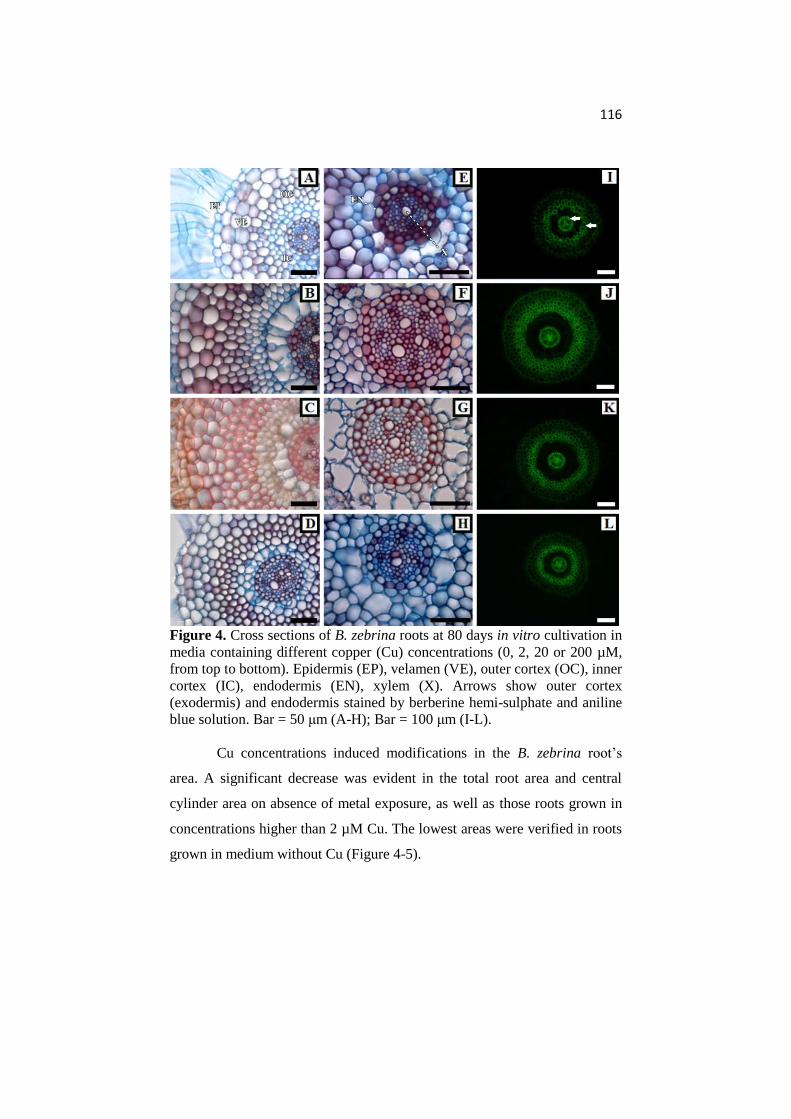
116
Figure 4. Cross sections of B. zebrina roots at 80 days in vitro cultivation in
media containing different copper (Cu) concentrations (0, 2, 20 or 200 µM,
from top to bottom). Epidermis (EP), velamen (VE), outer cortex (OC), inner
cortex (IC), endodermis (EN), xylem (X). Arrows show outer cortex
(exodermis) and endodermis stained by berberine hemi-sulphate and aniline
blue solution. Bar = 50 μm (A-H); Bar = 100 μm (I-L).
Cu concentrations induced modifications in the B. zebrina root’s
area. A significant decrease was evident in the total root area and central
cylinder area on absence of metal exposure, as well as those roots grown in
concentrations higher than 2 µM Cu. The lowest areas were verified in roots
grown in medium without Cu (Figure 4-5).

117
Figure 5. Root total area (mm2) and central cylinder area (mm2) of B.
zebrina roots as a function of Cu (µM). For each anatomical trait, averages
followed by the same letter do not differ according to Tukey’s test at 5%
probability.
The walls of outer cortex cells (exodermis) showed a lignin
thickening in all treatments. The roots exposed to Cu presented a thicker cell
wall. This thickening rose with increasing metal concentrations (Figure 4
and 6). However, endodermis thickness did not show any difference between
the treatments. Xylem diameter was different only in roots grown in medium
without Cu. This was lower than those cultivated with Cu. Nevertheless, the
number of xylem vessels decreased with increasing Cu concentration (Figure
4 and 6).
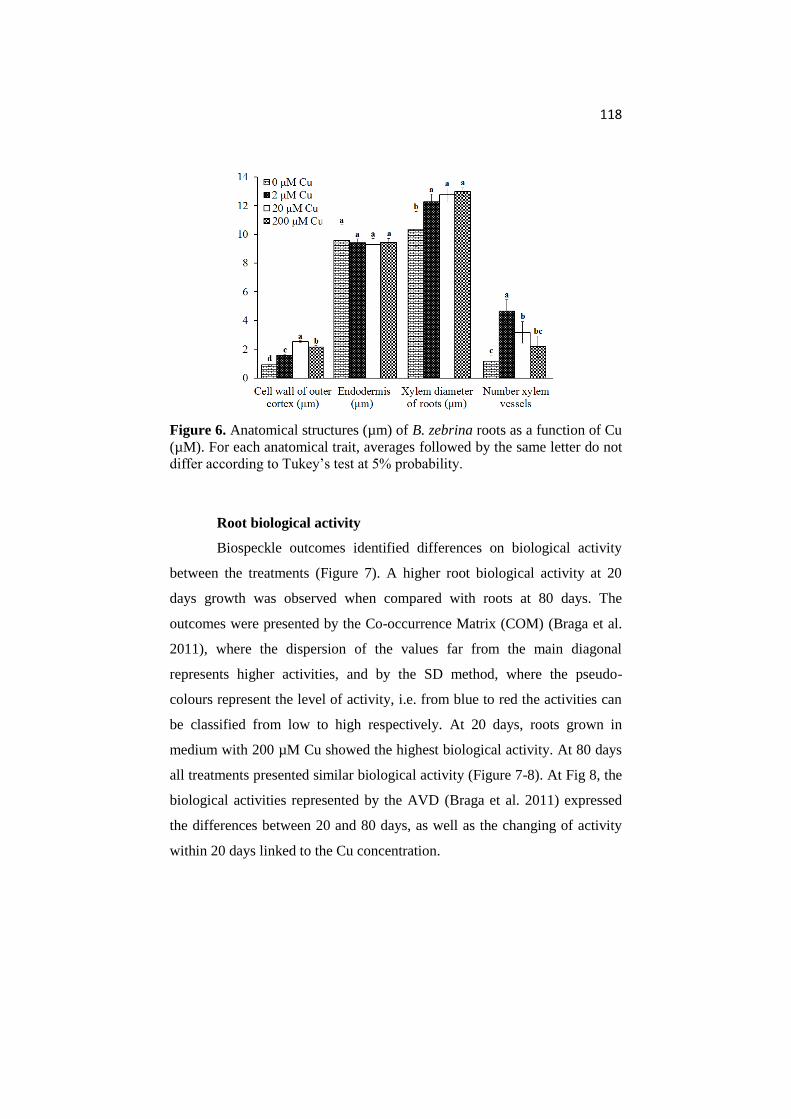
118
Figure 6. Anatomical structures (µm) of B. zebrina roots as a function of Cu
(µM). For each anatomical trait, averages followed by the same letter do not
differ according to Tukey’s test at 5% probability.
Root biological activity
Biospeckle outcomes identified differences on biological activity
between the treatments (Figure 7). A higher root biological activity at 20
days growth was observed when compared with roots at 80 days. The
outcomes were presented by the Co-occurrence Matrix (COM) (Braga et al.
2011), where the dispersion of the values far from the main diagonal
represents higher activities, and by the SD method, where the pseudo-
colours represent the level of activity, i.e. from blue to red the activities can
be classified from low to high respectively. At 20 days, roots grown in
medium with 200 µM Cu showed the highest biological activity. At 80 days
all treatments presented similar biological activity (Figure 7-8). At Fig 8, the
biological activities represented by the AVD (Braga et al. 2011) expressed
the differences between 20 and 80 days, as well as the changing of activity
within 20 days linked to the Cu concentration.

119
Figure 7. Biological activity by COM and SD analysis of B. zebrina roots
as a function of Cu (0, 2, 20 or 200 µM, from top to bottom) at 20 (A-D) and
80 (E-H) days of growth.

120
Figure 8. Biological activity by the AVD of B. zebrina roots as a function of
Cu (µM) at 20 and 80 days of in vitro growth. For each time, averages
followed by the same letter do not differ according to Tukey’s test at 5%
probability. For each Cu concentration, averages followed by asterisk differ
according to Tukey’s test at 5% probability.
Enzyme activity
SOD activity was influenced by concentrations and growth time, but
these factors acted separately. SOD presented the highest activity when the
B. zebrina plants were grown in medium with 200 µM independent of
growth time. Nevertheless, SOD activity increased over time, in general
(Figure 9). APX activity was constant and it did not show any difference
between the treatments (Table 1).

121
Figure 9. SOD activity of B. zebrina plants as a function of Cu (µM) (A)
and time of in vitro growth (B). For each time, averages followed by the
same letter do not differ according to Tukey’s test at 5% probability.
Table 1. APX activity of B. zebrina plants as a function of Cu (µM)
Cu (µM) APX (µM H2O2 min-1 mg-1 fw)
0 1.45 ± 0.69 a
2 1.35 ± 0.35 a
20 1.37 ± 0.45 a
200 1.81 ± 0.73 a
Averages (±SD) followed by the same letter in the column are not differ
according to Tukey’s test at 5% probability.
Growth traits
During in vitro culture the plants did not show any visible
disturbance, like necrosis on the leaves and all plants survived. The number
of leaves was not influenced by Cu concentrations at each time of analysis.
However, the number of leaves presented a positive linear relation with
increasing cultivation time (Figure 10a), showing that Cu concentrations did
not induce a lower growth of aerial part in any treatment. Nevertheless, the
size of leaves presented smaller as a function of Cu. The whole rooting
process occurred after transfer of shoots to the Cu treatments and it was
observed 100% of rooting in all treatments. The adventitious rooting began
before 20 days growth in treatments with Cu. The plants did not present a
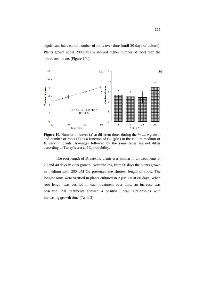
122
significant increase on number of roots over time (until 80 days of culture).
Plants grown under 200 µM Cu showed higher number of roots than the
others treatments (Figure 10b).
Figure 10. Number of leaves (a) at different times during the in vitro growth
and number of roots (b) as a function of Cu (µM) in the culture medium of
B. zebrina plants. Averages followed by the same letter are not differ
according to Tukey’s test at 5% probability.
The root length of B. zebrina plants was similar in all treatments at
20 and 40 days in vitro growth. Nevertheless, from 60 days the plants grown
in medium with 200 µM Cu presented the shortest length of roots. The
longest roots were verified in plants cultured in 2 µM Cu at 80 days. When
root length was verified in each treatment over time, an increase was
observed. All treatments showed a positive linear relationships with
increasing growth time (Table 2).

123
Table 2. Root length (cm) of B. zebrina as a function of Cu (µM) in the
medium and time of in vitro growth
Cu
(µM)
Root length (cm)
20 days 40 days 60 days 80 days
0 2.40 ± 0.19 a(1) 4.05 ± 0.32 a 6.16 ± 2.56 a 8.09 ± 0.72 b
2 2.12 ± 0.22 a(2) 4.66 ± 0.52 a 6.46 ± 0.97 a 10.36 ± 0.21 a
20 2.07 ± 0.12 a(3) 4.09 ± 0.18 a 5.19 ± 0.30 a 8.67 ± 1.05 ab
200 1.56 ± 0.13 a(4) 3.55 ± 0.64 a 2.60 ± 0.56 b 7.38 ± 0.87 b
Averages (±SD) followed by the same letter in the column are not differ
according to Tukey’s test at 5% probability. (1) ŷ = 0.3806 + 0.0959x**, R² =
0.997; (2) ŷ = - 0.735 + 0.1327x**, R² = 0.976; (3) ŷ = - 0.2167 + 0.1045x**,
R² = 0.953; (4) ŷ = - 0.3483 + 0.0825x**, R² = 0.704.
The Cu supplementation influenced on fresh weight of B. zebrina
plants. The lowest values were verified in plants cultured without Cu, but the
addition of Cu higher than 2 µM also showed a negative effect on growth of
B. zebrina (Figure 11).
Figure 11. Fresh weight (mg/plant) as a function of Cu (µM) in the culture
medium of B. zebrina plants. Averages followed by the same letter do not
differ according to Tukey’s test at 5% probability.

124
Discussion
This research reports for the first time the impact of Cu excess on
leaf and root anatomy as well as on physiology of a bromeliad. B. zebrina
plants showed different anatomical and physiological responses due to Cu
concentrations. However, under imposed conditions, plants did not present
mortality rate or any visible physiological disturb, like chlorosis, necrosis,
and leaf discoloration.
On leaf surface, the stomatal density is one of the most important
factors in plants under stress. Increasing stomatal density is an often
response to decreasing leaf surface area to ensure surface gas exchange and,
consequently, adequate carbon influx (Abbruzzese et al. 2009). An increased
stomatal density may represent anatomical leaf tolerance mechanisms for
controlling transpiration processes (Gomes et al. 2012). This increasing in
stomata number may not be due to an increased stomatal formation but
rather due to a reduction in epidermal cell size caused by water deficit
caused by heavy metals, like Cu (Wainwright and Woolhouse 1977).
However, from the stomatal index it was possible to verify a higher
investment in stomatal formation per area with an increment in Cu
concentration (Figure 1-2). An increased stomatal index with raised heavy
metal concentration has also been verified in other plant species, like Vigna
radiata (Gupta and Bhatnagar 2015). The increase in stomatal frequency is
supposed to be an adaptive response to sustain adequate rate of gaseous
exchange necessary for growth and development (Neelu et al. 2000).
Leaf epidermis thickness of B. zebrina did not change as a function
of Cu concentration (Figure 1, 3). It has also been verified in Myriophyllum
alterniflorum plants by Delmail et al. (2011). However, the chlorenchyma
125
and hydrenchyma thickness were related to Cu concentration (Figure 1, 3).
Chlorenchyma thickness presents a great plasticity and this tissue is sensitive
to heavy metals (Shi and Cai 2009). Exposure to heavy metals may lead to a
reduction in the mesophyll cell size (Zhao et al. 2000, Srighar et al. 2005).
Nevertheless, in our study, even though we did not measure cell size, the
reduction of the leaf blade seems to be due to lower number of hydrenchyma
and chlorenchyma cell layers in the treatments exposed to 200 µM Cu
(Figure 1, 3). Cu excess can inhibit cell elongation and cell division of plant
tissues (Panou-Filotheou and Bosabalidis 2004).
A reduction of xylem diameter was observed on leaves of B. zebrina
grown in medium with 200 µM Cu (Figure 1, 3). This reduction is one of the
factors affecting the capacity as translocation conduits (Poschenrieder and
Barceló 1999). A larger xylem vessel is related to a higher hydraulic
conductance (Rodríguez-Gamir et al. 2010). Thus, a smaller diameter of
xylem vessels may be related as a protection factor, since it is expected a
lower translocation of Cu through the plant, reducing the toxic effects of Cu
excess in the aerial part.
Cu concentration also presented an impact on anatomy at analyzed
root area (Figure 4-6). In young roots, four areas are traditionally recognized
in root cap, zone of active cell division, zone of cell elongation, and zone of
maturation. However, roots grown under heavy metal conditions may change
the position of cell division/maturation of some tissues from the root apex
(Lux et al. 2011, Vaculík et al. 2012). For this reason, in our study, we
focused the anatomical analysis in the basis of the roots. An excess of Cu
may increase abnormalities at root tip mitosis (Li and Xiong, 2004). Cu
affects the cell integrity in the transition zone, region where cells coming out
126
of the meristem prepare for fast expansion (Madejón et al. 2009). Thus,
abnormalities on rooting process may interfere on formation of root tissues
and this change might be clear in mature root areas.
The root cortex of bromeliads is differentiated outer and inner
cortex. The outer cortex is a multilayered tissue formed by sclerified cells
probably helps to hinder the water evaporation from the inner root cortex
(Proença and Sajo 2008). The inner cortex has thin walls that may collapse
at maturity and form air gaps, as verified in B. zebrina’s roots (Figure 4).
The formed intercellular spaces are related to transport water by capillary
action through these spaces (Segecin and Scatena 2004).
Outer cortex is also called exodermis (Silva and Scatena 2011) and
its lignification of cell walls is natural processes, as verified in all treatments
by berberine hemi-sulphate and aniline blue solution (Figure 4I-L).
However, the cell wall thickness was influenced by Cu concentration (Figure
4, 6). Cu contributes to cell wall metabolism among several physiological
processes in plants (Kabata-Pendias 2010). The functional adaptation of the
exodermis has been related to an external sealing tissue of the root that is in
direct contact with the surrounding environment and so serves as the first
guard preventing excessive biotic and abiotic toxins entering the root, like
heavy metals (Deng et al. 2009, Cheng et al. 2010, Cheng et al. 2012).
Exodermis tend to decrease the capacity for metal uptake by roots,
coinciding with increased lignification (Cheng et al. 2012).
The endodermis also acts as a barrier for diffusion of metals into the
vascular system (Eapen and D’Souza 2005). In our study thickness of this
tissue did not change as a function of Cu concentration (Figure 4, 6). A
thicker endodermis may not act as an improved apoplastic barrier against

127
heavy metals (Geldner 2013). Moreover, an increased thickness of the outer
cortex could have acted as a first apoplastic barrier, thereby reducing the
metal toxicity. The evidence that thicker cell walls are, in fact, effective
barriers to apoplastic ion movement is widespread and convincing (Enstone
et al. 2003).
A decrease in the size and/or number of xylem vessels in roots is a
common symptom of heavy metal toxicity that strongly diminishes the
uptake and transport of water by the plants (Gong et al. 2005). It agrees with
what we observed in B. zebrina, which presented lower number of xylem
vessels under Cu excess (Figure 4, 6). The root’s diameter of vessels of
xylem did not decrease under high Cu concentration and it, combined with
low number of vessels, could be a risk to the plant. Since any damage in a
vessel would interfere negatively on water and nutrients transport. However,
these plants have invested in a higher number of roots (Figure 10b), which
could potentially compensate a loss (due to eventual damage in a vessel) of
solute transport and explore a larger medium area.
Root growth involves cell division and enzyme activity and these
activities are differenced along the root (Schreibera et al. 2005). During the
root growth of B. zebrina plants, the biological activity showed differences
between the treatments at 20 days of growth (Figure 7-8). This may be
related to cell wall formation in exodermis (Figure 4, 6) as a reaction to the
metal concentration. Highest rates of biosynthesis of substances linked to
thickening of the cell walls are located in younger root zones (Schreibera et
al. 2005). At 20 days the root areas analysed by Biospeckle were probably
still immature, since roots were still short (Table 2). Those roots were longer
at 80 days and the analyzed root areas were probably mature. The similar

128
biological activity at 80 days may also be an indication that the anatomical
changes, i.g. thicker cell walls at exodermis, ensured the establishment of
plants at non-favorable conditions. Cheng et al. (2012) have also verified
changes in root anatomy as a strategy to avoid Cu entry into the plant.
Plant establishment under heavy metal conditions involves
physiological changes, e.g. on enzymatic and non-enzymatic system activity.
Several published works have discussed the involvement of SOD and APX
against reactive oxygen species (ROS) in plants under heavy metal stress
(Khatun et al. 2008, Madejón et al. 2009, Bermudez and Pignata 2011,
Giampaoli et al. 2012). The detoxification of excess ROS is achieved by
action of antioxidant enzymes. However, ROS are produced as an
unavoidable consequence of plant cellular metabolism (Sharma et al. 2012).
SOD is the first line of defence against ROS. SOD is a metalloenzyme that
dismutates superoxide radical to H2O2 and oxygen. Then, APX detoxify
H2O2 using ascorbate for reduction (Thounaojam et al. 2012). Increase in
SOD activities in stressed plants was indicative of enhanced O2- production
and oxidative stress tolerance. It has been proposed as an important enzyme
for plant stress tolerance (Sharma et al. 2012, Thounaojam et al. 2012). APX
is regarded as one of the most widely distributed antioxidant enzymes in
plant cells, and it has high affinity to H2O2, making APX efficient
scavengers of H2O2 under stressful conditions (Wang et al. 1999). In our
study, a suitable increase in SOD activity was detected in plants grown under
high Cu concentration (Figure 9). However, the increased H2O2 production
probably was not high enough to induce a higher increment in APX activity
in B. zebrina (Table 1). Antioxidant system probably was in good condition
and functional, since both a high concentration of ROS and low enzyme
129
activity of SOD and APX might induce serious physiological disturbs that
could be visualized as necrosis or chlorosis.
B. zebrina plants did not stop growing under any Cu treatment,
showing that those changes were effective in the establishment of B. zebrina
under imposed conditions (Figure 10 and Table 2). Nevertheless, plants
grown without Cu showed lower biomass accumulation (Figure 11). Cu is an
essential micronutrient and plays important roles in metal homeostasis and
normal metabolism in plants. It is involved in a wide range of biological
processes (Zhang et al. 2008). In our study, it was possible verify that some
biological processes related to Cu are also involved in leaf and root tissue
formation (Figure 1-6). However, Cu was toxic to plants at higher
concentrations. It may cause changes in nitrogen metabolism with a
reduction of total nitrogen (Llorens et al. 2000). A common effect of heavy
metals is a reduced growth (Mishra et al. 2008). This decline in growth,
especially on biomass production, can be explain, in part, due to inhibition of
cell division by copper dependent concentration. It agrees with the verified
on leaf and root cross-sections (Figure 3-8).
Conclusion
Cu affected the leaf and root anatomy of B. zebrina as well as its
growth, but this metal induced low changes in antioxidant enzyme activity.
B. zebrina tolerates high amounts of Cu and this species may be indicated
for biomonitoring use.

130
References
Abbruzzese G, Beritognolo I, Muleo R, Piazzai M, Sabatti M, Mugnozza
GS, Kuzminsky E (2009) Leaf morphological plasticity and stomatal
conductance in three Populus alba L. genotypes subjected to salt stress
Environmental and Experimental Botany 66:381–388
Alves ES, Moura BB, Domingos M (2008) Structural analysis of Tillandsia
usneoides L. exposed to air pollutants in São Paulo city–Brazil. Water Air
Soil Pollut 189:61–68
Amado-Filho GM, Andrade LR, Farina M, Malm O (2002) Hg localisation
in Tillandsia usneoides L. (Bromeliaceae), an atmospheric biomonitor.
Atmospheric Environment 36:881–887
Bermudez GMA, Pignata ML (2011) Antioxidant response of three
Tillandsia species transplanted to urban, agricultural, and industrial areas.
Archives of Environmental Contamination and Toxicology 61:401-413
Braga RA, Nobre CMB, Costa AG, Sáfadi T, Costa FM (2011) Evaluation
of activity through dynamic laser speckle using the absolute value of the
differences. Optics Communications 284:646-650
Brundrett MC, Enstone DE, Peterson CA (1988) A berberine-aniline blue
fluorescent staining procedure for suberin, lignin, and callose in plant tissue.
Protoplasma 146:133-142
Burkhead JL, Reynolds KAG, Abdel-Ghany SE, Cohu CM, Pilon M (2009)
Copper homeostasis. New Phytologist 182:799–816
Cheng H, Liu Y, Tam NFY, Wang X, Li SY, Chen GZ, Ye ZH (2010) The
role of radial oxygen loss and root anatomy on zinc uptake and tolerance in
mangrove seedlings. Environmental Pollution 158:1189–1196
Cheng H, Tam NFY, Wang Y, Li S, Chen G, Ye Z (2012) Effects of copper
on growth, radial oxygen loss and root permeability of seedlings of the
mangroves Bruguiera gymnorrhiza and Rhizophora stylosa. Plant Soil
359:255–266

131
Delmail D, Labrousse P, Hourdin P, Larcher L, Moesch C, Botineau M
(2011) Differential responses of Myriophyllum alterniflorum DC
(Haloragaceae) organs to copper: physiological and developmental
approaches. Hydrobiologia 664:95–105
Deng H, Ye ZH, Wong MH (2009) Lead, zinc and iron (Fe2+) tolerance in
wetland plants and relation to root anatomy and spatial pattern of ROL.
Environmental and Experimental Botany 65:353–362
Eapen S, D'Souza SF (2005) Prospects of genetic engineering of plants for
phytoremediation of toxic metals. Biotechnology Advances 23:97–114
Elias C, Fernandes EAN, França EJ, Bacchi MA, Tagliaferro FS (2008)
Native bromeliads as biomonitors of airborne chemical elements in a
Brazilian Restinga forest. Journal of Radioanalytical and Nuclear Chemistry
278:423-427
Enstone DE, Peterson CA, Ma F (2003) Root Endodermis and exodermis:
structure, function, and responses to the environment. Journal of Plant
Growth Regulation 21:335–351
Figueiredo AMG, Nogueira CA, Saiki M, Milian FM, Domingos M (2007)
Assessment of atmospheric metallic pollution in the metropolitan region of
São Paulo, Brazil, employing Tillandsia usneoides L. as biomonitor.
Environmental Pollution 145:279–292
Giampaoli P, Tresmondi F, Lima GPP, Kanashiro S, Alves ES, Domingos E,
Tavares AR (2012) Analysis of tolerance to copper and zinc in Aechmea
blanchetiana grown in vitro. Biologia Plantarum 56:83-88
Geldner N (2013) The Endodermis. Annual Review of Plant Biology
64:531–58
Giannopolitis CN, Ries SK (1977) Superoxide dismutases. Plant Physiology
59:309-314
Gomes MP, Sa TCLL, Marques M, Martins GA, Carneiro MMLC, Soares
AM (2012) Cd-tolerance markers of Pfaffia glomerata (Spreng.) Pedersen
132
plants: anatomical and physiological features. Brazilian Journal of Plant
Physiology 24:293-304
Gong HJ, Zhu XY, Chen KM, Wang S, Zhang C (2005). Silicon alleviates
oxidative damage of wheat plants in pots under drought. Plant Science
169:313–321.
Gupta P, Bhatnaga AK (2015) Spatial distribution of arsenic in different leaf
tissues and its effect on structure and development of stomata and trichomes
in mung bean, Vigna radiata (L.) Wilczek. Environmental and Experimental
Botany 109:12–22
Himelblau E, Amasino RM (2000) Delivering copper within plant cells.
Current Opinion in Plant Biology 3:205–210
Johansen DA (1940) Plant microtechnique. Mc Graw-Hill (2ª Ed.), New
York, p523
Kabata-Pendias A (2010) Trace Elements in Soils and Plants, (4thEd) CRC
Press, London, p505
Khatun S, Ali MB, Hahn EJ, Paek KY (2008) Copper toxicity in Withania
somnifera: Growth and antioxidant enzymes responses of in vitro grown
plants. Environmental and Experimental Botany 64:279–285
Kováčik J, Klejdus B, Štork F, Hedbavny J (2012) Physiological responses
of Tillandsia albida (Bromeliaceae) to long-term foliar metal application.
Journal of Hazardous Materials 239–240:175–182
Li T, Xiong ZT (2004) A novel response of wild type duckweed (Lemna
paucicostata Hegelm.) to heavy metals. Environmental Toxicology 19:95–
102
Llorens N, Arola L, Blade C, Mas A (2000) Effects of copper exposure upon
nitrogen metabolism in tissue cultured Vitis vinifera Plant Science 160:159–
163

133
Lüttge U (2010) Ability of crassulacean acid metabolism plants to overcome
interacting stresses in tropical environments. AoB Plants 10: plq005
Lux A, Martinka M, Vaculík M, White PJ (2011) Root responses to
cadmium in the rhizosphere: a review. Journal of Experimental Botany
62:21–37
Mackie KA, Müller T, Kandeler E (2012) Remediation of copper in
vineyards – A mini review. Environmental Pollution 167:16–26
Madejón P, Ramírez-Benítez JE, Corrales I, Barceló J, Poschenrieder C
(2009) Copper-induced oxidative damage and enhanced antioxidant defenses
in the root apex of maize cultivars differing in Cu tolerance. Environmental
and Experimental Botany 67:415–420
Martins JPR, Pasqual M, Martins AD, Ribeira SF (2015) Effects of salts and
sucrose concentrations on in vitro propagation of Billbergia zebrina
(Herbert) Lindley (Bromeliaceae). Australian Journal of Crop Science 9:85-
91
Martins JPR, Schimildt ER, Alexandre RS, Castro EM, Nani TF, Pires MF,
Pasqual M (2014) Direct organogenesis and leaf-anatomy modifications in
vitro of Neoregelia concentrica (Vellozo) L.B. Smith (Bromeliaceae).
Pakistan Journal of Botany 46:2179-2187
Mishra VK, Upadhyaya AR, Pandey SK, Tripathi BD (2008) Heavy metal
pollution induced due to coal mining effluent on surrounding aquatic
ecosystem and its management through naturally occurring aquatic
macrophytes. Bioresource Technology 99:930–936
Murashige, T., Skoog, F., 1962. A revised medium for rapid growth and
bioassays with tobacco tissue cultures. Physiol Plantarum 15:473-497.
Nagajyoti PC, Lee KD, Sreekanth TVM (2010) Heavy metals, occurrence
and toxicity for plants: a review. Environmental Chemistry Letters 8:99–216
134
Nakano Y, Asada K (1981) Hydrogen peroxide is scavenged by ascorbate-
specific peroxidase in spinach chloroplasts. Plant & Cell Physiology 22:867-
880
Neelu N, Kumar M, Tomar M, Bhatnagar AK (2000) Influence of cadmium
on growth and development of Vicia faba Linn. Indian Journal of
Experimental Biology 38:819–823
Nothdurft R, Yao G (2005). Imaging obscured subsurface inhomogeneity
using laser speckle. Optics Express 13:10034-10039
Panou-Filotheou H, Bosabalidis AM (2004) Root structural aspects
associated with copper toxicity in oregano (Origanum vulgare subsp.
hirtum). Plant Science 166:1497–1504
Panou-Filotheou H, Bosabalidis AM, Karataglis S (2001) Effects of copper
toxicity on leaves of oregano (Origanum vulgare subsp. hirtum) Annals of
Botany 88:207-214
Pereira TAR, Silva LC, Azevedo AA, Francino DMT, Coser TS, Pereira JD
(2013) Leaf morpho-anatomical variations in Billbergia elegans and
Neoregelia mucugensis (Bromeliaceae) exposed to low and high solar
radiation. Botany 91:327-334
Pilon M, Abdel-Ghany SE, Cohu CM, Gogolin KA, Ye H (2006) Copper
cofactor delivery in plant cells. Current Opinion in Plant Biology 9: 256–263
Poschenrieder C, Barceló J (1999) Water relations in heavy metal stressed
plants, In: Prasad, M.N.V. and J. Hagemeyer (eds.), Heavy Metal Stress in
Plants, Springer, Berlin, p207–29
Proença SL, Sajo MG (2008) Rhizome and root anatomy of 14 species of
Bromeliaceae. Rodriguésia 59:113-128
Ribeiro KM, Braga Jr RA, Horgan GW, Ferreira DD, Sáfadi T (2014)
Principal component analysis in the spectral analysis of the dynamic laser
speckle patterns. Journal of the European Optical Society. Rapid
Publications 9:14009

135
Rodríguez-Gamir J, Intrigliolo DS, Primo-Millo E, Forner-Giner MA (2010)
Relationships between xylem anatomy, root hydraulic conductivity, leaf/root
ratio and transpiration in citrus trees on different rootstocks. Physiologia
Plantarum 139:159–169.
Schreibera L, Frankea R, Lessire R (2005) Biochemical characterization of
elongase activity in corn (Zea mays L.) roots. Phytochemistry 66:131–138
Segecin S, Scatena VL (2004) Morfoanatomia de rizomas e raízes de
Tillandsia L. (Bromeliaceae) dos Campos Gerais, PR, Brasil. Acta Botanica
Brasilica 18:253-260
Sharma P, Jha AB, Dubey RS, Pessarakli M (2012) Reactive oxygen species,
oxidative damage, and antioxidative defence mechanism in plants under
stressful conditions. 2012:1:26
Silva IV, Scatena VL (2011) Anatomia de raízes de nove espécies de
Bromeliaceae (Poales) da região amazônica do estado de Mato Grosso,
Brasil. Acta Botanica Brasilica 25(3):618-627
Shi G, Cai Q (2009) Leaf plasticity in peanut (Arachis hypogaea L.) in
response to heavy metal stress. Environmental and Experimental Botany
67:112–117
Son JA, Narayanankutty DP, Roh KS (2014) Influence of exogenous
application of glutathione on rubisco and rubisco activase in heavy metal-
stressed tobacco plant grown in vitro. Saudi Journal of Biological Sciences
21:89–97
Srighar BBM, Diehl SV, Han FX, Monts DL, Su Y (2005) Anatomical
changes due to uptake and accumulation of Zn and Cd in Indian mustard
(Brassica juncea). Environmental and Experimental Botany 54: 131-141.
Thounaojam TC, Panda P, Mazumdar P, Kumar D, Sharma GD, Sahoo L,
Sanji P (2012) Excess copper induced oxidative stress and response of
antioxidants in rice. Plant Physiology and Biochemistry 53:33–39

136
Vaculík M, Konlechner C, Langer I, Adlassnig W, Puschenreiter M, Lux A,
Hauser MT (2012) Root anatomy and element distribution vary between two
Salix caprea isolates with different Cd accumulation capacities.
Environmental Pollution 163:117–126
Van Assche F, Clijsters H (1990) Effects of metals on enzyme activity in
plants. Plant, Cell & Environment 13:195–206
Wainwright SJ, Woolhouse HW (1977) Some physiological aspects of
copper and zinc tolerance in Agrostis tenuis sibth: cell elongation and
membrane damage. Journal of Experimental Botany 28:1029–1036
Wang J, Zhang H, Allen RD (1999) Overexpression of an Arabidopsis
peroxisomal ascorbate peroxidase gene in tobacco increases protection
against oxidative stress. Plant
and Cell Physiology 40:725–732
Wannaz LED, Zygadlo JA, Pignata ML (2003) Air pollutants effect on
monoterpenes composition and foliar chemical parameters in Schinus areira.
Science of the Total Environment 305:177-193
Zabalza A, Galvez L, Marino D, Royuela M, Arrese-Igor C, Gonzalez EM
(2008) The application of ascorbate or its immediate precursor, galactono-1,
4-lactone, does not affect the response of nitrogen-fixing pea nodules to
water stress. Journal of Plant Physiology 165:805–812
Zhao FJ, Lombi E, Breedon T, McGrath SP (2000) Zinc hyperaccumulation
and cellular distribution in Arabidopsis halleri. Plant Cell and
Environmental 23: 507-514.
Zhang LP, Mehta SK, Liu ZP, Yang ZM (2008) Copper-induced proline
synthesis is associated with nitric oxide generation in Chlamydomonas
reinhardtii. Plant Cell Physiology 49:411-419.