Physiological responses to low-force work and psychosocial stress in women with chronic trapezius...
16
BioMed Central Page 1 of 16 (page number not for citation purposes) BMC Musculoskeletal Disorders Open Access Research article Physiological responses to low-force work and psychosocial stress in women with chronic trapezius myalgia Anna Sjörs* 1 , Britt Larsson 1,2 , Joakim Dahlman 1 , Torbjörn Falkmer 1,3,4 and Björn Gerdle 1,2 Address: 1 Rehabilitation Medicine, Faculty of Health Sciences, Linköping University, SE 581 85 Linköping, Sweden, 2 Pain and Rehabilitation Centre, University Hospital, SE 581 85 Linköping, Sweden, 3 Department of Rehabilitation, School of Health Sciences, Jönköping University, Box 1026, SE 551 11 Jönköping, Sweden and 4 School of Occupational Therapy and Social Work, Curtin University of Technology, GPO Box U1987, Perth, WA 6845, Australia Email: Anna Sjörs* - [email protected]; Britt Larsson - [email protected]; Joakim Dahlman - [email protected]; Torbjörn Falkmer - [email protected]; Björn Gerdle - [email protected] * Corresponding author Abstract Background: Repetitive and stressful work tasks have been linked to the development of pain in the trapezius muscle, although the underlying mechanisms still remain unclear. In earlier studies, it has been hypothesized that chronic muscle pain conditions are associated with imbalance in the autonomic nervous system, predominantly expressed as an increased sympathetic activity. This study investigates whether women with chronic trapezius myalgia show higher muscle activity and increased sympathetic tone at baseline and during repetitive low-force work and psychosocial stress, compared with pain-free controls. Methods: Eighteen women with chronic trapezius myalgia (MYA) and 30 healthy female controls (CON) were studied during baseline rest, 100 min of repetitive low-force work, 20 min of psychosocial stress (Trier Social Stress Test, TSST), and 80 min recovery. The subjects rated their pain intensity, stress and energy level every 20 min throughout the experiment. Muscle activity was measured by surface electromyography in the trapezius muscle (EMGtrap) and deltoid muscle (EMGdelt). Autonomic reactivity was measured through heart rate (HR), skin conductance (SCL), blood pressure (MAP) and respiration rate (Resp). Results: At baseline, EMGtrap, stress ratings, and HR were higher in MYA than in CON. Energy ratings, EMGdelt, SCL, MAP and Resp were, however, similar in the two groups. Significant main group effects were found for pain intensity, stress ratings and EMGtrap. Deltoid muscle activity and autonomic responses were almost identical in MYA and CON during work, stress and recovery. In MYA only, pain intensity and stress ratings increased towards the end of the repetitive work. Conclusion: We found increased muscle activity during uninstructed rest in the painful muscle of a group of women with trapezius myalgia. The present study could not confirm the hypothesis that chronic trapezius myalgia is associated with increased sympathetic activity. The suggestion of autonomic imbalance in patients with chronic local or regional musculoskeletal pain needs to be further investigated. Published: 7 June 2009 BMC Musculoskeletal Disorders 2009, 10:63 doi:10.1186/1471-2474-10-63 Received: 4 February 2009 Accepted: 7 June 2009 This article is available from: http://www.biomedcentral.com/1471-2474/10/63 © 2009 Sjörs et al; licensee BioMed Central Ltd. This is an Open Access article distributed under the terms of the Creative Commons Attribution License (http://creativecommons.org/licenses/by/2.0 ), which permits unrestricted use, distribution, and reproduction in any medium, provided the original work is properly cited.
-
Upload
independent -
Category
Documents
-
view
5 -
download
0
Transcript of Physiological responses to low-force work and psychosocial stress in women with chronic trapezius...

BioMed CentralBMC Musculoskeletal Disorders
ss
Open AcceResearch articlePhysiological responses to low-force work and psychosocial stress in women with chronic trapezius myalgiaAnna Sjörs*1, Britt Larsson1,2, Joakim Dahlman1, Torbjörn Falkmer1,3,4 and Björn Gerdle1,2Address: 1Rehabilitation Medicine, Faculty of Health Sciences, Linköping University, SE 581 85 Linköping, Sweden, 2Pain and Rehabilitation Centre, University Hospital, SE 581 85 Linköping, Sweden, 3Department of Rehabilitation, School of Health Sciences, Jönköping University, Box 1026, SE 551 11 Jönköping, Sweden and 4School of Occupational Therapy and Social Work, Curtin University of Technology, GPO Box U1987, Perth, WA 6845, Australia
Email: Anna Sjörs* - [email protected]; Britt Larsson - [email protected]; Joakim Dahlman - [email protected]; Torbjörn Falkmer - [email protected]; Björn Gerdle - [email protected]
* Corresponding author
AbstractBackground: Repetitive and stressful work tasks have been linked to the development of pain inthe trapezius muscle, although the underlying mechanisms still remain unclear. In earlier studies, ithas been hypothesized that chronic muscle pain conditions are associated with imbalance in theautonomic nervous system, predominantly expressed as an increased sympathetic activity. Thisstudy investigates whether women with chronic trapezius myalgia show higher muscle activity andincreased sympathetic tone at baseline and during repetitive low-force work and psychosocialstress, compared with pain-free controls.
Methods: Eighteen women with chronic trapezius myalgia (MYA) and 30 healthy female controls(CON) were studied during baseline rest, 100 min of repetitive low-force work, 20 min ofpsychosocial stress (Trier Social Stress Test, TSST), and 80 min recovery. The subjects rated theirpain intensity, stress and energy level every 20 min throughout the experiment. Muscle activity wasmeasured by surface electromyography in the trapezius muscle (EMGtrap) and deltoid muscle(EMGdelt). Autonomic reactivity was measured through heart rate (HR), skin conductance (SCL),blood pressure (MAP) and respiration rate (Resp).
Results: At baseline, EMGtrap, stress ratings, and HR were higher in MYA than in CON. Energyratings, EMGdelt, SCL, MAP and Resp were, however, similar in the two groups. Significant maingroup effects were found for pain intensity, stress ratings and EMGtrap. Deltoid muscle activity andautonomic responses were almost identical in MYA and CON during work, stress and recovery.In MYA only, pain intensity and stress ratings increased towards the end of the repetitive work.
Conclusion: We found increased muscle activity during uninstructed rest in the painful muscle ofa group of women with trapezius myalgia. The present study could not confirm the hypothesis thatchronic trapezius myalgia is associated with increased sympathetic activity. The suggestion ofautonomic imbalance in patients with chronic local or regional musculoskeletal pain needs to befurther investigated.
Published: 7 June 2009
BMC Musculoskeletal Disorders 2009, 10:63 doi:10.1186/1471-2474-10-63
Received: 4 February 2009Accepted: 7 June 2009
This article is available from: http://www.biomedcentral.com/1471-2474/10/63
© 2009 Sjörs et al; licensee BioMed Central Ltd. This is an Open Access article distributed under the terms of the Creative Commons Attribution License (http://creativecommons.org/licenses/by/2.0), which permits unrestricted use, distribution, and reproduction in any medium, provided the original work is properly cited.
Page 1 of 16(page number not for citation purposes)

BMC Musculoskeletal Disorders 2009, 10:63 http://www.biomedcentral.com/1471-2474/10/63
BackgroundChronic myalgia is a complex and multifactorial condi-tion, affecting significantly more females than males,whose etiology and pathophysiology are sparsely known.Musculoskeletal pain is often exacerbated by mental andsocial stress and it is suggested that psychophysiologicalmechanisms play an important role in the developmentand maintenance of chronic pain states [1]. Passatore andRoatta [2] advocate that stress may facilitate the develop-ment of chronic pain states, irrespective of their origin.
There is a growing body of evidence for high quantitativedemands, lack of support from colleagues, low job controland low influence being related to the development ofneck pain [3]. Evidence for a relation between mentalstress at work and upper extremity complaints has beenreported by Malchaire et al. [4] and Bongers et al. [5].Mental stressors are thought to increase the risk of devel-oping a musculoskeletal disorder in the neck/shoulderregion, particularly so in occupations of low physicaldemand [6].
It has been proposed that low load repetitive work pro-motes over-activity of low threshold motor units resultingin muscle morphological changes, fatigue and pain [6].Surface electromyography (EMG) can be used to investi-gate force and endurance (fatigue) aspects of muscles.Altered neuromuscular control in patients with pain hasbeen a focus both in research and in clinical practice dur-ing several years. Using EMG, certain aspects of the neu-romuscular control such as muscle relaxation andsynchronization of activity between muscles have beeninvestigated [7]. For example, based on clinical observa-tions that patients with myalgia have tender muscles it isoften assumed that a vicious circle of pain and hyperactiv-ity exist in chronic pain. The supposed increased muscletension is clinically targeted for intervention with the pur-pose of reducing pain. However, research using EMGshows a more complex situation.
Acute nociception/pain can lead to altered sharing betweenmuscles within an anatomical region, but also to achanged spatial distribution of EMG activity within a mus-cle, i.e., trapezius [8,9]. During dynamic muscle contrac-tions, increased EMG activity has generally been found inparts of the contraction cycle [10-16]. Several of thesestudies relate their results to the pain-adaptation model[17]. According to this model, a decrease in agonist mus-cle activity and an increase in antagonist muscle activitywill be found as a consequence of nociception. There are,however, indications that during maximal contractionslocal pain inhibits activity specifically of painful musclesbut not activity of pain free synergistic muscles [18].
Previous studies have investigated the influence of mentalstress on EMG activity of the trapezius muscle, and a sig-
nificant increase in trapezius EMG activity has been foundduring mental stress and cognitive task performance[6,19]. Moreover, Lundberg et al. [20] found higher levelsof trapezius muscle activity during mental and physicalwork stress in people with trapezius myalgia and substan-tial neck/shoulder pain than in pain-free individuals.
Several models of the pathophysiology of chronic painpay attention to the autonomic involvement in the patho-genesis [2,21]. Altered activity in the sympathetic nervoussystem, i.e., increased or decreased reactivity in responseto stimuli, has been implicated in the genesis of musclepain [22]. Sympathetic involvement in the activation ofmuscle fibers is a potential explanation for the associationbetween altered autonomic activity and the developmentof musculoskeletal pain. A recent study [23] has shownthat the sympathetic nervous system modulates the con-tractility of skeletal muscle fibers, providing evidence fora link between the autonomic and motor systems. Fur-thermore, the autonomic nervous system is believed toundergo plastic changes in chronic pain states [24]. Thereis evidence that the autonomic state of patients with fibro-myalgia, i.e., persistent generalized pain and hyperalgesia,is characterized by increased sympathetic and decreasedparasympathetic tone at baseline [25], with concurrentsympathetic hyporeactivity to various stressors [26]. How-ever, little is known about the autonomic regulation inpatients with local or regional pain. Previous studies ofwhiplash associated disorders and chronic low back painhave shown indications of increased sympathetic anddecreased parasympathetic activity, which could be a signof autonomic imbalance [22,27].
Although several studies have been published on thetopic, mainly focusing on widespread pain, the mecha-nisms behind initiation and maintenance of chronic mus-culoskeletal pain still remain unclear. The potential linkbetween muscle over-activity and development of pain inthe neck/shoulder region is yet to be confirmed. Earlierlaboratory studies have used functional tests, low-grademental stress or repetitive tasks of short duration to inves-tigate if muscle activity or sympathetic activity is altered inpatients with chronic musculoskeletal pain. Contradic-tory results have been reported and in order to explorepotential alterations/dysfunctions further, we used repeti-tive work tasks of longer duration and a powerful psycho-social stressor in this study.
The aim of this study was to assess whether women withchronic trapezius myalgia show different physiologicalreactions, compared with pain-free controls, during exper-imental repetitive low-force work and a standardized psy-chosocial stress test. Although the study was mainlyexplorative, it was hypothesized that the chronic painpatients would show higher trapezius muscle activity and
Page 2 of 16(page number not for citation purposes)

BMC Musculoskeletal Disorders 2009, 10:63 http://www.biomedcentral.com/1471-2474/10/63
increased sympathetic tone at baseline and in response torepetitive work tasks and psychosocial stress.
MethodsSubjectsSubjects with trapezius myalgiaIn order to recruit subjects with trapezius myalgia(denoted MYA), the medical reports of former female out-ward patients who had been referred to the multidiscipli-nary Pain and Rehabilitation Centre at LinköpingUniversity Hospital due to: neck myalgia and with theinternational classification of diseases (ICD) number M79.1, or cervicalgia ICD number M 54.2, or cervico-bra-chial syndrome ICD number M 53.1 and with no otherdiagnosis were identified. Invitation letters with informa-tion about the study were sent to 220 former patients.Those who volunteered to participate were contacted bytelephone and 24 of them were invited to be examined bya standardized clinical neck and shoulder examination[28] and to complete the Nordic Ministry Council Ques-tionnaire (NMCQ), which was used to survey theirpresent pain [29]. The clinical examination includes ques-tions on pain, tiredness and stiffness on the day of exam-ination, as well as physical tests including; range ofmotion and tightness of muscles, pain threshold and sen-sitivity, muscle strength and palpation of tender points.Diagnosis of trapezius myalgia includes: neck pain at theexamination day, tightness of the trapezius muscle, i.e., afeeling of stiffness in the descending region of the trape-zius muscle reported by the subject at examination of lat-eral flexion of the head, and palpable tender parts in thetrapezius muscle. Range of motion of the cervicalcolumna is to be normal or slightly decreased. The exam-iner was a physician (BLa), specialized in occupationalmedicine. The examiner was aware of to which group theparticipants belonged.
Eligible subjects were those women who reported pain inthe descending region of the trapezius muscle during thelast seven days and reported neck and shoulder pain morethan 90 days over the last 12 months. Moreover, subjectsshould not report pain during the last seven days frommore than three body regions according to the NMCQ.
The following exclusion criteria were used: 1) signs oftendinitis or joint affections in the shoulders, 2) priorneck trauma, 3) rheumatoid arthritis or other systemicdiseases, 4) neurological diseases, 5) metabolic diseases,6) fibromyalgia syndrome (determined by tender pointexamination and pain drawing according to the ACR cri-teria of 1990) [30]. The exclusion criteria were assessed byinterview at the first telephone contact and by examina-tion.
Through these criteria eighteen women with chronic tra-pezius myalgia (MYA) were recruited for the study (Table
1). Two subjects reported that their pain was intermittentand 16 reported constant pain.
Healthy controlsThirty age-matched healthy women with no neck/shoul-der pain (denoted CON) comprised the control group(Table 1). The control subjects were recruited via adver-tisements in daily newspapers. They were investigatedusing brief versions of the clinical examination. Exclusioncriterion, in addition to the above mentioned, was thepresence of pain in the neck/shoulder region for morethan 2–3 days during the last 12 months.
EthicsAfter receiving verbal and written information about thestudy, all MYA and CON subjects signed a consent formthat was in accordance with the Declaration of Helsinki.The study was granted ethical clearance by the LinköpingUniversity Ethics Committee (Dnr M46-07).
ProcedureIn the experimental session, the subjects reported to thelaboratory in the morning. The subjects were asked not touse any medications two days before the experimentalday, except paracetamol preparations if needed, and torefrain from intake of caffeine and nicotine 12 hours priorto the study. Subjects were also instructed not to performany strenuous exercise of the neck/shoulders on the daypreceding the experiment. Firstly, one of the physicians(BGe or BLa) met the subject during approximately 15min. It was checked that she had understood the experi-
Table 1: Characteristics for 18 subjects with chronic trapezius myalgia (MYA) and 30 pain-free controls (CON).
MYA CONMean (SD) Mean (SD) p-value
Age 40.0 (6.0) 39.9 (5.6) >.3Height (cm) 168.4 (4.7) 167.5 (5.0) >.3Weight (kg) 74.8 (11.2) 67.0 (9.4) .014BMI (kg/m2) 26.4 (3.7) 23.8 (2.9) .012
MYA group's pain history and pain intensityMedian Range
Months with pain 126 37 – 273Months with chronic pain 101 36 – 273VAS neck 70 40 – 90VAS shoulders 67 19 – 88VAS arms 59 0 – 72VAS hands/wrists 49 0 – 86VAS upper back 42 0 – 77VAS lower back 26 0 – 85VAS hips 3 0 – 84VAS knees 0 0 – 78VAS feet 0 0 – 78
Pain intensity was measured using visual analogue scale (VAS) and concerned the previous 30 days.
Page 3 of 16(page number not for citation purposes)

BMC Musculoskeletal Disorders 2009, 10:63 http://www.biomedcentral.com/1471-2474/10/63
mental part of the study and still wanted to participate,that no important changes had occurred in her medicalstatus and that the instructions with respect to pharmaco-logical treatment, exercise, food intake and caffeine/nico-tine had been followed.
Thereafter, two custom-made microdialysis catheters wereinserted into the trapezius muscle at a standardized ana-tomical point on the most painful side for the MYA groupand on the dominant side for the CON group, asdescribed by Larsson et al. [31]. Results from microdialy-sis and details regarding the microdialysis method will bepresented elsewhere. The experiment started with a 120-min resting period to allow the tissue to recover from pos-sible changes in the interstitial environment caused by theminimal trauma of catheter insertion [32]. Equipmentsfor blood pressure measurements and continuous physio-logical recordings were fitted to the subject. During thisresting phase, the subject was given a standardized meal,which was served 80 min after start of the experiment.
The data acquisition began with a 20-min baseline period– still at rest. During baseline, the subjects were seated ina comfortable chair and were told to avoid movementsactivating the neck/shoulder muscles. A low-force repeti-tive work was then performed for 1 h and 40 min (Figure1), utilizing three work stations described below. The pur-pose was to exacerbate pain in the trapezius myalgiagroup, especially in their most painful trapezius muscle,by performing repetitive exercises predominantly withtheir most painful side.
During the work period, the three work stations werealternated in 20-min intervals starting with SimulatedAssembly, that was performed only once, followed by theFine Finger Dexterity and peg-board exercises that bothwere performed twice. Following these exercises, the sub-jects performed the Trier Social Stress Test (TSST). Theexperiment ended with an 80-min recovery period at rest.
Pain was rated shortly after catheter insertion and thenevery 20 min throughout the experiment, starting at thebaseline period. Electrocardiogram, skin conductance,
respiration, and surface electromyogram signals were con-tinuously recorded, starting at the baseline period. Bloodpressure measurements were made every 20 min duringbaseline and work periods, increasing to every 10 minduring TSST and recovery.
Work stationsTwo standardized work stations (Valpar ComponentWork Stations, VCWS; Valpar, Tucson, USA) and one peg-board exercise, previously described by Rosendal et al.[32] were utilized. The work pace for each station wasdetermined by pilot trials and set to meet the require-ments in the VCWS protocols.
The VCWS08, Simulated Assembly, is a repetitive assem-bly work requiring manipulation and bilateral use of theupper extremities. The work sample exercise is characteris-tic for conveyor belt-assembly jobs, in which the productmoves towards and away from the workers on an assem-bly line. The subject was positioned in front of the worksample and, as the assembly wheel turned at a constantspeed, she made as many three-part assembly operationsas possible within the 20 min time limit. First, the subjectplaced a pin in a hole on the assembly wheel. Next, sheplaced a spacer on the pin and finally a cap on top of thespacer. Correct assemblies were automatically countedand recycled back into part bins at the front of the worksample. The subjects were instructed to keep a rate of 13assemblies per minute, which was checked by the experi-menter.
The VCWS204, Fine Finger Dexterity work sample, simu-lates sedentary work and, in this study, two exercises wereperformed. In the first exercise, the subject used her handon the dominant/most painful side to turn five groovedmetal rods (finger screws) into a bar and then turn the fivescrews all the way out of the bar again, as quickly as shecould. In the second exercise, wiring, the subject threadeda nylon wire through 20 metal pins that first needed to belifted out of holes and held in place by reverse-tensiontweezers as the wire was threaded. The subjects wereinstructed to work as fast as they could and to strive fortwo or more complete cycles during the work period.
Experimental protocolFigure 1Experimental protocol. The continuous recordings were electromyogram, electrocardiogram, skin conductance and respi-ration. Subjective ratings comprised pain intensity ratings and the stress-energy questionnaire.
Page 4 of 16(page number not for citation purposes)

BMC Musculoskeletal Disorders 2009, 10:63 http://www.biomedcentral.com/1471-2474/10/63
The peg-board exercise, a repetitive arm movement taskthat was performed with the dominant/most painful arm,consisted of moving short wooden sticks (11.8 g) backand forth between standardized positions 30 cm apart ona pegboard at a frequency of 1 Hz indicated by an elec-tronic metronome (Korg Inc., Tokyo, Japan); for detailssee [32].
TSSTThe Trier Social Stress Test (TSST) is a valid and reliablestandardized psychosocial stressor, which was firstdescribed by Kirschbaum et al. [33]. The TSST protocolconsists of a 10-min preparatory and information period,a 5-min speech and a 5-min verbal arithmetic task. For allsubjects, the TSST took place between 1.30 pm and 3.30pm to minimize confounds from diurnal variation in hor-mone levels. The experimental sessions were scheduledday 1–10 in the menstrual cycle, i.e., the follicular phase,since cortisol reactivity to the TSST changes over the men-strual cycle [34].
Summarized, the subject was led to the TSST room whereshe was instructed to stand behind a microphone in frontof a committee, consisting of two men and one woman.The experimenter instructed the subject to deliver a 5-minspeech as for a job application, for which she had approx-imately 5 min to prepare, and that a second task wouldfollow. After the job interview, the subject had to solve averbal arithmetic task, in which she had to count back-wards from 1687 in steps of 13 as quickly and correctly aspossible. In case of miscalculation, the subject had to startover from 1687. The verbal arithmetic task also lasted 5min. Before starting, the subject was informed that thewhole session would be videotaped and voice recorded,and that the committee was trained in behavioral observa-tion. This incorrect information was given as an addi-tional stressor and was explained to the subject in adebriefing session after completion of the TSST.
MeasurementsPain ratingsThroughout the experiment, the subjects were asked torate their pain intensity on a graphic rating scale, i.e., a vis-ual analogue scale with numbers (0 – 10) provided alongthe scale for guidance. The scale was drawn on a 100-mmline and anchored with "no pain" and "worst possiblepain". All pain ratings concerned pain in the trapeziusmuscle of both the dominant (for CON) or most painful(for MYA) side (PAINdomp) and the contralateral side(PAINclat). Pain ratings were obtained at the end of each20-min period.
Stress-Energy questionnaireEvery 20 min throughout the experiment, subjects com-pleted the Stress-Energy Questionnaire[35], which is an
instrument with two scales that measure two criticalaspects of mood at work. The Stress scale represents adimension ranging from positively evaluated low activa-tion (relaxed) to negatively evaluated high activation (dis-tressed), whereas the Energy scale deals with thedimension ranging from negatively evaluated low activa-tion (dull) to positively evaluated high activation (enthu-siastic). The instrument includes 12 adjectives, six in eachdimension. Three adjectives within each dimension arepositively loaded and three are negatively loaded. Thechecklist uses a six-point response scale (0–5) for eachitem, ranging from "not at all" to "extremely". The follow-ing items are included: "rested", "relaxed" and "calm"(low stress); "tense", "stressed" and "pressured" (highstress); "active", "energetic" and "focused" (high energy);"dull", "inefficient" and "passive" (low energy).
Stress and energy scores are calculated as mean ratings ofthe six items after reversal of the items standing for lowstress and low energy. High values thus indicate a highstress and high energy level, respectively. For the Stressscale, the neutral point (neither stressed nor calm) hasbeen calculated to be 2.4, and for the Energy scale the cor-responding value is 2.7. The Stress-Energy questionnaire isa valid instrument for assessing stress at work [36].
ElectromyogramSurface electromyogram (EMG) signals were recordedfrom the descendent part of the trapezius muscle and thedeltoid muscle on the dominant/most painful side usingsurface electrodes positioned according to SENIAM rec-ommendations http://www.seniam.org[37]. The skin wasfirst dry shaved and then cleaned with an alcohol andether solution (4:1). Two recording silver-chloride elec-trodes (Ambu, Ballerup, Denmark), with a diameter of 7mm, abraded with redux paste, were placed 20 mm apart(center to center distance)on the skin. A reference elec-trode was attached over the process spinosus at C7 level.
The electric signals were recorded with a digital wirelessacquisition system featuring differential high impedance(>10 GΩ) inputs, -50 mV to +35 mV range, 0–280 Hzbandwidth, and <3 μVRMS noise. Each channel was A/Dconverted with 16 bit resolution at 1,000 samples per sec-ond, and the data were stored on a computer. The EMGrecording system was custom made by the Department ofBiomedical Engineering and Informatics, University Hos-pital, Umeå, Sweden. When the electrodes were applied,the signal quality was checked visually on the screen of thePC. The recording of EMG data started at the baselineperiod and continued throughout the experiment. For therecordings during the experiment, 5-min segments ofEMG data were collected from the middle of each 20-minperiod.
Page 5 of 16(page number not for citation purposes)
BMC Musculoskeletal Disorders 2009, 10:63 http://www.biomedcentral.com/1471-2474/10/63
Before the experiment, reference recordings were per-formed to relate the EMG activity during the experiment,since individual differences in muscle size, thickness ofskin and subcutaneous fat etc. can influence the signalstrength. The reference recordings also provide a possibil-ity to investigate the subject's ability to relax the trapeziusmuscle when instructed to do so. The subject was firstinstructed to relax the neck/shoulder muscles completelywith her hands passively resting on the thighs while sittingcomfortably. Thereafter, the subject performed two brief(5–6 sec) static shoulder forward flexions (90 degrees)with a 2 kg dumbbell, resting 1–2 minutes between thecontractions. The reference recordings ended with anotherneck/shoulder relaxation. From these recordings, four 5-sec segments were selected for analysis, i.e., two relaxationsegments and two contraction segments.
EMG data were high pass filtered with 6:th order digitalButterworth filters cutting off frequencies below 15 Hz. Inorder to suppress distinct interference from the powermains, 1 Hz wide 3:rd order Butterworth notch filters cen-tred at 50 and 100 Hz respectively were applied. Rootmean square (RMS) amplitude (μV) was calculated in thetime domain for each 5-min segment from the experimentand 5-sec segment from the reference recordings. The var-iables were denoted EMGtrap for the trapezius muscle andEMGdelt for the deltoid muscle. Prior to filtering and RMScalculation, the raw data were checked for amplifier out-put clipping due to large DC levels appearing if the sub-jects happened to squeeze the EMG electrodes: data pointsbeing closer than 0.5 mV to maximal positive or negativeamplifier input range, and their neighbour data points, 1s before and 1 s after, were considered unreliable and werediscarded. In total, 5.5% of the trapezius EMG data and5.8% of the deltoid EMG data were discarded.
Electrocardiogram, skin conductance and respirationElectrocardiogram (ECG), skin conductance level (SCL)and respiration measurements were made using ProCompInfiniti recording system (Thought Technology, Montreal,Canada), an 8-channel recording system with 14 bit reso-lution. After filtering and amplification, data were digi-tized and transmitted via Bluetooth for real-timepresentation and storage on a computer. SCL and respira-tion measures were sampled at 32 Hz, whereas ECG wassampled at 256 Hz.
The ECG was recorded with a standard lead II placement,using three electrodes placed on the left and right claviclesand on the lower left side of the chest. The electrodes usedwere disposable pre-gelled Ag/AgCl electrodes. Channelinput range was 0–12 mVRMS with a bandwidth of 0.05Hz-1 kHz and accuracy ± 3 μVRMS. The negative electrodewas placed on the right clavicle, the ground electrode onthe left clavicle and the positive electrode was placed on
the lower left side of the chest. Heart rate (HR, beats/min)was calculated via R-wave detection.
SCL was measured with two disposable pre-gelled Ag/AgCl electrodes (1 cm contact area diameter) on the the-nar and hypothenar eminences on the non-dominant/least painful side. Channel range was 0–30.0 μS with ± 0.2μS accuracy. During SCL recordings, the voltage across theelectrodes was held constant at 0.5 V.
Respiration, measured as chest expansion, was recordedusing a strain sensitive sensor strapped around the chest.Respiration rate (Resp, breaths/min) was computedbreath-to-breath from the respiration signal.
During the experiment, ECG, SCL and respiration datawere collected in 5-min segments from each 20-minperiod and HR and Resp were calculated from each seg-ment. Default 5-min segment was minute 10–14 withinthe 20-min period, which was chosen in order to mini-mize disturbances from transportation and other meas-urements made in the beginning and at the end of each20-min period.
Blood pressureBlood pressure measurements were carried out with theoscillometric method using an automatic ambulatoryblood pressure monitor (90217 ABP monitor; SpaceLabs,Redmond, USA). Measurements were done in the non-dominant/least painful arm and appropriately sized cuffsaccording to arm circumference were used. Systolic,diastolic and mean arterial blood pressures (MAP,mmHg) were recorded in each measurement. Blood pres-sure measurements were made every 20 min during base-line and work periods and every 10 min from TSST andonwards.
Statistical analysesAll statistical analyses were conducted in SPSS version15.0 for Windows (SPSS Inc.). Kolmogorov-Smirnov testswere performed to identify variables not normally distrib-uted.
As a simple approach to compare the two groups (MYAand CON), independent samples t-tests were performedon baseline data, as well as on mean values for the entireexperiment. To assess possible differences between MYAand CON in their autonomic responses and muscle activ-ity responses to psychosocial stress, the measurementstaken during TSST were tested. For EMG data, the refer-ence recordings were also compared between groups. Painratings and EMG data were not normally distributedaccording to the Kolmogorov-Smirnov test. Therefore,tests for differences between groups were instead con-ducted with the Mann-Whitney U-test.
Page 6 of 16(page number not for citation purposes)

BMC Musculoskeletal Disorders 2009, 10:63 http://www.biomedcentral.com/1471-2474/10/63
A linear mixed regression model was then fitted usingrestricted maximum likelihood with pain intensity, stress,energy, EMG, HR, Resp, MAP and SCL as the dependentvariable, respectively. In the mixed model analyses, sub-ject (1 to 48) was applied as a random factor and the fixedcategorical independent variables were group (MYA andCON) and time (20-min sampling period 1 to 11). Possi-ble interaction between group and time was also assessedand included in the model if the interaction effect was sig-nificant. Bonferroni corrected post hoc tests were per-formed when significant main effects were found. Thetime dependence within in each subject was modeled asautoregressive with a single time lag (AR [1]). Similarresults were obtained when we instead modeled the timedependence as autoregressive moving average with singletime lags (ARMA [1,1]); these results are therefore not pre-sented. Weight was applied as a covariate in the mixedmodel analyses to control for the difference in meanweight between the MYA and CON groups (Table 1). Forthe analyses of EMG data, all four reference recordings forEMGtrap and EMGdelt respectively were applied as addi-tional covariates. Since EMG data were not normally dis-tributed, EMGtrap and EMGdelt were ln-transformedbefore analysis.
Correlations between pain intensity, stress ratings and allphysiological variables were calculated for each 20 minperiod using Spearman rank correlations. In all statistical
analyses, the two-tailed significance level was set at α =0.05.
ResultsBaseline and reference recordingsThe MYA group reported significantly higher baselinepain ratings than the controls (Table 2) both in the dom-inant/most painful side (PAINdomp) and the contralat-eral side (PAINclat). They also reported higher stressratings during baseline than the controls but energy rat-ings did not differ between the groups. Baseline trapeziusmuscle activity (EMGtrap) was significantly higher inMYA than in CON. For the deltoid muscle, the baselinelevel of EMGdelt was not significantly different betweenthe two groups. The MYA group had significantly higherHR at baseline compared with the CON group (Table 2).There were no significant baseline differences in SCL, Respand MAP between the two groups.
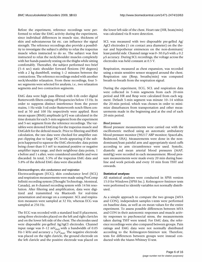
There were no significant differences between MYA andCON groups in the reference measurements of EMGtrap(Figure 2, p-values from .15 to .95). Likewise, the refer-ence relaxation measurements of EMGdelt were not sig-nificantly different between MYA and CON groups (p-values .28 and .47). However, the reference contractionmeasurements showed that MYA had significantly lowerEMGdelt than CON (p-values .009 and .013) when hold-ing a dumbbell at 90 degrees forward flexion.
Table 2: Tests for differences between the chronic pain group (MYA) and controls (CON) in their subjective ratings, muscle activity and autonomic variables.
Baseline Overall mean TSSTMeasure CON
Mean (SD)MYA
Mean (SD)p-value CON
Mean (SD)MYA
Mean (SD)p-value CON
Mean (SD)MYA
Mean (SD)p-value
PAINdomp(mm)
4 (10)a 50 (39)a <.001b 7 (10)a 68 (38)a <.001b 3 (10)a 72 (45)a .001b
PAINclat(mm)
0 (0)a 20 (25)a <.001b 0 (0)a 30 (36)a <.001b 0 (0)a 30(49)a <.001b
Stress(S-E score)
.7 (.5) 1.5 (.8) <.001 1.5 (.5) 2.2 (.5) <.001 3.6 (1) 3.7 (.9) >.3
Energy(S-E score)
1.9 (.9) 1.9 (.6) >.3 2.6 (.5) 2.5 (.4) >.3 3.4 (.6) 2.9 (.8) .015
EMGtrap(μV)
10.4 (17)a 25.9 (35)a .013b 37.6 (42)a 56.3 (58)a .055b 17.1(41)a 23.2 (28)a >.3b
EMGdelt(μV)
8.8 (15)a 11.8 (27)a .103b 32.5 (22)a 44.7 (42)a >.3b 10.1 (9)a 10.8 (31)a >.3b
HR(beats/min)
66.3 (8.3) 72.7 (8.0) .013 77.9 (8.1) 80.5 (10.9) >.3 94.6 (16) 93.2 (23) >.3
SCL(μS)
5.0 (4.2) 5.9 (5.6) >.3 10.5 (4.5) 11.4 (2.9) >.3 14.1 (5) 16.6 (5) .123
Resp(breaths/min)
16.3 (2.2) 16.7 (1.8) >.3 17.5 (1.9) 17.5 (1.5) >.3 15.6 (2) 15.1 (2) >.3
MAP(mmHg)
88.3 (7.7) 86.8 (9.4) >.3 93.5 (6.7) 93.3 (10.4) >.3 108.6 (12) 106.9 (16) >.3
Note: aMedian (interquartile distance); bMann-Whitney U-test; domp = dominant/most painful trapezius; clat = contralateral trapezius; EMGtrap = trapezius muscle activity; EMGdelt = deltoid muscle activity; HR = heart rate; SCL = skin conductance level; Resp = respiration rate; MAP = mean arterial pressure.
Page 7 of 16(page number not for citation purposes)

BMC Musculoskeletal Disorders 2009, 10:63 http://www.biomedcentral.com/1471-2474/10/63
Overall meanMean pain ratings over the entire experiment were signif-icantly higher in the MYA-group compared with the CON-group for both PAINdomp and PAINclat (Table 2). Stressratings were also higher for the trapezius myalgia groupthan the controls throughout the experiment. The differ-ences in mean EMGtrap from the entire experiment didnot reach statistical significance. Likewise, the overall
means for Energy, EMGdelt, HR, SCL, Resp and MAP werenot significantly different between the two groups.
TSSTTests for differences between the two groups during TSSTrevealed significantly higher pain ratings and significantlylower Energy ratings in MYA (Table 2). Stress ratings andphysiological measurements did not differ between thetwo groups during the psychosocial stress test.
EMG measured as mean RMS (± SEM) for the reference recordings and for each 20-min period throughout the experimentFigure 2EMG measured as mean RMS (± SEM) for the reference recordings and for each 20-min period throughout the experiment. The reference contraction (Flex) was a 90 degrees forward flexion of the arm holding a 2 kg dumbbell. BL denotes baseline.
Page 8 of 16(page number not for citation purposes)

BMC Musculoskeletal Disorders 2009, 10:63 http://www.biomedcentral.com/1471-2474/10/63
Mixed model analysesPain ratingsPain ratings for both sides were, as expected, significantlyhigher in MYA than CON at all time points.
In the mixed model analyses, group (F1,45 = 135.8, p <.001), time (F10,244 = 22.5, p < .001) and interaction(F10,244 = 4.8, p < .001) effects were significant for pain rat-ings in the dominant/most painful side. The main effectof group was 57.8 mm (95% CI, 47.3 to 68.2 mm) forPAINdomp, i.e., the dominant/most painful side. In theMYA group, PAINdomp continuously increased duringthe low-force work, peaking at the end of the work period,followed by a slight reduction during TSST and a slowdecrease during recovery (Figure 3), never returning tobaseline levels. The pain level at the end of the work
period was significantly higher than baseline, the firstwork period, and the three last recovery periods. Lookingonly at the CON group, none of the time points showedpain ratings significantly higher than baseline level.
For PAINclat, there were significant main effects of group(F1,44 = 45.7, p < .001) and time (F10,212 = 6.9, p < .001),as well as a significant group × time interaction (F10,212 =5.7, p < .001). The main effect of group was 28.6 mm(95% CI, 19.6 to 37.6). Pain intensity ratings increasedsignificantly from Start work to End work. PAINclatincreased significantly from baseline to the third workperiod, further increased during the remaining work peri-ods and then decreased very slowly towards the end of theexperiment in the MYA group. PAINclat was never signifi-cantly above zero in the CON group.
Subjective ratings (mean ± SEM) during Baseline (BL), work, TSST and recovery periodsFigure 3Subjective ratings (mean ± SEM) during Baseline (BL), work, TSST and recovery periods. The neutral points for Stress and Energy are indicated by the horizontal dashed lines.
Page 9 of 16(page number not for citation purposes)

BMC Musculoskeletal Disorders 2009, 10:63 http://www.biomedcentral.com/1471-2474/10/63
For the most painful side, pain ratings increased from 44mm at Baseline to 77 mm at the end of work and for thecontralateral side the corresponding increase was from 18mm to 38 mm.
Stress-Energy ratingsThe group (F1,45 = 20.3, p < .001), time (F10,245 = 74.0, p <.001) and interaction (F10,245 = 4.7, p < .001) effects wereall statistically significant for Stress ratings. The maineffect of group was .84 S-E points (95% CI, .4 to 1.3). TheMYA group had higher stress ratings than controls at base-line, work periods 3, 4 and 5, and during recovery. Forboth groups taken together, stress ratings were signifi-cantly higher during the work periods and TSST, com-pared with Baseline and recovery periods (Figure 3). Stressratings at TSST were significantly higher than all othertime points for both groups. Moreover, stress increased inMYA during the work period, whereas CON became morerelaxed towards the end of work. CON were below theneutral point (S-E score 2.4, neither stressed nor relaxed)during the entire work period. MYA, however, started nearthe neutral point and ended above it. Stress ratingsincreased to almost identical levels in MYA and CON dur-ing the stress test, indicating that the TSST was perceivedas equally stressful by both groups. Differences emergedagain during recovery where MYA were more stressed,although both groups were below the neutral point andreported stress ratings similar to their respective baselinelevels.
Energy ratings did not differ between the two groups (F1,44= .6, p = .434). However, the main effect of time was sig-nificant (F10,267 = 50.6, p < .001). Energy ratings were sig-nificantly higher during work and TSST compared withBaseline and recovery (Figure 3).
EMGThe mixed model analyses revealed larger trapezius mus-cle activity in MYA than in CON (F1,35 = 6.2, p = .017) anda significant time effect (F10,206 = 18.1, p < .001). Thegroup effect for EMGtrap was 1.7 μV (1.1 to 2.6). Thegroup effect of EMGtrap derived from the mixed modelanalysis may seem small since the analyses were madeusing ln-transformed EMG measurements. However, theestimated mean EMGtrap derived from the analysis was47.4 μV and 27.9 μV for MYA and CON, respectively. Forboth groups, the muscle activity increased over time dur-ing the repetitive work and was significantly elevatedcompared with baseline, TSST and the last recovery period(Figure 2). The muscle activity then decreased when thesubjects performed the TSST. During recovery, EMGtrapfirst increased to levels similar to the first work period andthen decreased over time, ending at levels similar to base-line at the last recovery period.
The deltoid muscle activity was not significantly differentbetween MYA and CON in the mixed model analyses(F1,67 = .4, p = .536). The time effect was, however, signif-icant for EMGdelt (F10,293 = 70.0, p < .010), showinghigher muscle activity during work compared with base-line and TSST. EMGdelt was higher than baseline level atthe start of recovery and did not return to baseline leveluntil the last recovery period.
The interaction effect between group and time was not sig-nificant for the mixed model analyses involving EMG dataand was therefore not included in the models.
Autonomic responsesIn the mixed models analysis, HR showed a significanttime effect (F10,387 = 71.4, p < .001), but group differenceswere not significant (F1,73 = 1.0, p = .332). HR was signif-icantly higher during work and TSST compared with Base-line and recovery (Figure 4). It was also significantlyhigher during TSST compared with the other time points.HR returned to baseline level within the first 20 min ofrecovery for both groups.
There was no significant difference in SCL between thegroups (F1,66 = .04, p = .851). The time effect was signifi-cant (F10,373 = 27.3, p < .001), with higher skin conduct-ance levels during work and TSST compared with Baselineand the second half of recovery (Figure 4). In addition,SCL was higher during TSST than all other time points.SCL was significantly elevated, compared with baseline,during the first recovery period.
Resp was similar in the two groups, resulting in no signif-icant group effects (F1,92 = .1, p = .804). There was, how-ever, a significant time effect for Resp (F10,343 = 25.5, p <.001) showing higher respiration rates during work com-pared with Baseline, TSST and recovery (Figure 4).
The MYA and CON groups showed no significant differ-ences in MAP (F1,79 = .2, p = .623). The time effect was sig-nificant for MAP (F10,348 = 27.0, p < .001). Blood pressuredid not rise significantly above baseline or recovery levelsduring the repetitive work (Figure 4). However, this meas-urement showed significantly higher values at TSST com-pared with all other time points.
CorrelationsIn MYA, correlations between pain in the dominant/mostpainful side and the contralateral side were high (rho =.54 to .74, p < .001 to .025) during TSST and recovery. Thepain ratings were not significantly correlated during base-line and work. As shown in Table 3, stress and pain ratingswere positively correlated during the first three recoveryperiods and during the third work period. Moreover,
Page 10 of 16(page number not for citation purposes)

BMC Musculoskeletal Disorders 2009, 10:63 http://www.biomedcentral.com/1471-2474/10/63
Page 11 of 16(page number not for citation purposes)
Physiological measurements (mean ± SEM) during Baseline (BL), work, TSST and recovery periodsFigure 4Physiological measurements (mean ± SEM) during Baseline (BL), work, TSST and recovery periods.
Table 3: Correlations between pain intensity, stress ratings and trapezius muscle activity (EMGtrap) for the MYA group.
Stress and PAINdomp p-value EMGtrap and PAINdomp p-value EMGtrap andStress
p-value
Baseline 0.05 >.3 0.25 >.3 0.45 0.061Work 1 -0.07 >.3 0.32 0.199 -0.21 >.3Work 2 0.26 >.3 0.10 >.3 -0.26 0.288Work 3 0.52 0.028 0.14 >.3 -0.13 >.3Work 4 0.16 >.3 0.24 >.3 0.06 >.3Work 5 0.35 0.151 0.28 0.289 -0.37 0.157TSST 0.27 0.275 0.37 0.241 0.48 0.113
Recovery 1 0.63 0.006 0.54 0.048 0.26 >.3Recovery 2 0.57 0.014 0.53 0.052 0.38 0.184Recovery 3 0.54 0.020 0.54 0.037 0.41 0.126Recovery 4 0.10 >.3 0.18 >.3 0.62 0.019
BMC Musculoskeletal Disorders 2009, 10:63 http://www.biomedcentral.com/1471-2474/10/63
EMGtrap was significantly correlated with pain intensityat the first and third recovery (Table 3). In addition, stressand EMGtrap were significantly correlated only at the endof recovery (Table 3). Possible significant correlationsbetween EMGtrap and ratings of pain and stress in theCON group were checked for but not found. Furthermore,no significant correlations were found between auto-nomic variables and pain intensity or stress in the twogroups.
DiscussionThe main finding in the present study was that muscleactivity in the painful muscle was higher in a group ofwomen with trapezius myalgia compared with pain-freecontrols. Furthermore, muscle activity in MYA was posi-tively correlated to pain intensity during recovery. The sig-nificant increases in HR, MAP and SCL during the stresstest and the subsequent reduction during recovery dem-onstrates that stress was successfully induced. Unexpect-edly, the two groups showed very similar autonomicresponses to low-force repetitive work and psychosocialstress. This finding does not support the hypothesis thatchronic trapezius myalgia is associated with increasedsympathetic activity. The perceived level of stress was,however, increased in the patient group during restingand work conditions, compared with controls.
Pain and stress ratingsAs expected and intended, the trapezius myalgia groupreported more pain than controls throughout the experi-ment. Pain intensity ratings confirmed that the low-forcework had the desired effect, i.e., it worsened the pain con-siderably for MYA, whereas pain ratings for the CONgroup never rose significantly above baseline level.
Our results regarding pain intensity and stress ratings dur-ing the psychosocial stress test are inconsistent with thefindings by Thieme et al. [38] that stress enhanced painintensity in fibromyalgia syndrome (FMS). The MYAgroup's reduction in pain intensity at the end of the psy-chosocial stress test and subsequent increase at the firstrecovery period could not be explained only by cessationof the repetitive work tasks. This reduction in pain inten-sity may be due to stress-induced analgesia [39] and/ordistraction from the sensation of pain [40].
MYA experienced higher level of stress at baseline com-pared with CON, which is consistent with previous find-ings in chronic pain patients by Thieme et al. [38].Chapman and Gavrin [41] concluded that pain is a pow-erful stressor per se, which would implicate that duringthe experiment, MYA were exposed to two different stres-sors simultaneously; pain and psychosocial stress. Theincreasing stress ratings in MYA during the work periodcould, thus, be due to increased pain. There are indica-
tions of attenuated responses to stress and functional testsin FMS [42]. A possible attenuation of the stress responseto the TSST could, hence, be masked in the additionalstress due to pain in the MYA group. However, introduc-ing pain intensity as a covariate in the analyses, thus con-trolling for differences in pain ratings, did not supportsuch a suggestion.
In contrast to CON, the MYA group's energy level actuallydecreased in response to the stress test which could indi-cate that the chronic pain group perceives stressful situa-tions as being more energy consuming. The CON groupfound ways to mobilize energy for the stress test, whichwas seen as increased energy ratings at TSST.
Muscle activitySimilar patterns of EMG activity in the prime mover del-toid were found in MYA and CON throughout the experi-ment (Figure 2). This was not the case for the posturaltrapezius muscle.
The chronic pain group's muscle activity was higher in thepainful trapezius muscle at baseline, which was an unin-structed rest, but not at the reference relaxation measure-ment where the subjects were given specific instructionson how to relax the trapezius muscle. Speculatively, thehigher trapezius EMG in MYA during uninstructed rest,but not when instructed to fully relax, can be an indica-tion that voluntary effort and attention are required inorder to relax the muscle. In daily regular activities, whenother aspects need attention, the EMG activity will behigher, contributing to a vicious circle of the muscle. Ourresults need to be confirmed in other studies but opens upfor myofeedback interventions using surface EMG. Hith-erto such myofeedback studies indicate that it is possibleto alter EMG patterns in healthy subjects [43] and inchronic myalgia [44]. Although the latter study did notfind that the EMG level per se – but rather the ability torelax – was linked to pain intensity decrease [44].
In agreement with other studies we found that the MYAgroup's muscle activity of the trapezius was higherthroughout the experiment [45-47]; there exist differentexplanations for the higher muscle activity in subjectswith chronic pain [48,49]. The direction of the causal rela-tionship between pain and EMG activity is not obvious[49] and in fact cannot be determined using the presentstudy design. We do not know if the increased muscleactivity is a cause or a consequence of pain, since it isunknown whether this was also the case before painonset. The literature is, however, not in consensus andsome studies have found no differences in EMG activity[50-52]. In contrast, we found no difference in the EMGactivity of the prime mover, i.e., the deltoid, between thetwo groups of subjects. There are reports that subjects with
Page 12 of 16(page number not for citation purposes)

BMC Musculoskeletal Disorders 2009, 10:63 http://www.biomedcentral.com/1471-2474/10/63
pain have difficulties in relaxing their trapezius after com-pletion of a task [53], in utilizing small pauses in work[54,55] and in parts of the contraction cycle duringdynamic muscle contractions [10-16], but the literature isnot in consensus [50,56-58]. With the present studydesign, it was not possible to determine whether the EMGactivity of trapezius in MYA was increased during theactive contraction, during the supposed passive parts ofthe contraction cycles, or during both these.
The increase in EMG activity of the trapezius in responseto work was similar in MYA and CON. A successiveincrease in EMG activity of trapezius during work has alsobeen reported by Strøm et al. [59]. Furthermore, muscleactivity was not correlated to pain development duringrepetitive work. Hence, muscle activity per se does notseem to be the cause for increased pain intensity. Strøm etal. [59] reported similar results but also that the increasein pain intensity correlated with muscle blood flux. It can-not be ruled out that higher EMG activity in trapezius canaffect blood flow and microcirculation in MYA.
Autonomic responsesNone of the autonomic responses showed any signs of anincreased sympathetic activity in MYA. Reactions to thelow-force work and TSST were almost identical in the twogroups.
The higher baseline HR found in MYA confirms previousfindings by Gockel et al. [60]. Regarding baseline differ-ences, a previous studie on patients with chronic low backpain (CLBP) found higher HR, higher low frequency heartrate variability (HRV) and lower high frequency HRV [61].In contrast to the study referred to above, there are severalstudies that fail to detect differences between CLBPpatients and controls in baseline levels [62,63]. Flor andTurk [64] concluded that regardless of type of physiologi-cal measure, baseline levels were not generally elevated inCLBP patients. Patients with whiplash associated disor-ders (WAD) have shown increased heart rate anddecreased heart rate variability (HRV) [22] at baseline.The basal autonomic state in widespread pain has beenmore extensively investigated. Cohen et al. [25] foundincreased sympathetic and decreased parasympatheticactivity during complete rest in the supine position. Theresults are, however, conflicting even for FMS patients butsome sort of dysautonomia has been found in most stud-ies [38,42]. The elevated baseline HR found in this studyshould probably not be interpreted as a sign of increasedsympathetic tone since none of the other measures gavesimilar indications and HR can be influenced by other fac-tors e.g., physical fitness.
Our findings of normal cardiovascular reactivity in MYAare in contrast to the study by Gockel et al. [60], where
patients with neck/shoulder pain showed attenuated car-diovascular responses to various functional tests (pacedbreathing, orthostatic, handgrip, valsalva). The resultsfrom earlier studies are inconsistent reporting bothincreased and decreased cardiovascular reactivity inchronic pain patients compared with controls. In a studycomparing WAD and controls [22], unpleasant soundinduced higher HR response in WAD, HR and HRV reac-tivity was lower in response to cognitive challenge (Stroopcolor-word test) whereas no differences emerged duringpaced breathing. No difference in autonomic reactivity tovarious tests (Stroop color-word, orthostatic, handgrip,paced breathing) was found between CLBP and controls[61]. Nilsen et al. [48] studied differences between neck/shoulder pain patients, FMS patients and healthy con-trols. They found attenuated cardiovascular reactions to alow-grade mental stressor in FMS. Neck/shoulder painpatients appeared to be in an intermediate positionbetween FMS and controls, but not significantly differentfrom the controls. FMS patients have also shown normalblood pressure and HR regulation during maximal volun-tary contractions [65] and lower HR reactivity to stressors(mental arithmetic, social conflict) [38].
Skin conductance data in the present study showed ten-dencies towards an increased stress response and delayedreturn to baseline in MYA, but the difference betweengroups was small and individual variations large. HigherSCL in response to stress and during recovery from stress,which indicates enhanced sympathetic response to stress,has previously been found in FMS [38]. Higher spontane-ous electrodermal activity [61] and enhanced activity inresponse to pain [62] and sound stimulation [66] has alsobeen found in CLBP patients.
The chronic pain group in this study did not have wide-spread pain but local or regional pain in the neck/shoul-der area. The autonomic dysfunctions found in FMS couldbe due to the alterations at different levels of the pain sys-tem in this generalized pain syndrome [67]. Alterations incardiovascular or sudomotor regulations could be presentin local or regional pain as well, but in this case localizedonly to the painful areas, as seen in complex regional painsyndromes [68].
Inconsistencies in the results from previous studies ofautonomic reactivity in patients with localized chronicpain may be due to the heterogeneity of this patientgroup. They represent a diversity of causes, localizationsand intensities of pain. The findings in FMS, WAD andCLBP discussed above may reflect nonspecific responsepatterns characteristic of confounding co-morbidities,e.g., anxiety or depression, in general, which was notfound in the present MYA population.
Page 13 of 16(page number not for citation purposes)

BMC Musculoskeletal Disorders 2009, 10:63 http://www.biomedcentral.com/1471-2474/10/63
Methodological considerationsPrevious studies concerning stress responses in chronicpain have used cognitive tasks (e.g., Stroop color-wordtest, mental arithmetic) or social conflict tasks, which arenot as powerful as the TSST in producing acute stressresponses [69]. This stress protocol has been found toinduce significant cardiovascular responses at the firstexposure in 70–80% of all subjects [70]. Laboratory stressprotocols offer the advantage of standardization acrosstest sessions but may lack the ecological validity of fieldstudies. However, the significant increases in HR, MAP,SCL and subjective stress ratings confirm that psychoso-cial stress was successfully induced.
The autonomic measures used here, i.e., instantaneousheart rate, skin conductance, and blood pressure only pro-vide gross estimates of end organ function, irrespective ofthe relative contribution of the sympathetic and parasym-pathetic branches. Simple examination of autonomicresponses to challenges does not provide a definitive testof the potential role of the sympathetic nervous system.More sophisticated methods, such as heart rate variability(HRV), could provide information regarding the balanceof the autonomic branches. However, in the presentstudy, the subjects assumed different body positions, i.e.,both sitting and standing, and were moving, circum-stances that affects HRV and makes it difficult to interpret.Moreover, valuable information about interactionsbetween different variables could be derived through mul-tivariate data analyses, possibly revealing response pat-terns not visible when analyzing one variable at a time.
Inclusion of only female subjects was based on the higherprevalence of chronic trapezius myalgia in women thanmen, also limiting the availability of eligible male sub-jects. Previous studies have concluded that healthy menand women are similar in their autonomic responses topsychosocial stress tests [71,72]. There are, however, sexdifferences in chronic pain (e. g., CLBP) patients' reactionsto experimental pain [73]. Including men is thereforeimportant for future research.
A limitation of the present study is that the statisticalpower did not allow differences below approximately10% in any of the outcome variables to be detected,increasing the risk for statistical type II error. Correction ofp-values for multiple comparisons, to avoid type I error, isoften performed in situations with multiple outcome var-iables. However, because of the explorative approach inthis study, correction for multiple comparisons were onlyperformed in the mixed model post-hoc analyses, toreduce the risk of type II error in the between groups tests[74]. The pain intensity distributions were somewhatskewed, implying that the results from the mixed regres-sion model should be interpreted with care. However,
residuals from the analyses were approximately symmet-rically distributed, the differences between groups werelarge and log- or square root-transformation of the varia-bles did not alter the results indicating that the conclu-sions regarding pain ratings were valid.
ConclusionWe found increased muscle activity during uninstructedrest in the painful muscle of a group of women with tra-pezius myalgia. The present study could not confirm thehypothesis that chronic trapezius myalgia is associatedwith increased sympathetic activity. The suggestion ofautonomic imbalance in patients with chronic local orregional musculoskeletal pain needs to be further investi-gated.
Competing interestsThe authors declare that they have no competing interests.
Authors' contributionsAS carried out the data collection, performed the statisti-cal analysis and was the main writer of the manuscript. BLdesigned the study, participated in subject inclusion anddata collection and helped to draft the manuscript. JD par-ticipated in the planning and coordination of the study,and revised the manuscript. TF was involved in methodo-logical considerations and revised the manuscript. BGdesigned and supervised the study and helped to draft themanuscript. All authors read and approved the final man-uscript.
AcknowledgementsThis study was supported by the Swedish Research Council (K2005-27X-15316-01A) and the Swedish Council for Working life and Social research (2004-0289).
References1. Flor H, Birbaumer N: Acquisition of chronic pain: Psychophysi-
ological mechanisms. Am Pain Soc J 1994, 3:119-127.2. Passatore M, Roatta S: Influence of sympathetic nervous system
on sensorimotor function: whiplash associated disorders(WAD) as a model. Eur J Appl Physiol 2006, 98:423-449.
3. Ariëns GA, van Mechelen W, Bongers PM, Bouter LM, Wal G van der:Psychosocial risk factors for neck pain: A systematic review.Am J Ind Med 2001, 39:180-193.
4. Malchaire J, Cock N, Vergracht S: Review of the factors associ-ated with musculoskeletal problems in epidemiological stud-ies. Int Arch Occup Environ Health 2001, 74:79-90.
5. Bongers PM, Kremer AM, Laak Jt: Are psychosocial factors, riskfactors for symptoms and signs of the shoulder, elbow, orhand/wrist?: A review of the epidemiological literature. Am JInd Med 2002, 41:315-342.
6. Wærsted M: Human muscle activity related to non-biome-chanical factors in the workplace. Eur J Appl Physiol 2000,83:151-158.
7. Gerdle B, Karlsson S, Day S, Djupsjöbacka M: Acquisition, process-ing and analysis of surface EMG signals. In Modern Techniques inNeuroscience Research Edited by: Johansson H, Winhorst U. Hamburg:Springer Verlag; 1999:705-755.
8. Madeleine P, Leclerc F, Arendt-Nielsen L, Ravier P, Farina D: Exper-imental muscle pain changes the spatial distribution of uppertrapezius muscle activity during sustained contraction. ClinNeurophysiol 2006, 117:2436-2445.
Page 14 of 16(page number not for citation purposes)

BMC Musculoskeletal Disorders 2009, 10:63 http://www.biomedcentral.com/1471-2474/10/63
9. Falla D, Farina D, Graven-Nielsen T: Experimental muscle painresults in reorganization of coordination among trapeziusmuscle subdivisions during repetitive shoulder flexion. ExpBrain Res 2007, 178:385-393.
10. Elert J, Kendall SA, Larsson B, Mansson B, Gerdle B: Chronic painand difficulty in relaxing postural muscles in patients withfibromyalgia and chronic whiplash associated disorders. JRheumatol 2001, 28:1361-1368.
11. Elert JE, Rantapaa Dahlqvist SB, Henriksson-Larsen K, Gerdle B:Increased EMG activity during short pauses in patients withprimary fibromyalgia. Scand J Rheumatol 1989, 18:321-323.
12. Elert JE, Rantapaa-Dahlqvist SB, Henriksson-Larsen K, Lorentzon R,Gerdle BU: Muscle performance, electromyography and fibretype composition in fibromyalgia and work-related myalgia.Scand J Rheumatol 1992, 21:28-34.
13. Arendt-Nielsen L, Graven-Nielsen T, Svarrer H, Svensson P: Theinfluence of low back pain on muscle activity and coordina-tion during gait: a clinical and experimental study. Pain 1996,64:231-240.
14. Fredin Y, Elert J, Britschgi N, Vaher A, Gerdle B: A decreased abil-ity to relax between repetitive muscle contractions inpatients with chronic symptoms after whiplash trauma ofthe neck. J Musculoskel Pain 1997, 5:55-70.
15. Graven-Nielsen T, Svensson P, Arendt-Nielsen L: Effects of exper-imental muscle pain on muscle activity and co-ordinationduring static and dynamic motor function. Electroen Clin Neuro-physiol 1997, 105:156-164.
16. Stohler CS, Ashton-Miller JA, Carlson DS: The effects of pain fromthe mandibular joint and muscles on masticatory motorbehaviour in man. Arch Oral Biol 1988, 33:175-182.
17. Lund JP, Donga R, Widmar CG, Stohler CS: The pain-adaptationmodel: a discussion of the relationship between chronic mus-culoskeletal pain and motor activity. Can J Physiol Pharmacol1990, 69:683-694.
18. Andersen LL, Nielsen PK, Søgaard K, Andersen CH, Skotte J, SjøgaardG: Torque-EMG-velocity relationship in female workers withchronic neck muscle pain. J Biomech 2008, 41:2029-2035.
19. Lundberg U, Forsman M, Zachau G, Eklöf M, Palmerud G, Melin B,Kadefors R: Effects of experimentally induced mental andphysical stress on motor unit recruitment in the trapeziusmuscle. Work Stress 2002, 16:166-178.
20. Lundberg U, Dohns IE, Melin B, Sandsjö L, Palmerud G, Kadefors R,Ekström M, Parr D: Psychophysiological Stress Responses,Muscle Tension, and Neck and Shoulder Pain Among Super-market Cashiers. J Occup Health Psych 1999, 4:245-255.
21. Johansson H, Arendt-Nilsen L, Bergenheim M, Blair S, van Dieen JH,Djupsjöbacka M, Fallentin N, Gold JE, Hägg G, Kalezic N, Larsson S-E, Ljubisavljevic M, Lyskov E, Mano T, Magnusson M, Passatore M,Pedrosa-Domellöf F, Punnett L, Roatta S, Thornell L-E, Windhorst U,Zukowska Z: Epilogue: An Integrated Model for ChronicWork-Realted Myalgia – "Brussels Model". In Chronic Work-Related Myalgia – Neuromuscular Mechanisms behind Work-RelatedChronic Muscle Pain Syndromes Edited by: Johansson H, Windhorst U,Djupsjöbacka M, Passatore M. Gävle: Gävle University Press;2003:291-300.
22. Kalezic N: Autonomic Reactivity in Muscle Pain – Clinical andExperimental Assessment. In Doctoral thesis Umeå University,Department of Surgical and Perioperative Science, Sports MedicineUnit; 2006.
23. Roatta S, Arendt-Nielsen L, Farina D: Sympathetic-inducedchanges in discharge rate and spike-triggered average twitchtorque of low-threshold motor units in humans. J Physiol 2008,586:5561-5574.
24. Roatta S, Kalezic N, Passatore M: Sympathetic Nervous System:Interaction with Muscle Function and Involvement in MotorControl. In Chronic work-related myalgia Edited by: Johansson H,Windhorst U, Djupsjöbacka M, Passatore M. Gävle: Gävle UniversityPress; 2003:265-276.
25. Cohen H, Neumann L, Shore M, Amir M, Cassuto Y, Buskila D: Auto-nomic dysfunction in patients with fibromyalgia: Applicationof power spectral analysis of heart rate variability. SeminArthritis Rheum 2000, 29:217-227.
26. Martinez-Lavin M: Biology and therapy of fibromyalgia. Stress,the stress response system, and fibromyalgia. Arthritis Res Ther2007, 9:216.
27. Gockel M, Lindholm H, Niemistö L, Hurri H: Perceived disabilitybut not pain is connected with autonomic nervous functionamong patients with chronic low back pain. J Rehabil Med 2008,40:355-358.
28. Ohlsson K, Attewell RG, Johnsson B, Ahlm A, Skerfving S: An assess-ment of neck and upper extremity disorders by question-naire and clinical examination. Ergonomics 1994, 37:891-897.
29. Kuorinka I, Jonsson B, Kilbom Å, Vinterberg H, Biering-Sørensen F,Andersson G, Jørgensen K: Standardised Nordic questionnairesfor the analysis of musculoskeletal symptoms. Appl Ergon1987, 18:233-237.
30. Wolfe F, Smythe HA, Yunus MB, Bennett RM, Bombardier C, Gold-enberg DL, Tugwell P, Campbell SM, Abeles M, Clark P, Fam AG, Far-ber SJ, Fiechtner JJ, Franklin CM, Gatter RA, Hamaty D, Lessard J,Lichtbroun AS, Masi AT, Mccain GA, Reynolds WJ, Romano TJ, Rus-sell IJ, Sheon RP: The american college of rheumatology 1990criteria for the classification of fibromyalgia. Arthritis Rheum1990, 33:160-172.
31. Larsson B, Rosendal L, Kristiansen J, Sjøgaard G, Søgaard K, GhafouriB, Abdiu A, Kjaer M, Gerdle B: Responses of algesic and meta-bolic substances to 8 h of repetitive manual work in myalgichuman trapezius muscle. Pain 2008, 140:479-490.
32. Rosendal L, Larsson B, Kristiansen J, Peolsson M, Sogaard K, Kjaer M,Sorensen J, Gerdle B: Increase in muscle nociceptive sub-stances and anaerobic metabolism in patients with trapeziusmyalgia: microdialysis in rest and during exercise. Pain 2004,112:324-334.
33. Kirschbaum C, Pirke KM, Hellhammer DH: The 'Trier SocialStress Test' – a tool for investigating psychobiological stressresponses in a laboratory setting. Neuropsychobiology 1993,28:76-81.
34. Kirschbaum C, Kudielka BM, Gaab J, Schommer NC, HellhammerDH: Impact of Gender, Menstrual Cycle Phase, and OralContraceptives on the Activity of the Hypothalamus-Pitui-tary-Adrenal Axis. Psychosom Med 1999, 61:154-162.
35. Kjellberg A, Wadman C: The role of the affective stressresponse as a mediator of the effect of psychosocial risk fac-tors on musculoskeletal complaints – Part 1: Assemblyworkers. Int J Ind Ergon 2007, 37:367-374.
36. Kjellberg A, Wadman C: Subjektiv stress och dess sambandmed psykosociala arbetsförhållanden och hälsobesvär. Enprövning av Stress-Energi-modellen. [Subjective stress andits relation to psychosocial work conditions and health com-plaints. A test of the Stress-Energy model]. In Arbete och HälsaVolume 2002. Stockholm: National Institute for Working Life; 2002.
37. Hermens HJ, Fredriks B, Merletti R, Stegeman D, Blok J, Rau G, Dis-selhorst-Klug C, Hägg G: SENIAM European recommendations for sur-face elecromyography Enschede, the Netherlands: Roessingh Researchand Development; 1999.
38. Thieme K, Rose U, Pinkpank T, Spies C, Turk DC, Flor H: Psycho-physiological responses in patients with fibromyalgia syn-drome. J Psychosom Res 2006, 61:671-679.
39. Amit Z, Galina ZH: Stress-induced analgesia: adaptive pain sup-pression. Physiol Rev 1986, 66:1091-1120.
40. Quiton RL, Greenspan JD: Sex differences in endogenous painmodulation by distracting and painful conditioning stimula-tion. Pain 2007, 132:S134-S149.
41. Chapman CR, Gavrin J: Suffering: the contributions of persist-ent pain. Lancet 1999, 353:2233-2237.
42. Cohen H, Neumann L, Kotler M, Buskila D: Autonomic nervoussystem derangement in fibromyalgia syndrome and relateddisorders. IMAJ 2001, 3:755-760.
43. Holtermann A, Søgaard K, Christensen H, Dahl B, Blangsted A: Theinfluence of biofeedback training on trapezius activity andrest during occupational computer work: a randomized con-trolled trial. Eur J Appl Physiol 2008, 104:983-989.
44. Vollenbroek-Hutten M, Hermens H, Voerman G, Sandsjö L, KadeforsR: Are changes in pain induced by myofeedback trainingrelated to changes in muscle activation patterns in patientswith work-related myalgia? Eur J Appl Physiol 2006, 96:209-215.
45. Johnston V, Jull G, Souvlis T, Jimmieson NL: Neck Movement andMuscle Activity Characteristics in Female Office WorkersWith Neck Pain. Spine 2008, 33:555-563.
46. Szeto GPY, Straker LM, O'Sullivan PB: A comparison of sympto-matic and asymptomatic office workers performing monot-
Page 15 of 16(page number not for citation purposes)
http://www.ncbi.nlm.nih.gov/entrez/query.fcgi?cmd=Retrieve&db=PubMed&dopt=Abstract&list_uids=2595350
http://www.ncbi.nlm.nih.gov/entrez/query.fcgi?cmd=Retrieve&db=PubMed&dopt=Abstract&list_uids=2595350
http://www.ncbi.nlm.nih.gov/entrez/query.fcgi?cmd=Retrieve&db=PubMed&dopt=Abstract&list_uids=2595350
http://www.ncbi.nlm.nih.gov/entrez/query.fcgi?cmd=Retrieve&db=PubMed&dopt=Abstract&list_uids=1570484
http://www.ncbi.nlm.nih.gov/entrez/query.fcgi?cmd=Retrieve&db=PubMed&dopt=Abstract&list_uids=1570484
http://www.ncbi.nlm.nih.gov/entrez/query.fcgi?cmd=Retrieve&db=PubMed&dopt=Abstract&list_uids=8740599
http://www.ncbi.nlm.nih.gov/entrez/query.fcgi?cmd=Retrieve&db=PubMed&dopt=Abstract&list_uids=8740599
http://www.ncbi.nlm.nih.gov/entrez/query.fcgi?cmd=Retrieve&db=PubMed&dopt=Abstract&list_uids=8740599
http://www.ncbi.nlm.nih.gov/entrez/query.fcgi?cmd=Retrieve&db=PubMed&dopt=Abstract&list_uids=3178536
http://www.ncbi.nlm.nih.gov/entrez/query.fcgi?cmd=Retrieve&db=PubMed&dopt=Abstract&list_uids=3178536
http://www.ncbi.nlm.nih.gov/entrez/query.fcgi?cmd=Retrieve&db=PubMed&dopt=Abstract&list_uids=3178536
http://www.ncbi.nlm.nih.gov/entrez/query.fcgi?cmd=Retrieve&db=PubMed&dopt=Abstract&list_uids=8206057
http://www.ncbi.nlm.nih.gov/entrez/query.fcgi?cmd=Retrieve&db=PubMed&dopt=Abstract&list_uids=8206057
http://www.ncbi.nlm.nih.gov/entrez/query.fcgi?cmd=Retrieve&db=PubMed&dopt=Abstract&list_uids=8206057
http://www.ncbi.nlm.nih.gov/entrez/query.fcgi?cmd=Retrieve&db=PubMed&dopt=Abstract&list_uids=2306288
http://www.ncbi.nlm.nih.gov/entrez/query.fcgi?cmd=Retrieve&db=PubMed&dopt=Abstract&list_uids=2306288
http://www.ncbi.nlm.nih.gov/entrez/query.fcgi?cmd=Retrieve&db=PubMed&dopt=Abstract&list_uids=8255414
http://www.ncbi.nlm.nih.gov/entrez/query.fcgi?cmd=Retrieve&db=PubMed&dopt=Abstract&list_uids=8255414
http://www.ncbi.nlm.nih.gov/entrez/query.fcgi?cmd=Retrieve&db=PubMed&dopt=Abstract&list_uids=8255414
http://www.ncbi.nlm.nih.gov/entrez/query.fcgi?cmd=Retrieve&db=PubMed&dopt=Abstract&list_uids=2876446

BMC Musculoskeletal Disorders 2009, 10:63 http://www.biomedcentral.com/1471-2474/10/63
Publish with BioMed Central and every scientist can read your work free of charge
"BioMed Central will be the most significant development for disseminating the results of biomedical research in our lifetime."
Sir Paul Nurse, Cancer Research UK
Your research papers will be:
available free of charge to the entire biomedical community
peer reviewed and published immediately upon acceptance
cited in PubMed and archived on PubMed Central
yours — you keep the copyright
Submit your manuscript here:http://www.biomedcentral.com/info/publishing_adv.asp
BioMedcentral
onous keyboard work – 1: Neck and shoulder musclerecruitment patterns. Manual Therapy 2005, 10:270-280.
47. Falla D, Bilenkij G, Jull G: Patients with chronic neck pain dem-onstrate altered patterns of muscle activation during per-formance of a functional upper limb task. Spine 2004,29:1436-1440.
48. Nilsen KB, Sand T, Westgaard RH, Stovner LJ, White LR, Bang LeistadR, Helde G, Rø M: Autonomic activation and pain in responseto low-grade mental stress in fibromyalgia and shoulder/neck pain patients. Eur J Pain 2007, 11:743-755.
49. Larsson B, Björk J, Elert J, Gerdle B: Mechanical performance andelectromyography during repeated maximal isokineticshoulder forward flexions in female cleaners with and with-out myalgia of the trapezius muscle and in healthy controls.Eur J Appl Physiol 2000, 83:257-267.
50. Voerman G, Vollenbroek-Hutten M, Hermens H: Upper trapeziusmuscle activation patterns in neck-shoulder pain patientsand healthy controls. Eur J Appl Physiol 2007, 102:1-9.
51. Westgaard RH, Vasseljen O, Holte KA: Trapezius muscle activityas a risk indicator for shoulder and neck pain in female serv-ice workers with low biomechanical exposure. Ergonomics2001, 44:339-353.
52. Larsson S-E, Ålund M, Cai H, Öberg ÅP: Chronic pain after soft-tissue injury of the cervical spine: trapezius muscle bloodflow and electromyography at static loads and fatigue. Pain1994, 57:173-180.
53. Johnston V, Jull G, Darnell R, Jimmieson N, Souvlis T: Alterations incervical muscle activity in functional and stressful tasks infemale office workers with neck pain. Eur J Appl Physiol 2008,103:253-264.
54. Hägg G, Åström A: Load pattern and pressure pain thresholdin the upper trapezius muscle and psychosocial factors inmedical secretaries with and without shoulder/neck disor-ders. Int Arch Occup Environ Health 1997, 69:423-432.
55. Sandsjö L, Melin B, Rissén D, Dohns I, Lundberg U: Trapezius mus-cle activity, neck and shoulder pain, and subjective experi-ences during monotonous work in women. Eur J Appl Physiol2000, 83:235-238.
56. Nordander C, Hansson G-A: Muscular rest and gap frequency asEMG measures of physical exposure: the impact of worktasks and individual related. Ergonomics 2000, 43:1904-1919.
57. Vasseljen O, Westgaard RH: A case-control study of trapeziusmuscle activity in office and manual workers with shoulderand neck pain and symptom-free controls. Int Arch Occup Envi-ron Health 1995, 67:11-18.
58. Thorn S, Søgaard K, Kallenberg LAC, Sandsjö L, Sjøgaard G, HermensHJ, Kadefors R, Forsman M: Trapezius muscle rest time duringstandardised computer work – A comparison of female com-puter users with and without self-reported neck/shouldercomplaints. J Electromyogr Kines 2007, 17:420-427.
59. Strøm V, Knardahl S, Stanghelle JK, Røe C: Pain induced by a singlesimulated office-work session: Time course and associationwith muscle blood flux and muscle activity. Eur J Pain in press.
60. Gockel M, Lindholm H, Alaranta H, Viljanen A, Lindquist A, LindholmT: Cardiovascular functional disorder and stress amongpatients having neck-shoulder symptoms. Ann Rheum Dis 1995,54:494-497.
61. Kalezic N, Åsell M, Kerschbaumer H, Lyskov E: Physiological reac-tivity to functional tests in patients with chronic low backpain. J Musculoskelet Pain 2007, 15:29-40.
62. Peters ML, Schmidt AJM: Psychophysiological responses torepeated acute pain stimulation in chronic low back painpatients. J Psychosom Res 1991, 35:59-74.
63. Cohen MJ, Swanson GA, Naliboff BD, Schandler SL, McArthur DL:Comparison of electromyographic response patterns duringposture and stress tasks in chronic low back pain patternsand control. J Psychosom Res 1986, 30:135-141.
64. Flor H, Turk DC: Psychophysiology of Chronic Pain: DoChronic Pain Patients Exhibit Symptom-Specific Psycho-physiological Responses? Psychol Bull 1989, 105:215-259.
65. Kadetoff D, Kosek E: The effects of static muscular contractionon blood pressure, heart rate, pain ratings and pressure painthresholds in healthy individuals and patients with fibromy-algia. Eur J Pain 2007, 11:39-47.
66. Bonnet A, Naveteur J: Electrodermal activity in low back painpatients with and without co-morbid depression. Int J Psycho-physiol 2004, 53:37-44.
67. Vierck CJJ: Mechanisms underlying development of spatiallydistributed chronic pain (fibromyalgia). Pain 2006,124:242-263.
68. Baron R, Levine JD, Fields HL: Causalgia and reflex sympatheticdystrophy: Does the sympathetic nervous system contributeto the generation of pain? Muscle Nerve 1999, 22:678-695.
69. Dickerson SS, Kemeny ME: Acute stressors and cortisol reactiv-ity: A theoretical integration and synthesis of laboratoryresearch. Psychol Bull 2004, 130:355-391.
70. Schommer NC, Hellhammer DH, Kirschbaum C: Dissociationbetween reactivity of the hypothalamus-pituitary-adrenalaxis and the sympathetic-adrenal-medullary system torepeated psychosocial stress. Psychosom Med 2004, 65:450-460.
71. Kelly MM, Tyrka AR, Anderson GM, Price LH, Carpenter LL: Sex dif-ferences in emotional and physiological responses to theTrier Social Stress Test. J Behav Ther Exp Psychiatry 2008,39:87-98.
72. Sgoifo A, Braglia F, Costoli T, Musso E, Meerlo P, Ceresini G, TroisiA: Cardiac autonomic reactivity and salivary cortisol in menand women exposed to social stressors: relationship withindividual ethological profile. Neurosci Biobehav Rev 2003,27:179-188.
73. Tousignant-Laflamme Y, Marchand S: Sex differences in cardiacand autonomic response to clinical and experimental pain inLBP patients. Eur J Pain 2006, 10:603-614.
74. Feise R: Do multiple outcome measures require p-valueadjustment? BMC Med Res Method 2002, 2:8.
Pre-publication historyThe pre-publication history for this paper can be accessedhere:
http://www.biomedcentral.com/1471-2474/10/63/prepub
Page 16 of 16(page number not for citation purposes)
http://www.ncbi.nlm.nih.gov/entrez/query.fcgi?cmd=Retrieve&db=PubMed&dopt=Abstract&list_uids=8090514
http://www.ncbi.nlm.nih.gov/entrez/query.fcgi?cmd=Retrieve&db=PubMed&dopt=Abstract&list_uids=8090514
http://www.ncbi.nlm.nih.gov/entrez/query.fcgi?cmd=Retrieve&db=PubMed&dopt=Abstract&list_uids=8090514
http://www.ncbi.nlm.nih.gov/entrez/query.fcgi?cmd=Retrieve&db=PubMed&dopt=Abstract&list_uids=9215929
http://www.ncbi.nlm.nih.gov/entrez/query.fcgi?cmd=Retrieve&db=PubMed&dopt=Abstract&list_uids=9215929
http://www.ncbi.nlm.nih.gov/entrez/query.fcgi?cmd=Retrieve&db=PubMed&dopt=Abstract&list_uids=9215929
http://www.ncbi.nlm.nih.gov/entrez/query.fcgi?cmd=Retrieve&db=PubMed&dopt=Abstract&list_uids=7622274
http://www.ncbi.nlm.nih.gov/entrez/query.fcgi?cmd=Retrieve&db=PubMed&dopt=Abstract&list_uids=7622274
http://www.ncbi.nlm.nih.gov/entrez/query.fcgi?cmd=Retrieve&db=PubMed&dopt=Abstract&list_uids=7622274
http://www.ncbi.nlm.nih.gov/entrez/query.fcgi?cmd=Retrieve&db=PubMed&dopt=Abstract&list_uids=7632093
http://www.ncbi.nlm.nih.gov/entrez/query.fcgi?cmd=Retrieve&db=PubMed&dopt=Abstract&list_uids=7632093
http://www.ncbi.nlm.nih.gov/entrez/query.fcgi?cmd=Retrieve&db=PubMed&dopt=Abstract&list_uids=1827154
http://www.ncbi.nlm.nih.gov/entrez/query.fcgi?cmd=Retrieve&db=PubMed&dopt=Abstract&list_uids=1827154
http://www.ncbi.nlm.nih.gov/entrez/query.fcgi?cmd=Retrieve&db=PubMed&dopt=Abstract&list_uids=1827154
http://www.ncbi.nlm.nih.gov/entrez/query.fcgi?cmd=Retrieve&db=PubMed&dopt=Abstract&list_uids=2941567
http://www.ncbi.nlm.nih.gov/entrez/query.fcgi?cmd=Retrieve&db=PubMed&dopt=Abstract&list_uids=2941567
http://www.ncbi.nlm.nih.gov/entrez/query.fcgi?cmd=Retrieve&db=PubMed&dopt=Abstract&list_uids=2941567
http://www.ncbi.nlm.nih.gov/entrez/query.fcgi?cmd=Retrieve&db=PubMed&dopt=Abstract&list_uids=2648442
http://www.ncbi.nlm.nih.gov/entrez/query.fcgi?cmd=Retrieve&db=PubMed&dopt=Abstract&list_uids=2648442