Changing the texture of footwear can alter gait patterns
11
Changing the texture of footwear can alter gait patterns Matthew A. Nurse a, * , Manuel Hulliger b , James M. Wakeling a , Benno M. Nigg a , Darren J. Stefanyshyn a a Human Performance Laboratory, Faculty of Kinesiology, University of Calgary, Canada b Departments of Clinical Neuroscience and Physiology and Biophysics, Faculty of Medicine, University of Calgary, Canada Received 8 December 2003; received in revised form 19 November 2004; accepted 15 December 2004 Abstract The foot provides an important source of afferent feedback for balance and locomotion. Sensory feedback from the feet can be altered by standing or walking on different surfaces. The purpose was to determine the effects of textured footwear on lower extrem- ity muscle activity, limb kinematics, and joint kinetics while walking. Three-dimensional kinematics and kinetics, as well as muscle EMG, were collected as subjects walked with a smooth and textured shoe insert. Muscle activity was analyzed using a wavelet tech- nique. The textured shoe insert caused a significant reduction in both soleus and tibialis anterior intensity during periods when these muscles are most active. Furthermore, the changes in muscle activity were only seen in the low frequency content of the EMG signal. The foot was significantly more plantar flexed at heel strike with the textured inserts. Small changes were also seen in vertical ground reaction forces and joint moments. It was assumed that the changes in gait patterns were due to a change in sensory feedback caused by the textured shoe insert. The possibilities of altered sensory feedback with footwear are discussed. Sensory feedback from the feet may affect specific motor unit pools during different activities. Changing the texture, without changing the geometry, of a shoe insert can alter muscle activity during walking. This may be useful in the prescription of footwear interventions and suggests that footwear may have sensory as well as mechanical effects. Ó 2005 Elsevier Ltd. All rights reserved. Keywords: Afferent; Sensory feedback; Biomechanics; Shoe inserts; Muscle; EMG; Wavelet; Kinematics; Footwear 1. Introduction The human foot is the first point of contact between the body and the external environment, and is ideally positioned to provide sensory information to the central nervous system during static and dynamic tasks. An important source of sensory feedback comes from spe- cialized mechanoreceptors found within both the hairy and glabrous skin of the foot. Afferent feedback from these receptors has been studied in both animal and human models, and in different experimental settings. Afferent feedback from the feet is important for balance and locomotion. During static postural tasks, cutaneous feedback originating from specialized mechanoreceptors in the foot is thought to have a strong influence on balance stability [16,20] and postural cor- rection strategies [9,17]. While walking and running, both noxious [4] and non-noxious [11] stimulation of cutaneous nerves that innervate the foot can affect a- motoneuron activity in the muscles of the legs, most likely via Ab reflex pathways [38]. Changes in lower extremity kinematics have also been reported following cutaneous stimulation during the gait cycle [11,41]. 1050-6411/$ - see front matter Ó 2005 Elsevier Ltd. All rights reserved. doi:10.1016/j.jelekin.2004.12.003 * Corresponding author. Present address: Nike, Inc., One Bowerman Drive, MH1 Beaverton, OR 97005, USA. Tel.: +1 503 532 5024; fax: +1 503 532 4677. E-mail address: [email protected] (M.A. Nurse). www.elsevier.com/locate/jelekin Journal of Electromyography and Kinesiology 15 (2005) 496–506
Transcript of Changing the texture of footwear can alter gait patterns
www.elsevier.com/locate/jelekin
Journal of Electromyography and Kinesiology 15 (2005) 496–506
Changing the texture of footwear can alter gait patterns
Matthew A. Nurse a,*, Manuel Hulliger b, James M. Wakeling a,Benno M. Nigg a, Darren J. Stefanyshyn a
a Human Performance Laboratory, Faculty of Kinesiology, University of Calgary, Canadab Departments of Clinical Neuroscience and Physiology and Biophysics, Faculty of Medicine, University of Calgary, Canada
Received 8 December 2003; received in revised form 19 November 2004; accepted 15 December 2004
Abstract
The foot provides an important source of afferent feedback for balance and locomotion. Sensory feedback from the feet can be
altered by standing or walking on different surfaces. The purpose was to determine the effects of textured footwear on lower extrem-
ity muscle activity, limb kinematics, and joint kinetics while walking. Three-dimensional kinematics and kinetics, as well as muscle
EMG, were collected as subjects walked with a smooth and textured shoe insert. Muscle activity was analyzed using a wavelet tech-
nique. The textured shoe insert caused a significant reduction in both soleus and tibialis anterior intensity during periods when these
muscles are most active. Furthermore, the changes in muscle activity were only seen in the low frequency content of the EMG signal.
The foot was significantly more plantar flexed at heel strike with the textured inserts. Small changes were also seen in vertical ground
reaction forces and joint moments. It was assumed that the changes in gait patterns were due to a change in sensory feedback caused
by the textured shoe insert. The possibilities of altered sensory feedback with footwear are discussed. Sensory feedback from the feet
may affect specific motor unit pools during different activities. Changing the texture, without changing the geometry, of a shoe insert
can alter muscle activity during walking. This may be useful in the prescription of footwear interventions and suggests that footwear
may have sensory as well as mechanical effects.
� 2005 Elsevier Ltd. All rights reserved.
Keywords: Afferent; Sensory feedback; Biomechanics; Shoe inserts; Muscle; EMG; Wavelet; Kinematics; Footwear
1. Introduction
The human foot is the first point of contact between
the body and the external environment, and is ideallypositioned to provide sensory information to the central
nervous system during static and dynamic tasks. An
important source of sensory feedback comes from spe-
cialized mechanoreceptors found within both the hairy
and glabrous skin of the foot. Afferent feedback from
1050-6411/$ - see front matter � 2005 Elsevier Ltd. All rights reserved.
doi:10.1016/j.jelekin.2004.12.003
* Corresponding author. Present address: Nike, Inc., One Bowerman
Drive, MH1 Beaverton, OR 97005, USA. Tel.: +1 503 532 5024; fax:
+1 503 532 4677.
E-mail address: [email protected] (M.A. Nurse).
these receptors has been studied in both animal and
human models, and in different experimental settings.
Afferent feedback from the feet is important for
balance and locomotion. During static postural tasks,cutaneous feedback originating from specialized
mechanoreceptors in the foot is thought to have a strong
influence on balance stability [16,20] and postural cor-
rection strategies [9,17]. While walking and running,
both noxious [4] and non-noxious [11] stimulation of
cutaneous nerves that innervate the foot can affect a-motoneuron activity in the muscles of the legs, most
likely via Ab reflex pathways [38]. Changes in lowerextremity kinematics have also been reported following
cutaneous stimulation during the gait cycle [11,41].
M.A. Nurse et al. / Journal of Electromyography and Kinesiology 15 (2005) 496–506 497
During gait, reflexes are dependent on the task, muscle,
phase of the step cycle, and location and intensity of the
stimulus [12,28,37,40]. However, electrical stimulation
of cutaneous nerves and the study of muscle reflexes
does not provide information about the effects of long
term changes in cutaneous sensory feedback.Sensory feedback from the feet may be influenced by
changing the characteristics of a shoe sole or surface.
Watanabe and Okubo [36] provided evidence that stand-
ing on different surfaces can alter the transmission of
afferent signals from the plantar surface of the foot. In-
creased tibial nerve activity was seen when standing on
surfaces that were textured with varying densities of
semi-circular shot pellets. Wu and Chiang [39] showeddifferences in the latency of muscle reflexes after pertur-
bations when standing on soft surfaces. The authors im-
plied that sensory feedback from the feet was altered
when standing on the different surfaces. Maki and
coworkers [21] showed a qualitative improvement in bal-
ance recovery in young and elderly populations when
sensory feedback was thought to be enhanced with spe-
cial tubing attached to the subject�s feet, although no di-rect evidence of increased sensory feedback was
provided. These studies examined the effects of a pro-
longed change in sensory feedback during postural
tasks. However, these effects have not been examined
during cyclical movements, such as walking or running.
Specifically, the effects of footwear induced changes in
sensory feedback on human gait patterns have not been
studied in a normal population.It has been recently speculated that the ability of shoe
inserts or orthoses to alter joint kinematics or kinetics
may be influenced by sensory feedback from the feet
[23,27]. Subject specific differences in sagittal plane kine-
matics and joint kinetics have been attributed in part to
sensory mechanisms. These speculations have not been
justified. By altering on the only the texture, but not
the shape of a shoe insert, the effects of altered sensoryinput can isolated and examined without the confound-
ing effects of mechanical shape changes.
Therefore, the purpose of this study was to determine
the effects of textured footwear on lower extremity mus-
cle activity, limb kinematics, and joint kinetics while
walking. It was hypothesized that the use of textured in-
serts would result in: (a) significant differences in muscle
activity at different phases of the step cycle; (b) signifi-cant differences in sagittal plane limb kinematics; and
(c) no differences in joint kinetics or impact forces.
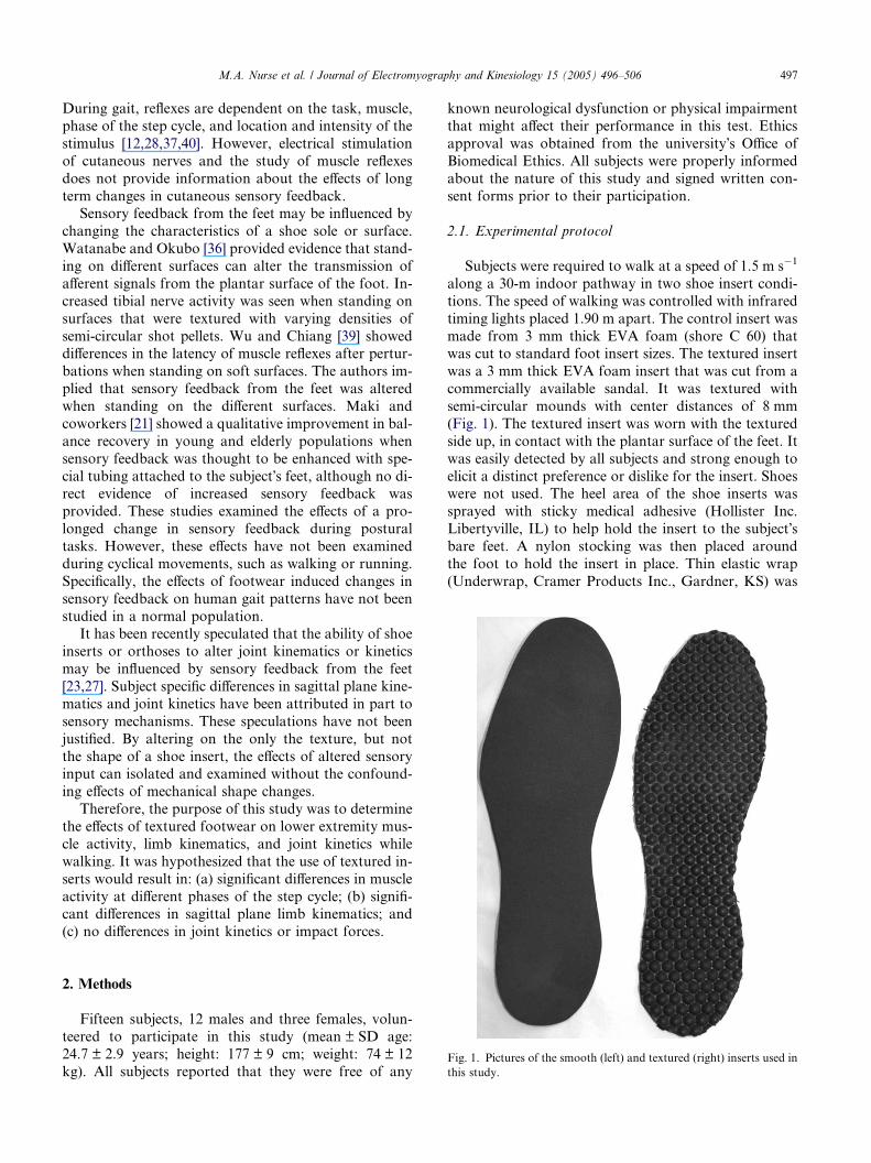
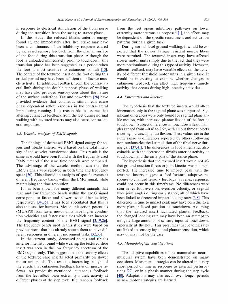
Fig. 1. Pictures of the smooth (left) and textured (right) inserts used in
this study.
2. Methods
Fifteen subjects, 12 males and three females, volun-
teered to participate in this study (mean ± SD age:24.7 ± 2.9 years; height: 177 ± 9 cm; weight: 74 ± 12
kg). All subjects reported that they were free of any
known neurological dysfunction or physical impairment
that might affect their performance in this test. Ethics
approval was obtained from the university�s Office of
Biomedical Ethics. All subjects were properly informed
about the nature of this study and signed written con-
sent forms prior to their participation.
2.1. Experimental protocol
Subjects were required to walk at a speed of 1.5 m s�1
along a 30-m indoor pathway in two shoe insert condi-
tions. The speed of walking was controlled with infrared
timing lights placed 1.90 m apart. The control insert was
made from 3 mm thick EVA foam (shore C 60) thatwas cut to standard foot insert sizes. The textured insert
was a 3 mm thick EVA foam insert that was cut from a
commercially available sandal. It was textured with
semi-circular mounds with center distances of 8 mm
(Fig. 1). The textured insert was worn with the textured
side up, in contact with the plantar surface of the feet. It
was easily detected by all subjects and strong enough to
elicit a distinct preference or dislike for the insert. Shoeswere not used. The heel area of the shoe inserts was
sprayed with sticky medical adhesive (Hollister Inc.
Libertyville, IL) to help hold the insert to the subject�sbare feet. A nylon stocking was then placed around
the foot to hold the insert in place. Thin elastic wrap
(Underwrap, Cramer Products Inc., Gardner, KS) was
498 M.A. Nurse et al. / Journal of Electromyography and Kinesiology 15 (2005) 496–506
then wrapped around the foot to reduce movement be-
tween the foot and the insert, and to increase friction be-
tween the nylon and the ground. The increased friction
prevented the foot from slipping on the floor surface,
especially during the push-off phase in late stance. All
subjects reported that they were comfortable and ableto produce natural gait patterns with the nylon stock-
ings and elastic wrap. Interventions were applied bilater-
ally to both feet.
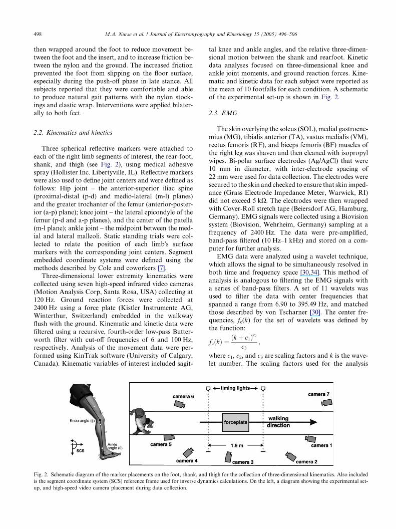
2.2. Kinematics and kinetics
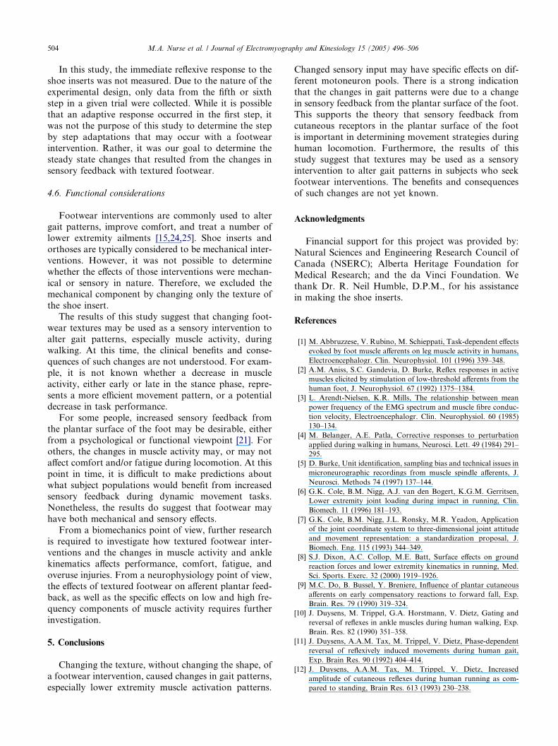
Three spherical reflective markers were attached to
each of the right limb segments of interest, the rear-foot,shank, and thigh (see Fig. 2), using medical adhesive
spray (Hollister Inc. Libertyville, IL). Reflective markers
were also used to define joint centers and were defined as
follows: Hip joint – the anterior-superior iliac spine
(proximal-distal (p-d) and medio-lateral (m-l) planes)
and the greater trochanter of the femur (anterior-poster-
ior (a-p) plane); knee joint – the lateral epicondyle of the
femur (p-d and a-p planes), and the center of the patella(m-l plane); ankle joint – the midpoint between the med-
ial and lateral malleoli. Static standing trials were col-
lected to relate the position of each limb�s surface
markers with the corresponding joint centers. Segment
embedded coordinate systems were defined using the
methods described by Cole and coworkers [7].
Three-dimensional lower extremity kinematics were
collected using seven high-speed infrared video cameras(Motion Analysis Corp, Santa Rosa, USA) collecting at
120 Hz. Ground reaction forces were collected at
2400 Hz using a force plate (Kistler Instrumente AG,
Winterthur, Switzerland) embedded in the walkway
flush with the ground. Kinematic and kinetic data were
filtered using a recursive, fourth-order low-pass Butter-
worth filter with cut-off frequencies of 6 and 100 Hz,
respectively. Analysis of the movement data were per-formed using KinTrak software (University of Calgary,
Canada). Kinematic variables of interest included sagit-
Fig. 2. Schematic diagram of the marker placements on the foot, shank, and
is the segment coordinate system (SCS) reference frame used for inverse dyna
up, and high-speed video camera placement during data collection.
tal knee and ankle angles, and the relative three-dimen-
sional motion between the shank and rearfoot. Kinetic
data analyses focused on three-dimensional knee and
ankle joint moments, and ground reaction forces. Kine-
matic and kinetic data for each subject were reported as
the mean of 10 footfalls for each condition. A schematicof the experimental set-up is shown in Fig. 2.
2.3. EMG
The skin overlying the soleus (SOL), medial gastrocne-
mius (MG), tibialis anterior (TA), vastus medialis (VM),
rectus femoris (RF), and biceps femoris (BF) muscles of
the right leg was shaven and then cleaned with isopropylwipes. Bi-polar surface electrodes (Ag/AgCl) that were
10 mm in diameter, with inter-electrode spacing of
22 mm were used for data collection. The electrodes were
secured to the skin and checked to ensure that skin imped-
ance (Grass Electrode Impedance Meter, Warwick, RI)
did not exceed 5 kX. The electrodes were then wrapped
with Cover-Roll stretch tape (Beiersdorf AG, Hamburg,
Germany). EMG signals were collected using a Biovisionsystem (Biovision, Wehrheim, Germany) sampling at a
frequency of 2400 Hz. The data were pre-amplified,
band-pass filtered (10 Hz–1 kHz) and stored on a com-
puter for further analysis.
EMG data were analyzed using a wavelet technique,
which allows the signal to be simultaneously resolved in
both time and frequency space [30,34]. This method of
analysis is analogous to filtering the EMG signals witha series of band-pass filters. A set of 11 wavelets was
used to filter the data with center frequencies that
spanned a range from 6.90 to 395.49 Hz, and matched
those described by von Tscharner [30]. The center fre-
quencies, fc(k) for the set of wavelets was defined by
the function:
fcðkÞ ¼ðk þ c1Þc2
c3;
where c1, c2, and c3 are scaling factors and k is the wave-
let number. The scaling factors used for the analysis
thigh for the collection of three-dimensional kinematics. Also included
mics calculations. On the left, a diagram showing the experimental set-
M.A. Nurse et al. / Journal of Electromyography and Kinesiology 15 (2005) 496–506 499
were equal to 1.45, 1.959, and 0.3 for c1, c2, and c3,
respectively [34].
The intensity of the wavelet transformed signals was
calculated as a function of both the square of the ampli-
tude and time-derivative (slope) of the wavelet-trans-
formed signal. A Gaussian filter was then applied toreduce high frequency artifacts that result fromfinite sam-
pling frequencies. The total signal intensity was defined as
the sum of all intensities from all wavelet bands, and is
equal to twice the square of the RMS value for the signal
when similar time periods are chosen. The wavelet tech-
nique has the advantage that it also allows the possibility
of examining the signal in different frequency (wavelet)
domains. Based on the results of previous studies [32],low and high frequency components of the EMG signal
were defined. The low frequency component (Ilow) was
calculated as the sum of the intensities of wavelets two
and three [30] with center frequencies of 37.71 and
62.09 Hz. The high frequency component (Ihigh) was de-
fined as the sum of wavelets six to eight, with center fre-
quencies ranging from 170.39 to 271.48 Hz.
The integrated intensity plot for any given waveletrepresents the energy of the signal in a given time and
frequency domain. For the purpose of this paper, refer-
ences about the total energy of a given muscle refer to
the total energy of the EMG signal of that muscle over
a given time frame. The total energy, as well as the low
and high frequency components of the EMG signal for
each muscle, were calculated for the entire stance phase,
defined as the period from heel strike to toe-off as deter-
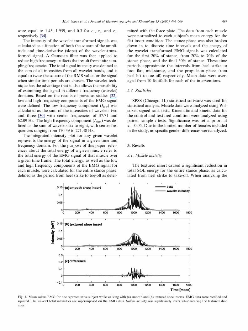
Fig. 3. Mean soleus EMG for one representative subject while walking with
squared. The wavelet total intensities are superimposed on the EMG data. S
insert.
mined with the force plate. The data from each muscle
were normalized to each subject�s mean energy for the
flat insert condition. The stance phase was also broken
down in to discrete time intervals and the energy of
the wavelet transformed EMG signals was calculated
for the first 20% of stance, from 20% to 70% of thestance phase, and the final 30% of stance. These time
periods approximate the intervals from heel strike to
foot flat, mid-stance, and the propulsion phase from
heel lift to toe off, respectively. Mean data were aver-
aged from 10 footfalls for each of the interventions.
2.4. Statistics
SPSS (Chicago, IL) statistical software was used for
statistical analysis. Muscle data were analyzed using Wil-
coxen signed rank tests. Kinematic and kinetic data for
the control and textured condition were analyzed using
paired sample t-tests. Significance was set a priori at
a = 0.05. Due to the limited number of females included
in the study, no specific gender differences were analyzed.
3. Results
3.1. Muscle activity
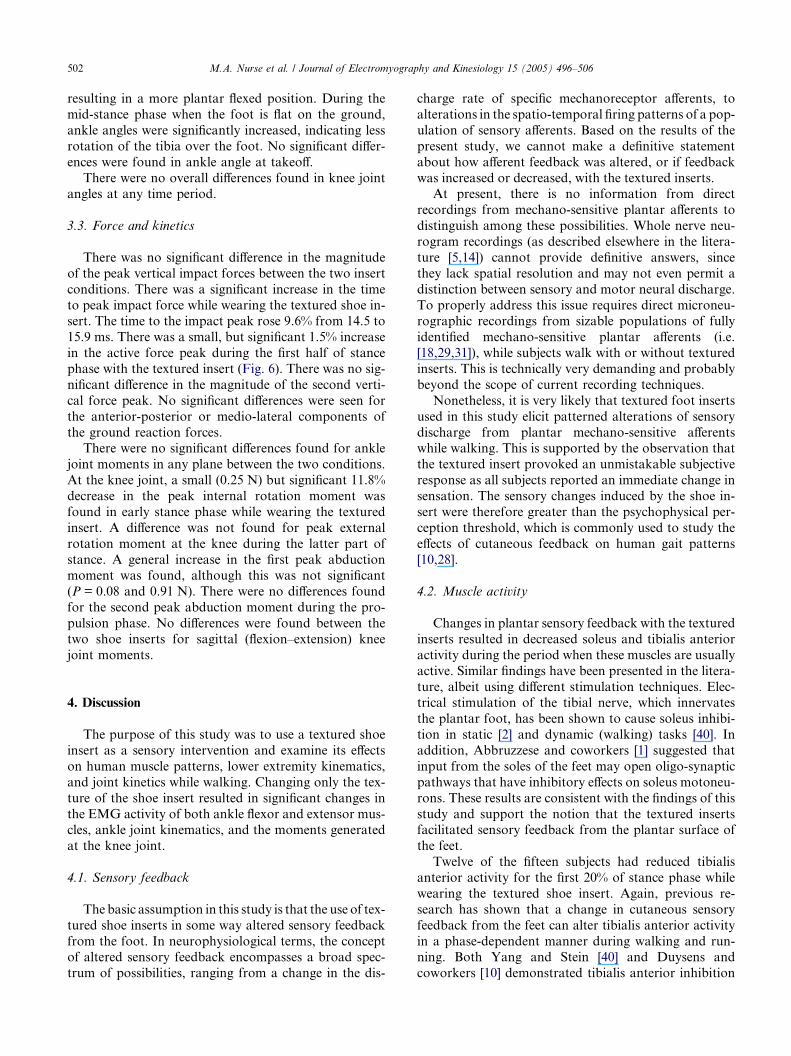
The textured insert caused a significant reduction in
total SOL energy for the entire stance phase, as calcu-
lated from heel strike to take-off. When analyzing the
(a) smooth and (b) textured shoe inserts. EMG data were rectified and
oleus activity was significantly lower while wearing the textured shoe
500 M.A. Nurse et al. / Journal of Electromyography and Kinesiology 15 (2005) 496–506
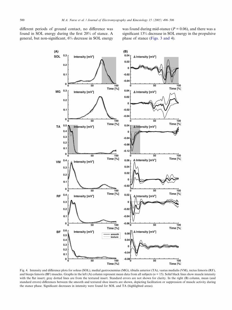
different periods of ground contact, no difference was
found in SOL energy during the first 20% of stance. A
general, but non-significant, 6% decrease in SOL energy
Fig. 4. Intensity and difference plots for soleus (SOL), medial gastrocnemius (
and biceps femoris (BF) muscles. Graphs in the left (A) column represent mea
with the flat insert; gray dotted lines are from the textured insert. Standard
standard errors) differences between the smooth and textured shoe inserts ar
the stance phase. Significant decreases in intensity were found for SOL and
was found during mid-stance (P = 0.06), and there was a
significant 13% decrease in SOL energy in the propulsive
phase of stance (Figs. 3 and 4).
MG), tibialis anterior (TA), vastus medialis (VM), rectus femoris (RF),
n data from all subjects (n = 15). Solid black lines show muscle intensity
errors are not shown for clarity. In the right (B) column, mean (and
e shown, depicting facilitation or suppression of muscle activity during
TA (highlighted areas).
M.A. Nurse et al. / Journal of Electromyography and Kinesiology 15 (2005) 496–506 501
A significant decrease in total TA energy was also
found for the entire stance phase while wearing the tex-
tured insert. In the first part of the stance phase imme-
diately after heel strike, there was a significant 13%
decrease in total TA energy (Fig. 4). There were no sig-
nificant differences found in TA energy for mid-stance,or the propulsion phase of ground contact.
In general, overall total RF energy was decreased
during for the entire stance phase (P = 0.08). The differ-
ences in RF energy occurred primarily in the early part
of stance phase. There were no significant differences
found for MG, VM, or BF energy while wearing the tex-
tured insert during the stance phase of gait.
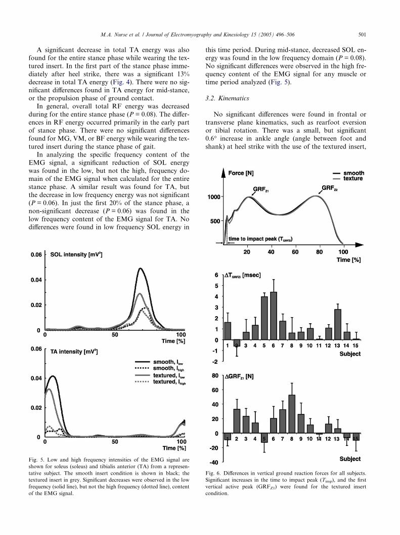
In analyzing the specific frequency content of theEMG signal, a significant reduction of SOL energy
was found in the low, but not the high, frequency do-
main of the EMG signal when calculated for the entire
stance phase. A similar result was found for TA, but
the decrease in low frequency energy was not significant
(P = 0.06). In just the first 20% of the stance phase, a
non-significant decrease (P = 0.06) was found in the
low frequency content of the EMG signal for TA. Nodifferences were found in low frequency SOL energy in
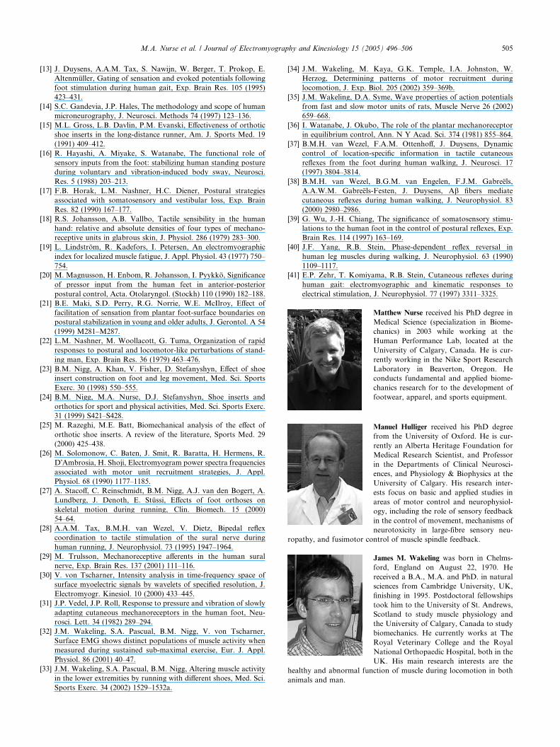
Fig. 5. Low and high frequency intensities of the EMG signal are
shown for soleus (soleus) and tibialis anterior (TA) from a represen-
tative subject. The smooth insert condition is shown in black; the
textured insert in grey. Significant decreases were observed in the low
frequency (solid line), but not the high frequency (dotted line), content
of the EMG signal.
this time period. During mid-stance, decreased SOL en-
ergy was found in the low frequency domain (P = 0.08).
No significant differences were observed in the high fre-
quency content of the EMG signal for any muscle or
time period analyzed (Fig. 5).
3.2. Kinematics
No significant differences were found in frontal or
transverse plane kinematics, such as rearfoot eversion
or tibial rotation. There was a small, but significant
0.6� increase in ankle angle (angle between foot and
shank) at heel strike with the use of the textured insert,
Fig. 6. Differences in vertical ground reaction forces for all subjects.
Significant increases in the time to impact peak (Timp), and the first
vertical active peak (GRFZ1) were found for the textured insert
condition.
502 M.A. Nurse et al. / Journal of Electromyography and Kinesiology 15 (2005) 496–506
resulting in a more plantar flexed position. During the
mid-stance phase when the foot is flat on the ground,
ankle angles were significantly increased, indicating less
rotation of the tibia over the foot. No significant differ-
ences were found in ankle angle at takeoff.
There were no overall differences found in knee jointangles at any time period.
3.3. Force and kinetics
There was no significant difference in the magnitude
of the peak vertical impact forces between the two insert
conditions. There was a significant increase in the time
to peak impact force while wearing the textured shoe in-sert. The time to the impact peak rose 9.6% from 14.5 to
15.9 ms. There was a small, but significant 1.5% increase
in the active force peak during the first half of stance
phase with the textured insert (Fig. 6). There was no sig-
nificant difference in the magnitude of the second verti-
cal force peak. No significant differences were seen for
the anterior-posterior or medio-lateral components of
the ground reaction forces.There were no significant differences found for ankle
joint moments in any plane between the two conditions.
At the knee joint, a small (0.25 N) but significant 11.8%
decrease in the peak internal rotation moment was
found in early stance phase while wearing the textured
insert. A difference was not found for peak external
rotation moment at the knee during the latter part of
stance. A general increase in the first peak abductionmoment was found, although this was not significant
(P = 0.08 and 0.91 N). There were no differences found
for the second peak abduction moment during the pro-
pulsion phase. No differences were found between the
two shoe inserts for sagittal (flexion–extension) knee
joint moments.
4. Discussion
The purpose of this study was to use a textured shoe
insert as a sensory intervention and examine its effects
on human muscle patterns, lower extremity kinematics,
and joint kinetics while walking. Changing only the tex-
ture of the shoe insert resulted in significant changes in
the EMG activity of both ankle flexor and extensor mus-cles, ankle joint kinematics, and the moments generated
at the knee joint.
4.1. Sensory feedback
The basic assumption in this study is that the use of tex-
tured shoe inserts in some way altered sensory feedback
from the foot. In neurophysiological terms, the conceptof altered sensory feedback encompasses a broad spec-
trum of possibilities, ranging from a change in the dis-
charge rate of specific mechanoreceptor afferents, to
alterations in the spatio-temporal firing patterns of a pop-
ulation of sensory afferents. Based on the results of the
present study, we cannot make a definitive statement
about how afferent feedback was altered, or if feedback
was increased or decreased, with the textured inserts.At present, there is no information from direct
recordings from mechano-sensitive plantar afferents to
distinguish among these possibilities. Whole nerve neu-
rogram recordings (as described elsewhere in the litera-
ture [5,14]) cannot provide definitive answers, since
they lack spatial resolution and may not even permit a
distinction between sensory and motor neural discharge.
To properly address this issue requires direct microneu-rographic recordings from sizable populations of fully
identified mechano-sensitive plantar afferents (i.e.
[18,29,31]), while subjects walk with or without textured
inserts. This is technically very demanding and probably
beyond the scope of current recording techniques.
Nonetheless, it is very likely that textured foot inserts
used in this study elicit patterned alterations of sensory
discharge from plantar mechano-sensitive afferentswhile walking. This is supported by the observation that
the textured insert provoked an unmistakable subjective
response as all subjects reported an immediate change in
sensation. The sensory changes induced by the shoe in-
sert were therefore greater than the psychophysical per-
ception threshold, which is commonly used to study the
effects of cutaneous feedback on human gait patterns
[10,28].
4.2. Muscle activity
Changes in plantar sensory feedback with the textured
inserts resulted in decreased soleus and tibialis anterior
activity during the period when these muscles are usually
active. Similar findings have been presented in the litera-
ture, albeit using different stimulation techniques. Elec-trical stimulation of the tibial nerve, which innervates
the plantar foot, has been shown to cause soleus inhibi-
tion in static [2] and dynamic (walking) tasks [40]. In
addition, Abbruzzese and coworkers [1] suggested that
input from the soles of the feet may open oligo-synaptic
pathways that have inhibitory effects on soleus motoneu-
rons. These results are consistent with the findings of this
study and support the notion that the textured insertsfacilitated sensory feedback from the plantar surface of
the feet.
Twelve of the fifteen subjects had reduced tibialis
anterior activity for the first 20% of stance phase while
wearing the textured shoe insert. Again, previous re-
search has shown that a change in cutaneous sensory
feedback from the feet can alter tibialis anterior activity
in a phase-dependent manner during walking and run-ning. Both Yang and Stein [40] and Duysens and
coworkers [10] demonstrated tibialis anterior inhibition
M.A. Nurse et al. / Journal of Electromyography and Kinesiology 15 (2005) 496–506 503
in response to electrical stimulation of the tibial nerve
during the transition from the swing to stance phase.
In this study, the reduced tibialis anterior energy
found at, and immediately after, heel strike may have
been a continuance of an inhibitory response caused
by increased sensory feedback from the plantar surfaceof the foot during this transition phase. Although the
foot is unloaded immediately prior to touchdown, this
transition phase has been suggested as a period when
the foot is most sensitive to cutaneous stimuli [13].
The contact of the textured insert on the foot during this
critical period may have been sufficient to influence mus-
cle activity. In addition, feedback from the contra-lat-
eral limb during the double support phase of walkingmay have also provided sensory cues about the nature
of the surface underfoot. Tax and coworkers [28] have
provided evidence that cutaneous stimuli can cause
phase dependent reflex responses in the contra-lateral
limb during running. It is reasonable to assume that
altering cutaneous feedback from the feet during normal
walking with textured inserts may also cause contra-lat-
eral responses.
4.3. Wavelet analysis of EMG signals
The findings of decreased EMG signal energy for so-
leus and tibialis anterior were based on the total inten-
sity of the wavelet transformed data. This result is the
same as would have been found with the frequently used
RMS method if the same time periods were compared.The advantage of the wavelet method was that the
EMG signals were resolved in both time and frequency
space [30]. This allowed an analysis of specific events at
different frequency bands within the EMG signal, while
maintaining the time resolution.
It has been shown for many different animals that
high and low frequency bands within the EMG signal
correspond to faster and slower twitch fiber activity,respectively [34,35]. It has been speculated that this is
also the case for humans. Motor unit action potentials
(MUAPS) from faster motor units have higher conduc-
tion velocities and faster rise times which can increase
the frequency content of the EMG signal [3,19,26].
The frequency bands used in this study were based on
previous work that has already shown them to have dif-
ferent responses in different movement tasks [32,33].In the current study, decreased soleus and tibialis
anterior intensity found while wearing the textured shoe
insert was seen in the low frequency spectrum of the
EMG signal only. This suggests that the sensory effects
of the textured shoe inserts acted primarily on slower
motor unit pools. This result is interesting in light of
the effects that cutaneous feedback have on muscle re-
flexes. As previously mentioned, cutaneous feedbackfrom the feet affect lower extremity muscle activity at
different phases of the step cycle. If cutaneous feedback
from the feet opens inhibitory pathways on lower
extremity motoneurons as proposed [1], the effects may
be dependent on the specific recruitment and activation
patterns during a given task.
During normal level-ground walking, it would be ex-
pected that the slower, fatigue resistant muscle fiberswere recruited. The textured insert may have affected
slower motor units simply due to the fact that they were
more predominant during this type of activity. However,
afferent feedback may have variable effects on the activ-
ity of different threshold motor units in a given task. It
would be interesting to examine whether changes in
cutaneous feedback can affect high frequency muscle
activity that occurs during high intensity activities.
4.4. Kinematics and kinetics
The hypothesis that the textured inserts would affect
kinematics only in the sagittal plane was supported. Sig-
nificant differences were only found for sagittal plane an-
kle motion, with increased plantar flexion of the foot at
touchdown. Subject differences in touchdown flexion an-gles ranged from�0.4� to 2.9�, with all but three subjects
showing increased plantar flexion. These values are in the
same range as differences reported by others following
non-noxious electrical stimulation of the tibial nerve dur-
ing gait [37,41]. The differences in foot kinematics also
coincide with the decrease in tibialis anterior activity at
touchdown and the early part of the stance phase.
The hypothesis that the textured insert would not af-fect ground reaction forces or joint kinetics was not sup-
ported. The increased time to impact peak with the
textured inserts suggest a feed-forward adaptive re-
sponse to changed sensory feedback, as reflex responses
could not occur in this timeframe. No differences were
seen in rearfoot eversion, eversion velocity, or sagittal
knee joint angles during early stance, all of which have
been linked to decreased impact loading rates [6,8]. Thisdifference in time to impact peak may have been due to a
more plantar flexed position at touchdown. Assuming
that the textured insert facilitated plantar feedback,
the changed loading rate may have been an attempt to
mitigate large amounts of sensory input at touchdown,
especially at the heel. This presumes that loading rates
are linked to sensory input and plantar sensation, which
may or may not be the case.
4.5. Methodological considerations
The adaptive capabilities of the mammalian neuro-
muscular system have been demonstrated on many
occasions. Movement strategies can be altered in a very
short period of time in response to external perturba-
tions [22], or in a phasic manner during the step cycle[40]. Adaptations may also occur over longer periods
as new motor strategies are learned.
504 M.A. Nurse et al. / Journal of Electromyography and Kinesiology 15 (2005) 496–506
In this study, the immediate reflexive response to the
shoe inserts was not measured. Due to the nature of the
experimental design, only data from the fifth or sixth
step in a given trial were collected. While it is possible
that an adaptive response occurred in the first step, it
was not the purpose of this study to determine the stepby step adaptations that may occur with a footwear
intervention. Rather, it was our goal to determine the
steady state changes that resulted from the changes in
sensory feedback with textured footwear.
4.6. Functional considerations
Footwear interventions are commonly used to altergait patterns, improve comfort, and treat a number of
lower extremity ailments [15,24,25]. Shoe inserts and
orthoses are typically considered to be mechanical inter-
ventions. However, it was not possible to determine
whether the effects of those interventions were mechan-
ical or sensory in nature. Therefore, we excluded the
mechanical component by changing only the texture of
the shoe insert.The results of this study suggest that changing foot-
wear textures may be used as a sensory intervention to
alter gait patterns, especially muscle activity, during
walking. At this time, the clinical benefits and conse-
quences of such changes are not understood. For exam-
ple, it is not known whether a decrease in muscle
activity, either early or late in the stance phase, repre-
sents a more efficient movement pattern, or a potentialdecrease in task performance.
For some people, increased sensory feedback from
the plantar surface of the foot may be desirable, either
from a psychological or functional viewpoint [21]. For
others, the changes in muscle activity may, or may not
affect comfort and/or fatigue during locomotion. At this
point in time, it is difficult to make predictions about
what subject populations would benefit from increasedsensory feedback during dynamic movement tasks.
Nonetheless, the results do suggest that footwear may
have both mechanical and sensory effects.
From a biomechanics point of view, further research
is required to investigate how textured footwear inter-
ventions and the changes in muscle activity and ankle
kinematics affects performance, comfort, fatigue, and
overuse injuries. From a neurophysiology point of view,the effects of textured footwear on afferent plantar feed-
back, as well as the specific effects on low and high fre-
quency components of muscle activity requires further
investigation.
5. Conclusions
Changing the texture, without changing the shape, of
a footwear intervention, caused changes in gait patterns,
especially lower extremity muscle activation patterns.
Changed sensory input may have specific effects on dif-
ferent motoneuron pools. There is a strong indication
that the changes in gait patterns were due to a change
in sensory feedback from the plantar surface of the foot.
This supports the theory that sensory feedback from
cutaneous receptors in the plantar surface of the footis important in determining movement strategies during
human locomotion. Furthermore, the results of this
study suggest that textures may be used as a sensory
intervention to alter gait patterns in subjects who seek
footwear interventions. The benefits and consequences
of such changes are not yet known.
Acknowledgments
Financial support for this project was provided by:
Natural Sciences and Engineering Research Council of
Canada (NSERC); Alberta Heritage Foundation for
Medical Research; and the da Vinci Foundation. We
thank Dr. R. Neil Humble, D.P.M., for his assistance
in making the shoe inserts.
References
[1] M. Abbruzzese, V. Rubino, M. Schieppati, Task-dependent effects
evoked by foot muscle afferents on leg muscle activity in humans,
Electroencephalogr. Clin. Neurophysiol. 101 (1996) 339–348.
[2] A.M. Aniss, S.C. Gandevia, D. Burke, Reflex responses in active
muscles elicited by stimulation of low-threshold afferents from the
human foot, J. Neurophysiol. 67 (1992) 1375–1384.
[3] L. Arendt-Nielsen, K.R. Mills, The relationship between mean
power frequency of the EMG spectrum and muscle fibre conduc-
tion velocity, Electroencephalogr. Clin. Neurophysiol. 60 (1985)
130–134.
[4] M. Belanger, A.E. Patla, Corrective responses to perturbation
applied during walking in humans, Neurosci. Lett. 49 (1984) 291–
295.
[5] D. Burke, Unit identification, sampling bias and technical issues in
microneurographic recordings from muscle spindle afferents, J.
Neurosci. Methods 74 (1997) 137–144.
[6] G.K. Cole, B.M. Nigg, A.J. van den Bogert, K.G.M. Gerritsen,
Lower extremity joint loading during impact in running, Clin.
Biomech. 11 (1996) 181–193.
[7] G.K. Cole, B.M. Nigg, J.L. Ronsky, M.R. Yeadon, Application
of the joint coordinate system to three-dimensional joint attitude
and movement representation: a standardization proposal, J.
Biomech. Eng. 115 (1993) 344–349.
[8] S.J. Dixon, A.C. Collop, M.E. Batt, Surface effects on ground
reaction forces and lower extremity kinematics in running, Med.
Sci. Sports. Exerc. 32 (2000) 1919–1926.
[9] M.C. Do, B. Bussel, Y. Breniere, Influence of plantar cutaneous
afferents on early compensatory reactions to forward fall, Exp.
Brain. Res. 79 (1990) 319–324.
[10] J. Duysens, M. Trippel, G.A. Horstmann, V. Dietz, Gating and
reversal of reflexes in ankle muscles during human walking, Exp.
Brain. Res. 82 (1990) 351–358.
[11] J. Duysens, A.A.M. Tax, M. Trippel, V. Dietz, Phase-dependent
reversal of reflexively induced movements during human gait,
Exp. Brain Res. 90 (1992) 404–414.
[12] J. Duysens, A.A.M. Tax, M. Trippel, V. Dietz, Increased
amplitude of cutaneous reflexes during human running as com-
pared to standing, Brain Res. 613 (1993) 230–238.
M.A. Nurse et al. / Journal of Electromyography and Kinesiology 15 (2005) 496–506 505
[13] J. Duysens, A.A.M. Tax, S. Nawijn, W. Berger, T. Prokop, E.
Altenmuller, Gating of sensation and evoked potentials following
foot stimulation during human gait, Exp. Brain Res. 105 (1995)
423–431.
[14] S.C. Gandevia, J.P. Hales, The methodology and scope of human
microneurography, J. Neurosci. Methods 74 (1997) 123–136.
[15] M.L. Gross, L.B. Davlin, P.M. Evanski, Effectiveness of orthotic
shoe inserts in the long-distance runner, Am. J. Sports Med. 19
(1991) 409–412.
[16] R. Hayashi, A. Miyake, S. Watanabe, The functional role of
sensory inputs from the foot: stabilizing human standing posture
during voluntary and vibration-induced body sway, Neurosci.
Res. 5 (1988) 203–213.
[17] F.B. Horak, L.M. Nashner, H.C. Diener, Postural strategies
associated with somatosensory and vestibular loss, Exp. Brain
Res. 82 (1990) 167–177.
[18] R.S. Johansson, A.B. Vallbo, Tactile sensibility in the human
hand: relative and absolute densities of four types of mechano-
receptive units in glabrous skin, J. Physiol. 286 (1979) 283–300.
[19] L. Lindstrom, R. Kadefors, I. Petersen, An electromyographic
index for localized muscle fatigue, J. Appl. Physiol. 43 (1977) 750–
754.
[20] M. Magnusson, H. Enbom, R. Johansson, I. Pyykko, Significance
of pressor input from the human feet in anterior-posterior
postural control, Acta. Otolaryngol. (Stockh) 110 (1990) 182–188.
[21] B.E. Maki, S.D. Perry, R.G. Norrie, W.E. McIlroy, Effect of
facilitation of sensation from plantar foot-surface boundaries on
postural stabilization in young and older adults, J. Gerontol. A 54
(1999) M281–M287.
[22] L.M. Nashner, M. Woollacott, G. Tuma, Organization of rapid
responses to postural and locomotor-like perturbations of stand-
ing man, Exp. Brain Res. 36 (1979) 463–476.
[23] B.M. Nigg, A. Khan, V. Fisher, D. Stefanyshyn, Effect of shoe
insert construction on foot and leg movement, Med. Sci. Sports
Exerc. 30 (1998) 550–555.
[24] B.M. Nigg, M.A. Nurse, D.J. Stefanyshyn, Shoe inserts and
orthotics for sport and physical activities, Med. Sci. Sports Exerc.
31 (1999) S421–S428.
[25] M. Razeghi, M.E. Batt, Biomechanical analysis of the effect of
orthotic shoe inserts. A review of the literature, Sports Med. 29
(2000) 425–438.
[26] M. Solomonow, C. Baten, J. Smit, R. Baratta, H. Hermens, R.
D�Ambrosia, H. Shoji, Electromyogram power spectra frequencies
associated with motor unit recruitment strategies, J. Appl.
Physiol. 68 (1990) 1177–1185.
[27] A. Stacoff, C. Reinschmidt, B.M. Nigg, A.J. van den Bogert, A.
Lundberg, J. Denoth, E. Stussi, Effects of foot orthoses on
skeletal motion during running, Clin. Biomech. 15 (2000)
54–64.
[28] A.A.M. Tax, B.M.H. van Wezel, V. Dietz, Bipedal reflex
coordination to tactile stimulation of the sural nerve during
human running, J. Neurophysiol. 73 (1995) 1947–1964.
[29] M. Trulsson, Mechanoreceptive afferents in the human sural
nerve, Exp. Brain Res. 137 (2001) 111–116.
[30] V. von Tscharner, Intensity analysis in time-frequency space of
surface myoelectric signals by wavelets of specified resolution, J.
Electromyogr. Kinesiol. 10 (2000) 433–445.
[31] J.P. Vedel, J.P. Roll, Response to pressure and vibration of slowly
adapting cutaneous mechanoreceptors in the human foot, Neu-
rosci. Lett. 34 (1982) 289–294.
[32] J.M. Wakeling, S.A. Pascual, B.M. Nigg, V. von Tscharner,
Surface EMG shows distinct populations of muscle activity when
measured during sustained sub-maximal exercise, Eur. J. Appl.
Physiol. 86 (2001) 40–47.
[33] J.M. Wakeling, S.A. Pascual, B.M. Nigg, Altering muscle activity
in the lower extremities by running with different shoes, Med. Sci.
Sports Exerc. 34 (2002) 1529–1532a.
[34] J.M. Wakeling, M. Kaya, G.K. Temple, I.A. Johnston, W.
Herzog, Determining patterns of motor recruitment during
locomotion, J. Exp. Biol. 205 (2002) 359–369b.
[35] J.M. Wakeling, D.A. Syme, Wave properties of action potentials
from fast and slow motor units of rats, Muscle Nerve 26 (2002)
659–668.
[36] I. Watanabe, J. Okubo, The role of the plantar mechanoreceptor
in equilibrium control, Ann. N Y Acad. Sci. 374 (1981) 855–864.
[37] B.M.H. van Wezel, F.A.M. Ottenhoff, J. Duysens, Dynamic
control of location-specific information in tactile cutaneous
reflexes from the foot during human walking, J. Neurosci. 17
(1997) 3804–3814.
[38] B.M.H. van Wezel, B.G.M. van Engelen, F.J.M. Gabreels,
A.A.W.M. Gabreels-Festen, J. Duysens, Ab fibers mediate
cutaneous reflexes during human walking, J. Neurophysiol. 83
(2000) 2980–2986.
[39] G. Wu, J.-H. Chiang, The significance of somatosensory stimu-
lations to the human foot in the control of postural reflexes, Exp.
Brain Res. 114 (1997) 163–169.
[40] J.F. Yang, R.B. Stein, Phase-dependent reflex reversal in
human leg muscles during walking, J. Neurophysiol. 63 (1990)
1109–1117.
[41] E.P. Zehr, T. Komiyama, R.B. Stein, Cutaneous reflexes during
human gait: electromyographic and kinematic responses to
electrical stimulation, J. Neurophysiol. 77 (1997) 3311–3325.
Matthew Nurse received his PhD degree in
Medical Science (specialization in Biome-
chanics) in 2003 while working at the
Human Performance Lab, located at the
University of Calgary, Canada. He is cur-
rently working in the Nike Sport Research
Laboratory in Beaverton, Oregon. He
conducts fundamental and applied biome-
chanics research for to the development of
footwear, apparel, and sports equipment.
Manuel Hulliger received his PhD degree
from the University of Oxford. He is cur-
rently an Alberta Heritage Foundation for
Medical Research Scientist, and Professor
in the Departments of Clinical Neurosci-
ences, and Physiology & Biophysics at the
University of Calgary. His research inter-
ests focus on basic and applied studies in
areas of motor control and neurophysiol-
ogy, including the role of sensory feedback
in the control of movement, mechanisms of
neurotoxicity in large-fibre sensory neu-
ropathy, and fusimotor control of muscle spindle feedback.
James M. Wakeling was born in Chelms-
ford, England on August 22, 1970. He
received a B.A., M.A. and PhD. in natural
sciences from Cambridge University, UK,
finishing in 1995. Postdoctoral fellowships
took him to the University of St. Andrews,
Scotland to study muscle physiology and
the University of Calgary, Canada to study
biomechanics. He currently works at The
Royal Veterinary College and the Royal
National Orthopaedic Hospital, both in the
UK. His main research interests are the
healthy and abnormal function of muscle during locomotion in both
animals and man.
506 M.A. Nurse et al. / Journal of Electromyography and Kinesiology 15 (2005) 496–506
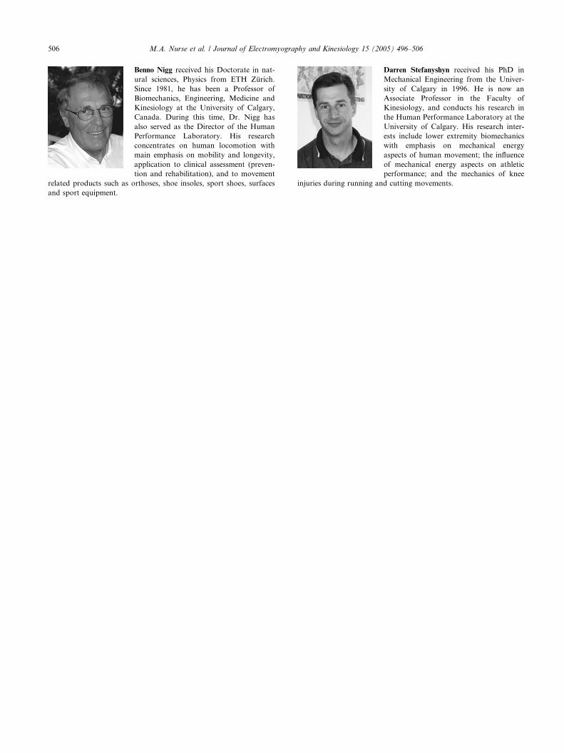
Benno Nigg received his Doctorate in nat-
ural sciences, Physics from ETH Zurich.
Since 1981, he has been a Professor of
Biomechanics, Engineering, Medicine and
Kinesiology at the University of Calgary,
Canada. During this time, Dr. Nigg has
also served as the Director of the Human
Performance Laboratory. His research
concentrates on human locomotion with
main emphasis on mobility and longevity,
application to clinical assessment (preven-
tion and rehabilitation), and to movement
related products such as orthoses, shoe insoles, sport shoes, surfaces
and sport equipment.
Darren Stefanyshyn received his PhD in
Mechanical Engineering from the Univer-
sity of Calgary in 1996. He is now an
Associate Professor in the Faculty of
Kinesiology, and conducts his research in
the Human Performance Laboratory at the
University of Calgary. His research inter-
ests include lower extremity biomechanics
with emphasis on mechanical energy
aspects of human movement; the influence
of mechanical energy aspects on athletic
performance; and the mechanics of knee
injuries during running and cutting movements.