Melatonin ameliorates ionizing radiation-induced oxidative organ damage in rats
Upload
uni-goettingenCategory
view
0download
0
This article appeared in a journal published by Elsevier. The attachedcopy is furnished to the author for internal non-commercial researchand education use, including for instruction at the authors institution
and sharing with colleagues.
Other uses, including reproduction and distribution, or selling orlicensing copies, or posting to personal, institutional or third party
websites are prohibited.
In most cases authors are permitted to post their version of thearticle (e.g. in Word or Tex form) to their personal website orinstitutional repository. Authors requiring further information
regarding Elsevier’s archiving and manuscript policies areencouraged to visit:
http://www.elsevier.com/copyright
Author's personal copy
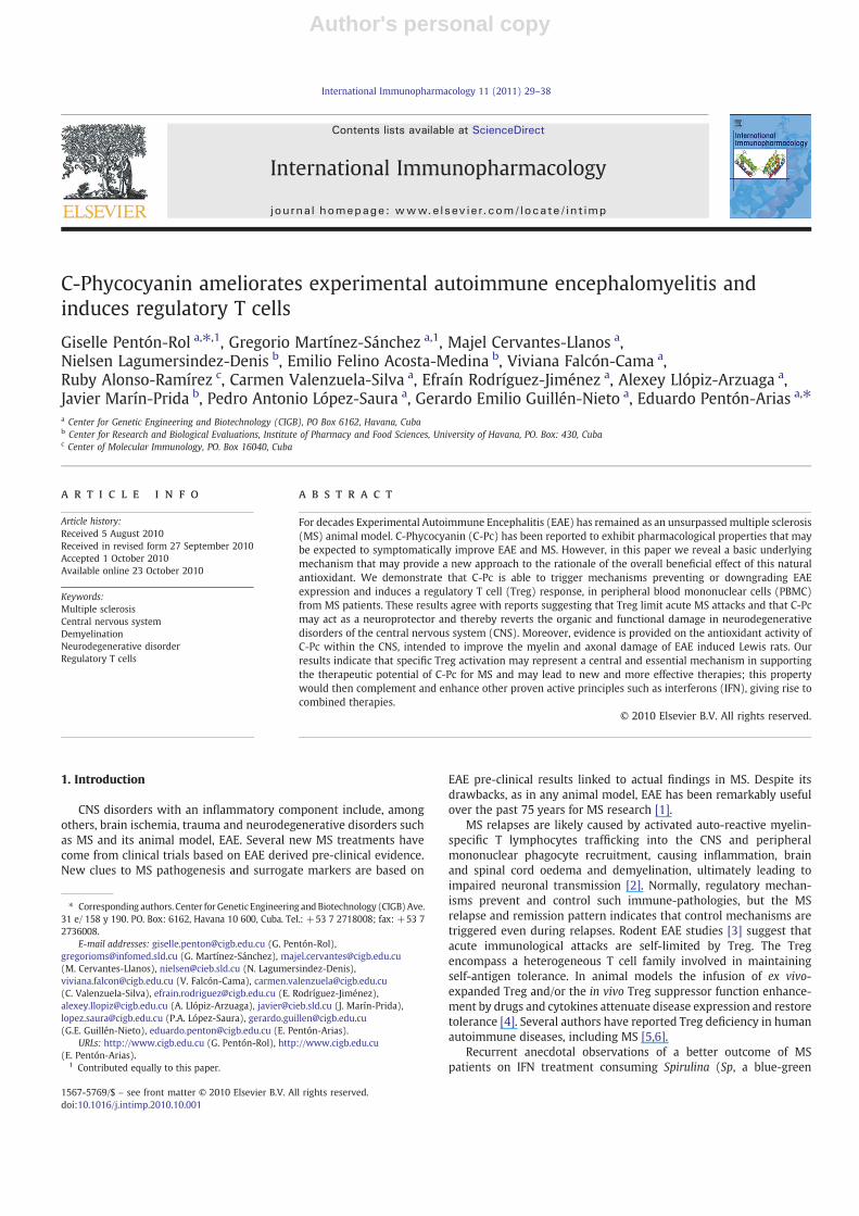
C-Phycocyanin ameliorates experimental autoimmune encephalomyelitis andinduces regulatory T cells
Giselle Pentón-Rol a,⁎,1, Gregorio Martínez-Sánchez a,1, Majel Cervantes-Llanos a,Nielsen Lagumersindez-Denis b, Emilio Felino Acosta-Medina b, Viviana Falcón-Cama a,Ruby Alonso-Ramírez c, Carmen Valenzuela-Silva a, Efraín Rodríguez-Jiménez a, Alexey Llópiz-Arzuaga a,Javier Marín-Prida b, Pedro Antonio López-Saura a, Gerardo Emilio Guillén-Nieto a, Eduardo Pentón-Arias a,⁎a Center for Genetic Engineering and Biotechnology (CIGB), PO Box 6162, Havana, Cubab Center for Research and Biological Evaluations, Institute of Pharmacy and Food Sciences, University of Havana, PO. Box: 430, Cubac Center of Molecular Immunology, PO. Box 16040, Cuba
a b s t r a c ta r t i c l e i n f o
Article history:Received 5 August 2010Received in revised form 27 September 2010Accepted 1 October 2010Available online 23 October 2010
Keywords:Multiple sclerosisCentral nervous systemDemyelinationNeurodegenerative disorderRegulatory T cells
For decades Experimental Autoimmune Encephalitis (EAE) has remained as an unsurpassed multiple sclerosis(MS) animal model. C-Phycocyanin (C-Pc) has been reported to exhibit pharmacological properties that maybe expected to symptomatically improve EAE and MS. However, in this paper we reveal a basic underlyingmechanism that may provide a new approach to the rationale of the overall beneficial effect of this naturalantioxidant. We demonstrate that C-Pc is able to trigger mechanisms preventing or downgrading EAEexpression and induces a regulatory T cell (Treg) response, in peripheral blood mononuclear cells (PBMC)from MS patients. These results agree with reports suggesting that Treg limit acute MS attacks and that C-Pcmay act as a neuroprotector and thereby reverts the organic and functional damage in neurodegenerativedisorders of the central nervous system (CNS). Moreover, evidence is provided on the antioxidant activity ofC-Pc within the CNS, intended to improve the myelin and axonal damage of EAE induced Lewis rats. Ourresults indicate that specific Treg activation may represent a central and essential mechanism in supportingthe therapeutic potential of C-Pc for MS and may lead to new and more effective therapies; this propertywould then complement and enhance other proven active principles such as interferons (IFN), giving rise tocombined therapies.
© 2010 Elsevier B.V. All rights reserved.
1. Introduction
CNS disorders with an inflammatory component include, amongothers, brain ischemia, trauma and neurodegenerative disorders suchas MS and its animal model, EAE. Several new MS treatments havecome from clinical trials based on EAE derived pre-clinical evidence.New clues to MS pathogenesis and surrogate markers are based on
EAE pre-clinical results linked to actual findings in MS. Despite itsdrawbacks, as in any animal model, EAE has been remarkably usefulover the past 75 years for MS research [1].
MS relapses are likely caused by activated auto-reactive myelin-specific T lymphocytes trafficking into the CNS and peripheralmononuclear phagocyte recruitment, causing inflammation, brainand spinal cord oedema and demyelination, ultimately leading toimpaired neuronal transmission [2]. Normally, regulatory mechan-isms prevent and control such immune-pathologies, but the MSrelapse and remission pattern indicates that control mechanisms aretriggered even during relapses. Rodent EAE studies [3] suggest thatacute immunological attacks are self-limited by Treg. The Tregencompass a heterogeneous T cell family involved in maintainingself-antigen tolerance. In animal models the infusion of ex vivo-expanded Treg and/or the in vivo Treg suppressor function enhance-ment by drugs and cytokines attenuate disease expression and restoretolerance [4]. Several authors have reported Treg deficiency in humanautoimmune diseases, including MS [5,6].
Recurrent anecdotal observations of a better outcome of MSpatients on IFN treatment consuming Spirulina (Sp, a blue-green
International Immunopharmacology 11 (2011) 29–38
⁎ Corresponding authors. Center forGenetic Engineering andBiotechnology (CIGB)Ave.31 e/ 158 y 190. PO. Box: 6162, Havana 10 600, Cuba. Tel.: +53 7 2718008; fax: +53 72736008.
E-mail addresses: [email protected] (G. Pentón-Rol),[email protected] (G. Martínez-Sánchez), [email protected](M. Cervantes-Llanos), [email protected] (N. Lagumersindez-Denis),[email protected] (V. Falcón-Cama), [email protected](C. Valenzuela-Silva), [email protected] (E. Rodríguez-Jiménez),[email protected] (A. Llópiz-Arzuaga), [email protected] (J. Marín-Prida),[email protected] (P.A. López-Saura), [email protected](G.E. Guillén-Nieto), [email protected] (E. Pentón-Arias).
URLs: http://www.cigb.edu.cu (G. Pentón-Rol), http://www.cigb.edu.cu(E. Pentón-Arias).
1 Contributed equally to this paper.
1567-5769/$ – see front matter © 2010 Elsevier B.V. All rights reserved.doi:10.1016/j.intimp.2010.10.001
Contents lists available at ScienceDirect
International Immunopharmacology
j ourna l homepage: www.e lsev ie r.com/ locate / in t imp

Author's personal copy
algae), suggested the hypothesis that the main active ingredient ofSpirulina, C-Pc, may be responsible for this effect and, if so, that thecombination of C-Pc/IFN could optimize the overall performance.Since IFNs have been thoroughly studied and established for MStreatment, in this paper the EAE model was used to characterize theeffect of C-Pc (the most important Sp biliprotein, i.e. proteins – e.g.,colored – bearing tetrapyrrolic light harvesting – phycocyanobilin –
prosthetic groups) that exhibits a variety of biological and pharma-cological properties. C-Pc, has significant antioxidant and radicalscavenging properties [7,8], it inhibits mouse ear inflammation [9],prevents acetic acid induced rat colitis [10] and has anti-arthriticproperties [11]. It also inhibits COX-2 [12,13], inflammatory allergicreactions [14], prostaglandins, leukotrienes [15] and TNF-alphaproduction [16].These actions are expected to improve EAE inducedanimals, mainly because Sp/C-Pc have been shown to restore theorganic and functional neuronal damage linked to senility [17] andneurodegenerative disorders [18–20]. However, here we demonstratean additional and probably more fundamental property of C-Pc, itsability to trigger Treg subset induction. The relative weight andimportance of each mechanism for the overall performance of C-Pcmust still be determined, but Tregmay have a main role given by theircentral position in regulating immune response. The hypothesis thatC-Pc could induce this Treg subset was tested in PBMC from MSpatients and healthy controls (HC). Results suggest that this Tregsubset would largely mediate the C-Pc effect.
2. Material and methods
2.1. C-Phycocyanin
C-Pc was purified according to Boussiba and Richmond [21] withcertainmodifications. The purity of the preparationwas always higherthan 90 %. Briefly, approximately 10 g of freshly Arthrospira platensisbiomass (Genix Co., Cuba) were resuspended in 200 mL of 0.1 Msodium phosphate buffer, pH 7.0. The cells were disrupted using aFrench pressure cell (Ohtake, Japan), with one cycle in the range of1200–1500 kg/cm2. The insoluble material was eliminated by centri-fugation at 10000 g for 15 min at 4 °C. The crude extract wasprecipitated with 50% saturated ammonium sulfate. The supernatantwas discarded and the blue pellet was dissolved in 10 mL of 2.5 mMsodium phosphate buffer, pH 7.0 and dialyzed overnight against thesame buffer.
The dialyzed protein was applied to a hydroxylapatite column(dimensions 25 cm) fromBio-Rad Laboratories, USA. The columnwasequilibrated with 2.5 mM sodium phosphate buffer, pH 7.0 whileallophycocyanin was eluted in 100 mM sodium phosphate buffer, pH7.0 at a flow rate of 37 mL/h. The C-Pc-rich fraction was subsequentlypurified in a DEAE-cellulose column (dimensions 4×30 cm; What-man, UK), pre-equilibrated with 5 mM sodium phosphate buffer, pH7.0 at a flow of 55 mL/h [22]. The C-Pc eluted in 0.29 M sodiumphosphate buffer, pH 7.0. Spectroscopic measurements were doneusing a UV–visible spectrophotometer (GE Healthcare, Waukesha,WI, USA). SDS-PAGE analysis was performed according to Laemmli[23] on 15% polyacrylamide gel at 30 mA. The proteins were stainedusing silver stain according to Heukeshoven and Dernick [24]. Themolecular weight marker was obtained from BioRad (BioRad,Hercules, CA, USA). A purity value of 4.1, measured as the A620/A280
absorbance ratio, was obtained (Fig. 1). The C-Pc was stored as alyophilized powder in the dark at −80 °C for later use.
The endotoxin content in the lyophilized C-Pc was determinedthrough the Limulus amoebocyte lysate (LAL) kinetic turbidimetricassay as described by Park et al., [25].
The purity level of the batches used in the preclinical studies waschecked just before the start of each study. The endotoxin level in thedoses used in rats never exceeded 5 units per kilogram of weight.
2.2. Animals
Male Lewis rats, 6–8 weeks old, were purchased from CENPALAB(Havana, Cuba). Rats were housed in Plexiglas cages, maintained in anair-filtered and temperature-conditioned (20–22 °C) room with arelative humidity of 50–52 % and under an artificial light/darknesscycle of 12 h. Rats were fed with a standard laboratory chow andwater ad libitum. Procedures involving animals and their care wereconducted according to institutional guidelines that comply withnational and international regulations and policies (EEC CouncilDirective 86/609, OJL 358, 1, 12 December 1987; Guide for the Care andUse of Laboratory Animals, US National Research Council, 1996).
2.3. EAE induction
On day 0, EAE was induced by a sub-plantar injection of 0.1 mL ofthe encephalitogen [3 g of the spinal cord from female SpragueDawley rats homogenized with 7.5 mL bi-distilled water, 3.8 mLphenol and 7.5 mL of complete Freund's adjuvant (CFA)] [26] into theleft hind paw. An equal volume of Bordetella pertussis vaccineconcentrate (200×109 organisms/mL, Institute Finlay Labs., LaHabana, Cuba) was injected into the same paw. Rats were examinedblindly for signs of EAE and scored as follows: 0, no disease; 1, flaccidtail; 2, hind limb weakness that impaired righting; 3, complete hindlimb paralysis with urinary incontinence; 4, paraplegia with forelimbweakness, moribund condition; 5, death. Animals were observed for24 days after immunization with encephalitogen.
2.4. Treatment schedules
Experimental groups formed by 10 animals were treated eitherwith saline, hydrocortisone (1 mg/kg/day) s.c or C-Pc (25 mg/kg/day)i.p. daily, from day 0 to day 12 after immunization (therapeutic). Anadditional group (prophylactic) was treated with C-Pc (25 mg/kg/day) on days 0 to 12 before immunization.
2.5. Samples
After the study period, all animals (including controls) wereanaesthetized with urethane (1 g/kg, i.p). Blood samples were obtainedfrom the abdominal aorta; the serum was obtained and stored at−70 °C until use. Afterwards, representative samples of brain weretaken for histo-pathological and biochemical studies. Brain homoge-nates were achieved using a tissue homogenator Edmund Bülher LBMAat4 °C. Thehomogenateswereprepared byusing a 50 mMKCl/histidinebuffer pH 7.4, 1:10 (w/v) and were spun downwith a Sigma Centrifuge2 K15, at 4 °C and 8500 g for 20 min. Redox biomarkerswere quantifiedin the supernatants of brain homogenates and serum.
Fig. 1. Evaluation of C-Pc purity obtained after ionic exchange through (A) Absorptionspectra and (B) silver staining PAGE gel. From left to right the lanes are: 1, molecularweight marker, 1 μL; 2, C-Pc, 10 μg. The ratio A620/A280 was greater than 4.
30 G. Pentón-Rol et al. / International Immunopharmacology 11 (2011) 29–38

Author's personal copy
2.6. Biochemical methods
All biochemical parameters were determined by spectrophoto-metric methods using an Ultrospect Plus Spectrophotometer fromPharmacia LKB.
Concentrations of Malondialdehyde (MDA) were analyzed withthe LPO-586 kit obtained from Calbiochem (La Jolla, CA., USA). Thestable chromophore produced after 40 min of incubation at 45 °C wasmeasured at a wavelength of 586 nm. Freshly prepared solutions ofmalondialdehyde bis [dimethyl acetal] (Sigma, St. Louis, MO., USA)assayed under identical conditions were used as reference standards[27].
For the evaluation of lipid peroxidation (PP) susceptibility,samples were incubated with a cupric sulfate solution (2 mM) at37 °C for 24 h. The PP was calculated by subtracting the MDAconcentration obtained at time 0 from that obtained at 24 h [28].
Total organoperoxides (TOP) were measured by Bioxytech H2O2-560 kit Cat. 21024 (Oxis International Inc. Portland, USA). The assay isbased on the oxidation of Fe 2+ ions to Fe 3+ ions by hydroperoxidesunder acidic conditions [29].
The oxidation of the iodide anion to diatomic iodine was used toassess Advanced Oxidation of Protein Products (AOPP) [30].
Ferric Reducing Ability of Plasma (FRAP) was assayed through thereduction of Fe3+ to Fe2+ by serum or reference (ascorbic acid). TheFe2+-2,4,6-tripiridil-s-triazine complex was detected at 593 nm [31].In brain homogenates total protein concentration was determined bythe Bradford method using bovine serum albumin as the standard[32]. Unless otherwise stated, all chemicals were obtained from SigmaChemical Company (St. Louis, MO., USA).
2.7. Electron microscopy studies
Transmission electron microscopy (TEM) studies were carried outin brain biopsies from EAE rats treated or not with C-Pc. Brain biopsiesof male Lewis rats were used as the negative control. Samples werefixed for 1 h at 4 °C in 1% (v/v) glutaraldehyde and 4% (v/v)paraformaldehyde, rinsed in 0.1 M sodium cacodylate (pH 7.4),post-fixed for 1 h at 4 °C in 1% OsO4, and dehydrated in increasingconcentrations of ethanol. Embedding was carried out as previouslydescribed with minor modifications [33]. Briefly, ultrathin sections(400–500 Ao) made with an ultramicrotome (NOVA, LKB) wereplaced on 400 mesh grids, stained with saturated uranyl acetate andlead citrate, and examined with a JEOL/JEM 2000 EX transmissionelectron microscope (JEOL, Japan). In order to avoid sample bias, 100microphotographs were analyzed in this study.
2.8. Isolation of human cells
PBMCs were isolated from heparinized blood of healthy humandonors and patients with MS by Ficoll density gradient centrifugation(Ficoll paque TM plus, Amersham Biosciences, Endotoxin tested).PBMC were resuspended at 2x10⁶ cells/mL with RPMI 1640supplemented with 25 mM HEPES buffer with L-glutamine (GIBCO)and cultured at 37 °Cwith 5% of CO2 in the presence or absence of C-Pc(200 μg/106 cells). Cells were harvested 4 and 72 h later, for RT-PCRand flow cytometry analysis, respectively. Human PBMCs wereobtained after informed consent in accordance to the proceduresapproved by the human research ethics committee and the studyprotocol complying with the institutional review board guidelines.
2.9. RT-PCR
Total RNA was isolated using Tri reagent (Sigma) [34] and wasreverse transcribed using the Perkin Elmer core kit [35]. cDNAreaction was divided into two Eppendorf tubes and a specific pair ofprimers was used for the polymerase chain reaction (PCR). Glycer-
aldehide-3 phosphate dehydrogenase (GAPDH) was used as thehousekeeping gene. The following primers were used, GAPDHprimers: for 5′-CCA TGG AGA AGG CTG GGG-3′ and back 5′-CAAAGT TGT CAT GGA TGA CC-3′; TGF-β primers: for 5′-CAA GCA GAGTAC ACA CAG CA-3′ and back 5′-GAT GCT GGG CCC TCT CAA GC-3′; IL-10 primers: for 5′-AAC AAG ACC CAG ACA TCA AG-3′ and back 5′-GAGGTA CAA TAA GGT TTC TCA AG-3′; CD25 primers: for 5′-CTA CAA GGAAGG AAC CAT GTT GAA C-3′ and back 5′-AGG TGA GCC CAC TCA GGAGGA GGA C-3′; Foxp3 primers: for 5′-CAG GCC ACA TTT CAT GCA CC-3′ and back 5′-ACA CCA TTT GCC AGC AGT GG-3′. Amplification wasperformed with an automated thermal cycler (Eppendorf) at 94 °C for30 s, 56 °C for 30 s and 72 °C for 30 s. It was stopped at 35 cycles. PCRproducts were run in 1.5 % agarose gel containing ethidium bromideand then the density of each band was quantified using the MolecularAnalysis Program (Molecular Analysis software for Windows, version1.4.1). The results were expressed as a ratio calculated from theoptical density (OD) of a defined area of the amplified gene productsdivided by the corresponding OD of the amplified GAPDH PCRproduct.
2.10. Flow cytometry analysis
Cells were washed with ice-cold PBS, adjusted at 0.5×10⁶cells/well, and incubated with heat inactivated rabbit sera for blocking theunspecific binding to the Fc receptor. They were directly stained for30 min at 4 °C with Phycoerythrin (PE) and CyChrome (PE-Cy5)labelled antibodies that are specific for human CD4 (MCA1267PE,Serotec) and CD25 (MCA1570C, Serotec) molecules. For the specificdetection of the Foxp3 subset, the cells were additionally fixed,permeabilized and stainedwith anti-human Foxp3-Fluorescein (FITC)conjugated (PCH101; eBiosciences, San Diego, CA, USA). The specificdetection of the CD69+ subset was achieved by directly stainingFluorescein (FITC) conjugated with anti-human CD69 (MCA1442F,Serotec). After staining, cells were washed and acquired using aFACScan flow cytometer (Becton Dickinson, Mansfield, MA). Theresults were analyzed with WinMDI 2.8 software. The region used toset Treg was the gate for CD4+CD25high T cells [36]. The Foxp3 subsetwas expressed as indicated by Oh et al., [37].
2.11. Statistical analysis
Data was processed with the EPIDAT software version 3.0(Bayesian module). Quantitative variables were expressed as mean±standard deviation (SD) or median±interquartile range (QR). Withthese variables the assumptions of normality (Shapiro-Wilks test) andvariance homogeneity (Levene's test) were both verified.
To analyze the markers we estimated the mean differencebetween the C-Pc treated and control groups. The groups werecompared estimating the 95% and 90% confidence interval (95% CI and90% CI) for the difference for each parameter with the Bayesianapproach.
3. Results
3.1. C-Pc ameliorates EAE clinical symptoms in Lewis rats
As functions of the clinical score patterns we estimated: the areaunder the curve (AUC, global magnitude of symptoms), themaximumscore (maxSC, max clinical score, maximum severity achieved) andthe duration of symptoms (maxTime), as global measurementsdescribing performance according to time.
We first analyzed the effects of C-Pc prophylactic and therapeuticschedules on EAE in adult male Lewis rats (Fig. 2). The treatment withC-Pc (25 mg/kg) in a prophylactic schedule prevented the develop-ment of EAE in all animals. In contrast, the non-treated animalsdisplayed severe EAE symptoms. With the three estimated variables
31G. Pentón-Rol et al. / International Immunopharmacology 11 (2011) 29–38

Author's personal copy
(AUC, maxSC, maxTime) the equality probability for the prophylacticschedule is higher than 0.95 [95% CI “C-Pc prophylactic–control”(−0.01; 0.01)], which indicates that C-Pc completely abrogateddisease onset (Fig. 2A).
The C-Pc therapeutic schedule reduced the maxSC of the diseasecompared to the untreated EAE group (Fig. 2B) [90% CI “C-Pctherapeutic–EAE” (0.12; 1.88)]. The therapeutic effect of C-Pc wasevidenced after 12 days of treatment, when a reduction of the clinicalscore was initially observed. However, from this point on, the diseasein the control group was exacerbated reaching the highest clinicalscore on day 18, contrasting with the C-Pc treated animals.
In the EAE group there was an increase in the severity of theclinical signs, which began to descend on day 18 and reached the level
of the control group on day 24 following induction, in contrast withthe C-Pc treated group where the duration of the disease was 19 days.In the hydrocortisone treated group we only observed mild signs andthe values were near those of the non-induced control group [95% CI“hydrocortisone–control” (−13.1; 2.1)]. These results support theconclusion that C-Pc doses of 25 mg/kg decreased the meancumulative score during the prophylactic or therapeutic schedule.
The maximal clinical score achieved in the disease is representedfor each animal in Fig. 2C. Animals receiving the C-Pc prophylactictreatment showed no clinical signs of the disease (score=0), whichwas similar to the control group (non-EAE induced). In the untreatedEAE group, the disease score ranged from 3–5, in the steroid treatedgroup it was 0–2 and in the C-Pc treated group 2–3.
Fig. 2. Protective effect of C-Phycocyanin treatment in acute EAE in Lewis rats. Rats were immunized for EAE induction at day 0. (A) Rats were treated with C-Pc 25 mg/kg b.w. i.p.starting 12 days before day 0 [95% CI “C-Pc prophylactic–control” (−0.01; 0.01)]. (B) C-Pc 25 mg/kg b.w ip [95% CI “c-Pc therapeutic–control” (2.0; 10.9)] or the standard drug(hydrocortisone 1 mg/kg b.w sc) once a day [95% CI “hydrocortisone–control” (−13.1; 2.1)], from day 0 until day 12 after immunization. Rats were examined every day for EAE scorefrom 0 to 5 as described inmaterial andmethods. (C) Maximum severity of clinical disease for each animal. Rectangles indicate the EAE control group; triangles indicate prophylacticC-Pc-treated group; inverted triangles indicate therapeutic C-Pc-treated group and rhombuses represent the hydrocortisone-treated group. Presented data are the mean of tenanimals per group.
32 G. Pentón-Rol et al. / International Immunopharmacology 11 (2011) 29–38

Author's personal copy
3.2. Redox markers improved after C-Pc treatment of EAE
Increased levels of biomarkers for damaged proteins and lipids(AOPP and MDA) respectively, were detected in serum samples fromEAE induced rats compared with non-induced rats (Table 1). In allcases the confidence intervals for the difference exclude 0, whichmeans that there is a high probability of a different performancebetween groups. We observed that AOPP and MDA levels were alsoup-regulated in EAE animals but significantly decreased with the C-Pcprophylactic treatment [95% CI “C-Pc prophylactic- EAE” (2.40; 3.80)for MDA and (2.83; 44.27) for AOPP]. The therapeutic C-Pc schedulealso reducedMDA and AOPP levels to the point of reaching the controllevel with a 95% CI “C-Pc therapeutic–EAE” (1.24; 3.38) for MDA and(2.86; 44.34) for AOPP.
PP and FRAP were also modified after EAE induction, althoughstatistical difference was only observed for PP between the C-Pctherapeutic schedule treated group and the EAE group with a 90% CI“C-Pc therapeutic–EAE” (1.39; 44.4); a decrease in both C-Pc treatedgroups was observed. Regarding FRAP, an attenuation of this markerin the prophylactic C-Pc treated group was detected with a 95% CI “C-Pc prophylactic–EAE” (10.7; 310.3).
A significant increase of the bio-molecule damagemarkers in brainhomogenates of EAE induced rats (Table 2) was shown. After the EAEinduction, MDA and AOPP levels were up-regulated in these animalsas compared to the control. The effect of prophylactic or therapeuticinterventions reduces the levels of AOPP. All treatments attenuatedthe lipid oxidative damage reaching values close to the non-EAEinduced rats; MDA levels significantly decreased with the C-Pc
Table 1Redox biomarkers in serum from C-Pc treated rats.
Control EAE Control EAE Hydrocortisone C-Pc Therapeutic C-Pc Prophylactic
MDA (μM) 1.27±0.47 4.22±0.62 5.15±1.19 1.91±1.04 1.12±0.36Bayesian analysis vs EAE CI 95% (1.24; 3.38) (2.40; 3.80)
CI 90% (1.42; 3.20) (2.51; 3.69)vs Control CI 95% (−0.39; 1.67) (−0.49; 0.79)
CI 90% (−0.22; 1.50) (−0.39; 0.69)AOPP (μM) 3,13±1,06 26.99±16.64 14.09±5.19 3.39±1.10 3.44±0.23
Bayesian analysis vs EAE CI 95% (2.86; 44.34) (2.83; 44.27)CI 90% (6.19; 41.00) (6.16; 40.94)
vs Control CI 95% (−1.06; 1.58) (−0.69; 1.31)CI 90% (−0.85; 1.37) (−0.53; 1.15)
PP* (μM) 0,77±0,71 38.67±19.11 9.73±7.82 15.77±11.69 15.00±14.53Bayesian analysis vs EAE CI 95% (−2.72; 48.52) (−9.36; 56.70)
CI 90% (1.39; 44.40) (−4.05; 51.39)vs Control CI 95% (5.46; 24.54) (−8.70; 37.16)
CI 90% (6.99; 23.00) (−5.01; 33.47)FRAP (μM) 316,5±56,8 481.9±94.3 471.5±77.2 367.8±83.9 321.4±59.0
Bayesian analysis vs EAE CI 95% (−18.8; 247.0) (10.7; 310.3)CI 90% (−2.54; 225.6) (34.8; 286.2)
vs Control CI 95% (−26.4; 128.9) (−99.0; 108.8)CI 90% (−13.9; 116.5) (−82.3; 92.1)
MDA, Malondialdehyde; AOPP, advanced oxidation protein products; PP, peroxidation potential; FRAP, Ferric Reducing Ability of Plasma; C-Pc, C-Phycocyanin.Values are the mean±SD or median (*)±QR, n=10 [95% or 90% CI “C-Pc treated groups–Control or EAE”] that do not contain zero indicate significant differences between values inthe same series.
Table 2Redox biomarkers in brain homogenates from C-Pc treated rats.
Control EAE Hydrocortisone C-Pc C-Pc
Therapeutic Prophylactic
MDA (μM)* 0.42±0.21 2.38±0.25 1.02±0.78 0.97±0.86 0.43±0.61Bayesian analysis vs EAE CI 95% (0.59; 2.22) (1.35; 2.55)
CI 90% (0.73, 2.09) (1.45; 2.45)vs Control CI 95% (−0.26; 1.36) (−0.59; 0.58)
CI 90% (−0.13; 1.23) (−0.50; 0.48)AOPP (μM)* 3.08±1.83 11.88±2.92 5.34±1.45 6.57±2.90 2.95±1.84
Bayesian analysis vs EAE CI 95% (−19.1; 29.7) (−15.4; 33.3)CI 90% (−15.2; 25.8) (−11.5; 29.4)
vs Control CI 95% (0.7; 6.3) (−2.9; 2.6)CI 90% (1.1; 5.8) (−2.4; 2.2)
PP (μM)* 0.68±0.66 1.38±0.35 1.78±0.92 1.18±1.91 4.02±2.69Bayesian analysis vs EAE CI 95% (−2.8; 3.2) (−8.6; 3.3)
CI 90% (−2.4, 2.8) (−7.6; 2.4)vs Control CI 95% (−2.6; 3.6) (−2.6; 9.3)
CI 90% (−2.1; 3.1) (−1.6; 8.3)FRAP (μM)* 142,4±54,5 318,2±168,9 238,7±111,3 319,7±125,7 243,4±86,5
Bayesian analysis vs EAE CI 95% (−235.3; 232.3) (−161.4; 310.9)CI 90% (−197.7; 194.7) (−123.4; 273.0)
vs Control CI 95% (67.2; 287.4) (−14.0; 216.0)CI 90% (84.9; 269.7) (4.4; 197.5)
MDA, Malondialdehyde; AOPP, advanced oxidation protein products; PP, peroxidation potential; FRAP, Ferric Reducing Ability of Plasma; C-Pc, C-Phycocyanin.Values are the mean±SD or median (*)±QR, n=10 [95% or 90% CI “C-Pc treated groups–Control or EAE”] that do not contain zero indicate significant differences between values inthe same series.
33G. Pentón-Rol et al. / International Immunopharmacology 11 (2011) 29–38

Author's personal copy
prophylactic treatment [95% CI “C-Pc prophylactic–EAE” (1.35; 2.55)]and the therapeutic schedule [95% CI “C-Pc therapeutic–EAE” (0.59;2.22)].
Summarizing, the prophylactic or therapeutic interventions withC-Pc attenuated oxidative damage as detected in serum and brainhomogenates.
Fig. 3. TEM analysis of brain biopsies. (A) Non EAE induced rats were used as the negative control. Myelin was compact and dense and no signs of axonal damage were observed.(B) In the EAE induced rats, myelin was loose, wobbly, and unfastened. Most axons had mitochondrial dilatation inside, reflecting axon injury. (C) C-Pc treated rats. Myelin wascompressed, solid and squashed and no signs of axonal breakdown were observed. Bar=500 nm. (My): myelin; (a): axon; (m) mitochondria.
Fig. 4. Up-regulation of regulatory T cells markers by C-Phycocyanin treatment. (A) RT-PCR analysis for the expression of CD25, Foxp3, TGF-β and IL-10 by PBMC fromMS patients after4 hours treatmentwith C-Pc. GAPDH control is shown as loading control. The results showed themean and standard deviation of 4 patients. (B) Representative examples of CD4+CD25+
dot plots generated by flow cytometry of PBMC either untreated (upper panel) or treatedwith C-Pc (lower panel). Regulatory subset CD4+CD25high regionwas settled in the areamarkedwith the rectangle. (C)Dose–responsecurve representationof CD4+CD25high subset inductionusingdifferent C-Pc doses ranging from0 to 600micrograms/106 cells. The results shownarerepresentative of four similar experiments. Linear correlation coefficient is represented. (D) Graphic showing the CD4+CD25high percentages of “in vitro” PBMCuntreated and C-Pc treatedcells (200 micrograms/106 cells) from 5 MS patients [90% CI “untreated cells–C-Pc treated cells” (0.11; 1.18)] and 3 HC [90% CI “untreated cells–C-Pc treated cells” (−0.46; 0.62)].
34 G. Pentón-Rol et al. / International Immunopharmacology 11 (2011) 29–38

Author's personal copy
3.3. C-Phycocyanin: remyelinating or protecting from demyelination?
Ultrastructural analyses using transmission electron microscopywere achieved. Biopsies were taken from the brain of each group(Fig. 3). Compact and dense myelin and no signs of axonal damagewere observed in biopsies from the control group (A). Diseaseinduction was accompanied by loosened, wobbly and unfastenedmyelin and axon injury reflected by the mitochondrial dilatationobserved within most axons (B). Rats treated with C-Pc hadcompressed, solid and squashed myelin and no signs of axonalbreakdown, a pattern comparable to the control animals (C).
3.4. C-Pcup-regulates Tregmarker transcripts and induces theCD4+CD25high
subset in PBMC from MS patients and HC
The expression of markers of Treg cells (CD25, Foxp3, TGF-β andIL-10) from MS patients and HC treated in vitro with C-Pc wereevaluated using RT-PCR (Fig. 4A). Four hours after the administration
of C-Pc we observed an up-regulation of all Treg markers studied. Theeffect on the expansion and/or induction of a regulatory T cell subsetwas assessed by flow cytometry evaluating CD4+CD25high subset(Fig. 4B). After 72 h of in-vitro stimulation of PBMC with C-Pc weobserved a dose-dependent induction of a CD4+CD25high subset witha linear correlation coefficient of 0.9727 (p=0.0053) (Fig. 4C). Fig. 4Dshows an up-regulation of CD4+CD25high subset in PBMC from MSpatients treated in vitro with C-Pc [90% CI “untreated cells–C-Pctreated cells” (0.11; 1.18)] but not for HC [90% CI “untreated cells–C-Pc treated cells” (−0.46; 0.62)].
3.5. C-Phycocyanin induces an authentic Treg subset
To demonstrate that CD25 modulation, upon PBMC stimulationwith C-Pc, identifies a regulatory (rather than an activated) profile, westudied the modulation of the expression of the early activationmarker CD69 (Fig. 5A) and the transcriptional factor Foxp3, (Fig. 5B).As expected, we found no modulation of the expression of CD69 in
Fig. 5.Modulation of the early activation marker CD69. FOXP3 is highly expressed in CD4+CD25high PBMC fromMS patients. (A) PBMCs were treated with C-Pc (200micrograms/106
cells) for 24 and 72 h and the CD69 expression was measured using the anti-CD69 antibody. (B) Representative analysis of Foxp3 protein expression in PBMC from MS patientstreated with C-Pc. The results shown are representative of five similar experiments. Histograms show frequency of Foxp3-expressing cells, gated in differently expressed CD4/CD25subsets.
35G. Pentón-Rol et al. / International Immunopharmacology 11 (2011) 29–38

Author's personal copy
PBMC at either 24 or 72 h after C-Pc stimulation compared tountreated cells. On the other hand, the C-Pc treatment achieved a74.25% of Foxp3+ cells staining in the CD4+CD25high gate.
4. Discussion
This study evaluates the effect of systemic C-Pc administration onLewis rats, for which we measured the severity of clinical signs, redoxbiochemical biomarkers in the serum and brain homogenates, andperformed histological analyses of brain sections. A down-regulationof the oxidative stress and other inflammatory markers in the serumand brain tissue from the C-Pc treated EAE induced rats wasdemonstrated, as expected. This is probably due to the antioxidantand anti-inflammatory properties of C-Pc [38,39], but the reduction ofthe clinical score of the disease could also be linked to theremyelinization and/or protection from demyelination observed inthe C-Pc treated animals.
Oxidative stress is a cellular condition where the intracellularoxide-reduction (OR) homeostasis is altered, specifically the balancebetween pro-and antioxidant species. This misbalance is due to anexcessive production of reactive oxygen species (ROS) or by thecellular damage due to deficient antioxidant mechanisms. Thebreakdown of the balance between pro-and antioxidant species isassociated to the physiopathology of different diseases such as theautoimmune group [40]. In these pathological circumstances theproduction of free radicals is substantially increased, developing anoxidative stress condition. Recent evidence correlates the ORimbalance with MS pathogenesis [41].
To characterize the EAE model we assessed the behavior ofbiochemical markers that indicate bio-molecule damage and totalantioxidant activity in serum and rat brain homogenates. Interest-ingly, the performance of OR markers in the target organ showedsimilar patterns to those of the serum. A significant up-regulation ofthe bio-molecule damage markers (MDA for lipids and AOPP forproteins) were observed when EAE is induced, suggesting oxidativedamage due to the immunological events that produce the clinicalsymptoms.
The up-regulation of MDA and AOPP levels as compared to the noninduced control group suggests an increase in the oxidative damage tolipids in serum and brain homogenates. This result is very much likethat of the activation of oxidative stress referred for the EAE model byLev et al., [42] and a similar performance has been reported for MSpatients, where an up-regulation of lipid peroxidation has been found[43]. The lowering of this process by C-Pc may be exploited,encouraging this therapeutic approach, since lipid oxidation inducesstructural and functional cell failure. Moreover, certain products oflipid metabolism act as second messengers in the pro-inflammatoryprocesses that could perpetuate the disease.
AOPP levels in the serum were also up-regulated in EAE animalsbut decreased with C-Pc in either the therapeutic or prophylacticregimen. The PP, an indicator of total antioxidant capacity, was alsoup-regulated after EAE induction; this increase was attenuated to thesame proportion for all treatments in serum samples, but it was onlysignificant for the therapeutic intervention. The up-regulation ofmarkers of lipid and protein damage observed by EAE induction couldsuggest oxidative damage linked to the immunological events thatproduce clinical symptoms. AOPP levels are considerably increased asa result of the inflammatory events. The inhibition of macrophageactivity referred for C-Pc could be related to this effect.
When adequate concentrations are reached, the time required forC-Pc to arrive at the target site (CNS) is reduced, apparentlyresembling the steroid effect. A group on hydrocortisone, under thesame experimental conditions, was used as a positive control of theeffect of a known antinflammatory and immunosuppressive drug.Usually this steroid, which should not be used for long periods, isintended for MS relapse treatment or for relapse resembling
conditions such as EAE, so that it is not surprising that it couldperform better than C-Pc. In contrast, C-Pc as a non-toxic naturalproduct is intended for relapse prevention and must be ideal for amaintenance treatment of a chronic disease. When the steroidstreatment in the EAE model is stopped (24 to 30 days of monitoring),a relapse of the clinical score, at a level higher than the first relapse, isproduced [44]. The capacity of C-Pc to prevent this event should beevaluated in further studies.
The effects observed could be attributed to C-Pc. The anti-inflammatory potential of C-Pc is a result of a set of multi-site actionssuch as: the scavenging of various ROS, anti-lipoperoxidative [45] andinhibitory effects on both pathways of AA metabolism (COX-2inhibitor) [12,13], inhibition of histamine release [14] from mastcells, C-Pc reduced oedema,myeloperoxidase activity and the levels ofPGE2 and leukotriene (LTB4) in the inflamed tissues [9]. C-Pc showedneuroprotective effects in rat cerebellar granule cell cultures andkainate-induced brain injury in rats [18,19].
In addition, there is considerable evidence that activated microgliaplays a central role in the pathogenesis of many prominentneurodegenerative disorders. The possibility that orally administeredphycocyanobilin could reach the brain parenchyma in sufficientconcentrations to influence microglial function is consistent with thefindings of two rodent studies: orally administered C-Pc (the spirulinaholoprotein that includes phycocyanobilin) suppresses the neurotoxicimpact of the excitotoxin kainite in rats, and a diet high in spirulinaameliorates the loss of dopaminergic neurons in the MPTP-inducedParkinsonian syndrome in mice. Hence, phycocyanobilin may have aconsiderable potential for preventing or slowing the progression of arange of neurodegenerative disorders. Some of the central physiolog-ical effects of phycocyanobilin may also reflect the inhibition ofneuronal NADPH oxidase, which is now known to have a modulatoryimpact on neuron function, and can mediate neurotoxicity in certaincircumstances [46].
Moreover, we demonstrated here a new property attributed to C-Pc, namely Treg induction. This result reveals the application of recentapproaches confirming the authenticity of the Tolerance DominantParadigm in autoimmune diseases.
Rodent EAE studies have demonstrated that MS develops aftersufficient T cells escape from the control mechanisms that keepautoimmunity in check. Inflammatory autoimmune responses drivenby these auto-reactive T cells are controlled or prevented byregulatory mechanisms. The main mechanism is based on naturallyoccurring Treg cells (CD4+CD25+ T regulatory subset originated inthymus). This subset of Treg, which is present in the thymus and theperiphery of mice, rats and humans, has been shown to suppress anumber of T cell-mediated responses in vitro and in vivo, includingautoimmune diseases [47] and allograft rejection [48].
Abnormal levels of Treg have been found in several autoimmunediseases and their normalization seems to be crucial in achievinglong-lasting remissions [49]. The number of CD31+-co-expressingCD4+CD25+CD45RA+CD45RO−Foxp3+ Treg (RTE-Treg) within pe-ripheral blood decline with age and are significantly reduced in MSpatients [50]. Though CD25 can be expressed by any activated T cell,and hence cannot be used as a suppressor cell marker [51], there hasbeen no clear evidence until now of any specific human Treg marker.Recent studies showed that Treg are enriched. The CD4+CD25high Tcells differ from CD4+CD25+ T cells in their expression levels ofCD45RO and HLA-DR [52].
Here we analyzed the frequencies of CD4+CD25high T cells inPBMC fromMS patients and HC treated with C-Pc, we also analyzed theCD4+CD25high cell expression of functionally important cell surfacemolecules such as CD69, an early activation marker. The lack of CD69modulation in treated PBMC helped us to distinguish the CD4+CD25high
phenotype as a genuine Treg subset and not as activated cells.We studied the modulation of CD4+CD25highFoxp3+ T cells by in
vitro C-Pc treatment of PBMC from MS patients and HC to investigate
36 G. Pentón-Rol et al. / International Immunopharmacology 11 (2011) 29–38

Author's personal copy
the role of this cellular subset in MS pathogenesis and to explain itsparticipation in the remyelination process or protection fromdemyelination observed in the above described EAE model.
The significance of the Foxp3 used in our study is not only that ofbeing a marker of the quantity of the Treg subset. It has been reportedthat the loss of the Foxp3 function and the resulting lack of Treg leadto lethal auto-aggressive lymphoproliferation, whereas the over-expression of this modulator results in severe immunodeficiency [53].Our current data provides evidence that Treg may be the maincontributor to the therapeutic action of C-Pc in MS. Furthermechanistic studies to reveal the molecular events linking C-Pc withTreg may optimize C-Pc treatment and lead to the development ofnew, even more effective therapies using this mechanism of action.
In the animal model, the therapeutic effect was reached with theuse of a 25 mg/kg/day oral dose of C-Pc. However, acute toxicity testsperformed in rats andmice, even at the highest dose tested (3000 mg/kg, p.o.), showed no toxicity for this compound [54]. This fact is a clearindication of the wide therapeutic security window of this potentialdrug.
5. Conclusions
Multiple sclerosis and EAE have been shown to be conditionswhere there is a marked dependence on the regulatory andpathogenic T cell balance. Underlying immunoregulatory defects,such as decreases of Treg in the bloodstream of patients with MS, leadto the further pathologic activation of auto-reactive T cells. Inaddition, oxidative stress, with an imbalance toward the pro-oxidantside of the pro-oxidant/antioxidant homeostasis occurs in MSpatients. As a result of the inflammatory, immunologic and pro-oxidant events, demyelization and axonal loss is produced. Conse-quently, therapies that induce Treg and had antioxidant effects, asshown for C-Pc treatment, could be promising for MS. We concludethat the newly described activity of C-Pc on Treg expressed in thispaper can explain the consistent suppression of EAE induction in ratsand its downgrading once established.
References
[1] Steinman L, Zamvil SS. How to successfully apply animal studies in experimentalallergic encephalomyelitis to research on multiple sclerosis. Ann Neurol 2006;60:12–21.
[2] Martin R, McFarland HF. Immunological aspects of experimental allergicencephalomyelitis and multiple sclerosis. Crit Rev Clin Lab Sci 1995;32:121–82.
[3] Khoury SJ, Hancock WW, Weiner HL. Oral tolerance to myelin basic protein andnatural recovery from experimental autoimmune encephalomyelitis are associ-ated with downregulation of inflammatory cytokines and differential upregula-tion of transforming growth factor beta, interleukin 4, and prostaglandin Eexpression in the brain. J Exp Med 1992;176:1355–64.
[4] Danese S, Rutella S. The Janus face of CD4+CD25+ regulatory T cells in cancer andautoimmunity. Curr Med Chem 2007;14:649–66.
[5] Haas J, Hug A, Viehöver A, Fritzsching B, Falk CS, Filser A, et al. Reduced suppressiveeffect of CD4+CD25high regulatory T cells on the T cell immune response againstmyelin oligodendrocyte glycoprotein in patients with multiple sclerosis. Eur JImmunol 2005;35:3343–52.
[6] Viglietta V, Baecher AC, Weiner HL, Hafler DA. Loss of functional suppression byCD4+CD25+ regulatory T cells in patients with multiple sclerosis. J Exp Med2004;199:971–9.
[7] Bhat VB, Madyastha KM. C-phycocyanin: a potent peroxyl radical scavenger invivo and in vitro. Biochem Biophys Res Commun 2000;275:20–5.
[8] Romay C, Armesto J, Remirez D, González R, Ledon N, García I. Antioxidant andanti-inflammatory properties of C-phycocyanin from blue-green algae. InflammRes 1998;47:36–41.
[9] Romay C, Ledón N, González R. Phycocyanin extract reduces leukotriene B4 levelsin arachidonic acid-induced mouse-ear inflammation test. J Pharm Pharmacol1999;51:641–2.
[10] González R, Rodríguez S, Romay C, Ancheta O, González A, Armesto J, et al. Anti-inflammatory activity of phycocyanin extract in acetic acid-induced colitis in rats.Pharmacol Res 1999;39:55–9.
[11] Remirez D, González R, Merino N, Rodriguez S, Ancheta O. Inhibitory effects ofSpirulina in zymosan-induced arthritis in mice. Mediat Inflamm 2002;11:75–9.
[12] Nantel F, Denis D, Gordon R, Northey A, Cirino M, Metters KM, et al. Distributionand regulation of cyclooxygenase-2 in carrageenan-induced inflammation. Br JPharmacol 1999;128:853–9.
[13] Reddy MC, Subhashini J, Mahipal SV, Bhat VB, Srinivas RP, et al. C-Phycocyanin, aselective cyclooxygenase-2 inhibitor, induces apoptosis in lipopolysaccharide-stimulated RAW 264.7 macrophages. Biochem Biophys Res Commun 2003;304:385–92.
[14] Remirez D, Ledón N, González R. Role of histamine in the inhibitory effects ofphycocyanin in experimental models of allergic inflammatory response. MediatInflamm 2002;11:81–5.
[15] Romay C, Ledón N, González R. Effects of phycocyanin extract on prostaglandin E2levels in mouse ear inflammation test. Arzneimittelforschung 2000;50:1106–9.
[16] Remirez D, Fernández V, Tapia G, González R, Videla LA. Influence of C-phycocyanin on hepatocellular parameters related to liver oxidative stress andKupffer cell functioning. Inflamm Res 2002;51:351–6.
[17] Gemma C, Mesches MH, Sepesi B, Choo K, Holmes DB, Bickford PC. Diets enrichedin foods with high antioxidant activity reverse age-induced decreases in cerebellarbeta-adrenergic function and increases in proinflammatory cytokines. J Neurosci2002;22:6114–20.
[18] Rimbau V, Camins A, Romay C, González R, Pallàs M. Protective effects of C-phycocyanin against kainic acid-induced neuronal damage in rat hippocampus.Neurosci Lett 1999;276:75–8.
[19] Rimbau V, Camins A, Pubill D, Sureda FX, Romay C, González R, et al. C-phycocyaninprotects cerebellar granule cells from low potassium/serum deprivation-inducedapoptosis. Naunyn Schmiedebergs Arch Pharmacol 2001;364:96–104.
[20] Romay C, González R, Ledón N, Remirez D, Rimbau V. C-phycocyanin: a biliproteinwith antioxidant, anti-inflammatory and neuroprotective effects. Curr ProteinPept Sci 2003;4:207–16.
[21] Boussiba S, Richmond AE. Isolation and characterization of phycocyanins from theblue-green alga Spirulina platensis. Arch Microbiol 1979;120:155–9.
[22] Bermejo R, Talavera EM, Alvarez JM, Orte JC. Chromatographic purification ofbiliproteins from Spirulina platensis. High-performance liquid chromatographicseparation of their α and β subunits. J Chromatogr A 1997;778:441–50.
[23] Laemmli UK. Cleavage of structural proteins during the assembly of the head ofbacteriophage T4. Nature 1970;227:680.
[24] Heukeshoven J, Dernick R. Simplified method for silver staining of proteins inpolyacrylamide gels and themechanism of silver staining. Electrophoresis 1985;6:103.
[25] Park CY, Jung SH, Bak JP, Lee SS, Rhee DK. Comparison of the rabbit pyrogen testand Limulus amoebocyte lysate (LAL) assay for endotoxin in hepatitis B vaccinesand the effect of aluminum hydroxide. Biologicals 2005;33:145–51.
[26] Vogel, H. Gerhard. Drug discovery and evaluation: pharmacological assays. Editedby Springer-Verlag Berlin; 1997:450.
[27] Esterbauer H, Cheeseman KH. Determination of aldehydic lipid peroxidationproducts: malonaldehyde and 4-hydroxynonenal. Meth Enzymol 1990;186:407–21.
[28] Ozdemirler G, Mehmetçik G, Oztezcan S, Toker G, Sivas A, Uysal M. Peroxidationpotential and antioxidant activity of serum in patients with diabetes mellitus andmyocard infarction. Horm Metab Res 1995;27:194–6.
[29] Song JH, Shin SH, Ross GM. Prooxidant effects of ascorbate in rat brain slices.J Neurosci Res 1999;58:328–36.
[30] Witko SV, Friedlander M, Nguyen KT, Capeillère BC, Nguyen AT, Canteloup S, et al.Advanced oxidation protein products as novel mediators of inflammation andmonocyte activation in chronic renal failure. J Immunol 1998;161:2524–32.
[31] Benzie IF, Strain JJ. The ferric reducing ability of plasma (FRAP) as a measure of“antioxidant power” the FRAP assay. Anal Biochem 1996;239:70–6.
[32] Bradford MM. A rapid and sensitive method for the quantization of microgramquantities of protein utilizing the principle of protein-dye binding. Anal Biochem1976;72:248–54.
[33] Spurr AR. A low-viscosity epoxy resin embedding medium for electronmicroscopy. J Ultrastruct Res 1969;26:31–43.
[34] Chomczynski P. A reagent for the single-step simultaneous isolation of RNA, DNAand proteins from cell and tissue samples. Biotechniques 1993;15:532–4.
[35] Colotta F, Polentarutti N, SironiM,Mantovani A. Expression and involvement of c-fosand c-jun protooncogenes in programmed cell death induced by growth factordeprivation in lymphoid cell lines. J Biol Chem 1992;267:18278–83.
[36] Lewkowicz N, Lewkowicz P, Banasik M, Kurnatowska A, Tchórzewski H.Predominance of Type 1 cytokines and decreased number of CD4+CD25+high Tregulatory cells in peripheral blood of patients with recurrent aphthousulcerations. Immunol Lett 2005;99:57–62.
[37] Oh U, Grant C, Griffith C, Fugo K, Takenouchi N, Jacobson S. Reduced Foxp3 proteinexpression is associated with inflammatory disease during human T lymphotropicvirus type 1 Infection. J Infect Dis 2006;193:1557–66.
[38] Guan XY, ZhangWJ, Zhang XW, Li YX, Wang JF, Lin HZ, et al. A potent anti-oxidantproperty: fluorescent recombinant alpha-phycocyanin of Spirulina. J ApplMicrobiol 2009;106:1093–100.
[39] Shih CM, Cheng SN, Wong CS, Kuo YL, Chou TC. Antiinflammatory andantihyperalgesic activity of C-phycocyanin. Anesth Analg 2009;108:1303–10.
[40] Valko M, Leibfritz D, Moncol J, Cronin MT, Mazur M, Telser J. Free radicals andantioxidants in normal physiological functions and human disease. Int J BiochemCell Biol 2007;39:44–84.
[41] Lutskii MA, Esaulenko IE. Oxidant stress in the pathogenesis of multiple sclerosis.Neurosci Behav Physiol 2007;37:209–13.
[42] Lev N, Ickowicz D, Barhum Y, Blondheim N, Melamed E, Offen D. Experimentalencephalomyelitis induces changes in DJ-1: implications for oxidative stress inmultiple sclerosis. Antioxid Redox Signal 2006;8:1987–95.
[43] Koch M, Mostert J, Arutjunyan AV, Stepanov M, Teelken A, Heersema D, et al.Plasma lipid peroxidation and progression of disability in multiple sclerosis. Eur JNeurol 2007;14:529–33.
37G. Pentón-Rol et al. / International Immunopharmacology 11 (2011) 29–38

Author's personal copy
[44] Agnello D, Bigini P, Villa P, Mennini T, Cerami A, Brines ML, et al. Erythropoietinexerts an anti-inflammatory effect on the CNS in a model of experimentalautoimmune encephalomyelitis. Brain Res 2002;952:128–34.
[45] Niki E. Antioxidants in relation to lipid peroxidation. Chem Phys Lipids 1987;44:227–53.
[46] McCarty MF, Barroso AJ, Contreras F. Oral phycocyanobilin may diminish thepathogenicity of activated brain microglia in neurodegenerative disorders. MedHypotheses 2010;74:601–5.
[47] Saresella M, Marventano I, Longhi R, Lissoni F, Trabattoni D, Mendozzi L, et al. CD4+CD25+FoxP3+PD1-regulatory T cells in acute and stable relapsing-remittingmultiple sclerosis and their modulation by therapy. FASEB J 2008;22:3500–8.
[48] Feng G, Wood KJ, Bushell A. Interferon-gamma conditioning ex vivo generatesCD25+CD62L+Foxp3+ regulatory T cells that prevent allograft rejection:potential avenues for cellular therapy. Transplantation 2008;86:578–89.
[49] Dazzi F, van Laar JM, Cope A, Tyndall A. Cell therapy for autoimmune diseases.Arthritis Res Ther 2007;9:206.
[50] Haas J, Fritzsching B, Trübswetter P, Korporal M, Milkova L, Fritz B, et al.Prevalence of newly generated naive regulatory T cells (Treg) is critical for Tregsuppressive function and determines Treg dysfunction in multiple sclerosis.J Immunol 2007;179:1322–30.
[51] Shevach EM. CD4+ CD25+ suppressor T cells: more questions than answers. NatRev Immunol 2002;2:389–400.
[52] Baecher AC, Brown JA, Freeman GJ, Hafler DA. CD4+CD25high regulatory cells inhuman peripheral blood. J Immunol 2001;167:1245–53.
[53] Villaseñor J, Benoist C, Mathis D. AIRE and APECED: molecular insights into anautoimmune disease. Immunol Rev 2005;204:156–64.
[54] Romay C, Ledón N, González R. Further studies on anti-inflammatory activity ofphycocyanin in some animal models of inflammation. Inflamm Res 1998;47:334–8.
38 G. Pentón-Rol et al. / International Immunopharmacology 11 (2011) 29–38
Copyright © 2022 FDOKUMEN