Hormones in experimental autoimmune encephalomyelitis ...
26
Review Article Majid Ghareghani, Amir Ghanbari, Ali Eid, Abdullah Shaito, Wael Mohamed, Stefania Mondello, Kazem Zibara* Hormones in experimental autoimmune encephalomyelitis (EAE) animal models https://doi.org/10.1515/tnsci-2020-0169 received January 17, 2021; accepted April 14, 2021 Abstract: Multiple sclerosis (MS) is an inflammatory demyelinating disease of the central nervous system (CNS) in which activated immune cells attack the CNS and cause inflammation and demyelination. While the etiology of MS is still largely unknown, the interaction between hormones and the immune system plays a role in disease progression, but the mechanisms by which this occurs are incompletely understood. Several in vitro and in vivo experimental, but also clinical studies, have addressed the possible role of the endocrine system in susceptibility and severity of autoimmune diseases. Although there are several demyelinating models, experi- mental autoimmune encephalomyelitis (EAE) is the oldest and most commonly used model for MS in laboratory ani- mals which enables researchers to translate their findings from EAE into human. Evidences imply that there is great heterogeneity in the susceptibility to the induction, the method of induction, and the response to various immuno- logical or pharmacological interventions, which led to conflicting results on the role of specific hormones in the EAE model. In this review, we address the role of endocrine system in EAE model to provide a comprehen- sive view and a better understanding of the interactions between the endocrine and the immune systems in various models of EAE, to open up a ground for further detailed studies in this field by considering and comparing the results and models used in previous studies. Keywords: multiple sclerosis, experimental autoimmune encephalomyelitis, EAE, endocrine system, hormone 1 Introduction The endocrine system (ES) consists of numerous glands which regulate many physiological processes by produ- cing hormones throughout the body [1]. The interconnec- tions between the ES and the central nervous system (CNS) are significant since both are involved in main- taining homeostasis whereby fluctuations in the ES could cause pathological situations in the CNS [2]. Over the past 40 years, it has been reported that activation of the immune system is associated with alterations in the neuroendocrine functions, indicating that its functions are under the supervision of the CNS [3]. ES hormones can bind to the receptors on a wide range of cell types ranging from immune cells to neural cells which would affect the function of these cells. The therapeutic activity of hormones has been inves- tigated in multiple sclerosis (MS), an inflammatory demye- linating disease of the CNS in which activated immune cells attack the CNS. Although the symptoms are different from patient to patient, all patients have demyelination in common. Demyelination interferes with normal signal conduction along neuronal axons, ultimately resulting in a number of clinical symptoms affecting the autoreactive immune responses and myelination [4]. Depending on Majid Ghareghani: Neuroscience Laboratory, CHU de Québec Research Center and Department of Molecular Medicine, Faculty of Medicine, Laval University, Québec City, QC, Canada Amir Ghanbari: Cellular and Molecular Research Center, Yasuj University of Medical Sciences, Yasuj, Iran Ali Eid: Biomedical and Pharmaceutical Research Unit and Department of Basic Medical Sciences, College of Medicine, QU Health, Qatar University, Doha, Qatar Abdullah Shaito: Department of Biological and Chemical Sciences, Faculty of Arts and Sciences, Lebanese International University, Beirut, Lebanon Wael Mohamed: Clinical Pharmacology Department, Menoufia Medical School, Menoufia University, Shibin Al Kawm, Egypt; Department of Basic Medical Sciences, Kulliyyah of Medicine, International Islamic University Malaysia (IIUM), Kuantan, Pahang, Malaysia Stefania Mondello: Department of Biomedical and Dental Sciences and Morphofunctional Imaging, University of Messina, Messina, Italy * Corresponding author: Kazem Zibara, PRASE, Lebanese University, Beirut, Lebanon; Biology Department, Faculty of Sciences – I, Lebanese University, Beirut, Lebanon, e-mail: [email protected] Translational Neuroscience 2021; 12: 164–189 Open Access. © 2021 Majid Ghareghani et al., published by De Gruyter. This work is licensed under the Creative Commons Attribution 4.0 International License.
-
Upload
khangminh22 -
Category
Documents
-
view
3 -
download
0
Transcript of Hormones in experimental autoimmune encephalomyelitis ...
Review Article
Majid Ghareghani, Amir Ghanbari, Ali Eid, Abdullah Shaito, Wael Mohamed,Stefania Mondello, Kazem Zibara*
Hormones in experimental autoimmuneencephalomyelitis (EAE) animal models
https://doi.org/10.1515/tnsci-2020-0169received January 17, 2021; accepted April 14, 2021
Abstract: Multiple sclerosis (MS) is an inflammatorydemyelinating disease of the central nervous system(CNS) in which activated immune cells attack the CNSand cause inflammation and demyelination. While theetiology of MS is still largely unknown, the interactionbetween hormones and the immune system plays a rolein disease progression, but the mechanisms by whichthis occurs are incompletely understood. Several in vitroand in vivo experimental, but also clinical studies, haveaddressed the possible role of the endocrine system insusceptibility and severity of autoimmune diseases.Although there are several demyelinating models, experi-mental autoimmune encephalomyelitis (EAE) is the oldestand most commonly used model for MS in laboratory ani-mals which enables researchers to translate their findingsfrom EAE into human. Evidences imply that there is great
heterogeneity in the susceptibility to the induction, themethod of induction, and the response to various immuno-logical or pharmacological interventions, which led toconflicting results on the role of specific hormones inthe EAE model. In this review, we address the role ofendocrine system in EAE model to provide a comprehen-sive view and a better understanding of the interactionsbetween the endocrine and the immune systems in variousmodels of EAE, to open up a ground for further detailedstudies in this field by considering and comparing theresults and models used in previous studies.
Keywords: multiple sclerosis, experimental autoimmuneencephalomyelitis, EAE, endocrine system, hormone
1 Introduction
The endocrine system (ES) consists of numerous glandswhich regulate many physiological processes by produ-cing hormones throughout the body [1]. The interconnec-tions between the ES and the central nervous system(CNS) are significant since both are involved in main-taining homeostasis whereby fluctuations in the ES couldcause pathological situations in the CNS [2]. Over the past40 years, it has been reported that activation of theimmune system is associated with alterations in theneuroendocrine functions, indicating that its functionsare under the supervision of the CNS [3]. ES hormonescan bind to the receptors on a wide range of cell typesranging from immune cells to neural cells which wouldaffect the function of these cells.
The therapeutic activity of hormones has been inves-tigated in multiple sclerosis (MS), an inflammatory demye-linating disease of the CNS in which activated immunecells attack the CNS. Although the symptoms are differentfrom patient to patient, all patients have demyelinationin common. Demyelination interferes with normal signalconduction along neuronal axons, ultimately resulting in anumber of clinical symptoms affecting the autoreactiveimmune responses and myelination [4]. Depending on
Majid Ghareghani: Neuroscience Laboratory, CHU de QuébecResearch Center and Department of Molecular Medicine, Faculty ofMedicine, Laval University, Québec City, QC, CanadaAmir Ghanbari: Cellular and Molecular Research Center, YasujUniversity of Medical Sciences, Yasuj, IranAli Eid: Biomedical and Pharmaceutical Research Unit andDepartment of Basic Medical Sciences, College of Medicine, QUHealth, Qatar University, Doha, QatarAbdullah Shaito: Department of Biological and Chemical Sciences,Faculty of Arts and Sciences, Lebanese International University,Beirut, LebanonWael Mohamed: Clinical Pharmacology Department, MenoufiaMedical School, Menoufia University, Shibin Al Kawm, Egypt;Department of Basic Medical Sciences, Kulliyyah of Medicine,International Islamic University Malaysia (IIUM), Kuantan, Pahang,MalaysiaStefania Mondello: Department of Biomedical and Dental Sciencesand Morphofunctional Imaging, University of Messina, Messina,Italy
* Corresponding author: Kazem Zibara, PRASE, LebaneseUniversity, Beirut, Lebanon; Biology Department, Faculty ofSciences – I, Lebanese University, Beirut, Lebanon,e-mail: [email protected]
Translational Neuroscience 2021; 12: 164–189
Open Access. © 2021 Majid Ghareghani et al., published by De Gruyter. This work is licensed under the Creative Commons Attribution 4.0International License.
the manner by which the disease attacks the body overtime, reversible/irreversible sequelae would develop. (A)Relapsing-remitting MS (RRMS) is the most common kindof MS in which the severity of the disease is followed bypartial or complete recovery, known as remission. Thisremission is itself followed by relapses and exacerbationsof symptoms, a cycle that is repeated over time. (B) Sec-ondary-Progressive MS (SPMS); patients do not show therelapse/remission cycle; instead, the disease severity pro-gresses steadily. (C) Primary-Progressive MS (PPMS) com-prises a small group of MS patients in which a steadyaccumulation of disabilities occurs without attacks. (D)Progressive-Relapsing MS (PRMS) is another rare kind inwhich the disease severity worsens steadily, with acuterelapses but without remissions [5]. Although the etiologyof MS is still not known, a wide range of factors have beenimplicated in the pathogenesis of the disease includinggenetic risk factors, environmental risk factors such asEpstein-Barr Virus, sunlight, vitamin D, and smoking,and lifestyle risk factors such as diet and microbiota.This has been nicely reviewed by Nourbakhsh and Mowry[6].
Animal models such as Experimental AutoimmuneEncephalomyelitis (EAE) have been extensively used toinvestigate the potential therapeutics for MS [7]. Whilethe sensitization to autoantigens naturally happensdue to an unknown mechanism, the EAE model needsan external immunization [8]. EAE is an organ-specificcell-mediated autoimmune demyelinating disease of theCNS in which macrophages and T lymphocytes mediatethe injury to myelin sheath. Considering the multifac-torial nature of MS etiology and the complexity of theinflammatory and immunological processes, several pro-tocols have been used for EAE induction in mice and rats[9,10]. There are some recognized antigens in MS, whichact as a target of B and T cell response, such as myelinoligodendrocyte glycoprotein (MOG), myelin basic pro-tein (MBP), myelin-associated glycoprotein (MAG), andproteolipid protein (PLP) [11]. These peptide antigensare emulsified in Complete Freund’s Adjuvant (CFA), anenhancer of autoantigens’ immunogenicity in EAE induc-tion, and then subsequently injected to induce demyeli-nation and neuroinflammation in mice. In addition, thismodel needs another agent, Pertussis toxin (PT), to openup the blood-brain barrier and facilitates the migration ofpathogenic T-cells into the CNS [12].
This review covers endogenous hormonal alterationsinvolved in immune cell functions and demyelination inEAE. Pharmacologically, this review includes exogenoushormonal administration that can influence EAE. Thisreview brings many hypotheses and ideas to the attention
of the reader to fill the current gaps in the existing studiesto ultimately reach a thorough understanding of the roleof all hormones in EAE onset and development. It isworth noting that some hormones have conflicting effectsin the EAE model. It is not clear whether this is due toage, gender, immunogen, or strain of the EAE animalmodel. We categorized in separate tables all the studiesmentioned in this review according to the above-men-tioned factors, indicating at the end of each endocrinesystem the hormones that have been studied in the EAEanimal models.
2 The pituitary gland
2.1 Anterior lobe of pituitary
2.1.1 Luteinizing hormone (LH)
Increased serum level of LH has been demonstrated inmale, but not female, passive EAE mice, compared tocontrol mice [13]. This suggested that gonadal dysfunc-tion occurs in males during passive EAE, but not females.Importantly, ovariectomized mice showed significantlyhigher level of LH in this group.
2.1.2 Prolactin (PRL)
Riskind et al. investigated prolactin in response to EAEinduction and bromocriptine, a dopamine agonist. Elevatedlevels of prolactin were found after immunization with EAEinduction emulsion and before EAE onset [14]. Importantly,bromocriptine not only suppressed the severity of EAE,but also inhibited the secretion of prolactin both in serumand pituitary. This suggested that prolactin is involved inimmune response during EAE. Moreover, plasma prolactinwas markedly suppressed by bromocriptine treatmentinitiated after the onset of EAE symptoms [15]. To supportthis idea, Canonico et al. treated Lewis rats, 2 days aheadof EAE induction, with dihydroergocryptine, a naturalalkaloid derivative which exhibits D2 dopaminomimeticproperties [16]. They demonstrated that dihydroergocryp-tine medication markedly reduced the prolactin level,compared to untreated-EAE rats, which also led to ameli-oration of EAE severity.
Since dopaminergic agonists therapy in EAE reducesthe severity of disease and prolactin level, Esquifino et al.investigated whether ectopic pituitary would affect EAE
Hormones in EAE animal models 165
and prolactin [17]. Therefore, pituitary grafting from lit-termate donors was performed and then rats were immu-nized by EAE induction emulsion. Only sham-operatedrats exhibited EAE symptoms, whereas prolactin plasmalevel started to increase from the first day postimmuniza-tion. In contrast, prolactin decreased in pituitary-graftedrats and reached its lowest level on day 15 after immuni-zation, without showing any signs of EAE symptoms. Inanother group of experiments, bromocriptine-treated ratsdid not show any sign of EAE and also had very lowconcentration of prolactin in the plasma, whereas pro-lactin was constantly high in untreated-EAE rats, whichshowed first sign of EAE symptoms on day 15. In sum-mary, low level of circulating prolactin coincided withabsence of clinical symptoms of EAE. However, Costanzaet al. challenged this common view and reported, usingPRL-deficient (Prl−/−) and PRL receptor-deficient (Prlr−/−)mice, that prolactin is not required for the developmentof EAE [18]. Indeed, PRL or PRLR couldn’t impair thedevelopment of Th1 and Th17 responses against MOG35-55, but only delayed their generation before a full clinicalseverity was observed, similar to wild-type EAE mice.Interestingly, it has been recently claimed that prolactinsynergistically enhances the efficiency of interferon-β inamelioration of EAE severity, while prolactin alone didnot affect EAE development. These findings introducedprolactin as a beneficial factor to increase the therapeuticpotential of interferon-β in MS [19]. In contrast, anotherstudy suggested that inhibition of elevated prolactin byantigen-presenting cells ameliorates the EAE severity.Taking into account that induction of pathogenic eome-sodermin-positive CD4+ T cells is associated with a tran-sition from an acute stage to a later stage in EAE, Zhang etal. showed that the expression of prolactin is enhanced inlate EAE lesions by B cells and MHC class II+ myeloidcells [20].
2.1.3 Growth hormone (GH)
EAE can be significantly modified by caloric restrictionsince calorie restricted EAE rats showed significantlyreduced levels of GH, compared to controls [21]. To distin-guish the role of GH and GH-releasing hormone (GHRH) inEAE development, Shohreh et al. used a GHRH knockoutmouse (GHRH−/−) to induce EAE [22]. Results showed thatuntreated and GHRH super agonist JI-38-treated GHRH−/−
mice had lower susceptibility to EAE, compared to controland GH-treated GHRH−/− mice. Importantly, GH-treatedGHRH−/− mice showed severe clinical scores, comparedto controls, associated with a considerable increase in
spleen size and T-cell proliferation specific to MOG pep-tide. Therefore, GH plays a key role in the development ofEAE, but not GHRH.
2.1.4 Adrenocorticotropic hormone (ACTH)
The neurotrophic peptide [H-Met(O2)-Glu-His-Phe-D-Lys-Phe-OH], a synthetic analog of ACTH [4–9], was used byDuckers et al., to study its effect in EAE model. Theyfound that repeated subcutaneous injections of this analogsuppressed the development of EAE-related clinical symp-toms, improved motor performance, and reduced the reac-tion time upon thermal stimulation, in comparison tountreated-EAE mice [23]. This analog also protectedagainst the development of EAE symptoms by improvingthe visual evoked potentials of animals suffering fromEAE [24].
Weidenfeld et al. examined the effect of linomide(quinoline-3-carboxamide), a novel synthetic compoundwith various immunomodulatory properties, on adrena-lectomized EAE rats. Linomide-treated EAE-adrenalecto-mized rats did not develop any signs of EAE, in comparisonto untreated EAE-adrenalectomized rats which showedhigh EAE clinical scores. Moreover, a marked reductionin thymus weight was seen in the linomide-treated, com-pared to untreated, EAE-adrenalectomized rats [25].
The hormonal change in EAE was also investigated inLewis (LEW), Brown Norway (BN), and Dark Agouti (DA)rat strains. Basal ACTH levels were found in BN and DArats, whereas lower levels were observed in LEW rats.These strains showed different susceptibilities to EAEinduction [26]. On the other hand, although some studiesreported a slight insignificant increase in ACTH plasmalevel in EAE Lewis rat, compared to control [27–30],Ruocco et al. showed a sharp and significant increasein ACTH in the same animal model [31].
Treatment with Acthar gel, an FDA-approved medi-cation for acute-MS and a highly purified gel preparationof porcine ACTH1-39 [32], was used in female EAE mice.This gel ameliorated the severity of EAE clinical scoresand protected EAE by reducing immune cell infiltrationand demyelination of spinal cord. In addition, activatedmicroglia and macrophages were measured via Ricinuscommunis agglutinin (RCA)-I lectin histochemistry andfound fewer RCA-1+ cells in the spinal cords of Acthar-treated mice compared to untreated-EAE mice at, or after,the onset of relapse. Moreover, Acthar gel treatmentmarkedly suppressed ex vivo myelin peptide-inducedCD4+ T cell proliferation [33], as activation of T cellsreactive to MBP was found in MS patients compared to
166 Majid Ghareghani et al.
healthy subjects [34]. In accordance, Brod and Hood alsoreported a two-fold increase in T regulator cells (Tregs) inthe periphery of MOG-induced C57BL/6 EAE ACTH-fedmice, in comparison to control; hence, ACTH is able toshift the Th1/Th17 CD4+ T cells to the Th2/Treg phenotype[35]. Recently, it has been shown that oral ACTH therapyin EAE caused a reduction in the expression of CD11b+,IL-6, and IL-17 producing lymphocytes in the lamina pro-pria (LP), where the majority of cells involved in immunereactions are located. Indeed, they observed a reductionin IL-17 and IFN-γ, while CD4+ FoxP3+ Treg cell frequencyincreased [36].
2.1.5 Thyroid-stimulating hormone (TSH) and follicle-stimulating hormone (FSH)
No studies were found concerning TSH and FSH in EAEmodel.
2.2 Posterior lobe of pituitary
2.2.1 Vasopressin
Induction of EAE does not significantly alter the plasmalevel of vasopressin. In fact, a single injection with IL-1bin EAE Lewis rats, 1 week prior to immunization withEAE induction emulsion, decreased the severity of EAE;however, it also significantly increased the plasma vaso-pressin, compared to untreated-EAE rats [27]. In accor-dance, Ruocco et al. reported an elevated content of thehypothalamic arginine vasopressin in male Lewis EAE rats,compared to controls [31]. Moreover, Quintanar-Stephanoet al. showed that administration of desmopressin, anagonist of arginine vasopressin, markedly amelioratesthe severity of EAE rats as measured by decreased clinicalscores and immune cell infiltration into the spinal cord,suggesting that arginine vasopressin could have an impor-tant role in the maintenance of immune competence [29].A recent study on Lewis rats showed that low levels ofvasopressin or inhibition of its receptor by conivaptan,an antagonist, resulted in the reduction in disease severity.This was associated with a decrease in BBB permeabilitythrough V1a and V2 vasopressin receptors [37].
2.2.2 Oxytocin
Recently, a study on EAE rats investigated the changes inexpression levels of hypothalamic feeding-related peptide
genes and neuroendocrine responses and showed an acti-vation in the oxytocinergic pathway as they observed anincrease in mRNA and plasma oxytocin in EAE rats [38].
A summary of EAE animal models used to study hor-mones in the pituitary gland is given in Table 1.
3 Hypothalamus
3.1 Thyrotropin-releasing hormone (TRH)
Early investigation showed that injury in TRH-containingaxons occurs early in the development of EAE rats [39].Later, Brod and Bauer examined whether TRH has ther-apeutic activity in MS using active and adaptive EAEmouse model [40]. Results revealed that oral administra-tion of TRH markedly ameliorates the severity of EAEscores and symptoms by reducing Th1-like, Th17, andTNF-α cytokines and increasing Th2-like cytokines ofIL-13 in spinal cord lymphocytes. Therefore, TRH wasintroduced as a hormone with therapeutic potential in MS.
3.2 Gonadotropin-releasinghormone (GnRH)
Quintanar et al. investigated the role of GnRH in EAEmodel since GnRH has neurotrophic properties and thespinal cord possesses GnRH receptor [41]. GnRH therapyconsiderably reduced the clinical scores of EAE andincreased the number of larger area axons in GnRH-EAErats, compared to untreated-EAE rats. In addition, GnRH-treated EAE rats showed increased levels of expressionof neurofilaments NF-68, NF-160, and NF-200 comparedto untreated-EAE rats, whereas the latter showed higherNF-68 and NF-160 levels compared to control rats. Onthe other hand, mice treated with GnRH and Antide, aGnRH receptor antagonist, showed similar areas to therats without GnRH therapy. Moreover, GnRH+ Antide-EAE mice showed similar levels of NFs to untreated-EAEgroup. Furthermore, Guzman-Soto et al. investigated therole of leuprolide acetate, a GnRH agonist, in EAEmodel ofMS. Results confirmed that leuprolide acetate reduced theseverity of EAE scores while significantly increasing theexpression of NF-68 and NF-160, compared to untreated-EAE mice [42]. This was coincident with higher levels ofMBP protein expression and fewer numbers of infiltratedcells in the spinal cord. Leuprolide acetate seemed to bemore efficient than GnRH in inducing the expression
Hormones in EAE animal models 167
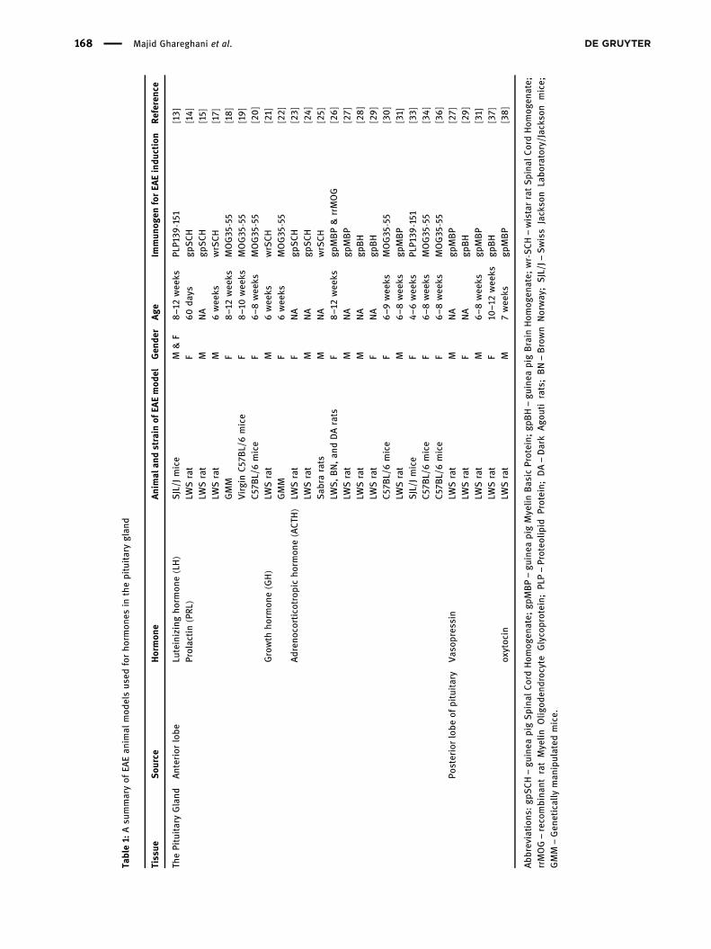
Table1:
Asu
mmaryof
EAEan
imal
mod
elsus
edforho
rmon
esin
thepituitaryglan
d
Tissue
Sou
rce
Hormon
eAnimal
andstrain
ofEA
Emod
elGen
der
Age
Immun
ogen
forEA
Eindu
ction
Referen
ce
ThePituitaryGland
Anteriorlobe
Luteinizingho
rmon
e(LH)
SJL/J
mice
M&F
8–1
2wee
ksPL
P139
-151
[13 ]
Prolactin(PRL
)LW
Srat
F60da
ysgp
SCH
[14]
LWSrat
MNA
gpSCH
[15]
LWSrat
M6wee
kswrSCH
[17]
GMM
F8–1
2wee
ksMOG35
-55
[18]
VirginC57
BL/6mice
F8–1
0wee
ksMOG35
-55
[19]
C57
BL/6mice
F6–8
wee
ksMOG35
-55
[20]
Growth
horm
one(GH)
LWSrat
M6wee
kswrSCH
[21]
GMM
F6wee
ksMOG35
-55
[22]
Adren
ocortico
trop
icho
rmon
e(ACTH
)LW
Srat
FNA
gpSCH
[23]
LWSrat
MNA
gpSCH
[24]
Sab
rarats
MNA
wrSCH
[25]
LWS,BN,an
dDArats
F8–1
2wee
ksgp
MBP&rrMOG
[26]
LWSrat
MNA
gpMBP
[27]
LWSrat
MNA
gpBH
[28]
LWSrat
FNA
gpBH
[29]
C57
BL/6mice
F6–9
wee
ksMOG35
-55
[30]
LWSrat
M6–8
wee
ksgp
MBP
[31]
SJL/J
mice
F4–
6wee
ksPL
P139
-151
[33]
C57
BL/6mice
F6–8
wee
ksMOG35
-55
[34]
C57
BL/6mice
F6–8
wee
ksMOG35
-55
[36]
Posteriorlobe
ofpituitary
Vas
opressin
LWSrat
MNA
gpMBP
[27]
LWSrat
FNA
gpBH
[29]
LWSrat
M6–8
wee
ksgp
MBP
[31]
LWSrat
F10
–12wee
ksgp
BH
[37]
oxytoc
inLW
Srat
M7wee
ksgp
MBP
[38]
Abb
reviations
:gp
SCH–gu
inea
pigSpina
lCordHom
ogen
ate;
gpMBP–gu
inea
pigMyelin
Bas
icProtein;
gpBH–gu
inea
pigBrain
Hom
ogen
ate;
wr-S
CH–wistarratSpina
lCordHom
ogen
ate;
rrMOG–reco
mbina
ntratMyelin
Olig
oden
droc
yte
Glyco
protein;
PLP–Proteo
lipid
Protein;
DA–Dark
Ago
utirats;BN–Brown
Norway
;SJL/J–Swiss
Jackso
nLabo
ratory/Jacks
onmice;
GMM
–Gen
etically
man
ipulated
mice.
168 Majid Ghareghani et al.
of NFs. Later, leuprolide acetate-treated EAE rats weredemonstrated to have a decline in the activation of NF-κB, a transcription factor mainly involved in inflammation,immune response, and cell survival, but also proinflam-matory mediators including TNF-α, IL-1β, and IL-17A, incomparison to untreated-EAE rats [43].
3.3 Growth hormone-releasinghormone (GHRH)
The potential involvement of GHRH in the pathogenesisof EAE was addressed using GHRH receptor-deficientmice (Ghrhrlit/lit) which showed no clinical symptoms ofEAE during the entire period of study [44]. In addition,GHRHR deficiency did not affect the activation of T cellsand GHRHR-deficient EAE mice had higher levels of anti-MOG IgG, compared to control, suggesting that resistanceof GHRHR−/− mice to EAE could be due to a deficientimmune response. Therefore, GHRH plays an importantrole in the development of EAE and could be a thera-peutic target.
3.4 Corticotropin-releasing hormone (CRH)
The hypothalamo-pituitary-adrenal axis (HPA-axis) activityis regulated by CRH and arginine vasopressin (AVP) in theparaventricular nucleus (PVN) of the hypothalamus [45].Calzà et al. showed a reduction in CRH mRNA levels inthe PVN 4 days post EAE induction, which recovered at10 days but decreased again until day 20, the last dayof experiment [46]. In contrast, Huitinga et al. showedunchanged CRH levels in the median eminence of maleEAE Lewis rats, compared to controls [27]. On the otherhand, CRH-deficient mice were found to be resistant toEAE induction and developed a mild form of EAE [47].Moreover, administration of Astressin, a specific CRHantagonist, reduced the disease severity in mice. Indeed,CRH’s effects on EAE development were shown to be inde-pendent of its glucocorticoid-inducing effect. In addition,results showed that IκBα phosphorylation, which inhibitsthe NF-κB transcription factor, was decreased in CRH-defi-cient T cells. Finally, peripheral CRH was suggested toapply its proinflammatory activity in EAE through a selec-tive increase in Th1-type responses.
3.5 Somatostatin (SST)
Muhvic and his colleagues introduced the role of soma-tostatin in EAE since 1992 [48]. On the other hand, theyinvestigated the effect of the somatostatin analogue SMS201-995 (octreotide) on EAE in the relatively resistantAlbino Oxford (AO) strain of rats [49]. Results showedthat the number of EAE-induced rats was higher in soma-tostatin-treated rats compared to controls. Moreover, theproportion of CD8+ T cells in the popliteal lymph nodes(PLN) decreased in somatostatin-treated rats, in compar-ison to control, while the proportion of CD25+ cellsincreased. Therefore, somatostatin increased the suscept-ibility of AO rats to EAE. In contrast, another study inmice, instead of AO rats, showed that oral somatostatininhibits EAE [50]. Indeed, the authors showed a reduc-tion of Th1 and Th17 and an induction of Th2-like IL-4cytokines in the spleen and CNS which caused an inhibi-tion of the disease. Further investigations are needed toclarify the role of somatostatin in EAE.
A summary of EAE animal models used to studyhypothalamus hormones is given in Table 2.
4 Pineal gland
4.1 Melatonin
Melatonin was investigated in EAE for the first time byConstantinescu et al., when the effect of luzindole, a mel-atonin receptor antagonist, was examined [51]. In fact,luzindole inhibited the development of EAE through inhi-bition of melatonin. Later, several studies showed thatEAE severity is reduced by melatonin therapy [52–54].The reduced level of EAE scores in melatonin-treatedgroup was coincident with lower number of inflammatorycells infiltration (CD4, CD8, NK) into the spinal cord [52].They also found that only ICAM-1 expression, but notLFA-1a, is considerably decreased by melatonin therapy.Since NK cells play a suppressive role in EAE, it’s sug-gested that melatonin reduces the severity of EAE througha mechanism involving ICAM-1.
On the other hand, the effects of melatonin therapyon T effector/regulatory responses of EAE showed anincreased level of Treg frequency and IL-10 synthesis inthe CNS, whereas Th1/Th17 responses in both peripheraland CNS were reduced inmelatonin-treated EAEmice [53].
Hormones in EAE animal models 169
Moreover, melatonin reduced the frequencies of T effectorand T central memory cells, and TNF-producing CD4+
cells. In addition, mononuclear infiltration (CD4 andCD11b cells) in the CNS was also reduced by melatonintherapy. This suggested that melatonin ameliorates EAEby controlling both peripheral and central T effector/reg-ulatory responses. Further work by Chen et al. showed,however, that the percentage of sorted splenic dendriticcells and the population of Foxp3+ CD4+ T cells in thespleens were not significantly different between groups[54]. Furthermore, melatonin enhanced splenic IL-10 inregulatory T cells by inducing IL-27 expression in thesplenic dendritic cells which suppresses the expressionlevel of IFN-γ, IL-17, IL-6, and CCL20 in the CNS and inhi-bits antigen-specific T cell proliferation. In line, Ghare-ghani et al. reported for the first time that the age ofEAE models plays a substantial role in melatonintherapy [55]. Indeed, as the administration of melatoninin EAE Lewis rats exacerbated the clinical symptoms of EAEby enhancing the immune cell infiltration into the CNS,
demyelinated plaques (low MBP-positive cells) activatedastrocytes, serum lactate, and the ratio of IFN-γ/IL-4. Ourstudy on EAE mice showed that melatonin alone or incombination with baclofen reduces clinical scores anddemyelination which is associated with an increase in IL-4and a decrease in IFN-γ serum levels. Furthermore, catalaseand superoxide dismutase, as antioxidant enzymes,increased following the treatment, while the oxidativestress-related malondialdehyde levels decreased [56]. Wefurther showed that melatonin therapy modulates cerebralmetabolism and enhances remyelinationwhich is associatedwith an increase in pyruvate dehydrogenase kinase-4(PDK-4), an enzyme involved in fatty acid synthesisduring remyelination, which we believe is a side effectof melatonin therapy [57]. Further studies are needed todetermine the precise role of melatonin in the EAE model,especially with respect to age, gender, strain of mice/ratsused, and melatonin dosage.
A summary of EAE animal models used to studypineal gland hormones is given in Table 2.
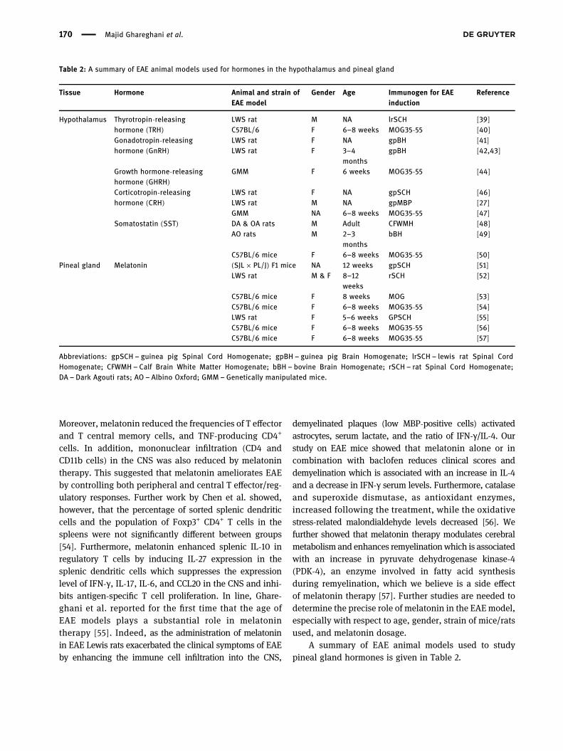
Table 2: A summary of EAE animal models used for hormones in the hypothalamus and pineal gland
Tissue Hormone Animal and strain ofEAE model
Gender Age Immunogen for EAEinduction
Reference
Hypothalamus Thyrotropin-releasinghormone (TRH)
LWS rat M NA lrSCH [39]C57BL/6 F 6–8 weeks MOG35-55 [40]
Gonadotropin-releasinghormone (GnRH)
LWS rat F NA gpBH [41]LWS rat F 3–4
monthsgpBH [42,43]
Growth hormone-releasinghormone (GHRH)
GMM F 6 weeks MOG35-55 [44]
Corticotropin-releasinghormone (CRH)
LWS rat F NA gpSCH [46]LWS rat M NA gpMBP [27]GMM NA 6–8 weeks MOG35-55 [47]
Somatostatin (SST) DA & OA rats M Adult CFWMH [48]AO rats M 2–3
monthsbBH [49]
C57BL/6 mice F 6–8 weeks MOG35-55 [50]Pineal gland Melatonin (SJL × PL/J) F1 mice NA 12 weeks gpSCH [51]
LWS rat M & F 8–12weeks
rSCH [52]
C57BL/6 mice F 8 weeks MOG [53]C57BL/6 mice F 6–8 weeks MOG35-55 [54]LWS rat F 5–6 weeks GPSCH [55]C57BL/6 mice F 6–8 weeks MOG35-55 [56]C57BL/6 mice F 6–8 weeks MOG35-55 [57]
Abbreviations: gpSCH – guinea pig Spinal Cord Homogenate; gpBH – guinea pig Brain Homogenate; lrSCH – lewis rat Spinal CordHomogenate; CFWMH – Calf Brain White Matter Homogenate; bBH – bovine Brain Homogenate; rSCH – rat Spinal Cord Homogenate;DA – Dark Agouti rats; AO – Albino Oxford; GMM – Genetically manipulated mice.
170 Majid Ghareghani et al.
5 Thyroid gland
5.1 Thyroxine (T4)The effects of thyroid hormones are predominantlymediatedby thyroid hormone receptors (THRs), which are part ofthe family of nuclear receptors. The 2 receptor genesTHRA and THRB encode the 2 proteins THRα and THRβ,respectively. In addition, each of these genes generatestwo isoforms (α1-2, β1-2) by alternative spicing. The αisoforms are widely expressed in the fetal brain as wellas oligodendrocyte precursor cells (OPCs), while the βisoforms expression is more restricted to differentiatedoligodendrocytes with a dramatic increase that starts atbirth in the rat [58,59].
Investigating the effect of thyroid hormone (thyr-oxine, T4) on EAE mice showed that EAE inductioncaused a sharp increase in the number of Ki-67+ andBrdU+ nuclei, a marker of proliferating cells, in theSVZ, white and gray matter of brain and spinal cord,and between ependymal cells in the central canal. Fewnumbers of Ki-67+ and BrdU+ cells belong to perivascularinflammatory cells and parenchymal glial cells, whichare strongly expressed in the acute stage of EAE. In con-trast, T4 administration reduced the rate of proliferationand undifferentiated precursors in the spinal cord andSVZ. Moreover, codistribution of Ki-67 and nestin immu-noreactivity showed a reduction in the expression ofnestin-positive cells in the SVZ. Moreover, they showedthat the administration of thyroid hormone in EAE reducesthe rate of proliferation, while it favors differentiationtowards oligodendrocytes and their maturation; hence,T4 therapy ameliorates the regulation of myelin-formingproteins in EAE model. Importantly, the increased level ofMBP in EAE model was found to be caused not only by adirect regulation of MBP gene expression, but also bynewly formed oligodendrocytes [60]. The fact that MBPis a target gene for thyroid hormone receptor has beenalready reported by Farsetti et al. [61].
In line, Gallo and Armstrong reported that the levelof nestin protein expression is high in proliferating pro-genitors, while it declines in differentiated oligodendro-cytes [62]. In support of these findings, Lezoualc’h et al.demonstrated that the nestin promoter has a binding sitefor nuclear receptors, which allows the thyroid hormoneto bind nestin in neural progenitor cells of the embryonicCNS [63].
Albornoz et al. induced EAE in the gestated miceunder hypothyroidism which already were treated withor without T4. Their findings showed higher clinical scoreof EAE, demyelination area, immune cell infiltration,
the number of CD4+ and CD8+ T cells, percentage of deathin oligodendrocytes (labeled with TUNEL) in the spinalcord, and lower expression of myelin basic protein (MBP)of EAE mice gestated in hypothyroid mothers, comparedto EAE mice gestated in normal mother. All of thesechanges reverted when T4 was added during gestation[64]. This suggested that gestational hypothyroidismcauses more severe inflammation and demyelination inEAE mice. Given the fact that Nerve Growth Factor [30]drops in acute phase of EAE [46], Calza et al. reportedthat T4 therapy restored the NGF level. NGF administra-tion showed a beneficial role by diminishing the clinicalscore of EAE and by protecting oligodendrocytes againstinjury induced by TNF-α proinflammatory cytokine [65].Moreover, several studies investigated the role of nuclearhormone receptor superfamily members that are knownto affect the immune response, mainly the receptors forretinoids, steroids, and thyroid hormone. Results revealedan expression of the peroxisome proliferator-activatedreceptors (PPARs) in the inflammatory infiltrate of EAEmice spinal cord. In contrast, administration of PPAR-γligand 15d-PGJ2 reduced the severity of EAE by inhibitingthe activation and expansion of encephalitogenic T cells,which suggests that PPAR-γ agonists may act as a newtherapeutic strategy for MS in which thyroid hormonesare most probably involved [66–68]. Furthermore, Fer-nandez et al. showed that T4 treatment is effective in redu-cing the severity of the relapse in Lewis, but not Dark Agouti(DA), rats of EAE model. They also showed that T4 favorsmyelin sheath reconstruction by increasing the mRNAexpression levels of platelet-derived growth factor α receptor(PDGFαR) marker of the oligodendrocyte precursor cells(OPC) in the spinal cord of EAE-DA rats on day 18 postim-munization [69]. More details about the role of TH on oligo-dendrocytes have been reviewed by Calzà et al. [70].
Two types of circulating TH are present: L-triio-dothyronine (T3), the active form, and tetra-iodothyro-nine (T4), the precursor of T3. Deiodinase enzymes (D)type 1 and 2 catalyze conversion of T4 to T3, whereas Dtype 3 (D3) catalyzes inactivation of T4 and T3 intoreverse (r)T3 and 3,3-diiodothyronine (T2), respectively[71,72]. Since D3 expression may play an important rolein decreasing the local bioavailability of TH at the inflam-mation sites, Boelen et al. showed that D3 is considerablyincreased in the inflammatory sites of EAE mice spinalcords [71]. While previous studies reported that most ofthe cellular effects of T3 are mediated by THRα and THRβ[73,74], Fernández et al. showed that THRα-1 and THRβ-1mRNA expression are markedly increased in proliferatingneurospheres media isolated from SVZ of adult EAE rats;however, THRα-2 expression did not show any significant
Hormones in EAE animal models 171
changes. Moreover, treatment by T3 increased the expres-sion of oligodendrocyte transcription factor Olig-1, a basichelix–loop–helix (bHLH) transcription factor expressed incells of the oligodendrocyte lineage, and stimulated oligo-dendrocytes maturation. Indeed, T3 increases more than 3-and 10- fold the generation of GFAP+ astrocytes and CNPase+
oligodendrocytes when used during proliferation and differ-entiation conditions [75]. Importantly, this elevated expres-sion of astrocytes favors the conversion of T4 to T3 by D2 andbenefits an increased bioavailability of the active T3 andcould also be detrimental for myelin repair [72,75].
On the other hand, the effects of T3 on EAE wereinvestigated by D’Intino et al., using the nonhuman pri-mate Callithrix Jacchus (marmoset), instead of EAEmice. Clinical scores of EAE severity were significantlydecreased by T3 therapy; however, there was a significantincrease in the mRNA expression levels of PDGFαR inthe spinal cord and MBP in the optic nerve and cerebralcortex of T3-treated EAE group. Moreover, D3, but not D2,mRNA levels were significantly increased by T3 therapy,compared to untreated EAE. While THRβ-1 and THRβ-2significantly elevated in T3-EAE, THRα-1 and THRα-1 didnot show significant changes in response to T3 therapy,compared to EAE [76]. A year later, Dell’Acqua et al.showed that T3 therapy causes a better outcome of clin-ical and neurophysiological parameters; however, the plasmalevels of T3 and T4 did not show significant differencesbetween EAE and EAE-T3 groups on day 39 postimmuni-zation. Indeed, T3 therapy did not affect inflammatorycellular infiltrate, but it strongly suppressed the loss ofmyelin staining, increased myelination, and did not pre-sent any nude axons, compared to untreated EAE. Then,they also demonstrated that the dysregulation of THRexpression in the EAE was corrected by T3, as THRα-1and THRβ expression significantly decreased in T3-treated mice, although THRα-2 showed no significant dif-ferences [77]. Finally, Castelo-Branco et al. showed thattreatment of MBP63–88-specific T cell lines with T3, priorto transfer to naïve rats to establish passive EAE model, hadno effect on T cell proliferation, but significantly decreasedthe frequency of IL-17-producing cells and increased theexpression of C–C chemokine receptor type 7 (Ccr7) andL-selectin (D62L) [78]. CCR7 ligands have a novel functionin stimulating the pathogenic Th17 cells in EAE inductionthrough IL-23-depedent generation from dendritic cells [79],while CD62L regulates naïve T cell homing into lymph nodesand the migration of leucocytes to sites of inflammation[80]. Castelo-Branco et al. suggested that T3 has thepotential to inhibit lymphocytes migration from lymphnodes into the CNS [78]. Investigation of the adult off-springs gestated in hypothyroxinemia demonstrated that
it leads to imprint the immune response and subse-quently early appearance of EAE symptoms [81].
5.2 Calcitonin (CT)
CT therapy was shown to delay the onset of EAE, while itscombination administration with 1,25(OH)2D3 loweredthe effective dose required to suppress EAE. Importantly,this combination therapy is dependent on dietary calcium[82]. However, at the molecular level, CT administrationdoes not seem to be a necessary factor for 1,25(OH)2D3-mediated suppression of EAE. Indeed, knock-out mice forCT and calcitonin gene-related peptide-α (CGRP) genesdid not change the EAE progression or serum calcium,while administration of 1,25(OH)2D3 significantly amelio-rated the EAE severity and enhanced the serum level ofcalcium [83].
Moreover, it has been reported that SA13353, a noveltransient receptor potential vanilloid 1 (TRPV1) agonist,and capsaicin (Cap) elevate the serum level of CGRP [84].The therapeutic activity of CGRP was then examined byMatsuda et al., using a human CGRP-expressing plasmidtransfected to C57BL/6 mouse bone marrow-derivedmatured DC (mDC) followed by the induction of EAE inthis mouse. It was observed that only 50% of mice devel-oped EAE in the CGRP-transfected group, compared with80% in the mock-transfected group. In addition, cyto-kines production of IL-10, IL-2, IL-6, IL-17, TNF-α, andIFN-γ from spleen cells of EAE mice increased in CGRP-transfected mice, which was dependent on MOG concen-tration, compared with the mock-transfected mice. More-over, the proportion of CD4+ CD25+ Foxp3+ cells increasedin the CGRP-transfected mice, suggesting that gene therapywith CGRP-expressing mDC would be a novel strategy tosuppress the development of EAE [85].
Since CGRP binds to its specific receptor composedof calcitonin receptor-like receptor (CLR) and receptoractivity-modifying protein 1 (RAMP1) [86], Mikami et al.examined the role of CGRP in EAE using RAMP1-deficientmice. Although a lower EAE score and delayed peak scorewere observed in RAMP1−/− mice, compared with wild-type, the population of Th17 cells was also decreased inthe first group suggesting that CGRP promotes Th17-mediated inflammation by increasing IL-17 and IL-21 pro-duction in EAE mice [87]. Finally, CGRP-deficient miceshowed aggravated EAE signs, while injection of CGRPinto the lumbar CSF decreased EAE severity. Moreover,the production of TNF-α, IFN-γ, IL-17, IL-2, and IL-4 didnot demonstrate any alterations in the peripheral
172 Majid Ghareghani et al.
lymphocytes of CGRP-treated mice; however, it reducedmicroglia transition to bushy morphology. Indeed, theexpression of Iba1+microglia showed slightly more bipolarand less round-fitting morphology in CGRP-treated micewhich suggests that CGRP can inhibit microglia activationin EAE [88].
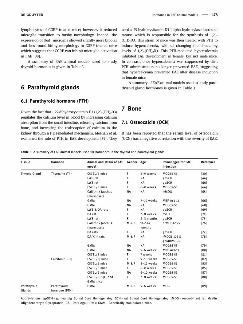
A summary of EAE animal models used to studythyroid hormones is given in Table 3.
6 Parathyroid glands
6.1 Parathyroid hormone (PTH)
Given the fact that 1,25-dihydroxyvitamin D3 (1,25-(OH)2D3)regulates the calcium level in blood by increasing calciumabsorption from the small intestine, releasing calcium frombone, and increasing the reabsorption of calcium in thekidney through a PTH-mediated mechanism, Meehan et al.examined the role of PTH in EAE development [89]. They
used a 25-hydroxyvitamin D3-1alpha-hydroxylase knockoutmouse which is responsible for the synthesis of 1,25-(OH)2D3. This strain of mice was then treated with PTH toinduce hypercalcemia, without changing the circulatinglevels of 1,25-(OH)2D3. This PTH-mediated hypercalcemiainhibited EAE development in female, but not male mice.In contrast, once hypercalcemia was suppressed by diet,PTH administration no longer prevented EAE, suggestingthat hypercalcemia prevented EAE after disease inductionin female mice.
A summary of EAE animal models used to study para-thyroid gland hormones is given in Table 3.
7 Bone
7.1 Osteocalcin (OCN)
It has been reported that the serum level of osteocalcin(OCN) has a negative correlation with the severity of EAE.
Table 3: A summary of EAE animal models used for hormones in the thyroid and parathyroid glands
Tissue Hormone Animal and strain of EAEmodel
Gender Age Immunogen for EAEinduction
Reference
Thyroid Gland Thyroxine (T4) C57BL/6 mice F 6–9 weeks MOG35-55 [30]LWS rat F NA gpSCH [46]LWS rat F NA gpSCH [60]C57BL/6 mice F 6–8 weeks MOG35-55 [64]Callithrix Jacchus(marmoset)
NA NA rrMOG [65]
GMM NA 7–10 weeks MBP Ac1-11 [66]GMM NA NA MOG35-55 [68]LWS & DA rats F NA gpSCH [69]DA rat F 7–8 weeks rSCH [71]LWS rat F 2–3 months gpSCH [75]Callithrix Jacchus(marmoset)
M & F 11–144months
hrMOG1-125 [76]
DA rats F NA gpSCH [77]DA/Kini rats M & F NA rMOG1-125 &
gpMBP63-88[78]
GMM NA NA MOG35-55 [79]GMM NA 5–6 weeks MBP Ac1-11 [80]C57BL/6 mice F 7 weeks MOG35-55 [81]
Calcitonin (CT) C57BL/6J mice F 9–10 weeks MOG35-55 [82]C57BL/6 mice M & F 8–12 weeks MOG35-55 [83]C57BL/6 mice F 6–8 weeks MOG35-55 [85]C57BL/6 mice NA 6–10 weeks MOG35-55 [87]C57BL/6, SJL, andGMM mice
F 7–8 weeks MOG35-55 [88]
ParathyroidGlands
Parathyroidhormone (PTH)
GMM M & F 5–6 weeks MOG [89]
Abbreviations: gpSCH – guinea pig Spinal Cord Homogenate; rSCH – rat Spinal Cord Homogenate; rrMOG – recombinant rat MyelinOligodendrocyte Glycoprotein; DA – Dark Agouti rats; GMM – Genetically manipulated mice.
Hormones in EAE animal models 173
Indeed, Ghareghani et al. reported that OCN serum levelsignificantly decreased in EAE model, while medicationswhich ameliorated EAE severity were associated with anincrease in osteocalcin [90]. For instance, the serum levelof osteocalcin significantly increased in EAE mice treatedwith cyclosporine A and 1α,25-(OH)2D3 (active form ofVitamin D) [91] or melatonin [90], but was insignificantlyincreased in 1,25(OH)2D3-treated EAE mice [92].
7.2 Lipocalin 2
Berard et al. used an Affymetrix gene array to track thealterations in the gene encoding Lipocalin 2, involvedin matrix metalloproteinase (MMP) stabilization, glialactivation, and cellular iron flux [93]. Lipocalin 2 geneexpression increased in monocytes and reactive astro-cytes by about 60-fold at the onset of EAE, while itsreceptor 24p3R was also shown to be expressed by mono-cytes, macrophages/microglia. Importantly, Lipocalin2-deficent mice showed more severe EAE, in comparisonto wild-type mice. In accordance, another study by Namet al. demonstrated that EAE has been suppressed inLipocalin 2−/− mice [94]. In addition, Lipocalin 2-treatedglial cells, isolated from Lipocalin 2−/− EAE mice, hadhigher levels of proinflammatory cytokines, MMP-9, andchemokines. These findings highlighted that Lipocalin 2is a critical mediator of autoimmune inflammation in EAEpathogenesis. Some pharmacological studies examinedthe effect of EAE medications on the expression of Lipo-calin 2. For instance, increased levels of Lipocalin 2 wereseen at active phases, onset and relapse, in blood-CSFsample as well as choroid plexus (CP) of EAE mice [95];however, administration of Natalizumab, a well-knownmedication for MS [96], ameliorated the severity of EAEby modulating and normalizing CSF Lipocalin 2, whilemarkedly reducing its astrocytic expression. Similarly,Ebrahimi-Kalan et al. showed the inhibitory effect of MS14,an herbal-marine drug with anti-inflammatory activity, onthe severity of EAE through reduction of Lipocalin 2 [97].
7.3 Erythropoietin (EPO)
The neuroprotective role of erythropoietin in EAE wasshown for the first time by Brines and colleagues whoadministrated the recombinant human erythropoietin(r-Hu-EPO), 3 days after immunization until day 18,causing a delay in EAE onset and reduction of peak clin-ical scores, in comparison to untreated-EAE rats [98]. It
is well-known that astrocytes and microglia play a sub-stantial role in MS progression, as activated and prolifer-ating forms of these cells have been observed in demyelinatedareas [99]. Agnello and colleagues reported that EPO con-siderably reduces both glial proliferation and activationand subsequently diminishes inflammation through redu-cing spinal cord TNF-α and IL-6 [100]. Importantly, Li andcolleagues reported that erythropoietin also reduces theblood-brain barrier (BBB) leakage and major histocompat-ibility complex class II (MHC-II) in the spinal cord [101].This reduced level of MHC-II was further observed byZhang et al., who demonstrated that EPO suppressesthe levels of IFN-γ and IL-17 both in peripheral spleniccells and CNS-infiltrating cells [102]. Similarly, it wasshown that EPO improved neurological functional recoveryin EAE mice by increasing the level of BDNF+ cells andproliferation of oligodendrocyte progenitor cells (OPCs),stimulating oligodendrogenesis, and by diminishing proin-flammatory infiltration [103]. In line, combination therapyof methotrexate and EPO revealed a stimulating effect onoligodendrogenesis [104].
On the other hand, to clarify the best time of EPOadministration in EAE, Savino and colleagues [105] showedthat EPO therapy works in all stages of EAE; however, itsefficiency is reduced once it is administrated late, 15 daysafter the onset of symptoms. The effects of EPO on hema-tocrit and EAE pathological outcomes were investigatedby Yuan et al., using truncated small linear or cyclic EPO-derived peptides from various domains of the full-lengthEPO [106]. The 19-mer JM-4 peptide showed the bestpotential to treat EAE without producing hematocritalterations in EAE mice, similar to the full-length EPO,and modulated immune/inflammatory reaction in EAE.EPO induces its neuroprotective role via signaling toseveral critical subsets of immune cells residing in theperipheral lymphatic system. Indeed, EPO reduced theproliferation of MOG-specific T cells in vitro, suppressedT cell cytokine production, reduced the total number ofmononuclear and T cells in EAE peripheral lymph nodes,increased the expansion of peripheral Tregs, and inhib-ited the polarization of Th17 cells in EAE mice.
It has been shown that expression of the inhibitorof metalloproteases (TIMPs) is increased in astrocytesforming the BBB and surrounding demyelinated area[107], while TIMP-1-deficient mice have elevated levelsof demyelination and microglial activation in EAE mice[108]. Thorne et al. showed that darbepoetin alfa therapy,an EPO analogue, reduced the clinical scores of EAEmice, accompanied with an increased number of TIMP-1-expressing astrocytes, both in the brain and spinal cord[109]. Although TIMP-1-deficient mice showed severe
174 Majid Ghareghani et al.
EAE outcomes, compared to wild-type, darbepoetin alfatherapy could not affect EAE severity in TIMP-1−/− mice,highlighting TIMP-1 importance in EPO therapy of EAE.Similarly, ARA290, a nonerythropoietic analogue, hasbeen shown to considerably ameliorate the severity ofEAE [110]. In accordance, the expression of EPO wasshown to be induced in spinal cord of EAE mice, clearlylocalized to motor neurons, and the authors suggestedthat EPO should be viewed as part of the inflammatory/anti-inflammatory network in MS [111]. On the otherhand, EPO therapy in EAE mice induced endogenousheme oxygenase-1 (HO-1), an anti-oxidative stress pro-tein, a possible factor in diminishing the inflammatoryresponses involved in EAE [112]. Recently, Moransard etal. used transgenic mouse strains to investigate whetherEPO primarily applies its effects in the CNS or the per-iphery [113]. Although EAE onset did not differ betweencontrol and two transgenic EAE mice with overexpressedr-Hu-EPO either in CNS only (tg21) or systemically (tg6),their progression markedly increased in transgenic micecompared to controls. However, no peripheral immuno-modulatory effects were seen in tg21 strain whose mod-ulation was limited to a reduction in CNS macrophages.In contrast, macrophages were upregulated in the CNS,but not the periphery, of tg6 strain.
On the other hand, Sattler et al. showed on EAE micethat EPO promotes the survival and function of retinalganglion cells (RGCs), the neurons that form the axonsof the optic nerve, through three independent intracel-lular signaling pathways of p-Akt, p-MAPK 1 and 2, andBcl-2 [114]. In accordance, the work by Diem and collea-gues revealed that coadministration of EPO and highdose methylprednisolone, a steroid medication in MS,had relatively higher potential to induce neuroprotectionin RGCs and optic nerves, compared to respective mono-therapies [115].
Long-term effects of EPO were investigated, for thefirst time, by treating EAE mice using JM4, an EPO-derived small peptide, which is free from any side effects.Following 12 days of JM4 therapy, they observed a reduc-tion in disease severity after day 60 until day 100 post-immunization [116].
7.4 FGF-23 (phosphatonin)
No studies were found concerning FGF-23 in EAE model.A summary of EAE animal models used to study bone
hormones is given in Table 4.
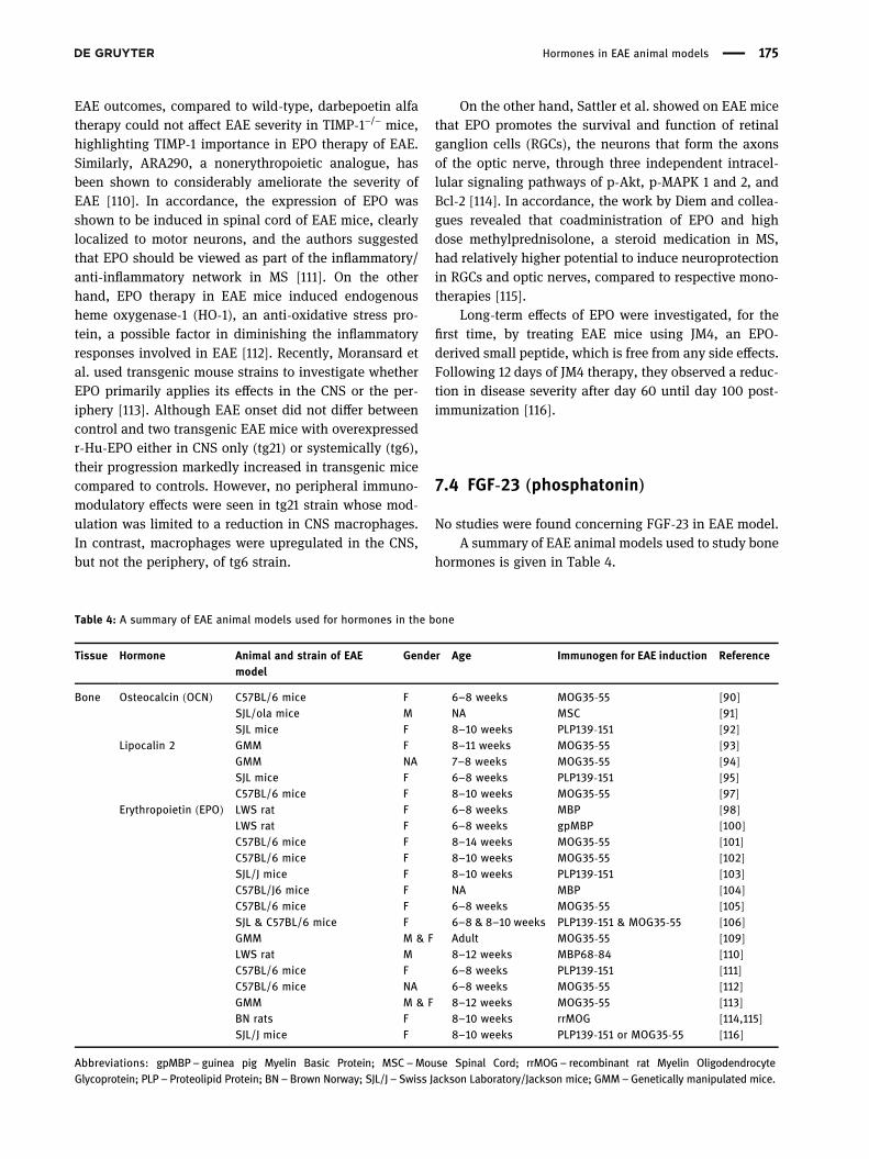
Table 4: A summary of EAE animal models used for hormones in the bone
Tissue Hormone Animal and strain of EAEmodel
Gender Age Immunogen for EAE induction Reference
Bone Osteocalcin (OCN) C57BL/6 mice F 6–8 weeks MOG35-55 [90]SJL/ola mice M NA MSC [91]SJL mice F 8–10 weeks PLP139-151 [92]
Lipocalin 2 GMM F 8–11 weeks MOG35-55 [93]GMM NA 7–8 weeks MOG35-55 [94]SJL mice F 6–8 weeks PLP139-151 [95]C57BL/6 mice F 8–10 weeks MOG35-55 [97]
Erythropoietin (EPO) LWS rat F 6–8 weeks MBP [98]LWS rat F 6–8 weeks gpMBP [100]C57BL/6 mice F 8–14 weeks MOG35-55 [101]C57BL/6 mice F 8–10 weeks MOG35-55 [102]SJL/J mice F 8–10 weeks PLP139-151 [103]C57BL/J6 mice F NA MBP [104]C57BL/6 mice F 6–8 weeks MOG35-55 [105]SJL & C57BL/6 mice F 6–8 & 8–10 weeks PLP139-151 & MOG35-55 [106]GMM M & F Adult MOG35-55 [109]LWS rat M 8–12 weeks MBP68-84 [110]C57BL/6 mice F 6–8 weeks PLP139-151 [111]C57BL/6 mice NA 6–8 weeks MOG35-55 [112]GMM M & F 8–12 weeks MOG35-55 [113]BN rats F 8–10 weeks rrMOG [114,115]SJL/J mice F 8–10 weeks PLP139-151 or MOG35-55 [116]
Abbreviations: gpMBP– guinea pig Myelin Basic Protein; MSC–Mouse Spinal Cord; rrMOG– recombinant rat Myelin OligodendrocyteGlycoprotein; PLP– Proteolipid Protein; BN–Brown Norway; SJL/J–Swiss Jackson Laboratory/Jackson mice; GMM–Genetically manipulated mice.
Hormones in EAE animal models 175
8 The adrenal glands
8.1 Adrenal cortex
8.1.1 Glucocorticoids (e.g., cortisol)
Several studies have shown that EAE is accompanied by areduction in the intensity of cortisol metabolism underconditions of in situ perfusion of the liver by solutionscontaining cortisol in different concentrations as well asin lymphocytes from the cervical lymph nodes of guineapigs [117–119].
8.1.2 Mineralocorticoids (e.g., aldosterone)
Herrada and colleagues demonstrated that administra-tion of deoxycorticosterone acetate (DOCA), a stablemineralocorticoid analogue of aldosterone, leads to anexacerbation of EAE severity through promotion of Th17immunity. It is well-known that high levels of aldos-terone lead to hypertension and cardiovascular diseasewhere T cell immunity is involved [120].
8.2 Adrenal medulla
8.2.1 Adrenaline (epinephrine)
Wesselmann and colleagues investigated splenic cate-cholamine in EAE and showed that elevated concentra-tions of epinephrine were not statistically significant,compared to controls [121]. Since epinephrine action ismediated by β adrenergic receptors, an antagonist of βadrenergic receptors called nadolol was used in EAEmodel and it was found that the suppression of epi-nephrine did not affect the clinical outcomes of EAE[122]. The function of epinephrine was recently furtherinvestigated by Yan and colleagues. They observed ahigh expression of the rate-limiting enzyme of epinephrinesynthesis, phenylethanol N-methyltransferase (PNMT),in tissue-resident TH17 cells of EAE mice, not in othereffector T cell subsets or regulatory T cells. They used aknockout mouse model for PNMT expressing immunecells. Although they observed a lower level of immunecell infiltration in these mice compared to normal EAEmice, the clinical scores did not show significant differ-ences [123].
8.2.2 Noradrenaline (norepinephrine)
White and colleagues showed that norepinephrinedecreased sharply in the spinal cord of EAE rats, espe-cially in the lumbar region [124]. In addition, norepi-nephrine depletion was more evident in lumbosacraland craniothoracal region of spinal cord EAE rats [125].In contrast, development of acute EAE was suppressedby administration of neurotransmitter-depleting agents[126]. Indeed, depletion of norepinephrine using 6-hydro-xydopamine suppressed EAE possibly through deletionof effector amplification mechanism and/or triggering aT suppressor cell mechanism [127]. However, anotherstudy suggested that this observation could be due toalteration in the blood-spinal cord barrier function and/or CNS blood flow [128]. In contrast, a study demon-strated that selective elevation of CNS NA levels couldprovide benefits in EAE, as increasing the norepinephrinelevels using atomoxetine, a norepinephrine reuptake inhi-bitor, or l-threo-3,4-dihydroxyphenylserine (l-DOPS), asynthetic norepinephrine precursor, did not affect clin-ical scores and prevented further worsening of EAE,respectively [129]. In addition, Polak and colleagues sug-gested that raising norepinephrine levels is an efficientmethod to treat EAE and MS [130]. It is well-known thatthe primary source of norepinephrine are tyrosine hydro-xylase-positive neurons in the Locus coeruleus, and thatdysregulation of neurons was observed in EAE and MS.Therefore, Vindeburnol was selectively used to increaseLocus coeruleus neuronal viability or activity whichresulted in a reduction in astrocyte activation in theLocus coeruleus, hypertrophy of tyrosine hydroxylase-positive neurons, and elevation in spinal cord contentof norepinephrine and genes involved in Locus coeruleussurvival and maturation [131].
Moreover, Leonard et al. reported an inverse correla-tion between hypothalamic norepinephrine and corticos-terone serum level, both at the peak clinical scores ofEAE and in norepinephrine-depleted peripheral nervoussystem EAE mice [132]. However, a direct correlation wasfound between corticosterone serum levels and EAEseverity, while the striatum showed no change in its con-tent of norepinephrine in EAE mice [133]. Finally, Shakedand colleagues suggested that the transcription factorNr4a1 regulates the production of norepinephrine inmacrophages, as Nr4a1-deficient myeloid cells in miceled to an increase in norepinephrine and EAE severity[134]. In contrast, myeloid-specific deletion of tyrosinehydroxylase, the rate-limiting enzyme in norepinephrineproduction, demonstrated a protective role against EAE.Although males have a stronger sympathetic nervous
176 Majid Ghareghani et al.
system, Vujnović and colleagues reported that β-adreno-receptor-mediated signaling plays a key role in sexualdimorphism in primary CD4+ T-cell responses in EAErats. Indeed, rats treated with Propranolol, a nonselectiveβ-adrenergic receptor antagonist, showed higher nora-drenaline concentration in draining lymph node (dLN)in males, compared to females [135]. Furthermore, theydid another study on α 1-adrenoceptor and suggested thatthis receptor is also involved in interactions between sub-types of CD4+ T cells and antigen-presenting cells [136]
A summary of EAE animal models used to studyadrenal gland hormones is given in Table 5.
9 Stomach, pancreas, and intestine
9.1 Cholecystokinin (CCK)
The therapeutic potential of GB-115 compound (N-phenyl-hexanoyl-glycyl-L-tryptophan amide), a dipeptide chole-cystokinin receptor antagonist, was demonstrated bysuppression of ROS production, improved spontaneouslocomotor activity, and reduced edema and neutrophilinfiltration of the perivascular space of the brain [137].
9.2 Neuropeptide Y
The role of neuropeptide-Y in EAE was first reported byBedoui and colleagues [138]. Neuropeptide-Y decreasesthe susceptibly of mice to EAE induction and was shownto exert its pleiotropic functions via the activation of sev-eral G-protein coupled receptor subtypes such as Y(1)receptor, but not Y(5). Moreover, EAE induction was facili-tated by administration of Y(1) receptor antagonist. In con-trast, the immunomodulatory action of neuropeptide-Yin DA EAE rats showed that CD28 and CD80/CD86 costi-mulatory molecules acted as a target for neuropeptide-Y-mediated amelioration in EAE through Y(2) and Y(5)receptors [139]. Moreover, Brod and Bauer showed thatingested (oral) neuropeptide-Y inhibits CNS inflammationin EAE mice by diminishing Th17 and Th1-like cytokinesand elevating Th2-like cytokines within the CNS [140].
9.3 Ghrelin
Theil et al. examined the role of ghrelin in EAE pathogen-esis and showed that although its administration Ta
ble5:
Asu
mmaryof
EAEan
imal
mod
elsus
edforho
rmon
esin
thead
rena
lglan
ds
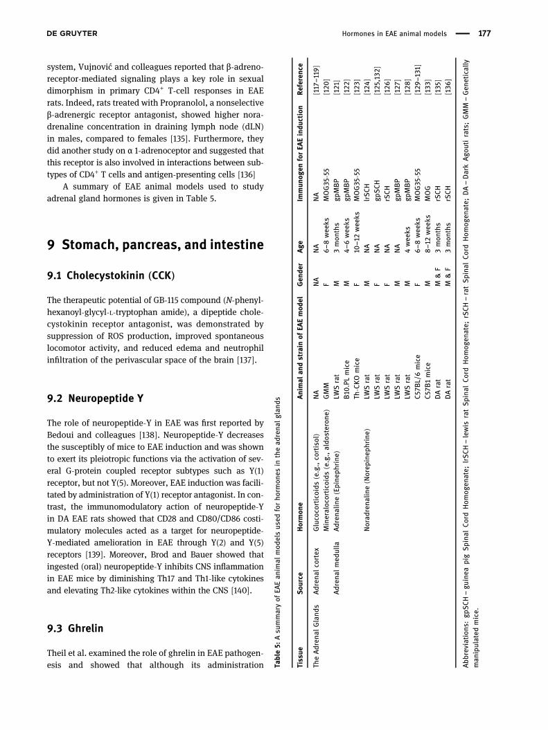
Tissue
Sou
rce
Hormon
eAnimal
andstrain
ofEA
Emod
elGen
der
Age
Immun
ogen
forEA
Eindu
ction
Referen
ce
TheAdren
alGland
sAdren
alco
rtex
Gluco
corticoids
(e.g.,co
rtisol)
NA
NA
NA
NA
[117–1
19]
Mineraloc
ortico
ids(e.g.,aldo
steron
e)GMM
F6–8
wee
ksMOG35
-55
[120
]Adren
almed
ulla
Adren
aline(Epine
phrine
)LW
Srat
M3mon
ths
gpMBP
[121]
B10
.PLmice
M4–
6wee
ksgp
MBP
[122
]Th
-CKOmice
F10
–12wee
ksMOG35
-55
[123
]Norad
rena
line(Norep
inep
hrine)
LWSrat
MNA
lrSCH
[124
]LW
Srat
FNA
gpSCH
[125
,132
]LW
Srat
FNA
rSCH
[126
]LW
Srat
MNA
gpMBP
[127
]LW
Srat
M4wee
ksgp
MBP
[128
]C57
BL/6mice
F6–8
wee
ksMOG35
-55
[129
–131]
C57
B1mice
M8–1
2wee
ksMOG
[133
]DArat
M&F
3mon
ths
rSCH
[135
]DArat
M&F
3mon
ths
rSCH
[136
]
Abb
reviations
:gp
SCH–gu
inea
pigSpina
lCordHom
ogen
ate;
lrSCH–lewis
ratSpina
lCordHom
ogen
ate;
rSCH–ratSpina
lCordHom
ogen
ate;
DA–DarkAgo
utirats;GMM
–Gen
etically
man
ipulated
mice.
Hormones in EAE animal models 177
ameliorates the severity of EAE clinical scores, it had nobeneficial effect in reducing the numbers of inflammatorycells in the spinal cord [141]. Indeed, this has been deter-mined by counting the number of NK cells (NK1.1+CD3−),B cells (CD19+), NKT cells (NK1.1+CD3+), macrophages(F4/80+), and CD25+ FOXP3+ cells (in the CD4+ T cellpopulation) in the spinal cord lesions. Theil et al. sug-gested that monocytes could be potential targets in theghrelin-mediated EAE inhibition. While the levels ofTNF-α, IL-1β, and IL-6 significantly decreased in thespinal cord of ghrelin-treated mice, only TNF-α mRNAlevels decreased in their spleens, whereas no significantchanges were seen in any of the cytokines in the lymphnode or thymus of ghrelin-treated mice. To investigatewhich cells were important in the ghrelin-mediated sup-pression of EAE, mRNA levels of IL-1β, IL-6, and TNF-αwere assessed in macrophages, microglia, and T cells;however, they only showed marked changes in microglia,suggesting that it might play a key role in ghrelin-mediated suppression of EAE. The effect of ghrelin wasfurther investigated by Liu and colleagues who observeda reduction in the levels of inflammatory cytokines andfactors involved in NLRP3 inflammasome signaling andPyroptosis [142].
9.4 Serotonin
An early study demonstrated that a segment of MBP iscomposed of one of the 5-hydroxytryptamine (5-HT; ser-otonin) receptors in the CNS which played an importantrole in EAE [143]. Later, it was reported that CNS 5-HTneurotransmission is impaired in EAE rats with hind limbparalysis [144] which was correlated with motor deficitsmanifested during the acute paralytic stage of EAE [145].In line, administration of some drugs led to modificationsin endogenous serotonin levels within the body. Treat-ment of EAE guinea pigs with serotonin suppressor, espe-cially the antihypertensive medication reserpine, led toquick death of EAE animals. However, serotonin enhancermedication, particularly imipramine hydrochloride anti-depressant, showed better survival, even longer thancontrols [146]. Moreover, using serotonin antagonistand inactivation of neurogenic 5-HT receptors signifi-cantly reduced the severity of EAE [147–150]. In contrast,the serotonin antagonist Methysergide did not inhibit theseverity of EAE in rabbits when administrated 6 dayspostimmunization [151].
On the other hand, it was reported that serotoninaxons were swollen and distorted at early stages of EAE
and get worse when neurological scores increase.Interestingly, the severity of axonal damage correlatedwith the severity of inflammation and the injured axonswere located adjacent to blood vessels or the pial surface,sites at which inflammation occurs during EAE [39].Moreover, reduced levels of serotonin and the serotoninmetabolite, 5-hydroxyindoleacetic acid, were shown inEAE rats which suggested that the reduction is probablya consequence of the damage to descending 5-HT axons[152]. Another study reported that factors associatedwith spinal cord inflammation may be responsible forthe bulbospinal monoaminergic axonal damage in EAE.Indeed, prazosin therapy, an alpha 1-adrenergic antago-nist, reduced EAE severity and pathological outcomes[153].
Preventive and therapeutic outcomes were found forantidepressant drugs on EAE such as the use of selectiveserotonin/norepinephrine reuptake inhibitors (SNRI) ser-traline [154], fluoxetine [155,156], and fluvoxamine [157].Also, immunomodulatory effects of antipsychotic agentswere reported in EAE such as the use of risperidone,known to antagonize serotonin 5-HT2A receptors [158],and clozapine [159]. Moreover, monoamine oxidase inhi-bitor phenelzine therapy, an antidepressant and anxiolyticdrug, was reported as an efficient strategy to ameliorateEAE severity [160,161]. Furthermore, attenuation of EAEwas shown in mice lacking the 5-HT transporter (5-HTT),which was more prominent in females [162]. Finally, ele-vated levels of serotonin were found in the brain striatumof EAE mice [133].
9.5 Amylin, secretin, fibroblast growthfactor 19, PYY3-36, and incretins
No studies were found concerning the above hormones inEAE model.
A summary of EAE animal models used to study hor-mones of the stomach, pancreas, and intestine is given inTable 6.
10 Liver
10.1 Insulin-like growth factor-1 (IGF-1)
Liu and colleagues showed that the levels of IGF-1 andGFAP, an astrocytic marker, are elevated in demyelination
178 Majid Ghareghani et al.
areas in EAE model [163]. These astrocytes co-expressedIGF-I with IGFBP-2, a member of the six IGF bindingproteins that modulate IGF-1 actions, suggesting thatastrocytic expression of IGF-1-related peptides couldreduce the immune-mediated demyelination. Indeed,another study reported that IGF-1-induced reductions inimmune cell responses can occur even in the absence ofdemyelination [164]. Similar studies further reported thebeneficial role of IGF-1 in EAE and suggested that IGF-1therapeutic activity is mediated by reducing EAE-inducedblood-spinal cord barrier changes, modifying oligoden-drocytes to increase myelin regeneration [165–167]. Usingin vivo three-dimensional MR microscopy imaging tech-nique, IGF-I was shown to reflect changes in stabilizationor permeability of BBB and/or cell membranes to amelio-rate EAE [168]. On the other hand, it has been suggested
that coadministration of IGF-1 and IGFBP3 can enhancethe efficiency of IGF-1 therapy in EAE [169].
In contrast, Cannella and colleagues were doubtful ofIGF-1 being a good therapeutic agent in MS as theyshowed that administration of IGF-1 at different timepoints during the acute and chronic phases of EAEhad different outcomes and even failed to enhance CNSmyelin repair in EAE [170]. Similarly, Bilbao et al. showedthat not only administration of recombinant human IGF-1(rhIGF-1) stimulates regulatory T cells in culture, but italso ameliorates the severity of EAE. Indeed, they indi-cated that existence and function of IGF-1 receptor arethe necessary factors to induce IGF-1 stimulatory effecton regulatory T cell proliferation [171]. In accordance,Genoud and colleagues showed that adeno-associatedvirus (AAV)-mediated delivery of IGF-1 in the spinal
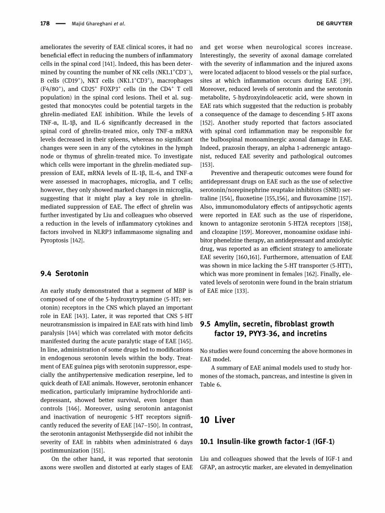
Table 6: A summary of EAE animal models used for hormones in stomach, pancreas, and intestine
Tissue Hormone Animal and strain ofEAE model
Gender Age Immunogen for EAEinduction
Reference
Stomach, Pancreas, andIntestine
Cholecystokinin (CCK) C57Bl/6 mice F 5–6 weeks MOG35-55 [137]Neuropeptide Y B6 & SJL/J mice F 6–10
weeksMOG35-55 & PLP139151 [138]
DA rat M 7 months gpSCH [139]C57BL/6 F 6–8 weeks MOG35-55 [140]
Ghrelin B6 mice F 6–10weeks
MOG35-55 [141]
CD rat F NA gpSCH [142]Serotonin LWS rat M NA lrSCH [39,152,153]
Guinea pigs NA Youngadult
bBH [143]
CRW-L rats M NA w-lrSCH [144]guinea pigs NA NA gpMBP [146,148]LWS rat F NA gpBH [149]LWS rat M NA gpMBP [150]White rabbits F Adult rSCH [151]C57/bl mice F NA MOG [154]WSR rats F 6–8 weeks gpSCH [155]SJL/J mice F 8–10
weeksMBP AC1-11 &PLP139-151
[156]
LWS rat F 8–12weeks
gpSCH [157]
C57BL/6 mice F 8–12weeks
MOG35-55 [158,159]
C57BL mice F NA MOG35-55 [160,161]C57BL/6 mice M & F 2–5
monthsMBP & MOG [162]
C57B1 mice M 8–12weeks
MOG [133]
Abbreviations: gpSCH – guinea pig Spinal Cord Homogenate; gpBH – guinea pig Brain Homogenate; lrSCH – lewis rat Spinal CordHomogenate; w-lrSCH –wistar-lewis rats Spinal Cord Homogenate; bBH – bovine Brain Homogenate; rSCH – rat Spinal CordHomogenate; PLP – Proteolipid Protein; DA – Dark Agouti rats; CRW-L – Charles River Wistar-Lewis rats; WSR –wistar rat; SJL/J – SwissJackson Laboratory/Jackson mice.
Hormones in EAE animal models 179
cord had no effect on EAE and even worsened the diseasewhen injected after EAE induction [172]. Considering thefact that Th17 and Treg cells balance is disrupted in MS,which play a critical role in immune tolerance, DiToroand colleagues showed that increased aerobic glycolysisfollowing activation of IGF1R leads to a higher Th17 celldifferentiation than Treg cells, resulting in an increase inproinflammatory cytokines [173].
10.2 Thrombopoietin (TPO)
Although TPO, a hematopoietic growth factor, inducedan increase in the number of white blood cells in TPO-treated EAE mice, the clinical scores of EAE severity didnot differ either after immunization or after the diseasereached its peak [174]. The mechanism of TPO’s effect onthe immune response of EAE remains unknown.
10.3 Hepcidin
Iron accumulates in the CNS of MS patients and in EAEmodels [175,176]. Iron efflux from cells is mediated bythe multi-pass membrane transporter ferroportin (Fpn)which is regulated posttranscriptionally by hepcidin,thus reducing iron efflux and leading to iron accumula-tion. Zarruk et al. showed that iron and ferritin elevationin the CNS of EAE mice was coincident with about 5-foldhepcidin mRNA increase at all stages of chronic and atthe peak of relapsing-remitting EAE [177]. In addition,hepcidin was found in infiltrating macrophages as earlyas the onset stage in chronic EAE, suggesting that hep-cidin-mediated internalization of the Fpn and disruptionof iron efflux could be the cause of iron accumulationin macrophages/microglia. In accordance, Ćurko-Cofeket al. revealed that chronic iron accumulation affectsthe clinical course of DA EAE model and acceleratedthe onset of EAE; however, it accelerated the progressionand severity of EAE in male rats [178]. This differencecould be due to the variations in the expression of stressresponse proteins hepcidin andmetallothioneins in femaleand male iron overloaded rats.
10.4 Angiotensinogen and betatrophin
No studies were found concerning these hormones in EAEmodel.
A summary of EAE animal models used to study liverhormones is given in Table 7.
11 Fat cells (adipocytes)
11.1 Leptin
Matarese et al. used female C57BL/6J wild-type andleptin-deficient (ob/ob) mice to study the influence ofleptin on EAE, with or without leptin replacement [179].They showed that leptin plays a key role in the inductionand development of EAE, as leptin-deficient mice wereresistant to EAE induction; however, administration ofrecombinant leptin caused susceptibility to EAE induc-tion and an increase in IFN-γ levels, accompanied bymodifying Th1 proinflammatory immune responses andconsequent reversal of Ig subclass production. Moreover,these observations were confirmed in a different type ofEAE model using SJL (H-2s) female mice in which leptintreatment, before or after EAE onset, significantly exacer-bated the disease [180]. In accordance, it was furtherdemonstrated that blocking leptin with antibodies orwith a soluble mouse leptin receptor chimera amelioratesthe severity of EAE symptoms [181].
On the other hand, the effects of leptin on the kineticsof two models of chronic-progressive and relapsing-remit-ting EAE, using C57BL/6J (H-2b) and SJL/J (H-2s) micestrains, respectively, were investigated by Sanna et al.Results suggested that leptin is required for the inductionand development of proinflammatory response in EAE.Hence, the authors introduced the modulation of leptin-mediated inflammation as an innovative therapeuticachievement particularly in the treatment of Th1 auto-immune diseases including MS. Moreover, histamine recep-tors type 3 (H3R) is involved in leptin action sinceH3R-deficient mice developed an obese phenotype relatedto increased levels of leptin [182]. Therefore, Musio et al.used HDC-deficient mice, which are unable to synthesizehistamine, and showed that leptin levels were elevated,suggesting that histamine receptors are involved in regu-lating autoimmunity in the EAE, in which leptin plays akey role [183].
As shown above, conflicting studies exist about therole of leptin in EAE, being detrimental or beneficial.Recently, Wu et al. demonstrated that EAE is associatedwith a sharp increase in leptin receptor (ObR) in the reac-tive astrocytes of hippocampus and hypothalamus [184].Astrocyte-specific GFAP mRNA and protein were bothincreased; however, ObRa mRNA was elevated only afterresolution of EAE symptoms, whereas ObRb mRNA wasdecreased at the peak of EAE. Indeed, regulation of LepRmRNA in the hippocampus is subtype-specific with a lateincrease of LepRa that lags behind the increase of protein
180 Majid Ghareghani et al.
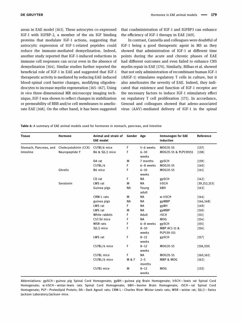
Table7:
Asu
mmaryof
EAEan
imal
mod
elsus
edforho
rmon
esin
theliv
eran
dfatcells
(adipo
cytes)
Tissue
Hormon
eAnimal
andstrain
ofEA
Emod
elGen
der
Age
Immun
ogen
forEA
Eindu
ction
Referen
ce
Liver
Insu
lin-like
grow
thfactor-1
(IGF-1)
LWSrat
MNA
gpSCH
[163 ,16
5–16
7](PL*SJL)F1mice
NA
8–1
0wee
ksgp
MBP
[169]
SJL/J
mice
F5–
12wee
ksMBP
[170
]C57
BL/6mice
NA
NA
MOG35
-55
[172
]C57
BL/6mice
F5–
12wee
ksMOG35
-55
[173
]Th
rombo
poietin(TPO
)SJL/J
mice
F5–
6wee
ksPL
P139
-151
[174
]Hep
cidin
C57
BL/6mice
F8–1
0wee
ksMOG35
-55
[177
]DArats
M&F
7–8wee
ksbB
H[178
]Fatcells
(adipo
cytes)
Leptin
GMM
F8–1
0wee
ksMOG35
-55
[179
]SJL/J
mice
M&F
4–6wee
ksPL
P139
-151
[180]
SJL/J
mice
F6–8
wee
ksPL
P139
-151
[181]
GMM
F8–1
2wee
ksMOG35
-55
[183]
SJL/J
mice
F6wee
ksPL
P139
-151
[184]
GMM
F8–1
0wee
ksMOG79
-96
[185]
GMM
F8–1
0wee
ksMOG35
-55
[186]
SJL/J
&C57
BL/6Jmice
F10
wee
ksPL
P139
-151
&MOG35
-55
[187]
C57
BL/6Jmice
NA
8–1
0wee
ksMOG
[188]
Adipo
nectin
GMM
NA
NA
MOG35
-55
[189]
GMM
F6–8
wee
ksMOG35
-55
[190
]
Abb
reviations
:gpS
CH–gu
inea
pigSpina
lCordHom
ogen
ate;
bBH–bo
vine
Brain
Hom
ogen
ate;
PLP–Proteo
lipid
Protein;
DA–DarkAgo
utirats;
SJL/J–SwissJackso
nLabo
ratory/Jacks
onmice;
GMM
–Gen
etically
man
ipulated
mice.
Hormones in EAE animal models 181
expression and a transient reduction of LepRb duringthe peak of the disease. In contrast, Mishra et al. showedthat astrocytic leptin signaling plays a beneficial role inreducing disease severity [185]. Indeed, astrocyte-specificleptin receptor knockout mice had severe EAE symptoms,compared to wild-type, suggesting that astrocytic leptinsignaling serves as a beneficial factor in clearing the infil-trating leukocytes and decreasing autoimmune destruc-tion of the CNS. In accordance, endothelial leptin sig-naling in EAE showed that removal of leptin signaling,using endothelial leptin receptor-specific knockout mice,reduced EAE severity which emphasized that endothelialleptin signaling increases BBB dysfunction to exacerbateEAE [186].
Calorie-restriction or fasting improved EAE severity,which was concomitant with lower levels of leptin [187].In order to study the role of energy in EAE development,hypoleptinemia was induced by keeping the mice ina fasting situation, with or without leptin injectionsfor 48 h at the onset of EAE [188]. Fasted mice showedlower EAE clinical scores, compared to control-fed mice.Moreover, fasted mice receiving leptin injection duringthis period had more severe EAE symptoms than fastedmice without leptin injection. These observations demon-strated that leptin alone can reverse the protection againstautoimmunity seen during fasting.
11.2 Adiponectin
Piccio et al. reported for the first time that adiponectinconfers a neuroprotective role in EAE [189]. Indeed, adi-ponectin-deficient mice were used to induce EAE andshowed that lack of adiponectin exacerbates EAE severityby inducing higher CNS inflammation, demyelination,and axonal injury. In fact, lack of adiponectin directlyaffects immune responses, by promoting T cell responsesto antigen while Th1 profile was prominent. Moreover,adiponectin receptors 1 and 2 were found to be markedlyexpressed by murine lymph node cells and splenocytes.In the absence of adiponectin and in response to anti-CD3stimulation, increased proliferation of ADPKO CD4+ T cellwas obtained. Complementary to this study, Zhang et al.revealed the role and mechanism of adiponectin onpathogenic Th17 cells [190]. They reported that adipo-nectin-deficient mice enhance both Th1 and Th17 cellcytokines in the peripheral immune system and CNS ofEAE mice as well as upregulated PPARγ and sirtuin 1(SIRT1) and suppressed retinoid-related orphan receptor-γt(RORγt), a key transcription factor during the differentiation
of Th17 cells. These results suggested that adiponectinmodulates the immune response in EAE by inhibitingTh17 cell-mediated autoimmune CNS inflammation.
11.3 Betatrophin, retinol binding protein 4,and asprosin
No studies were found concerning these hormones in EAEmodel.
A summary of EAE animal models used to study hor-mones of the fat cells (adipocytes) is given in Table 7.
12 Conclusion
Based on investigations carried out on the endocrinesystem and EAE, most hormones show conflicting rolesin susceptibility to EAE and on therapeutic targets forEAE. This could be due, among others, to different factorssuch as sampling in different phases of EAE progression,sex, age, strain of mice/rat models used, the type of EAEinduction, and the concentration of hormones in thera-peutic studies. While some studies reported the beneficialrole of melatonin in EAE amelioration, others showedopposite and conflicting results and reported age- anddose-dependent response of EAE models to melatonintherapy. Indeed, it is suggested that the timing of mela-tonin administration leads to different outcomes in EAE.We strongly believe that studies on the endocrine systemin EAE are still at their beginning and we are far awayfrom understanding the role of hormones in EAE suscept-ibility; hence, further studies are required. New studiesshould consider the above-mentioned factors to achievemore precise results. For example, hormones or theirreceptor’s agonists/antagonists should be administeredat different times of day/night, at different physiologicaland pharmacological doses, use mice or rats at differentages, and study the effects at different stages of EAE. Thisalso can be considered for the studies investigating theeffect of EAE induction on endocrine system function.
Funding information: Authors state no funding involved.
Conflict of interest: Authors state no conflict of interest.
Data availability statement: Data sharing is not applic-able to this article as no datasets were generated or ana-lyzed during the current study.
182 Majid Ghareghani et al.
References
[1] Garcia-Reyero N. The clandestine organs of the endocrinesystem. Gen Comp Endocrinol. 2018 Feb;257:264–71.
[2] Yu J. Endocrine disorders and the neurologic manifestations.Ann Pediatr Endocrinol Metab. 2014 Dec;19(4):184–90.
[3] ThyagaRajan S, Priyanka HP. Bidirectional communicationbetween the neuroendocrine system and the immunesystem: relevance to health and diseases. Ann Neurosci.2012 Jan;19(1):40–6.
[4] Noseworthy JH, Lucchinetti C, Rodriguez M, Weinshenker BG.Multiple sclerosis. N Engl J Med. 2000 Sep;343(13):938–52.
[5] Iwanowski P, Losy J. Immunological differences betweenclassical phenothypes of multiple sclerosis. J Neurol Sci.2015 Feb;349(1–2):10–4.
[6] Nourbakhsh B, Mowry EM. Multiple sclerosis risk factors andpathogenesis. Continuum (Minneap Minn). 2019Jun;25(3):596–610.
[7] Constantinescu CS, Farooqi N, O’Brien K, Gran B.Experimental autoimmune encephalomyelitis (EAE) as amodel for multiple sclerosis (MS). Br J Pharmacol. 2011Oct;164(4):1079–106.
[8] Gran B, O’Brien K, Fitzgerald D, Rostami A. Experimentalautoimmune encephalomyelitis. Handbook of neurochem-istry and molecular neurobiology A. Lajtha. Heidelberg:Springer; 2007.
[9] Bert A, Gran B, Weissert R. EAE: imperfect but useful modelsof multiple sclerosis. Trends Mol Med. 2011Mar;17(3):119–25.
[10] Teixeira SA, Varriano AA, Bolonheis SM, Muscará MN.Experimental autoimmune encephalomyelitis: a heteroge-neous group of animal models to study human multiplesclerosis. Drug Discov Today Dis Models. 2005;2(2):127–34.
[11] Glatigny S, Bettelli E. Experimental autoimmune encephalo-myelitis (EAE) as animal models of multiple sclerosis (MS).Cold Spring Harb Perspect Med. 2018 Nov;8(11):a028977.
[12] Hofstetter HH, Shive CL, Forsthuber TG. Pertussis toxinmodulates the immune response to neuroantigens injectedin incomplete Freund’s adjuvant: induction of Th1 cells andexperimental autoimmune encephalomyelitis in the presenceof high frequencies of Th2 cells. J Immunol. 2002Jul;169(1):117–25.
[13] Foster SC, Daniels C, Bourdette DN, Bebo BF Jr. Dysregulationof the hypothalamic-pituitary-gonadal axis in experimentalautoimmune encephalomyelitis and multiple sclerosis.J Neuroimmunol. 2003 Jul;140(1–2):78–87.
[14] Riskind PN, Massacesi L, Doolittle TH, Hauser SL. The role ofprolactin in autoimmune demyelination: suppression ofexperimental allergic encephalomyelitis by bromocriptine.Ann Neurol. 1991 May;29(5):542–7.
[15] Dijkstra CD, van der Voort ER, De Groot CJ, Huitinga I,Uitdehaag BM, Polman CH, et al. Therapeutic effect of the D2-dopamine agonist bromocriptine on acute and relapsingexperimental allergic encephalomyelitis.Psychoneuroendocrinology. 1994;19(2):135–42.
[16] Canonico PL, Sortino MA, Favit A, Aleppo G, Scapagnini U.Dihydroergocryptine protects from acute experimentalallergic encephalomyelitis in the rat. Funct Neurol. 1993May–Jun;8(3):183–8.
[17] Esquifino AI, Cano P, Zapata A, Cardinali DP. Experimentalallergic encephalomyelitis in pituitary-grafted Lewis rats.J Neuroinflammation. 2006 Aug;3(1):20.
[18] Costanza M, Musio S, Abou-Hamdan M, Binart N, Pedotti R.Prolactin is not required for the development of severechronic experimental autoimmune encephalomyelitis.J Immunol. 2013 Sep;191(5):2082–8.
[19] Zhornitsky S, Johnson TA, Metz LM, Weiss S, Yong VW.Prolactin in combination with interferon-β reduces diseaseseverity in an animal model of multiple sclerosis.J Neuroinflammation. 2015 Mar;12(1):55.
[20] Zhang C, Raveney BJ, Hohjoh H, Tomi C, Oki S, Yamamura T.Extrapituitary prolactin promotes generation of Eomes-posi-tive helper T cells mediating neuroinflammation. Proc NatlAcad Sci USA. 2019 Oct;116(42):21131–9.
[21] Esquifino AI, Cano P, Jiménez V, Cutrera RA, Cardinali DP.Experimental allergic encephalomyelitis in male Lewis ratssubjected to calorie restriction. J Physiol Biochem. 2004Dec;60(4):245–52.
[22] Shohreh R, Pardo CA, Guaraldi F, Schally AV, Salvatori R. GH,but not GHRH, plays a role in the development of experi-mental autoimmune encephalomyelitis. Endocrinology. 2011Oct;152(10):3803–10.
[23] Duckers HJ, van Dokkum RP, Verhaagen J, Lopes da Silva FH,Gispen WH. Functional and neurophysiological evidence ofthe efficacy of trophic pharmacotherapy using an adreno-corticotrophic hormone4-9 analog in experimental allergicencephalomyelitis, an animal model of multiple sclerosis.Neuroscience. 1996 Mar;71(2):507–21.
[24] Duckers HJ, van Dokkum RP, Verhaagen J, van Luijtelaar EL,Coenen AM, Lopes da Silva FH, et al. Neurotrophic ACTH4-9analogue therapy normalizes electroencephalographicalterations in chronic experimental allergic encephalomye-litis. Eur J Neurosci. 1998 Dec;10(12):3709–20.
[25] Weidenfeld J, Karussis D, Abramsky O, Lehmann D, Arbell I,Ovadia H. Linomide activates the adrenocortical axis in therat: inhibition of experimental autoimmune encephalomye-litis by linomide is not related to the increase of corticos-terone. J Neuroimmunol. 1997 Oct;79(1):49–53.
[26] Stefferl A, Linington C, Holsboer F, Reul JM. Susceptibilityand resistance to experimental allergic encephalomyelitis:relationship with hypothalamic-pituitary-adrenocortical axisresponsiveness in the rat. Endocrinology. 1999Nov;140(11):4932–8.
[27] Huitinga I, Schmidt ED, van der Cammen MJ, Binnekade R,Tilders FJ. Priming with interleukin-1beta suppressesexperimental allergic encephalomyelitis in the Lewis rat.J Neuroendocrinol. 2000 Dec;12(12):1186–93.
[28] Quintanar-Stephano A, Chavira-Ramírez R, Kovacs K, Berczi I.Neurointermediate pituitary lobectomy decreases the inci-dence and severity of experimental autoimmune encephalo-myelitis in Lewis rats. J Endocrinol. 2005 Jan;184(1):51–8.
[29] Quintanar-Stephano A, Organista-Esparza A, Chavira-Ramírez R, Kovacs K, Berczi I. Effects of neurointermediatepituitary lobectomy and desmopressin on acute experi-mental autoimmune encephalomyelitis in Lewis rats.Neuroimmunomodulation. 2012;19(3):148–57.
[30] Wei Z, Deng X, Hong M, Su Q, Liu A, Huang Y, et al. Icariin hassynergistic effects with methylprednisolone to ameliorateEAE via modulating HPA function, promoting anti-
Hormones in EAE animal models 183
inflammatory and anti-apoptotic effects. Int J Clin Exp Med.2015 Nov;8(11):20188–97.
[31] Ruocco HH, Fernandes GA, Namer IJ, Depaulis A, Levy S.Hypothalamic response to experimental allergic encephalo-myelitis: role of substance P. Neuroimmunomodulation.2004;11(1):28–35.
[32] Miller H, Newell DJ, Ridley A. Multiple sclerosis. Treatment ofacute exacerbations with corticotrophin (A.C.T.H.). Lancet.1961 Nov;2(7212):1120–2.
[33] Cusick MF, Libbey JE, Oh L, Jordan S, Fujinami RS. Acthar geltreatment suppresses acute exacerbations in a murine modelof relapsing-remitting multiple sclerosis. Autoimmunity.2015 Jun;48(4):222–30.
[34] Zhang J, Markovic-Plese S, Lacet B, Raus J, Weiner HL,Hafler DA. Increased frequency of interleukin 2-responsive Tcells specific for myelin basic protein and proteolipid proteinin peripheral blood and cerebrospinal fluid of patients withmultiple sclerosis. J Exp Med. 1994 Mar;179(3):973–84.
[35] Brod SA, Hood ZM. Ingested (oral) ACTH inhibits EAE.J Neuroimmunol. 2011 Mar;232(1–2):131–5.
[36] Dittel LJ, Dittel BN, Brod SA. Ingested ACTH blocks Th17production by inhibiting GALT IL-6. J Neurol Sci. 2020Feb;409:116602.
[37] Viñuela-Berni V, Gómez-González B, Quintanar-Stephano A.Blockade of arginine vasopressin receptors prevents blood-brain barrier breakdown in experimental autoimmune ence-phalomyelitis. Sci Rep. 2020 Jan;10(1):467.
[38] Tanaka K, Saito R, Sanada K, Nishimura H, Nishimura K,Sonoda S, et al. Expression of hypothalamic feeding-relatedpeptide genes and neuroendocrine responses in an experi-mental allergic encephalomyelitis rat model. Peptides. 2020Jul;129:170313.
[39] White SR, Samathanam GK, Bowker RM, Wessendorf MW.Damage to bulbospinal serotonin-, tyrosine hydroxylase-,and TRH-containing axons occurs early in the development ofexperimental allergic encephalomyelitis in rats. J NeurosciRes. 1990 Sep;27(1):89–98.
[40] Brod SA, Bauer V. Ingested (oral) thyrotropin releasing factor(TRH) inhibits EAE. Cytokine. 2013 Jan;61(1):323–8.
[41] Quintanar JL, Salinas E, Quintanar-Stephano A.Gonadotropin-releasing hormone reduces the severity ofexperimental autoimmune encephalomyelitis, a model ofmultiple sclerosis. Neuropeptides. 2011 Feb;45(1):43–8.
[42] Guzmán-Soto I, Salinas E, Hernández-Jasso I, Quintanar JL.Leuprolide acetate, a GnRH agonist, improves experimentalautoimmune encephalomyelitis: a possible therapy for mul-tiple sclerosis. Neurochem Res. 2012 Oct;37(10):2190–7.
[43] Guzmán-Soto I, Salinas E, Quintanar JL. Leuprolide acetateinhibits spinal cord inflammatory response in experimentalautoimmune encephalomyelitis by suppressing NF-κB acti-vation. Neuroimmunomodulation. 2016;23(1):33–40.
[44] Ikushima H, Kanaoka M, Kojima S. Cutting edge: requirementfor growth hormone-releasing hormone in the developmentof experimental autoimmune encephalomyelitis. J Immunol.2003 Sep;171(6):2769–72.
[45] Purba JS, Raadsheer FC, Hofman MA, Ravid R, Polman CH,Kamphorst W, et al. Increased number of corticotropin-releasing hormone expressing neurons in the hypothalamicparaventricular nucleus of patients with multiple sclerosis.Neuroendocrinology. 1995 Jul;62(1):62–70.
[46] Calzà L, Giardino L, Pozza M, Micera A, Aloe L. Time-coursechanges of nerve growth factor, corticotropin-releasing hor-mone, and nitric oxide synthase isoforms and their possiblerole in the development of inflammatory response inexperimental allergic encephalomyelitis. Proc Natl Acad SciUSA. 1997 Apr;94(7):3368–73.
[47] Benou C, Wang Y, Imitola J, VanVlerken L, Chandras C,Karalis KP, et al. Corticotropin-releasing hormone contri-butes to the peripheral inflammatory response in experi-mental autoimmune encephalomyelitis. J Immunol. 2005May;174(9):5407–13.
[48] Muhvić D, Radosević-Stasić B, Pugel E, Rukavina D, Sepcić J,Efendić S. Modulation of experimental allergic encephalo-myelitis by somatostatin. Ann N Y Acad Sci. 1992 Apr1;650:170–8.
[49] Muhvić D, Barac-Latas V, Rukavina D, Radosević-Stasić B.Induction of experimental allergic encephalomyelitis in alow-susceptible Albino Oxford rat strain by somatostatinanalogue SMS 201-995. Neuroimmunomodulation.2005;12(1):20–8.
[50] Brod SA, Hood ZM. Ingested (oral) SST inhibits EAE.Autoimmunity. 2011 Aug;44(5):437–43.
[51] Constantinescu CS, Hilliard B, Ventura E, Rostami A.Luzindole, a melatonin receptor antagonist, suppressesexperimental autoimmune encephalomyelitis.Pathobiolology. 1997;65(4):190–4.
[52] Kang JC, Ahn M, Kim YS, Moon C, Lee Y, Wie MB, et al.Melatonin ameliorates autoimmune encephalomyelitisthrough suppression of intercellular adhesion molecule-1.J Vet Sci. 2001 Aug;2(2):85–9.
[53] Álvarez-Sánchez N, Cruz-Chamorro I, López-González A,Utrilla JC, Fernández-Santos JM, Martínez-López A, et al.Melatonin controls experimental autoimmune encephalo-myelitis by altering the T effector/regulatory balance. BrainBehav Immun. 2015 Nov;50:101–14.
[54] Chen SJ, Huang SH, Chen JW, Wang KC, Yang YR, Liu PF, et al.Melatonin enhances interleukin-10 expression andsuppresses chemotaxis to inhibit inflammation in situand reduce the severity of experimental autoimmuneencephalomyelitis. Int Immunopharmacol. 2016Feb;31:169–77.
[55] Ghareghani M, Dokoohaki S, Ghanbari A, Farhadi N, Zibara K,Khodadoust S, et al. Melatonin exacerbates acute experi-mental autoimmune encephalomyelitis by enhancing theserum levels of lactate: a potential biomarker of multiplesclerosis progression. Clin Exp Pharmacol Physiol. 2017Jan;44(1):52–61.
[56] Ghareghani M, Zibara K, Sadeghi H, Farhadi N. Spasticitytreatment ameliorates the efficacy of melatonin therapy inexperimental autoimmune encephalomyelitis (EAE) mousemodel of multiple sclerosis. Cell Mol Neurobiol. 2018Jul;38(5):1145–51.
[57] Ghareghani M, Scavo L, Jand Y, Farhadi N, Sadeghi H,Ghanbari A, et al. Melatonin therapy modulates cerebralmetabolism and enhances remyelination by increasing PDK4in a mouse model of multiple sclerosis. Front Pharmacol.2019 Feb;10:147.
[58] Ortiga-Carvalho TM, Sidhaye AR, Wondisford FE. Thyroidhormone receptors and resistance to thyroid hormonedisorders. Nat Rev Endocrinol. 2014 Oct;10(10):582–91.
184 Majid Ghareghani et al.
[59] Rogister B, Ben-Hur T, Dubois-Dalcq M. From neural stemcells to myelinating oligodendrocytes. Mol Cell Neurosci.1999 Oct-Nov;14(4–5):287–300.
[60] Calza L, Fernandez M, Giuliani A, Aloe L, Giardino L. Thyroidhormone activates oligodendrocyte precursors and increasesa myelin-forming protein and NGF content in the spinal cordduring experimental allergic encephalomyelitis. Proc NatlAcad Sci USA. 2002 Mar;99(5):3258–63.
[61] Farsetti A, Mitsuhashi T, Desvergne B, Robbins J,Nikodem VM. Molecular basis of thyroid hormone regulationof myelin basic protein gene expression in rodent brain.J Biol Chem. 1991 Dec;266(34):23226–32.
[62] Gallo V, Armstrong RC. Developmental and growth factor-induced regulation of nestin in oligodendrocyte lineage cells.J Neurosci. 1995 Jan;15(1 Pt 1):394–406.
[63] Lezoualc’h F, Seugnet I, Monnier AL, Ghysdael J, Behr JP,Demeneix BA. Inhibition of neurogenic precursor prolifera-tion by antisense alpha thyroid hormone receptor oligo-nucleotides. J Biol Chem. 1995 May;270(20):12100–8.
[64] Albornoz EA, Carreño LJ, Cortes CM, Gonzalez PA,Cisternas PA, Cautivo KM, et al. Gestational hypothyroidismincreases the severity of experimental autoimmune ence-phalomyelitis in adult offspring. Thyroid. 2013Dec;23(12):1627–37.
[65] Villoslada P, Hauser SL, Bartke I, Unger J, Heald N,Rosenberg D, et al. Human nerve growth factor protectscommon marmosets against autoimmune encephalomyelitisby switching the balance of T helper cell type 1 and 2 cyto-kines within the central nervous system. J Exp Med. 2000May;191(10):1799–806.
[66] Diab A, Deng C, Smith JD, Hussain RZ, Phanavanh B, Lovett-Racke AE, et al. Peroxisome proliferator-activated receptor-gamma agonist 15-deoxy-Delta(12,14)-prostaglandin J(2)ameliorates experimental autoimmune encephalomyelitis.J Immunol. 2002 Mar;168(5):2508–15.
[67] Racke MK, Gocke AR, Muir M, Diab A, Drew PD, Lovett-Racke AE. Nuclear receptors and autoimmune disease: thepotential of PPAR agonists to treat multiple sclerosis. J Nutr.2006 Mar;136(3):700–3.
[68] Klotz L, Burgdorf S, Dani I, Saijo K, Flossdorf J, Hucke S, et al.The nuclear receptor PPAR γ selectively inhibits Th17 differ-entiation in a T cell-intrinsic fashion and suppresses CNSautoimmunity. J Exp Med. 2009 Sep;206(101):3159.
[69] Fernandez M, Giuliani A, Pirondi S, D’Intino G, Giardino L,Aloe L, et al. Thyroid hormone administration enhancesremyelination in chronic demyelinating inflammatory dis-ease. Proc Natl Acad Sci USA. 2004 Nov;101(46):16363–8.
[70] Calzà L, Fernandez M, Giuliani A, D’Intino G, Pirondi S,Sivilia S, et al. Thyroid hormone and remyelination in adultcentral nervous system: a lesson from an inflammatory-demyelinating disease. Brain Res Brain Res Rev. 2005Apr;48(2):339–46.
[71] Boelen A, Mikita J, Boiziau C, Chassande O, Fliers E, Petry KG.Type 3 deiodinase expression in inflammatory spinal cordlesions in rat experimental autoimmune encephalomyelitis.Thyroid. 2009 Dec;19(12):1401–6.
[72] Guadaño-Ferraz A, Obregón MJ, St Germain DL, Bernal J. Thetype 2 iodothyronine deiodinase is expressed primarily inglial cells in the neonatal rat brain. Proc Natl Acad Sci USA.1997 Sep;94(19):10391–6.
[73] Sonoda J, Pei L, Evans RM. Nuclear receptors: decodingmetabolic disease. FEBS Lett. 2008 Jan;582(1):2–9.
[74] Oetting A, Yen PM. New insights into thyroid hormone action.Best Pract Res Clin Endocrinol Metab. 2007Jun;21(2):193–208.
[75] Fernández M, Paradisi M, Del Vecchio G, Giardino L, Calzà L.Thyroid hormone induces glial lineage of primary neuro-spheres derived from non-pathological and pathological ratbrain: implications for remyelination-enhancing therapies.Int J Dev Neurosci. 2009 Dec;27(8):769–78.
[76] D’Intino G, Lorenzini L, Fernandez M, Taglioni A, Perretta G,Del Vecchio G, et al. Triiodothyronine administration ame-liorates the demyelination/remyelination ratio in a non-human primate model of multiple sclerosis by correctingtissue hypothyroidism. J Neuroendocrinol. 2011Sep;23(9):778–90.
[77] Dell’Acqua ML, Lorenzini L, D’Intino G, Sivilia S,Pasqualetti P, Panetta V, et al. Functional and molecularevidence of myelin- and neuroprotection by thyroid hormoneadministration in experimental allergic encephalomyelitis.Neuropathol Appl Neurobiol. 2012 Aug;38(5):454–70.
[78] Castelo-Branco G, Stridh P, Guerreiro-Cacais AO,Adzemovic MZ, Falcão AM, Marta M, et al. Acute treatmentwith valproic acid and l-thyroxine ameliorates clinical signsof experimental autoimmune encephalomyelitis and pre-vents brain pathology in DA rats. Neurobiol Dis. 2014Nov;71:220–33.
[79] Kuwabara T, Ishikawa F, Yasuda T, Aritomi K, Nakano H,Tanaka Y, et al. CCR7 ligands are required for developmentof experimental autoimmune encephalomyelitis throughgenerating IL-23-dependent Th17 cells. J Immunol. 2009Aug;183(4):2513–21.
[80] Li O, Liu JQ, Zhang H, Zheng P, Liu Y, Bai XF. CD62L is requiredfor the priming of encephalitogenic T cells but does not play amajor role in the effector phase of experimental autoimmuneencephalomyelitis. Scand J Immunol. 2006Aug;64(2):117–24.
[81] Haensgen H, Albornoz E, Opazo MC, Bugueño K, JaraFernández EL, Binzberger R, et al. Gestational hypothyroxi-nemia affects its offspring with a reduced suppressivecapacity impairing the outcome of the experimental autoim-mune encephalomyelitis. Front Immunol. 2018 Jun;9:1257.
[82] Becklund BR, Hansen DW Jr, Deluca HF. Enhancement of 1,25-dihydroxyvitamin D3-mediated suppression of experimentalautoimmune encephalomyelitis by calcitonin. Proc Natl AcadSci USA. 2009 Mar;106(13):5276–81.
[83] Becklund BR, James BJ, Gagel RF, DeLuca HF. The calcitonin/calcitonin gene related peptide-α gene is not required for1α,25-dihydroxyvitamin D3-mediated suppression of experi-mental autoimmune encephalomyelitis. Arch BiochemBiophys. 2009 Aug;488(2):105–8.
[84] Tsuji F, Murai M, Oki K, Seki I, Ueda K, Inoue H, et al.Transient receptor potential vanilloid 1 agonists as candi-dates for anti-inflammatory and immunomodulatory agents.Eur J Pharmacol. 2010 Feb;627(1–3):332–9.
[85] Matsuda R, Kezuka T, Nishiyama C, Usui Y, Matsunaga Y,Okunuki Y, et al. Suppression of murine experimental auto-immune optic neuritis by mature dendritic cells transfectedwith calcitonin gene-related Peptide gene. InvestOphthalmol Vis Sci. 2012 Aug;53(9):5475–85.
Hormones in EAE animal models 185
[86] McLatchie LM, Fraser NJ, Main MJ, Wise A, Brown J,Thompson N, et al. RAMPs regulate the transport and ligandspecificity of the calcitonin-receptor-like receptor. Nature.1998 May;393(6683):333–9.
[87] Mikami N, Watanabe K, Hashimoto N, Miyagi Y, Sueda K,Fukada S, et al. Calcitonin gene-related peptide enhancesexperimental autoimmune encephalomyelitis by promotingTh17-cell functions. Int Immunol. 2012 Nov;24(11):681–91.
[88] Sardi C, Zambusi L, Finardi A, Ruffini F, Tolun AA,Dickerson IM, et al. Involvement of calcitonin gene-relatedpeptide and receptor component protein in experimentalautoimmune encephalomyelitis. J Neuroimmunol. 2014Jun;271(1–2):18–29.
[89] Meehan TF, Vanhooke J, Prahl J, Deluca HF. Hypercalcemiaproduced by parathyroid hormone suppresses experimentalautoimmune encephalomyelitis in female but not male mice.Arch Biochem Biophys. 2005 Oct;442(2):214–21.
[90] Ghareghani M, Scavo L, Arnoult D, Zibara K, Farhadi N.Melatonin therapy reduces the risk of osteoporosis andnormalizes bone formation in multiple sclerosis. Fundam ClinPharmacol. 2018 Apr;32(2):181–7.
[91] Branisteanu DD, Waer M, Sobis H, Marcelis S, Vandeputte M,Bouillon R. Prevention of murine experimental allergicencephalomyelitis: cooperative effects of cyclosporine and 1alpha, 25-(OH)2D3. J Neuroimmunol. 1995 Sep;61(2):151–60.
[92] Van Etten E, Branisteanu DD, Overbergh L, Bouillon R,Verstuyf A, Mathieu C. Combination of a 1,25-dihydroxyvi-tamin D3 analog and a bisphosphonate prevents experi-mental autoimmune encephalomyelitis and preserves bone.Bone. 2003 Apr;32(4):397–404.
[93] Berard JL, Zarruk JG, Arbour N, Prat A, Yong VW, Jacques FH,et al. Lipocalin 2 is a novel immune mediator of experimentalautoimmune encephalomyelitis pathogenesis and is modu-lated in multiple sclerosis. Glia. 2012 Jul;60(7):1145–59.
[94] Nam Y, Kim JH, Seo M, Kim JH, Jin M, Jeon S, et al. Lipocalin-2protein deficiency ameliorates experimental autoimmuneencephalomyelitis: the pathogenic role of lipocalin-2 in thecentral nervous system and peripheral lymphoid tissues.J Biol Chem. 2014 Jun;289(24):16773–89.
[95] Marques F, Mesquita SD, Sousa JC, Coppola G, Gao F,Geschwind DH, et al. Lipocalin 2 is present in the EAE brainand is modulated by natalizumab. Front Cell Neurosci. 2012Aug;6:33.
[96] Iaffaldano P, Lucchese G, Trojano M. Treating multiplesclerosis with natalizumab. Expert Rev Neurother. 2011Dec;11(12):1683–92.
[97] Ebrahimi-Kalan A, Soleimani Rad J, Kafami L,Mohammadnejad D, Habibi Roudkenar M, Khaki AA, et al.MS14 down-regulates lipocalin2 expression in spinal cordtissue in an animal model of multiple sclerosis in femaleC57BL/6. Iran Biomed J. 2020 Nov;24(6):409.
[98] Brines ML, Ghezzi P, Keenan S, Agnello D, de Lanerolle NC,Cerami C, et al. Erythropoietin crosses the blood-brain bar-rier to protect against experimental brain injury. Proc NatlAcad Sci USA. 2000 Sep;97(19):10526–31.
[99] Mayo L, Quintana FJ, Weiner HL. The innate immune system indemyelinating disease. Immunol Rev. 2012Jul;248(1):170–87.
[100] Agnello D, Bigini P, Villa P, Mennini T, Cerami A, Brines ML,et al. Erythropoietin exerts an anti-inflammatory effect on the
CNS in a model of experimental autoimmune encephalo-myelitis. Brain Res. 2002 Oct;952(1):128–34.
[101] Li W, Maeda Y, Yuan RR, Elkabes S, Cook S, Dowling P.Beneficial effect of erythropoietin on experimental allergicencephalomyelitis. Ann Neurol. 2004 Dec;56(6):767–77.
[102] Zhang X, Li QY, Xiao BG. Anti-inflammatory effect of ery-thropoietin therapy on experimental autoimmune encepha-lomyelitis. Int J Neurosci. 2012 May;122(5):255–62.
[103] Zhang J, Li Y, Cui Y, Chen J, Lu M, Elias SB, et al.Erythropoietin treatment improves neurological functionalrecovery in EAE mice. Brain Res. 2005 Feb;1034(1–2):34–9.
[104] Dasgupta S, Mazumder B, Ramani YR, Bhattacharyya SP,Das MK. Evaluation of the role of erythropoietin and metho-trexate in multiple sclerosis. Indian J Pharmacol. 2011Sep;43(5):512–5.
[105] Savino C, Pedotti R, Baggi F, Ubiali F, Gallo B, Nava S, et al.Delayed administration of erythropoietin and its non-ery-thropoietic derivatives ameliorates chronic murine autoim-mune encephalomyelitis. J Neuroimmunol. 2006Mar;172(1–2):27–37.
[106] Yuan R, Wang B, Lu W, Maeda Y, Dowling P. A distinct regionin erythropoietin that induces immuno/inflammatory modu-lation and tissue protection. Neurotherapeutics. 2015Oct;12(4):850–61.
[107] Boz C, Ozmenoglu M, Velioglu S, Kilinc K, Orem A, Alioglu Z,et al. Matrix metalloproteinase-9 (MMP-9) and tissue inhi-bitor of matrix metalloproteinase (TIMP-1) in patients withrelapsing-remitting multiple sclerosis treated with interferonbeta. Clin Neurol Neurosurg. 2006 Feb;108(2):124–8.
[108] Crocker SJ, Whitmire JK, Frausto RF, Chertboonmuang P,Soloway PD, Whitton JL, et al. Persistent macrophage/microglial activation and myelin disruption after experi-mental autoimmune encephalomyelitis in tissue inhibitor ofmetalloproteinase-1-deficient mice. Am J Pathol. 2006Dec;169(6):2104–16.
[109] Thorne M, Moore CS, Robertson GS. Lack of TIMP-1 increasesseverity of experimental autoimmune encephalomyelitis:effects of darbepoetin alfa on TIMP-1 null and wild-type mice.J Neuroimmunol. 2009 Jun;211(1–2):92–100.
[110] Chen H, Luo B, Yang X, Xiong J, Liu Z, Jiang M, et al.Therapeutic effects of nonerythropoietic erythropoietinanalog ARA290 in experimental autoimmune encephalo-myelitis rat. J Neuroimmunol. 2014 Mar;268(1–2):64–70.
[111] Mengozzi M, Cervellini I, Bigini P, Martone S, Biondi A,Pedotti R, et al. Endogenous erythropoietin as part of thecytokine network in the pathogenesis of experimental auto-immune encephalomyelitis. Mol Med. 2008Nov–Dec;14(11–12):682–8.
[112] Chen SJ, Wang YL, Lo WT, Wu CC, Hsieh CW, Huang CF, et al.Erythropoietin enhances endogenous haem oxygenase-1 andrepresses immune responses to ameliorate experimentalautoimmune encephalomyelitis. Clin Exp Immunol. 2010Nov;162(2):210–23.
[113] Moransard M, Bednar M, Frei K, Gassmann M, Ogunshola OO.Erythropoietin reduces experimental autoimmune encepha-lomyelitis severity via neuroprotective mechanisms.J Neuroinflammation. 2017 Oct;14(1):202.
[114] Sättler MB, Merkler D, Maier K, Stadelmann C, Ehrenreich H,Bähr M, et al. Neuroprotective effects and intracellular sig-naling pathways of erythropoietin in a rat model of multiple
186 Majid Ghareghani et al.
sclerosis. Cell Death Differ. 2004 Dec;11(S2 Suppl 2):S181–92.
[115] Diem R, Sättler MB, Merkler D, Demmer I, Maier K,Stadelmann C, et al. Combined therapy with methylpredni-solone and erythropoietin in a model of multiple sclerosis.Brain. 2005 Feb;128(Pt 2):375–85.
[116] Gaindh D, Choi YB, Marchese M, Dowling P, Cook S,Blumberg B, et al. Prolonged beneficial effect of brief ery-thropoietin peptide JM4 therapy on chronic relapsing EAE.Neurotherapeutics. 2020 Sep:1–11. doi: 10.1007/s13311-020-00923-5.
[117] Pytskiĭ VI, Siusiukin IP. Conversion of cortisone into cortisolin the liver of guinea pigs during allergic processes ofimmediate and delayed types. Probl Endokrinol (Mosk). 1972May–Jun;18(3):96–100.
[118] Pytskiĭ VI, Siusiukin IP. Cortisol metabolic intensity in theliver of guinea pigs in allergic processes of the immediateand delayed types. Biull Eksp Biol Med. 1976Aug;82(8):981–4.
[119] Pytskiĭ VI, Arutiunova EE. Kinetics and properties of a cor-tisol-resistant population of lymphocytes from the lymphnodes of guinea pigs with experimental allergic encephalo-myelitis. Biull Eksp Biol Med. 1977 Feb;83(2):200–2.
[120] Herrada AA, Contreras FJ, Marini NP, Amador CA,Gonzalez PA, Cortes CM, et al. Aldosterone promotes auto-immune damage by enhancing Th17-mediated immunity.J Immunol. 2010 Jan;184(1):191–202.
[121] Wesselmann U, Konkol RJ, Leo GL, Roerig DL, Harder DR.Altered splenic catecholamine concentrations duringexperimental allergic encephalomyelitis. Pharmacol BiochemBehav. 1987 Apr;26(4):851–4.
[122] Dowdell KC, Gienapp IE, Stuckman S, Wardrop RM,Whitacre CC. Neuroendocrine modulation of chronic relap-sing experimental autoimmune encephalomyelitis: a criticalrole for the hypothalamic-pituitary-adrenal axis.J Neuroimmunol. 1999 Dec;100(1–2):243–51.
[123] Yang P, Tian H, Zou YR, Chambon P, Ichinose H, Honig G,et al. Epinephrine production in Th17 cells and experimentalautoimmune encephalitis. Front Immunol. 2021Feb;12:616583.
[124] White SR, Bhatnagar RK, Bardo MT. Norepinephrine deple-tion in the spinal cord gray matter of rats with experimentalallergic encephalomyelitis. J Neurochem. 1983Jun;40(6):1771–3.
[125] Krenger W, Honegger CG, Feurer C, Cammisuli S. Changes ofneurotransmitter systems in chronic relapsing experimentalallergic encephalomyelitis in rat brain and spinal cord.J Neurochem. 1986 Oct;47(4):1247–54.
[126] Abramsky O, Wertman E, Reches A, Brenner T, Ovadia H.Effect of hypothalamic lesions on experimental autoimmunediseases in rats. Ann N Y Acad Sci. 1987;496(1):360–5.
[127] Karpus WJ, Konkol RJ, Killen JA. Central catecholamine neu-rotoxin administration. 1. Immunological changes associatedwith the suppression of experimental autoimmune ence-phalomyelitis. J Neuroimmunol. 1988 Apr;18(1):61–73.
[128] Konkol RJ, Wesselmann U, Karpus WJ, Leo GL, Killen JA,Roerig DL. Suppression of clinical weakness in experimentalautoimmune encephalomyelitis associated with weightchanges, and post-decapitation convulsions after
intracisternal-ventricular administration of 6-hydroxydopa-mine. J Neuroimmunol. 1990 Jan;26(1):25–34.
[129] Simonini MV, Polak PE, Sharp A, McGuire S, Galea E,Feinstein DL. Increasing CNS noradrenaline reduces EAEseverity. J Neuroimmune Pharmacol. 2010 Jun;5(2):252–9.
[130] Polak PE, Kalinin S, Feinstein DL. Locus coeruleus damageand noradrenaline reductions in multiple sclerosis andexperimental autoimmune encephalomyelitis. Brain. 2011Mar;134(Pt 3):665–77.
[131] Polak PE, Kalinin S, Braun D, Sharp A, Lin SX, Feinstein DL.The vincamine derivative vindeburnol provides benefit in amouse model of multiple sclerosis: effects on the Locuscoeruleus. J Neurochem. 2012 Apr;121(2):206–16.
[132] Leonard JP, MacKenzie FJ, Patel HA, Cuzner ML. Hypothalamicnoradrenergic pathways exert an influence on neuroendo-crine and clinical status in experimental autoimmune ence-phalomyelitis. Brain Behav Immun. 1991 Dec;5(4):328–38.
[133] Bałkowiec-Iskra E, Kurkowska-Jastrzebska I, Joniec I,Ciesielska A, Członkowska A, Członkowski A. Dopamine,serotonin and noradrenaline changes in the striatum ofC57BL mice following myelin oligodendrocyte glycoprotein(MOG) 35-55 and complete Freund adjuvant (CFA) adminis-tration. Acta Neurobiol Exp (Wars). 2007;67(4):379–88.
[134] Shaked I, Hanna RN, Shaked H, Chodaczek G, Nowyhed HN,Tweet G, et al. Transcription factor Nr4a1 couples sympa-thetic and inflammatory cues in CNS-recruited macrophagesto limit neuroinflammation. Nat Immunol. 2015Dec;16(12):1228–34.
[135] Vujnović I, Pilipović I, Jasnić N, Petrović R, Blagojević V,Arsenović-Ranin N, et al. Noradrenaline through β-adreno-ceptor contributes to sexual dimorphism in primary CD4+
T-cell response in DA rat EAE model? Cell Immunol. 2019Feb;336:48–57.
[136] Pilipović I, Vujnović I, Stojić-Vukanić Z, Petrović R, Kosec D,Nacka-Aleksić M, et al. Noradrenaline modulates CD4+ T cellpriming in rat experimental autoimmune encephalomyelitis:a role for the α1-adrenoceptor. Immunol Res. 2019Jun;67(2–3):223–40.
[137] Shipaeva EV, Kovalenko LP, Sorokina AV, Kovalev GI,Tallerova AV, Kolik LG, et al. Study of anti-inflammatoryeffects of GB-115, a glycine-containing retropeptide chole-cystokinin analog. Bull Exp Biol Med. 2011Mar;150(5):599–602.
[138] Bedoui S, Miyake S, Lin Y, Miyamoto K, Oki S, Kawamura N,et al. Neuropeptide Y (NPY) suppresses experimental auto-immune encephalomyelitis: NPY1 receptor-specific inhibitionof autoreactive Th1 responses in vivo. J Immunol. 2003Oct;171(7):3451–8.
[139] Dimitrijević M, Mitić K, Kuštrimović N, Vujić V, Stanojević S.NPY suppressed development of experimental autoimmuneencephalomyelitis in Dark Agouti rats by disrupting costi-mulatory molecule interactions. J Neuroimmunol. 2012Apr;245(1–2):23–31.
[140] Brod SA, Bauer VL. Ingested (oral) neuropeptide Y inhibitsEAE. J Neuroimmunol. 2012 Sep;250(1–2):44–9.
[141] Theil MM, Miyake S, Mizuno M, Tomi C, Croxford JL,Hosoda H, et al. Suppression of experimental autoimmuneencephalomyelitis by ghrelin. J Immunol. 2009Aug;183(4):2859–66.
Hormones in EAE animal models 187
[142] Liu F, Li Z, He X, Yu H, Feng J. Ghrelin attenuates neuroin-flammation and demyelination in experimental autoimmuneencephalomyelitis involving NLRP3 inflammasome signalingpathway and pyroptosis. Front Pharmacol. 2019 Nov;10:1320.
[143] Lycke E, Roos BE. Brain monoamines in guinea pigs withexperimental allergic encephalitis. Int Arch Allergy ApplImmunol. 1973;45(3):341–51.
[144] White SR. Failure of 5-hydroxytryptophan to increase lumbarMSR amplitude in rats paralyzed with experimental allergicencephalomyelitis. Brain Res. 1979 Nov;177(1):157–63.
[145] White SR, Bieger D. Central serotonin receptor sensitivity inrats with experimental allergic encephalomyelitis. ResCommun Chem Pathol Pharmacol. 1980 Nov;30(2):269–81.
[146] Scott CF Jr, Cashman N, Spitler LE. Experimental allergicencephalitis; treatment with drugs which alter CNS serotoninlevels. J Immunopharmacol. 1982–1983;4(3):153–62.
[147] Babington RG, Wedeking PW. The influence of cinanserin andselected pharmacologic agents on experimental allergicencephalomyelitis (EAE). J Pharmacol Exp Ther. 1971May;177(2):454–60.
[148] Weistock M, Shoham-Moshonov S, Teitelbaum D, Arnon R.Inactivation of neurogenic 5-hydroxytryptamine receptors inguinea pigs with experimental allergic encephalomyelitis(EAE) induced paralysis. Brain Res. 1977 Apr;125(1):192–5.
[149] Dietsch GN, Hinrichs DJ. The role of mast cells in the elici-tation of experimental allergic encephalomyelitis. J Immunol.1989 Mar;142(5):1476–81.
[150] Freire-Garabal M, Núñez MJ, Balboa J, García-Vallejo LA,Argibay S, Rodrigo E, et al. Administration of the 5-hydroxy-tryptamine(1A) receptor antagonist WAY100635 suppressesacute experimental allergic encephalomyelitis in Lewis rats.Neurosci Lett. 2003 May;342(1–2):33–6.
[151] Simmons RD, Buzbee TM, Linthicum DS. Methysergide, aserotonin antagonist, does not inhibit the expression ofautoimmune encephalomyelitis in the rabbit.J Neuroimmunol. 1989 Mar;22(1):77–9.
[152] Samathanam GK, White SR, Kalivas PW, Duffy P. Effects of5-hydroxytryptophan on extracellular serotonin in the spinalcord of rats with experimental allergic encephalomyelitis.Brain Res. 1991 Sep;559(1):37–43.
[153] White SR, Black PC, Samathanam GK, Paros KC. Prazosinsuppresses development of axonal damage in rats inocu-lated for experimental allergic encephalomyelitis.J Neuroimmunol. 1992 Aug;39(3):211–8.
[154] Taler M, Gil-Ad I, Korob I, Weizman A. The immunomodulatoryeffect of the antidepressant sertraline in an experimentalautoimmune encephalomyelitis mouse model of multiplesclerosis. Neuroimmunomodulation. 2011;18(2):117–22.
[155] Yuan XQ, Qiu G, Liu XJ, Liu S, Wu Y, Wang X, et al. Fluoxetinepromotes remission in acute experimental autoimmuneencephalomyelitis in rats. Neuroimmunomodulation.2012;19(4):201–8.
[156] Bhat R, Mahapatra S, Axtell RC, Steinman L. Amelioration ofongoing experimental autoimmune encephalomyelitis withfluoxetine. J Neuroimmunol. 2017 Dec;313:77–81.
[157] Ghareghani M, Zibara K, Sadeghi H, Dokoohaki S, Sadeghi H,Aryanpour R, et al. Fluvoxamine stimulates oligodendrogen-esis of cultured neural stem cells and attenuates inflamma-tion and demyelination in an animal model of multiplesclerosis. Sci Rep. 2017 Jul;7(1):4923.
[158] O’Sullivan D, Green L, Stone S, Zareie P, Kharkrang M,Fong D, et al. Treatment with the antipsychotic agent, ris-peridone, reduces disease severity in experimental autoim-mune encephalomyelitis. PLoS One. 2014 Aug;9(8):e104430.
[159] Green LK, Zareie P, Templeton N, Keyzers RA, Connor B,La Flamme AC. Enhanced disease reduction using clozapine,an atypical antipsychotic agent, and glatiramer acetatecombination therapy in experimental autoimmune encepha-lomyelitis. Mult Scler J Exp Transl Clin [Internet]. 2017 Mar[cited 2021 Apr];3(1):13. Available from: https://journals.sagepub.com/doi/10.1177/2055217317698724
[160] Musgrave T, Benson C, Wong G, Browne I, Tenorio G, Rauw G,et al. The MAO inhibitor phenelzine improves functionaloutcomes in mice with experimental autoimmune encepha-lomyelitis (EAE). Brain Behav Immun. 2011Nov;25(8):1677–88.
[161] Benson CA, Wong G, Tenorio G, Baker GB, Kerr BJ. The MAOinhibitor phenelzine can improve functional outcomes inmice with established clinical signs in experimental auto-immune encephalomyelitis (EAE). Behav Brain Res. 2013Sep;252:302–11.
[162] Hofstetter HH, Mössner R, Lesch KP, Linker RA, Toyka KV,Gold R. Absence of reuptake of serotonin influences sus-ceptibility to clinical autoimmune disease and neuroantigen-specific interferon-gamma production in mouse EAE. Clin ExpImmunol. 2005 Oct;142(1):39–44.
[163] Liu X, Yao DL, Bondy CA, Brenner M, Hudson LD, Zhou J, et al.Astrocytes express insulin-like growth factor-I (IGF-I) and itsbinding protein, IGFBP-2, during demyelination induced byexperimental autoimmune encephalomyelitis. Mol CellNeurosci. 1994 Oct;5(5):418–30.
[164] Liu X, Linnington C, Webster HD, Lassmann S, Yao DL,Hudson LD, et al. Insulin-like growth factor-I treatmentreduces immune cell responses in acute non-demyelinativeexperimental autoimmune encephalomyelitis. J NeurosciRes. 1997 Mar;47(5):531–8.
[165] Liu X, Yao DL, Webster HF. Insulin-like growth factor I treat-ment reduces clinical deficits and lesion severity in acutedemyelinating experimental autoimmune encephalomyelitis.Mult Scler J. 1995 Apr;1(1):2–9.
[166] Yao DL, Liu X, Hudson LD, Webster HD. Insulin-like growthfactor I treatment reduces demyelination and up-regulatesgene expression of myelin-related proteins in experimentalautoimmune encephalomyelitis. Proc Natl Acad Sci USA.1995 Jun;92(13):6190–4.
[167] Yao DL, Liu X, Hudson LD, Webster HD. Insulin-like growthfactor-I given subcutaneously reduces clinical deficits,decreases lesion severity and upregulates synthesis ofmyelin proteins in experimental autoimmune encephalo-myelitis. Life Sci. 1996;58(16):1301–6.
[168] Xu S, Jordan EK, Li W, Yang Y, Chesnick SA, Webster HD, et al.In vivo three-dimensional MR microscopy of mice withchronic relapsing experimental autoimmune encephalomye-litis after treatment with insulin-like growth factor-I. AJNR AmJ Neuroradiol. 1998 Apr;19(4):653–8.
[169] Lovett-Racke AE, Bittner P, Cross AH, Carlino JA, Racke MK.Regulation of experimental autoimmune encephalomyelitiswith insulin-like growth factor (IGF-1) and IGF-1/IGF-bindingprotein-3 complex (IGF-1/IGFBP3). J Clin Invest. 1998Apr;101(8):1797–804.
188 Majid Ghareghani et al.
[170] Cannella B, Pitt D, Capello E, Raine CS. Insulin-like growthfactor-1 fails to enhance central nervous system myelinrepair during autoimmune demyelination. Am J Pathol. 2000Sep;157(3):933–43.
[171] Bilbao D, Luciani L, Johannesson B, Piszczek A, Rosenthal N.Insulin-like growth factor-1 stimulates regulatory T cells andsuppresses autoimmune disease. EMBO Mol Med. 2014Nov;6(11):1423–35.
[172] Genoud S, Maricic I, Kumar V, Gage FH. Targeted expressionof IGF-1 in the central nervous system fails to protect micefrom experimental autoimmune encephalomyelitis.J Neuroimmunol. 2005 Nov;168(1–2):40–5.
[173] DiToro D, Harbour SN, Bando JK, Benavides G, Witte S,Laufer VA, et al. Insulin-like growth factors are key regulatorsof T helper 17 regulatory T cell balance in autoimmunity.Immunity. 2020 Apr;52(4):650–67.
[174] Verda L, Luo K, Kim DA, Bronesky D, Kohm AP, Miller SD, et al.Effect of hematopoietic growth factors on severity of experi-mental autoimmune encephalomyelitis. Bone MarrowTransplant. 2006 Sep;38(6):453–60.
[175] Neema M, Arora A, Healy BC, Guss ZD, Brass SD, Duan Y,et al. Deep gray matter involvement on brain MRI scans isassociated with clinical progression in multiple sclerosis.J Neuroimaging. 2009 Jan;19(1):3–8.
[176] Schuh C, Wimmer I, Hametner S, Haider L, Van Dam AM,Liblau RS, et al. Oxidative tissue injury in multiple sclerosis isonly partly reflected in experimental disease models. ActaNeuropathol. 2014 Aug;128(2):247–66.
[177] Zarruk JG, Berard JL, Passos dos Santos R, Kroner A, Lee J,Arosio P, et al. Expression of iron homeostasis proteins in thespinal cord in experimental autoimmune encephalomyelitisand their implications for iron accumulation. Neurobiol Dis.2015 Sep;81:93–107.
[178] Ćurko-Cofek B, Grubić Kezele T, Barac-Latas V. Hepcidin andmetallothioneins as molecular base for sex-dependent differencesin clinical course of experimental autoimmune encephalomyelitisin chronic iron overload. Med Hypotheses. 2017 Sep;107:51–4.
[179] Matarese G, Di Giacomo A, Sanna V, Lord GM, Howard JK,Di Tuoro A, et al. Requirement for leptin in the induction andprogression of autoimmune encephalomyelitis. J Immunol.2001 May;166(10):5909–16.
[180] Matarese G, Sanna V, Di Giacomo A, Lord GM, Howard JK,Bloom SR, et al. Leptin potentiates experimental autoim-mune encephalomyelitis in SJL female mice and confers
susceptibility to males. Eur J Immunol. 2001May;31(5):1324–32.
[181] De Rosa V, Procaccini C, La Cava A, Chieffi P, Nicoletti GF,Fontana S, et al. Leptin neutralization interferes with patho-genic T cell autoreactivity in autoimmune encephalomyelitis.J Clin Invest. 2006 Feb;116(2):447–55.
[182] Takahashi K, Suwa H, Ishikawa T, Kotani H. Targeted dis-ruption of H3 receptors results in changes in brain histaminetone leading to an obese phenotype. J Clin Invest. 2002Dec;110(12):1791–9.
[183] Musio S, Gallo B, Scabeni S, Lapilla M, Poliani PL,Matarese G, et al. A key regulatory role for histamine inexperimental autoimmune encephalomyelitis: diseaseexacerbation in histidine decarboxylase-deficient mice.J Immunol. 2006 Jan;176(1):17–26.
[184] Wu X, Hsuchou H, Kastin AJ, Mishra PK, Pan W. Upregulationof astrocytic leptin receptor in mice with experimental auto-immune encephalomyelitis. J Mol Neurosci.2013;49(3):446–56.
[185] Mishra PK, Hsuchou H, Ouyang S, Kastin AJ, Wu X, Pan W.Loss of astrocytic leptin signaling worsens experimentalautoimmune encephalomyelitis. Brain Behav Immun. 2013Nov;34:98–107.
[186] Ouyang S, Hsuchou H, Kastin AJ, Mishra PK, Wang Y, Pan W.Leukocyte infiltration into spinal cord of EAE mice is attenu-ated by removal of endothelial leptin signaling. Brain BehavImmun. 2014 Aug;40:61–73.
[187] Piccio L, Stark JL, Cross AH. Chronic calorie restrictionattenuates experimental autoimmune encephalomyelitis.J Leukoc Biol. 2008 Oct;84(4):940–8.
[188] Gerriets VA, Danzaki K, Kishton RJ, Eisner W, Nichols AG,Saucillo DC, et al. Leptin directly promotes T-cell glycolyticmetabolism to drive effector T-cell differentiation in a mousemodel of autoimmunity. Eur J Immunol. 2016Aug;46(8):1970–83.
[189] Piccio L, Cantoni C, Henderson JG, Hawiger D,RamsbottomM, Mikesell R, et al. Lack of adiponectin leads toincreased lymphocyte activation and increased diseaseseverity in a mouse model of multiple sclerosis. Eur JImmunol. 2013 Aug;43(8):2089–100.
[190] Zhang K, Guo Y, Ge Z, Zhang Z, Da Y, Li W, et al. Adiponectinsuppresses T helper 17 cell differentiation and limits auto-immune CNS inflammation via the SIRT1/PPARγ/RORγtpathway. Mol Neurobiol. 2017 Sep;54(7):4908–20.
Hormones in EAE animal models 189