Hormon Endokrin pada Pankreas (Endocrine Hormones of Pancreas)
Upload
khangminh22Category
view
0download
0
EFFECTS IN VIVO OF NEUROHYPOPHYSIALHORMONES ON THE CONTRACTILE ACTIVITY OF
ACCESSORY SEX ORGANS IN MALE RABBITS
PER MELIN
Institute of ^oophysiology, University of Uppsala, Sweden
(Received 23rd June 1969, revised 22nd September 1969)
Summary. The effects of single intravenous injections of synthetic oxy-tocin and lysine-vasopressin on the contractile activity of the distal partof the ductus epididymidis, the proximal and distal parts of the vasdeferens and the vesicula seminalis were studied in seventy-twoanaesthetized rabbits. The muscular contractions were registered as
changes in the intraluminal pressure of the organs, and the amplitude,frequency, duration and tonus were measured.
An injection of 20 m-i.u. of oxytocin/kg body weight caused a smallbut significant increase of the amplitude of the contractions in theductus epididymidis and the proximal part of the vas deferens. Aninjection of 100 m-i.u. of oxytocin increased the tone in the proximalpart of the vas deferens but decreased this parameter in the distal partof the organ. The infusion of 20 m-i.u. of vasopressin enhanced thefrequency, decreased the duration and elevated the muscular tone inthe ductus epididymidis and increased the frequency and the tone in theproximal part of the vas deferens. The infusion of 100 m-i.u. of vaso-
pressin resulted in increased amplitudes in both these regions. A gradualincrease in the effects of oxytocin and vasopressin towards the testis was
observed, indicating a polarity in response to the hormones of the malereproductive ducts.
The results support the theory that neurohypophysial hormones mayregulate sperm transport in the male through their effects on the con-
tractile elements of the genital organs, possibly being complementary intheir actions.
INTRODUCTIONIn recent years, oxytocin has been observed to have immediate effects on malesexual functions, such as libido, transport of spermatozoa and the discharge ofsemen (Bereznev, 1963; Ewy & Bielansky, 1962; Kihlström & Melin, 1963;Melin & Kihlström, 1963; Levin, 1966; Fj eilström, Kihlström 8c Melin, 1968).
It seems reasonable to attribute the reported effects of oxytocin to a stimu¬lating action of the hormone on the contractile elements of the male genitalorgans, though the majority of investigations on the subject do not give much
283 Downloaded from Bioscientifica.com at 01/09/2022 01:01:32AMvia free access

284 Per Melinevidence for such an effect either in vitro (Waddell, 1916, 1917; Perutz &Merdier, 1924; Martins & Valle, 1939; Wojcik, 1966; Sjöstrand & Swedin,1968) or in vivo (Cross & Glover, 1958; Gross, 1959).
This study was undertaken in view of the previous conflicting reports andthe apparent lack of systematic observations of the effects of oxytocin on themale genital organs. Since the secretion of oxytocin from the neurohypophysisis, as a rule, accompanied by a simultaneous release of vasopressin (Heller,1961), this hormone was included in the investigation.
The present work deals with the influence of oxytocin and vasopressin on thecontractile activity of the distal part of the ductus epididymidis, the vas deferensand the vesícula seminalis, using a quantitative method, an adequate recordingsystem and statistical treatment.
MATERIAL AND METHODS
Seventy-two male rabbits about 1 year old, weighing 2-6 to 3-4 kg and of mixedbreeds were used. The animals had been caged separately and been given thesame kind of food at regular times, for at least a fortnight before the experiments.
The rabbits were anaesthetized by intravenous injections of Numal (Roche)(0-5 ml/kg body weight). Supplementary doses of the anaesthetic were adminis¬tered throughout the experiments, to keep the depth of anaesthesia as constantas possible. Tracheotomy was generally performed to facilitate respiration andan external jugular vein was cannulated to assist the giving of injections duringthe experiments. The remaining surgical procedures and the intraluminalpressure measurements in the different parts of the male genital tract were
performed according to the methods described by Melin (1970).The organs were allowed to adapt themselves to the new conditions for about
30 min before the experimental injections started. Only one kind of preparationwas studied in each animal and for each preparation the following main criteriawere to be fulfilled before the start of the injections : there was to be no excessivebleeding caused by the surgical manipulations, the drift of muscular tone was
not to exceed 0-2 mm H20/min and the different regions of the tract were torespond to intravenous injections of adrenalin (2 to 4 /ig/kg body weight) witha single momentary contraction (latency 8 to 15 sec) (Plate 1). As a rule, therewas a slow decrease with time in the muscular activities of the different prepara¬tions of the genital tract (probably mainly due to absorption of the intraluminalfluid by tissue elements and consequent decrease of the initial pressure). Toreduce this drawback, both sides of the animals were used for the pressuremeasurements.
The hormones used were synthetic oxytocin (Partocon, Ferring AB) andlysine-vasopressin (Postacton, Ferring AB) in initial concentrations of 10 i.u. /ml.Two different doses of the hormones were tested, 20 m-i.u. and 100 m-i.u./kgbody weight. After the requisite dilution with saline, the hormones were eachgiven as a single intravenous injection in a volume of 0-5 ml. Immediatelyafter each infusion, the injection cannula was rinsed with an injection of thesame amount of saline.
Differences in the contractile patterns were observed for two consecutive
Downloaded from Bioscientifica.com at 01/09/2022 01:01:32AMvia free access
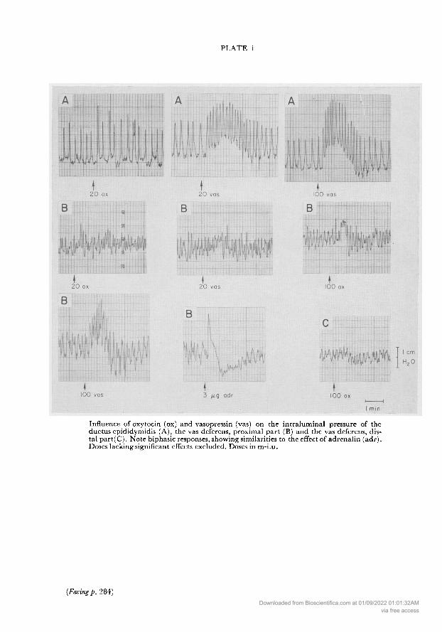
PLATE 1
Influence of oxytocin (ox) and vasopressin (vas) on the intraluminal pressure of theductus epididymidis (A), the vas deferens, proximal part (B) and the vas deferens, dis¬tal part(C). Note biphasic responses, showing similarities to the effect of adrenalin (adr).Doses lacking significant effects excluded. Doses in m-i.u.
(Facing p. 284)Downloaded from Bioscientifica.com at 01/09/2022 01:01:32AM
via free access

Oxytocin and vasopressin on male sex organs 285
periods of 3 min, one period starting with a saline injection and the other witha hormone injection. At least 15 min were allowed to intervene between eachnew injection of the hormone. To avoid statistical bias due to tachyphylaxis,the order of infusion of the hormones was altered for each preparation.
The following characteristics of contractile activity were noted :The amplitude of the contractions, measured as the registered height of each
contraction (variations in pressure of 1 mm H20 or less were neglected).The frequency, counted as the number of contractions per min.The duration, giving the time in seconds from the start of each contraction to
the end of the relaxation phase.The tonus change, determined as a change in the level of the base line (a line
drawn through the mean value of the minimum level of each contraction).The data obtained were calculated as mean values from at least two pairs of
observation periods, per dose and per animal, each period of the pair startingwith an injection of saline and of a hormone, respectively. A value of = 0-05less was considered significant.
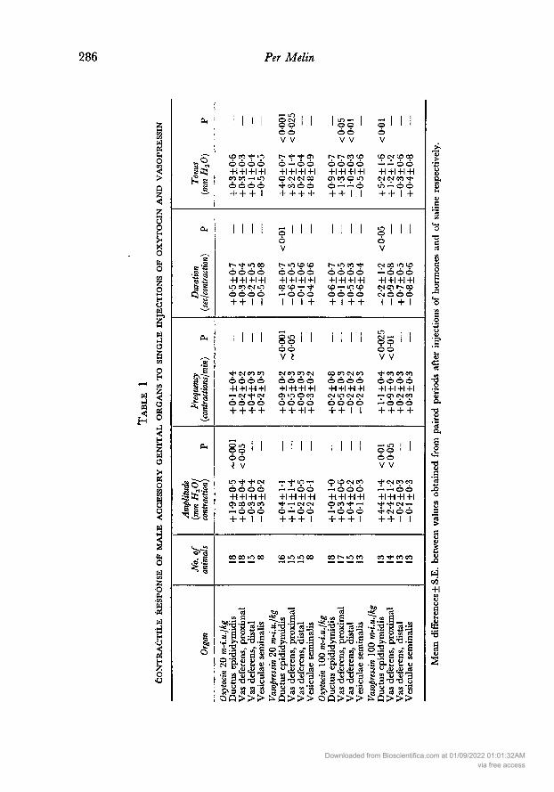
RESULTSThe general effects of the hormones on muscle contractility are shown in Table1, which gives the significances of the pooled data. As a rule, the effects per¬sisted for only a few minutes (latency 10 to 15 sec) after injection of the hor¬mones. Representative records of the significant responses are shown in Plate 1.
Ductus epididymidis20 m-i.u. of oxytocin. This dose caused a moderate but highly significant
enhancement of the amplitude of muscular contractions (P<0-001, Table 1,Plate 1). The mean frequency and the mean duration were not influenced byinjections of 20 m-i.u. of oxytocin, nor was the tone significantly altered.
In a few cases (Table 2), there was a slight but significant biphasic responseto the hormone : an increase of the tone occurred and was followed by a de¬crease (latency 45 to 80 sec).
100 m-i.u. ofoxytocin. This higher dose ofoxytocin had no significant influenceon any of the parameters investigated (Table 1, Plate 1 ). The biphasic responseappeared in two cases out of eighteen (Table 2).
The lack ofeffect of the larger amount ofoxytocin on the amplitude is mainlydue to the fact that the number of individuals showing a decrease of thisparameter has increased.
20 m-i.u. of vasopressin. There was no significant change in the amplitude ofthe contractions as a result of the vasopressin injections (Table 1, Plate 1).However, this amount of vasopressin increased the number of contractions permin in fifteen out of eighteen individuals by more than 30% (P<0-001). Theduration of the contraction was shortened (P<0-01) and there was an elevationof the muscular tone (P<0-001). The biphasic responses were shown by 25%of the individuals (Table 2).
100 m-i.u. of vasopressin. This dose of the hormone caused significant enhance¬ment of the amplitude (P<0-01), as well as of the frequency (P<0-025) of the
Downloaded from Bioscientifica.com at 01/09/2022 01:01:32AMvia free access
286 Per Melin
là«<
waCho<>ay<
oo
SohOH
oHOW
wÜ
oHy] oaoj<
wÜ><nioc/:cßwuo<
<Sb.o
z;oS,maciw¡JPÜ<a,H
OU
I» s
eu
a-aOh (jai «S*
ì°JtIa51 Se«
^~
IMI
IO CO LO +1 +1 +1 +1CO CO — LO + + + !
- LO , I
OCMoo '
V V
1^- CTi - +1 +1 +1 +1 CM CM CO4 + + + +
LO- Io o '
Ó ÔV V
r^ r^. co io +1 +1 +1 +1CD CO O LO
+ + I I
ò
CO es LO CO
+1 +1 +1 +1CM CM CO LO — OÔ+ + I +
I I I I
~ LO CO - +1 +1 - LO CO CM lo + + I I
si I V
t^ LO CO CO +1 +1 +1 +1co to — — 111 +
I I o V
I I
-~ LO co - - +1 +1 +1CO — LO ID + I + +
CM CO LO CO— +1 +1 +1 +1cm r^. coCM O Ó 0
+
CM CO CO +1 - +! +1— CM - CM + + + +
LO ' V ?
CM CO CO CM +1 +1 +1 +1 lo o co + + + +
I I
CO CO CM CO +1 +1 +1 +1CM LO CM CM + + I I
cm- Ioo ' '
V V
CO CO co +1 +1 +1 +1— CT) CM co— + + + +
Il I I I I I I I Ioo
<· V
LO · - CM - - +1+1 co co co— + + I I
— LO —
— — +1 +1 +1 +1• — CM CM — + + + I
CO CM CO— +1 +1 +1 +1OCO —
— + + +
-LO ' V V
CM CO CO— — +1 +1 +1 +1• CM —
• CM + +
CO LO LO co
LG g~3" fJ S s Sa F b; — e
¿lilit>0 03
• P S.« S
^j
Ê levo .5o S „r M- 6^-o c c ge S eu <u
ï 2 s; 3•g.3.- - S y m c« 'En!t> 3 $ a S3OQ>>> a 3 ra u
.3'aS.-g0-5Sa
1) S »
S3
S.» sCVO.Hw w Sa a U
€ Cl 3
£ 'S¿"¡g s_ w-· 2 'S S -
¿Ib.- - o ^
Wî cfl "S ^
ci cd «u>>>
t· K 4) CU
S o. S; t. <uS <ää tía rt
S3 u cu *B
§ ë a a sSû>»
c«
cden
T)
o
cfiV a
wco+1
ceT3
Downloaded from Bioscientifica.com at 01/09/2022 01:01:32AMvia free access

Oxytocin and vasopressin on male sex organs 287contractions (Table 1). In addition, there was a shortening of the duration(P<0-05) and an increased tone (P<0-01). Twenty-three per cent of the indi¬viduals exhibited biphasic responses (Table 2).Vas deferens, proximal part
20 m-i.u. of oxytocin. The oxytocin injections caused a slight but significantincrease in amplitude, less obvious than in the ductus epididymidis (P<0-05,Table 1, Plate 1).
100 m-i.u. of oxytocin. As with the preparations of the ductus epididymidis,there were no significant changes of any of the parameters studied, followinginjections of the higher dose of oxytocin, except for a slight elevation of tone
(P<0-05). The biphasic responses occurred in 29% of the cases.
Table 2influence of oxytocin and vasopressin on contractile
behaviour of the male genital tract
Organ
Oxytocin 20 m-i.u./kgDuctus epididymidisVas deferens, proximalVas deferens, distalVasopressin 20 m-i.u./kgDuctus epididymidisVas deferens, proximalVas deferens, distalOxytocin 100 m-i.u./kgDuctus epididymidisVas deferens, proximalVas deferens, distalVasopressin 100 m-i.u./kgDuctus epididymidisVas deferens, proximalVas deferens, distal
No. of animals:Total With biphasic
response*
181815
161515
181715
131413
% animalswith biphasic
response
117
27
251340
112927
235054
* Number of individuals showing significant biphasic responses (P<0-05).
20 m-i.u. ofvasopressin. The stimulating effect of the hormone on the frequencyof contractions in the region of the ductus epididymidis was considerablysmaller in the proximal part of the vas deferens (P<0-05). The vasopressininjection induced rhythmic contractions in a single preparation lacking spon¬taneous motility. There was a moderate increase in tone during the observationperiod after the hormone administration (P< 0-025, Plate 1). The biphasicresponses were rarely seen (13%, Table 2).
100 m-i.u. of vasopressin. The effects of this amount seemed to be accentuatedin comparison with the lower dose (Table 1). There was a moderate elevationof the amplitude of each contraction (P<0-05), as well as an increase in thefrequency of contractions (jP<0-01). Significant biphasic responses were shownby half the preparations (Table 2) : the momentary elevation in musculartone was followed by a marked decrease (Plate 1), which made changes in the
Downloaded from Bioscientifica.com at 01/09/2022 01:01:32AMvia free access
288 Per Melin
average tone during the whole period after the hormone injection insignificant.In addition, there was sometimes an inhibition of the contractions followingupon the decrease in tone.
Vas deferens, distal part20 m-i.u. ofoxytocin. No significant changes were observed (Table 1). However,
in 27% of the individuals (Table 2), there was a biphasic response of the same
type as described earlier.100 m-i.u. of oxytocin. The higher dose of oxytocin used did not cause any
significant change in the amplitude, frequency or duration of contractions. In27% of the preparations, there was a significant biphasic response to the hor¬mone (Table 2) with a low tonus predominating. However, in most individuals,only the second phase of the original biphasic response was present (Plate 1).Consequently, there was a lessening of the average tone (P<0-01) during theobservation period after the oxytocin injection.
20 m-i.u. of vasopressin. The dose did not produce significant changes in any ofthe different parameters of motility, though biphasic responses were rathercommon (40%, Table 2).
100 m-i.u. of vasopressin. There was no alteration of any of the parametersstudied, though in the majority of the preparations, the vasopressin infusionsdid produce a significant biphasic response (Table 2) : the tonus increase duringthe first minute after the injection was followed by a later decrease, resulting ina total tonus change, during the period, of about zero (Table 1).Vesícula seminalis
20 and 100 m-i.u. ofoxytocin. There were no significant changes in any charac¬teristics of the motility pattern of this organ following injections of the hormone(Table 1), though as before, individual responses occurred.
20 and 100 m-i.u. of vasopressin. Neither of these doses of vasopressin were ableto produce any changes in the motility of the organ (Table 1). However, insome cases, there was a lessening of the tone during the latter half of the periodfollowing upon the hormone injection.
Significant biphasic responses were not observed in this region.
DISCUSSIONIt is known from studies of the rabbit uterus (Csapo, 1961) that the thresholddose of oxytocin is low and the effect more pronounced, when the organ pos¬sesses a high degree of excitability, manifested, as a rule, in well-developed,spontaneous, rhythmic contractions. It appears to be of interest to investigatewhether this observation also holds good for the male genital organs. Thus, forexample, the highly significant reaction to neurohypophysial hormones by theductus epididymidis may be connected with the great amplitude of the con¬
tractions observed in this region (Melin, 1970). Besides, the differences inmotility found between the genital tracts investigated in untreated animals(Melin, 1970) may possibly be dependent on the experimental situation. Thisbeing the case, the present observations on differences between regions in
Downloaded from Bioscientifica.com at 01/09/2022 01:01:32AMvia free access

Oxytocin and vasopressin on male sex organs 289
response to the hormones may not represent true phenomena. It has, thus, beenconsidered relevant, in the analysis presented below, to include only thosecases in which injections of the hormones have produced significant changes ofthe different parameters of contraction. The term 'magnitude of response',here denoted by AR, is to be understood as the mean difference between eachof the parameters of muscular contraction observed during periods immediatelyfollowing an injection of a hormone and of saline, respectively. This differencehas been referred to the different parameters of spontaneous motility, studiedduring periods preceding a hormone injection. The correlations are given inTable 3.
In the ductus epididymidis, neither 20 m-i.u. of oxytocin nor 100 m-i.u. ofvasopressin yielded any significant correlation between AR and the amplitudeofcontractions. Moreover, both doses ofvasopressin failed to yield any significant
Table 3MAGNITUDE OF RESPONSE (Ar) TO OXYTOCIN AND VASOPRESSIN, CORRELATED WITH
DIFFERENT PARAMETERS OF CONTRACTILE ACTIVITY
Region HormoneDose
(m-i.u./kg)Independent
variableDependent
variableNo. ofanimals
Ductusepididymidis
Vas deferens,proximalpart
OxytocinVasopressinVasopressinVasopressinVasopressinVasopressinOxytocinVasopressinVasopressinVasopressin
2010020
10020
10020
10020
100
AmplitudeAmplitudeFrequencyFrequencyDurationDurationAmplitudeAmplitudeFrequencyFrequency
ARaARaAR,AR,ARdARdARaARaAR,AR,
18181613161318141514
+ 0-34+ 0-23-0-19-0-30-0-73-0-73+ 0-33-0-59-0-22-0-71
<0·01<0·01
<0·05
<0·01
Only parameters giving significant changes following injections of the hormones have been included(see text).
ARa = AR of the amplitude; AR, = AR of the frequency; ARd = AR of the duration.
correlation between AR and the frequency of contractions. However, AR forthe amounts of vasopressin studied was correlated negatively with the durationof contractions (P<0-01). The effect of vasopressin upon duration seemed to bemore pronounced when the contractions lasted for a long period of time.
In the proximal part of the vas deferens, there was a significant negativecorrelation (P <0-05) between AR and the amplitude only after injections of100 m-i.u. of vasopressin. In addition, AR for this dose of vasopressin was cor¬related negatively with the frequency (P<0-001). Apparently, the higher dose ofvasopressin has an increased capacity to stimulate the amplitude as well as thefrequency of the contractions in those preparations which have poorly developedmuscular activity.
This analysis shows that the effects of the neurohypophysial hormones arenot dependent on the amplitude and the frequency of the contractions, exceptin the proximal part of the vas deferens after infusion of the high dose of vaso¬
pressin (Table 3). With this exception, the facts eliminate the possibility thatthe observed differences in these parameters between the response to the hor-
Downloaded from Bioscientifica.com at 01/09/2022 01:01:32AMvia free access

290 Per Melin
mones of the various parts of the genital tract investigated could have beenconditioned by the experimental situation. The present observations thus indi¬cate a heterogeneous distribution of the receptors sensitive to neurohypophysialhormones in the different parts of the male genital tract. The effects of oxytocinon the analogous organs in the female—the oviduct-uterus complex, may alsobe considered. These organs have been found to have a polarity in their muscu¬lar response to the hormone (Sandberg, Ingelman-Sundberg, Lindgren &Rydén, 1961, and others).
In agreement with the present data, Cross & Glover (1958) were unable tofind any effect of oxytocin and vasopressin on the contractions of the vesículaseminalis in vivo. Earlier investigations of the effects of oxytocin on the motilityof the vas deferens and the epididymis are somewhat contradictory but themajority of these observations disagree with the present findings (Waddell,1916, 1917; Perutz & Merdier, 1924; Martins & Valle, 1939; Martins, Valle &Porto, 1940; Wojcik, 1966). All these studies, however, were performed invitro and were based on the muscle-lever and kymographic technique, whichseems too insensitive for the recording of small manifestations of contractileactivity, in that the longitudinal contractions of the organ will mainly berecorded. Cross (1959) reports no effect of oxytocin on the contractility of theepididymis of anaesthetized rabbits. However, as no data were presented, itseems difficult to draw any definite conclusions from these observations. Itmust be kept in mind that the present effects of oxytocin on the contractilityof the genital organs, though significant, are moderate.
The observed effects of oxytocin and vasopressin on male genital tract
motility must by mediated by a direct mechanism. The latency (10 to 15sec) seems too short for vascular changes, for example, to produce anyalteration of muscular activity. On the other hand, such a mechanism mayoperate to give the biphasic response observed in the present investigationafter injections of oxytocin and particularly of vasopressin (Plate 1). A similarresponse was also apparent after injections of adrenalin (Plate 1), the later partof which was characterized by a lowering of the muscular tone and sometimesfollowed by a temporary inhibition of the muscular contractions. The inhibitoryeffect of adrenalin on the contractility of the male genital organs has also beennoted by Cross (1959).
It is well known that a varying response of the uterus to oxytocin occurs
during the sexual cycle, as well as during pregnancy. Even in males, the impor¬tance of sex hormones for the effect of oxytocin has been considered (Honoré 8cLloyd, 1961; Kihlström & Melin, 1963; Niemi & Kormano, 1965). Accord¬ingly, the possible existence of a sexual cycle in males (Kihlström, 1966) mayexplain the great individual variations observed, not only of the motility of thegenital organs in untreated animals (Melin, 1970) but also of their responses tothe neurohypophysial hormones found in the present investigation. There maywell be an underlying hormonal factor to account for the fact that an injectionof 100 m-i.u. of oxytocin increases the amplitude in the ductus epididymidisin one animal but decreases the same parameter in another individual.
The failure of the higher dose of oxytocin to influence the amplitude of thecontractions in the ductus epididymidis and the proximal part of the vas
Downloaded from Bioscientifica.com at 01/09/2022 01:01:32AMvia free access

Oxytocin and vasopressin on male sex organs 291deferens is partly caused by the increased number of preparations showing a
reduction in amplitude of contraction as a result of hormone injection. Thesefindings, as well as the significant lessening in the muscular tone of the distalpart of the vas deferens caused by oxytocin, may be related to the observationsof Wojcik (1966), who found a reduction in longitudinal muscle activity andin the tone of the male genital organs in response to high doses of oxytocin. Thehormone may have qualitatively different effects on the longitudinal and cir¬cular muscle elements of the male genital tract.
The lower dose of oxytocin (20 m-i.u./kg) used in the present experiments,corresponds to, or is less than, the amounts of the hormone endogenouslyreleased by faradic or physiological stimuli (Harris, 1948; Cross, 1955; Hawker,1961 ; Fuchs & Wagner, 1963; Fuchs, 1964). These facts suggest that at leastthe effects produced by 20 m-i.u. ofoxytocin maybe ofphysiologicalsignificance.Moreover, the inhibitory effect of anaesthesia on the action of oxytocin (whichmay be valid for vasopressin as well) must be considered (Fuchs & Wagner,1963). Consequently, in order to reproduce the effects of the hormones inanaesthetized animals, it is probably necessary to use amounts of the substancesthat appear unphysiological. The present highly significant effects of 20 m-i.u.of oxytocin and vasopressin (as well as the tetanic effect of this dose of vaso¬
pressin) in the ductus epididymidis indicate a supra-threshold dose.The present observations support the theory that the effects of oxytocin on the
male sexual function, referred to earlier, are partly caused by the action ofthis hormone on the contractile elements of the genital organs. This investi¬gation indicates that vasopressin may also be important in this respect. Itappears that there is a gradual increase of the effects of the neurohypophysialhormones towards the testis. The findings agree with observations in rats byNiemi 8c Kormano (1965) on the influence of oxytocin on the contractility ofthe seminiferous tubules. Of course, the more testicular parts of the malegenital ducts are the most logical target organs for the hormones studied. It isnoticeable that oxytocin and vasopressin in the lowest doses used, exclusivelyaffect the amplitude and frequency respectively. In view of the simultaneousrelease of the hormones from the neurohypophysis (Heller, 1961), these obser¬vations indicate complementary effects of the hormones on the male genitaltract.
ACKNOWLEDGMENTS
This investigation was supported by grants from the Magnus Bergwall andHierta-Retzius Foundations and from the Faculty of Mathematics and Scienceof the University of Uppsala. For helpful criticism, I am much indebted toDr J. E. Kihlström and Professor P. E. Lindahl. Oxytocin (Partocon) andlysine-vasopressin (Postacton) were generously supplied by Ferring AB, Malmö,Sweden.
REFERENCES
Bereznev, A. P. (1963) Oxytocin a stimulator of semen emission in bulls. (In Russian). Seljskochozjaist-vennoi Nauki. 8, 91.
Cross, B. A. (1955) Neurohormonal mechanisms in emotional inhibition of milk ejection. J. Endocr.12, 29.
Downloaded from Bioscientifica.com at 01/09/2022 01:01:32AMvia free access

292 Per MelinCross, B. A. (1959) Hypothalamic influences on sperm transport in the male and female genital tract. In: Endo¬
crinology of Reproduction, p. 167. Ed. C. W. Lloyd. Academic Press, New York.Cross, B. A. & Glover, T. D. (1958) The hypothalamus and seminal emission. J. Endocr. 16, 385.Csapo, A. (1961) The effects of oxytocic substances on the excitability of the uterus. In: Oxytocin, p. 100. Eds.
R. Caldeyro-Barcia and H. Heller. Pergamon Press, London.Ewy, Z. & Bielansky, W. (1962) Influence of oxytocin on spermatozoa transport in the ductus deferens
in the ram. Proc. int. Union. Physiol. Sci. 11, No. 545.Fjellström, D., Kihlström, J. . & Melin, P. (1968) The effect of synthetic oxytocin upon seminal
characteristics and sexual behaviour in male rabbits. J. Reprod. Fert. 17, 207.Fuchs, A. R. (1964) Oxytocin and the onset of labour in rabbits. J. Endocr. 30, 217.Fuchs, A. R. & Wagner, G. (1963) Quantitative aspects of release of oxytocin by suckling in unanaes-
thetized rabbits. Acta endocr., Copenh. 44, 581.Harris, G. W. (1948) Further evidence regarding the endocrine status of the neurohypophysis. J.
Physiol., Land. 107, 436.Hawker, R. (1961) Oxytocin and an unidentified oxytocic substance in extracts of blood. In: Oxytocin, p. 425.
Eds. R. Caldeyro-Barcia and H. Heller. Pergamon Press, London.Heller, H. (1961) Occurrence, storage and metabolism of oxytocin. In: Oxytocin, p. 3. Eds. R. Caldeyro-
Barcia and H. Heller. Pergamon Press, London.Honoré, L. H. & Lloyd, S. (1961) The effects of castration on the vascular responses of male rats to
posterior pituitary hormones. J. Physiol., Lond. 159, 183.Kihlström, J. E. (1966) A sex cycle in the male. Experientia, 22, 630.Kihlström, J. E. & Melin, P. (1963) The influence of oxytocin upon some seminal characteristics in
the rabbit. Acta physiol. scand. 59, 363.Levin, . L. (1966) The effect of oxytocin and proserine on increased sperm production in hogs.
(In Russian). Veterinariyja, 43, 96.Martins, T. & Valle, J. R. (1939) Endocrine control of the motility of the male accessory genital
organs. Endocrinology, 25, 80.Martins, T., Valle, J. R. & Porto, A. (1940) Pharmacology in vitro of the human vasa deferentia and
epididymis : the question of the endocrine control of the motility of the male accessory genitals.J. Urol. 44, 682.
Melin, P. (1970) In vivo recording of contractile activity of male accessory genital organs in rabbits.Acta physiol. scand. 79, (in press).
Melin, P. & Kihlström, J. E. (1963) Influence of oxytocin on sexual behavior in male rabbits.Endocrinology, 73, 433.
Niemi, . & Kormano, M. (1965) Contractility of the seminiferous tubule of the postnatal rat testisand its response to oxytocin. Annls Med. exp. Biol. Fenn. 43, 40.
Perutz, A. & Merdler, M. (1924) Beiträge zur experimentellen Pharmacologie des männlichenGenitales. Arch. Derm. Syph. 148, 104.
Sandberg, F., Ingelman-Sundberg, ., Lindgren, L. & Rydén, G. (1961) The effect of oxytocin on themotility of the different parts of the pregnant and nonpregnant human uterus in vitro as compared with otheroxytocic drugs. In: Oxytocin, p. 295. Eds. R. Caldeyro-Barcia and H. Heller. Pergamon Press,London.
Sjöstrand, . O. & Swedin, G. (1968) Potentiation by smooth muscle stimulants of the hypogastricnerve-vas deferens preparation from normal and castrated guinea-pigs. Ada physiol. scand. 74,472.
Waddell, J. A. (1916) The pharmacology of the vas deferens. J. Pharmac. exp. Ther. 8, 551.Waddell, J. A. (1917) The pharmacology of the uterus masculinus. J. Pharmac. exp. Ther. 9, 171.Wojcik, K. (1966) Mechanisms of transport in the male genital organs. 1. Action of cholinergic and
adrenergic mediators and hypothalamic hormones on contractions of isolated preparations ofrabbits' genital organs. Ada physiol. pol. 17, 78.
Downloaded from Bioscientifica.com at 01/09/2022 01:01:32AMvia free access
Copyright © 2022 FDOKUMEN