Venezuelan equine encephalomyelitis : the goat as a ... - CORE
Upload
independentCategory
view
3download
0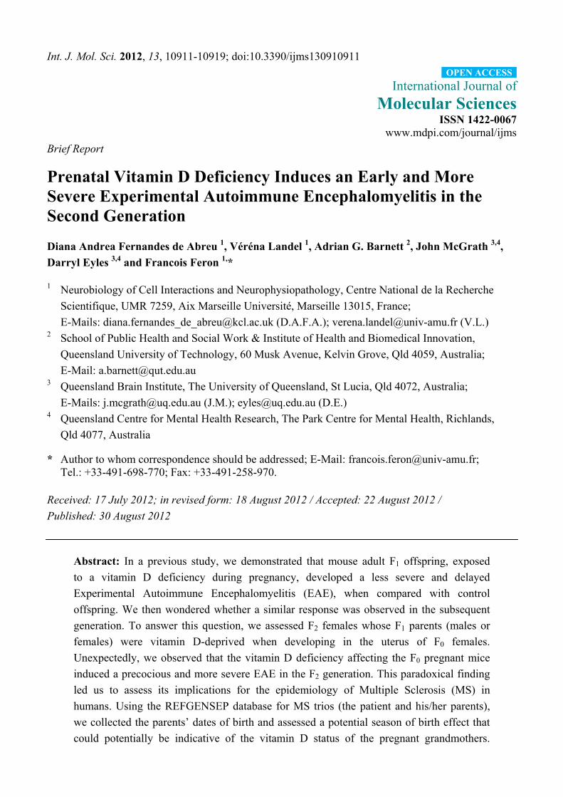
Int. J. Mol. Sci. 2012, 13, 10911-10919; doi:10.3390/ijms130910911
International Journal of
Molecular Sciences ISSN 1422-0067
www.mdpi.com/journal/ijms
Brief Report
Prenatal Vitamin D Deficiency Induces an Early and More Severe Experimental Autoimmune Encephalomyelitis in the Second Generation
Diana Andrea Fernandes de Abreu 1, Véréna Landel 1, Adrian G. Barnett 2, John McGrath 3,4,
Darryl Eyles 3,4 and Francois Feron 1,*
1 Neurobiology of Cell Interactions and Neurophysiopathology, Centre National de la Recherche
Scientifique, UMR 7259, Aix Marseille Université, Marseille 13015, France;
E-Mails: [email protected] (D.A.F.A.); [email protected] (V.L.) 2 School of Public Health and Social Work & Institute of Health and Biomedical Innovation,
Queensland University of Technology, 60 Musk Avenue, Kelvin Grove, Qld 4059, Australia;
E-Mail: [email protected] 3 Queensland Brain Institute, The University of Queensland, St Lucia, Qld 4072, Australia;
E-Mails: [email protected] (J.M.); [email protected] (D.E.) 4 Queensland Centre for Mental Health Research, The Park Centre for Mental Health, Richlands,
Qld 4077, Australia
* Author to whom correspondence should be addressed; E-Mail: [email protected]; Tel.: +33-491-698-770; Fax: +33-491-258-970.
Received: 17 July 2012; in revised form: 18 August 2012 / Accepted: 22 August 2012 /
Published: 30 August 2012
Abstract: In a previous study, we demonstrated that mouse adult F1 offspring, exposed
to a vitamin D deficiency during pregnancy, developed a less severe and delayed
Experimental Autoimmune Encephalomyelitis (EAE), when compared with control
offspring. We then wondered whether a similar response was observed in the subsequent
generation. To answer this question, we assessed F2 females whose F1 parents (males or
females) were vitamin D-deprived when developing in the uterus of F0 females.
Unexpectedly, we observed that the vitamin D deficiency affecting the F0 pregnant mice
induced a precocious and more severe EAE in the F2 generation. This paradoxical finding
led us to assess its implications for the epidemiology of Multiple Sclerosis (MS) in
humans. Using the REFGENSEP database for MS trios (the patient and his/her parents),
we collected the parents’ dates of birth and assessed a potential season of birth effect that
could potentially be indicative of the vitamin D status of the pregnant grandmothers.
OPEN ACCESS

Int. J. Mol. Sci. 2012, 13 10912
A trend for a reduced number of births in the Fall for the parents of MS patients was
observed but statistical significance was not reached. Further well powered studies are
warranted to validate the latter finding.
Keywords: vitamin D experimental autoimmune encephalomyelitis; multiple sclerosis;
deficiency; season of birth; transgenerational
Abbreviation: DVD: Developmental Vitamin D; EAE: Experimental Autoimmune Encephalomyelitis;
MOG: Myelin Oligodendrocyte Glycoprotein; MS: Multiple Sclerosis; REFGENSEP: Reseau
d’Etudes Francais GENetique sur la Sclerose En Plaques.
1. Introduction
It has been proposed that those born in winter/spring months have an increased risk of Multiple
Sclerosis (MS) [1–6]. While not all studies have confirmed this season of birth [7,8], the most
well-powered study to date based on data from Northern Hemisphere countries, reported that the risk
of MS was significantly higher for those born in May and lower for those born in November [9]. This
season of birth effect, combined with an observed latitude gradient in incidence (higher risk at higher
latitude) [10], suggest that an environmental factor, possibly vitamin D exposure, may be important in
predicting MS risk.
In order to test the hypothesis that low vitamin D status during gestation is a risk factor for MS, we
explored a well-described developmental vitamin D (DVD) deficiency rodent model, [11,12]. In this
model, the vitamin D deficiency experienced by the offspring is restored by the 1st week of life,
therefore the model is not associated with any abnormal bone development and calcium and PTH
levels in the adult offspring are normal [13]. Based on findings from epidemiology, we predicted that
offspring exposed to low prenatal vitamin D would be at increased risk of MS. In order to recreate
such an MS-related phenotype in the mouse, we have recently used the well-described experimental
autoimmune encephalomyelitis (EAE) model in DVD-deficient mice [14]. EAE induces an immune
response to components of myelin and has utility to explore pathways mediating demyelination and, as
an experimental platform, to screen potential therapeutic agents to treat MS. It is important to note that
this model does not recreate the entire etiopathogenesis of MS, which is still poorly understood.
Contrary to our prediction, we observed that C57BL/6 mouse adult offspring exposed to DVD
deficiency developed a less severe and delayed EAE, when compared with control offspring [14,15].
Curiously, DeLuca and Plum [16] have demonstrated that prolonged vitamin D deficiency, maintained
for two generations, also diminished the severity and delayed the onset of EAE in B10PL mice.
Though paradoxical, the response of the first generation to the EAE model suggests that a transient
maternal hypovitaminosis D results in an altered immune response after exposure to the myelin-related
protein. It remained however to be seen whether this environment-related change could be associated
with any altered immune-related phenotype in the subsequent generation, as has been previously
demonstrated in rats exposed to a fungicide [17]. We wondered if the second generation (F2) had any
persisting alterations in immune function related to the vitamin D status of the pregnant F0 female
Int. J. Mol. Sci. 2012, 13 10913
mice. With the aim of exploring this hypothesis, we designed a new experimental model, schematized
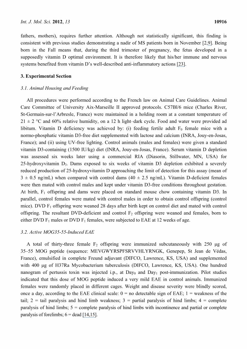
in Figure 1.
Figure 1. Schematic view of the experimental model. All mice, except F0 females, were
fed with a standard vitamin D-containing mouse chow. F0 females were vitamin
D-deprived six weeks prior mating and maintained on a deficient diet during pregnancy.
F1 DVD-deficient offspring, females and males, were mated with control mice. Only F2
female offspring were subjected to Experimental Autoimmune Encephalomyelitis (EAE).
In this series of experiments, only the pregnant F0 female mice and their growing fetuses were
vitamin D deficient. F0 female C57BL/6 mice were fed a vitamin D-free diet before and during
pregnancy. In order to highlight potential differences between male and female germlines, we set up
two groups for the F1 generation: DVD-deficient males were mated with control females, while
DVD-deficient females were mated with control males. A vitamin D-containing diet was provided to
all animals, before, during and after pregnancy. F2 offspring were weaned and adult females were
subjected to EAE.
Paradoxically, we observed that the vitamin D deficiency affecting the F0 pregnant mice induced a
precocious and more severe EAE in the F2 generation. This unexpected finding led us to assess its
implications for the epidemiology of MS in humans. The question we have addressed was whether
one’s parents’ season of birth (as a proxy-measure of developmental vitamin D status) could be an
additional risk factor for MS patients? Using the REFGENSEP database for MS trios (the patient and
his/her parents), we collected the parents’ dates of birth and assessed a potential season of birth effect
that could potentially be indicative of the vitamin D status of the pregnant grandmothers.
2. Results and Discussion
2.1. A Prenatal Vitamin D Deficiency Displays a Transgenerational Effect and Induces a More Severe
EAE in F2 Female Adult Mice
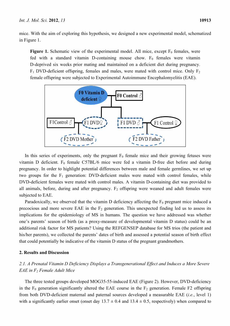
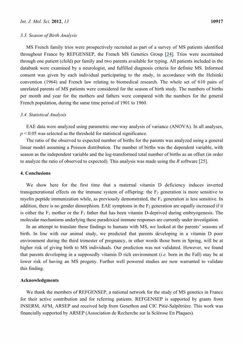
The three tested groups developed MOG35-55-induced EAE (Figure 2). However, DVD-deficiency
in the F0 generation significantly altered the EAE course in the F2 generation. Female F2 offspring
from both DVD-deficient maternal and paternal sources developed a measurable EAE (i.e., level 1)
with a significantly earlier onset (onset day 13.7 ± 0.4 and 13.4 ± 0.5, respectively) when compared to
Int. J. Mol. Sci. 2012, 13 10914
controls (onset day 16.3 ± 0.6, F = 8.6, p = 0.0011). Moreover, both DVD groups displayed an increased
peak in clinical score (3.1 ± 0.3 and 3.4 ± 0.5, respectively) when compared to controls (1.6 ± 0.4,
F = 5.95, p = 0.0066). At Day0 of immunization, the mean weight of the three tested groups was
similar (DVD Mother: 19.2 ± 0.5; DVD Father: 20.7 ± 0.4 g; controls: 21.2 ± 0.9).
Figure 2. Clinical course of MOG35-55-induced EAE in F2 mice. F2 female mice born to
either a DVD-deficient F1 Mother or a DVD-deficient F1 Father display an early and more
severe EAE when compared with control mice.
In a previous experiment, we demonstrated that a prenatal vitamin D deficiency induces a milder
and delayed EAE in the F1 generation [14]. In line with other studies describing a generational
transmission of molecular disturbances ([18], for a recent review), we initially hypothesized that the F2
generation would display a phenotype similar to the F1 generation. However, we clearly observed a
greater sensitization to the MOG immunization F2 females from DVD-deficient F0 females when
compared to controls. In addition, it can be highlighted that, contrary to a human study indicating a
female-associated transmission of HLA anomalies in MS families [19], no gender specific transmission
was observed in our study.
This discordant behavior between that reported in DVD-deficient F1 offspring and the F2
generations is unusual and, to the best of our knowledge, has never been reported. It is not unlikely
that a positive effect of a potentially deleterious environmental factor remains unnoticed. Conversely,
an impaired response is easily noticeable but sometimes can only be associated to the grandparents’
and not the parents’ lifestyle, as exemplified by a seminal epidemiological study on ancestral food
supply [20]. If confirmed, our findings may provide additional evidence for future studies aiming to
explain generation-skipping transmission of deleterious effects.
The molecular basis of this inheritance is unclear. The role of chromatin and DNA methylation in
epigenetics has been extensively studied during the past three decades. However, recent evidence
supports a bigger role for RNA in gametes, including piRNAs and miRNAs that can travel between
cells and silence transposable elements [18]. Among the current candidates, we can cite miR-22 that
is induced by vitamin D and acts as an antiproliferative and antimigratory agent in cancer cells [21]
or miR-125b that regulates the expression of human vitamin D receptor and abolishes the
Int. J. Mol. Sci. 2012, 13 10915
anti-proliferative action of calcitriol [22]. Nonetheless, to date, not a single study has yet demonstrated
a piRNA- or a miRNA-associated action of vitamin D on the immune or the nervous system.
We previously demonstrated that a postnatal vitamin D supplementation reduced the severity of
EAE and delayed the onset of symptoms [15]. It would be now of great interest to confirm that a
similar phenotype is observed when the supplementation occurs during pregnancy. Additionally, we
should perform a transgenerational study in order to assess whether the F2 generation is positively
affected by a high dose of vitamin D delivered to the F0 generation.
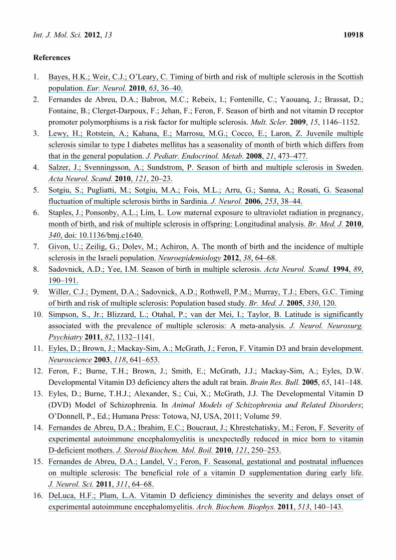
2.2. Observation of a Trend for a Reduced Number of Births in the Fall for the Parents of MS Patients
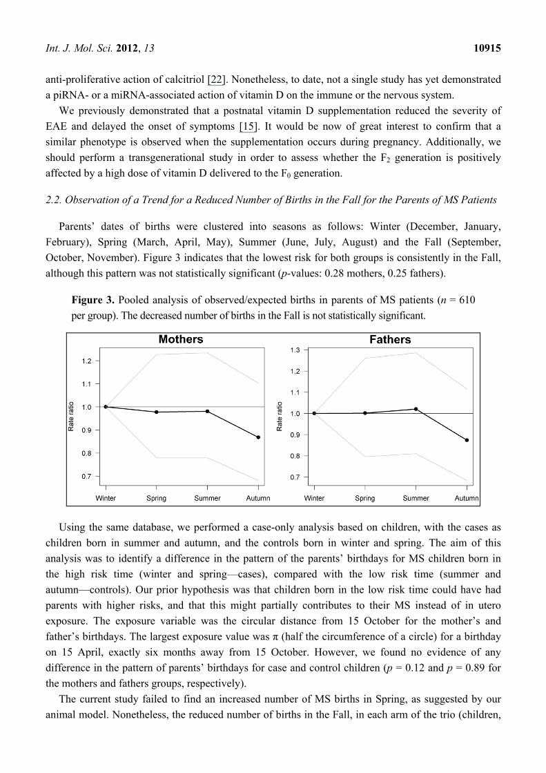
Parents’ dates of births were clustered into seasons as follows: Winter (December, January,
February), Spring (March, April, May), Summer (June, July, August) and the Fall (September,
October, November). Figure 3 indicates that the lowest risk for both groups is consistently in the Fall,
although this pattern was not statistically significant (p-values: 0.28 mothers, 0.25 fathers).
Figure 3. Pooled analysis of observed/expected births in parents of MS patients (n = 610
per group). The decreased number of births in the Fall is not statistically significant.
Using the same database, we performed a case-only analysis based on children, with the cases as
children born in summer and autumn, and the controls born in winter and spring. The aim of this
analysis was to identify a difference in the pattern of the parents’ birthdays for MS children born in
the high risk time (winter and spring—cases), compared with the low risk time (summer and
autumn—controls). Our prior hypothesis was that children born in the low risk time could have had
parents with higher risks, and that this might partially contributes to their MS instead of in utero
exposure. The exposure variable was the circular distance from 15 October for the mother’s and
father’s birthdays. The largest exposure value was π (half the circumference of a circle) for a birthday
on 15 April, exactly six months away from 15 October. However, we found no evidence of any
difference in the pattern of parents’ birthdays for case and control children (p = 0.12 and p = 0.89 for
the mothers and fathers groups, respectively).
The current study failed to find an increased number of MS births in Spring, as suggested by our
animal model. Nonetheless, the reduced number of births in the Fall, in each arm of the trio (children,
Int. J. Mol. Sci. 2012, 13 10916
fathers, mothers), requires further attention. Although not statistically significant, this finding is
consistent with previous studies demonstrating a nadir of MS patients born in November [2,9]. Being
born in the Fall means that, during the third trimester of pregnancy, the fetus developed in a
supposedly vitamin D optimal environment. It is therefore likely that his/her immune and nervous
systems benefited from vitamin D’s well-described anti-inflammatory actions [23].
3. Experimental Section
3.1. Animal Housing and Feeding
All procedures were performed according to the French law on Animal Care Guidelines. Animal
Care Committee of University Aix-Marseille II approved protocols. C57Bl/6 mice (Charles River,
St-Germain-sur-l’Arbresle, France) were maintained in a holding room at a constant temperature of
21 ± 2 °C and 60% relative humidity, on a 12 h light–dark cycle. Food and water were provided ad
libitum. Vitamin D deficiency was achieved by: (i) feeding fertile adult F0 female mice with a
normo-phosphatic vitamin D3-free diet supplemented with lactose and calcium (INRA, Jouy-en-Josas,
France); and (ii) using UV-free lighting. Control animals (males and females) were given a standard
vitamin D3-containing (1500 IU/kg) diet (INRA, Jouy-en-Josas, France). Serum vitamin D depletion
was assessed six weeks later using a commercial RIA (Diasorin, Stillwater, MN, USA) for
25-hydroxyvitamin D3. Dams exposed to six weeks of vitamin D3 depletion exhibited a severely
reduced production of 25-hydroxyvitamin D approaching the limit of detection for this assay (mean of
3 ± 0.5 ng/mL) when compared with control dams (40 ± 2.5 ng/mL). Vitamin D-deficient females
were then mated with control males and kept under vitamin D3-free conditions throughout gestation.
At birth, F1 offspring and dams were placed on standard mouse chow containing vitamin D3. In
parallel, control females were mated with control males in order to obtain control offspring (control
mice). DVD F1 offspring were weaned 28 days after birth kept on control diet and mated with control
offspring. The resultant DVD-deficient and control F2 offspring were weaned and females, born to
either DVD F1 males or DVD F1 females, were subjected to EAE at 12 weeks of age.
3.2. Active MOG35-55-Induced EAE
A total of thirty-three female F2 offspring were immunized subcutaneously with 250 μg of
35–55 MOG peptide (sequence: MEVGWYRSPFSRVVHLYRNGK, Genepep, St Jean de Védas,
France), emulsified in complete Freund adjuvant (DIFCO, Lawrence, KS, USA) and supplemented
with 400 μg of H37Ra Mycobacterium tuberculosis (DIFCO, Lawrence, KS, USA). One hundred
nanogram of pertussis toxin was injected i.p., at Day0 and Day1 post-immunization. Pilot studies
indicated that this dose of MOG peptide induced a very mild EAE in control animals. Immunized
females were randomly placed in different cages. Weight and disease severity were blindly scored,
once a day, according to the EAE clinical scale: 0 = no detectable sign of EAE; 1 = weakness of the
tail; 2 = tail paralysis and hind limb weakness; 3 = partial paralysis of hind limbs; 4 = complete
paralysis of hind limbs; 5 = complete paralysis of hind limbs with incontinence and partial or complete
paralysis of forelimbs; 6 = dead [14,15].
Int. J. Mol. Sci. 2012, 13 10917
3.3. Season of Birth Analysis
MS French family trios were prospectively recruited as part of a survey of MS patients identified
throughout France by REFGENSEP, the French MS Genetics Group [24]. Trios were ascertained
through one patient (child) per family and two parents available for typing. All patients included in the
databank were examined by a neurologist, and fulfilled diagnosis criteria for definite MS. Informed
consent was given by each individual participating to the study, in accordance with the Helsinki
convention (1964) and French law relating to biomedical research. The whole set of 610 pairs of
unrelated parents of MS patients were considered for the season of birth study. The numbers of births
per month and year for the mothers and fathers were compared with the numbers for the general
French population, during the same time period of 1901 to 1960.
3.4. Statistical Analysis
EAE data were analyzed using parametric one-way analysis of variance (ANOVA). In all analyses,
p < 0.05 was selected as the threshold for statistical significance.
The ratio of the observed to expected number of births for the parents was analyzed using a general
linear model assuming a Poisson distribution. The number of births was the dependent variable, with
season as the independent variable and the log-transformed total number of births as an offset (in order
to analyze the ratio of observed to expected). This analysis was made using the R software [25].
4. Conclusions
We show here for the first time that a maternal vitamin D deficiency induces inverted
transgenerational effects on the immune system of offspring: the F2 generation is more sensitive to
myelin peptide immunization while, as previously demonstrated, the F1 generation is less sensitive. In
addition, there is no gender dimorphism. EAE symptoms in the F2 generation are equally increased if it
is either the F1 mother or the F1 father that has been vitamin D-deprived during embryogenesis. The
molecular mechanisms underlying these paradoxical immune responses are currently under investigation.
In an attempt to translate these findings to humans with MS, we looked at the parents’ seasons of
birth. In line with our animal study, we predicted that parents developing in a vitamin D poor
environment during the third trimester of pregnancy, in other words those born in Spring, will be at
higher risk of giving birth to MS individuals. Our prediction was not validated. However, we found
that parents developing in a supposedly vitamin D rich environment (i.e. born in the Fall) may be at
lower risk of having an MS progeny. Further well powered studies are now warranted to validate
this finding.
Acknowledgments
We thank the members of REFGENSEP, a national network for the study of MS genetics in France
for their active contribution and for referring patients. REFGENSEP is supported by grants from
INSERM, AFM, ARSEP and received help from Genethon and CIC Pitié-Salpêtrière. This work was
financially supported by ARSEP (Association de Recherche sur la Sclérose En Plaques).
Int. J. Mol. Sci. 2012, 13 10918
References
1. Bayes, H.K.; Weir, C.J.; O’Leary, C. Timing of birth and risk of multiple sclerosis in the Scottish
population. Eur. Neurol. 2010, 63, 36–40.
2. Fernandes de Abreu, D.A.; Babron, M.C.; Rebeix, I.; Fontenille, C.; Yaouanq, J.; Brassat, D.;
Fontaine, B.; Clerget-Darpoux, F.; Jehan, F.; Feron, F. Season of birth and not vitamin D receptor
promoter polymorphisms is a risk factor for multiple sclerosis. Mult. Scler. 2009, 15, 1146–1152.
3. Lewy, H.; Rotstein, A.; Kahana, E.; Marrosu, M.G.; Cocco, E.; Laron, Z. Juvenile multiple
sclerosis similar to type I diabetes mellitus has a seasonality of month of birth which differs from
that in the general population. J. Pediatr. Endocrinol. Metab. 2008, 21, 473–477.
4. Salzer, J.; Svenningsson, A.; Sundstrom, P. Season of birth and multiple sclerosis in Sweden.
Acta Neurol. Scand. 2010, 121, 20–23.
5. Sotgiu, S.; Pugliatti, M.; Sotgiu, M.A.; Fois, M.L.; Arru, G.; Sanna, A.; Rosati, G. Seasonal
fluctuation of multiple sclerosis births in Sardinia. J. Neurol. 2006, 253, 38–44.
6. Staples, J.; Ponsonby, A.L.; Lim, L. Low maternal exposure to ultraviolet radiation in pregnancy,
month of birth, and risk of multiple sclerosis in offspring: Longitudinal analysis. Br. Med. J. 2010,
340, doi: 10.1136/bmj.c1640.
7. Givon, U.; Zeilig, G.; Dolev, M.; Achiron, A. The month of birth and the incidence of multiple
sclerosis in the Israeli population. Neuroepidemiology 2012, 38, 64–68.
8. Sadovnick, A.D.; Yee, I.M. Season of birth in multiple sclerosis. Acta Neurol. Scand. 1994, 89,
190–191.
9. Willer, C.J.; Dyment, D.A.; Sadovnick, A.D.; Rothwell, P.M.; Murray, T.J.; Ebers, G.C. Timing
of birth and risk of multiple sclerosis: Population based study. Br. Med. J. 2005, 330, 120.
10. Simpson, S., Jr.; Blizzard, L.; Otahal, P.; van der Mei, I.; Taylor, B. Latitude is significantly
associated with the prevalence of multiple sclerosis: A meta-analysis. J. Neurol. Neurosurg.
Psychiatry 2011, 82, 1132–1141.
11. Eyles, D.; Brown, J.; Mackay-Sim, A.; McGrath, J.; Feron, F. Vitamin D3 and brain development.
Neuroscience 2003, 118, 641–653.
12. Feron, F.; Burne, T.H.; Brown, J.; Smith, E.; McGrath, J.J.; Mackay-Sim, A.; Eyles, D.W.
Developmental Vitamin D3 deficiency alters the adult rat brain. Brain Res. Bull. 2005, 65, 141–148.
13. Eyles, D.; Burne, T.H.J.; Alexander, S.; Cui, X.; McGrath, J.J. The Developmental Vitamin D
(DVD) Model of Schizophrenia. In Animal Models of Schizophrenia and Related Disorders;
O’Donnell, P., Ed.; Humana Press: Totowa, NJ, USA, 2011; Volume 59.
14. Fernandes de Abreu, D.A.; Ibrahim, E.C.; Boucraut, J.; Khrestchatisky, M.; Feron, F. Severity of
experimental autoimmune encephalomyelitis is unexpectedly reduced in mice born to vitamin
D-deficient mothers. J. Steroid Biochem. Mol. Boil. 2010, 121, 250–253.
15. Fernandes de Abreu, D.A.; Landel, V.; Feron, F. Seasonal, gestational and postnatal influences
on multiple sclerosis: The beneficial role of a vitamin D supplementation during early life.
J. Neurol. Sci. 2011, 311, 64–68.
16. DeLuca, H.F.; Plum, L.A. Vitamin D deficiency diminishes the severity and delays onset of
experimental autoimmune encephalomyelitis. Arch. Biochem. Biophys. 2011, 513, 140–143.
Int. J. Mol. Sci. 2012, 13 10919
17. Anway, M.D.; Cupp, A.S.; Uzumcu, M.; Skinner, M.K. Epigenetic transgenerational actions of
endocrine disruptors and male fertility. Science 2005, 308, 1466–1469.
18. Daxinger, L.; Whitelaw, E. Understanding transgenerational epigenetic inheritance via the
gametes in mammals. Nat. Rev. Genet. 2012, 13, 153–162.
19. Chao, M.J.; Ramagopalan, S.V.; Herrera, B.M.; Lincoln, M.R.; Dyment, D.A.; Sadovnick, A.D.;
Ebers, G.C. Epigenetics in multiple sclerosis susceptibility: Difference in transgenerational risk
localizes to the major histocompatibility complex. Hum. Mol. Genet. 2009, 18, 261–266.
20. Pembrey, M.E.; Bygren, L.O.; Kaati, G.; Edvinsson, S.; Northstone, K.; Sjostrom, M.; Golding, J.
Sex-specific, male-line transgenerational responses in humans. Eur. J. Hum. Genet. 2006, 14,
159–166.
21. Alvarez-Diaz, S.; Valle, N.; Ferrer-Mayorga, G.; Lombardia, L.; Herrera, M.; Dominguez, O.;
Segura, M.F.; Bonilla, F.; Hernando, E.; Munoz, A. MicroRNA-22 is induced by vitamin D and
contributes to its antiproliferative, antimigratory and gene regulatory effects in colon cancer cells.
Hum. Mol. Genet. 2012, 21, 2157–2165.
22. Mohri, T.; Nakajima, M.; Takagi, S.; Komagata, S.; Yokoi, T. MicroRNA regulates human
vitamin D receptor. Int. J. Cancer 2009, 125, 1328–1333.
23. Cantorna, M.T.; Zhao, J.; Yang, L. Vitamin D, invariant natural killer T-cells and experimental
autoimmune disease. Proc. Nutr. Soc. 2012, 71, 62–66.
24. Cournu-Rebeix, I.; Genin, E.; Leray, E.; Babron, M.C.; Cohen, J.; Gout, C.; Alizadeh, M.;
Perdry, H.; Semana, G.; Brassat, D.; et al. HLA-DRB1*15 allele influences the later course of
relapsing remitting multiple sclerosis. Genes Immun. 2008, 9, 570–574.
25. The R Project for Statistical Computing. Available online: http://www.r-project.org (accessed on
7 May 2012).
© 2012 by the authors; licensee MDPI, Basel, Switzerland. This article is an open access article
distributed under the terms and conditions of the Creative Commons Attribution license
(http://creativecommons.org/licenses/by/3.0/).
Copyright © 2022 FDOKUMEN