Suppression of experimental autoimmune encephalomyelitis by oral administration of myelin basic...
10
The Journal of Experimental Medicine ARTICLE Vol. 203, No. 4, April 17, 2006 985–994 www.jem.org/cgi/doi/10.1084/jem.20051681 985 Multiple sclerosis (MS) and its animal model, experimental autoimmune encephalomyelitis (EAE), are demyelinating diseases of the central nervous system (CNS) mediated by autoreac- tive T cells that penetrate through the blood– brain barrier (BBB) into the brain parenchyma. These T cells initiate autoimmune responses against antigens in the myelin sheath of the CNS, including myelin basic protein or myelin oligodendrocyte protein (1). Thus, the recruit- ment of autoreactive lymphocytes represents a crucial pathogenetic event in initiating CNS inflammation, and interfering with the homing of these cells may represent a feasible therapeu- tic approach for both EAE and MS. Lymphocyte extravasation requires a well- coordinated sequence of adhesive and signal- ing events, including selectin-mediated rolling, chemoattractant-induced integrin activation, integrin-dependent firm adhesion, and the sub- sequent transendothelial migration (2). During adhesion and transmigration, integrins of the β1 and β2 family, such as VLA-4 (α4β1) or LFA-1 (αLβ2), bind to their endothelial counter- receptors vascular cell adhesion molecule (VCAM)-1 and intercellular adhesion molecule (ICAM)-1, respectively. Numerous studies have shown that adhesion of lymphocytes to inflamed brain vessels during EAE is mainly mediated by the VLA-4–VCAM-1 system (3, 4), although there is also evidence pointing to the role of the LFA-1–ICAM-1 interaction (5, 6). Recent studies indicated that both sys- tems may have a distinct contribution in the Suppression of experimental autoimmune encephalomyelitis by extracellular adherence protein of Staphylococcus aureus Changping Xie, 2 Pilar Alcaide, 4 Brian V. Geisbrecht, 3 Darius Schneider, 2 Mathias Herrmann, 5 Klaus T. Preissner, 6 Francis W. Luscinskas, 4 and Triantafyllos Chavakis 1 1 Experimental Immunology Branch, National Cancer Institute, National Institutes of Health, Bethesda, MD 20892 2 Department of Internal Medicine I, University Heidelberg, D-69120 Heidelberg, Germany 3 Division of Cell Biology and Biophysics, School of Biological Sciences, University of Missouri-Kansas City, Kansas City, MO 64110 4 Department of Pathology, Brigham and Women’s Hospital, Harvard Medical School, Boston, MA 02115 5 Institute of Medical Microbiology and Hygiene, University of Saarland Hospital, D-66421 Homburg/Saar, Germany 6 Institute for Biochemistry, Medical School, Justus-Liebig-University, D-35392 Giessen, Germany Multiple sclerosis (MS) is a devastating inflammatory disorder of the central nervous system (CNS). A major hallmark of MS is the infiltration of T cells reactive against myelin compo- nents. T cell infiltration is mediated by the interaction of integrins of the 1 and 2 family expressed by lymphocytes with their endothelial counter-receptors, vascular cell adhesion molecule 1 and intercellular adhesion molecule (ICAM)-1, respectively. We have reported previously that extracellular adherence protein (Eap) of Staphylococcus aureus exerts anti- inflammatory activities by interacting with ICAM-1 and blocking 2-integrin–dependent neutrophil recruitment. Here, we report that Eap inhibits experimental autoimmune enceph- alomyelitis (EAE) in mice. In vitro, Eap reduced adhesion of peripheral blood T cells to immobilized ICAM-1 as well as their adhesion and transmigration of TNF-activated human endothelium under static and shear flow conditions. These inhibitory effects were corrobo- rated in two mouse models of inflammation. In a delayed-type hypersensitivity model, both T cell infiltration and the corresponding tissue edema were significantly reduced by Eap. In addition, Eap administration prevented the development of EAE and markedly decreased infiltration of inflammatory cells into the CNS. Strikingly, intervention with Eap after the onset of EAE suppressed the disease. Collectively, our findings indicate that Eap represents an attractive treatment for autoimmune neuroinflammatory disorders such as MS. CORRESPONDENCE Triantafyllos Chavakis: [email protected] Abbreviations used: BBB, blood–brain barrier; CNS, central nervous system; DTH, delayed-type hypersensitivity; EAE, experimental autoimmune encephalomyelitis; Eap, extracel- lular adherence protein; HU- VEC, human umbilical vein endothelial cell; ICAM, inter- cellular adhesion molecule; MOG, myelin oligodendrocyte glyco- protein; MS, multiple sclerosis; PBT, peripheral blood T; VCAM, vascular cell adhesion molecule.
-
Upload
independent -
Category
Documents
-
view
1 -
download
0
Transcript of Suppression of experimental autoimmune encephalomyelitis by oral administration of myelin basic...

The
Journ
al o
f Exp
erim
enta
l M
edic
ine
ARTICLE
Vol. 203, No. 4, April 17, 2006 985–994 www.jem.org/cgi/doi/10.1084/jem.20051681 985
Multiple sclerosis (MS) and its animal model, experimental autoimmune encephalomyelitis (EAE), are demyelinating diseases of the central nervous system (CNS) mediated by autoreac-tive T cells that penetrate through the blood–brain barrier (BBB) into the brain parenchyma. These T cells initiate autoimmune responses against antigens in the myelin sheath of the CNS, including myelin basic protein or myelin oligodendrocyte protein (1). Thus, the recruit-ment of autoreactive lymphocytes represents a crucial pathogenetic event in initiating CNS infl ammation, and interfering with the homing of these cells may represent a feasible therapeu-tic approach for both EAE and MS.
Lymphocyte extravasation requires a well-coordinated sequence of adhesive and signal-
ing events, including selectin-mediated rolling, chemoattractant-induced integrin activation, integrin-dependent fi rm adhesion, and the sub-sequent transendothelial migration (2). During adhesion and transmigration, integrins of the β1 and β2 family, such as VLA-4 (α4β1) or LFA-1 (αLβ2), bind to their endothelial counter-receptors vascular cell adhesion molecule (VCAM)-1 and intercellular adhesion molecule (ICAM)-1, respectively. Numerous studies have shown that adhesion of lymphocytes to infl amed brain vessels during EAE is mainly mediated by the VLA-4–VCAM-1 system (3, 4), although there is also evidence pointing to the role of the LFA-1–ICAM-1 interaction (5, 6). Recent studies indicated that both sys-tems may have a distinct contribution in the
Suppression of experimental autoimmune encephalomyelitis by extracellular adherence protein of Staphylococcus aureus
Changping Xie,2 Pilar Alcaide,4 Brian V. Geisbrecht,3 Darius Schneider,2 Mathias Herrmann,5 Klaus T. Preissner,6 Francis W. Luscinskas,4 and Triantafyllos Chavakis1
1Experimental Immunology Branch, National Cancer Institute, National Institutes of Health, Bethesda, MD 208922Department of Internal Medicine I, University Heidelberg, D-69120 Heidelberg, Germany3Division of Cell Biology and Biophysics, School of Biological Sciences, University of Missouri-Kansas City,
Kansas City, MO 641104Department of Pathology, Brigham and Women’s Hospital, Harvard Medical School, Boston, MA 021155Institute of Medical Microbiology and Hygiene, University of Saarland Hospital, D-66421 Homburg/Saar, Germany6Institute for Biochemistry, Medical School, Justus-Liebig-University, D-35392 Giessen, Germany
Multiple sclerosis (MS) is a devastating infl ammatory disorder of the central nervous system
(CNS). A major hallmark of MS is the infi ltration of T cells reactive against myelin compo-
nents. T cell infi ltration is mediated by the interaction of integrins of the 𝛃1 and 𝛃2 family
expressed by lymphocytes with their endothelial counter-receptors, vascular cell adhesion
molecule 1 and intercellular adhesion molecule (ICAM)-1, respectively. We have reported
previously that extracellular adherence protein (Eap) of Staphylococcus aureus exerts anti-
infl ammatory activities by interacting with ICAM-1 and blocking 𝛃2-integrin–dependent
neutrophil recruitment. Here, we report that Eap inhibits experimental autoimmune enceph-
alomyelitis (EAE) in mice. In vitro, Eap reduced adhesion of peripheral blood T cells to
immobilized ICAM-1 as well as their adhesion and transmigration of TNF-activated human
endothelium under static and shear fl ow conditions. These inhibitory effects were corrobo-
rated in two mouse models of infl ammation. In a delayed-type hypersensitivity model, both
T cell infi ltration and the corresponding tissue edema were signifi cantly reduced by Eap.
In addition, Eap administration prevented the development of EAE and markedly decreased
infi ltration of infl ammatory cells into the CNS. Strikingly, intervention with Eap after the
onset of EAE suppressed the disease. Collectively, our fi ndings indicate that Eap represents
an attractive treatment for autoimmune neuroinfl ammatory disorders such as MS.
CORRESPONDENCE
Triantafyllos Chavakis:
Abbreviations used: BBB,
blood–brain barrier; CNS,
central nervous system; DTH,
delayed-type hypersensitivity;
EAE, experimental autoimmune
encephalomyelitis; Eap, extracel-
lular adherence protein; HU-
VEC, human umbilical vein
endothelial cell; ICAM, inter-
cellular adhesion molecule; MOG,
myelin oligodendrocyte glyco-
protein; MS, multiple sclerosis;
PBT, peripheral blood T; VCAM,
vascular cell adhesion molecule.

986 INHIBITION OF EAE BY EAP | Xie et al.
recruitment of encephalitogenic T cells. Although the VLA-4–VCAM-1 interaction predominantly mediates the initial adhesion to the infl amed vessels, LFA-1–ICAM-1 are engaged in the subsequent transendothelial migration of T cells (7, 8).
To date, therapeutic approaches targeting adhesion mol-ecules have focused predominantly on the blockade of the VLA-4–VCAM-1 system (9). An antibody to VLA-4 that ameliorated EAE in mice was humanized (natalizumab) and demonstrated encouraging clinical effi cacy for the treatment of patients with relapsing forms of multiple sclerosis. How-ever, a few cases of fatal progressive multifocal leukoenceph-alopathy attributed to natalizumab prompted its withdrawal (9). Thus, the need for other adhesion molecule–based thera-pies is emerging. In contrast with the VLA-4–VCAM-1 sys-tem, only a few studies have addressed the role of the LFA-1–ICAM-1 system as a target in EAE, although ICAM-1 is clearly up-regulated in EAE and MS, and serum levels of soluble ICAM-1 were found to be elevated in MS patients with clinically symptomatic disease (5, 6, 10).
Extracellular adherence protein (Eap), also designated MHC class II analogous protein (Map) or P70, is a secreted protein from Staphylococcus aureus. Eap has a very broad rep-ertoire of binding interactions to host extracellular matrix components (11, 12). Recently, we demonstrated a mecha-nism used by S. aureus to escape the host immune system that was attributed to the very potent antiinfl ammatory function of Eap. In particular, we found Eap to undergo a direct in-teraction with ICAM-1 that resulted in the disruption of integrin-dependent neutrophil–endothelial interactions (13). Strikingly, Eap was even more potent than blocking antibod-ies against the β2-integrin receptors or against ICAM-1.
These observations prompted us to investigate whether Eap could be feasible to interfere with T cell–adhesive mech-anisms in EAE. In the present study, we show that Eap inhib-its the LFA-1–ICAM-1 interaction and the T cell–endothelial interactions in vitro and in vivo. Furthermore, administration of Eap not only prevented the development of EAE, but Eap treatment reverted the disease and repressed its progress after its onset. Thus, Eap may represent a promising new therapy for chronic infl ammatory diseases such as EAE and MS.
RESULTS
Eap blocks adhesion of T cells to ICAM-1
We have previously shown that Eap interferes with the in-teraction between ICAM-1 and its β2-integrin counter- receptors Mac-1 and LFA-1, thereby blocking neutrophil recruitment (13). To test whether Eap might interfere with T cell recruitment, we fi rst examined the adhesion of peripheral blood T (PBT) cells to immobilized ICAM-1 and VCAM-1.
Figure 1. Eap inhibits T cell adhesion to ICAM-1. (A) The adhesion
of T cells to immobilized ICAM-1 is shown in the absence (white bars) or
presence of blocking mAb against VLA-4, mAb against LFA-1 (each at
20 μg/ml), increasing concentrations of purifi ed (gray bars), or recombi-
nant (black bars) Eap, or cationic BSA (20 μg/ml). (B) The adhesion of
T cells to immobilized VCAM-1 is shown in the absence or presence of
blocking mAb against VLA-4, mAb against LFA-1 (each at 20 μg/ml), puri-
fi ed Eap, recombinant Eap, or cationic BSA (each at 20 μg/ml). Cell adhe-
sion is expressed relative to control (in the absence of competitor). Data
are mean ± SD (n = 3) of a typical experiment; similar results were ob-
tained in three separate experiments. (C and D) Adhesion of
T cells to immobilized ICAM-1 (C) or VCAM-1 (D) under laminar shear fl ow
conditions is shown in the absence (white bars) or presence of cationic
BSA (diagonally striped bars), purifi ed Eap (gray bars), recombinant Eap
(black bars), or anti-LFA-1 mAb (dotted bars) (each at 20 μg/ml). Data are
mean ± SEM, n = 3 separate experiments. *, P < 0.05. **, P < 0.01.

JEM VOL. 203, April 17, 2006 987
ARTICLE
As shown in Fig. 1, the adhesion of PBT cells to immobilized ICAM-1 was mediated by LFA-1, whereas adhesion to VCAM-1 was dependent on β1-integrins. Under both static (Fig. 1, A and B) and physiologic fl ow conditions (Fig. 1, C and D), both purifi ed and recombinant Eap dose-dependently inhibited the adhesion of PBT cells to immobilized ICAM-1, whereas adhesion of PBT cells to VCAM-1 was not aff ected in the presence of Eap. The inhibitory eff ect of Eap on the adhesion of PBT cells to ICAM-1 was comparable to the eff ect of blocking mAb to LFA-1 (Fig. 1). As Eap is a cationic protein, we engaged cationic BSA as a negative control, which did not aff ect LFA-1– or VLA-4–dependent adhesion to ICAM-1 or VCAM-1, respectively (Fig. 1).
Interference of Eap with T cell–endothelial cell interactions
under static and physiologic fl ow conditions
As Eap blocks the adhesion of PBT cells to ICAM-1, we further determined whether Eap interferes with PBT cell–endothelial cell interactions. Therefore, the adhesion of PBT cells to endothelial cells as well as the transmigration of PBT cells through endothelial cell monolayers was studied under static and physiologic fl ow conditions. Under static condi-tions, the adhesion of PBT cells to TNF-α–prestimulated en-dothelial cells was mainly dependent on VLA-4, whereas the LFA-1–ICAM-1 system mediates a smaller portion of the T cell adhesion to endothelial cells under these conditions (Fig. 2 A). Both purifi ed and recombinant Eap partially blocked adhesion of PBT cells to endothelial cells; the maxi-mal inhibition observed was �35–40% and was comparable to the inhibition obtained with blocking mAb to LFA-1 (Fig. 2 A). In contrast, transendothelial migration of PBT cells was predominantly mediated by the LFA-1–ICAM-1 interaction; in this case, both purifi ed and recombinant Eap were more potent in blocking PBT cell transendothelial migration (Fig. 2 B). As a negative control, cationic BSA did not infl uence PBT cell adhesion or transmigration.
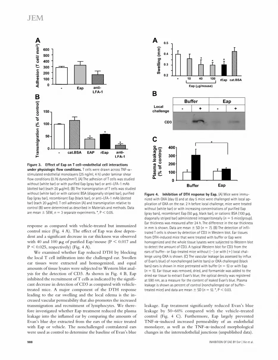
Because the PBT cell–endothelial cell interactions under physiologic fl ow conditions simulate in vivo physiological shear fl ow, we studied the eff ect of Eap under defi ned shear fl ow in vitro (14, 15). As previously reported, PBT cell adhe-sion to TNF-α–prestimulated endothelial cells under physio-logic fl ow in vitro is mainly dependent on the VLA-4–VCAM-1 adhesion pathway and to a lesser extent on the LFA-1–ICAM-1 pathway, whereas transmigration was primarily LFA-1–ICAM-1 dependent (7, 14, 16). Consistent with these prior observations, here we show that purifi ed or re-combinant Eap (data with recombinant Eap not shown) as well as blocking mAb to LFA-1 had no statistically signifi cant eff ect on adhesion of PBT cells to TNF-α–activated human umbilical vein endothelial cell (HUVEC) monolayers under fl ow conditions (Fig. 3 A), whereas transendothelial migra-tion was signifi cantly blocked by Eap or mAb to LFA-1 (Fig. 3 B). In contrast, no eff ect of cationic BSA was observed. Collectively, these data (Figs. 1–3) indicate that Eap inter-feres with the LFA-1–ICAM-1–dependent component of T cell–endothelial interaction pathway under both static and
physiologic fl ow conditions in vitro. This inhibitory eff ect of Eap is specifi c and cannot be attributed to a nonspecifi c eff ect of the cationic charge of Eap.
Inhibition of T cell recruitment and delayed type
hypersensitivity (DTH) responses in vivo by Eap
To test whether Eap aff ects T cell–mediated immune re-sponses in vivo, the ability of Eap to interfere with T cell recruitment and cellular immunity was assessed in a DTH model. DTH responses are mediated by infi ltrating T cells in response to formerly encountered antigens, resulting in a spe-cifi c infl ammation at the site of local challenge. Mice immu-nized with oxazolone at day 0 and challenged locally on the right ear on day 5 developed a signifi cant DTH response as measured by ear swelling at day 6 (Fig. 4). Mice treated sys-temically with either purifi ed or recombinant Eap on day 5 (3 h before local challenge) had a signifi cantly reduced DTH
Figure 2. Eap inhibits T cell–endothelial cell interactions. (A) The
adhesion of T cells to TNF-α prestimulated HUVEC (10 ng/ml, 16 h) is
shown in the absence or presence of blocking mAb against VLA-4, block-
ing mAb against LFA-1, blocking mAb against Mac-1 (each at 20 μg/ml)
(white bars), increasing concentrations of purifi ed Eap (gray bars), recom-
binant Eap (black bars) or cationic BSA (20 μg/ml; white bars). (B) SDF-
1α–stimulated transendothelial migration of T cells is shown in the
absence (white bars) or presence of blocking mAb against VLA-4, blocking
mAb against LFA-1, blocking mAb against Mac-1 (each at 20 μg/ml)
(white bars), increasing concentrations of purifi ed Eap (gray bars), recom-
binant Eap (black bars), or cationic BSA (20 μg/ml; white bars). Cell
adhesion and cell transmigration is expressed relative to control (in the
absence of competitor). Data are mean ± SD (n = 3) of a typical
experiment; similar results were obtained in three separate experiments.
988 INHIBITION OF EAE BY EAP | Xie et al.
response as compared with vehicle-treated but immunized control mice (Fig. 4 A). The eff ect of Eap was dose depen-dent and a signifi cant decrease in ear thickness was observed with 40 and 100 μg of purifi ed Eap/mouse (P < 0.017 and P < 0.025, respectively) (Fig. 4 A).
We examined whether Eap reduced DTH by blocking the local T cell infi ltration into the challenged ear. Swollen ear tissues were extracted and homogenized, and equal amounts of tissue lysates were subjected to Western blot anal-ysis for the detection of CD3. As shown in Fig. 4 B, Eap inhibited the recruitment of T cells as indicated by the signifi -cant decrease in detection of CD3 as compared with vehicle-treated mice. A major component of the DTH response leading to the ear swelling and the local edema is the in-creased vascular permeability that also promotes the increased transmigration and recruitment of lymphocytes. We there-fore investigated whether Eap treatment reduced the plasma leakage into the infl amed ear by comparing the amounts of Evan’s blue dye extracted from the ears of the mice treated with Eap or vehicle. The nonchallenged contralateral ears were used as control to determine the baseline of Evan’s blue
leakage. Eap treatment signifi cantly reduced Evan’s blue leakage by 50–60% compared with the vehicle-treated control (Fig. 4 C). Furthermore, Eap largely prevented TNF-α–induced increased permeability of an endothelial monolayer, as well as the TNF-α–induced morphological changes in the interendothelial junctions (unpublished data).
Figure 3. Effect of Eap on T cell–endothelial cell interactions
under physiologic fl ow conditions. T cells were drawn across TNF-α–
stimulated endothelial monolayers (25 ng/ml, 4 h) under laminar shear
fl ow conditions (0.76 dynes/mm2). (A) The adhesion of T cells was studied
without (white bar) or with purifi ed Eap (gray bar) or anti–LFA-1 mAb
(dotted bar) (each 20 μg/ml). (B) The transmigration of T cells was studied
without (white bar) or with cationic BSA (diagonally striped bar), purifi ed
Eap (gray bar), recombinant Eap (black bar), or anti–LFA-1 mAb (dotted
bar) (each 20 μg/ml). T cell adhesion (A) and transmigration relative to
control (B) were determined as described in Materials and methods. Data
are mean ± SEM, n = 3 separate experiments. *, P < 0.05.
Figure 4. Inhibition of DTH response by Eap. (A) Mice were immu-
nized with OXA (day 0) and at day 5 mice were challenged with local ap-
plication of OXA on the ear. 2 h before local challenge, mice were treated
without (white bar) or with increasing concentrations of purifi ed Eap
(gray bars), recombinant Eap (50 μg, black bar), or cationic BSA (100 μg,
diagonally striped bar) administered intraperitoneally (n = 5 mice/group).
Ear thickness was measured after 24 h. The difference in the ear thickness
in mm is shown. Data are mean ± SD (n = 7). (B) The detection of infi l-
trated T cells is shown by detection of CD3 in Western blot. Ear tissues
from DTH-induced mice that were treated with buffer or Eap were
homogenized and the whole tissue lysates were subjected to Western blot
to detect the amount of CD3. A typical Western blot for CD3 from the
ears of buffer- or Eap-treated mice without (−) or with (+) local chal-
lenge using OXA is shown. (C) The vascular leakage (as assessed by infl ux
of Evan’s blue) of nonchallenged (white bars) or OXA challenged (black
bars) ears is shown in mice pretreated with buffer (n = 5) or with Eap
(n = 5). Ear tissue was removed, dried, and formamide was added to the
dried ear tissue to extract Evan’s blue; the optical density was registered
at 590 nm, as a measure for the content of leaked Evan’s blue. Plasma
leakage is shown as percent of control (nonchallenged ear of buffer-
treated mice) and data are mean ± SD (n = 5). *, P < 0.03.
JEM VOL. 203, April 17, 2006 989
ARTICLE
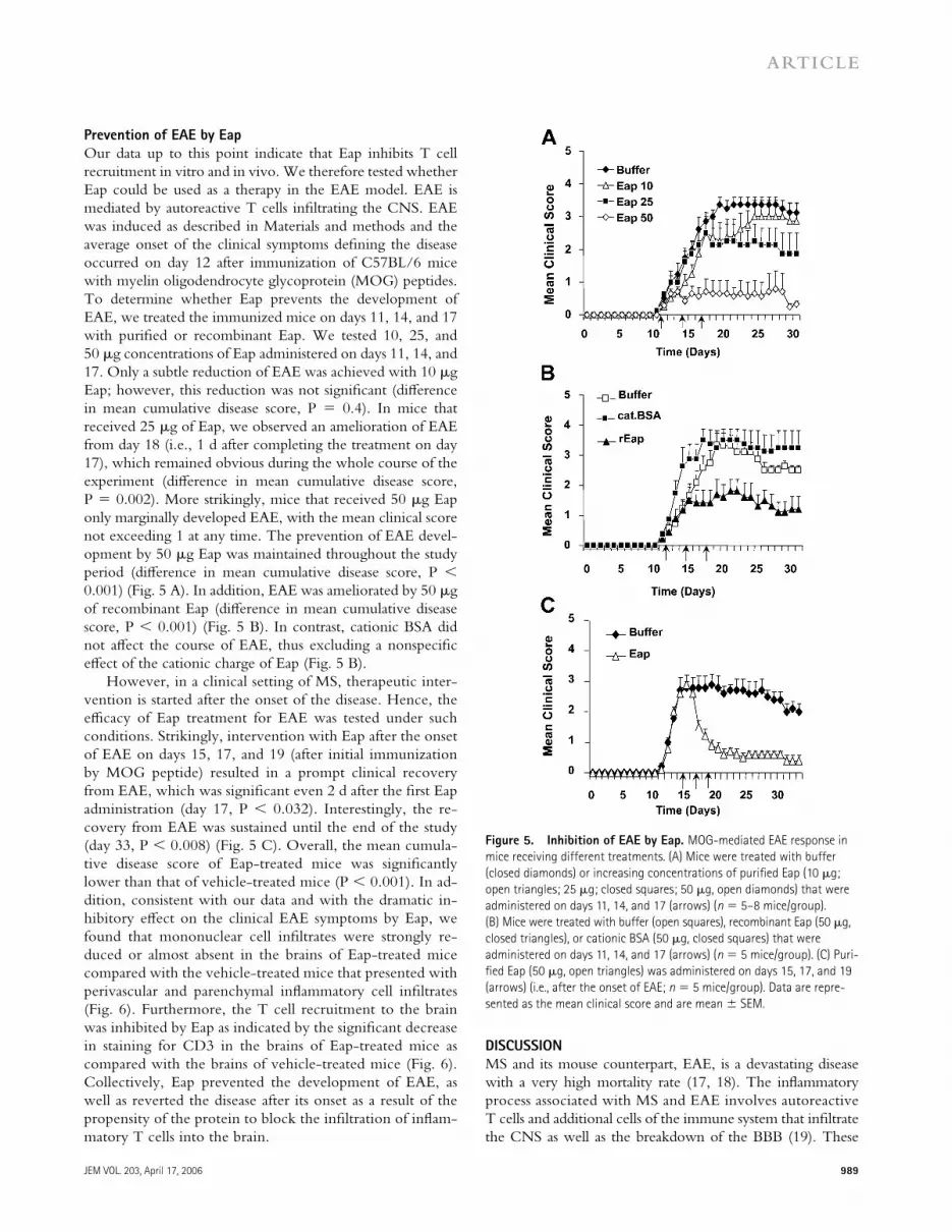
Prevention of EAE by Eap
Our data up to this point indicate that Eap inhibits T cell recruitment in vitro and in vivo. We therefore tested whether Eap could be used as a therapy in the EAE model. EAE is mediated by autoreactive T cells infi ltrating the CNS. EAE was induced as described in Materials and methods and the average onset of the clinical symptoms defi ning the disease occurred on day 12 after immunization of C57BL/6 mice with myelin oligodendrocyte glycoprotein (MOG) peptides. To determine whether Eap prevents the development of EAE, we treated the immunized mice on days 11, 14, and 17 with purifi ed or recombinant Eap. We tested 10, 25, and 50 μg concentrations of Eap administered on days 11, 14, and 17. Only a subtle reduction of EAE was achieved with 10 μg Eap; however, this reduction was not signifi cant (diff erence in mean cumulative disease score, P = 0.4). In mice that received 25 μg of Eap, we observed an amelioration of EAE from day 18 (i.e., 1 d after completing the treatment on day 17), which remained obvious during the whole course of the experiment (diff erence in mean cumulative disease score, P = 0.002). More strikingly, mice that received 50 μg Eap only marginally developed EAE, with the mean clinical score not exceeding 1 at any time. The prevention of EAE devel-opment by 50 μg Eap was maintained throughout the study period (diff erence in mean cumulative disease score, P < 0.001) (Fig. 5 A). In addition, EAE was ameliorated by 50 μg of recombinant Eap (diff erence in mean cumulative disease score, P < 0.001) (Fig. 5 B). In contrast, cationic BSA did not aff ect the course of EAE, thus excluding a nonspecifi c eff ect of the cationic charge of Eap (Fig. 5 B).
However, in a clinical setting of MS, therapeutic inter-vention is started after the onset of the disease. Hence, the effi cacy of Eap treatment for EAE was tested under such conditions. Strikingly, intervention with Eap after the onset of EAE on days 15, 17, and 19 (after initial immunization by MOG peptide) resulted in a prompt clinical recovery from EAE, which was signifi cant even 2 d after the fi rst Eap administration (day 17, P < 0.032). Interestingly, the re-covery from EAE was sustained until the end of the study (day 33, P < 0.008) (Fig. 5 C). Overall, the mean cumula-tive disease score of Eap-treated mice was signifi cantly lower than that of vehicle-treated mice (P < 0.001). In ad-dition, consistent with our data and with the dramatic in-hibitory eff ect on the clinical EAE symptoms by Eap, we found that mononuclear cell infi ltrates were strongly re-duced or almost absent in the brains of Eap-treated mice compared with the vehicle-treated mice that presented with perivascular and parenchymal infl ammatory cell infi ltrates (Fig. 6). Furthermore, the T cell recruitment to the brain was inhibited by Eap as indicated by the signifi cant decrease in staining for CD3 in the brains of Eap-treated mice as compared with the brains of vehicle-treated mice (Fig. 6). Collectively, Eap prevented the development of EAE, as well as reverted the disease after its onset as a result of the propensity of the protein to block the infi ltration of infl am-matory T cells into the brain.
D I S C U S S I O N
MS and its mouse counterpart, EAE, is a devastating disease with a very high mortality rate (17, 18). The infl ammatory process associated with MS and EAE involves autoreactive T cells and additional cells of the immune system that infi ltrate the CNS as well as the breakdown of the BBB (19). These
Figure 5. Inhibition of EAE by Eap. MOG-mediated EAE response in
mice receiving different treatments. (A) Mice were treated with buffer
(closed diamonds) or increasing concentrations of purifi ed Eap (10 μg;
open triangles; 25 μg; closed squares; 50 μg, open diamonds) that were
administered on days 11, 14, and 17 (arrows) (n = 5–8 mice/group).
(B) Mice were treated with buffer (open squares), recombinant Eap (50 μg,
closed triangles), or cationic BSA (50 μg, closed squares) that were
administered on days 11, 14, and 17 (arrows) (n = 5 mice/group). (C) Puri-
fi ed Eap (50 μg, open triangles) was administered on days 15, 17, and 19
(arrows) (i.e., after the onset of EAE; n = 5 mice/group). Data are repre-
sented as the mean clinical score and are mean ± SEM.

990 INHIBITION OF EAE BY EAP | Xie et al.
events are coordinated by chemokines and cytokines as well as by adhesion molecules expressed by both the immune cells and the activated endothelium, especially involving the VLA-4–VCAM-1 and the LFA-1–ICAM-1 adhesion systems (20). The need of therapies for MS and EAE particularly targeting adhesion molecules to limit T cell homing is growing, espe-cially after the withdrawal of natalizumab, an antibody against VLA-4 that demonstrated encouraging effi cacy in ameliorat-ing clinical relapses of MS (9). The design of such therapies has to be specifi c in targeting to increase effi cacy and reduce the risk of potential side eff ects. Our present work clearly demon-strates that the potent antiinfl ammatory S. aureus–derived Eap may provide the basis for the development of a promising novel therapeutic approach for the treatment of MS.
The following three features concur with a potent inhibi-tory eff ect of Eap on T cell recruitment in vitro and in vivo to eff ectively interfere with the development and progression of EAE. First, in vitro, purifi ed and recombinant Eap speci-fi cally targeted LFA-1–ICAM-1– but not VLA-4–VCAM-1–mediated T cell recruitment. Consistent with a stronger involvement of the latter adhesion pathway in T cell adhe-
sion to endothelial cells and of the former with T cell tran-sendothelial migration (7, 8, 14, 16), we observed a strong inhibitory eff ect of Eap on T cell transendothelial migration as opposed to a weak inhibitory eff ect on T cell adhesion to endothelial cells under physiologic fl ow conditions. Second, in vivo, we found that purifi ed and recombinant Eap blocked T cell recruitment and the consequent clinical responses to exogenous antigen in the DTH model. Our data in this model are consistent with a previous report by Lee et al. (21); although in that report, Eap was administered during both the induction and eff ector phases, whereas in our study ad-ministration of Eap during the eff ector phase was suffi cient to block DTH. Moreover, we demonstrate here that Eap reduced the vascular permeability that occurred during the infl ammatory response in DTH. Third, Eap prevented the development of EAE and markedly reduced infi ltration of immune cells to the CNS. Strikingly, intervention with Eap after the onset of EAE clinical symptoms eff ectively sup-pressed EAE and reversed the clinical symptoms almost com-pletely. Interestingly, the inhibitory eff ect of Eap on EAE persisted even after Eap was discontinued, whereas “rebound”
Figure 6. Eap inhibits the infi ltration of the CNS by infl ammatory
cells during EAE. Histology (hematoxylin and eosin staining; A–D) and
immunohistology (E–F) from brain sections of EAE mice. Typical photo-
micrographs indicating parenchymal (A) or perivascular (B) infl ammatory
cell infi ltration in the CNS of buffer-treated mice during EAE. The arrows
indicate the infl ammatory cell infi ltrates. No parenchymal or perivascular
cell infi ltration was noted in Eap-treated mice (C and D). Massive T cell
infi ltrates were noted in the brains of buffer-treated mice as indicated by
the CD3 staining (E), whereas CD3-positive cells were hardly present in
the brains of Eap-treated mice (F).

JEM VOL. 203, April 17, 2006 991
ARTICLE
elevated EAE symptoms were observed after stopping ad-ministration of the antibody against VLA-4 (22). The inhibi-tory eff ects of Eap could not be attributed to its cationic charge because cationic BSA was not eff ective in any of the experimental systems used in our study.
Our fi nding that Eap blocks the LFA-1–ICAM-1 system as a therapeutic modality for EAE are in line with the delay of EAE onset by anti–LFA-1 or anti–ICAM-1 antibodies (5, 6). In contrast, ICAM-1–defi cient mice exhibited enhanced EAE symptoms (23). A similar controversy has also been reported for PECAM-1–defi cient mice, which were shown to have an earlier onset of EAE (24). Thus, besides diff er-ences in the animal strains and the experimental models used that may account for these discrepancies, it is clear that thera-peutic studies may not correspond to studies with genetically manipulated mice.
Our data support the conclusion that Eap blocks EAE through interfering with the LFA-1–ICAM-1 adhesion path-way that mediates T cell recruitment most likely as a result of its inhibitory eff ect on transendothelial migration. Consistent with our fi ndings, Laschinger et al. (8) demonstrated that en-cephalitogenic T cells use LFA-1 for transendothelial migra-tion but not for adhesion to spinal cord microvessels in vivo. However, during the course of EAE, migration of immune cells to the CNS takes place in two phases (25). In the fi rst phase, newly emigrated T lymphocytes recognize their anti-gen and trigger the infl ammatory response, which leads to initiation of the second phase, during which the infl ammation-dependent breakdown of the BBB and the intense secondary infi ltration of other infl ammatory cells such as macrophages is prominent (26). Activated T cells with a TH1 cytokine pro-fi le are central in coordinating these processes (27). This in-fl ammatory response also involves the enhanced expression of adhesion molecules on the endothelial cells closely related to the damage of the BBB (28). As the LFA-1–ICAM-1 adhesion pathway mediates this secondary infl ammatory cell recruitment in part, we speculate that Eap interferes with this process and thus accounts for the strong Eap-induced sup-pression of EAE, even after its onset. However, most studies to date have focused on the adhesive events regulating the infi ltration of encephalitogenic T cells, although interfering with this secondary infl ammatory response may be a more feasible therapeutic approach in a clinical setting where MS is diagnosed after its onset.
The LFA-1–ICAM-1 interaction is also involved in the antigen presentation process (29, 30, 31). However, our data demonstrate a therapeutic eff ect of Eap given during the eff ector phase of EAE and suppressing EAE after its onset. This indicates that, in our study, Eap cannot interfere with the induction phase to inhibit early interactions between immunocompetent cells after exposure to antigen. However, from a (patho-)biologic point of view, it would be intriguing to test whether Eap interferes with antigen presentation. The underlying mechanisms of the immunomodulatory role of Eap (13, 21) will be addressed in detail in future studies. Here, the recent crystallographic analysis of Eap revealed structural
similarity to bacterial superantigens (32) that should also be taken into account as well.
The endothelium as an essential architectural component of the BBB plays a crucial role in CNS infl ammation. Under physiological conditions, the BBB limits the infi ltration of infl ammatory cells to the CNS. However, the breakdown of the BBB is a hallmark in the development of EAE and MS, thereby potentiating the infl ammatory response (33). Inter-estingly, we observed that Eap may reduce the TNF-α– induced disruption of endothelial cell contacts and the consequent increase in endothelial permeability in vitro (un-published data) as well as the barrier dysfunction and plasma leakage in the DTH response in vivo. One could assume that Eap might also have a protective eff ect on the BBB during the course of EAE that may very well contribute to and account for its excellent therapeutic effi cacy.
The advantages of the use of Eap as an antiadhesive treat-ment of EAE and MS may be manifold. One advantage is that Eap targets endothelial ICAM-1, which is up-regulated on the infl amed endothelium during the EAE (34, 35), whereas antibodies against VLA-4 target a constitutively expressed integrin on the lymphocytes as well as other types of leukocytes. Moreover, Eap seems to interfere specifi cally with LFA-1–ICAM-1–mediated transendothelial migration, which is downstream of the VLA-4–VCAM-1–mediated ini-tial adhesion of encephalitogenic T cells. Another advantage is that Eap reversed EAE after its onset, whereas application of anti–VLA-4 prevented or ameliorated EAE only if it was initiated before the onset of disease and treatment with anti-VLA-4 during acute disease exacerbated EAE (36). A third advantage is that Eap may exert more benefi cial eff ects as it may interfere with the infl ammatory cell recruitment sec-ondary to the infi ltration of encephalitogenic T cells and may protect BBB breakdown. Together, our present work clearly demonstrates that Eap may prove a novel and promising ther-apy for the treatment of MS.
MATERIALS AND METHODSMice and cell culture. Female C57BL/6 mice were obtained from The
Charles River Wiga and housed in the animal facilities of the University of
Heidelberg. Mice were used between 10 and 12 wk of age. All animal stud-
ies were approved by the governmental offi ce in Karlsruhe, Germany.
HUVECs were cultivated in endothelial cell culture media from PromoCell
as described previously (37). In experiments performed under fl ow condi-
tions, HUVECs were isolated and cultured as described previously (15), and
CD3+ T cells were isolated from sodium-citrated anticoagulated whole
blood drawn from healthy volunteers as described previously (15). Informed
consent for blood donations was provided according to the Declaration of
Helsinki and according to the Brigham and Women’s Hospital Institutional
Review Board–approved protocols for protection of human subjects.
Reagents. The following reagents were provided by the following
sources: blocking mAb 6S6 against β1-chain (CD29) (Chemicon); block-
ing mAb LPM19c against Mac-1 (Acris); and mAb HP2.1 against integrin
α4-chain, (Immunotech). Blocking mAb against LFA-1, L15 was provided
by Y. van Kooyk (VU University Medical Center, Amsterdam, The Neth-
erlands). Recombinant ICAM-1 and VCAM-1, TNF-α were obtained from
R&D Systems. FITC-conjugated mAb to human CD3 for fl ow cytometry
was obtained from BD Biosciences. Rat anti–mouse CD3 (MCA1477)
992 INHIBITION OF EAE BY EAP | Xie et al.
for Western blot and immunostaining as well as stromal cell–derived
factor-1α (SDF1α) were obtained from Serotec. Cationic BSA was pur-
chased from Sigma-Aldrich.
Eap purifi cation. Eap from S. aureus strain Newman was purifi ed as de-
scribed previously with modifi cations (13). In brief, bacteria were harvested
after a 20-h incubation in BHI medium (4 × 500 ml) at 37°C. The resultant
1-M lithium chloride-treated extract was dialyzed against PBS, concentrated,
and adsorbed onto SP Sepharose (GE Healthcare) in loading buff er (30 mM
phosphate buff er, pH 7.0, 200 mM NaCl) at 4°C overnight. After stepwise
elution with increasing NaCl concentrations, the pooled eluted fractions
(between 0.6 and 0.8 M NaCl) were dialyzed against 1:4 diluted PBS at
4°C and concentrated using Millipore centricon centrifugal fi lter devices
(MWCO 30 kD; Millipore). The rententate (containing 2 mg/ml Eap) was
further purifi ed by cation exchange chromatography on Mono S 10/100 GL
tricorn column using an ÄKTA fast performance liquid chromatography
system (GE Healthcare), operated with 10 mM Tris/HCl, pH 8.0, and an
increasing linear NaCl gradient (0–1 M NaCl). Eap-containing fractions
were purifi ed on a Superdex 75 HR 10/30 gel fi ltration column and equili-
brated with TBS at pH 7.4, and Eap-positive fractions were pooled, concen-
trated by ultrafi ltration (1–1.5 mg/ml), sterile fi ltered, and snap frozen
at −80°C until further use. Eap revealed a single protein at 64 kD upon
SDS-PAGE and was devoid of detectable endotoxin.
Expression and purifi cation of recombinant Eap. A designer gene
fragment encoding the entire predicted mature form of Eap from S. aureus
strain Mu50 was PCR amplifi ed from genomic DNA using oligonucleotides
to append SalI and NotI sites at its 5′ and 3′ ends, respectively. The ampli-
fi ed product was digested with the appropriate restriction endonucleases,
subcloned into the corresponding sites of the prokaryotic expression vector
pT7HMT (38), and sequenced in its entirety to confi rm the integrity of the
Eap coding region.
After sequencing, the Eap expression plasmid was transformed into
Escherichia coli strain BL21 (DE3) and the resulting strain was cultured,
induced, harvested, and lysed under denaturing conditions according to
published protocols (38). After centrifugation (25,000 g for 30 min at 20°C),
the clarifi ed extract from 1 L of original culture was applied to a 7.5-ml
column of chelating sepharose fast fl ow (GE Healthcare) that had been
previously charged with Ni2SO4 and equilibrated at 20°C in denaturing
wash buff er (20 mM NaH2PO4, pH 6.0, 500 mM NaCl, 20 mM imidazole,
8 M urea). Once the entire sample had entered the column, contaminating
proteins were washed away with 75 ml of denaturing wash buff er and the
specifi cally bound proteins were eluted with 15 ml of a similar buff er con-
taining 200 mM imidazole. The denatured, purifi ed Eap was refolded by
rapid dilution into 150 ml of native binding buff er as described previously
(20 mM Tris, pH 8.0, 0.5 M NaCl; reference 38). Then, the refolded
recombinant Eap was concentrated by application to an identical chelating
column equilibrated at 4°C in native binding buff er. Once the entire sam-
ple had entered, the column was washed with 75 ml of native binding buf-
fer and the bound proteins were eluted with 15 ml of a similar buff er
containing 500 mM imidazole.
After initial purifi cation, the vector-encoded affi nity tag was proteolyti-
cally removed from Eap by digestion with recombinant Tobacco Etch Virus
protease as described previously (38). Once digestion was complete (as judged
by SDS-PAGE), the entire proteolysis reaction was buff er exchanged into
20 mM ethanolamine, pH 9.0, using a HiPrep 26/10 Desalting column (GE
Healthcare). Untagged Eap was separated from the residual protease and affi n-
ity tagged by cation-exchange chromatography on a 6-ml Resource S column
(GE Healthcare), where the bound proteins were resolved by gradient elution
to 1 M NaCl in ethanolamine buff er over 60 ml. The fi nal purity of Eap was
estimated at >95% (as judged by SDS-PAGE) with a yield of �15 mg Eap
per liter of original culture. Purifi ed Eap was buff er exchanged into double-
deionized water, lyophilized, and stored at −80°C before reconstitution. The
fi nal Eap protein contained the additional residues Gly-Ser-Thr at its amino
terminus as a result of the subcloning and proteolysis procedures.
Preparation of PBT cells. Human PBMCs were isolated from heparin
anticoagulated blood from healthy donors by Histopaque-1107 (Sigma-
Aldrich) density gradient centrifugation. PBT cells were separated and puri-
fi ed from PBMCs using Pan-T cells isolation kit obtained from Miltenyi
Biotec. The purity of T cells was shown to be >97% measured as CD3
positive using fl ow cytometry.
PBT adhesion to endothelial cells and transendothelial migration
under static conditions. Adhesion of PBT cells to immobilized ICAM-1
or VCAM-1 or to cultured monolayers of HUVECs prestimulated for 16 h
with TNF-α was tested as described previously (37, 39). In brief, plates were
coated with ICAM-1 or VCAM-1 (10 μg/ml each) in PBS for 16 h at 4°C.
HUVECs were grown to confl uency onto 96-well plates and were stimu-
lated for 16 h with TNF-α, and medium was changed before the addition of
PBT cells. Fluorescently labeled PBT cells (105/well) were washed twice and
added to the ICAM-1– or VCAM-1–coated wells or to HUVECs and were
incubated at 37°C for 60 min in the absence or presence of inhibitors. After
washing, adhesion of PBT cells was quantifi ed using a fl uorescence micro-
plate reader (Bio-Tek).
Transendothelial migration was performed as described previously (39).
In brief, transmigration assays were performed using 6.5-mm transwell fi lters
with a 5-μm pore size (Corning Costar). After inserts were coated with gela-
tine (Sigma-Aldrich), HUVECs were seeded onto transwell fi lters 2 d before
the assay and grown without medium in the lower compartment for 48 h
in a humidifi ed atmosphere (37°C, 5% CO2). At the beginning of the trans-
migration assay, 600 μl migration assay medium (serum-free RPMI 1640 in
the absence or presence of SDF1α) was added to the lower compartment of
the transwell system. PBT cells (3 × 10⁵ in 100 μl) were added to the upper
compartment on top of the endothelial monolayer. After incubation for 4 h
at 37°C the number of transmigrated cells in the lower compartment was
estimated with a cell-counter (CASY-Counter; Schärfe-System). The inserts
were washed twice in PBS and fi xed with methanol, stained with crystal vio-
let, and mounted on glass slides to confi rm the confl uence of the endothelial
monolayer of the fi lters after the assay.
PBT adhesion and transmigration assays under physiological fl ow
conditions. Glass coverslips (25-mm dia, Carolina Biological Supply) were
coated with 2.5 μg/ml of ICAM-1 or VCAM-1 and 2 μg/ml of stromal
cell-derived factor-1α (CXCL12, SDF-1α)(Peprotech). SDF-1α pretreat-
ment has been shown to promote T cell adhesion to the integrin ligands
(14). Where indicated, coverslips were treated with purifi ed Eap, recombi-
nant Eap, or cationic BSA or T cells were treated with function-blocking
anti–LFA-1 mAb (TS1/22; American Type Culture Collection). PBT cell
interactions with immobilized ICAM-1 or VCAM-1 were examined under
conditions of fl uid shear stress in a parallel plate fl ow chamber as described
previously (40). In brief, PBT cells were drawn through the chamber at
decreasing fl ow rates corresponding to an estimated shear stress of 1 dyne/cm2,
0.76 dynes/cm2, and 0.5 dynes/cm2. T cell accumulation was determined
after the initial minute of each fl ow rate by counting the number of cells in
four diff erent fi elds. T cell interactions with substrates were recorded using
a 20× phase contrast objective, videomicroscopy, and VideoLab software.
For PBT cell adhesion to HUVECs and transendothelial migration, confl u-
ent HUVECs grown on 25-mm dia glass coverslips coated with 5 μg/ml
fi bronectin (Sigma-Aldrich) were stimulated with TNF-α (25 ng/ml) for
4 h and inserted into the fl ow chamber. Where indicated, HUVECs were
treated with 20 μg/ml Eap. Lymphocytes (106/ml) suspended in fl ow buf-
fer (Dulbecco phosphate-buff ered saline/0.1% human serum albumin) were
drawn across HUVECs at 0.76 dyne/cm2 for 3 min, followed by buff er
alone for 10 min. HUVEC monolayers were incubated with 50 ng/ml of
SDF-1α during 15 min before assay to promote T-lymphocyte TEM (14,
41). Live-cell imaging of leukocyte TEM was performed using a digital im-
aging system coupled to a Nikon TE2000 inverted microscope as described
previously (15). Sequential images of diff erential interference contrast (DIC)
were taken every 15 s for 10 min in a representative fi eld from 3 min after
the start of leukocyte perfusion. The total number of accumulated leukocytes

JEM VOL. 203, April 17, 2006 993
ARTICLE
was determined by counting total adherent and transmigrated cells in fi ve
fi elds. The percentage of TEM = total transmigrated leukocytes/(total ad-
hered + transmigrated leukocytes) × 100. P ≤ 0.05 was considered sta-
tistically signifi cant using the paired Student’s t test or one-way analysis of
variance for multiple groups.
DTH responses. Mice were sensitized by topical application of a 2%
oxazolone (4-ethoxymethylene-2-phenyl-2-oxazoline-5-one; OXA; Sigma-
Aldrich) solution in acetone/olive oil (4:1 vol/vol) onto the shaved abdo-
men (50 μl) (day 0). On day 5, the right ears were challenged by topical
application of 10 μl of a 1% oxazolone solution on both sides of the ear,
whereas the left ears were treated with vehicle alone. 3 h before local chal-
lenge, mice were treated with diff erent concentrations of purifi ed or recom-
binant Eap administered intraperitoneally. Ear thickness was measured after
24 h (day 6) using a spring-loaded micrometer (Mitutoyo). Measurement of
ear thickness was performed by a researcher who was not aware of the treat-
ment groups. The ear swelling was calculated by the diff erence in the thick-
ness between right ears (OXA treated) and left ears (vehicle treated).
For the detection and quantifi cation of CD3, ear tissues from DTH-
induced mice were homogenized and equal amounts of tissue lysates were
subjected to SDS-PAGE. Western blot analysis with antibody MCA1477
was performed to detect CD3.
Measurement of vascular leakage. Vascular leakage was measured as
described previously (42). DTH-induced mice were injected intracardially
with 100 μl Evan’s blue dye (30 mg/kg) 24 h after OXA challenge. After
10 min, mice were killed and ear tissues were removed, dried at 55°C over-
night, and weighed. Evan’s blue was extracted from the dried ear tissue by
incubation in formamide for 48 h and absorbance was measured at 590 nm
in a microplate reader (BIO-TEK).
Induction of EAE. MOG p35–55, M E V G W Y R S P F S R V V H L Y R N G K ,
was purchased from TebuBio. Mice were immunized with 200 μg MOG
peptide emulsifi ed in Freund’s incomplete adjuvant (Sigma-Aldrich) to-
gether with 5 mg/ml Mycobacterium tuberculosis H37RA (Difco). A total of
100 μl emulsion was subcutaneously injected into four sites on the fl anks of
mice near the tail. At days 0 and 2 after the initial peptide injections, animals
received additional injections of 400 ng pertussis toxin (Difco) intraperitone-
ally. Diff erent concentrations of purifi ed or recombinant Eap or cationic
BSA in 400 μl PBS (or the same volume of vehicle) were administered
intraperitoneally at the times indicated in the fi gure legends.
Mice were scored daily for clinical assessment of disease based on the
following criteria: 0, normal; 1, limp tail or hind limb weakness; 2, limp tail
and hind limb weakness; 3, partial hind limb paralysis; 4, complete hind limb
paralysis; and 5, moribund or dead. Food and water was made accessible to
immobile animals, and moribund animals with a score of 5 were killed.
A score of 5 was not included in the calculation of daily mean clinical score.
Clinical scoring was performed by a researcher who was not aware of the
treatment groups.
For the histological and immunohistological analysis of the CNS in the
EAE model, mice brains were snap frozen in liquid nitrogen and embedded
in OCT compound. Specimens were cut into 3-μm cross sections. To eval-
uate CNS infi ltrates, sections were stained with hematoxylin and eosin or
anti-CD3 mAb followed by eosin counterstaining.
Statistical analysis. For the analysis of the eff ect of Eap on the DTH response,
one-way analysis of variance with Holm-Sidak post-hoc analysis (α value was
set to 0.05) was performed. For the analysis of the eff ect of Eap on EAE, the
Mann-Whitney U test was used for comparison between groups. Statistical
analysis was performed by using SigmaStat3.1 (Systat Software Inc.).
We acknowledge U. Schubert and A. Sobke for technical assistance and Dr. G.M.
Shearer for critically reading this manuscript.
This work was supported in part by the Intramural Research Program of the
National Institutes of Health (NIH), National Cancer Institute (to T. Chavakis); by
grants from the Deutsche Forschungsgemeinschaft: nos. SFB405 (to T. Chavakis)
and SPP1130 (to T. Chavakis, M. Herrmann, K.T. Preissner); by NIH grant nos.
HL56985, HL36028, and HL53393 (to F.W. Luscinskas); by a Fullbright/Spanish
Ministry of Education and Science grant (to P. Alcaide), and by grant no. 2509 from
the University of Missouri Research Board (to B.V. Geisbrecht).
The authors have no confl icting fi nancial interests.
Submitted: 19 August 2005
Accepted: 10 March 2006
R E F E R E N C E S 1. ‘t Hart, B.A., and S. Amor. 2003. The use of animal models to inves-
tigate the pathogenesis of neuroinfl ammatory disorders of the central nervous system. Curr. Opin. Neurol. 16:375–383.
2. Springer, T.A. 1994. Traffi c signals for lymphocyte recirculation and leukocyte emigration: the multistep paradigm. Cell. 76:301–314.
3. Yednock, T.A., C. Cannon, L.C. Fritz, F. Sanchez-Madrid, L. Steinman, and N. Karin. 1992. Prevention of experimental autoimmune encepha-lomyelitis by antibodies against α4 β1 integrin. Nature. 356:63–66.
4. Baron, J.L., J.A. Madri, N.H. Ruddle, G. Hashim, and C.A. Janeway Jr. 1993. Surface expression of α4 integrin by CD4 T cells is required for their entry into brain parenchyma. J. Exp. Med. 177:57–68.
5. Gordon, E.J., K.J. Myers, J.P. Dougherty, H. Rosen, and Y. Ron. 1995. Both anti-CD11a (LFA-1) and anti-CD11b (MAC-1) therapy delay the onset and diminish the severity of experimental autoimmune encephalomyelitis. J. Neuroimmunol. 62:153–160.
6. Archelos, J.J., S. Jung, M. Maurer, M. Schmied, H. Lassmann, T. Tamatani, M. Miyasaka, K.V. Toyka, and H.P. Hartung. 1993. Inhibition of experimental autoimmune encephalomyelitis by an an-tibody to the intercellular adhesion molecule ICAM-1. Ann. Neurol. 34:145–154.
7. Oppenheimer-Marks, N., L.S. Davis, D.T. Bogue, J. Ramberg, and P.E. Lipsky. 1991. Diff erential utilization of ICAM-1 and VCAM-1 during the adhesion and transendothelial migration of human T lym-phocytes. J. Immunol. 147:2913–2921.
8. Laschinger, M., P. Vajkoczy, and B. Engelhardt. 2002. Encephalitogenic T cells use LFA-1 for transendothelial migration but not during capture and initial adhesion strengthening in healthy spinal cord microvessels in vivo. Eur. J. Immunol. 32:3598–3606.
9. Steinman, L. 2005. Blocking adhesion molecules as therapy for multiple sclerosis: natalizumab. Nat. Rev. Drug Discov. 4:510–518.
10. Cannella, B., A.H. Cross, and C.S. Raine. 1993. Anti-adhesion molecule therapy in experimental autoimmune encephalomyelitis. J. Neuroimmunol. 46:43–55.
11. Harraghy, N., M. Hussain, A. Haggar, T. Chavakis, B. Sinha, M. Herrmann, and J.I. Flock. 2003. The adhesive and immunomodulat-ing properties of the multifunctional Staphylococcus aureus protein Eap. Microbiology. 149:2701–2707.
12. Chavakis, T., K. Wiechmann, K.T. Preissner, and M. Herrmann. 2005. Staphylococcus aureus interactions with the endothelium: the role of bacterial “secretable expanded repertoire adhesive molecules” (SERAM) in disturbing host defense systems. Thromb. Haemost. 94:278–285.
13. Chavakis, T., M. Hussain, S.M. Kanse, G. Peters, R.G. Bretzel, J.I. Flock, M. Herrmann, and K.T. Preissner. 2002. Staphylococcus aureus extracellular adherence protein serves as anti-infl ammatory factor by inhibiting the recruitment of host leukocytes. Nat. Med. 8:687–693.
14. Cinamon, G., V. Shinder, and R. Alon. 2001. Shear forces promote lymphocyte migration across vascular endothelium bearing apical che-mokines. Nat. Immunol. 2:515–522.
15. Rao, R.M., T.V. Betz, D.J. Lamont, M.B. Kim, S.K. Shaw, R.M. Froio, F. Baleux, F. Arenzana-Seisdedos, R. Alon, and F.W. Luscinskas. 2004. Elastase release by transmigrating neutrophils deactivates endothe-lial-bound SDF-1α and attenuates subsequent T lymphocyte transendo-thelial migration. J. Exp. Med. 200:713–724.
16. Luscinskas, F.W., H. Ding, and A.H. Lichtman. 1995. P-selectin and vascular cell adhesion molecule 1 mediate rolling and arrest, respec-tively, of CD4+ T lymphocytes on tumor necrosis factor α-activated vascular endothelium under fl ow. J. Exp. Med. 181:1179–1186.

994 INHIBITION OF EAE BY EAP | Xie et al.
17. Lublin, F.D. 2005. Clinical features and diagnosis of multiple sclerosis. Neurol. Clin. 23:1–15 (v.).
18. Flachenecker, P., and P. Rieckmann. 2004. Health outcomes in mul-tiple sclerosis. Curr. Opin. Neurol. 17:257–261.
19. Sospedra, M., and R. Martin. 2005. Immunology of multiple sclerosis. Annu. Rev. Immunol. 23:683–747.
20. Kennedy, K.J., and W.J. Karpus. 1999. Role of chemokines in the regulation of Th1/Th2 and autoimmune encephalomyelitis. J. Clin. Immunol. 19:273–279.
21. Lee, L.Y., Y.J. Miyamoto, B.W. McIntyre, M. Hook, K.W. McCrea, D. McDevitt, and E.L. Brown. 2002. The Staphylococcus aureus Map protein is an immunomodulator that interferes with T cell-mediated responses. J. Clin. Invest. 110:1461–1471.
22. Brocke, S., C. Piercy, L. Steinman, I.L. Weissman, and T. Veromaa. 1999. Antibodies to CD44 and integrin α4, but not L-selectin, prevent central nervous system infl ammation and experimental encephalomye-litis by blocking secondary leukocyte recruitment. Proc. Natl. Acad. Sci. USA. 96:6896–6901.
23. Samoilova, E.B., J.L. Horton, and Y. Chen. 1998. Experimental auto-immune encephalomyelitis in intercellular adhesion molecule-1-defi -cient mice. Cell. Immunol. 190:83–89.
24. Graesser, D., A. Solowiej, M. Bruckner, E. Osterweil, A. Juedes, S. Davis, N.H. Ruddle, B. Engelhardt, and J.A. Madri. 2002. Altered vas-cular permeability and early onset of experimental autoimmune enceph-alomyelitis in PECAM-1-defi cient mice. J. Clin. Invest. 109:383–392.
25. Chavarria, A., and J. Alcocer-Varela. 2004. Is damage in central nervous system due to infl ammation? Autoimmun. Rev. 3:251–260.
26. Hickey, W.F. 1999. Leukocyte traffi c in the central nervous system: the participants and their roles. Semin. Immunol. 11:125–137.
27. Krakowski, M.L., and T. Owens. 1997. The central nervous system environment controls eff ector CD4+ T cell cytokine profi le in experi-mental allergic encephalomyelitis. Eur. J. Immunol. 27:2840–2847.
28. Karpus, W.J., and R.M. Ransohoff . 1998. Chemokine regulation ofexperimental autoimmune encephalomyelitis: temporal and spatial ex-pression patterns govern disease pathogenesis. J. Immunol. 161:2667–2671.
29. Jenkins, M.K., and J.G. Johnson. 1993. Molecules involved in T-cell costimulation. Curr. Opin. Immunol. 5:361–367.
30. Sharpe, A.H. 1995. Analysis of lymphocyte costimulation in vivo using transgenic and ‘knockout’ mice. Curr. Opin. Immunol. 7:389–395.
31. Lebedeva, T., M.L. Dustin, and Y. Sykulev. 2005. ICAM-1 co-stimu-lates target cells to facilitate antigen presentation. Curr. Opin. Immunol. 17:251–258.
32. Geisbrecht, B.V., B.Y. Hamaoka, B. Perman, A. Zemla, and D.J. Leahy. 2005. The crystal structures of EAP domains from Staphylococcus aureus reveal an unexpected homology to bacterial superantigens. J. Biol. Chem. 280:17243–17250.
33. Petty, M.A., and E.H. Lo. 2002. Junctional complexes of the blood-brain barrier: permeability changes in neuroinfl ammation. Prog. Neurobiol. 68:311–323.
34. Sharief, M.K., M.A. Noori, M. Ciardi, A. Cirelli, and E.J. Thompson. 1993. Increased levels of circulating ICAM-1 in serum and cerebro-spinal fl uid of patients with active multiple sclerosis. Correlation with TNF-α and blood-brain barrier damage. J. Neuroimmunol. 43:15–21.
35. Lou, J., M. Choffl on, C. Juillard, Y. Donati, N. Mili, C.A. Siegrist, and G.E. Grau. 1997. Brain microvascular endothelial cells and leukocytes derived from patients with multiple sclerosis exhibit increased adhesion capacity. Neuroreport. 8:629–633.
36. Theien, B.E., C.L. Vanderlugt, T.N. Eagar, C. Nickerson-Nutter, R. Nazareno, V.K. Kuchroo, and S.D. Miller. 2001. Discordant eff ects of anti-VLA-4 treatment before and after onset of relapsing experimental autoimmune encephalomyelitis. J. Clin. Invest. 107:995–1006.
37. Chavakis, T., A. Bierhaus, N. Al-Fakhri, D. Schneider, S. Witte, T. Linn, M. Nagashima, J. Morser, B. Arnold, K.T. Preissner, and P.P. Nawroth. 2003. The pattern recognition receptor (RAGE) is a counter-receptor for leukocyte integrins: a novel pathway for infl ammatory cell recruitment. J. Exp. Med. 198:1507–1515.
38. Geisbrecht, B.V., S. Bouyain, and M. Pop. 2006. An optimized system for expression and purifi cation of secreted bacterial proteins. Protein. Expr. Purif. 46:23–32.
39. Chavakis, T., T. Keiper, R. Matz-Westphal, K. Hersemeyer, U.J. Sachs, P.P. Nawroth, K.T. Preissner, and S. Santoso. 2004. The junctional adhesion molecule-C promotes neutrophil transendothelial migration in vitro and in vivo. J. Biol. Chem. 279:55602–55608.
40. Lim, Y.C., L. Henault, A.J. Wagers, G.S. Kansas, F.W. Luscinskas, and A.H. Lichtman. 1999. Expression of functional selectin ligands on Th cells is diff erentially regulated by IL-12 and IL-4. J. Immunol. 162:3193–3201.
41. Muller, W.A. 2001. Migration of leukocytes across endothelial junc-tions: some concepts and controversies. Microcirculation. 8:181–193.
42. Kunstfeld, R., S. Hirakawa, Y.K. Hong, V. Schacht, B. Lange-Asschenfeldt, P. Velasco, C. Lin, E. Fiebiger, X. Wei, Y. Wu, et al. 2004. Induction of cutaneous delayed-type hypersensitivity reactions in VEGF-A transgenic mice results in chronic skin infl ammation associated with persistent lymphatic hyperplasia. Blood. 104:1048–1057.