Myeloperoxidase-targeted imaging of active inflammatory lesions in murine experimental autoimmune...
11
Myeloperoxidase-targeted imaging of active inflammatory lesions in murine experimental autoimmune encephalomyelitis John W. Chen, 1,2 Michael O. Breckwoldt, 2 Elena Aikawa, 2 Gloria Chiang 2 and Ralph Weissleder 1,2 1 Center for Systems Biology and 2 Center for Molecular Imaging Research, Massachusetts General Hospital, Harvard Medical School, 5404 Building 149, 13th Street, Charlestown, MA 02129, USA Correspondence to: John W. Chen, 5404 Building 149, 13th Street, Charlestown, MA 02129, USA E-mail: [email protected] Inflammatory demyelinating plaques are the pathologic hallmark of active multiple sclerosis and often precede clinical manifestations. Non-invasive early detection of active plaques would thus be crucial in establishing pre- symptomatic diagnosis and could lead to early preventive treatment strategies. Using murine experimental autoimmune encephalomyelitis as a model of multiple sclerosis, we demonstrate that a prototype paramagnetic myeloperoxidase (MPO) sensor can detect and confirm more, smaller, and earlier active inflammatory lesions in living mice by in vivo MRI. We show that MPO expression corresponded with areas of inflammatory cell infiltration and demyelination, and higher MPO activity as detected by MPO imaging, biochemical assays, and histopathological analyses correlated with increased clinical disease severity. Our findings present a potential new translational approach for specific non-invasive inflammatory plaque imaging. This approach could be used in longitudinal studies to identify active demyelinating plaques as well as to more accurately track disease course following treatment in clinical trials. Keywords: myeloperoxidase; neuroinflammation; demyelination; targeted imaging; MRI Abbreviations: MPO = myeloperoxidase; DTPA(Gd) = diethylenetriamine-pentaacetate gadolinium; 5 -HT-DTPA(Gd) = bis-5 -hydroxytryptamide-diethylenetriamine-pentaacetate gadolinium; EAE = experimental autoimmune encephalomyelitis; BBB = blood ^ brain barrier; ROI = region of interest; PLP = proteolipid protein; MS = multiple sclerosis; R 1 = longitudinal relaxivity; PBS = phosphate-buffered saline; FITC = fluorescein isothiocyanate; CNR = contrast-to-noise ratio; SD = standard deviation; SEM = standard error of measurement; PET = positron emission tomography; SPECT = single photon emission computed tomography Received September 3, 2007 . Revised January 4, 2008. Accepted January 7 , 2008. Advance Access publication January 30, 2008 Introduction Multiple sclerosis (MS), the leading cause of non-traumatic neurological disability in young adults (Noseworthy et al., 2000), is characterized by the formation of demyelinating plaques in the CNS from an immune-mediated inflamma- tory response induced by lymphocytes, macrophages and microglia (Noseworthy et al., 2000; Bruck, 2005; Frohman et al., 2006; Imitola et al., 2006). In the active stage of the disease, macrophages/microglia are abundant in perivascu- lar locations (Noseworthy et al., 2000). Inflammatory plaques form early in MS and often precede clinical symp- toms (Frohman et al., 2003; Miller et al., 2005). Early diagnosis with prompt treatment has been found to delay relapse (Jacobs et al., 2000) and decrease axonal loss from inflammation (Bruck, 2005; Kutzelnigg et al., 2005; Frohman et al., 2006). Currently, active disease is inferred from contrast enhancement identified on MRI. However, it is widely recognized that these MRI techniques have limitations because contrast enhancement reflects break- down in the blood–brain barrier (BBB) with leakage of paramagnetic chelates rather than active inflammation, and the two may not always correspond. In particular, MS lesions at all stages demonstrate some BBB breakdown (Cotton et al., 2003), and lesions can remain enhanced 1 to 13 weeks after the onset of clinical symptoms (Bruck et al., 1997; Cotton et al., 2003). Furthermore, to be detectable by contrast enhanced MRI, a lesion needs to have signifi- cant breakdown of the BBB to allow micromolar accumula- tion of conventional gadolinium (Gd) agents. However, early demyelinating lesions may only have subtle BBB doi:10.1093/brain/awn004 Brain (2008), 131 , 1123^1133 ß The Author (2008). Published by Oxford University Press on behalf of the Guarantors of Brain. All rights reserved. For Permissions, please email: [email protected] by guest on April 5, 2016 http://brain.oxfordjournals.org/ Downloaded from
-
Upload
hms-harvard -
Category
Documents
-
view
2 -
download
0
Transcript of Myeloperoxidase-targeted imaging of active inflammatory lesions in murine experimental autoimmune...

Myeloperoxidase-targeted imaging of activeinflammatory lesions in murine experimentalautoimmune encephalomyelitisJohnW.Chen,1,2 Michael O. Breckwoldt,2 Elena Aikawa,2 Gloria Chiang2 and RalphWeissleder1,2
1Center for Systems Biology and 2Center for Molecular Imaging Research, Massachusetts General Hospital,Harvard Medical School, 5404 Building 149, 13th Street, Charlestown, MA 02129, USA
Correspondence to: JohnW.Chen, 5404 Building 149, 13th Street, Charlestown, MA 02129, USAE-mail: [email protected]
Inflammatory demyelinating plaques are the pathologic hallmark of active multiple sclerosis and often precedeclinical manifestations.Non-invasive early detection of active plaques would thus be crucial in establishing pre-symptomatic diagnosis and could lead to early preventive treatment strategies. Using murine experimentalautoimmune encephalomyelitis as amodel ofmultiple sclerosis, we demonstrate that a prototype paramagneticmyeloperoxidase (MPO) sensor can detect and confirm more, smaller, and earlier active inflammatory lesionsin living mice by in vivo MRI. We show that MPO expression corresponded with areas of inflammatory cellinfiltration and demyelination, and higher MPO activity as detected by MPO imaging, biochemical assays, andhistopathological analyses correlated with increased clinical disease severity. Our findings present a potentialnew translational approach for specific non-invasive inflammatory plaque imaging.This approach could be usedin longitudinal studies to identify active demyelinating plaques aswell as tomore accurately track disease coursefollowing treatment in clinical trials.
Keywords: myeloperoxidase; neuroinflammation; demyelination; targeted imaging; MRI
Abbreviations: MPO=myeloperoxidase; DTPA(Gd)=diethylenetriamine-pentaacetate gadolinium;5-HT-DTPA(Gd)=bis-5-hydroxytryptamide-diethylenetriamine-pentaacetate gadolinium; EAE=experimentalautoimmune encephalomyelitis; BBB=blood^brain barrier; ROI=region of interest; PLP=proteolipid protein;MS=multiple sclerosis; R1= longitudinal relaxivity; PBS=phosphate-buffered saline; FITC= fluorescein isothiocyanate;CNR=contrast-to-noise ratio; SD=standard deviation; SEM=standard error of measurement; PET=positron emissiontomography; SPECT=single photon emission computed tomography
Received September 3, 2007. Revised January 4, 2008. Accepted January 7, 2008. Advance Access publication January 30, 2008
IntroductionMultiple sclerosis (MS), the leading cause of non-traumaticneurological disability in young adults (Noseworthy et al.,2000), is characterized by the formation of demyelinatingplaques in the CNS from an immune-mediated inflamma-tory response induced by lymphocytes, macrophages andmicroglia (Noseworthy et al., 2000; Bruck, 2005; Frohmanet al., 2006; Imitola et al., 2006). In the active stage of thedisease, macrophages/microglia are abundant in perivascu-lar locations (Noseworthy et al., 2000). Inflammatoryplaques form early in MS and often precede clinical symp-toms (Frohman et al., 2003; Miller et al., 2005). Earlydiagnosis with prompt treatment has been found to delayrelapse (Jacobs et al., 2000) and decrease axonal lossfrom inflammation (Bruck, 2005; Kutzelnigg et al., 2005;
Frohman et al., 2006). Currently, active disease is inferredfrom contrast enhancement identified on MRI. However,it is widely recognized that these MRI techniques havelimitations because contrast enhancement reflects break-down in the blood–brain barrier (BBB) with leakage ofparamagnetic chelates rather than active inflammation,and the two may not always correspond. In particular,MS lesions at all stages demonstrate some BBB breakdown(Cotton et al., 2003), and lesions can remain enhanced 1 to13 weeks after the onset of clinical symptoms (Bruck et al.,1997; Cotton et al., 2003). Furthermore, to be detectableby contrast enhanced MRI, a lesion needs to have signifi-cant breakdown of the BBB to allow micromolar accumula-tion of conventional gadolinium (Gd) agents. However,early demyelinating lesions may only have subtle BBB
doi:10.1093/brain/awn004 Brain (2008), 131, 1123^1133
� The Author (2008). Published by Oxford University Press on behalf of the Guarantors of Brain. All rights reserved. For Permissions, please email: [email protected]
by guest on April 5, 2016
http://brain.oxfordjournals.org/D
ownloaded from

impairment, insufficient for a large amount of Gd agents toextravasate. As such, contrast-enhanced MRI has beenfound to underreport active MS lesions (Filippi et al., 1996;Kidd et al., 1999) and correlate poorly with immunologicalmarkers (Giovannoni et al., 2000). Therefore, a non-invasive method to detect and confirm acute inflammatoryplaques in patients suspected of MS would allow promptdiagnosis and preventive treatment before irreversibledisabilities occur.Myeloperoxidase (MPO), one of the most abundant
enzymes secreted by inflammatory cells (Bradley et al.,1982) including neutrophils, macrophages and microglia, isfound in active MS plaques (Nagra et al., 1997). Individualswith higher MPO expression have increased susceptibilityto MS (Chataway et al., 1999; Zakrzewska-Pniewska et al.,2004). MPO generates highly reactive molecular moieties,such as hypochlorite, tyrosyl radicals and aldehydes and cancause local damage and further activate the inflammatorycascade (Heinecke, 1997). MPO would thus be a goodimaging target for the detection and confirmation of activeinflammation in MS.In the present study, we use a prototype MPO-activatable
paramagnetic sensor (Chen et al., 2006) to show that MPO-targeted MR imaging can increase our ability to detect moreand confirm smaller and earlier active demyelinating lesions.When converted by MPO in the presence of hydrogen per-oxide (e.g. from NADPH oxidase), the sensor is radicalizedand forms oligomers of higher longitudinal relaxivity (R1),and in addition can covalently bind to proteins, againaccompanied by R1 increases (Fig. 1). These MPO-inducedchemical changes result in markedly increased MR signal onT1-weighted MRI sequence and improved pharmacokineticsbecause converted products are locally retained.
MethodsExperimental autoimmuneencephalomyelitis (EAE)The protocol for animal experiments was approved by theinstitutional animal care committee. SJL female mice 6–10 weeks
of age were obtained from Jackson Laboratories (Bar Harbor,
ME). A total of 40 SJL mice were used for this study. Five mice
were used as controls with sham induction by injecting saline
instead of proteolipid protein. EAE in the remaining 35 mice was
induced with synthetic proteolipid protein (PLP139–151, Axxora,
CA) according to Greer et al. (1996). Briefly, 2mg of PLP and
8mg M. tuberculosis H37Ra (Difco, MI) were dissolved in 1ml
H2O, and then combined with 1ml of complete Freud’s adjuvant
(Sigma-Aldrich, MO), and transferred to a 3ml glass syringe
connected to another 3ml glass syringe via a 20-G needle
connector. The content was emulsified by transferring between
the two syringes while on ice. Each mouse received 100ml of thePLP emulsion (25 ml each in the bilateral inguinal and axillary
regions). On days 0 and 2, 0.1 mg of pertussinogen dissolved
in 200ml of PBS was injected intravenously via the tail vein.
The animals were monitored at least daily with the following
clinical grading: 0 = normal, 1 = complete tail limpness with no
limb weakness, 2 = hind limb weakness but no obvious paralysis
on ambulation, 3 = partial hind limb paralysis, 4 = complete hind
limb paralysis, 5 =moribund.
Imaging agentsThe chemicals were purchased from Sigma-Aldrich (St Louis,
MO). The MPO-sensitive imaging agent bis-5-hydroxytryptamide-
diethylenetriamine-pentaacetate gadolinium (bis-5HT-DTPA(Gd))
was synthesized according to Querol et al. (2005) Briefly, DTPA-
bisanhydride was reacted with serotonin in dimethylformamide in
the presence of an excess of triethylamine. The product bis-5HT-
DTPA was isolated by recrystallization from methanol and
acetone. Complexation with gadolinium was performed in the
presence of 1% citric acid (w/w), and purified by high perfor-
mance liquid chromatography. DTPA(Gd) (Magnevist) was
purchased from Berlex Laboratories (Berlex, NJ).
Blood half-lifeSix C57BL/6 mice were injected with 100 mCi of 111In-bis-5HT-
DTPA. Blood was extracted at 1, 10, 30, 60, 240, 480 and
1400min after injection and radioactivity measured on a 1480
Wizard gamma counter (Perkin-Elmer, MA).
Fig. 1 Structure of the MPO agent and mechanism of action. In the presence of MPO, the agent is radicalized and forms higherrelaxivity oligomers, which can also bind to proteins.
1124 Brain (2008), 131, 1123^1133 J.W.Chen et al.
by guest on April 5, 2016
http://brain.oxfordjournals.org/D
ownloaded from

Histopathological analyses
Detection of demyelinationFresh-frozen sections of brains were prepared by embedding the
tissue in OCT and snap-frozen with isopentane on dry ice.
Demyelination of axons was detected with the luxol fast blue stain,
which binds to phospholipids of myelin to result in a blue
staining. Five micrometres fresh-frozen sections were incubated
with 0.1% of luxol fast blue solution at room temperature
overnight and differentiated in lithium carbonate and 70% ethyl
alcohol. Quantification of demyelination was performed by tracing
the areas resistant to the luxol fast blue stain and dividing the
resultant area by the total white matter area. The tracing was
performed using the software OsiriX (version 2.7.5, www.osirix-
viewer.com).
Immunohistochemical analysesFive micrometres sections of fresh-frozen tissues were examined
for the presence of myeloperoxidase (rabbit polyclonal antibody;
AbCam, MA) and macrophages/microglia (mac-3, BD Biosciences,
CA). The avidin–biotin peroxidase method was employed. The
reaction was visualized with 3-amino-9-athyl-carbazol substrate
(AEC, Sigma Chemical, Mo). Tissue sections from healthy animals
were used as controls. Haematoxylin–eosin staining was also
performed to study the overall morphology. All sections were also
counterstained with haematoxylin. Images were captured with
a digital camera (Nikon DXM 1200-F, Nikon Inc., NY).
Double immunofluorescence confocal microscopyTo show co-localization of MPO and macrophages/microglia,
we performed dual channel fluorescence confocal microscopy.
The same antibodies were used as for immunohistochemistry.
Secondary antibodies were detected with streptavidin conjugated
with Texas Red (MPO) and streptavidin coupled to FITC (Mac-3)
(both 1:100, Amersham, NJ) and an avidin/biotin blocking kit
(Vector Laboratories, CA) to prevent cross-reaction of the anti-
bodies. A Nikon 80i microscope and an Axiovert 200M inverted
confocal microscope (Carl Zeiss, NY) equipped with an LSM
Pascal Vario RGB Laser (Arg 458/488/514 nm, HeNE 543 nm,
HeNe 633 nm) were used. Summation of projection of all
background-corrected slices was produced using the LSM 5
Pascal Software (v 3.2WS). Final images were colour-coded
green for FITC and red for Texas Red.
Western blot analysisTo confirm the presence of MPO in the brains, mice were
sacrificed, brains homogenized and proteins extracted in 1%
cetyltrimethylammonium bromide (Sigma-Aldrich, MO) in PBS
w/v. The resultant suspension was sonicated for 30 s and then
underwent three cycles of freeze-thaw in liquid nitrogen.
Subsequently, the suspensions were centrifuged at 14 000 rpm
for 15min, and the supernatant used for protein analysis
(Bicinchoninic acid kit, Sigma-Aldrich, MO). The blots were
performed using a monoclonal rabbit anti-MPO (Upstate, CA),
1:1000 dilution, and a rabbit polyclonal ß-Actin antibody (Abcam,
MA), 1:5,000 dilution using chemiluminescence detection. Thirty
micrograms of protein from the samples were loaded and ß-Actin
was used as a loading control.
MPO activity assayTo quantify MPO activity and to correlate the activity to clinicaldisease severity, we performed MPO activity assays accordingto the method established by Klebanoff et al. (1984) againstguaiacol using a UV/vis spectrometer (Varian Cary 50 BioUV-Vis spectrometer, CA) at 470 nm. Mouse brains were preparedas described earlier for Western blot analyses. Forty micro-grams of protein were used for each assay. The units ofactivity were computed according to the following formula:Activity = (�OD�Vt� 4)/(E��t�Vs), where �OD=changein absorbance; Vt = total volume; Vs = sample volume; E (extinc-tion coefficient) = 26.6mM�1; �t= change in time.
ImagingMR imaging was performed using a 4.7 T Bruker Pharmascan MRIscanner with a mouse brain coil under respiration-monitoredisoflurane gas anaesthesia (Bruker Biosciences, Billerica, MA).Some animals were also imaged on a 7 T Bruker Pharmascan MRIscanner (Bruker Biosciences, Billerica, MA) to demonstrate thatthe MPO sensor can report MPO activity at higher field strengthsdue to improved pharmacokinetics after MPO activation, which isindependent of field strength. Comparison between MPO imagingand DTPA(Gd) imaging was also performed at the same fieldstrength for each animal. Pre- and post-contrast spin-echoT1-weighted images (TR= 800, TE= 13, four signal acquired,acquisition time of 6min 57 s, matrix size 192� 192, field of view2.5� 2.5 cm2, slice thickness 0.7mm and 16 sections wereacquired) were obtained after the administration of 0.3mmol/kgof either agent. Post-contrast imaging was obtained sequentiallyfor at least 60min after contrast administration. To minimizedifferences resulting from lesion progression between imagingsessions, we randomized the order of the agent administration,with half of the animals administered with the MPO sensor first,and the other half of the animals administered with the conven-tional agent first. We also only imaged those animals that didnot change clinical staging between the imaging periods. BothMPO-imaging and DTPA(Gd) imaging were performed within24 h of each other, with a minimum of 6 h between agentinjections to ensure clearance of the previously injected agent,resulting in an average time between different agent administra-tion of 15.1� 8.5 h.
Statistical analysisContrast-to-noise ratios (CNR) were computed for each regionof interest (ROI) according to the formula: CNR= (ROIlesion –ROInormal brain)/SDnoise, where ROIlesion is the ROI of an enhanc-ing lesion, ROInormal brain indicates the ROI of an unaffected areaof the brain, and SDnoise is the standard deviation of noise froman ROI measuring empty space. The resultant curves werecompared using the paired permutative Kolmogorov–Smirnovtest, which does not assume normal distributions. A P-value50.05was considered to be statistically significant.Comparisons of lesion detection and lesion burden were
performed by visually counting the number of and area ofenhanced lesions over the entire brain for each mouse by twoindependent observers blinded to the injected agent (JWC andMOB), and the results were averaged. Only parenchymal lesionswere included in the analyses. For volumetric analysis, the areaof the lesion was multiplied by the slice thickness to arrive at the
MPO imaging in murine EAE Brain (2008), 131, 1123^1133 1125
by guest on April 5, 2016
http://brain.oxfordjournals.org/D
ownloaded from

lesion volume. The resultant data were analysed with the student
t-test. MPO activity assay was also analysed with the two-tailed
student t-test. The number of animals used for each part ofthe study was chosen to achieve 90% power. A P-value 50.05
was considered to indicate a statistically significant difference.
All results were reported with standard deviation, except where
indicated. All statistical computations were performed using astatistical software package (R, version 2.4.1, R Foundation for
Statistical Computing, Vienna, Austria).
ResultsMPO is present in the brains of MS modelmice and correlates with disease severityExperimental autoimmune encephalomyelitis (EAE) is ananimal model of MS most often used in experimentaland clinical trials. To determine baseline levels of MPO weperformed Western blotting on whole brain specimen ofmice with and without EAE. Control mice have negligibleMPO heavy chain levels while MPO was markedlyup-regulated in EAE mice (Fig. 2a). We next assessedMPO enzymatic activity in EAE mice at different stages ofdisease progression. In control mice, no significant MPOactivity was detected in the brain. In EAE mice, MPO
activity in the brain was substantially higher (from 4.8 to55 U/mg of protein, with an average of 26 (SEM 8.2) U/mgof protein). When stratified by clinical staging, there was astatistically significant difference between the MPO activityof stages 0–1 (no limb weakness) and stages 2–3 (with limbweakness) (Fig. 2b, P= 0.012). There was increased percentdemyelination at increasing clinical disease severity (Fig. 2c,R2 = 0.93, P= 0.0021). When the individual animal’sMPO activity was plotted against its corresponding clinicaldisease stage (Fig. 2c), we found a positive correlation(R2 = 0.73, P= 0.014).
Areas of increased MPO activity co-localizedwith demyelination and tissue destructionIn EAE mice sacrificed at clinical stages 2 and 3, therewere multiple areas of macrophage/microglia accumulation,a finding most pronounced in the cerebellum. Macrophage/microglia accumulated primarily around microvascularstructures and contained MPO (Fig. 3). In one of themice a lesion was detected by MPO imaging as early as day5 after induction in the cerebellum (Fig. 3a). The areas ofdemyelination in the white matter corresponded well toareas identified by MPO and the MR images obtained with
Fig. 2 MPO in EAE. (a) Western blotting confirmed the presence of MPO heavy chain in the brain specimen of mice induced with EAE.(b) Biochemical analysis of the MPO activity as a function of clinical staging demonstrated that higher clinical staging is associatedwith increased MPO activity. (c) There was increased demyelination at increasing clinical disease severity (R2=0.94, P=0.0021).(d) MPO activity of each animal versus its clinical disease stage. A positive correlation was found with R2=0.73, P=0.014.
1126 Brain (2008), 131, 1123^1133 J.W.Chen et al.
by guest on April 5, 2016
http://brain.oxfordjournals.org/D
ownloaded from

the MPO sensor (Fig. 3b). Double immunofluorescencelabelling (Fig. 4a) showed co-localization of MPO positivecells to macrophage/microglial cells, confirming macro-phage/microglial cells as the source of MPO. Confocalmicroscopy (Fig. 4b) further demonstrated widespreadarchitectural distortion and loss of normal white matterin areas of macrophage/microglia accumulation and MPOexpression (Fig. 4b). MPO and inflammatory cells infil-trated not only white matter but also affected the cortex(Fig. 3). Similar findings have been reported in mouse, ratand marmoset monkey EAE models (Pomeroy et al., 2005;
MacKenzie-Graham et al., 2006; Merkler et al., 2006;Storch et al., 2006), and in human MS (Kidd et al., 1999;Kutzelnigg et al., 2005).
Active lesions show increased enhancementand retention of the MPO sensorTo determine whether MPO imaging would allow in vivosensing of active inflammation we performed serial compar-ative MRI studies using the MPO sensor and the conven-tional agent DTPA(Gd). Similar to previous studies,
Fig. 3 MPO imaging and histopathological correlation. (a) A lesion in the cerebellum positive for the MPO sensor, and correspondedto MPO and macrophage/microglia positive areas on histopathology. Note the increased enhancement at 90min compared to at 6min.This lesion was detected by MPO imaging on day 5 after induction. Imaging was performed at 4.7T. (b) A large area in the cerebellumwith marked delayed enhancement that corresponded to areas positive for MPO and macrophages/microglia. The areas ofdemyelination corresponded closely to MPO positive areas and areas exhibiting delayed enhancement on MPO imaging. Imagingwas performed at 7T.
MPO imaging in murine EAE Brain (2008), 131, 1123^1133 1127
by guest on April 5, 2016
http://brain.oxfordjournals.org/D
ownloaded from

there was diffuse breakdown in the BBB seen by bothDTPA(Gd) and MPO imaging, particularly in the cerebel-lum and the brainstem (Seeldrayers et al., 1993; Floris et al.,2004). With the conventional contrast agent DTPA(Gd)there was an initial increase and subsequent rapid lossof enhancement over time (Fig. 5c), similar as reportedclinically. In contradistinction, because of activation byMPO, imaging with bis-5HT-DTPA(Gd) showed higherincrease in contrast at the same dosage as DTPA(Gd) andin addition demonstrated prolonged tissue enhancement
persisting over at least 60min (Fig. 5c). Some representativeexamples are shown in Fig. 5a and b where many lesionswere detected by MPO imaging but not by conventionalcontrast enhanced MRI. Delayed MPO images confirmedMPO activation by demonstrating persistent enhancement(examples given by arrows in Fig. 5a). Comparing theimmediate post contrast images, conventional MRI showeda more diffuse, less well-defined pattern of enhancement(Fig. 5b), while MPO imaging demonstrated a morediscrete enhancement pattern (Fig. 5a) that reflected focal
Fig. 4 Macrophages/microglia are the primary source of MPO. (a) Double immunofluorescence microscopy of a perivascularlesion shows that MPO co-localized with macrophages/microglia. (b) Confocal microscopy confirmed MPO correlatedwith macrophages/microglia. In addition, there was widespread architectural distortion in MPO positive areas.
1128 Brain (2008), 131, 1123^1133 J.W.Chen et al.
by guest on April 5, 2016
http://brain.oxfordjournals.org/D
ownloaded from

areas of inflammation corresponding to inflamed perivas-cular lesions histopathologically (Fig. 4a). In addition,delayed images demonstrated widespread retained enhance-ment (Fig. 5), indicating more diffuse MPO infiltration,similar to histopathological results (Fig. 3). Furthermore,larger lesion volume determined on delayed MPO imagescorrelated with higher MPO activity (R2 = 0.72, Fig. 5d) andcorresponded to higher clinical disease severity (P= 0.0052,Fig. 5e). Furthermore, enhancing areas measured from
MPO imaging correlated better (R2 = 0.96, P= 0.002,Fig. 5f) with percent demyelination than those measuredfrom conventional imaging (R2 = 0.65, P= 0.098, Fig. 5f).Areas of persistent enhancement on MPO imaging thusrepresented areas with elevated MPO expression andactivity, and co-localized to regions of increased inflamma-tion with macrophage/microglia infiltration, demyelination,and disruption of the normal brain structure. Given theshort blood half-life of the non-activated MPO-sensitive
Fig. 5 Representative MPO versus DTPA(Gd) imaging. Both MPO imaging and conventional imaging (top versus bottom) representedthe same animal at the same level. Three representative animals are shown. In the first two animals (left to right), conventional gadoliniumwas administered first followed by MPO sensor. In the last animal, the MPO sensor was administered first, followed by conventionalgadolinium. (a) MPO imaging demonstrated more lesions on early post contrast images compared to conventional imaging, shown in(b). In addition, delayed enhancement confirmed MPO-mediated activation with the resultant prolonged pharmacokinetics. Arrows identifyseveral focal active inflammatory lesions that are confirmed on the delayed MPO images. Note that some of the lesions detected byMPO imaging were either absent or barely perceptible, even retrospectively, on conventional DTPA(Gd) imaging. (b) ConventionalDTPA(Gd) imaging for comparison. Imaging was performed at 4.7T. (c) Time course evaluations of normalized CNR (nCNR) revealedincreased contrast enhancement and prolonged pharmacokinetics in mice EAE lesions when imaged with the MPO sensor compared tothe nonspecific DTPA(Gd). (d) Lesion volume measured from delayed MPO imaging correlated with MPO activity (R2=0.72, P=0.034).(e) Larger lesion volume measured on delayed MPO images corresponded to worse clinical disease. (f) Enhancing area from MPOimaging correlated significantly better with percent demyelination (solid black line, R2=0.96, P=0.002) than did conventional imaging(dashed gray line, R2=0.65, P=0.098).
MPO imaging in murine EAE Brain (2008), 131, 1123^1133 1129
by guest on April 5, 2016
http://brain.oxfordjournals.org/D
ownloaded from

agent bis-5HT-DTPA(Gd) monomer [5.4� 0.9min, similaras DTPA(Gd)], the prolonged retention is consistent withMPO-agent activation and protein binding. While there isprotein binding once the MPO sensor is activated, resultingin prolonged enhancement, we found that within 6 h afterinjection, the MRI signal changes had reverted to back-ground (Fig. 5a/b, third animal from the left) potentiallyallowing for serial/longitudinal imaging.
MPO imaging improves sensitivityof lesion detectionThere was consistently improved lesion detection byMPO imaging compared to conventional MRI (Fig. 6a).MPO imaging was able to detect more lesions (Fig. 6a,P= 0.0013) and much smaller lesions (about 40% smaller,P= 0.031, Fig. 6b). Furthermore, MPO imaging detected
lesions at an earlier time point (e.g. Fig. 5, third animalfrom left) where more lesions were detected and confirmedby MPO imaging but not by conventional DTPA(Gd)imaging performed 6 h later where some of the lesionsdetected by MPO imaging were either absent or barelyperceptible, even retrospectively. Correlative MPO histo-pathology confirmed the improved imaging sensitivityby MPO imaging (Fig. 6c).
DiscussionIn the present study, we demonstrate that a small moleculemyeloperoxidase substrate can be used to non-invasivelyimage active inflammatory, demyelinating lesions in theCNS. Similar to previous studies (Nagra et al., 1997;Chataway et al., 1999; Brennan et al., 2001), we have foundthat MPO, being a key modulator of inflammation
Fig. 6 MPO imaging increases lesions detection sensitivity. (a) MPO imaging detected more lesions, and (b) MPO imaging detectedsmaller lesions. (c) Correlative MPO histopathology to imaging for the first and third mice shown in Fig. 5 illustrates the increasedlesion sensitivity of MPO imaging compared to conventional imaging.
1130 Brain (2008), 131, 1123^1133 J.W.Chen et al.
by guest on April 5, 2016
http://brain.oxfordjournals.org/D
ownloaded from
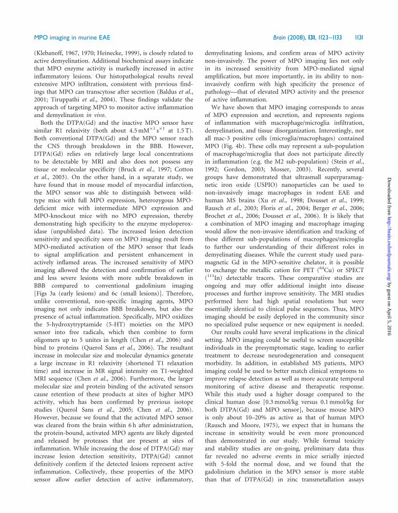
(Klebanoff, 1967, 1970; Heinecke, 1999), is closely related toactive demyelination. Additional biochemical assays indicatethat MPO enzyme activity is markedly increased in activeinflammatory lesions. Our histopathological results revealextensive MPO infiltration, consistent with previous find-ings that MPO can transcytose after secretion (Baldus et al.,2001; Tiruppathi et al., 2004). These findings validate theapproach of targeting MPO to monitor active inflammationand demyelination in vivo.Both the DTPA(Gd) and the inactive MPO sensor have
similar R1 relaxivity (both about 4.5mM�1 s�1 at 1.5 T).Both conventional DTPA(Gd) and the MPO sensor reachthe CNS through breakdown in the BBB. However,DTPA(Gd) relies on relatively large local concentrationsto be detectable by MRI and also does not possess anytissue or molecular specificity (Bruck et al., 1997; Cottonet al., 2003). On the other hand, in a separate study, wehave found that in mouse model of myocardial infarction,the MPO sensor was able to distinguish between wild-type mice with full MPO expression, heterozygous MPO-deficient mice with intermediate MPO expression andMPO-knockout mice with no MPO expression, therebydemonstrating high specificity to the enzyme myeloperox-idase (unpublished data). The increased lesion detectionsensitivity and specificity seen on MPO imaging result fromMPO-mediated activation of the MPO sensor that leadsto signal amplification and persistent enhancement inactively inflamed areas. The increased sensitivity of MPOimaging allowed the detection and confirmation of earlierand less severe lesions with more subtle breakdown inBBB compared to conventional gadolinium imaging[Figs 3a (early lesions) and 6c (small lesions)]. Therefore,unlike conventional, non-specific imaging agents, MPOimaging not only indicates BBB breakdown, but also thepresence of actual inflammation. Specifically, MPO oxidizesthe 5-hydroxytryptamide (5-HT) moieties on the MPOsensor into free radicals, which then combine to formoligomers up to 5 unites in length (Chen et al., 2006) andbind to proteins (Querol Sans et al., 2006). The resultantincrease in molecular size and molecular dynamics generatea large increase in R1 relaxivity (shortened T1 relaxationtime) and increase in MR signal intensity on T1-weightedMRI sequence (Chen et al., 2006). Furthermore, the largermolecular size and protein binding of the activated sensorscause retention of these products at sites of higher MPOactivity, which has been confirmed by previous isotopestudies (Querol Sans et al., 2005; Chen et al., 2006).However, because we found that the activated MPO sensorwas cleared from the brain within 6 h after administration,the protein-bound, activated MPO agents are likely digestedand released by proteases that are present at sites ofinflammation. While increasing the dose of DTPA(Gd) mayincrease lesion detection sensitivity, DTPA(Gd) cannotdefinitively confirm if the detected lesions represent activeinflammation. Collectively, these properties of the MPOsensor allow earlier detection of active inflammatory,
demyelinating lesions, and confirm areas of MPO activitynon-invasively. The power of MPO imaging lies not onlyin its increased sensitivity from MPO-mediated signalamplification, but more importantly, in its ability to non-invasively confirm with high specificity the presence ofpathology—that of elevated MPO activity and the presenceof active inflammation.
We have shown that MPO imaging corresponds to areasof MPO expression and secretion, and represents regionsof inflammation with macrophage/microglia infiltration,demyelination, and tissue disorganization. Interestingly, notall mac-3 positive cells (microglia/macrophages) containedMPO (Fig. 4b). These cells may represent a sub-populationof macrophage/microglia that does not participate directlyin inflammation (e.g. the M2 sub-population) (Stein et al.,1992; Gordon, 2003; Mosser, 2003). Recently, severalgroups have demonstrated that ultrasmall superparamag-netic iron oxide (USPIO) nanoparticles can be used tonon-invasively image macrophages in rodent EAE andhuman MS brains (Xu et al., 1998; Dousset et al., 1999;Rausch et al., 2003; Floris et al., 2004; Berger et al., 2006;Brochet et al., 2006; Dousset et al., 2006). It is likely thata combination of MPO imaging and macrophage imagingwould allow the non-invasive identification and tracking ofthese different sub-populations of macrophages/microgliato further our understanding of their different roles indemyelinating diseases. While the current study used para-magnetic Gd in the MPO-sensitive chelator, it is possibleto exchange the metallic cation for PET (64Cu) or SPECT(111In) detectable tracers. These comparative studies areongoing and may offer additional insight into diseaseprocesses and further improve sensitivity. The MRI studiesperformed here had high spatial resolutions but wereessentially identical to clinical pulse sequences. Thus, MPOimaging should be easily deployed in the community sinceno specialized pulse sequence or new equipment is needed.
Our results could have several implications in the clinicalsetting. MPO imaging could be useful to screen susceptibleindividuals in the presymptomatic stage, leading to earliertreatment to decrease neurodegeneration and consequentmorbidity. In addition, in established MS patients, MPOimaging could be used to better match clinical symptoms toimprove relapse detection as well as more accurate temporalmonitoring of active disease and therapeutic response.While this study used a higher dosage compared to theclinical human dose [0.3mmol/kg versus 0.1mmol/kg forboth DTPA(Gd) and MPO sensor], because mouse MPOis only about 10–20% as active as that of human MPO(Rausch and Moore, 1975), we expect that in humans theincrease in sensitivity would be even more pronouncedthan demonstrated in our study. While formal toxicityand stability studies are on-going, preliminary data thusfar revealed no adverse events in mice serially injectedwith 5-fold the normal dose, and we found that thegadolinium chelation in the MPO sensor is more stablethan that of DTPA(Gd) in zinc transmetallation assays
MPO imaging in murine EAE Brain (2008), 131, 1123^1133 1131
by guest on April 5, 2016
http://brain.oxfordjournals.org/D
ownloaded from

(unpublished data). Therefore, enzymatic imaging targetingMPO points to a promising new technology for non-invasive confirmation of active inflammatory lesions in MS,potentially not only improve disease diagnosis and treat-ment assessment in the clinical setting, but may also lead tobetter evaluation of drug development and clinical trialsof new therapies.
FundingNational Institute of Health (R24-CA92782, R01-HL078641to R.W.); the National Multiple Sclerosis Society (J.W.C.);the Dana Foundation (J.W.C.); the German NationalAcademic Foundation and the Boehringer IngelheimFonds (M.O.B.).
AcknowledgementsWe thank the CMIR Chemistry Core (F. Reynolds andL. Josephson) for performing the chemical synthesis. Wealso thank C. Rangel, C. Kaufman, T. Sponholtz, V. Lokand L. Stangenberg for experimental assistance.
ReferencesBaldus S, Eiserich JP, Mani A, Castro L, Figueroa M, Chumley P, et al.
Endothelial transcytosis of myeloperoxidase confers specificity to
vascular ECM proteins as targets of tyrosine nitration. J Clin Invest
2001; 108: 1759–70.
Berger C, Hiestand P, Kindler-Baumann D, Rudin M, Rausch M. Analysis
of lesion development during acute inflammation and remission in a rat
model of experimental autoimmune encephalomyelitis by visualization
of macrophage infiltration, demyelination and blood-brain barrier
damage. NMR Biomed 2006; 19: 101–7.
Bradley PP, Christensen RD, Rothstein G. Cellular and extracellular
myeloperoxidase in pyogenic inflammation. Blood 1982; 60: 618–22.
Brennan M, Gaur A, Pahuja A, Lusis AJ, Reynolds WF. Mice lacking
myeloperoxidase are more susceptible to experimental autoimmune
encephalomyelitis. J Neuroimmunol 2001; 112: 97–105.
Brochet B, Deloire MS, Touil T, Anne O, Caille JM, Dousset V, et al. Early
macrophage MRI of inflammatory lesions predicts lesion severity and
disease development in relapsing EAE. Neuroimage 2006; 32: 266–74.
Bruck W. The pathology of multiple sclerosis is the result of focal
inflammatory demyelination with axonal damage. J Neurol 2005; 252
(Suppl 5): v3–9.
Bruck W, Bitsch A, Kolenda H, Bruck Y, Stiefel M, Lassmann H.
Inflammatory central nervous system demyelination: correlation of
magnetic resonance imaging findings with lesion pathology. Ann Neurol
1997; 42: 783–93.
Chataway J, Sawcer S, Feakes R, Coraddu F, Broadley S, Jones HB, et al.
A screen of candidates from peaks of linkage: evidence for the involve-
ment of myeloperoxidase in multiple sclerosis. J Neuroimmunol 1999;
98: 208–13.
Chen JW, Querol Sans M, Bogdanov AA Jr, Weissleder R. Imaging
myeloperoxidase in mice using novel amplifiable paramagnetic sub-
strates. Radiology 2006; 240: 473–81.
Cotton F, Weiner HL, Jolesz FA, Guttmann CR. MRI contrast uptake in
new lesions in relapsing-remitting MS followed at weekly intervals.
Neurology 2003; 60: 640–6.
Dousset V, Ballarino L, Delalande C, Coussemacq M, Canioni P, Petry KG,
et al. Comparison of ultrasmall particles of iron oxide (USPIO)-
enhanced T2-weighted, conventional T2-weighted, and gadolinium-
enhanced T1-weighted MR images in rats with experimental
autoimmune encephalomyelitis. AJNR Am J Neuroradiol 1999; 20:
223–7.
Dousset V, Brochet B, Deloire MS, Lagoarde L, Barroso B, Caille JM, et al.
MR imaging of relapsing multiple sclerosis patients using ultra-small-
particle iron oxide and compared with gadolinium. AJNR Am J
Neuroradiol 2006; 27: 1000–5.
Filippi M, Yousry T, Campi A, Kandziora C, Colombo B, Voltz R, et al.
Comparison of triple dose versus standard dose gadolinium-DTPA for
detection of MRI enhancing lesions in patients with MS. Neurology
1996; 46: 379–84.
Floris S, Blezer EL, Schreibelt G, Dopp E, van der Pol SM, Schadee-
Eestermans IL, et al. Blood-brain barrier permeability and monocyte
infiltration in experimental allergic encephalomyelitis: a quantitative
MRI study. Brain 2004; 127: 616–27.
Frohman EM, Goodin DS, Calabresi PA, Corboy JR, Coyle PK, Filippi M,
et al. The utility of MRI in suspected MS: report of the Therapeutics
and Technology Assessment Subcommittee of the American Academy of
Neurology. Neurology 2003; 61: 602–11.
Frohman EM, Racke MK, Raine CS. Multiple sclerosis–the plaque and its
pathogenesis. N Engl J Med 2006; 354: 942–55.
Giovannoni G, Silver NC, Good CD, Miller DH, Thompson EJ.
Immunological time-course of gadolinium-enhancing MRI lesions in
patients with multiple sclerosis. Eur Neurol 2000; 44: 222–8.
Gordon S. Alternative activation of macrophages. Nat Rev Immunol 2003;
3: 23–35.
Greer JM, Sobel RA, Sette A, Southwood S, Lees MB, Kuchroo VK.
Immunogenic and encephalitogenic epitope clusters of myelin proteo-
lipid protein. J Immunol 1996; 156: 371–9.
Heinecke JW. Pathways for oxidation of low density lipoprotein by
myeloperoxidase: tyrosyl radical, reactive aldehydes, hypochlorous acid
and molecular chlorine. Biofactors 1997; 6: 145–55.
Heinecke JW. Mechanisms of oxidative damage by myeloperoxidase in
atherosclerosis and other inflammatory disorders. J Lab Clin Med 1999;
133: 321–5.
Imitola J, Chitnis T, Khoury SJ. Insights into the molecular pathogenesis
of progression in multiple sclerosis: potential implications for future
therapies. Arch Neurol 2006; 63: 25–33.
Jacobs LD, Beck RW, Simon JH, Kinkel RP, Brownscheidle CM,
Murray TJ, et al. Intramuscular interferon beta-1a therapy initiated
during a first demyelinating event in multiple sclerosis. CHAMPS Study
Group. N Engl J Med 2000; 343: 898–904.
Kidd D, Barkhof F, McConnell R, Algra PR, Allen IV, Revesz T. Cortical
lesions in multiple sclerosis. Brain 1999; 122 (Pt 1): 17–26.
Klebanoff SJ. A peroxidase-mediated antimicrobial system in leukocytes.
J Clin Invest 1967; 46: 1078–85.
Klebanoff SJ. Myeloperoxidase: contribution to the microbicidal activity of
intact leukocytes. Science 1970; 169: 1095–7.
Klebanoff SJ, Waltersdorph AM, Rosen H. Antimicrobial activity of
myeloperoxidase. Methods Enzymol 1984; 105: 399–403.
Kutzelnigg A, Lucchinetti CF, Stadelmann C, Bruck W, Rauschka H,
Bergmann M, et al. Cortical demyelination and diffuse white matter
injury in multiple sclerosis. Brain 2005; 128: 2705–12.
MacKenzie-Graham A, Tinsley MR, Shah KP, Aguilar C, Strickland LV,
Boline J, et al. Cerebellar cortical atrophy in experimental autoimmune
encephalomyelitis. Neuroimage 2006; 32: 1016–23.
Merkler D, Boscke R, Schmelting B, Czeh B, Fuchs E, Bruck W, et al.
Differential macrophage/microglia activation in neocortical EAE lesions
in the marmoset monkey. Brain Pathol 2006; 16: 117–23.
Miller D, Barkhof F, Montalban X, Thompson A, Filippi M. Clinically
isolated syndromes suggestive of multiple sclerosis, part I: natural
history, pathogenesis, diagnosis, and prognosis. Lancet Neurol 2005; 4:
281–8.
Mosser DM. The many faces of macrophage activation. J Leukoc Biol
2003; 73: 209–12.
Nagra RM, Becher B, Tourtellotte WW, Antel JP, Gold D, Paladino T,
et al. Immunohistochemical and genetic evidence of myeloperoxidase
involvement in multiple sclerosis. J Neuroimmunol 1997; 78: 97–107.
1132 Brain (2008), 131, 1123^1133 J.W.Chen et al.
by guest on April 5, 2016
http://brain.oxfordjournals.org/D
ownloaded from

Noseworthy JH, Lucchinetti C, Rodriguez M, Weinshenker BG. Multiple
sclerosis. N Engl J Med 2000; 343: 938–52.
Pomeroy IM, Matthews PM, Frank JA, Jordan EK, Esiri MM.
Demyelinated neocortical lesions in marmoset autoimmune encephalo-
myelitis mimic those in multiple sclerosis. Brain 2005; 128: 2713–21.
Querol M, Chen JW, Weissleder R, Bogdanov A Jr. DTPA-bisamide-
based MR sensor agents for peroxidase imaging. Org Lett 2005; 7:
1719–22.
Querol Sans M, Chen JW, Bogdanov AA Jr. A paramagnetic contrast agent
with myeloperoxidase-sensing properties. Org Biomol Chem 2006; 4:
1887–95.
Querol Sans M, Chen JW, Weissleder R, Bogdanov AA Jr.
Myeloperoxidase activity imaging using (67)Ga labeled substrate. Mol
Imaging Biol 2005; 7: 403–10.
Rausch M, Hiestand P, Baumann D, Cannet C, Rudin M. MRI-based
monitoring of inflammation and tissue damage in acute and chronic
relapsing EAE. Magn Reson Med 2003; 50: 309–14.
Rausch PG, Moore TG. Granule enzymes of polymorphonuclear
neutrophils: a phylogenetic comparison. Blood 1975; 46: 913–9.
Seeldrayers PA, Syha J, Morrissey SP, Stodal H, Vass K, Jung S, et al.
Magnetic resonance imaging investigation of blood-brain barrier
damage in adoptive transfer experimental autoimmune encephalomy-
elitis. J Neuroimmunol 1993; 46: 199–206.
Stein M, Keshav S, Harris N, Gordon S. Interleukin 4 potently enhances
murine macrophage mannose receptor activity: a marker of alternative
immunologic macrophage activation. J Exp Med 1992; 176: 287–92.
Storch MK, Bauer J, Linington C, Olsson T, Weissert R, Lassmann H.
Cortical demyelination can be modeled in specific rat models of
autoimmune encephalomyelitis and is major histocompatability complex
(MHC) haplotype-related. J Neuropathol Exp Neurol 2006; 65: 1137–42.
Tiruppathi C, Naqvi T, Wu Y, Vogel SM, Minshall RD, Malik AB.
Albumin mediates the transcytosis of myeloperoxidase by means of
caveolae in endothelial cells. Proc Natl Acad Sci U S A 2004; 101:
7699–704.
Xu S, Jordan EK, Brocke S, Bulte JWM, Quigley L, Tresser N, et al. Study
of relapsing remitting experimental allergic encephalomyelitis SJL Mouse
Model using MION-46L enhanced in vivo MRI: early histopathological
correlation. J Neurosci Res 1998; 52: 549–58.
Zakrzewska-Pniewska B, Styczynska M, Podlecka A, Samocka R,
Peplonska B, Barcikowska M, et al. Association of apolipoprotein E
and myeloperoxidase genotypes to clinical course of familial and
sporadic multiple sclerosis. Mult Scler 2004; 10: 266–71.
MPO imaging in murine EAE Brain (2008), 131, 1123^1133 1133
by guest on April 5, 2016
http://brain.oxfordjournals.org/D
ownloaded from