Altered inflammatory response and increased neurodegeneration in metallothionein I+II deficient mice...
13
Ž . Journal of Neuroimmunology 119 2001 248–260 www.elsevier.comrlocaterjneuroim Altered inflammatory response and increased neurodegeneration in metallothionein I q II deficient mice during experimental autoimmune encephalomyelitis Milena Penkowa a,1,2 , Carmen Espejo b,1,3 , Eva M. Martınez-Caceres b,3 , ´ ´ Christian Bjørn Poulsen a,2 , Xavier Montalban b,3 , Juan Hidalgo c, ) a Department of Medical Anatomy, The Panum Institute, UniÕersity of Copenhagen, DK-2200, Copenhagen, Denmark b Unitat de Neuroimmunologia Clınica, Department of Neurology, Hospital General Vall d’Hebron, UniÕersitat Autonoma de Barcelona, Barcelona, Spain ´ ` c Departamento de Biologıa Celular Fisiologıa e Inmunologıa, Unidad de Fisiologıa Animal, Facultad de Ciencias, UniÕersidad Autonoma de Barcelona, ´ ´ ´ ´ ´ Bellaterra, Barcelona 08193, Spain Received 23 April 2001; received in revised form 1 June 2001; accepted 4 June 2001 Abstract Ž . Metallothionein-I qII MT-I qII are antioxidant, neuroprotective proteins, and in this report we have examined their roles during Ž . Ž . experimental autoimmune encephalomyelitis EAE by comparing MT-I qII-knock-out MTKO and wild-type mice. We herewith show that EAE susceptibility is higher in MTKO mice relatively to wild-type mice, and that the inflammatory responses elicited by EAE in the Ž . central nervous system CNS are significantly altered by MT-I qII deficiency. Thus, during EAE the MTKO mice showed increased macrophage and T-lymphocytes infiltration in the CNS, while their reactive astrogliosis was significantly decreased. In addition, the expression of the proinflammatory cytokines interleukin-1b, interleukin-6, and tumor necrosis factor-a elicited by EAE was further increased in the MTKO mice, and oxidative stress and apoptosis were also significantly increased in MTKO mice compared to normal mice. The present results strongly suggest that MT-I qII are major factors involved in the inflammatory response of the CNS during EAE and that they play a neuroprotective role in this scenario. q 2001 Elsevier Science B.V. All rights reserved. Ž . Keywords: EAErMS; Metallothionein MT ; Cytokines; Oxidative stress; Apoptosis 1. Introduction Ž . Experimental autoimmune encephalomyelitis EAE is considered to be an autoimmune disease of the CNS and is Ž . used as an animal model for multiple sclerosis MS . Animals with EAE show paralysis and histopathological infiltrates of microgliarmacrophages, T lymphocytes, and Ž reactive astrocytes Benveniste, 1997; Lassmann, 1999; . Popovich et al., 1997; Swanborg, 1995 . During EAErMS, a number of detrimental factors are produced and these can contribute to the ongoing inflammation and brain tissue ) Corresponding author. Tel.: q 34-93-5812037; fax: q 34-93-5812390. Ž . E-mail address: [email protected] J. Hidalgo . 1 These authors contributed equally to this paper. 2 Tel.: q 45-35327222; fax: q 45-35327217. 3 Tel.: q 34-93-2746202; fax: q 34-93-2746084. Ž . damage Brosnan and Raine, 1996 . On the one hand, the Ž production of proinflammatory cytokines interleukin-1 IL- . Ž . 1 , IL-6 and tumor necrosis factor-a TNF-a has been correlated with enhanced EAE disease, inflammatory re- Ž sponse, and tissue destruction Dal Canto et al., 1999; Douni et al., 1995–1996; Eng et al., 1996; Gijbels et al., 1995; Jacobs et al., 1991; Loughlin et al., 1994; Rajan et al., 1998; Renno et al., 1995; Taupin et al., 1997; Wie- . mann et al., 1998 . Interestingly, mice with genetical IL-6 Ž deficiency resist induction of EAE Mendel et al., 1998; . Okuda et al., 1998, 1999; Samoilova et al., 1998 . Also, transgenic mice with overexpression of TNF-a show in- creased severity of EAE andror spontaneous demyelinat- Ž ing disease Campbell, 1998a,b; Dal Canto et al., 1999; Douni et al., 1995–1996; Probert et al., 1995; Taupin et . al., 1997 . Ž . On the other hand, reactive oxygen species ROS are Ž produced during EAE Bagasra et al., 1995; Scott et al., . 1997; Van Dam et al., 1995 . ROS are major mediators of 0165-5728r01r$ - see front matter q 2001 Elsevier Science B.V. All rights reserved. Ž . PII: S0165-5728 01 00357-5
-
Upload
independent -
Category
Documents
-
view
5 -
download
0
Transcript of Altered inflammatory response and increased neurodegeneration in metallothionein I+II deficient mice...
Ž .Journal of Neuroimmunology 119 2001 248–260www.elsevier.comrlocaterjneuroim
Altered inflammatory response and increased neurodegeneration inmetallothionein Iq II deficient mice during experimental
autoimmune encephalomyelitis
Milena Penkowaa,1,2, Carmen Espejob,1,3, Eva M. Martınez-Caceresb,3,´ ´Christian Bjørn Poulsena,2, Xavier Montalbanb,3, Juan Hidalgoc,)
a Department of Medical Anatomy, The Panum Institute, UniÕersity of Copenhagen, DK-2200, Copenhagen, Denmarkb Unitat de Neuroimmunologia Clınica, Department of Neurology, Hospital General Vall d’Hebron, UniÕersitat Autonoma de Barcelona, Barcelona, Spain´ `c Departamento de Biologıa Celular Fisiologıa e Inmunologıa, Unidad de Fisiologıa Animal, Facultad de Ciencias, UniÕersidad Autonoma de Barcelona,´ ´ ´ ´ ´
Bellaterra, Barcelona 08193, Spain
Received 23 April 2001; received in revised form 1 June 2001; accepted 4 June 2001
Abstract
Ž .Metallothionein-Iq II MT-I q II are antioxidant, neuroprotective proteins, and in this report we have examined their roles duringŽ . Ž .experimental autoimmune encephalomyelitis EAE by comparing MT-Iq II-knock-out MTKO and wild-type mice. We herewith show
that EAE susceptibility is higher in MTKO mice relatively to wild-type mice, and that the inflammatory responses elicited by EAE in theŽ .central nervous system CNS are significantly altered by MT-Iq II deficiency. Thus, during EAE the MTKO mice showed increased
macrophage and T-lymphocytes infiltration in the CNS, while their reactive astrogliosis was significantly decreased. In addition, theexpression of the proinflammatory cytokines interleukin-1b, interleukin-6, and tumor necrosis factor-a elicited by EAE was furtherincreased in the MTKO mice, and oxidative stress and apoptosis were also significantly increased in MTKO mice compared to normalmice. The present results strongly suggest that MT-Iq II are major factors involved in the inflammatory response of the CNS during EAEand that they play a neuroprotective role in this scenario.q2001 Elsevier Science B.V. All rights reserved.
Ž .Keywords: EAErMS; Metallothionein MT ; Cytokines; Oxidative stress; Apoptosis
1. Introduction
Ž .Experimental autoimmune encephalomyelitis EAE isconsidered to be an autoimmune disease of the CNS and is
Ž .used as an animal model for multiple sclerosis MS .Animals with EAE show paralysis and histopathologicalinfiltrates of microgliarmacrophages, T lymphocytes, and
Žreactive astrocytes Benveniste, 1997; Lassmann, 1999;.Popovich et al., 1997; Swanborg, 1995 . During EAErMS,
a number of detrimental factors are produced and these cancontribute to the ongoing inflammation and brain tissue
) Corresponding author. Tel.:q34-93-5812037; fax:q34-93-5812390.Ž .E-mail address: [email protected] J. Hidalgo .
1 These authors contributed equally to this paper.2 Tel.: q45-35327222; fax:q45-35327217.3 Tel.: q34-93-2746202; fax:q34-93-2746084.
Ž .damage Brosnan and Raine, 1996 . On the one hand, theŽproduction of proinflammatory cytokines interleukin-1 IL-
. Ž .1 , IL-6 and tumor necrosis factor-a TNF-a has beencorrelated with enhanced EAE disease, inflammatory re-
Žsponse, and tissue destruction Dal Canto et al., 1999;Douni et al., 1995–1996; Eng et al., 1996; Gijbels et al.,1995; Jacobs et al., 1991; Loughlin et al., 1994; Rajan etal., 1998; Renno et al., 1995; Taupin et al., 1997; Wie-
.mann et al., 1998 . Interestingly, mice with genetical IL-6Ždeficiency resist induction of EAE Mendel et al., 1998;
.Okuda et al., 1998, 1999; Samoilova et al., 1998 . Also,transgenic mice with overexpression of TNF-a show in-creased severity of EAE andror spontaneous demyelinat-
Žing disease Campbell, 1998a,b; Dal Canto et al., 1999;Douni et al., 1995–1996; Probert et al., 1995; Taupin et
.al., 1997 .Ž .On the other hand, reactive oxygen species ROS are
Žproduced during EAE Bagasra et al., 1995; Scott et al.,.1997; Van Dam et al., 1995 . ROS are major mediators of
0165-5728r01r$ - see front matterq2001 Elsevier Science B.V. All rights reserved.Ž .PII: S0165-5728 01 00357-5
( )M. Penkowa et al.rJournal of Neuroimmunology 119 2001 248–260 249
Žtissue damage in the CNS Cassarino and Bennett, 1999;.Sun and Chen, 1998 , and they have been implicated in
ŽEAE pathogenesis and disease Cross et al., 1994;.MacMicking et al., 1992; Ruuls et al., 1995 . Moreover, a
number of studies have shown that blocking inducibleŽ .nitric oxide synthase iNOS may prevent EAE develop-
Žment Brenner et al., 1997; Ding et al., 1998; Zhao et al.,.1996 . Thus, a concomitant inhibition of agents such as
proinflammatory cytokines and ROS during EAE couldhave a therapeutic potential for the treatment of EAErMSand is likely to prove superior to a single agent therapy.
During CNS inflammation including EAE,microgliarmacrophages and reactive astrocytes increase
Ž .their expression of metallothionein Iq II MT-I q II ,which possess antioxidant and tissue protective functionsŽAschner, 1996, 1998b; Espejo et al., in press; Hidalgo etal., 1997; Lazo and Pitt, 1995; Lazo et al., 1998; Molineroet al., 1998; Penkowa and Hidalgo, 2000a,b; Penkowa etal., 1999b, 2000b; Tate et al., 1995; Thornalley and Vasak,
.1985 . In order to examine MT functions in vivo, a geneticŽapproach using either MT-Iq II deficient MT knock-out,
. ŽMTKO or MT-I overexpressing mice has been used Car-rasco et al., 2000; Penkowa et al., 1999a,b, 2000a; Van
.Lookeren Campagne et al., 1999 . In MTKO mice, analtered inflammatory response, increased oxidative stressand apoptosis, and a decreased neuroregenerative capacitywere observed following traumatic brain damage or kainic
Žacid-induced seizures Carrasco et al., 2000; Penkowa et.al., 1999a, 2000a . Moreover, transgenic overexpression of
MT-I reduces tissue loss, vascular edema, and improvesfunctional outcome following focal cerebral ischemia and
Ž .reperfusion Van Lookeren Campagne et al., 1999 . Inaddition, we recently showed that exogenous administra-
Ž .tion of metallothionein-II MT-II significantly decreasedthe clinical symptoms, mortality, and leukocyte infiltration
Ž .of the CNS during EAE Penkowa and Hidalgo, 2000b asŽwell as oxidative stress and apoptotic cell death Penkowa
.and Hidalgo, 2000b, 2001 in Lewis rats. The antioxidantand antiapoptotic effects of MT during EAE are likelybeneficial, in that oxidative stress and apoptotic cell deathof both oligodendrocytes and neurons are implicated in
ŽEAErMS pathogenesis Aktas et al., 2000; Alcazar et al.,1998; Arbizu-Urdiain and Martinez-Yelamos, 2000; Crosset al., 1994; MacMicking et al., 1992; Ruuls et al., 1995;Sabelko-Downes et al., 1999; Hisahara et al., 2001;Honegger and Langemann, 1989; Penkowa and Hidalgo,
.2000b, 2001 .In this study, we analyzed the effect of genetical MT-I
Ž .q II deficiency in 129rSv mice MTKO mice duringactively induced EAE. We hereby show that MT-Iq IIdeficiency causes an increased susceptibility to the diseaseand an increased inflammatory response of microgliarmacrophages and T lymphocytes while reactive astroglio-sis is reduced. Also, the proinflammatory cytokines IL-1b,IL-6 and TNF-a and oxidative stress markers iNOS, ni-
Ž . Ž .trotyrosine NITT , and malondialdehyde MDA were
significantly increased in MTKO mice with EAE. As alikely consequence, these mice also showed an increasednumber of TUNELq apoptotic cells, which were identi-fied as neurons, oligodendrocytes, and astroglia. Therefore,we suggest that MT-Iq II are potentially beneficial factorsfor the treatment of EAErMS.
2. Materials and methods
2.1. Mice
Homozygous MTKO mice were generated as previouslyŽ .described Masters et al., 1994 . The MTKO mice were
raised on the 129rSv genetic background; therefore, micefrom this strain were used as controls. Mice were 8 to 14weeks old, and they were fed with standard chow and hadaccess to water ad libitum. Anaesthesia was induced by
Ž .intraperitoneal i.p. injection of 37 mgrkg of ketamineŽ w .Ketolar , Parke and Davis, Morris Plains, NJ, USA and
Ž w5.5 mgrkg of xylazine Rompun , Bayer, Leverkusen,.Germany . Animal welfare was observed in compliance
with the European Community regulations on this subject,and all animal experiments have adhered to the standardsof the National Research Council’s Guide for the care anduse of laboratory animals.
2.2. Immunization
Animals were immunized by a single subcutaneousŽ .s.c. injection of 0.9% saline containing 100mg of pep-tide 40–55 of rat myelin oligodendrocyte glycoproteinŽ . ŽMOG emulsified in Freund’s adjuvant Sigma, St.40-55
. Ž .Louis, MO, USA 9:11 vrv containing 4 mgrml My-Žcobacterium tuberculosis H37RA Difco Laboratories, De-
.troit, MI, USA . Rat MOG was synthesised by Dr. D.40-55ŽAndreu Departament de Sıntesi de Peptids, Facultat de´ `
.Quımica, Universitat de Barcelona, Spain . Mice received´0.05 ml of emulsion s.c. in the four limb flanks. At days 0
Žand 2 post-immunization, each mouse received 0.2 ml 2. Ž wIUrml of inactivated Bordetella pertussis Vaxicoq ,
.Pasteur Merieux, Lyon, France intravenously.
2.3. Clinical eÕaluation
All animals were weighed and examined daily, fromŽday 7 to the end of the experiment day 37 post-immuniza-
.tion , for neurological signs according to the followingcriteria: 0, no clinical signs; 0.5, partial loss of tail tonusfor two consecutive days; 1, paralysis of whole tail; 2,mild paraparesis of one or both hind limbs; 3, paraplegia;4, tetraparesis; 5, tetraplegia; 6, death. All experimentswere performed in a blinded manner. The blind was estab-lished and preserved throughout the entire experimentalprocess in such a way that the investigator examining and
( )M. Penkowa et al.rJournal of Neuroimmunology 119 2001 248–260250
clinically evaluating the animal was kept unaware of thestrain of the mice.
2.4. Tissue processing
Animals were sacrificed with CO at day 37 post-im-2
munization and the CNS was resected. Tissues were fixedovernight in 4% paraformaldehyderPBS. Following fixa-tion, the samples were cryoprotected in 30% sucroserPBS,frozen in 2-methylbutane cooled in liquid nitrogen andthen stored aty80 8C until use.
Brain and spinal cord were cut into 30mm thick serial,coronal sections on a cryostat and used for histochemistryand immunohistochemistry. The sections were incubated in3.0% H O in TBSrNonidet for 15 min at room tempera-2 2
ture to quench endogenous peroxidase. After washing inrunning tapwater, sections were washed in TBSrNonidetthree times for 5 min. The sections were preincubated with
Ž . Ž . Žpronase E protease type XIV Sigma, code P5147 0.025.g dissolved in 50 ml TBS for 10 min, pH 7.4, at 378C .
All the sections were incubated in 10% normal goat serumin TBSrNonidet for 30 min at room temperature to blocknonspecific binding. Moreover, sections prepared for incu-bation with monoclonal mouse-derived antibodies were inaddition incubated with Blocking Solutions AqB fromHistoMouse-SP Kit to quench endogenous mouse IgGŽZymed Lab., South San Francisco, CA, USA, code 95-
.9544 .
2.5. Histochemistry
Standard HE stainings and stainings for biotinylatedŽtomato lectin fromLycopersicon esculentum Sigma, code
. ŽL9389 were performed as previously described Penkowa.and Hidalgo, 2000b .
2.6. Immunohistochemistry
Sections were incubated overnight at 48C with one ofthe following primary antibodies: rabbit anti-rat MT-Iq II
Ž1:500 Gasull et al., 1993; Penkowa et al., 1997, 1999a,b,. Ž2000b ; mouse anti-human CD14 as a marker for mono-
. Ž .cytes-macrophages 1:20 Zymed, code 180121 ; mouseŽ . Žanti-rat CD3 as a marker for T lymphocytes 1:50 Sero-
.tec, Oxford, UK, code KD MCA 772 ; rabbit anti-cowŽ . ŽGFAP as a marker for astrocytes 1:250 Dakopatts,
.Glostrup, DK, code Z 334 ; mouse anti-human IL-1b1.50Ž .Biogenesis, Kingston, NH, USA, code 5375-4329 ; rat
Žanti-mouse IL-6 1:10 Harlan Sera Lab., Sussex, UK, code. ŽMAS 584 ; rabbit anti-mouse TNF-a 1:100 Biosource
.Int., Camarillo, CA, USA, code AMC 3012 ; rabbit anti-Ž . Žmouse inducible nitric-oxide synthase iNOS as a marker
. Žfor oxidative stress 1:100 Alexis Biochemicals, San.Diego, CA, USA, code 210-503-R050 ; rabbit anti-nitro-
Ž . Ž .tyrosine NITT as a marker for oxidative stress 1:100ŽAlpha Diagnostic Int., San Antonio, TX, USA, code
. Ž . ŽNITT 12-A ; rabbit anti-malondialdehyde MDA as a. Žmarker for oxidative stress 1:100 Alpha Diagnostic, code
.MDA 11-S ; rabbit anti-mouse ICErCaspase-1 1:100Ž .Santa Cruz Biotech., Santa Cruz, USA, code sc-1218R ;
Žand rabbit anti-human caspase-3 1:50 Santa Cruz Biotech.,.code sc-7148 . The primary antibodies were detected by
Žusing biotinylated mouse anti-rabbit IgG 1:400 Sigma,.code B3275 , or biotinylated goat anti-mouse IgG 1:200
Ž .Sigma, code B8774 , or biotinylated goat anti-mouse IgMŽ1:10 Jackson ImmunoResearch Lab., West Baltimore Pike,
.West Grove, PA, USA, code 115-065-020 , or biotinylatedŽgoat anti-rat IgG 1:1000 Amersham Pharmacia Biotech.,
.Buckinghamshire, UK, code RPN 1005 for 30 min atroom temperature. These secondary antibodies were de-
Žtected by StreptABComplexrHRP StreptABComplexr.HRP, Dakopatts, code K377 prepared at manufacturer’s
recommended dilution and incubated for 30 min at roomtemperature. The immunoreaction was visualized using
Ž0.015% H O in DABrTBS TBS: 0.05 M TRIS, pH 7.4,2 2.0.15 M NaCl for 10 min at room temperature.
In order to evaluate the extent of non-specific bindingin the immunohistochemical experiments, control sectionswere incubated in the absence of primary antibody. Resultswere considered only if these controls were negative. Afurther control for the cytokines was performed by preab-sorption of the primary antibodies with their correspondingantigenic proteins. For this purpose we used: mouse IL-1b
Ž .RD Systems, Minneapolis, MN, USA, code 401-ML-005 ;Ž .mouse IL-6 RD Systems, code 406-ML-005 ; mouse
Ž .TNF-a RD Systems, code 410-MT-010 . Results wereconsidered only if these controls were negative. Compar-isons of sections, which were either incubated or notincubated with Blocking Solutions AqB from Histo-Mouse-SP Kit, were also done.
2.7. In situ detection of DNA fragmentation by terminal( )deoxynucleotidyl transferase TdT -mediated deoxyuridine
( )triphosphate dUTP -digoxigenin nick end labeling( )TUNEL
TUNEL staining was performed as previously describedŽ .Penkowa et al., 1999a .
2.8. Immunofluorescence histochemistry and TUNEL
In order to examine which cells are expressing IL-1b,IL-6, and TNF-a during EAE, triple immunofluorescencewas performed after tissue processing as mentioned above.Sections were incubated overnight at 48C with TexasRedŽ .TXRD -linked tomato lectin from L. esculentum 1:50Ž . ŽSigma, code L9139 , polyclonal rabbit anti-cow GFAP as
.mentioned above , and one of the following: mouse anti-Ž .human IL-1b as mentioned above ; rat anti-mouse IL-6
Ž .as mentioned above ; or mouse anti-human TNF-a 1:50Ž .Santa Cruz Biotech., code sc-7317 . The anti-GFAP anti-bodies were detected by using goat anti-rabbit IgG linked
Ž . Žwith aminomethylcoumarin AMCA 1:40 Jackson Im-
( )M. Penkowa et al.rJournal of Neuroimmunology 119 2001 248–260 251
.munoRes. Lab., code 111-155-144 . The anti-IL-1b andanti-TNF-a antibodies were detected by using goat anti-
Ž . Žmouse IgG linked with fluorescein FITC 1:30 Jackson.ImmunoRes. Lab., code 115-095-146 . The anti-IL-6 anti-
bodies were detected by using FITC-linked goat anti-ratŽ .IgG 1:30 Jackson ImmunoRes. Lab., code 112-095-143 .
In order to examine which cells are suffering fromoxidative stress, triple immunofluorescence stainings wereperformed by incubating sections overnight at 48C with
Ž .TXRD-linked tomato lectin as mentioned above , andŽmouse anti-human NF 1:10 Biogenex, USA, code
.AM073-10M , and either rabbit anti-mouse iNOS, or rab-Ž .bit anti-NITT, or rabbit anti-MDA all as mentioned above .
The anti-NF antibodies were detected by using FITC-lin-Ž .ked goat anti-mouse as above . The anti-iNOS, anti-NITT,
and anti-MDA antibodies were detected by using AMCA-Ž .linked goat anti-rabbit IgG as above .
In order to examine which cells were undergoing apop-totic cell death, other triple fluorescence stainings wereperformed by incubating sections with FITC-linked
Ž .TUNEL Oncor, USA, code S7111-KIT performed ac-cording to manufacturer’s protocol, and afterwards thesesections were incubated overnight at 48C with mouse
Žanti-porcine vimentin as a marker for macrophages and. Ž .reactive astrocytes 1:50 Dakopatts, code M0725 , and
Žrabbit anti-human NSE 1:1000 Calbiochem, USA, code.D05059 . The anti-vimentin antibodies were detected by
Žusing AMCA-linked goat anti-mouse IgG 1:30 Jackson.ImmunoRes. Lab., code 115-155-146 . The anti-NSE anti-
bodies were detected by using TXRD-goat anti-rabbit IgGŽ .1:50 Jackson ImmunoRes. Lab., code 111-075-144 . Also,
other sections were incubated with FITC-linked TUNELŽ . Žas mentioned above and mouse anti-human CNPase as a
. Žmarker for oligodendrocytes 1:50 Biogenesis, code 2406-.3006 . The anti-CNPase antibodies were detected by using
ŽAMCA-linked goat anti-mouse IgG 1:30 as mentioned.above .
In order to confirm the notion of apoptotic cell death asjudged by TUNEL, neighbouring sections were incubated
Žwith rabbit anti-mouse caspase-1 1:100 Santa Cruz.Biotech., code sc-1281R , and mouse anti-human caspase-3
Ž .1:50 Santa Cruz Biotech., code sc-7272 . These primaryantibodies were detected as mentioned above for anti-NSEand anti-vimentin antibodies. The sections were embedded
Ž .in 20 ml fluorescent mounting Dakopatts, code S3023and kept in darkness at 48C. Control sections wereprocessed in parallel, in which the primary antibody wasomitted. Results were considered only if these controlswere unstained. For the simultaneous examination andrecording of the three stains, a Zeiss Axioplan2 light
Žmicroscope equipped with a triple band DAPIrFITCr.Texas Red filter was used.
2.9. Cell counts of stained cells
For most of the variables analyzed, cell counts werecarried out in 1 mm2 matched areas of brain stem, spinalcord, and cerebellum of all mice for statistical evaluationof the results. To this end, positively stained cells, defined
Ž .as cells with staining of the soma cytoplasm , or in thecase of TUNEL cells with nuclear staining, were counted.Cell counts were performed in at least three sections ofbrain stem, spinal cord, and cerebellum of each mouse, anda mean value was calculated for each animal. Cell countswere performed in a blinded manner in at least three miceper group.
2.10. Statistical analysis
Clinical results were analyzed with the Studentt-test orŽChi-square test depending on the variables analyzed see
.Table 1 . For cell counts, the results were evaluated byŽ .two-way analysis of variance ANOVA , with strain and
EAE as main factors. When the interaction was significant,it was interpreted to be the consequence of the effect ofMT-I q II deficiency on EAE effects. A significant differ-ence was considered whenp-0.05.
3. Results
3.1. General
In three independent experiments, 32 wild-type miceŽ . Žn s12, n s11 andn s9 and 30 MTKO mice n s1 2 3 1
.11, n s10 and n s9 were immunized with MOG2 3 40-55
as described in Materials and methods. Pooled results ofthe clinical data are shown in Table 1. Daily score was
Table 1Clinical evaluation of the mice during EAEClinical course of normal and MTKO mice after EAE induction with MOG peptide.40 – 55
a aIncidence Mortality Day of onset Maximum Daily scoreaŽ . Ž .% % score
qrq Ž .MT ns32 43.75 9.37 23.93"4.25 1.66"2.15 1.43"1.88yry )Ž .MT ns30 70.00 16.67 24.75"5.07 2.52"2.09 2.03"1.71
aResults are mean"SD.) p-0.05 vs. wild-type mice.
( )M. Penkowa et al.rJournal of Neuroimmunology 119 2001 248–260252
performed from day 7 to day 37 post-immunization. Theday of onset of EAE was similar in both strains, but anincreased susceptibility to EAE induction in the MTKOmice compared to normal mice was evident. Thus, 70% ofthe immunized MTKO mice developed EAE, while only
Ž .43.75% of the wild-type mice didp-0.05 , while mor-tality, mean daily score and maximum score also tended to
Ž .be higher in the MTKO mice Table 1 . These results wereconsistent in the three separate experiments carried out. Adetailed histopathological analysis of the effects of EAE inthe CNS of normal and MTKO mice was carried out inone experiment and is described below.
3.2. Inflammatory response
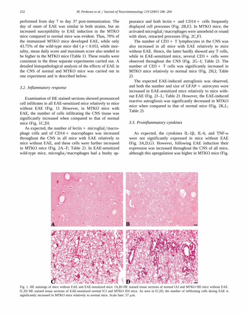
Examination of HE stained sections showed pronouncedcell infiltrates in all EAE-sensitized mice relatively to mice
Ž .without EAE Fig. 1 . However, in MTKO mice withEAE, the number of cells infiltrating the CNS tissue wassignificantly increased when compared to that of normal
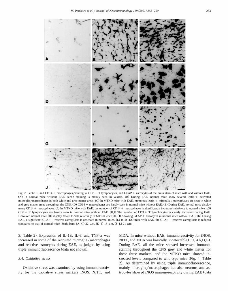
Ž .mice Fig. 1C,D .As expected, the number of lectinq microglialrmacro-
phage cells and of CD14q macrophages was increasedthroughout the CNS in all mice with EAE relatively tomice without EAE, and these cells were further increased
Ž .in MTKO mice Fig. 2A–F; Table 2 . In EAE-sensitizedwild-type mice, microgliarmacrophages had a bushy ap-
pearance and both lectinq and CD14q cells frequentlyŽ .displayed cell processes Fig. 2B,E . In MTKO mice, the
activated microglialrmacrophages were amoeboid or roundŽ .with short, retracted processes Fig. 2C,F .
The number of CD3q T lymphocytes in the CNS wasalso increased in all mice with EAE relatively to micewithout EAE. Hence, the latter hardly showed any T cells,while in EAE-sensitized mice, several CD3q cells were
Ž .observed throughout the CNS Fig. 2G–I; Table 2 . Thenumber of CD3q T cells was significantly increased in
ŽMTKO mice relatively to normal mice Fig. 2H,I; Table.2 .
The expected EAE-induced astrogliosis was observed,and both the number and size of GFAPq astrocytes wereincreased in EAE-sensitized mice relatively to mice with-
Ž .out EAE Fig. 2J–L; Table 2 . However, the EAE-inducedreactive astrogliosis was significantly decreased in MTKO
Žmice when compared to that of normal mice Fig. 2K,L;.Table 2 .
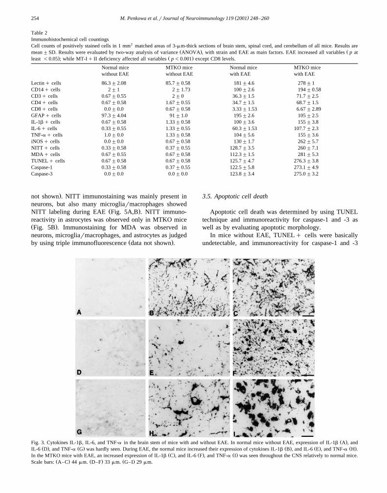
3.3. Proinflammatory cytokines
As expected, the cytokines IL-1b, IL-6, and TNF-awere not significantly expressed in mice without EAEŽ .Fig. 3A,D,G . However, following EAE induction theirexpression was increased throughout the CNS of all mice,
Žalthough this upregulation was higher in MTKO mice Fig.
Ž . Ž . Ž .Fig. 1. HE stainings of mice without EAE and EAE-sensitized mice. A,B HE stained tissue sections of normal A and MTKO B mice without EAE.Ž . Ž . Ž . Ž .C,D HE stained tissue sections of EAE-sensitized normal C and MTKO D mice. As seen in C,D , the number of infiltrating cells during EAE issignificantly increased in MTKO mice relatively to normal mice. Scale bars: 57mm.
( )M. Penkowa et al.rJournal of Neuroimmunology 119 2001 248–260 253
Fig. 2. Lectinq and CD14q macrophagesrmicroglia, CD3q T lymphocytes, and GFAPq astrocytes of the brain stem of mice with and without EAE.Ž . Ž .A In normal mice without EAE, lectin staining is mainly seen in vessels. B During EAE, normal mice show several lectinq activated
Ž .microgliarmacrophages in both white and grey matter areas. C In MTKO mice with EAE, numerous lectinq microgliarmacrophages are seen in whiteŽ . Ž .and grey matter areas throughout the CNS. D CD14q macrophages are hardly seen in normal mice without EAE. E During EAE, normal mice display
Ž . Ž .many CD14q macrophages. F In MTKO mice with EAE, the number of CD14q macrophages is significantly increased relatively to normal mice. GŽ .CD3q T lymphocytes are hardly seen in normal mice without EAE. H,I The number of CD3q T lymphocytes is clearly increased during EAE.
Ž . Ž . Ž . Ž .However, normal mice H display fewer T cells relatively to MTKO mice I . J Showing GFAPq astrocytes in normal mice without EAE. K DuringŽ .EAE, a significant GFAPq reactive astrogliosis is observed in normal mice. L In MTKO mice with EAE, the GFAPq reactive astrogliosis is reduced
Ž . Ž . Ž .compared to that of normal mice. Scale bars: A–C 22mm. D–I 18mm. J–L 21mm.
.3; Table 2 . Expression of IL-1b, IL-6, and TNF-a wasincreased in some of the recruited microgliarmacrophagesand reactive astrocytes during EAE, as judged by using
Ž .triple immunofluorescence data not shown .
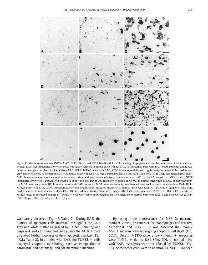
3.4. OxidatiÕe stress
Oxidative stress was examined by using immunoreactiv-ity for the oxidative stress markers iNOS, NITT, and
MDA. In mice without EAE, immunoreactivity for iNOS,Ž .NITT, and MDA was basically undetectable Fig. 4A,D,G .
During EAE, all the mice showed increased immuno-staining throughout the CNS grey and white matter forthese three markers, and the MTKO mice showed in-
Žcreased levels compared to wild-type mice Fig. 4; Table.2 . As determined by using triple immunofluorescence,
mainly microgliarmacrophages but also neurons and as-Žtrocytes showed iNOS immunoreactivity during EAE data
( )M. Penkowa et al.rJournal of Neuroimmunology 119 2001 248–260254
Table 2Immunohistochemical cell countingsCell counts of positively stained cells in 1 mm2 matched areas of 3-mm-thick sections of brain stem, spinal cord, and cerebellum of all mice. Results are
Ž . Žmean"SD. Results were evaluated by two-way analysis of variance ANOVA , with strain and EAE as main factors. EAE increased all variablesp at. Ž .least-0.05 ; while MT-Iq II deficiency affected all variablesp-0.001 except CD8 levels.
Normal mice MTKO mice Normal mice MTKO micewithout EAE without EAE with EAE with EAE
Lectinq cells 86.3"2.08 85.7"0.58 181"4.6 278"1CD14q cells 2"1 2"1.73 100"2.6 194"0.58CD3q cells 0.67"0.55 2"0 36.3"1.5 71.7"2.5CD4q cells 0.67"0.58 1.67"0.55 34.7"1.5 68.7"1.5CD8q cells 0.0"0.0 0.67"0.58 3.33"1.53 6.67"2.89GFAPq cells 97.3"4.04 91"1.0 195"2.6 105"2.5IL-1bq cells 0.67"0.58 1.33"0.58 100"3.6 155"3.8IL-6q cells 0.33"0.55 1.33"0.55 60.3"1.53 107.7"2.3TNF-aq cells 1.0"0.0 1.33"0.58 104"5.6 155"3.6iNOSq cells 0.0"0.0 0.67"0.58 130"1.7 262"5.7NITTq cells 0.33"0.58 0.37"0.55 128.7"3.5 260"7.1MDA q cells 0.67"0.55 0.67"0.58 112.3"1.5 281"5.3TUNELq cells 0.67"0.58 0.67"0.58 125.7"4.7 276.3"3.8Caspase-1 0.33"0.58 0.37"0.55 122.5"5.8 273.1"4.9Caspase-3 0.0"0.0 0.0"0.0 123.8"3.4 275.0"3.2
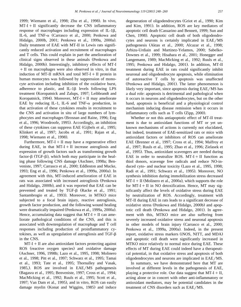
.not shown . NITT immunostaining was mainly present inneurons, but also many microgliarmacrophages showed
Ž .NITT labeling during EAE Fig. 5A,B . NITT immuno-reactivity in astrocytes was observed only in MTKO miceŽ .Fig. 5B . Immunostaining for MDA was observed inneurons, microgliarmacrophages, and astrocytes as judged
Ž .by using triple immunofluorescence data not shown .
3.5. Apoptotic cell death
Apoptotic cell death was determined by using TUNELtechnique and immunoreactivity for caspase-1 and -3 aswell as by evaluating apoptotic morphology.
In mice without EAE, TUNELq cells were basicallyundetectable, and immunoreactivity for caspase-1 and -3
Ž .Fig. 3. Cytokines IL-1b, IL-6, and TNF-a in the brain stem of mice with and without EAE. In normal mice without EAE, expression of IL-1b A , andŽ . Ž . Ž . Ž . Ž .IL-6 D , and TNF-a G was hardly seen. During EAE, the normal mice increased their expression of cytokines IL-1b B , and IL-6 E , and TNF-a H .
Ž . Ž . Ž .In the MTKO mice with EAE, an increased expression of IL-1b C , and IL-6 F , and TNF-a I was seen throughout the CNS relatively to normal mice.Ž . Ž . Ž .Scale bars: A–C 44mm. D–F 33mm. G–I 29mm.
( )M. Penkowa et al.rJournal of Neuroimmunology 119 2001 248–260 255
Ž . Ž . Ž .Fig. 4. Oxidative stress markers iNOS A–C , NITT D–F , and MDA G–I and TUNEL labeling of apoptotic cells in the brain stem of mice with andŽ . Ž .without EAE. A Immunoreactivity for iNOS was hardly detected in normal mice without EAE. B In normal mice with EAE, iNOS immunostaining was
Ž .increased compared to that of mice without EAE. C In MTKO mice with EAE, iNOS immunoreactivity was significantly increased in both white andŽ . Ž .grey matter relatively to normal mice. D In normal mice without EAE, NITT immunoreactivity was hardly detected. E In EAE-sensitized normal mice,
Ž .NITT immunostaining was increased in brain stem white and grey matter relatively to mice without EAE. F In EAE-sensitized MTKO mice, NITTŽ .immunoreactivity was significantly increased in both white and grey matter relatively to normal mice. G In normal mice without EAE, immunoreactivity
Ž . Ž .for MDA was barely seen. H In normal mice with EAE, increased MDA immunoreactivity was detected compared to that of mice without EAE. I InŽ .MTKO mice with EAE, MDA immunoreactivity was significantly increased relatively to normal mice with EAE. J TUNELq apoptotic cells were
Ž . Ž .hardly detected in normal mice without EAE. K In EAE-sensitized normal mice, many cells of the brain stem were TUNELq . L In EAE-sensitizedŽ .MTKO mice, an increased number of TUNELq cells were observed throughout the CNS relatively to normal mice with EAE. Scale bars: A–C 22mm.
Ž . Ž . Ž .D,G 30mm. E,F,H,I 40mm. J–L 16mm.
Ž .was barely observed Fig. 4J; Table 2 . During EAE, thenumber of apoptotic cells increased throughout the CNSgrey and white matter as judged by TUNEL labeling andcaspase-1 and -3 immunoreactivity, and the MTKO mice
Ždisplayed further increases of these apoptotic markers Fig..4K,L; Table 2 . In all mice with EAE, the TUNELq cells
displayed apoptotic morphology such as compaction ofchromatin, cell shrinkage, andror membrane blebbing.
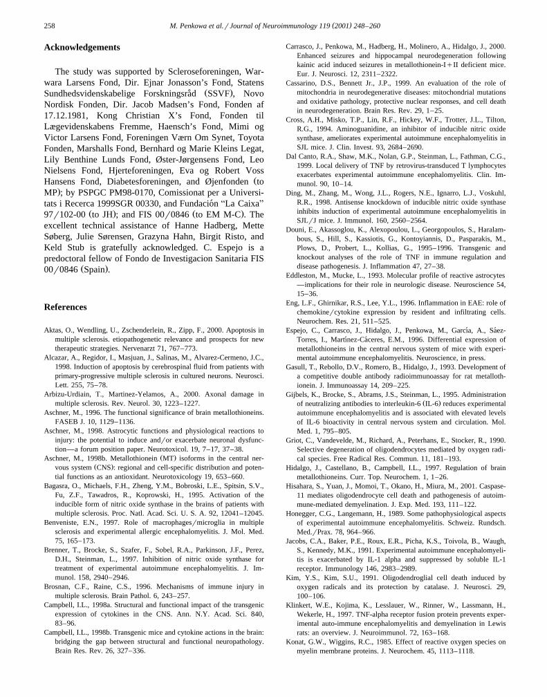
ŽBy using triple fluorescence for NSE a neuronal. Žmarker , vimentin a marker for macrophages and reactive
.astrocytes , and TUNEL, it was observed that mainlyŽNSEq neurons were undergoing apoptotic cell death Fig.
.5C,D . Only in MTKO mice, a few vimentinq astrocytesŽ .were TUNELq during EAE Fig. 5D . In normal mice
Žwith EAE, astrocytes were not labeled by TUNEL Fig.. Ž5C . Some other cells were in addition TUNELq as seen
( )M. Penkowa et al.rJournal of Neuroimmunology 119 2001 248–260256
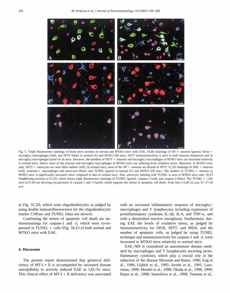
Ž . Ž .Fig. 5. Triple fluorescence stainings of brain stem sections of normal and MTKO mice with EAE. A,B Stainings of NFq neurons green , lectinqŽ . Ž . Ž . Ž . Ž .microgliarmacrophages red , and NITT blue in normal A and MTKO B mice. NITT immunoreactivity is seen in both neurons turquoise and inŽ .microgliarmacrophages pink of all mice. However, the numbers of NITTq neurons and microgliarmacrophages of MTKO mice are increased relatively
to normal mice. Hence, most of the neurons and microgliarmacrophages of MTKO mice are suffering from oxidative stress. Moreover, in MTKO miceŽ . Ž .only, NITTq astrocytes are seen blue stellate cells . In normal mice, most of the NFq neurons are devoid of NITT. C,D Stainings of NSEq neurons
Ž . Ž . Ž . Ž . Ž .red , vimentinq macrophages and astrocytes blue , and TUNEL green in normal C and MTKO D mice. The number of TUNELq neurons inŽ .MTKO mice is significantly increased when compared to that of normal mice. Also, astrocytic labeling with TUNEL is seen in MTKO mice only. E,F
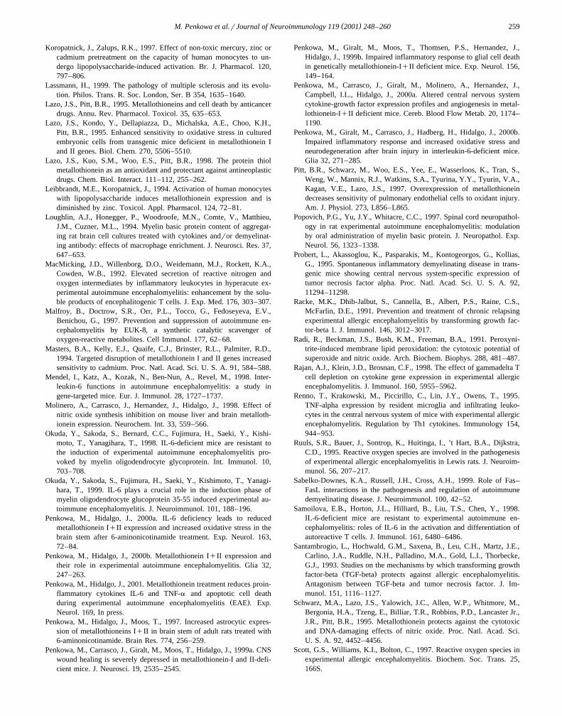
Ž . Ž . Ž . Ž .Neighboring sections to C,D , which shows triple fluorescence stainings of TUNEL green , caspase-1 red , and caspase-3 blue . The TUNELq cellsŽ . Ž . Ž . Ž .seen in C,D are showing coexpression of caspase-1 and -3 pink , which supports the notion of apoptotic cell death. Scale bars: A,B 22mm. C–F 18
mm.
.in Fig. 5C,D , which were oligodendrocytes as judged byusing double immunofluorescence for the oligodendrocytic
Ž .marker CNPase and TUNEL data not shown .Confirming the notion of apoptotic cell death are im-
munostainings for caspase-1 and -3, which were co-ex-Ž .pressed in TUNELq cells Fig. 5E,F of both normal and
MTKO mice with EAE.
4. Discussion
The present report demonstrated that genetical defi-ciency of MT-Iq II is accompanied by increased diseasesusceptibility to actively induced EAE in 129rSv mice.This clinical effect of MT-Iq II deficiency was associated
with an increased inflammatory response of microgliarmacrophages and T lymphocytes including expression ofproinflammatory cytokines IL-1b, IL-6, and TNF-a , andwith a diminished reactive astrogliosis. Furthermore, dur-ing EAE the levels of oxidative stress, as judged byimmunoreactivity for iNOS, NITT, and MDA, and thenumber of apoptotic cells, as judged by using TUNELtechnique and immunoreactivity for caspase-1 and -3, wereincreased in MTKO mice relatively to normal mice.
EAErMS is considered an autoimmune disease medi-ated by macrophages and T lymphocytes secreting proin-flammatory cytokines, which play a crucial role in the
Žinduction of the disease Brosnan and Raine, 1996; Eng etal., 1996; Gijbels et al., 1995; Jacobs et al., 1991; Lass-mann, 1999; Mendel et al., 1998; Okuda et al., 1998, 1999;Rajan et al., 1998; Samoilova et al., 1998; Tanuma et al.,
( )M. Penkowa et al.rJournal of Neuroimmunology 119 2001 248–260 257
.1999; Wiemann et al., 1998; Zhu et al., 1998 . In vivo,MT-I q II significantly decrease the CNS inflammatoryresponse of macrophages including expression of IL-1b,
ŽIL-6, and TNF-a Carrasco et al., 2000; Penkowa and.Hidalgo, 2000b, 2001; Penkowa et al., 1999a, 2000a .
Daily treatment of EAE with MT-II in Lewis rats signifi-cantly reduced activation and recruitment of macrophagesand T cells. This could explain in part the amelioration of
Žclinical signs observed in these animals Penkowa and.Hidalgo, 2000b . Interestingly, inhibitory effects of MT-I
q II on macrophages were also observed in vitro, in thatinduction of MT-II mRNA and total MT-Iq II protein inhuman monocytes was followed by suppression of mono-cyte activation including inhibition of the oxidative burst,adherence to plastic, and IL-1b levels following LPS
Žtreatment Koropatnick and Zalups, 1997; Leibbrandt and.Koropatnick, 1994 . Moreover, MT-Iq II may ameliorate
EAE by reducing IL-1, IL-6 and TNF-a production, inthat activation of these cytokines results in recruitment tothe CNS and activation in situ of large numbers of lym-
Žphocytes and macrophages Brosnan and Raine, 1996; Eng.et al., 1996; Woodroofe, 1995 . Accordingly, an inhibition
Žof these cytokines can suppress EAE Gijbels et al., 1995;Klinkert et al., 1997; Jacobs et al., 1991; Rajan et al.,
.1998; Wiemann et al., 1998 .Furthermore, MT-Iq II may have a regenerative effect
during EAE, in that MT-Iq II increase astrogliosis andexpression of growth factors such as transforming growth
Ž .factor-b TGF-b , which both may participate in the heal-Žing phase following CNS damage Aschner, 1998a; Ben-
veniste, 1997; Carrasco et al., 2000; Eddleston and Mucke,.1993; Eng et al., 1996; Penkowa et al., 1999a, 2000a . In
agreement with this, MT-induced amelioration of EAE inŽrats was associated with increased astrogliosis Penkowa
.and Hidalgo, 2000b , and it was reported that EAE can beŽprevented and treated by TGF-b Racke et al., 1991;
.Santambrogio et al., 1993 . Moreover, in MTKO micesubjected to a focal brain injury, reactive astrogliosis,growth factor production, and the following wound healing
Ž .were dramatically impaired Penkowa et al., 1999a, 2000a .Hence, accumulating data suggest that MT-Iq II can ame-liorate pathological conditions of the CNS, and this isassociated with downregulation of macrophage and T cellresponses including production of proinflammatory cy-tokines, as well as upregulation of astrogliosis and TGF-b
in the CNS.MT-I q II are also antioxidant factors protecting against
Ž .ROS reactive oxygen species and oxidative damageŽAschner, 1996, 1998b; Lazo et al., 1995, 1998; Molineroet al., 1998; Pitt et al., 1997; Schwarz et al., 1995; Tamaiet al., 1993; Tate et al., 1995; Thornalley and Vasak,
.1985, . ROS are involved in EAErMS pathogenesisŽBagasra et al., 1995; Benveniste, 1997; Cross et al., 1994;MacMicking et al., 1992; Ruuls et al., 1995; Scott et al.,
.1997; Van Dam et al., 1995 , and in vitro, ROS can easilyŽ .damage myelin Konat and Wiggins, 1985 and induce
Ždegeneration of oligodendrocytes Griot et al., 1990; Kim.and Kim, 1991 . In addition, ROS are key mediators of
Žapoptotic cell death Cassarino and Bennett, 1999; Sun and.Chen, 1998 . Apoptotic cell death of both oligodendro-
cytes and neurons is certainly implicated in EAErMSŽpathogenesis Aktas et al., 2000; Alcazar et al., 1998;
Arbizu-Urdiain and Martinez-Yelamos, 2000; Sabelko-Downes et al., 1999; Hisahara et al., 2001; Honegger andLangemann, 1989; MacMicking et al., 1992; Ruuls et al.,
.1995; Penkowa and Hidalgo, 2001 . In addition, MT-IItreatment during EAE in rats could significantly inhibitneuronal and oligodendrocyte apoptosis, while eliminationof autoreactive T cells by apoptosis was unaffectedŽ .Penkowa and Hidalgo, 2001 . The latter phenomenon islikely very important, since apoptosis during EAErMS hasa dual role: apoptosis is detrimental and pathological whenit occurs in neurons and oligodendrocytes, but on the otherhand, apoptosis is beneficial and a physiological controlmechanism inducing disease remission when it occurs in
Ž .inflammatory cells such as T cells Zipp, 2000 .Whether or not this antiapoptotic effect of MT-II treat-
ment is due to antioxidant functions of MT or yet un-known mechanisms of actions is currently not elucidated,but indeed, treatment of EAE-sensitized rats or mice withantioxidant factors or inhibitors of ROS can ameliorate
ŽEAE Brenner et al., 1997; Cross et al., 1994; Malfroy etal., 1997; Ruuls et al., 1995; Zhao et al., 1996; Zielasek et
.al., 1995 . Thus, antioxidant scavengers are needed duringEAE in order to neutralize ROS. MT-Iq II function asthiol donors, scavenge free radicals and reduce NO-in-
Žduced cyto- and nuclear toxicity Lazo et al., 1995, 1998;.Radi et al., 1991; Schwarz et al., 1995 . Moreover, NO
synthesis inhibition during immobilization stress decreasedŽ .MT-I q II Molinero et al., 2000a , which supports the role
for MT-I q II in NO detoxification. Hence, MT may sig-nificantly affect the levels of oxidative stress during EAEby neutralization of ROS. Accordingly, treatment withMT-II during EAE in rats leads to a significant decrease of
Ž .oxidative stress Penkowa and Hidalgo, 2000b and apop-Ž .totic cell death Penkowa and Hidalgo, 2001 . In agree-
ment with this, MTKO mice are also suffering fromseverely increased oxidative stress and neuronal apoptosis
Žin other models of brain injury Carrasco et al., 2000;.Penkowa et al., 1999a, 2000a . Indeed, in the presentŽ .report, oxidative stress markers iNOS, NITT, and MDA
and apoptotic cell death were significantly increased inMTKO mice relatively to normal mice during EAE. Theseeffects of MT during EAE could indeed have a therapeuti-cal potential, in that oxidative stress and apoptosis of botholigodendrocytes and neurons are implicated in EAErMS.
In summary, we have demonstrated here that MT areinvolved at different levels in the pathogenesis of EAE,playing a protective role. Our data suggest that MT-Iq II,acting alone or in concert with other anti-inflammatory orantioxidant mediators, may be potential candidates in thetreatment of CNS disorders such as EAErMS.
( )M. Penkowa et al.rJournal of Neuroimmunology 119 2001 248–260258
Acknowledgements
The study was supported by Scleroseforeningen, War-wara Larsens Fond, Dir. Ejnar Jonasson’s Fond, Statens
Ž .Sundhedsvidenskabelige Forskningsrad SSVF , Novo˚Nordisk Fonden, Dir. Jacob Madsen’s Fond, Fonden af17.12.1981, Kong Christian X’s Fond, Fonden tilLægevidenskabens Fremme, Haensch’s Fond, Mimi ogVictor Larsens Fond, Foreningen Værn Om Synet, ToyotaFonden, Marshalls Fond, Bernhard og Marie Kleins Legat,Lily Benthine Lunds Fond, Øster-Jørgensens Fond, LeoNielsens Fond, Hjerteforeningen, Eva og Robert Voss
ŽHansens Fond, Diabetesforeningen, and Øjenfonden to.MP ; by PSPGC PM98-0170, Comissionat per a Universi-
tats i Recerca 1999SGR 00330, and FundacionALa CaixaB´Ž . Ž .97r102-00 to JH ; and FIS 00r0846 to EM M-C . The
excellent technical assistance of Hanne Hadberg, MetteSøberg, Julie Sørensen, Grazyna Hahn, Birgit Risto, andKeld Stub is gratefully acknowledged. C. Espejo is apredoctoral fellow of Fondo de Investigacion Sanitaria FIS
Ž .00r0846 Spain .
References
Aktas, O., Wendling, U., Zschenderlein, R., Zipp, F., 2000. Apoptosis inmultiple sclerosis. etiopathogenetic relevance and prospects for newtherapeutic strategies. Nervenarzt 71, 767–773.
Alcazar, A., Regidor, I., Masjuan, J., Salinas, M., Alvarez-Cermeno, J.C.,1998. Induction of apoptosis by cerebrospinal fluid from patients withprimary-progressive multiple sclerosis in cultured neurons. Neurosci.Lett. 255, 75–78.
Arbizu-Urdiain, T., Martinez-Yelamos, A., 2000. Axonal damage inmultiple sclerosis. Rev. Neurol. 30, 1223–1227.
Aschner, M., 1996. The functional significance of brain metallothioneins.FASEB J. 10, 1129–1136.
Aschner, M., 1998. Astrocytic functions and physiological reactions toinjury: the potential to induce andror exacerbate neuronal dysfunc-tion—a forum position paper. Neurotoxicol. 19, 7–17, 37–38.
Ž .Aschner, M., 1998b. Metallothionein MT isoforms in the central ner-Ž .vous system CNS : regional and cell-specific distribution and poten-
tial functions as an antioxidant. Neurotoxicology 19, 653–660.Bagasra, O., Michaels, F.H., Zheng, Y.M., Bobroski, L.E., Spitsin, S.V.,
Fu, Z.F., Tawadros, R., Koprowski, H., 1995. Activation of theinducible form of nitric oxide synthase in the brains of patients withmultiple sclerosis. Proc. Natl. Acad. Sci. U. S. A. 92, 12041–12045.
Benveniste, E.N., 1997. Role of macrophagesrmicroglia in multiplesclerosis and experimental allergic encephalomyelitis. J. Mol. Med.75, 165–173.
Brenner, T., Brocke, S., Szafer, F., Sobel, R.A., Parkinson, J.F., Perez,D.H., Steinman, L., 1997. Inhibition of nitric oxide synthase fortreatment of experimental autoimmune encephalomyelitis. J. Im-munol. 158, 2940–2946.
Brosnan, C.F., Raine, C.S., 1996. Mechanisms of immune injury inmultiple sclerosis. Brain Pathol. 6, 243–257.
Campbell, I.L., 1998a. Structural and functional impact of the transgenicexpression of cytokines in the CNS. Ann. N.Y. Acad. Sci. 840,83–96.
Campbell, I.L., 1998b. Transgenic mice and cytokine actions in the brain:bridging the gap between structural and functional neuropathology.Brain Res. Rev. 26, 327–336.
Carrasco, J., Penkowa, M., Hadberg, H., Molinero, A., Hidalgo, J., 2000.Enhanced seizures and hippocampal neurodegeneration followingkainic acid induced seizures in metallothionein-IqII deficient mice.Eur. J. Neurosci. 12, 2311–2322.
Cassarino, D.S., Bennett Jr., J.P., 1999. An evaluation of the role ofmitochondria in neurodegenerative diseases: mitochondrial mutationsand oxidative pathology, protective nuclear responses, and cell deathin neurodegeneration. Brain Res. Rev. 29, 1–25.
Cross, A.H., Misko, T.P., Lin, R.F., Hickey, W.F., Trotter, J.L., Tilton,R.G., 1994. Aminoguanidine, an inhibitor of inducible nitric oxidesynthase, ameliorates experimental autoimmune encephalomyelitis inSJL mice. J. Clin. Invest. 93, 2684–2690.
Dal Canto, R.A., Shaw, M.K., Nolan, G.P., Steinman, L., Fathman, C.G.,1999. Local delivery of TNF by retrovirus-transduced T lymphocytesexacerbates experimental autoimmune encephalomyelitis. Clin. Im-munol. 90, 10–14.
Ding, M., Zhang, M., Wong, J.L., Rogers, N.E., Ignarro, L.J., Voskuhl,R.R., 1998. Antisense knockdown of inducible nitric oxide synthaseinhibits induction of experimental autoimmune encephalomyelitis inSJLrJ mice. J. Immunol. 160, 2560–2564.
Douni, E., Akassoglou, K., Alexopoulou, L., Georgopoulos, S., Haralam-bous, S., Hill, S., Kassiotis, G., Kontoyiannis, D., Pasparakis, M.,Plows, D., Probert, L., Kollias, G., 1995–1996. Transgenic andknockout analyses of the role of TNF in immune regulation anddisease pathogenesis. J. Inflammation 47, 27–38.
Eddleston, M., Mucke, L., 1993. Molecular profile of reactive astrocytes—implications for their role in neurologic disease. Neuroscience 54,15–36.
Eng, L.F., Ghirnikar, R.S., Lee, Y.L., 1996. Inflammation in EAE: role ofchemokinercytokine expression by resident and infiltrating cells.Neurochem. Res. 21, 511–525.
Espejo, C., Carrasco, J., Hidalgo, J., Penkowa, M., Garcıa, A., Saez-´ ´Torres, I., Martınez-Caceres, E.M., 1996. Differential expression of´ ´metallothioneins in the central nervous system of mice with experi-mental autoimmune encephalomyelitis. Neuroscience, in press.
Gasull, T., Rebollo, D.V., Romero, B., Hidalgo, J., 1993. Development ofa competitive double antibody radioimmunoassay for rat metalloth-ionein. J. Immunoassay 14, 209–225.
Gijbels, K., Brocke, S., Abrams, J.S., Steinman, L., 1995. AdministrationŽ .of neutralizing antibodies to interleukin-6 IL-6 reduces experimental
autoimmune encephalomyelitis and is associated with elevated levelsof IL-6 bioactivity in central nervous system and circulation. Mol.Med. 1, 795–805.
Griot, C., Vandevelde, M., Richard, A., Peterhans, E., Stocker, R., 1990.Selective degeneration of oligodendrocytes mediated by oxygen radi-cal species. Free Radical Res. Commun. 11, 181–193.
Hidalgo, J., Castellano, B., Campbell, I.L., 1997. Regulation of brainmetallothioneins. Curr. Top. Neurochem. 1, 1–26.
Hisahara, S., Yuan, J., Momoi, T., Okano, H., Miura, M., 2001. Caspase-11 mediates oligodendrocyte cell death and pathogenesis of autoim-mune-mediated demyelination. J. Exp. Med. 193, 111–122.
Honegger, C.G., Langemann, H., 1989. Some pathophysiological aspectsof experimental autoimmune encephalomyelitis. Schweiz. Rundsch.Med.rPrax. 78, 964–966.
Jacobs, C.A., Baker, P.E., Roux, E.R., Picha, K.S., Toivola, B., Waugh,S., Kennedy, M.K., 1991. Experimental autoimmune encephalomyeli-tis is exacerbated by IL-1 alpha and suppressed by soluble IL-1receptor. Immunology 146, 2983–2989.
Kim, Y.S., Kim, S.U., 1991. Oligodendroglial cell death induced byoxygen radicals and its protection by catalase. J. Neurosci. 29,100–106.
Klinkert, W.E., Kojima, K., Lesslauer, W., Rinner, W., Lassmann, H.,Wekerle, H., 1997. TNF-alpha receptor fusion protein prevents exper-imental auto-immune encephalomyelitis and demyelination in Lewisrats: an overview. J. Neuroimmunol. 72, 163–168.
Konat, G.W., Wiggins, R.C., 1985. Effect of reactive oxygen species onmyelin membrane proteins. J. Neurochem. 45, 1113–1118.
( )M. Penkowa et al.rJournal of Neuroimmunology 119 2001 248–260 259
Koropatnick, J., Zalups, R.K., 1997. Effect of non-toxic mercury, zinc orcadmium pretreatment on the capacity of human monocytes to un-dergo lipopolysaccharide-induced activation. Br. J. Pharmacol. 120,797–806.
Lassmann, H., 1999. The pathology of multiple sclerosis and its evolu-tion. Philos. Trans. R. Soc. London, Ser. B 354, 1635–1640.
Lazo, J.S., Pitt, B.R., 1995. Metallothioneins and cell death by anticancerdrugs. Annu. Rev. Pharmacol. Toxicol. 35, 635–653.
Lazo, J.S., Kondo, Y., Dellapiazza, D., Michalska, A.E., Choo, K.H.,Pitt, B.R., 1995. Enhanced sensitivity to oxidative stress in culturedembryonic cells from transgenic mice deficient in metallothionein Iand II genes. Biol. Chem. 270, 5506–5510.
Lazo, J.S., Kuo, S.M., Woo, E.S., Pitt, B.R., 1998. The protein thiolmetallothionein as an antioxidant and protectant against antineoplasticdrugs. Chem. Biol. Interact. 111–112, 255–262.
Leibbrandt, M.E., Koropatnick, J., 1994. Activation of human monocyteswith lipopolysaccharide induces metallothionein expression and isdiminished by zinc. Toxicol. Appl. Pharmacol. 124, 72–81.
Loughlin, A.J., Honegger, P., Woodroofe, M.N., Comte, V., Matthieu,J.M., Cuzner, M.L., 1994. Myelin basic protein content of aggregat-ing rat brain cell cultures treated with cytokines andror demyelinat-ing antibody: effects of macrophage enrichment. J. Neurosci. Res. 37,647–653.
MacMicking, J.D., Willenborg, D.O., Weidemann, M.J., Rockett, K.A.,Cowden, W.B., 1992. Elevated secretion of reactive nitrogen andoxygen intermediates by inflammatory leukocytes in hyperacute ex-perimental autoimmune encephalomyelitis: enhancement by the solu-ble products of encephalitogenic T cells. J. Exp. Med. 176, 303–307.
Malfroy, B., Doctrow, S.R., Orr, P.L., Tocco, G., Fedoseyeva, E.V.,Benichou, G., 1997. Prevention and suppression of autoimmune en-cephalomyelitis by EUK-8, a synthetic catalytic scavenger ofoxygen-reactive metabolites. Cell Immunol. 177, 62–68.
Masters, B.A., Kelly, E.J., Quaife, C.J., Brinster, R.L., Palmiter, R.D.,1994. Targeted disruption of metallothionein I and II genes increasedsensitivity to cadmium. Proc. Natl. Acad. Sci. U. S. A. 91, 584–588.
Mendel, I., Katz, A., Kozak, N., Ben-Nun, A., Revel, M., 1998. Inter-leukin-6 functions in autoimmune encephalomyelitis: a study ingene-targeted mice. Eur. J. Immunol. 28, 1727–1737.
Molinero, A., Carrasco, J., Hernandez, J., Hidalgo, J., 1998. Effect ofnitric oxide synthesis inhibition on mouse liver and brain metalloth-ionein expression. Neurochem. Int. 33, 559–566.
Okuda, Y., Sakoda, S., Bernard, C.C., Fujimura, H., Saeki, Y., Kishi-moto, T., Yanagihara, T., 1998. IL-6-deficient mice are resistant tothe induction of experimental autoimmune encephalomyelitis pro-voked by myelin oligodendrocyte glycoprotein. Int. Immunol. 10,703–708.
Okuda, Y., Sakoda, S., Fujimura, H., Saeki, Y., Kishimoto, T., Yanagi-hara, T., 1999. IL-6 plays a crucial role in the induction phase ofmyelin oligodendrocyte glucoprotein 35-55 induced experimental au-toimmune encephalomyelitis. J. Neuroimmunol. 101, 188–196.
Penkowa, M., Hidalgo, J., 2000a. IL-6 deficiency leads to reducedmetallothionein IqII expression and increased oxidative stress in thebrain stem after 6-aminonicotinamide treatment. Exp. Neurol. 163,72–84.
Penkowa, M., Hidalgo, J., 2000b. Metallothionein IqII expression andtheir role in experimental autoimmune encephalomyelitis. Glia 32,247–263.
Penkowa, M., Hidalgo, J., 2001. Metallothionein treatment reduces proin-flammatory cytokines IL-6 and TNF-a and apoptotic cell death
Ž .during experimental autoimmune encephalomyelitis EAE . Exp.Neurol. 169, In press.
Penkowa, M., Hidalgo, J., Moos, T., 1997. Increased astrocytic expres-sion of metallothioneins IqII in brain stem of adult rats treated with6-aminonicotinamide. Brain Res. 774, 256–259.
Penkowa, M., Carrasco, J., Giralt, M., Moos, T., Hidalgo, J., 1999a. CNSwound healing is severely depressed in metallothionein-I and II-defi-cient mice. J. Neurosci. 19, 2535–2545.
Penkowa, M., Giralt, M., Moos, T., Thomsen, P.S., Hernandez, J.,Hidalgo, J., 1999b. Impaired inflammatory response to glial cell deathin genetically metallothionein-IqII deficient mice. Exp. Neurol. 156,149–164.
Penkowa, M., Carrasco, J., Giralt, M., Molinero, A., Hernandez, J.,Campbell, I.L., Hidalgo, J., 2000a. Altered central nervous systemcytokine-growth factor expression profiles and angiogenesis in metal-lothionein-IqII deficient mice. Cereb. Blood Flow Metab. 20, 1174–1190.
Penkowa, M., Giralt, M., Carrasco, J., Hadberg, H., Hidalgo, J., 2000b.Impaired inflammatory response and increased oxidative stress andneurodegeneration after brain injury in interleukin-6-deficient mice.Glia 32, 271–285.
Pitt, B.R., Schwarz, M., Woo, E.S., Yee, E., Wasserloos, K., Tran, S.,Weng, W., Mannix, R.J., Watkins, S.A., Tyurina, Y.Y., Tyurin, V.A.,Kagan, V.E., Lazo, J.S., 1997. Overexpression of metallothioneindecreases sensitivity of pulmonary endothelial cells to oxidant injury.Am. J. Physiol. 273, L856–L865.
Popovich, P.G., Yu, J.Y., Whitacre, C.C., 1997. Spinal cord neuropathol-ogy in rat experimental autoimmune encephalomyelitis: modulationby oral administration of myelin basic protein. J. Neuropathol. Exp.Neurol. 56, 1323–1338.
Probert, L., Akassoglou, K., Pasparakis, M., Kontogeorgos, G., Kollias,G., 1995. Spontaneous inflammatory demyelinating disease in trans-genic mice showing central nervous system-specific expression oftumor necrosis factor alpha. Proc. Natl. Acad. Sci. U. S. A. 92,11294–11298.
Racke, M.K., Dhib-Jalbut, S., Cannella, B., Albert, P.S., Raine, C.S.,McFarlin, D.E., 1991. Prevention and treatment of chronic relapsingexperimental allergic encephalomyelitis by transforming growth fac-tor-beta 1. J. Immunol. 146, 3012–3017.
Radi, R., Beckman, J.S., Bush, K.M., Freeman, B.A., 1991. Peroxyni-trite-induced membrane lipid peroxidation: the cytotoxic potential ofsuperoxide and nitric oxide. Arch. Biochem. Biophys. 288, 481–487.
Rajan, A.J., Klein, J.D., Brosnan, C.F., 1998. The effect of gammadelta Tcell depletion on cytokine gene expression in experimental allergicencephalomyelitis. J. Immunol. 160, 5955–5962.
Renno, T., Krakowski, M., Piccirillo, C., Lin, J.Y., Owens, T., 1995.TNF-alpha expression by resident microglia and infiltrating leuko-cytes in the central nervous system of mice with experimental allergicencephalomyelitis. Regulation by Th1 cytokines. Immunology 154,944–953.
Ruuls, S.R., Bauer, J., Sontrop, K., Huitinga, I., ’t Hart, B.A., Dijkstra,C.D., 1995. Reactive oxygen species are involved in the pathogenesisof experimental allergic encephalomyelitis in Lewis rats. J. Neuroim-munol. 56, 207–217.
Sabelko-Downes, K.A., Russell, J.H., Cross, A.H., 1999. Role of Fas–FasL interactions in the pathogenesis and regulation of autoimmunedemyelinating disease. J. Neuroimmunol. 100, 42–52.
Samoilova, E.B., Horton, J.L., Hilliard, B., Liu, T.S., Chen, Y., 1998.IL-6-deficient mice are resistant to experimental autoimmune en-cephalomyelitis: roles of IL-6 in the activation and differentiation ofautoreactive T cells. J. Immunol. 161, 6480–6486.
Santambrogio, L., Hochwald, G.M., Saxena, B., Leu, C.H., Martz, J.E.,Carlino, J.A., Ruddle, N.H., Palladino, M.A., Gold, L.I., Thorbecke,G.J., 1993. Studies on the mechanisms by which transforming growth
Ž .factor-beta TGF-beta protects against allergic encephalomyelitis.Antagonism between TGF-beta and tumor necrosis factor. J. Im-munol. 151, 1116–1127.
Schwarz, M.A., Lazo, J.S., Yalowich, J.C., Allen, W.P., Whitmore, M.,Bergonia, H.A., Tzeng, E., Billiar, T.R., Robbins, P.D., Lancaster Jr.,J.R., Pitt, B.R., 1995. Metallothionein protects against the cytotoxicand DNA-damaging effects of nitric oxide. Proc. Natl. Acad. Sci.U. S. A. 92, 4452–4456.
Scott, G.S., Williams, K.I., Bolton, C., 1997. Reactive oxygen species inexperimental allergic encephalomyelitis. Biochem. Soc. Trans. 25,166S.
( )M. Penkowa et al.rJournal of Neuroimmunology 119 2001 248–260260
Sun, A.Y., Chen, Y.M., 1998. Oxidative stress and neurodegenerativedisorders. J. Biomed. Sci. 5, 401–414.
Swanborg, R.H., 1995. Experimental autoimmune encephalomyelitis inrodents as a model for human demyelinating disease. Clin. Immunol.Immunopathol. 77, 4–13.
Tamai, K.T., Gralla, E.B., Ellerby, L.M., Valentine, J.S., Thiele, D.J.,1993. Yeast and mammalian metallothioneins functionally substitutefor yeast copper–zinc superoxide dismutase. Proc. Natl. Acad. Sci.U. S. A. 90, 8013–8017.
Tanuma, N., Shin, T., Kogure, K., Matsumoto, Y., 1999. Differential roleof TNF-alpha and IFN-gamma in the brain of rats with chronicrelapsing autoimmune encephalomyelitis. J. Neuroimmunol. 96, 73–79.
Tate Jr., D.J., Miceli, M.V., Newsome, D.A., 1995. Phagocytosis andH O induce catalase and metallothionein gene expression in human2 2
retinal pigment epithelial cells. Invest. Ophthalmol. Visual Sci. 36,1271–1279.
Taupin, V., Renno, T., Bourbonniere, L., Peterson, A.C., Rodriguez, M.,Owens, T., 1997. Increased severity of experimental autoimmuneencephalomyelitis, chronic macrophagermicroglial reactivity, and de-myelination in transgenic mice producing tumor necrosis factor-alphain the central nervous system. Eur. J. Immunol. 27, 905–913.
Thornalley, P.J., Vasak, M., 1985. Possible role for metallothionein inprotection against radiation-induced oxidative stress. Kinetics andmechanism of its reaction with superoxide and hydroxyl radicals.Biochim. Biophys. Acta 827, 36–44.
Van Dam, A.M., Bauer, J., Man-A-Hing, W.K., Marquette, C., Tilders,
F.J., Berkenbosch, F., 1995. Appearance of inducible nitric oxidesynthase in the rat central nervous system after rabies virus infectionand during experimental allergic encephalomyelitis but not afterperipheral administration of endotoxin. Neurosci. Res. 40, 251–260.
Van Lookeren Campagne, M., Thibodeaux, H., Van Bruggen, N., Cairns,B., Gerlai, R., Palmer, J.T., Williams, S.P., Lowe, D.G., 1999.Evidence for a protective role of metallothionein-1 in focal cerebralischemia. Proc. Natl. Acad. Sci. U. S. A. 96, 12870–12875.
Wiemann, B., Van, G.Y., Danilenko, D.M., Yan, Q., Matheson, C.,Munyakazi, L., Ogenstad, S., Starnes, C.O., 1998. Combined treat-ment of acute EAE in Lewis rats with TNF-binding protein andinterleukin-1 receptor antagonist. Exp. Neurol. 149, 455–463.
Woodroofe, M.N., 1995. Cytokine production in the central nervoussystem. Neurology 45, S6–S10.
Zhao, W., Tilton, R.G., Corbett, J.A., McDaniel, M.L., Misko, T.P.,Williamson, J.R., Cross, A.H., Hickey, W.F., 1996. Experimentalallergic encephalomyelitis in the rat is inhibited by aminoguanidine,an inhibitor of nitric oxide synthase. J. Neuroimmunol. 64, 123–133.
Zhu, J., Diab, A., Mustafa, M., Levi, M., Wahren, B., Bjork, J., Hedlund,G., 1998. Linomide suppresses chronic-relapsing experimental au-toimmune encephalomyelitis in DA rats. J. Neurol. Sci. 160, 113–120.
Zielasek, J., Jung, S., Gold, R., Liew, F.Y., Toyka, K.V., Hartung, H.P.,1995. Administration of nitric oxide synthase inhibitors in experimen-tal autoimmune neuritis and experimental autoimmune en-cephalomyelitis. J. Neuroimmunol. 58, 81–88.
Zipp, F., 2000. Apoptosis in multiple sclerosis. Cell Tissue Res. 301,163–171.