
Stress influence in neurodegeneration - TDX (Tesis Doctorals ...
264
Stress influence in neurodegeneration Unravelling the mechanisms underlying stress response in brain ageing by 11ß-HSD1 inhibition Maria Dolors Puigoriol Illamola ADVERTIMENT. La consulta d’aquesta tesi queda condicionada a l’acceptació de les següents condicions d'ús: La difusió d’aquesta tesi per mitjà del servei TDX (www.tdx.cat) i a través del Dipòsit Digital de la UB (diposit.ub.edu) ha estat autoritzada pels titulars dels drets de propietat intel·lectual únicament per a usos privats emmarcats en activitats d’investigació i docència. No s’autoritza la seva reproducció amb finalitats de lucre ni la seva difusió i posada a disposició des d’un lloc aliè al servei TDX ni al Dipòsit Digital de la UB. No s’autoritza la presentació del seu contingut en una finestra o marc aliè a TDX o al Dipòsit Digital de la UB (framing). Aquesta reserva de drets afecta tant al resum de presentació de la tesi com als seus continguts. En la utilització o cita de parts de la tesi és obligat indicar el nom de la persona autora. ADVERTENCIA. La consulta de esta tesis queda condicionada a la aceptación de las siguientes condiciones de uso: La difusión de esta tesis por medio del servicio TDR (www.tdx.cat) y a través del Repositorio Digital de la UB (diposit.ub.edu) ha sido autorizada por los titulares de los derechos de propiedad intelectual únicamente para usos privados enmarcados en actividades de investigación y docencia. No se autoriza su reproducción con finalidades de lucro ni su difusión y puesta a disposición desde un sitio ajeno al servicio TDR o al Repositorio Digital de la UB. No se autoriza la presentación de su contenido en una ventana o marco ajeno a TDR o al Repositorio Digital de la UB (framing). Esta reserva de derechos afecta tanto al resumen de presentación de la tesis como a sus contenidos. En la utilización o cita de partes de la tesis es obligado indicar el nombre de la persona autora. WARNING. On having consulted this thesis you’re accepting the following use conditions: Spreading this thesis by the TDX (www.tdx.cat) service and by the UB Digital Repository (diposit.ub.edu) has been authorized by the titular of the intellectual property rights only for private uses placed in investigation and teaching activities. Reproduction with lucrative aims is not authorized nor its spreading and availability from a site foreign to the TDX service or to the UB Digital Repository. Introducing its content in a window or frame foreign to the TDX service or to the UB Digital Repository is not authorized (framing). Those rights affect to the presentation summary of the thesis as well as to its contents. In the using or citation of parts of the thesis it’s obliged to indicate the name of the author.
-
Upload
khangminh22 -
Category
Documents
-
view
3 -
download
0
Transcript of Stress influence in neurodegeneration - TDX (Tesis Doctorals ...
Stress influence in neurodegeneration Unravelling the mechanisms underlying stress response
in brain ageing by 11ß-HSD1 inhibition
Maria Dolors Puigoriol Illamola
ADVERTIMENT. La consulta d’aquesta tesi queda condicionada a l’acceptació de les següents condicions d'ús: La difusió d’aquesta tesi per mitjà del servei TDX (www.tdx.cat) i a través del Dipòsit Digital de la UB (diposit.ub.edu) ha estat autoritzada pels titulars dels drets de propietat intel·lectual únicament per a usos privats emmarcats en activitats d’investigació i docència. No s’autoritza la seva reproducció amb finalitats de lucre ni la seva difusió i posada a disposició des d’un lloc aliè al servei TDX ni al Dipòsit Digital de la UB. No s’autoritza la presentació del seu contingut en una finestra o marc aliè a TDX o al Dipòsit Digital de la UB (framing). Aquesta reserva de drets afecta tant al resum de presentació de la tesi com als seus continguts. En la utilització o cita de parts de la tesi és obligat indicar el nom de la persona autora. ADVERTENCIA. La consulta de esta tesis queda condicionada a la aceptación de las siguientes condiciones de uso: La difusión de esta tesis por medio del servicio TDR (www.tdx.cat) y a través del Repositorio Digital de la UB (diposit.ub.edu) ha sido autorizada por los titulares de los derechos de propiedad intelectual únicamente para usos privados enmarcados en actividades de investigación y docencia. No se autoriza su reproducción con finalidades de lucro ni su difusión y puesta a disposición desde un sitio ajeno al servicio TDR o al Repositorio Digital de la UB. No se autoriza la presentación de su contenido en una ventana o marco ajeno a TDR o al Repositorio Digital de la UB (framing). Esta reserva de derechos afecta tanto al resumen de presentación de la tesis como a sus contenidos. En la utilización o cita de partes de la tesis es obligado indicar el nombre de la persona autora. WARNING. On having consulted this thesis you’re accepting the following use conditions: Spreading this thesis by the TDX (www.tdx.cat) service and by the UB Digital Repository (diposit.ub.edu) has been authorized by the titular of the intellectual property rights only for private uses placed in investigation and teaching activities. Reproduction with lucrative aims is not authorized nor its spreading and availability from a site foreign to the TDX service or to the UB Digital Repository. Introducing its content in a window or frame foreign to the TDX service or to the UB Digital Repository is not authorized (framing). Those rights affect to the presentation summary of the thesis as well as to its contents. In the using or citation of parts of the thesis it’s obliged to indicate the name of the author.
University of Barcelona
Faculty of Pharmacy and Food Sciences
Department of Pharmacology, Toxicology and Medicinal
Chemistry
Stress influence in neurodegeneration
Unravelling the mechanisms underlying stress response
in brain ageing by 11β-HSD1 inhibition
Dolors Puigoriol Illamola
2020

2
3
Faculty of Pharmacy and Food Sciences
Department of Pharmacology, Toxicology and Medicinal
Chemistry
PhD program in Biotechnology
Stress influence in neurodegeneration
Unravelling the mechanisms underlying stress response in
brain ageing by 11β-HSD1 inhibition
Dissertation presented by Dolors Puigoriol Illamola to apply for
the doctorate degree by the University of Barcelona Director Director Doctoral student
Dra Mercè Pallàs Dr Christian Griñán Dolors Puigoriol
Dolors Puigoriol Illamola 2020

4
5
This doctoral thesis has been done in the Department of Pharmacology, Toxicology and Medicinal Chemistry from the Pharmacy and Food Sciences Faculty – University of Barcelona. It has been funded by:
Generalitat de Catalunya
Ministerio de Economía y Competitividad Gobierno de España
Ministerio de Educación, Cultura y Deporte
Gobierno de España
Fundació Universitària Agustí Pedro i Pons Universitat de Barcelona
Institut de Neurociències Universitat de Barcelona
And it has been done in collaboration with the following institution:
Queen’s Medical Research Institute The University of Edinburgh

6

VII
“Don't let anyone rob you of your imagination, your creativity, or your curiosity. It's your place in the world; it's your life.
Go on and do all you can with it, and make it the life you want to live.”
Mae Jemison
“Life is not easy for any of us. But what of that? We must have perseverance and above all confidence in ourselves.
We must believe that we are gifted for something and that this thing must be attained.” Marie Sklodowska Curie

VIII

IX
STATEMENT OF ORIGINALITY I declare that this PhD thesis is my own original work and includes material from three
articles that have been previously published in international peer-reviewed journals. One
other article is currently pending to submit.
Dolors Puigoriol, BSc
June 2020

X

XI
ABSTRACT
The world is facing an unprecedented situation: soon the number of people over 60
years old will exceed the number of children, and more people at extreme old age than
ever before. This is due to medical advances that have increased life expectancy and with it,
the number of elderly. The latest data published by the WHO predicts that by 2050 the
world’s population aged 60 years and older is expected to nearly double from 12% to 22%
achieving a total 2 billion, up from 900 million in 2015. A longer life brings with it
opportunities, however, there is little evidence to suggest that older people today are
experiencing their later years in better health than their parents. While rates of severe
disability have declined over the past 30 years, there has been no significant change in mild
to moderate disability over the same period. If this added years are dominated by declines
in physical and mental capacity, the implications for older people and for society will be
more negative. Therefore, in the last years research has focused on the biology of ageing
with the purpose of achieving better understanding of its mechanisms for preventing the
onset and progression of age-related conditions.
Besides, modern society is experiencing an increasingly common stressful lifestyle
together to increased rates of metabolic stress caused, in part, by high-fat diet
consumption. Several pieces of evidence state that environmental factors are essential in
determining the development of different diseases as well as compromising healthy ageing.
With upcoming age, the capability to fight against harmful stimuli decreases and the
organism becomes more vulnerable to infections and disease. In agreement, stressful
experiences have been identified as an important risk factor for cognitive impairment.
Therefore, it is important to study the molecular mechanisms underpinning the effects of
chronic stress on cognition and its relationship with ageing in order to unveil what
challenges we might have to cope with as a society in the not-so-far future.
In parallel with ageing, stress and neurodegenerative diseases, such as Alzheimer’s
disease (AD), there is impaired glucocorticoid (GC) signalling. Disturbances in the GC-
mediated stress response and individual’s adaptive abilities appear to increase the
vulnerability of elderly to age-related pathologies. In consequence, the present doctoral
dissertation has been focused on the study of the mechanisms involved in age-related
neurodegeneration modified by GC excess attenuation through the inhibition of the
enzyme 11β-HSD1 in an animal model of accelerated ageing, as well as, their response to
XII
chronic mild stress exposure. Last but not least, the present doctoral thesis has been
devoted to evaluate the GC-mediated stress response to chronic moderate stressful
situations and to metabolic stress underlying neurodegeneration and the potential role of
11β-HSD1 inhibition to restore those detrimental effects induced by stress.
In summary, results obtained pointed out a protective role of the 11β-HSD1 inhibitor
tested as improved cognitive and behavioural abilities of aged mice, as well as restored the
deleterious effects induced by stressful conditions applied. Additionally, some of the
molecular pathways related to ageing and neurodegeneration – particularly AD
neurodegeneration – were altered as a consequence of stress, but most of them were re-
established after 11β-HSD1 inhibition, such as proteostasis, oxidative stress,
neuroinflammation and epigenetics, among others. Overall, GC excess attenuation may
become a potential therapeutic strategy for age-related cognitive decline.
XIII
RESUM
El món s’està enfrontant una situació sense precedents: aviat el nombre de persones
majors de 60 anys superarà el nombre d’infants i, el nombre de persones d’edat avançada
extrema és més elevat que mai. Això es deu als avenços mèdics que han incrementat
l’esperança de vida i amb ella, el nombre de persones envellides. Les últimes dades
publicades per la OMS preveuen que al 2050 la població mundial amb 60 anys o més
gairebé es doblarà passant del 12% actual al 22%, estimant arribar a un total de 2 bilions, en
comparació dels 900 milions de persones descrites el 2015. Una vida més llarga comporta
oportunitats, encara que existeixen poques evidències que suggereixin que les persones
envellides actualment visquin els últim dies amb més salut que els seus avantpassats. Si bé
és cert que els índex de discapacitat severa han disminuït durant els darrers 30 anys, no hi
ha hagut canvis significatius en la discapacitat lleu o moderada durant el mateix període. Si
en aquests anys afegits domina el deteriorament de les capacitats físiques i mentals, les
implicacions per les persones grans i per la societat seran més negatives. Per tant, en els
últims anys la recerca s’ha centrat en la biologia de l’envelliment amb el propòsit
d’aconseguir comprendre millor els mecanismes que el determinen per tal de prevenir
l’aparició i la progressió de les condicions relacionades amb l’edat.
A més, a la societat moderna cada vegada és més comú un estil de vida estressant,
que ha incrementat juntament amb l’estrès metabòlic, causat, en part, pel consum de dieta
rica en grassa. Diferents evidències donen suport al fet que els factors ambientals són
essencials per determinar el desenvolupament de determinades malalties, comprometent
així l’envelliment saludable. A mesura que s’envelleix, la capacitat per lluitar contra estímuls
nocius disminueix i l’organisme esdevé més vulnerable a infeccions i malalties. D’acord amb
això, s’han identificat les experiències estressants com un factor de risc important pel
deteriorament cognitiu. Per tant, és important l’estudi dels mecanismes moleculars que
fonamenten els efectes de l’estrès crònic a nivell cognitiu i la seva relació amb l’envelliment,
amb l’objectiu de revelar quins reptes podríem afrontar com a societat en un futur no tant
llunyà.
En paral·lel amb l’envelliment, l’estrès i les malalties neurodegeneratives, com ara la
malaltia d’Alzheimer, es situa el deteriorament de la senyalització dels glucocorticoides
(GCs). Les alteracions en la resposta a l’estrès pròpia dels GCs i per tant, en les capacitats
adaptatives dels individus semblen augmentar la vulnerabilitat de la gent gran a patologies
XIV
relacionades amb l’edat. En conseqüència, la present tesi doctoral s’ha centrat en l’estudi
dels mecanismes implicats en la neurodegeneració relacionada amb l’edat modificats per
l’atenuació de l’excés de GCs mitjançant la inhibició de l’enzim 11β-HSD1 en un model animal
d’envelliment accelerat, així com la seva resposta a l’exposició a estrès crònic lleu. Per últim,
però no per això menys important, la present tesi doctoral s’ha dedicat a avaluar la resposta
a l’estrès regulada pels GCs, tant per l’exposició a estímuls estressants crònics com per
estrès metabòlic, que participen en el desenvolupament de la neurodegeneració i el
potencial paper de la inhibició de l’enzim 11β-HSD1 per restaurar els efectes perjudicials
induïts per l’estrès.
En resum, els resultats obtinguts assenyalen un paper protector de l’inhibidor 11β-
HSD1 estudiat, ja que millora les habilitats cognitives i conductuals en ratolins envellits, com
també evita els efectes nocius induïts per les condicions estressants utilitzades.
Addicionalment, algunes de les vies moleculars relacionades amb l’envelliment i la
neurodegeneració – en particular la neurodegeneració característica de la malaltia
d’Alzheimer – es van alterar a conseqüència de l’estrès, però la majoria d’elles es van tornar
a re-establir després de la inhibició de l’enzim 11β-HSD1, com ara la proteostàsia, l’estrès
oxidatiu, la neuroinflamació i els canvis epigenètics, entre d’altres. En general, l’atenuació
de l’excés de GCs és una estratègia terapèutica potencial per al tractament del
deteriorament cognitiu relacionat amb l’edat.
XV
RESUMEN
El mundo está enfrentándose a una situación sin precedentes: pronto el número de
personas mayores de 60 años superará el número de niños y, el número de personas de
edad avanzada extrema es el más elevado jamás registrado. Eso se debe a que los avances
médicos han incrementado la esperanza de vida y con ella, el número de personas
envejecidas. Los últimos datos publicados por la OMS prevén que al 2050 la población
mundial con 60 años o más prácticamente se doblará pasando del 12% actual al 22%,
estimándose alcanzar un total de 2 billones, en comparación con los 900 millones de
personas contabilizadas en 2015. Una vida más larga conlleva oportunidades, aunque
existen pocas evidencias que, actualmente, las personas envejecidas vivan los últimos días
con una salud mejor de la que lo hicieron sus antepasados. Si bien es cierto que los índices
de discapacidad severa han disminuido durante los últimos 30 años, no ha habido cambios
significativos en la discapacidad leve o moderada durante el mismo período. Si en estos
años añadidos predomina el declive de las capacidades físicas y mentales, las implicaciones
para les personas mayores y para la sociedad serán más negativas. Por tanto, en los últimos
años la investigación se ha centrado en la biología del envejecimiento con el propósito de
conseguir comprender mejor los mecanismos que lo determinan con la finalidad de prevenir
la aparición y la progresión de las condiciones relacionadas con la edad.
Además, en la sociedad moderna cada vez es más común un estilo de vida estresante,
que se ha incrementado juntamente con el estrés metabólico, causado en parte, por el
consumo de una dieta rica en grasas. Diferentes evidencias indican que los factores
ambientales son esenciales para determinar el desarrollo de determinadas enfermedades,
comprometiendo así el envejecimiento saludable. A medida que se envejece, la capacidad
para luchar contra estímulos nocivos disminuye y el organismo se hace más vulnerable a
infecciones y enfermedades. De hecho, se han identificado las experiencias estresantes
como un factor de riesgo importante para el deterioro cognitivo. Por lo tanto, es
importante el estudio de los mecanismos moleculares que fundamentan los efectos del
estrés crónico a nivel cognitivo y su relación con el envejecimiento, con el fin de revelar qué
retos podríamos afrontar como sociedad en un futuro no tan lejano.
En paralelo al envejecimiento, el estrés y las enfermedades neurodegeneratives,
como la enfermedad de Alzheimer, se encuentra el deterioro en la señalización de los
XVI
glucocorticoides (GCs). Los cambios en la respuesta al estrés propia de los GCs y por tanto,
en las capacidades adaptativas de los individuos parecen aumentar la vulnerabilidad de las
personas mayores a patologías relacionadas con la edad. En consecuencia, la presente tesis
doctoral se ha centrado en el estudio de los mecanismos implicados en la
neurodegeneración relacionada con la edad, modificados por la atenuación del exceso de
GCs mediante la inhibición de la enzima 11β-HSD1 en un modelo animal de envejecimiento
acelerado, así como su respuesta a la exposición a estrés crónico leve. Por último, pero no
por ello menos importante, la presente tesis doctoral se ha centrado a evaluar la respuesta
al estrés regulada por los GCs, tanto por la exposición a estímulos estresantes crónicos
como por estrés metabólico, que participan en el desarrollo de la neurodegeneración y el
potencial papel de la inhibición de la enzima 11β-HSD1 para restaurar los efectos
perjudiciales inducidos por el estrés.
En resumen, los resultados obtenidos señalan el papel protector del inhibidor 11β-
HSD1 estudiado, ya que mejora las habilidades cognitivas y conductuales en ratones
envejecidos, como también previene los efectos nocivos inducidos por las condiciones
estresantes utilizadas. Además, algunas de las vías moleculares relacionadas con el
envejecimiento y la neurodegeneración - particularmente la neurodegeneración ligada a la
enfermedad de Alzheimer - se alteraron a consecuencia del estrés, pero la mayoría de ellas
se volvieron a restablecer tras la inhibición de la enzima 11β-HSD1, como la proteostasis, el
estrés oxidativo, la neuroinflamación y los cambios epigenéticos, entre otros. En general, la
atenuación del exceso de GCs es una estrategia terapéutica potencial para el tratamiento
del deterioro cognitivo relacionado con la edad.
XVII
THESIS STRUCTURE
The present doctoral dissertation is presented in several sections as follows: firstly, a
general Introduction places the reader in the context of the thesis, providing a
comprehensive overview of the topic in which it is framed, in this case, stress, ageing and
neurodegeneration, and more specifically, the relationship between attenuating
glucocorticoids excess and the improvement of age-related cognitive decline. Afterwards,
the section Objectives collects the principal issues that were aimed to be addressed at the
beginning of each investigation. Following, Methods and Results section is divided into four
chapters defining the beneficial effects resulting from treatment with RL-118, a drug that
reduces glucocorticoid activity, both on cognition and molecular pathways in aged mice. In
addition, it is determined that the drug is able to improve cognition and related molecular
mechanisms, even in a situation of metabolic stress and chronic moderate stress in adult
mice, which mimic situations similar to ageing. Thereafter, a general Discussion compares
and contextualizes the results achieved with the evidence already described in the
literature, always trying to clarify the likely existing discrepancies. Finally, the thesis ends
with the Conclusions derived from the whole study and the Bibliography comprises all the
scientific literature consulted and cites during the thesis development, which, in addition,
helped to further discuss the results.
Four scientific papers are the result of the present dissertation, three of them already
published, and the another pending to submit:
Chapter 1
Puigoriol-Illamola, D., Griñán-Ferré, C., Vasilopoulou, F., Leiva, R., Vázquez, S., Pallàs, M.
(2018). 11β-HSD1 inhibition by RL-118 promotes autophagy and correlates with reduced
oxidative stress and inflammation, enhancing cognitive performance in SAMP8 mouse
model. Molecular Neurobiology, 55:8904-8915. doi:10.1007/s12035-018-1026-8.
Chapter 2
Puigoriol-Illamola, D., Martínez-Damas, M., Griñán-Ferré, C., Pallàs, M. (2020). Chronic Mild
Stress Modified Epigenetic Mechanisms Leading to Accelerated Senescence and Impaired
Cognitive Performance in Mice. International Journal of Molecular Sciences, 21(3):1154.
doi:10.3390/ijms21031154.

XVIII
Chapter 3
Puigoriol-Illamola, D., Companys-Alemany, J., Homer, N., Leiva, R., Vázquez, S., Mole, D.,
Griñán-Ferré, C., Pallàs, M. Chronic Mild Stress Modified Epigenetic Mechanisms Leading to
Accelerated Senescence and Impaired Cognitive Performance in Mice [pending to submit].
Chapter 4
Puigoriol-Illamola, D., Leiva, R., Vázquez-Carrera, M., Vázquez, S., Griñán-Ferré, C., Pallàs, M.
(2020). 11β-HSD1 Inhibition Rescues SAMP8 Cognitive Impairment Induced by Metabolic
Stress. Molecular Neurobiology, 57(1):551-565. doi:10.1007/s12035-019-01708-4. Epub 2019 Aug
09.
Importantly, the third arises from the author’s short-term stay at the Queen’s Medical
Research Institute – The University of Edinburgh (Edinburgh, United Kingdom) during the
last year of the thesis preparation period.
Additionally, seven extra scientific papers have been published during the time period of
the thesis as a result of the author’s participation in other projects carried out by members
of the same research group. Nevertheless, the content of these publications has not been
included in the core thesis since they are beyond its scope and/or the author contributed as
a collaborator instead of leading the investigation.
Palomera-Ávalos, V., Griñán-Ferré, C., Puigoriol-Illamola, D., Camins, A., Sanfeliu, C., Canudas, A.M., Pallàs, M. (2016). Resveratrol protects SAMP8 brain under metabolic stress: focus on mitochondrial function and Wnt pathway. Molecular Neurobiology, 54:1661-1676. doi:10.10007/s12035-016-9770-0. Griñán-Ferré, C., Sarroca, S., Ivanova, A., Puigoriol-Illamola, D., Aguado, F., Camins, A., Sanfeliu, C., Pallàs, M. (2016). Epigenetic mechanisms underlying cognitive impairment and Alzheimer’s disease hallmarks in 5XFAD mice. Aging, 8:664-684. doi:10.18632/aging.100906. Griñán-Ferré, C., Palomera-Ávalos, V., Puigoriol-Illamola, D., Camins, A., Porquet, D., Plá, V., Aguado, F., Pallàs, M. (2016). Behaviour and cognitive changes correlated with hippocampal neuroinflammaging and neuronal markers in female SAMP8, a model of accelerated senescence. Experimental Gerontology, 80:57-69. doi:10.1016/j.exger.2016.03.014. Griñán-Ferré, C., Puigoriol-Illamola, D., Pérez-Cáceres, D., Palomera-Ávalos, V., Rodrigo, M.T., Pallàs, M. (2016). Environmental enrichment modifies epigenetic mechanisms in SAMP8 reducing oxidative stress and inflammaging and achieving neuroprotection. Frontiers Aging Neuroscience, 8:241. doi:10.3389/fnagi.2016.00241.
XIX
Griñán-Ferré, C., Corpas, R., Puigoriol-Illamola, D., Palomera-Ávalos, V., Sanfeliu, C., Pallàs, M. (2018). Understanding epigenetics in the neurodegeneration of Alzheimer’s disease: SAMP8 mouse model. Journal Alzheimers Disease, 62:943-963. doi:10.3233/JAD-170664. Griñán-Ferré, C., Izquierdo, V., Otero, E., Puigoriol-Illamola, D., Corpas, R., Sanfeliu, C., Ortuño-Sahagún, D., Pallàs, M. (2018). Environmental enrichment improves cognitive deficits, AD Hallmarks and epigenetic alterations presented in 5xFAD mouse model. Frontiers Cell Neuroscience, 12:224. doi:10.3389/fncel.2018.00224. Pérez-Areales, F.J., Garrido, M., Aso, E., Bartolini, M., De Simone, A., Espargaro, A., Ginex, T., Sabaté, R., Pérez, B., Andrisano, V., Puigoriol-Illamola, D., Pallàs , M., Luque, F.J., Ferrer, I., Ciruela, F., Messeguer, A., Muñoz-Torrero, D. (2020). Centrally Active Multitarget Anti- Alzheimer Agents Derived from the Antioxidant Lead CR-6. Journal of Medicinal Chemistry [submitted].
The scientific outputs of the thesis and the extra contributions were shared with the
scientific community in different national and international congresses as poster
presentations, as well as oral presentation in a research meeting:
Poster presentations
Griñán-Ferré, C., Palomera-Ávalos, V., Puigoriol- Illamola, D., Alvarez-López, M.J., Cosín- Tomás, M., Camins, A., Kaliman, P., Pallàs, M. Could microRNA modulation overcome senescence process in SAMP8 strain? V Chromatin and epigenetics annual meeting SCB. 10 March 2015. Barcelona (Spain). Griñán-Ferré, C., Palomera-Ávalos, V., Puigoriol-Illamola, D., Camins, A., Pallàs, M. Time course screening of miRNAs in female SAMP8: implications in senescence process. EMBO Conference of Chromatin and Epigenetics. 6-10 May 2015. Heidelberg (Germany). Palomera-Ávalos, V., Griñán-Ferré, C., Camins, A., Amaro-Umbert, N., Puigoriol-Illamola, D., Sanfeliu, C., Canudas, A.M., Pallàs, M. Reframing the role of resveratrol in neurodegeneration: oxidative stress, mitocondrial function and Wnt-pathway modulation in the brain of metabolically stressed SAMP8 mice. Society for Neuroscience 45thanual meeting. 17-21 October 2015. Chicago (USA). Griñán-Ferré, C., Palomera-Ávalos, V., Puigoriol-Illamola, D., Camins, A., Ortuño-Sahagún, D., Pallàs, M. Epigenetic changes mediated by miRNAs as a cause of rapidly aging and cognitive impairments in female SAMP8 mouse model. Society for Neuroscience 45th annual meeting. 17-21 October 2015. Chicago (USA). Pallàs, M., Puigoriol-Illamola, D., Palomera-Ávalos, V., Camins, A., Griñán-Ferré, C. Cognition and behaviour impairment: is oxidative stress the earliest change commanding senescence? Lessons from senescence accelerated P8 mice. Society for Neuroscience 45th anual meeting.
XX
17-21 October 2015. Chicago (USA). Griñán-Ferré, C., Puigoriol-Illamola, D., Pérez-Cáceres, D., Palomera-Ávalos, V., Rodrigo, M.T., Pallàs, M. Environmental enrichment modifies epigenetic mechanisms in SAMP8 reducing oxidative stress and inflammaging and achieving neuroprotection. VI Chromatin and Epigenetics annual meeting SCB. 8 April 2016. Barcelona (Spain). As well as in the 10th FENS Forum of Neuroscience. 2-6 July 2016. Copenhagen (Denmark). Puigoriol-Illamola, D., Griñán-Ferré, C., Vasilopoulou, F., Leiva, R., Webster, S.P., Vázquez, S., Pallàs M. Neuroprotective effect of 11β-HSD1 inhibition through autophagy activation in SAMP8 mouse model. X Neurobiology Symposium of the SCB. 6-7 October 2016. Barcelona (Spain). As well as in the I PhD Workshop of the Institute of Neuroscience of the UB. 15 December 2016. Barcelona (Spain). Puigoriol-Illamola, D., Griñán-Ferré, C., Companys-Alemany, J., Leiva, R., Otero, E., Vázquez, S., Pallàs, M. 11β-HSD1 inhibition improves cognitive decline modifying epigenetic marks in SAMP8 mice under chronic mild stress exposure. 17th meeting of the Spanish Society for Neuroscience. 27-30 September 2017. Alicante (Spain). As well as in the II PhD Workshop of the Institute of Neuroscience of the UB. 30 November – 1 December 2017. Barcelona (Spain). Puigoriol-Illamola, D., Griñán-Ferré, C., Companys-Alemany, J., Leiva, R., Vázquez, S., Vázquez, M., Pallàs, M. 11β-HSD1 inhibition improved cognitive decline associated with highfat diet in female SAMP8. 11th FENS Forum of Neuroscience. 7-11 July 2018. Berlin (Germany). As well as in the III PhD Workshop of the Institute of Neuroscience of the UB. 16-17 October 2018. Barcelona (Spain). Puigoriol-Illamola, D., Griñán-Ferré, C., Companys-Alemany, J., Leiva, R., Vázquez, S., Vázquez, M., Pallàs, M. Beneficial effects of 11β-HSD1 inhibition on cognitive performance in metabolic stressed SAMP8 female. XI Neurobiology Symposium of the SCB. 12-13 November 2018. Barcelona (Spain). Puigoriol-Illamola, D., Griñán-Ferré, C., Pallàs, M. Epigenetic changes after unpredictable chronic mild stress in female SAMR1 and SAMP8: Effects on behaviour. 49th annual meeting of the Society for Neuroscience. 19-23 October 2019. Chicago (USA). As well as in the IV PhD Workshop of the Institute of Neurosciences of the UB. 29 November 2019. Barcelona (Spain).
Oral communication
Puigoriol-Illamola, D. Beneficial effects of 11β-HSD1 inhibition on cognitive performance in a mouse model of Alzheimer’s disease. Doctoral Journey in Pharmacology of the Universitat Autònoma de Barcelona (UAB) and Universitat de Barcelona (UB). Organized by the Academy of Medical and Health Sciences of Catalonia and the Balearic Islands, Catalan Society of Pharmacology. 19 June 2018. Barcelona (Spain).

XXI
INDEX
ABBREVIATIONS..............................................................................................XXIII
1. INTRODUCTION..................................................................................................1
1.1 Stress......................................................................................................3
1.1.1 History ..........................................................................................................4
1.1.2 Stress response............................................................................................5
1.1.3 Glucocorticoids............................................................................................9
1.1.4 11β-hydroxysteroid dehydrogenase………..............................................11
1.1.5 Stress effects on the development of several disorders.........................12
1.1.6 Stress effects on cognition and cognitive................................................13
1.1.7 11β-HSD1 inhibition .....................................................................................15
1.2 Ageing..................................................................................................17
1.2.1 Theories of ageing.....................................................................................22
1.2.2 Ageing and neurodegeneration ...............................................................24
1.3 Epigenetics and neurodegeneration.................................................24
1.3.1 DNA-methylation and hydroxymethylation.............................................25
1.3.2 Post-translational histone modifications.................................................26
1.3.3 non-coding RNAs......................................................................................27
1.4 Proteostasis and neurodegeneration...............................................28
1.5 Oxidative stress and neurodegeneration..........................................31
1.6 Neuroinflammation and neurodegeneration...................................33
1.7 Metabolic stress as an accelerator of cognitive decline..................35
1.8 Alzheimer’s disease............................................................................36
1.8.1 History.......................................................................................................37
1.8.2 Risk factors...............................................................................................37
1.8.3 Neuropathology.......................................................................................38
1.8.3.1 Macroscopic features...............................................................38
1.8.3.2 Microscopic features................................................................38
1.8.4 Classification.............................................................................................41
1.8.5 Hypothesis................................................................................................42
1.8.6 Treatment.................................................................................................43
1.9 Ageing animal models – Senescence accelerated mouse...............44
XXII
1.9.1 APP processing and Aβ accumulation......................................................45
1.9.2 Tau hyperphosphorylation.......................................................................46
1.9.3 Neuroinflammation..................................................................................46
1.9.4 Oxidative stress........................................................................................46
1.9.5 Glucose metabolism.................................................................................46
2. Objectives........................................................................................................49
3. Methods and Results.......................................................................................55
3.1 Chapter 1..............................................................................................57
11β-HSD1 Inhibition by RL-118 Promotes Autophagy and Correlates with Reduced Oxidative Stress and Inflammation, Enhancing Cognitive Performance in SAMP8 Mouse Model
3.2 Chapter 2.............................................................................................81
Chronic Mild Stress Modified Epigenetic Mechanisms Leading to Accelerated Senescence and Impaired Cognitive Performance in Mice
3.3 Chapter 3............................................................................................117
RL-118 and 11β-HSD1 target engagement through TAPS assay: behaviour and molecular
analysis
3.4 Chapter 4...........................................................................................153
11β-HSD1 Inhibition Rescues SAMP8 Cognitive Impairment Induced by Metabolic Stress
4. Discussion......................................................................................................183
4. 1 11β-HSD1 inhibition...........................................................................186
4.2 Neurodegeneration..........................................................................187
4.3 Epigenetics........................................................................................188
4.4 Proteostasis.......................................................................................191
4.5 Nutrient sensing ...............................................................................193
4.6 Oxidative Stress ...............................................................................194
4.7 Neuroinflammation..........................................................................195
4.8 Cognitive and behavioural changes................................................197
4.9 Final considerations........................................................................200
5. Conclusions...................................................................................................203
6. Bibliography..................................................................................................207
XXIII
ABBREVIATIONS
5-mC 5-methylcytosine
5-hmC 5-hydroxymethylcytosine
11β-HSD1 11beta-hydroxysteroid dehydrogenase type 1
11β-HSD2 11beta-hydroxysteroid dehydrogenase type 2
Aβ Amyloid beta
ACTH Adrenocorticotropic hormone
AD Alzheimer’s disease
ADAM10 A disintegrin and metalloproteinase domain-containing protein 10
ADE Aβ-degrading enzymes
Aldh2 Aldehyde dehydrogenase 2
AMP Adenosine monophosphate
AMPK AMP-activated protein kinase
AOX1 Aldehyde oxidase 1
ApoE Apolipoprotein E
APP Amyloid precursor protein
ARE Antioxidant response element
ATF4 Activating transcription factor 4
ATP Adenosine triphosphate
AUC Area under the curve
AVP Arginine vasopressin
BACE1 Beta-site APP cleaving enzyme 1
BBB Blood-brain barrier
BCL2 B-cell lymphoma 2
BDNF Brain-derived neurotrophic factor
BIP/GRP78 Binding immunoglobulin protein
CA1 Cornu Ammonis
CAT Catalase
CCL3 C-C-motif ligand 3
CDK5 Cell division protein kinase 5
CHOP C/EBP homologous protein
CMS Chronic mild stress
CNS Central nervous system
CREB cAMP response element-binding
XXIV
CRH Corticotropin-releasing hormone
CTF Carboxy-terminal fragment
CXCL2 Chemokine (C-X-C motif) ligand 2
DI Discrimination index
DNA Deoxyribonucleic acid
DNMT DNA methyltransferase
eIF2-α Eukaryotic initiation factor 2 alpha
EPM Elevated plus maze
ER Endoplasmic reticulum
FACS Fluorescence-activated cell sorting
fAD Familial Alzheimer’s disease
FGF-21 Fibroblast growth factor 21
FOXO Forkhead box O
GAS General adaptation syndrome
GC Glucocorticoid
GFAP Glial fibrillar acidic protein
GPX Glutathione peroxidase
GR Glucocorticoid receptor
GSH Glutathione
GSK3 Glycogen-synthase kinase 3
H2O2 Hydrogen peroxide
HAT Histone acetyltransferase
HDAC Histone deacetylase
HDM Histone demethylase
HEK Human embryonic kidney
HFD High-fat diet
HLM Human liver microsomes
Hmox1 Heme oxygenase 1
HMT Histone methyltransferase
HPA Hypothalamic-pituitary-adrenal
IBA1 Ionized calcium-binding adapter molecule 1
IDE Insulin-degrading enzyme
IGF-1 Insulin-like growth factor 1
IIS Insulin and insulin-like growth factor 1 signalling
IL Interleukin

XXV
iNOS Inducible nitric oxide synthase
i.p. Intraperitoneally
JNK c-Jun N-terminal protein kinase
LC3 Microtubule-associated protein light chain 3
LDL Low-density lipoprotein
Leu Leucine
LKB1 Liver kinase B1
Lys Lysine
MAPK Mitogen-activated protein kinase
miRNA MicroRNA
MnSOD Manganese SOD
MPO Myeloperoxidase
MR Mineralocorticoid receptor
MS Mass spectrometry
mTOR Mammalian target of rapamycin
mTORC1 Mammalian target of rapamycin complex 1
MWM Morris water maze
NAD Nicotinamide adenine dinucleotide
NADPH Nicotinamide adenine dinucleotide phosphate
NF-κB Nuclear factor kappa-light chain enhancer of activated B cells
NFT Neurofibrillary tangles
NMDA N-methyl-D-aspartate
NO Nitric oxide
NORT Novel object recognition test
NOS Nitric oxide synthase
NRF2 Nuclear factor erythroid-derived 2
NSAID Non-steroid anti-inflammatory drug
O2 Oxygen
O2- Superoxide
OFT Open field test
OH- Hydroxyl ion
OLT Object location test
ONO2- Nitric peroxide
OS Oxidative stress
PERK Serine/threonine kinase RNA-like ER kinase

XXVI
PGC1-α Peroxisome proliferator-activated receptor gamma coactivator alpha
PSD95 Postsynaptic density protein 95
PSEN Presenilin
PTM Post-translational modifications
PVN Paraventricular nucleus
RNA Ribonucleic acid
RONS Reactive oxygen and nitrogen species
ROS Reactive oxygen species
sAD Sporadic Alzheimer’s disease
SAM Senescence-accelerated mice
SAMP Senescence-accelerated mice prone
SAMR Senescence-accelerated mice resistant
sAPPα Soluble APP alpha
Ser Serine
SIRT Sirtuins
SOD Superoxide dismutase
SNAP Synaptosomal-nerve associated protein 25
SNP Single-nucleotide polymorphism
SNS Sympathetic nervous system
T2DM Type 2 diabetes mellitus
TAPS Toxicity-affinity-permeability-selectivity
TCT Three-chamber test
TET Ten-eleven translocation
TGF-β Transforming growth factor beta
Thr Threonine
TNF-α Tumour necrosis factor alpha
TREM2 Triggering receptor expressed on myeloid cells 2
UPR Unfolded protein response
WHO World Health Organization

1. INTRODUCTION

2
3
1.1 STRESS
Considering the changes in our society in the last few years, the life rhythm has
changed becoming more and more intense and demanding. Apart from all the benefits and
advantages that technological advances have supposed to us, they have contributed to
increasing our need for immediacy and, in consequence, our feeling of stress. In fact, the
World Health Organization (WHO, 2010) has named stress as the “Health Epidemic of the
21st Century”, affecting more than 40 million individuals across the European Union. Thus in
such scenario, it is essential to study stress and the mechanisms underlying stress response.
So, what is stress?
Stress is defined as a physical or emotional factor that causes great bodily or mental
tension, anxiety, discomfort and difficulty in adjustment. The stress source (i.e. the
stressor) can be psychogenic (psychologically-based disturbance) and systemic (including
external – from the environment or social situations – and internal – illness or
inflammation) (Fink, 2017). However, an issue to consider in terms of stressors is the
subjectivity, as usually a stressful event is not perceived in the same way among different
individuals. Therefore, stress and stress response consist of a complex interplay between
physiological, psychological and behavioural processes that varies across situations (Thiel &
Dretsch, 2011). Nevertheless, other authors introduce a three-component definition,
accounting that stress requires heightened excitability, aversive experience perception and
lack of control. The last component is the variable that ultimately determines the
magnitude of the stress experience and the susceptibility of the individual to develop
stress-induced behavioural and physiological sequelae (Fink, 2017).
To appreciate the function of stress in a given situation, it is important to consider
the stressor, the magnitude (high or mild/moderate stress) and the duration of the stressful
events (acute or chronic stress). Chronic stress is characterized by prolonged exposure to a
given stress condition, broadly defined as stresses lasting from four to six hours and it is
supposed to cause harmful effects (Eisenmann et al., 2016; Reineke & Neilson, 2019).
Causes of chronic stress can differ from a wide range of issues, such as job pressure, poor
working conditions, financial difficulties, health, unfulfilling or conflicting intimate
relationships, nutrition, media overload or sleep deprivation, among others (Hammen et al.,
2009).
4
1.1.1 History
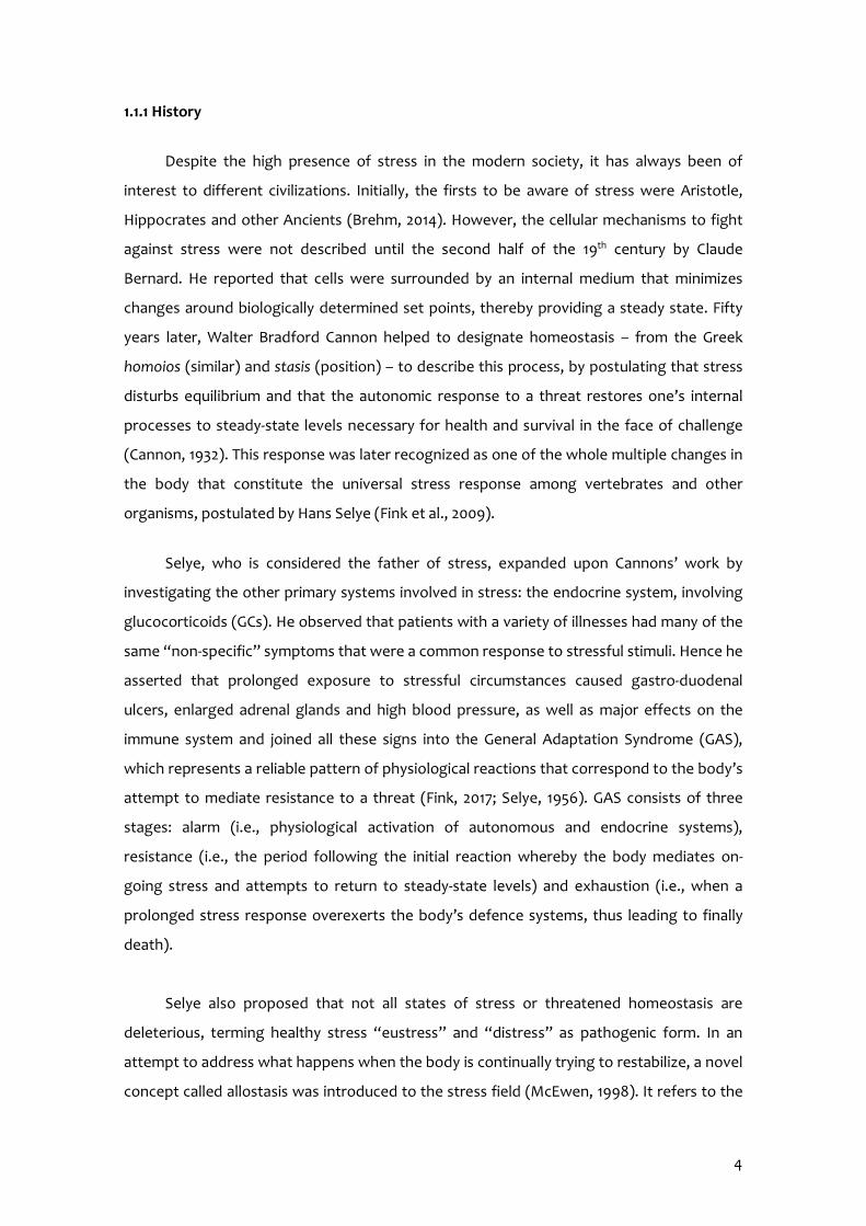
Despite the high presence of stress in the modern society, it has always been of
interest to different civilizations. Initially, the firsts to be aware of stress were Aristotle,
Hippocrates and other Ancients (Brehm, 2014). However, the cellular mechanisms to fight
against stress were not described until the second half of the 19th century by Claude
Bernard. He reported that cells were surrounded by an internal medium that minimizes
changes around biologically determined set points, thereby providing a steady state. Fifty
years later, Walter Bradford Cannon helped to designate homeostasis – from the Greek
homoios (similar) and stasis (position) – to describe this process, by postulating that stress
disturbs equilibrium and that the autonomic response to a threat restores one’s internal
processes to steady-state levels necessary for health and survival in the face of challenge
(Cannon, 1932). This response was later recognized as one of the whole multiple changes in
the body that constitute the universal stress response among vertebrates and other
organisms, postulated by Hans Selye (Fink et al., 2009).
Selye, who is considered the father of stress, expanded upon Cannons’ work by
investigating the other primary systems involved in stress: the endocrine system, involving
glucocorticoids (GCs). He observed that patients with a variety of illnesses had many of the
same “non-specific” symptoms that were a common response to stressful stimuli. Hence he
asserted that prolonged exposure to stressful circumstances caused gastro-duodenal
ulcers, enlarged adrenal glands and high blood pressure, as well as major effects on the
immune system and joined all these signs into the General Adaptation Syndrome (GAS),
which represents a reliable pattern of physiological reactions that correspond to the body’s
attempt to mediate resistance to a threat (Fink, 2017; Selye, 1956). GAS consists of three
stages: alarm (i.e., physiological activation of autonomous and endocrine systems),
resistance (i.e., the period following the initial reaction whereby the body mediates on-
going stress and attempts to return to steady-state levels) and exhaustion (i.e., when a
prolonged stress response overexerts the body’s defence systems, thus leading to finally
death).
Selye also proposed that not all states of stress or threatened homeostasis are
deleterious, terming healthy stress “eustress” and “distress” as pathogenic form. In an
attempt to address what happens when the body is continually trying to restabilize, a novel
concept called allostasis was introduced to the stress field (McEwen, 1998). It refers to the
5
ability of the body to achieve and maintain stability through change and includes active
physiological and behavioural responses to a specific threat with the aim of re-establishing
homeostasis.
To this end, allostasis is useful for illustrating the importance of adaptation to
promote and maintain survival mechanisms. However, the cost that these responses exert
over time is named allostatic load, which can result from either too much stress output or
inefficient stress response (McEwen, 2000). The chronic impact of stress is named allostatic
overload, and could manifest as metabolic syndrome, anxiety and neuropsychological
disorders, among others (Weger & Sandi, 2018).
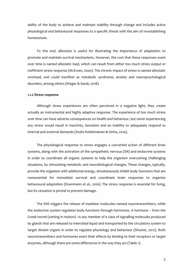
1.1.2 Stress response
Although stress experiences are often perceived in a negative light, they create
actually an instrumental and highly adaptive response. The experience of too much stress
over time can have adverse consequences on health and behaviour, but never experiencing
any stress would result in inactivity, boredom and an inability to adequately respond to
internal and external demands (Stults-Kolehmainen & Sinha, 2014).
The physiological response to stress engages a concerted action of different brain
systems, along with the activation of the sympathetic nervous (SN) and endocrine systems
in order to coordinate all organic systems to help the organism overcoming challenging
situations, by stimulating metabolic and neurobiological changes. These changes, typically,
provide the organism with additional energy, simultaneously inhibit body functions that are
nonessential for immediate survival and coordinate brain responses to organize
behavioural adaptation (Eisenmann et al., 2016). The stress response is essential for living,
but its cessation is pivotal to prevent damage.
The SNS triggers the release of mediator molecules named neurotransmitters, while
the endocrine system regulates body functions through hormones. A hormone – from the
Greek hormé (setting in motion) –is any member of a class of signalling molecules produced
by glands that are released to interstitial liquid and transported by the circulatory system to
target distant organs in order to regulate physiology and behaviour (Shuster, 2012). Both
neurotransmitters and hormones exert their effects by binding to their receptors or target
enzymes, although there are some differences in the way they act (Table 1).

6
Table 1. Comparison of the control exerted by nervous and endocrine systems.
Characteristics Nervous system Endocrine system Mediating molecules Neurotransmitters released locally
in response to nervous impulses Hormones distributed to the whole body through bloodstream
Targets of mediator Close from the release place; binding to a postsynaptic membrane receptor
Far from the release place (usually); binding to the inner or outer cellular receptor
Types of cellular targets Muscular cells, glandular cells and neurons
Whole body
Time to start the action Milliseconds (ms) From seconds (s) to hours (h) or days
Duration of the action Short (ms) Long (from s to days)
Activation of the SNS results in the local release of catecholamines (i.e.,
noradrenaline) onto target organs, and stimulates additional catecholamine (i.e., both
adrenaline and noradrenaline) release from the adrenal medulla (Thiel & Dretsch, 2011). This
constitutes the first response to acute stress, which is generally known as the fight-or-flight
response. The collective result of catecholamine release is a cascade of physiological effects
including increased respiration and heart rate, dilation of skeletal muscle blood vessels,
glycogen to glucose conversion and vasoconstriction of digestive and reproductive organ
blood vessels (Figure 1) (McCarty, 2000).
On the other hand, the hallmark of neuroendocrine system response to stress
involves activation of the hypothalamic – pituitary – adrenal (HPA) axis, which administer
Pupils dilation
Salivation decreases
Heart rateincreases
Respirationincreases
Digestiveprocessesdecrease
Glucose release
Bladderrelaxes
Colon activityinhibition
Reproductiveorgans inhibition
Figure 1. Representation of the SNS stress response with several of its targets.

7
hormone secretion. As far as the synthesis is concerned, three types of mechanisms
regulate hormone secretion: other hormones, chemical changes in the bloodstream and
nervous system signalling. HPA axis encompasses a complex set of direct influences and
feedback interactions among the hypothalamus, the hypophysis and the adrenal glands.
Firstly, painful experiences, stressful events and emotional situations cause changes
in hypothalamic activity and activate the secretion of different hormones in the
paraventricular nucleus (PVN), such as corticotropin-releasing hormone (CRH) and arginine
vasopressin (AVP) and release them into the portal blood system. The hypothalamus is a
small brain region under thalamus that comprises the connection between nervous and
endocrine systems. It receives afferent signals from the limbic system, cerebral cortex,
thalamus and reticular activator system, as well as sensorial signals from internal organs
and the retina. Besides, this cerebral unit regulates body temperature, food and thirst
sensations, sexual conduct, fear, anger and the autonomous nervous system. Overall the
hypothalamus is both, an essential nervous system regulatory centre as well as a crucial
endocrine gland.
Downstream the portal blood system, the pituitary gland or hypophysis – from the
Greek hypophysis (growing from below) – responds to hypothalamic releasing or inhibitory
hormones that stimulate or inhibit pituitary activity, respectively. CRH and AVP, although
predominantly CRH, specifically target the synthesis and release of adrenocorticotropic
hormone (ACTH) from pituitary corticotrophs cells located within the anterior pituitary
gland to the circulating bloodstream. Importantly, AVP synergistically potentiates CRH-
elicited ACTH secretion, but it is typically relegated to promote maintenance of basal ACTH
production (Thiel & Dretsch, 2011). Indeed under chronic stress conditions, there is a
marked shift in hypothalamic signal in favour of AVP as CRH receptors in the anterior
pituitary gland become down-regulated (Scott & Dinan, 1998). The pituitary gland rests
upon the hypophysial fossa of the sphenoid bone in the centre of the middle cranial fossa
and surrounded by a small bone cavity, named sella turcica (Figure 2). It is connected to the
hypothalamus by the pituitary stalk or infundibulum and has two anatomically and
functionally separate lobes:
The anterior lobe: also called the adenohypophysis. It constitutes about 75% of the
total weight of the gland and it secretes hormones. The anterior lobe consists of
two parts in the adult:
o Pars distalis: representing the most substantial portion
8
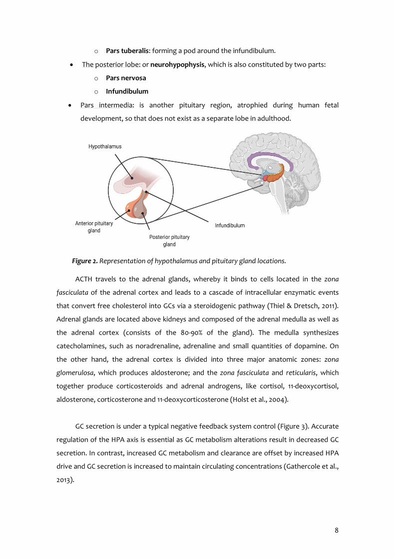
o Pars tuberalis: forming a pod around the infundibulum.
The posterior lobe: or neurohypophysis, which is also constituted by two parts:
o Pars nervosa
o Infundibulum
Pars intermedia: is another pituitary region, atrophied during human fetal
development, so that does not exist as a separate lobe in adulthood.
ACTH travels to the adrenal glands, whereby it binds to cells located in the zona
fasciculata of the adrenal cortex and leads to a cascade of intracellular enzymatic events
that convert free cholesterol into GCs via a steroidogenic pathway (Thiel & Dretsch, 2011).
Adrenal glands are located above kidneys and composed of the adrenal medulla as well as
the adrenal cortex (consists of the 80-90% of the gland). The medulla synthesizes
catecholamines, such as noradrenaline, adrenaline and small quantities of dopamine. On
the other hand, the adrenal cortex is divided into three major anatomic zones: zona
glomerulosa, which produces aldosterone; and the zona fasciculata and reticularis, which
together produce corticosteroids and adrenal androgens, like cortisol, 11-deoxycortisol,
aldosterone, corticosterone and 11-deoxycorticosterone (Holst et al., 2004).
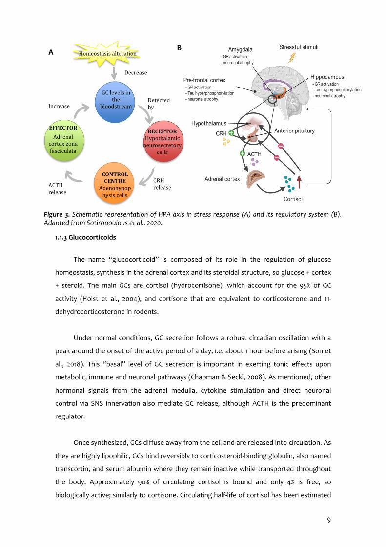
GC secretion is under a typical negative feedback system control (Figure 3). Accurate
regulation of the HPA axis is essential as GC metabolism alterations result in decreased GC
secretion. In contrast, increased GC metabolism and clearance are offset by increased HPA
drive and GC secretion is increased to maintain circulating concentrations (Gathercole et al.,
2013).
Figure 2. Representation of hypothalamus and pituitary gland locations.
9
1.1.3 Glucocorticoids
The name “glucocorticoid” is composed of its role in the regulation of glucose
homeostasis, synthesis in the adrenal cortex and its steroidal structure, so glucose + cortex
+ steroid. The main GCs are cortisol (hydrocortisone), which account for the 95% of GC
activity (Holst et al., 2004), and cortisone that are equivalent to corticosterone and 11-
dehydrocorticosterone in rodents.
Under normal conditions, GC secretion follows a robust circadian oscillation with a
peak around the onset of the active period of a day, i.e. about 1 hour before arising (Son et
al., 2018). This “basal” level of GC secretion is important in exerting tonic effects upon
metabolic, immune and neuronal pathways (Chapman & Seckl, 2008). As mentioned, other
hormonal signals from the adrenal medulla, cytokine stimulation and direct neuronal
control via SNS innervation also mediate GC release, although ACTH is the predominant
regulator.
Once synthesized, GCs diffuse away from the cell and are released into circulation. As
they are highly lipophilic, GCs bind reversibly to corticosteroid-binding globulin, also named
transcortin, and serum albumin where they remain inactive while transported throughout
the body. Approximately 90% of circulating cortisol is bound and only 4% is free, so
biologically active; similarly to cortisone. Circulating half-life of cortisol has been estimated
Decrease
Stressful stimuli
Hippocampus - GR activation - Tau hyperphosphorylation - neuronal atrophy
Amygdala - GR activation - neuronal atrophy
Pre-frontal cortex - GR activation - Tau hyperphosphorylation - neuronal atrophy
Hypothalamus
CRH Anterior pituitary
Adrenal cortex
ACTH
Cortisol
B Homeostasis alteration
GC levels in the
bloodstream
RECEPTOR Hypothalamic
neurosecretory cells
EFFECTOR
Adrenal cortex zona fasciculata
cells
CONTROL CENTRE
Adenohypophysis cells
ACTH release
CRH release
Detected by
Increase
A
Figure 3. Schematic representation of HPA axis in stress response (A) and its regulatory system (B).Adapted from Sotiropoulous et al., 2020.

10
to vary between 70 and 120 minutes and it is cleared through several pathways (Giannini &
Mohn, 2015).
GCs serve essential functions related to stress response. In one respect, GCs play a
permissive role as stimulate gluconeogenesis (especially in those organs that require high-
energy demand like central nervous system (CNS)), increase protein degradation and
lipolysis for energy production. Also, aid the catabolic processes mediated by
catecholamines, prime neural regions involved in sensory processing, attention and
adaptive responding, as well as regulate immune and inflammatory response mediators
accounting for a protective role via immunosuppressive and anti-inflammatory effects
(Sapolsky et al., 2000; Thiel & Dretsch, 2011; Tortora & Derrickson, 2010). In addition, GCs
affect critical brain regions such as the hippocampus and amygdala to regulate mood and
activate learning and memory processes that promote adaptive behaviours in the future in
response to a particular stressor (Chapman & Seckl, 2008; Korte et al., 2001). In conclusion,
GCs are vital to facilitate stress response and reduce it once the response is no longer
necessary, as well as preparing the organism for future threats.
Its activity occurs through two types of receptors located in the brain, primarily the
hippocampus: mineralocorticoid and glucocorticoid receptors (MR and GR, respectively)
(Reul & De Kloet, 1985). A differential affinity profile characterises each receptor for its
endogenous ligands. Despite the terminology, MRs possess heightened and relatively equal
affinity for both GCs and aldosterone. In contrast, GRs have a much lower affinity for GCs
compared to MRs, but they are more selective for GCs over aldosterone. Accordingly, it has
been demonstrated that corticosterone shows higher affinity for MR (Kd=0.1-0.5 nM) than
GR (Kd=2-5 nM) (Mifsud & Reul, 2016). Therefore, it has been estimated that more than 80%
of MRs are occupied with the endogenous GC under resting conditions, whereas GRs
become substantially occupied during elevated GC levels periods, such as after stressful
stimuli and at the circadian peak of GC secretion. These data suggests that MRs exert tonic
actions on brain, while GRs mediate the negative feedback, and long-term cognitive
changes evoked by GCs (Mifsud & Reul, 2016). GRs are located within the cell cytoplasm
and are translocated into the nucleus upon binding with a GC that enters the cell via passive
diffusion. Once in the nucleus, they function as transcription factors to regulate gene
expression. However, GCs are able to produce faster actions whereby they rapidly
hyperpolarize and inhibit neuron firing within regions such as the hippocampus and
hypothalamus (Gathercole et al., 2013).
11
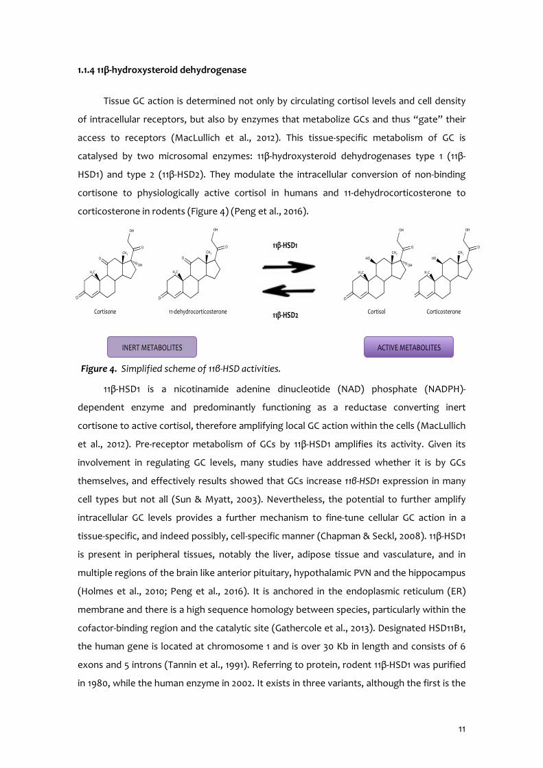
1.1.4 11β-hydroxysteroid dehydrogenase
Tissue GC action is determined not only by circulating cortisol levels and cell density
of intracellular receptors, but also by enzymes that metabolize GCs and thus “gate” their
access to receptors (MacLullich et al., 2012). This tissue-specific metabolism of GC is
catalysed by two microsomal enzymes: 11β-hydroxysteroid dehydrogenases type 1 (11β-
HSD1) and type 2 (11β-HSD2). They modulate the intracellular conversion of non-binding
cortisone to physiologically active cortisol in humans and 11-dehydrocorticosterone to
corticosterone in rodents (Figure 4) (Peng et al., 2016).
11β-HSD1 is a nicotinamide adenine dinucleotide (NAD) phosphate (NADPH)-
dependent enzyme and predominantly functioning as a reductase converting inert
cortisone to active cortisol, therefore amplifying local GC action within the cells (MacLullich
et al., 2012). Pre-receptor metabolism of GCs by 11β-HSD1 amplifies its activity. Given its
involvement in regulating GC levels, many studies have addressed whether it is by GCs
themselves, and effectively results showed that GCs increase 11β-HSD1 expression in many
cell types but not all (Sun & Myatt, 2003). Nevertheless, the potential to further amplify
intracellular GC levels provides a further mechanism to fine-tune cellular GC action in a
tissue-specific, and indeed possibly, cell-specific manner (Chapman & Seckl, 2008). 11β-HSD1
is present in peripheral tissues, notably the liver, adipose tissue and vasculature, and in
multiple regions of the brain like anterior pituitary, hypothalamic PVN and the hippocampus
(Holmes et al., 2010; Peng et al., 2016). It is anchored in the endoplasmic reticulum (ER)
membrane and there is a high sequence homology between species, particularly within the
cofactor-binding region and the catalytic site (Gathercole et al., 2013). Designated HSD11B1,
the human gene is located at chromosome 1 and is over 30 Kb in length and consists of 6
exons and 5 introns (Tannin et al., 1991). Referring to protein, rodent 11β-HSD1 was purified
in 1980, while the human enzyme in 2002. It exists in three variants, although the first is the
Figure 4. Simplified scheme of 11β-HSD activities.
Cortisone Cortisol Corticosterone 11-dehydrocorticosterone
INERT METABOLITES ACTIVE METABOLITES
11β-HSD1
11β-HSD2
12
predominant. It consists of 292 amino acids, with a molecular weight of 32401 Da and its
quaternary structure is a homodimer (NCBI GenBank nucleotide, 2020).
In contrast, 11β-HSD2 is a NAD-dependent dehydrogenase that catalyses the
conversion of cortisol into inactive metabolites in the kidney, thus reducing local GC action.
It is distributed mainly in the kidney, colon, placenta and discrete areas of the brain involved
in salt regulation (Peng et al., 2016; MacLullich et al., 2012). Notably in the kidney and colon,
11β-HSD2 activity protect MRs from inappropriate activation by cortisol and allow
aldosterone to act as the ligand, whereas in the placenta protects the fetus from maternal
GCs (Chapman & Seckl, 2008; Gathercole et al., 2013). The brain is nearly devoid of 11β-HSD2
(Thiel & Dretsch, 2011).
1.1.5 Stress effects on the development of several disorders
Different studies have provided evidence that the HPA axis can show abnormalities
during ageing and disease (Bloss et al., 2011). Despite the fact that GCs play a pivotal role in
orchestrating adaptive responses to stressful challenges to maintain health and wellbeing,
aberrant GC secretion as a result of chronic stress damages the whole body, negatively
affects cognition and increases the susceptibility to mental diseases, such as major
depression, anxiety and posttraumatic stress disorder (Mifsud & Reul, 2016). In particular,
prolonged exposure to heightened GCs levels has been associated with the deployment of
immunosuppression; osteoporosis and osteonecrosis; increment in fat tissue and
redistribution to the abdomen, shoulders and face; skin thinning with dermal atrophy
leading to bruising, purpura and livid striae; the appearance of acne; hirsutism; irregular
menstruation and reproductive failure (Sato et al., 2018; Sotiropoulous et al., 2008). Not
only that but also stress precipitates physical health outcomes leading to muscle atrophy,
proximal myopathy, diabetes, obesity, metabolic disturbances, hypertension and
cardiovascular disease (Eisenmann et al., 2016; Harman & Martín, 2019; You et al., 2020).
Altogether, signs and symptoms converged into Cushing’s syndrome. In the last decade,
the role of 11β-HSD1 enzyme in the causation of metabolic alterations of GCs overexposure
has been extensively documented an all converge in that GC-induced insulin resistance was
dependent on 11β-HSD1 (Peng et al.,2016; Qi et al., 2005). Accordingly, lower 11β-HSD1 levels
in the brain correlate with higher body mass index (BMI), which is indicative of obesity (Bini
et al., 2020).

13
Also, numerous mechanistic studies in animal models have determined that low birth
weight was associated with high fasting cortisol levels, both in rodents and humans.
Interestingly, birth weight is negatively correlated with high circulating levels of maternal
cortisol during pregnancy, supporting that GC excess can by-pass the placental barrier and
affect fetal development (Reynolds, 2013).
1.1.6 Stress effects on cognition and cognitive decline
Cognition is a broad concept involving a variety of processes that deal with
information and manipulate representations in the brain to produce a suitable response.
They range from perception and attention to various types of memory, language and
executive control processes (Sandi, 2013).
Nowadays, there is great consensus in the literature that stress is a potent modulator
of cognitive function, and more precisely, of learning and memory processes (Sandi &
Pinelo-Nava, 2007). Although the cognitive effects of stress are frequently assumed to be
detrimental, there are many instances in which cognitive functions are not impaired by
stress, or on the contrary, are even improved (Shields et al., 2016). Considering stress
intensity effects on memory, it is believed to follow an inverted-U-shape, meaning that low
and high stress levels impair memory, whereas intermediate stress facilitates them. Several
studies, including hippocampal-dependent tasks have successfully substantiated the
inverted-U-shape relationship between hormonal levels and both learning and synaptic
plasticity (Sandi, 2013). In other words, implicit memory (non-conscious learning) follows a
linear relation but explicit memory (conscious memory) an inverted-U-shape (Figure 5). In
addition, chronic stress effects on memory are overall consistent with those observed
under high-acute stress conditions (Luethi et al., 2009).
In conclusion, mild stress tends to facilitate cognitive function, particularly in implicit
memory or simple declarative tasks, or when the cognitive load is not excessive. However,
0
50
100
150
200
Low Medium High Extreme
Explicit memory
Intrinsic stress
Extrinsic stress
0
50
100
150
Low Medium High Extreme
Mem
ory
stre
nght
Implicit memory
Figure 5. Scheme showing the effects of intrinsic and extrinsic stresses in different memory types depending on the intensity of the stressful event.
14
high or very-high stress exposure, rather acutely or chronically, is associated with
impairment of explicit memories formation and, more generally, of those that require
complex and flexible reasoning (as typically observed for hippocampal-related functions),
while improving the performance of implicit memory (Sandi, 2013).
Nevertheless, it is important to consider that individual differences in the cognitive
impact of stress exist. In fact, considering how an individual responds to threats, we could
differ between “vulnerable” and “resilient” individuals (Weger & Sandi, 2018). Vulnerability
to stress is generally accepted to result from both genetic factors and exposure to
environmental adversity, in particular, adversity that occurs during early life. In addition,
these traits seem to be propagated across generations via epigenetic mechanisms,
highlighting the overall complexity of the underlying factors involved in stress response
(Weger & Sandi, 2018).
Of note, GCs play an important role in motivational behaviours and regulate mood
and cognition may be due to GC-induced changes in brain structure and function, involving
neuronal loss, deleterious neurotransmission, dendritic atrophy, electrophysiological
activity impairment and altered neuronal cellular signalling (Sandi, 2013; Sooy et al., 2015;
Sotiropoulous et al., 2008). Within the brain, the hippocampus is particularly sensitive to
maladaptive responses, and the damaging effects of chronic GC excess on neuronal
structure and function become more marked with ageing (Chapman & Seckl, 2008). How
stress and GCs may contribute to hippocampal ageing was described with the GC cascade
hypothesis (Sapolsky et al., 1986) based on that GCs secreted during periods of stress
desensitize the hippocampus to further GC exposure, by down-regulating GRs. However,
the GC hypersecretion continues and at some point, hippocampal cell loss occurs. The loss
of neurons is irreversible, and this permanent hippocampal damage was proposed to make
the hippocampus forever insensitive to further GC elevations, creating a feed-forward cycle
of elevated GCs and hippocampal destruction as ageing. However, this hypothesis has some
inconsistencies and has been modified to GC vulnerability hypothesis, which states that
cumulative stress causes heightened GC levels and may make the hippocampus vulnerable
to disruption (Conrad et al., 2011).
Chronic stress in midlife exerts persisting effects leading to cognitive and affective
dysfunctions in old age via mechanisms that depend, at least in part, on brain GCs
generated locally by 11β-HSD1 (Wheelan et al., 2018). Thus, 11β-HSD1 may be an important
15
factor in the regulation of the HPA axis and may itself be relevant to age-related diseases
susceptibility, severity or outcome (Carter et al., 2009). Evidence to date has suggested that
sustained stress exposure mimics the effects of age on multiple neurobiological measures
of neuronal structure and function (Bloss et al., 2011). In line with this, it has been described
that elderly who exhibit learning and memory impairments showed high GC levels and
correlated with greater hippocampal atrophy (Lara et al., 2013). In fact, cortisol has been
postulated as a potential biomarker for neurodegenerative disorders (Dhama et al., 2019).
Notwithstanding, it has been suggested a causal role of stress in the onset and
progression of age-related cognitive decline and neurodegenerative disorders like
Alzheimer’s disease (AD), as sustained GC overexposure has been found to increase
amyloid β (Aβ) formation and hyperphosphorylated Tau accumulation, hallmarks of human
AD; and adversely affect behaviour (Bini et al., 2020; Bisht et al., 2018; Green et al., 2006;
MacLullich et al., 2012). Accordingly, clinical evidence has shown that elevated cortisol may
predict a faster progression of AD as cortisol levels were inversely correlated with cognitive
performance and hippocampal volume (Bloss et al., 2011). Furthermore, a rare single-
nucleotide polymorphism (SNP) in the 11β-HSD1 gene has been reported to increase the risk
of developing sporadic AD (de Quervain et al., 2004), supporting that local tissue levels of
GCs may be a significant risk factor for AD.
Overall, these data provide compelling evidence that chronic stressors modulate
ageing and suggest that enhancing a healthy lifestyle might result in resilience to ageing
and age-related pathologies. On the other hand, the maintenance of GC sensitivity during
ageing might be neuroprotective, as rodent and human studies have shown that successful
ageing is associated with neuroendocrine responses similar to younger subjects (Bloss et
al., 2011; Yau et al., 2015).
1.1.7 11β-HSD1 inhibition
As mentioned, 11β-HSD1 participates in the development of different features related
to metabolic and cognitive disturbances. On the one hand, its overexpression in rodents
exerts increased corticosterone levels and displays a phenotype mimicking human
metabolic syndrome, which is prevented by its inhibition, proposing that intracellular
metabolism of GCs by 11β-HSD1 is critical to the development of insulin resistance rather
than the circulating GCs (Bujalska et al., 2006; Peng et al., 2016; Schnackenberg et al., 2013).
16
On the other hand, available literature demonstrates that this enzyme is implied in
age-related cognitive decline. For instance, a recent clinic study published a positive
correlation between increased brain 11β-HSD1 expression with advancing age (Bini et al.,
2020). Not only in humans, but also in rodents 11β-HSD1 expression was increased with
ageing and its overexpression accelerated age-related cognitive decline, while 11β-HSD1-
knockout mice resisted age-dependent cognitive loss (Caughey et al., 2017; Mohler et al.,
2011; Yau et al., 2015). Consistent with this, aged 11β-HSD1-knockout mice performed better
than aged wild-type mice in hippocampal-dependent behavioural tests, similarly to young
wild-type mice, despite elevated plasma GCs, thus agreeing with that tissue-specific control
may be more important than systemic levels (Yau et al., 2007).
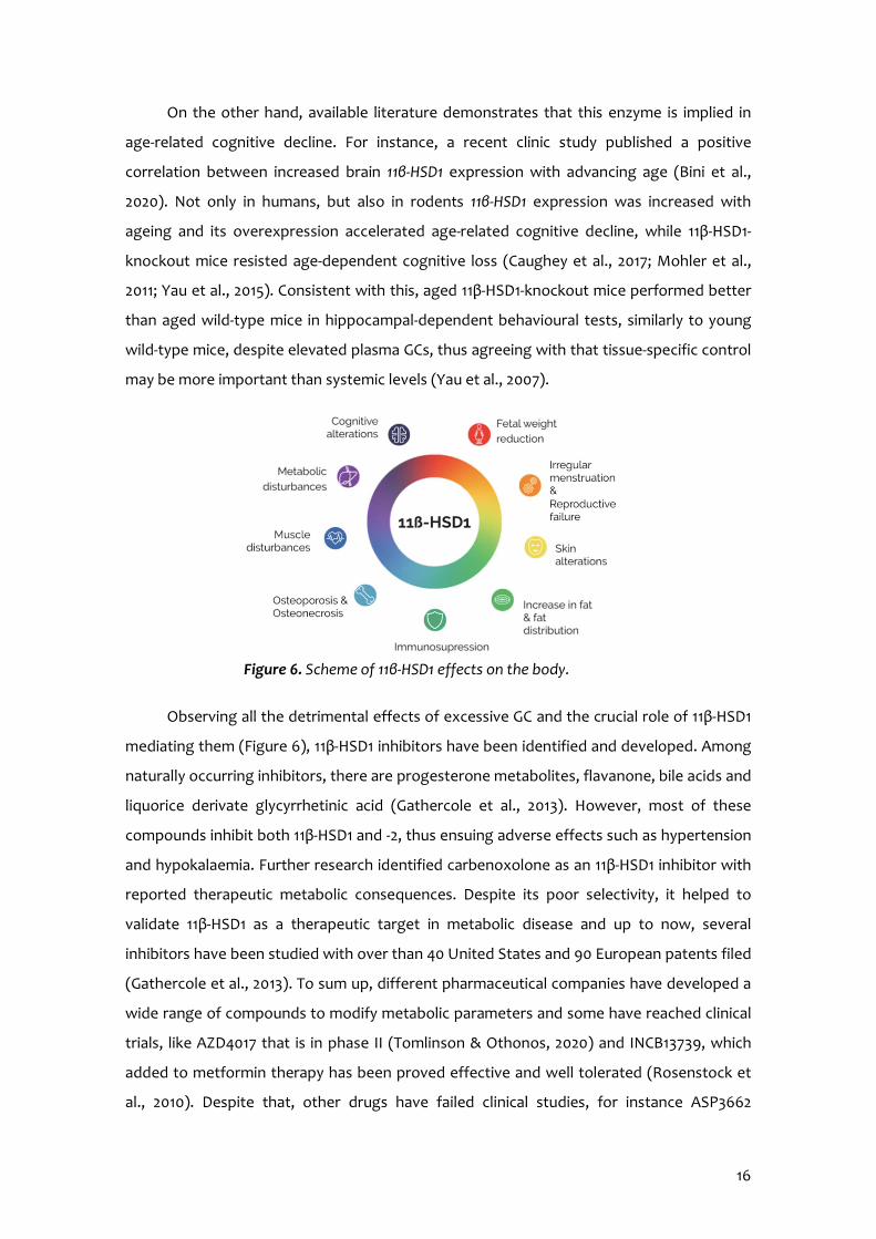
Observing all the detrimental effects of excessive GC and the crucial role of 11β-HSD1
mediating them (Figure 6), 11β-HSD1 inhibitors have been identified and developed. Among
naturally occurring inhibitors, there are progesterone metabolites, flavanone, bile acids and
liquorice derivate glycyrrhetinic acid (Gathercole et al., 2013). However, most of these
compounds inhibit both 11β-HSD1 and -2, thus ensuing adverse effects such as hypertension
and hypokalaemia. Further research identified carbenoxolone as an 11β-HSD1 inhibitor with
reported therapeutic metabolic consequences. Despite its poor selectivity, it helped to
validate 11β-HSD1 as a therapeutic target in metabolic disease and up to now, several
inhibitors have been studied with over than 40 United States and 90 European patents filed
(Gathercole et al., 2013). To sum up, different pharmaceutical companies have developed a
wide range of compounds to modify metabolic parameters and some have reached clinical
trials, like AZD4017 that is in phase II (Tomlinson & Othonos, 2020) and INCB13739, which
added to metformin therapy has been proved effective and well tolerated (Rosenstock et
al., 2010). Despite that, other drugs have failed clinical studies, for instance ASP3662
Figure 6. Scheme of 11β-HSD1 effects on the body.
17
(Astellas), PF915275 (Pfizer) and AMG-221 (Amgen) due to futility analysis, formulation
issues and lack of efficacy, respectively (Astellas Pharma Global Development Inc., 2019;
Harno & White, 2010).
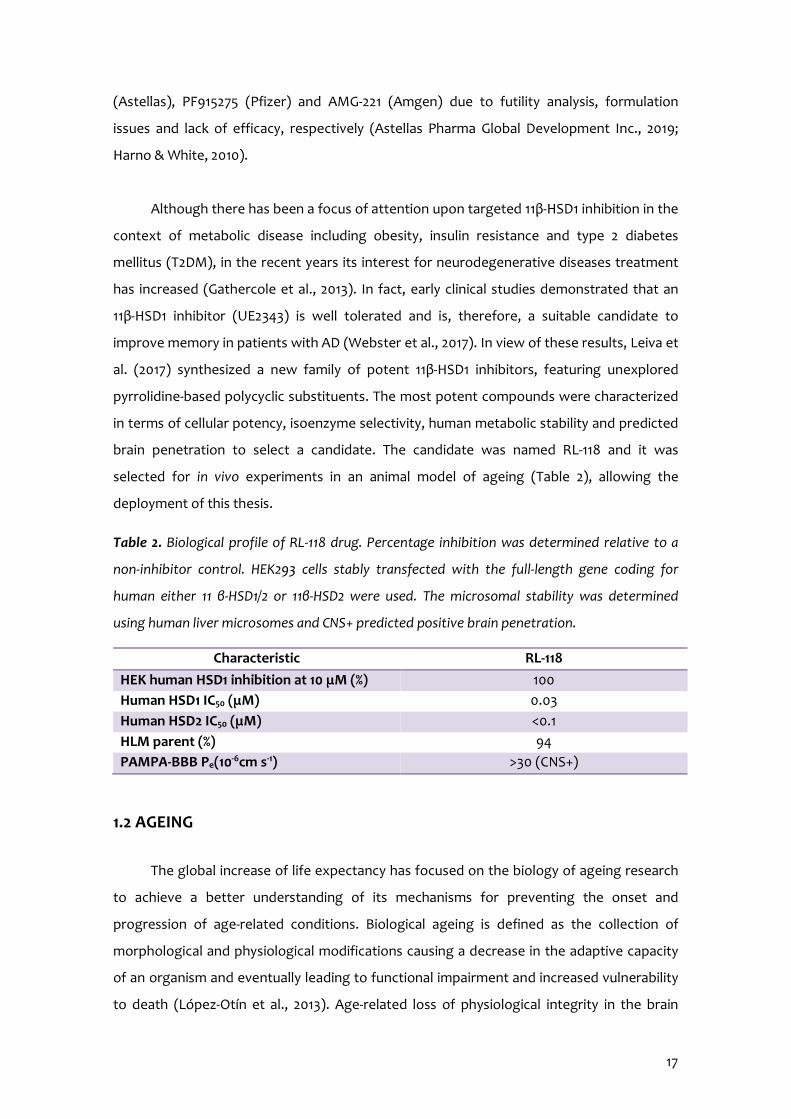
Although there has been a focus of attention upon targeted 11β-HSD1 inhibition in the
context of metabolic disease including obesity, insulin resistance and type 2 diabetes
mellitus (T2DM), in the recent years its interest for neurodegenerative diseases treatment
has increased (Gathercole et al., 2013). In fact, early clinical studies demonstrated that an
11β-HSD1 inhibitor (UE2343) is well tolerated and is, therefore, a suitable candidate to
improve memory in patients with AD (Webster et al., 2017). In view of these results, Leiva et
al. (2017) synthesized a new family of potent 11β-HSD1 inhibitors, featuring unexplored
pyrrolidine-based polycyclic substituents. The most potent compounds were characterized
in terms of cellular potency, isoenzyme selectivity, human metabolic stability and predicted
brain penetration to select a candidate. The candidate was named RL-118 and it was
selected for in vivo experiments in an animal model of ageing (Table 2), allowing the
deployment of this thesis.
Table 2. Biological profile of RL-118 drug. Percentage inhibition was determined relative to a
non-inhibitor control. HEK293 cells stably transfected with the full-length gene coding for
human either 11 β-HSD1/2 or 11β-HSD2 were used. The microsomal stability was determined
using human liver microsomes and CNS+ predicted positive brain penetration.
1.2 AGEING
The global increase of life expectancy has focused on the biology of ageing research
to achieve a better understanding of its mechanisms for preventing the onset and
progression of age-related conditions. Biological ageing is defined as the collection of
morphological and physiological modifications causing a decrease in the adaptive capacity
of an organism and eventually leading to functional impairment and increased vulnerability
to death (López-Otín et al., 2013). Age-related loss of physiological integrity in the brain
Characteristic RL-118 HEK human HSD1 inhibition at 10 μM (%) 100 Human HSD1 IC50 (μM) 0.03 Human HSD2 IC50 (μM) <0.1 HLM parent (%) 94 PAMPA-BBB Pe(10-6cm s-1) >30 (CNS+)
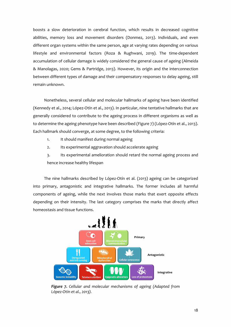
18
boosts a slow deterioration in cerebral function, which results in decreased cognitive
abilities, memory loss and movement disorders (Donmez, 2013). Individuals, and even
different organ systems within the same person, age at varying rates depending on various
lifestyle and environmental factors (Roza & Rughwani, 2019). The time-dependent
accumulation of cellular damage is widely considered the general cause of ageing (Almeida
& Manolagas, 2020; Gems & Partridge, 2013). However, its origin and the interconnection
between different types of damage and their compensatory responses to delay ageing, still
remain unknown.
Nonetheless, several cellular and molecular hallmarks of ageing have been identified
(Kennedy et al., 2014; López-Otín et al., 2013). In particular, nine tentative hallmarks that are
generally considered to contribute to the ageing process in different organisms as well as
to determine the ageing phenotype have been described (Figure 7) (López-Otín et al., 2013).
Each hallmark should converge, at some degree, to the following criteria:
1. It should manifest during normal ageing
2. Its experimental aggravation should accelerate ageing
3. Its experimental amelioration should retard the normal ageing process and
hence increase healthy lifespan
The nine hallmarks described by López-Otín et al. (2013) ageing can be categorized
into primary, antagonistic and integrative hallmarks. The former includes all harmful
components of ageing, while the next involves those marks that exert opposite effects
depending on their intensity. The last category comprises the marks that directly affect
homeostasis and tissue functions.
Figure 7. Cellular and molecular mechanisms of ageing (Adapted from López-Otín et al., 2013).
Primary
Antagonistic
Integrative

19
Genomic instability
One common denominator of ageing is the accumulation of genetic damage throughout
life (Moskalev et al., 2012), which involve exogenous physical, chemical and biological
agents, as well as endogenous threats, including DNA replication errors, DNA mutations,
spontaneous hydrolytic reactions and reactive oxygen species (ROS) accumulation
(Niedernhofer et al., 2018; Vigj & Montagna, 2017). Additionally, exposure to genotoxics
accelerates the ageing process (Niedernhofer, et al., 2018). Therefore, targeting DNA repair
mechanisms has been proposed as a strategy to alleviate age-associated dysfunctions (Petr
et al., 2020).
Telomere attrition
Telomeres are a chromosomal region particularly susceptible to age-related deterioration.
Replicative DNA polymerases lack the capacity to replicate completely the terminal ends of
linear DNA molecules, a function of telomerase. However, most mammalian somatic cells
do not express it, and this leads to progressive and cumulative loss of telomere-protective
sequences from chromosome ends, inducing senescence and/or apoptosis (Fumagalli et al.,
2012; Rossi & Gorospe, 2020). Accordingly, pathological telomere dysfunction accelerates
ageing in mice and humans, whereas experimental telomerase stimulation delays ageing.
Epigenetic alterations
Epigenetic changes involve alterations in DNA methylation patterns, histone post-
translational modifications (PTM) and chromatin remodelling, through DNA
methyltransferases (DNMTs), histone acetyltransferases (HATs), deacetylases (HDACs),
methyltransferases (HMTs) and demethylases (HDMs); as well as protein complexes. For
instance, deletion of histone methylation extends longevity (Greer et al., 2010; Molina-
Serrano et al., 2019), whereas global DNA hypomethylation has been associated with
ageing (Salas-Pérez et al., 2019). Of note, unlike DNA mutations, epigenetic modifications
are reversible, hence offering opportunities for the design of novel anti-ageing treatments.
Loss of proteostasis
Proteostasis (protein homeostasis) involves mechanisms for the stabilization of incorrectly
folded proteins – most prominently, the heat shock family of proteins – and mechanisms
for the degradation of protein by the ubiquitin-proteasome system or the autophagy-
lysosomal system. Moreover, there are other regulators of age-related proteotoxicity that
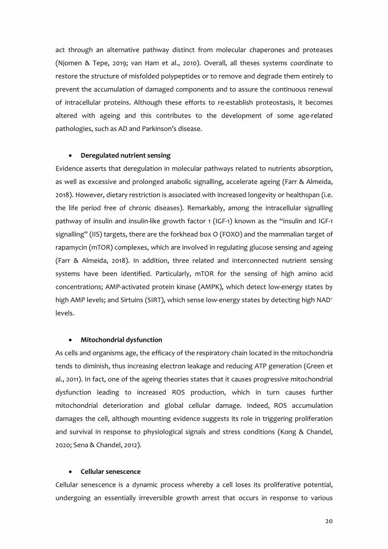
20
act through an alternative pathway distinct from molecular chaperones and proteases
(Njomen & Tepe, 2019; van Ham et al., 2010). Overall, all theses systems coordinate to
restore the structure of misfolded polypeptides or to remove and degrade them entirely to
prevent the accumulation of damaged components and to assure the continuous renewal
of intracellular proteins. Although these efforts to re-establish proteostasis, it becomes
altered with ageing and this contributes to the development of some age-related
pathologies, such as AD and Parkinson’s disease.
Deregulated nutrient sensing
Evidence asserts that deregulation in molecular pathways related to nutrients absorption,
as well as excessive and prolonged anabolic signalling, accelerate ageing (Farr & Almeida,
2018). However, dietary restriction is associated with increased longevity or healthspan (i.e.
the life period free of chronic diseases). Remarkably, among the intracellular signalling
pathway of insulin and insulin-like growth factor 1 (IGF-1) known as the “insulin and IGF-1
signalling” (IIS) targets, there are the forkhead box O (FOXO) and the mammalian target of
rapamycin (mTOR) complexes, which are involved in regulating glucose sensing and ageing
(Farr & Almeida, 2018). In addition, three related and interconnected nutrient sensing
systems have been identified. Particularly, mTOR for the sensing of high amino acid
concentrations; AMP-activated protein kinase (AMPK), which detect low-energy states by
high AMP levels; and Sirtuins (SIRT), which sense low-energy states by detecting high NAD+
levels.
Mitochondrial dysfunction
As cells and organisms age, the efficacy of the respiratory chain located in the mitochondria
tends to diminish, thus increasing electron leakage and reducing ATP generation (Green et
al., 2011). In fact, one of the ageing theories states that it causes progressive mitochondrial
dysfunction leading to increased ROS production, which in turn causes further
mitochondrial deterioration and global cellular damage. Indeed, ROS accumulation
damages the cell, although mounting evidence suggests its role in triggering proliferation
and survival in response to physiological signals and stress conditions (Kong & Chandel,
2020; Sena & Chandel, 2012).
Cellular senescence
Cellular senescence is a dynamic process whereby a cell loses its proliferative potential,
undergoing an essentially irreversible growth arrest that occurs in response to various

21
stressors, triggering activation of several pathways that induce senescence. This process
involves increased cell size, extensive gene expression changes, telomere shortening and
chromatin modifications (Farr & Almeida, 2018). Conceivably, the accumulation of
senescent cells with ageing may be due to increased rate of senescence and/or decreased
rate of clearance, maybe as a consequence of an attenuated immune response.
Stem cell exhaustion
One of the most obvious characteristics of ageing is the decline in the regenerative capacity
of tissues. With ageing haematopoiesis deteriorates, resulting in diminished adaptive
immune cells production, and increased incidence of anaemia and myeloid malignancies
(Shaw et al., 2010). Although the deficient proliferation of stem and progenitor cells is
obviously detrimental, an excessive proliferation of these cells can also be deleterious as
accelerates the exhaustion of stem cell niches.
Altered intercellular communication
Compelling evidence indicates that beyond cell-autonomous alterations, age-related
dysfunction in one cell can spread and cause age-related deterioration systemically in other
cells located in various tissues throughout an organism (Farr & Almeida, 2018). Ageing
alters intercellular communication at different levels, including elevated inflammation,
exacerbation of oxidative stress (OS), as well as deregulated endocrine and neuronal
signalling.
Noteworthy, the differentiation between physiological and pathological ageing, is
often subtle but fundamental (Figure 8). Ageing should naturally be a physiological process
occurring prior to death. Thus, physiological ageing features a slow process of balanced
functional decline across various organs in a coordinated manner, manifesting healthy or
successful ageing and longevity. In contrast, pathological ageing originates from premature
ageing often in a tissue-specific manner, resulting in impairment of adaptive responses and
finally, chronic diseases (Liu, 2017). Up to now, the mechanisms that evolve physiological to
pathological ageing remain unclear. However, it is currently thought that mTOR operates as
a thermostat switching in between physiological and pathological ageing, with inhibition of
mTOR robustly lengthening animal lifespans due to all the mediator effects commented
above (Liu, 2017).
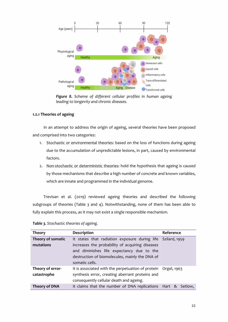
22
1.2.1 Theories of ageing
In an attempt to address the origin of ageing, several theories have been proposed
and comprised into two categories:
1. Stochastic or environmental theories: based on the loss of functions during ageing
due to the accumulation of unpredictable lesions, in part, caused by environmental
factors.
2. Non-stochastic or deterministic theories: hold the hypothesis that ageing is caused
by those mechanisms that describe a high number of concrete and known variables,
which are innate and programmed in the individual genome.
Trevisan et al. (2019) reviewed ageing theories and described the following
subgroups of theories (Table 3 and 4). Notwithstanding, none of them has been able to
fully explain this process, as it may not exist a single responsible mechanism.
Table 3. Stochastic theories of ageing.
Theory Description Reference Theory of somatic mutations
It states that radiation exposure during life increases the probability of acquiring diseases and diminishes life expectancy due to the destruction of biomolecules, mainly the DNA of somatic cells.
Szilard, 1959
Theory of error-catastrophe
It is associated with the perpetuation of protein synthesis error, creating aberrant proteins and consequently cellular death and ageing.
Orgel, 1963
Theory of DNA It claims that the number of DNA replications Hart & Setlow,
Figure 8. Scheme of different cellular profiles in human ageing leading to longevity and chronic diseases.

23
reparation determine the lifespan and could generate more probability of mutations within the DNA itself, therefore impairing proteins through the process of transcription.
1974
Theory of the breaking of biomolecular bonds
The increase of chemical bonds in DNA causes protein modifications that could generate the functional failure of cell metabolism, resulting in physiological processes decline during ageing.
Cristofalo et al., 1994
Theory of advanced glycosylation
High concentration of glucose in the blood and tissues due to impaired nutrient sensing leads to protein glycosylation, perturbing biological functions.
Cerami, 1985
Free radical theory Based on the reactions oxidizing biomolecules, thus destructing them, and causing many degenerative alterations. It evolved to the mitochondrial theory of ageing when mitochondria were implicated as the primary source of ROS (Barja, 2013).
Harman, 1991
Theory of adaptive homeostasis
It is the most recent theory and considers anti-stress as a form of protection that maintains homeostasis within the organism.
Pomatto & Davies, 2018
Table 4. Non-stochastic theories of ageing.
Theory Description Reference Theory of telomeres
Considers the cell repositioning diminishment linked to telomeres modification, which is responsible for chromosome integrity, affecting cellular functions.
Blackburn, 1999
Theory of intrinsic mutagenesis
The longevity depends on the reliability of genetic material in its replication and maintenance of the restorative mechanisms.
Burnet, 1974
Neuroendocrine theory
It proposes that ageing results from functional perturbations, both in neuronal control and in endocrine output, of the HPA axis, thereby causing dysfunction of various endocrine glands and their target organs.
Sonntag et al., 1999
Immunological theory
Immunological responses decrease with ageing, thus generating low resistance to infections and diseases.
Walford, 1969
Theory of programmed senescence
Ageing is based on genetic programming that controls cell development and determines the lifespan of each of the cells.
Hayflic & Moorhead, 1961

24
1.2.2 Ageing and neurodegeneration
Ageing is a process regulated by different molecular and genetic mechanisms. Three
pathways modifying longevity have been described: increase of AMPK and reduction of
mTOR, reduction of insulin signalling and reduction of SIRT. Relevantly, mTOR complex
(mTORC) 1 inhibition by rapamycin can postpone ageing by affecting energy sensing,
maintenance of proteostasis, improving mitochondrial dysfunction and cellular senescence,
but also modulating stem cell functions in different tissues (Papadopoli et al., 2019).
Recently, AMPK signalling activation with age has been identified as a critical factor
determining age-related decline of hippocampal neurogenesis and thus, increased
susceptibility to age-associated neurological diseases (B. Z. Wang et al., 2019). The later is a
family of HDACs, which activation has been associated with neuroprotective effects.
Extended lifespan is associated with an increase in the prevalence of age-related
diseases. Indeed, ageing is a major risk factor for several chronic pathologies, including
osteoporosis, cardiovascular and metabolic disorders, as well as neurodegenerative
diseases, such as AD (Almeida & Manolagas, 2020; Salas-Pérez et al., 2019; Trevisan et al.,
2019).
Referring to brain ageing, it produces changes in neuronal and cognitive abilities, like
mental speed and executive function. In the hippocampus, a brain area that comprises the
spatial and episodic learning, memory and is predisposed to the detrimental effects of
ageing, long-term potentiation results impaired at synapses and immediate early genes for
the synaptic plasticity are down-regulated when ageing (Liu, 2017). However, human
umbilical cord plasma treatment, which is abundant of plasticity-enhancing proteins,
revitalizes the hippocampus and improves cognitive function in aged mice (Castellano et al.,
2017).
1.3 EPIGENETICS AND NEURODEGENERATION
During ageing, biological, environmental, and lifestyle factors drive epigenetic
modifications resulting in phenotypic differences. Environmental stressors interact with the
genome leading to stable changes in DNA structure, gene expression and behaviour. In
turn, these changes may underlie and contribute to the development and progression of
mental disorders, including AD (Park et al., 2019).

25
Epigenetics refers to potentially heritable and environmentally modifiable changes in
gene expression that are mediated via non-DNA-encoded mechanisms (Sun et al., 2013).
The three main epigenetic marks are DNA-methylation, histone PTM and non-coding RNA
regulation. All these mechanisms play a key role in regulating transcriptional activity,
modulating chromatin structure through recruitment, assembly or retention of chromatin-
associated factors (Harman & Martín, 2019; Nair et al., 2017).
1.3.1 DNA methylation and hydroxymethylation
DNA methylation consists in the addition of methyl groups to the DNA, which alters
the DNA segment transcriptional activity without altering its sequence (Park et al., 2019).
DNMT enzymes – DNMT1, DNMT3A and DNMT3B – catalyse the transference of a methyl
group at the fifth position of cytosine in order to generate 5-methylcytosine (5-mC). DNMT1
is responsible for maintaining the methyl groups by copying the methylation pattern of the
parent DNA strand during cell division so that afterwards methylation is inherited (Hermann
et al., 2004). On the other hand, DNMT3A and DNMT3B catalyse the de novo methylation,
adding methyl groups to non-methylated CpG cytosine dinucleotide. Methylation on gene
promoter regions, particularly at CpG islands, is the canonical mechanism for cell-specific
gene silencing during development, thus plays a critical role in neural plasticity (Miller et al.,
2010). According to the primary role of DNA methylation in brain function, DNMT1 and
DNMT3A deletion in mouse prefrontal cortex leads to deficits in neuronal morphology,
synaptic plasticity, learning and memory (Harman & Martín, 2019). Although initially it was
thought that this epigenetic mark was irreversible, recent research has demonstrated that
DNA may experience fast and reversible methylation changes and may also undergo
demethylation (Figure 9 A) (Miller & Sweatt, 2007).
On the other hand, demethylation that consists in removing methyl groups is
performed by ten-eleven translocation (TET) enzymes. TET1, 2 and 3 oxidize 5-mC to 5-
hydroxymethylcytosine (5-hmC). It is more labile and can rapidly generate demethylated
cytosines. Although 5-hmC and 5-mC appear to be negatively related, according to Chen et
al. (2012) increased global 5-hmC in the hippocampus of aged mice do not correlate with
decreased 5-mC neither to increased TETs, suggesting that 5-hmC could act as an epigenetic
marker itself and not only as an intermediary in DNA demethylation. TET1 and TET2
participate in controlling gene expression and cell death and are associated with ageing and

26
neurodegeneration, while TET3 is related to early embryonic development (Fetahu et al.,
2019; Ficz et al., 2011).
1.3.2 Post-translational histone modifications
DNA is wound around histone proteins conforming nucleosomes. It consists of a core
histone octamer (H3, H4, H2A and H2B) and the N-terminal tail of each extends and
contains sites that can expect more than 100 PTM, involving acetylation, methylation,
ubiquitinylation and phosphorylation (Harman & Martín, 2019). Multiple PTM can be added
to a single histone molecule. These epigenetic marks modulate chromatin compaction,
allowing or denying the transcriptional machinery access to the regulatory regions of
genes. Highly compacted chromatin, also known as heterochromatin, is generally
associated with low transcription, while less compacted regions of chromatin, named
euchromatin, are associated with active transcription (Taylor et al., 2019).
Among the histone modifications known to be involved in longevity and ageing, the
most widely described are acetylation and methylation of lysine (Lys) residues (Figure 9 B)
(Pal & Tyler, 2016). Acetylation removes histone positive charge, thereby increasing
transcriptional activity of the acetylated segment, while methylation generally the contrary.
HATs acetylate Lys residues, and conversely HDACs catalyse histone deacetylation
and function as transcription repressors (Harman & Martín, 2019). The expression of HATs
and HDACs has been reported to be altered in the aged brain and could be linked to age-
related altered gene transcription (Barter & Foster, 2018). Studies using pharmacological
and transgenic approaches demonstrated that HDAC2 is the most linked to negatively
regulate cognition, as its levels increased in the hippocampus of aged mice and its inhibition
enhanced synaptic plasticity, the number of dendritic spines and memory (Harman &
Martín, 2019). In line with this, increased HDAC2 activity correlated with diminished Lys 9
and Lys 14 acetylated H3 (H3K9 and H3K14) levels (Singh & Thakur, 2018).
Considering histone methylation, it has been linked to either transcriptional
activation or repression depending on the particular methylated residue and extent of
methylation (mono, di or trimethylation), although ageing has been related to increased
histone methylation and repressed transcriptional activity (Pal & Tyler, 2016). Compelling

27
evidence proposed that methylation of H3K9 promotes DNA methylation maintenance in
mammals and subsequent gene silencing (Zhao et al., 2016).
1.3.3 non-coding RNAs
MicroRNAs (miRNAs) are small non-coding RNA molecules containing about 18-25
nucleotides involved in gene expression regulation. miRNAs bind to complementary sites
on 3’ untranslated regions of target mRNA molecules and direct their degradation or inhibit
their translation (Harman & Martín, 2019). One of the most abundantly expressed miRNAs
in the aged brain is let-7 family, but on the contrary, miR-101 and -433 were down-regulated,
indicating miRNAs changes might be implicated in cognitive decline (Inukai et al., 2012).
Other miRNA have been implicated in AD pathogenesis (Table 5), such as miR-107 as it
targets β-site amyloid precursor protein (APP) cleaving enzyme 1 (BACE1) (Wang et al.,
2008) and miR-106b (Hébert et al., 2009).
Table 5. Summary of miRNA modulating Amyloid-β plaques formation (M. Wang et al., 2019).
Target mRNA miRNA Main effect Reference APP miR-106a/b, -520c, -
20a, -17-5p, -101, -16, -153, -200b
Overexpression results in translational repression of APP mRNA
Patel et al., 2008; Hébert et al., 2009; Vilardo et al. & Liu et al., 2010; Long et al., 2012; Liu et al., 2014
Figure 9. Epigenetic modifications. A. DNA modifications. B. Histone modifications (Hwang et al., 2017).
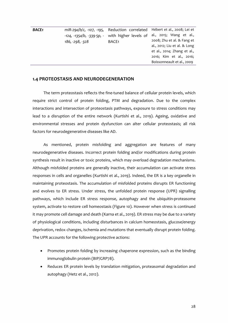
28
BACE1 miR-29a/b/c, -107, -195, -124, -135a/b, -339-5p, -186, -298, -328
Reduction correlated with higher levels of BACE1
Hébert et al., 2008; Lei et al., 2015; Wang et al., 2008; Zhu et al. & Fang et al., 2012; Liu et al. & Long et al., 2014; Zhang et al., 2016; Kim et al., 2016; Boissonneault et al., 2009
1.4 PROTEOSTASIS AND NEURODEGENERATION
The term proteostasis reflects the fine-tuned balance of cellular protein levels, which
require strict control of protein folding, PTM and degradation. Due to the complex
interactions and intersection of proteostasis pathways, exposure to stress conditions may
lead to a disruption of the entire network (Kurtishi et al., 2019). Ageing, oxidative and
environmental stresses and protein dysfunction can alter cellular proteostasis; all risk
factors for neurodegenerative diseases like AD.
As mentioned, protein misfolding and aggregation are features of many
neurodegenerative diseases. Incorrect protein folding and/or modifications during protein
synthesis result in inactive or toxic proteins, which may overload degradation mechanisms.
Although misfolded proteins are generally inactive, their accumulation can activate stress
responses in cells and organelles (Kurtishi et al., 2019). Indeed, the ER is a key organelle in
maintaining proteostasis. The accumulation of misfolded proteins disrupts ER functioning
and evolves to ER stress. Under stress, the unfolded protein response (UPR) signalling
pathways, which include ER stress response, autophagy and the ubiquitin-proteasome
system, activate to restore cell homeostasis (Figure 10). However when stress is continued
it may promote cell damage and death (Karna et al., 2019). ER stress may be due to a variety
of physiological conditions, including disturbances in calcium homeostasis, glucose/energy
deprivation, redox changes, ischemia and mutations that eventually disrupt protein folding.
The UPR accounts for the following protective actions:
Promotes protein folding by increasing chaperone expression, such as the binding
immunoglobulin protein (BIP/GRP78).
Reduces ER protein levels by translation mitigation, proteasomal degradation and
autophagy (Hetz et al., 2012).

29
Protein translational attenuation is mediated by serine (Ser)/threonine (Thr) kinase
RNA-like ER kinase (PERK), which phosphorylates the eukaryotic initiation factor 2 α (eIF2α)
in Ser 51 causing general protein synthesis reduction and promoting cell survival. Moreover,
PERK activation has been shown to modulate the activity of nuclear factor erythroid-
derived 2 (NRF2) and FOXO proteins, linking this pathway to the antioxidant response,
insulin responsiveness and autophagy (Martínez et al., 2017). In particular, PERK
phosphorylates NRF2 in Thr 80 in order to activate it and therefore translocate to the
nucleus where it binds to the antioxidant response element (ARE) inducing an increase in
glutathione (GSH) levels and a reduction of ROS levels, promoting cell survival (Cullinan &
Diehl, 2004; Ma et al., 2013). Additionally, eIF2-α is activated by AD hallmarks, inflammation
and OS pathways, and nutrient imbalance. By contrast, its reduction in mice has been linked
to enhanced synapse plasticity and cognition (Ma et al., 2013). As misfolded proteins
accumulate during neurodegeneration, ER capability becomes overrun and eventually
proves insufficient, leading to proteotoxicity and protein aggregation (Kurtishi et al., 2019).
Accordingly, evidence for canonical UPR activation in AD neurons has determined that
increased BIP/GRP78 protein levels in the hippocampus and prefrontal cortex of AD animal
models, as well as the higher presence of p-PERK and p-eIF2α in the neurons of AD patients
(Chang et al., 2002; Scheper & Hoozemans, 2015). These markers appeared in
morphologically healthy neurons or neurons with an abnormally phosphorylated Tau
protein, indicating that the UPR is involved in the early stages of AD pathology (Scheper &
Hoozemans, 2015). In accordance, induction of ER stress leads to neuronal metabolic stress,
AD hallmarks and cognitive impairment in mice (Lourenco et al., 2013; van der Harg et al.,
2014; Yoon et al., 2012).
On another note, the UPR signalling also activates autophagy. However, ER stress
response and autophagy are linked in different ways. Among others, it has been described
that PERK reduces nuclear factor kappa-light-chain-enhancer of activated B cells (NF-κB)
activation, which promotes the induction of autophagy. Also, PERK-mediated activating
transcription factor 4 (ATF4) activation is required for gene expression of various
autophagy genes. Autophagy degrades protein aggregates that cannot be processed by
the proteasome (Blasiak et al., 2019) to promote cell survival. For instance, the
autophagosome can degrade and recycle entire damaged organelles. Autophagy means
“self-eating” and is defined as a catabolic process in eukaryotic cells in which intracellular
and extracellular altered components are delivered into lysosomes, where their
degradation occurs (Boya et al., 2013). Of interest, autophagy declines with ageing; so that
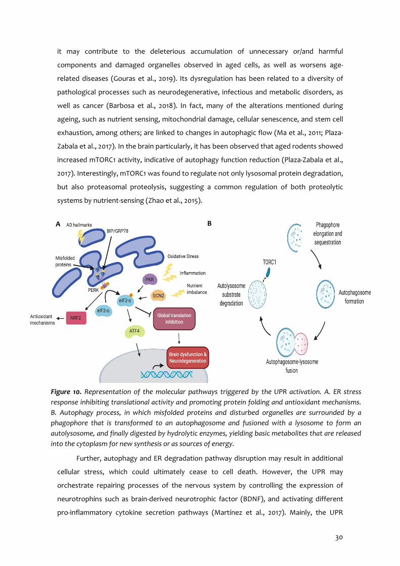
30
it may contribute to the deleterious accumulation of unnecessary or/and harmful
components and damaged organelles observed in aged cells, as well as worsens age-
related diseases (Gouras et al., 2019). Its dysregulation has been related to a diversity of
pathological processes such as neurodegenerative, infectious and metabolic disorders, as
well as cancer (Barbosa et al., 2018). In fact, many of the alterations mentioned during
ageing, such as nutrient sensing, mitochondrial damage, cellular senescence, and stem cell
exhaustion, among others; are linked to changes in autophagic flow (Ma et al., 2011; Plaza-
Zabala et al., 2017). In the brain particularly, it has been observed that aged rodents showed
increased mTORC1 activity, indicative of autophagy function reduction (Plaza-Zabala et al.,
2017). Interestingly, mTORC1 was found to regulate not only lysosomal protein degradation,
but also proteasomal proteolysis, suggesting a common regulation of both proteolytic
systems by nutrient-sensing (Zhao et al., 2015).
Further, autophagy and ER degradation pathway disruption may result in additional
cellular stress, which could ultimately cease to cell death. However, the UPR may
orchestrate repairing processes of the nervous system by controlling the expression of
neurotrophins such as brain-derived neurotrophic factor (BDNF), and activating different
pro-inflammatory cytokine secretion pathways (Martínez et al., 2017). Mainly, the UPR
Figure 10. Representation of the molecular pathways triggered by the UPR activation. A. ER stress response inhibiting translational activity and promoting protein folding and antioxidant mechanisms. B. Autophagy process, in which misfolded proteins and disturbed organelles are surrounded by a phagophore that is transformed to an autophagosome and fusioned with a lysosome to form an autolysosome, and finally digested by hydrolytic enzymes, yielding basic metabolites that are released into the cytoplasm for new synthesis or as sources of energy.
A B
31
activates NF-κB, mitogen-activated protein kinase (MAPK), c-Jun N-terminal protein kinase
(JNK) and p38 (Brown et al., 2016; Sprenkle et al., 2017). Chronic ER dysfunction has been
associated with memory and cognitive deficits observed in AD patients (Duran-Aniotz et al.,
2017; Sprenkle et al., 2017).
1.5 OXIDATIVE STRESS AND NEURODEGENERATION
OS is the redox state resulting from an imbalance between reactive oxygen and
nitrogen species (RONS) generation and detoxification. RONS are highly reactive molecules
with one or more unpaired electrons that behave as oxidants or reductants and antioxidant
defences neutralize their harmful effects.
On the one hand, RONS production is not only limited to exert deleterious effects,
but also involved in energy production and immune defence (Liguori et al., 2018). The main
RONS studied are superoxide (O2-), hydrogen peroxide (H2O2), hydroxyl ion (OH-), nitric
peroxide (ONO2-) and nitric oxide (NO), the majority (95-98%) of which come from the by-
products of the mitochondrial electron transport chain. However, there are other sources
of RONS:
Endogenous: include NADPH-oxidase, myeloperoxidase (MPO), lipoxygenase and
angiotensin II. The first is the prevalent source of the O2-, which is formed during
cellular respiration and, mostly, it is converted into H2O2 by superoxide dismutase
(SOD) (Liguori et al., 2018). It, in turn, can form the highly reactive ROS OH- that is
extremely reactive, especially with phospholipids in cell membranes and proteins,
thus contributing to increase oxidative damage. H2O2 in turn, is metabolised by
catalase (CAT) to water and oxygen. NO is produced by NO synthase (NOS) and
combined with oxygen (O2) react to form another relatively reactive molecule,
ONO2- (Genestra, 2007).
Exogenous: involve air and water pollution, tobacco, alcohol, heavy or transition
metals, drugs, industrial solvents and radiation (Liguori et al., 2018).
RONS overproduction, together to mitochondrial dysfunction, produce oxidative
damage to cells, embracing protein oxidation and nitration, glycoloxidation and lipid
peroxidation. Altogether cause alterations in cellular biochemical and physiological

32
functions leading to apoptosis and, therefore, to neurodegenerative pathologies (Fivenson
et al., 2017; Peterson & Smith, 2016).
On the other hand, antioxidant defence protects biological systems from RONS
toxicity and involves both endogenous and exogenous molecules:
Endogenous antioxidants: contain enzymatic and non-enzymatic pathways.
o Antioxidant enzymes: the primary antioxidant enzymes are SOD, CAT and
glutathione peroxidase (GPX). As mentioned above, SOD dismutase O2- to
H2O2, which is decomposed to water and O2 by CAT, preventing OH-
production. In addition, GPX converts peroxides and OH- into nontoxic
forms by the oxidation of reduced GSH into glutathione disulphide and then
reduced to GSH by glutathione reductase. Other antioxidant enzymes are
glutathione-S-transferase and glucose-6-phosphate dehydrogenase (Liguori
et al., 2018).
o Non-enzymatic antioxidants: they are molecules that interact with RONS
terminating the free radical chain reactions, such as bilirubin, α-tocopherol
(vitamin E), β-carotene, albumin and uric acid (Wu et al., 2013).
Exogenous antioxidants: include ascorbic acid (vitamin C), which scavenges OH- and
O2-; α-tocopherol, which is involved against lipid peroxidation of cell membranes;
and phenolic antioxidants like stilbene derivatives (resveratrol and flavonoids), oil
lecithins, selenium, zinc and drugs such as acetylcysteine (Liguori et al., 2018;
Pisoschi & Pop, 2015).
Despite ageing is the leading cause of cognitive impairment; patients with high GPX
activity showed slower progression of cognitive deficits, while on the contrary, low GPX
activity was related to cognitive decline acceleration (Revel et al., 2015). In addition, it has
been demonstrated that increased OS biomarkers correlated with raised levels of
inflammatory cytokines and both were associated with poor cognitive performance in
institutionalized elderly people (Baierle et al., 2015). The brain is especially exposed to
RONS as it is a high energy-demanding organ, i.e. requires more oxygen than other tissues –
approximately 20% of the basal body oxygen – so undergoes mitochondrial respiration,
which increases ROS production (Du et al., 2018). Mitochondrial abnormalities, initially
caused by gradual oxidative disturbances, are major contributors to OS (Tadokoro et al.,
2020). Indeed, perturbed mitochondrial operations result in impaired metabolic capacity

33
(Unzeta et al., 2016). This abnormality creates a vicious positive-feedback cycle where RONS
accumulation achieves a threshold and activates unavoidably a compensatory mechanism
in order to control the cellular environment. This hyper-defensive state, intended to
prolong cell life, raises vulnerability to additional insults such as AD hallmarks (Cheignon et
al., 2018; Zhu et al., 2007), which in consequence, leads to neuroinflammation,
mitochondrial damage and further RONS generation (Unzeta et al., 2016). Also, altered
antioxidant enzymes levels were found in specific AD brain regions (Sultana et al., 2011).
Similarly, RONS could be involved in OS influencing AD, as it has been described that affect
the activity of the N-methyl-D-aspartate (NMDA) receptors triggering a cascade of events
that increase RONS production, inflammation, AD hallmarks, lipid peroxidation, and
ultimately, leading to synaptic dysfunction responsible for AD (Ligouri et al., 2018).
1.6 NEUROINFLAMMATION AND NEURODEGENERATION
Neuroinflammation is defined as a cerebral physiological response to injury, infection
or disease in order to prevent potential damage, neuronal destruction or to eliminate the
damaging agent. To date, several alterations of innate and acquired immunity have been
observed in the elderly, like diminished adaptive immune cells production. Altogether are
comprised in the immunosenescence term. It refers to increased susceptibility of aged
people to infection and may be explained in terms of molecular and cellular mechanisms
responsible for inflammatory age-associated disorders (Fard & Stough, 2019).
One feature of immunosenescence is a chronic subclinical elevated production of pro-
inflammatory mediators typical of elderly as a consequence of cumulative lifetime exposure
to antigenic load due to infections and non-infective antigens (Baylis et al., 2013;
Blagosklonny, 2010). In response to an insult, inflammatory pathways activate and several
cytokines are released from glial cells, lymphocytes, monocytes and macrophages (Figure
11). As mentioned, stress can also increase pro-inflammatory cytokines levels. In fact, it has
been described that stress raises tumour necrosis factor-alpha (Tnf-α), interleukin (Il) 1-β
and Il-6 expression, as well as reduce anti-inflammatory cytokines expression like Il-10, Il-4
and transforming growth factor β (Tgf-β). In addition, cytokines are potent regulators of
the HPA axis and subsequent eliciting GC secretion, so they can alter local GC availability
and GR function, therefore affecting an individual’s GC sensitivity (Marques et al., 2009;
Thiel & Dretsch, 2011). Besides, stress contributes to increase ROS production and in
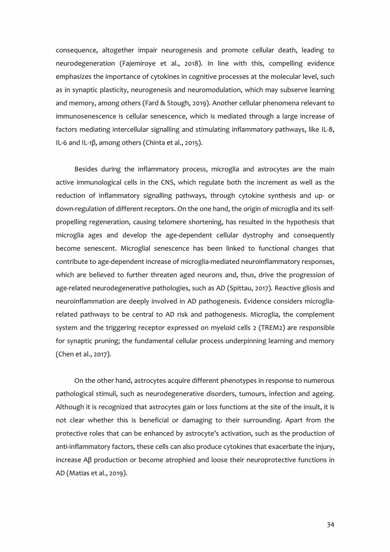
34
consequence, altogether impair neurogenesis and promote cellular death, leading to
neurodegeneration (Fajemiroye et al., 2018). In line with this, compelling evidence
emphasizes the importance of cytokines in cognitive processes at the molecular level, such
as in synaptic plasticity, neurogenesis and neuromodulation, which may subserve learning
and memory, among others (Fard & Stough, 2019). Another cellular phenomena relevant to
immunosenescence is cellular senescence, which is mediated through a large increase of
factors mediating intercellular signalling and stimulating inflammatory pathways, like IL-8,
IL-6 and IL-1β, among others (Chinta et al., 2015).
Besides during the inflammatory process, microglia and astrocytes are the main
active immunological cells in the CNS, which regulate both the increment as well as the
reduction of inflammatory signalling pathways, through cytokine synthesis and up- or
down-regulation of different receptors. On the one hand, the origin of microglia and its self-
propelling regeneration, causing telomere shortening, has resulted in the hypothesis that
microglia ages and develop the age-dependent cellular dystrophy and consequently
become senescent. Microglial senescence has been linked to functional changes that
contribute to age-dependent increase of microglia-mediated neuroinflammatory responses,
which are believed to further threaten aged neurons and, thus, drive the progression of
age-related neurodegenerative pathologies, such as AD (Spittau, 2017). Reactive gliosis and
neuroinflammation are deeply involved in AD pathogenesis. Evidence considers microglia-
related pathways to be central to AD risk and pathogenesis. Microglia, the complement
system and the triggering receptor expressed on myeloid cells 2 (TREM2) are responsible
for synaptic pruning; the fundamental cellular process underpinning learning and memory
(Chen et al., 2017).
On the other hand, astrocytes acquire different phenotypes in response to numerous
pathological stimuli, such as neurodegenerative disorders, tumours, infection and ageing.
Although it is recognized that astrocytes gain or loss functions at the site of the insult, it is
not clear whether this is beneficial or damaging to their surrounding. Apart from the
protective roles that can be enhanced by astrocyte’s activation, such as the production of
anti-inflammatory factors, these cells can also produce cytokines that exacerbate the injury,
increase Aβ production or become atrophied and loose their neuroprotective functions in
AD (Matias et al., 2019).
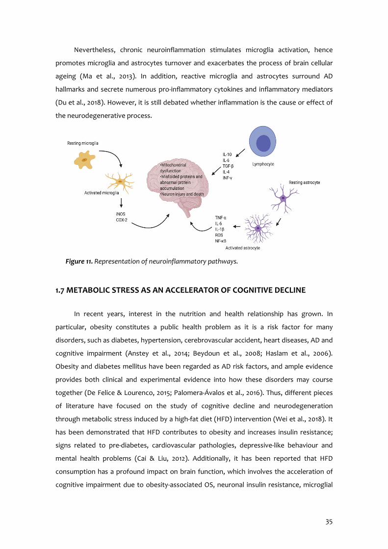
35
Nevertheless, chronic neuroinflammation stimulates microglia activation, hence
promotes microglia and astrocytes turnover and exacerbates the process of brain cellular
ageing (Ma et al., 2013). In addition, reactive microglia and astrocytes surround AD
hallmarks and secrete numerous pro-inflammatory cytokines and inflammatory mediators
(Du et al., 2018). However, it is still debated whether inflammation is the cause or effect of
the neurodegenerative process.
1.7 METABOLIC STRESS AS AN ACCELERATOR OF COGNITIVE DECLINE
In recent years, interest in the nutrition and health relationship has grown. In
particular, obesity constitutes a public health problem as it is a risk factor for many
disorders, such as diabetes, hypertension, cerebrovascular accident, heart diseases, AD and
cognitive impairment (Anstey et al., 2014; Beydoun et al., 2008; Haslam et al., 2006).
Obesity and diabetes mellitus have been regarded as AD risk factors, and ample evidence
provides both clinical and experimental evidence into how these disorders may course
together (De Felice & Lourenco, 2015; Palomera-Ávalos et al., 2016). Thus, different pieces
of literature have focused on the study of cognitive decline and neurodegeneration
through metabolic stress induced by a high-fat diet (HFD) intervention (Wei et al., 2018). It
has been demonstrated that HFD contributes to obesity and increases insulin resistance;
signs related to pre-diabetes, cardiovascular pathologies, depressive-like behaviour and
mental health problems (Cai & Liu, 2012). Additionally, it has been reported that HFD
consumption has a profound impact on brain function, which involves the acceleration of
cognitive impairment due to obesity-associated OS, neuronal insulin resistance, microglial
Figure 11. Representation of neuroinflammatory pathways.
36
activation and neuroinflammation, which are considered important risk factors for
neurodegeneration (Hahm et al., 2020).
Diversely, obesity has been linked to abnormalities in myelin, white and grey matter
(Mueller et al., 2014) and cerebral atrophy (Brooks et al., 2013). Particularly, the
hippocampus and frontal cortex are very sensitive to those disturbances as Debette et al.
(2011) related obesity to altered memory and executive function.
Metabolic derangements, including inflammation, insulin resistance and ER stress are
known to underlie glucose intolerance and T2DM, which are present in AD brains (De Felice
et al., 2014; Freeman et al., 2014). In line with this, recent data has demonstrated that Aβ
actions stimulate pro-inflammatory mechanisms to impair neuronal insulin signalling and
trigger stress kinase activation, resulting in synapse and memory deteriorations (De Felice
& Lourenco, 2015). Although classical alterations in glucose metabolism have been
observed in AD cerebral tissue, metabolic stress also comprises disturbances in proteostasis
and activation of signalling pathways that mediate cellular stress (De Felice & Lourenco,
2015). In fact, stress modulates insulin signalling. Initially, catecholamines inhibit insulin but
increase glucagon secretion, while later GCs also cause global hyperinsulinemia, which may
further fuel insulin resistance. However, it has been demonstrated tissue-specific regulation
of insulin signalling by GCs (Gathercole et al., 2011). Interestingly, a recent study
demonstrates insulin-signalling potentiation after chronic stress exposure and prevention
from fructose diet-induced attenuation of the insulin metabolic pathway (Romic et al.,
2020).
1.8 ALZHEIMER’S DISEASE
AD has been a focus of recent research as it represents between 60-80% of total
cases of dementia over the world, with the prevalence continuing to grow in part because
of the ageing world population. It is one of the most common neurodegenerative diseases
and consists in an irreversible, progressive cognitive disorder characterized by a progressive
decline in two or more cognitive domains, including memory, language, executive and
visuospatial function, movement, personality and behaviour, which in turn causes loss of
abilities to perform instrumental and/or basic activities of daily living (Weller & Budson,
2018). The other most common forms of dementia include vascular dementia, Lewy body
37
dementia and Parkinson’s disease with dementia, frontotemporal lobar degeneration and
normal pressure hydrocephalus (DeTure & Dickson, 2019).
1.8.1 History
AD was the term coined by Dr. Alois Alzheimer in 1906 when he observed a patient
with a striking cluster of symptoms including reduced comprehension and memory, as well
as aphasia (difficulty to comprehend or formulate language), disorientation, unpredictable
behaviour, paranoia, auditory hallucinations and pronounced psychosocial impairment
(Maurer et al., 1997).
Regarding his scientific outcomes, Alzheimer anticipated dementia a century before
its discovery in arteriosclerotic patients. Particularly, he could differ between pre-senile
dementia and dementia. This was key in the diagnosis of August D, whose case was studied
by Alzheimer in Frankfurt from 1901 until she died on 9 April 1906. Auguste D manifested
progressive cognitive impairment, focal symptoms, hallucinations, amnestic writing,
anxiety, delusions and psychosocial incompetence (Maurer et al., 1997). At necropsy, he
detected atrophy of the cerebral cortex accompanied by the loss of cells and a great
excrescence of the glia. Moreover, numerous plaques throughout the cortex were
associated with excrescences of the vessels (arteriosclerotic changes). This pattern, which
could hardly be described as atypical for a patient of 70 to 80 years of age, was present in
Auguste D, who was only aged 51 years when she died. He presented these results in a
meeting and published them in 1907 under the title “A characteristic serious disease of the
cerebral cortex”. Despite that, nobody attached importance to Alzheimer’s results until
1909 and in 1911 the first case of Alzheimer’s disease was described by Gonzalo Lafora, a
histopathologist from Madrid and co-worker of Ramon y Cajal. Lafora associated William CF
pathology to AD in an article titled “Contribution to the knowledge of Alzheimer’s disease
or pre-senile dementia with focal symptoms” revised by Alois Alzheimer and published in
the Neurology and Psychiatry Journal (Tagarelli et al., 2006).
1.8.2 Risk factors
Given the high personal and social burden, it is imperative to identify a
comprehensive list of risk factors (Table 6).
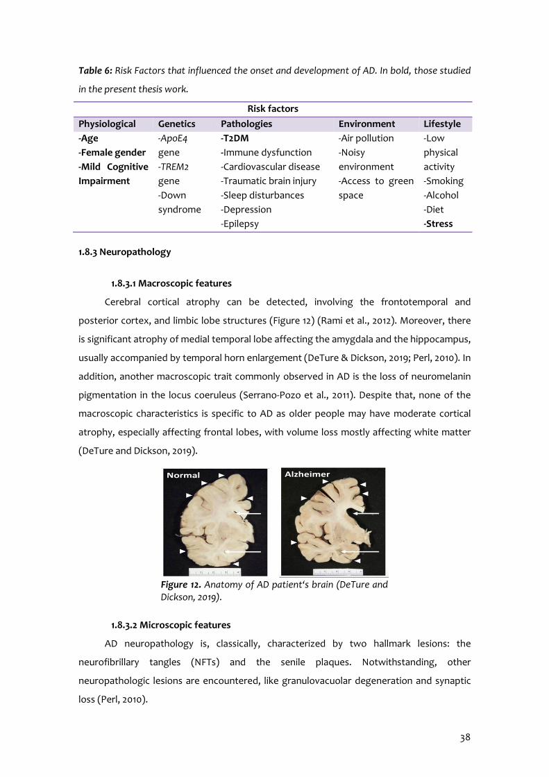
38
Table 6: Risk Factors that influenced the onset and development of AD. In bold, those studied
in the present thesis work.
Risk factors Physiological Genetics Pathologies Environment Lifestyle -Age -Female gender -Mild Cognitive Impairment
-ApoE4 gene -TREM2 gene -Down syndrome
-T2DM -Immune dysfunction -Cardiovascular disease -Traumatic brain injury -Sleep disturbances -Depression -Epilepsy
-Air pollution -Noisy environment -Access to green space
-Low physical activity -Smoking -Alcohol -Diet -Stress
1.8.3 Neuropathology
1.8.3.1 Macroscopic features
Cerebral cortical atrophy can be detected, involving the frontotemporal and
posterior cortex, and limbic lobe structures (Figure 12) (Rami et al., 2012). Moreover, there
is significant atrophy of medial temporal lobe affecting the amygdala and the hippocampus,
usually accompanied by temporal horn enlargement (DeTure & Dickson, 2019; Perl, 2010). In
addition, another macroscopic trait commonly observed in AD is the loss of neuromelanin
pigmentation in the locus coeruleus (Serrano-Pozo et al., 2011). Despite that, none of the
macroscopic characteristics is specific to AD as older people may have moderate cortical
atrophy, especially affecting frontal lobes, with volume loss mostly affecting white matter
(DeTure and Dickson, 2019).
1.8.3.2 Microscopic features
AD neuropathology is, classically, characterized by two hallmark lesions: the
neurofibrillary tangles (NFTs) and the senile plaques. Notwithstanding, other
neuropathologic lesions are encountered, like granulovacuolar degeneration and synaptic
loss (Perl, 2010).
Figure 12. Anatomy of AD patient‘s brain (DeTure and Dickson, 2019).
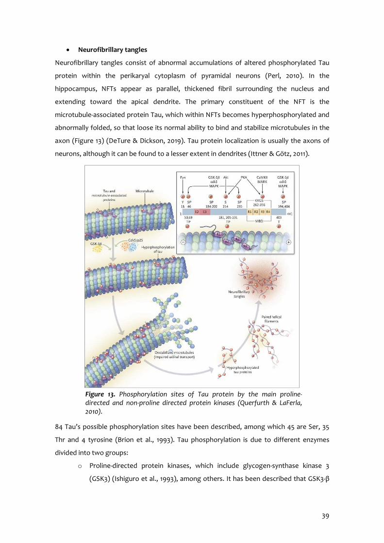
39
Neurofibrillary tangles
Neurofibrillary tangles consist of abnormal accumulations of altered phosphorylated Tau
protein within the perikaryal cytoplasm of pyramidal neurons (Perl, 2010). In the
hippocampus, NFTs appear as parallel, thickened fibril surrounding the nucleus and
extending toward the apical dendrite. The primary constituent of the NFT is the
microtubule-associated protein Tau, which within NFTs becomes hyperphosphorylated and
abnormally folded, so that loose its normal ability to bind and stabilize microtubules in the
axon (Figure 13) (DeTure & Dickson, 2019). Tau protein localization is usually the axons of
neurons, although it can be found to a lesser extent in dendrites (Ittner & Götz, 2011).
84 Tau’s possible phosphorylation sites have been described, among which 45 are Ser, 35
Thr and 4 tyrosine (Brion et al., 1993). Tau phosphorylation is due to different enzymes
divided into two groups:
o Proline-directed protein kinases, which include glycogen-synthase kinase 3
(GSK3) (Ishiguro et al., 1993), among others. It has been described that GSK3-β
Figure 13. Phosphorylation sites of Tau protein by the main proline-directed and non-proline directed protein kinases (Querfurth & LaFerla, 2010).
40
activity increases in AD brains as well as hyperphosphorylated Tau (Hernandez
et al., 2013).
o Non-proline directed protein kinases.
Several studies have demonstrated a correlation between NFT extent and distribution and
the degree of dementia. Although NFTs are a cardinal hallmark of AD, this neuropathologic
feature may also be encountered in other disorders, like type C Niemann-Pick disease, post-
traumatic dementia and amyotrophic lateral sclerosis (Perl, 2010).
Senile plaques
Senile or neuritic plaques are formed by the extracellular nonvascular accumulation of Aβ40
and Aβ42 peptides, surrounded by abnormally configured neuronal processes or neurites
(either dendrites or axons) (Perl, 2010). Additional Aβ peptides containing between 38 and
43 amino acids are also detected, but Aβ42 is the most fibrillogenic, neurotoxic and the
predominant component of amyloid plaques in AD, as it can form monomers, oligomers,
protofibrils and fibrils (DeTure & Dickson, 2019). Monomers are not pathologic, although
oligomers and protofibrils are associated with favoured Tau hyperphosphorylation,
proteasome disruption, mitochondrial dysfunction, calcium homeostasis dysregulation, pro-
inflammatory cytokines release, synaptic damage and cognitive decline (Hughes et al.,
2020; Pákáski & Kálmán, 2008). Importantly, post-mortem evidence asserts that a
significant proportion of individuals with brain amyloidopathy did not express disease when
alive (Dubois et al., 2016).
Aβ peptides result from abnormal processing of APP, a highly conserved transmembrane
glycoprotein that can be catalysed through two mechanisms: non-amyloidogenic and
amyloidogenic pathways (Figure 14). The main APP processing pathway is the non-
amyloidogenic, which comprises an α-secretase that cleavages the peptide between Lys 16
and Leu 17 releasing a soluble peptide named sAPPα and staying a 10 kDa C-terminal
fragment (p3CTF) inside the membrane (Pákáski & Kálmán, 2008). sAPPα may possess
neuroprotective actions. Diversely in the amyloidogenic pathway, the β- and γ-secretases
act and generate Aβ, an insoluble peptide that aggregates and constitutes senile plaques.
Notwithstanding, they may be a consequence of an imbalance in the production and
clearance pathways.
0

41
As far as Aβ clearance is concerned, in the brain, it is accomplished by several mechanisms
that involve non-enzymatic and enzymatic pathways. The first includes interstitial fluid
drainage, uptake by microglial phagocytosis and transport across the blood vessels walls
into the circulation. Diversely, multiple Aβ-degrading enzymes (ADE) have been identified
like neprilysin, insulin-degrading enzyme (IDE), matrix metalloproteinase-9, glutamate
carboxypeptidase II and others (Yoon & Jo, 2012). Besides, the cell possesses other
mechanisms to eliminate unfolded aberrant proteins, which include autophagy. Recent
pieces of evidence demonstrate that defective Aβ autophagy vacuoles clearance creates
favourable conditions for Aβ accumulation and increased autophagy flux reduced amyloid
burden (Ries & Sastre, 2016). Likewise, astrocytes from AD animal models showed strong
expression of microtubule-associated protein light chain 3 (LC3) involved in autophagy,
providing a link between this process and inflammation (Pomilio et al., 2016).
1.8.4 Classification
Depending on the age at onset, two subtypes of AD have been described:
Familial AD
Familial AD (fAD) represents only between 1 and 5% of AD cases and is caused by dominant
autosomal mutations in APP, presenilin 1 (PSEN1) or PSEN2 genes, located in chromosome
21, 14 and 1, respectively (Pimenova & Goate, 2020). FAD cases can appear as early as 20
years, with the average age of onset at 46.2 years (DeTure & Dickson, 2019). Although
clinically it does not differ from sporadic AD, it is associated with an aggressive course.
α-secretase β-secretase
sAPP-α
C83 APP
sAPP-β
C99
γ-secretase Aβ
CTF
p3
CTF
γ-secretase
Aβ aggregation and oligomerization
Senile plaque
Figure 14. Scheme of APP processing through non-amyloidogenic pathway represented to the left and to the right, amyloidogenic pathway leading to Aβ accumulation and formation of extracellular senile plaques.
42
Sporadic AD
Sporadic AD (sAD) accounts for nearly 99% of AD patients, and is caused by a combination
of genetic factors and environmental risk factors without documented familial history of
AD (Zhang et al., 2020). It courses with deposition of Aβ, Tau hyperphosphorylation, OS,
neuroinflammation, cholinergic neuron degeneration, gut microbiota disorders, lipid
metabolism abnormalities, autophagy dysfunction, insulin resistance, synapse dysfunction
and metal ions disorders (Zhang et al., 2020; DeTure & Dickson, 2019). The onset is typically
at 65 years or more.
1.8.5 Hypothesis
Many factors are involved in the pathogenesis of AD. Due to the complexity of
human brains and the lack of reasonable animal models, the detailed AD pathogenesis is
still unclear (Du et al., 2018). So far, several hypotheses have been described and
consequently, many efforts have been made to develop drugs based on these hypotheses.
Aβ cascade hypothesis
Some of the strategies studied consisted of targeting β- and γ-secretase; however,
undesirable side effects were the overriding problem. Another approach was targeting Aβ
clearance. To sum up, all the Aβ modulator drugs have failed in clinical trials, which insight
doubts on this theory (Karran et al., 2011). However, a phase Ib trial of aducanumab
(Biogen) showed a positive correlation between brain Aβ levels and disease exacerbation
(Sevigny et al., 2017) and fortunately, in early 2020 aducanumab entered phase IIIb.
Although it has been planned to apply for regulatory approval in the USA in 2020, its clinical
trials are projected to run through September 2023 (Alzforum, 2020).
Tau hypothesis
Strategies developed to target Tau involve blocking its aggregation, utilizing Tau
vaccinations, stabilizing microtubules, manipulating kinases and phosphatases that govern
Tau modifications. However, most of these efforts have failed in clinical trials, although
some drugs are currently under study (Li & Götz, 2017).
Oxidative stress hypothesis
The presence of OS markers in early AD stages indicate that it precedes other hallmarks,
although other studies identify OS as a result of brain damage and as a contributor to AD
43
progression (Unzeta et al., 2016). In consequence, up to date, disease-modifying
antioxidant approaches are part of combined treatments (Du et al., 2018).
Neuroinflammation hypothesis
The first strategy consisted of using non-steroid anti-inflammatory drugs (NSAIDs), but they
did not show enough benefits in the clinic stage, as the immune response can be either
deleterious or beneficial depending on the context (Du et al., 2018). It was hypothesized
that such usage might only be beneficial in the very early stages of AD pathogenesis,
coincident with initial Aβ deposition, microglial activation and release of pro-inflammatory
cytokines. Instead, NSAIDs may produce a detrimental effect owing to their inhibitory
activity on already activated microglial cells once Aβ deposition has started (Ali et al., 2019).
Of note, the recent advances in understanding the mechanism underlying microglia
dysfunction are opening possibilities for new AD therapeutic interventions (Jetvic et al.,
2017). For instance, Vorinostat, an HDAC inhibitor, has been yielded efficiency in attenuating
human microglial-mediated immune responses, and it is currently under clinical trial
(Esposito & Sherr, 2019).
Glucocorticoid hypothesis
Although it is not an AD hypothesis but considering that directly targeting AD hallmarks
have not yet been proved effective to redress the insidious shift from physiological to
pathological actions along the course of AD, several pieces of evidence have pointed the
central role played by the HPA axis, GCs and GR in the aetiology of AD. It has been reported
that life events like chronic stress or stress-related disorders may increase the probability to
develop AD (Canet et al., 2018). Several strategies directly targeting GR or inhibiting the 11β-
HSD1 enzyme were tested for neutralizing the HPA axis dysregulation and GC
overproduction (Lesuis et al., 2018; Webster et al., 2017).
1.8.6 Treatment
At present, there is no cure for AD. Few drugs have been approved for the treatment
of AD, none of which are modifying or slowing down the disease progression; however,
they can relieve some of the symptoms (Oudin, 2020). Available treatments include
cholinesterase inhibitors like donepezil for patients with any stage of AD dementia, and
memantine for people with moderate-to-severe AD dementia, which has activity as both a
non-competitive NMDA receptor antagonist and a dopamine agonist.

44
1.9 AGEING ANIMAL MODELS – SENESCENCE ACCELERATED MOUSE
The use of humans in ageing research is complicated by many factors, including
ethical issues, environmental and social factors, and perhaps most importantly, their long
natural lifespan. Diversely, although cellular models of human disease provide valuable
mechanistic information, they are limited in that they may not replicate the in vivo biology
(Mitchell et al., 2015). In consequence, animal models become a useful tool for studying
ageing.
Senescence accelerated mice (SAM) are one of the accelerated senescence strains
that spontaneously evolved from AKR/J strain inbreeds at Kyoto University (Takeda et al.,
1981). After endogamous inbreeding for colony maintenance, it was observed that various
litters displayed phenotypic characteristics indicative of rapid ageing, including loss of hair,
lordokyphosis, peri-ophthalmic injuries, locomotor activity reduction and lower lifespan,
among others. The selection of this phenotype and through siblings’ breeding along various
generations led to the development of several sublines of SAM prone (SAMP) and SAM
resistant (SAMR) mice by 1991 (Figure 15) (Takeda et al., 1991). There are 12 lines; SAMP
inbred strains include SAMP1-11, while SAMR inbred strains are SAMR1,4,5. The median
survival time of SAMP and SAMR was 9.7 months and 16.3 months, respectively (Takeda et
al., 1994).
SAMP strain has a relatively strain-specific pathologic phenotype, which is
characteristic enough to distinguish them. In particular, SAMP8 exhibit significant brain
atrophy and age-related deteriorations in memory and learning abilities (Akiguchi et al.,
Figure 15. SAMR1 and SAMP8 mice at 6 months of age respectively (Delerue et al., 2013).
45
2017; Pallàs, 2012). Although no specific pathological hallmarks such as senile plaques or
NFTs have been found, a variety of age-associated pathomorphological alterations and
blood-brain barrier (BBB) dysfunction involving neurons, glia and vessels have been
identified in SAMP8 mice brains (Akiguchi et al., 2017; Virgili et al., 2018). Accordingly, some
specific reactions of microglia and astrocytes have been found (Kawamata et al., 1998).
Thus, the morphological alterations with abnormal glial responses, age-related increases in
phosphorylated Tau and early Aβ accumulation in the hippocampus of SAMP8 mice brains
may result in impaired memory and behavioural and learning disturbances (Pallàs, 2012).
In line with the ageing phenotype, SAMP8 mice show emotional disorders and
abnormal circadian rhythms together to auditory, epigenetic, neuropathologic and
neurochemical alterations (Griñán-Ferré et al., 2018; Morley et al., 2012).
1.9.1 APP processing and Aβ accumulation
Apart from Aβ and APP overproduction, SAMP8 mice show increased cortical and
hippocampal APP mRNA expression (Morley et al., 2000). Other genes related to AD
pathology have been reported to be altered in SAMP8 mice, like PSEN-1 and -2, whose
mutations result in an increase of the amyloidogenic APP processing pathway (Kumar et al.,
2009). Furthermore, ApoE gene expression has been found to be less expressed in SAMP8
than SAMR1. On the other hand, gene expression reduction of genes involved in Aβ
clearance and OS signalling has also been described in SAMP8 compared to SAMR1 (Pallàs,
2012). In addition, several studies have been performed on the regulation of APP in SAMP8
to improve cognitive and behavioural deficits, with different strategies, like the use of
specific oligonucleotides to negatively regulate APP expression and therefore improve
cerebral senescence parameters typical of SAMP8 (Kumar et al., 2000). Other consisted of
using miRNAs, post-transcriptional regulators, particularly miR-16, -31, -144, -135 and -383
(Griñán-Ferré et al., 2018; Liu et al., 2010; Zhang et al., 2016); or administrating a monoclonal
antibody targeting Aβ oligomers (Zhang et al., 2011).
Overall, these results indicate that SAMP8 is a valuable animal model for
understanding mechanisms involved in Aβ alteration of AD and that age-related cognitive
impairment observed in SAMP8 is dependent of APP processing.
46
1.9.2 Tau hyperphosphorylation
Hyperphosphorylated Tau protein was found in the hippocampus, cortex and
striatum of SAMP8 mice in comparison to SAMR1 mice as early as 5 months of age (Álvarez-
García et al., 2006; Canudas et al., 2005). Also, kinases such as GSK3β and cell division
protein kinase 5 (CDK5), were highly expressed in SAMP8 than SAMR1 mice in an age-
dependent manner (Canudas et al., 2005; Casadesús et al., 2012).
1.9.3 Neuroinflammation
Astrocytic clusters have been identified in SAM mice hippocampus and cortex,
although to a greater extent in SAMP8 compared to SAMR1 (Cristòfol et al., 2012).
Consistent with this, astrocyte hypertrophy and astrogliosis were observed in SAMP8 mice
brains (Han et al., 2010; Morley et al., 2012). In addition, the proliferation of microglia,
characteristic of ageing, was observed in cerebral cortex and hippocampus (Kawamata et
al., 1998), and activated microglia produces abundant amounts of toxic substances,
including free radicals and glutamate (Akiguchi et al., 2017).
1.9.4 Oxidative stress
As discussed earlier, OS is the result of ROS generation exceeding the cellular
antioxidant capacity, causing neuronal death and neurodegeneration in the brain. It has
been demonstrated that SAMP8 of 4- and 8- weeks showed higher lipid peroxidation and
protein carbonilation levels in the cerebral cortex than SAMR1 (Albasanz et al., 2011; Sato et
al., 1996). Moreover, excessive oxidation of cortical proteins was found in the brains of
aged SAMP8 mice but not of aged SAMR1 (Butterfield et al., 1997). However, literature
related to OS in SAMP8 is not always conclusive. For instance, decreased expression of
detoxifying enzymes such as manganese SOD (MnSOD), CAT or GPX were found in SAMP8
compared to age-matched SAMR1 mice (Álvarez-García et al., 2006; Schmitt et al., 2012). By
contrast, Griñán-Ferré et al. (2016) described higher protein levels in SAMP8 in comparison
with SAMR1 mice; those discrepancies can be explained by the age when determinations
were performed.
1.9.5 Glucose metabolism
Studies have demonstrated that glucose metabolism is decreased in SAMP8 aged 2
months (Cuesta et al., 2013). Additionally, it has been described that SAMP8 mice brain

47
tissue shows a transient enhancement of anaerobic glycolysis in 2 months-old mice and a
subsequent decline in mitochondrial function in comparison with SAMR1 mice (Omata et al.,
2001). Moreover, another study revealed a correlation between glucose metabolism
impairment and the severity of the learning deficits (Akiguchi et al., 2017; Mehla et al.,
2014).

48

2. OBJECTIVES

50
51
Ageing, stress and obesity are three factors that define modern society. Modern
society has to cope with higher levels of stress than past societies. Despite not having to
worry about hunting and whether we will be attacked for an animal, humans are subjected
to various stressors; not only psychological but also physical, environmental and social. For
instance, high workloads, economic difficulties, relationships, nutrition, media overload,
sleep deprivation and social pressure to boost our careers, among others. The continued
presence of these stressful situations influences our health and also predicts our ageing.
Additionally, although life expectancy has increased, age-related pathologies, such as
degenerative diseases and in particular AD, have become a problem for the health sector,
as it has been estimated that people over the age of 60 will increase from 12 to 22 % in the
world in the following years. Consequently, finding strategies that entail therapeutic
benefits for patients and reduction of the impact of ageing on health, particularly brain
ageing, is of paramount importance and constitute an exciting challenge. In this sense, the
use of drugs that modify the main pathways activated by stress and, therefore increase
individuals’ adaptive ability arises as an optimal strategy for promoting healthy ageing.
In this context, as ageing is the major risk factor for age-related neurodegenerative
disorders, the general aim of the present doctoral thesis has been to study the mechanisms
involved in stress-induced neurodegeneration and evaluate the neuroprotective effect of
RL-118 drug, an 11β-HSD1 inhibitor, in a senescence animal model.
In order to pursue this aim, several specific objectives have been set up:
1. Assess the protective effect of RL-118 on cognition in SAMP8 mice aged 12 months
and reveal which underlying mechanisms might modulate it.
To evaluate cognitive function and behavioural alterations.
To investigate the protective effect of RL-118 in the process of autophagy.
To study the changes induced by RL-118 treatment in APP processing and
tau hyperphosphorylation.
To elucidate the changes in antioxidant enzymes and cerebral inflammatory
markers.

52
To determine the correlation between autophagy markers and cognitive
parameters, as well as between autophagy, pro-inflammatory and OS
markers.
2. Investigate the effects on brain function of a stressful lifestyle, by chronic mild
stress, in animal models prone and resistant to accelerated senescence, SAMP8 and
SAMR1 respectively.
To study different epigenetic marks related to learning and memory.
To analyse the cerebral OS state and antioxidant enzymes changes.
To assess the effects of CMS on neuroinflammation and autophagy.
To determine the effects of CMS on different pathways implicated in
neurodegeneration.
To evaluate cognitive performance and behavioural alterations.
3. Determine the target engagement between RL-118 and 11β-HSD1 and evaluate
whether 11β-HSD1 inhibition by RL-118 is able to face the detrimental effects of CMS
in SAMP8 mice aged 6 months.
To investigate RL-118 target engagement.
To study different epigenetic marks related to learning and memory.
To assess the effects of CMS and RL-118 treatment on neuroinflammatory
pathways.
To elucidate cellular OS state and determine antioxidant mechanisms
changes after CMS exposure and 11β-HSD1 inhibition.
To describe the effects of CMS and RL-118 treatment on autophagy and
synaptic plasticity markers.
To analyse different pathways altered implicated in neurodegeneration by
CMS and potential reversal by 11β-HSD1 inhibition.
To evaluate cognitive performance and behavioural alterations.

53
4. Study the detrimental effect of metabolic stress by HFD and the beneficial effect of
11β-HSD1 inhibition in SAMP8 mice aged 6 months.
To elucidate the inhibitory effect of RL-118 by determining and comparing
corticosterone levels in blood and brain tissue.
To determine the glucose tolerance and triglyceride levels following HFD
feeding and RL-118 treatment.
To analyse the impact of HFD-induced metabolic stress and RL-118
treatment on different pathways implicated in neurodegeneration and
energy metabolism.
To investigate the effects of HFD and 11β-HSD1 inhibition on cerebral
OS state and antioxidant mechanisms.
To evaluate different neuroinflammatory pathways activated after
HFD-induced metabolic stress and 11β-HSD1 inhibition effects.
To analyse the molecular changes produced by metabolic stress and
RL-118 treatment regarding ER stress response and autophagy.
To evaluate the deleterious effect of HFD on cognitive abilities and
behavioural performance and potential reversal by RL-118
treatment.

54

3. METHODS AND RESULTS


3.1 11β-HSD1 Inhibition by RL-118 Promotes Autophagy and Correlates with Reduced Oxidative Stress and Inflammation, Enhancing Cognitive Performance in SAMP8 Mouse
Model
Adapted from: Puigoriol-Illamola, D.1,2, Griñán-Ferré, C.1,2, Vasilopoulou, F.1,2, Leiva, R.3,4, Vázquez, S.3,4, Pallàs, M1,2. 11β-HSD1 inhibition by RL-118 promotes autophagy and correlates
with reduced oxidative stress and inflammation, enhancing cognitive performance in SAMP8 mouse model
Molecular Neurobiology 2018; 55:8904-8915. doi:10.1007/s12035-018-1026-8. Epub 2018 Apr 02.

58
Author information: 1 Department of Pharmacology, Toxicology and Therapeutic Chemistry. Pharmacology Section. Faculty of Pharmacy and Food Sciences. University of Barcelona, Av Joan XXIII 27-31, 08028 Barcelona, Spain. 2 Institute of Neuroscience, University of Barcelona (NeuroUB), Passeig de la Vall d’Hebron 171, Barcelona, Spain. 3 Department of Pharmacology, Toxicology and Therapeutic Chemistry. Medicinal Chemistry Section. Faculty of Pharmacy and Food Sciences, University of Barcelona, Av Joan XXIII 27-31, 08028 Barcelona, Spain. 4 Institute of Biomedicine (IBUB). Av Diagonal 643, 08028 Barcelona, Spain.

59
3.1.1 Abstract
With the increase in life expectancy, the study of age-related diseases and the
development of different strategies to deal with them become mandatory. Among the
features of ageing, disturbances in the GC stress response and individual’s adaptive abilities
appear to increase the vulnerability of the elderly to age-related pathologies. In fact,
elevated GC exposure is widely accepted as a key factor in age-related cognitive decline in
rodents and humans. Of note, 11β-HSD1 is a key enzyme controlling GC action as it catalyses
the conversion of 11β-dehydrocorticosterone to corticosterone in mice (i.e. from inactive to
active GC forms). Compelling evidence has demonstrated prolonged GC excess implication
in neurodegenerative processes and cognitive impairment. Therefore, the study of the
effects on age-related alterations due to GC attenuation by 11β-HSD1 inhibition results
essential. The study aimed to assess the potential protective effect of RL-118, an 11β-HSD1
inhibitor, on cognitive performance and which molecular mechanisms were modified in an
ageing animal model.
Here, we determined the effect of RL-118 administered to 12-month-old SAMP8 mice
that display neuropathologic Alzheimer’s disease-like alterations and are widely used as a
rodent model of cognitive dysfunction. Behavioural tests were conducted – particularly the
open field and object location tests – and neurodegenerative molecular markers were
studied.
RL-118 treatment increased locomotor activity and improved cognitive performance.
Likewise, 11β-HSD1 inhibition by RL-118 induced changes in hippocampal autophagy markers
such as Beclin1, LC3B, AMPKα, and mTOR, indicating enhancement of the intracellular
degradation system. In line with the autophagic increase, a diminution in phosphorylated
tau species (Ser 396 and Ser 404) jointly with an increase in non-amyloidogenic APP
processing pathway mediators suggested that the increase of the removal of abnormal
proteins by autophagy might underlie the neuroprotective effect of the 11β-HSD1 inhibitor
studied. In addition, gene expression of OS and inflammatory markers, such as heme
oxygenase 1 (Hmox1), aldehyde dehydrogenase 2 (Aldh2), Il-1β, and C-C motif chemokine ligand
3 (Ccl3) were reduced in treated mice in comparison to control group. Consistent with this,
we further demonstrated a significant direct correlation between autophagy markers and
cognitive improvement and significant inverse correlation with autophagy, OS, and
neuroinflammatory markers.
60
We concluded that inhibition of 11β-HSD1 by RL-118 prevented neurodegenerative
processes and cognitive decline, acting on the autophagy process, being an additional
neuroprotective mechanism involved in 11β-HSD1 inhibition effects not described
previously.
3.1.2 Introduction
With the increase in life expectancy, ageing and age-related cognitive impairments
are becoming one of the most important challenges for human health. Ageing is a
multifaceted process characterized by an intricate and irreversible accumulation of
physiological changes and is associated with an increase in transcriptional noise, aberrant
protein production, altered intercellular communication, cellular senescence and loss in
proteostatic mechanism, among others (López-Otín et al., 2013).
Cellular proteostasis loss has been proposed as one of the primary molecular changes
that induce progressive loss of physiological integrity in ageing (López-Otín et al., 2013).
Damaged proteins can be degraded through the ubiquitin proteasome system (UPS) or
through the autophagy lysosome pathway (Boya et al., 2013; Vílchez et al., 2014).
Autophagy is a self-degradative process involved in elimination of proteins and organelles,
cellular remodelling, and survival during starvation (Levine, 2005). Beclin1, mammalian
target of the rapamycin (mTOR), microtubule-associated protein 1 light chain 3B (LC3B) and
p62 proteins have a key role in this process (Son et al., 2012). Moreover, autophagy has
been postulated to participate in the degradation of extra- and intracellular Aβ deposition
and neurodegeneration (Yu et al., 2004; Komatsu et al., 2006).
On the other hand, there is overwhelming evidence that brain tissue is exposed to OS
in cognitive decline and age-related neurodegeneration. Evidences are manifested through
high levels of oxidized proteins, advanced glycation and lipid peroxidation end products,
toxic species formation, such as peroxides, alcohols and aldehydes; and oxidative
modifications in nuclear and mitochondrial DNA (Gella & Durany, 2009). Cellular damage by
OS is one factor that induces cell senescence promoting protein modification and
proteolysis mechanism alterations (Höhn et al., 2016; López-Otín et al., 2013). Moreover, OS
is related to inflammaging and development of cognitive impairment and anxious
behaviour. Of note, several studies have showed that pro-inflammatory cytokines release is
higher when OS increases (Anderson et al., 2013; Casadesús et al., 2002; Terao A, et al.,
61
2002; Ye et al., 1999). Interestingly, defects in the autophagy machinery led to increased
mitochondrial dysfunction and, consequently, increased levels of oxidative stress (Mathew
et al., 2009).
Recent studies suggest the excess of GCs exerts deleterious effects on the brain,
particularly on the hippocampus, and causes impaired spatial memory, a key feature of age-
related cognitive dysfunction (Lara et al., 2013), in rodents and humans, being part of
altered intercellular communication described as one of the nine hallmarks of aging (López-
Otín et al., 2013). 11β-hydroxysteroid dehydrogenase type 1 (11β-HSD1) is a key enzyme that
mediates the intracellular conversion of cortisone to cortisol in humans (11β-
dehydrocorticosterone to corticosterone in mice), the active form of GCs, thus amplifying
steroid action. High levels of GCs have been found in elderly who exhibit learning and
memory impairments and correlate with higher hippocampal atrophy. Increased circulating
cortisol levels are detected in patients with Alzheimer’s disease (AD) causing faster disease
progression (De Quervain et al., 2004). AD is the most common form of dementia and
represents a progressive brain disorder characterized by loss of cognitive functions (i.e.
memory impairments) and behavioural abilities. AD presents three representative
pathological hallmarks: synapse loss, extracellular senile plaques (SPs) and intracellular
neurofibrillary tangles (NFTs). Furthermore, it has been demonstrated that aged mice with
cognitive deficits showed increased gene expression of 11β-HSD1 in the hippocampus and
parietal cortex (Holmes et al., 2010). Likewise, overexpression of this enzyme displays a
similar premature memory decline (Yau et al., 2001). In line with this, some preclinical
studies demonstrate that 11β-HSD1 inhibition improved cognition and AD hallmarks
suggesting a neuroprotective effect (Yau et al., 2001, 2007; Mohler et al., 2011).
Design and chemical structure of the compound RL-118 [(4-
azatetracyclo[5.3.2.02,6.08,10]dodec-4yl) (cyclohexyl)methanone], one of the first pyrrolidine-
based 11β-HSD1 inhibitor, was rationally developed using the X-ray structure of human
enzyme 4BB6 (Goldberg et al., 2012; Leiva et al., 2017). Its potency against the human and
murine enzyme (IC50 values of 29 and 81 nM, respectively), its microsomal stability (94% and
93% in HLM and MLM, respectively) and its proven in vivo efficacy (Leiva et al., 2017)
makes RL-118 an interesting drug to further study its potential neuroprotective effects on
an age-related cognitive decline mice model.

62
To explore the possible RL-118 neuroprotective role by 11β-HSD1 inhibition, we
employed a senescence accelerated mice-prone strain, SAMP8, a well-characterized model
for studying neurodegeneration and cognitive deficits related to aging, as a model of early
AD (Morley et al., 2012; Nomura et al., 1999; Pallàs et al., 2008). Therefore, the present
study sought to assess how the reduction of active GCs regeneration, through the inhibition
of 11β-HSD1 by RL-118, leads to neuroprotective effects and which underlying mechanisms
might modulate the reduction of AD hallmarks, opening new ways to face challenges in AD
prevention and treatment.
3.1.3 Material and methods
Animals
Female SAMP8 mice (n=14) (12-months-old) were used to carry out cognitive and
molecular analyses. We divided these animals into two groups: Control (SP8 Ct, n=6) and
treated with RL-118, an 11β-HSD1 inhibitor (SP8 treated, n=8). RL-118, 21 mg/kg was
administered through drinking water for 4 weeks. Animals had free access to food and
water and were kept under standard temperature conditions (22±2°C) and 12h:12h light-dark
cycles (300 lx/0 lx).
Studies and procedures involving mice brain dissection and subcellular fractionation
were performed in accordance with the institutional guidelines for the care and use of
laboratory animals established by the Ethical Committee for Animal Experimentation at the
University of Barcelona.
Behavioural tests
Open Field Test
The Open Field Test (OFT) is an experiment used to assay general locomotor activity
and anxiety in rodents. The test is based on the fear and anxiety of rodents to be in opened
and luminous spaces. The OFT apparatus was a white polywood box (50x50x25 cm). The
floor was divided in two areas defined as centre zone and periphery zone (15 cm between
centre zone and the wall). Behaviour was scored with SMART® ver.3.0 software and each
trial was recorded for later analysis, utilizing a camera situated above the apparatus. Mice
were placed at the centre and allowed to explore the box for 5 min. Afterwards, mice were
63
returned to their home cages and the OFT apparatus was cleaned with 70% ethanol. The
parameters scored included centre staying duration, number of rears, defecations and the
distance travelled, calculated as the sum of total distance travelled in 5 min.
Object Location Test
The Object Location Test (OLT) assesses cognition deficits and evaluates novel
chemical entities by their effects on cognition, specifically spatial memory and
discrimination (Murai et al., 2007). This task involves exploiting rodent’s ability to recognize
when an object has been relocated, and they are inherently stressless. The test was
performed during 4 days in a wooden box (50x50x25 cm), in which three walls were white
but one black. The first day, the box was empty and the animals just habituated to the open
field cage for 10 min. The second day, two objects were placed in front of the black wall,
equidistant from each other and from the wall. Objects were 10 cm high and had different
shapes, but were composed of similar materials. The animals were placed in the open field
cage and allowed to explore both, objects and surroundings, for 10 min. Afterwards, mice
were returned to their home cages and the OLT apparatus was cleaned with 70% ethanol.
The third day, one object was moved in front of the white wall to test spatial memory. Trials
were recorded using a camera mounted above the working area and the total exploration
time was determined by scoring the amount of time (s) spent sniffing the two objects in the
new (novel) and old (familiar) locations. In order to analyse the cognitive performance, a
location index (%) was calculated as follows: (Tnovel x 100)/(Tnovel + Tfamiliar), where the
Tnovel is the time spent exploring the objects in the new location and Tfamiliar is the time
spent exploring the object in the old location.
Brain processing
Mice were euthanized one day after the behavioural test finished by cervical
dislocation. Brains were immediately removed from the skull. Hippocampus were then
isolated and frozen on powdered dry ice. They were maintained at -80°C for further use.
Tissue samples were homogenized in lysis buffer containing phosphatase and protease
inhibitors (Cocktail II, Sigma). Total protein levels were obtained and protein concentration
was determined by the method of Bradford.
64
Protein levels determination by Western blotting
For Western Blotting (WB), aliquots of 15 µg of hippocampal protein extracted with
lysis buffer composed by Tris HCl pH7.4 50mM, NaCl 150mM, EDTA 5mM and 1X-Triton X-100
were used. Protein samples were separated by SDS-PAGE (8-12%) and transferred onto
PVDF membranes (Millipore). Afterwards, membranes were blocked in 5% non-fat milk in
0,1% Tween20 TBS (TBS-T) for 1 h at room temperature, followed by an overnight incubation
at 4°C with the primary antibodies listed in Table S1.
Afterwards, membranes were washed and incubated with secondary antibodies for 1
h at room temperature. Immunoreactive proteins were observed with a
chemiluminescence-based detection kit, following the manufacturer’s protocol (ECL Kit,
Millipore) and digital images were acquired using a ChemiDoc XRS+ System (BioRad). Semi-
quantitative analyses were carried out using ImageLab software (BioRad) and results were
expressed in Arbitrary Units (AU), considering control protein levels as 100%. Protein
loading was routinely monitored by immunodetection of Glyceraldehyde 3-phosphate
dehydrogenase (GAPDH) or β-tubulin.
RNA extraction and gene expression determination
Total RNA isolation was carried out by means of TRIzol reagent following the
manufacturer’s instructions. RNA content in the samples was measured at 260 nm, and the
purity of the samples was determined by the A260/280 and A260/230 ratios in a NanoDrop™
ND-1000 (Thermo Scientific). Reverse transcription-polymerase chain reaction (RT-PCR) was
performed as follows: 2 μg of messenger RNA (mRNA) was reverse-transcribed using the
High Capacity cDNA Reverse Transcription kit (Applied Biosystems). Real-time quantitative
PCR (qPCR) was used to quantify the mRNA expression of inflammatory genes such as
interleukin 1β (Il-1β), C-C-motif ligand 3 (Ccl3), heme oxygenase 1 (Hmox1) and aldehyde
dehydrogenase 2 (Aldh2) (Table S2). SYBER Green real-time PCR was performed on the Step
One Plus Detection System (AppliedBiosystems) employing the SYBR Green PCR Master
Mix (AppliedBiosystems). Each reaction mixture contained 7.5 μL of complementary DNA
(cDNA), whose concentration was 2 μg, 0.75 μL of each primer (whose concentration was
100 nM), and 7.5 μL of SYBR Green PCR Master Mix (2X) and for TaqMan gene expression
assays (Applied Biosystems), each 20 μL of TaqMan reaction, 9 μL cDNA (25 ng) was mixed
with 1 μL 20x probe of TaqMan Gene Expression Assays and 10 μL of 2x TaqMan Universal
PCR Master Mix.
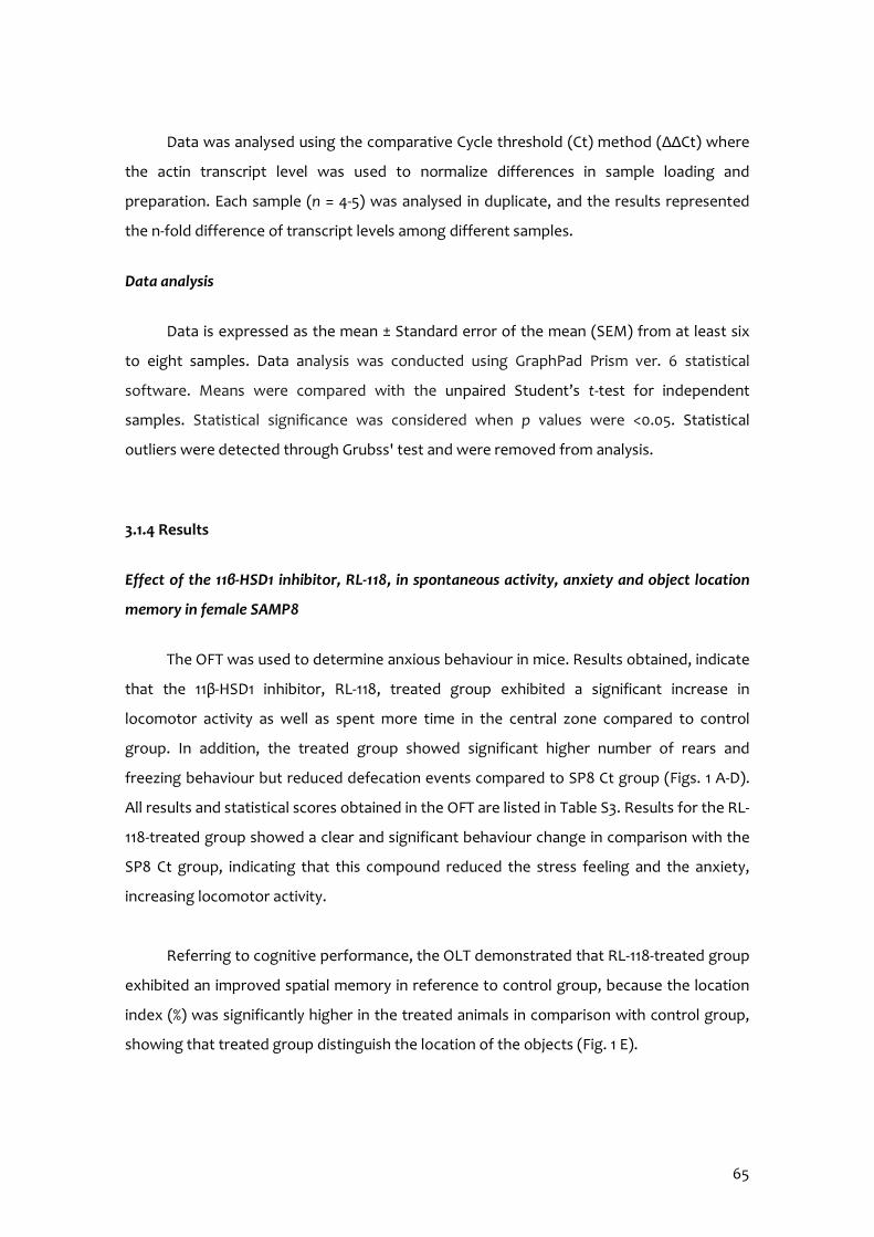
65
Data was analysed using the comparative Cycle threshold (Ct) method (ΔΔCt) where
the actin transcript level was used to normalize differences in sample loading and
preparation. Each sample (n = 4-5) was analysed in duplicate, and the results represented
the n-fold difference of transcript levels among different samples.
Data analysis
Data is expressed as the mean ± Standard error of the mean (SEM) from at least six
to eight samples. Data analysis was conducted using GraphPad Prism ver. 6 statistical
software. Means were compared with the unpaired Student’s t-test for independent
samples. Statistical significance was considered when p values were <0.05. Statistical
outliers were detected through Grubss' test and were removed from analysis.
3.1.4 Results
Effect of the 11β-HSD1 inhibitor, RL-118, in spontaneous activity, anxiety and object location
memory in female SAMP8
The OFT was used to determine anxious behaviour in mice. Results obtained, indicate
that the 11β-HSD1 inhibitor, RL-118, treated group exhibited a significant increase in
locomotor activity as well as spent more time in the central zone compared to control
group. In addition, the treated group showed significant higher number of rears and
freezing behaviour but reduced defecation events compared to SP8 Ct group (Figs. 1 A-D).
All results and statistical scores obtained in the OFT are listed in Table S3. Results for the RL-
118-treated group showed a clear and significant behaviour change in comparison with the
SP8 Ct group, indicating that this compound reduced the stress feeling and the anxiety,
increasing locomotor activity.
Referring to cognitive performance, the OLT demonstrated that RL-118-treated group
exhibited an improved spatial memory in reference to control group, because the location
index (%) was significantly higher in the treated animals in comparison with control group,
showing that treated group distinguish the location of the objects (Fig. 1 E).
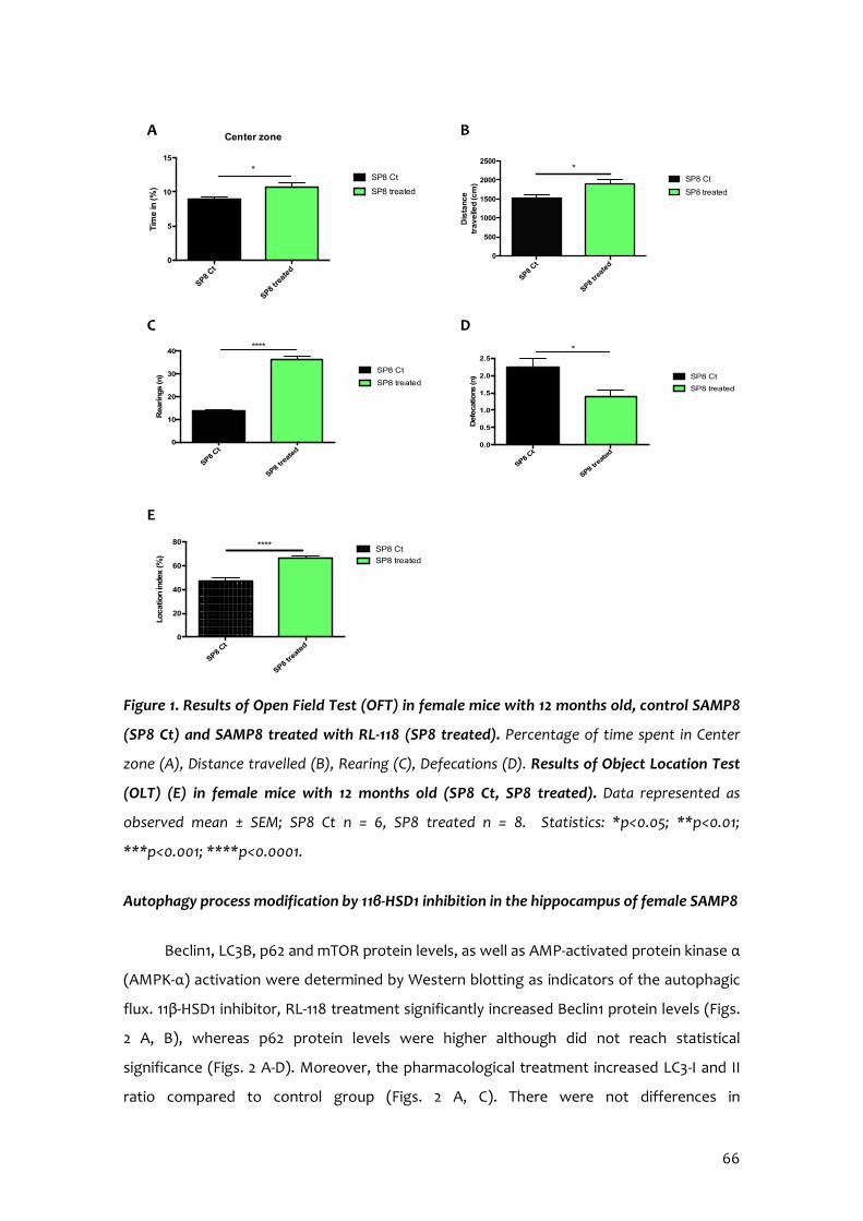
66
Figure 1. Results of Open Field Test (OFT) in female mice with 12 months old, control SAMP8
(SP8 Ct) and SAMP8 treated with RL-118 (SP8 treated). Percentage of time spent in Center
zone (A), Distance travelled (B), Rearing (C), Defecations (D). Results of Object Location Test
(OLT) (Ε) in female mice with 12 months old (SP8 Ct, SP8 treated). Data represented as
observed mean ± SEM; SP8 Ct n = 6, SP8 treated n = 8. Statistics: *p<0.05; **p<0.01;
***p<0.001; ****p<0.0001.
Autophagy process modification by 11β-HSD1 inhibition in the hippocampus of female SAMP8
Beclin1, LC3B, p62 and mTOR protein levels, as well as AMP-activated protein kinase α
(AMPK-α) activation were determined by Western blotting as indicators of the autophagic
flux. 11β-HSD1 inhibitor, RL-118 treatment significantly increased Beclin1 protein levels (Figs.
2 A, B), whereas p62 protein levels were higher although did not reach statistical
significance (Figs. 2 A-D). Moreover, the pharmacological treatment increased LC3-I and II
ratio compared to control group (Figs. 2 A, C). There were not differences in
SP8 Ct
SP8 tre
ated
0
500
1000
1500
2000
2500
Dis
tan
ce
tra
ve
lled
(cm
)
SP8 Ct
SP8 treated
*
SP8 Ct
SP8 tre
ated
0
5
10
15T
ime
in (%
)
Center zone
SP8 Ct
SP8 treated
*
SP8 Ct
SP8 tre
ated
0
10
20
30
40
Rearings (n
)
SP8 Ct
SP8 treated
****
SP8 Ct
SP8 tre
ated
0.0
0.5
1.0
1.5
2.0
2.5
Defe
catio
ns (n
) SP8 Ct
SP8 treated
*
A B
C D
SP8 Ct
SP8 tre
ated
0
20
40
60
80
Locatio
n in
dex
(%)
SP8 Ct
SP8 treated
****
E
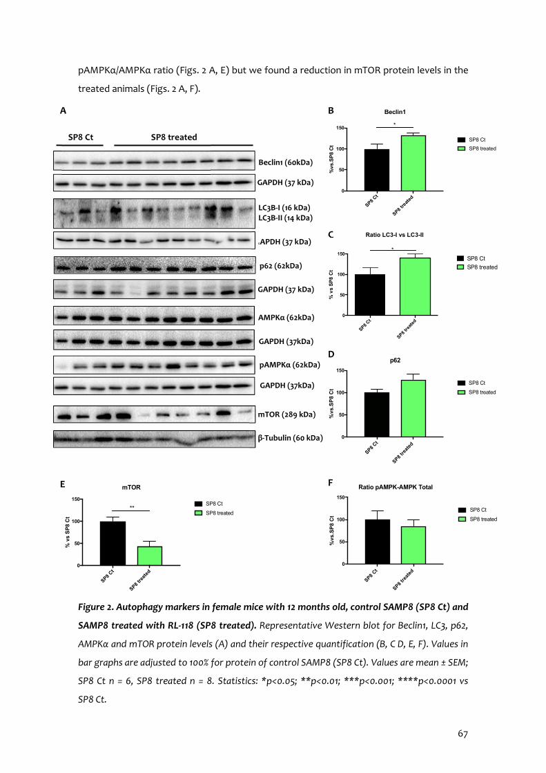
67
pAMPKα/AMPKα ratio (Figs. 2 A, E) but we found a reduction in mTOR protein levels in the
treated animals (Figs. 2 A, F).
Figure 2. Autophagy markers in female mice with 12 months old, control SAMP8 (SP8 Ct) and
SAMP8 treated with RL-118 (SP8 treated). Representative Western blot for Beclin1, LC3, p62,
AMPKα and mTOR protein levels (A) and their respective quantification (B, C D, E, F). Values in
bar graphs are adjusted to 100% for protein of control SAMP8 (SP8 Ct). Values are mean ± SEM;
SP8 Ct n = 6, SP8 treated n = 8. Statistics: *p<0.05; **p<0.01; ***p<0.001; ****p<0.0001 vs
SP8 Ct.
AMPKα (62kDa)
GAPDH (37kDa)
pAMPKα (62kDa)
GAPDH (37kDa)
mTOR (289 kDa)
β-Tubulin (60 kDa)
p62 (62kDa)
GAPDH (37 kDa)
Beclin1 (60kDa) GAPDH (37 kDa)
LC3B-I (16 kDa) LC3B-II (14 kDa)
SP8 Ct SP8 treated
A
SP8 Ct
SP8 tre
ated
0
50
100
150
p62
%vs
.SP
8 C
t
SP8 Ct
SP8 treated
SP8 Ct
SP8 tre
ated
0
50
100
150
Beclin1
%vs
.SP
8 C
t
SP8 Ct
SP8 treated
*
B
SP8 Ct
SP8 tre
ated
0
50
100
150
% v
s S
P8
Ct
Ratio LC3-II vs LC3-I
SP8 Ct
SP8 treated
*
C
D
SP8 Ct
SP8 tre
ated
0
50
100
150
mTOR
% v
s S
P8
Ct
SP8 Ct
SP8 treated**
SP8 Ct
SP8 tre
ated
0
50
100
150
Ratio pAMPK-AMPK Total
%vs
.SP
8 C
t
SP8 Ct
SP8 treated
F
E
Ratio LC3-I vs LC3-II GAPDH (37 kDa)
68
Effect of RL-118, 11β-HSD1 inhibitor, in neurodegenerative, oxidative stress and
neuroinflammation markers in the hippocampus of female SAMP8
11β-HSD1 inhibition by RL-118 increased a disintegrin and metalloproteinase domain-
containing protein 10 (ADAM10) and soluble APPα (sAPPα) protein levels compared to SP8
Ct (Figs. 3 A-C). A significant decrease in tau hyperphosphorylation in Ser 396 and Ser 404 in
SP8 treated compared to SP8 Ct, was found (Figs. 3 D-F).
SP8 Ct o
ld
SP8 tre
ated
0
50
100
150
200ADAM10
%vs
.SP
8 C
t
SP8 Ct old
SP8 treated
B
SP8 Ct
SP8 treated
SP8 tre
ated
SP8 Ct
SP8 Ct o
ld
SP8 tre
atmen
t0
100
200
300
400
sAPPa
%vs
.SP
8 C
t
SP8 Ct old
SP8 treatment
**
C
SP8 Ct
SP8 treated
SP8 tre
ated
SP8 Ct
SP8 Ct SP8 treated
ADAM 10 (84 kDa)
GAPDH (37 kDa)
sAPPα (60 kDa)
GAPDH (37 kDa)
A
Tau total (55 kDa)
GAPDH (37 kDa)
D
SP8 Ct SP8 treated
pTau s396 (55 kDa)
GAPDH (37 kDa)
pTau s404 (55 kDa)
GAPDH (37 kDa)
SP8 Ct o
ld
SP8 tre
ated
0
50
100
150
Ratio pTau s396-Tau Total
%vs
.SP
8 C
t
SP8 Ct old
SP8 treated
*
E
SP8 Ct
SP8 treated
SP8 Ct o
ld
SP8 tre
ated
0
50
100
150
Ratio pTau s404-Tau Total
%vs
.SP
8 C
t
SP8 Ct old
SP8 treated
**
SP8 Ct
SP8 treated
F

69
Figure 3. Alzheimer’s Disease markers in female mice with 12 months-old control, SAMP8
(SP8 Ct) and SAMP8 treated with RL-118 (SP8 treated). Representative Western blot for
ADAM10 and sAPPα protein levels (A) and quantification (B, C), Tau total protein levels and
pTau ser396 and pTau ser404 protein levels (D) and quantification (E, F). Values in bar graphs
are adjusted to 100% for protein of control SAMP8 (SP8 Ct). Values are the mean ± SEM; SP8 Ct
n = 6, SP8 treated n = 8. Statistics: *p<0.05; **p<0.01 vs SP8 Ct.
Neuroimmunological responsive parameters were determined by gene expression of
Il-1β and Ccl3, and also p65 as active form of NF-κβ protein expression. There was a
significant increase in p65 content in SP8 treated group (Figs. 4 A-B). Moreover, SP8
treated group showed a significant lower gene expression of Il-1β and Ccl3 in hippocampus
(Figs. 4 C, D). Response to OS was determined by Hmox1 and Aldh2 gene expression. SP8
treated group showed lower gene expression levels of Hmox1 and Aldh2, suggesting a
lower OS state than in SP8 Ct group (Figs. 4 E, F).
SP8 Ct
SP8 tre
ated
0
50
100
150
NF-kB %
vs
SP
8 C
t
SP8 Ct
SP8 treated
*
B
NF-κB (65 kDa)
GAPDH (37 kDa)
SP8 Ct SP8 treated A
F E
SP8 Ct
SP8 tre
ated
0.0
0.5
1.0
1.5
Aldh2
Re
lativ
e m
RN
A
ex
pre
ss
ion
SP8 Ct
SP8 treated
SP8 Ct
SP8 tre
ated
0.0
0.5
1.0
1.5
Re
lativ
e m
RN
A
ex
pre
ss
ion
IL-1b
SP8 Ct
SP8 treated
**
C
SP8 Ct
SP8 tre
ated
0.0
0.5
1.0
1.5
Re
lativ
e m
RN
A
ex
pre
ss
ion
Ccl3
SP8 Ct
SP8 treated
*
D
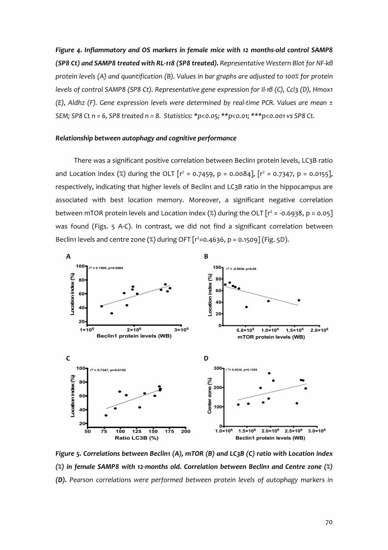
70
Figure 4. Inflammatory and OS markers in female mice with 12 months-old control SAMP8
(SP8 Ct) and SAMP8 treated with RL-118 (SP8 treated). Representative Western Blot for NF-kβ
protein levels (A) and quantification (B). Values in bar graphs are adjusted to 100% for protein
levels of control SAMP8 (SP8 Ct). Representative gene expression for Il-1β (C), Ccl3 (D), Hmox1
(E), Aldh2 (F). Gene expression levels were determined by real-time PCR. Values are mean ±
SEM; SP8 Ct n = 6, SP8 treated n = 8. Statistics: *p<0.05; **p<0.01; ***p<0.001 vs SP8 Ct.
Relationship between autophagy and cognitive performance
There was a significant positive correlation between Beclin1 protein levels, LC3B ratio
and Location index (%) during the OLT [r2 = 0.7459, p = 0.0084], [r2 = 0.7347, p = 0.0155],
respectively, indicating that higher levels of Beclin1 and LC3B ratio in the hippocampus are
associated with best location memory. Moreover, a significant negative correlation
between mTOR protein levels and Location index (%) during the OLT [r2 = -0.6938, p = 0.05]
was found (Figs. 5 A-C). In contrast, we did not find a significant correlation between
Beclin1 levels and centre zone (%) during OFT [r2=0.4636, p = 0.1509] (Fig. 5D).
Figure 5. Correlations between Beclin1 (A), mTOR (B) and LC3B (C) ratio with Location index
(%) in female SAMP8 with 12-months old. Correlation between Beclin1 and Centre zone (%)
(D). Pearson correlations were performed between protein levels of autophagy markers in
5.0×10⁵ 1.0×10⁶ 1.5×10⁶ 2.0×1060
20
40
60
80
100
mTOR protein levels (WB)
Loca
tion in
dex
(%
)
r2 = -0.6938, p=0.05
1×10⁶ 2×10⁶ 3×10⁶
20
40
60
80
100
Beclin1 protein levels (WB)
Loca
tion in
dex
(%
)
r2 = 0.7459, p=0.0084
A B
1.0×10⁶ 1.5×10⁶ 2.0×10⁶ 2.5×10⁶ 3.0×1060
100
200
300
Beclin1 protein levels (WB)
Cen
ter
zone
(%)
r 2= 0.4636, p=0.1509
50 75 100 125 150 175 200
20
40
60
80
100
Ratio LC3B (%)
Locat
ion in
dex
(%
)
r2 = 0.7347, p=0.0155
C D
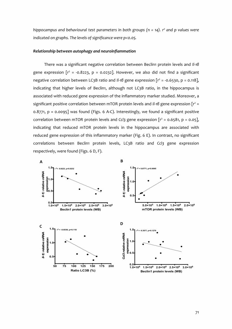
71
hippocampus and behavioural test parameters in both groups (n = 14). r2 and p values were
indicated on graphs. The levels of significance were p<0.05.
Relationship between autophagy and neuroinflammation
There was a significant negative correlation between Beclin1 protein levels and Il-1β
gene expression [r2 = -0.8223, p = 0.0232]. However, we also did not find a significant
negative correlation between LC3B ratio and Il-1β gene expression [r2 = -0.6530, p = 0.118],
indicating that higher levels of Beclin1, although not LC3B ratio, in the hippocampus is
associated with reduced gene expression of the inflammatory marker studied. Moreover, a
significant positive correlation between mTOR protein levels and Il-1β gene expression [r2 =
0.8771, p = 0.0095] was found (Figs. 6 A-C). Interestingly, we found a significant positive
correlation between mTOR protein levels and Ccl3 gene expression [r2 = 0.6581, p = 0.05],
indicating that reduced mTOR protein levels in the hippocampus are associated with
reduced gene expression of this inflammatory marker (Fig. 6 E). In contrast, no significant
correlations between Beclin1 protein levels, LC3B ratio and Ccl3 gene expression
respectively, were found (Figs. 6 D, F).
1.0×10⁶ 1.5×10⁶ 2.0×10⁶ 2.5×10⁶ 3.0×1060.0
0.5
1.0
1.5
Beclin1 protein levels (WB)
Il-1b
rel
ativ
e m
RN
A
exp
ress
ion
r 2= -0.8223, p=0.0232
5.0×10⁵ 1.0×10⁶ 1.5×10⁶ 2.0×10⁶
0.5
1.0
1.5
mTOR protein levels (WB)
Il-1b
rela
tive
mR
NA
ex
pre
ssio
n
r2 = 0.8771, p=0.0095
A B
50 75 100 125 150 175 200
0.5
1.0
1.5
Ratio LC3B (%)
Il-1b
rel
ativ
e m
RN
A
expre
ssio
n
r2 = -0.6530, p=0.118
1.0×10⁶ 1.5×10⁶ 2.0×10⁶ 2.5×10⁶ 3.0×1060.0
0.5
1.0
1.5
Beclin1 protein levels (WB)
Ccl
3 re
lative
mR
NA
ex
pre
ssio
n
r2 = -0.3577, p=0.1279
D C
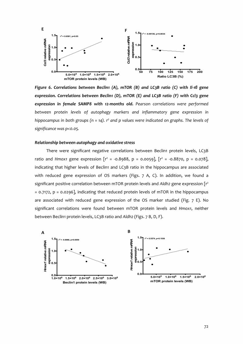
72
Figure 6. Correlations between Beclin1 (A), mTOR (B) and LC3B ratio (C) with Il-1β gene
expression. Correlations between Beclin1 (D), mTOR (E) and LC3B ratio (F) with Ccl3 gene
expression in female SAMP8 with 12-months old. Pearson correlations were performed
between protein levels of autophagy markers and inflammatory gene expression in
hippocampus in both groups (n = 14). r2 and p values were indicated on graphs. The levels of
significance was p<0.05.
Relationship between autophagy and oxidative stress
There were significant negative correlations between Beclin1 protein levels, LC3B
ratio and Hmox1 gene expression [r2 = -0.8988, p = 0.0059], [r2 = -0.8870, p = 0.078],
indicating that higher levels of Beclin1 and LC3B ratio in the hippocampus are associated
with reduced gene expression of OS markers (Figs. 7 A, C). In addition, we found a
significant positive correlation between mTOR protein levels and Aldh2 gene expression [r2
= 0.7172, p = 0.0296], indicating that reduced protein levels of mTOR in the hippocampus
are associated with reduced gene expression of the OS marker studied (Fig. 7 E). No
significant correlations were found between mTOR protein levels and Hmox1, neither
between Beclin1 protein levels, LC3B ratio and Aldh2 (Figs. 7 B, D, F).
50 75 100 125 150 175 2000.0
0.5
1.0
1.5
Ratio LC3B (%)
Ccl
3 re
lative
mR
NA
ex
pre
ssio
n
r2 = -0.09726, p=0.8034
5.0×10⁵ 1.0×10⁶ 1.5×10⁶ 2.0×1060.0
0.5
1.0
1.5
mTOR protein levels (WB)
Ccl
3 re
lative
mR
NA
ex
pre
ssio
n
r2 = 0.6581, p=0.05
E F
1.0×10⁶ 1.5×10⁶ 2.0×10⁶ 2.5×10⁶ 3.0×1060.0
0.5
1.0
1.5
Beclin1 protein levels (WB)
Hm
ox1
rel
ativ
e m
RN
A
expre
ssio
n
r2 = -0.8988, p=0.0059
5.0×10⁵ 1.0×10⁶ 1.5×10⁶ 2.0×1060.0
0.5
1.0
1.5
mTOR protein levels (WB)
Hm
ox1
rel
ativ
e m
RN
A
expre
ssio
n
r2 = 0.5574, p=0.1936
A B
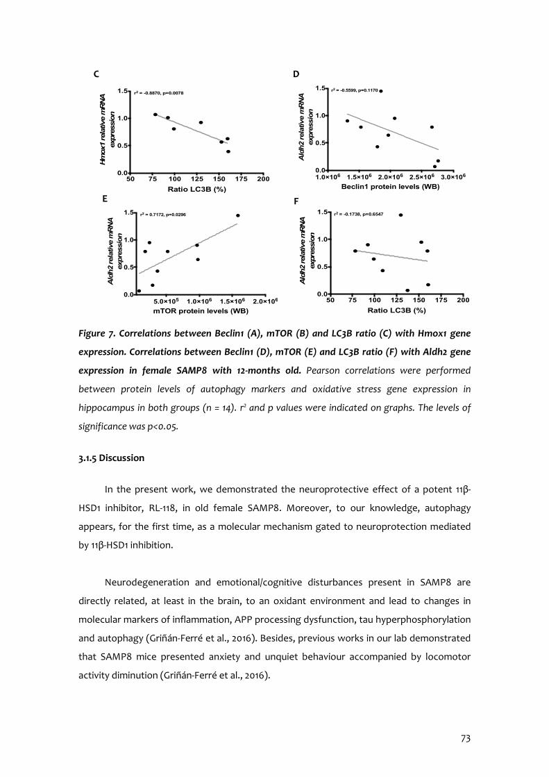
73
Figure 7. Correlations between Beclin1 (A), mTOR (B) and LC3B ratio (C) with Hmox1 gene
expression. Correlations between Beclin1 (D), mTOR (E) and LC3B ratio (F) with Aldh2 gene
expression in female SAMP8 with 12-months old. Pearson correlations were performed
between protein levels of autophagy markers and oxidative stress gene expression in
hippocampus in both groups (n = 14). r2 and p values were indicated on graphs. The levels of
significance was p<0.05.
3.1.5 Discussion
In the present work, we demonstrated the neuroprotective effect of a potent 11β-
HSD1 inhibitor, RL-118, in old female SAMP8. Moreover, to our knowledge, autophagy
appears, for the first time, as a molecular mechanism gated to neuroprotection mediated
by 11β-HSD1 inhibition.
Neurodegeneration and emotional/cognitive disturbances present in SAMP8 are
directly related, at least in the brain, to an oxidant environment and lead to changes in
molecular markers of inflammation, APP processing dysfunction, tau hyperphosphorylation
and autophagy (Griñán-Ferré et al., 2016). Besides, previous works in our lab demonstrated
that SAMP8 mice presented anxiety and unquiet behaviour accompanied by locomotor
activity diminution (Griñán-Ferré et al., 2016).
50 75 100 125 150 175 2000.0
0.5
1.0
1.5
Ratio LC3B (%)A
ldh2
rela
tive
mR
NA
ex
pre
ssio
n
r2 = -0.1738, p=0.6547
5.0×10⁵ 1.0×10⁶ 1.5×10⁶ 2.0×1060.0
0.5
1.0
1.5
mTOR protein levels (WB)
Ald
h2
rela
tive
mR
NA
ex
pre
ssio
n
r2 = 0.7172, p=0.0296
E F
1.0×10⁶ 1.5×10⁶ 2.0×10⁶ 2.5×10⁶ 3.0×1060.0
0.5
1.0
1.5
Beclin1 protein levels (WB)
Ald
h2
rela
tive
mR
NA
ex
pre
ssio
n
r2 = -0.5599, p=0.1170
50 75 100 125 150 175 2000.0
0.5
1.0
1.5
Ratio LC3B (%)
Hm
ox1
rel
ativ
e m
RN
A
expre
ssio
n
r2 = -0.8870, p=0.0078
C D
74
According to previous reports (Holmes et al., 2010; Mohler et al., 2011; Sooy et al.,
2010; Yau et al., 2001, 2007), modulation of 11β-HSD1 or pharmacological treatment with 11β-
HSD1 inhibitors leads to ameliorations in memory abilities and beneficial effects on
behaviour, reducing fear sensation and increasing locomotor activity. Also, it has been
published that cognitive improvement delivered by RL-118 occurs jointly with synaptic
plasticity recovery (assessed by PSD95 marker) and reduction in amyloidogenic APP
processing pathway, tau hyperphosphorylation and inflammatory markers (Leiva et al.,
2017). Altogether indicate a putative therapeutic role for 11β-HSD1 in neurodegenerative
disorders like AD.
There is evidence that higher plasma GC levels due to stress influence hippocampal
atrophy (Mah et al., 2015). Besides, patients with mild cognitive impairment (MCI) and
mood disorders have neuropathological AD-like hallmarks in the cerebrospinal fluid,
supporting GCs role in cognitive impairment (Ramakers et al., 2013). Moreover, GCs are
physiological immunomodulatory hormones that not only regulate immune cell
development, but also decrease autophagy process and trigger apoptosis (El Zaoui et al.,
2015; Harr et al., 2010).
The two major systems responsible for the digestion of most cytosolic and
aggregated or misfolded protein in brain cells are UPS and the autophagy-lysosome
pathway. The decline in autophagy function could determine cell and individual lifespan
(Guo et al., 2017; Maiese, 2016; Triplett et al., 2017). Thus, autophagy impairment has been
related to several neurodegenerative disorders characterized by accumulation of
intracellular protein aggregates in specific brain regions, such as AD, Parkinson’s and
Huntington’s disease (Koyuncu et al., 2017; Rahman & Rhim, 2017). Specifically in AD,
autophagy dysfunction leads to β-amyloid aggregation, accumulation, therefore favouring
the formation of SPs (Funderbunk et al., 2010). Compounds improving autophagy-lysosome
pathway have been reported to be neuroprotective in experimental models of AD
(Caballero & Coto-Montes, 2012; Friedman et al., 2015).
Herein, increased levels of Beclin1 and p65 were found in RL-118 treated mice. It is
well known that Beclin1 regulates autophagic flux as it orchestrates autophagosome
formation (Kang et al., 2011; Wirawan et al., 2012). On the other hand, p65 directly binds to
Beclin1 promoter and up-regulates gene expression of proteins involved in autophagy,
leading to its activation. In turn, p62 protein recognizes toxic cellular waste and ubiquitin-
75
containing aggregates, ensuing the subsequent turnover of potentially harmful proteins by
autophagy (Rusten et al., 2010).
To complete autophagic process, autophagosome formation is needed and LC3B is
one of the mediators. LC3B-I is the cytoplasm active form and it conjugates with
phosphatydilethanolamine through two consecutive ubiquitylation-like reactions to
become LC3B-II that is recruited to autophagosome membrane (Klionsky et al., 2008;
Tanida et al., 2008). 11β-HSD1 inhibition by R-118 promoted LC3B-II levels in detriment to
LC3B-I indicating autophagosome formation and enhanced autophagy. In addition,
upstream in this process there are AMPKα and mTOR, which modulate autophagic flux. On
one hand, AMPKα activation acts as an autophagy activator signal, whereas mTOR pathway
is one of the main cellular autophagy inhibitors. The mTOR is a serine/threonine protein
kinase involved in the process of cell growth, regulation of energy homeostasis and
metabolism after nutrient ingestion. The importance and complexity of mTOR signalling is
evident, as it is linked to several neurodegenerative diseases via its role in autophagy
regulation. We found an increase in the ratio pAMPKα/AMPKα induced by 11β-HSD1
inhibitor, demonstrating that RL-118 renders a better autophagic flux process in SAMP8
brain. Autophagy promotion by RL-118 should force the removal of aberrant proteins in the
hippocampus and, in consequence, improve cognitive and behavioural disturbances.
Moreover, correlations between the autophagy protein levels studied and behavioural
parameters suggest that the cognitive amelioration found after RL-118 treatment is related
to an autophagy potentiation. Of note, RL-118 induced a diminution in hyperphosphorylated
tau protein at Ser396 and Ser404 residues indicating, once more, an improvement in
autophagy.
Molecular, biochemical and cellular changes implicated in cellular senescence are
intrinsically gated to inflammation and OS (López-Otín et al., 2013). Aged population show a
low chronic inflammatory state, which worsens in AD patients. In fact, AD pathology is
characterized by a chronic elevation of OS and increased gliosis and neurodegeneration
(Ginaldi et al., 2001; Williams et al., 2015).
Because altered inflammatory and OS markers levels are already described in SAMP8,
here we screened gene expression of some of the most important proteins implicated in
those processes. The results obtained demonstrated a lower gene expression of Il-1β and
Hmox1 in SP8 treated mice and an increase in the active fraction of NF-κB indicating that
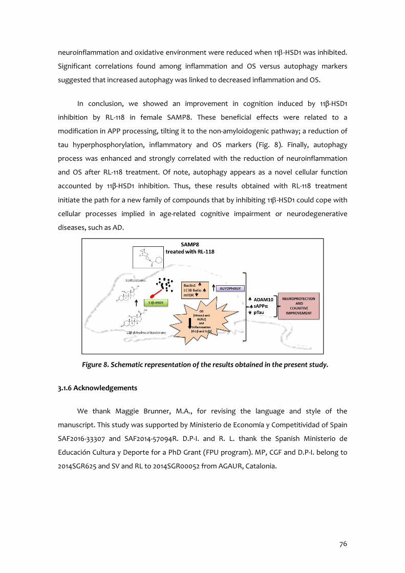
76
neuroinflammation and oxidative environment were reduced when 11b-HSD1 was inhibited.
Significant correlations found among inflammation and OS versus autophagy markers
suggested that increased autophagy was linked to decreased inflammation and OS.
In conclusion, we showed an improvement in cognition induced by 11β-HSD1
inhibition by RL-118 in female SAMP8. These beneficial effects were related to a
modification in APP processing, tilting it to the non-amyloidogenic pathway; a reduction of
tau hyperphosphorylation, inflammatory and OS markers (Fig. 8). Finally, autophagy
process was enhanced and strongly correlated with the reduction of neuroinflammation
and OS after RL-118 treatment. Of note, autophagy appears as a novel cellular function
accounted by 11β-HSD1 inhibition. Thus, these results obtained with RL-118 treatment
initiate the path for a new family of compounds that by inhibiting 11b-HSD1 could cope with
cellular processes implied in age-related cognitive impairment or neurodegenerative
diseases, such as AD.
Figure 8. Schematic representation of the results obtained in the present study.
3.1.6 Acknowledgements
We thank Maggie Brunner, M.A., for revising the language and style of the
manuscript. This study was supported by Ministerio de Economía y Competitividad of Spain
SAF2016-33307 and SAF2014-57094R. D.P-I. and R. L. thank the Spanish Ministerio de
Educación Cultura y Deporte for a PhD Grant (FPU program). MP, CGF and D.P-I. belong to
2014SGR625 and SV and RL to 2014SGR00052 from AGAUR, Catalonia.
77
3.1.7 References
Anderson RA, Qin B, Canini F, Poulet L, Roussel AM (2013) Cinnamon counteracts the negative effects of a high fat/high fruc- tose diet on behavior, brain insulin signalling and Alzheimer- associated changes. PLoS One 8(12):e83243.
Boya P, Reggiori F, Codogno P (2013) Emerging regulation and functions of autophagy. Nat Cell Biol 15(7):713–720.
Caballero B, Coto-Montes A (2012) An insight into the role of autophagy in cell responses in the aging and neurodegenerative brain. Histol Histopathol 27(3):263–275.
Casadesús G, Shukitt-Hale B, Joseph JA (2002) Qualitative versus quantitative caloric intake: are they equivalent paths to successful aging? Neurobiol Aging 23(5):747–769.
De Quervain DJ, Poirier R, Wollmer MA, Grimaldi LM, Tsolaki M, Streffer JR, Hock C, Nitsch RM et al (2004) Glucocorticoid-related genetic susceptibility for Alzheimer’s disease. Hum Mol Genet 13 (1):47–52.
El Zaoui I, Behar-Cohen F, Torriglia A (2015) Glucocorticoids exert direct toxicity on microvasculature: analysis of cell death mechanisms. Toxicol Sci 143(2):441–453.
Friedman LG, Qureshi YH, Yu WH (2015) Promoting autophagic clearance: viable therapeutic targets in Alzheimer’s disease. Neurotherapeutics 12(1):94–108.
Funderburk SF, Marcellino BK, Yue Z (2010) Cell Bself-eating^ (autophagy) mechanism in Alzheimer’s disease. Mt Sinai J Med 77(1):59–68.
Gella A, Durany N (2009) Oxidative stress in Alzheimer disease. Cell Adhes Migr 3(1):88–93.
Ginaldi L, De Martinis M, D’Ostilio A, Marini L, Loreto F, Modesti M, Quaglino D (2001) Changes in the expression of surface recep- tors on lymphocyte subsets in the elderly: quantitative flow cyto- metric analysis. Am J Hematol 67(2):63–72.
Griñán C, Palomera-Ávalos V, Puigoriol-Illamola D, Camins A, Porquet D, Plá V, Aguado F, Pallàs M (2016) Behavior and cogni- tive changes correlated with hippocampal neuroinflammaging and neuronal markers in female SAMP8, a model of accelerated senes- cence. Exp Gerontol 80:57–69.
Guo H, Chen Y, Liao L, Wu W (2013) Resveratrol protects HUVECs from oxidized-LDL induced oxidative damage by au- tophagy upregulation via the AMPK7SIRT1 pathway. Cardiovasc Drugs Ther 27:189–198.
Harr MW, Distelhorst CW (2010) Apoptosis and autophagy: decoding calcium signals that mediate life or death. Cold Spring Harb Perspect Biol 2(10):a005579.
78
Höhn A, Weber D, Jung T, Ott C, Hugo M, Kochlik B, Kehm R, König J et al (2016) Happily (n)ever after: aging in the context of oxidative stress, proteostasis loss and cellular senescence. Redox Biol S2213-2317(16):30351–30352.
Holmes MC, Carter RN, Noble J, Chitnis S, Dutia A, Paterson JM, Yau JLW (2010) 11β-Hydroxysteroid dehydrogenase type 1 ex- pression is increased in the aged mouse hippocampus and parietal cortex and causes memory impairments. J Neurosci 30(20):6916– 6920.
Kang R, Zeh HJ, Lotze MT, Tang D (2011) The Beclin 1 network regulates autophagy and apoptosis. Cell Death Differ 18(4):571– 580.
Klionsky DJ, Abeliovich H, Agostinis P, Agrawal DK, Aliev G, Askew DS, Baba M, Baehrecke EH et al (2008) Guidelines for the use and interpretation of assays for monitoring autophagy in higher eukaryotes. Autophagy 4(2):151–175.
Komatsu M, Waguri S, Chiba T, Murata S, Iwata J, Tanida I, Ueno T, Koike M et al (2006) Loss of autophagy in the central nervous system causes neurodegeneration in mice. Nature 441:880–884.
Koyuncu S, Fatima A, Gutierrez-Garcia R, Vilchez D (2017) Proteostasis of huntingtin in health and disease. Int J Mol Sci 18 (7):1568.
Lara VP, Caramelli P, Teixeira AL, Barbosa MT, Carmona KC, Carvalho MG, Fernandes AP, Gomes KB (2013) High cortisol levels are associated with cognitive impairment no-dementia (CIND) and dementia. Clin Chim Acta 423:18–22.
Leiva R, Griñán-Ferré C, Seira C, Valverde E, McBride A, Binnie M, Pérez B, Luque FJ et al (2017) Design, synthesis and in vivo study of novel pyrrolidine-based 11β-HSD1 inhibitors for age- related cognitive dysfunction. Eur J Med Chem 139:412–428.
Levine B (2005) Eating oneself and uninvited guests: autophagy- related pathways in cellular defense. Cell 120:159–162.
López-Otín C, Blasco MA, Partridge L, Serrano M, Kroemer G (2013) The hallmarks of aging. Cell 153(6):1194–1217.
Mah L, Binns MA, Steffens DC, Alzheimer’s Disease Neuroimaging Initiative (2015) Anxiety symptoms in amnestic mild cognitive impairment are associated with medial temporal atrophy and predict conversion to Alzheimer disease. Am J Geriatr Psychiatry 23:466–476.
Maiese K (2016) Targeting molecules to medicine with mTOR, autophagy and neurodegenerative disorders. Br J Clin Pharmacol 82(5):1245–1266.
79
Mathew R, Karp C, Beaudoin B, Vuong N, Chen G, Chen HY, Bray K, Reddy A et al (2009) Autophagy suppresses tumorigenesis through elimination of p62. Cell 137(6):1062–1075.
Mohler EG, Browman KE, Roderwald VA, Cronin EA, Markosyan S, Scott Bitner R, Strakhova MI, Drescher KU et al (2011) Acute inhibition of 11β hydroxysteroid dehydrogenase type-1 improves memory in rodent models of cognition. J Neurosci 31(14):5406– 5413.
Morley JE, Farr SA, Kumar VB, Armbrecht HJ (2012) The SAMP8 mouse: a model to develop therapeutic interventions for Alzheimer’s disease. Curr Pharm 18(8):1123–1130.
Murai T, Okuda S, Tanaka T, Ohta H (2007) Characteristics of object location memory in mice: behavioral and pharmacological studies. Physiol Behav 90:116–124.
Nomura Y, Okuma Y (1999) Age-related defects in lifespan and learning ability in SAMP8 mice. Neurobiol Aging 20(2):111–115
Pallàs M, Camins A, Smith MA, Perry G, Lee HG, Casadesus G (2008) From aging to Alzheimer’s disease: unveiling Bthe switch^ with the senescence-accelerated mouse model (SAMP8). J Alzheimers Dis 15(4):615–624.
Rahman MA, Rhim H (2017) Therapeutic implication of autophagy in neurodegenerative diseases. BMB Rep 50(7):345–354.
Ramakers IHGB, Verhey FRJ, Scheltens P, Hampel H, Soininen H, Aalten P, Rikkert MO, Verbeek MM et al (2013) Anxiety is related to Alzheimer cerebrospinal fluid markers in subjects with mild cog- nitive impairment. Psychol Med 43:911–920.
Rusten TE, Stenmark H (2010) p62, an autophagy hero or culprit? Nat Cell Biol 12(3):207–209.
Son JH, Shim JH, Kim K-H, Ha J-Y, Han JY (2012) Neuronal autophagy and neurodegenerative diseases. Exp Mol Med 44(2): 89–98.
Sooy K, Webster SP, Noble J, Binnie M, Walker BR, Seckl JR, Yau JLW (2010) Partial deficiency or short-term inhibition of 11β- hydroxysteroid dehydrogenase type 1 improves cognitive function in ageing mice. J Neurosci 30(41):13867–13872.
Tanida I, Ueno T, Kominami E (2008) LC3 and autophagy. Methods Mol Biol 445:77–88.
Terao A, Apte-Deshpande A, Dousman L, Morairty S, Eynon BP, Kilduff TS, Freund YR (2002) Immune response gene expression increases in the aging murine hippocampus. J Neuroimmunol 132 (1–2):99–112.

80
Triplett JC, Tramutola A, Swomley A, Kirk J, Grimes K, Lewis K, Orr M, Rodriguez K et al (2015) Age-related changes in the proteostasis network in the brain of the naked mole-rat: implications promoting healthy longevity. Biochim Biophys Acta 1852:2213– 2224.
Vílchez D, Saez I, Dillin A (2014) The role of protein clearance mechanisms in organismal ageing and age-related diseases. Nat Commun 5:5659.
Williams AD, Brown BE, Putchim L, Sweet MJ (2015) Age-related shifts in bacterial diversity in a reef coral. PLoS One 10(12): e0144902.
Wirawan E, Lippens S, Vanden Berghe T, Romagnoli A, Fimia GM, Piacentini M, Vandenabeele P (2012) Beclin 1: a role in mem- brane dynamics and beyond. Autophagy 8(1):6–17.
Yau JL, McNair KM, Noble J, Brownstein D, Hibberd C, Morton N, Mullins JJ, Morris RG et al (2007) Enhanced hippocampal longterm potentiation and spatial learning in aged 11β- hydroxysteroid dehydrogenase type 1 knock-out mice. J Neurosci 27:10487–10496.
Yau JL, Noble J, Kenyon CJ, Hibberd C, Kotelevtsev Y, Mullins JJ, Seckl JR (2001) Lack of tissue glucocorticoid reactivation in 11β- hydroxysteroid dehydrogenase type 1 knockout mice ameliorates age-related learning impairments. Proc Natl Acad Sci U S A 98: 4716–4721.
Ye SM, Johnson RW (1999) Increased interleukin-6 expression by microglia from brain of aged mice. J Neuroimmunol 93 (1–2): 139– 148
Yu WH, Kumar A, Peterhoff C, Shapiro Kulnane L, Uchiyama Y, Lamb BT, Cuervo AM, Nixon RA (2004) Autophagic vacu- oles are enriched in amyloid precursor protein-secretase activi- ties: implications for beta-amyloid peptide over-production and localization in Alzheimer’s disease. Int J Biochem Cell Biol 36: 2531–2540.
3.1.8 Supplementary material
The online version of this article (https://doi.org/10.1007/s12035-018-1026-8) contains
electronic supplementary material, which is available to authorized users.

3.2 Chronic Mild Stress Modified Epigenetic Mechanisms Leading to Accelerated Senescence and Impaired Cognitive Performance in Mice
Adapted from: Puigoriol-Illamola, D.1,3, Martínez-Damas, M.2, Griñán-Ferré, C.1,3, Pallàs, M1,3. Chronic Mild Stress Modified Epigenetic Mechanisms Leading to Accelerated Senescence
and Impaired Cognitive Performance in Mice
International Journal of Molecular Sciences 2020; 21(3):1154. doi:10.3390/ijms21031154. Epub 2020 Feb 10.

82
Author information: 1 Department of Pharmacology, Toxicology and Therapeutic Chemistry. Pharmacology Section. Faculty of Pharmacy and Food Sciences. University of Barcelona, Av Joan XXIII 27-31, 08028 Barcelona, Spain. 2 Institute of Biomedical Research (IIBO), Universidad Nacional Autónoma de México (UNAM). Subdirección de Enseñanza e Investigación, División de Investigación Biomédica, Centro Médico Nacional 20 de Noviembre. México City, México. 3 Institute of Neuroscience, University of Barcelona (NeuroUB), Passeig de la Vall d’Hebron 171, Barcelona, Spain.

83
3.2.1 Abstract
Cognitive and behavioural disturbances are growing public healthcare issue for
modern society, which is experiencing an increasingly common stressful lifestyle. Besides,
several pieces of evidence state that the environment is crucial in the development of
several diseases as well as compromising healthy ageing. With upcoming age, the capability
to fight against harmful stimuli decreases and the organism becomes more vulnerable to
infections and disease. In agreement, stressful experiences have been identified as an
important risk factor for cognitive impairment. Therefore, it is important to study the
molecular mechanisms underpinning the effects of chronic stress on cognition and its
relationship with ageing in order to unveil what challenges we might have to cope with as a
society in the not-so-far future. The study hypothesised that chronic stress would modulate
a large constellation of cellular mechanisms implied in ageing and age-related
neurodegenerative pathologies. Thus, the aim was to determine which molecular
mechanisms were altered after chronic stress situations in an ageing animal model that
could be responsible of cognitive decline.
To address these queries, Chronic Mild Stress (CMS) paradigm was used in the
SAMP8 and SAMR1. On the one hand, we determined the changes produced in the three
main epigenetic marks after 4 weeks of CMS treatment, such as a reduction in histone PTM
and DNA methylation, and up- or down-regulation of several miRNAs involved in different
cellular processes in mice. In addition, CMS treatment induced ROS damage accumulation
and loss of antioxidant defence mechanisms like NRF2, GPX1, SOD1 and CAT, as well as
inflammatory signalling activation through NF-κB pathway and astrogliosis markers, like Il-6,
Tnf-α and glial fibrillar acidic protein (Gfap). Remarkably, CMS altered mTORC1 signalling in
both strains, decreasing autophagy only in SAMR1 mice.
We found a decrease in GSK-3β inactivation, hyperphosphorylation of Tau and an
increase in soluble amyloid precursor protein β (sAPPβ) protein levels in mice under CMS.
Moreover, the non-amyloidogenic secretase, a disintegrin and metalloproteinase-domain
containing protein 10 (ADAM10) protein levels were reduced in SAMR1 CMS group.
Consequently, detrimental effects on behaviour and cognitive performance were detected
in CMS treated mice, affecting mainly SAMR1 mice, promoting a turning to SAMP8
phenotype. In conclusion, CMS is a feasible intervention to understand the influence of
stress on epigenetic mechanisms underlying cognition and accelerating senescence.

84
3.2.2 Introduction
Aging is a multifactorial process characterized by a progressive loss of physiological
integrity, resulting in an impaired function and increased vulnerability to death.
Physiological brain aging involves cognitive impairment, which includes decreased learning
and memory abilities and slower responses to different stimulus (Cristòfol et al., 2012).
Indeed, normal aging differs from pathological aging and this might be explained by the
lifestyle. Environmental factors drive epigenetic modifications resulting in phenotypic
differences, which alter almost all tissues and organs. Although the brain is one of the most
affected structures, such modifications lead to a progressive decline in the cognitive
function and create a favourable context for the development of neurodegenerative
diseases (Harman & Martín, 2019). Aging is the most significant risk factor for most chronic
diseases, such as age-related cognitive decline and Alzheimer’s disease (AD) (Guerreiro &
Bras, 2015). In fact, AD is the most prevalent dementia and its progression is influenced by
both genetic and environmental factors (Griñán-Ferré et al., 2016).
Several studies define that a good environment is essential to enhance learning and
cognitive abilities, besides the continued presence of stressors is related to the opposite
effects. Epigenetics refers to the potentially heritable and environmentally modifiable
changes in gene expression mediated via non-encoded DNA mechanisms (Sun et al., 2013).
Recent evidence has demonstrated significant associations between epigenetic alterations
and stress (Harman & Martín, 2019; Turecki & Meaney, 2016). Potential threats cause a
course of action releasing numerous transmitters and hormones throughout our body,
particularly catecholamines and glucocorticoids. The interaction of glucocorticoids and
adrenergic systems in specific brain regions has proved an essential mediating mechanism
for a wide variety of actions displayed by stress on cognition (Sandi, 2013). Firstly, stress
response allows body adaptation. However, when stress is prolonged it has been shown
that decreases synaptic plasticity (Wang et al., 2019), alters hippocampal volume (Borcel et
al., 2008; Rahman et al., 2016) and neurotransmitters (Jett et al., 2017) and may lead to
Alzheimer’s disease (Justice, 2018). In particular, chronic stress is associated with increased
Aβ deposits and hyperphosphorylated Tau (Bisht et al., 2018).
Oxidative stress (OS) and inflammation are deeply involved in age-related deleterious
disorders. Along with the aging process, several factors, such as a naturally decreased
capacity of the antioxidant enzymes system, create an imbalance between antioxidant
85
mechanisms and reactive oxygen system (ROS)-production equilibrium, accumulating ROS
beyond the detoxifying capacity of the antioxidant system resulting in OS, eventually
causing cellular damage that can no longer be repaired by internal mechanisms, finally
causing the dysfunction of the system (Griñán-Ferré et al., 2018). In addition, one of the
major changes while aging is the dysregulation of the immune response, leading to chronic
systemic inflammatory state (Chung et al., 2019). Overall, OS imbalance, mitochondrial
dysfunction and the inflammatory response have been linked to accelerate aging and
fasten progression of neurodegenerative diseases (Griñán-Ferré et al., 2018; Petersen &
Smith, 2016). Moreover, accumulation of dysfunctional and damaged cellular proteins and
organelles occurs during aging, resulting in a disruption of cellular homeostasis and
progressive degeneration and increasing the risk of cell death (Escobar et al., 2019).
Autophagy is particularly important in the maintenance of homeostasis, as it participates in
the elimination of disrupted proteins and molecules and helps to maintain a clean cellular
environment. It has been widely described that there are alterations in autophagy while
aging as well as in age-related cognitive decline, AD and other dementias (Gouras, 2019;
Martinez-Lopez, 2015; Puigoriol-Illamola et al., 2018). Moderating all these detrimental
components is key in the promotion of cell survival and longevity.
The senescence-accelerated mouse (SAM) models include senescence-accelerated
mice prone (SAMP) and resistant (SAMR) mice (Takeda et al., 1991). SAMP8 strain has been
widely used as an aging model for the study of brain aging and age-related pathologies. Its
cognitive impairment is linked with alterations in both hippocampal structure and activity,
causing OS imbalance and driving to AD neuropathology, such as tau- and amyloid-related
alterations (Cristòfol et al., 2012; Wang et al., 2016). On the contrary, SAMR1 strain
manifests a normal aging process.
Understanding the epigenetic modifications that stressful environment triggers in
neurodegeneration mechanisms is essential to develop novel therapeutics for age-related
cognitive decline. This study aims to determine the effects on brain function of a stressful
lifestyle in an animal model with accelerated senescence, SAMP8, and in the resistant
senescence strain, SAMR1.
86
3.2.3 Materials and methods
Animals and Chronic Mild Stress Procedure
Females SAMP8 mice (n=56) were used to perform behavioural, cognitive and
molecular analyses. We divided these animals into four groups: SAMR1 (SAMR1 control,
n=14), SAMR1 treated with CMS (SAMR1 CMS, n=14), SAMP8 (SAMP8 control, n=14) and
SAMP8 treated with CMS (SAMP8 CMS, n=14). Animals had free access to food and water
and were kept under standard temperature conditions (22±2°C) and 12h: 12h light-dark
cycles (300lux/0 lux). Starting at 4 months of age, CMS groups received the Chronic Mild
Stress (CMS) treatment. CMS procedure that was used in the present study had previously
been validated in SAMR1 and SAMP8 mice, with some modifications (Wang et al., 2016).
Mice were exposed to various randomly scheduled, low-intensity environmental stressors
every day for 4 weeks. Different stressful stimuli were applied every day and the sequence
of the stressors was altered every week to guarantee the degree of unpredictability.
Among others, CMS stimuli were 24h of water deprivation, 24h of food deprivation, 2h of
physical restraint, 24h of sawdust removal, 24h of wet bedding, overnight illumination and
1min of tail nipping at 1cm from the tip of the tail. Before the performance of the cognitive
tests, the glucose tolerance test was conducted.
All experimental procedures involving animals were performed followed by standard
ethical guidelines European Communities Council Directive 86/609/EEC and by the
Institutional Animal Care and Use Committee of the University of Barcelona (670/14/8102,
approved at 11/14/2014) and by Generalitat de Catalunya (10291, approved 1/28/2018). All
efforts were made to minimize the number of mice used and their suffering.
Glucose tolerance test
Intraperitoneal (i.p.) glucose tolerance test was performed following 4 weeks of CMS
treatment, as described previously. In brief, mice fasted overnight for 12h. The test was
performed in a quiet room and 2g/kg i.p. glucose injection was administered (diluted in H2O)
and blood glucose levels were measured at 0, 5, 10, 15, 30, 60 and 120 min after the injection
with the Accu-Chek® Aviva blood glucose meter (Accu-Chek® Aviva, Roche, Barcelona,
Spain).
87
Behavioural and cognitive test
Open Field Test (OFT)
The OFT was performed as previously described in Griñán-Ferré et al. (2015). It
evaluates anxiety-like behaviour. Mice were placed at the centre of a white polywood box
(50x50x25cm) and allowed to explore it for 5 minutes. Behaviour was scored with SMART®
ver.3.0 software and each trial was recorded for later analysis. The parameters scored
included centre staying duration, rearing, grooming and the distance travelled.
Novel Object Recognition Test (NORT)
The Novel Object Recognition Test (NORT) protocol employed was as described in
Puigoriol-Illamola et al. (Puigoriol-Illamola et al., 2020). In brief, mice were placed in a 90°,
two-arms, 25cm-long, 20cm-high, 5cm-wide black maze. Before performing the test, the
mice were individually habituated to the apparatus for 10min for 3 days. On day 4, the
animals were submitted to a 10min acquisition trial (first trial), during which they were
placed in the maze in the presence of two identical, novel objects at the end of each arm.
After a delay (2h and 24h), the animal was exposed to two objects one old object and one
novel object. The Time that mice explored the Novel object (TN) and Time that mice
explored the Old object (TO) were measured. A Discrimination Index (DI) was defined as
(TN−TO)/(TN+TO). To avoid object preference biases, objects were counterbalanced. The
maze, the surface, and the objects were cleaned with 70% ethanol between the animals’
trials to eliminate olfactory cues.
Morris Water Maze (MWM)
This test evaluates both learning and spatial memory (Vorhees et al., 2006). An open
circular pool (100 cm in diameter, 50 cm in height) filled with water was used. Water was
painted white with latex in order to make it opaque and its temperature was 22± 1°C. Two
main perpendicular axes were established (North-South and East-West), thus configuring
four equal quadrants (NE, NW, SE, and SW). Four visual clues (N, S, E, W) were placed on
the walls of the tank so that the animal could orientate and could fulfil the objective. The
test consists of training a mouse to find a submerged platform (Learning phase) and assess
whether the animal has learned and remembered where the platform was the day that it is
removed (Test). The training lasts five consecutive days and every day five trials are

88
performed, which have a different starting point (NE, E, SE, S, and SW), with the aim that
the animal recognizes the visual clues and learns how to locate the platform, avoiding
learning the same path. At each trial, the mouse was placed gently into the water, facing
the wall of the pool, allowed to swim for 60 seconds and there was not a resting time
between trials. If the animal was not able to locate the platform, the investigator guided it
to the platform and was allowed to rest and orientate for 30 seconds. The platform was
placed approximately in the middle of one of the quadrants, 1.5 cm below the water level.
Above the pool there was a camera that recorded the animals’ swimming paths and the
data was analysed with the statistical program SMART® ver.3.0. During the learning phase,
a learning curve was drawn, in which is represented the latency to find the platform every
training day. On the day test, more parameters were measured, such as the target
crossings and the swum distance in the platform zone.
Immunodetection experiments
Brain processing
Three days after the behavioural and cognitive tests, animals were euthanized for
protein extraction, RNA and DNA isolation. Brains were immediately removed, and the
hippocampus was isolated, frozen on powdered dry ice and maintained at -80°C until
procedures.
Western blotting
Tissue samples were homogenized in lysis buffer (Tris HCl pH 7.4 50mM, NaCl 150mM,
EDTA 5mM and 1X-Triton X-100) containing phosphatase and protease inhibitors (Cocktail II,
Sigma-Aldrich) to obtain total protein homogenates. Aliquots of 15 μg of hippocampal
protein extraction per sample were used. Protein samples were separated by Sodium
dodecyl sulphate-polyacrylamide gel electrophoresis (SDS-PAGE) (8-14%) and transferred
onto Polyvinylidene difluoride (PVDF) membranes (Millipore). Afterwards, membranes
were blocked in 5% non-fat milk in Tris-buffered saline (TBS) solution containing 0.1% Tween
20 TBS (TBS-T) for 1 hour at room temperature, followed by overnight incubation at a 4°C
with the primary antibodies listed in (Table S1). Then, the membranes were washed and
incubated with secondary antibodies listed in (Table S1) for 1 hour at room temperature.
Immunoreactive proteins were viewed with the chemiluminescence-based ChemiLucentTM
detection kit, following the manufacturer’s protocol (ECL Kit, Millipore), and digital images
89
were acquired using ChemiDoc XRS+System (BioRad). Semi-quantitative analyses were
done using ImageLab software (BioRad) and results were expressed in Arbitrary Units (AU),
considering control protein levels as 100%. Protein loading was routinely monitored by
immunodetection of Glyceraldehyde-3-phosphate dehydrogenase (GAPDH) or β-tubulin.
RNA extraction and gene expression determination by q-PCR
Total RNA isolation was carried out using TRIsureTM reagent according to the
manufacturer’s instructions (Bioline Reagent, UK). The yield, purity, and quality of RNA
were determined spectrophotometrically with a NanoDrop™ ND-1000 (Thermo Scientific)
apparatus and an Agilent 2100B Bioanalyzer (Agilent Technologies). RNAs with 260/280
ratios and RIN higher than 1.9 and 7.5, respectively, were selected. Reverse Transcription-
Polymerase Chain Reaction (RT-PCR) was performed as follows: 2 μg of messenger RNA
(mRNA) was reverse-transcribed using the High Capacity cDNA Reverse Transcription Kit
(Applied Biosystems). Real-time quantitative PCR (qPCR) was used to quantify mRNA
expression of OS and inflammatory genes listed in (Table S2). SYBR® Green real-time PCR
was performed in a Step One Plus Detection System (Applied-Biosystems) employing SYBR®
Green PCR Master Mix (Applied-Biosystems). Each reaction mixture contained 6.75 μL of
complementary DNA (cDNA) (which concentration was 2 μg), 0.75 μL of each primer (which
concentration was 100 nM), and 6.75 μL of SYBR® Green PCR Master Mix (2X).
Data was analysed utilizing the comparative Cycle threshold (Ct) method (ΔΔCt),
where the housekeeping gene level was used to normalize differences in sample loading
and preparation (Cosín-Tomás et al., 2014). Normalization of expression levels was
performed with β-actin for SYBR® Green-based real-time PCR results. Each sample was
analysed in duplicate, and the results represent the n-fold difference of the transcript levels
among different groups.
Global DNA Methylation and Hydroxymethylation Determination
Isolation of genomic DNA was conducted using the FitAmpTM Blood and Cultured Cell
DNA Extraction Kit (EpiGentek, Farmingdale, NY, USA) according to the manufacturer’s
instructions. Following this, Methylflash Methylated DNA Quantification Kit (Epigentek,
Farmingdale, NY, USA) and MethylFlash HydroxyMethylated DNA Quantification Kit were
used in order to detect methylated and hydroxymethylated DNA. Briefly, these kits are

90
based on specific antibody detection of 5-mC and 5-hmC residues, which trigger an ELISA-
like reaction that allows colorimetric quantification at 450 nm.
miRNA Expression Array and Validation by Single Real-Time PCR
For microRNA (miRNA) expression array, total RNA and miRNA were extracted
employing the miRNeasy Mini Kit (Qiagen) according to the manufacturer’s instructions.
The yield, purity and quality of the samples were determined by the A260/280 ratio in a
NanoDrop® ND-1000 apparatus (Thermo Scientific). RNA samples from 24 females (n=6 per
group) were converted into cDNA through a Reverse Transcription (RT) reaction using the
miRCURYTM LNATM miRNA RT Kit (Exiqon) according to the manufacturer’s instructions. The
expression of 22 mature miRNAs was then analysed using Mouse Pick&Mix miRNA PCR
panel (Exiqon) (Table S3 and S4). miRNA expression was measured in a StepOnePlus Real-
Time PCR system (Applied Biosystems).
SYBR Green-based real-time PCR was performed on the detection system
StepOnePlus (Applied Biosystems) for miRNA expression. In compliance with the miRCURY
LNATM SYBR Green PCR Kit Protocol (Qiagen), each reaction mixture contained 4,5μL of
SYBR Green PCR Master Mix (2X), 0,5μL of ROX, 3μL of product from RT reaction diluted
1:60, 1μL of nuclease-free water and 1μL of miRNA probes (Exiqon) (Table S2). Data were
analysed using the comparative cycle threshold (Ct) method in which the SNORD68 small
non-coding RNA transcript level was employed to normalize differences, since it presented
similar expression level between groups. Each sample was analysed in duplicate and results
represent the n-fold difference of the transcript levels among different groups.
Oxidative Stress Determination
Hydrogen peroxide was measured in hippocampus protein homogenates as an
indicator of OS, and it was quantified using the Hydrogen Peroxide Assay Kit (Sigma-
Aldrich, St. Louis, MI) according to the manufacturer’s instructions.
Data analysis
Data analysis was conducted using GraphPad Prism ver. 7 statistical software. Data is
expressed as the mean ± standard error of the mean (SEM) of at least 6 samples per group.
Diet and treatment effects were assessed by the Two-Way ANOVA analysis of variance,

91
followed by Tukey post-hoc analysis or two-tail Student’s t-test when it was necessary.
Statistical significance was considered when p-values were <0.05. The statistical outliers
were determined with Grubbs’ test and subsequently removed from the analysis when
necessary.
3.2.4 Results
Epigenetic modulation triggers compacted chromatin after Chronic Mild Stress
In attempting to systematically address which are the epigenetic modifications
caused by chronic mild stressful stimuli, we studied the major epigenetic marks that include
histone modifications, DNA methylation and non-coding RNAs.
Regarding histone modifications, we studied global histone 3 (H3) and histone 4 (H4)
acetylation, phosphorylation of histone 2A (H2A.X) protein levels, methylation of H3
acetylated at Lys9 protein levels, as well as, several deacetylases gene expression and/or
protein levels, such as HDAC2, Sirt1, Sirt2 and Sirt6. CMS decreased H3 acetylation in
senescence-resistant mice, similarly to SAMP8 control group, and increased in SAMP8 mice
(Fig. 1 A). Considering H4, no statistically significant differences were determined, although
a tendency to diminish in SAMR1 was observed (Fig. 1 B). On one hand, in accordance to H3
acetylation levels, there was a huge difference in histone deacetylase 2 (HDAC2) protein
levels between SAMR1 and SAMP8 control groups, being higher in SAMP8 (Fig. 1 C). On the
other hand, CMS modified HDAC2 protein levels, particularly decreased in SAMP8 and
increased in SAMR1 compared to their littermates. Other deacetylases enzymes are sirtuins
(Sirt) family. Not only gene expression (Figs. 1 D-E), but also protein levels of Sirt1, Sirt2 and
Sirt6 were clearly decreased in SAMP8 mice compared to their control strain (SAMR1). In
addition, CMS reduced Sirt1 (Fig. 1 D) and Sirt2 (Fig. S1) gene expression and protein levels
(Fig. S1) in SAMR1 mice, but not in SAMP8. Moreover, histones can suffer different
modifications such as phosphorylation or methylation, among others. In our hands, p-H2A-X
protein levels did not differ between SAMR1 and SAMP8 control groups. Methylation of
H3K9 was also higher in SAMP8 (Fig. 1 G). Of note, CMS increased phosphorylated form of
histones p-H2A-X and methylation of H3K9 in SAMP8 in reference to their control
littermates, as well as, compared to stressed SAMR1 (Figs. 1 F-G).
Considering DNA methylation, global 5-mC DNA was slightly lower in SAMP8
compared to SAMR1. CMS significantly reduced global methylation both in SAMP8 and
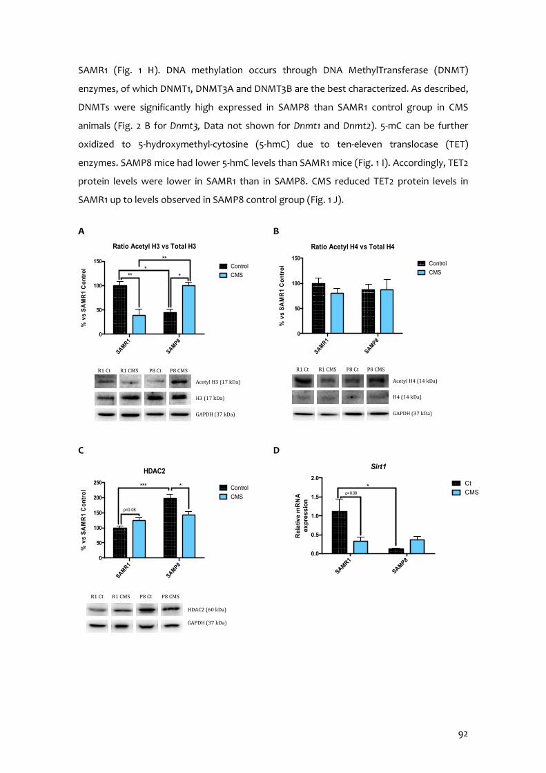
92
SAMR1 (Fig. 1 H). DNA methylation occurs through DNA MethylTransferase (DNMT)
enzymes, of which DNMT1, DNMT3A and DNMT3B are the best characterized. As described,
DNMTs were significantly high expressed in SAMP8 than SAMR1 control group in CMS
animals (Fig. 2 B for Dnmt3, Data not shown for Dnmt1 and Dnmt2). 5-mC can be further
oxidized to 5-hydroxymethyl-cytosine (5-hmC) due to ten-eleven translocase (TET)
enzymes. SAMP8 mice had lower 5-hmC levels than SAMR1 mice (Fig. 1 I). Accordingly, TET2
protein levels were lower in SAMR1 than in SAMP8. CMS reduced TET2 protein levels in
SAMR1 up to levels observed in SAMP8 control group (Fig. 1 J).
A B
C D
SAMR1
SAMP8
0
50
100
150
200
250
HDAC2
% v
s S
AM
R1
Co
ntr
ol Control
CMS
***
p=0.08
*
HDAC2 (60 kDa) GAPDH (37 kDa)
R1 Ct R1 CMS P8 Ct P8 CMS
SAMR1
SAMP8
0.0
0.5
1.0
1.5
2.0
Sirt1
Re
lati
ve
mR
NA
e
xp
res
sio
n
Ct
CMS*
p=0.08
Acetyl H3 (17 kDa)
H3 (17 kDa)
GAPDH (37 kDa)
SAMR1
SAMP8
0
50
100
150
Ratio Acetyl H3 vs Total H3
% v
s S
AM
R1
Co
ntr
ol
Control
CMS***
**
*
R1 Ct R1 CMS P8 Ct P8 CMS
SAMR1
SAMP8
0
50
100
150
Ratio Acetyl H4 vs Total H4
% v
s S
AM
R1
Co
ntr
ol Control
CMS
R1 Ct R1 CMS P8 Ct P8 CMS
Acetyl H4 (14 kDa)
H4 (14 kDa)
GAPDH (37 kDa)
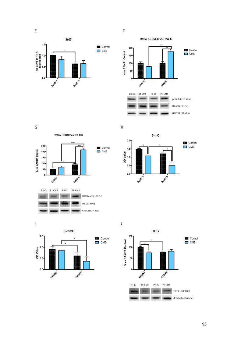
93
E F
G H
I J
SAMR1
SAMP8
0.0
0.5
1.0
1.5
Sirt6R
ela
tiv
e m
RN
A
ex
pre
ss
ion
Control
CMS*
SAMR1
SAMP8
0.0
0.5
1.0
1.5
2.0
5-mC
OD
Va
lue
Control
CMS*
**
SAMR1
SAMP8
0.0
0.5
1.0
1.5
5-hmC
OD
Va
lue
Control
CMS*
*
SAMR1
SAMP8
0
50
100
150
200
Ratio p-H2A.X vs H2A.X
% v
s S
AM
R1
Co
ntr
ol Control
CMS
*****
p-H2A.X (15 kDa)
H2A.X (15 kDa)
GAPDH (37 kDa)
R1 Ct R1 CMS P8 Ct P8 CMS
SAMR1
SAMP8
0
100
200
300
400
500
Ratio H3K9me2 vs H3
% v
s S
AM
R1
Co
ntr
ol Control
CMS
*******
*
H3K9me2 (17 kDa)
H3 (17 kDa)
GAPDH (37 kDa)
R1 Ct R1 CMS P8 Ct P8 CMS
SAMR1
SAMP8
0
50
100
150
TET2
% v
s S
AM
R1
Co
ntr
ol Control
CMS**
R1 Ct R1 CMS P8 Ct P8 CMS
TET2 (130 kDa)
β-Tubulin (55 kDa)
94
Figure 1. Epigenetic markers changes after CMS. Representative Western Blot for the ratio of
Acetylated at Lys9 H3 protein levels and quantification (A), the ratio of Acetylated at Lys 12 H4
protein levels and quantification (B), HDAC2 protein levels and quantification (C), the ratio of
p-H2A.X protein levels and quantification (F), the ratio of H3K9me2 protein levels and
quantification (G) and TET2 protein levels and quantification (J). Relative gene expression of
Sirt1 (D) and Sirt6 (E). Global 5-methylated cytosine (H) and 5-hydroxymethylated cytosine
levels (I). Gene expression levels were determined by real-time PCR. Values in bar graphs are
adjusted to 100% for protein levels of SAMR1 control (R1 Ct). Values are mean ± Standard error
of the mean (SEM); (n = 4 for each group). *p<0.05; **p<0.01; ***p<0.001; ****p<0.0001.
miRNA also play an important role in regulating gene expression. Thereby we decided
to perform a microarray with 84 miRNAs related with neurodegeneration and aging in
order to evaluate the influence of our experimental conditions (strain and CMS) those
miRNA expression. Considering these results (Table S3 and S4), we proceeded to validate
the relative expression of those modified miRNA in a significant manner regarding strain
and CMS (Fig. S2).
miR-29c is involved in neural proliferation regulation by Dnmt3. Although no statistical
differences were seen, it seems that SAMP8 have higher relative expression than SAMR1
and CMS contributed to increase it (Figs. 2 A-B). Those changes correlate with the
tendencies seen in miR-29c-3p relative expression. miR-431-5p, miR-298-5p, miR-98-5p and
miR-140-5p expression were lower in SAMP8 than in SAMR1 (Figs. 2 C,E,G,I). Those miRNAs
are related to inhibition of neurodegenerative pathways, its expression diminution
correlated with changes in effector protein expression as β-catenin, β-site APP Cleaving
Enzyme 1 (BACE1), soluble amyloid precursor protein-β (sAPPβ) and A Disintegrin and
Metalloproteinase Domain-containing protein 10 (ADAM10) (Figs. 2 D,F,H,J). Furthermore,
CMS seemed to reduce miR-431-5p expression in SAMR1 group, although not statistical
differences were observed (Figs. 2 C-D). miR-181a-5p expression was higher in SAMP8 than
in SAMR1 (Fig. 2 K). Mammalian Target Of Rapamycin Complex 1 (mTORC1) is targeted by
this miRNA, regulating cell growth, proliferation and survival. Likewise, mTORC1 protein
levels were significant higher in SAMP8 than in SAMR1 mice (Fig. 2 L). Stressful stimuli only
increased miR-181a-5p relative expression in SAMR1 mice compared to their control
littermates but not in SAMP8 (Fig. 2 K). Correlating with miRNA expression, SAMR1 stressed
group showed higher protein levels than SAMR1 control (Fig. 2 L). BCL2 protein levels are
controlled by miR-106b-5p. SAMP8 mice showed higher relative expression in comparison
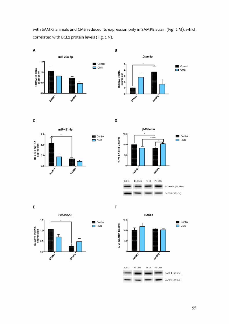
95
with SAMR1 animals and CMS reduced its expression only in SAMP8 strain (Fig. 2 M), which
correlated with BCL2 protein levels (Fig. 2 N).
A B
C D
E F
SAMR1
SAMP8
0.0
0.5
1.0
1.5
miR-431-5p
Re
lativ
e m
iRN
A
ex
pre
ss
ion
Control
CMS
*
SAMR1
SAMP8
0.0
0.5
1.0
1.5
miR-298-5p
Re
lati
ve
miR
NA
e
xp
res
sio
n
Control
CMS
*
SAMR1
SAMP8
0
1
2
3
4
5
Dnmt3a
Re
lati
ve
mR
NA
e
xp
res
sio
n
Control
CMS
*
SAMR1
SAMP8
0.0
0.5
1.0
1.5
miR-29c-3p
Rel
ativ
e m
iRN
A
exp
res
sio
n
Control
CMS
SAMR1
SAMP8
0
50
100
150
b-Catenin
% v
s S
AM
R1
Co
ntr
ol Control
CMS*
p=0.0587
*
R1 Ct R1 CMS P8 Ct P8 CMS
β-Catenin (85 kDa)
GAPDH (37 kDa)
SAMR1
SAMP8
0
50
100
150
BACE1
% v
s S
AM
R1
Co
ntr
ol Control
CMS
R1 Ct R1 CMS P8 Ct P8 CMS
BACE 1 (56 kDa)
GAPDH (37 kDa)
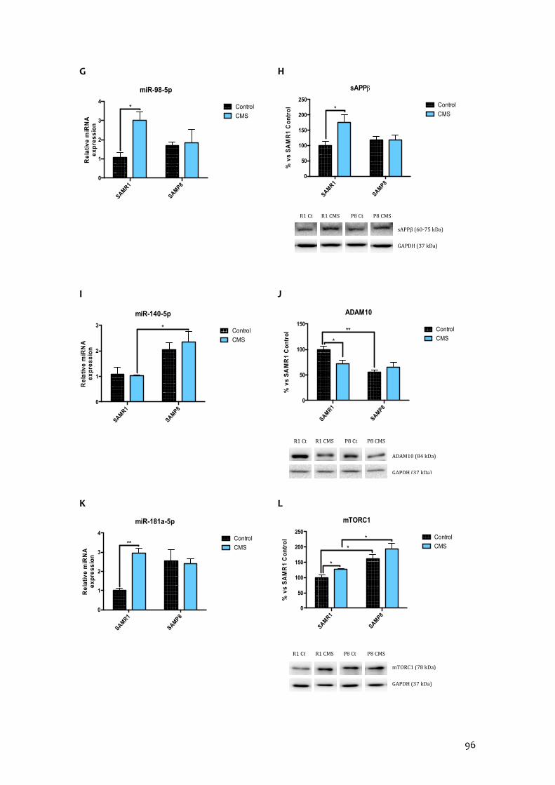
96
G H
I J
K L
SAMR1
SAMP8
0
1
2
3
4
miR-181a-5p
Re
lativ
e m
iRN
A
exp
res
sio
n
Control
CMS**
SAMR1
SAMP8
0
1
2
3
miR-140-5p
Re
lativ
e m
iRN
A
ex
pre
ss
ion
Control
CMS
*
SAMR1
SAMP8
0
1
2
3
4
miR-98-5pR
ela
tiv
e m
iRN
A
ex
pre
ss
ion
Control
CMS*
SAMR1
SAMP8
0
50
100
150
200
250
sAPPb
% v
s S
AM
R1
Co
ntr
ol Control
CMS*
sAPPβ (60-75 kDa)
GAPDH (37 kDa)
R1 Ct R1 CMS P8 Ct P8 CMS
SAMR1
SAMP8
0
50
100
150
200
250
mTORC1
% v
s S
AM
R1
Co
ntr
ol Control
CMS*
*
*
R1 Ct R1 CMS P8 Ct P8 CMS
mTORC1 (78 kDa)
GAPDH (37 kDa)
SAMR1
SAMP8
0
50
100
150
ADAM10
% v
s S
AM
R1
Co
ntr
ol Control
CMS*
**
R1 Ct R1 CMS P8 Ct P8 CMS
ADAM10 (84 kDa)
GAPDH (37 kDa)

97
M N
Figure 2. Validation of a representative subset of miRNA involved in the brain aging,
neurodegeneration and autophagy. Relative expression of miR-29c-3p, miR-431-5p, miR-298-
5p, miR-98-5p, miR-140-5p, miR-181a-5p and miR-106b-5p in the hippocampus of SAMR1 and
SAMP8 mice (A, C, E, G, I, K, M). Relative gene expression of Dnmt3a (B) and representative
Western Blot for β-Catenin protein levels and quantification (D), BACE1 protein levels and
quantification (F), sAPPβ protein levels and quantification (H), ADAM10 protein levels and
quantification (J), mTORC1 protein levels and quantification (L) and BCL2 protein levels and
quantification (N). Gene expression levels were determined by real-time PCR. Values in bar
graphs are adjusted to 100% for protein levels of SAMR1 control (R1 Ct). Values are mean ±
Standard error of the mean (SEM); (n = 6 for each group in miRNAs validation; n=4 for each
group in WB and real-time PCR studies). *p<0.05; **p<0.01.
Chronic Mild Stress attenuates antioxidant defence mechanisms
As mentioned before, CMS modulated cell OS mechanisms favouring an oxidative
state. SAMP8 animals showed higher H2O2 levels in the hippocampus than SAMR1 groups,
as well as reduced protein levels of the antioxidant enzymes superoxide dismutase (SOD),
catalase (CAT) and glutathione peroxidase (GPX1), suggesting impaired mechanisms to
fight against OS (Figs. 3 A-E). CMS induced a diminution in Nuclear factor erythroid 2-related
factor 2 (NRF2) protein levels only in SAMR1, but reduced antioxidant enzymes studied in
both strains. In addition, CMS produced a slight but not significant tendency to increase
aldehyde oxidase 1 (Aox1) gene expression, a pro-oxidant enzyme, in both strains (Fig. 3 F).
SAMR1
SAMP8
0
1
2
3
4
miR-106b-5pR
ela
tive
miR
NA
e
xpre
ss
ion
Control
CMS* *
SAMR1
SAMP8
0
50
100
150
200
BCL2
% v
s S
AM
R1
Co
ntr
ol Control
CMS*
BCL2 (26 kDa)
GAPDH (37 kDa)
R1 Ct R1 CMS P8 Ct P8 CMS
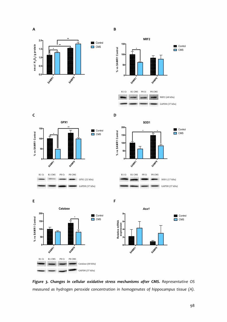
98
A B
C D
E F
Figure 3. Changes in cellular oxidative stress mechanisms after CMS. Representative OS
measured as hydrogen peroxide concentration in homogenates of hippocampus tissue (A).
SAMR1
SAMP8
0.0
0.5
1.0
1.5
2.0
nm
ol
H2O
2/ g
pro
tein
Control
CMS***
**
SAMR1
SAMP8
0
2
4
6
8
Aox1
Re
lativ
e m
RN
A
ex
pre
ss
ion
Control
CMS
SAMR1
SAMP8
0
50
100
150
% v
s S
AM
R1
Co
ntr
ol
NRF2
Control
CMS*
NRF2 (68 kDa)
GAPDH (37 kDa)
R1 Ct R1 CMS P8 Ct P8 CMS
SAMR1
SAMP8
0
50
100
150
GPX1
% v
s S
AM
R1
Co
ntr
ol
Control
CMS*
**
R1 Ct R1 CMS P8 Ct P8 CMS
GPX1 (22 kDa)
GAPDH (37 kDa)
SAMR1
SAMP8
0
50
100
150
200
SOD1
% v
s S
AM
R1
Co
ntr
ol Control
CMS**
R1 Ct R1 CMS P8 Ct P8 CMS
SOD1 (17 kDa)
GAPDH (37 kDa)
SAMR1
SAMP8
0
50
100
150
200
Catalase
% v
s S
AM
R1
Co
ntr
ol Control
CMS*
R1 Ct R1 CMS P8 Ct P8 CMS
Catalase (60 kDa)
GAPDH (37 kDa)
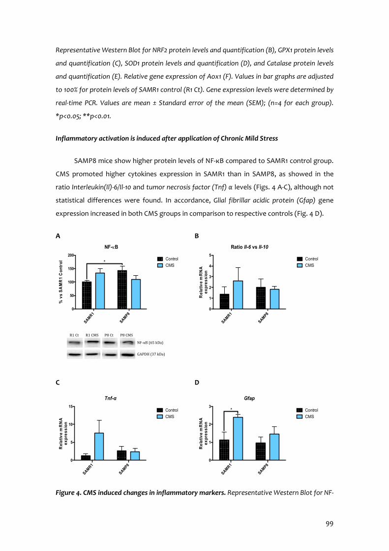
99
Representative Western Blot for NRF2 protein levels and quantification (B), GPX1 protein levels
and quantification (C), SOD1 protein levels and quantification (D), and Catalase protein levels
and quantification (E). Relative gene expression of Aox1 (F). Values in bar graphs are adjusted
to 100% for protein levels of SAMR1 control (R1 Ct). Gene expression levels were determined by
real-time PCR. Values are mean ± Standard error of the mean (SEM); (n=4 for each group).
*p<0.05; **p<0.01.
Inflammatory activation is induced after application of Chronic Mild Stress
SAMP8 mice show higher protein levels of NF-κB compared to SAMR1 control group.
CMS promoted higher cytokines expression in SAMR1 than in SAMP8, as showed in the
ratio Interleukin(Il)-6/Il-10 and tumor necrosis factor (Tnf) α levels (Figs. 4 A-C), although not
statistical differences were found. In accordance, Glial fibrillar acidic protein (Gfap) gene
expression increased in both CMS groups in comparison to respective controls (Fig. 4 D).
A B
C D
Figure 4. CMS induced changes in inflammatory markers. Representative Western Blot for NF-
SAMR1
SAMP8
0
1
2
3
4
5
Ratio Il-6 vs Il-10
Re
lativ
e m
RN
A
ex
pre
ss
ion
Control
CMS
SAMR1
SAMP8
0
5
10
15
Tnf-α
Re
lativ
e m
RN
A
ex
pre
ss
ion
Control
CMS
SAMR1
SAMP8
0
1
2
3
Gfap
Re
lati
ve
mR
NA
e
xp
res
sio
n
Control
CMS
*
SAMR1
SAMP8
0
50
100
150
200
NF-kB
% v
s S
AM
R1
Co
ntr
ol Control
CMS*
NF-κB (65 kDa)
GAPDH (37 kDa)
R1 Ct R1 CMS P8 Ct P8 CMS

100
κB protein levels and quantification (A). Relative gene expression of Il-6/Il-10 ratio, Tnf-α and
Gfap (B-D). Values in bar graphs are adjusted to 100% for protein levels of SAMR1 control (R1
Ct). Gene expression levels were determined by real-time PCR. Values are mean ± Standard
error of the mean (SEM); (n=4 for each group). *p<0.05.
Chronic Mild Stress modulates APP processing as well as alters microtubule stabilization
through hyperphosphorylation of Tau protein
Observing the results, SAMP8 showed lower ADAM10 protein levels than SAMR1
animals. CMS promoted β-amyloid amyloidogenic pathway in SAMR1 mice, increased sAPPβ
protein levels, increased Aβ-precursor gene expression and BACE1 protein levels as well as
reduced ADAM10 protein levels (Figs. 2 F, H, J; 5 A).
SAMP8 had higher Ser396 p-Tau levels (Figs. 5 B-C) than SAMR1. Similarly, GSK-3β
phosphorylation at Ser9 protein level was higher in SAMP8 in reference to SAMR1 (Fig. 5 D).
CMS increased tau hyperphosphorylation at Ser396 and Ser404 compared to control
groups; surprisingly, CMS reduced GSK-3β protein levels (Fig. 5 D).
A B
SAMR1
SAMP8
0.0
0.5
1.0
1.5
2.0
Ab-precursor
Rel
ativ
e m
RN
A
ex
pre
ssio
n
Control
CMSp=0.06p=0.08
SAMR1
SAMP8
0
100
200
300
400
500
Ratio pS396-Tau vs Tau total
% v
s S
AM
R1
Co
ntr
ol Control
CMS
****
*
R1 Ct R1 CMS P8 Ct P8 CMS
p-Tau S396 (75 kDa)
Tau total (55 kDa)
GAPDH (37 kDa)
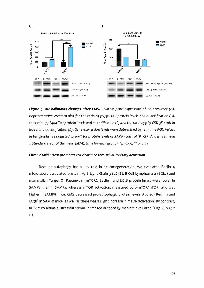
101
C D
Figure 5. AD hallmarks changes after CMS. Relative gene expression of Aβ-precursor (A).
Representative Western Blot for the ratio of pS396-Tau protein levels and quantification (B),
the ratio of pS404-Tau protein levels and quantification (C) and the ratio of pS9-GSK-3β protein
levels and quantification (D). Gene expression levels were determined by real-time PCR. Values
in bar graphs are adjusted to 100% for protein levels of SAMR1 control (R1 Ct). Values are mean
± Standard error of the mean (SEM); (n=4 for each group). *p<0.05; **p<0.01.
Chronic Mild Stress promotes cell clearance through autophagy activation
Because autophagy has a key role in neurodegeneration, we evaluated Beclin 1,
microtubule-associated protein 1A/1B-Light Chain 3 (LC3B), B-Cell Lymphoma 2 (BCL2) and
mammalian Target Of Rapamycin (mTOR). Beclin 1 and LC3B protein levels were lower in
SAMP8 than in SAMR1, whereas mTOR activation, measured by p-mTOR/mTOR ratio was
higher in SAMP8 mice. CMS decreased pro-autophagic protein levels studied (Beclin 1 and
LC3B) in SAMR1 mice, as well as there was a slight increase in mTOR activation. By contrast,
in SAMP8 animals, stressful stimuli increased autophagy markers evaluated (Figs. 6 A-C; 2
N).
SAMR1
SAMP8
0
100
200
300
400
500
Ratio pS404-Tau vs Tau total%
vs
SA
MR
1 C
ontr
ol Control
CMS
*******
*
p-Tau S404 (79 kDa)
Tau total (55 kDa)
GAPDH (37 kDa)
R1 Ct R1 CMS P8 Ct P8 CMS
SAMR1
SAMP80
50
100
150
Ratio pS9-GSK-3b vs GSK-3btotal
% v
s S
AM
R1
Co
ntr
ol Control
CMS**
R1 Ct R1 CMS P8 Ct P8 CMS
pS9-GSK-3β Tyr216 (46 kDa)
GSK-3β total (46 kDa)
GAPDH (37 kDa)
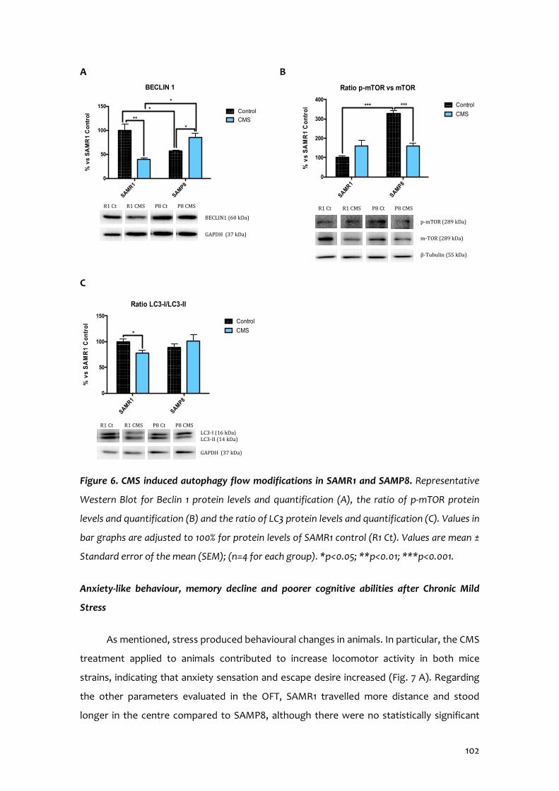
102
A B
C
Figure 6. CMS induced autophagy flow modifications in SAMR1 and SAMP8. Representative
Western Blot for Beclin 1 protein levels and quantification (A), the ratio of p-mTOR protein
levels and quantification (B) and the ratio of LC3 protein levels and quantification (C). Values in
bar graphs are adjusted to 100% for protein levels of SAMR1 control (R1 Ct). Values are mean ±
Standard error of the mean (SEM); (n=4 for each group). *p<0.05; **p<0.01; ***p<0.001.
Anxiety-like behaviour, memory decline and poorer cognitive abilities after Chronic Mild
Stress
As mentioned, stress produced behavioural changes in animals. In particular, the CMS
treatment applied to animals contributed to increase locomotor activity in both mice
strains, indicating that anxiety sensation and escape desire increased (Fig. 7 A). Regarding
the other parameters evaluated in the OFT, SAMR1 travelled more distance and stood
longer in the centre compared to SAMP8, although there were no statistically significant
SAMR1
SAMP8
0
50
100
150
BECLIN 1%
vs
SA
MR
1 C
on
tro
l Control
CMS***
*
*
R1 Ct R1 CMS P8 Ct P8 CMS
BECLIN1 (60 kDa)
GAPDH (37 kDa)
SAMR1
SAMP8
0
100
200
300
400
Ratio p-mTOR vs mTOR
% v
s S
AM
R1
Co
ntr
ol
Control
CMS*** ***
p-mTOR (289 kDa)
m-TOR (289 kDa)
β-Tubulin (55 kDa)
R1 Ct R1 CMS P8 Ct P8 CMS
SAMR1
SAMP8
0
50
100
150
Ratio LC3-I/LC3-II
% v
s S
AM
R1
Co
ntr
ol Control
CMS*
R1 Ct R1 CMS P8 Ct P8 CMS
LC3-I (16 kDa) LC3-II (14 kDa) GAPDH (37 kDa)
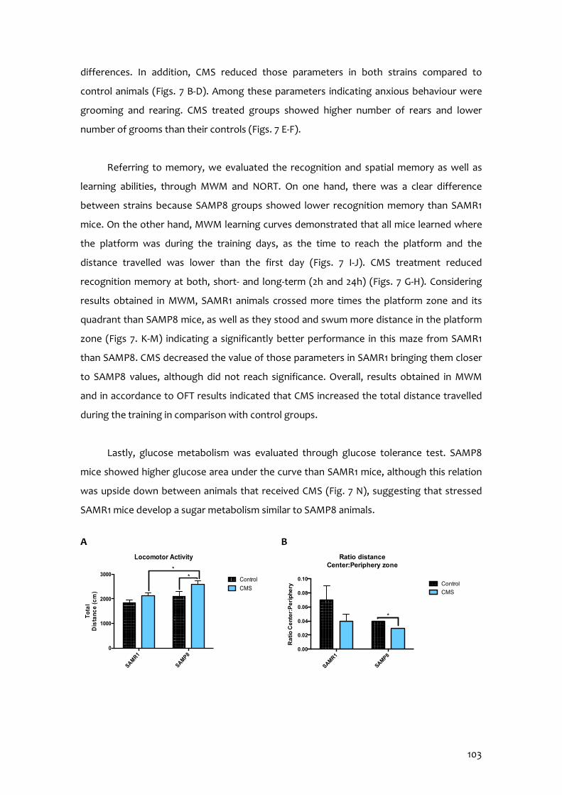
103
differences. In addition, CMS reduced those parameters in both strains compared to
control animals (Figs. 7 B-D). Among these parameters indicating anxious behaviour were
grooming and rearing. CMS treated groups showed higher number of rears and lower
number of grooms than their controls (Figs. 7 E-F).
Referring to memory, we evaluated the recognition and spatial memory as well as
learning abilities, through MWM and NORT. On one hand, there was a clear difference
between strains because SAMP8 groups showed lower recognition memory than SAMR1
mice. On the other hand, MWM learning curves demonstrated that all mice learned where
the platform was during the training days, as the time to reach the platform and the
distance travelled was lower than the first day (Figs. 7 I-J). CMS treatment reduced
recognition memory at both, short- and long-term (2h and 24h) (Figs. 7 G-H). Considering
results obtained in MWM, SAMR1 animals crossed more times the platform zone and its
quadrant than SAMP8 mice, as well as they stood and swum more distance in the platform
zone (Figs 7. K-M) indicating a significantly better performance in this maze from SAMR1
than SAMP8. CMS decreased the value of those parameters in SAMR1 bringing them closer
to SAMP8 values, although did not reach significance. Overall, results obtained in MWM
and in accordance to OFT results indicated that CMS increased the total distance travelled
during the training in comparison with control groups.
Lastly, glucose metabolism was evaluated through glucose tolerance test. SAMP8
mice showed higher glucose area under the curve than SAMR1 mice, although this relation
was upside down between animals that received CMS (Fig. 7 N), suggesting that stressed
SAMR1 mice develop a sugar metabolism similar to SAMP8 animals.
A B
SAMR1
SAMP8
0
1000
2000
3000
Locomotor Activity
To
tal
Dis
tan
ce
(cm
)
Control
CMS
**
SAMR1
SAMP8
0.00
0.02
0.04
0.06
0.08
0.10
Ratio distance Center:Periphery zone
Ra
tio
Ce
nte
r:P
eri
ph
ery Control
CMS
*

104
C D
E F
G H
I J
SAMR1
SAMP8
0
2
4
6
8
CenterT
ime
in (
%)
Control
CMS
SAMR1
SAMP8
0
10
20
30
Re
ar (n
)
Control
CMS*
***
SAMR1
SAMP8
0.0
0.5
1.0
1.5
2.0
Gro
om
(n)
Control
CMS*
SAMR1
SAMP8
0
50
100
150
Periphery
Tim
e in
(%
)
Control
CMS
SAMR1
SAMP8
-0.10
-0.05
0.00
0.05
0.10
NORT 2h - SummaryShort Term Memory
Dis
cri
min
atio
n
Ind
ex
(DI)
Control
CMS****
**
SAMR1
SAMP8
-0.1
0.0
0.1
0.2
NORT 24h - SummaryLong-term memory
Dis
cri
min
atio
n
Inde
x (D
I)
Control
CMS
*****
*
*
1 2 3 4 50
10
20
30
40
50
Days
Tim
e to
re
ac
h p
latf
orm
(s)
Learning Curve
SAMR1 Control
SAMR1 CMS
SAMP8 Control
SAMP8 CMS
1 2 3 4 50
200
400
600
Days
To
tal
Dis
tan
ce
(cm
)
Learning Curve
SAMR1 Control
SAMR1 CMS
SAMP8 Control
SAMP8 CMS

105
K L
M N
Figure 7. Behavioural and cognitive parameters after CMS in SAMR1 and SAMP8 mice. Results
of OFT for all mice groups. Locomotor Activity (A), Ratio of the distance travelled in the
Center/Periphery zone (B), Time in the Center (C) and in the Periphery (D), number of Rearing
(E) and number of Grooming (F). Results of NORT for all mice groups. Summary of DI from 2
and 24 hours after familiarization phase (G-H). Results of MWM for all mice groups. Learning
curves of MWM during the spatial acquisition phase (I-J), number of Entries (K), Distance
travelled (L) and Time (M) in platform zone during the test. Plasma levels of glucose 2g/kg
intraperitoneal (i.p.) administration (N). Values are mean ± Standard error of the mean (SEM)
(n=14 for each group). *p<0.05; **p<0.01; ***p<0.001; ****p<0.0001.
3.2.5 Discussion
Understanding the mechanisms that define aging and differentiate what determines
whether it is pathologic or healthy is one of the challenges that science has faced in recent
years. Life expectancy has increased and so the number of older people. In addition, the life
rhythm has changed becoming increasingly stressful. It has been shown that the
environment is extremely important in the development of several diseases, compromising
healthy aging; specifically, stressful lifestyle has been identified as an important risk factor
for cognitive decline. Therefore, it is crucial to study the effects of stress on cognition and
SAMR1
SAMP8
0
1
2
3
4
5T
arg
et c
ros
sin
gs
(n) Control
CMS**
SAMR1
SAMP8
0
10
20
30
Platform zone
Dis
tan
ce (c
m)
Control
CMS*
SAMR1
SAMP8
0
1
2
3
Platform zone
Tim
e in
(%)
Control
CMS
**
SAMR1
SAMP8
0
500
1000
1500
AU
C G
luc
os
e
mg
/dL
x 1
20
min
Control
CMS
*
**

106
its relationship with aging in order to unveil what challenges we might have to cope with as
a society in a not so far future. We hypothesize that chronic stress would modulate a large
constellation of cellular mechanisms implied in aging and age-related neurodegenerative
pathologies.
During aging, there are several epigenetic mechanisms altered, which include DNA
methylation, histone modifications, nucleosome remodelling and miRNA-mediated gene
regulation (Harman & Martín, 2019; Foster et al., 2017). In the present study, we evaluated
the three main epigenetic marks, which modulate chromatin structure and act as platforms
for recruitment, assembly or retention of chromatin-associated factors: histone
posttranslational modifications, DNA methylation and miRNA-mediated gene regulation.
One of the posttranslational modifications of histones is acetylation. This process removes
histone positive charge; thereby the condensed chromatin (heterochromatin) is
transformed into a more relaxed structure (euchromatin) that is associated with greater
levels of gene transcription (Turner, 2000; Watson et al., 2014). However, chromatin
condensation depends on different processes including methylation and histone
deacetylation. This last process is due to histone deacetylases (HDAC) enzymes.
Deregulation of histone acetylation has been related to increase the risk of age-dependent
memory impairment in mice (Cuadrado-Tejedor et al., 2019; Griñán-Ferré et al., 2016; Peleg
et al., 2010). Specifically, lysine 12 histone H4 acetylation alterations cause impaired memory
consolidation as well as its restoration reinstates the expression of learning-induced genes
and in consequence, cognitive abilities (Peleg et al., 2010). In accordance to Cosín-Tomás
(Cosín-Tomás et al., 2014), we found that CMS increased HDAC2 protein levels only in
SAMR1 females, similarly to SAMP8 control mice, which leads us to hypothesize that
acetylated histone protein levels were diminished. This was confirmed evaluating
acetylated H3 and H4 protein levels. However, while stress produced changes in H3
acetylation, these changes were not observed in H4. Nevertheless, the decrease in H3
acetylation in SAMR1 mice under CMS, similarly to not stressed SAMP8 was correlated with
changes on cognition. Other deacetylase enzymes are the Sirt family; however, it is worth
noting that several members of this family do not have deacetylase activity (Herskovits &
Guarente, 2014). Sirt have been linked to aging as they modulate genomic stability, stress
resistance and energy metabolism. Activation of Sirt1 enables the deacetylation of a variety
of proteins, resulting in a robust, protective cellular response, as it regulates processes such
as cell death, metabolism or neurodegeneration (Pallàs, 2012); while Sirt2 has been
reported to regulate OS, genome integrity and myelination and its dysfunction is found in

107
most of age-related neurodegenerative disorders such as AD, Parkinson’s disease or
Amyotrophic Lateral Sclerosis, as well as in physiological aging (Fourcade et al., 2018).
Accumulated evidence indicates that Sirt6 gene expression is lower in the hippocampus
and cerebral cortex of aged mice (Fourcade et al., 2018; Jesko et al., 2017) and that it is
concerned with acetylated lysine 9 histone 3 (H3K9) (Khan et al., 2018). In reference to this
family of deacetylases our results demonstrate decreased Sirt1, Sirt2 and Sirt6 gene
expression in SAMP8 mice compared to SAMR1 and to SAMR1 under CMS. Compelling
evidence has proposed that methylation of H3K9 promotes DNA methylation maintenance
in mammals and is a hallmark of heterochromatin formation and subsequent gene silencing
(Harman and Martín, 2019; Palmer et al., 2008; Zhao et al., 2016). As with histone
phosphorylation, CMS treatment increased H3K9 methylation in SAMP8 animals but not in
SAMR1 suggesting that even they showed more H3 acetylation, also resulted in more
methylation. Therefore, we demonstrate that CMS favoured chromatin condensation and in
consequence, promoted gene silencing.
In reference to other modifications, histone phosphorylation belongs to the cellular
response to DNA damage, as phosphorylated histone H2A.X demarcates large chromatin
domains around the site of DNA breakage (Harman and Martín, 2019). Additionally, multiple
studies have also shown that histone phosphorylation plays crucial roles in other nuclear
processes, such as DNA replication because of apoptosis or DNA damage (Rossetto et al.,
2012). Here CMS raised phosphorylated H2A.X protein level in senescence-accelerated but
not in senescence-resistant mice, suggesting that stressful stimuli activate repair/survival
mechanisms rather than apoptotic response in SAMP8 mice but not in SAMR1. As two
major mechanisms for epigenetic regulation, DNA methylation and histone modifications
must act coordinately (Zhao et al., 2016). It is well known that DNA methylation at the fifth
position of cytosine (5-mC) plays an important role in neuronal gene expression and neural
development. Several studies support that dysregulated DNA methylation/demethylation is
linked to many neuronal disorders, including AD onset and progression; however, the
relationship between AD and altered 5-mC levels is not known (Fetahu et al., 2019). 5-mC
can be further oxidized to 5-hmC, among others, by the TET family of dioxygenases. 5-mC
and 5-hmC exert opposite effects on gene expression; the former is general associated with
gene silencing, whereas the latter is mainly involved in up-regulation of gene expression
(Sherwani and Khan, 2015). In this study, stressful environment produced lower 5-mC in
both mice strains and differences between strains were observed in 5-hmC marker.
Accordingly, TET2 protein levels differed between SAMR1 and SAMP8 animals. Despite our
108
previous results, conversely 5-mC and 5-hmC results appear to contradict the transcriptional
access to DNA, but as is known, the term global DNA methylation describes this process
across the entire genome and does not represent a precise landscape for specific
transcriptional activity; however, global methylation determination is useful as it provides
an over-arching picture of methylation status, it is misleading which genes show altered
DNA methylation and which do not (Yokoyama et al., 2017).
Furthermore, we evaluated expression changes of miRNAs related to OS, AD
neurophatological hallmarks, autophagy and neurodegeneration. Of 22 miRNA evaluated,
those that presented statistically significant changes under experimental conditions were
further validated and target genes studied (Fig. S2). Firstly, we studied miR-29c relative
expression, which is involved in neural proliferation regulation through Dnmt3a. Dnmt3a
expression was higher in SAMP8 compared to SAMR1 mice correlating with miR-29c-3p
gene expression. In our hands CMS slightly contributed to change Dnmt3a expression in
mice, being increased in SAMR1. Because, recent studies demonstrate that DNMT inhibitors
provided neuroprotection in cellular cultures (Chestnut et al., 2011; Hernandez et al., 2011),
the increase in Dnmt3a induced by CMS could mean a deleterious effect on SAMR1 health.
Considering AD neuropathology, we studied miR-431-5p, miR-298-5p, miR-98-5p and
miR-140-5p. It has been described that miR-431-5p cooperates with DKK1 to inhibit Wnt/β-
catenin pathway. SAMP8 showed lower relative expression of this miRNA in comparison to
SAMR1 mice; changes in β-catenin protein levels were in line with miR-431-5p. Furthermore,
CMS reduced mildly miR-431-5p gene expression and β-catenin protein levels in SAMR1. β-
catenin is regulated by GSK-3β activity, depending on its phosphorylation. As reported,
GSK3-β inactive form (Ser9 phosphorylated) protein levels were lower in SAMP8 in
reference to SAMR1 (Pallàs, 2012), and also CMS decreased them, especially in SAMP8. It is
known that GSK3-β activity is associated with AD neuropathology as it exacerbates
cognitive impairment (Llorens-Martín et al., 2014). In fact, higher activity of this kinase has
been found in AD patients and its inhibition restores spatial memory deficits, reduces Tau
hyperphosphorylation and decreases reactive gliosis and neuronal death in rodents
(Llorens-Martín et al., 2014). Furthermore, it has been described that GSK3-β inhibition
reduces BACE1-mediated cleavage of APP through NF-κB signalling-mediated mechanism,
so that it reduces β-amyloid pathology (Ly et al., 2013). Accordingly, we found that CMS
increased Ser396 and Ser404 Tau hyperphosphorylation, most pronounced in SAMP8, and
promoted amyloid precursor protein (APP) gene expression in both mice strains. In fact,
109
APP processing is regulated by miR-298-5p, which in turn regulates BACE1. miR-298-5p
expression differed between SAMR1 and SAMP8 animals, in contrast with BACE1 protein
levels. It is reported that BACE1, sAPPβ and β-Carboxiterminal fragments protein levels
were increased when miR-98-5p up-regulated. CMS increased miR-98-5p relative expression
and sAPPβ in SAMR1 compared to the control group, but not in SAMP8. However as
mentioned, BACE1 was not modulated under CMS. Taking into account that BACE1 can be
up- or down-regulated by different miRNAs, discrepancies can be explained by
compensatory responses among different signals. ADAM10 protein levels are controlled by
miR-140-5p. Again, huge differences in miR-140-5p gene expression and ADAM10 protein
levels were observed between strains. Differences were between mice under CMS in
SAMR1 but not in SAMP8.
miR-181a-5p is involved in regulation of cell growth, proliferation and survival through
mTOR complex 1 (mTORC1) and downstream pathway, which protein levels were increased
in SAMP8 in comparison to SAMR1 mice. In addition, stressful stimuli increased miR-181a-5p
relative expression in SAMR1 mice compared to their control littermates and no changes
were observed between SAMP8 animals. By contrast, CMS groups in both strains showed
higher mTORC1 protein levels than respective control littermates. Lastly, BCL2 protein levels
were controlled by miR-106b-5p. SAMP8 mice had higher miR-106b-5p compared to SAMR1,
but no changes were found in BCL2 protein levels. CMS reduced miR-106b-5p gene
expression, increasing BCL2 protein levels in SAMP8, without affecting SAMR1 strain.
Autophagy declines during aging, so that it may contribute to the deleterious
accumulation of aberrant proteins observed in aged cells. Interestingly, failure of this
process has been reported to worsen aging-associated diseases, such as
neurodegeneration or cancer (Gouras, 2019). Pro autophagic proteins such as Beclin1 and
LC3B became decreased after CMS treatment and in concordance p-mTOR ratio was
increased. Moreover, we demonstrated huge differences between SAMR1 and SAMP8
control groups, indicating a reduced autophagic flux in senescent mice. CMS induced
impairment in autophagy was more robust and consistent in SAMR1 than in SAMP8,
supporting the hypothesis that CMS accelerates the senescence process.
Following the same line about overall processes linked to senescence, it has been
widely described that OS possesses a pre-eminent role in pathological senescence and the
pathogenesis of AD. In general, cells possess antioxidant mechanisms to cope with OS,
110
such as GPX, Catalase and SOD, among others; which in turn are regulated by NRF2
transcriptional pathway. Herein we found that mice under CMS had lower NRF2 protein
levels than control groups and this in turn could explain the lowest protein levels of
antioxidant defence as GPX1, SOD1 and Catalase. Consistent with this, CMS promoted an
increase in the pro-oxidant enzyme Aox1 gene expression and higher accumulation of ROS.
Noteworthy, as described, differences between strains were found in most of the
evaluated markers (Griñán-Ferré et al., 2016).
As far as aging and AD are concerned, dysregulation of inflammatory mediators and
astrogliosis are major culprits in the development of chronic inflammation and the
immunosenescence process, as well as are related to cognitive decline and progression of
neurodegenerative diseases (Chung et al., 2019). For instance, our results demonstrate
significant increase in protein levels for NF-κB, transcription factor regulating pro-
inflammatory signals, in SAMP8 mice in comparison with SAMR1. In accordance to Chung et
al. (Chung et al., 2019), Il-6 and Tnf-α gene expression were up regulated in SAMP8 mice.
Interestingly CMS increased proinflammatory pathways in SAMR1 but not in SAMP8.
Moreover, we found an increase in Gfap gene expression in SAMR1 CMS compared to
SAMR1 control group. In accordance, it has been recently described that modulating
astrogliosis enhances AD pathology in mice (Reichenbach et al., 2019).
It is widely accepted the detrimental effect of stress on psychological well-being and
cognitive functioning, emphasizing the relationship between stress and memory. It is
noteworthy that chronic stress signs underlie some of the characteristics described in
cognitive decline, either in aging or in neurodegenerative diseases, such as AD (Puigoriol-
Illamola et al., 2018; Turner & McCarthy, 2017). It has been stated that epigenetic machinery
is essential for cognitive function (Harman & Martín, 2019; Peleg et al., 2010; Stilling &
Fischer, 2011). Likewise, DNA methylation influences hippocampal memory formation and
growing evidence suggests that the modulation of epigenetic processes by stress,
environmental enrichment and/or hormones is key in regulating memory function.
In line with the molecular results presented, behavioural changes induced by CMS
were explored in SAMP8 and SAMR1 mice. Results obtained pointed out that not only a
stressful environment triggers anxiety-like behaviour, but also it mitigates cognitive
performance. Locomotor activity, the distance travelled in the central zone and other
parameters indicating well-being/discomfort were altered in mice under CMS. Regarding

111
recognition memory, clear differences were found between the different strains, although
we can also assert a detrimental effect caused by CMS, especially on long-term memory. In
agreement with these results, learning abilities and spatial memory in SAMR1 and SAMP8 at
6 months of age were significantly different and were negatively affected by the presence
of chronic stressors. While stressful situations, glucose levels increase in order to have
enough energy available to cope with stress. Aberrant glucose metabolism potentiates the
aging phenotype and contributes to early stage central nervous system pathology (Currais
et al., 2012). According to previous works, we found differences on blood glucose levels due
to stress in SAMR1 mice, exhibiting similar glucose tolerance to SAMP8 control mice.
3.2.6 Conclusions
Overall, CMS treatment produces detrimental effects in female SAM mice, such as
inflammatory signalling activation, loss of antioxidant defence mechanisms, changes in
behaviour and reduced cognitive abilities (Fig. 8). Interestingly, CMS promoted significant
epigenetic and biochemical changes in SAMR1 animals, a normal mice strain, driving them
to an aging-specific phenotype represented by SAMP8 mice, a senescence mice model, in
which the negative effects of CMS are probably limited because its senescence level is
already elevated.
Figure 8. Representative scheme of molecular pathways altered after Chronic Mild Stress.
112
3.2.7 Acknowledgements
This study was supported by Ministerio de Economía y Competitividad of Spain and
FEDER (SAF2016-77703) and 2017SGR106 (AGAUR, Catalonia). Financial support was
provided for D.P.I. (FPU program).
3.2.8 References
Bisht, K.; Sharma, K.; Tremblay, M. Chronic stress as a risk factor for Alzheimer’s disease: Roles of microglia-mediated synaptic remodeling, inflammation, and oxidative stress. Neurobiol Stress. 2018, 9, 9-21.
Borcel, E.; Pérez-Alvarez, L.; Herrero, A.I.; Brionne, T.; Varea, E.; Berezin, V.; Bock, E.;
Sandi, C.; Venero, C. Chronic stress in adulthood followed by intermittent stress impairs spatial memory and the survival of newborn hippocampal cells in aging animals: prevention by FGL, a peptide mimetic of neural cell adhesion molecule. Behav Pharmacol. 2008, 19 (1), 41-9.
Chestnut, B.A.; Chang, Q.; Prince, A.; Lesuisse, C.; Wong, M.; Martin, L.J. Epigenetic
regulation of motor neuron cell death through DNA methylation. J Neurosci. 2011, 31, 16619-16636.
Chung, H.Y.; Kim, D.H.; Lee, E.K.; Chung, K.W.; Chung, S.; Lee, B.; Seo, A.Y.; Chung,
J.H.; Jung, Y.S.; Im, E.; Lee, J.; Kim, N.D.; Choi, Y.J.; Im, D.S.; Yu, B.P. Redefining Chronic Inflammation in Aging and Age-Related Diseases: Proposal of the Senoinflammation Concept. Aging Dis. 2019, 10(2), 367-382.
Cosín-Tomás, M.; Alvarez-López, M.J.; Sanchez-Roige, S.; Lalanza, J.F.; Bayod, S.;
Sanfeliu, C.; Pallàs, M.; Escorihuela, R.M.; Kaliman, P. Epigenetic alterations in hippocampus of SAMP8 senescent mice and modulation by voluntary physical exercise. Front Aging Neurosci. 2014, 6, 51.
Cristòfol, R.; Porquet, D.; Corpas, R.; Coto-Montes, A.; Serret, J.; Camins, A.; Pallàs, M.;
Sanfeliu, C. Neurons from senescence-accelerated SAMP8 mice are protected against frailty by the Sirtuin 1 promoting agents melatonin and resveratrol. J Pineal Res 2012, 52, 271-281.
Cuadrado-Tejedor, M.; Pérez-González, M.; García-Muñoz, C.; Muruzabal, D.; García-
Barroso, C.; Rabal, O.; Segura, V.; Sánchez-Arias, J.A.; Oyarzabal, J.O.; Garcia-Osta, A. Taking Advantage of the Selectivity of Histone Deacetylases and Phosphodiesterase Inhibitors to Design Better Therapeutic Strategies to Treat Alzheimer’s Disease. Front. Aging Neurosci. 2019, 11, 149.
113
Currais, A.; Prior, M.; Lo, D.; Jolivalt, C.; Schubert, D.; Maher, P. Diabetes exacerbates amyloid and neurovascular pathology in aging-accelerated mice. Aging Cell 2012, 11, 1017-1026.
Escobar, K.A.; Cole, N.H.; Mermier, C.M.; VanDusseldorp, T.A. Autophagy and aging:
Maintaining the proteome through exercise and caloric restriction. Aging Cell 2019, 18(1), e12876.
Fetahu, I.S.; Ma, D.; Rabidou, K.; Argueta, C.; Smith, M.; Liu, H.; Wu, F.; Shi, Y.G.
Epigenetic signatures of methylated DNA cytosine in Alzheimer’s disease. Sci Adv 2019, 5(8), eaaw2880.
Foster, J.A.; Rinaman, L.; Cryan, J.F. Stres & the gut-brain axis: Regulation by the
microbiome. Neuribiol Stress. 2017, 7, 124-136. Fourcade, S.; Outeiro, T.F.; Pujol, A. SIRT2 in age-related neurodegenerative disorders.
Aging 2018, 10. Gouras, G.K. Aging, Metabolism, Synaptic Activity, and Aβ in Alzheimer’s Disease.
Front Aging Neurosci. 2019, 11, 185. Griñán-Ferré, C.; Corpas, R.; Puigoriol-Illamola, D.; Palomera-Ávalos, V.; Sanfeliu, C.;
Pallàs, M. Understanding Epigenetics in the Neurodegeneration of Alzheimer’s Disease: SAMP8 Mouse Model. J Alzheimers Dis. 2018, 62(3), 943-963.
Griñán-Ferré, C.; Pérez-Cáceres, D.; Gutiérrez-Zetina, S.M.; Camins, A.; Palomera-
Avalos, V.; Ortuño-Sahagún, D.; Rodrigo, M.T.; Pallàs, M. Environmental Enrichment Improves Behavior, Cognition, and Brain Functional Markers in Young Senescence-Accelerated Prone Mice (SAMP8). Mol Neurobiol. 2015, 53, 2435-2450.
Griñan-Ferré, C.; Puigoriol-Illamola, D.; Palomera-Ávalos, V.; Pérez-Cáceres, D.;
Companys-Alemany, J.; Camins, A.; Ortuño-Sahagún, D.; Rodrigo, M.T.; Pallàs, M. Environmental Enrichment Modified Epigenetic Mechanisms in SAMP8 Mouse Hippocampus by Reducing Oxidative Stress and Inflammaging and Achieving Neuroprotection. Front. Aging Neurosci. 2016, 8, 241.
Guerreiro, R.; Bras, J. The age factor in Alzheimer’s disease. Genome Medicine 2015, 7,
106. Harman, M.F.; Martín, M.G. Epigenetic mechanisms related to cognitive decline
during aging. J Neuro Res 2019, 00, 1-13. Hernandez, D.G.; Nalls, M.A.; Gibbs, J.R.; Arepalli, S.; van der Brug, M.; Chong, S.;
Moore, M.; Longo, D.L.; Cookson, M.R.; Traynor, B.J.; Singleton, A.B. Distinct DNA

114
methylation changes highly correlated with chronological age in the human brain. Hum Mol Genet 2011, 20(6), 1164-72.
Herskovits, A.Z.; Guarente, L. SIRT1 in neurodevelopment and brain senescence.
Neuron. 2014, 81(3), 471-83. Jesko, H.; Wencel, P.; Strosznajder, R.P.; Strosznajder, J.B. Sirtuins and Their Roles in
Brain Aging and Neurodegenerative Disorders. Neurochem Res 2017, 42(3), 876-890. Jett, J.D.; Bulin, S.E.; Hatherall, L.C.; McCartney, C.M.; Morilak, D.A. Deficits in
cognitive flexibility induced by chronic unpredictable stress are associated with impaired glutamate neurotransmission in the rat medial prefrontal cortex. Neurosci. 2017, 346, 284-297.
Justice, N.J. The relationship between stress and Alzheimer’s disease. Neurobiol
Stress. 2018, 8, 127-133. Khan, R.I.; Nirzhor, S.S.R.; Akter, R. A Review of the Recent Advances Made with
SIRT6 and its Implications on Aging Related Processes, Major Human Diseases, and Possible Therapeutic Targets. Biomolecules 2018, 8(3), 44.
Llorens-Martín, M.; Jurado, J.; Hernández, F.; Avila, J. GSK-3β, a pivotal kinase in
Alzheimer disease. Front Mol Neurosci. 2014, 7, 46. Ly, P.T.T.; Wu, Y.; Zou, H.; Wang, R.; Zhou, W.; Kinoshita, A.; Zhang, M.; Yang, Y.; Cai,
F.; Woodgett, J.; Song, W. Inhibition of GSK-3β-mediated BACE1 expression reduces Alzheimer-associated phenotypes. J Clin Invest. 2013, 123(1), 224-235.
Martinez-Lopez, N. Autophagy and Aging. Adv Exp Med Biol. 2015, 847, 73-87. Pallàs, M. Senescence-Accelerated Mice P8: A Tool to Study Brain Aging and
Alzheimer’s Disease in a Mouse Model. Cell Biology 2012, 12. Palmer, J.M.; Perrin, R.M.; Dagenais, T.R.; Keller, N.P. H3K9 methylation regulates
growth and development in Aspergillus fumigatus. Eukaryot Cell 2008, 7(12), 2052-60. Peleg, S.; Sananbenesi, F.; Zovoilis, A.; Burkhardt, S.; Bahari-Javan, S.; Agis-Balboa,
R.C.; Cota, P.; Wittnam, J.L.; Gogol-Doering, A.; Opitz, L.; Salinas-Riester, G.; Dettenhofer, M.; Kang, H.; Farinelli, L.; Chen, W.; Fischer, A. Altered histone acetylation is associated with age-dependent memory impairment in mice. Science. 2010, 328(5979), 753-6.
Petersen, K.S.; Smith, C. Ageing-Associated Oxidative Stress and Inflammation Are
Alleviated by Products from Grapes. Oxidative Medicine and Cellular Longevity 2016, 2016, 12.
115
Puigoriol-Illamola, D.; Griñán-Ferré, C.; Vasilopoulou, F.; Leiva, R.; Vázquez, S.; Pallàs, M. 11β-HSD1 Inhibition by RL-118 Promotes Autophagy and Correlates with Reduced Oxidative Stress and Inflammation, Enhancing Cognitive Performance in SAMP8 Mouse Model. Mol Neurobiol. 2018, 55(12), 8904-8915.
Puigoriol-Illamola, D.; Leiva, R.; Vázquez-Carrera, M.; Vázquez, S.; Griñán-Ferré, C.;
Pallàs, M. 11β-HSD1 Inhibition Rescues SAMP8 Cognitive Impairment Induced by Metabolic Stress. Mol Neurobiol. 2020, 1-15.
Rahman, M.M.; Callaghan, C.K.; Kerskens, C.M.; Chattarji, S.; O’Mara, S.M. Early
hippocampal volume loss as a marker of eventual memory deficits caused by repeated stress. Sci. Rep. 2016, 6, 29127.
Reichenbach, N.; Delekate, A.; Plescher, M.; Schmitt, F.; Krauss, S.; Blank, N.; Halle, A.;
Petzold, G.C. Inhibition of Stat3-mediated astrogliosis ameliorates pathology in an Alzheimer’s disease model. EMBO Mol Med. 2019, 11(2), e9665.
Rossetto, D.; Avvakumov, N.; Côte, J. Histone phosphorylation: a chromatin
modification involved in diverse nuclear events. Epigenetics 2012, 7 (10), 1098-108. Sandi, C. Stress and Cognition. WIREs Cogn Sci 2013, 4, 245-261. Sherwani, S.I.; Khan, H.A. Role of 5-hydroxymethylcytosine in neurodegeneration.
Gene 2015, 570(1), 17-24. Stilling, R.M.; Fischer, A. The role of histone acetylation in age-associated memory
impairment and Alzheimer’s disease. Neurobiol Learning Memory 2011, 96(1), 19-26. Sun, H.; Kennedy, P.; Nestler, E. Epigenetics of the Depressed Brain: Role of Histone
Acetylation and Methylation. Neuropsychopharmacol 2013, 38, 124-137. Takeda, T.; Hosokawa, M.; Higuchi, K. Senescence-Accelerated Mouse (SAM): A Novel
Murine Model of Accelerated Senescence. Journal of the American Geriatrics Society 1991, 39(9), 911-919.
Turecki, G.; Meaney, M.J. Effects of the Social Environment and Stress on
Glucocorticoid Receptor Gene Methylation: A Systematic Review. Biol Psychiatry 2016, 79 (2), 87-96.
Turner, B.M. Histone acetylation and an epigenetic code. BioEssays 2000, 22(9), 836-
45. Turner, K.; McCarthy, V.L. Stress and anxiety among nursing students: A review of
intervention strategies in literature between 2009 and 2015. Nurse Educ Pract. 2017, 22, 21-29.
116
Vorhees, C.V.; Williams, M.T. Morris water maze: procedures for assessing spatial and
related forms of learning and memory. Nature protocols 2006, 1(2), 848-858. Wang, S.E.; Ko, S.Y.; Jo, S.; Jo, H.; Han, J.; Kim, Y.; Son, H. Downregulation of SIRT2 by
Chronic Stress Reduces Expression of Synaptic Plasticity-related Genes through the Upregulation of Ehmt2. Exp Neurobiol. 2019, 28 (4), 537-546.
Wang, J.; Yuan, J.; Pang, J.; Ma, J.; Han, B.; Geng, Y.; Shen, L.; Wang, H.; Ma, Q.; Wang,
Y.; Wang, M. Effects of Chronic Stress on Cognition in Male SAMP8 Mice. Cell Physiol Biochem 2016, 39, 1078-1086.
Watson, J.D.; Baker, T.A.; Gann, A.; Levine, M.; Losik, R. Molecular biology of the gene;
Pearson/CSH Press, 7th Ed.; Boston, USA, 2014. Yokoyama, A.S.; Rutledge, J.C.; Medici, V. DNA methylation alterations in Alzheimer’s
disease. Environ Epigenet. 2017, 3(2), dvx008. Zhao, Q.; Zhang, J.; Chen, R.; Wang, L.; Li, B.; Cheng, H.; Duan, X.; Zhu, H.; Wei, W.; Li,
J.; Wu, Q.; Han, J-D.J.; Yu, W.; Gao, S.; Li, G.; Wong, J. Dissecting the precise role of H3K9 methylation in crosstalk with DNA maintenance methylation in mammals. Nat Commun 2016, 7, 12464.
3.2.9 Supplementary material
Supplementary materials can be found at http://www.mdpi.com/1422-
0067/21/3/1154/s1.
3.3 RL-118 and 11β-HSD1 target engagement through TAPS assay: behaviour and molecular analysis
Puigoriol-Illamola, D.1,2, Companys-Alemany, J.1,2, Homer, N.3, Leiva, R.4, Vázquez, S.4, Mole, D.5, Griñán-Ferré, C.1,2, Pallàs, M1,2. RL-118 and 11β-HSD1 target engagement through TAPS
assay: behaviour and molecular analysis
[pending to submit]
118
Author information: 1 Department of Pharmacology, Toxicology and Therapeutic Chemistry. Pharmacology Section. Faculty of Pharmacy and Food Sciences. University of Barcelona, Av Joan XXIII 27-31, 08028 Barcelona, Spain. 2 Institute of Neuroscience, University of Barcelona (NeuroUB), Passeig de la Vall d’Hebron 171, Barcelona, Spain. 3 Mass Spectrometry Core, Edinburgh Clinical Research Facility, Queen’s Medical Research Institute, Edinburgh, United Kingdom. 4 Medicinal Chemistry Section. Department of Pharmacology, Toxicology and Therapeutic Chemistry. Faculty of Pharmacy and Food Sciences, University of Barcelona, Av. Joan XXIII, 27-31, 08028 Barcelona, Spain. 5 MRC Centre for Inflammation Research, Queen's Medical Research Institute, University of Edinburgh, Edinburgh, United Kingdom. * Chapter 3 is adapted from a paper pending to be submitted in near future. Therefore, it has
been included and structured as a full article, but it may be modified as new results are
obtained prior to being sent for peer review. On date of its final edition, it constitutes the
most updated version.
119
3.3.1 Abstract
Taking into consideration the convergence of ageing, stress and neurodegenerative
diseases, such as AD, there is impaired GC signalling. Therefore, the study of GC-mediated
stress response to chronic moderate stressful situations, as account in daily life, becomes of
huge interest to design pharmacological strategies to prevent neurodegeneration.
To address this issue, SAMP8 were exposed for 4 weeks to the CMS paradigm and
treated with RL-118, an 11β-HSD1 inhibitor. In fact, several pieces of evidence link CMS
exposure to reduced cognitive performance, while inhibition of 11β-HSD1 has been
associated with reduction of GC activity and cognitive improvement. The aim of this project
was to assess whether RL-118 treatment could restore the deleterious effects of CMS on
cognition and behavioural abilities, but also on molecular mechanisms that compromise
healthy ageing in SAMP8 mice.
On the one hand, we determined the target engagement between RL-118 and 11β-
HSD1. Therefore all the beneficial effects previously described in SAMP8 treated with the
drug can undoubtedly be attributed to the inhibition of this enzyme. Besides, herein we
observed decreased DNA methylation, hydroxymethylation and histone phosphorylation
induced by CMS but, on the contrary, increased after RL-118 treatment. In addition, CMS
exposure produced ROS damage accumulation, and increments of pro-oxidant enzymes as
well as pro-inflammatory mediators through NF-κB pathway and astrogliosis markers, like
Gfap. Of note, those modifications were recovered by 11β-HSD1 inhibition. Remarkably,
although CMS altered mTORC1 signalling, autophagy was increased in SAMP8 treated with
RL-118 mice. Also, we found that amyloidogenic APP processing pathway was favoured and
decreased synaptic plasticity and neuronal remodelling markers in mice under CMS, but
changed after RL-118 treatment. In consequence, detrimental effects on behaviour and
cognitive performance were detected in CMS exposed mice, although recovered after
concomitant 11β-HSD1 inhibition by RL-118.
Overall, CMS is a feasible intervention to understand the influence of stress on
epigenetic mechanisms underlying cognition and accelerating senescence. However and
most important, 11β-HSD1 inhibition through RL-118 turned up to restore the majority of
these detrimental effects caused by CMS, indicating that GC excess attenuation may
become a potential therapeutic strategy for age-related cognitive decline and AD.
120
3.3.2 Introduction
In the past few years, stress and stress response have been an overwhelming public
health issue with a huge amount of literature, and in turn misinformation of the detrimental
effects that stress may have in modifying aging, determining healthy or pathological aging
of the brain, as well as in the development of several diseases (Catania et al., 2009;
Sotiropoulous et al., 2008). Drug discovery studies have become of paramount importance
in order to offer different drugs to treat or prevent the detrimental effects on cognition
during aging and under aversive conditions. In consequence, diverse tools have recently
been developed considering the drug-target binding, as well as cellular permeability,
specificity and cytotoxicity, such as the toxicity-affinity-permeability-selectivity (TAPS) assay
that is based on overexpressing the protein of interest and treating the cells with the drug,
followed by FACS (fluorescence-activated cell sorting) coupled to mass spectrometry (MS)
analysis.
Stressful situations activate a neuroendocrine response, which leads to the release of
catecholamines at the first stage, and later with glucocorticoids (GCs). Active GCs bind to
their receptors promoting slow genomic actions as well as rapid nongenomic effects, such
as glucose release, lipolysis, motivation to eat palatable food and up-regulation of the
expression of anti-inflammatory cytokines (Pazirandeh et al., 2002; Sandi, 2013). The ability
to mount GC release is determined by the quality, intensity and chronicity of the stressful
stimulus (Dhabhar, 2018). Primarily, stressful experiences are adaptive, facilitating the
restoration of physiological and behavioural homeostasis and necessary for the
establishment of enduring memories. However, when stressful situations are persistent in
time, memory formation and reasoning become impaired (Tatomir et al., 2014; Wang et al.,
2016). In this context it has been suggested that the deleterious effects of stress and GC are
transient in nature, because stress-induced hippocampal atrophy and hippocampal-
dependent behaviour may be reversed after a stress-free period. Overall, it is proposed that
stress and GC can have widespread detrimental effects on mood and cognition, by inducing
changes in brain structure (involving the generation and loss of neurons and dendritic
atrophy) and function (electrophysiological activity and cellular signalling) (Sotiropoulous
et al., 2011).
Several studies suggest that environmental stressors influence hypothalamic-
pituitary-adrenal (HPA) axis activity and behaviour by altering the methylation status of key
121
genes concerned with the regulation of the stress response (Harman & Martín, 2019;
Sotiropoulous et al., 2008). Recent evidence has demonstrated significant associations
between epigenetic alterations and stress showing that under chronic stressors influence,
histone acetylation and DNA methylation are decreased, among others (Harman & Martín,
2019; Puigoriol-Illamola et al., 2020a). Accordingly, it has been demonstrated that chronic
mild stress (CMS) paradigm in mice induces epigenetic changes, reduces cognitive abilities
and increases anxiety-like behaviour (Puigoriol-Illamola et al., 2020a; Wang et al., 2016).
Moreover, prolonged exposure to GCs has been related to immunosuppression,
metabolic syndrome, diabetes, osteoporosis, reproductive failure, hypertension and mood
and affective disorders. Maladaptive adjustments to stress may, sequentially, lead to
symptoms of depression and Alzheimer’s disease (AD). Additionally, recent clinical studies
suggest that GCs are implicated in the pathogenesis and/or progression of AD (Canet et al.,
2018; Sotiropoulous et al., 2011). In particular, chronic stress has been associated with
increased Aβ deposits and hyperphosphorylated Tau (Bisht et al., 2018; Puigoriol-Illamola et
al.,2020a). In view of these affections, it is important to note that stressful events can have
long-term consequences for the immune system. On one hand, pro-inflammatory cytokines
have been implicated in the genesis of AD, becoming part of the amyloid plaques and
triggering Aβ production. Additionally, NF-κB appears to be essential as it triggers a feed-
forward cycle in which increased cytokine levels result in resistance to the GC
immunosuppression, leading to further increases in cytokine release and, therefore,
activation of the HPA axis (Irwin & Miller, 2007; Pace et al., 2007). Increasing evidence has
reported a link between the HPA axis and Oxidative Stress (OS) in reactive oxygen species
(ROS) production (Bonet-Costa et al., 2016; Schiavone et al., 2013), another factor related to
AD pathology and progression.
In addition, GCs are also involved in the regulation of cell fate, modulating pro-/anti-
apoptotic mechanisms and survival proteins, such BDNF, CREB, Bcl2 and NCAM, so that may
exert significant influence over neuroplasticity (Pittenger & Duman, 2008; Sandi, 2004, et
al., 2013; Sotiropoulous et al., 2008; Wang et al., 2016). Noteworthy for cell survival and
longevity, autophagy activation becomes crucial, as it participates in the elimination of
disrupted proteins and is implied in the maintenance of cellular homeostasis (Puigoriol-
Illamola et al., 2020a).

122
Disposition of active GCs is controlled by 11β-hydroxysteroid dehydrogenase type 1
(11β-HSD1) enzyme and up to now, our group has demonstrated that its inhibition exerts
neuroprotective effects (Leiva et al., 2017; Puigoriol-Illamola et al., 2018, 2020b). The aim of
this project is to assess RL-118 and 11β-HSD1 target engagement and evaluate whether 11β-
HSD1 inhibition is able to face the detrimental effects of CMS.
3.3.3 Material and Methods
Cloning
The E2-Crimson-human HSD11B1 gene (variant 1) was synthesised by GenScript in
vector pUC57. The DNA sequence for E2-Crimson was sourced from Clontech. The gene for
the fluorescent-HSD11B1 was ligated into vector pcDNA3.1 (Invitrogen) using restriction
sites NheI (N-terminus) and NotI (C-terminus) (Fig. S1).
Transient expression of fluorescent target protein
HEK293 cells were passaged in poly-D-Lysine treated plates and incubated overnight
in OPTI-MEM medium (Lonza) at 37°C, 5% CO2. The cells were transiently transfected the
following day with pcDNA3.1-E1-Crimson-huHSD11B1 DNA using Lipofectamine 2000
(Invitrogen) in OPTI-MEM medium by standard transfection protocol. The transfected cells
were maintained at 37°C, 5% CO2 for 24 h post-transfection before the TAPS assay was
performed. Transfection of the cells and compound screening were performed in a 6-well
plate format.
TAPS Assay
Compound incubation
RL-118 drug was diluted to a concentration of 20 μM (DMSO<1%) in tissue culture
medium (DMEM with 10% FBS, 1% L-Glutamine, 1% penicillin-streptomycin, (Life
Technologies)), then incubated with transfected cells for three hours at 37°C, 5% CO2. 20 μM
compound concentration was selected as an appropriate concentration for assay
development and screening. Following incubation, the cells were detached from the plate
by gentle pipetting and centrifuged for 5 min at 1000 rpm. Culture medium was removed by
pipetting and the cell pellet re-suspended in FACS buffer (PBS + 2% FBS) to wash off
unbound compound. The cells were centrifuged as above, and the wash buffer removed.
123
The cells were then re-suspended in FACS buffer and transferred to 5 ml FACS tubes. The
tubes were wrapped in foil and placed on ice until sorting.
FACS sorting
Cells were sorted using BD FACS Aria II system fitted with 100 μm nozzle. Data was
acquired and processed using BD FACS Diva Software version 8.0.1. Cell fluorescence was
detected using the 640 nm laser for E2-Crimson excitation. The filter used was 670/14 nm
detecting fluorescence emission of E2-Crimson in the 663-677 nm range. Cells were sorted
and collected into four cell populations defined by fluorescence intensity, forward (FSC)
and side scatter (SSC) gating was used to exclude dead cells and only live cells were
collected. Cells were collected in 5 ml FACS tubes containing 1 ml of DMEM with 10% FBS, 1%
L-Glutamine and 1% penicillin-streptomycin. Each population of cells was centrifuged at 1000
rpm for 5 min. Medium was removed and the cell pellet resuspended in 20 mM HEPES
pH7.0. The suspension was briefly sonicated to lyse the cells before centrifugation, as
before, to pellet cell debris. The lysate (supernatant) was transferred to LC-MS vials and
stored at -20°C prior to MS analysis.
Mass Spectrometry detection of the compounds in cell lysates
The chromatographic and mass spectrometer used was the SCIEX Triple Quad 5500+
LC-MS/MS System- QTRAP (Triple QuadTM). 10μl injection of the cell lysate was loaded
directly onto a HSST3 (150 x 2.1 mm, Thermo Fisher Scientific) column at a high flow rate,
causing the proteinaceous material to flow to waste. A series of valve switches led to the
elution of the extracted sample from the column directly onto the analytical column.
Solvent A was water with 0.1% formic acid and solvent B was methanol with 0.1% of formic
acid. Automated tune settings were used to achieve the maximum ion signal for the analyte
for initial validation experiments, optimising on tube lens voltage, parent to product
transitions and collision energy for transition. Peaks detected in this initial scan were then
identified by molecular weight and checked versus their mass-charge ratio. Data was
acquired and processed using Sciex OS-MQ software.
Animals
Female SAMP8 mice (n=48) 4 months old were used to carry out behavioural,
cognitive and molecular analyses. We divided these animals into four groups: SAMP8
124
Control (Control, n=12), SAMP8 treated with RL-118 (11β-HSD1i, n=12), SAMP8 under CMS
(CMS, n=12) and SAMP8 treated with RL-118 under CMS (SAMP8 11β-HSD1i+CMS, n=12).
Animals had free access to food and water and were kept under standard temperature
conditions (22±2°C) and 12h: 12h light-dark cycles (300lux/0 lux). RL-118 was administered at
21 mg/kg/day by oral gavage for 4 weeks. CMS consisted of several different stressful
stimulus applied to the corresponding animals daily during 4 weeks.
Studies and procedures involving mice brain dissection and subcellular fractionation
were performed following the institutional guidelines for the care and use of laboratory
animals established by the Ethical Committee for Animal Experimentation at the University
of Barcelona.
Chronic Mild Stress Treatment
CMS procedure used in the present study has been previously validated in SAMR1 and
SAMP8 mice (Puigoriol-Illamola et al., 2020b). For 4 weeks, mice were daily exposed to
various randomly scheduled, low-intensity environmental stressors. The number and the
type of stressful stimuli applied changed everyday as well as the sequence of the stressors
in order to guarantee the degree of unpredictability (Puigoriol-Illamola et al., 2020a).
Stressful stimuli included 2 h of physical restraint, 24 h of sawdust removal, 24 h of food
deprivation, 24 h of water deprivation, 24 h of wet bedding, 1 min of tail nipping at 1 cm
from the tip of the tail and overnight illumination.
Behavioural and cognitive tests
Elevated Plus Maze (EPM)
The Elevated Plus Maze (EPM) was performed as previously described (Griñán-Ferré
et al., 2016). It is based on mice preference for dark enclosed places, therefore it evaluates
anxiety-related and risk-taking behaviours. EPM apparatus consists of 2 opened and 2
closed arms elevated 50 cm from the floor. Mice were placed on the central platform,
facing opened arms and allowed to explore the apparatus for 5 min. After that, mice were
returned to their home cages and EPM arms were cleaned with 70% ethanol in order to
avoid any olfactory clues. Behaviour was scored with SMART® ver. 3.0 software and each
trial were recorded for later analysis. Parameters evaluated included time spent on opened
and closed arms, rearing, defecation and urination.
125
Novel Object Recognition Test (NORT)
The Novel Object Recognition Test (NORT) protocol employed was as described in
Puigoriol-Illamola et al. (2020b). In brief, mice were placed in a 90°, two-arms, 25-cm-long,
20-cm-high, 5-cm-wide black maze. Before performing the test, the mice were individually
habituated to the apparatus for 10 min for 3 days. On day 4, the animals were submitted to
a 10-min acquisition trial (first trial), during which they were placed in the maze in the
presence of two identical, novel objects at the end of each arm. After a delay (2h and 24h),
the animal was exposed to two objects one old object and one novel object. The Time that
mice explored the Novel object (TN) and Time that mice explored the Old object (TO) were
measured. A Discrimination Index (DI) was defined as (TN−TO)/(TN+TO). To avoid object
preference biases, objects were counterbalanced. The maze, the surface, and the objects
were cleaned with 70% ethanol between the animals’ trials to eliminate olfactory cues.
Morris Water Maze (MWM)
This test evaluates both learning and spatial memory (Vorhees, 2006). An open
circular pool (100 cm in diameter, 50 cm in height) filled with water was used. Water was
painted white with latex in order to make it opaque and its temperature was 22± 1°C. Two
main perpendicular axes were established (North-South and East-West), thus configuring
four equal quadrants (NE, NW, SE, and SW). Four visual clues (N, S, E, W) were placed on
the walls of the tank so that the animal could orientate and could fulfil the objective. The
test consists of training a mouse to find a submerged platform (Learning phase) and
assesses whether the animal has learned and remembered where the platform was the day
that it is removed (Test). The training lasts five consecutive days and every day five trials
are performed, which have a different starting point (NE, E, SE, S, and SW), with the aim
that the animal recognizes the visual clues and learns how to locate the platform, avoiding
learning the same path. At each trial, the mouse was placed gently into the water, facing
the wall of the pool, allowed to swim for 60 seconds and there was not a resting time
between trials. If the animal was not able to locate the platform, the investigator guided it
to the platform and was allowed to rest and orientate for 30 seconds. The platform was
placed approximately in the middle of one of the quadrants, 1.5 cm below the water level.
Above the pool there was a camera that recorded the animals’ swimming paths and the
data was analysed with the statistical program SMART® ver.3.0. During the learning phase,
a learning curve was drawn, in which the latency to find the platform each training day is
126
represented. On the day test, more parameters were measured, such as the target
crossings and the swum distance in the platform zone.
Brain Processing
Three days after the behavioural and cognitive tests, 12 animals per group were
euthanized for protein extraction, RNA and DNA isolation. Brains were immediately
removed from the skull and the hippocampus was isolated, frozen on powdered dry ice and
maintained at -80°C until procedures.
Western Blotting
Tissue samples were homogenized in lysis buffer (Tris HCl pH 7.4 50 mM, NaCl 150
mM, EDTA 5 mM and 1 X-Triton X-100) containing phosphatase and protease inhibitors
(Cocktail II, Sigma-Aldrich) to obtain total protein homogenates. For subcellular
fractionation, 150 μL of buffer A (10 mM HEPES pH 7.9, 10 mM KCl, 0.1 mM EDTA pH 8, 0.1
mM EGTA pH 8, 1 mM DTT, 1 mM PMSF, protease inhibitors) were added to each sample
and incubated on ice for 15 min. After this time, the samples were homogenized with a
tissue homogenizer, 12.5 μL Igepal 1% were added, and mixed for 15 s. Following 30 s of full-
speed centrifugation at 4°C, supernatants were collected (cytoplasmic fraction); 80 μL of
buffer C (20 mM HEPES pH 7.9, 0,4M NaCl, 1 mM EDTA pH 8, 0.1 mM EGTA pH 8, 20%
Glycerol 1 mM DTT, 1 mM PMSF, protease inhibitors) were added to each pellet and
incubated under agitation at 4°C for 15 min. Subsequently, samples were centrifuged for 10
min at full speed at 4°C. Supernatants were collected (nuclear fraction) and 40 μL of buffer
A+HCl (buffer A with 0.2 N HCl) were added to the pellet. After a 30-min incubation on ice,
samples were centrifuged, again at full speed, at 4°C for 10 min and the supernatants were
collected (the histone fraction). Aliquots of 15 μg of hippocampal protein extraction per
sample were used. Protein samples were separated by Sodium dodecyl sulphate-
polyacrylamide gel electrophoresis (SDS-PAGE) (8-14%) and transferred onto Polyvinylidene
difluoride (PVDF) membranes (Millipore). Afterwards, membranes were blocked in 5% non-
fat milk in Tris-buffered saline (TBS) solution containing 0.1% Tween 20 TBS (TBS-T) for 1 h at
room temperature, followed by overnight incubation at 4°C with the primary antibodies
listed in (Table S1). Then, the membranes were washed and incubated with secondary
antibodies listed in (Table S1) for 1 h at room temperature. Immunoreactive proteins were
viewed with the chemiluminescence-based ChemiLucentTM detection kit, following the
manufacturer’s protocol (ECL Kit, Millipore), and digital images were acquired using
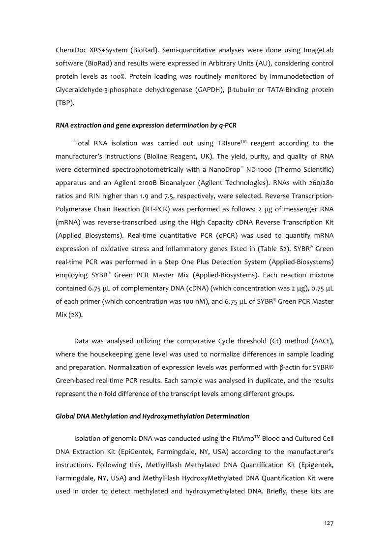
127
ChemiDoc XRS+System (BioRad). Semi-quantitative analyses were done using ImageLab
software (BioRad) and results were expressed in Arbitrary Units (AU), considering control
protein levels as 100%. Protein loading was routinely monitored by immunodetection of
Glyceraldehyde-3-phosphate dehydrogenase (GAPDH), β-tubulin or TATA-Binding protein
(TBP).
RNA extraction and gene expression determination by q-PCR
Total RNA isolation was carried out using TRIsureTM reagent according to the
manufacturer’s instructions (Bioline Reagent, UK). The yield, purity, and quality of RNA
were determined spectrophotometrically with a NanoDrop™ ND-1000 (Thermo Scientific)
apparatus and an Agilent 2100B Bioanalyzer (Agilent Technologies). RNAs with 260/280
ratios and RIN higher than 1.9 and 7.5, respectively, were selected. Reverse Transcription-
Polymerase Chain Reaction (RT-PCR) was performed as follows: 2 μg of messenger RNA
(mRNA) was reverse-transcribed using the High Capacity cDNA Reverse Transcription Kit
(Applied Biosystems). Real-time quantitative PCR (qPCR) was used to quantify mRNA
expression of oxidative stress and inflammatory genes listed in (Table S2). SYBR® Green
real-time PCR was performed in a Step One Plus Detection System (Applied-Biosystems)
employing SYBR® Green PCR Master Mix (Applied-Biosystems). Each reaction mixture
contained 6.75 μL of complementary DNA (cDNA) (which concentration was 2 μg), 0.75 μL
of each primer (which concentration was 100 nM), and 6.75 μL of SYBR® Green PCR Master
Mix (2X).
Data was analysed utilizing the comparative Cycle threshold (Ct) method (ΔΔCt),
where the housekeeping gene level was used to normalize differences in sample loading
and preparation. Normalization of expression levels was performed with β-actin for SYBR®
Green-based real-time PCR results. Each sample was analysed in duplicate, and the results
represent the n-fold difference of the transcript levels among different groups.
Global DNA Methylation and Hydroxymethylation Determination
Isolation of genomic DNA was conducted using the FitAmpTM Blood and Cultured Cell
DNA Extraction Kit (EpiGentek, Farmingdale, NY, USA) according to the manufacturer’s
instructions. Following this, Methylflash Methylated DNA Quantification Kit (Epigentek,
Farmingdale, NY, USA) and MethylFlash HydroxyMethylated DNA Quantification Kit were
used in order to detect methylated and hydroxymethylated DNA. Briefly, these kits are
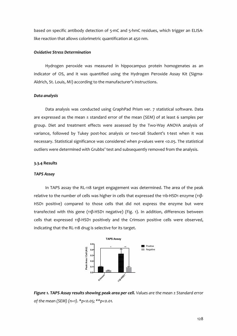
128
based on specific antibody detection of 5-mC and 5-hmC residues, which trigger an ELISA-
like reaction that allows colorimetric quantification at 450 nm.
Oxidative Stress Determination
Hydrogen peroxide was measured in hippocampus protein homogenates as an
indicator of OS, and it was quantified using the Hydrogen Peroxide Assay Kit (Sigma-
Aldrich, St. Louis, MI) according to the manufacturer’s instructions.
Data analysis
Data analysis was conducted using GraphPad Prism ver. 7 statistical software. Data
are expressed as the mean ± standard error of the mean (SEM) of at least 6 samples per
group. Diet and treatment effects were assessed by the Two-Way ANOVA analysis of
variance, followed by Tukey post-hoc analysis or two-tail Student’s t-test when it was
necessary. Statistical significance was considered when p-values were <0.05. The statistical
outliers were determined with Grubbs’ test and subsequently removed from the analysis.
3.3.4 Results
TAPS Assay
In TAPS assay the RL-118 target engagement was determined. The area of the peak
relative to the number of cells was higher in cells that expressed the 11b-HSD1 enzyme (11β-
HSD1 positive) compared to those cells that did not express the enzyme but were
transfected with this gene (11β-HSD1 negative) (Fig. 1). In addition, differences between
cells that expressed 11β-HSD1 positively and the Crimson positive cells were observed,
indicating that the RL-118 drug is selective for its target.
Figure 1. TAPS Assay results showing peak area per cell. Values are the mean ± Standard error
of the mean (SEM) (n=1). *p<0.05; **p<0.01.
Crimso
n
11β-H
SD10.0
0.1
0.2
0.3
0.4
0.5
Pe
ak
Are
a /
Ce
ll (A
U) Positive
Negative***
TAPS Assay
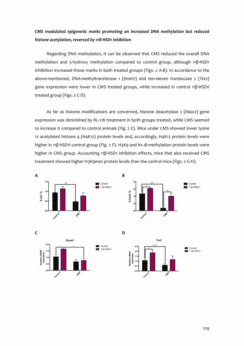
129
CMS modulated epigenetic marks promoting an increased DNA methylation but reduced
histone acetylation, reversed by 11β-HSD1 inhibition
Regarding DNA methylation, it can be observed that CMS reduced the overall DNA
methylation and 5-hydroxy methylation compared to control group, although 11β-HSD1
inhibition increased those marks in both treated groups (Figs. 2 A-B). In accordance to the
above-mentioned, DNA-methyltransferase 1 (Dnmt1) and ten-eleven translocase 2 (Tet2)
gene expression were lower in CMS treated groups, while increased in control 11β-HSD1i
treated group (Figs. 2 C-D).
As far as histone modifications are concerned, histone deacetylase 2 (Hdac2) gene
expression was diminished by RL-118 treatment in both groups treated, while CMS seemed
to increase it compared to control animals (Fig. 2 E). Mice under CMS showed lower lysine
12 acetylated histone 4 (H4K12) protein levels and, accordingly, H4K12 protein levels were
higher in 11β-HSD1i control group (Fig. 2 F). H3K9 and its di-methylation protein levels were
higher in CMS group. Accounting 11β-HSD1 inhibition effects, mice that also received CMS
treatment showed higher H3K9me2 protein levels than the control mice (Figs. 2 G-H).
A B
C D
Control
CMS
0
2
4
6
8
5-h
mC
%
Control
11β-HSD1i
**
****
Control
CMS
0.0
0.5
1.0
1.5
5-m
C %
Control
11β-HSD1i***
Control
CMS
0.0
0.5
1.0
1.5
2.0
2.5
Tet2
Rela
tive m
RN
A
expre
ssio
n
Control
11β-HSD1i*
p=0.07
Control
CMS
0.0
0.5
1.0
1.5
2.0
Dnmt1
Rela
tive m
RN
A
ex
pre
ssio
n
Control
11β-HSD1i* *
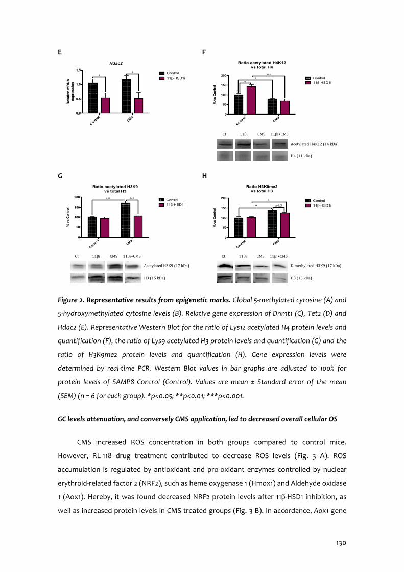
130
E F
G H
Figure 2. Representative results from epigenetic marks. Global 5-methylated cytosine (A) and
5-hydroxymethylated cytosine levels (B). Relative gene expression of Dnmt1 (C), Tet2 (D) and
Hdac2 (E). Representative Western Blot for the ratio of Lys12 acetylated H4 protein levels and
quantification (F), the ratio of Lys9 acetylated H3 protein levels and quantification (G) and the
ratio of H3K9me2 protein levels and quantification (H). Gene expression levels were
determined by real-time PCR. Western Blot values in bar graphs are adjusted to 100% for
protein levels of SAMP8 Control (Control). Values are mean ± Standard error of the mean
(SEM) (n = 6 for each group). *p<0.05; **p<0.01; ***p<0.001.
GC levels attenuation, and conversely CMS application, led to decreased overall cellular OS
CMS increased ROS concentration in both groups compared to control mice.
However, RL-118 drug treatment contributed to decrease ROS levels (Fig. 3 A). ROS
accumulation is regulated by antioxidant and pro-oxidant enzymes controlled by nuclear
erythroid-related factor 2 (NRF2), such as heme oxygenase 1 (Hmox1) and Aldehyde oxidase
1 (Aox1). Hereby, it was found decreased NRF2 protein levels after 11β-HSD1 inhibition, as
well as increased protein levels in CMS treated groups (Fig. 3 B). In accordance, Aox1 gene
Control
CMS
0.0
0.5
1.0
1.5
Hdac2R
ela
tive
mR
NA
expre
ssio
nControl
11β-HSD1i
**
Control
CMS
0
50
100
150
200
Ratio acetylated H4K12 vs total H4
% v
s C
ontr
ol
Control
11β-HSD1i*
****
Acetylated H4K12 (14 kDa)
H4 (11 kDa)
Ct 11βi CMS 11βi+CMS
Control
CMS
0
50
100
150
200
Ratio acetylated H3K9 vs total H3
% v
s C
ontr
ol
Control
11β-HSD1i
*** ***
Acetylated H3K9 (17 kDa)
H3 (15 kDa)
Ct 11βi CMS 11βi+CMS
Control
CMS
0
50
100
150
200
Ratio H3K9me2vs total H3
% v
s C
ontr
ol
Control
11β-HSD1i**
*p=0.07
Dimethylated H3K9 (17 kDa)
H3 (15 kDa)
Ct 11βi CMS 11βi+CMS
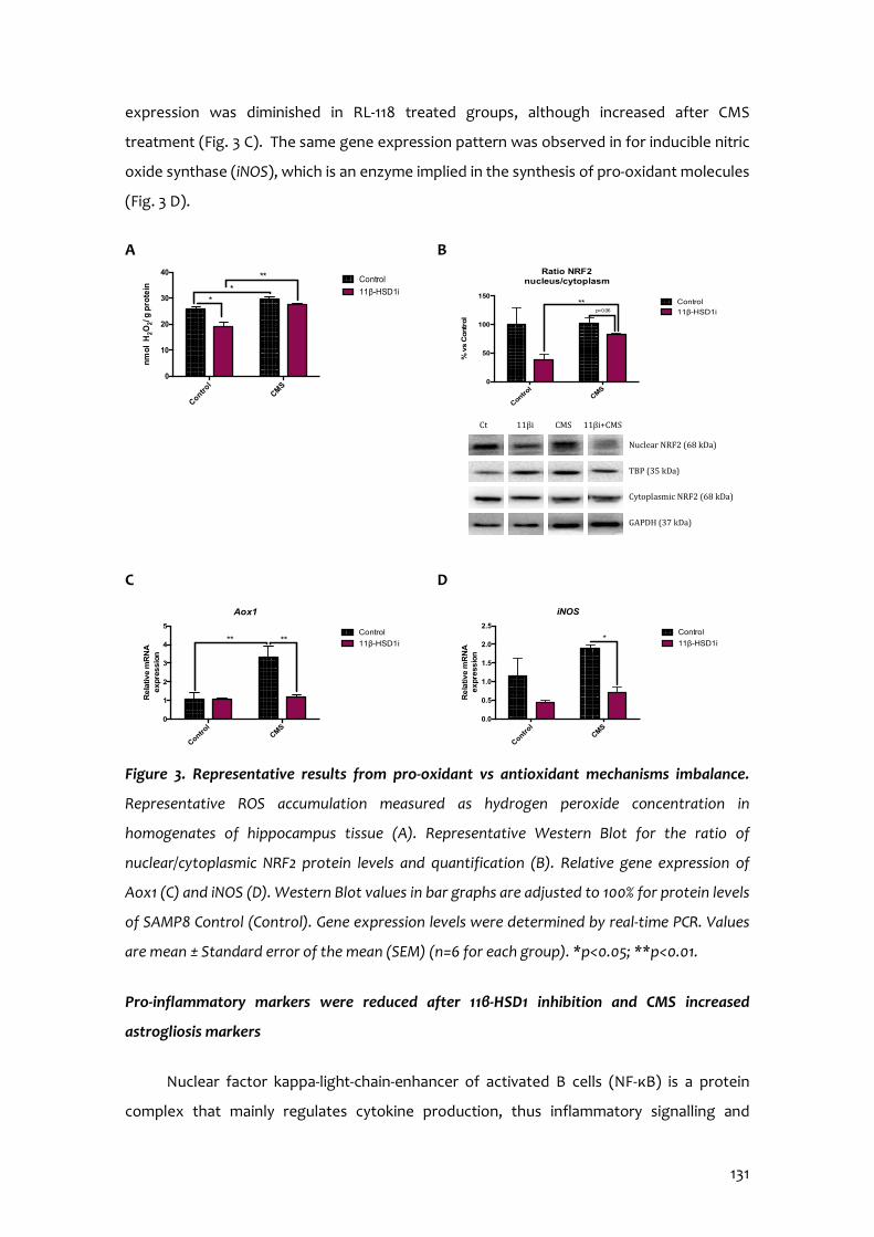
131
expression was diminished in RL-118 treated groups, although increased after CMS
treatment (Fig. 3 C). The same gene expression pattern was observed in for inducible nitric
oxide synthase (iNOS), which is an enzyme implied in the synthesis of pro-oxidant molecules
(Fig. 3 D).
A B
C D
Figure 3. Representative results from pro-oxidant vs antioxidant mechanisms imbalance.
Representative ROS accumulation measured as hydrogen peroxide concentration in
homogenates of hippocampus tissue (A). Representative Western Blot for the ratio of
nuclear/cytoplasmic NRF2 protein levels and quantification (B). Relative gene expression of
Aox1 (C) and iNOS (D). Western Blot values in bar graphs are adjusted to 100% for protein levels
of SAMP8 Control (Control). Gene expression levels were determined by real-time PCR. Values
are mean ± Standard error of the mean (SEM) (n=6 for each group). *p<0.05; **p<0.01.
Pro-inflammatory markers were reduced after 11β-HSD1 inhibition and CMS increased
astrogliosis markers
Nuclear factor kappa-light-chain-enhancer of activated B cells (NF-κB) is a protein
complex that mainly regulates cytokine production, thus inflammatory signalling and
Control
CMS
0
10
20
30
40
nm
ol H
2O2/
g p
rote
in
Control
11β-HSD1i*
***
Control
CMS
0
1
2
3
4
5
Aox1
Rela
tive m
RN
A
expre
ssio
n
Control
11β-HSD1i** **
Control
CMS
0.0
0.5
1.0
1.5
2.0
2.5
iNOS
Re
lativ
e m
RN
A
expre
ssio
n
Control
11β-HSD1i*
Control
CMS
0
50
100
150
Ratio NRF2nucleus/cytoplasm
% v
s C
ontr
ol
Control
11β-HSD1ip=0.06**
Ct 11βi CMS 11βi+CMS
Nuclear NRF2 (68 kDa)
TBP (35 kDa)
Cytoplasmic NRF2 (68 kDa)
GAPDH (37 kDa)
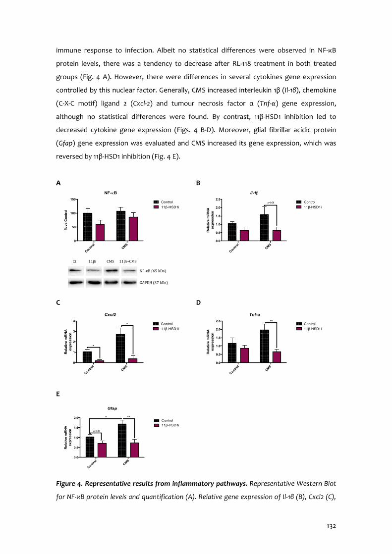
132
immune response to infection. Albeit no statistical differences were observed in NF-κB
protein levels, there was a tendency to decrease after RL-118 treatment in both treated
groups (Fig. 4 A). However, there were differences in several cytokines gene expression
controlled by this nuclear factor. Generally, CMS increased interleukin 1β (Il-1β), chemokine
(C-X-C motif) ligand 2 (Cxcl-2) and tumour necrosis factor α (Tnf-α) gene expression,
although no statistical differences were found. By contrast, 11β-HSD1 inhibition led to
decreased cytokine gene expression (Figs. 4 B-D). Moreover, glial fibrillar acidic protein
(Gfap) gene expression was evaluated and CMS increased its gene expression, which was
reversed by 11β-HSD1 inhibition (Fig. 4 E).
A B
C D
E
Figure 4. Representative results from inflammatory pathways. Representative Western Blot
for NF-κB protein levels and quantification (A). Relative gene expression of Il-1β (B), Cxcl2 (C),
Control
CMS
0.0
0.5
1.0
1.5
2.0
Gfap
Rela
tive m
RN
A
expr
ess
ion
Control
11β-HSD1i
p=0.09
* **
Control
CMS
0.0
0.5
1.0
1.5
2.0
2.5
Il-1b
Rela
tive
mR
NA
expre
ssio
n
Control
11β-HSD1i
p=0.08
Control
CMS
0
1
2
3
4
Cxcl2
Rela
tive m
RN
A
expr
essio
n
Control
11β-HSD1i*
*
Control
CMS
0.0
0.5
1.0
1.5
2.0
2.5
Tnf-α
Rela
tiv
e m
RN
A
exp
ress
ion
Control
11β-HSD1i
**
Control
CMS
0
50
100
150
NF-kB
% v
s C
ontr
ol
Control
11β-HSD1i
Ct 11βi CMS 11βi+CMS
NF-κB (65 kDa)
GAPDH (37 kDa)

133
Tnf-α (D) and Gfap (E). Western Blot values in bar graphs are adjusted to 100% for protein
levels of SAMP8 Control (Control). Gene expression levels were determined by real-time PCR.
Values are mean ± Standard error of the mean (SEM) (n=6 for each group). *p<0.05; **p<0.01.
11β-HSD1 inhibition and CMS promoted autophagy
Several autophagy markers were evaluated in the present work, such as Beclin1,
rapamycin-sensitive TOR complex 1 (TORC1) and microtubule-associated protein 1A/1B light
chain 3B (LC3B). Of note, Beclin1 and LC3B are activators of this cellular cleaning process,
while TORC1 is associated with autophagy inhibition. After 11β-HSD1 inhibitor treatment,
Beclin1 and LC3B protein levels were increased, while serine 151 phosphorylated TORC1
protein levels were decreased. Accordingly, mice under CMS showed higher protein levels
of p-TORC1. However and strikingly, mice under CMS also presented higher protein levels of
LC3B (Figs. 5 A-C).
A B
C
Figure 5. Representative results from autophagy process. Representative Western Blot for
Beclin1 protein levels and quantification (A), the ratio of p-TORC1 protein levels and
Control
CMS
0
50
100
150
200
250
Beclin1
% v
s C
ontr
ol
Control
11β-HSD1i***
*
Ct 11βi CMS 11βi+CMS
Beclin1 (60 kDa)
GAPDH (37 kDa)
p-TORC1 (78 kDa)
TORC1 (78 kDa)
GAPDH (37 kDa)
Ct 11βi CMS 11βi+CMS
Control
CMS
0
100
200
300
400
Ratio p-TORC1 vs TORC1
% v
s C
ontr
ol
Control
11β-HSD1i*
Control
CMS
0
200
400
600
Ratio LC3-I/LC3-II
% v
s C
ontr
ol
Control
11β-HSD1i**
**
LC3-I (16 kDa) LC3-II (14 kDa) GAPDH (37 kDa)
Ct 11βi CMS 11βi+CMS

134
quantification (B) and the ratio of LC3 protein levels and quantification (C). Values in bar
graphs are adjusted to 100% for protein levels of SAMP8 Control (Control). Values are mean ±
Standard error of the mean (SEM) (n=4 for each group). *p<0.05; **p<0.01.
11β-HSD1 inhibition rescued mice from CMS pejorative effects on APP processing
Herein, a disintegrin and metalloproteinase domain protein 10 (Adam10) gene
expression was increased in mice treated with RL-118 as well as decreased in CMS group
(Fig. 6 A). Also, β-secretase 1 (Bace1) gene expression was decreased in both RL-118 treated
groups and there was a trend towards increasing Bace1 gene expression after CMS
exposure (Fig. 6 B). In line with these results, Aβ-precursor gene expression was increased
in CMS group and reversed by 11β-HSD1 inhibition (Fig. 6 C). Finally, β-amyloid degradation
was assessed by Neprilisin12 gene expression. It was found that RL-118 treatment raised its
gene expression, while CMS reduced it (Fig. 6 D).
A B
C D
Figure 6. Representative results from APP processing pathways. Relative gene expression of
Adam10 (A), Bace1 (B), Aβ-precursor (C) and Neprilisin12 (D). Gene expression levels were
determined by real-time PCR. Values are mean ± Standard error of the mean (SEM) (n=6 for
each group). *p<0.05; **p<0.01.
Control
CMS
0.0
0.5
1.0
1.5
2.0
2.5
Adam10
Rela
tive m
RN
A
expre
ssio
n
Control
11β-HSD1i** *
*
*
Control
CMS
0.0
0.5
1.0
1.5
2.0
Bace1
Rela
tive
mR
NA
expre
ssio
n
Control
11β-HSD1i
*
*
Control
CMS
0.0
0.5
1.0
1.5
2.0
Ab-precursor
Rela
tive m
RN
A
expre
ssio
n
Control
11β-HSD1i*
Control
CMS
0.0
0.5
1.0
1.5
2.0
2.5
Neprilisin12
Rela
tive m
RN
A
exp
ressio
n
Control
11β-HSD1i****

135
Changes in synaptic plasticity were produced after GC attenuation
To address whether GC attenuation through 11β-HSD1 inhibition modulated synaptic
plasticity, we evaluated different proteins involved in those mechanisms. cAMP response
element-binding (Creb) gene expression and protein levels were increased in RL-118 treated
mice and decreased after CMS exposure. In turn, the same pattern was found in the Creb’s
downstream mediator, the brain-derived neurotrophic factor (Bdnf) (Figs. 7 A-C).
Accordingly, synaptic modulators such as postsynaptic density protein 95 (PSD95),
synaptophysin and synaptosomal nerve-associated protein 25 (SNAP25) protein levels were
increased after 11β-HSD1 inhibition treatment, although not decreased after CMS (Figs. 7 E-
H).
A B
C D
Control
CMS
0.0
0.5
1.0
1.5
2.0
2.5
Creb
Rela
tive m
RN
A
ex
pressio
n
Control
11β-HSD1i*
Control
CMS
0.0
0.5
1.0
1.5
2.0
Bdnf
Rela
tive m
RN
A
ex
pressio
n
Control
11β-HSD1i*
Control
CMS
0
50
100
150
BDNF
% v
s C
ontr
ol
Control
11β-HSD1i**
BDNF (22 kDa)
GAPDH (37 kDa)
Ct 11βi CMS 11βi+CMS
Ct 11βi CMS 11βi+CMS
p-CREB (42 kDa)
CREB (42 kDa)
Tubulin (55 kDa)
Control
CMS
0
50
100
150
200
Ratio p-CREB vs CREB%
vs C
ontr
ol
Control
11β-HSD1i*
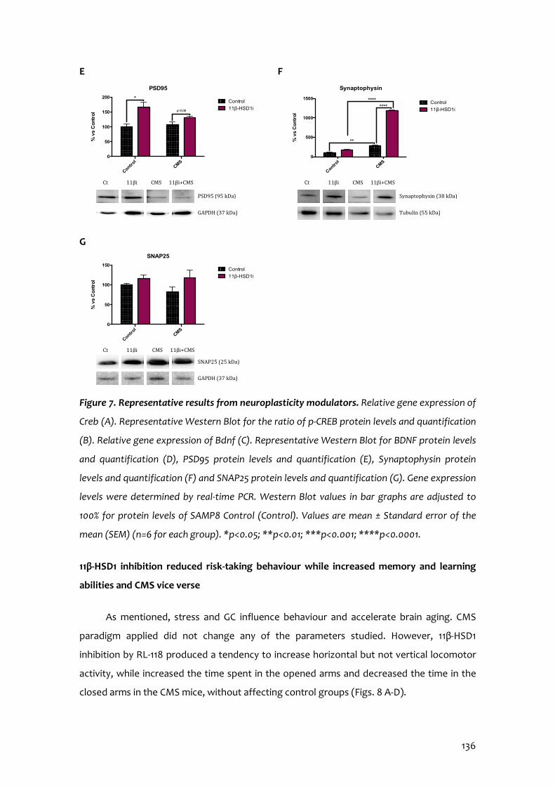
136
E F
G
Figure 7. Representative results from neuroplasticity modulators. Relative gene expression of
Creb (A). Representative Western Blot for the ratio of p-CREB protein levels and quantification
(B). Relative gene expression of Bdnf (C). Representative Western Blot for BDNF protein levels
and quantification (D), PSD95 protein levels and quantification (E), Synaptophysin protein
levels and quantification (F) and SNAP25 protein levels and quantification (G). Gene expression
levels were determined by real-time PCR. Western Blot values in bar graphs are adjusted to
100% for protein levels of SAMP8 Control (Control). Values are mean ± Standard error of the
mean (SEM) (n=6 for each group). *p<0.05; **p<0.01; ***p<0.001; ****p<0.0001.
11β-HSD1 inhibition reduced risk-taking behaviour while increased memory and learning
abilities and CMS vice verse
As mentioned, stress and GC influence behaviour and accelerate brain aging. CMS
paradigm applied did not change any of the parameters studied. However, 11β-HSD1
inhibition by RL-118 produced a tendency to increase horizontal but not vertical locomotor
activity, while increased the time spent in the opened arms and decreased the time in the
closed arms in the CMS mice, without affecting control groups (Figs. 8 A-D).
Control
CMS
0
50
100
150
200
PSD95%
vs C
ontr
ol
Control
11β-HSD1i
*
p=0.08
PSD95 (95 kDa)
GAPDH (37 kDa)
Ct 11βi CMS 11βi+CMS
Control
CMS
0
500
1000
1500
Synaptophysin
% v
s C
ontr
ol
Control
11β-HSD1i
**
********
Synaptophysin (38 kDa)
Tubulin (55 kDa)
Ct 11βi CMS 11βi+CMS
Control
CMS
0
50
100
150
SNAP25
% v
s C
ontr
ol
Control
11β-HSD1i
SNAP25 (25 kDa)
GAPDH (37 kDa)
Ct 11βi CMS 11βi+CMS
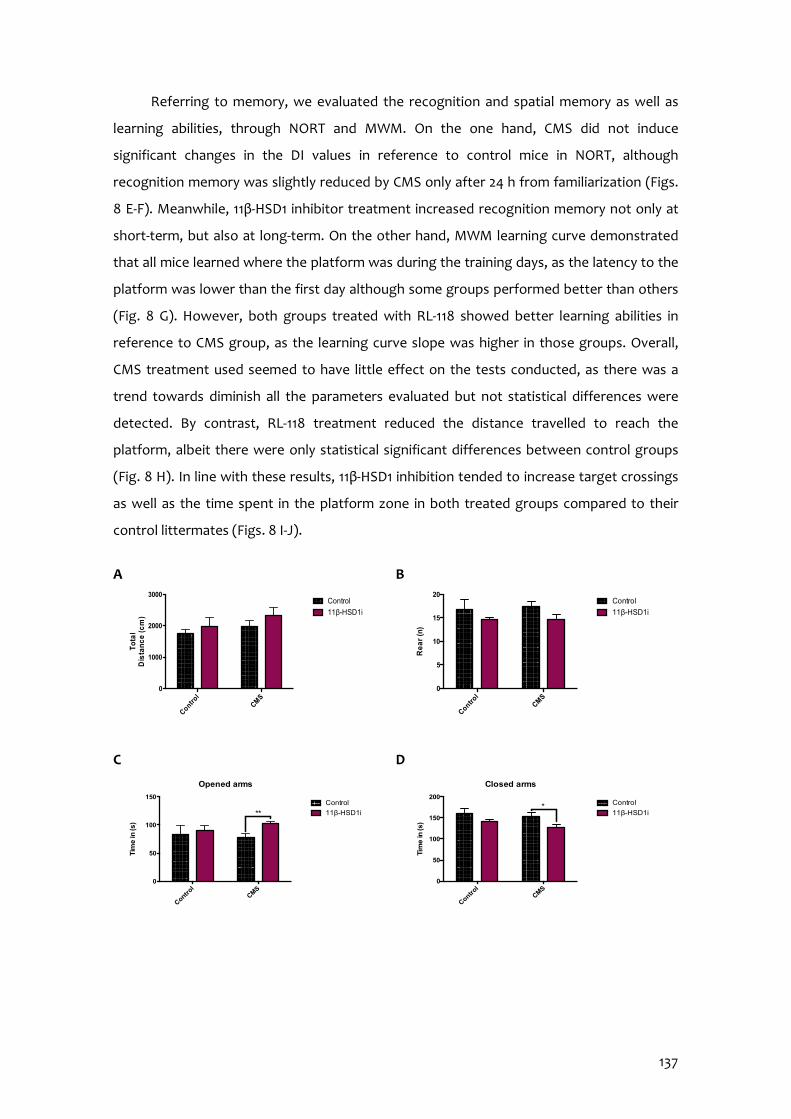
137
Referring to memory, we evaluated the recognition and spatial memory as well as
learning abilities, through NORT and MWM. On the one hand, CMS did not induce
significant changes in the DI values in reference to control mice in NORT, although
recognition memory was slightly reduced by CMS only after 24 h from familiarization (Figs.
8 E-F). Meanwhile, 11β-HSD1 inhibitor treatment increased recognition memory not only at
short-term, but also at long-term. On the other hand, MWM learning curve demonstrated
that all mice learned where the platform was during the training days, as the latency to the
platform was lower than the first day although some groups performed better than others
(Fig. 8 G). However, both groups treated with RL-118 showed better learning abilities in
reference to CMS group, as the learning curve slope was higher in those groups. Overall,
CMS treatment used seemed to have little effect on the tests conducted, as there was a
trend towards diminish all the parameters evaluated but not statistical differences were
detected. By contrast, RL-118 treatment reduced the distance travelled to reach the
platform, albeit there were only statistical significant differences between control groups
(Fig. 8 H). In line with these results, 11β-HSD1 inhibition tended to increase target crossings
as well as the time spent in the platform zone in both treated groups compared to their
control littermates (Figs. 8 I-J).
A B
C D
Control
CMS
0
1000
2000
3000
To
tal
Dis
tan
ce
(cm
)
Control
11β-HSD1i
Control
CMS
0
5
10
15
20
Re
ar
(n)
Control
11β-HSD1i
Control
CMS
0
50
100
150
200
Closed arms
Tim
e in
(s)
Control
11β-HSD1i*
Control
CMS
0
50
100
150
Opened arms
Tim
e in
(s)
Control
11β-HSD1i**

138
E F
G H
I J
Figure 8. Behavioural tests results from EPM, NORT and MWM respectively. Total Distance
(A), number of Rearing (B), Time in the Opened arms (C), Time in the Closed arms (D),
Summary of DI from 2 and 24 hours after familiarization phase (E-F), MWM learning curve (G),
Distance to reach the platform (H), number of Entries (I) and Time in the platform (J). Values
are mean ± Standard error of the mean (SEM) (n=12 for each group). *p<0.05; **p<0.01;
***p<0.001; ****p<0.0001.
3.3.5 Discussion
Taking into consideration the convergence of aging, stress and neurodegenerative
diseases, such as AD, there is impaired GC signalling. Therefore, the study of GC response to
chronic moderate stressful situations, as account in the daily life, results of huge interest in
order to design pharmacological strategies to prevent neurodegeneration.
Control
CMS
0
500
1000
1500
Dis
tan
ce
to p
latf
orm
(cm
)
Control
11β-HSD1ip=0.07
1 2 3 4 50
20
40
60
Learning curve
Time (days)
Late
nc
y to
pla
tfo
rm (s
) Control
11β-HSD1i
CMS
CMS + 11β-HSD1i
Control
CMS
0
10
20
30
40
Tim
e in
pla
tfo
rm (%
)
Control
11β-HSD1i
Control
CMS
0
1
2
3
4
5
Tar
ge
t cro
ss
ing
s (n
) Control
11β-HSD1i
Control
CMS
-0.2
-0.1
0.0
0.1
0.2
0.3
NORT 2h - SummaryShort Term Memory
Dis
cri
min
atio
n
Inde
x (D
I)
Control
11β-HSD1i
**** ****
Control
CMS
-0.1
0.0
0.1
0.2
0.3
NORT 24h - SummaryLong-term memory
Dis
cri
min
atio
n
Inde
x (D
I)
Control
11β-HSD1i****
*****
139
Several pieces of evidence demonstrate a strong association between prolonged
exposure to GC excess and diminished cognitive abilities. It may be due to alterations in
hippocampal electrophysiology, structure and function, as well as deleterious effects on
neurotransmission, metabolism, cell division and death. GC secretion responds to a feed-
forward signalling involving the hypothalamic-pituitary-adrenal (HPA) axis conforming the
stress response. As far as stress is concerned, stressful stimuli increase GC secretion and
release to confer energy to cope with the stress.
Previously, we affirmed that 11β-HSD1 enzyme regulates the disposure of active GCs
(Leiva et al., 2017; Puigoriol-Illamola et al., 2018). Not only that, but also 11β-HSD1 inhibition
has been linked to enhance cognitive abilities and AD hallmarks (Mohler et al., 2011;
Puigoriol-Illamola et al., 2018, 2020b). In line with these results, it has been described that
11β-HSD1 expression in mouse hippocampus and parietal cortex increases with aging and
that its overexpression accelerated age-related cognitive decline (Holmes et al., 2010). By
contrast, 11β-HSD1 knockout mice resist age-dependent cognitive loss (Yau et al., 2007,
2015). In line with this, in our hands, we demonstrated that RL-118, a brain penetrant 11β-
HSD1 inhibitor drug, treatment promoted autophagy flux, as well as ER stress activation in
order to restore the deleterious effects exerted by prolonged exposure to GCs (Leiva et al.,
2017; Puigoriol-Illamola et al., 2018, 2020b). In conclusion, it is suggested that it could be a
feasible target to fight against cognitive decline in age-related pathologies. In fact, early
clinical studies demonstrate that 11β-HSD1 inhibitor (UE2343) is well tolerated in AD patients
(Webster et al., 2017) and currently is at phase II. Besides, many selective 11β-HSD1 inhibitors
have reached clinical stages for metabolic diseases, for instance AZD8329 and BVT.2733
(Puigoriol-Illamola et al., 2020b).
Up to now, studies demonstrating that RL-118 is able to reach the brain, bind the 11β-
HSD1 enzyme and inhibit it leading to therapeutic effects, have not been addressed. Thus, in
the present work, the drug-target binding (Target engagement) of RL-118 and 11β-HSD1 was
determined through a novel methodology consisting in FACS coupled to MS analysis
(Wilson et al., 2017). Effectively, target engagement results showed that RL-118 drug binds
to its target and also in a selectively way, since it did not bind to other proteins expressed.
In consequence, we have determined that actually the molecular and behavioural effects
observed after RL-118 administration were due to its binding to the 11β-HSD1 enzyme.

140
Stress response may have a genetic and epigenetic origin, reflected in the efficiency
of the GC receptor (GR)-mediated negative feedback in the brain and/or the pituitary gland
that causes HPA axis hyperactivity (Fink et al., 2017; Harman & Martin, 2019; Sotiropoulous
et al., 2008). In an attempt to address whether and which changes produced the chronic
presence of stressors we decided to evaluate two main epigenetic marks: DNA methylation
and histone post-translational modifications.
Regarding DNA methylation, it is implied in controlling neuronal gene expression and
neural development. Dysregulation of this process is linked to a wide range of neuronal
disorders, including AD onset and progression. However, the relationship between AD and
altered 5-mC levels is not known (Fetahu et al., 2019; Puigoriol-Illamola et al., 2020a). TET
family can further oxidize 5-mC to 5-hmC. Generally, 5-mC is associated with gene silencing,
while 5-hmC with up-regulation of gene expression (Fetahu et al., 2019; Sherwani & Khan,
2015). Herein CMS reduced those epigenetic marks and accordingly to Puigoriol-Illamola et
al. (2020a), attenuating GCs levels led to increase 5-mC as well as 5-hmC percentages in
both treated groups. Therefore, it may suggest that RL-118 is able to restore DNA
methylation pattern although the influence of detrimental stimuli such as CMS. Although
these results seem to contradict what is established for most scientific literature,
methylation of specific genes should be evaluated in order to know which translational
activity is being repressed. Despite that, DNA hydroxymethylation, which is linked to
promote gene expression, was increased in RL-118 treated mice. Overall, those results
suggest that further evaluation of gene-promoter specific methylation are required to
elucidate the clear epigenetic mechanisms to explain the neuroprotective effects observed
in the cognitive tests performed. Likewise, the enzymes responsible for those processes as
Dnmt1 and Tet2 gene expression were increased in both groups treated with 11β-HSD1
inhibitor, whereas it decreased in the CMS group.
DNA methylation and histone modifications must act coordinately (Zhao et al., 2016).
Generally histone acetylation is related to favouring gene transcription through removing
histone positive charge and thereby transforming condensed chromatin (heterochromatin)
into a relaxed structure (euchromatin) (Turner, 2000; Watson et al., 2014). 11β-HSD1
inhibitor treatment altered Hdac2 gene expression, promoting a reduction in both groups.
In line with this, acetylated H4 studied showed higher protein levels in RL-118 group treated,
but not in mice under CMS too. In accordance to Puigoriol-Illamola et al. (2020a), SAMP8
mice under CMS treatment showed higher H3K9 protein levels in respect to control group.
141
However, it could be reversed after concomitant RL-118 treatment. In turn, the same profile
was observed in H3K9me2 protein levels evaluated, suggesting that although the increase
in histone acetylation as it is also methylated, it finally results in transcriptional repression
or a more compacted chromatin state in CMS groups compared to control mice.
Of relevance, although RL-118 is not a direct epigenetic modulator drug, it modifies
the epigenetic landscape. Therefore, our results point out to the paramount importance of
studying different drugs at epigenetic level in order to possess a deep knowledge of the
pharmacological effects exerted by the drug.
Additional data (Kadmiel & Cidlowski, 2013; Picard et al., 2018; Puigoriol-Illamola et al.,
2020a) reports that a stressful environment affects oxidative balance. In normal conditions,
pro-oxidant molecules and antioxidant defence mechanisms are balanced; however, under
the presence of chronic stressors as well as along with aging and in several disorders, there
is a decrease in the capacity of the antioxidant enzymes, allowing ROS accumulation and
eventually causing cellular damage and finally dysfunction of the system (Griñán-Ferré et al,
2018). In concordance, in the present work SAMP8 mice under CMS increased ROS
accumulation and on the contrary, those animals treated with RL-118 diminished it. OS is
frequently invoked as a potential factor in progression of AD, yet whether it is a cause or
consequence of the pathology is still debated (Bonet-Costa et al., 2016). Among antioxidant
defence mechanisms, NRF2 pathway has been defined as a key indicator and modulator of
OS in neurodegeneration (Griñán-Ferré et al., 2018; Johnson et al., 2008; Maeda et al., 2012),
given that it regulates the gene expression of different antioxidant enzymes. While NRF2 is
a protector against oxidative and electrophilic tissue injury, persistent activation of NRF2
signalling may also contribute to disease pathophysiology (Li et al., 2019). Interestingly,
NRF2 protein levels were decreased after RL-118 treatment, suggesting that attenuating GC
levels induce lower oxidative damage. However, CMS stimulated the increase in Aox1 gene
expression, which was reduced after concomitant RL-118 treatment, indicating that
stressful environment increases pro-oxidant mechanisms while 11β-HSD1 inhibition is able to
restore them. Likewise, other OS modulators evaluated as iNOS agreed with the increase in
CMS group compared to control group and decreased protein levels of gene expression
after RL-118 treatment. Moreover, iNOS gene expression has been reported to be regulated
by inflammatory signalling, particularly through NF-κB (Arias-Salvatierra et al., 2011).
Overall, these results suggest that 11β-HSD1 inhibition promoted antioxidant mechanisms to
cope with OS in a mice model of aging, even under CMS conditions.
142
GCs effects include inflammatory signalling regulation. Although normal GC activity
involves immune system suppression, GC excess deploys the contrary effect. Despite
differences did not reach statistical significance, 11β-HSD1 inhibition reduced NF-κB protein
levels. It appears to be a pivotal mediator of inflammatory responses as it induces the
expression of various pro-inflammatory genes, including those encoding cytokines and
chemokines, and participates in inflammasome regulation (Liu et al., 2017). It triggers a
feed-forward cycle in which increases cytokine production and this in turn result in
resistance to the GC immunosuppression, leading to further increases in cytokine release
and, therefore, activation of the HPA axis (Irwin & Miller, 2007; Pace et al., 2007).
Accordingly, we found decreased pro-inflammatory cytokine gene expression after 11β-
HSD1 inhibitor treatment and increased after CMS appliance. Moreover, Il-10 and Il-6 gene
expression results reinforce the same assertion (Data not shown). Pro-inflammatory
cytokines like IFN-α, Il-1, IL-6 and TNF-α have been described to activate the HPA axis and
potentiate GC resistance (Sotiropoulous et al., 2008). Additionally, they have been
implicated in the genesis of AD, becoming part of the amyloid plaques or triggering
amyloid-β (Aβ) production, among others. Dysregulation of inflammatory mediators and
astrogliosis are major culprits in the development of chronic inflammation and
immunosenescence process, as well as are related to cognitive decline and progression of
neurodegenerative diseases (Chung et al., 2019).
Recent studies have indicated that prolonged OS may limit autophagy flux (Bonet-
Costa et al., 2016). In addition, altered cellular loss of proteostasis is one of the nine
hallmarks of aging postulated by López-Otín et al. (2013). Moreover, our group had
previously demonstrated that attenuating GC excess by RL-118 promotes autophagy
activation (Puigoriol-Illamola et al., 2018) and, by contrast, CMS treatment induced
autophagy deterioration (Puigoriol-Illamola et al., 2020a). In accordance, autophagy flux
was decreased in female mice under CMS but those treated with RL-118 showed higher
protein levels of autophagy activators, like Beclin1 and LC3B, and lower protein levels of p-
TORC1.
11β-HSD1 inhibition has been linked to an extensive number of disorders, among
which there is AD. In particular, it has been associated with reduced Aβ neurotoxicity and
Tau hyperphosphorylation (Dong & Csernansky, 2009; Ouanes & Popp, 2019), both
hallmarks of AD. Analysing deeper this issue, amyloid precursor protein (APP) can be
processed through two mechanisms: non-amyloidogenic and amyloidogenic pathways. The

143
main indicator of the former is Adam10, whereas the later is Bace1 (Shen et al., 2018). In this
study, Adam10 gene expression was diminished and Bace1 increased after CMS. Importantly,
in RL-118 treated groups occurred the opposite, then favouring the slow down formation of
Aβ. Moreover, the Aβ degrading enzyme, neprilisin12 gene expression increased after 11β-
HSD1 inhibitor treatment but reduced after CMS.
As mentioned above, prolonged exposure to GCs in the brain induces several changes
and ample experimental evidence state that repeated exposure to stressful conditions
induce structural remodelling of neurons with synaptic loss as well as alterations in glial
functions, which are frequently maladaptive (Vyas et al., 2016). In agreement, our results
demonstrated a reduction in Creb and Bdnf gene expression and protein levels after CMS
exposure, but conversely RL-118 treatment was not able to prevent it (Steffke et al., 2020).
In a similar way, CMS reduced synaptic plasticity markers, like PSD95, synaptophysin and
SNAP25, and by contrast 11β–HSD1 inhibition increased them, particularly synaptophysin
(Mango et al., 2019).
The effects of GCs on cognition have been widely studied, stating that acute stress
enhances cognitive abilities, while chronic stress negatively affects memory and learning
processes as well as accelerates brain aging (Ouanes & Popp, 2019; Sandi et al., 2013;
Sotiropoulous et al., 2008; Weger & Sandi, 2018). In fact, GC levels have been found to
correlate with the severity of the cognitive impairment (Ouanes & Popp, 2019). Assessing
EPM results, it is well known that vertical locomotor activity indicates anxiety-like behaviour
and, although not statistical differences were observed, together with the total horizontal
distance travelled, we could affirm that RL-118 drug treatment helped to reduce the feeling
of anxiety and desire to escape from an inhospitable environment, so that risk-taking
behaviour becomes reduced. This is supported by the time spent in opened and closed
arms, as it can be seen that after 4 weeks of 11β-HSD1 inhibition together to detrimental
stimuli such as CMS, animals stood less time in the closed arms and, on the contrary, more
time in the opened arms respect to CMS group. In accordance to Puigoriol-Illamola et al.
(2020 a & b), mice under RL-118 treatment clearly showed improved recognition memory
than control mice. However, the pejorative effect caused by CMS was only detected at
long-term recognition memory evaluation. Regarding MWM results, it seemed that CMS
treatment might not be strong enough to produce clearly changes in the tests performed.
11β-HSD1 inhibition tended to improve the mice performance, although not statistical
differences were detected. However, both groups treated with RL-118 showed better

144
learning abilities in reference to CMS group, as the learning curve slope is higher in those
groups, indicative of a putative role for RL-118 as a neuroprotectant or cognitive enhancer
of cognition (nootropic drugs).
3.3.6 Conclusions
In view of these results, target engagement between 11β-HSD1 enzyme and RL-118
drug was demonstrated and therefore, we can surely attribute all the beneficial effects
observed in SAMP8 treated with RL-118 to 11β-HSD1 inhibition. Diversely, CMS declined
cognitive and behavioural abilities, as well as synaptic plasticity, autophagy and antioxidant
mechanisms, while modulated epigenetic markers and increased inflammatory signalling
and Aβ formation and accumulation. However and most important, 11β-HSD1 inhibition
through RL-118 turned up to restore the majority of these detrimental effects caused by
CMS (Fig. 9).
Figure 9. Representative scheme of the molecular pathways altered after RL-118 treatment in
SAMP8 mice under chronic mild stress.
3.3.7 Acknowledgements
This study was supported by Ministerio de Economía y Competitividad of Spain and
FEDER (SAF2016-77703) and 2017SGR106 (AGAUR, Catalonia). Financial support was
provided for D.P.I. (FPU program).
145
3.3.8 References
Arias-Salvatierra, D.; Silbergeld, E.K.; Acosta-Saavedra, L.C.; Calderon-Aranda, E.S. Role of nitric oxide produced by iNOS through NF-kB pathway in migration of cerebellar granule neurons induced by Lipopolysaccharide. Cell Signal. 2011, 23(2):425-435.
Bisht, K.; Sharma, K.; Tremblay, M.È. Chronic stress as a risk factor for Alzheimer’s disease: Roles of microglia-mediated synaptic remodeling inflammation, and oxidative stress. Neurobiol Stress 2018, 9:9-21.
Bonet-Costa, V.; Pomatto, L.C.; Davies, K.J.A. The proteasome and oxidative stress in
Alzheimer’s disease. Antioxid Redox Signal 2016, 25(16):886-901. Canet, G.; Chevallier, N.; Zussy, C.; Desrumaux, C.; Givalois, L. Central role of
glucocorticoids receptors in Alzheimer’s disease and depression. Front in Neurosci 2018 12, 789.
Catania, C.; Sotiropoulous, I.; Silva, R.; Onofri, C.; Breen, K.C.; Sousa, N.; Almeida,
O.F.X. The amyloidogenic potential and behavioral correlates of stress. Mol Psychiatry 2009, 14(1): 95-105.
Chung, H.Y.; Kim, D.H.; Lee, E.K.; Chung, K.W.; Chung, S.; Lee, B.; Seo, A.Y.; Chung,
J.H.; Jung, Y.S.; Im, E.; Lee, J.; Kim, N.D.; Choi, Y.J., Im, D.S.; Yu, B.P. Redefining Chronic Inflammation in Aging and Age-Related Diseases: Proposal of the Senoinflammation Concept. Aging Dis. 2019,10(2):367-382.
Dhabhar, F.S. The Short-Term Stress Response – Mother Nature’s Mechanism for
Enhancing Protection and Performance Under Conditions of Threat, Challenge, and Opportunity. Front Neuroendocrinol 2018, 49, 175-192.
Dong, H.; Csernansky, J.G. Effects of stress and stress hormones on amyloid-beta
protein and plaque deposition. J Alzheimers Dis. 2009, 18, 459-469. Fetahu, I.S.; Ma, D.; Rabidou, K.; Argueta, C.; Smith, M.; Liu, H.; Wu, F.; Shi, Y.G.
Epigenetic signatures of methylated DNA cytosine in Alzheimer’s disease. Science Advances 2019, 5(8).
Fink, G. Stress: Concepts, Definition and History. Neurosci Biobehav Psychol 2017. Griñán-Ferré, C.; Corpas, R.; Puigoriol-Illamola, D.; Palomera-Ávalos, V.; Sanfeliu, C.;
Pallàs, M. Understanding Epigenetics in the Neurodegeneration of Alzheimer’s Disease: SAMP8 Mouse Model. J Alzheimers Dis 2018, 62(3): 943-963.
Griñán-Ferré, C.; Puigoriol-Illamola, D.; Palomera-Ávalos, V.; Pérez-Cáceres, D.;
Companys-Alemany, J.; Camins, A.; Ortuño-Sahagún, D.; Rodrigo, M.T.; Pallàs, M.
146
Environmental Enrichment Modified Epigenetic Mechanisms in SAMP8 Mouse Hippocampus by Reducing Oxidative Stress and Inflammaging and Achieving Neuroprotection. Front. Aging Neurosci. 2016, 8, 241.
Harman, M.F.; Martín, M.G. Epigenetic mechanisms related to cognitive decline
during aging. J Neurosci Res 2019, 98(2):234-246. Holmes, M.C.; Carter, R.N.; Noble, J.; Chitnis, S.; Dutia, A.; Paterson, J.M.; Mullins, J.J.;
Seckl, J.R.; Yau, J.L.W. 11β-Hydroxysteroid dehydrogenase type 1 expression is increased in
the aged mouse hippocampus and parietal cortex and causes memory impairments. J
Neurosci, 2010, 30(20), 6916-6920.
Irwin, M.R.; Miller, A.H. Depressive disorders and immunity: 20 years of progress and
discovery. Brain Behav Immun 2007, 21(4):374-83. Johnson, J.A.; Johnson, D.A.; Kraft, A.D.; Calkins, M.J.; Jakel, R.J.; Vargas, M.R.; Chen,
P-C. The Nrf2-ARE Pathway: An Indicator and Modulator of Oxidative Stress in Neurodegeneration. Ann N Y Acad Sci. 2008, 1147:61-69.
Kadmiel, M.; Cidlowski, J.A. Glucocorticoid receptor signalling in health and disease.
Trends Pharmacol Sci. 2013, 34(9):518-530. Leiva, R.; Griñán-Ferré, C.; Seira, C.; Valverde, E.; McBride, A.; Binnie, M.; Pérez, B.;
Luque, F.J., Pallàs, M.; Bidon-Chanal, A.; Webster, S.P.; Vázquez, S. Design, synthesis and in vivo study of novel pyrrolidine-based 11β-HSD1 inhibitors for age-related cognitive dysfunction. Eur J Med Chem 2017, 139:412-428.
Li, R.; Jia, Z.; Zhu, H. Regulation of Nrf2 signaling. React Oxyg Spec (Apex). 2019, 8(24):
312-322. Liu, T.; Zhang, L.; Joo, D.; Sun, S.C. NF-κB signaling in inflammation. Signal Transduct
Target Ther. 2017, 2:17023. López-Otín, C.; Blasco, M.A.; Partridge, L.; Serrano, M.; Kroemer,G. The Hallmarks of
Aging. Cell 2013, 153(6):1194-217. Maeda, K.; Ohno, T.; Igarashi, S.; Yoshimura, T.; Yamashiro, K.; Sakai, M. Aldehyde
oxidase 1 gene is regulated by Nrf2 pathway. Gene 2012, 505(2), 274-378. Mango, D.; Saidi, A.; Cisale G.Y.; Feligioni, M.; Corbo, M.; Nisticò, R. Targeting synaptic
plasticity in experimental models of Alzheimer’s disease. Front Pharmacol. 2019, 10:778. Mohler, E.G.; Browman, K.E.; Roderwald, V.A.; Cronin, E.A.; Markosyan, S.; Bitner R.S.;
Strakhova, M.I.; Drescher, K.U.; Hornberger, W.; Rohde, J.J.; Brune, M.E.; Jacobson, P.B.;
147
Rueter, L.E. Acute Inhibition of 11β-Hydroxysteroid Dehydrogenase Type-1 Improves Memory in Rodent Models of Cognition. J Neurosci. 2011, 31(14):5406-5413.
Ouanes, S.; Popp, J. High Cortisol and the Risk of Dementia and Alzheimer’s Disease:
A Review of the Literature. Front Aging Neurosci. 2019, 11:43. Pace, T.W.; Hu, F.; Miller, A.H. Cytokine-effects on glucocorticoid receptor function:
relevance to glucocorticoid resistance and the pathophysiology and treatment of major depression. Brain Behav Immun 2007, 21(1):9-19.
Pazirandeh, A.; Xue, Y.; Prestegaard, T.; Jondal, M.; Okret, S. Effects of altered
glucocorticoid sensitivity in the T cell lineage on thymocyte and T cell homeostasis. FASEB J 2002, 16(17):727-9.
Picard, M., McEwen, B.S.; Epel, E.S.; Sandi, C. An energetic view of stress: Focus on
mitochondria. Front Neuroendocrinol. 2018, 49: 72-85. Pittenger, C.; Duman, R.S. Stress, depression, and neuroplasticity: a convergence of
mechanisms. Neuropsychopharmacology 2008, 33(1):88-109. Puigoriol-Illamola, D.; Griñán-Ferré, C.; Vasilopoulou, F.; Leiva, R.; Vázquez, S.; Pallàs,
M. 11β-HSD1 inhibition by RL-118 promotes autophagy and correlates with reduced oxidative stress and inflammation, enhancing cognitive performance in SAMP8 mouse model. Mol Neurobiol 2018, 55(12):8904-8915.
Puigoriol-Illamola, D.; Leiva, R.; Vázquez-Carrera, M.; Vázquez, S.; Griñán-Ferré, C.;
Pallàs, M. 11β-HSD1 Inhibition Rescues SAMP8 Cognitive Impairment Induced by Metabolic Stress. Mol Neurobiol 2020b, 57(1):551-565.
Puigoriol-Illamola, D.; Martínez-Damas, M.; Griñán-Ferré, C.; Pallàs, M. Chronic Mild
Stress Modified Epigenetic Mechanisms Leading to Accelerated Senescence and Impaired Cognitive Performance in Mice. Int J Mol Sci 2020a, 21(3):1154.
Sandi, C. Stress, cognitive impairment and cell adhesion molecules. Nat Rev Neurosci
2004, 5(12):917-30. Sandi, C. Stress and cognition. WIREs Cogn Sci 2013, 4: 245-261. Schiavone, S.; Jaquet, V.; Trabace, L.; Krause, K.H. Severe life stress and oxidative
stress in the brain: from animal models to human pathology. Antioxid Redox Signal 2013, 18(12):1475-90.
Shen, Y.; Wang, H.; Sun, Q.; Yao, H.; Keegan, A.P.; Mullan, M.; Wilson, J.; Lista, S.;
Leyhe, T.; Laske, C.; Rujescu, D.; Levey, A.; Wallin, A.; Blennow, K.; Li, R.; Hampel, H.
148
Increased plasma beta-secretase 1 may predict conversion to Alzheimer’s disease dementia in individuals with mild cognitive impairment. Biol Psychiatry 2018, 83(5):447-455.
Sherwani, S.I.; Khan, H.A. Role of 5-hydroxymethylcytosine in neurodegeneration.
Gene 2015, 570(1):17-24. Sotiropoulous, I.; Catania, C.; Pinto, L.G.; Silva, R.; Pollerberg, G.E.; Takashima, A.;
Sousa, N.; Almeida, O.F. Stress acts cumulatively to precipitate Alzheimer’s disease-like tau pathology and cognitive deficits. J Neurosci 2011, 31(21):7840-7.
Sotiropoulous, I.; Cerqueira, J.J.; Catania, C.; Takashima, A.; Sousa, N.; Almeida, O.F.X.
Stress and glucocorticoid footprints in the brain – The path from depression to Alzheimer’s disease. Neurosci. Biobehav. Rev. 2008, 32, 1161-1173.
Steffke, E.E.; Kirca, D.; Mazei-Robison, M.S.; Robison, A.J. Serum- and glucocorticoid-
inducible kinase 1 activity reduces dendritic spines in dorsal hippocampus. Neurosci Lett. 2020, 725: 134909.
Tatomir, A.; Micu, C.; Crivii, C. The impact of stress and glucocorticoids on memory.
Clujul Med 2014, 87(1): 3-6. Turner, B.M. Histone acetylation and an epigenetic code. BioEssays 2000, 22(9), 836-
845. Vorhees, C.V.; Williams, M.T. Morris water maze: procedures for assessing spatial and
related forms of learning and memory. Nature protocols 2006, 1(2), 848-858. Vyas, S.; Rodrigues, A.J.; Silva, J.M.; Tronche, F.; Almeida, O.F.X.; Sousa, N.;
Sotiropoulous, I. Chronic Stress and Glucocorticoids: From Neuronal Plasticity to Neurodegeneration. Neural Plast. 2016, 2016:6391686.
Wang, J.; Yuan, J.; Pang, J.; Ma, J.; Han, B.; Geng, Y.; Shen, L.; Wang, H.; Ma, Q.; Wang,
Y.; Wang, M. Effects of Chronic Stress on Cognition in Male SAMP8 Mice. Cell Physiol Biochem 2016, 39, 1078-1086.
Watson, J.D.; Baker, T.A.; Gann, A.; Levine, M.; Losik, R. Molecular biology of the gene;
Pearson/CSH Press, 7th Ed.; Boston, USA, 2014. Webster, S.P.; McBride, A.; Binnie, M.; Sooy, K.; Seckl, J.R.; Andrew, R.; Pallin, T.D.;
Hunt, H.J.; Perrior, T.R.; Ruffles, V.S.; Ketelbey, J.W.; Boyd, A.; Walker, B.R. Selection and early clinical evaluation of the brain-penetrant 11β-hydroxysteroid dehydrogenase type 1 (11β-HSD1) inhibitor UE2343 (XanamemTM). Br J Pharmacol. 2017, 174(5):396-408.
Weger, M.; Sandi, C. High anxiety trait: A vulnerable phenotype for stress-induced
depression. Neurosci Biobehav Rev. 2018, 87:27-37.
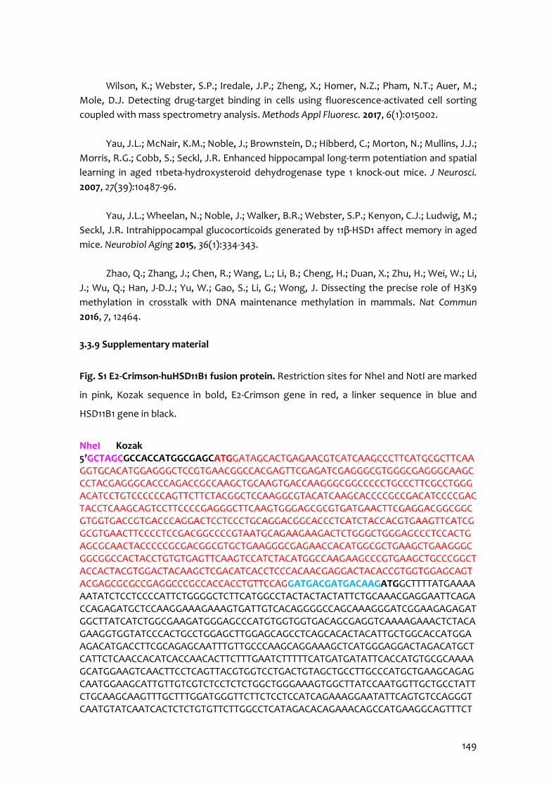
149
Wilson, K.; Webster, S.P.; Iredale, J.P.; Zheng, X.; Homer, N.Z.; Pham, N.T.; Auer, M.;
Mole, D.J. Detecting drug-target binding in cells using fluorescence-activated cell sorting coupled with mass spectrometry analysis. Methods Appl Fluoresc. 2017, 6(1):015002.
Yau, J.L.; McNair, K.M.; Noble, J.; Brownstein, D.; Hibberd, C.; Morton, N.; Mullins, J.J.;
Morris, R.G.; Cobb, S.; Seckl, J.R. Enhanced hippocampal long-term potentiation and spatial learning in aged 11beta-hydroxysteroid dehydrogenase type 1 knock-out mice. J Neurosci. 2007, 27(39):10487-96.
Yau, J.L.; Wheelan, N.; Noble, J.; Walker, B.R.; Webster, S.P.; Kenyon, C.J.; Ludwig, M.;
Seckl, J.R. Intrahippocampal glucocorticoids generated by 11β-HSD1 affect memory in aged mice. Neurobiol Aging 2015, 36(1):334-343.
Zhao, Q.; Zhang, J.; Chen, R.; Wang, L.; Li, B.; Cheng, H.; Duan, X.; Zhu, H.; Wei, W.; Li,
J.; Wu, Q.; Han, J-D.J.; Yu, W.; Gao, S.; Li, G.; Wong, J. Dissecting the precise role of H3K9 methylation in crosstalk with DNA maintenance methylation in mammals. Nat Commun 2016, 7, 12464.
3.3.9 Supplementary material
Fig. S1 E2-Crimson-huHSD11B1 fusion protein. Restriction sites for NheI and NotI are marked
in pink, Kozak sequence in bold, E2-Crimson gene in red, a linker sequence in blue and
HSD11B1 gene in black.
NheI Kozak 5’GCTAGCGCCACCATGGCGAGCATGGATAGCACTGAGAACGTCATCAAGCCCTTCATGCGCTTCAAGGTGCACATGGAGGGCTCCGTGAACGGCCACGAGTTCGAGATCGAGGGCGTGGGCGAGGGCAAGCCCTACGAGGGCACCCAGACCGCCAAGCTGCAAGTGACCAAGGGCGGCCCCCTGCCCTTCGCCTGGGACATCCTGTCCCCCCAGTTCTTCTACGGCTCCAAGGCGTACATCAAGCACCCCGCCGACATCCCCGACTACCTCAAGCAGTCCTTCCCCGAGGGCTTCAAGTGGGAGCGCGTGATGAACTTCGAGGACGGCGGCGTGGTGACCGTGACCCAGGACTCCTCCCTGCAGGACGGCACCCTCATCTACCACGTGAAGTTCATCGGCGTGAACTTCCCCTCCGACGGCCCCGTAATGCAGAAGAAGACTCTGGGCTGGGAGCCCTCCACTGAGCGCAACTACCCCCGCGACGGCGTGCTGAAGGGCGAGAACCACATGGCGCTGAAGCTGAAGGGCGGCGGCCACTACCTGTGTGAGTTCAAGTCCATCTACATGGCCAAGAAGCCCGTGAAGCTGCCCGGCTACCACTACGTGGACTACAAGCTCGACATCACCTCCCACAACGAGGACTACACCGTGGTGGAGCAGTACGAGCGCGCCGAGGCCCGCCACCACCTGTTCCAGGATGACGATGACAAGATGGCTTTTATGAAAAAATATCTCCTCCCCATTCTGGGGCTCTTCATGGCCTACTACTACTATTCTGCAAACGAGGAATTCAGACCAGAGATGCTCCAAGGAAAGAAAGTGATTGTCACAGGGGCCAGCAAAGGGATCGGAAGAGAGATGGCTTATCATCTGGCGAAGATGGGAGCCCATGTGGTGGTGACAGCGAGGTCAAAAGAAACTCTACAGAAGGTGGTATCCCACTGCCTGGAGCTTGGAGCAGCCTCAGCACACTACATTGCTGGCACCATGGAAGACATGACCTTCGCAGAGCAATTTGTTGCCCAAGCAGGAAAGCTCATGGGAGGACTAGACATGCTCATTCTCAACCACATCACCAACACTTCTTTGAATCTTTTTCATGATGATATTCACCATGTGCGCAAAAGCATGGAAGTCAACTTCCTCAGTTACGTGGTCCTGACTGTAGCTGCCTTGCCCATGCTGAAGCAGAGCAATGGAAGCATTGTTGTCGTCTCCTCTCTGGCTGGGAAAGTGGCTTATCCAATGGTTGCTGCCTATTCTGCAAGCAAGTTTGCTTTGGATGGGTTCTTCTCCTCCATCAGAAAGGAATATTCAGTGTCCAGGGTCAATGTATCAATCACTCTCTGTGTTCTTGGCCTCATAGACACAGAAACAGCCATGAAGGCAGTTTCT
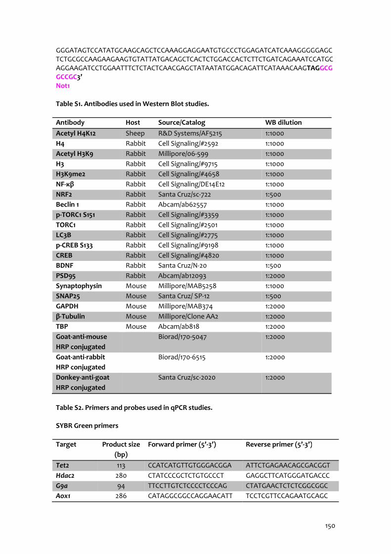
150
GGGATAGTCCATATGCAAGCAGCTCCAAAGGAGGAATGTGCCCTGGAGATCATCAAAGGGGGAGCTCTGCGCCAAGAAGAAGTGTATTATGACAGCTCACTCTGGACCACTCTTCTGATCAGAAATCCATGCAGGAAGATCCTGGAATTTCTCTACTCAACGAGCTATAATATGGACAGATTCATAAACAAGTAGGCGGCCGC3’ Not1
Table S1. Antibodies used in Western Blot studies.
Antibody Host Source/Catalog WB dilution Acetyl H4K12 Sheep R&D Systems/AF5215 1:1000 H4 Rabbit Cell Signaling/#2592 1:1000 Acetyl H3K9 Rabbit Millipore/06-599 1:1000 H3 Rabbit Cell Signaling/#9715 1:1000 H3K9me2 Rabbit Cell Signaling/#4658 1:1000 NF-κβ Rabbit Cell Signaling/DE14E12 1:1000 NRF2 Rabbit Santa Cruz/sc-722 1:500 Beclin 1 Rabbit Abcam/ab62557 1:1000 p-TORC1 S151 Rabbit Cell Signaling/#3359 1:1000 TORC1 Rabbit Cell Signaling/#2501 1:1000 LC3B Rabbit Cell Signaling/#2775 1:1000 p-CREB S133 Rabbit Cell Signaling/#9198 1:1000 CREB Rabbit Cell Signaling/#4820 1:1000 BDNF Rabbit Santa Cruz/N-20 1:500 PSD95 Rabbit Abcam/ab12093 1:2000 Synaptophysin Mouse Millipore/MAB5258 1:1000 SNAP25 Mouse Santa Cruz/ SP-12 1:500 GAPDH Mouse Millipore/MAB374 1:2000 β-Tubulin Mouse Millipore/Clone AA2 1:2000 TBP Mouse Abcam/ab818 1:2000 Goat-anti-mouse HRP conjugated
Biorad/170-5047 1:2000
Goat-anti-rabbit HRP conjugated
Biorad/170-6515 1:2000
Donkey-anti-goat HRP conjugated
Santa Cruz/sc-2020 1:2000
Table S2. Primers and probes used in qPCR studies.
SYBR Green primers
Target Product size (bp)
Forward primer (5’-3’) Reverse primer (5’-3’)
Tet2 113 CCATCATGTTGTGGGACGGA ATTCTGAGAACAGCGACGGT Hdac2 280 CTATCCCGCTCTGTGCCCT GAGGCTTCATGGGATGACCC G9a 94 TTCCTTGTCTCCCCTCCCAG CTATGAACTCTCTCGGCGGC Aox1 286 CATAGGCGGCCAGGAACATT TCCTCGTTCCAGAATGCAGC
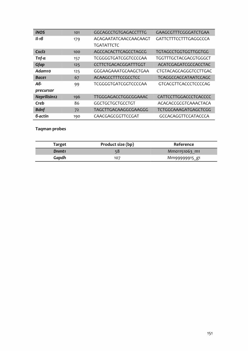
151
iNOS 101 GGCAGCCTGTGAGACCTTTG GAAGCGTTTCGGGATCTGAA Il-1β 179 ACAGAATATCAACCAACAAGT
TGATATTCTC GATTCTTTCCTTTGAGGCCCA
Cxcl2 100 AGCCACACTTCAGCCTAGCG TGTAGCCTGGTGGTTGGTGG Tnf-α 157 TCGGGGTGATCGGTCCCCAA TGGTTTGCTACGACGTGGGCT Gfap 125 CCTTCTGACACGGATTTGGT ACATCGAGATCGCCACCTAC Adam10 125 GGGAAGAAATGCAAGCTGAA CTGTACAGCAGGGTCCTTGAC Bace1 67 ACAAGCCTTTCCGCCTCC TCAGGCCACCATAATCCAGC Aβ-precursor
99 TCGGGGTGATCGGTCCCCAA GTCACGTTCACCCTCCCCAG
Neprilisin12 196 TTGGGAGACCTGGCGGAAAC CATTCCTTGGACCCTCACCCC Creb 86 GGCTGCTGCTGCCTGT ACACACCGCGTCAAACTACA Bdnf 72 TAGCTTGACAAGGCGAAGGG TCTGGCAAAGATGAGCTCGG β-actin 190 CAACGAGCGGTTCCGAT GCCACAGGTTCCATACCCA
Taqman probes
Target Product size (bp) Reference Dnmt1 58 Mm01151063_m1 Gapdh 107 Mm99999915_g1

152
3.4 11β-HSD1 Inhibition Rescues SAMP8 Cognitive Impairment Induced by Metabolic Stress
Adapted from: Puigoriol-Illamola, D.1,2, Leiva, R.3,4, Vázquez-Carrera, M.1,4,5,6, Vázquez, S.3,4, Griñán-Ferré, C.1,2, Pallàs, M1,2. 11β-HSD1 Inhibition Rescues SAMP8 Cognitive Impairment
Induced by Metabolic Stress
Molecular Neurobiology 2020; 57(1):551-565. doi:10.1007/s12035-019-01708-4. Epub 2019 Aug 09.
154
Author information: 1 Department of Pharmacology, Toxicology and Therapeutic Chemistry. Pharmacology Section. Faculty of Pharmacy and Food Sciences. University of Barcelona, Av Joan XXIII 27-31, 08028 Barcelona, Spain. 2 Institute of Neuroscience, University of Barcelona (NeuroUB), Passeig de la Vall d’Hebron 171, Barcelona, Spain. 3 Department of Pharmacology, Toxicology and Therapeutic Chemistry. Medicinal Chemistry Section. Faculty of Pharmacy and Food Sciences, University of Barcelona, Av Joan XXIII 27-31, 08028 Barcelona, Spain. 4 Institute of Biomedicine (IBUB). Av Diagonal 643, 08028 Barcelona, Spain. 5 Spanish Biomedical Research Center in Diabetes and Associated Metabolic Diseases (CIBERDEM) – Instituto de Salud Carlos III, Spain. 6 Pediatric Research Institute-Hospital Sant Joan de Déu, Esplugues de Llobregat, Spain.
155
3.4.1 Abstract
In recent years, the interest in the impact of nutrition in health has grown since
obesity is one of the features of modern society. Particularly, obesity is a risk factor for
many disorders, including diabetes, hypertension, cardiovascular alterations, mild cognitive
impairment and AD. In fact, ample evidence provides both clinical and experimental
evidence into how obesity and AD may course together. However, the main risk factor for
AD is ageing. While ageing, several disturbances lead to impaired GC signalling, resulting in
high GC levels. Besides, GCs excess cause metabolic disturbances and have been associated
with neurodegeneration. GC activity is regulated by 11β-HSD1 enzyme, which inhibition has
been proved to enhance cognitive abilities. The study aimed to determine the potential
beneficial effects of RL-118 drug treatment, an 11β-HSD1 inhibitor (11β-HSD1i), after HFD
exposure in the SAMP8.
In the present approach, in order to achieve the objective, 4 groups were established:
control fed with normal diet (ND), ND-fed treated with the 11β-HSD1i, HFD-fed and HFD-fed
treated with the 11β-HSD1i. Since weaning, mice consumed their dietary condition and at
the age of 4 months, drug treatment started for 4 weeks. Our results demonstrated
improvement in glucose intolerance induced by HFD in mice treated with RL-118, a
significant reduction in 11β-HSD1 and GR protein levels. Furthermore, specific modification
in the fibroblast growth factor 21 (FGF21) activation after treatment with 11β-HSD1i was
found, which induced changes in the SIRT1/ peroxisome proliferator-activated receptor
gamma coactivator (PGC) 1α/ AMPKα signalling pathway. OS and ROS, as well as
inflammatory markers and microglial activation were significantly diminished in HFD-fed
mice treated with 11β-HSD1i. Remarkably, treatment with 11β-HSD1i altered ER stress
response in both diet groups, increasing autophagy only in HFD mice group.
RL-118 treatment induced a decrease in GSK3β activation, tau hyperphosphorylation
and amyloidogenic APP processing. In agreement, increased non-amyloidogenic APP
markers were detected in both treated mice groups, regardless of the dietary condition.
Consequently, beneficial effects on social behaviour and cognitive performance were found
in treated mice. Thus, our results support the therapeutic strategy of GC excess attenuation
by selective 11β-HSD1i for the treatment of age-related cognitive decline and AD through
improving metabolic and eventually cognitive disturbances caused by HFD.

156
3.4.2 Introduction
Aging is the most significant risk factor for a majority of chronic diseases, such as age-
related cognitive decline and AD (Guerreiro & Bras, 2015). Obesity has recently been
considered to play an essential role in mild cognitive impairment and dementia (Bischof &
Park, 2015; Feinkohl et al., 2018). Abundant pieces of evidence suggest that pathological
prolonged GCs excess not only is associated with age-related cognitive decline but also with
metabolic stress, obesity, and diabetes (Bujalska et al., 2006; De Quervain et al., 2004;
Holmes et al., 2010; Kumar & Datusalia, 2018). Thus, it has been described that prolonged
exposure to increased levels of GCs exerts deleterious effects on hippocampal
electrophysiology, structure and function, both in rodents (Canet et al., 2018) and humans
(Tatomir et al., 2014), being part of altered intercellular communication, considered one of
the nine hallmarks of aging (López-Otín et al., 2013; Mattson & Arumugam, 2018). The
availability of natural GCs in tissues is regulated by corticosteroid-binding globulin in serum
and by locally expressed 11β-HSD enzyme, a microsomal enzyme that catalyses the
interconversion of active GCs (corticosterone in rodents but cortisol in humans) and inert
11-keto-forms [11-dehydrocorticosterone (11-DHC), cortisone] (Yau & Seckl, 2012). Two
isoenzymes have been identified: 11β-HSD type 1 (11β-HSD1) and type 2 (11β-HSD2) (Kadmiel
& Cidlowski, 2013). The former acts as a reductase, thus locally potentiating GCs activity,
and is the predominant form in the brain, both in rodents and humans. In fact, 11β-HSD1
expression in mouse hippocampus and parietal cortex increases with aging, correlating
with impaired spatial memory (Yau et al., 2015) and its overexpression accelerates age-
related cognitive decline (Holmes et al., 2010). Conversely, 11β-HSD1 knockout mice resist
age-dependent cognitive loss (Yau et al., 2001, 2007). In line with these findings, some
preclinical studies demonstrate that 11β-HSD1 inhibition improved cognition and AD
hallmarks suggesting a neuroprotective effect (Mohler et al., 2011; Puigoriol-Illamola et al.,
2018). On the other hand, preclinical studies have demonstrated that sub-maximal inhibition
of central 11β-HSD1 is able to prevent cognitive impairments in AD and ageing animal
models (Sooy et al., 2010, 2015) and early preclinical studies demonstrated that
administration of brain penetrant 11β-HSD1 inhibitor (UE2343) is well tolerated in humans
(Webster et al., 2017). Besides, many selective 11β-HSD1 inhibitors have reached clinical
stages for metabolic diseases like type 2 diabetes mellitus (e.g., AZD8329, ABT-384, and
BVT-2733).

157
Indeed, several hypotheses argument a link between altered glucose metabolism and
dementia, considering an altered metabolic pathway as a potential contributor to
persistent oxidative stress (OS) that culminates into neuronal dysfunction and dementia
(Mule & Singh, 2018). Increased OS, neuroinflammation, endoplasmic reticulum (ER) stress,
misfolded proteins removal pathways, and autophagy, have been identified as components
of neuronal metabolic stress, thus developing and aggravating neurological disorders and
cognitive impairment, pointing to their implication in the dysregulated energy metabolism
characteristic of ageing (Kumar & Datusalia, 2018). Thus, recent studies have focused on the
pathology of cognitive decline and neurodegeneration through HFD feeding that induce
metabolic stress (Lee et al., 2018; Wei et al., 2018). For instance, HFD-fed aged animals
showed insulin resistance and increased weight gain among others, both signs of pre-
diabetes, obesity, cardiovascular disease, depressive-like behaviour and mental health
problems (Bowles et al., 2017; Karatsoreos et al., 2010; McEwen et al., 2015). However, the
current knowledge about the molecular mechanism responsible for these affections is
controversial, even though most of the information is related to reduced insulin sensitivity
mediated by HFD. Hence, HFD-induced metabolic stress could be linked with the
development of physiopathological conditions, such as those observed in AD and other
neurodegenerative diseases (Cai et al., 2012).
Recently, we have developed a brain penetrant 11β-HSD1 inhibitor (RL-118) that was
characterized chemically and pharmacologically in vivo in Leiva et al. (2017). RL-118
attenuates neuroinflammation, increases the antioxidant defence, promotes autophagy,
improves mitochondrial function and reverts memory deficits in senescent mice model
(Leiva et al., 2017; Puigoriol-Illamola et al., 2018). Therefore, the targeted inhibition of 11β-
HSD1 may become a potential therapeutic strategy for age-related cognitive decline and
AD. Here, we assessed the neuroprotective effects of 11β-HSD1 inhibition on metabolic
stress through behaviour, cognitive and molecular changes induced by HFD in a mice model
of age-related cognitive decline and late-onset AD, the senescence-accelerated mouse
prone 8 (SAMP8).
3.4.3 Materials and methods
Animals
Females SAMP8 mice (n=48) were used to carry out behavioural, cognitive and
molecular analyses. We divided these animals into 4 groups: normal diet chow (ND Ct,
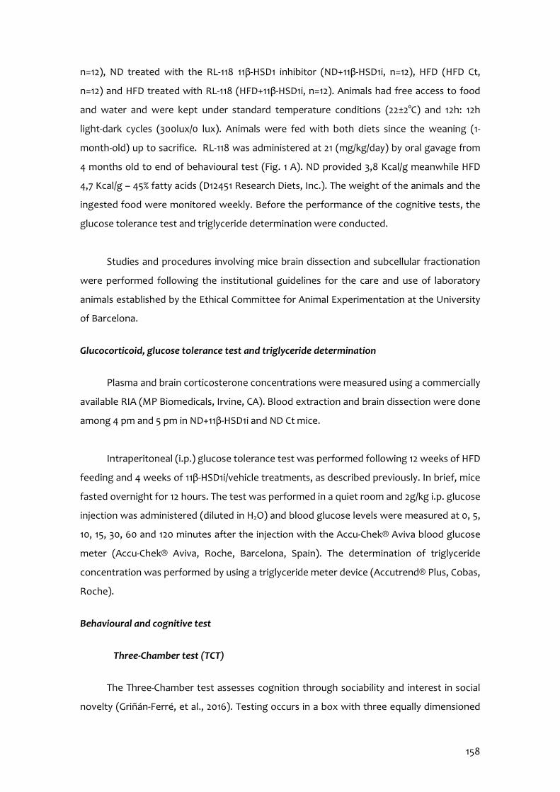
158
n=12), ND treated with the RL-118 11β-HSD1 inhibitor (ND+11β-HSD1i, n=12), HFD (HFD Ct,
n=12) and HFD treated with RL-118 (HFD+11β-HSD1i, n=12). Animals had free access to food
and water and were kept under standard temperature conditions (22±2°C) and 12h: 12h
light-dark cycles (300lux/0 lux). Animals were fed with both diets since the weaning (1-
month-old) up to sacrifice. RL-118 was administered at 21 (mg/kg/day) by oral gavage from
4 months old to end of behavioural test (Fig. 1 A). ND provided 3,8 Kcal/g meanwhile HFD
4,7 Kcal/g – 45% fatty acids (D12451 Research Diets, Inc.). The weight of the animals and the
ingested food were monitored weekly. Before the performance of the cognitive tests, the
glucose tolerance test and triglyceride determination were conducted.
Studies and procedures involving mice brain dissection and subcellular fractionation
were performed following the institutional guidelines for the care and use of laboratory
animals established by the Ethical Committee for Animal Experimentation at the University
of Barcelona.
Glucocorticoid, glucose tolerance test and triglyceride determination
Plasma and brain corticosterone concentrations were measured using a commercially
available RIA (MP Biomedicals, Irvine, CA). Blood extraction and brain dissection were done
among 4 pm and 5 pm in ND+11β-HSD1i and ND Ct mice.
Intraperitoneal (i.p.) glucose tolerance test was performed following 12 weeks of HFD
feeding and 4 weeks of 11β-HSD1i/vehicle treatments, as described previously. In brief, mice
fasted overnight for 12 hours. The test was performed in a quiet room and 2g/kg i.p. glucose
injection was administered (diluted in H2O) and blood glucose levels were measured at 0, 5,
10, 15, 30, 60 and 120 minutes after the injection with the Accu-Chek® Aviva blood glucose
meter (Accu-Chek® Aviva, Roche, Barcelona, Spain). The determination of triglyceride
concentration was performed by using a triglyceride meter device (Accutrend® Plus, Cobas,
Roche).
Behavioural and cognitive test
Three-Chamber test (TCT)
The Three-Chamber test assesses cognition through sociability and interest in social
novelty (Griñán-Ferré, et al., 2016). Testing occurs in a box with three equally dimensioned
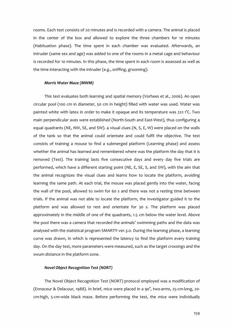
159
rooms. Each test consists of 20 minutes and is recorded with a camera. The animal is placed
in the center of the box and allowed to explore the three chambers for 10 minutes
(Habituation phase). The time spent in each chamber was evaluated. Afterwards, an
intruder (same sex and age) was added to one of the rooms in a metal cage and behaviour
is recorded for 10 minutes. In this phase, the time spent in each room is assessed as well as
the time interacting with the intruder (e.g., sniffing, grooming).
Morris Water Maze (MWM)
This test evaluates both learning and spatial memory (Vorhees et al., 2006). An open
circular pool (100 cm in diameter, 50 cm in height) filled with water was used. Water was
painted white with latex in order to make it opaque and its temperature was 22± 1°C. Two
main perpendicular axes were established (North-South and East-West), thus configuring 4
equal quadrants (NE, NW, SE, and SW). 4 visual clues (N, S, E, W) were placed on the walls
of the tank so that the animal could orientate and could fulfil the objective. The test
consists of training a mouse to find a submerged platform (Learning phase) and assess
whether the animal has learned and remembered where was the platform the day that it is
removed (Test). The training lasts five consecutive days and every day five trials are
performed, which have a different starting point (NE, E, SE, S, and SW), with the aim that
the animal recognizes the visual clues and learns how to locate the platform, avoiding
learning the same path. At each trial, the mouse was placed gently into the water, facing
the wall of the pool, allowed to swim for 60 s and there was not a resting time between
trials. If the animal was not able to locate the platform, the investigator guided it to the
platform and was allowed to rest and orientate for 30 s. The platform was placed
approximately in the middle of one of the quadrants, 1.5 cm below the water level. Above
the pool there was a camera that recorded the animals’ swimming paths and the data was
analysed with the statistical program SMART® ver.3.0. During the learning phase, a learning
curve was drawn, in which is represented the latency to find the platform every training
day. On the day test, more parameters were measured, such as the target crossings and the
swum distance in the platform zone.
Novel Object Recognition Test (NORT)
The Novel Object Recognition Test (NORT) protocol employed was a modification of
(Ennaceur & Delacour, 1988). In brief, mice were placed in a 90°, two-arms, 25-cm-long, 20-
cm-high, 5-cm-wide black maze. Before performing the test, the mice were individually

160
habituated to the apparatus for 10 min for 3 days. On day 4, the animals were submitted to
a 10-min acquisition trial (first trial), during which they were placed in the maze in the
presence of two identical, novel objects at the end of each arm. After a delay (2h and 24h),
the animal was exposed to two objects one old object and one novel object. The Time that
mice explored the Novel object (TN) and Time that mice explored the Old object (TO) were
measured. A Discrimination Index (DI) was defined as (TN−TO)/(TN+TO). To avoid object
preference biases, objects were counterbalanced. The maze, the surface, and the objects
were cleaned with 70% ethanol between the animals’ trials to eliminate olfactory cues.
Brain processing
Three days after the behavioural and cognitive tests, 8 animals per group were
euthanized for protein extraction, RNA and DNA isolation, and 4 animals per group were
euthanized for immunohistochemistry (IHQ).
When the animals were for protein extraction, RNA and DNA isolation, brains were
immediately removed and the hippocampus was isolated, frozen on powdered dry ice and
maintained at -80°C until procedures. When the animals were for IHQ, mice were
intracardially perfused with 4 % paraformaldehyde (PFA) diluted in 0.1M phosphate buffer
solution after being anesthetized by intraperitoneal injection of ketamine 100 mg/Kg and
xylazine 10 mg/Kg. Afterwards, brains were removed and post-fixed in 4% PFA overnight 4oC
then the solution was changed into PFA + 15% sucrose. Finally, brains were frozen burying
directly on powdered dry ice (around 5 min) and store at -80°C until sectioned.
Immunodetection experiments
Western blotting
Tissue samples were homogenized in lysis buffer (Tris HCl pH 7.4 50mM, NaCl 150mM,
EDTA 5mM and 1X-Triton X-100) containing phosphatase and protease inhibitors (Cocktail II,
Sigma-Aldrich) to obtain total protein homogenates.
For Western Blotting (WB), aliquots of 15 μg of hippocampal protein extraction per
sample were used. Protein samples were separated by Sodium dodecyl sulphate-
polyacrylamide gel electrophoresis (SDS-PAGE) (8-14%) and transferred onto Polyvinylidene
difluoride (PVDF) membranes (Millipore). Afterwards, membranes were blocked in 5% non-
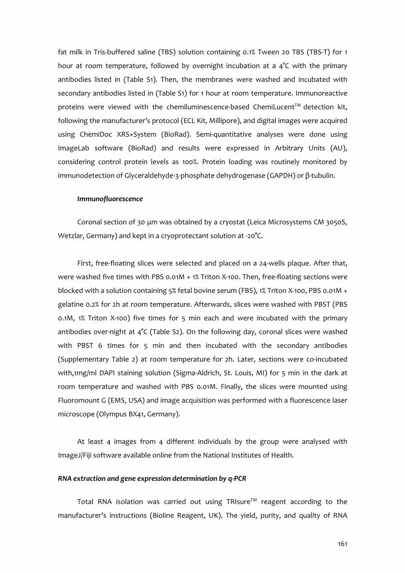
161
fat milk in Tris-buffered saline (TBS) solution containing 0.1% Tween 20 TBS (TBS-T) for 1
hour at room temperature, followed by overnight incubation at a 4°C with the primary
antibodies listed in (Table S1). Then, the membranes were washed and incubated with
secondary antibodies listed in (Table S1) for 1 hour at room temperature. Immunoreactive
proteins were viewed with the chemiluminescence-based ChemiLucentTM detection kit,
following the manufacturer’s protocol (ECL Kit, Millipore), and digital images were acquired
using ChemiDoc XRS+System (BioRad). Semi-quantitative analyses were done using
ImageLab software (BioRad) and results were expressed in Arbitrary Units (AU),
considering control protein levels as 100%. Protein loading was routinely monitored by
immunodetection of Glyceraldehyde-3-phosphate dehydrogenase (GAPDH) or β-tubulin.
Immunofluorescence
Coronal section of 30 μm was obtained by a cryostat (Leica Microsystems CM 3050S,
Wetzlar, Germany) and kept in a cryoprotectant solution at -20°C.
First, free-floating slices were selected and placed on a 24-wells plaque. After that,
were washed five times with PBS 0.01M + 1% Triton X-100. Then, free-floating sections were
blocked with a solution containing 5% fetal bovine serum (FBS), 1% Triton X-100, PBS 0.01M +
gelatine 0.2% for 2h at room temperature. Afterwards, slices were washed with PBST (PBS
0.1M, 1% Triton X-100) five times for 5 min each and were incubated with the primary
antibodies over-night at 4°C (Table S2). On the following day, coronal slices were washed
with PBST 6 times for 5 min and then incubated with the secondary antibodies
(Supplementary Table 2) at room temperature for 2h. Later, sections were co-incubated
with,1mg/ml DAPI staining solution (Sigma-Aldrich, St. Louis, MI) for 5 min in the dark at
room temperature and washed with PBS 0.01M. Finally, the slices were mounted using
Fluoromount G (EMS, USA) and image acquisition was performed with a fluorescence laser
microscope (Olympus BX41, Germany).
At least 4 images from 4 different individuals by the group were analysed with
ImageJ/Fiji software available online from the National Institutes of Health.
RNA extraction and gene expression determination by q-PCR
Total RNA isolation was carried out using TRIsureTM reagent according to the
manufacturer’s instructions (Bioline Reagent, UK). The yield, purity, and quality of RNA
162
were determined spectrophotometrically with a NanoDrop™ ND-1000 (Thermo Scientific)
apparatus and an Agilent 2100B Bioanalyzer (Agilent Technologies). RNAs with 260/280
ratios and RIN higher than 1.9 and 7.5, respectively, were selected. Reverse Transcription-
Polymerase Chain Reaction (RT-PCR) was performed as follows: 2 μg of messenger RNA
(mRNA) was reverse-transcribed using the High Capacity cDNA Reverse Transcription Kit
(Applied Biosystems). Real-time quantitative PCR (qPCR) was used to quantify mRNA
expression of OS and inflammatory genes listed in Table S3. SYBR® Green real-time PCR was
performed in a Step One Plus Detection System (Applied-Biosystems) employing SYBR®
Green PCR Master Mix (Applied-Biosystems). Each reaction mixture contained 6.75 μL of
complementary DNA (cDNA) (which concentration was 2 μg), 0.75 μL of each primer (which
concentration was 100 nM), and 6.75 μL of SYBR® Green PCR Master Mix (2X).
Data was analysed utilizing the comparative Cycle threshold (Ct) method (ΔΔCt),
where the housekeeping gene level was used to normalize differences in sample loading
and preparation. Normalization of expression levels was performed with β-actin for SYBR®
Green-based real-time PCR results. Each sample was analysed in duplicate, and the results
represent the n-fold difference of the transcript levels among different groups.
Oxidative stress determination
Hydrogen peroxide was measured in hippocampus protein homogenates as an
indicator of OS, and it was quantified using the Hydrogen Peroxide Assay Kit (Sigma-
Aldrich, St. Louis, MI) according to the manufacturer’s instructions.
Data analysis
Data analysis was conducted using GraphPad Prism ver. 7 statistical software. Data
are expressed as the mean ± standard error of the mean (SEM) of at least 6 samples per
group. Diet and treatment effects were assessed by the Two-Way ANOVA analysis of
variance, followed by Tukey post-hoc analysis or two-tail Student’s t-test when it was
necessary. Statistical significance was considered when p-values were <0.05. The statistical
outliers were determined with Grubbs’ test and subsequently removed from the analysis.
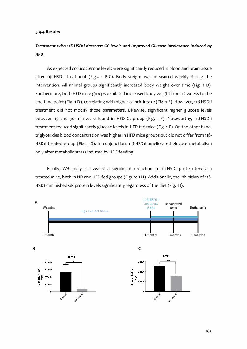
163
3.4.4 Results
Treatment with 11β-HSD1i decrease GC levels and Improved Glucose Intolerance Induced by
HFD
As expected corticosterone levels were significantly reduced in blood and brain tissue
after 11β-HSD1i treatment (Figs. 1 B-C). Body weight was measured weekly during the
intervention. All animal groups significantly increased body weight over time (Fig. 1 D).
Furthermore, both HFD mice groups exhibited increased body weight from 12 weeks to the
end time point (Fig. 1 D), correlating with higher caloric intake (Fig. 1 E). However, 11β-HSD1i
treatment did not modify those parameters. Likewise, significant higher glucose levels
between 15 and 90 min were found in HFD Ct group (Fig. 1 F). Noteworthy, 11β-HSD1i
treatment reduced significantly glucose levels in HFD fed mice (Fig. 1 F). On the other hand,
triglycerides blood concentration was higher in HFD mice groups but did not differ from 11β-
HSD1i treated group (Fig. 1 G). In conjunction, 11β-HSD1i ameliorated glucose metabolism
only after metabolic stress induced by HDF feeding.
Finally, WB analysis revealed a significant reduction in 11β-HSD1 protein levels in
treated mice, both in ND and HFD fed groups (Figure 1 H). Additionally, the inhibition of 11β-
HSD1 diminished GR protein levels significantly regardless of the diet (Fig. 1 I).
A B C
Weaning
1 month
High-Fat Diet Chow
11β-HSD1i treatment
starts
4 months
Behavioural tests
5 months
Euthanasia
6 months
Contro
l
11-H
SD1i
Control
11-H
SD1i
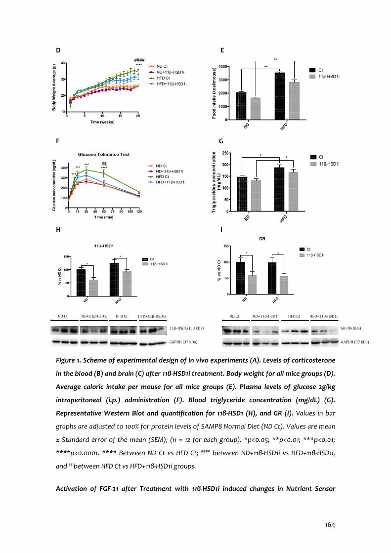
164
D E
F G
H I
Figure 1. Scheme of experimental design of in vivo experiments (A). Levels of corticosterone
in the blood (B) and brain (C) after 11β-HSD1i treatment. Body weight for all mice groups (D).
Average caloric intake per mouse for all mice groups (E). Plasma levels of glucose 2g/kg
intraperitoneal (i.p.) administration (F). Blood triglyceride concentration (mg/dL) (G).
Representative Western Blot and quantification for 11β-HSD1 (H), and GR (I). Values in bar
graphs are adjusted to 100% for protein levels of SAMP8 Normal Diet (ND Ct). Values are mean
± Standard error of the mean (SEM); (n = 12 for each group). *p<0.05; **p<0.01; ***p<0.01;
****p<0.0001. **** Between ND Ct vs HFD Ct; #### between ND+11β-HSD1i vs HFD+11β-HSD1i,
and $$ between HFD Ct vs HFD+11β-HSD1i groups.
Activation of FGF-21 after Treatment with 11β-HSD1i induced changes in Nutrient Sensor
NDHFD
0
50
100
150
% v
s N
D C
t
11b-HSD1
Ct
11β-HSD1i *
*
NDHFD
0
50
100
150
GR
% v
s N
D C
t
Ct
11β-HSD1i * *
0 5 10 15 2010
20
30
40
Time (weeks)
Bo
dy
We
igh
t A
va
rag
e (g
) ND Ct
ND+11β-HSD1i
HFD Ct
HFD+11β-HSD1i
#### ****
0 15 30 45 60 75 90 105 1200
100
200
300
400
Time (min)
Glu
cose c
oncentr
atio
n (n
g/d
L)
Glucose Tolerance Test
ND Ct
ND+11β-HSD1i
HFD Ct
HFD+11β-HSD1i
***
******
****$$
NDHFD
0
50
100
150
200
250
Tri
gly
ce
rid
es c
on
ce
ntr
atio
n (m
g/d
L)
Ct
11β-HSD1i**
NDHFD
0
1000
2000
3000
4000
Fo
od
inta
ke
(k
ca
l/mo
us
e)
Ct
11β-HSD1i
**
**
ND Ct ND+11β-HSD1i HFD+11β-HSD1i HFD Ct
11β-HSD1i (30 kDa) GAPDH (37 kDa)
GR (86 kDa) GAPDH (37 kDa)
ND Ct ND+11β-HSD1i HFD+11β-HSD1i HFD Ct
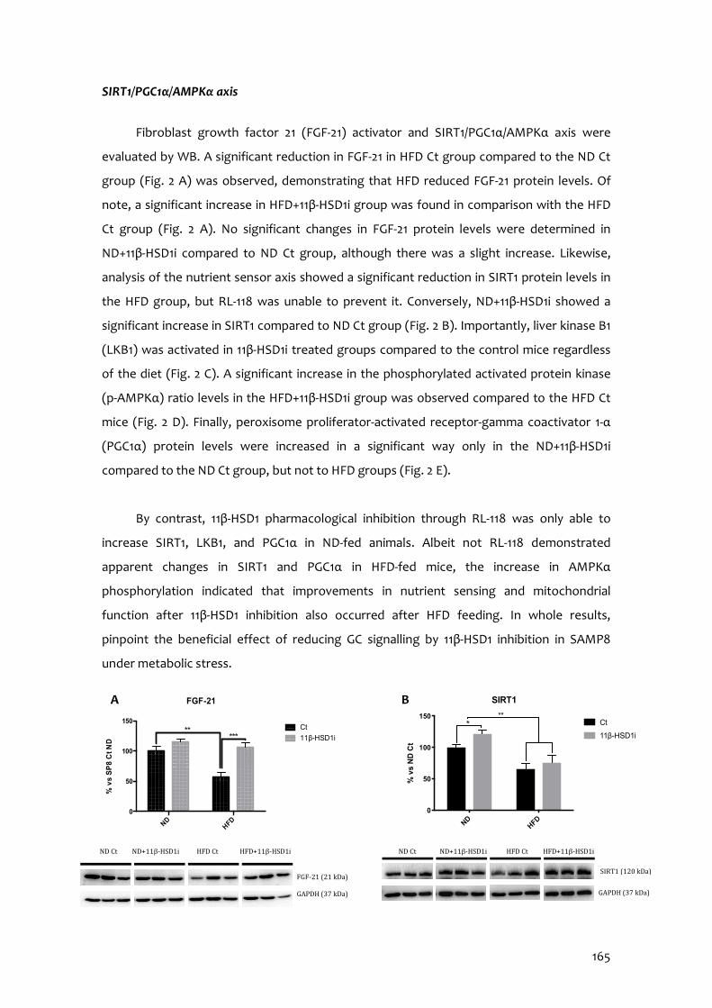
165
SIRT1/PGC1α/AMPKα axis
Fibroblast growth factor 21 (FGF-21) activator and SIRT1/PGC1α/AMPKα axis were
evaluated by WB. A significant reduction in FGF-21 in HFD Ct group compared to the ND Ct
group (Fig. 2 A) was observed, demonstrating that HFD reduced FGF-21 protein levels. Of
note, a significant increase in HFD+11β-HSD1i group was found in comparison with the HFD
Ct group (Fig. 2 A). No significant changes in FGF-21 protein levels were determined in
ND+11β-HSD1i compared to ND Ct group, although there was a slight increase. Likewise,
analysis of the nutrient sensor axis showed a significant reduction in SIRT1 protein levels in
the HFD group, but RL-118 was unable to prevent it. Conversely, ND+11β-HSD1i showed a
significant increase in SIRT1 compared to ND Ct group (Fig. 2 B). Importantly, liver kinase B1
(LKB1) was activated in 11β-HSD1i treated groups compared to the control mice regardless
of the diet (Fig. 2 C). A significant increase in the phosphorylated activated protein kinase
(p-AMPKα) ratio levels in the HFD+11β-HSD1i group was observed compared to the HFD Ct
mice (Fig. 2 D). Finally, peroxisome proliferator-activated receptor-gamma coactivator 1-α
(PGC1α) protein levels were increased in a significant way only in the ND+11β-HSD1i
compared to the ND Ct group, but not to HFD groups (Fig. 2 E).
By contrast, 11β-HSD1 pharmacological inhibition through RL-118 was only able to
increase SIRT1, LKB1, and PGC1α in ND-fed animals. Albeit not RL-118 demonstrated
apparent changes in SIRT1 and PGC1α in HFD-fed mice, the increase in AMPKα
phosphorylation indicated that improvements in nutrient sensing and mitochondrial
function after 11β-HSD1 inhibition also occurred after HFD feeding. In whole results,
pinpoint the beneficial effect of reducing GC signalling by 11β-HSD1 inhibition in SAMP8
under metabolic stress.
ND
HFD0
50
100
150
% v
s S
P8
Ct N
D
FGF-21
Ct
11β-HSD1i *****
FGF-21 (21 kDa) GAPDH (37 kDa)
ND Ct HFD Ct HFD+11β-HSD1i ND+11β-HSD1i ND Ct HFD Ct HFD+11β-HSD1i ND+11β-HSD1i SIRT1 (120 kDa)
GAPDH (37 kDa)
NDHFD
0
50
100
150
SIRT1
% v
s N
D C
t
Ct
11β-HSD1i
***
B A

166
Figure 2. Representative Western Blot and protein levels quantification for FGF21 (A), SIRT1
(B), LKB1 (C), ratio of p-AMPKα (D), and PGC-1α (E). Values in bar graphs are adjusted to 100%
for protein levels of SAMP8 Normal Diet (ND Ct). Values are mean ± Standard error of the
mean (SEM); (n = 6 for each group). *p<0.05; **p<0.01.
Treatment with 11β-HSD1i Reduced OS Markers
HFD induced a significant increase in GPX1 and a moderate increase in SOD1 protein
levels that were prevented by RL-118 treatment (Figs. 3 A-B). In addition, iNOS gene
expression was reduced in 11β-HSD1i treated animals in comparison with the control groups
that reached significance in HFD Ct mice (Fig. 3 C). Likewise, analysis of hydrogen peroxide
levels showed that 11β-HSD1i can reduce it although in a not significant way (Fig. 3 D).
NDHFD
0
50
100
150
200
PGC1a
% v
s N
D C
t
Ct
11β-HSD1i *
**
ND Ct ND+11β-HSD1i HFD+11β-HSD1i HFD Ct
PGC1α (90 kDa)
GAPDH (37 kDa)
E
NDHFD
0
50
100
150
LKB1%
vs
ND
Ct
Ct
11β-HSD1i
***
ND Ct ND+11β-HSD1i HFD+11β-HSD1i HFD Ct
LKB1 (55 kDa)
GAPDH (37 kDa)
C
NDHFD
0
50
100
150
200
Ratio p-AMPKa vs AMPKa total
% v
s N
D C
t
Ct
11β-HSD1i **
p-AMPKα (62 kDa)
GAPDH (37 kDa)
AMPKα (62 kDa)
GAPDH (37 kDa)
ND Ct ND+11β-HSD1i HFD+11β-HSD1i HFD Ct
D
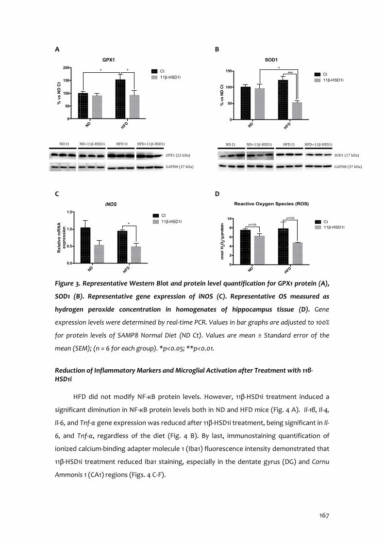
167
A B
C D
Figure 3. Representative Western Blot and protein level quantification for GPX1 protein (A),
SOD1 (B). Representative gene expression of iNOS (C). Representative OS measured as
hydrogen peroxide concentration in homogenates of hippocampus tissue (D). Gene
expression levels were determined by real-time PCR. Values in bar graphs are adjusted to 100%
for protein levels of SAMP8 Normal Diet (ND Ct). Values are mean ± Standard error of the
mean (SEM); (n = 6 for each group). *p<0.05; **p<0.01.
Reduction of Inflammatory Markers and Microglial Activation after Treatment with 11β-HSD1i
HFD did not modify NF-κB protein levels. However, 11β-HSD1i treatment induced a
significant diminution in NF-κB protein levels both in ND and HFD mice (Fig. 4 A). Il-1β, Il-4,
Il-6, and Tnf-α gene expression was reduced after 11β-HSD1i treatment, being significant in Il-
6, and Tnf-α, regardless of the diet (Fig. 4 B). By last, immunostaining quantification of
ionized calcium-binding adapter molecule 1 (Iba1) fluorescence intensity demonstrated that
11β-HSD1i treatment reduced Iba1 staining, especially in the dentate gyrus (DG) and Cornu
Ammonis 1 (CA1) regions (Figs. 4 C-F).
NDHFD
0
50
100
150
200
GPX1%
vs
ND
Ct
Ct
11β-HSD1i
**
GPX1 (22 kDa)
GAPDH (37 kDa)
ND Ct HFD Ct HFD+11β-HSD1i ND+11β-HSD1i
NDHFD
0
50
100
150
SOD1
% v
s N
D C
t
Ct
11β-HSD1i
****
SOD1 (17 kDa)
GAPDH (37 kDa)
ND Ct HFD Ct HFD+11β-HSD1i ND+11β-HSD1i
NDHFD
0.0
0.5
1.0
1.5
Re
lativ
e m
RN
A
ex
pre
ss
ion
iNOS
Ct
11β-HSD1i*
NDHFD
0
2
4
6
8
10nm
ol
H2O
2/ g
pro
tein
Ct
11β-HSD1i
p=0.08
p=0.06
Reactive Oxygen Species (ROS)
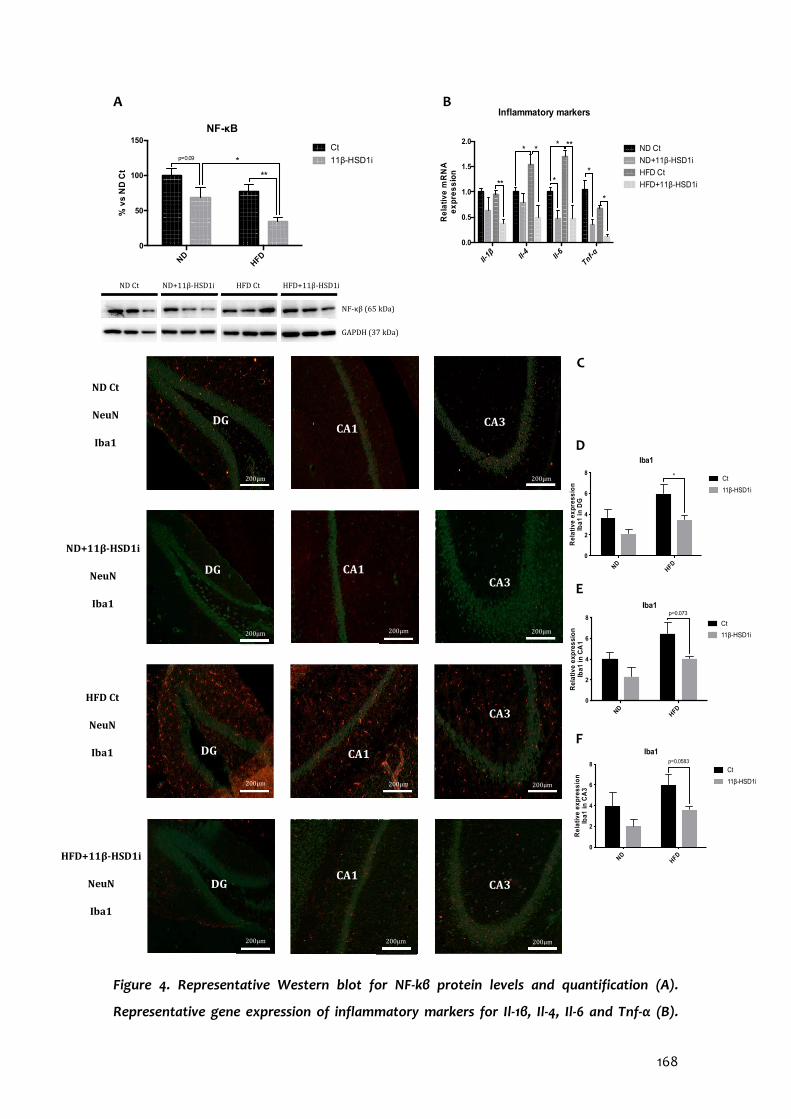
168
Il-1β Il-
4Il-6
Tnf-α
0.0
0.5
1.0
1.5
2.0
Re
lativ
e m
RN
A
ex
pre
ss
ion
Inflammatory markers
ND Ct
ND+11β-HSD1i
HFD Ct
HFD+11β-HSD1i**
**
*
** *
*
*
A B
C
Figure 4. Representative Western blot for NF-kβ protein levels and quantification (A).
Representative gene expression of inflammatory markers for Il-1β, Il-4, Il-6 and Tnf-α (B).
NDHFD
0
50
100
150
NF-κb%
vs
ND
Ct
Ct
11β-HSD1i
**
p=0.09 *
ND Ct HFD Ct HFD+11β-HSD1i ND+11β-HSD1i
NF-κβ (65 kDa)
GAPDH (37 kDa)
DG
ND Ct
NeuN
Iba1
ND+11β-HSD1i
NeuN
Iba1
HFD+11β-HSD1i
NeuN
Iba1
HFD Ct
NeuN
Iba1
DG
200μm 200μm 200μm
CA3 CA1
200μm
CA3
DG CA1
CA3
200μm 200μm 200μm
DG CA1
CA3
200μm 200μm
E
D
F
NDHFD
0
2
4
6
8
Iba1
Rel
ativ
e e
xpre
ssio
nIb
a1 in
CA
3
Ct
11β-HSD1i
p=0.0583
NDHFD
0
2
4
6
8
Iba1
Rel
ativ
e e
xpre
ssi
on
Iba1
in C
A1
Ct
11β-HSD1i
p=0.073
NDHFD
0
2
4
6
8
Iba1
Rel
ativ
e ex
pre
ssio
nIb
a1 in
DG
Ct
11β-HSD1i
*
CA1
CA1
DG
DG
200μm 200μm
200μm
200μm
DG
NF-κB
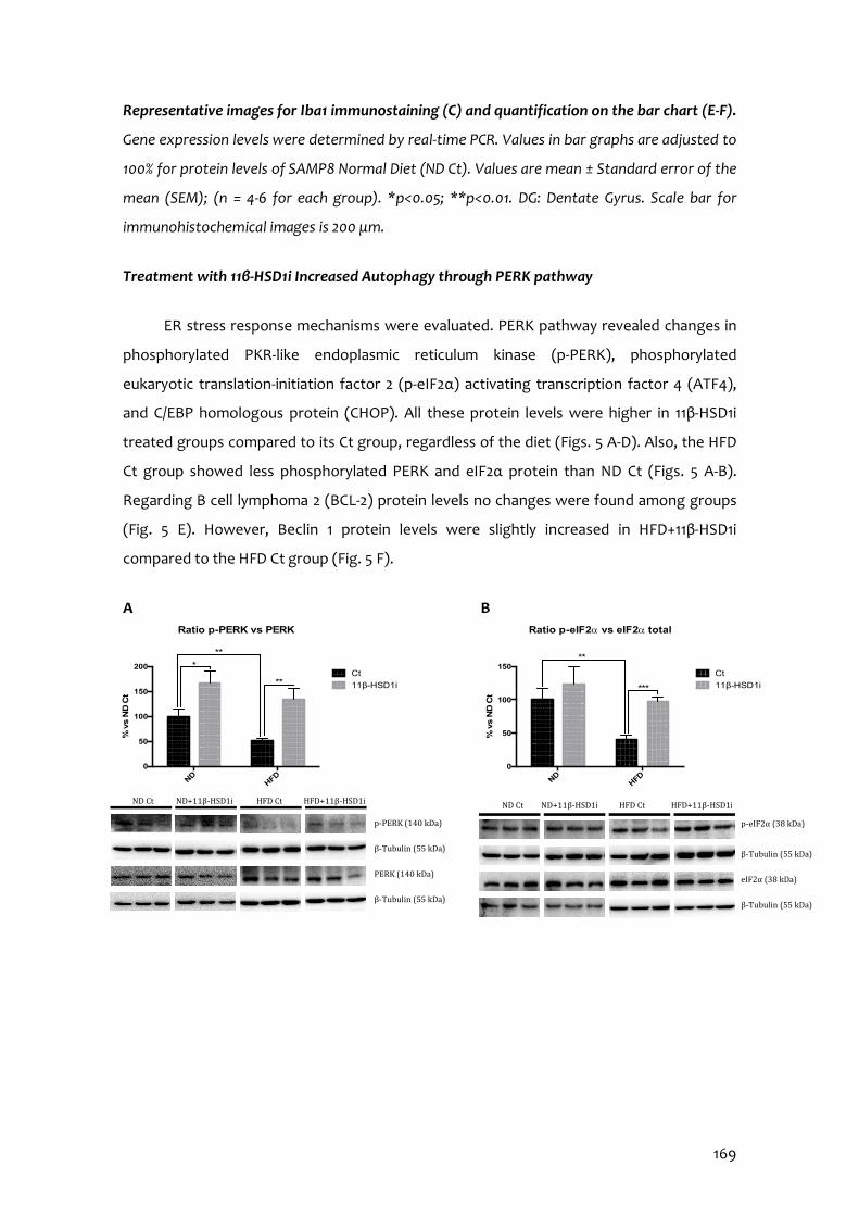
169
Representative images for Iba1 immunostaining (C) and quantification on the bar chart (E-F).
Gene expression levels were determined by real-time PCR. Values in bar graphs are adjusted to
100% for protein levels of SAMP8 Normal Diet (ND Ct). Values are mean ± Standard error of the
mean (SEM); (n = 4-6 for each group). *p<0.05; **p<0.01. DG: Dentate Gyrus. Scale bar for
immunohistochemical images is 200 μm.
Treatment with 11β-HSD1i Increased Autophagy through PERK pathway
ER stress response mechanisms were evaluated. PERK pathway revealed changes in
phosphorylated PKR-like endoplasmic reticulum kinase (p-PERK), phosphorylated
eukaryotic translation-initiation factor 2 (p-eIF2α) activating transcription factor 4 (ATF4),
and C/EBP homologous protein (CHOP). All these protein levels were higher in 11β-HSD1i
treated groups compared to its Ct group, regardless of the diet (Figs. 5 A-D). Also, the HFD
Ct group showed less phosphorylated PERK and eIF2α protein than ND Ct (Figs. 5 A-B).
Regarding B cell lymphoma 2 (BCL-2) protein levels no changes were found among groups
(Fig. 5 E). However, Beclin 1 protein levels were slightly increased in HFD+11β-HSD1i
compared to the HFD Ct group (Fig. 5 F).
A B
NDHFD
0
50
100
150
% v
s N
D C
t
Ratio p-eIF2a vs eIF2a total
Ct
11β-HSD1i ***
**
ND Ct HFD Ct HFD+11β-HSD1i ND+11β-HSD1i
p-eIF2α (38 kDa) β-Tubulin (55 kDa)
eIF2α (38 kDa)
β-Tubulin (55 kDa)
p-PERK (140 kDa)
β-Tubulin (55 kDa)
PERK (140 kDa)
β-Tubulin (55 kDa)
ND Ct ND+11β-HSD1i HFD+11β-HSD1i HFD Ct
NDHFD
0
50
100
150
200
Ratio p-PERK vs PERK
% v
s N
D C
t
Ct
11β-HSD1i **
***
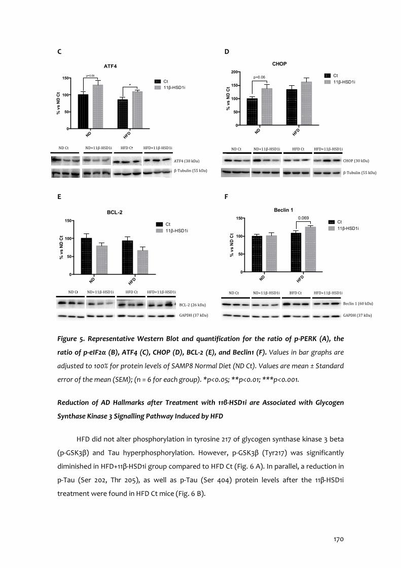
170
C D
E F
Figure 5. Representative Western Blot and quantification for the ratio of p-PERK (A), the
ratio of p-eIF2α (B), ATF4 (C), CHOP (D), BCL-2 (E), and Beclin1 (F). Values in bar graphs are
adjusted to 100% for protein levels of SAMP8 Normal Diet (ND Ct). Values are mean ± Standard
error of the mean (SEM); (n = 6 for each group). *p<0.05; **p<0.01; ***p<0.001.
Reduction of AD Hallmarks after Treatment with 11β-HSD1i are Associated with Glycogen
Synthase Kinase 3 Signalling Pathway Induced by HFD
HFD did not alter phosphorylation in tyrosine 217 of glycogen synthase kinase 3 beta
(p-GSK3β) and Tau hyperphosphorylation. However, p-GSK3β (Tyr217) was significantly
diminished in HFD+11β-HSD1i group compared to HFD Ct (Fig. 6 A). In parallel, a reduction in
p-Tau (Ser 202, Thr 205), as well as p-Tau (Ser 404) protein levels after the 11β-HSD1i
treatment were found in HFD Ct mice (Fig. 6 B).
NDHFD
0
50
100
150
ATF4
% v
s N
D C
t
Ct
11β-HSD1i *
p=0.09
ND Ct HFD Ct HFD+11β-HSD1i ND+11β-HSD1i
ATF4 (38 kDa) β-Tubulin (55 kDa)
NDHFD
0
50
100
150
200
% v
s N
D C
t
CHOP
Ct
11β-HSD1i p=0.06
ND Ct HFD Ct HFD+11β-HSD1i ND+11β-HSD1i
CHOP (30 kDa)
β-Tubulin (55 kDa)
ND Ct HFD Ct HFD+11β-HSD1i ND+11β-HSD1i
Beclin 1 (60 kDa)
GAPDH (37 kDa)
NDHFD
0
50
100
150
Beclin 1
% v
s N
D C
t
Ct
11β-HSD1i
0.069
ND Ct HFD Ct HFD+11β-HSD1i ND+11β-HSD1i
BCL-2 (26 kDa)
GAPDH (37 kDa)
NDHFD
0
50
100
150
BCL-2
% v
s N
D C
t
Ct
11β-HSD1i

171
HFD was unable to alter APP processing in SAMP8, neither soluble APP fragment
alpha (sAPPα) nor the fragment delivered by β-secretase (β-CTF) protein levels were
modified. 11β-HSD1i treatment caused a significant increase in sAPPα in ND-fed group but
not in HFD-fed mice (Fig. 6 C). β-CTF protein levels were decreased after 11β-HSD1i
regardless of the diet (Fig. 6 D). Finally, a significant increase in ADAM10 protein levels was
only found in ND+11β-HSD1i group compared to the ND Ct group (Fig. 6 E), whereas a
significant reduction in BACE1 protein levels was found in 11β-HSD1i treated animals,
reaching significance in ND-fed mice (Fig. 6 F).
A B
C D
p-Tau
(Ser
202,
Thr2
05)
p-Tau
(Ser
404)
0
50
100
150
% v
s N
D C
t
ND Ct
ND+11β-HSD1i
HFD Ct
HFD+11β-HSD1i
* p=0.09*
ND Ct ND+11β-HSD1i HFD+11β-HSD1i HFD Ct
p-Tau S202 T205 (75 kDa)
GAPDH (37 kDa)
p-Tau S404 (75 kDa)
GAPDH (37 kDa)
Tau (70 kDa)
GAPDH (37 kDa)
NDHFD
0
50
100
150
200
sAPPa
% v
s N
D C
t
Ct
11β-HSD1i ** *
ND Ct HFD Ct ND+11β-HSD1i HFD+11β-HSD1i
sAPP α (60-75 kDa)
GAPDH (37 kDa)
APP (100 kDa)
β-CTF (10 kDa)
GAPDH (37 kDa)
ND Ct ND+11β-HSD1i HFD+11β-HSD1i HFD Ct
NDHFD
0
50
100
150
Ratio b-CTF vs APP
% v
s N
D C
t
Ct
11β-HSD1i ***
ND Ct HFD Ct HFD+11β-HSD1i ND+11β-HSD1i
pGSK3β Y216 (47 kDa)
GAPDH (37 kDa)
GSK3β (46 kDa)
GAPDH (37 kDa)
NDHFD
0
50
100
150
Ratio pGSK3b Y216 vs GSK3b total
% v
s N
D C
t
Ct
11β-HSD1i *
*
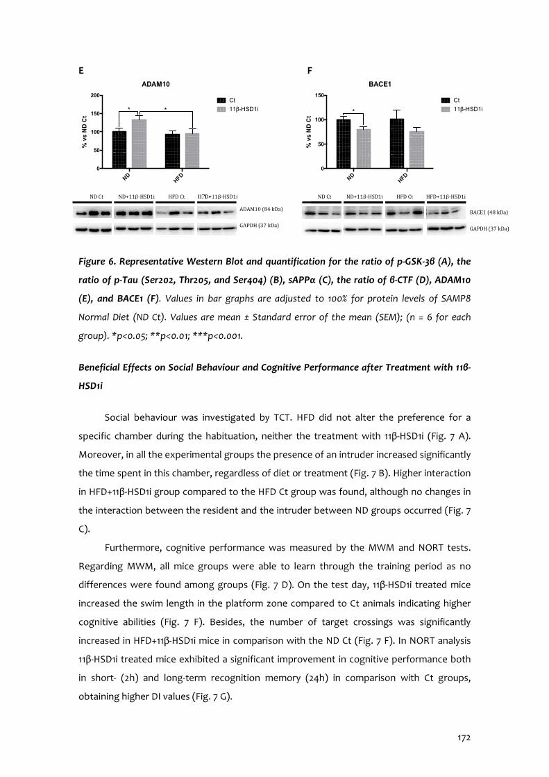
172
E F
Figure 6. Representative Western Blot and quantification for the ratio of p-GSK-3β (A), the
ratio of p-Tau (Ser202, Thr205, and Ser404) (B), sAPPα (C), the ratio of β-CTF (D), ADAM10
(E), and BACE1 (F). Values in bar graphs are adjusted to 100% for protein levels of SAMP8
Normal Diet (ND Ct). Values are mean ± Standard error of the mean (SEM); (n = 6 for each
group). *p<0.05; **p<0.01; ***p<0.001.
Beneficial Effects on Social Behaviour and Cognitive Performance after Treatment with 11β-
HSD1i
Social behaviour was investigated by TCT. HFD did not alter the preference for a
specific chamber during the habituation, neither the treatment with 11β-HSD1i (Fig. 7 A).
Moreover, in all the experimental groups the presence of an intruder increased significantly
the time spent in this chamber, regardless of diet or treatment (Fig. 7 B). Higher interaction
in HFD+11β-HSD1i group compared to the HFD Ct group was found, although no changes in
the interaction between the resident and the intruder between ND groups occurred (Fig. 7
C).
Furthermore, cognitive performance was measured by the MWM and NORT tests.
Regarding MWM, all mice groups were able to learn through the training period as no
differences were found among groups (Fig. 7 D). On the test day, 11β-HSD1i treated mice
increased the swim length in the platform zone compared to Ct animals indicating higher
cognitive abilities (Fig. 7 F). Besides, the number of target crossings was significantly
increased in HFD+11β-HSD1i mice in comparison with the ND Ct (Fig. 7 F). In NORT analysis
11β-HSD1i treated mice exhibited a significant improvement in cognitive performance both
in short- (2h) and long-term recognition memory (24h) in comparison with Ct groups,
obtaining higher DI values (Fig. 7 G).
NDHFD
0
50
100
150
200
ADAM10%
vs
ND
Ct
Ct
11β-HSD1i * *
ND Ct ND+11β-HSD1i HFD+11β-HSD1i HFD Ct
ADAM10 (84 kDa)
GAPDH (37 kDa)
NDHFD
0
50
100
150
BACE1
% v
s N
D C
t
Ct
11β-HSD1i *
ND Ct ND+11β-HSD1i HFD+11β-HSD1i HFD Ct
BACE1 (48 kDa)
GAPDH (37 kDa)
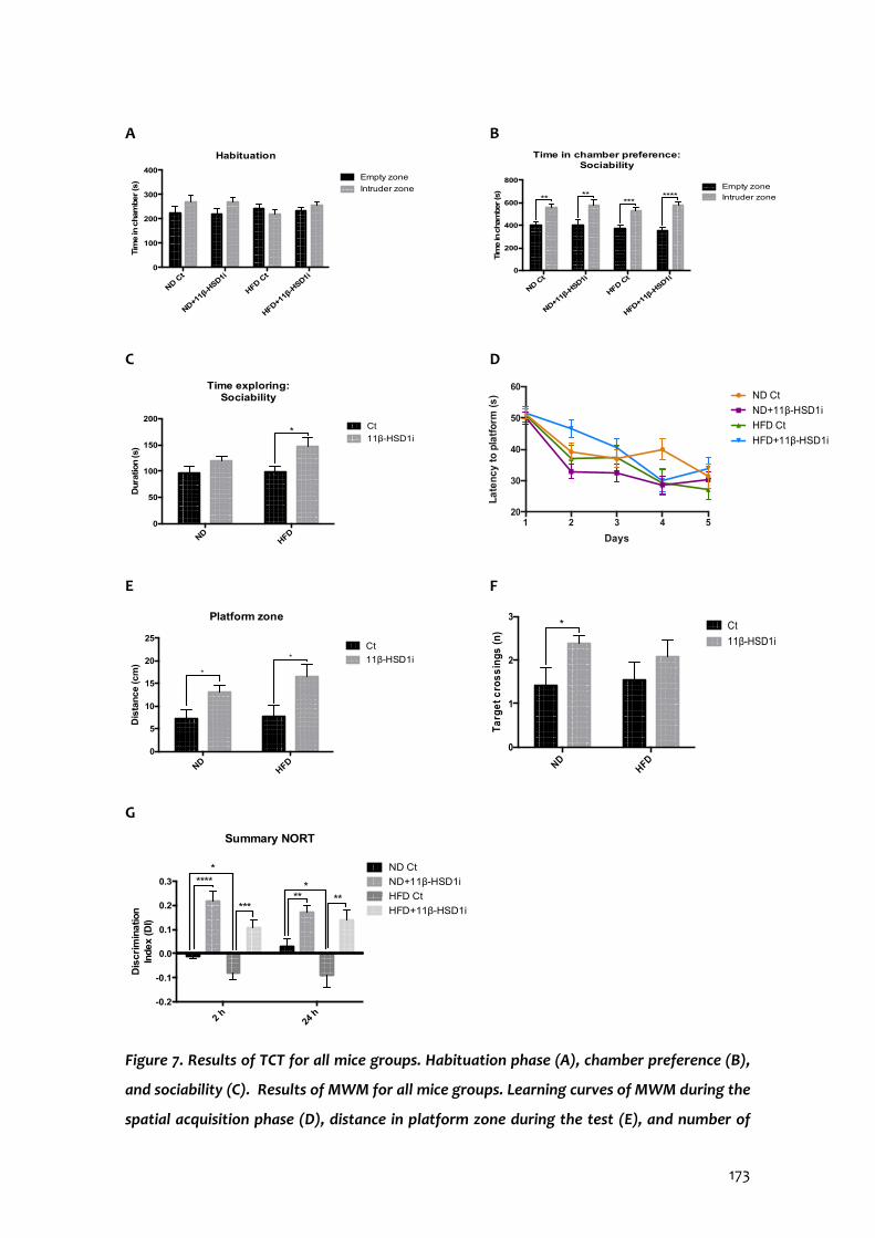
173
A B
C D
E F
G
Figure 7. Results of TCT for all mice groups. Habituation phase (A), chamber preference (B),
and sociability (C). Results of MWM for all mice groups. Learning curves of MWM during the
spatial acquisition phase (D), distance in platform zone during the test (E), and number of
ND Ct
ND+11β
-HSD1i
HFD Ct
HFD+11β
-HSD1i
0
100
200
300
400
Habituation
Tim
e in
cham
ber (s
) Empty zone
Intruder zone
ND Ct
ND+11β
-HSD1i
HFD Ct
HFD+1
1β-H
SD1i0
200
400
600
800
Time in chamber preference: Sociability
Tim
e in
cham
ber (s
)
Empty zone
Intruder zone*****
******
NDHFD
0
50
100
150
200
Time exploring: Sociability
Dura
tion (s)
Ct
11β-HSD1i*
1 2 3 4 520
30
40
50
60
Days
La
ten
cy to
pla
tfo
rm (s
) ND Ct
ND+11β-HSD1i
HFD Ct
HFD+11β-HSD1i
NDHFD
0
1
2
3
Ta
rge
t cro
ss
ings
(n
) Ct
11β-HSD1i
*
NDHFD
0
5
10
15
20
25
Platform zone
Dis
tan
ce (c
m)
Ct
11β-HSD1i*
*
2 h
24 h
-0.2
-0.1
0.0
0.1
0.2
0.3
Summary NORT
Dis
cri
min
atio
n
Index (D
I)
ND Ct
ND+11β-HSD1i
HFD Ct
HFD+11β-HSD1i
*****
*******
*
174
entries in the platform zone during the test (F). Results of NORT for all mice groups.
Summary of DI from 2 and 24 hours after the familiarization phase (G). Values are mean ±
Standard error of the mean (SEM) (n=12 for each group). *p<0.05; **p<0.01; ***p<0.001;
****p<0.0001.
3.4.5 Discussion
In this study, we provide new evidence that inhibition of 11β-HSD1 improves cognitive
impairment after metabolic stress induced by HFD intervention in a mice model of cognitive
decline. To this end, various molecular pathways influenced by HFD and GCs, as well as
social and cognitive impairment were evaluated to elucidate new mechanisms by which 11β-
HSD1 inhibition exerts neuroprotection.
Our group had previously described that HFD induces metabolic stress in a murine
model of accelerated senescence, SAMP8 (Palomera-Ávalos et al., 2017a) and in C57BL/6J
aged mice (Palomera-Ávalos et al., 2017b), which was prevented by resveratrol (Palomera-
Ávalos et al., 2017 a-b). Moreover, in previous reports, the neuroprotective effect of 11β-
HSD1 inhibitor (RL-118) was demonstrated in old female SAMP8 (Leiva et al., 2017; Puigoriol-
Illamola et al., 2018).
Animals fed with HFD increased body weight in comparison with ND fed mice
showing an alteration in glucose metabolism. The hugest increase in animal weight was not
only associated with impaired glucose tolerance but also with an alteration in lipid
metabolism. Barroso and co-workers described that the excessive consumption of
hypercaloric and high saturated fat food causes atherogenic dyslipidaemia (Barroso et al.,
2013). Accordingly, our results showed higher blood triglycerides concentration in HFD-fed
groups.
As aforementioned, there is extensive evidence that HFD impairs glucose
metabolism, altering the insulin signalling (Shoelson et al., 2007; von Frankenberg et al.,
2017). Recently, a close relationship between insulin insensitivity and neurodegenerative
disorders such as AD has been extensively reported (Griffith et al., 2018; Tumminia et al.,
2018) and it is still being discussed whether to consider AD as a type 3 Diabetes mellitus
(T3DM) (de la Monte and Wands, 2008). Additionally, high GC exposure can mediate insulin
resistance response, increasing glucose levels and favouring insulin resistance and obesity
175
(Geer et al., 2014; Ye, 2013). Thus, selective inhibitors of 11β-HSD1 have been postulated as a
neuroprotective strategy in several pathological scenarios, including metabolic
disturbances (Joharapurkar et al., 2012). RL-118 treatment reduced the 11β-HSD1 enzyme and
GC receptor protein levels both in ND- and HFD-fed SAMP8. In concordance, GC levels were
reduced both in blood and in plasma of RL-118 treated animals. Results are in line with
reports describing a reduction in gene expression of 11β-HSD1 and GR in diet-induced obese
mice after treatment with carbenoxolone, a 11β-HSD1 inhibitor (Liu et al., 2008).
Recently, FGF-21 has been demonstrated to modulate energy homeostasis of glucose
and lipid through activation of SIRT1/PGC1α/AMPKα axis, mainly through LKB1 activity (Chau
et al., 2010). Consistent with this hypothesis, treatment with 11β-HSD1 inhibitor significantly
increased protein levels of FGF-21 and LKB1 under both dietary conditions. Albeit RL-118 did
not induce direct changes in SIRT1 and PGC1α protein levels in HFD fed SAMP8, the increase
in AMPKα phosphorylation also indicated an improvement in nutrient sensing and
mitochondrial function. In sum, these results demonstrate the beneficial effects of reducing
GC signalling by 11β-HSD1 inhibition in SAMP8 under HFD-induced metabolic stress.
It has been reported that HFD increases OS and inflammation (Newsholme et al.,
2016). Accordingly, in HFD treated groups OS markers, such as GPX1, SOD1 and iNOS were
increased, and 11β-HSD1i treatment was only able to reduce them when animals were fed
with HFD. Whereas a clear tendency to reduce hippocampal ROS levels in all 11β-HSD1i
treated animals was found. Regarding neuroinflammation, Il-6 and Il-4 gene expression and
microglial activation evaluated through Iba-1, increased under HFD confirming the cellular
dysfunction induced by impaired energy metabolism. Importantly, 11β-HSD1 inhibition
diminished gene expression of Tnf-α and Il-6, as well as p65 (NF-κB fraction) protein levels,
inhibiting microglial reactivity. These findings agree with already published evidence
describing that 11β-HSD1 inhibition modulates OS and inflammatory processes (Gathercole
et al., 2013; Leiva et al., 2017; Puigoriol-Illamola et al., 2018).
11β-HSD1 inhibition by RL-118 reduced autophagy and apoptosis markers in old SAMP8
(Puigoriol-Illamola et al., 2018), but in the present work, we did not observe significant
changes in Beclin 1 or BCL2, probably because of the young age of mice. Because ER stress
response activates proteostatic mechanisms, (Cuanalo-Contreras et al., 2013; Hetz et al.,
2015) ER stress markers were studied. While HFD did not modify ATF4 protein levels in a
significant way, PERK and eIF2α activation were significantly reduced in those animals. Of
176
interest, inhibition of 11β-HSD1 by RL-118 recovers the ratio of phosphorylation of PERK and
eIF2α, and in addition, increases ATF4 protein levels. By contrast, in our hands CHOP
showed a narrow but not significant increase after RL-118 treatment (Mahdi et al., 2015).
Regarding the accumulation of misfolded proteins, one characteristic of ageing and AD, 11β-
HSD1 inhibition reduced GSK3β activation and tau phosphorylation after HFD feeding.
Regarding the β-amyloid pathway, RL-118 promoted APP processing by the non-
amyloidogenic pathway, decreasing pro-amyloidogenic APP fragments and BACE1 protein
levels, as well as increasing ADAM10 protein levels. Results suggested that 11β-HSD1
inhibition reduces the negative impact of HFD on cellular and tissue hallmarks of cognitive
decline and AD (Palomera-Ávalos et al., 2017a-b; Petrov et al., 2015).
Of paramount importance, RL-118 promoted changes in sociability behaviour,
recognition memory, in both short- and long-term, and spatial memory in both dietary
conditions. These results point out that the GC levels have a key role in memory, learning
and socialization processes, influencing mood-like behaviours in mice, both physiological
and pathological conditions, supported by the molecular results, and in accordance to
Raulo & Dantzer (2018).
In conclusion, our results demonstrate that HFD induced systemic metabolic
dysfunctions and exacerbated cognitive impairment in adult SAMP8 mediated by
alterations in insulin signalling, OS, neuroinflammation, and aberrant protein processing.
Decreasing GC levels with RL-118 reduced global stress and led to beneficial effects in most
of the markers evaluated in HFD and ND fed mice. Finally, modulation of GC activity by 11β-
HSD1 inhibition contribute to enhance cognitive performance in senescent mice regardless
of the dietary influence. Because new approaches are needed to fight against cognitive
decline and dementia, such as AD, the control of GC levels may open new avenues to
prevent these devastating conditions.
3.4.6 Acknowledgements
This study was supported by Ministerio de Economía y Competitividad of Spain and
FEDER(SAF2016-77703 to MP and SAF2017-82771 to SV, and SAF2015-64146-R to MVC) and
2017SGR106 (AGAUR, Catalonia). Financial support was provided for D.P.I. and R.L. (FPU
program). ACCIÓ (Generalitat de Catalunya) provided financial support (Programa Nuclis,
RD14-1-0057, SAFNAD).
177
3.4.7 References
Barroso, E., Astudillo, A.M., Balsinde, J., Vázquez-Carrera, M. (2013) PPARβ/δ Activation prevents hypertriglyceridemia caused by a high fat diet. Involvement of AMPK and PGC-1α-Lipin1-PPARα pathway. 25(2), Clínica e Investigación en Arteriosclerosis, 63-73. doi: 10.1016/j.arteri. 2013.01.001.
Bischof, G. N., & Park, D. C. (2015) Obesity and Aging: Consequences for Cognition,
Brain Structure, and Brain Function. Psychosomatic medicine, 77(6), 697–709. doi:10.1097/PSY.0000000000000212.
Bowles, N.P., McEwen, B.S., Boutin-Foster, C. (2017) Trouble in transit: organizational
barriers to worker’s health. Am J Ind Med, 60:350-367. Bujalska, I.J., Quinkler, M., Tomlinson, J.W., Montague, C.T., Smith, D.M., Stewart,
P.M. (2006) Expression profiling of 11β-hydroxysteroid dehydrogenase type-1 and glucocorticoid-target genes in subcutaneous and omental human preadipocytes. J Mol Endocrinol, 37(2):327-40.
Cai D. (2012) Neuroinflammation and neurodegeneration in overnutrition-induced
diseases. Trends in endocrinology and metabolism: TEM, 24(1), 40–47. doi:10.1016/j.tem.2012.11.003.
Canet, G., Chevallier, N., Zussy, C., Desrumaux, C., Givalois, L. (2018) Central Role of
Glucocorticoid Receptors in Alzheimer's Disease and Depression. Frontiers in neuroscience, 12, 739. doi:10.3389/fnins.2018.00739.
Chau, M. D., Gao, J., Yang, Q., Wu, Z., & Gromada, J. (2010) Fibroblast growth factor 21
regulates energy metabolism by activating the AMPK-SIRT1-PGC-1alpha pathway. Proceedings of the National Academy of Sciences of the United States of America, 107(28), 12553–12558. doi:10.1073/pnas.1006962107.
Cuanalo-Contreras, K., Mukherjee, A., & Soto, C. (2013) Role of protein misfolding and
proteostasis deficiency in protein misfolding diseases and aging. International journal of cell biology, 2013, 638083. doi:10.1155/2013/638083.
de la Monte, S. M., & Wands, J. R. (2008) Alzheimer's disease is type 3 diabetes-
evidence reviewed. Journal of diabetes science and technology, 2(6), 1101–1113. doi:10.1177/193229680800200619.
de Quervain, D.J., Poirier, R., Wollmer, M.A., Grimaldi, L.M., Tsolaki, M., Streffer, J.R.,
Hock, C., Nitsch, R.M., Mohajeri, M.H., Papassotiropoulos, A. (2004) Glucocorticoid-related genetic susceptibility for Alzheimer’s disease. Hum Mol Genet, 13(1):47-52.
178
Ennaceur, A., Delacour, J. (1988) A new one-trial test for neurobiological studies of memory in rats. 1: Behavioral data. Behav Brain Res, 31:47–59.
Feinkohl, I., Lachmann, G., Brockhaus, W. R., Borchers, F., Piper, S. K., Ottens, T. H., …
Spies, C. (2018) Association of obesity, diabetes and hypertension with cognitive impairment in older age. Clinical epidemiology, 10, 853–862. doi:10.2147/CLEP.S164793.
Gathercole, L.L., Lavery, G.G., Morgan, S.A., Cooper, M.S., Sinclair, A.J., Tomlinson,
J.W., Stewart, P.M. (2013) 11β-Hydroxysteroid dehydrogenase 1: translational and therapeutic aspects. Endocr Rev, 34(4): 525-55. doi: 10.1210/er.2012-1050.
Geer, E.B., Islam, J., Buettner, C. (2014) Mechanisms of glucocorticoid-induced insulin
resistance: focus on adipose tissue function and lipid metabolism. Metab Clin North Am, 43(1):75-102. doi.10.1016/j.ecl.2013.10.005.
Griffith CM, Eid T, Rose GM, Patrylo PR. (2018) Evidence for altered insulin receptor
signalling in Alzheimer’s disease. Neuropharmacology, 136(Pt B): 202-215. doi:10.1016/j.neuropharm.2018.01.008.
Griñan-Ferré, C., Pérez-Cáceres, D., Gutiérrez-Zetina, S.M., Camins, A., Palomera-
Ávalos, V., Ortuño-Sahagún, D., Rodrigo, M.T., Pallàs, M. (2016) Environmental Enrichment Improves Behavior, Cognition, and Brain Functional Markers in Young Senescence-Accelerated Prone Mice (SAMP8). Mol Neurobiol, 53(4):2435-50. doi: 10.1007/s12035-015-9210-6.
Guerreiro, R., & Bras, J. (2015) The age factor in Alzheimer's disease. Genome
medicine, 7, 106. doi:10.1186/s13073-015-0232-5. Hetz, C., Chevet, E., & Oakes, S. A. (2015) Proteostasis control by the unfolded protein
response. Nature cell biology, 17(7), 829–838. doi:10.1038/ncb3184. Holmes, M.C., Carter, R.N., Noble, J., Chitnis, S., Dutia, A., Paterson, J.M., Mullins, J.J.,
Seckl, J.R., Yau, J.L. (2010) 11beta-hydroxysteroid dehydrogenase type 1 expression is increased in the aged mouse hippocampus and parietal cortex and causes memory impairments. J Neurosci, 30(20):6916-20. doi: 10.1523/JNEUROSCI.0731-10.2010.
Joharapurkar, A., Dhanesha, N., Shah, G., Kharul, R., Jain, M. (2012) 11β-
Hydroxysteroid dehydrogenase type 1: potential therapeutic target for metabolic syndrome. Pharmacol Rep, 64(5):1055-65.
Kadmiel, M., Cidlowski, J.A. (2013) Glucocorticoid receptor signalling in health and
disease. Trends Pharmacol Sci, 34(9):518-30. doi: 10.1016/j.tips.2013.07.003. Karatsoreos, I.N., Bhagat, S.M., Bowles, N.P., Weil, Z.M., Pfaff, D.W., McEwen, B.S.
(2010) Endocrine and physiological changes in response to chronic corticosterone: a
179
potential model of the metabolic syndrome in mouse. Endocrinology, 151(5):2117-27. doi: 10.1210/en.2009-1436.
Kesby, J.P., Kim, J.J., Scadeng, M., Woods, G., Kado, D.M., Olefsky, J.M., Jeste, D.V.,
Achim, C.L., Semenova, S. (2015) Spatial Cognition in Adult and Aged Mice Exposed to High-Fat Diet. PLoS One, 8;10(10):e0140034. doi: 10.1371/journal.pone.0140034. eCollection 2015.
Kumar, A., Datusalia, A.K. (2018) Metabolic Stress and Inflammation: Implication in
Treatment for Neurological Disorders. CNS Neurol Disord Drug Targets, 17(9):642-643. doi: 10.2174/187152731709180926121555.
Lee, S., Kim, J. Y., Kim, E., Seo, K., Kang, Y. J., Kim, J. Y., … Lee, J. E. (2018)
Assessment of Cognitive Impairment in a Mouse Model of High-Fat Diet-Induced Metabolic Stress with Touchscreen-Based Automated Battery System. Experimental neurobiology, 27(4), 277–286. doi:10.5607/en.2018.27.4.277.
Leiva, R., Griñán-Ferré, C., Seira, C., Valverde, E., McBride, A., Binnie, M., Pérez, B.,
Luque, F.J., Pallàs, M., Bidon-Chanal, A., Webster, S.P., Vázquez, S. (2017) Design, synthesis and in vivo study of novel pyrrolidine-based 11β–HSD1 inhibitors for age-related cognitive dysfunction. Eur J Med Chem, 139:412-428. doi: 10.1016/j.ejmech.2017.08.003.
Liu, Y., Nakagawa, Y., Wang, Y., Liu, L., Du, H., Wang, W., … Friedman, T. C. (2008)
Reduction of hepatic glucocorticoid receptor and hexose-6-phosphate dehydrogenase expression ameliorates diet-induced obesity and insulin resistance in mice. Journal of molecular endocrinology, 41(2), 53–64. doi:10.1677/JME-08-0004.
López-Otín, C., Blasco, M. A., Partridge, L., Serrano, M., Kroemer, G. (2013) The
hallmarks of aging. Cell, 153(6), 1194–1217. doi:10.1016/j.cell.2013.05.039. Mahdi, A. A., Rizvi, S. H., & Parveen, A. (2015) Role of Endoplasmic Reticulum Stress
and Unfolded Protein Responses in Health and Diseases. Indian journal of clinical biochemistry : IJCB, 31(2), 127–137. doi:10.1007/s12291-015-0502-4.
Mattson, M. P., Arumugam, T. V. (2018) Hallmarks of Brain Aging: Adaptive and
Pathological Modification by Metabolic States. Cell metabolism, 27(6), 1176–1199. doi:10.1016/j.cmet.2018.05.011.
McEwen, B.S., Karatsoreos, I.N. (2015) Sleep Deprivation and Circadian Disruption:
Stress, Allostasis and Allostatic Load. Sleep Med Clin, 10(1):1-10. doi: 10.1016/j.jsmc.2014.11.007.
Mohler, E.G., Browman, K.E., Roderwald, V.A., Cronin, E.A., Markosyan, S., Scott
Bitner, R., Strakhova, M.I., Drescher, K.U., Hornberger, W., Rohde, J.J., Brune, M.E., Jacobson, P.B., Rueter, L.E. (2011) Acute inhibition of 11beta-hydroxysteroid dehydrogenase

180
type-1 improves memory in rodent models of cognition. J Neurosci, 31(14):5406-13. doi: 10.1523/JNEUROSCI.4046-10.2011.
Mule, N.K., Singh, J.N. (2018) Diabetes Mellitus to Neurodegenerative Disorders: Is
Oxidative Stress Fueling the Flame? CNS Neurol Disord Drug Targets, 17(9):644-653. doi: 10.2174/1871527317666180809092359.
Newsholme, P., Cruzat, V.F., Keane, K.N., Carlessi, R., de Bittencourt, P.I. Jr. (2016)
Molecular mechanisms of ROS production and oxidative stress in diabetes. Biochem J, 15;473(24):4527-4550.
Palomera-Ávalos, V., Griñán-Ferré, C., Puigoriol-Ilamola, D., Camins, A., Sanfeliu, C.,
Canudas, A.M., Pallàs, M. (2017a). Resveratrol Protects SAMP8 Brain Under Metabolic Stress: Focus on Mitochondrial Function and Wnt Pathway. Mol Neurobiol, 54(3):1661-1676.
Palomera-Ávalos, V., Griñán-Ferré, C., Izquierdo, V., Camins, A., Sanfeliu, C., Pallàs, M.
(2017b). Metabolic Stress Induces Cognitive Disturbances and Inflammation in Aged Mice: Protective Role of Resveratrol. Rejuvenation Res, 20(3):202-217. doi: 10.1089/rej.2016.1885.
Petrov, D., Pedrós, I., Artiach, G., Sureda, F.X., Barroso, E., Pallàs, M., Casadesús, G.,
Beas-Zarate, C., Carro, E., Ferrer, I., Vazquez-Carrera, M., Folch, J., Camins, A. (2015) High-fat diet-induced deregulation of hippocampal insulin signalling and mitochondrial homeostasis deficiencies contribute to Alzheimer disease pathology in rodents. Biochim Biophys Acta, 1852(9):1687-99. doi: 10.1016/j.bbadis.2015.05.004.
Puigoriol-Illamola, D., Griñán-Ferré, C., Vasilopoulou, F., Leiva, R., Vázquez, S., Pallàs,
M. (2018) 11β-HSD1 inhibition by RL-118 promotes autophagy and correlates with reduced oxidative stress and inflammation, enhancing cognitive performance in SAMP8 mouse model. Mol Neurobiol. 55(12):8904-8915. doi: 10.1007/s12035-018-1026-8.
Raulo, A., & Dantzer, B. (2018). Associations between glucocorticoids and sociality
across a continuum of vertebrate social behavior. Ecol Evol, 8(15), 7697-7716. Shoelson, S.E., Herrero, L., Naaz, A. (2007) Obesity, Inflammation, and insulin
resistance. Gastroenterology, 132(6):2169-80. Sooy, K., Noble, J., McBride, A., Binnie, M., Yau, J.L., Seckl, J.R., Walker, B.R., Webster,
S.P. (2015) Cognitive and disease-modifying effects of 11β-hydroxysteroid dehydrogenase type 1 inhibition in male Tg2576 mice, a model of Alzheimer’s disease. Endocrinology156:4592–4603
Sooy, K.,Webster, S.P., Noble, J., Binnie, M., Walker, B.R., Seckl, J.R., Yau, J.L. (2010)
Partial deficiency or short term inhibition of 11β-hydroxysteroid dehydrogenase type 1 improves cognitive function in aging mice. J Neurosci 30:13867–13872.
181
Tatomir, A., Micu, C., & Crivii, C. (2014) The impact of stress and glucocorticoids on memory. Clujul medical (1957), 87(1), 3–6. doi:10.15386/cjm.2014.8872.871.at1cm2.
Tumminia, A., Vinciguerra, F., Parisi, M., & Frittitta, L. (2018) Type 2 Diabetes Mellitus
and Alzheimer's Disease: Role of Insulin Signalling and Therapeutic Implications. International journal of molecular sciences, 19(11), 3306. doi:10.3390/ijms19113306.
von Frankenberg, A. D., Marina, A., Song, X., Callahan, H. S., Kratz, M., &
Utzschneider, K. M. (2015) A high-fat, high-saturated fat diet decreases insulin sensitivity without changing intra-abdominal fat in weight-stable overweight and obese adults. European journal of nutrition, 56(1), 431–443. doi:10.1007/s00394-015-1108-6.
Vorhees, C. V., & Williams, M. T. (2006) Morris water maze: procedures for assessing
spatial and related forms of learning and memory. Nature protocols, 1(2), 848–858. doi:10.1038/nprot.2006.116.
Wei, L., Yao, M., Zhao, Z., Jiang, H., & Ge, S. (2018) High-fat diet aggravates
postoperative cognitive dysfunction in aged mice. BMC anesthesiology, 18(1), 20. doi:10.1186/s12871-018-0482-z.
Yau, J.L., Seckl, J.R. (2012) Local amplification of glucocorticoids in the aging brain
and impaired spatial memory. Front Aging Neurosci, 4:24. doi: 10.3389/fnagi.2012.00024. Yau, J.L., Wheelan, N., Noble, J., Walker, B.R., Webster, S.P., Kenyon, C.J., Ludwig, M.,
Seckl, J.R. (2015) Intrahippocampal glucocorticoids generated by 11β-HSD1 affect memory in aged mice. Neurobiol Aging, 36(1):334-43. doi: 10.1016/j.neurobiolaging.2014.07.007.
Yau, J.L., McNair, K.M., Noble, J., Brownstein, D., Hibberd, C., Morton, N., Mullins, J.J.,
Morris, R.G., Cobb, S., Seckl, J.R. (2007) Enhanced hippocampal long-term potentiation and spatial learning in aged 11beta-hydroxysteroid dehydrogenase type 1 knock-out mice. J Neurosci, 27(39):10487-96.
Yau, J.L., Noble, J., Kenyon, C.J., Hibberd, C., Kotelevstev, Y., Mullins, J.J., Seckl, J.R.
(2001) Lack of tissue glucocorticoid reactivation in 11beta-hydroxysteroid dehydrogenase type 1 knockout mice ameliorates age-related learning impairments. Proc Natl Acad Sci U S A, 98(8):4716-21.
Ye, J. (2013) Mechanisms of insulin resistance in obesity. Frontiers of medicine, 7(1),
14–24. doi:10.1007/s11684-013-0262-6.
3.4.8 Supplementary material
The online version of this article (https://doi.org/10.1007/s12035-019-01708-4) contains
electronic supplementary material, which is available to authorized users.

182

4. DISCUSSION

184

185
Ageing is a physiological and multifactorial process that provokes a gradual cellular
deterioration in most living organisms, which is not only associated with compromised
lifespan, but it also has a marked effect on healthspan. In fact, it results in increased
susceptibility to a variety of neurodegenerative diseases as AD, eating disorders, viral
infections and cancers (Jura & Kozak, 2016; Leng & Goldstein, 2010; Niccoli & Partridge,
2012). Additionally, obesity and HFD consumption are two key factors in the development
and progression of a wide range of disorders, such as T2DM, metabolic syndrome, and
cardiovascular diseases, as well as contribute to neurodegenerative processes like AD (De
Felice & Lourenco, 2015; Keshk et al., 2020). Although it is completely assumed that stress is
one of the factors that most directly influence the frailty phenotype, there are strikingly
few measures to restrain stressful lifestyles at different levels, both metabolically and
physically, in order to reduce the progression of pathological towards successful ageing.
The study of stress effects on cognition and its relationship with ageing is of the utmost
importance in order to unveil what challenges we might have to cope with as a society in a
not so far future. Therefore, and taking into account that ageing leads to a gradual decline
of cognitive abilities and it is the main risk factor for AD, we firmly believe that it is
necessary taking a step forward in the study of the molecular mechanisms underlying
neurodegeneration and find a different approach of the disease to achieve an appropriate
treatment.
In an attempt to address this issue, several pieces of literature defining stress, stress
response and its implications in cognitive decline have been reviewed. It has been
established that the environment affects individuals’ cognitive performance and brain
structure (Crous-Bou et al., 2020; Griñán-Ferré et al., 2016; Oudin, 2020). Indeed, the stress
response is actually a very useful and highly adaptive response. Stress exposure activate
HPA axis to re-establish homeostasis and release a wide variety of hormones that exert
their effects in numerous central and peripheral sites to produce adaptive effects, including
the mobilisation of energy from storage, maintenance of the immune system and inhibition
of nonessential processes such as reproductive function. Collectively, these functions
enable the fight or flight behaviours to remove the organism from immediate danger, while
later restoring bodily homeostasis (Goosens & Sapolsky, 2007). However, the experience of
too much stress over time can have adverse consequences on health and behaviour, but
never experiencing any stress would result in inactivity, boredom and inability to
adequately respond to internal and external demands (Stults-Kolehmainen & Sinha, 2014).
186
As stress response modulates a large constellation of cellular mechanisms implied in ageing
and age-related neurodegenerative pathologies, we assessed some proper features of
ageing, such as loss of proteostasis, altered nutrient sensing, mitochondrial dysfunction, OS
and inflammatory responses, in reference to HPA axis alterations.
Compelling evidence has demonstrated that as ageing, and more pronouncedly when
it has been subjected to continuously stressful stimuli exposure, elevated GCs levels due to
HPA axis dysregulation have an impact in our cognitive performance and impulse various
mechanisms underlying neurodegeneration. In consequence, controlling GC activity by 11β-
HSD1 becomes essential to prevent that.
4. 1 11β-HSD1 inhibition
The 11β-HSD1 enzyme has been recognized as an essential modulator in the
intracellular control of GC actions, as it catalyses the conversion of inert cortisone to active
cortisol in primates or 11-dehydrocorticosterone to corticosterone in rodents. It is anchored
in the ER membrane and is present in peripheral tissues, notably in the liver, adipose tissue
and vasculature, as well as in multiple regions of the brain; in particular, the anterior
pituitary gland, hypothalamic PVN and the hippocampus (Holmes et al., 2010; Peng et al.,
2016).
Considering that 11β-HSD1 enzyme presents a high sequence homology between
species, particularly within the cofactor-binding region and the catalytic site (Gathercole et
al., 2013), we decided to clone the human gene encoding this protein and transfect human
embryonic kidney (HEK) 293 cells in order to express that enzyme and in consequence
study one of the key factors in drug discovery: the specific binding of the drug to its target,
i.e. target engagement. Measuring and quantifying the binding of a drug to a protein target
inside living cells and thereby correlating biochemical or biophysical activity with target
engagement in cells or tissue represents a key step that should allow for unbiased
determination of small molecule-protein interactions with the aim of confirming cellular
mechanism-of-action while avoiding major artificial perturbations of cellular homeostasis
and integrity (Stefaniak & Huber, 2020).
Up to now, studies demonstrating that RL-118 can reach the brain, bind the 11β-HSD1
enzyme and inhibit it, leading to therapeutic effects have not been addressed. In
consequence, in the present work, the target engagement of RL-118 and 11β-HSD1 was
187
determined, through a novel methodology consisting in FACS coupled to MS analysis called
TAPS assay (Wilson et al., 2017). Effectively, target engagement results showed that RL-118
drug binds to its target in a selectively way, since it did not bind to other proteins
expressed. Hence, we have determined that actually the molecular and behavioural effects
observed after RL-118 administration are due to its binding to the 11β-HSD1 enzyme.
In agreement with that, we have detected lower levels of the active GC in blood and
brain of SAMP8 mice treated with the 11β-HSD1 inhibitor in comparison to the control
group, and additional data supporting this fact revealed that 11β-HSD1 protein levels were
decreased after RL-118 treatment and GR protein levels too, indicative of GC signalling
modulation.
4.2 Neurodegeneration
Ageing is closely linked to neuronal changes and cognitive deficits, and therefore to
neurodegeneration, being the major risk factor for neurodegenerative disorders, like AD.
The ability of misfolded forms of the two key proteins of AD – Aβ and hyperphosphorylated
Tau – to disrupt hippocampal synaptic plasticity engenders impending synaptic failure,
subsequent structural pathology and eventually contributes to cognitive decline (Balschun
& Rowan, 2018). Also, it has been suggested a causal role of stress in the onset and
progression of AD, as sustained stress-level GC exposure has been found to increase Aβ
formation and hyperphosphorylated Tau accumulation, hallmarks of human AD; and
adversely affect behaviour (Bini et al., 2020; Green et al., 2006; MacLullich et al., 2012).
Consistently with this, reducing GC exposure in the brain via intracellular inhibition of 11β-
HSD1 has emerged as a therapeutic strategy to treat cognitive impairment in early AD
(Webster et al., 2017). In consequence, throughout the development of this doctoral
dissertation, we decided to study specific markers of AD, as well as other indicators of brain
function, such as markers of synaptic plasticity.
Regarding the AD hallmarks, not only APP processing and Tau hyperphosphorylation
markers have been evaluated but also other markers controlling them like GSK3-β. It has
been reported that GSK3-β regulates Aβ production by down-regulating the activity of the
α-secretase and interfering with the γ-secretase activity, thus resulting in Aβ-induced
neurotoxicity reduction (Llorens-Martín et al., 2014). Accordingly, GSK3-β participates in Tau
phosphorylation and growing evidence indicates that hyperphosphorylated Tau activates
GSK3-β through an increase in OS, neuroinflammation and apoptosis (Saeki et al., 2011). On
188
the one hand, CMS increased Tau hyperphosphorylation and induced changes in APP
processing towards an amyloidogenic pathway, especially in SAMP8 mice (Canet et al.,
2018; Mravec et al., 2018). Notwithstanding, CMS also decreased Aβ clearance mechanism
similar to what is described in Morgese et al. (2017) study. Surprisingly, those results were
not observed in SAMP8 under metabolic stress. However, 11β-HSD1 inhibition by RL-118
clearly promoted non-amyloidogenic processing of APP and reduced Tau
hyperphosphorilation, allowing the recovery of those marks regardless of the stressful
event (Canet et al., 2018).
Diversely, it has been determined that male SAMP8 showed reduced neuronal
synaptic plasticity markers in comparison with SAMR1, as well as lower in SAMP8 mice
under CMS than the control group (Wang et al., 2016), although in our hands CMS did not
impair synaptic plasticity. Previously, our lab demonstrated that RL-118 treatment enhanced
synaptic plasticity in SAMP8 mice (Leiva et al., 2017), and in accordance, we observed that
although exposure to stressful situations, after 11β-HSD1 inhibition by RL-118 treatment,
protein levels of synaptic plasticity markers studied increased. In relation to synaptic
function, it has been recently described that CREB-BDNF signalling pathway in the
hippocampus mediates learning and memory deficits of rats subjected to CMS, decreasing
its activation in CMS groups (Luo et al., 2017). In agreement with that, we found changes in
the ratio of p-CREB protein levels but not in Creb gene expression in SAMP8 under CMS.
However, regarding 11β-HSD1 inhibition effects, similarly to Numakawa et al. (2017), RL-118
treatment increased BDNF protein levels and gene expression.
4.3 Epigenetics
In line with stressful events influence in cognition, the way the environment modifies
gene expression is comprised under the term epigenetics. While ageing and in
neurodegenerative pathologies, there are several epigenetic modifications, which include
DNA methylation, histone modifications and miRNA-mediated gene regulation (Barter &
Foster, 2018; Harman & Martín, 2019).
Given that SAMP8 is a spontaneous ageing model, it is suggested that underlying
mechanisms orchestrating accelerated senescence must be linked to epigenetics (Griñán-
Ferré et al., 2018). In this respect, several studies corroborate the existence of epigenetic
modifications related to ageing and neurodegeneration in SAMP8 (Alvarez-López et al.,
2014; Cosín-Tomás et al., 2014).
189
On another note, convergent evidence from both preclinical and clinical studies has
demonstrated significant associations between stress and epigenetic alterations,
predominantly in genes involved in mediating resilience or vulnerability to stress, including
stress-response genes, genes involved in neurotransmission and neurotrophin genes (Jiang
et al., 2019; Park et al., 2019). Herein, we evaluated three main epigenetic marks that
regulate gene expression, modulating chromatin structure through recruitment, assembly
or retention of chromatin-associated factors after CMS exposure.
As far as DNA methylation is concerned, 5-mC DNA contributes to neural
development, and its dysregulation is implicated in neuronal disorders, although its
relationship is still debated. For instance, heightened 5-mC levels have been related to AD
(Mastroeni, 2016), while others described the contrary (Ellison et al., 2017). This difference
may be attributed to specific regions of the genome. Typically, 5-mC is associated with gene
silencing and deregulated transcription. However, oxidation of 5-mC by TET enzymes to 5-
hmC is thought to have the opposite effect (Fetahu et al., 2019). In the present studies, CMS
reduced 5-mC and 5-hmC in both mouse strains, although in the second study, there were
no significant differences in 5-hmC DNA between CMS groups and their control littermates.
TET2 protein levels and gene expression and Dnmt1 gene expression were agreeable to the
above results, although Dnmt3a gene expression failed to significance. Referring to RL-118
treatment, it increased 5-mC and 5-hmC in accordance with Dnmt1 and Tet2 gene
expression, recovering those marks in SAMP8 under CMS treated with the 11β-HSD1
inhibitor studied. In agreement with this, it has been described that GCs can rapidly induce
methylation changes in CpG islands of the DNA, most notably global decreases in
methylation, although some studies have documented GC-induced DNA hypermethylation
in some sites (Zannas & Chrousos, 2017). Of note, global DNA methylation does not
represent a precise landscape for specific transcriptional activity as it is misleading which
genes are methylated and which are not.
As two major mechanisms for epigenetic regulation, DNA methylation and histone
modifications must act coordinately (Zhao et al., 2016). One of the histone PTM is
acetylation that is associated with relaxed chromatin structure and thereby greater levels
of gene expression (Turner, 2000). CMS and thus heightened GC exposure is supposed to
decrease histone acetylation (Bagot et al., 2014; Harman & Martín, 2019). In the same line,
we found that CMS reduced acetylated histone protein levels that correlate with increased
HDAC2 protein levels. Also, acetylation was decreased in SAMP8 compared to SAMR1
190
animals. Albeit, higher H3K9 protein levels were found in SAMP8 under CMS group, it might
not translate to relaxed chromatin state, as dimethylation of this specific acetylated histone
protein levels were increased in this group in both projects conducted. On the contrary, 11β-
HSD1 inhibition reduced Hdac2 gene expression and contributed to increasing acetylated
histone protein levels, but not in SAMP8 under CMS too. In addition, other HDACs include
SIRT family, which are intimately linked to ageing as they modulate genomic stability, stress
resistance and energy metabolism. In particular, activation of SIRT1 enables deacetylation
of a variety of proteins resulting in a robust protective cellular response because it
regulates processes such as cell death, metabolism or neurodegeneration (Herskovits &
Guarente, 2014); while SIRT2 regulates OS, genomic integrity and myelination (Jesko et al.,
2017), and Sirt6 gene expression has been reported to be decreased in the hippocampus
and cerebral cortex of aged mice and that is concerned with H3K9 (Khan et al., 2018). Our
results agree with those above mentioned, as Sirt studied gene expression were decreased
in SAMP8 mice compared to SAMR1 and SAMR1 under CMS. Other histone PTM involve
methylation and phosphorylation. The former is linked to gene silencing, whereas the later
to DNA damage response. The pattern of histone methylation has been shown to change
during ageing depending on the tissue and organisms, although a global increase in
activation histone methylations concomitant with a reduction of repressive methylated
histone marks has been reported (Harman & Martín, 2019). Our results showed increases in
these marks in SAMP8 under CMS and trend toward reduction in RL-118 treated groups.
Finally, emerging evidence also supports the role of GC signalling in the regulation of
several miRNAs (Zannas & Chrousos, 2017). We evaluated the expression of miRNAs
regulating OS and cellular pathways implicated in AD neuronal death, autophagy and
neurodegeneration. Altogether, miRNA expression studied correlated with their target
genes. In our hands, CMS slightly provided an increase in DNA methylation and
amyloidogenic APP processing, while a decrease in Wnt pathway and autophagy. Taking
into account that DNMT inhibitors have provided neuroprotection in cell cultures (Chestnut
et al., 2011) and β-catenin reduction has been found in the brain of AD patients in
accordance to decreased GSK3-β inactive form (Llorens-Martín et al., 2014) that is related to
increased Tau hyperphosphorylation, and accounting that CMS favoured amyloidogenic
APP processing, while decreased non-amyloidogenic APP processing mediators as well as
autophagy flux (Uddin et al., 2018), we concluded that CMS could produce deleterious
effects on health.
191
Noteworthy, stress exposure, which may influence GC signalling, can result in lasting
epigenetic modifications that persist throughout life and even across generations (Zannas
& Chrousos, 2017). Therefore, it is of paramount importance to study drug actions at the
epigenetic level in order to possess a deep knowledge of the pharmacological effects
exerted by the drug. To this regard, although RL-118 is not a direct epigenetic modulator
drug, it modifies the global epigenetic landscape.
4.4 Proteostasis
In reference to proteostasis mechanisms, it involves autophagy and ER stress
response. Under stress conditions, the accumulation of misfolded proteins disrupts ER
function and induces its stress response. To respond to ER stress conditions, the UPR
signalling pathway activates in order to restore cell homeostasis. However, when it is
continued, stress may promote cell damage and death (Karna et al., 2019). The UPR
signalling includes ER stress response and autophagy, among others.
ER stress response accounts for several actions to promote correct protein folding
and inhibit protein translation. Through different pathways, mediators of this response
increase the expression of different genes to cope with OS as well as inflammatory stimuli
and promote longevity (Ma et al., 2013; Schmitz et al., 2018). Among others, it has been
described that PERK as well as other mechanisms promote NF-κB activation, which up-
regulates the expression of pro-inflammatory genes. Similarly, an increase in the ER protein
load may lead to an overproduction of ROS and, in turn, may initiate an inflammatory
response. Thus, to control OS, the PERK pathway through NRF2 and ATF4 induces
transcription of antioxidant and oxidant-detoxifying enzymes (Colla, 2019). Additionally, the
eIF2-α factor is activated by AD hallmarks, inflammation and OS pathways, and nutrient
imbalance; features of pathological ageing. Also, its reduction in mice has been linked to
enhanced synapse plasticity and cognition (Ma et al., 2013). As misfolded proteins
accumulate during neurodegeneration, ER capability becomes overrun and eventually
proves insufficient leading to proteotoxicity and protein aggregation (Kurtishi et al., 2019).
Accordingly, evidence for canonical UPR activation in AD neurons has been determined,
and induction of ER stress leads to neuronal metabolic stress, Tau hyperphosphorylation
and cognitive impairment in mice (Lourenco et al., 2013; van der Harg et al., 2014). Our
results pointed out a reduction in ER stress response mediators in mice under metabolic
stress, which was recovered when treated with RL-118, except for C/EBP homologous
192
protein (CHOP) protein levels that showed a narrow but not significant increase after RL-118
treatment, similarly to Mihailidou et al. (2016).
Diversely, the UPR signalling activates autophagy. ER stress response and autophagy
are linked in different ways. Under ER stress conditions, PERK-mediated ATF4 activation is
required for up-regulating the expression of genes involved in autophagy, ER chaperone
proteins like BIP/GRP78, amino acid biosynthesis, antioxidant response, and the
transcription factor CHOP (Kabir et al., 2018). In addition, ATF4 directly binds to cyclic AMP
response component binding site of LC3, a vital component of autophagosomal
membranes (Luhr et al., 2019). However, in the last few years, research has shown that the
ER stress response can not only initiate autophagy cut can also negatively regulate it to
maintain cell survival (Kabir et al., 2018). Of interest, autophagy declines with ageing; so
that it may participate in the deleterious accumulation of unnecessary or/and harmful
components and damaged organelles observed in aged cells, as well as worsens age-
related diseases (Gouras, 2019). Accordingly, Li et al. (2020) reported that stress-induced
decline of hippocampal neurogenesis and cognitive deficits are mediated by autophagic
death. In our hands, SAMP8 mice showed increased mTORC1 and phosphorylated mTOR
protein levels compared to SAMR1, suggesting reduced autophagic flux. In concordance,
CMS reduced pro-autophagic protein levels, such as Beclin1 and LC3, in SAMR1 but not in
SAMP8 in which it triggered them, perhaps as a defence mechanism to try to repair the
serious damage of these cells (Kabir et al., 2018). Accordingly, CMS increased anti-
autophagic protein levels like mTOR and mTORC1 in SAMP8 mice. In addition, CMS
increased B-cell lymphoma 2 (BCL2) protein levels in SAMP8 mice but not in SAMR1. BCL2
not only inhibits apoptosis but also negatively regulates autophagy (Pihán et al., 2017).
However, HFD-induced metabolic stress did not modify BCL2 protein expression neither
Beclin1. Altogether, this data supports the fact that CMS impairs the autophagic process. By
contrast, RL-118 treatment induced the opposite effects, i.e. increased protein levels of pro-
autophagic mediators studied recovered after CMS exposure and decreased anti-
autophagy markers protein levels, likely Wang et al. (2017) who described that excessive GC
levels suppress autophagy.
Noteworthy, we determined significant correlations between some of the autophagy
markers evaluated and inflammatory and OS mediators; specifically, between Beclin1,
mTOR and ratio of LC3 protein levels and Il-1β, Ccl3, Hmox1 and Aldh2 gene expression in
aged mice. Also, correlations between autophagy and cognitive abilities assessed in the
193
behavioural tests performed were established. Interestingly, Beclin1 and LC3B correlated
positively with cognitive improvement, while mTOR negatively. On the same mood,
negative correlations were determined between pro-inflammatory and OS markers and
pro-autophagic protein levels, but the opposite for mTOR protein levels. Altogether results
highlight the importance of proteostasis for cell survival and the evolution of cognitive
impairment.
4.5 Nutrient sensing
Likewise, altered nutrient sensing is considered a hallmark of ageing. In recent years,
the interest in the impact of nutrition to health has grown, as obesity is a risk factor for
many metabolic disorders, such as diabetes, hypertension, cerebrovascular accident, heart
diseases, but also for neurodegenerative diseases and cognitive impairment (Edwards III et
al., 2019; Palomera-Ávalos et al., 2016). Ample evidence provides both clinical and
experimental evidence into how metabolic disorders and AD may course together (Bini et
al., 2020; De Felice & Lourenco, 2015). Thus, different pieces of literature have focused on
the study of cognitive decline and neurodegeneration through metabolic stress induced by
HFD intervention (Wei et al., 2018) and have described that HFD contributes to insulin
resistance and obesity; signs related to cardiometabolic disorders and mental health
problems (Edwards III et al., 2019). Besides, it is well known that anabolic signalling
accelerates ageing and that decreased nutrient signalling achieved with caloric restricted
diets or by stimulation of SIRT promotes healthspan and longevity (Farr & Almeida, 2018).
Indeed, some of the signalling pathways more intrinsically associated with longevity are
those of IIS, mTOR and SIRT1, involved in nutrient sensing and glucose metabolism,
autophagy and histone deacetylases, respectively (Salas-Pérez et al., 2019).
Results obtained agree as mice that received HFD increased pronouncedly their body
weight gain, consumed more calories and showed higher glucose tolerance curve profile
than mice in the control group, thus suggesting that HFD impairs glucose metabolism. Also,
blood triglycerides determination revealed higher levels in the groups exposed to HFD.
Despite the studies affirming the use of 11β-HSD1 inhibitors for the treatment of metabolic
syndrome (Schnackenberg et al., 2013), in our project RL-118 drug did not improve glucose
metabolism under normal conditions, although it did in mice receiving concomitant HFD.
Interestingly, not only HFD induces changes in glucose metabolism, but also CMS exposure.
SAMP8 showed a higher area under the curve (AUC) in the glucose tolerance test
compared to SAMR1 mice, although SAMR1 under CMS displayed a similar glucose
194
metabolism profile to SAMP8 suggesting that CMS also contributes to detrimental glucose
metabolism alterations characteristic of aged mice (Chia et al., 2018).
Hormones such as FGF21 and GCs play crucial roles in coordinating the adaptive
starvation response. Explicitly, it has been demonstrated that FGF21 modulates energy
homeostasis of glucose and lipid through activation of SIRT1/PGC1-α/AMPKα axis, mainly
through liver kinase B1 (LKB1) activation, resulting in enhanced mitochondrial activity (Chau
et al., 2010; Patel et al., 2014). Consistently, our results determined that HFD feeding
decreased their expression, although treatment with the 11β-HSD1 inhibitor significantly
increased FGF21 and LKB1 protein levels under both dietary conditions (Lützner et al., 2012;
Tezze et al., 2019). Albeit RL-118 did not induce direct changes in SIRT1 and PGC1-α protein
levels in HFD-fed SAMP8, the increase in AMPKα phosphorylation suggested an
improvement in nutrient sensing and mitochondrial function.
In sum, these results demonstrate the beneficial effects of reducing GC signalling in
mice under HFD-induced metabolic stress. Additionally, impaired nutrient sensing has been
associated with OS, as it has been reported that high levels of circulating lipids and glucose,
present in AD patients, amplify lipid peroxidation that gradually diminishes the antioxidant
systems leading to neuronal damage (Rojas-Gutierrez et al., 2017).
4.6 Oxidative Stress
OS reflects an imbalance between the systemic free radical production and the
biological system’s ability to detoxify the reactive intermediates and repair the resulting
damage. As ageing, the cellular antioxidant capacity diminishes and jointly with
mitochondrial disruption lead to increased ROS overproduction. Altogether produce
oxidative damage to cells causing alterations in their biochemical and physiological
functions leading to apoptosis and, therefore to neurodegenerative pathologies (Fivenson
et al., 2017; Petersen & Smith, 2016). In line with ageing disturbances, stress exposure
produces GCs rise and induces OS (Majer et al., 2019).
To address how OS is affected by HPA axis dysregulation, we assessed various
antioxidant and pro-oxidant enzymes and global hippocampal ROS levels. On the one hand,
additional data (Farr & Almeida, 2018; Picard et al., 2018) reported that prolonged exposure
to stressful events affects cellular oxidative balance, prompting inability of antioxidant
enzymes and thus allowing ROS accumulation and eventually causing cellular damage and
195
finally dysfunction of the system (Schiavone et al., 2013). In concordance, CMS increased
ROS levels in both mice strains, although SAMP8 showed heightened levels in comparison
to SAMR1 mice. However, HFD-induced metabolic stress tended towards increase ROS
levels, but no significant differences were detected. On the contrary, attenuation of GC
exposure by 11β-HSD1 inhibition contributed to decreasing ROS levels in control groups,
especially in mice fed with HFD, but not in mice under concomitant CMS treatment.
Likewise, GPX1, SOD1 and CAT protein levels were decreased in CMS groups of both mouse
strains, but to a greater extent in SAMP8 animals. Despite CMS, HFD-fed group showed
increased GPX1 and SOD1 protein levels that were decreased after RL-118 treatment.
Besides, NRF2 pathway is defined as an indicator and modulator of OS in
neurodegeneration (Griñán-Ferré et al., 2018; Shintani et al., 2015), as it regulates the gene
expression of different anti- and pro-oxidant enzymes like Hmox1 and Aox1, respectively.
Although ROS levels were increased in mice under CMS, no changes were observed in NRF2
translocation to the nucleus. However, Aox1 gene expression was up-regulated after CMS
exposure in SAMR1 and SAMP8 mice, although only significant differences were detected in
SAMP8, and its gene expression was decreased after 11β-HSD1 inhibition. Also, the same
profile was observed in another pro-oxidant enzyme assessed: iNOS. Moreover, iNOS gene
expression was evaluated in HFD study, in which mice that received HFD and RL-118
treatment showed clearly reduced gene expression (Leiva et al., 2017). In line with that,
without the presence of stressors and in 12 months-aged SAMP8, RL-118 treatment also
reduced antioxidant defence enzymes gene expression like Hmox1 and Aldh2 (Schiavone et
al., 2013).
Overall, these results suggest that 11β-HSD1 inhibition promoted antioxidant
mechanisms to cope with OS in a mice model of ageing, even under CMS or HFD-feeding
conditions, corroborating other studies (Kratschmar et al., 2012; Park et al., 2016).
4.7 Neuroinflammation
Following the same line about overall processes linked to senescence,
neuroinflammation is correlated with the onset and progression of several
neurodegenerative diseases of both an acute and chronic nature. In fact, dysregulation of
inflammatory mediators and astrogliosis have been identified as major culprits in the
development of chronic inflammation and the immunosenescence process, prompting
cognitive deficits and progression of neurodegenerative diseases (Chung et al., 2019;
196
Cianciulli et al., 2020). Indeed, compelling evidence has observed elevated cytokine and
chemokine levels in the brain of individuals under chronic stress or with AD and in animal
models of the disease (Fakhoury, 2018; Hou & Yuan, 2014). Moreover, although GCs are the
mainstay therapy for several inflammatory diseases, GCs activity alterations are a major
problem concerning the treatment of chronic inflammatory disease and their side effects
(Ronchetti et al., 2018). Accordingly, in our hands CMS appeared to increase pro-
inflammatory cytokine gene expression but not significantly. However, HFD-induced
metabolic stress increased them. By contrast, attenuating GC levels decreased pro-
inflammatory mediators gene expression, and also RL-118 treatment was able to restore
normal expression of those mediators after HFD and CMS (Duque & Munhoz, 2016).
Besides, current evidence strongly suggests that NF-κB, the central core
inflammatory mediator, is a key transcriptional factor in the initiation and progression of
neurodegeneration (Chung et al., 2019). Particularly, disruption of NF-κB signalling has been
linked to AD, as it has been reported that GSK3-β inhibition reduces BACE1-mediated
cleavage of APP through NF-κB signalling, so that it reduces Aβ pathology (Ly et al., 2013).
Not only that but also it participates in ER stress response. As above mentioned, the UPR
and NF-κB pathways converge within the nucleus and coordinate the transcriptional activity
or repression of hundreds of genes that collectively determine the balance between
metabolic and inflammatory phenotypes and the extent of apoptosis and autophagy
(Schmitz et al., 2018). On another note, pro-inflammatory cytokines have been implicated in
the cellular and behavioural effects of stress, given that stress activates NF-κB signalling,
triggering this nuclear factor to become a critical mediator of antineurogenic and
behavioural actions of stress (Koo et al., 2010). Indeed, GCs exhibit anti-inflammatory
effects, although some studies have described that under aversive situations, unexpectedly
GCs promote survival of altered cells, maybe due to NF-κB over-expression that can cause
cellular GC insensitivity. Therefore, the crosstalk between NF-κB and GCs exerts important
functions in determining cell survival or apoptosis and it depends on the context (Altonsy
et al., 2014; Ling & Kumar, 2012). Surprisingly, the results obtained did not show differences
between HFD neither CMS exposed SAMP8 groups and their control littermates, while
differences between SAMP8 and SAMR1 animals were found, confirming the intensified
inflammatory mediators expression ageing feature. Despite that, NF-κB protein levels were
decreased in RL-118 treated groups in both stress experimental approaches in SAMP8 aged
6 months, but not in mice aged 12 months. Of note, RL-118 drug was able to decrease NF-κB
signalling after concomitant HFD treatment but not in mice under CMS conditions.
197
Also, there is abundant evidence showing that activation of glial cells, including
microglia and astrocytes, are essential for eliciting the inflammatory signalling pathways
involved in neurodegeneration and in particular in AD because it has been described that
these cells are found near amyloid burden (Wang et al., 2015). Active microglia have
beneficial functions, promoting pathogen clearance and tissue regeneration and
contributing to neuronal survival through the release of trophic factors. However, they may
change their phenotype and function during neurodegenerative disorders triggering pro-
inflammatory responses (Cianciulli et al., 2020). Deleterious effects of HFD-induced
metabolic stress on microglia were studied by ionized calcium-binding adapter molecule 1
(Iba1) detection in immunofluorescence experiments. Our results demonstrated increased
Iba1 expression in different parts of the hippocampus, although no significant differences
were detected, and reduced expression after GC attenuation by 11β-HSD1 inhibition (Liu et
al., 2016).
Moreover, brain astrocytes and oligodendrocytes also participate in the inflammatory
process by producing or responding to pro-inflammatory mediators (Chung et al., 2019).
Indeed, astrocytes serve many basic roles for brain functioning, sensing and integrating
environmental signals including GC levels modulation, immune signals and nutritional
information (Abbink et al., 2019). In agreement with Naskar & Chattarji (2019), herein mice
under CMS exposure showed increased Gfap gene expression in both SAMR1 and SAMP8
hippocampal tissue, which was reversed by RL-118 treatment. Moreover, astrocytes have a
key role in synaptic regulation, as they can act not only in the formation and maturation of
synapses but also in the maintenance, pruning and remodelling of synapses in the
development, ageing and diseases (Matias et al., 2019), suggesting a link between reduced
neuroinflammation and enhanced synaptic plasticity.
4.8 Cognitive and behavioural changes
Brain ageing involves changes in neuronal and cognitive abilities, like mental speed
and executive function, as well as social impairments. One structure of particular interest
when considering ageing and cognitive decline is the hippocampus, a brain region that
comprises learning and memory consolidation as well as affective behaviours and mood
regulation, and where both functional and structural plasticity occur well into adulthood
(Bettio et al., 2017). Accordingly when ageing, long-term potentiation results impaired at
synapses and immediate early genes for the synaptic plasticity are down-regulated (Liu,
198
2017). Specifically, the hippocampus is an intricate brain structure part of the limbic system
embedded deep into the temporal lobe that plays major roles in learning and memory. In
particular, it has been proposed that it participates in the emotional response and in
recollecting the experience and imagining future (Anand & Dhikav, 2012). Additionally,
mounting evidence states that contribute to the consolidation of information from short-
term and long-term declarative and contextual memories, and in spatial memory that
enables navigation (Einchenbaum et al., 1996; Vyas et al., 2016). Furthermore, hippocampal
hyperexcitability has been observed in individuals with cognitive decline and has been
proposed to participate in the development of AD, which interestingly may be attenuated
by lifestyle changes (Setti et al., 2017).
Although the cognitive effects of stress are frequently assumed to be detrimental,
there are many instances in which cognitive functions are not impaired by stress, or on the
contrary, are even improved (Shields et al., 2016). Considering stress intensity, it is believed
that low and high stress levels impair memory, whereas intermediate levels facilitate it.
Accordingly, several studies, including hippocampal-dependent tasks, have successfully
substantiated this hypothesis and confirmed this relationship between hormonal levels and
both, learning and synaptic plasticity (Sandi, 2013). Two tests to evaluate hippocampal-
dependent memory are the NORT, the MWM and the OLT. Briefly, in the first animals are
exposed to familiar and new objects. A discrimination index is calculated referring to the
time exploring both objects, and if it is positive indicates that the animal remembers and
recognizes the familiar object, so that spends more time exploring the novel object. On the
other hand, MWM is based on spatial memory in which animals have to learn a platform
location, whereas OLT evaluates the animal’s ability to detect a location change.
However, the chronicity of the stressful events is key in determining their effects on
memory. In fact, uncontrollable chronic stress has been recognized to influence the
hippocampus at various levels of analysis, generally impairing hippocampal-dependent
memory tasks, ensuing synaptic plasticity of hippocampal neurons, and changing neuronal
morphology, suppressing neuronal proliferation and reducing hippocampal volume (Kim et
al., 2015). In accordance, mounting evidence linked GCs to hippocampal atrophy in elderly
who exhibited memory impairments (Goosens & Sapolsky, 2007). Not only hippocampal
structure but also electrophysiology, metabolism, plasticity and survival have been
associated with prolonged exposure to GCs (Vyas et al., 2016). In fact, the idea that GCs
might contribute to brain ageing emerged in the early 1970s when studies had revealed that

199
the hippocampus was especially enriched for GR and particularly vulnerable to both normal
and pathological changes observed with ageing (McEwen et al., 1968; Tomlinson &
Henderson, 1976). Altogether, scientist proposed the GC hypothesis of ageing, which stated
that life-long exposure to normal GC levels would cause deleterious effects of GCs to
accumulate in GC-sensitive neurons, such as those of the hippocampus. Moreover, because
hippocampal dysfunction could reduce hippocampal-mediated inhibition of the HPA axis,
GC secretion was predicted to gradually increase over time, leading to an acceleration of
both damage and dysfunction (Goosens & Sapolsky, 2007).
Accordingly to Webster et al. (2017), regulation of GC levels by 11β-HSD1 inhibition
appears to be a good strategy for treating age-related cognitive deficits. In the projects of
this thesis, aged mice showed worse recognition memory as well as mice under metabolic
stress and CMS. In marked contrast, mice treated with the 11β-HSD1 inhibitor under study,
RL-118, improved these parameters regardless of the age of the mice and could reverse the
aversive effects of both HFD-induced metabolic stress and CMS. Moreover, enhanced
spatial memory was found in all mice treated with RL-118. In accordance, Sooy et al. (2015)
and Leiva et al. (2017) described that 11β-HSD1 inhibition improved spatial memory abilities
and recognition memory in the MWM and NORT tasks.
Indeed, stress and GCs are intimately related to fear and anxiety. The stress response
is variable and may hold important clues to individual experiences of mood disorders, as
some individuals have lower-than-usual release of GCs in fearful situations, whereas others
showed higher release. However, symptoms of anxiety and depression have been linked to
heightened levels of GCs (Raglan et al., 2017). Herein, anxious behaviour was assessed using
the EPM and the OFT, in which animals are exposed to uncomfortable environments –
bright opened arms and an opened arena respectively –and several parameters indicating
novelty-induced arousal and anxiety are recorded. In spite of that, in our hands CMS failed
to increase recklessness and risk-taking behaviours, albeit 11β-HSD1 inhibition decreased
those feelings, similar to Sandi (2011). Regarding the stress effects on SAMP8 animals in
comparison to SAMR1, CMS reduced cognitive performance and increased anxiety-like
behaviour in both mice strains, as described by Wang et al. (2016).
Besides, fear and recklessness are intrinsically associated with social behaviour. It was
evaluated by the TCT, which assesses cognition through sociability and interest in social
novelty and consists of adding an intruder to a particular mouse’s cage and animals were
200
allowed to interact. HFD-induced metabolic stress did not modify social abilities and
strikingly, RL-118 treatment only increased sociability in HFD-fed mice. However, it has been
described that HFD reduces sociability (Hassan et al., 2019) and that GCs are negatively
associated with social behaviour (Raulo & Dantzer, 2018). In conclusion, by understanding
the mechanism by which stress and GCs exacerbate brain ageing, we also provide insights
into ways to promote healthy ageing.
4.9 Final considerations
As observed throughout the studies of this doctoral dissertation, stress modulates a
large constellation of cellular mechanisms involved in ageing and neurodegeneration. In
stressful situations, the body increases GCs release generating an adaptive response.
However, when stress is constant, either HFD or CMS, the synthesis and release of GCs,
which in physiological conditions is regulated by strict control of the HPA axis, become
altered in such a way that large amounts of GCs are released and produce detrimental
effects, in particular, on cognition. Both, the metabolic stress induced by HFD as well as
exposure to CMS have contributed to increasing the deregulation of the HPA axis and thus,
GC excess and detrimental molecular mechanisms underpinning neurodegeneration.
However, this alteration does not occur only under stressful situations, but also as we age.
It is widely recognized that as we age, the body’s ability to adapt decreases. Taking into
consideration all these reasons, in the studies of this doctoral thesis, an animal model of
accelerated ageing has been used and the implication of 11β-HSD1 in the senescence
demonstrated. To address the objectives planned, mice were treated with a drug that
inhibits the 11β-HSD1 enzyme in order to decrease the conversion of inactive to active GCs.
RL-118 treatment, by inhibiting 11β-HSD1 and therefore reduce GCs exposure, leads to
cognitive improvement, epigenetic changes and decreased OS, neuroinflammation and AD
neurodegeneration markers. By contrast, RL-118 increased the UPR response, energy
sensing mechanisms and synaptic plasticity markers assessed; therefore, providing a
protective cellular and effect (Figure 16). Of note, RL-118 treatment was able to restore
most of the deleterious effects produced by either HFD or CMS. Consequently, 11β-HSD1
could be a feasible target to fight against cognitive decline in age-related pathologies.

201
Figure 16. Schematic representation of the molecular pathways affected after HFD and/or CMS and the protective role of RL-118 drug. Stress has a negative impact on cognitive and behavioural abilities, induces epigenetic changes, increases OS and neuroinflammatory markers, promotes AD hallmarks, attenuates the UPR response and energy sensing as well as has deleterious effects on synaptic plasticity. By contrast, 11β-HSD1 inhibition by RL-118 leads to the opposite actions. (In red the unfavourable effects, whereas in green the beneficial. A positive sign indicates activation or increment, while a negative sign inhibition or decrease, and both signs modifications related to both increases and reductions).

202

5. CONCLUSIONS

204

205
The general conclusion of the studies conducted in this doctoral dissertation is that
RL-118, a brain penetrant drug, treatment promotes healthy ageing by modulating different
metabolic pathways underpinning neurodegeneration and affected by metabolic and
chronic mild stresses. In particular, RL-118 treatment promotes healthy ageing in 12-month-
old SAMP8 mice, but in younger mice, it is able to restore most of the deleterious effects
produced by either HFD or CMS. Because new approaches are needed to fight against
cognitive decline and dementia, the control of GC levels may open new avenues to prevent
these devastating conditions. Consequently, 11β-HSD1 could be a feasible target to fight
against cognitive decline in age-related neurodegenerative diseases.
Overall, the results obtained lead to the following conclusions:
1. RL-118 drug is capable of crossing the BBB into the brain of SAMP8 mice, as the
effects that result from 11β-HSD1 enzyme inhibition are observed in both blood and
cerebral tissues. Besides, modulation of GC activity by 11β-HSD1 inhibition
contributes to decreasing 11β-HSD1 and GR protein levels in senescent mice
regardless of the dietary influence.
2. RL-118 drug binds specifically to its target, the 11β-HSD1 enzyme. Therefore, all the
effects observed in SAMP8 mice treated with the drug described can be attributed
to the inhibition of this enzyme.
3. RL-118 treatment decreased anxiety and enhanced the cognitive abilities of aged
SAMP8 mice.
4. Inhibition of the 11β-HSD1 enzyme by the RL-118 drug exerts a neuroprotective
effect in the brain of aged SAMP8 animals through the promotion of autophagy,
favouring the non-amyloidogenic APP processing pathway, the attenuation of tau
hyperphosphorylation and reduction of pro-inflammatory as well as OS mediators.
5. The improvement of the autophagic process by RL-118 treatment in aged SAMP8
mice is strongly correlated with enhanced cognitive performance and decreased
anxiety-like behaviour. In addition, autophagy activation was demonstrated to have
a negative correlation with the neuroinflammation and OS markers evaluated.
206
6. Significant epigenetic and biochemical changes in SAMR1 animals driving them to an
ageing-specific phenotype represented by SAMP8 mice are due to CMS paradigm.
CMS adverse effects of SAMP8 are probably limited because its senescence level is
already elevated.
7. CMS exposure produces detrimental effects in SAM strains; such as inflammatory
signalling activation, loss of antioxidant defence mechanisms, autophagy inhibition,
AD hallmarks increases, the decrease of factors that improve neurogenesis,
synaptic plasticity and glucose metabolism alterations, as well as, changes in
behaviour and cognitive performance. Interestingly, RL-118 treatment is able to
restore the majority of the detrimental effects of CMS in adult SAMP8.
8. HFD-induced metabolic stress increases the negative impact of neurodegeneration
and energy metabolism present in SAMP8 mice, as on the one hand it causes a
decrease in nutrient and energy-sensing through SIRT1/PGC1α/AMPKα axis
regulated in part by LKB1 and FGF-21; and on the other hand, increases antioxidant
defences to cope with OS and neuroinflammation, while decreasing ER stress
response. In contrast, 11β-HSD1 inhibition promotes the recovery to the physiologic
profile of these processes as well as causes a slight increase in the analysed
autophagy marker.
9. RL-118 treatment in HFD-fed SAMP8 mice reduces GSK3-β active protein levels,
reflected by lower tau hyperphosphorylation, and stimulates non-amyloidogenic
APP processing in both dietary conditions.
10. Metabolic stress due to HFD feeding exacerbates cognitive impairment in adult
SAMP8, which is reversed by RL-118 treatment. Also, 11β-HSD1 inhibition enhances
social behavioural abilities.

6. BIBLIOGRAPHY

208
209
A
Abbink, M. R., van Dejik, A. F., Heine, V. M., Verheijen, M. H., & Korosi, A. (2019). The involvement of astrocytes in early-life adversity induced programming of the brain. Glia, 67(9), 1637-1653.
Akiguchi, I., Pallàs, M., Budka, H., Akiyama, H., Ueno, M., Han, J., … & Hosokawa, M.
(2017). SAMP8 mice as a neuropathological model of accelerated brain aging and dementia: Toshio Takeda’s legacy and future directions. Neuropathol, 37, 293-305.
Albasanz, J. L., Castillo, C. A., Barrachina, M., Ferrer, I., & Martín, M. (2011).
Modulation of signal transduction pathways in senescence-accelerated mice P8 strain: a useful tool for Alzheimer’s disease research. In S. De la Monte (Ed.). The clinical Spectrum of Alzheimer’s disease – The charge toward comprehensive diagnostic and therapeutic strategies. Rijeka, Croatia: InTech Europe.
Ali, M. M., Ghouri, R. G., Ans, A. H., Akbar, A., & Toheed, A. (2019). Recommendations
for anti-inflammatory treatments in Alzheimer’s disease: A comprehensive review of the literature. Cureus, 11(5), e4620.
Almeida, M., & Manolagas, S. (2020). Aging and bone. In J. P. Bilezikian, T. J. Martin, T.
L. Clemens, & C. J. Rosen (Ed.). Principles of bone biology (pp 275-292). 4th ed. Philadelphia, USA: Elsevier Inc.
Altonsy, M. O., Sasse, S. K., Phang, T. L., & Gerber, A. N. (2014). Context-dependent
cooperation between NF-κB and the glucocorticoid receptor at a TNFAIP3 enhancer: A mechanism to maintain negative feedback control of inflammation. J Biol Chem, 289(12), 8231-8239.
Álvarez-García, Ó., Vega-Naredo, I., Sierra, V., Caballero, B., Tomás-Zapico, C., Camins,
A., … & Coto-Montes, A. (2006). Elevated oxidative stress in the brain of senescence-accelerated mice at 5 months of age. Biogerontology, 7, 43-52.
Alvarez-López, M. J., Molina-Martínez, P., Castro-Freire, M., Cosín-Tomás, M.,
Cristòfol, R., Párrizas, M., ... & Kaliman, P. (2014). Rcor2 underexpression in senescent mice: A target for inflammaging? J Neuroinflammation, 11, 126.
Alzforum Networking for a cure – Therapeutics. (2020, February 03). Aducanumab.
Retrieved from: https://www.alzforum.org/therapeutics/aducanumab. Anand, K. S., & Dhikav, V. (2012). Hippocampus in health and disease: an overview.
Ann Indian Acad Neurol, 15(4), 239-246.
210
Anstey, K. J., Kingston, A., Kiely, K. M., Luszcz, M. A., Mitchell, P., & Jagger, C. (2014). The influence of smoking, sedentary lifestyle and obesity on cognitve impairment-free life expectancy. Int J Epidemiol, 43(6), 1874-1883.
Astellas Global Development Inc. (2019, March 26). Study to assess analgesic efficacy
and safety of ASP3662 in subjects with painful diabetic peripheral neuropathy. Retrieved from https://clinicaltrials.gov/ct2/show/NCT02372578.
B
Bagot, R. C., Labonté, B., Peña, C. J., & Nestler, E. J. (2014). Epigenetic signaling in psychiatric disorders: stress and depression. Dialogues Clin Neurosci, 16(3), 281-295.
Baierle, M., Nascimento, S. N., Moro, A. M., Brucker, N., Freitas, F., Gauer, B., ... &
Garcia, S. C. (2015). Relationship between inflammation and oxidative stress and cognitive decline in the institutionalized elderly. Oxid Med Cell Longev, 2015, 804198.
Balschun, D., & Rowan, M. J. (2018). Hippocampal synaptic plasticity in
neurodegenerative diseases: Aß, tau and beyond. Neuroforum, 24(3), A133-A141. Barbosa, M. C., Grosso, R. A., & Fader, C. M. (2018). Hallmarks of aging: an autophagic
perspective. Front Endocrinol (Lausanne), 9, 790. Barja, G. (2013). Updating the mitochondrial free radical theory of aging: an
integrated view, key aspects, and confounding concepts. Antioxid Redox Signal, 19(12), 1420-1445.
Barter, J. D., & Foster, T. C. (2018). Aging in the brain: new roles of epigenetics in
cognitive decline. Neuroscientist, 24(5), 516-525. Baylis, D., Bartlett, D. B., Patel, H. P., & Roberts, H. C. (2013). Understanding how we
age: insights into inflammaging. Longev Healthspan, 2(1), 8. Bettio, L. E. B., Rajendran, L., & Gil-Mohapel, J. (2017). The effects of aging in the
hippocampus and cognitive decline. Neurosci Biobehav Rev, 79, 66-86. Beydoun, M. A., Beydoun, H. A., & Wang, Y. (2008). Obesity and central obesity as risk
factors for incident dementia and its subtypes: a systematic review and meta-analysis. Obes Rev, 9(3), 204-218.
Bini, J., Bhatt, S., Hillmer, A. T., Gallezot, J-D., Nabulsi, N., Pracitto, R., … & Huan, Y.
(2020). Body mass index and age effects on brain 11β-hydroxysteroid dehydrogenase type 1: a positron emission tomography study. Mol Imaging Biol. ePub ahead of print.
211
Bisht, K., Sharma, K., & Tremblay, M-E. (2018). Chronic stress as a risk factor for Alzheimer’s disease: Roles of microglia-mediated synaptic remodelling, inflammation, and oxidative stress. Neurobiol Stress, 9, 9-21.
Blackburn, E. (1999). The telomere and telomerase: how do they interact? Mt Sinai J
Med, 66(5-6), 292-300. Blagosklonny, M. V. (2010). Revisiting the antagonistic pleiotropy theory of aging:
TOR-driven program and quasi-program. Cell Cycle, 9(16), 3151-3156. Blasiak, J., Pawlowska, E., Szczepanska, J., & Kaarniranta, K. (2019). Interplay
between autophagy and the ubiquitin-proteasome system and its role in the pathogenesis of age-related macular degeneration. Int J Mol Sci, 20(1), 210.
Bloss, E. B., Morrison, J. H., & McEwen, B.S. (2011). Stress and aging – A question of
resilience with implications for disease. In C.D. Conrad (Ed.). The Handbook of Stress – Neuropsychological effects on the brain. 1st ed. Massachussets, USA: John Wiley & Sons Ltd.
Boissonneault, V., Plante, I., Rivest, S., & Provost, P. (2009). MicroRNA-298 and
microRNA-328 regulate gene expression of mouse beta-amyloid precursor protein-converting enzyme 1. J Biol Chem, 284(4), 1971-1981.
Boya, P., Reggiori, F., & Codogno, P. (2013). Emerging regulation and functions of
autophagy. Nat Cell Biol, 15(7), 713-720. Brehm, B. A. (2014). Introduction to Psychology, Health, Fitness, and Behavior. In B. A.
Brehm (Ed.). Psychology of health and fitness (pp. 3-34). 1st ed. Philadelphia, USA: F. A. Davis Company.
Brion, J-P., Smith, C., Couck, A-M., Gallo, J-M., & Anderton, B. H. (1993).
Developmental changes in τ phosphorylation: fetal τ is transiently phosphorylated in a manner similar to paired helical filament-τ characteristic of Alzheimer’s disease. J Neurochem, 61(6), 2071-2080.
Brooks, S. J., Benedict, C., Burgos, J., Kempton, M. J., Kullberg, J., Nordenskjold, R.,
… & Schioth, H. B. (2013). Late-life obesity is associated with smaller global and regional gray matter volumes: a voxel-based morphometric study. Int J Obes (Lond), 37, 230-236.
Brown, M., Strudwick, N., Suwara, M., Sutcliffe, L. K., Mihai, A. D., Ali, A. A., … &
Schröder, M. (2016). An initial phase of JNK activation inhibits cell death early in the endoplasmic reticulum stress response. J Cell Sci, 129(12), 2317-2328.
Bujalska, I. J., Quinkler, M., Tomlinson, J. W., Montague, C. T., Smith, D.M., & Stewart,
P. M. (2006). Expression profiling of 11β-hydroxysteroid dehydrogenase type-1 and
212
glucocorticoid-target genes in subcutaneous and omental human preadipocytes. J Mol Endocrinol, 37(2), 327–340.
Burnet, F. M. (1974). Intrinsic mutagenesis: a genetic basis of ageing. Pathology, 6(1),
1-11. Butterfield, D. A., Howard, B. J., Yatin, S., Allen, K. L., & Carney, J. M. (1997). Free
radical oxidation of brain proteins in accelerated senescence and its modulation by N-tert-butyl-alpha-phenylnitrone. Proc Natl Acad Sci U S A, 94(2), 674-678.
C
Cai, D., & Liu, T. (2012). Inflammatory cause of metabolic syndrome via brain stress and NF-κB. Aging (Albany NY), 4(2), 98-115.
Canet, G., Chevallier, N., Zussy, C., Desrumaux, C., & Givalois, L. (2018). Central role of
glucocorticoid receptors in Alzheimer’s disease and depression. Front Neurosci, 12, 739. Cannon, W.B. (1932). Wisdom of the body. New York, USA: Norton. Canudas, A. M., Gutierrez-Cuesta, J., Rodríguez, M. I., Acuña-Castroviejo, D., Sureda,
F. X., Camins, A., & Pallàs, M. (2005). Hyperphosphorylation of microtubule-associated protein tau in senescence-accelerated mouse (SAM). Mech Ageing Dev, 126(12), 1300-1305.
Carter, R. N., Paterson, J. M., Tworowska, U., Stenvers, D. J., Mullins, J. J., Seckl, J. R.,
& Holmes, M. C. (2009). Hypothalamic-Pituitary-Adrenal axis abnormalities in response to deletion of 11β-HSD1 is strain-dependent. J Neuroendocrinol, 21(11), 879-887.
Casadesús, G., Gutierrez-Cuesta, J., Lee, H. G., Jiménez, A., Tajes, M., Ortuño-Sahagún,
D., … & Pallàs, M. (2012). Neuronal cell cycle re-entry markers are altered in the senescence accelerated mouse P8 (SAMP8). J Alzheimers Dis, 30(3), 573-583.
Castellano, J. M., Mosher, K. I., Abbey, R. J., McBride, A. A., James, M. L., Berdnik, D.,
… & Wyss-Coray, T. (2017). Human umbilical cord plasma proteins revitalize hippocampal function in aged mice. Nature, 544, 488-492.
Caughey, S., Harris, A. P., Seckl, J. R., Holmes, M. C., & Yau, J. L. W. (2017). Forebrain-
specific transgene rescue of 11β-HSD1 associates with impaired spatial memory and reduced hippocampal brain-derived neurotrophic factor mRNA levels in aged 11β-HSD1 deficient mice. J Neuroendocrinol, 29(1).
Cerami, A. (1985). Hypothesis – Glucose as a mediator of aging. J Am Geriatr Soc, 33,
626-634.
213
Chang, R. C., Suen, K. C., Ma, C. H., Elyaman, W., Ng, H. K., & Hugon, J. (2002). Involvement of double-stranded RNA-dependent protein kinase and phosphorylation of eukaryotic initiation factor-2 alpha in neuronal degeneration. J Neurochem, 83, 1215-1225.
Chapman, K. E., & Seckl, J. R. (2008). 11β-HSD1, inflammation, metabolic disease and
age-reatled cognitive (dys)function. Neurochem Res, 33, 624-636. Chau, M. D., Gao, J., Yang, Q., Wu, Z., & Gromada, J. (2010). Fibroblast growth factor
21 regulates energy metabolism by activating the AMPK-SIRT1-PGC-1alpha pathway. Proc Natl Acad Sci U S A, 107(28), 12553-12558.
Cheignon, C., Tomas, M., Bonnefont-Rousselot, D., Faller, P., Hureau, C., & Collin, F.
(2018). Oxidative stress and the amyloid beta peptide in Alzheimer's disease. Redox Biol, 14, 450–64.
Chen, H., Dzitoyeva, S., & Manev, H. (2012). Effect of aging on 5-
hydroxymethylcytosine in the mouse hippocampus. Restor Neurol Neurosci, 30(3), 237-245. Chen, J., Sun, Z., Jin, M., Tu, Y., Wang, S., Yang, X., … Pi, R. (2017). Inhibition of
AGEs/RAGE/rho/ROCK pathway suppresses nonspecific neuroinflammation by regulating BV2 microglial M1/M2 polarization through the NF-kappaB pathway. J Neuroimmunol, 305, 108–14.
Chestnut, B. A., Chang, Q., Price, A., Lesuisse, C., Wong, M., & Martin, L. J. (2011).
Epigenetic regulation of motor neuron cell death through DNA methylation. J Neurosci, 31(46), 16619-16636.
Chia, C. W., Egan, J. M., & Ferrucci, L. (2018). Age-related changes in glucose
metabolism, hyperglycemia, and cardiovascular risk. Circ Res, 123(7), 886-904. Chinta, S. J., Woods, G., Rane, A., Demaria, M., Campisi, J., & Andersen, J. K. (2015).
Cellular senescence and the aging brain. Experiment gerontol, 68, 3-7. Chung, H. Y., Kim, D. H., Lee, E. K., Chung, K. W., Chung, S., Lee, B., ... & Yu, B. P.
(2019). Redefining chronic inflammation in aging and age-related diseases: proposal of the senoinflammation concept. Aging Dis, 10(2), 367-382.
Cianciulli, A., Porro, C., Calvello, R., Trotta, T., Lofrumento, D. D., & Panaro, M. A.
(2020). Microglia mediated neuroinflammation: focus on PI3K modulation. Biomolecules, 10(1), 137.
Colla, E. (2019). Linking the endoplasmic reticulum to Parkinson’s disease and alpha-
synucleinopathy. Front Neurosci, 13, 560.
214
Conrad, C.D. (2011). Chronic stress and hippocampus vulnerability to functional changes and health in the adult. In C.D. Conrad (Ed.). The Handbook of Stress – Neuropsychological effects on the brain. 1st ed. Massachussets, USA: John Wiley & Sons Ltd.
Cosín-Tomás, M., Alvarez-López, M. J., Sanchez-Roige, S., Lalanza, J. F., Bayod, S.,
Sanfeliu, C., ... & Kaliman, P. (2014). Epigenetic alterations in hippocampus of SAMP8 senescent mice and modulation by voluntary physical exercise. Front Aging Neurosci, 6, 51.
Cristofalo, V. J., Gerhard, G. S., & Pignolo, R. J. (1994). Molecular biology of aging.
Surg Clin N Am, 74(1), 1-21. Cristòfol, R., Porquet, D., Corpas, R., Coto-Montes, A., Serret, J., Camins, A., … &
Sanfeliu, C. (2012). Neurons from senescence-accelerated SAMP8 mice are protected against frailty by the sirtuin 1 promoting agents melatonin and resveratrol. J Pineal Res, 52(3), 271-281.
Crous-Bou, M., Gascon, M., Gispert, J. D., Cirach, M., Sánchez-Benavides, G., Falcon,
C., ... & Molinuevo, J. L. (2020). Impact of urban environmental exposures on cognitive performance and brain structure of healthy individuals at risk for Alzheimer’s dementia. Environ Int, 138, 105546.
Cuesta, S., Kireev, R., García, C., Rancan, L., Vara, E., & Tresguerres, J. A. F. (2013).
Melatonin can improve insulin resistance and aging-induced pancreas alterations in senescence-accelerated prone male mice (SAMP8). Age (Dordr), 35(3), 659-671.
Cullinan, S. B., & Diehl, J. A. (2004). PERK-dependent activation of Nrf2 contributes to
redox homeostasis and cell survival following endoplasmic reticulum stress. J Biol Chem, 279, 20108-20117.
D
De Felice, F.G., & Lourenco, M.V. (2015). Brain metabolic stress and neuroinflammation at the basis of cognitive impairment in Alzheimer’s disease. Front Aging Neurosci. 7, 94.
De Felice, F. G., Lourenco, M. V., & Ferreira, S. T. (2014). How does brain insulin
resistance develop in Alzheimer’s disease? Alzheimers Dement, 10(1 Suppl), S26-32. De Quervain, D. J. F., Poirier, R., Wollmer, M. A., Grimaldi, L. M. E., Tsolaki, M.,
Streffer, J. R., … & Papassotiropoulos, A. (2004). Glucocorticoid-related genetic susceptibility for Alzheimer’s disease. Hum Mol Genet, 13(1), 47-52.
DeTure, M. A., & Dickson, D. W. (2019). The neuropathological diagnosis of
Alzheimer’s disease. Mol Neurodegener, 14(1), 32.

215
Debette, S., Seshadri, S., Beiser, A., Au, R., Himali, J. J., Palumbo, C., … & DeCarli, C. (2011). Midlife vascular risk factor exposure accelerates structural brain aging and cognitive decline. Neurology, 77(5), 461-468.
Delerue, F., Sjollema, G., Whittle, B., Krüger, S., Andrews, D., & Götz, J. (2013). Single
nucleotide variants (SNVs) define senescence-accelerated SAMP8 mice, a model of a geriatric condition. J Alzheimers Dis, 36(2), 349-363.
Dhama, K., Latheef, S. K., Dadar, M., Samad, H. A., Munjal, A., Khandia, R., … & Joshi,
S. K. (2019). Biomarkers in stress related diseases/disorders: diagnostic, prognostic and therapeutic values. Front Mol Biosci, 6, 91.
Donmez, G. (2013). Aging and neurodegeneration. J Mol Genet Med, 7, 071. Du, X., Wang, X., & Geng, M. (2018). Alzheimer’s disease hypothesis and related
therapies. Transl Neurodegener,7, 2. Dubois, B., Hampel, H., Feldman, H. H., Scheltens, P., Aisen, P., Andrieu, S., … & Jack,
C. R. Jr. (2016). Preclinical Alzheimer’s disease: definition, natural history and diagnostic criteria. Alzheimer’s & Dementia, 12, 292-323.
Duran-Aniotz, C., Cornejo, V. H., Espinoza, S., Ardiles, Á. O., Medinas, D. B., Salazar, C.,
... & Hetz, C. (2017). IRE1 signaling exacerbates Alzheimer’s disease pathogenesis. Acta Neuropathol, 134(3), 489-506.
Duque Ede, A., & Munhoz, C. D. (2016). The pro-inflammatory effects of
glucocorticoids in the brain. Front Endocrinol (Laussane), 7, 78.
E
Edwards III, G. A., Gamez, N., Escobedo, G. J., Calderon, O., & Moreno-Gonzalez, I. (2019). Modifiable risk factors for Alzheimer’s disease. Front Aging Neurosci, 11, 146.
Eichenbaum, H., Schoenbaum, G., Young, B., & Bunsey, M. (1996). Functional
organization of the hippocampal memory system. PNAS, 93(24), 13500-13507. Eisenmann, E. D., Rorabaugh, B. R., & Zoladz, P. R. (2016). Acute stress decreases but
chronic stress increases myocardial sensitivity to ischemic injury in rodents. Front Psychiatry, 7, 71.
Ellison, E. M., Abner, E. L., & Lovell, M. A. (2017). Multiregional analysis of global 5-
methylcytosine and 5-hydroxymethylcytosine throughout the progression of Alzheimer’s disease. J Neurochem, 140(3), 383-394.
216
Esposito, M., & Sherr, G. L. (2019). Epigenetic modifications in Alzheimer’s neuropathology and therapeutics. Front Neurosci. 13, 476.
F
Fakhoury, M. (2018). Microglia and astrocytes in Alzheimer’s disease: implications for therapy. Curr Neuropharmacol, 16(5), 508-518.
Fajemiroye, J. O., da Cunha, L. C., Saavedra-Rodríguez, R., Rodrigues, K. L., Naves, L.
M., Mourâo, A. A., ... & Pedrino, G. R. (2018). Aging-induced biological changes and cardiovascular diseases. Biomed Res Int, 2018, 7156435.
Fang, M., Wang, J., Zhang, X., Geng, Y., Hu, Z., Rudd, J. A., … & Han, S. (2012). The
miR-124 regulates the expression of BACE1/β-secretase correlated with cell death in Alzheimer’s disease. Toxicol Lett, 209(1), 94-105.
Fard, M. T., & Stough, C. (2019). A review and hypothesized model of the mechanisms
that underpin the relationship between inflammation and cognition in the elderly. Front Aging Neurosci, 11, 56.
Farr, J. N., & Almeida, M. (2018). The spectrum of fundamental basic science
discoveries contributing to organismal aging. J Bone Miner Res, 33(9), 1568-1584. Fetahu, I. S., Ma, D., Rabidou, K., Argueta, C., Smith, M., Liu, H., … & Shi, Y. G. (2019).
Epigenetic signatures of methylated DNA cytosine in Alzheimer’s disease. Sci Adv, 5(8), eaaw2880.
Ficz, G., Branco, M. R., Seisenberger, S., Santos, F., Krueger, F., Hore, T. A., … & Reik,
W. (2011). Dynamic regulation of 5-hydroxymethylcytosine in mouse ES cells and during differentiation. Nature, 473(7347), 398-402.
Fink, G. (2009). Stress:definition and history. In: LR, Squire (Ed.). Encyclopaedia of
neuroscience (pp. 549-555). Philadelpia, USA: Elsevier Ltd. Fink, G. (2017). Stress: concepts, definition and history. Neurosci and Biobehavioral, 1-
9. Fivenson, E. M., Lautrup, S., Sun, N., Scheibye-Knudsen, M., Stevnsner, T. V., Nilsen,
H., … & Fang, E. F. (2017). Mitophagy in neurodegeneration and aging. Neurochem Int, 109, 202-209.
Freeman, L. R., Haley-Zitlin, V., Rosenberger, D. S., & Granholm, A-C. (2014). Damaging
effects of a high-fat diet to the brain and cognition: a review of proposed mechanisms. Nutr Neurosci, 17(6), 241-251.
217
Fumagalli, M., Rossiello, F., Clerici, M., Barozzi, S., Cittaro, D., Kaplunov, J.M., … & Fagagna, F. (2012). Telomeric DNA damage is irreparable and causes persistent DNA-damage-response. Nat Cell Biol, 14, 355-365.
G
Gathercole, L. L., Lavery, G. G., Morgan, S. A., Cooper, M. S., Sinclair, A. J., Tomlinson, J. W., … & Stewart, P. M. (2013). 11β-hydroxysteroid dehydrogenase 1: translational and therapeutic aspects. Endocr Rev, 34(4), 525-555.
Gathercole, L. L., Morgan, S. A., Bujalska, I. J., Stewart, P. M., & Tomlinson, J. W.
(2011). Short-and long-term glucocorticoid treatment enhances insulin signalling in human subcutaneous adipose tissue. Nutr & Diabetes, 1(1), e3.
Gems, D., & Partridge, L. (2013). Genetics of longevity in model organisms: debates
and paradigm shifts. Annu Rev Physiol, 75, 621-644. Genestra, M. (2007). Oxyl radicals, redox-sensitive signalling cascades and
antioxidants. Cell Signal, 19(9), 1807-1819. Giannini, C., & Mohn, A. (2015). Corticosteroid in pediatric endocrinology. In R. Cimaz
(Ed.). Systemic corticosteroids for inflammatory disorders in pediatrics (pp. 135- 162). Switzerland: Adis – Springer.
Goosens, K. A., & Sapolsky, R. M. (2007). Stress and glucocorticoid contributions to
normal and pathological aging. In D. R. Riddle (Ed.). 1st ed. Brain aging. Models, methods, and mechanisms. Boca Raton. Florida, USA: CRC Press/ Taylor & Francis.
Gouras, G. K. (2019). Aging, metabolism, synaptic activity, and Aβ in Alzheimer’s
disease. Front Aging Neurosci, 11, 185. Green, K. N., Billings, L. M., Roozendaal, B., McGaugh J. L., & LaFerla, F. M. (2006).
Glucocorticoids increase amyloid-beta and tau pathology in a mouse model of Alzheimer’s disease. J Neurosci, 26(35), 9047-9056.
Green, D. R., Galluzzi, L., & Kroemer, G. (2011). Mitochondria and the autophagy-
inflammation-cell death axis in organismal aging. Science, 333(6046), 1109-1112. Greer, E. L., Maures, T. J., Hauswirth, A. G., Green, E. M., Leeman, D. S., Maro, G. S., …
& Brunet, A. (2010). Members of the H3K4 trimethylation complex regulate lifespan in a germline-dependent manner in C. elegans. Nature, 466(7304), 383-387.
Griñán-Ferré, C., Corpas, R., Puigoriol-Illamola, D., Palomera-Ávalos, V., Sanfeliu, C., &
Pallàs M. (2018). Understanding epigenetics in the neurodegeneration of Alzheimer’s disease: SAMP8 mouse model. J Alzheimers Dis, 62(3), 943-963.
218
Griñán-Ferré, C., Puigoriol-Illamola, D., Palomera-Ávalos, V., Pérez-Cáceres, D.,
Companys-Alemany, J., Camins, A., … & Pallàs, M. (2016). Environmental enrichment modified epigenetic mechanisms in SAMP8 mouse hippocampus by reducing oxidative stress and inflammaging and achieving neuroprotection. Front Aging Neurosci, 8, 241.
H
Hahm, J. R., Jo, M. H., Ullah, R., Kim, M. W., & Kim, M. O. (2020). Metabolic stress alters antioxidant systems, supresses the adiponectin receptor 1 and induces Alzheimer’s like pathology in mice brain. Cells, 9(1), 249.
Hammen, C., Kim, E. Y., Eberhart, N. K., & Brennan, P. A. (2009). Depress Anxiety,
26(8), 718-723. Han, S., Rudd, J. A., Hu, Z. Y., Zhang, L., Yew, D. T., & Fang, M. (2010). Analysis of
neuronal nitric oxide synthase expression and increasing astrogliosis in the brain of senescence-accelerated-prone 8 mice. Int J Neurosci, 120, 602-608.
Harman, D. (1991). The aging process: a major risk factor for disease and death. Proc
Natl Acad Sci U S A, 88(12), 5360-5363. Harman, M. F., & Martín, M. G. (2019). Epigenetic mechanisms related to cognitive
decline during aging. J Neurosci Res, 98(2), 234-246. Harno, E., & White, A. (2010). Will treating diabetes with 11β-HSD1 inhibitors affect the
HPA axis? Trends Endocrinol Metab, 21(10), 619-627. Hart, R. W., & Setlow, R. B. (1974). Correlation between deoxyribonucleic acid
excision-repair and life-span in a number of mammalian species. Proc Natl Sci U S A, 71(6), 2169-2173.
Haslam, D., Sattar, N., & Lean, M. (2006). Obesity – time to wake up. BMJ, 333(7569),
640-642. Hassan, A. M., Mancano, G., Kashofer, K., Fröhlich, E. E., Matak, A., Mayerhofer, R., ...
& Holzer P. (2019). High-fat diet induces depression-like behaviour in mice associated with changes in microbiome, neuropeptide Y, and grain metabolome. Nutr Neurosci, 22(12), 877-893.
Hayflic, L., & Moorhead, P. S. (1961). The serial cultivation of human diploid cell
strains. Exp Cell Res, 25, 585-621.
219
Hébert, S. S., Horré, K., Nicolaï, L., Bergmans, B., Papadopoulou, A. S., Delacourte, A., & Strooper, B. (2009). MicroRNA regulation of Alzheimer’s amyloid precursor protein expression. Neurobiol Dis, 33(3), 422-428.
Hébert, S. S., Horré, K., Nicolaï, L., Papadopoulou, A. S., Mandemakeres, W.,
Silahtaroglu, A. N., … & De Strooper, B. (2008). Loss of microRNA cluster miR-29a/b-1 in sporadic Alzheimer’s disease correlates with increased BACE1/beta-secretase expression. Proc Natl Acad Sci U S A, 105(17), 6415-6420.
Hermann, A., Gowher, H., & Jeltsch, A. (2004). Biochemistry and biology of
mammalian DNA methyltransferases. CMLS Cell Mol Life Sci, 61, 2571-2587. Hernandez, F., Lucas, J. J., & Avila, J. (2013). GSK3 and tau: two convergence points in
Alzheimer’s disease. J Alzheimers Dis, 1, S141-144. Herskovits, A. Z., & Guarente, L. (2014). SIRT1 in neurodevelopment and brain
senescence. Neuron, 81(3), 471-483. Hetz, C. (2012). The unfolded protein response: controlling cell fate decisions under
ER stress and beyond. Nat Rev Mol Cell Biol, 13(2), 89-102. Holmes, M. C., Roderick, N. C., Noble, J., Chitnis, S., Dutia, A., Paterson, J. M., … &
Yau, J. L. W. (2010). 11β-Hydroxysteroid dehydrogenase type 1 expression is increased in the aged mouse hippocampus and parietal cortex and causes memory impairments. J Neurosci, 30(20), 6916-6920.
Holst, J. P., Soldin, O. P., Guo, T., & Soldinm, S. J. (2004). Steroid hormones: relevance
and measurement in the clinical laboratory. Clin Lab Med, 24(1), 105-118. Hou, G., & Yuan, T-F. (2014). A possible change process of inflammatory cytokines in
the prolonged chronic stress and its ultimate implications for health. Sci World J, 2014(780616), 8.
Hughes, C., Choi, M. L., Yi, J-H., Kim, S-C., Drews, A., George-Hyslop, P. S., …. &
Klenerman, D. (2020). Beta amyloid aggregates induce sensitised TLR4 signalling causing long-term potentiation deficit and rat neuronal cell death. Commun Biol, 3(1), 79.
Hwang, J. Y., Aromolaran, K. A., & Zukin, S. (2017). The emerging field of epigenetics
in neurodegeneration and neuroprotection. Nat Rev Neurosci, 18, 347-361.
I
Inukai, S., de Lencastre, A., Turner, M., & Slack, F. (2012). Novel microRNAs differentially expressed during aging in the mouse brain. PLoS ONE, 7(7), e40028.
220
Ishiguro, K., Shiratsuchi, A., Sato, S., Omori, A., Arioka, M., Kobayashi, S., … & Imahori, K. (1993). Glycogen synthase kinase 3β is identical to tau protein kinase I generating several epitopes of paired helical filaments. FEBS, 325(3), 167-172.
Ittner, L. M., & Götz, J. (2011). Amyloid-β and tau – a toxic pas de deux in Alzheimer’s
disease. Nat Rev Neurosci, 12(2), 65-72.
J
Jesko, H., Wencel, P., Strosznajder, R. P., & Strosznadjer, J. B. (2017). Sirtuins and their roles in brain aging and neurodegenerative disorders. Neurochem Res, 42(3), 876-890.
Jetvic, S., Sengar, A. S., Salter, M. W., & McLaurin, J. (2017). The role of the immune
system in Alzheimer disease: etiology and treatment. Ageing Res Rev, 40, 84–94. Jiang, S., Postovit, L., Cattaneo, A., Binder, E. B., & Aitchison, K. J. (2019). Epigenetic
modifications in stress response genes associated with childhood trauma. Front Psychiatry, 10, 808.
Jura, M., & Kozak, L. P. (2016). Obesity and related consequences to ageing. Age
(Dordr), 38(1), 23.
K
Kabir, M. F., Kim, H-R., & Chae, H-J. (2018). Endoplasmic reticulum stress and autophagy. In. A. Català (Ed.). Endoplasmic reticulum (pp 790-814). 1st ed. London, United Kingdom: IntechOpen.
Karna, K., K., Shin, Y. S., Choi, B. R., Kim, H. K., & Park, J. K. (2019). The role of
endoplasmic reticulum stress response in male reproductive physiology and pathology: a review. World J Mens Health, 37, e40.
Karran, E., Mercken, M., & De Strooper, B. (2011). The amyloid cascade hypothesis for
Alzheimer's disease: an appraisal for the development of therapeutics. Nat Rev Drug Discov, 10(9), 698–712.
Kawamata, T., Akiguchi, I., Maeda, K., Tanaka, C., Higuchi, K., Hosokawa, M., &
Takeda, T. (1998). Age-related changes in the brains of senescence-accelerated mice (SAM): association with glial and endothelial reactions. Microsc Res Tech, 43(1), 59-67.
Kennedy, B. K., Berger, S. L., Brunet, A., Campisi, J., Cuervo, A. M., Epel, E. S., … &
Sierra, F. (2014). Geroscience: linking aging to chronic disease. Cell, 159(4), 709-713. Keshk, W. A., Ibrahim, M. A., Shalaby, S. M., Zalat, Z. A., & Elseady, W. S. (2020).
Redox status, inflammation, necroptosis and inflammasome as indispensable contributors

221
to high fat diet (HFD)-induced neurodegeneration; Effect of N-acetylcysteine (NAC). Arch Biochem Biophys, 680, 108227.
Khan, R. I., Nirzhor, S. S. R., & Akter, R. (2018). A review of the recent advances made
with SIRT6 and its implications on aging related processes, major human diseases, and possible therapeutic targets. Biomolecules, 8(3), E44.
Kim, E. J., Pellman, B., & Kim, J. J. (2015). Stress effects on the hippocampus: a critical
review. Learn Mem, 22(9), 411-416. Kim, J., Yoon, H., Chung, D-E., Brown, J. L., Belmonte, K. C., Kim, J. (2016). miR-186 is
decreased in aged brain and suppresses BACE1 expression. J Neurochem, 137(3), 436-445. Kong, H., & Chandel, N. S. (2020). Reactive oxygen species and cancer. In H. Sies
(Ed.). Oxidative stress – Eustress and distress (pp. 619-637) . 1st ed. Philadelphia, USA : Elsevier Inc.
Koo, J. W., Russo, S. J., Ferguson, D., Nestler, E. J., & Duman, R. S. (2010). Nuclear
factor-κB is a critical mediator of stress-impaired neurogenesis and depressive behavior. Proc Natl Acad Sci U S A, 107(6), 2669-2674.
Korte, S. M. (2001). Corticosteroids in relation to fear, anxiety and psychopathology.
Neurosci and Biobehav Rev, 25(2), 117-142. Kratschmar, D. V., Calabrese, D., Walsh, J., Lister, A., Birk, J., Appenzeller-Herzog, C.,
... & Odermatt, A. (2012). Suppression of the Nrf2-dependent antioxidant response by glucocorticoids and 11ß-HSD1-mediated glucocorticoid activation in hepatic cells. PLoS One, 7(5), e36774.
Kumar, V. B., Farr, S. A., Flood, J. F., Kamlesh, V., Franko, M., Banks, W. A., … &
Morley, J. E. (2000). Site-directed antisense oligonucleotide decreases the expression of amyloid precursor protein and reverses deficits in learning and memory in aged SAMP8 mice. Peptides, 21(12), 1769-1775.
Kumar, V. B., Franko, M., Banks, W. A., Kasinadhuni, P., Farr, S. A., Vyas, K., … &
Morley, J. E. (2009). Increase in presenilin 1 (PS1) levels in senescence-accelerated mice (SAMP8) may indirectly impair memory by affecting amyloid precursor protein (APP) processing. J Exp Biol, 212, 494-498.
Kurtishi, A., Rosen, B., Patil, K. S., Alves, G. W., & Moller, S. G. (2019). Cellular
proteostasis in neurodegeneration. Mol Neurobiol, 56(5), 3676-3689.
222
L
Lara, V. P., Caramelli, P., Teixeira, A. L., Barbosa, M. T., Carmona, K. C., Carvalho, M. G., … & Gomes, K. B. (2013). High cortisol levels are associated with cognitive impairment no-dementia (CIND) and dementia. Clin Chim Acta, 423, 18-22.
Lei, Z., Lv, Y., Wang, W., Guo, Q., Zou, F., Hu, S., … & Zhu, C. (2015). MiR-278-3p
regulates pyrethroid resistance in Culex pipiens pallens. Parasitol Res, 114(2), 699-706. Leiva, R., Griñán-Ferré, C., Seira, C., Valverde, E., McBride, A., Binnie, M., ... & Vázquez,
S. (2017). Design, synthesis and in vivo study of novel pyrrolidine-based 11β-HSD1 inhibitors for age-related cognitive dysfunction. Eur J Med Chem, 139, 412-428.
Leng, J., & Goldstein, D. R. (2010). Impact of aging on viral infections. Microbes Infect,
12(14-15), 1120-1124. Lesuis, S. L., Weggen, S., Baches, S., Lucassen, P. J., & Krugers, H. J. (2018). Targeting
glucocorticoid receptors prevents the effects of early life stress on amyloid pathology and cognitive performance in APP/PS1 mice. Transl Psychiatry, 8, 53.
Li, C., & Götz, J. (2017). Tau-based therapies in neurodegeneration: opportunities and
challenges. Nat Rev Drug Discov, 16(12), 863-883. Li, N., Zhang, R-X., Xie, X-J., & Gu, H-F. (2020). Autophagy in chronic stress induced
atherosclerosis. Clin Chim Acta, 503, 70-75. Liguori, I., Russo, G., Curcio, F., Bulli, G., Aran, L., Della-Morte, D., ... & Abete, P. (2018).
Oxidative stress, aging, and diseases. Clin Interv Aging, 13, 757-772. Ling, J., & Kumar, R. (2012). Crosstalk between NFkB and glucocorticoid signaling: a
potential target of breast cancer therapy. Cancer Lett, 322(2), 119-126. Liu, J. P. (2017). Aging mechanisms and intervention targets. Clin Exp Pharmacol
Physiol, 44 Suppl 1, 3-8. Liu, H-S., Shi, H-L., Huan, F., Peterson, K. E., Wu, H., Lan, Y-Y., … & Wang, Z-T. (2016).
Astragaloside IV inhibits microglia activation via glucocorticoid receptor mediated signaling pathway. Sci Rep, 6, 19137.
Liu, Q., Tang, H., Liu, X., Liao, Y., Li, H., Zhao, Z., … & Jiang, W. (2014). miR-200b as a
prognostic factor targets multiple members of RAB family in glioma. Med Oncol, 31, 859. Liu, C. J., Tsai, M. M., Hung, P. S., Kao, S. Y., Liu, T. Y., Wu, K. J., … & Chang, K. W.
(2010). miR-31 ablates expression of the HIF regulatory factor FIH to activate the HIF pathway in head and neck carcinoma. Cancer Res, 70(4), 1635-1644.

223
Llorens-Martín, M., Jurado, J., Hernández, F., & Avila, J. (2014). GSK-3β, a pivotal
kinase in Alzheimer disease. Front Mol Neurosci, 7, 46. Long, J. M., Ray, B., & Lahiri, D. K. (2014). MicroRNA-339-5p down-regulates protein
expression of β-site amyloid precursor protein-cleaving enzyme 1 (BACE1) in human primary brain cultures and is reduced in brain tissue specimens of Alzheimer disease subjects. J Biol Chem, 289, 5184-5198.
Long, G., Wang, F., Duan, Q., Yang, S., Chen, F., Gong, W., … & Wang, D. W. (2012).
Circulating miR-30a, miR-195 and let-7b associated with acute myocardial infarction. PLoS ONE, 7(12), e50926.
López-Otín, C., Blasco, M.A., Partridge L., Serrano, M., & Kroemer, G. (2013). The
hallmarks of aging. Cell, 153(6), 1194-1217. Lourenco, M. V., Clarke, J. R., Frozza, R. L., Bomfim, T. R., Forny-Germano, L., Batista,
A. F., … & De Felice, F. G. (2013). TNF-α mediates PKR-dependent memory impairment and brain IRS-1 inhibiton induced by Alzheimer’s β-amyloid oligomers in mice and monkeys. Cell Metab, 18(6), 831-843.
Luethi, M., Meier, B., & Sandi, C. (2009). Stress effects on working memory, explicit
memory, and implicit memory for neutral and emotional stimuli in healthy men. Front Behav Neurosci. 15, 2, 5.
Luhr, M., Torgersen, M. L., Szalai, P., Hashim, A., Brech, A., Staerk, J., & Engedal, N.
(2019). The kinase PERK and the transcription factor ATF4 play distinct and essential roles in autophagy resulting from tunicamycin-induced ER stress. J Biol Chem, 294(20), 8197-8217.
Luo, Y., Kuang, S., Li, H., Ran, D., & Yang, J. (2017). cAMP/PKA-CREB-BDNF signaling
pathway in hippocampus mediates cyclooxygenase 2-induced learning/memory deficits of rats subjected to chronic unpredictable mild stress. Oncotarget, 8(22), 35558-35572.
Lützner, N., Kalbacher, H., Krones-Herzig, A., & Rösl, F. (2012). FOXO3 is a
glucocorticoid receptor target and regulates LKB1 and its own expression based on cellular AMP levels via a positive autoregulatory loop. PLoS One, 7(7), e42166.
Ly, P. T., Wu, Y., Zou, H., Wang, R., Zhou, W., Kinoshita, A., … & Song, W. (2013).
Inhibition of GSK3β-mediated BACE1 expression reduces Alzheimer-associated phenotypes. J Clin Invest, 123(1), 224-235.
M
Ma, Y., Galluzzi, L., Zitvogel, L., & Kromer, G. (2011). Autophagy and cellular immune responses. Immunity, 39(2), 211-227.

224
Ma, T., Trinh, M. A., Wexler, A. J., Bourbon, C., Gatti, E., Pierre, P., … & Klann, E.
(2013). Supression of eIF2-α kinases alleviates Alzheimer’s disease- related plasticity and memory deficits. Nat Neurosci, 16(9), 1299-1305.
MacLullich, A. M. J., Ferguson, K. J., Reid, L. M., Deary, I. J., Starr, J. M., Wardlaw,
J.M., … & Seckl, J.R. (2012). 11β-Hydroxysteroid dehydrogenase type 1, brain atrophy and cognitive decline. Neurobiol Aging, 33(1), 207.e1 – 207.e8.
Majer, A. D., Fasanello, V. J., Tindle, K., Frenz, B. J., Ziur, A. D., Fischer, C. P., ... &
Haussmann, M. F. (2019). Is there an oxidative cost of acute stress? Characterization, implication of glucocorticoids and modulation by prior stress experience. Proc Biol Sci, 286(1915), 20191698.
Marques, A. H., Silverman, M. N., & Sternberg, E. M. (2009). Glucocorticoid
dysregulations and their clinical correlates. From receptors to therapeutics. Ann N Y Acad Sci, 1179, 1-18.
Martínez, G., Duran-Aniotz, C., Cabral-Miranda, F., Vivar, J. P., & Hetz, C. (2017).
Endoplasmic reticulum proteostasis impairment in aging. Aging Cell, 16, 615-623. Mastroeni, D. C. L., Van den Hove, D., Nolz, J., Rutten, B., Delvaux, E., & Coleman, P.
(2016). Increased 5-hydroxymethylation levels in the sub ventricular zone of the Alzheimer’s brain. Neuroepigenetics, 6, 26-31.
Matias, I., Morgado, J., & Gomes, F. C. A. (2019). Atrocyte heterogeneity: Impact to
brain aging and disease. Front Aging Neurosci, 11, 59. Maurer, K., Volk, S., & Gerbaldo, H. (1997). Auguste D and Alzheimer’s disease. Lancet,
349(9064), 1546-1549. McCarty, R. (2000). Fight-or-flight response. In G. Fink (Ed.). Encyclopedia of stress
(pp. 143-145). New York, USA: Academic Press. McEwen, B.S. (1998). Stress, adaptation, and disease. Allostasis and allostatic load.
Annals of the New York Academy of Sciences, 840, 33-44. McEwen, B. S. (2000). Allostasis and allostatic load: implications for
neuropsychopharmacology. Neuropsychopharmacology, 22(2), 108-124. McEwen, B.S., Weiss, J.M. & Schwartz, L.S. (1968). Selective retention of
corticosterone by limbic structures in rat brain. Nature, 220, 911–912.
225
Mehla, J., Chauhan, B. C., & Chauhan, N. B. (2014). Experimental induction of type 2 diabetes in aging-accelerated mice triggered Alzheimer-like pathology and memory deficits. J Alzheimers Dis, 39(1), 145-162.
Mifsud, K. R., & Reul, J. M. (2016). Acute stress enhances heterodimerization and
binding of corticosteroid receptors at glucocorticoid target genes in the hippocampus. PNAS, 113(40), 11336-11341.
Mihailidou, C., Panagiotou, C., Kiaris, H., Kassi, E., & Moutsatsou, P. (2016). Crosstalk
between C/EBP homologous protein (CHOP) and glucocorticoid receptor in lung cancer. Mol Cell Endocrinol, 436, 211-223.
Miller, C. A., Gavin, C. F., White, J. A., Parrish, R. R., Honasoge, A., Yancey, C. R., … & Sweatt, J. D. (2010). Cortical DNA methylation maintains remote memory. Nat Neurosci, 13(6), 664-666.
Miller, C. A., & Sweatt, J. D. (2007). Covalent modification of DNA regulates memory
formation. Neuron, 53(6), 857-869. Mitchell, S. J., Scheibye-Knudsen, M., Longo, D. L., & de Cabo, R. (2015). Animal
models of aging research: implications for human Aging and age-related diseases. Annu Rev Anim Biosci, 3, 283-303.
Mohler, E. G., Browman, K. E., Roderwald, V. A., Cronin, E. A., Markosyan, S., Bitner, R.
S., … & Rueter, L. E. (2011). Acute inhibition of 11β-Hydroxysteroid dehydrogenase type-1 improves memory in rodent models of cognition. J Neurosci, 31(14), 5406-5413.
Molina-Serrano, D., Kyriakou, D., & Kirmizis, A. (2019). Histone modifications as an
intersection between diet and longevity. Front Genet, 10, 192. Morgese, M. G., Schiavone, S., & Trabace, L. (2017). Emerging role of amyloid beta in
stress response: implication for depression and diabetes. Eur J Pharmacol, 817, 22-29. Morley, J. E., Armbrecht, H. J., Farr, S. A., & Kumar, V. B. (2012). The senescence
accelerated mouse (SAMP8) as a model for oxidative stress and Alzheimer’s disease. Biochim Biophys Acta, 1822(5), 650-656.
Morley, J. E., Kumar, V. B., Bernardo, A. E., Farr, S. A., Uezu, K., Tumosa, N., … &
Flood, J. F. (2000). β-amyloid precursor polypeptide in SAMP8 mice affects learning and memory. Peptides, 21(12), 1761-1767.
Moskalev, A. A., Smit-McBride, Z., Shaposhnikov, M. V., Plyusnina, E. N., Zhavoronkov,
A., Budovsky, A., … & Fraifeld, V. E. (2012). Gadd45 proteins: relevance to aging, longevity and age-related pathologies. Ageing Res Rev, 11(1), 51-66.
226
Mravec, B., Horvathova, L., & Padova, A. (2018). Brain under stress and Alzheimer’s disease. Cell Mol Neurobiol, 38(1), 73-84.
Mueller, K., Horstmann, A., Möller, H. E., Anwander, A., Lepsien, J., Schroeter, M. L.,
… & Pleger, B. (2014). Obesity associated cerebral gray and white matter alterations are interrelated in the female brain. PLoS One, 9(12), e114206.
N
Nair, N., Shoaib, M., & Sorensen, C. S. (2017). Chromatin dynamics in genome stability: roles in suppressing endogenous DNA damage and facilitating DNA repair. Int J Mol Sci, 18(7), 1486.
Naskar, S., & Chattarji, S. (2019). Stress elicits contrasting effects on the structure and
number of astrocytes in the amygdala versus hippocampus. eNeuro, 6(1), ENEURO.0338-18.2019.
National Center for Biotechnology Information – Nucleotide. (2020). HSD11B1
hydroxysteroid 11-beta dehydrogenase 1 [Homo sapiens (human)]. Retrieved from https://www.ncbi.nlm.nih.gov/gene/3290.
Niccoli, T., & Partridge, L. (2012). Ageing as a risk factor for disease. Curr Biol, 22(17),
R741-752. Niedernhofer, L. J., Gurkar, A. U., Wang, Y., Vijg, J., Hoeijmakers, J. H. J., & Robbins,
P.D. (2018). Nuclear Genomic Instability and Aging. Annu Rev Biochem, 87, 295-322. Njomen, E., & Tepe, J. T. (2019). Proteasome activation as new therapeutic approach
to target proteotoxic disorders. J Med Chem, 62(14), 6469-6481. Numakawa, T., Odaka, H., & Adachi, N. (2017). Actions of brain-derived neurotrophic
factor and glucocorticoid stress in neurogenesis. Int J Mol Sci, 18(11), 2312.
O
Omata, N., Murata, T., Fujibayashi, Y., Waki, A., Sadato, N., Yoshimoto, M., … & Yonekura, Y. (2001). Age-related changes in energy production in fresh senescence-accelerated mouse brain slices as revealed by positron. Dement Geriatr Cogn Disord, 12(2), 78-84.
Orgel, L. E. (1963). The maintenance of the accuracy of protein synthesis and its
relevance to ageing. Proc Natl Acad Sci, 49, 517-521.
227
Oudin, A. (2020). Short review: air pollution, noise and lack of greenness as risk factors for Alzheimer’s disease- epidemiologic and experimental evidence. Neurochem Int, 134, 104646.
P
Pákáski, M., & Kálmán, J. (2008). Interactions between the amyloid and cholinergic mechanisms in Alzheimer’s disease. Neurochem Int, 53(5), 103-111.
Pal, S., & Tyler, J. K. (2016). Epigenetics and aging. Sci Adv, 2(7), e1600584. Pallàs M. (2012). Senescence-accelerated mice P8: a tool to study brain aging and
Alzheimer’s disease in a mouse model. Cell Biology, 2012(1), 12. Palomera-Ávalos, V., Griñán-Ferré, C., Puigoriol-Illamola, D., Camins, A., Sanfeliu, C.,
Canudas, A. M., & Pallàs, M. (2016). Resveratrol protects SAMP8 brain under metabolic stress: focus on mitochondrial function and Wnt pathway. Mol Neurobiol, 54(3), 1661-1676.
Papadopoli, D., Boulay, K., Kazak, L., Pollak, M., Mallette, F. A., Topisirovic, I., … &
Hulea, L. (2019). mTOR as a central regulator of lifespan and aging. F1000Res, 8(F1000 Faculty Rev), 998.
Park, S. B., Park, J. S., Jung, W. H., Kim, H. Y., Kwak, H. J., Ahn, J. H., ... & Kim, K. Y.
(2016). Anti-inflammatory effect of a selective 11ß-hydroxysteroid dehydrogenase type 1 inhibitor via the stimulation of heme oxygenase-1 in LPS-activated mice and J774.1 murine macrophages. J Pharmacol Sci, 131(4), 241-250.
Park, C., Rosenblat, J. D., Brietzke, E., Pan, Z., Lee, Y., Cao, B., … & McIntyre, R. S.
(2019). Stress, epigenetics and depression: a systematic review. Neurosci Biobehav Rev, 102, 139-152.
Patel, K., Foretz, M., Marion, A., Campbell, D. G., Gourlay, R., Boudaba, N., ... &
Sakamoto, K. (2014). The LKB1-salt-inducible kinase pathway functions as a key gluconeogenic suppressor in the liver. Nat Commun, 5, 4535.
Patel, N., Hoang, D., Miller, N., Ansaloni, S., Huang, Q., Rogers, J. T., … & Saunders, A.
J. (2008). MicroRNAs can regulate human APP levels. Mol Neurodegener, 3, 10. Peng, K., Pan, Y., Li, J., Khan, Z., Fan, M., Yin, H., … & Zheng, C. (2016). 11β-
Hydroxysteroid dehydrogenase type 1 (11β-HSD1) mediates insulin resistance through JNK activation in adipocytes. Sci Rep, 6, 37160.
Perl, D.P. (2010). Neuropathology of Alzheimer’s disease. Mt Sinai J Med, 77(1), 32-42.
228
Petersen, K. S., & Smith, C. (2016). Ageing-associated oxidative stress and inflammation are alleviated by products from grapes. Oxid Med Cell Longev, 2016(6236309), 12.
Petr, M. A., Tulika, T., Carmona-Marin, L. M., & Scheiby-Knudsen, M. (2020).
Protecting the Aging Genome. Trends Cell Biol, 30(2), 117-132. Picard, M., McEwen, B. S., Epel, E. S., & Sandi, C. (2018). An energetic view of stress:
Focus on mitochondria. Front Neuroendocrinol, 49, 72-85. Pihán, P., Carreras-Sureda, A., & Hetz, C. (2017). BCL-2 family: integrating stress
responses at the ER to control cell demise. Cell Death Diffe, 24, 1478-1487. Pimenova, A. A., & Goate, A. M. (2020). Novel presenilin 1 and 2 double knock-out cell
line for in vitro validation of PSEN1 and PSEN2 mutations. Neurobiol Dis, 138, 104785. Pisochi, A. M., & Pop, A. (2015). The role of antioxidants in the chemistry of oxidative
stress: a review. Eur J Med Chem, 97, 55-74. Plaza-Zabala, A., Sierra-Torre, V., & Sierra, A. (2017). Autophagy and microglia: Novel
partners in neurodegeneration and aging. Int J Mol Sci, 18(3), 598. Pomatto, L. C. D., & Davies, K. J. A. (2018). Adaptive homeostasis and the free radical
theory of ageing. Free Radic Biol Med, 124, 420-430. Pomilio, C., Pavia, P., Gorojod, R.M., Vinuesa, A., Alaimo, A., Galvan, V., …& Saravia, F.
(2016). Glial alterations from early to late stages in a model of Alzheimer’s disease: evidence of autophagy involvement in Aβ internalization. Hippocampus, 26, 194-210.
Q
Qi, A., Qiu, J., Xiao, L., & Chen, Y. Z. (2005). Rapid activation of JNK and p38 by glucocorticoids in primary cultured hippocampal cells. J Neurosci Res, 80, 510-517.
Querfurth, H. W., & LaFerla, F. M. (2010). Mechanisms of disease Alzheimer’s disase. N
Engl J Med, 362, 329-344.
R
Raglan, G. B., Schmidt, L. A., & Schulkin, J. (2017). The role of glucocorticoids and corticotropin-releasing hormone regulation on anxiety symptoms and response to treatment. Endocr Connect, 6(2), R1-R7.
Rami, L., Sala-Llonch, R., Sole-Padulles, C., Fortea, J., Olives, J., Llado, A., … &
Molinuevo, J. L. (2012). Distinct functional activity of the precuneus and posterior cingulate
229
cortex during encoding in the preclinical stage of Alzheimer's disease. J Alzheimers Dis, 31, 517–26.
Raulo, A., & Dantzer, B. (2018). Associations between glucocorticoids and sociality
across a continuum of vertebrate social behavior. Ecol Evol, 8(15), 7697-7716. Reineke, L. C., & Neilson, J. R. (2019). Differences between acute and chronic stress
granules, and how these differences may impact function in human disease. Biochem Pharmacol, 162, 123-131.
Reul, J. M., & de kloet, E. R. (1985). Two receptor systems for corticosterone in rat
grain: microdistribution and differential occupation. Endocrinology, 117(6), 2505-2511. Revel, F., Gilbert, T., Roche, S., Drai, J., Blond, E., Ecochard, R., & Bonnefoy, M. (2015).
Influence of oxidative stress biomarkers on cognitive decline. J Alzheimers Dis, 45(2), 553-560.
Reynolds, R. M. (2013). Glucocorticoid excess and the developmental origins of
disease: two decades of testing the hypothesis -- 2012 Curt Richer Award Winner. Psychoneuroendocrinology, 38(1), 1-11.
Ries, M., & Sastre, M. (2016). Mechanisms of Aβ clearance and degradation by glial
cells. Front Aging Neurosci, 8, 160. Rojas-Gutierrez, E., Muñoz-Arenas, G., Treviño, S., Espinosa, B., Chavez, R., Rojas, K.,
... & Guevara, J. (2017). Alzheimer’s disease and metabolic syndrome: A link from oxidative stress and inflammation to neurodegeneration. Synapse, 71(10), e21990.
Romic, S., Djordjevic, A., Tepavcevic, S., Culafic, T., Stojiljkovic, M., Bursac, B., … &
Koricanac, G. (2020). Effects of a fructose-rich diet and chronic stress on insulin signaling and regulation of glycogen synthase kinase-3 beta and the sodium-potassium pump in the hearts of male rats. Food Funct, 11, 1455.
Ronchetti, S., Migliorati, G., Bruscoli, S., & Riccardi, C. (2018). Defining the role of
glucocorticoids in inflammation. Clin Sci, 132, 1529-1543. Rosenstock, J., Banarer, S., Fonseca, V. A., Inzucchi, S. E., Sun, W., Yao, W., ... &
Huber, R. (2010). The 11-beta-hydroxysteroid dehydrogenase type 1 inhibitor INCB13739 improves hyperglycemia in patients with type 2 diabetes inadequately controlled by metformin monotherapy. Diabetes Care, 33(7), 1516-1522.
Rossi, M., & Gorospe, M. (2020). Noncoding RNAs controlling telomere homeostasis
in senescence and aging. Trends Mol Med. doi:10.1016/j.molmed.2020.01.010.

230
Roza, K., & Rughwani, N. (2019). Physiology of aging. In A. Chun (Ed.). Geriatric Practice – A competency based approach to caring for older adults (pp. 31-48). 1st ed. Heidelberg, Germany: Springer Nature Switzerland AG 2020.
S
Saeki, K., Machida, M., Kinoshita, Y. Takasawa, R., & Tanuma, S. (2011). Glycogen synthase kinase-3beta2 has lower phosphorylation activity to tau than glycogen synthase kinase-3beta1. Biol Pharm Bull, 34, 146-149.
Salas-Pérez, F., Ramoz-Lopez, O., Mansego, M. L., Milagro, F. I., Santos, J. L., Riezu-
Boj, J. I., & Martínez, J. A. (2019). DNA methylation in genes of longevity-regulating pathways: association with obesity and metabolic complications. Aging (Albany NY), 11(6), 1874-1899.
Sandi, C. (2011). Healing anxiety disorders with glucocorticoids. PNAS, 108(16), 6343-
6344. Sandi, C. (2013). Stress on cognition. WIREs Cogn Sci, 4, 245-261. Sandi, C. & Pinelo-Nava, M. T. (2007). Stress and memory: behavioral effects and
neurobiological mechanisms. Neural Plast, 2007, 78970. Sapolsky, R. M., Krey, L. C., & McEwen, B. S. (1986). The neuroendocrinology of stress
and aging: the glucocorticoid cascade hypothesis. Endocr Rev, 7(3), 284-301. Sapolsky, R.M., Romero, L.M., & Munck, A.U. (2000). How do glucocorticoids
influence stress response? Integrating permissive, suppressive, stimulatory, and preparative actions. Endocrine Reviews, 21(1), 55-89.
Sato, E., Oda, N., Ozaki, N., Hashimoto, S., Kurokawa, T., & Ishibashi, S. (1996). Early
and transient increase in oxidative stress in the cerebral cortex of senescence-accelerated mouse. Mech Ageing Dev, 92(2-3), 175-184.
Sato, A. Y., Peacock, M., & Bellido, T. (2018). Glucocorticoid excess in bone and
muscle. Clin Rev Bone Miner Metab, 16(1), 33-47. Schnackenberg, C. G., Costell, M. H., Krosky, D. J., Cui, J., Wu, C. W., Hong, V. S., ... &
Yue, T-L. (2013). Chronic inhibition of 11β-Hydroxysteroid dehydrogenase type 1 activity decreases hypertension, insulin resistance, and hypertriglyceridemia in metabolic syndrome. Biomed Res Int, 2013, 427640.
Scheper, W., & Hoozemans, J. J. M. (2015). The unfolded protein response in
neurodegenerative diseases: a neuropathological perspective. Acta Neuropathol, 130(3), 315-331.

231
Schiavone, S., Jaquet, V., Trabace, L., & Krause, K-H. (2013). Severe life stress and
oxidative stress in the brain: from animal models to human pathology. Antioxid Redox Signal, 18(12), 1475-1490.
Schmitt, K., Grimm, A., Kazmierczak, A., Strosznajder, J. B., Götz, J., & Eckert, A.
(2012). Insights into mitochondrial dysfunction: aging, amyloid-ß, and tau-A deleterious trio. Antiox Redox Signal, 16(12), 1456-1466.
Schmitz, M. L., Shaban, M. S., Albert, B. V., Gökçen, A., & Kracht, M. (2018). The
crosstalk of endoplasmic reticulum (ER) stress pathways with NF-κB: complex mechanisms relevant for cancer, inflammation and infection. Biomedicines, 6(2), E58.
Scott, L.V., & Dinan, T.G. (1998). Vasopressin and the regulation of hypothalamic –
pituitary – adrenal axis function: implications for the pathophysiology of depression. Life Sciences, 62(22), 1985-1998.
Selye, H. (1956). The stress of life. New York, USA: McGraw-Hill. Sena, L. A., & Chandel, N. S. (2012). Physiological roles of mitochondrial reactive
oxygen species. Mol Cell, 48(2), 158-167. Serrano-Pozo, A., Frosch, M.P., Masliah, E., & Hyman, B.T. (2011). Neuropathological
alterations in Alzheimer disease. Cold Spring Harb Perspect Med, 1, a006189. Setti, S. E., Hunsberger, H. C., & Reed, M. N. (2017). Alterations in hippocampal
activity and Alzheimer’s disease. Transl Issues psychol Sci, 3(4), 348-356. Sevigny, J., Chiao, P., Bussière, T., Weinreb, P.H., Williams, L., Maier, M., … &
Sandrock, A. (2017). Addendum: the antibody aducanumab reduces Aβ plaques in Alzheimer's disease. Nature, 546(7659), 564.
Shaw, A. C., Joshi, S., Greenwood, H., Panda, A., & Lord, J. M. (2010). Aging of the
innate immune system. Curr Opin Immunol, 22(4), 507-513. Shields, G. S., Sazma, M. A., & Yonelinas, A. P. (2016). Neurosci Biobehav Rev, 68, 651-
668. Shintani, Y., Maruoka, S., Gon, Y., Koyama, D., Yoshida, A., Kozu, Y., ... & Hashimoto,
S. (2015). Nuclear factor erythroid 2-related factor 2 (Nrf2) regulates airway epithelial barrier integrity. Allergol Int, 64, 54-63.
Shuster, M., Vigna, J., Sinha, G., & Tontonoz, M. (2012). Biology for a changing world,
with physiology. 2nd ed. US (NY): W.H. Freeman and Company.

232
Singh, P., & Thakur, M. K. (2018). Histone deacetylase 2 inhibition attenuates downregulation of hippocampal plasticity gene expression during aging. Mol Neurobiol, 55(3), 2432-2442.
Son, G.H., Cha, H.K., Chung, S., & Kim, K. (2018). Multimodal regulation of circadian
glucocorticoid rhythm by central and adrenal clocks. J Endocr Soc, 2(5), 444-459. Sonntag, W. E., Lynch, C. D., Cefalu, W. T., Ingram, R. L., Bennett, S. A., Thornton, P.
L., & Khan, A. S. (1999). Peiotrophic effects of growth hormone and insulin-like growth factor (IGF)-1 on biological aging: inferences from moderate caloric-restricted animals. J Gerontol, 54A(12), B521-B538.
Sooy, K., Noble, J., McBride, A., Binnie, M., Yau, J. L., Seckl, J. R., … & Webster, S. P.
(2015). Cognitive and disease-modifying effects of 11β-Hydroxysteroid dehydrogenase type 1 inhibition in male Tg2576 mice, a model of Alzheimer’s disease. Endocrinology, 156(12), 4592-4603.
Sotiropoulous, I., Cerqueira, J. J., Catania, C., Takashima, A., Sousa, N., & Almeida, O.
F. (2008). Stress and glucocorticoid footprints in the brain-the path from depression to Alzheimer’s disease. Neurosci Biobehav Rev, 32(6), 116-1173.
Spittau, B. (2017). Aging microglia – phenotypes, functions and implications for age-
related neurodegenerative diseases. Front Aging Neurosci, 9, 194. Sprenkle, N. T., Sims, S. G., Sánchez, C. L., & Meares, G. P. (2017). Endoplasmic
reticulum stress and inflammation in the central nervous system. Mol Neurodegener, 12(1), 42.
Stefaniak, J., & Huber, K. V. M. (2020). Importance of quantifying drug-target
engagement in cells. ACS Med Chem Lett, 11(4), 403-406. Stults-Kolehmainen, M. A., & Sinha, R. (2014). The effects of stress on physical activity
and exercise. Sports Med, 44(1), 81-121. Sultana, R., Mecocci, P., Mangialasche, F., Cecchetti, R., Baglioni, M., & Butterfield, D.
A. (2011). Increased protein and lipid oxidative damage in mitochondria isolated from lymphocytes from patients with Alzheimer’s disease: insights into the role of oxidative stress in Alzheimer’s disease and initial investigations into a potential biomarker for this dementing disorder. J Alzheimers Dis, 24, 77-84.
Sun, H., Kennedy, P. J., & Nestler, E. J. (2013). Epigenetics of the depressed brain: role
of histone acetylation and methylation. Neuropsycholopharmacology, 38(1), 124-137.
233
Sun, K., & Myatt, L. (2003). Enhancement of glucocorticoid-induced 11beta-hydroxysteroid dehydrogenase type 1 expression by proinflammatory cytokines in cultured human amnion fibroblasts. Endocrinology, 144, 5568-5577.
Szilard, L. (1959). On the nature of the aging process. Proc Natl Acad Sci U S A, 45(1),
30-45.
T
Tadokoro, K., Ohta, Y., Inufusa, H., Loon A. F. N., & Abe, K. (2020). Prevention of cognitive decline in Alzheimer’s disease by novel antioxidant supplements. Int J Mol Sci. 21(6), E1974.
Tagarelli, A., Piro, A., Tagarelli, G., Lagonia, P., & Quattrone, A. (2006). Alois
Alzheimer: a hundred years after the discovery of the eponymous disorder. Int J Biomed Sci, 2(2), 196-204.
Takeda, T., Hosokawa, M., & Higuchi, K. (1991). Senescence-accelerated mouse
(SAM): a novel murine model of accelerated senesce. J Amer Geriatr Soc, 39, 911-919. Takeda, T., Hosokawa, M., Higuchi, K., Hosono, M., Akiguchi, I. & Katoh, H. (1994). A
novel murine model of aging, Senescence-Accelerated Mouse (SAM). Arch Gerontol Geriatr, 19, 185-192.
Takeda, T., Hosokawa, M., Takeshita, S., Irino, M., Higuchi, M., Matsushita, T., … &
Yamamuro, T. (1981). A new murine model of accelerated senescence. Mech Ageing Dev, 17, 183-194.
Tannin, G. M., Agarwal, A. K., Monder, C., New, M. I., & White, P. C. (1991). The human
gene for 11 beta-hydroxysteroid dehydrogenase. Structure, tissue distribution, and chromosomal localization. J Biol Chem, 266(25), 16653, 16658.
Taylor, E. M., Koss, B., Davis, L. E., & Tackett, A. J. (2019). Histone modifications as
Biomarkers for Immunotherapy. In M. Thurin, A. Cesano & F.M. Marincola (Eds.). Biomarkers for immunotherapy of cancer. 1st ed. New York, NY: Springer Nature 2020.
Tezze, C. Romanello, V., & Sandri, M. (2019). FGF21 as modulator of metabolism in
health and disease. Front Physiol, 10, 419. Thiel, K. J., & Dretsch, M. N. (2011). Historial context and introduction. In C.D. Conrad
(Ed.). The Handbook of Stress – Neuropsychological effects on the brain. 1st ed. Massachussets, USA: John Wiley & Sons Ltd.

234
Tomlinson, B. E., & Henderson, G. (1976). Some quantitative cerebral findings in normal and demented old people. In. R. D. Terry & S. Gershon (Eds). Neurobiology of aging (pp 183-204). New York, USA: Raven Press.
Tomlinson, J. W., & Othonos, N. (2020, March 24). Targeting iatrogenic Cushing’s
syndrome with 11β-hydroxysteroid dehydrogenase type 1 inhibition (TICSI). Retrieved from https://clinicaltrials.gov/ct2/show/NCT03111810.
Tortora, G. J. & Derrickson, B. (2010). Sistema endocrino. In G. J. Tortora, & B.
Derrickson (Eds.). Principios de anatomía y fisiología (pp 410-430). 11th ed. Madrid, Spain: Editorial Médica Panamericana.
Trevisan, K., Cristina-Pereira, R., Silva-Amaral, D., & Aversi-Ferreira, A. (2019). Theories
of aging and the prevalence of Alzheimer’s disease. Biomed Res Int, 9171424. Turner, B. M. (2000). Histone acetylation and an epigenetic code. Bioessays, 22, 836-
845.
U
Uddin, M. S., Stachowiak, A., Mamun, A. A., Tzvetkov, N. T., Takeda, S., Atanasov, A. G., ... & Stankiewicz, A. M. (2018). Autophagy and Alzheimer’s disease: From molecular mechanisms to therapeutic implications. Front Aging Neurosci, 10, 04.
Unzeta, M., Esteban, G., Bolea, I. Foger, W. A., Ramsay, R. R., Youdim, M. B. H. … & Marco-Contelles, J. (2016). Multi-target directed donepezil-like ligands for Alzheimer’s disease. Front Neurosci, 10, 205.
V
Van der Harg, J. M., Nölle, A., Zwart, R., Boerema, A. S., van Haastert, E. S., Strijkstra, A. M., … & Scheper, W. (2014). The unfolded protein response mediates reversible tau phosphorylation induced by metabolic stress. Cell Death & Disease, 5, e1393.
Van Ham, T., Holmberg, M. A., van der Goot, A. T., Teuling, E., Garcia-Arencibia, M.,
Kim, H-E., … & Nollen, E. A. A. (2010). Identification of MOAG-4/SERF as a regulator of age-related proteotoxicity. Cell, 142(4), 601-612.
Vijg, J., & Montagna, C. (2017). Genome instability and aging: cause or effect? Transl
Med Aging, 1, 5-11. Vilardo, E., Barbato, C., Ciotti, M. T., Cogoni, C., & Ruberti, F. (2010). MicroRNA-101
regulates amyloid precursor protein expression in hippocampal neurons. J Biol Chem, 285(24), 18344-18351.
235
Virgili, J., Lebbadi, M., Tremblay, C., St-Amour, I., Pierrisnard, C., Faucher-Genest, A., … & Calon, F. (2018). Characterization of a 3xTg-AD mouse model of Alzheimer’s disease with the senescence accelerated mouse prone 8 (SAMP8) background. Synapse, 72(4), e22025.
Vyas, S., Rodrigues, A. J., Silva, J. M., Tronche, F., Almeida, O. F. X., Sousa, N., &
Sotiropoulos, I. (2016). Chronic stress and glucocorticoids: from neuronal plasticity to neurodegeneration. Neural Plast, 2016, 6391686.
W
Walford, R. L. (1969). The immunologic theory of aging. Immunol Rev, 2(1), 171-171. Wang, M., Qin, L., & Tang, B. (2019). MicroRNAs in Alzheimer’s disease. Front Genet.
10, 153. Wang, W. X., Rajeev, B. W., Stromberg, A. J., Ren, N., Tang, G., Huang, Q., … &
Nelson, P. T. (2008). The expression of microRNA miR-107 decreases early in Alzheimer’s disease and may accelerate disease progression through regulation of beta-site amyloid precursor protein-cleaving enzyme 1. J Neurosci, 28(5), 1213-1223.
Wang, W-Y., Tan, M-S, Yu, J-T., & Tan, L. (2015). Role of pro-inflammatory cytokines
released from microglia in Alzheimer’s disease. Ann Transl Med, 3(10), 136. Wang, J., Wang, R., Wang, H., Yang, X., Yang, J., Xiong, W., ... Ma, L. (2017).
Glucocorticoids suppress antimicrobial autophagy and nitric oxide production and facilitate mycobacterial survival in macrophages. Sci Rep, 7, 982.
Wang, B. Z., Yang, J. J., Zhang, H., Smith, C. A., & Jin, K. (2019). AMPK signaling
regulates the age-related decline of hippocampal neurogenesis. Aging Dis, 10(5), 1058-1074. Wang, J., Yuan, J., Pang, J., Ma, J., Han, B., Geng, Y., ... & Wang, M. (2016). Effects of
chronic mild stress on cognition in male SAMP8 mice. Cell Physiol Biochem, 39(3), 1078-1086. Webster, S. P., McBride, A., Binnie, M., Sooy, K., Seckl, J. R., Andrew, R., … & Walker,
B.R. (2017). Selection and early clinical evaluation of the brain-penetrant 11β-hydroxysteroid dehydrogenase type 1 (11β-HSD1) inhibitor UE2343 (XanamemTM). Br J Pharmacol, 174(5), 396-408.
Weger, M., & Sandi, C. (2018). High anxiety trait: a vulnerable phenotype for stress-
induced depression. Neurosci Biobehav Rev, 87, 27-37. Wei, L., Yao, M., Zhao, Z., Jiang, H., & Ge, S. (2018). High-fat diet aggravates
postoperative cognitive dysfunction in aged mice. BMC Anesthesiol, 18(1), 20.

236
Wheelan, N., Kenyon, C. J., Harris, A. P., Cairns, C., Dujaili, E. A., Seckl., J. R., & Yau, J., L. W. (2018). Midlife stress alters memory and mood-related behaviors in old age: Role of locally activated glucocorticoids. Psychoneuroendocrinology, 89, 13-22.
Weller, J., & Budson, A. (2018). Current understanding of Alzheimer’s disease
diagnosis and treatment. F1000 Faculty Rev, 7(F1000 Faculty Rev), 1161. Wilson, K., Webster, S. P., Iredale, J. P., Zheng, X., Homer, N. Z., Pham, N. T., ... &
Mole, D. J. (2017). Detecting drug-target binding in cells using fluorescence-activated cell sorting coupled with mass spectrometry analysis. Methods Appl Fluoresc, 6(1), 015002.
World Health Organization (WHO). (2010). A conceptual framework for action on the
social determinants of health. Retrieved from https://www.who.int/sdhconference/ resources/ConceptualframeworkforactiononSDH_eng.pdf.
Wu, J. Q., Kosten, T. R., & Zhang, X. Y. (2013). Free radicals, antioxidant defense
systems, and schizophrenia. Prog Neurpsychopharmacol Biol Psychiatry, 46, 200-206.
X
Y
Yau, J. L. W., McNair, K. M., Noble, J., Brownstein, D., Hibberd, C., Morton, N., … & Seckl, J. R. (2007). Enhanced hippocampal long-term potentiation and spatial learning in aged 11β-hydroxysteroid dehydrogenase type 1 knock-out mice. J Neurosci, 27(39), 10487-10496.
Yau, J. L. W., Noble J., Kenyon, C. J., Ludwig, M., & Seckl, J. R. (2015). Diurnal and
stress-induced intra-hippocampal corticosterone rise attenuated in 11β-HSD1-deficient mice: a microdialysis study in young and aged mice. Eur J Neurosci, 41(6), 787-792.
Yoon, S-S., & Jo, S. A. (2012). Mechanisms of amyloid-β peptide clearance: potential
therapeutic targets for Alzheimer’s disease. Biomol Ther (Seoul), 20(3), 245-255. You, D. S., Ziadni, M. S., Gilam, G., Darnall, B., & Mackey S. C. (2020). Evaluation of
candidate items for severe PTSD screening for patients with chronic pain: pilot data analysis with the IRT approach. Pain pract, 20, 262-268.
Z
Zannas, A. S., & Chrousos, G. P. (2017). Epigenetic programming by stress and glucocorticoids along the human lifespan. Mol Psychiatry, 22(5), 640-646.
Zhang, L., Chen, C., Mak, M. S. H., Lu, J., Wu, Z., Chen, Q., … Pi, R. (2020). Advance of
sporadic Alzheimer’s disease animal models. Med Res Rev, 40(1), 431-458.
237
Zhang, Y., He, J. S., Wang, X., Wang, J., Bao, F. X., Pang, S. Y., … & Hong T. (2011).
Administration of amyloid-β42 oligomer-specific monoclonal antibody improved memory performance in SAMP8 mice. J Alzheimers Dis, 23(3), 551-561.
Zhang, Y., Xing, H., Guo, S., Zheng, Z., Wang, H., & Xu, D. (2016). MicroRNA-135b has a
neuroprotective role via targeting of β-site APP-cleaving enzyme 1. Exp Ther Med, 12(2), 809-814.
Zhao, J., Zhai, B., Gygi, S. P., & Goldberg, A. L. (2015). mTOR inhibition activates
overall protein degradation by the ubiquitin proteasome system as well as by autophagy. Proc Natl Acad Sci U S A, 112(52), 15790-15797.
Zhao, Q., Zhang, J., Chen, R., Wang, L., Li, B., Cheng, H., … Wong, J. (2016). Dissecting
the precise role of H3K9 methylation in crosstalk with DNA maintenance methylation in mammals. Nat Commun, 7, 12464.
Zhu, S., Pan, W., Song, X., Liu, Y., Shao, X., Tang, Y., … & Qian Y. (2012). The microRNA
miR-23b suppresses IL-17-associated autoimmune inflammation by targeting TAB2, TAB3 and IKK-α. Nat Med, 18(7), 1077-1086.
Zhu, X., Su, B., Wang, X., Smith, M. A., & Perry, G. (2007). Causes of oxidative stress in
Alzheimer disease. Cell Mol Life Sci, 64, 2202-2210.