xima1de1.pdf - TDX (Tesis Doctorals en Xarxa)
207
ADVERTIMENT. Lʼaccés als continguts dʼaquesta tesi queda condicionat a lʼacceptació de les condicions dʼús establertes per la següent llicència Creative Commons: http://cat.creativecommons.org/?page_id=184 ADVERTENCIA. El acceso a los contenidos de esta tesis queda condicionado a la aceptación de las condiciones de uso establecidas por la siguiente licencia Creative Commons: http://es.creativecommons.org/blog/licencias/ WARNING. The access to the contents of this doctoral thesis it is limited to the acceptance of the use conditions set by the following Creative Commons license: https://creativecommons.org/licenses/?lang=en
-
Upload
khangminh22 -
Category
Documents
-
view
0 -
download
0
Transcript of xima1de1.pdf - TDX (Tesis Doctorals en Xarxa)
ADVERTIMENT. Lʼaccés als continguts dʼaqestablertes per la següent llicència Creative Co
ADVERTENCIA. El acceso a los contenidos deestablecidas por la siguiente licencia Creative
WARNING. The access to the contents of thisby the following Creative Commons license:
uesta tesi queda condicionat a lʼacceptació de les condicions dʼúsmmons: http://cat.creativecommons.org/?page_id=184
esta tesis queda condicionado a la aceptación de las condiciones de usoCommons: http://es.creativecommons.org/blog/licencias/
doctoral thesis it is limited to the acceptance of the use conditions sethttps://creativecommons.org/licenses/?lang=en

Departament de Química
Facultat de Ciències
“PLATINUM-BASED NANOSTRUCTURED
POLYMERS FOR GLIOBLASTOMA TREATMENT”
Xiaoman Mao
Ph.D. Thesis
Ph.D. in Materials Science
2021
Supervisors:
Dr. Daniel Ruiz Molina
Dr. Fernando Novio Vázquez
Tutor:
Jordi Hernando Campos


Memòria presentada per aspirar al Grau de Doctor per Xiaoman Mao.
Xiaoman Mao
Vist i plau,
Dr. Daniel Ruiz Molina Dr. Fernando Novio Vázquez
Bellaterra, 28 de Febrer del 2021


Acknowledgments
I would like to express my deep and sincere gratitude to everyone who played a role in the
accomplishment of this thesis.
First and foremost, praises and thanks to my research supervisors, Dr. Daniel Ruiz-Molina
and Dr. Fernando Novio, the group leader and senior researcher in Nanostructured Functional
(NanosFun) group at Institut Català de Nanociència i Nanotecnologia (ICN2). They gave me
enormous guide, valuable advice, extended help, and constant encouragement during my
whole PhD period. Their strict academic spirit, open-minded personality, and inclusive while
positive attitude towards life inspired me so much, which will keep influencing me in future.
Besides, my biological advisors, Dr. Julia Lorenzo from Institut de Biotecnologia I de
Biomedicina (IBB), and Dr. Ana Paula Candiota from Dept. Bioquimica i Biologia Molecular at
UAB, also helped me a lot during the latter part of my work, especially the biological
experiments. Without their guidance and help, this thesis could not have been completed.
Also, special thanks to my comrade, Ms. Shuang Wu, my collaborator of in vivo experiments
who worked extremely hard and just obtained her PhD degree. And thanks to Ms. Pilar Calero,
who took over Shuang’s position to finish the work.
In addition, I would like to thank my colleagues from NanosFun group. To Salvio and Miguel,
who helped me a lot at the beginning of my study. To David, Eddy, both Sergi, and Rafa who
supported my work at IBB with the in vitro experiments. To Carolina, Noe, Junda, Sergio,
Hector, Bea, Claudio, Jaume and all other fellows at ICN2 and CM3, for your kindness,
sympathy, and joy when we were together.
I also want to especially thank the directors of all the groups where I stayed during my
research: thanks to Prof. Zhuang Liu for supporting my work at Soochow University, China;
and to Prof. Stefan Jurga and Dr. Radosław Mrówczyński for helping me at NanoBioMedical
Centre at Adam Michiewicz University in Poznan, Poland.
And thanks to La Caixa Foundation, who financed my PhD study here.
Finally, I would like to express my deep thanks to my families and friends, who are always
behind me and support all my decisions and work.


Abstract
Nanostructured Coordination polymers (NCPs) have emerged as a new family of
nanoparticles with interesting properties due to the versatility of coordination chemistry. The
multiple combinations between metal ions and organic ligands as precursors of self-
assembled materials have attracted scientists for decades. The potential multifunctionality of
these nanosystems and the facility for the modification of their physicochemical properties
open new perspectives in different fields, including medicine. The recent advances have
showed the potentiality of NCPs as smart drug delivery systems, bioimaging probes or a
combination of both. The application of coordination chemistry at the nanoscale is considered
one of the most versatile approaches for the development of new nanostructured materials
with unprecedented properties.
This Thesis has been focused in the design, synthesis and characterization of platinum-
based NCPs for exploring the possibilities of these nanosystems in the treatment of cancer,
and in particular in the treatment of glioblastoma. Different platinum-based coordination
polymers have been obtained and evaluated for the biomedical use based on different studies
in silico, in vitro and in vivo. The synthetic methodology, the proper selection of precursors
and reaction conditions, and the study of the final physicochemical and biological properties
has centered the work carried out. A big effort has been put in the obtaining of chemical and
colloidal stable nanoparticles in physiological conditions and in the evaluation of their
therapeutic activity against glioblastoma in vitro and in vivo. For that, a multidisciplinary work
has been carried out with the collaboration of specialized research groups in different areas
(i.e., materials chemistry, biology, medicine). Achieving this objective was possible thanks to
a proper design of the strategy followed together with a complete characterization of the
prepared nanostructures and evaluation of their potentiality as anticancer nanoparticles.
In the first part of this Thesis, the nanostructuration of Pt(IV)-based coordination polymers
was achieved by the coordination of a Pt(IV)-prodrug with iron(III) ions (Pt-Fe NCPs). The
resulting nanoparticles were evaluated in terms of chemical and water-colloidal stability,
cytotoxicity, biocompatibility and controlled drug delivery profile. Moreover, it has been
evaluated their anticancer activity in vitro and in vivo, and also their potentiality for being
used as MRI contrast agents. Specifically, these nanoparticles were validated by preclinical in
vivo tests using GB murine models and using the intranasal administration as administration

route. The nanoparticles showed interesting performance as potential anticancer agents for
glioblastoma disease. On the one hand, a chemical polymerization of a catechol-based Pt(IV)
prodrug was carried out to obtain a robust and colloidal stable nanoparticles. In this case, the
resulting nanoparticles showed low cytotoxicity, good biocompatibility and interesting
controlled drug delivery profile. These nanoparticles were evaluated for their anticancer
activity against GB in vitro as alternative nanosystems to the Pt-Fe NCPs. The characteristic of
this new nanosystem make it an interesting candidate to future developments.

List of abbreviations
ABC ATP-binding cassette
AFM Atomic force microscopy
BBB Blood-brain barrier
BBTB Blood-brain tumour barrier
BSA Bovine serum albumin
CAs Contrast agents
Calcd Calculated
CDDP Cisplatin
CED convection enhanced delivery
CK2 Protein kinase CK2
CNS Central nervous system
CT Computed tomography
CTLs Cytotoxic T-lymphocytes
DCs Dendritic cells
DCC N,N’-dicyclohex ylcarbodiimide
DLS Dynamic light scattering
DMEM Dulbecco’s modified eagle
medium
DMF Dimethylformamide
DMSO Dimethyl sulfoxide
DNA Deoxyribonucleic acid
DSB Double-strand break
DSC Differential scanning calorimetry
DSCP Disuccinic cisplatin
DTT Dithiothreitol
ECM Extracellular matrix
EDTA Ethylenediaminetetraacetic acid
EDX Elemental energy dispersive X-
ray
EE Enriched environment
EGFR Epidermal growth factor
receptor
EPR Enhanced permeability and
retention
ERK Extracellular signal-regulated
kinases
EtOH Ethanol
FBS Fetal bovine serum
FDA Food and Drug Administration
GAMs Glioma-associated
microglia/macrophages
GB Glioblastoma
GSCs Glioma stem cells
GSH Glutathione
HGGs High-grade gliomas
HIF Hypoxia-inducible factor
HMDA Hexamethylenediamine
HMGB-1 High-mobility group
protein box-1
HRMS High-resolution mass
spectrometry
IC50 Half maximal inhibitory
concentration
ICD Immunogenic cell death
ICPs Infinite coordination polymers
ICP-MS Inductively coupled plasma-
Mass spectrometry
ID Injected/initial dose
IMS Immune-enhancing metronomic
schedule
IL Interleukin
IV Intravenous
IN Intranasal
JAK Janus kinases
LGGs Low-grade gliomas
mAbs Monoclonal antibodies
MDR Multidrug resistance
MEM Minimum essential medium
MeOH Methanol
MMR Mismatch repair
MR Magnetic resonance
MRI Magnetic resonance imaging
MW Molecular weight
NADPH Nicotinamide adenine
dinucleotide phosphate hydrogen
NCPs Nanoscale coordination
polymers
NK Natural killer
NMM N-methylmorpholine
NMR Nuclear magnetic resonance
NP Nanoparticle
PB Phosphate buffer
PBS Phosphate buffer saline
PCL Poly(ε-caprolactone)
PD-1 Programmed cell death protein 1
PD-L1 Programmed death-ligand 1
PDA Polydopamine
PDI polydispersity index
PEG Poly(ethylene glycol)
p.i. post-implantation
PLGA Poly(lactic-co-glycolic acid)
ppm Part per million
ppb Part per billion
PtBC Pt(IV)-biscatechol prodrug
PVA Poly(vinyl alcohol)
PVP Polyvinylpyrrolidone
PXRD Powder X-ray diffraction
r1 Longitudinal relaxivity value
r2 Transversal relaxivity value
RES Reticuloendothelial system
RNA Ribonucleic acid
ROI Region of interest
ROS Reactive oxygen species
RPMI Roswell Park Memorial Institute
rt Room temperature
SEM Scanning electron microscopy
SPIONs Superparamagnetic iron
oxide nanoparticles
STAT Signal transducer and activator
of transcription
T1 Longitudinal relaxation time
T2 Transversal relaxation time
TAMs Tumour-associated
macrophages
TEM Transmission electron
microscopy
TGA Thermogravimetric analysis
THF Tetrahydrofuran
TILs Tumour-infiltrating lymphocytes
TLC Thin layer chromatography
TME Tumor microenvironment
TMZ Temozolomide
TNF- Tumor necrosis factor-
TP53 Tumor protein p53
Treg Regulatory T cells
T1w T1-weighted MRI
T2w T2-weighted MRI
VEGF Vascular endothelial growth
factor
WHO World health organization
XRD X-ray diffraction

i
Table of contents
Chapter 1 General Introduction ....................................................................................... 1
1.1 Glioblastoma: Definition, Characteristics and Clinical Treatment .......................... 3
1.1.1 Overview on glioblastoma ............................................................................... 3
1.1.2 Glioblastoma therapy .......................................................................................... 5
1.1.3 Nanoparticles as platforms for drug delivery .................................................. 7
1.1.4. Nanoparticles for Glioblastoma ...................................................................... 9
1.2 Platinum anticancer drugs as alternative treatment for GB ................................ 11
1.2.1 Platinum complexes as anticancer drugs ....................................................... 11
1.2.2 Pt(II) drugs for GB treatment ......................................................................... 15
1.2.3 Pt(IV) drugs for GB treatment ........................................................................ 16
1.2.4 Pt-based NPs for GB treatment ...................................................................... 17
1.2.5 Combined therapies for GB treatment using Pt-containing NPs ................... 20
1.3. Administration routes for nanoparticles to reach the brain ............................... 23
1.4 Our approach ........................................................................................................ 26
1.5 References ............................................................................................................ 28
Chapter 2 Objectives ...................................................................................................... 41
Chapter 3 Development of nanoscale coordination polymers for glioblastoma treatment
based on Pt(IV) prodrug ................................................................................................. 45
3.1 Nanoscale coordination polymers (NCPs) in medicine ......................................... 47
3.2 NCPs based on Pt(IV) prodrugs for cancer treatment .......................................... 50
3.3 Scope of this chapter ............................................................................................ 54
3.4 Results and discussion .......................................................................................... 56
3.4.1 Synthesis and characterization of Pt(IV) prodrug .......................................... 56

ii
3.4.2 Synthesis and characterization of Pt-Fe NCPs ............................................... 61
3.4.3 Colloidal stability of Pt-Fe NCPs ..................................................................... 66
3.4.3.1 PDA-like coating for stabilizing Pt-Fe NCPs .............................................. 66
3.4.3.2 Use of stabilizers for increasing dispersity of Pt-Fe NCPs ........................ 70
3.4.4 Relaxivity properties of Pt-Fe NCPs: in vitro magnetic resonance imaging
(MRI) studies ........................................................................................................... 72
3.4.5 In vitro drug release of Pt-Fe NCPs ................................................................ 74
3.4.6 In vitro anticancer studies of Pt-Fe NCPs ....................................................... 76
3.4.6.1 Cytotoxicity assays .................................................................................... 76
3.4.6.2 Generation of reactive oxygen species .................................................... 80
3.4.6.3 Cellular internalization and DNA-bound Pt .............................................. 82
3.4.6.4 Cellular morphology alteration during cytotoxic studies ......................... 85
3.4.7 In vivo anticancer studies of Pt-Fe NCPs ........................................................ 87
3.4.7.1 Preclinical models of GB ........................................................................... 87
3.4.7.2 Safety and tolerability evaluation ............................................................ 89
3.4.7.3 Biodistribution study via intranasal administration ................................. 90
3.4.7.4 In vivo anticancer efficacy for GB treatment ............................................ 94
3.4.7.5 in vivo imaging ........................................................................................ 106
3.5 Conclusion ........................................................................................................... 107
3.6 Experimental ...................................................................................................... 109
3.7 References .......................................................................................................... 124
Chapter 4 Development of nanoscale polymeric particles for glioblastoma treatment
based on Pt(IV) prodrug ............................................................................................... 134
4.1 Polymeric nanoparticles as drug delivery systems ............................................. 136
4.2 Catechol-based polymeric nanoparticles for GB therapy ................................... 138
4.3 Scope of this chapter .......................................................................................... 141

iii
4.4 Results and discussion ........................................................................................ 141
4.4.1 Synthesis and characterization of polymeric pPtBC NPs ............................. 142
4.4.2 In vitro drug release ..................................................................................... 145
4.4.3 Anticancer efficacy of pPtBC NPs in vitro ..................................................... 148
4.4.3.1 Cytotoxicity assays .................................................................................. 148
4.4.3.2 ROS generation ....................................................................................... 152
4.4.3.3 Cellular uptake and DNA-bound Pt ........................................................ 153
4.4.3.4 Cell morphology observation ................................................................. 156
4.4.4 In vivo studies of tolerability and biodistribution ........................................ 158
4.4.4.1 Safety and tolerability assessment ......................................................... 158
4.4.4.2 Biodistribution in mice ........................................................................... 160
4.5 Conclusions ......................................................................................................... 162
4.6 Experimental ....................................................................................................... 163
4.7 References .......................................................................................................... 174
Chapter 5 General conclusions .................................................................................... 179
Annex I .......................................................................................................................... 183
Annex II......................................................................................................................... 187

iv

1
Chapter 1
General introduction

Chapter 1
2

General introduction
3
1.1 Glioblastoma: Definition, Characteristics and Clinical Treatment
1.1.1 Overview on glioblastoma
Glioblastoma, also called Glioblastoma Multiforme (GB), is the most common and
devastating tumor of the central nervous system (CNS) in adults, accounting for about
15% of all brain tumors, > 48% of malignant CNS tumors, and > 57% of gliomas (glial
tumors). The average annual incidence rate of GB is the highest in brain tumors as 3.22
per 100 000 population (Figure 1.1), while 5-year relative survival is the lowest less than
7%.1 With extremely poor prognosis and low median survival, it is classified as the
highest Grade IV in brain tumors by the World Health Organization (WHO).2-3 The poor
clinical outcome of GB is usually attributed to its heterogeneity, complicated tumor
microenvironment, and acquisition of resistance,4-6, aggressive infiltration, and rapid
growth.7 Due to the fast development, most patients with GB are diagnosed at stage IV,
especially magnetic resonance imaging (MRI).8
Figure 1.1 Global incidence of brain/CNS tumors in 2018 in estimated age-standardized incidence rates (ASR), extracted from Global Cancer Observatory https://gco.iarc.fr/.
The complex tumor microenvironment (TME) plays a pivotal role in the growth,
proliferation, and invasion of GB and other brain tumors.9 GB TME consists of GB cells,

Chapter 1
4
GB stem cells (GSC), extracellular matrix (ECM), interstitial fluid, and stromal cells
including resident glial cells (astrocytes, microglia, endothelial cells, etc.) and infiltrating
immune cells like monocytes, macrophages and lymphocytes (Figure 1.2).10-11 As
showed in Figure 1.2, TME components contribute to the growth, invasion, suppression
of antitumor immunity and metastasis of GB. 11-12
Figure 1.2 Communication between GB cells and cells from the TME, mainly the effects via GB-derived extracellular vesicles (EVs) on TME cells, which promote the GB growth in turn.11
Besides, the unique location of GB into the brain poses more challenges for its
treatment, especially the presence of blood-brain barrier (BBB).9,13 The BBB is a physical
barrier formed by specialized neurovascular units between the CNS and blood
circulation, responsible for blocking harmful substances from entering the brain and
maintaining the cerebral ion homeostasis. It is estimated that all macromolecules, and
over 98% of small molecules, are normally excluded from the CNS by the BBB, leading
to an extremely low effective drug concentration and availability of chemotherapeutic
agents at the desired target sites in the treatment for all CNS diseases, including GB.14
Although the integrity of the BBB might be compromised by developed GB and form so-
called blood-brain tumor barrier (BBTB), the permeability of BBTB may increase

General introduction
5
compared to BBB but it still remains a formidable obstacle to the transport of
therapeutics.15-16
Additionally, GB is of highly intrinsic and highly adaptive resistance, which is
considered as a highly immunologically “cold” tumor, with limited clinical response to
the therapy and high recurrence.17 Commonly, the existence of GSCs is considered as
the dominant driver accountable for the resistance of GB, due to their robust DNA-repair
mechanisms imparting resistance to chemotherapy and radiation, and strong capacity
to differentiate into stromal and vascular structures supporting tumor regrowth.18
Besides, other changes in the GB also promote the tumor cell survival and adaptation,
thus reinforce the development of resistance, for example, the hypoxia
microenvironment, induced autophagy for cytoprotection, overexpressed vascular
endothelial growth factor (VEGF) and other functional proteins, etc.19 Apart from the
factors originating in the tumor cells, new evidence substantiates that the TME also
plays a critical role in the GB resistance.5,20
1.1.2 Glioblastoma therapy
In 2004, phase III trials of temozolomide (TMZ) demonstrated its superior outcome
for GB treatment with significantly improved overall survival. Since then standard clinical
treatment for GB includes maximal surgical resection, followed by radiotherapy and
concurrent chemotherapy, typically with temozolomide (TMZ),3,21-22 a standard care
drug for GB for almost the last two decades.23 First synthesized in the early 60s from
reaction of diazonium and methyl isocyanate,24 this lipophilic compound showed
effective activity against various tumors,25 ability to cross the BBB and stability in acidic
pH of the stomach suitable for oral administration.26 The anticancer action of TMZ is due
to the formation of diazomethane after TMZ hydrolysis, which alkylates DNA and
therefore triggers further apoptosis (Figure 1.3).27-28

Chapter 1
6
Figure 1.3 Mechanism of action of TMZ, illustrating the release of diazomethane, which alkylates DNA and therefore triggers further apoptosis.27
However, although it is beneficial to the survival of patients, the therapeutic outcome
of TMZ treatment is still limited and unsatisfying, and the clinical responses lacks robust
predictive markers.28 The compound is not soluble under physiologic conditions, easily
inactivated via hydrolysis in the cells, and causes harmful side effects because of off-
target DNA damage.29 Moreover, the failure of clinically adjuvant chemotherapy is often
attributed to the heterogeneity nature of GB and the emergence of resistance to TMZ.30-
31 Indeed, the chemoresistance of GB to TMZ is commonly attributed to the DNA repair
mechanisms including direct repair by O6-methylguanine-DNA methyltransferase
(MGMT), which are activated to eliminate the DNA damage and reduce apoptosis.32
Despite the specific TMZ chemoresistance, the presence of cancer stem cells, GSCs in
GB, is a major contributor to the resistance and recurrence of GB.33-34 Additionally,
multidrug resistance based on overexpressed drug efflux transporters like ATP-binding
cassette (ABC) family difficult that therapeutic agents reach the brain.35 Thus, the
unsatisfactory clinical outcome of TMZ treatment and the development of TMZ
resistance urge for novel chemotherapeutic drugs or alternative therapeutic strategies.
However, this is not an easy task as brain location and characteristics of GB challenge
the effectivity of drug delivery, including the crossing of BBB and the BBTB, the GB
nature like its heterogeneity and the presence of glioma stem cells (GSCs), and the intra-
brain transport, among others (see Figure 1.4).36

General introduction
7
Figure 1.4 Major challenges in GB chemotherapy.36
1.1.3 Nanoparticles as platforms for drug delivery
Despite the pursuit of progress in necessary technologies assisting diagnosis, surgery,
and radiotherapy, multiple therapeutic strategies were explored and investigated by
researchers to tackle the challenges faced by GB treatment, including chemical
modification of therapeutics, combined therapies, BBB-penetrating strategies (though
modifications normally affect target binding or decrease compound effectivity), efflux
transporter inhibition, GB microenvironment targeting, and phenotype polarization of
tumor associated microglia/macrophage (TAM).37-40 Among them, nanotechnology offer
a novel and potential opportunity for the treatment of GB, due to its size effects,
multifunctional versatility, and other advantages.
Compared to conventional therapeutic agents, nanodrug delivery systems
demonstrate enormous advantages, including stability, feasibility of incorporation of
both hydrophilic and hydrophobic substances, improved bioavailability, prolonged
blood circulation, and enhanced accumulation in tumor sites. Indeed, based on the
enhanced permeability and retention (EPR) effect, NPs with long circulation can
passively target and accumulate into the tumor sites due to the intrinsic leaky
vasculature and poor lymphatic drainage in tumor.41 Moreover, the BBB was
compromised and disrupted because of the progressed GB, which allows passive

Chapter 1
8
accumulation of chemotherapeutic agents, especially NPs, in the vicinity of the
disruption.42 These features are key factors to increase the therapeutic efficiency,
reducing dose and side effects, and improving patient compliance.43
However, the extent of BBB disruption varies due to the heterogeneity of GB tumor,
and it can be negligible at some point/region or remain intact in some extreme cases.44
Thus, relying solely on the passive targeting may be insufficient for the nanodrug
delivery systems to effectively deliver the therapeutic drugs, especially if we also
consider the heterogeneous BBTB, dense brain matrix impeding diffusion and elevated
interstitial fluid pressure.45 To increase the targeting effect, targeting molecules are
integrated such as antibodies, peptides or proteins, aptamers, or small molecules, that
can bind to antigens or receptors on the target tumor cells to increase cellular uptake
and accumulation thus improve the therapeutic efficacy.46
Figure 1.5 Schematic illustration of theranostic applications and key features of nanodrug delivery systems for brain tumors.47 The nanocarriers can vary with different types of NPs; The geometry, size, surface charge, and surface modifications are among key features of theranostic NPs for brain tumors.
The integration of a specific drug on the nanostructure could be performed through
physical encapsulation methods, absorption procedures, or generating covalent bonds

General introduction
9
between the drug and the nanoparticle body.46 According to the structure of the
nanocarriers, they can be classified into different types, liposomes, micelles,
dendrimers, solid lipid nanoparticles (NPs), nanorods, inorganic NPs, etc.46 Besides, the
diversity of the biomaterials for nanodrug delivery systems can enable the loading of
various therapeutic and/or diagnostic agents simultaneously, which makes the delivery
system a multifunctional “n-in-one” platform. There are many reports where nanodrug
delivery systems serve as theranostic (Figure 1.5) or combinational therapeutic
platforms.48 Besides, the nanocarriers can be designed or modified to enable controlled
drug release and selective targeting to desired location.
Overall, the surge in nanomedicine research in past few decades has witnessed the
translation from bench to bed, with many products available and a growing number in
the pipeline, among which, applications as drug delivery systems account for more 75%
of the total sales of nanoformulations.46,49
1.1.4. Nanoparticles for Glioblastoma
The most investigated NP delivery systems for the CNS (Figure 1.6) include vesicles
(lipidic, micellar or exosomes),50-52 organic polymers,53 mesoporous silica,54 metal NPs,55
dendrimers,56 and quantum dots57. In most of the studies the size of the NPs is
determinant in the BBB crossing; several studies indicated an inverse correlation
between the size and BBB penetration.58-59 The influence of NP surface
charge/modification on the brain permeability was also well studied; a moderate or high
positive zeta potential (> 15 mV) was reported to enable the NPs to cross the BBB and
result in efficient brain delivery.60-62 Also the geometry and shape of the NPs also greatly
affected the clearance and biodistribution of the NPs.63-65 In addition, the decoration of
NPs with targeting molecules is another important strategy to increase the BBB
penetration and drug accumulation in GB.66-67 The critical influence of all these factors
is important and they should be taken into account when designing NP drug delivery
systems for GB treatment.
Some excellent reviews have detailed different NPs used for GB treatment.68-69 A
selection of the most representative examples are showed in this section.

Chapter 1
10
Figure 1.6 Schematic of different nanocarriers commonly used for BBB crossing.70
Polymeric NPs. Polymeric NPs are a typical and main family used for the drug delivery,
rapidly biodegradable in vivo. Chitosan-based NPs have been used to encapsulate TMZ,
leading to significant increased half-life activity from 1.8 h to 13.4 h.29 PLGA is a
copolymer approved by US FDA owing to its biodegradability and biocompatibility,
which is also widely used in a host of NP drug delivery systems against GB. Paclitaxel
(PTX)-loaded PLGA NPs were reported to reach tumor tissue when administered by CED
in a intracranial glioma rat model, resulting in a longer median survival compared to
treatments with free PTX or PTX-loaded PLGA NPs without CED delivery.71-72 In another
work, DOX-conjugated PEG-poly(aspartic acid) micelles were delivered into the brain
parenchyma of L9 GB-bearing rats using CED, showing prolonged median survival
compared to CED liposomal or free DOX, indicating its potential for improved treatment
for GB.73
Liposomes. The use of liposomes for brain delivery has been broadly explored for many
years. Camptothecin (CPT)-encapsulated liposomes were reported back in 2006, co-
delivering topoisomerase I inhibitor (irinotecan) using CED to obtain higher
chemotherapeutic dose to the brain with lower systemic toxicity.74 Later on the targeted
delivery of liposomal nanocontainers to the peritumoral regions of GB was reported,75
and since then more successful examples emerged. For instance, cationic liposomes
were used to load different therapeutic agents (siRNA and small molecules) targeting
GB and functionalized with an antibody targeting transferrin receptor, which allowed
General introduction
11
transport across the BBB and entrance into tumor cells, obtaining a more efficient effect
compared to free TMZ.76
Metallic NPs. The surface of metallic NPs is usually modified with specific moieties or
biomolecules or imaging contrast agents to fabricate multifunctional nanoplatforms in
addition to deliver therapeutic drugs into tumor sites.77 With the innate properties of
the metallic NPs, combined therapies could also be realized. For example, iron oxide NPs
(IONPs) can used as MRI imaging agents except as delivery nanocarriers. After coating
with a copolymer of chitosan, PEG, and poly(ethylenimine) (PEI), IONPs provided T2
contrast in MRI while delivering siRNA against apurinic endonuclease 1 (Ape1), an
enzyme crucial in base excision repair pathway. The penetration in GB resulted in
successful knockdown of the Ape1 expression and increased radiosensitivity in GB cells
and tumors.78 Besides, IONPs decorated with gemcitabine, chlorotoxin and hyaluronic
acid showed targeting ability to GB tumor cells probably through a glioma-specific
chloride ion channel. It also inhibited the infiltration of GB probably through interaction
with MMP-2 receptor.79-80
Others. Other nanoplatforms are based on nanocomposites able to encapsulate
different chemotherapeutics and reach the brain with BBB-crossing designs. For
instance, a CPT prodrug linked to tetraethylene glycol and α-lipoic acid was encapsulated
in nanoemulsions which can be enzymatically degraded by oxidation to act as a ROS
scavenger and to finally release CPT in its active form.81 Within U87 GB cell model, it
showed notable intracellular uptake via direct cell membrane penetration instead of
endocytosis, and the in vivo study demonstrated a higher accumulation of the nanodrug
in brain tumor tissue compared with other organs, leading to beneficial and significant
effects in tumor growth rate and survival rate in U87 xenograft models.
1.2 Platinum anticancer drugs as alternative treatment for GB
1.2.1 Platinum complexes as anticancer drugs
cis-diamminedichloroplatinum(II), cisplatin (CDDP), is also known as Peyrone’s
chloride at the end of nineteenth century named after Michele Peyrone who first

Chapter 1
12
synthesized it.82 The antibacterial effect was first discovered by Barnett Rosenberg while
doing experiments to assess the effect of electric field on bacterial growth in the late
1960s.83 Subsequent investigations testing it in a sarcoma mouse model showed the
capacity of cisplatin to block cell division in tumors.84 Clinical trials of cisplatin started in
1971, and became the first platinum complex introduced to the market since 1979 by
the US Food and Administration (FDA) and several European countries.85 From then on,
cisplatin and subsequent expansion of platinum-based drug family remains the most
widely used anticancer chemotherapeutics, accounting for almost 50% of clinically used
anticancer therapeutic agents.86 Up to date, there are still a large number of clinical trials
undertaking with cisplatin, as shown in Figure 1.7.
Figure 1.7 NIH-registered clinical trials involving cisplatin globally in last two years till the end of 2019. The numbers reflect only those open and verified by the NIH. Graphic generated using search tools from www.clinicaltrials.org.
Beyond CDDP, other platinum-based drugs are approved worldwide for combating
cancer in humans, carboplatin, and oxaliplatin. In specific countries, other three Pt(II)
compounds (nedaplatin, lobaplatin, and heptaplatin) are approved for clinical use.87
Figure 1.8 depicts the chemical structures of the related Pt drugs. The discovery of

General introduction
13
cisplatin and subsequent expansion of the platinum-based anticancer family witnessed
its revolutionizing worldwide use against a wide spectrum of cancers, including
testicular, cervical, ovarian, bladder, lung, colorectal, and head and neck cancers.88-89
The enlisting fact of cisplatin, carboplatin and oxaliplatin in Model List of Essential
Medicines by the WHO implies that the clinical potential of these drugs might be further
underscored.90 Moreover, the use frequency of platinum complex family in the medical
charts of the US patients was only surpassed by five other anticancer agents
(methotrexate, raloxifene, medroxyprogesterone, tamoxifen, and leuprolide), according
to the 2009 Ambulatory Care Drug Database maintained by the US Centers for Disease
Control and Prevention.91 Cisplatin remains more active in clinical trials than any other
anticancer drug,92 with more than 343.000 trails in USA93 and over 25 000 trials in
Europe.94
Figure 1.8 Chemical structures of approved platinum drugs.87
The anticancer effect of these drugs has been researched comprehensively which
generally arises from the formation of Pt-DNA adducts and interfering with
transcription, thus leading to the death of cancer cells.87,95 More in detail, classical Pt(II)
complexes are neutral and square-planar, bearing similar structure as cis-[PtX2A2], like
cisplatin, where A2 represents two monodentate or one bidentate ligand(s) with
nitrogen donor atoms, referred to as “non-leaving group ligands” because they remain
bound to the metal centre throughout the intracellular transformation. In contrast, X2

Chapter 1
14
refers to the “leaving group”, usually two anionic ligands, which leave the Pt
coordination intracellularly.96 Due to their similar structure, these classical complexes
also share similar mechanisms of action even side effects. Commonly, four steps are
involved in the mechanism of action of Pt(II) compounds (Figure 1.9): cellular uptake of
the Pt complex, intracellular aquation/activation, DNA binding, and cellular processing
of DNA lesions leading to apoptosis.87
Figure 1.9 Four steps of mechanism of cisplatin: i) cellular uptake, ii) aquation/activation, iii) DNA binding, and iv) cellular processing of DNA lesions leading to apoptosis.87
Passive diffusion through the plasma membrane has long been considered as the
major cellular internalization pathway of cisplatin.97 Conversely, recent studies suggest
that it is taken up by cells predominantly via active transport mediated by membrane
proteins, like copper transporters (CTRs) and organic cation transporters (OCTs).98-99
Once cisplatin enters the cells, it is activated via the aquation of one chloride leaving
group due to the drastic decrease of chloride level. The monoaquated cisplatin can enter
the nucleus and bind to DNA base and form monofunctional DNA adducts. Crosslinks
can also form after the remaining chloride ligand is replaced for a second purine base,
giving rise to intrastrand and interstrand DNA crosslinks.100 Without repair these adducts
distort the DNA structure and arrest the cell circle, leading to apoptotic cell death.101
New evidence proves that the targets and mechanisms of action of Pt drugs are
increasingly complex. Despite the well-known DNA binding mechanism, other targets

General introduction
15
are also involved in their anticancer efficacy such as RNA binding, protein binding and
damage, as well as immunogenic effects.102-105 Interestingly, lower sustained dose of Pt
drug may be required over a relatively long term to achieve the immunogenic effects
and permit extended cell viability. These immunomodulation effects induced by Pt
agents encourage the repurposing of Pt drugs for GB treatment.106
1.2.2 Pt(II) drugs for GB treatment
The potential of Pt compounds were long explored since 1990s in clinical studies
against recurrent gliomas,107 and CDDP was reported to show synergistic antitumor
effect with TMZ.108-109 The combination of CDDP and TMZ showed partial responses with
a 6-month progression free survival of 35% in a phase II trial in pretreated recurrent GB
patients.110 In the preclinical studies, CDDP enhanced the antiangiogenic effects of an
octamer peptide and the combination inhibited GB growth in both in vitro and in vivo
assays.111 Favorable and synergistic effects were also reported when CDDP combined
with an inhibitor of tyrosine kinase.112 On the other hand, in vitro studies reported that
non-conventional Pt complexes were synthesized and exhibited toxicities superior to
CDDP or TMZ,113-114 and CDDP in combination with p(65)+Be neutron irradiation showed
marked reinforcement of cytotoxicity against U87 GB cells.115
Since then, Pt complexes have been increasingly investigated alone114 or in combined
therapies with other agents, including dose-intense TMZ,116-117 bee venom,118 -
elemene extracted from curcuma wenyujin,119 nitrosoureas,120-121 targeting peptide,122
various functional inhibitors,123-127 and immunotherapy128-132. Convection enhanced
delivery (CED) was adopted to deliver Pt complexes in combination with photon
irradiation, leading to augmented survival of GB-bearing animals.133-135 The relative
success of Pt drugs implied their considerable anti-GB potential when effectively
delivered to the targeting tumor sites.
However, the clinical responses of Pt therapeutical agents were still very limited with
GB, nor in combination with radiotherapy or other chemotherapies like carmustine or
TMZ136. For example, single-agent of Pt complexes was used as “rescue” treatment
recently in patients with HGG pretreated with nitrosoureas or TMZ, showing modest
activity, and resulting in partial responses and slightly improved median survival.137

Chapter 1
16
Nowadays, Pt agents have been considered fourth-line chemotherapeutics for GB
treatment, for instance, only used after all the standard protocols failed.
The limited delivery into the brain tumor has long been considered as one of the main
reasons for the unsatisfactory efficacy of Pt chemotherapy for GB. The concentrations
of Pt complexes in cerebrospinal fluid were reported < 5% of plasma counterparts via
intravenous administration due to the poor penetration across the BBB into the brain
tissue.138 No significant improvements were detected even if the BBB was disrupted by
the progress of GB, and the BBTB/neovasculature in GB was more permeable than intact
BBB. Current strategies to improve the delivery of Pt agents include: 1) increasing BBB
permeability,139 2) delivering with biodegradable implants in the tumor bed of
patients,140 and 3) bypassing the BBB via CED delivery141. The second reason considered
for the failure is the limited half-life in most tissues, which can be only a few hours in
rodent brains.142 These factors often requires high doses of Pt agents, which may
increase their extensive off-target toxicities143, contribute to the emergence of
chemoresistance and immunosuppression like lymphodepletion to further hinder the
antitumor efficacy144. One of the possible approaches to decrease the toxicity is the use
of Pt(IV) complexes.
1.2.3 Pt(IV) drugs for GB treatment
Pt(IV) complexes feature with two additional ligands in the axial positions based on
the structure of Pt(II) agents, which turn to six-coordinate octahedral geometries. The
saturated coordination sphere of Pt (IV) is more substitution inert than four-coordinate
Pt(II) centers, thus minimizing unwanted side reactions with biomolecules prior to DNA
binding.87 The two extra ligands allow further impartment of desired properties such as
lipopholicity, redox stability, cancer cell-targeting, and bioactivity. Thus, the advantages
of Pt(IV) prodrugs over their Pt(II) counterparts include: (1) increased stability before
reaching targeting sites; (2) diminished side effects due to less side reactions; (3)
improved pharmacological properties gained with the axial ligands, e.g., cancer cell
targeting, modified lipophilicity, improved cellular uptake, and attachment to NPs or
other carrier systems.88 There are some excellent reviews focused on Pt(IV) prodrugs;87-
88 however, we will mainly summarize Pt(IV) prodrugs specifically used for GB treatment.
General introduction
17
Very recently, a cisplatin prodrug MP-Pt(IV) targeting mitochondria of GB was
synthesized and showed high cytotoxicity against GB cells potentiated by elevating
mitochondrial ascorbate, exhibited potent chemotherapeutic effect in intracranial GB
patient-derived xenograft mouse models, and potentiated both TMZ and TMZ-
chemoradiation therapies.145-146 Another prodrug Pt(IV)Ac-POA was synthesized and
evaluated against rat C6 GB cells, showing promising improvement chemotherapy in
vitro via inducing cell death through different pathways.123 Sabo et. cl. synthesized novel
Pt(IV) complexes containing cyclohexyl ethylenediamine-N,N′-diacetate ligands, and
modified them with methylene recently. 147-148 The biological activities were evaluated
in a panel of cells including U251 GB cells, showing significantly increased cytotoxicity
compared to the ligands and cisplatin. In an earlier report from Sabo, the cytotoxicity of
the Pt(IV) prodrug was not affected by aloe emodin, while anticancer activity of cisplatin
was eliminated by abolishing extracellular signal-regulated kinase (ERK) in tumor cells
induced by cisplatin, implying ERK-independent toxicity of the Pt(IV) prodrug.149 Back in
1999, a Pt(IV)-bis(monoglutarate) complex was synthesized and evaluated against
glioma cells, showing significantly higher cytotoxic effect than cisplatin, probably
inducing apoptosis in glioma cells through a p53-dependent pathway.150
1.2.4 Pt-based NPs for GB treatment
Although the large number of examples described in the previous sections and the
fact that Pt agents accounts for a large proportion as anticancer drugs against a wide
range of tumors, they stumbled over the treatment in GB in clinical trials with patients.
Like other chemotherapeutics for GB, the most fundamental hurdle to the unsuccessful
treatment of Pt agents lies in the difficulty for crossing the BBB and the requirement of
relatively high doses to reach effective concentration in tumor sites thus resulting in
dose-related toxicities. NPs may improve the therapeutic effects of the Pt agents,
attributable to the advantages brought by nanoscale platforms to overcome such drug
delivery challenges. NP formulations may also possess advantages to address the off-
target toxicities over free drugs by their targeting capability. Representative examples
so far described with this aim are described next and listed in Table 1.1.

Chapter 1
18
To decrease the opsonization in bloodstream and prolong circulation time while
increasing accumulation in tumor sites, PEG/PEO was used.151-152 In fact, PEG itself is
already used to stabilize solid lipid NPs loading Pt(IV) prodrugs to enhance BBB
penetration, resulting in significantly enhanced cellular uptake and cytotoxicity in U87
GB cells.151 PEO-triblock polymer was incorporated into hybrid Gd(III)-cisplatin NPs,
showing hypersignal on T2-weighted MR images in vivo, and significantly enhanced Pt
accumulation and subsequent Pt-DNA adduct formation in GB cells.152 The cisplatin-
loaded poly(aspartic acid)-PEG NPs were delivered by local injection or CED into F98
glioma-bearing rats, exhibiting reduced cisplatin-induced toxicities, and significantly
enhanced median survival time compared to free cisplatin or cisplatin-loaded non-
PEGylated NPs.153
Alternatively, PCL/PLGA polymeric NPs also functioned as safe and effective carriers,
providing sustained and controlled release of Pt drugs. Carboplatin-loaded PCL NPs can
enhance the uptake in U87 glioma cells and reduce the carboplatin-induced hemolysis
in vivo.154 Another report showed that carboplatin-loaded PCL NPs can enhance the
cytotoxicity compared to free drug, gain controlled release in vitro and ex vivo, and
significantly enhance the in situ nasal absorption than the free drug.155 In addition,
carboplatin/PLGA NPs showed enhanced cytotoxicity compared to the free drug; after
delivered to rat and pig brain using CED, the NPs resulted in prolonged tissue half-life,
and reduced neuronal toxicity.156 The authors presented for the first time the possible
application of carboplatin/PLGA NPs as a potential treatment for GB by CED.
On the other hand, other polymeric NPs were coated with surfactant or small protein
to facilitate the transport across the BBB. PBCA NPs were coated with polysorbate 80
(Tween® 80) as endocytosis receptor via endothelial cells. Polysorbate 80 was reported
to have high affinity to apolipoprotein E in plasma, with corona of which the NPs are
identified as low-density lipoprotein (LDL) and are absorbed into the brain.157
Unfortunately, it failed to transport the PBCA NPs passing BBB in vivo in this case.158
PLGA NPs were coated with a small protein, namely protamine, to facilitate the
penetration of the NPs across the BBB via adsorptive-mediated transcytosis and
augment the brain delivery of NPs and cisplatin loaded. The in vitro results showed that

General introduction
19
protamine-coated PLGA NPs significantly increased the cellular uptake and cytotoxicity
compared to uncoated NPs or cisplatin.159
Inorganic NP delivery systems without surface modifications showed relatively limited
advantage over the free prodrugs. Still, they demonstrated the versatility of nanocarrier
platforms. For instance, a hybrid nanocomposite composed of -Fe2O3 NPs and
nanographene oxide showed high affinity to cisplatin, which exhibited
superparamagnetic-like behavior and comparative anticancer activity to cisplatin.160 Pt-
NPs and cisplatin were compared against U87 glioma cells or tumor tissue, finding that
the Pt-NPs showed antiproliferative activity but significantly lower than cisplatin.161
Coiled nanotubes were also employed to associate with a Pt(IV) compound, showing
selective cytotoxicity against GB cancer cells but not human astrocytes probable via
activation of caspase 3/7.162
Pt agent Form Cells/Animal models Effect/Mechanism of action Reference
Pt(IV)
prodrugs
Solid lipid NPs
(SLNs)
U87 human
glioblastoma cells,
hCMEC/D3
endothelial cells
Enhanced permeability,
improved cell uptake compared
to free drug
151
Pt(IV)
prodrug
Coil
nanotubes
U87, U251 and
patient GB cells,
human astrocytes,
U87 xenografts
Enhanced in vitro and in vivo
toxicity than free prodrug,
activating multiple cell death
pathways in GB cells without
affecting astrocytes in vitro or
causing damage to normal
mouse brain
162
Cisplatin
Liposomes
functionalized
with
antibodies
against the
native form of
VEGF or
VEGFR2
receptor
C6, U87 cells Prolonged blood circulation
time in glioma C6-bearing rats.
In vitro data confirmed that
conjugation of specific
antibodies increased the
intracellular concentration of
the liposomes and improved
cytotoxicity
163
Cisplatin PBCA NPs C6 cells, C6
xenografts
Very slow release for
encapsulated cisplatin,
comparative cytotoxicity as
cisplatin, slightly longer mean
survival time than cisplatin,
reduced side effect
158
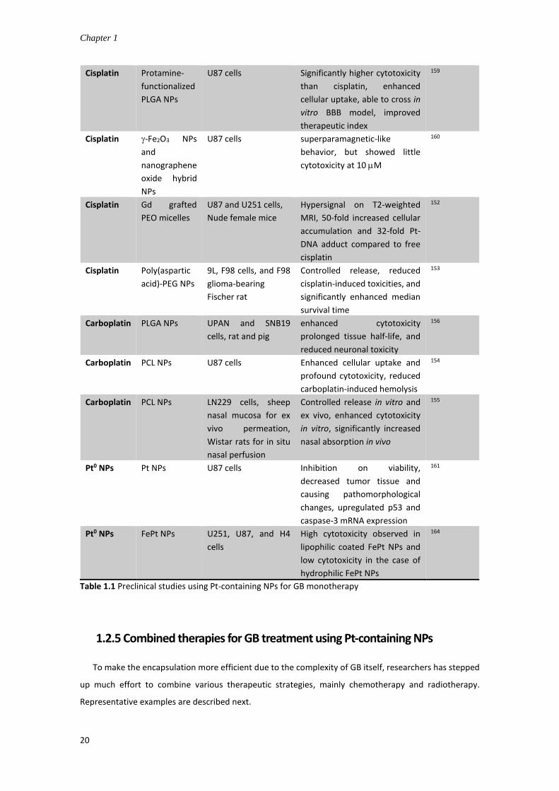
Chapter 1
20
Cisplatin Protamine-
functionalized
PLGA NPs
U87 cells Significantly higher cytotoxicity
than cisplatin, enhanced
cellular uptake, able to cross in
vitro BBB model, improved
therapeutic index
159
Cisplatin -Fe2O3 NPs
and
nanographene
oxide hybrid
NPs
U87 cells superparamagnetic-like
behavior, but showed little
cytotoxicity at 10 M
160
Cisplatin Gd grafted
PEO micelles
U87 and U251 cells,
Nude female mice
Hypersignal on T2-weighted
MRI, 50-fold increased cellular
accumulation and 32-fold Pt-
DNA adduct compared to free
cisplatin
152
Cisplatin Poly(aspartic
acid)-PEG NPs
9L, F98 cells, and F98
glioma-bearing
Fischer rat
Controlled release, reduced
cisplatin-induced toxicities, and
significantly enhanced median
survival time
153
Carboplatin PLGA NPs UPAN and SNB19
cells, rat and pig
enhanced cytotoxicity
prolonged tissue half-life, and
reduced neuronal toxicity
156
Carboplatin PCL NPs U87 cells Enhanced cellular uptake and
profound cytotoxicity, reduced
carboplatin-induced hemolysis
154
Carboplatin PCL NPs LN229 cells, sheep
nasal mucosa for ex
vivo permeation,
Wistar rats for in situ
nasal perfusion
Controlled release in vitro and
ex vivo, enhanced cytotoxicity
in vitro, significantly increased
nasal absorption in vivo
155
Pt0 NPs Pt NPs U87 cells Inhibition on viability,
decreased tumor tissue and
causing pathomorphological
changes, upregulated p53 and
caspase-3 mRNA expression
161
Pt0 NPs FePt NPs U251, U87, and H4
cells
High cytotoxicity observed in
lipophilic coated FePt NPs and
low cytotoxicity in the case of
hydrophilic FePt NPs
164
Table 1.1 Preclinical studies using Pt-containing NPs for GB monotherapy
1.2.5 Combined therapies for GB treatment using Pt-containing NPs
To make the encapsulation more efficient due to the complexity of GB itself, researchers has stepped
up much effort to combine various therapeutic strategies, mainly chemotherapy and radiotherapy.
Representative examples are described next.

General introduction
21
Ruiz etc. developed a novel dendritic silver-platinum NPs which showed selective
anticancer efficacy in glioblastoma and melanoma cell lines but no side effect in
fibroblast cells at lower concentration, and higher bactericidal effect than silver NPs.165
In another example, cisplatin-tethered Au nanospheres (Figure 1.10) sensitized the
patient-derived resistant GB cells, resulting in enhanced synergy between cisplatin and
radiotherapy. As a consequence, significant enhancement in DNA DSB, increased
apoptosis rate, and photo/Auger electron mediated radiosensitization, leading to
complete ablation of the tumor cells in vitro. These results highlighted the potential of
Pt agents to abrogate treatment resistance in GB cells, opening the way for possible Pt-
radiation combined therapies in the future.166
Figure 1.10 Cisplatin-tethered gold NPs combining radiotherapy for glioblastoma treatment.166
Coluccia et al. developed cisplatin conjugated gold NPs combining MR-guided focused
ultrasound as well as targeting peptide to intensify GB treatment. It greatly inhibited the
GB cell growth compared to free cisplatin and showed marked synergy with
radiotherapy in vitro. Furthermore, this system reduced the growth of GB tumors in vivo
and the MR-guided focused ultrasound enhanced BBB permeability and cisplatin
delivery in brain.167 The work led by M. De Waard evaluated the anti-glioma efficacy of
a novel Pt-maurocalcine conjugate and explored its working mechanism. They proved

Chapter 1
22
this conjugate of Pt with cell-penetrating peptide had enhanced anticancer effect than
cisplatin, working by targeting the intracellular redox system, inducing apoptosis
through modulation of PI3K/AKT/FoxO3a signaling pathway.122,168 In another work,
cyclic Arg-Gly-Asp (cRGD) was incorporated into the polymeric micelles loading Pt(II)
complex for targeted delivery into U87 GB (Figure 1.11). cRGD is able to target
αvβ3/αvβ5 integrins, which are overexpressed in angiogenic sites and tumors, as well as
intractable human glioblastoma (U87MG). The results showed rapid and high
accumulation of drug from vessels to tumor parenchyma compared with non-targeted
micelles, suggesting cRGD may target via active internalization pathway.169
Figure 1.11 a) Schematic of cRGD-Pt-micelles (cRGD/m) and description of the mechanism of transport through BBB; b) In vitro cytotoxicities of cRGD-Pt-micelles in the U87MG cells; c) Effects of cRGD-Pt-micelles in orthotopic U87MG glioblastoma in comparison with the non-targeted ligand bearing micelles (cRAD/m).169
Finally, the research group led by Benoit Paquette published a series of papers
concerning the combination of Pt drugs with concomitant radiotherapy, optimizing the
formulation and administration routes to achieve the best therapeutic outcome.133,170-
171 Step by step, they optimized the combinational therapies, from the comparison of
free drugs and liposomal formulations of Pt compounds to the optimization of
General introduction
23
administration routes. Recently, these researchers optimized the combined therapies,
and adopted CED to deliver the Pt drugs or their liposomal formulations, following
radiation treatment. The local delivery considerably increased the drug retention in
tumor compared to intravenous or intra-arterial administration, reduced the side
effects, and resulted in a higher median survival time when combined with radiotherapy.
When tested in F98 glioma-bearing Fischer rats, the liposomal formulations enhanced
the tumor uptake, and improved specificity in vivo.172
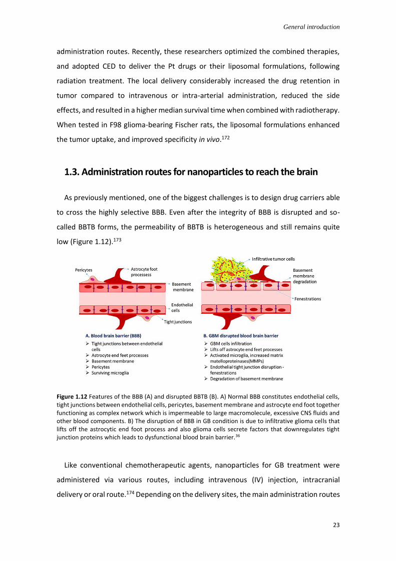
1.3. Administration routes for nanoparticles to reach the brain
As previously mentioned, one of the biggest challenges is to design drug carriers able
to cross the highly selective BBB. Even after the integrity of BBB is disrupted and so-
called BBTB forms, the permeability of BBTB is heterogeneous and still remains quite
low (Figure 1.12).173
Figure 1.12 Features of the BBB (A) and disrupted BBTB (B). A) Normal BBB constitutes endothelial cells, tight junctions between endothelial cells, pericytes, basement membrane and astrocyte end foot together functioning as complex network which is impermeable to large macromolecule, excessive CNS fluids and other blood components. B) The disruption of BBB in GB condition is due to infiltrative glioma cells that lifts off the astrocytic end foot process and also glioma cells secrete factors that downregulates tight junction proteins which leads to dysfunctional blood brain barrier.36
Like conventional chemotherapeutic agents, nanoparticles for GB treatment were
administered via various routes, including intravenous (IV) injection, intracranial
delivery or oral route.174 Depending on the delivery sites, the main administration routes

Chapter 1
24
can be divided into three types for nanoparticles treating brain tumors like GB: 1) direct
delivery to the brain, 2) direct systemic delivery to the brain, and 3) indirect systemic
delivery to the brain.68 In general, the choice of a particular administration route largely
depends on the characteristics of the therapeutic formulations, the required time of
onset of action, the required site of action, among other factors.
• Direct delivery. This type of administration is usually invasive, including
intracerebroventricular administration, intracerebral administration, convection
enhanced delivery (CED), intrathecal administration, etc.175-176 Among them, CED is
an invasive administration way where catheters are placed inside the interstitial
space in parenchyma, drugs driven into the brain by positive pressure gradient using
a pump. CED has been utilized to deliver paclitaxel-loaded lipid nanocapsules directly
into the brain of mice, leading to significantly increased overall survival compared
with mice treated with free paclitaxel.177-178 TMZ-loaded micelles embedded in
hydrogel were injected directly through an incision drilled in the skull of mice, which
was well tolerated and leaded to improved drug release profile.179 However, the
direct delivery to the brain is limited to its invasive nature, the risk of infection, the
need to control critical parameters to avoid brain damage, and difficulty in
intracerebral diffusion.180
• Direct systemic delivery. This approach involves nanoparticles administered directly
into blood stream through carotid artery and transported to the brain avoiding the
rest of the systemic circulation. This administration technique showed improved
survival with reduced risk of brain damage compared to CED.181 For example,
ferrociphekunol-loaded nanoparticles were administered into the brain of GB-
bearing rats using both direct systemic delivery and CED. Direct systemic delivery to
brain exhibited a modest increase in survival compared to CED.180,182
• Indirect systemic delivery. This approach aims to deliver nanoparticles into the
systemic circulation via routes requiring indirect absorption of the brain, including
intravenous (IV) injection or intraperitoneal injection and oral administration. Among
these indirect brain delivery routes, IV administration is most frequently used in
many preclinical studies for GB treatment.183-185 The main advantages of IV injection
set root in its rapid and accurate delivery into the systemic circulation, avoiding first-

General introduction
25
pass metabolism, leading to relatively fast response and high bioavailability. It is
extremely suitable for drugs which cannot absorbed by the gastrointestinal tract or
cannot be injected into muscles or other tissues.174 Similarly, intraperitoneal injection
is commonly used when administering large doses into peritoneal tissues or when it
is difficult to locate a vein for direct systemic delivery, which can avoid anaphylactic
shock after IV injection and was also used in the battle against GB.186 In addition, oral
administration features with convenience, non-invasiveness, and patient
compliance, which has also been applied to the research on nanoparticles for GB
treatment.187 However, the efficiency of these routes of indirect systemic delivery is
highly challenging and considerably compromised due to the RES clearance of
nanoparticles from the systemic circulation and the presence of the BBB blocking the
transport into the brain.
However, in practice, the local delivery augment the therapeutic concentration in
tumor sites while the clinical outcome was still unsatisfactory in treating GB, and the low
intra-brain drug diffusion rate may cause undesired high local toxicity.188 Fortunately,
intranasal (IN) administration has been proposed as a non-invasive alternative route to
enable the nose-to-brain delivery of therapeutic agents and showed promising results
against GB.189 Intranasal route provides the possibility to bypass the BBB and deliver
drugs into the brain directly. The pathways of drugs delivered by IN route from nasal
mucosa to the brain (Figure 1.13) have been extensively researched and reviewed in
recent years. The nasal cavity can be simply divided into the respiratory and the
olfactory regions. In the respiratory region, after the drugs escape from mucociliary
clearance, they can either directly enter the brain through the trigeminal nerve pathway
or enter the systemic circulation. On the other hand, drugs can be transported or diffuse
directly into the brain through the olfactory mucosa pathway in the olfactory region.190
Existing preclinical and clinical studies support the idea that IN delivery of nanosized
therapeutic agents may become a breakthrough in the fight against GB.191
Chapter 1
26
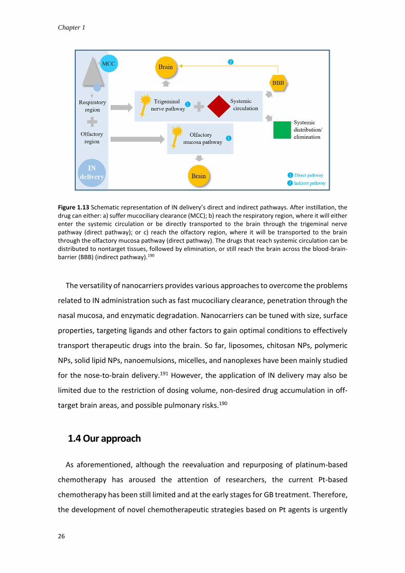
Figure 1.13 Schematic representation of IN delivery’s direct and indirect pathways. After instillation, the drug can either: a) suffer mucociliary clearance (MCC); b) reach the respiratory region, where it will either enter the systemic circulation or be directly transported to the brain through the trigeminal nerve pathway (direct pathway); or c) reach the olfactory region, where it will be transported to the brain through the olfactory mucosa pathway (direct pathway). The drugs that reach systemic circulation can be distributed to nontarget tissues, followed by elimination, or still reach the brain across the blood-brain-barrier (BBB) (indirect pathway).190
The versatility of nanocarriers provides various approaches to overcome the problems
related to IN administration such as fast mucociliary clearance, penetration through the
nasal mucosa, and enzymatic degradation. Nanocarriers can be tuned with size, surface
properties, targeting ligands and other factors to gain optimal conditions to effectively
transport therapeutic drugs into the brain. So far, liposomes, chitosan NPs, polymeric
NPs, solid lipid NPs, nanoemulsions, micelles, and nanoplexes have been mainly studied
for the nose-to-brain delivery.191 However, the application of IN delivery may also be
limited due to the restriction of dosing volume, non-desired drug accumulation in off-
target brain areas, and possible pulmonary risks.190
1.4 Our approach
As aforementioned, although the reevaluation and repurposing of platinum-based
chemotherapy has aroused the attention of researchers, the current Pt-based
chemotherapy has been still limited and at the early stages for GB treatment. Therefore,
the development of novel chemotherapeutic strategies based on Pt agents is urgently

General introduction
27
required to pave the way for possible improvement for the dismayed GB therapy. To
take advantage of Pt(IV) prodrug and nanodrug delivery systems, a novel Pt(IV) prodrug
functionalized with catechol moieties (PtBC) has been synthesized in this thesis, and
used as building block for the fabrication of nanoparticle delivery systems (Figure 1.14)
able to be addressed to the current challenges faced in GB chemotherapy. The chemical
versatility brought by the catechol moiety present in the Pt(IV) prodrug allows different
approaches to form two types of NPs, termed Pt-Fe NCPs obtained by the coordination
of the Pt(IV) prodrug with iron ions (nanostructured coordination polymers) and pPtBC
NPs obtained by chemical polymerization using an oxidizing agent (nanostructured
polymers).
Figure 1.14 Nanodrug delivery systems formed by two different approaches using the Pt(IV) prodrug PtBC.
Both NPs has been evaluated for their potential as GB chemotherapeutic agents.
Specific in vitro and in vivo studies using GB models have been stablished to determine
the potentiality of the developed nanoformulated species for GB therapy. Taken the
unique location and features of GB into consideration, intranasal administration has
been adopted as an alternative to conventional systemic routes in the in vivo studies,
aiming to deliver both NPs into the brain and GB tumors more effectively, thus achieving
optimal responses while reducing side effects.

Chapter 1
28
1.5 References
(1) Ostrom, Q. T.; Cioffi, G.; Gittleman, H.; Patil, N.; Waite, K.; Kruchko, C.; Barnholtz-Sloan, J. S. Cbtrus Statistical Report: Primary Brain and Other Central Nervous System Tumors Diagnosed in the United States in 2012–2016. Neuro Oncol. 2019, 21 (Supplement_5), v1-v100. DOI: 10.1093/neuonc/noz150. (2) Louis, D. N.; Perry, A.; Reifenberger, G.; von Deimling, A.; Figarella-Branger, D.; Cavenee, W. K.; Ohgaki, H.; Wiestler, O. D.; Kleihues, P.; Ellison, D. W. The 2016 World Health Organization Classification of Tumors of the Central Nervous System: A Summary. Acta Neuropathol. 2016, 131 (6), 803-820. DOI: 10.1007/s00401-016-1545-1. (3) Stupp, R.; Mason, W. P.; van den Bent, M. J.; Weller, M.; Fisher, B.; Taphoorn, M. J. B.; Belanger, K.; Brandes, A. A.; Marosi, C.; Bogdahn, U.; Curschmann, J.; Janzer, R. C.; Ludwin, S. K.; Gorlia, T.; Allgeier, A.; Lacombe, D.; Cairncross, J. G.; Eisenhauer, E.; Mirimanoff, R. O.; Van Den Weyngaert, D., et al. Radiotherapy Plus Concomitant and Adjuvant Temozolomide for Glioblastoma. N. Engl. J. Med. 2005, 352 (10), 987-996. DOI: 10.1056/NEJMoa043330. (4) Schiffer, D.; Annovazzi, L.; Casalone, C.; Corona, C.; Mellai, M. Glioblastoma: Microenvironment and Niche Concept. Cancers (Basel) 2018, 11 (1), 5. DOI: 10.3390/cancers11010005. (5) Zhang, X.; Ding, K.; Wang, J.; Li, X.; Zhao, P. Chemoresistance Caused by the Microenvironment of Glioblastoma and the Corresponding Solutions. Biomed. Pharmacother. 2019, 109, 39-46. DOI: 10.1016/j.biopha.2018.10.063. (6) Yadavalli, S.; Yenugonda, V. M.; Kesari, S. Repurposed Drugs in Treating Glioblastoma Multiforme: Clinical Trials Update. Cancer J. 2019, 25 (2), 139-146. DOI: 10.1097/ppo.0000000000000365. (7) Tabatabai, G.; Wakimoto, H. Glioblastoma: State of the Art and Future Perspectives. Cancers (Basel) 2019, 11 (8), 1091. DOI: 10.3390/cancers11081091. (8) Omuro, A.; DeAngelis, L. M. Glioblastoma and Other Malignant Gliomas: A Clinical Review. JAMA 2013, 310 (17), 1842-1850. DOI: 10.1001/jama.2013.280319. (9) Quail, D. F.; Joyce, J. A. The Microenvironmental Landscape of Brain Tumors. Cancer Cell 2017, 31 (3), 326-341. DOI: 10.1016/j.ccell.2017.02.009. (10) Broekman, M. L.; Maas, S. L. N.; Abels, E. R.; Mempel, T. R.; Krichevsky, A. M.; Breakefield, X. O. Multidimensional Communication in the Microenvirons of Glioblastoma. Nat. Rev. Neurol. 2018, 14 (8), 482-495. DOI: 10.1038/s41582-018-0025-8. (11) Matarredona, E. R.; Pastor, A. M. Extracellular Vesicle-Mediated Communication between the Glioblastoma and Its Microenvironment. Cells 2019, 9 (1). DOI: 10.3390/cells9010096. (12) Nieland, L.; Morsett, L. M.; Broekman, M. L. D.; Breakefield, X. O.; Abels, E. R. Extracellular Vesicle-Mediated Bilateral Communication between Glioblastoma and Astrocytes. Trends Neurosci. 2020. DOI: 10.1016/j.tins.2020.10.014. (13) Wolf, K. J.; Chen, J.; Coombes, J. D.; Aghi, M. K.; Kumar, S. Dissecting and Rebuilding the Glioblastoma Microenvironment with Engineered Materials. Nature Reviews Materials 2019, 4 (10), 651-668. DOI: 10.1038/s41578-019-0135-y. (14) Fricker, G.; Ott, M.; Mahringer, A., The Blood Brain Barrier (Bbb). 2014. (15) Wolburg, H.; Noell, S.; Fallier-Becker, P.; Mack, A. F.; Wolburg-Buchholz, K. The Disturbed Blood–Brain Barrier in Human Glioblastoma. Mol. Aspects Med. 2012, 33 (5), 579-589. DOI: 10.1016/j.mam.2012.02.003. (16) Watkins, S.; Robel, S.; Kimbrough, I. F.; Robert, S. M.; Ellis-Davies, G.; Sontheimer, H. Disruption of Astrocyte–Vascular Coupling and the Blood–Brain Barrier by Invading Glioma Cells. Nat. Commun. 2014, 5 (1), 4196. DOI: 10.1038/ncomms5196. (17) Jackson, C. M.; Choi, J.; Lim, M. Mechanisms of Immunotherapy Resistance: Lessons from Glioblastoma. Nat. Immunol. 2019, 20 (9), 1100-1109. DOI: 10.1038/s41590-019-0433-y.

General introduction
29
(18) Balça-Silva, J.; Matias, D.; Carmo, A. d.; Sarmento-Ribeiro, A. B.; Lopes, M. C.; Moura-Neto, V. Cellular and Molecular Mechanisms of Glioblastoma Malignancy: Implications in Resistance and Therapeutic Strategies. Semin. Cancer Biol. 2019, 58, 130-141. DOI: 10.1016/j.semcancer.2018.09.007. (19) Hu, Y.-L.; DeLay, M.; Jahangiri, A.; Molinaro, A. M.; Rose, S. D.; Carbonell, W. S.; Aghi, M. K. Hypoxia-Induced Autophagy Promotes Tumor Cell Survival and Adaptation to Antiangiogenic Treatment in Glioblastoma. Cancer Res. 2012, 72 (7), 1773. DOI: 10.1158/0008-5472.CAN-11-3831. (20) Brandao, M.; Simon, T.; Critchley, G.; Giamas, G. Astrocytes, the Rising Stars of the Glioblastoma Microenvironment. Glia 2019, 67 (5), 779-790. DOI: 10.1002/glia.23520. (21) Han, S. J.; Englot, D. J.; Birk, H.; Molinaro, A. M.; Chang, S. M.; Clarke, J. L.; Prados, M. D.; Taylor, J. W.; Berger, M. S.; Butowski, N. A. Impact of Timing of Concurrent Chemoradiation for Newly Diagnosed Glioblastoma: A Critical Review of Current Evidence. Neurosurgery 2015, 62 (CN_suppl_1), 160-165. DOI: 10.1227/NEU.0000000000000801. (22) Wang, H.; Cai, S.; Ernstberger, A.; Bailey, B. J.; Wang, M. Z.; Cai, W.; Goebel, W. S.; Czader, M. B.; Crean, C.; Suvannasankha, A.; Shokolenkoc, I.; Wilson, G. L.; Baluyut, A. R.; Mayo, L. D.; Pollok, K. E. Temozolomide-Mediated DNA Methylation in Human Myeloid Precursor Cells: Differential Involvement of Intrinsic and Extrinsic Apoptotic Pathways. Clin. Cancer Res. 2013, 19 (10), 2699. DOI: 10.1158/1078-0432.CCR-12-2671. (23) Stupp, R.; Mason, W. P.; van den Bent, M. J.; Weller, M.; Fisher, B.; Taphoorn, M. J. B.; Belanger, K.; Brandes, A. A.; Marosi, C.; Bogdahn, U.; Curschmann, J.; Janzer, R. C.; Ludwin, S. K.; Gorlia, T.; Allgeier, A.; Lacombe, D.; Cairncross, J. G.; Eisenhauer, E.; Mirimanoff, R. O. Radiotherapy Plus Concomitant and Adjuvant Temozolomide for Glioblastoma. N. Engl. J. Med. 2005, 352 (10), 987-996. DOI: 10.1056/NEJMoa043330. (24) Stevens, M. F. G.; Newlands, E. S. From Triazines and Triazenes to Temozolomide. Eur. J. Cancer 1993, 29 (7), 1045-1047. DOI: 10.1016/S0959-8049(05)80221-7. (25) Tatar, Z.; Thivat, E.; Planchat, E.; Gimbergues, P.; Gadea, E.; Abrial, C.; Durando, X. Temozolomide and Unusual Indications: Review of Literature. Cancer Treat. Rev. 2013, 39 (2), 125-135. DOI: 10.1016/j.ctrv.2012.06.002. (26) Newlands, E. S.; Stevens, M. F.; Wedge, S. R.; Wheelhouse, R. T.; Brock, C. Temozolomide: A Review of Its Discovery, Chemical Properties, Pre-Clinical Development and Clinical Trials. Cancer Treat. Rev. 1997, 23 (1), 35-61. DOI: 10.1016/s0305-7372(97)90019-0. (27) Rai, R.; Banerjee, M.; Wong, D. H.; McCullagh, E.; Gupta, A.; Tripathi, S.; Riquelme, E.; Jangir, R.; Yadav, S.; Raja, M.; Melkani, P.; Dixit, V.; Patil, U.; Shrivastava, R.; Middya, S.; Olivares, F.; Guerrero, J.; Surya, A.; Pham, S. M.; Bernales, S., et al. Temozolomide Analogs with Improved Brain/Plasma Ratios - Exploring the Possibility of Enhancing the Therapeutic Index of Temozolomide. Bioorg. Med. Chem. Lett. 2016, 26 (20), 5103-5109. DOI: 10.1016/j.bmcl.2016.08.064. (28) Arora, A.; Somasundaram, K. Glioblastoma Vs Temozolomide: Can the Red Queen Race Be Won? Cancer Biol. Ther. 2019, 20 (8), 1083-1090. DOI: 10.1080/15384047.2019.1599662. (29) Fang, C.; Wang, K.; Stephen, Z. R.; Mu, Q.; Kievit, F. M.; Chiu, D. T.; Press, O. W.; Zhang, M. Temozolomide Nanoparticles for Targeted Glioblastoma Therapy. ACS Appl. Mater. Interfaces 2015, 7 (12), 6674-6682. DOI: 10.1021/am5092165. (30) Patel, A. P.; Tirosh, I.; Trombetta, J. J.; Shalek, A. K.; Gillespie, S. M.; Wakimoto, H.; Cahill, D. P.; Nahed, B. V.; Curry, W. T.; Martuza, R. L.; Louis, D. N.; Rozenblatt-Rosen, O.; Suvà, M. L.; Regev, A.; Bernstein, B. E. Single-Cell Rna-Seq Highlights Intratumoral Heterogeneity in Primary Glioblastoma. Science 2014, 344 (6190), 1396-1401. DOI: 10.1126/science.1254257. (31) Osuka, S.; Van Meir, E. G. Overcoming Therapeutic Resistance in Glioblastoma: The Way Forward. J. Clin. Invest. 2017, 127 (2), 415-426. DOI: 10.1172/jci89587. (32) Hombach-Klonisch, S.; Mehrpour, M.; Shojaei, S.; Harlos, C.; Pitz, M.; Hamai, A.; Siemianowicz, K.; Likus, W.; Wiechec, E.; Toyota, B. D.; Hoshyar, R.; Seyfoori, A.; Sepehri, Z.; Ande, S. R.; Khadem, F.; Akbari, M.; Gorman, A. M.; Samali, A.; Klonisch, T.; Ghavami, S.

Chapter 1
30
Glioblastoma and Chemoresistance to Alkylating Agents: Involvement of Apoptosis, Autophagy, and Unfolded Protein Response. Pharmacol. Ther. 2018, 184, 13-41. DOI: 10.1016/j.pharmthera.2017.10.017. (33) Piccirillo, S. G.; Combi, R.; Cajola, L.; Patrizi, A.; Redaelli, S.; Bentivegna, A.; Baronchelli, S.; Maira, G.; Pollo, B.; Mangiola, A.; DiMeco, F.; Dalprà, L.; Vescovi, A. L. Distinct Pools of Cancer Stem-Like Cells Coexist within Human Glioblastomas and Display Different Tumorigenicity and Independent Genomic Evolution. Oncogene 2009, 28 (15), 1807-1811. DOI: 10.1038/onc.2009.27. (34) Pistollato, F.; Abbadi, S.; Rampazzo, E.; Persano, L.; Della Puppa, A.; Frasson, C.; Sarto, E.; Scienza, R.; D'Avella, D.; Basso, G. Intratumoral Hypoxic Gradient Drives Stem Cells Distribution and Mgmt Expression in Glioblastoma. Stem Cells 2010, 28 (5), 851-862. DOI: 10.1002/stem.415. (35) Dréan, A.; Rosenberg, S.; Lejeune, F. X.; Goli, L.; Nadaradjane, A. A.; Guehennec, J.; Schmitt, C.; Verreault, M.; Bielle, F.; Mokhtari, K.; Sanson, M.; Carpentier, A.; Delattre, J. Y.; Idbaih, A. Atp Binding Cassette (Abc) Transporters: Expression and Clinical Value in Glioblastoma. J. Neurooncol. 2018, 138 (3), 479-486. DOI: 10.1007/s11060-018-2819-3. (36) Ganipineni, L. P.; Danhier, F.; Préat, V. Drug Delivery Challenges and Future of Chemotherapeutic Nanomedicine for Glioblastoma Treatment. J. Control. Release 2018, 281, 42-57. DOI: 10.1016/j.jconrel.2018.05.008. (37) Li, M.; Luo, Z.; Xia, Z.; Shen, X.; Cai, K. Time-Sequenced Drug Delivery Approaches Towards Effective Chemotherapeutic Treatment of Glioma. Materials Horizons 2017, 4 (6), 977-996. DOI: 10.1039/C7MH00342K. (38) Le Rhun, E.; Preusser, M.; Roth, P.; Reardon, D. A.; van den Bent, M.; Wen, P.; Reifenberger, G.; Weller, M. Molecular Targeted Therapy of Glioblastoma. Cancer Treat. Rev. 2019, 80, 101896. DOI: 10.1016/j.ctrv.2019.101896. (39) Lu, C. T.; Zhao, Y. Z.; Wong, H. L.; Cai, J.; Peng, L.; Tian, X. Q. Current Approaches to Enhance Cns Delivery of Drugs across the Brain Barriers. Int J Nanomedicine 2014, 9, 2241-2257. DOI: 10.2147/ijn.s61288. (40) Liu, D. Z.; Cheng, Y.; Cai, R. Q.; Wang Bd, W. W.; Cui, H.; Liu, M.; Zhang, B. L.; Mei, Q. B.; Zhou, S. Y. The Enhancement of Siplk1 Penetration across Bbb and Its Anti Glioblastoma Activity in Vivo by Magnet and Transferrin Co-Modified Nanoparticle. Nanomedicine 2018, 14 (3), 991-1003. DOI: 10.1016/j.nano.2018.01.004. (41) Maeda, H. The Enhanced Permeability and Retention (Epr) Effect in Tumor Vasculature: The Key Role of Tumor-Selective Macromolecular Drug Targeting. Adv. Enzyme Regul. 2001, 41, 189-207. DOI: 10.1016/s0065-2571(00)00013-3. (42) Bhowmik, A.; Khan, R.; Ghosh, M. K. Blood Brain Barrier: A Challenge for Effectual Therapy of Brain Tumors. BioMed Research International 2015, 2015, 320941. DOI: 10.1155/2015/320941. (43) Attia, M. F.; Anton, N.; Wallyn, J.; Omran, Z.; Vandamme, T. F. An Overview of Active and Passive Targeting Strategies to Improve the Nanocarriers Efficiency to Tumour Sites. J. Pharm. Pharmacol. 2019, 71 (8), 1185-1198. DOI: 10.1111/jphp.13098. (44) Sarkaria, J. N.; Hu, L. S.; Parney, I. F.; Pafundi, D. H.; Brinkmann, D. H.; Laack, N. N.; Giannini, C.; Burns, T. C.; Kizilbash, S. H.; Laramy, J. K.; Swanson, K. R.; Kaufmann, T. J.; Brown, P. D.; Agar, N. Y. R.; Galanis, E.; Buckner, J. C.; Elmquist, W. F. Is the Blood-Brain Barrier Really Disrupted in All Glioblastomas? A Critical Assessment of Existing Clinical Data. Neuro Oncol. 2018, 20 (2), 184-191. DOI: 10.1093/neuonc/nox175. (45) Zhao, M.; van Straten, D.; Broekman, M. L. D.; Préat, V.; Schiffelers, R. M. Nanocarrier-Based Drug Combination Therapy for Glioblastoma. Theranostics 2020, 10 (3), 1355-1372. DOI: 10.7150/thno.38147. (46) Bamrungsap, S.; Zhao, Z.; Chen, T.; Wang, L.; Li, C.; Fu, T.; Tan, W. Nanotechnology in Therapeutics: A Focus on Nanoparticles as a Drug Delivery System. Nanomedicine (London, England) 2012, 7 (8), 1253-1271. DOI: 10.2217/nnm.12.87.
General introduction
31
(47) Wu, X.; Yang, H.; Yang, W.; Chen, X.; Gao, J.; Gong, X.; Wang, H.; Duan, Y.; Wei, D.; Chang, J. Nanoparticle-Based Diagnostic and Therapeutic Systems for Brain Tumors. Journal of Materials Chemistry B 2019, 7 (31), 4734-4750. DOI: 10.1039/C9TB00860H. (48) Yang, J.; Shi, Z.; Liu, R.; Wu, Y.; Zhang, X. Combined-Therapeutic Strategies Synergistically Potentiate Glioblastoma Multiforme Treatment <I>Via</I> Nanotechnology. Theranostics 2020, 10 (7), 3223-3239. DOI: 10.7150/thno.40298. (49) Havel, H. A. Where Are the Nanodrugs? An Industry Perspective on Development of Drug Products Containing Nanomaterials. The AAPS journal 2016, 18 (6), 1351-1353. DOI: 10.1208/s12248-016-9970-6. (50) Rehman, M.; Madni, A.; Shi, D.; Ihsan, A.; Tahir, N.; Chang, K. R.; Javed, I.; Webster, T. J. Enhanced Blood Brain Barrier Permeability and Glioblastoma Cell Targeting Via Thermoresponsive Lipid Nanoparticles. Nanoscale 2017, 9 (40), 15434-15440. DOI: 10.1039/c7nr05216b. (51) Ying, X.; Wang, Y.; Xu, H.; Li, X.; Yan, H.; Tang, H.; Wen, C.; Li, Y. The Construction of the Multifunctional Targeting Ursolic Acids Liposomes and Its Apoptosis Effects to C6 Glioma Stem Cells. Oncotarget 2017, 8 (38), 64129-64142. DOI: 10.18632/oncotarget.19784. (52) Singleton, W. G.; Collins, A. M.; Bienemann, A. S.; Killick-Cole, C. L.; Haynes, H. R.; Asby, D. J.; Butts, C. P.; Wyatt, M. J.; Barua, N. U.; Gill, S. S. Convection Enhanced Delivery of Panobinostat (Lbh589)-Loaded Pluronic Nano-Micelles Prolongs Survival in the F98 Rat Glioma Model. Int J Nanomedicine 2017, 12, 1385-1399. DOI: 10.2147/ijn.s125300. (53) Lin, T.; Zhao, P.; Jiang, Y.; Tang, Y.; Jin, H.; Pan, Z.; He, H.; Yang, V. C.; Huang, Y. Blood-Brain-Barrier-Penetrating Albumin Nanoparticles for Biomimetic Drug Delivery Via Albumin-Binding Protein Pathways for Antiglioma Therapy. ACS Nano 2016, 10 (11), 9999-10012. DOI: 10.1021/acsnano.6b04268. (54) Hu, J.; Zhang, X.; Wen, Z.; Tan, Y.; Huang, N.; Cheng, S.; Zheng, H.; Cheng, Y. Asn-Gly-Arg-Modified Polydopamine-Coated Nanoparticles for Dual-Targeting Therapy of Brain Glioma in Rats. Oncotarget 2016, 7 (45), 73681-73696. DOI: 10.18632/oncotarget.12047. (55) Shen, Z.; Liu, T.; Li, Y.; Lau, J.; Yang, Z.; Fan, W.; Zhou, Z.; Shi, C.; Ke, C.; Bregadze, V. I.; Mandal, S. K.; Liu, Y.; Li, Z.; Xue, T.; Zhu, G.; Munasinghe, J.; Niu, G.; Wu, A.; Chen, X. Fenton-Reaction-Acceleratable Magnetic Nanoparticles for Ferroptosis Therapy of Orthotopic Brain Tumors. ACS Nano 2018, 12 (11), 11355-11365. DOI: 10.1021/acsnano.8b06201. (56) Lu, Y.; Han, S.; Zheng, H.; Ma, R.; Ping, Y.; Zou, J.; Tang, H.; Zhang, Y.; Xu, X.; Li, F. A Novel Rgdyc/Peg Co-Modified Pamam Dendrimer-Loaded Arsenic Trioxide of Glioma Targeting Delivery System. Int J Nanomedicine 2018, 13, 5937-5952. DOI: 10.2147/IJN.S175418. (57) Qian, M.; Du, Y.; Wang, S.; Li, C.; Jiang, H.; Shi, W.; Chen, J.; Wang, Y.; Wagner, E.; Huang, R. Highly Crystalline Multicolor Carbon Nanodots for Dual-Modal Imaging-Guided Photothermal Therapy of Glioma. ACS Appl. Mater. Interfaces 2018, 10 (4), 4031-4040. DOI: 10.1021/acsami.7b19716. (58) Etame, A. B.; Smith, C. A.; Chan, W. C.; Rutka, J. T. Design and Potential Application of Pegylated Gold Nanoparticles with Size-Dependent Permeation through Brain Microvasculature. Nanomedicine 2011, 7 (6), 992-1000. DOI: 10.1016/j.nano.2011.04.004. (59) Shilo, M.; Sharon, A.; Baranes, K.; Motiei, M.; Lellouche, J. P.; Popovtzer, R. The Effect of Nanoparticle Size on the Probability to Cross the Blood-Brain Barrier: An in-Vitro Endothelial Cell Model. Journal of nanobiotechnology 2015, 13, 19. DOI: 10.1186/s12951-015-0075-7. (60) Hanada, S.; Fujioka, K.; Inoue, Y.; Kanaya, F.; Manome, Y.; Yamamoto, K. Cell-Based in Vitro Blood-Brain Barrier Model Can Rapidly Evaluate Nanoparticles' Brain Permeability in Association with Particle Size and Surface Modification. Int J Mol Sci 2014, 15 (2), 1812-1825. DOI: 10.3390/ijms15021812. (61) Lockman, P. R.; Koziara, J. M.; Mumper, R. J.; Allen, D. D. Nanoparticle Surface Charges Alter Blood-Brain Barrier Integrity and Permeability. J. Drug Target. 2004, 12 (9-10), 635-641. DOI: 10.1080/10611860400015936.

Chapter 1
32
(62) Jallouli, Y.; Paillard, A.; Chang, J.; Sevin, E.; Betbeder, D. Influence of Surface Charge and Inner Composition of Porous Nanoparticles to Cross Blood-Brain Barrier in Vitro. Int. J. Pharm. 2007, 344 (1-2), 103-109. DOI: 10.1016/j.ijpharm.2007.06.023. (63) Sonavane, G.; Tomoda, K.; Makino, K. Biodistribution of Colloidal Gold Nanoparticles after Intravenous Administration: Effect of Particle Size. Colloids Surf. B. Biointerfaces 2008, 66 (2), 274-280. DOI: 10.1016/j.colsurfb.2008.07.004. (64) Kolhar, P.; Anselmo, A. C.; Gupta, V.; Pant, K.; Prabhakarpandian, B.; Ruoslahti, E.; Mitragotri, S. Using Shape Effects to Target Antibody-Coated Nanoparticles to Lung and Brain Endothelium. Proceedings of the National Academy of Sciences 2013, 110 (26), 10753. DOI: 10.1073/pnas.1308345110. (65) Huang, X.; Li, L.; Liu, T.; Hao, N.; Liu, H.; Chen, D.; Tang, F. The Shape Effect of Mesoporous Silica Nanoparticles on Biodistribution, Clearance, and Biocompatibility in Vivo. ACS Nano 2011, 5 (7), 5390-5399. DOI: 10.1021/nn200365a. (66) Zhan, C.; Gu, B.; Xie, C.; Li, J.; Liu, Y.; Lu, W. Cyclic Rgd Conjugated Poly(Ethylene Glycol)-Co-Poly(Lactic Acid) Micelle Enhances Paclitaxel Anti-Glioblastoma Effect. J. Control. Release 2010, 143 (1), 136-142. DOI: 10.1016/j.jconrel.2009.12.020. (67) Zhou, J.-e.; Yu, J.; Gao, L.; Sun, L.; Peng, T.; Wang, J.; Zhu, J.; Lu, W.; Zhang, L.; Yan, Z.; Yu, L. Ingr-Modified Liposomes for Tumor Vascular Targeting and Tumor Tissue Penetrating Delivery in the Treatment of Glioblastoma. Mol. Pharm. 2017, 14 (5), 1811-1820. DOI: 10.1021/acs.molpharmaceut.7b00101. (68) Mahmoud, B. S.; AlAmri, A. H.; McConville, C. Polymeric Nanoparticles for the Treatment of Malignant Gliomas. Cancers (Basel) 2020, 12 (1), 175. DOI: 10.3390/cancers12010175. (69) Ortiz, R.; Cabeza, L.; Perazzoli, G.; Jimenez-Lopez, J.; García-Pinel, B.; Melguizo, C.; Prados, J. Nanoformulations for Glioblastoma Multiforme: A New Hope for Treatment. Future Med. Chem. 2019, 11 (18), 2459-2480. DOI: 10.4155/fmc-2018-0521. (70) Mc Carthy, D. J.; Malhotra, M.; O’Mahony, A. M.; Cryan, J. F.; O’Driscoll, C. M. Nanoparticles and the Blood-Brain Barrier: Advancing from in-Vitro Models Towards Therapeutic Significance. Pharm. Res. 2015, 32 (4), 1161-1185. DOI: 10.1007/s11095-014-1545-6. (71) Zhou, J.; Patel, T. R.; Sirianni, R. W.; Strohbehn, G.; Zheng, M.-Q.; Duong, N.; Schafbauer, T.; Huttner, A. J.; Huang, Y.; Carson, R. E.; Zhang, Y.; Sullivan, D. J., Jr.; Piepmeier, J. M.; Saltzman, W. M. Highly Penetrative, Drug-Loaded Nanocarriers Improve Treatment of Glioblastoma. Proc. Natl. Acad. Sci. U. S. A. 2013, 110 (29), 11751-11756. DOI: 10.1073/pnas.1304504110. (72) Li, Y.; Wu, M.; Zhang, N.; Tang, C.; Jiang, P.; Liu, X.; Yan, F.; Zheng, H. Mechanisms of Enhanced Antiglioma Efficacy of Polysorbate 80-Modified Paclitaxel-Loaded Plga Nanoparticles by Focused Ultrasound. J. Cell. Mol. Med. 2018, 22 (9), 4171-4182. DOI: 10.1111/jcmm.13695. (73) Inoue, T.; Yamashita, Y.; Nishihara, M.; Sugiyama, S.; Sonoda, Y.; Kumabe, T.; Yokoyama, M.; Tominaga, T. Therapeutic Efficacy of a Polymeric Micellar Doxorubicin Infused by Convection-Enhanced Delivery against Intracranial 9l Brain Tumor Models. Neuro Oncol. 2009, 11 (2), 151-157. DOI: 10.1215/15228517-2008-068. (74) Noble, C. O.; Krauze, M. T.; Drummond, D. C.; Yamashita, Y.; Saito, R.; Berger, M. S.; Kirpotin, D. B.; Bankiewicz, K. S.; Park, J. W. Novel Nanoliposomal Cpt-11 Infused by Convection-Enhanced Delivery in Intracranial Tumors: Pharmacology and Efficacy. Cancer Res. 2006, 66 (5), 2801-2806. DOI: 10.1158/0008-5472.can-05-3535. (75) Chekhonin, V. P.; Baklaushev, V. P.; Yusubalieva, G. M.; Belorusova, A. E.; Gulyaev, M. V.; Tsitrin, E. B.; Grinenko, N. F.; Gurina, O. I.; Pirogov, Y. A. Targeted Delivery of Liposomal Nanocontainers to the Peritumoral Zone of Glioma by Means of Monoclonal Antibodies against Gfap and the Extracellular Loop of Cx43. Nanomedicine 2012, 8 (1), 63-70. DOI: 10.1016/j.nano.2011.05.011. (76) Kim, S. S.; Rait, A.; Kim, E.; DeMarco, J.; Pirollo, K. F.; Chang, E. H. Encapsulation of Temozolomide in a Tumor-Targeting Nanocomplex Enhances Anti-Cancer Efficacy and Reduces Toxicity in a Mouse Model of Glioblastoma. Cancer Lett. 2015, 369 (1), 250-258. DOI: 10.1016/j.canlet.2015.08.022.

General introduction
33
(77) Cheng, Y.; Dai, Q.; Morshed, R. A.; Fan, X.; Wegscheid, M. L.; Wainwright, D. A.; Han, Y.; Zhang, L.; Auffinger, B.; Tobias, A. L.; Rincón, E.; Thaci, B.; Ahmed, A. U.; Warnke, P. C.; He, C.; Lesniak, M. S. Blood-Brain Barrier Permeable Gold Nanoparticles: An Efficient Delivery Platform for Enhanced Malignant Glioma Therapy and Imaging. Small 2014, 10 (24), 5137-5150. DOI: 10.1002/smll.201400654. (78) Kievit, F. M.; Wang, K.; Ozawa, T.; Tarudji, A. W.; Silber, J. R.; Holland, E. C.; Ellenbogen, R. G.; Zhang, M. Nanoparticle-Mediated Knockdown of DNA Repair Sensitizes Cells to Radiotherapy and Extends Survival in a Genetic Mouse Model of Glioblastoma. Nanomedicine 2017, 13 (7), 2131-2139. DOI: 10.1016/j.nano.2017.06.004. (79) Mu, Q.; Lin, G.; Patton, V. K.; Wang, K.; Press, O. W.; Zhang, M. Gemcitabine and Chlorotoxin Conjugated Iron Oxide Nanoparticles for Glioblastoma Therapy. Journal of materials chemistry. B 2016, 4 (1), 32-36. DOI: 10.1039/c5tb02123e. (80) Deshane, J.; Garner, C. C.; Sontheimer, H. Chlorotoxin Inhibits Glioma Cell Invasion Via Matrix Metalloproteinase-2. J. Biol. Chem. 2003, 278 (6), 4135-4144. DOI: 10.1074/jbc.M205662200. (81) Lee, B. S.; Amano, T.; Wang, H. Q.; Pantoja, J. L.; Yoon, C. W.; Hanson, C. J.; Amatya, R.; Yen, A.; Black, K. L.; Yu, J. S. Reactive Oxygen Species Responsive Nanoprodrug to Treat Intracranial Glioblastoma. ACS Nano 2013, 7 (4), 3061-3077. DOI: 10.1021/nn400347j. (82) Peyrone, M. Ueber Die Einwirkung Des Ammoniaks Auf Platinchlorür. Justus Liebigs Ann. Chem. 1844, 51 (1), 1-29. DOI: 10.1002/jlac.18440510102. (83) Rosenberg, B.; Vancamp, L.; Krigas, T. Inhibition of Cell Division in Escherichia Coli by Electrolysis Products from a Platinum Eletrode Nature 1965, 205, 698-699. DOI: 10.1038/205698a0. (84) Rosenberg, B.; Vancamp, L.; Trosko, J. E.; Mansour, V. H. Platinum Compounds: A New Class of Potent Antitumour Agents. Nature 1969, 222 (5191), 385-386. DOI: 10.1038/222385a0. (85) Wiltshaw, E. Cisplatin in the Treatment of Cancer. Platinum Met. Rev. 1979, 23 (3), 90-98. DOI: N/A. (86) Browning, R. J.; Reardon, P. J. T.; Parhizkar, M.; Pedley, R. B.; Edirisinghe, M.; Knowles, J. C.; Stride, E. Drug Delivery Strategies for Platinum-Based Chemotherapy. ACS Nano 2017, 11 (9), 8560-8578. DOI: 10.1021/acsnano.7b04092. (87) Johnstone, T. C.; Suntharalingam, K.; Lippard, S. J. The Next Generation of Platinum Drugs: Targeted Pt(Ii) Agents, Nanoparticle Delivery, and Pt(Iv) Prodrugs. Chem. Rev. 2016, 116 (5), 3436-3486. DOI: 10.1021/acs.chemrev.5b00597. (88) Dilruba, S.; Kalayda, G. V. Platinum-Based Drugs: Past, Present and Future. Cancer Chemother. Pharmacol. 2016, 77 (6), 1103-1124. DOI: 10.1007/s00280-016-2976-z. (89) Galanski, M. Recent Developments in the Field of Anticancer Platinum Complexes. Recent Pat. Anticancer Drug Discov. 2006, 1 (2), 285-295. DOI: 10.2174/157489206777442287. (90) Who Model List of Essential Medicines; World Health Organization: 2019. (91) Centers for Disease Control and Prevention Ambulatory Care Drug Database System. http://www.cdc.gov/nchs/ahcd/ahcd_database.htm (accessed: 2015). (92) World Health Organization. International Clinical Trials Registry Platform (Ictrp). http://www.who.int/ictrp/en/ (accessed: 2020). (93) National Institutes of Health. Clinical Trials Database. http://www.clinicaltrials.gov/ (accessed: 2020). (94) Haug, C.; Gøtzsche, P. C.; Schroeder, T. V. Registries and Registration of Clinical Trials. N. Engl. J. Med. 2005, 353 (26), 2811-2812. DOI: 10.1056/NEJMe058280. (95) Oberoi, H. S.; Nukolova, N. V.; Kabanov, A. V.; Bronich, T. K. Nanocarriers for Delivery of Platinum Anticancer Drugs. Adv Drug Deliv Rev 2013, 65 (13-14), 1667-1685. DOI: 10.1016/j.addr.2013.09.014. (96) Xiao, H.; Yan, L.; Dempsey, E. M.; Song, W.; Qi, R.; Li, W.; Huang, Y.; Jing, X.; Zhou, D.; Ding, J.; Chen, X. Recent Progress in Polymer-Based Platinum Drug Delivery Systems. Prog. Polym. Sci. 2018, 87, 70-106. DOI: 10.1016/j.progpolymsci.2018.07.004.
Chapter 1
34
(97) Gately, D. P.; Howell, S. B. Cellular Accumulation of the Anticancer Agent Cisplatin: A Review. Br. J. Cancer 1993, 67 (6), 1171-1176. DOI: 10.1038/bjc.1993.221. (98) Howell, S. B.; Safaei, R.; Larson, C. A.; Sailor, M. J. Copper Transporters and the Cellular Pharmacology of the Platinum-Containing Cancer Drugs. Mol. Pharmacol. 2010, 77 (6), 887-894. DOI: 10.1124/mol.109.063172. (99) Yonezawa, A.; Masuda, S.; Yokoo, S.; Katsura, T.; Inui, K. Cisplatin and Oxaliplatin, but Not Carboplatin and Nedaplatin, Are Substrates for Human Organic Cation Transporters (Slc22a1-3 and Multidrug and Toxin Extrusion Family). J. Pharmacol. Exp. Ther. 2006, 319 (2), 879-886. DOI: 10.1124/jpet.106.110346. (100) Fichtinger-Schepman, A. M.; van der Veer, J. L.; den Hartog, J. H.; Lohman, P. H.; Reedijk, J. Adducts of the Antitumor Drug Cis-Diamminedichloroplatinum(Ii) with DNA: Formation, Identification, and Quantitation. Biochemistry 1985, 24 (3), 707-713. DOI: 10.1021/bi00324a025. (101) Wang, D.; Lippard, S. J. Cellular Processing of Platinum Anticancer Drugs. Nat. Rev. Drug Discov. 2005, 4 (4), 307-320. DOI: 10.1038/nrd1691. (102) Chapman, E. G.; DeRose, V. J. Enzymatic Processing of Platinated Rnas. J. Am. Chem. Soc. 2010, 132 (6), 1946-1952. DOI: 10.1021/ja908419j. (103) Akaboshi, M.; Kawai, K.; Maki, H.; Akuta, K.; Ujeno, Y.; Miyahara, T. The Number of Platinum Atoms Binding to DNA, Rna and Protein Molecules of Hela Cells Treated with Cisplatin at Its Mean Lethal Concentration. Jpn. J. Cancer Res. 1992, 83 (5), 522-526. DOI: 10.1111/j.1349-7006.1992.tb01959.x. (104) Tesniere, A.; Schlemmer, F.; Boige, V.; Kepp, O.; Martins, I.; Ghiringhelli, F.; Aymeric, L.; Michaud, M.; Apetoh, L.; Barault, L.; Mendiboure, J.; Pignon, J. P.; Jooste, V.; van Endert, P.; Ducreux, M.; Zitvogel, L.; Piard, F.; Kroemer, G. Immunogenic Death of Colon Cancer Cells Treated with Oxaliplatin. Oncogene 2010, 29 (4), 482-491. DOI: 10.1038/onc.2009.356. (105) Hato, S. V.; Khong, A.; de Vries, I. J.; Lesterhuis, W. J. Molecular Pathways: The Immunogenic Effects of Platinum-Based Chemotherapeutics. Clin. Cancer Res. 2014, 20 (11), 2831-2837. DOI: 10.1158/1078-0432.ccr-13-3141. (106) Roberts, N. B.; Wadajkar, A. S.; Winkles, J. A.; Davila, E.; Kim, A. J.; Woodworth, G. F. Repurposing Platinum-Based Chemotherapies for Multi-Modal Treatment of Glioblastoma. Oncoimmunology 2016, 5 (9), e1208876. DOI: 10.1080/2162402x.2016.1208876. (107) Yung, W. K.; Mechtler, L.; Gleason, M. J. Intravenous Carboplatin for Recurrent Malignant Glioma: A Phase Ii Study. J. Clin. Oncol. 1991, 9 (5), 860-864. DOI: 10.1200/jco.1991.9.5.860. (108) Piccioni, D.; D'Atri, S.; Papa, G.; Caravita, T.; Franchi, A.; Bonmassar, E.; Graziani, G. Cisplatin Increases Sensitivity of Human Leukemic Blasts to Triazene Compounds. J. Chemother. 1995, 7 (3), 224-229. DOI: 10.1179/joc.1995.7.3.224. (109) Britten, C. D.; Rowinsky, E. K.; Baker, S. D.; Agarwala, S. S.; Eckardt, J. R.; Barrington, R.; Diab, S. G.; Hammond, L. A.; Johnson, T.; Villalona-Calero, M.; Fraass, U.; Statkevich, P.; Von Hoff, D. D.; Eckhardt, S. G. A Phase I and Pharmacokinetic Study of Temozolomide and Cisplatin in Patients with Advanced Solid Malignancies. Clin. Cancer Res. 1999, 5 (7), 1629-1637. DOI: N/A. (110) Silvani, A.; Eoli, M.; Salmaggi, A.; Lamperti, E.; Maccagnano, E.; Broggi, G.; Boiardi, A. Phase Ii Trial of Cisplatin Plus Temozolomide, in Recurrent and Progressive Malignant Glioma Patients. J. Neurooncol. 2004, 66 (1-2), 203-208. DOI: 10.1023/b:neon.0000013479.64348.69. (111) Mishima, K.; Mazar, A. P.; Gown, A.; Skelly, M.; Ji, X. D.; Wang, X. D.; Jones, T. R.; Cavenee, W. K.; Huang, H. J. A Peptide Derived from the Non-Receptor-Binding Region of Urokinase Plasminogen Activator Inhibits Glioblastoma Growth and Angiogenesis in Vivo in Combination with Cisplatin. Proc. Natl. Acad. Sci. U. S. A. 2000, 97 (15), 8484-8489. DOI: 10.1073/pnas.150239497. (112) Nagane, M.; Narita, Y.; Mishima, K.; Levitzki, A.; Burgess, A. W.; Cavenee, W. K.; Huang, H. J. Human Glioblastoma Xenografts Overexpressing a Tumor-Specific Mutant Epidermal Growth Factor Receptor Sensitized to Cisplatin by the Ag1478 Tyrosine Kinase Inhibitor. J. Neurosurg. 2001, 95 (3), 472-479. DOI: 10.3171/jns.2001.95.3.0472.
General introduction
35
(113) Hess, S. M.; Anderson, J. G.; Bierbach, U. A Non-Crosslinking Platinum-Acridine Hybrid Agent Shows Enhanced Cytotoxicity Compared to Clinical Bcnu and Cisplatin in Glioblastoma Cells. Bioorg. Med. Chem. Lett. 2005, 15 (2), 443-446. DOI: 10.1016/j.bmcl.2004.10.049. (114) Posadas, I.; Alonso-Moreno, C.; Bravo, I.; Carrillo-Hermosilla, F.; Garzon, A.; Villaseca, N.; Lopez-Solera, I.; Albaladejo, J.; Cena, V. Synthesis, Characterization, DNA Interactions and Antiproliferative Activity on Glioblastoma of Iminopyridine Platinum(Ii) Chelate Complexes. J. Inorg. Biochem. 2017, 168, 46-54. DOI: 10.1016/j.jinorgbio.2016.11.032. (115) Benzina, S.; Fischer, B.; Miternique-Grosse, A.; Dufour, P.; Denis, J. M.; Bergerat, J. P.; Gueulette, J.; Bischoff, P. Cell Death Induced in a Human Glioblastoma Cell Line by P(65)+Be Neutrons Combined with Cisplatin. Life Sci. 2006, 79 (6), 513-518. DOI: 10.1016/j.lfs.2006.01.037. (116) Brandes, A. A.; Basso, U.; Reni, M.; Vastola, F.; Tosoni, A.; Cavallo, G.; Scopece, L.; Ferreri, A. J.; Panucci, M. G.; Monfardini, S.; Ermani, M. First-Line Chemotherapy with Cisplatin Plus Fractionated Temozolomide in Recurrent Glioblastoma Multiforme: A Phase Ii Study of the Gruppo Italiano Cooperativo Di Neuro-Oncologia. J. Clin. Oncol. 2004, 22 (9), 1598-1604. DOI: 10.1200/jco.2004.11.019. (117) Wang, Y.; Kong, X.; Guo, Y.; Wang, R.; Ma, W. Continuous Dose-Intense Temozolomide and Cisplatin in Recurrent Glioblastoma Patients. Medicine (Baltimore) 2017, 96 (10), e6261. DOI: 10.1097/md.0000000000006261. (118) Gajski, G.; Čimbora-Zovko, T.; Rak, S.; Osmak, M.; Garaj-Vrhovac, V. Antitumour Action on Human Glioblastoma A1235 Cells through Cooperation of Bee Venom and Cisplatin. Cytotechnology 2016, 68 (4), 1197-1205. DOI: 10.1007/s10616-015-9879-4. (119) Zhu, T.; Xu, Y.; Dong, B.; Zhang, J.; Wei, Z.; Xu, Y.; Yao, Y. Β-Elemene Inhibits Proliferation of Human Glioblastoma Cells through the Activation of Glia Maturation Factor Β and Induces Sensitization to Cisplatin. Oncol. Rep. 2011, 26 (2), 405-413. DOI: 10.3892/or.2011.1276. (120) Park, C. K.; Park, S. H.; Lee, S. H.; Kim, C. Y.; Kim, D. W.; Paek, S. H.; Kim, D. G.; Heo, D. S.; Kim, I. H.; Jung, H. W. Methylation Status of the Mgmt Gene Promoter Fails to Predict the Clinical Outcome of Glioblastoma Patients Treated with Acnu Plus Cisplatin. Neuropathology 2009, 29 (4), 443-449. DOI: 10.1111/j.1440-1789.2008.00998.x. (121) Buckner, J. C.; Ballman, K. V.; Michalak, J. C.; Burton, G. V.; Cascino, T. L.; Schomberg, P. J.; Hawkins, R. B.; Scheithauer, B. W.; Sandler, H. M.; Marks, R. S.; O'Fallon, J. R. Phase Iii Trial of Carmustine and Cisplatin Compared with Carmustine Alone and Standard Radiation Therapy or Accelerated Radiation Therapy in Patients with Glioblastoma Multiforme: North Central Cancer Treatment Group 93-72-52 and Southwest Oncology Group 9503 Trials. J. Clin. Oncol. 2006, 24 (24), 3871-3879. DOI: 10.1200/jco.2005.04.6979. (122) Aroui, S.; Dardevet, L.; Ben Ajmia, W.; de Boisvilliers, M.; Perrin, F.; Laajimi, A.; Boumendjel, A.; Kenani, A.; Muller, J. M.; De Waard, M. A Novel Platinum-Maurocalcine Conjugate Induces Apoptosis of Human Glioblastoma Cells by Acting through the Ros-Erk/Akt-P53 Pathway. Mol. Pharm. 2015, 12 (12), 4336-4348. DOI: 10.1021/acs.molpharmaceut.5b00531. (123) Ferrari, B.; Urselli, F.; Gilodi, M.; Camuso, S.; Priori, E. C.; Rangone, B.; Ravera, M.; Veneroni, P.; Zanellato, I.; Roda, E.; Osella, D.; Bottone, M. G. New Platinum-Based Prodrug Pt(Iv)Ac-Poa: Antitumour Effects in Rat C6 Glioblastoma Cells. Neurotox. Res. 2020, 37 (1), 183-197. DOI: 10.1007/s12640-019-00076-0. (124) Macieja, A.; Kopa, P.; Galita, G.; Pastwa, E.; Majsterek, I.; Poplawski, T. Comparison of the Effect of Three Different Topoisomerase Ii Inhibitors Combined with Cisplatin in Human Glioblastoma Cells Sensitized with Double Strand Break Repair Inhibitors. Mol. Biol. Rep. 2019, 46 (4), 3625-3636. DOI: 10.1007/s11033-019-04605-0. (125) Carminati, P. O.; Donaires, F. S.; Marques, M. M.; Donadi, E. A.; Passos, G. A. S.; Sakamoto-Hojo, E. T. Cisplatin Associated with Ly294002 Increases Cytotoxicity and Induces Changes in Transcript Profiles of Glioblastoma Cells. Mol. Biol. Rep. 2014, 41 (1), 165-177. DOI: 10.1007/s11033-013-2849-z.

Chapter 1
36
(126) Gwak, H.-S.; Shingu, T.; Chumbalkar, V.; Hwang, Y.-H.; DeJournett, R.; Latha, K.; Koul, D.; Alfred Yung, W. K.; Powis, G.; Farrell, N. P.; Bögler, O. Combined Action of the Dinuclear Platinum Compound Bbr3610 with the Pi3-K Inhibitor Px-866 in Glioblastoma. Int. J. Cancer 2011, 128 (4), 787-796. DOI: 10.1002/ijc.25394. (127) Rosa, P.; Catacuzzeno, L.; Sforna, L.; Mangino, G.; Carlomagno, S.; Mincione, G.; Petrozza, V.; Ragona, G.; Franciolini, F.; Calogero, A. Bk Channels Blockage Inhibits Hypoxia-Induced Migration and Chemoresistance to Cisplatin in Human Glioblastoma Cells. J. Cell. Physiol. 2018, 233 (9), 6866-6877. DOI: 10.1002/jcp.26448. (128) Li, J.; Song, J.; Guo, F. Mir-186 Reverses Cisplatin Resistance and Inhibits The formation of the Glioblastoma-Initiating Cell phenotype by Degrading Yin Yang 1 in Glioblastoma. Int. J. Mol. Med. 2019, 43 (1), 517-524. DOI: 10.3892/ijmm.2018.3940. (129) Tallen, U. G.; Truss, M.; Kunitz, F.; Wellmann, S.; Unryn, B.; Sinn, B.; Lass, U.; Krabbe, S.; Holtkamp, N.; Hagemeier, C.; Wurm, R.; Henze, G.; Riabowol, K. T.; von Deimling, A. Down-Regulation of the Inhibitor of Growth 1 (Ing1) Tumor Suppressor Sensitizes P53-Deficient Glioblastoma Cells to Cisplatin-Induced Cell Death. J. Neurooncol. 2008, 86 (1), 23-30. DOI: 10.1007/s11060-007-9436-x. (130) Baldwin, R. M.; Garratt-Lalonde, M.; Parolin, D. A. E.; Krzyzanowski, P. M.; Andrade, M. A.; Lorimer, I. A. J. Protection of Glioblastoma Cells from Cisplatin Cytotoxicity Via Protein Kinase Cι-Mediated Attenuation of P38 Map Kinase Signaling. Oncogene 2006, 25 (20), 2909-2919. DOI: 10.1038/sj.onc.1209312. (131) Ding, L.; Yuan, C.; Wei, F.; Wang, G.; Zhang, J.; Bellail, A. C.; Zhang, Z.; Olson, J. J.; Hao, C. Cisplatin Restores Trail Apoptotic Pathway in Glioblastoma-Derived Stem Cells through up-Regulation of Dr5 and Down-Regulation of C-Flip. Cancer Invest. 2011, 29 (8), 511-520. DOI: 10.3109/07357907.2011.605412. (132) Wang, M.; Wu, Q.; Fang, M.; Huang, W.; Zhu, H. Mir-152-3p Sensitizes Glioblastoma Cells Towards Cisplatin Via Regulation of Sos1. Onco Targets Ther. 2019, 12, 9513-9525. DOI: 10.2147/ott.s210732. (133) Shi, M.; Fortin, D.; Sanche, L.; Paquette, B. Convection-Enhancement Delivery of Platinum-Based Drugs and Lipoplatintm to Optimize the Concomitant Effect with Radiotherapy in F98 Glioma Rat Model. Invest. New Drugs 2015, 33 (3), 555-563. DOI: 10.1007/s10637-015-0228-4. (134) Rousseau, J.; Barth, R. F.; Fernandez, M.; Adam, J.-F.; Balosso, J.; Estève, F.; Elleaume, H. Efficacy of Intracerebral Delivery of Cisplatin in Combination with Photon Irradiation for Treatment of Brain Tumors. J. Neurooncol. 2010, 98 (3), 287-295. DOI: 10.1007/s11060-009-0074-3. (135) White, E.; Bienemann, A.; Pugh, J.; Castrique, E.; Wyatt, M.; Taylor, H.; Cox, A.; McLeod, C.; Gill, S. An Evaluation of the Safety and Feasibility of Convection-Enhanced Delivery of Carboplatin into the White Matter as a Potential Treatment for High-Grade Glioma. J. Neurooncol. 2012, 108 (1), 77-88. DOI: 10.1007/s11060-012-0833-4. (136) Balaña, C.; López-Pousa, A.; Berrocal, A.; Yaya-Tur, R.; Herrero, A.; García, J. L.; Martín-Broto, J.; Benavides, M.; Cerdá-Nicolás, M.; Ballester, R.; Balart, J.; Capellades, J. Phase Ii Study of Temozolomide and Cisplatin as Primary Treatment Prior to Radiotherapy in Newly Diagnosed Glioblastoma Multiforme Patients with Measurable Disease. A Study of the Spanish Medical Neuro-Oncology Group (Genom). J. Neurooncol. 2004, 70 (3), 359-369. DOI: 10.1007/s11060-004-9175-1. (137) Roci, E.; Cakani, B.; Brace, G.; Bushati, T.; Rroji, A.; Petrela, M.; Kaloshi, G. Platinum-Based Chemotherapy in Recurrent High-Grade Glioma Patients: Retrospective Study. Medical archives (Sarajevo, Bosnia and Herzegovina) 2014, 68 (2), 140-143. DOI: 10.5455/medarh.2014.68.140-143. (138) Jacobs, S.; McCully, C. L.; Murphy, R. F.; Bacher, J.; Balis, F. M.; Fox, E. Extracellular Fluid Concentrations of Cisplatin, Carboplatin, and Oxaliplatin in Brain, Muscle, and Blood Measured Using Microdialysis in Nonhuman Primates. Cancer Chemother. Pharmacol. 2010, 65 (5), 817-824. DOI: 10.1007/s00280-009-1085-7.

General introduction
37
(139) Prados, M. D.; Schold, S. C., Jr.; Fine, H. A.; Jaeckle, K.; Hochberg, F.; Mechtler, L.; Fetell, M. R.; Phuphanich, S.; Feun, L.; Janus, T. J.; Ford, K.; Graney, W. A Randomized, Double-Blind, Placebo-Controlled, Phase 2 Study of Rmp-7 in Combination with Carboplatin Administered Intravenously for the Treatment of Recurrent Malignant Glioma. Neuro Oncol. 2003, 5 (2), 96-103. DOI: 10.1093/neuonc/5.2.96. (140) Sheleg, S. V.; Korotkevich, E. A.; Zhavrid, E. A.; Muravskaya, G. V.; Smeyanovich, A. F.; Shanko, Y. G.; Yurkshtovich, T. L.; Bychkovsky, P. B.; Belyaev, S. A. Local Chemotherapy with Cisplatin-Depot for Glioblastoma Multiforme. J. Neurooncol. 2002, 60 (1), 53-59. DOI: 10.1023/a:1020288015457. (141) White, E.; Bienemann, A.; Taylor, H.; Hopkins, K.; Cameron, A.; Gill, S. A Phase I Trial of Carboplatin Administered by Convection-Enhanced Delivery to Patients with Recurrent/Progressive Glioblastoma Multiforme. Contemp. Clin. Trials 2012, 33 (2), 320-331. DOI: 10.1016/j.cct.2011.10.010. (142) Parsa, A. T.; Waldron, J. S.; Panner, A.; Crane, C. A.; Parney, I. F.; Barry, J. J.; Cachola, K. E.; Murray, J. C.; Tihan, T.; Jensen, M. C.; Mischel, P. S.; Stokoe, D.; Pieper, R. O. Loss of Tumor Suppressor Pten Function Increases B7-H1 Expression and Immunoresistance in Glioma. Nat. Med. 2007, 13 (1), 84-88. DOI: 10.1038/nm1517. (143) McWhinney, S. R.; Goldberg, R. M.; McLeod, H. L. Platinum Neurotoxicity Pharmacogenetics. Mol. Cancer Ther. 2009, 8 (1), 10-16. DOI: 10.1158/1535-7163.mct-08-0840. (144) Saida, Y.; Watanabe, S.; Tanaka, T.; Baba, J.; Sato, K.; Shoji, S.; Igarashi, N.; Kondo, R.; Okajima, M.; Koshio, J.; Ichikawa, K.; Nozaki, K.; Ishikawa, D.; Koya, T.; Miura, S.; Tanaka, J.; Kagamu, H.; Yoshizawa, H.; Nakata, K.; Narita, I. Critical Roles of Chemoresistant Effector and Regulatory T Cells in Antitumor Immunity after Lymphodepleting Chemotherapy. J. Immunol. 2015, 195 (2), 726-735. DOI: 10.4049/jimmunol.1401468. (145) Raghavan, S.; Baskin, D. S.; Sharpe, M. A. Mp-Pt(Iv): A Maob-Sensitive Mitochondrial-Specific Prodrug for Treating Glioblastoma. Mol. Cancer Ther. 2020, 19 (12), 2445-2453. DOI: 10.1158/1535-7163.mct-20-0420. (146) Sharpe, M.; Baskin, D. Cadd-18. Mp-Pt(Iv): A Maob Sensitive Mitochondrial Smart Bomb for Treating Glioma. Neuro Oncol. 2018, 20 (suppl_6), vi279-vi279. DOI: 10.1093/neuonc/noy148.1163. (147) Mihajlović, L. E.; Savić, A.; Poljarević, J.; Vučković, I.; Mojić, M.; Bulatović, M.; Maksimović-Ivanić, D.; Mijatović, S.; Kaluđerović, G. N.; Stošić-Grujičić, S.; Miljković, Đ.; Grgurić-Šipka, S.;
Sabo, T. J. Novel Methylene Modified Cyclohexyl Ethylenediamine-N,N′-Diacetate Ligands and Their Platinum(Iv) Complexes. Influence on Biological Activity. J. Inorg. Biochem. 2012, 109, 40-48. DOI: 10.1016/j.jinorgbio.2012.01.012. (148) Lazić, J. M.; Vučićević, L.; Grgurić-Šipka, S.; Janjetović, K.; Kaluđerović, G. N.; Misirkić, M.; Gruden-Pavlović, M.; Popadić, D.; Paschke, R.; Trajković, V.; Sabo, T. J. Synthesis and in Vitro Anticancer Activity of Octahedral Platinum(Iv) Complexes with Cyclohexyl-Functionalized
Ethylenediamine-N,N′-Diacetate-Type Ligands. ChemMedChem 2010, 5 (6), 881-889. DOI: https://doi.org/10.1002/cmdc.201000058. (149) Mijatovic, S.; Maksimovic-Ivanic, D.; Radovic, J.; Miljkovic, D.; Kaludjerovic, G. N.; Sabo, T. J.; Trajkovic, V. Aloe Emodin Decreases the Erk-Dependent Anticancer Activity of Cisplatin. Cellular and Molecular Life Sciences CMLS 2005, 62 (11), 1275-1282. DOI: 10.1007/s00018-005-5041-3. (150) Perez, J. M.; Camazón, M.; Alvarez-Valdes, A.; Quiroga, A. G.; Kelland, L. R.; Alonso, C.; Navarro-Ranninger, M. C. Synthesis, Characterization and DNA Modification Induced by a Novel Pt(Iv)-Bis(Monoglutarate) Complex Which Induces Apoptosis in Glioma Cells. Chem. Biol. Interact. 1999, 117 (2), 99-115. DOI: 10.1016/S0009-2797(98)00100-8. (151) Arduino, I.; Depalo, N.; Re, F.; Dal Magro, R.; Panniello, A.; Margiotta, N.; Fanizza, E.; Lopalco, A.; Laquintana, V.; Cutrignelli, A.; Lopedota, A. A.; Franco, M.; Denora, N. Pegylated Solid Lipid Nanoparticles for Brain Delivery of Lipophilic Kiteplatin Pt(Iv) Prodrugs: An in Vitro Study. Int. J. Pharm. 2020, 583, 119351. DOI: 10.1016/j.ijpharm.2020.119351.

Chapter 1
38
(152) Lajous, H.; Riva, R.; Lelievre, B.; Tetaud, C.; Avril, S.; Hindre, F.; Boury, F.; Jerome, C.; Lecomte, P.; Garcion, E. Hybrid Gd3+/Cisplatin Cross-Linked Polymer Nanoparticles Enhance Platinum Accumulation and Formation of DNA Adducts in Glioblastoma Cell Lines. Biomaterials Science 2018, 6 (9), 2386-2409. DOI: 10.1039/c8bm00346g. (153) Zhang, C.; Nance, E. A.; Mastorakos, P.; Chisholm, J.; Berry, S.; Eberhart, C.; Tyler, B.; Brem, H.; Suk, J. S.; Hanes, J. Convection Enhanced Delivery of Cisplatin-Loaded Brain Penetrating Nanoparticles Cures Malignant Glioma in Rats. J. Control. Release 2017, 263, 112-119. DOI: 10.1016/j.jconrel.2017.03.007. (154) Karanam, V.; Marslin, G.; Krishnamoorthy, B.; Chellan, V.; Siram, K.; Natarajan, T.; Bhaskar, B.; Franklin, G. Poly (Ɛ-Caprolactone) Nanoparticles of Carboplatin: Preparation, Characterization and in Vitro Cytotoxicity Evaluation in U-87 Mg Cell Lines. Colloids Surf. B. Biointerfaces 2015, 130, 48-52. DOI: 10.1016/j.colsurfb.2015.04.005. (155) Alex, A. T.; Joseph, A.; Shavi, G.; Rao, J. V.; Udupa, N. Development and Evaluation of Carboplatin-Loaded Pcl Nanoparticles for Intranasal Delivery. Drug Deliv. 2016, 23 (7), 2144-2153. DOI: 10.3109/10717544.2014.948643. (156) Arshad, A.; Yang, B.; Bienemann, A. S.; Barua, N. U.; Wyatt, M. J.; Woolley, M.; Johnson, D. E.; Edler, K. J.; Gill, S. S. Convection-Enhanced Delivery of Carboplatin Plga Nanoparticles for the Treatment of Glioblastoma. PLoS One 2015, 10 (7), e0132266. DOI: 10.1371/journal.pone.0132266. (157) Petri, B.; Bootz, A.; Khalansky, A.; Hekmatara, T.; Müller, R.; Uhl, R.; Kreuter, J.; Gelperina, S. Chemotherapy of Brain Tumour Using Doxorubicin Bound to Surfactant-Coated Poly(Butyl Cyanoacrylate) Nanoparticles: Revisiting the Role of Surfactants. J. Control. Release 2007, 117 (1), 51-58. DOI: 10.1016/j.jconrel.2006.10.015. (158) Ebrahimi Shahmabadi, H.; Movahedi, F.; Koohi Moftakhari Esfahani, M.; Alavi, S. E.; Eslamifar, A.; Mohammadi Anaraki, G.; Akbarzadeh, A. Efficacy of Cisplatin-Loaded Polybutyl Cyanoacrylate Nanoparticles on the Glioblastoma. Tumour Biol. 2014, 35 (5), 4799-4806. DOI: 10.1007/s13277-014-1630-9. (159) Dhami, N. K.; Pandey, R. S.; Jain, U. K.; Chandra, R.; Madan, J. Non-Aggregated Protamine-Coated Poly(Lactide-Co-Glycolide) Nanoparticles of Cisplatin Crossed Blood-Brain Barrier, Enhanced Drug Delivery and Improved Therapeutic Index in Glioblastoma Cells: In Vitro Studies. J. Microencapsul. 2014, 31 (7), 685-693. DOI: 10.3109/02652048.2014.913725. (160) Makharza, S. A.; Cirillo, G.; Vittorio, O.; Valli, E.; Voli, F.; Farfalla, A.; Curcio, M.; Iemma, F.; Nicoletta, F. P.; El-Gendy, A. A.; Goya, G. F.; Hampel, S. Magnetic Graphene Oxide Nanocarrier for Targeted Delivery of Cisplatin: A Perspective for Glioblastoma Treatment. Pharmaceuticals (Basel) 2019, 12 (2). DOI: 10.3390/ph12020076. (161) Kutwin, M.; Sawosz, E.; Jaworski, S.; Hinzmann, M.; Wierzbicki, M.; Hotowy, A.; Grodzik, M.; Winnicka, A.; Chwalibog, A. Investigation of Platinum Nanoparticle Properties against U87 Glioblastoma Multiforme. Arch. Med. Sci. 2017, 13 (6), 1322-1334. DOI: 10.5114/aoms.2016.58925. (162) Thanasupawat, T.; Bergen, H.; Hombach-Klonisch, S.; Krcek, J.; Ghavami, S.; Del Bigio, M. R.; Krawitz, S.; Stelmack, G.; Halayko, A.; McDougall, M.; Meier, M.; Stetefeld, J.; Klonisch, T. Platinum (Iv) Coiled Coil Nanotubes Selectively Kill Human Glioblastoma Cells. Nanomedicine-Nanotechnology Biology and Medicine 2015, 11 (4), 913-925. DOI: 10.1016/j.nano.2015.01.014. (163) Shein, S. A.; Kuznetsov, II; Abakumova, T. O.; Chelushkin, P. S.; Melnikov, P. A.; Korchagina, A. A.; Bychkov, D. A.; Seregina, I. F.; Bolshov, M. A.; Kabanov, A. V.; Chekhonin, V. P.; Nukolova, N. V. Vegf- and Vegfr2-Targeted Liposomes for Cisplatin Delivery to Glioma Cells. Mol. Pharm. 2016, 13 (11), 3712-3723. DOI: 10.1021/acs.molpharmaceut.6b00519. (164) Liang, S.; Zhou, Q.; Wang, M.; Zhu, Y.; Wu, Q.; Yang, X. Water-Soluble L-Cysteine-Coated Fept Nanoparticles as Dual Mri/Ct Imaging Contrast Agent for Glioma. Int J Nanomedicine 2015, 10, 2325-2333. DOI: 10.2147/IJN.S75174.

General introduction
39
(165) Lopez Ruiz, A.; Bartomeu Garcia, C.; Navarro Gallon, S.; Webster, T. J. Novel Silver-Platinum Nanoparticles for Anticancer and Antimicrobial Applications. Int J Nanomedicine 2020, 15, 169-179. DOI: 10.2147/ijn.s176737. (166) Setua, S.; Ouberai, M.; Piccirillo, S. G.; Watts, C.; Welland, M. Cisplatin-Tethered Gold Nanospheres for Multimodal Chemo-Radiotherapy of Glioblastoma. Nanoscale 2014, 6 (18), 10865-10873. DOI: 10.1039/c4nr03693j. (167) Coluccia, D.; Figueiredo, C. A.; Wu, M. Y.; Riemenschneider, A. N.; Diaz, R.; Luck, A.; Smith, C.; Das, S.; Ackerley, C.; O'Reilly, M.; Hynynen, K.; Rutka, J. T. Enhancing Glioblastoma Treatment Using Cisplatin-Gold-Nanoparticle Conjugates and Targeted Delivery with Magnetic Resonance-Guided Focused Ultrasound. Nanomedicine 2018, 14 (4), 1137-1148. DOI: 10.1016/j.nano.2018.01.021. (168) Aroui, S.; Dardevet, L.; Najlaoui, F.; Kammoun, M.; Laajimi, A.; Fetoui, H.; De Waard, M.; Kenani, A. Pten-Regulated Akt/Foxo3a/Bim Signaling Contributes to Human Cell Glioblastoma Apoptosis by Platinum-Maurocalcin Conjugate. Int. J. Biochem. Cell Biol. 2016, 77, 15-22. DOI: 10.1016/j.biocel.2016.05.013. (169) Miura, Y.; Takenaka, T.; Toh, K.; Wu, S.; Nishihara, H.; Kano, M. R.; Ino, Y.; Nomoto, T.; Matsumoto, Y.; Koyama, H.; Cabral, H.; Nishiyama, N.; Kataoka, K. Cyclic Rgd-Linked Polymeric Micelles for Targeted Delivery of Platinum Anticancer Drugs to Glioblastoma through the Blood-Brain Tumor Barrier. Acs Nano 2013, 7 (10), 8583-8592. DOI: 10.1021/nn402662d. (170) Charest, G.; Sanche, L.; Fortin, D.; Mathieu, D.; Paquette, B. Glioblastoma Treatment: Bypassing the Toxicity of Platinum Compounds by Using Liposomal Formulation and Increasing Treatment Efficiency with Concomitant Radiotherapy. International Journal of Radiation Oncology Biology Physics 2012, 84 (1), 244-249. DOI: 10.1016/j.ijrobp.2011.10.054. (171) Charest, G.; Sanche, L.; Fortin, D.; Mathieu, D.; Paquette, B. Optimization of the Route of Platinum Drugs Administration to Optimize the Concomitant Treatment with Radiotherapy for Glioblastoma Implanted in the Fischer Rat Brain. J. Neurooncol. 2013, 115 (3), 365-373. DOI: 10.1007/s11060-013-1238-8. (172) Shi, M.; Fortin, D.; Paquette, B.; Sanche, L. Convection-Enhancement Delivery of Liposomal Formulation of Oxaliplatin Shows Less Toxicity Than Oxaliplatin yet Maintains a Similar Median Survival Time in F98 Glioma-Bearing Rat Model. Invest. New Drugs 2016, 34 (3), 269-276. DOI: 10.1007/s10637-016-0340-0. (173) Arvanitis, C. D.; Ferraro, G. B.; Jain, R. K. The Blood-Brain Barrier and Blood-Tumour Barrier in Brain Tumours and Metastases. Nat. Rev. Cancer 2020, 20 (1), 26-41. DOI: 10.1038/s41568-019-0205-x. (174) Chenthamara, D.; Subramaniam, S.; Ramakrishnan, S. G.; Krishnaswamy, S.; Essa, M. M.; Lin, F.-H.; Qoronfleh, M. W. Therapeutic Efficacy of Nanoparticles and Routes of Administration. Biomaterials Research 2019, 23 (1), 20. DOI: 10.1186/s40824-019-0166-x. (175) Yi, X.; Manickam, D. S.; Brynskikh, A.; Kabanov, A. V. Agile Delivery of Protein Therapeutics to Cns. J. Control. Release 2014, 190, 637-663. DOI: 10.1016/j.jconrel.2014.06.017. (176) Bennewitz, M. F.; Saltzman, W. M. Nanotechnology for Delivery of Drugs to the Brain for Epilepsy. Neurotherapeutics 2009, 6 (2), 323-336. DOI: 10.1016/j.nurt.2009.01.018. (177) Lollo, G.; Vincent, M.; Ullio-Gamboa, G.; Lemaire, L.; Franconi, F.; Couez, D.; Benoit, J. P. Development of Multifunctional Lipid Nanocapsules for the Co-Delivery of Paclitaxel and Cpg-Odn in the Treatment of Glioblastoma. Int. J. Pharm. 2015, 495 (2), 972-980. DOI: 10.1016/j.ijpharm.2015.09.062. (178) Allard, E.; Jarnet, D.; Vessières, A.; Vinchon-Petit, S.; Jaouen, G.; Benoit, J. P.; Passirani, C. Local Delivery of Ferrociphenol Lipid Nanocapsules Followed by External Radiotherapy as a Synergistic Treatment against Intracranial 9l Glioma Xenograft. Pharm. Res. 2010, 27 (1), 56-64. DOI: 10.1007/s11095-009-0006-0. (179) Fourniols, T.; Randolph, L. D.; Staub, A.; Vanvarenberg, K.; Leprince, J. G.; Préat, V.; des Rieux, A.; Danhier, F. Temozolomide-Loaded Photopolymerizable Peg-Dma-Based Hydrogel for
Chapter 1
40
the Treatment of Glioblastoma. J. Control. Release 2015, 210, 95-104. DOI: 10.1016/j.jconrel.2015.05.272. (180) Huynh, N. T.; Passirani, C.; Allard-Vannier, E.; Lemaire, L.; Roux, J.; Garcion, E.; Vessieres, A.; Benoit, J.-P. Administration-Dependent Efficacy of Ferrociphenol Lipid Nanocapsules for the Treatment of Intracranial 9l Rat Gliosarcoma. Int. J. Pharm. 2012, 423 (1), 55-62. DOI: 10.1016/j.ijpharm.2011.04.037. (181) Laine, A.-L.; Huynh, N. T.; Clavreul, A.; Balzeau, J.; Béjaud, J.; Vessieres, A.; Benoit, J.-P.; Eyer, J.; Passirani, C. Brain Tumour Targeting Strategies Via Coated Ferrociphenol Lipid Nanocapsules. Eur. J. Pharm. Biopharm. 2012, 81 (3), 690-693. DOI: 10.1016/j.ejpb.2012.04.012. (182) Huynh, N. T.; Morille, M.; Bejaud, J.; Legras, P.; Vessieres, A.; Jaouen, G.; Benoit, J.-P.; Passirani, C. Treatment of 9l Gliosarcoma in Rats by Ferrociphenol-Loaded Lipid Nanocapsules Based on a Passive Targeting Strategy Via the Epr Effect. Pharm. Res. 2011, 28 (12), 3189-3198. DOI: 10.1007/s11095-011-0501-y. (183) Gregory, J. V.; Kadiyala, P.; Doherty, R.; Cadena, M.; Habeel, S.; Ruoslahti, E.; Lowenstein, P. R.; Castro, M. G.; Lahann, J. Systemic Brain Tumor Delivery of Synthetic Protein Nanoparticles for Glioblastoma Therapy. Nat. Commun. 2020, 11 (1), 5687. DOI: 10.1038/s41467-020-19225-7. (184) Meng, X.; Zhao, Y.; Han, B.; Zha, C.; Zhang, Y.; Li, Z.; Wu, P.; Qi, T.; Jiang, C.; Liu, Y.; Cai, J. Dual Functionalized Brain-Targeting Nanoinhibitors Restrain Temozolomide-Resistant Glioma Via Attenuating Egfr and Met Signaling Pathways. Nat. Commun. 2020, 11 (1), 594. DOI: 10.1038/s41467-019-14036-x. (185) Galstyan, A.; Markman, J. L.; Shatalova, E. S.; Chiechi, A.; Korman, A. J.; Patil, R.; Klymyshyn, D.; Tourtellotte, W. G.; Israel, L. L.; Braubach, O.; Ljubimov, V. A.; Mashouf, L. A.; Ramesh, A.; Grodzinski, Z. B.; Penichet, M. L.; Black, K. L.; Holler, E.; Sun, T.; Ding, H.; Ljubimov, A. V., et al. Blood–Brain Barrier Permeable Nano Immunoconjugates Induce Local Immune Responses for Glioma Therapy. Nat. Commun. 2019, 10 (1), 3850. DOI: 10.1038/s41467-019-11719-3. (186) Verreault, M.; Wehbe, M.; Strutt, D.; Masin, D.; Anantha, M.; Walker, D.; Chu, F.; Backstrom, I.; Kalra, J.; Waterhouse, D.; Yapp, D. T.; Bally, M. B. Determination of an Optimal Dosing Schedule for Combining Irinophore C™ and Temozolomide in an Orthotopic Model of Glioblastoma. J. Control. Release 2015, 220 (Pt A), 348-357. DOI: 10.1016/j.jconrel.2015.10.053. (187) Kumar, P.; Sharma, G.; Gupta, V.; Kaur, R.; Thakur, K.; Malik, R.; Kumar, A.; Kaushal, N.; Katare, O. P.; Raza, K. Oral Delivery of Methylthioadenosine to the Brain Employing Solid Lipid Nanoparticles: Pharmacokinetic, Behavioral, and Histopathological Evidences. AAPS PharmSciTech 2019, 20 (2), 74. DOI: 10.1208/s12249-019-1296-0. (188) Giese, A.; Bjerkvig, R.; Berens, M. E.; Westphal, M. Cost of Migration: Invasion of Malignant Gliomas and Implications for Treatment. J. Clin. Oncol. 2003, 21 (8), 1624-1636. DOI: 10.1200/jco.2003.05.063. (189) Sabir, F.; Ismail, R.; Csoka, I. Nose-to-Brain Delivery of Antiglioblastoma Drugs Embedded into Lipid Nanocarrier Systems: Status Quo and Outlook. Drug Discov. Today 2020, 25 (1), 185-194. DOI: 10.1016/j.drudis.2019.10.005. (190) Pires, P. C.; Santos, A. O. Nanosystems in Nose-to-Brain Drug Delivery: A Review of Non-Clinical Brain Targeting Studies. J. Control. Release 2018, 270, 89-100. DOI: 10.1016/j.jconrel.2017.11.047. (191) Bruinsmann, F. A.; Richter Vaz, G.; de Cristo Soares Alves, A.; Aguirre, T.; Raffin Pohlmann, A.; Staniscuaski Guterres, S.; Sonvico, F. Nasal Drug Delivery of Anticancer Drugs for the Treatment of Glioblastoma: Preclinical and Clinical Trials. Molecules (Basel, Switzerland) 2019, 24 (23). DOI: 10.3390/molecules24234312.

41
Chapter 2
Objectives

Chapter 2
42

Objectives
43
The main general objectives set for this Thesis are:
1. To design and synthesize through different approximations novel functional
nanostructured polymers containing a cisplatin Pt(IV) prodrugs as building blocks: i)
nanostructured coordination polymers (NCPs), and ii) polymeric nanoparticles (PNPs).
The nanosystems obtained must demonstrate chemical and colloidal stability in
physiological conditions.
2. To validate the potential use of the developed nanostructured systems as drug
delivery systems and contrast agents for clinical imaging in the treatment of GB.
3. To study the in vitro and in vivo behavior of the set of nanoparticles and their
therapeutic effect in comparison with cisplatin.
4. To validate preclinically the obtained nanoparticles as therapeutic agents for GB
therapy using the intranasal administration as novel and alternative administration
route.
To achieve the proposed main objectives, the following specific objectives were set:
• Synthesis of Pt(IV) prodrug starting from cisplatin molecule
• Synthesis of Pt(IV)-based polymeric nanoparticles establishing an specific
protocol to ensure high yield synthesis and reproducibility
• Physicochemical characterization of the obtained nanoparticles
• Studied of the chemical and colloidal stability in physiological media
• Study of the in vitro cellular uptake and cytotoxic effect in glioblastoma cell
lines.
• Study of the properties of the resulting nanoparticles as contrast agent for
clinical imaging
• Evaluation of the in vivo therapeutic effect in GB mice models using intranasal administration as the administration route.

Chapter 2
44
45
Chapter 3
Development of nanoscale coordination
polymers for glioblastoma treatment
based on Pt(IV) prodrug
In this chapter the state of the art related to the use of nanostructured coordination
polymers (NCPs) in medicine is presented (section 3.1). Specifically, in section 3.2
different examples of Pt(IV)-based NCPs is presented in terms of synthetic methodology
and therapeutic applications in cancer treatment. The rest of the chapter is devoted to
detail the scope of this part and to show the results of the synthesis and characterization
of an specific class of robust, reproducible and scalable Pt(IV)-based NCPs. Finally, the
obtained Pt-Fe NCPs have been evaluated in vitro and in vivo to know the potentiality of
the obtained nanoparticles in GB therapy. In our case, intranasal administration has
been selected as the administration route in the in vivo experiments in order to evaluate
its feasibility as an alternative methodology to administer the nanoformulation and
reach the brain.

Chapter 3
46

Development of nanoscale coordination polymers for glioblastoma treatment based on Pt(IV) prodrug
47
3.1 Nanoscale coordination polymers (NCPs) in medicine
Coordination polymers (CPs) are formed by the reaction of metal ions or clusters with
organic bridging ligands via coordination bonds. The outstanding characteristic of CPs is
their chemical versatility and flexibility permitting the rational design of novel
multifunctional materials with interesting magnetic, electronic, optical, and catalytic
properties. When the resulting polymers fall into the nanoscale range, they could merge
the advantages of nanomaterials such as improvement of colloidal dispersion, increase
of surface area and therefore improved catalytic/sensing/storage capabilities.1
According to the structural rigidity of the nanosized CPs, they can be divided into two
categories: i) amorphous coordination polymer particles, referred as nanoscale
coordination polymers (NCPs), also called infinite coordination polymers (ICPs) or
coordination polymer particles (CPPs), and ii) nanoscale crystalline and porous
coordination polymers, referred as nanoscale metal-organic frameworks (NMOFs).2-5
The last family, NMOFs, allows control over the release of cargos by modification of
tunable pores and the organic ligands to facilitate desired host-guest interactions. On
the other hand, its amorphous counterpart, NCPs, allow more synthetic flexibility in the
incorporation and release of cargos since they can be either physically entrapped or
chemically attached as a composition of the building blocks of the nanostructures. NCPs
have been investigated in many research fields, including gas storage and separation,
catalysis, spin-crossover sensing, biomedical imaging, drug delivery or theragnostic
platforms due to the virtually infinite combinations between metal ions and organic
ligands.3,6 Although both families of nanosized CPs have been utilized in the field of drug
delivery, this work will focus on the first family of amorphous NCPs, especially their
applications for cancer treatment.
Since the first report of NCPs for biomedical applications in 2005,7-8 NCPs have
attracted great attention in the field of nanomedicine due to their unique advantages.
Typically, NCPs possess the following outstanding benefits over other existing
nanomaterials (e.g., liposomes, inorganic NPs, among others) in biomedical
applications: i) chemical diversities allowing tailoring in compositions, sizes, shapes,
porous structures, and chemical properties; ii) possibility to incorporate active

Chapter 3
48
pharmaceutical ingredients or bioactive molecules as building blocks permitting high
loading efficiency or multifunctional integration; and iii) intrinsic biodegradability due to
the relatively labile coordination bonds. Specifically, the application of NCPs covers main
areas in nanomedicine such as bioimaging, chemotherapy, phototherapy, radiotherapy,
immunotherapy, and the combination of bioimaging and therapy (theranostics).9-10
The presence of metal nodes in the NCPs allows taking advantage of the metal
properties (optical, magnetic, and catalytic) for the design of nanoprobes showing
multiple features with interesting applications in clinical diagnosis and/or therapy. For
example, the integration of paramagnetic ions such Gd(III), Fe(III) and Mn(II) or the
inclusion of radionuclei in the polymeric network afford NCPs active as contrast agents
for MRI or radioimaging, respectively.11-15 The incorporation of fluorescent or
phosphorescent metal complexes has afforded nanoprobes able to be tracked by
fluorescence imaging to work as contrast agents for optical imaing.16
Among the large number of NCPs reported for their use in nanomedicine, the vast
majority of the research works focuses on their application for cancer therapy, either as
therapeutic drug delivery systems, contras agents for diagnosis imaging or as
theranostic systems. NCPs have demonstrated their function as nanocarriers to deliver
various chemotherapeutic agents for cancer therapy.17-18 Moreover, NCPs containing
different photosensitizers as bridging ligands have been developed for phototherapy
applications.19 And many other examples also can be found for radiotherapy,20-21 and
immunotherapy22.
Most of the materials developed by our group are based on the mixture of metal ions
and organic ligands in the presence of a poor solvent that induces fast precipitation. This
one-pot methodology usually leads to the formation of nanoparticles with spherical
shape due to the tendency to minimize the interfacial free energy between the surface
of the precipitated NCPs and the solvent. NCPs have demonstrated their ability for the
encapsulation of therapeutic-active molecules through different approaches (Figure
3.1): i) using the drug or prodrug as building block in the NCPs, and ii) entrapping
mechanically the therapeutic agent within the metal-organic matrix.23 Moreover, the
use of ligands with available functional groups for subsequent functionalization has
allowed including extra functionalization after NCPs synthesis via condensation
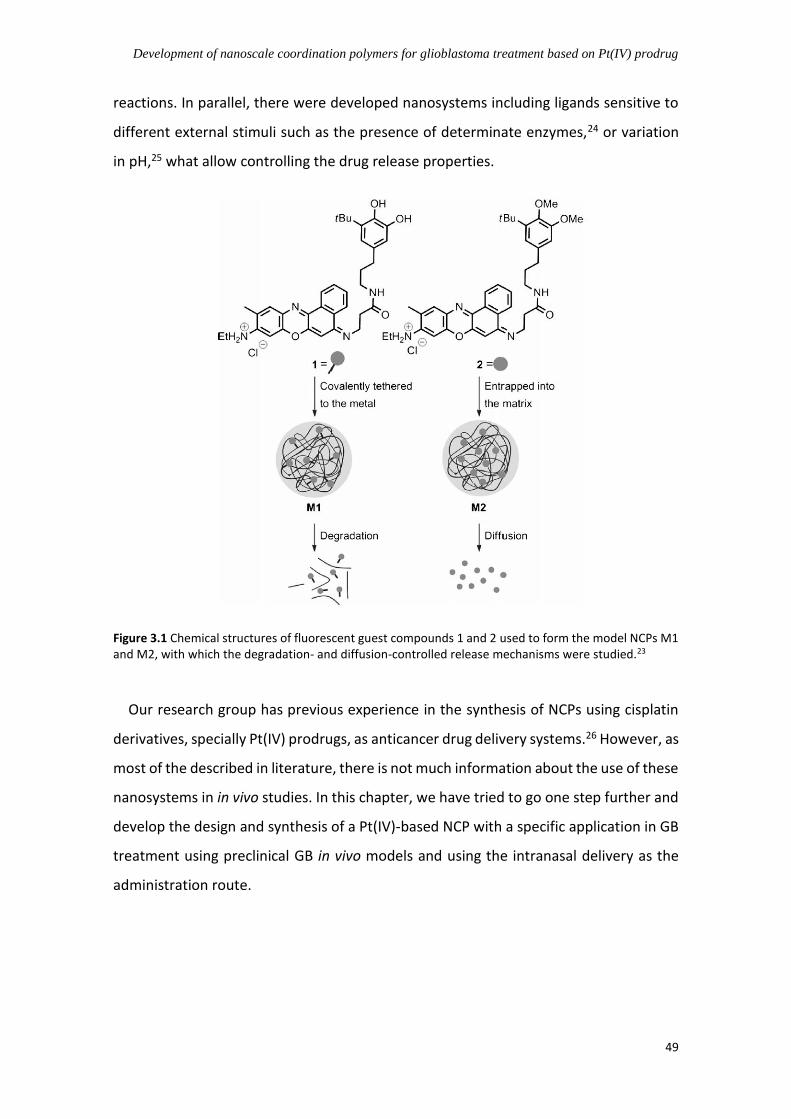
Development of nanoscale coordination polymers for glioblastoma treatment based on Pt(IV) prodrug
49
reactions. In parallel, there were developed nanosystems including ligands sensitive to
different external stimuli such as the presence of determinate enzymes,24 or variation
in pH,25 what allow controlling the drug release properties.
Figure 3.1 Chemical structures of fluorescent guest compounds 1 and 2 used to form the model NCPs M1 and M2, with which the degradation- and diffusion-controlled release mechanisms were studied.23
Our research group has previous experience in the synthesis of NCPs using cisplatin
derivatives, specially Pt(IV) prodrugs, as anticancer drug delivery systems.26 However, as
most of the described in literature, there is not much information about the use of these
nanosystems in in vivo studies. In this chapter, we have tried to go one step further and
develop the design and synthesis of a Pt(IV)-based NCP with a specific application in GB
treatment using preclinical GB in vivo models and using the intranasal delivery as the
administration route.

Chapter 3
50
3.2 NCPs based on Pt(IV) prodrugs for cancer treatment
Pt(IV) prodrugs and Pt(IV)-based nanomedicines have been increasingly studied due
to their potential advantages over conventional Pt(II) complexes (e.g., increased
stability, surface modification, and property tunability). Similarly, a host of Pt(IV)-based
NCPs have also emerged and investigated for their potential medical applications.
Herein, some interesting NCPs based on Pt(IV) prodrugs for cancer treatment have been
summarized.
Lin W. has been a pioneer in the field of functional NCPs, who first summarized the
modular synthesis of NCPs in 2009,2 and published a series of papers about functional
NCPs fabricated based on Pt(IV) prodrugs for cancer treatment. As Figure 3.2 shown,
they synthesized NCPs with a Pt(IV) prodrug disuccinic cisplatin (DSCP) and Tb(III) as
building blocks, and coated them with a silica shell to increase the stability, leading to
controlled release with tunable release half-life ranging from 5.5 h to 9 h by tuning the
coating shell thickness from 2 nm to 7 nm. Decorated with cyclic peptide c(RGDfK)
targeting v3 integrin, the Pt(IV)-based NCPs displayed enhanced cytotoxicity
compared to cisplatin due to the sufficient cellular uptake.27 Later on, this same Pt(IV)
prodrug was used to obtain NCPs by the coordination with Zr(IV) and La(III) ions, and
stabilized by coating with a lipid layer to have a precise control on the drug release. In
addition, the NCPs could specifically target sigma receptors by conjugating anisamide to
the lipid, resulting in a significant increase in cytotoxicity and enhanced cellular uptake
compared to non-targeted NCPs.28
Figure 3.2 Scheme of cyclic peptide decorated Pt(IV) NCPs. Figure adapted from reference 27.

Development of nanoscale coordination polymers for glioblastoma treatment based on Pt(IV) prodrug
51
Lin W. reported other NCPs (Figure 3.3) based on bisphosphonate cisplatin or
oxaliplatin Pt(IV) prodrug with Zn(II) and coated with cationic lipid layer. The resulting
NCPs showed minimal RES uptake and excellent blood circulation half-lives of 16.4 ± 2.9
and 12.0 ± 3.9 h for cisplatin and oxaliplatin NCPs respectively, and exhibited
significantly higher in vivo potency and efficacy compared with free drugs.29 The
oxaliplatin Pt(IV) prodrug NCP system was further studied to co-deliver another
anticancer agent, gemcitabine monophosphate (GMP), resulting in strong synergistic
therapeutic effect of oxaliplatin and GMP against pancreatic cancer cells. In vivo studies
showed that the co-delivery NCPs can avoid RES uptake and gain prolonged circulation
half-life, and potently inhibit growth of pancreatic tumor compared to NCPs carrying
oxaliplatin or GMP alone.30 Similarly, carboplatin Pt(IV) prodrug and GMP were
incorporated into the NCP delivery system, leading to a strong synergistic effect against
Pt-resistant ovarian cancer cells, and enhanced circulation half-life and improved tumor
uptake, resulting in significant growth inhibition of resistant SKOV-3 and A2780/cis
tumors in vivo.31
Figure 3.3 Schematic showing the composition of NCPs based on cisplatin/oxaliplatin prodrug. The figure
was adapted from reference 29.

Chapter 3
52
On the other hand, the cisplatin Pt(IV) prodrug NCP system was also investigated the
combination with gene therapy by adsorption multidrug resistance (MDR) genes-
targeting siRNA on the lipid shell. After surface modification, the NCPs promoted cellular
uptake and enhanced the chemotherapeutic effects greatly by downregulating the
expression of the MDR genes, and local administration effectively inhibited tumor
growth of cisplatin-resistant SKOV-3 ovarian xenografts.32-33 Recently, Lin et. al.
fabricated a novel NCP platform containing nontoxic Pt(IV) prodrug to deliver miRNA,
leading to inhibition on epithelial-to-mesenchymal transition by preventing β-catenin
nuclear translocation and tumor cell invasion. The NCPs showed effective transfection
to reduce liver colonization and tumor burden after systemic administrations in hepatic
metastatic xenografts of HCT116 without observable toxicity.34
Despite the combination of chemotherapy and gene therapy, Lin and colleagues also
explored the combined immunotherapy. Thus, the oxaliplatin Pt(IV) prodrug NCPs were
coated with photosensitizer lipid shell, resulting in effectively synergistic effects of
chemotherapy and photodynamic therapy. Combining with anti-PD-L1 therapy, the
NCPs mediated regression of both light-irradiated primary tumors and non-irradiated
distant tumors by inducing tumor-specific immune response.22 More recently, they
reported the Pt(IV) prodrug-based NCPs incorporating a ROS-inducing agent, which
activated the co-delivered anti-PD-L1 antibody to synergize the immunotherapy and
chemotherapy from oxaliplatin prodrug.35 The NCPs can be dosed repeatedly to
eradicate the murine colorectal tumors due to the favorable biodistribution and tumor
uptake without peripheral neuropathy, and long-term antitumor immunity was elicited
and prevented tumor relapse when cured mice were challenged with cancer cells.
Recently, the research group led by Liu Z. also published some papers regarding Pt(IV)
prodrug-based NCPs for cancer treatment. For instance, they synthesized a pH-
responsive charge-switchable PEGylated NCPs (Figure 3.4) using one-pot method with
Pt(IV) prodrug and Ca(II). The NCPs were highly sensitive to pH changes, the acidic pH
would switch the slightly negative NCPs to positive to enhance tumor retention and
cellular internalization, and they could significantly inhibit the growth of 4T1 murine
breast tumors in nude mice.19 The same authors obtained a new NCP by integration of
cisplatin prodrug DSCP and Fe(III) (Figure 3.4a) stabilized by PEG and coated with cRGD

Development of nanoscale coordination polymers for glioblastoma treatment based on Pt(IV) prodrug
53
(Figure 3.4b), which showed excellent targeting v3 integrin-overexpressed tumor
cells, and enabled chemotherapy and chemodynamic therapy with dramatically
enhanced efficacy compared to relative monotherapies.36
Figure 3.4 Schematic illustration of the NCPs developed by Liu et.al. a) One-pot synthesis of PEGylated NCPs and its application for TME-responsive anticancer therapy,19 and b) scheme showing fabrication of Fe-DSCP NCPs, followed by PEG modification36.
In 2018, our group reported Zn-Pt(IV)-NCPs consisting of a cisplatin Pt(IV) prodrug
DSCP and Zn(II) with a linker ligand 1,4-Bis(imidazol-1-ylmethyl)benzene.26 The resulting
NCPs displayed a good stability in physiological environments, and a controlled Pt
release in vitro. The NCPs showed enhanced cytotoxic effects against cervical cancer,
neuroblastoma and human adenocarcinoma cells, in comparison to the free Pt(IV)
prodrug. A higher amount of Pt was found bound to the DNA in the cells treated with
the NCPs, although no difference was found in the cellular uptake of Pt, indicating that
the nanostructure facilitated the activation of the prodrug and may induce a change in
the mechanism of action resulting in an increased interaction with the DNA.
a
b

Chapter 3
54
3.3 Scope of this chapter
Despite the progress of NCPs for anticancer diagnosis and treatment during the last
decade, some challenges remain to be faced before the real application of these
nanosystems in the clinics:
i. Facile and low-cost synthetic procedures for the synthesis of prodrugs and
nanoparticles.
ii. Chemical stability in a biological environment being highly biocompatible, non-toxic
and non-accumulative in the organism.
iii. Specific control of the drug delivery profile in the site of action.
iv. High efficiency in different tissues by suitable biodistribution
v. In vitro and in vivo proof-of-concept of their anticancer effect
vi. Design of nanoformulations to be administered by novel administration routes
specifically to reach the brain (i.e., intranasal)
vii. Evaluation of multifunctional features (i.e., drug delivery systems and contrast
agents for bioimaging)
As described previously, the standard of care for GB (temozolomide, TMZ) presents
challenges related to poor drug delivery, degradation processes, limited BBB crossing
and drug resistance. The integration of this chemotherapeutic compound in
nanoparticles have showed interesting results at in vitro and in vivo level, but non-
conclusive. Toxicity issues related to nanoparticles drive researchers to develop more
efficient and safer alternatives with tunable options. Moreover, the interesting activity
of TMZ for GB treatment has presumably diminished the number of developments of
nanoparticles containing other potentially relevant therapeutic agents like platinum
derivatives that have showed for long time a very remarkable anticancer activity. For
this reason, providing novel systems aiming at efficient drug delivery into brain and GB
could help to the increase of the effectiveness and decrease of side effects of therapies.

Development of nanoscale coordination polymers for glioblastoma treatment based on Pt(IV) prodrug
55
Based on this, in this work, it is proposed for the first time, the use of NCPs as potential
candidates for GB therapy via IN delivery. In this Chapter, the synthetic protocol includes
the synthesis of the Pt(IV) prodrug which is the building block of the nanostructured
coordination polymers obtained by the reaction of the Pt(IV) complex with iron(III) ions
(Figure 3.5). Through this rationale design, the NCPs named Pt-Fe NCPs have been
proposed. The systems aim to accomplish: i) moderate chemical stability, ii) water-
colloidal stability, iii) low toxicity and biocompatibility, iv) controlled drug delivery
profile, vi) intrinsic properties to be used as contrast agents for clinical image, and v)
potential anticancer activity in vitro against glioblastoma cell lines and in vivo against GB
murine models using intranasal administration as the administration route. The
nanoparticles will be characterized by different physicochemical techniques and its
behavior in physiological environment both in vitro and in vivo will be tested. To evaluate
the delivery into brain and GB tumor of the NCPs after intranasal administration, the in
vivo studies will be the performed in orthotopic GL261 murine models. The syngeneic
immunocompetent GL261 murine glioma model is one of the most widely used to study
the glioblastoma, which is usually established by inoculation of GL261 murine cells into
C57BL/6 substrain, resulting in invasive and infiltrative GB tumors, similar to those found
in human patients.37-41
Figure 3.5 Scheme of the synthesis of the Fe-Pt NCPs developed in this chapter.
To sum up, the synthesis novel Pt-Fe NCPs for its potential use as therapeutic agents
for GB is presented. Despite remarkable advances in basic and clinical research, the
clinicians are yet unable to provide realistic therapy for this kind of tumors, and the use
of Pt-based chemotherapies are not well developed. For this reason, providing to the

Chapter 3
56
community with novel systems that enhance the effectiveness of therapies could help
to find alternative and more suitable treatments.
The specific objectives for this Chapter are:
i. Synthesis of the nanoparticles Pt-Fe NCPs.
ii. Physicochemical characterization and chemical/colloidal stability studies of NCPs in
vitro.
iii. Preclinical in vivo studies with a GB murine model
iv. Comparison of the therapeutic activity between Pt-Fe NCPs and cisplatin/prodrug
3.4 Results and discussion
3.4.1 Synthesis and characterization of Pt(IV) prodrug
Figure 3.6 The synthetic route of Pt(IV) prodrug PtBC.
The synthesis of Pt(IV) prodrug was developed from the commercially available
anticancer platinum agent, cisplatin. The synthetic route (Figure 3.6) comprised of
several steps: 1) the oxidation of cisplatin to c,c,t-Pt(NH3)2Cl2(OH)2 (oxoplatin) by
reacting with hydrogen peroxide; 2) oxoplatin was converted into of c,c,t-
Pt(NH3)2Cl2(O2CCH2CH2COOH)2 (DSCP) by the addition of succinic anhydride; and 3)
dopamine was attached to DSCP via a typical DCC/NHS coupling using NMM as base. The
synthesis of the intermediate DSCP was slightly modified according to previous report
Development of nanoscale coordination polymers for glioblastoma treatment based on Pt(IV) prodrug
57
from our group.26 Specific reaction conditions and detailed synthetic method is
described in section 3.6. PtBC was successfully synthesized with an overall yield of
24.7%.
All compounds obtained after reactions were characterized and compared by FT-IR
(Figure 3.7). In the spectrum of oxoplatin, the peak at 559 cm-1 was characteristic from
the vibration of Pt-O coordination bonding, and the intensely sharp peak at 3516 cm-1
belongs to O-H vibration.42 In the spectrum of DSCP, the peak at 3516 cm-1 disappeared,
and the new band around 1630-1734 cm-1 typically can be assigned to C-O stretching
from the carboxyl groups, while new band at 1180-1347 cm-1 also implied the presence
of succinic acid attached. Finally, in the spectrum of PtBC, the band from carboxyl groups
shifted to lower wavenumber at 1527-1645 cm-1 indicating the formation of amide bond
(Amide I at 1621 cm-1 and Amide II at 1559 cm-1), and the characteristic peak of catechol
showed up at 1198 cm-1 from C-O stretching on the aromatic ring. To further confirm
the chemical syntheses results, DSCP, its NHS-activated ester and PtBC were checked by
1H NMR spectra (Figures 3.8-3.10, respectively). In the 1H NMR spectrum of DSCP (Figure
3.8), the ratio between all protons perfectly fit the theoretical value including the -OH
group at ~12 ppm in deuterated DMSO. The ratios of protons also fit well for the
activated intermediate DSCP-NHS (Figure 3.9) and final product PtBC (Figure 3.10a).
Further 13C NMR and 2D 1H-1H COSY NMR spectra (Figure 3.10b-c) of PtBC confirmed its
expected chemical structure. High-resolution mass spectrum (Figure 3.11) also showed
correct mass of PtBC (found 803.1284, calc. 803.1300). All these spectra substantiated
that the compounds were all successfully synthesized with high purity level.

Chapter 3
58
Figure 3.7 FT-IR spectra of cisplatin, oxoplatin, DSCP and PtBC.
Figure 3.8 1H NMR spectrum of DSCP.

Development of nanoscale coordination polymers for glioblastoma treatment based on Pt(IV) prodrug
59
Figure 3.9 1H NMR spectrum of DSCP-NHS.
a
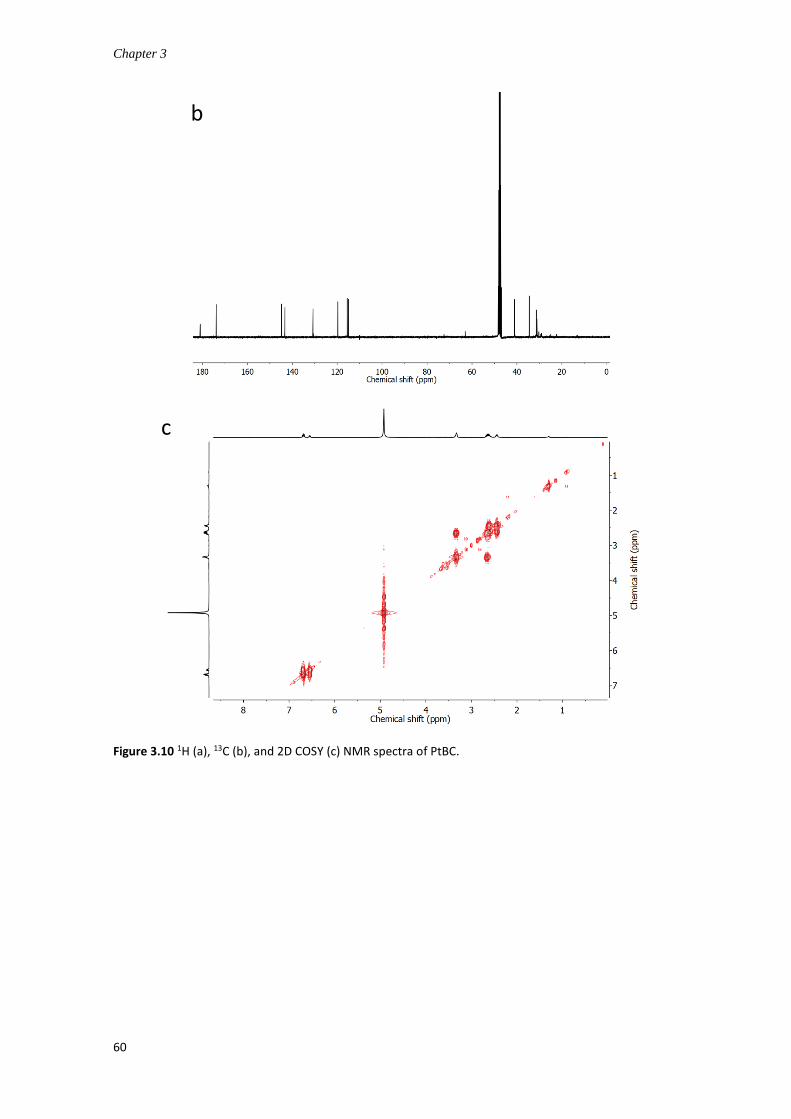
Chapter 3
60
Figure 3.10 1H (a), 13C (b), and 2D COSY (c) NMR spectra of PtBC.
b
c

Development of nanoscale coordination polymers for glioblastoma treatment based on Pt(IV) prodrug
61
Figure 3.11 High-resolution mass spectrum of PtBC.
3.4.2 Synthesis and characterization of Pt-Fe NCPs
The Pt-Fe NCPs formed immediately after the addition of Fe(OAc)2 into PtBC solution
(1:1 in molar ratio). The reaction leads to the formation of a black precipitate, due to the
coordination between the terminal catechol moieties and iron ions at rt, as shown in
Figure 3.12. This reaction generated the Pt-Fe NCPs nanoparticles with a 49.3% yield and
high reproducibility.
Figure 3.12 Schematic illustration of the synthesis of Pt-Fe NCPs.

Chapter 3
62
The morphology and size of the NCPs was observed by SEM, STEM and TEM (Figure
3.13). Relatively monodispersed and uniform spherical NPs were observed under all
electronic microscopy techniques, with a size around 70 nm. This size is in good
accordance with the result of DLS after NCPs redispersion in phosphate buffer saline
(PBS). In fact, both size and ζ-potential (Figure 3.14) of Pt-Fe NCPs were measured,
showing an average size of 66.0 ± 2.3 nm, with a PDI of 0.258 ± 0.013, and a slightly
negative ζ-potential of -8.53 ± 0.418 mV.
Figure 3.13 Representative morphologies of Pt-Fe NCPs under SEM (a), STEM (b, HAADF mode), and
TEM (c).
Figure 3.14 Representatively size distribution (a) and ζ-potential (b) of Pt-Fe NCPs by DLS.
The NCPs present good monodispersion values (PDI < 0.20) and colloidal properties.
The sizes of the obtained NCPs are in an optimal range for their medical use. In fact, this
parameter plays an important role in the pharmacology and metabolism of NPs. It is well
know that nanoparticles < 30 nm are more likely to be cleared by the renal system, while
particles > 300 nm tend to be phagocytosed by the reticuloendothelial system (RES) and
accumulated in liver and spleen.43-44 Particle sizes < 100 nm were reported to facilitate
a b c
a b
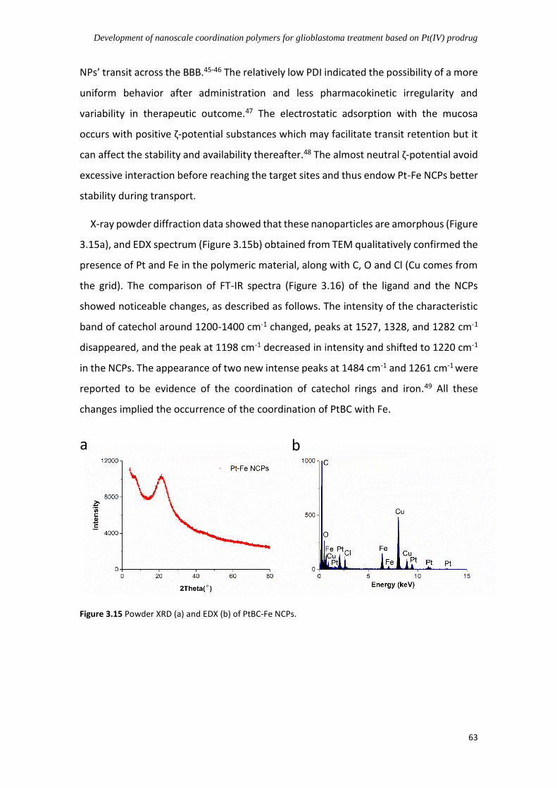
Development of nanoscale coordination polymers for glioblastoma treatment based on Pt(IV) prodrug
63
NPs’ transit across the BBB.45-46 The relatively low PDI indicated the possibility of a more
uniform behavior after administration and less pharmacokinetic irregularity and
variability in therapeutic outcome.47 The electrostatic adsorption with the mucosa
occurs with positive ζ-potential substances which may facilitate transit retention but it
can affect the stability and availability thereafter.48 The almost neutral ζ-potential avoid
excessive interaction before reaching the target sites and thus endow Pt-Fe NCPs better
stability during transport.
X-ray powder diffraction data showed that these nanoparticles are amorphous (Figure
3.15a), and EDX spectrum (Figure 3.15b) obtained from TEM qualitatively confirmed the
presence of Pt and Fe in the polymeric material, along with C, O and Cl (Cu comes from
the grid). The comparison of FT-IR spectra (Figure 3.16) of the ligand and the NCPs
showed noticeable changes, as described as follows. The intensity of the characteristic
band of catechol around 1200-1400 cm-1 changed, peaks at 1527, 1328, and 1282 cm-1
disappeared, and the peak at 1198 cm-1 decreased in intensity and shifted to 1220 cm-1
in the NCPs. The appearance of two new intense peaks at 1484 cm-1 and 1261 cm-1 were
reported to be evidence of the coordination of catechol rings and iron.49 All these
changes implied the occurrence of the coordination of PtBC with Fe.
Figure 3.15 Powder XRD (a) and EDX (b) of PtBC-Fe NCPs.
a b

Chapter 3
64
Figure 3.16 FT-IR spectra of the prodrug and Pt-Fe NCPs.
Figure 3.17 TGA and DSC of PtBC (black) and Pt-Fe NCPs (red).
Thermal gravimetric analysis (TGA) coupled with differential scanning calorimetry
(DSC, Figure 3.17), elemental analysis (EA) and inductively coupled plasma mass
spectrometry (ICP-MS) were employed to further determine the quantitative
composition of Pt-Fe NCPs. From the TGA curves, the prodrug PtBC was stable till 200 ℃,
and started to lose weight slowly to 50% at 400 ℃, then a fast loss occurred around
450 ℃ till reaching stable again at 540 ℃ with a total weight loss of 76%. In contrast,
500 1000 1500 2000 2500 3000 3500
Wavenumber cm-1
—PtBC —Pt-Fe NCPs
Development of nanoscale coordination polymers for glioblastoma treatment based on Pt(IV) prodrug
65
the Pt-Fe NCPs started to lose weight slowly from the beginning with a 10% slow loss till
200 ℃, then lost weight quickly from 270 ℃ till 400 ℃, and slowly reached stable till
500 ℃ with a total weight loss around 43%. The decomposition of Pt-Fe NCPs went
through a different pathway which was also indicated by the DSC curves with dissimilar
valleys. The difference of the thermal stability between them could be attributed to the
distinct structures of these two samples. The slow decomposition of Pt-Fe NCPs from
the beginning may result from the labile coordination bonds between the metal nodes
and the organic ligand. In Table 3.1, the results are shown obtained from analyses of C%,
H%, N%, Pt% and Fe%, together with the calculated theoretical values. These
correlations offer a proposed chemical formula [(PtBC)1Fe3(OH-)5]n for Pt-Fe NCPs.
C H N Pt Fe
Exp. % wt 28.63 2.54 5.79 18.87 15.13
Cal. % wt 27.27 3.72 5.30 18.45 15.83
Error % 0.99 1.18 0.69 0.42 0.7
Table 3.1 Experimental composition obtained by elemental analysis and ICP-MS, and correlation with the theoretical values for the proposed chemical formula [(PtBC)1Fe3(OH-)5]n.
Additionally, Mössbauer spectroscopy confirmed that Pt-Fe NCPs showed a high-spin
Fe(III) oxidation state (Figure 3.18), thus corroborating the oxidation process from Fe(II)
to Fe(III) during the synthesis of the Pt-Fe NCPs. This electronic modification results from
a redox interplay during the synthesis reaction between the metal ion and the
electroactive catechol ligands in the presence of oxygen, as previously reported.50
The hyperfine parameters of the fitting of Mössbauer at 300 K showed the isomer
shift relative to the metallic iron ((δFe), quadrupolar splitting (ΔEq) and the full width at
half maximum (Γ). The spectrum was fitted to a single doublet with a ΔEq= 0.81 ± 0.04
mm/s and Γ= 0.32 mm/s. The fitting was centered at an isomeric shift δ= 0.39±0.03
mm/s.

Chapter 3
66
Figure 3.18 Representative Mössbauer spectra of Pt-Fe NCPs at 300 K and 80 K. Experimental data (blue dots), and computation fitted (orange line) spectra.
3.4.3 Colloidal stability of Pt-Fe NCPs
Since the naked NCPs tended to form small clusters ~ 300 nm even aggregates > 1 m,
it is necessary to improve the colloidal stability for further in vitro and in vivo studies.
Different methods were adopted to improve the stability, such as 1) coating with
polydopamine-like (PDA-like) polymer, or 2) using different stabilizers to increase the
dispersity.
3.4.3.1 PDA-like coating for stabilizing Pt-Fe NCPs
Previous studies carried out in our group showed how NCPs could be coated with a
PDA-like polymer to increase the stability of NCPs, overall achieving better
biodistribution and favoring the delivery of cargos.51 Therefore, the same idea was come
up with to stabilize the Pt-Fe NCPs. The coating process commonly occurs in two main
steps: 1) the primer formation of PDA via the oxidative polymerization of catechol which
can adhere to a wide range of materials and surfaces, and 2) the deposition of the PDA
onto the surface of the materials for coating. Following this approach, a catechol-based
coating was obtained through the crosslinking of pyrocatechol (cat) and
hexamethylenediamine (HMDA), as shown in Figure 3.19.
1.40E+06
1.40E+06
1.41E+06
1.41E+06
1.42E+06
1.42E+06
1.43E+06
1.43E+06
1.44E+06
1.44E+06
1.45E+06
-4 -2 0 2 4
Tran
smis
sio
n (
a.u
.)
Velocity (mm/s)
300K
184000
186000
188000
190000
192000
194000
196000
198000
200000
202000
204000
-4 -2 0 2 4
Tran
smis
sio
n (
a.u
.)
Velocity (mm/s)
80k
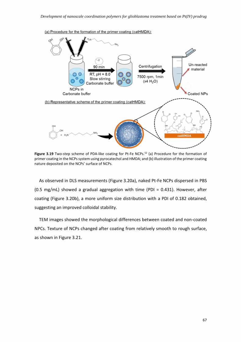
Development of nanoscale coordination polymers for glioblastoma treatment based on Pt(IV) prodrug
67
Figure 3.19 Two-step scheme of PDA-like coating for Pt-Fe NCPs.52 (a) Procedure for the formation of primer coating in the NCPs system using pyrocatechol and HMDA; and (b) illustration of the primer coating nature deposited on the NCPs’ surface of NCPs.
As observed in DLS measurements (Figure 3.20a), naked Pt-Fe NCPs dispersed in PBS
(0.5 mg/mL) showed a gradual aggregation with time (PDI = 0.431). However, after
coating (Figure 3.20b), a more uniform size distribution with a PDI of 0.182 obtained,
suggesting an improved colloidal stability.
TEM images showed the morphological differences between coated and non-coated
NPCs. Texture of NCPs changed after coating from relatively smooth to rough surface,
as shown in Figure 3.21.

Chapter 3
68
Figure 3.20 Representative size distribution of Pt-Fe NCPs before (a) and after (b) coating with time. In
the graph, blue curve was measured at 0 h, green curve at 1 h, and red curve at 24 h after preparation,
respectively.
Figure 3.21 Representative TEM images of Pt-Fe NCPs before (left) and after (right) coating.
a
b
a b

Development of nanoscale coordination polymers for glioblastoma treatment based on Pt(IV) prodrug
69
To confirm the presence of the coating layer, line profile EDX analysis was carried out
using TEM, as shown in Figure 3.22. In this analysis the film covering the NCPs can be
distinguished from the Pt-Fe NCPs as the PDA-like coating comparing carbon versus iron
profile.
Figure 3.22 Line profiles (a) of carbon and iron across the surface of coated Pt-Fe NCPs (b)
To check the chemical composition of synthesized NCPs before and after coating, EDX
(Figure 3.23) and ICP-MS were performed to compare the values of Pt and Fe. Although
EDX indicate the persistence of Pt and Iron in the coated NCPs, ICP-MS corroborated a
dramatic decrease of Iron and Platinum content. In fact, Pt decreased drastically by
85.746%, while Fe decreased by 50.485% (Table 3.2). With the organic coating, the
contents of both metals should decrease, but the ratio between them should keep as a
constant if the composition of the NCPs remains stable during the coating process.
However, the ratio between Pt and Fe dropped drastically from 1.252 to 0.361,
indicating a sharp reduction of Pt compared to Fe drop after coating. This probably is
due to the competition between the catechol moieties from our ligand PtBC and those
from pyrocatechol during the coating formation. In this dynamic competition of
cleavage and re-coordination, the NCPs probably degraded somehow and lost Pt.
a b
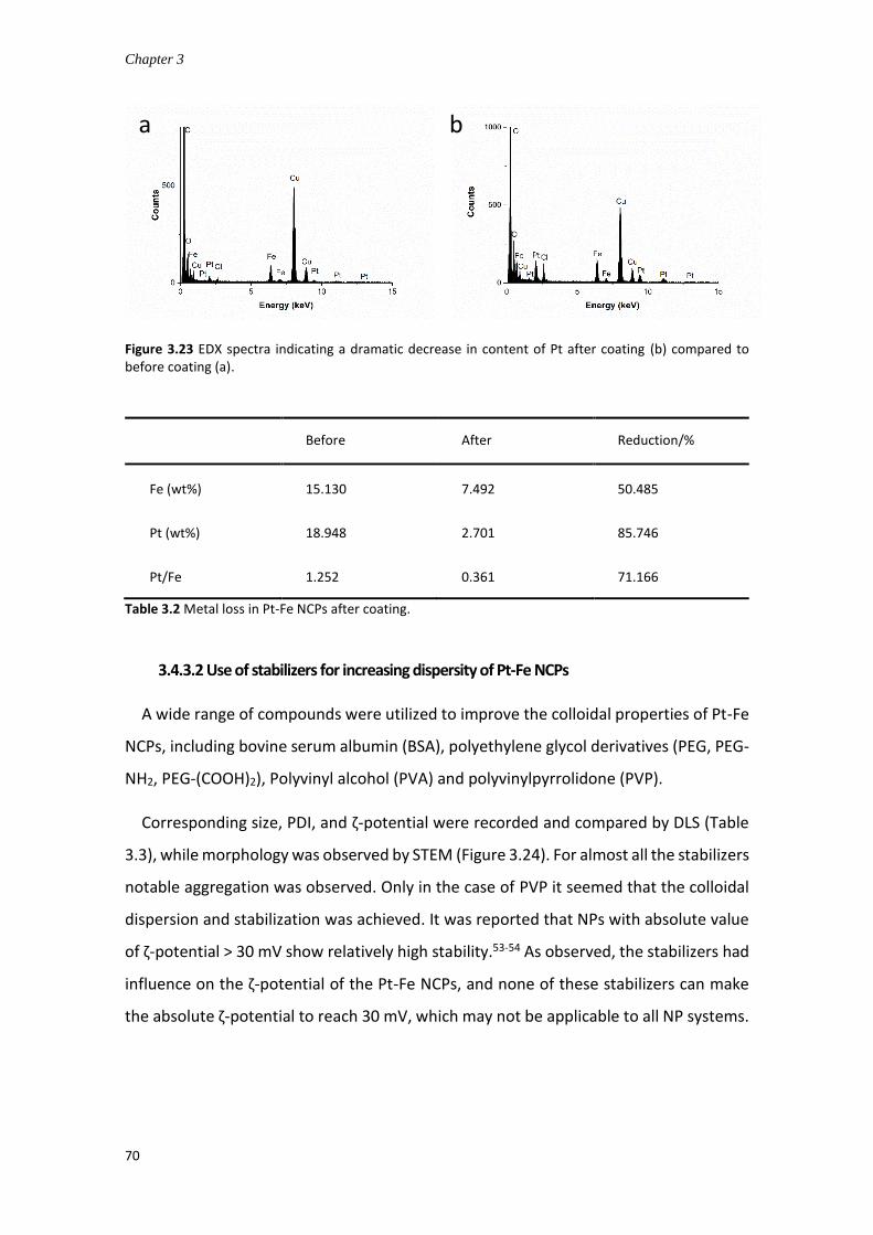
Chapter 3
70
Figure 3.23 EDX spectra indicating a dramatic decrease in content of Pt after coating (b) compared to before coating (a).
Before After Reduction/%
Fe (wt%) 15.130 7.492 50.485
Pt (wt%) 18.948 2.701 85.746
Pt/Fe 1.252 0.361 71.166
Table 3.2 Metal loss in Pt-Fe NCPs after coating.
3.4.3.2 Use of stabilizers for increasing dispersity of Pt-Fe NCPs
A wide range of compounds were utilized to improve the colloidal properties of Pt-Fe
NCPs, including bovine serum albumin (BSA), polyethylene glycol derivatives (PEG, PEG-
NH2, PEG-(COOH)2), Polyvinyl alcohol (PVA) and polyvinylpyrrolidone (PVP).
Corresponding size, PDI, and ζ-potential were recorded and compared by DLS (Table
3.3), while morphology was observed by STEM (Figure 3.24). For almost all the stabilizers
notable aggregation was observed. Only in the case of PVP it seemed that the colloidal
dispersion and stabilization was achieved. It was reported that NPs with absolute value
of ζ-potential > 30 mV show relatively high stability.53-54 As observed, the stabilizers had
influence on the ζ-potential of the Pt-Fe NCPs, and none of these stabilizers can make
the absolute ζ-potential to reach 30 mV, which may not be applicable to all NP systems.
b a

Development of nanoscale coordination polymers for glioblastoma treatment based on Pt(IV) prodrug
71
Figure 3.24 Pt-Fe NCPs stabilized by a) BSA, b) PEG, c) PEG-(COOH)2, d) PEG-(NH2)2, e) PVA, and f) PVP
under STEM. BSA was added to a concentration of 10 mg/ml, other stabilizers at 1%, whereas, the NCPs
at a concentration of 0.5 mg/mL.
BSA PVA PEG PEG-NH2 PEG-(COOH)2 PVP
Size / nm 548.5 ± 81.10 398.7 ± 125.9 681.4 ± 97.38 1181 ± 99.47 4491 ± 711.8 72.12 ± 0.86
PDI 0.585 ± 0.033 0.654 ± 0.206 0.66 ± 0.018 0.584 ± 0.02 0.516 ± 0.468 0.271 ± 0.006
ζ-pot. / mV -10.3 ± 0.435 -7.47 ± 0.771 -13.2 ± 0.265 6.05 ± 0.754 -14.8 ± 0.44 -8.09 ± 0.100
Table 3.3 Colloidal stability of Pt-Fe NCPs using different stabilizers in terms of size, PDI, and ζ-potential.
Additionally, sucrose also was tested as a stabilizer. There has been some previous
reports concerning the use of this sugar as stabilizer for NCPs.24 Thus, different
concentrations of sucrose ( with final concentrations between 0.5% - 50% in PBS) were
added into Pt-Fe NCPs suspension, and the dispersions were checked by DLS (Table 3.4).
In basis of DLS measurements only the 50% concentrate solution was able to keep the
NCPs suspension stable up to 3 days.
a b c
d e f
Chapter 3
72
Sucrose/% 0 0.5 1 5 10 25 50
Size/nm 365.1 486.0 385.7 385.5 323.8 280.6 117.4
PDI 0.431 0.316 0.228 0.215 0.200 0.225 0.205
Table 3.4 Colloidal stability of NCPs in PBS with different concentrations of sucrose in terms of size and PDI.
However, such a high concentration (50%) of sucrose was not reasonable to add into
a formulation for anticancer treatment. Taken previously tested substances into
consideration, PVP was finally chosen as the stabilizer for the Pt-Fe NCPS for further
studies.
3.4.4 Relaxivity properties of Pt-Fe NCPs: in vitro magnetic resonance imaging
(MRI) studies
The presence of high-spin Fe(III) within the NCPs provided the possibility for this
material to serve as a MRI contrast agent. MRI contrast agents are able to improve
intrinsic contrast by modifying the relaxation times of water protons under a magnetic
field in the tissues, thus enhancing the contrast to better visualize the interested
anatomical region. Contrast agents can be classified into two categories: positive ones,
which are usually based on paramagnetic metal ions, like Gd(III) and Mn(II), to shorten
longitudinal relaxation time (T1); and negative ones, which mainly involve
superparamagnetic iron oxide compounds to shorten transversal relaxation time (T2).
To confirm the features of Pt-Fe NCPs, magnetic resonance relaxometry experiments
were carried out at escalated concentrations under an external magnetic field in two
phantom sequences.
Quantitative MRI phantoms were carried out which further substantiated the results
from Mössbauer spectra. As shown in Figure 3.25, T1-weighted (T1W) and T2-weighted
(T2W) imaging maps were obtained at a series of Fe concentrations of the NCPs dispersed
in 1% agarose PBS measured by a Biospec MRI 7 T horizonal scanner. The longitudinal
(r1) and transversal (r2) relaxivity parameters were determined by the obtained T1W and
T2W map acquisitions, which indicates how the corresponding relaxation changes as a

Development of nanoscale coordination polymers for glioblastoma treatment based on Pt(IV) prodrug
73
function of the concentration. The agarose-based method has been widely used in
reports to ensure the good dispersion of nanoparticles and to mimic the viscosity and
consistency of biological tissues.55
Along with the increased Fe concentration, the intensity of T1w image increased while
the intensity of T2W image decreased. According to the calculation of the slopes of the
regression curves (Figure 3.25), the relaxivities r1 and r2 were 0.407 and 21.613 mM-1s-1
respectively. Interestingly, the NCPs exhibited a low r1 compared to the commercial
gadolinium contrast agents, for example Gd-DTPA with a r1 of 3.3 mM-1s-1.56 Similar low
r1 values were reported in Gd-based NCPs and attributed to a low water accessibility to
the metal ions.57 On the other hand, the Pt-Fe NCPs showed a high r2 value, and the ratio
of r2/r1 was as high as 53.15, which implies that Pt-Fe NCPs bear the potential to serve
as a promising contrast agent for T2 MRI.
Figure 3.25 a) In vitro results obtained with relaxometry studies with Pt-Fe NCPs at different Fe concentrations. b) T1W and T2W MRI obtained with Pt-Fe NCPs which were suspended in PBS containing 1% agarose at pH 7.4 at different concentrations (0, 0.1, 1, 1.97 and 3.94 mM, referred to Fe concentration).
a
b

Chapter 3
74
3.4.5 In vitro drug release of Pt-Fe NCPs
In our work, the Pt(IV) prodrug and resulting Pt-Fe NCPs were supposed to be pH-
responsive due to the presence of ester bonds and amide bonds in the molecule which
can remain stable in physiological conditions and be cleaved in the weekly acidic
extracellular environment of tumors or in endo/lysosomal compartments.24
Furthermore, the coordination bonds between catechol and iron are usually stable
under slightly basic conditions but can cleave due to the demetallation process with
decreased pH.58-60
Herein, the drug release from Pt-Fe NCPs was first studied in phosphate buffers at
different pH using dialysis method. The release profiles were obtained at different pH
(pH 7.4 and pH 5.5) to mimic the physiological and mildly acidic conditions for the drug
release study in vitro. In a typical experiment, Pt-Fe NCPs were incubated in the dialysis
bag at 37 ℃ over 48 h in presence of buffers and the released Pt outside the bag was
determined by ICP-MS to obtain the release profiles in terms of concentration as a
function of time. As observed in Figure 3.26, both release curves are typically bimodal,
while Pt was released faster from the NCPs in the first 6-8 hours, a more sustained and
slower release was observed after that time in both pH values. Pt was released faster at
pH 5.5 than that at pH 7.4, indicating the nanoparticles’ pH-responsive behavior.
Specifically, 46.78 ± 0.33% of Pt was released at pH 7.4 in the first 4 h, and 67.92 ± 2.01%
released after 48 h. At acidic pH, the release of Pt was slightly faster with around 60.45
± 0.32% of Pt released after 4 h of incubation, and 72.43 ± 1.56% at 48 h. This
corroborated the effect of pH on the release profile that was notable increased in acidic
pH.
a b

Development of nanoscale coordination polymers for glioblastoma treatment based on Pt(IV) prodrug
75
Figure 3.26 a) Release profiles of Pt from Pt-Fe NCPs at 37 ℃ at pH 7.4 and 5.5 in PBS using dialysis method; b) Inset of release in the first 8 h of the whole release profiles. Each value represents the mean ± SE of three independent experiments.
We also evaluated the effect of reducing agents on the release profile. The redox
couple, glutathione (GSH) and glutathione disulfide (GSSG), plays a pivotal role in the
anti-oxidative capacity of cells. The most important function of GSH is to serve as a
reducing agent in intracellular ambient protecting cells from oxidative damage and the
toxicity of xenobiotic electrophiles, and maintaining redox homeostasis.61 Usually, the
intracellular concentration of GSH is reported to be 1 - 10 mM in normal cells, while its
concentration decreased to 2-3 orders of magnitude lower levels in extracellular fluids.62
However, the intracellular level of GSH increases up to fourfold more in cancer cells than
that in normal cells. The elevated levels of GSH protect tumor cells from the anticancer
action of numerous therapeutics, including cisplatin, contributing to the multidrug
resistance (MDR).63 The different GSH levels in extra- and intra-cellular circumstances
and in normal and tumor tissues have prompted the emergence of GSH-responsive
nano-formulations to facilitate the preferential drug release in tumor tissues and cells.64-
66
GSH has been reported to enable the reduction of Pt(IV) prodrugs into their active
Pt(II) forms, which induced apoptosis of cancer cells by covalent binding to DNA.67-69
Therefore, the release behavior of Pt-Fe NCPs was furthermore assayed in the presence
of GSH at different pH values. The GSH levels in brain were reported to be around 1 - 2
mM in normal conditions.70 Therefore, 2 mM and 10 mM were chosen as the
concentrations of GSH to be tested at pH 7.4 and 5.5, using the same method described
previously. As Figure 3.27 shown, indeed, the release of Pt was obviously promoted by
GSH. Even at 2 mM, GSH accelerated the release of Pt dramatically; 62.89 ± 0.54% of Pt
was released from the NCPs after 4 h of incubation at pH 7.4, which was close to the
amount of released Pt after 48 h (67.92 ± 2.01%) in the absence of GSH. After 48 h, 83.54
± 0.24% of Pt was released in total with 2 mM of GSH. Notably, the release was further
sped up while the concentration of GSH increased to 10 mM; about 70.23 ± 0.28% of Pt
was released from the NCPs after 4 h of incubation, and the release reached 93.49 ±

Chapter 3
76
0.39% after 48 h. At pH 5.5, the release of Pt was accelerated in the same trend as at pH
7.4 in the presence of GSH.
Figure 3.27 Cumulative release of Pt from Pt-Fe NCPs in the absence or in the presence of GSH at a) pH 7.4 and b) pH 5.5. Each value represents the mean ± SE of three independent experiments.
Therefore, the results demonstrated that Pt-Fe NCPs could be both pH- and GSH-
responsive. These properties could provide a precise controlled release regulated by pH
and the presence of a reducing ambient while keeping the nanoparticles stable in
absence of these external stimuli. This behavior could increase the chemotherapy
effectivity against cancer cells and avoiding side effects.
3.4.6 In vitro anticancer studies of Pt-Fe NCPs
3.4.6.1 Cytotoxicity assays
The cytotoxic effects of the prodrug PtBC and Pt-Fe NCPs were evaluated in vitro and
compared to cisplatin (CDDP) using different cell lines, including Human cervical cancer
cell HeLa, murine GB GL261 cells, ovarian cancer cells A2780 (including cisplatin resistant
cell line A2780/cis) and non-malignant human fibroblast cell 1Br3G. We included a
comparative study between non-resistant and resistant cancer cells because it has been
reported that nanoformulations with proper modifications can bypass the efflux of
resistant cells which the free drug is commonly subjected to, therefore modulating or
overcoming the resistance of cancer cells.71-72 The well-established PrestoBlue method
was used to assess the cytotoxicity at 24 h and 72 h (Figure 3.28).
a b
Development of nanoscale coordination polymers for glioblastoma treatment based on Pt(IV) prodrug
77
The half maximal inhibitory concentrations (IC50s) referred to Pt concentration,
calculated using Graphpad Prism 7, were summarized in Table 3.5 and 3.6. Interesting
results were obtained for the three Pt-based species. As observed in Table 3.5, PtBC and
Pt-Fe NCPs showed slightly lower cytotoxicity at 24 h for almost all cell lines in
comparison with CDDP. Interestingly, both PtBC and Pt-Fe NCPs exerted higher cell
killing capability than CDDP in 1Br3G cells, with IC50 of 27.78 ± 1.75 M and 45.07 ± 4.60
M respectively. Moreover, in the case of A2780 the Pt(IV) prodrug was highly effective
against these cell lines, even more pronounced in the case of cisplatin-resistant
A2780/cis cells. After 72 h (Table 3.6), the Pt(IV) prodrug and NCPs exhibited comparable
or even slightly higher anticancer ability than CDDP. Whereas, in the case of A2780 and
A2780/cis in which the prodrug and the nanoformulation exhibited less cytotoxic
effectivity in comparison with cisplatin.
The difference between the results suggests different explanations. It could be
attributed to the distinctive cellular internalization pathways of these three
formulations. Due to their small size, CDDP and PtBC probably entered cells by passive
diffusion common for small molecules, while the Pt-Fe NCPs internalized via
endocytosis, which is typically slower than diffusion in a short term observed for other
NCPs.26 While at 24 h it is supposed that most of the NCPs entered the cells, the
degradation of the nanoparticles and the subsequent reduction of Pt(IV) complexes to
generate the active cisplatin retards the cytotoxic effect which is notably superior at 72
h reaching IC50 values comparable or even higher than cisplatin. Apart from the
differences attributable to the delay concerning NCPs degradation and prodrug
activation, different mechanisms of action should be taken into account as has been
observed in previously published research in our group.26 Concerning the molecular
compounds (prodrug PtBC and cisplatin) that are supposed to internalize via passive
diffusion, the observed effect depends notably of the studied cell line. In this case,
different rates of cell internalization and/or mechanism of action could take part. We
should have into account also that the uptake pathways and detoxification of NPs are
greatly influenced by the cell type, as well as the intrinsic properties of NPs.73 Moreover,
recent reports pointed out that the presence of catechol moiety in structure contributed
to the antibacterial or even anticancer potential in some compounds.74-75 These

Chapter 3
78
promising anticancer effects in vitro lay the foundation for the exploration of further
biomedical evaluations.

Development of nanoscale coordination polymers for glioblastoma treatment based on Pt(IV) prodrug
79
Figure 3.28 Cytotoxicity of CDDP, PtBC and Pt-Fe NCPs against a panel of cell lines for 24 h and 72 h. All data represented as mean ± SE of three independent experiments.
IC50 (µM)a
Cell line
Compound HeLa 1Br3G GL261 A2780 A2780/cis
Pt-Fe NCPs 31.45 ± 1.10 27.78 ± 1.75 13.48 ± 0.90 15.76 ± 1.13 43.40 ± 3.75
PtBC 29.94 ± 1.04 56.09 ± 1.18 17.40 ± 1.08 10.98 ± 0.56 18.39 ± 1.85
CDDP 15.98 ± 1.04 45.07 ± 4.60 5.614 ± 0.28 13.95 ± 1.24 23.68 ± 1.74 a The IC50s of the compounds against different cell lines were determined by PrestoBlue assay. Each value
represented the mean ± SE of 3 independent experiments.
Table 3.5 Cytotoxicity after 24h.
IC50 ( M)a
Cell line
Compound HeLa 1Br3G GL261 A2780 A2780/cis
Pt-Fe NCPs 2.56 ± 0.63 5.32 ± 1.10 2.00 ± 0.18 6.31 ± 1.56 11.04 ± 1.69
PtBC 1.85 ± 0.36 10.80 ± 0.60 4.17 ± 0.12 2.59 ± 0.33 9.12 ± 0.69
CDDP 2.34 ± 0.30 4.63 ± 0.42 2.16 ± 0.26 1.57 ± 0.08 5.22 ± 1.21 a The IC50s of the compounds against different cell lines were determined by PrestoBlue assay. Each value
represented the mean ± SE of three independent experiments.
Table 3.6 Cytotoxicity after 72h.

Chapter 3
80
3.4.6.2 Generation of reactive oxygen species
The redox property of catechol moiety makes it an interesting functional group in
biomedical field. On one hand, it can function as antioxidant with beneficial
physiological effects. Many reports confirmed that compounds with catechol, generally
polyphenols and flavonoids, exhibit hepatoprotective, anti-inflammatory, antioxidant,
and even anticancer properties.76 On the other hand, the high reactivity of catechol may
trigger harmful effects including toxicity, damage, and inflammation in cells, which
mainly arise from the excessive oxidative stress of reactive oxygen species (ROS)
generated during the oxidation process of catechol.77 Recently, researchers reported
that singlet oxygen (1O2) was generated between catechol and transition metal ions.78
Besides, ROS accumulation is considered a pivotal phenomenon related to apoptosis
induced by DNA damage.79
Figure 3.29 Conversion of DCFDA to a fluorescent product DCF.80
To assess if such oxidative stress also plays a role in the cytotoxicity of our prodrug
and nanoparticles, the ROS generation was evaluated using a fluorescent probe 2’,7’-

Development of nanoscale coordination polymers for glioblastoma treatment based on Pt(IV) prodrug
81
dichlorofluorescin diacetate (DCFCDA) in HeLa, 1Br3G and GL261 cells. DCFCDA is a non-
fluorescent permeable compound which can be hydrolyzed by esterase and oxidized by
intracellular ROS to generate fluorescent and non-permeable 2’,7’-dichlorofluorescein
(DCF) as detailed in Figure 3.29. DCF fluorescence intensity has been correlated well with
the intracellular ROS levels.80 The ROS production evaluation was carried out for
cisplatin, Pt(IV) prodrug, Pt-Fe NCPs, H2O2 and Fe(OAc)2. This latter specie was used to
discard ROS production associated to the presence of Iron ions. As observed in Figure
3.30, after 24 h treatment, intensive DCF fluorescence was detected Pt-Fe NCPs in HeLa
and 1Br3G cells, 4-fold and 2-fold respectively compared to the control group.
Interestingly, no fluorescence was observed in GL261 cells for all agents. This implied
that only Pt-Fe NCP triggered notable ROS formation in HeLa and 1Br3G cells, but not in
the case of GL261 cells, indicating a cell-dependent effect. These results suggested that
oxidative stress might contribute partially in the cytotoxic effect of Pt-Fe against HeLa
and 1Br3G cells in addition to the chemotherapeutical effect of the generated cisplatin
resulting from the reduction of Pt(IV) prodrug. However, it cannot rule out other
possibilities since the sensitivity of cancer cells to oxidative stress is greatly dependent
on the redox balance in the cells as well as on their antioxidative capacities.81
Figure 3.30 ROS generation triggered by different compounds and Pt-Fe NCPs in HeLa, 1Br3G and GL261 cells. The concentration of H2O2 and Fe(OAc)2 were 0.1 mM, other agents at their corresponding IC50. Data represented as Mean ± SE of three independent experiments. * stands for p < 0.05, ** for p < 0.001, *** for p < 0.0001.
It is well known that many transition metals may trigger the generation of ROS and
cause cellular damage by interfering with cellular redox pathways via binding to
biomolecules involved or affecting enzyme activities via lipid peroxidation.82-83
Interestingly, Fe(OAc)2 did trigger the formation of ROS in HeLa cells, but not in 1Br3G
a b c

Chapter 3
82
or GL261 cells. These may be explained by the specific sensitivities to ROS and distinct
antioxidative capabilities of these cell lines.
3.4.6.3 Cellular internalization and DNA-bound Pt
The cellular uptake of all formulations was quantitatively evaluated in HeLa, 1Br3G
and GL261 cells at 6 h and 24 h using ICP-MS (Figure 3.31). Notably, at 6 h PtBC and Pt-
Fe showed a significantly higher uptake than CDDP in all cell lines. In GL261, PtBC and
Pt-Fe NCPs were internalized 4-fold and 11.6-fold of that from CDDP for 6 h. At 24 h, the
enhancement trend remained in HeLa and 1Br3G cells. However, in GL261 cells, Pt-Fe
NCPs accumulated significantly higher with a 4.1-fold increase compared to CDDP, while
PtBC showed a less uptake than CDDP. Interestingly, the uptake of CDDP and PtBC did
not increased significantly in a longer period while the uptake of Pt-Fe NCPs augmented
dramatically at 24 h than 6 h. The cellular uptake of cisplatin and its analogues is
commonly limited because of the tendency of thiol-containing biomolecules, like
cytoplasmic GSH and metallothionein, to avidly bind with platinum therefore leading to
the active efflux of platinum complexes from the cancer cells by corresponding export
pumps.84-86 Besides, the lipophilicity of the prodrug and nanocomposite increased
compared to cisplatin which could endow the originally less lipid-soluble agents’ ability
to cross the cell membrane and thus enhance the cellular uptake.87
Figure 3.31 Cellular uptake of CDDP, PtBC and Pt-Fe NCPs in HeLa, 1Br3G and GL261 cells for 6 h (a) and 24 h (b). All agents were incubated at 0.1 mM Pt. Data represented as Mean ± SE of three independent experiments. * stands for p < 0.05, ** for p < 0.001, *** for p < 0.0001.
a b

Development of nanoscale coordination polymers for glioblastoma treatment based on Pt(IV) prodrug
83
Figure 3.32 Cellular uptake of CDDP, PtBC and Pt-Fe NCPs at a concentration of 10 M Pt along with time in GL261 cells. Data represented as Mean ± SE of three independent experiments. * stands for p < 0.05, ** for p < 0.001, *** for p < 0.0001.
Specifically, GL261 is the most intriguing cell line. To further investigate the
internalization of these formulations, the concentration was decreased to 10 M
(referring to Pt concentration), in order to exclude the effect of saturated extracellular
Pt agents on the cellular uptake. The results (Figure 3.33) showed that the cellular
uptake of small molecules increased along with time, while the uptake of Pt-Fe NCPs
kept at a high level and reached a maximum after 24 h. Except at 8 h, the uptake of NCPs
was significantly higher than CDDP and PtBC at all time points. Compared to CDDP, the
cellular uptake of PtBC was significantly lower. Apart from the different conformation of
both species, the difference in cellular uptake could be explained in term of electrostatic
interaction between the cellular membrane and charged molecules. CDDP is slightly
positive in aqueous solution because of the hydrolysis which would justify the attraction
by the negative cellular membrane, while the deprotonated PtBC is slightly negative
which makes it more difficult to be internalized due to electrostatic repulsion.
Additionally, it has been reported that the cancer cells generally have an increased
demand for iron as an essential nutrient for various biochemical activities associated
with cell growth and proliferation.88 Some researchers have reported that
pathobiological events, most notably mTORC1 hyperactivity, common to high-grade
glioma can increase Fe(III) uptake into cancer cells by regulating the activity of the
transferrin receptor (TfR) in preclinical models.89 The presence of Fe(III) in the NCPs may

Chapter 3
84
also contribute to the significantly higher cellular uptake of Pt-Fe NCPs compared to
other agents.
Figure 3.33 a) Representative UV absorbance curves for DNA concentration determined at 260 nm using NanoDropTM spectrophotometer. b-c) DNA-bound Pt in HeLa, 1Br3G, and GL261 cells after exposure to CDDP, PtBC, and Pt-Fe NCPs for 24 h at a Pt concentration of 0.1 mM. Data represented as Mean ± SE of three independent experiments. * stands for p < 0.05, ** for p < 0.001, *** for p < 0.0001.
Notably, the cytotoxicity did not exhibit significant improvement, although the uptake
increased significantly either the prodrug or the NCPs compared to CDDP. This
imbalance urged us to figure out what fraction of the internalized agents can enter the
nucleus and crosslink with the DNA to exert the cytotoxicity. DNA-bound Pt was
quantified after internalization for 24 h for each formulation using ICP-MS (Figure 3.33).
The normal fibroblast 1Br3G cells had least Pt bound to DNA in all cell lines. The nuclear
pore complexes, the gatekeeper of nucleus, only allow the diffusion of ions and small
metabolites, and molecules > 40 kDa cannot pass through except with active transport.90
In another word, the permeability of nuclear pore complexes usually is limited to inert
substances less than 10 nm.91-93 Given that the disassociation of the NCPs may take time
c
a b

Development of nanoscale coordination polymers for glioblastoma treatment based on Pt(IV) prodrug
85
to release the Pt(IV) prodrug and be reduced to Pt(II) active form to bind to DNA, it can
partially explain that the increased cellular uptake of Pt-Fe NCPs did not result in
augmented DNA-bound Pt or enhanced cytotoxicity in comparison to CDDP.
Combining the cellular uptake and DNA-bound Pt results, Pt-Fe NCPs were
internalized by the cells quickly, but it might take a relatively long time for Pt-Fe NCPs to
be reduced into the active form (cisplatin) and to bind to DNA to exhibit its anticancer
activity. Thus, the intracellular release of active Pt was sustained from Pt-Fe NCPs which
guarantee the effective concentration to exert anticancer efficacy. This was in good
accordance with the higher cytotoxicity of 72 h than that of 24 h.
3.4.6.4 Cellular morphology alteration during cytotoxic studies
Cell morphology is a direct indicator of cytotoxicity of therapeutic agents. Therefore,
the morphologies of GL261 cells incubated with CDDP, PtBC, and Pt-Fe NCPs were
observed and recorded by optical microscopy.
As Figure 3.34 shown, the morphologies of GL261 cells changed a lot after incubation
with those Pt agents for 24 h, which was more obvious with higher concentrations
(Annex I). Specifically, exposure to PtBC at a concentration of 1 M for 24 h, obvious
decreased cell density, rounding-up, pseudopodes retraction, plasma membrane
budding, and cell volume reduction were observed, but still the number of cells with
normal morphology accounted for the majority. When the concentration increased to
10 M, the proportion of tumoral cells with normal morphology decreased dramatically,
and most of the cells got rounded and shrunken, with cellular debris observed. At the
concentration of 25 M, the shrunken cells disappeared, the cell fragments occupied
the field, and little living cells could be found. The apoptotic morphologies were also
observed in other two Pt agents to a more severe degree, which was in good accordance
of cytotoxic effects of these agents indicated by their IC50s.
After 72 h of exposure (Annex I) to these agents, morphology changes showed a
similar trend to the high concentrations at 24 h, and the number of non-affected cells
decreased drastically even at the lowest concentration of 1 M. The intracellular vesicle
swelling became extremely obvious in cells treated with 1 M of Pt agents, which
implied that probably the cells underwent necrosis as well as apoptosis.94 When the

Chapter 3
86
concentration went to 10 M, almost only cell fragments were observed in the field,
accompanied by a few rounding-up cells for all agents. With agents at 25 M for 24 h,
little living cells were observed but only floating dead cells or cell fragments.
Figure 3.34 Representative morphologies of GL261 cells without/with exposure to Pt-Fe NCPs for 24 h.

Development of nanoscale coordination polymers for glioblastoma treatment based on Pt(IV) prodrug
87
3.4.7 In vivo anticancer studies of Pt-Fe NCPs
The in vivo studies of Pt-Fe NCPs were performed in collaboration with the Biomedical
Applications of Nuclear Magnetic Resonance research group (GABRMN) led by Dr. Carles
Arús at Universitat Autònoma de Barcelona (UAB, Cerdanyola del Vallès, Spain) under
the supervision of Dr. Ana Paula Candiota with the contribution of Dr. Shuang Wu and
Ms. Pilar Calero.
3.4.7.1 Preclinical models of GB
Repeated collection of brain tumor tissue from human subjects along treatment is not
practical or advisable for obvious ethical reasons, whereas in vitro models generally lack
TME, physiological reactions, and overall heterogeneity and are far too simplistic to
predict organismal response to pharmaceuticals.95 Therefore, preclinical models are
essential surrogates to analyze the biology of GB, identify therapeutic targets, and
evaluate the potential of novel therapeutic strategies.
Ideally, GB preclinical models should be reproducible and stable over time, reiterate
key features of histopathology and evolution of human GB in vivo, be able to grow in
immunocompetent way preferably in the same site of origin, be non-immunogenic, and
allow accurate prediction of novel therapeutic strategies. However, the perfect
preclinical model meeting all the conditions does not exist. Thus, it is very important to
consider the relevancy, advantage, and disadvantage of different preclinical models to
select the most suitable GB model based on the nature and purpose of the experiment.
The available GB models can be established on rodents or dogs. Spontaneous models
in dogs are usually not chosen given their larger brains than rodents, since rodents are
more feasible and easier to manipulate and to obtain homogeneous cohorts.96 Among
rodents, mice are commonly used to generate the preclinical models of GB. With mice,
the preclinical models can be divided into three categories: (1) chemically induced
models, (2) transplantation models, and (3) genetically engineered models.95,97
In chemically induced models, GB tumors were commonly induced in pregnant or
adult rat by intravenous, local, oral or transplacental exposure to DNA alkylation agents
like N-nitroso compounds. However, this approach is controversial, since resulting
Chapter 3
88
tumors appear different from human GB and are questionable how valid they can reflect
human GB.95,98 Besides, other chemicals could be used to develop tumors in rats,
however, no single chemical has been associated with causing GB in human brain.
Therefore, use of these models should take careful consideration of histopathological
features to avoid overestimation of therapeutic efficacy.
By transplanting GB cells in subjects, preclinical models can be generated, which are
considered to be genetically closer to human GB than chemically induced models.95
Depending on the source of the cells, these models can be divided into: (a) syngeneic
and (b) xenograft models. Syngeneic models are transplantation models obtained by
injecting a recipient with cell lines previously established through isolation of tumor cells
from the subject of the same genetic background. Whereas, xenograft models are
generated by transplanting human GB cells or graft materials into immunodeficient mice
that do not reject human cells. Depending on the sites for implantation, the preclinical
models can be classified into two categories: (a) ectopic/heterotopic and (b) orthotopic
models. In ectopic preclinical models, the GB cells are transplanted in sites different
from the sites of the original tumor. In contrast, in orthotopic models, tumor cells are
implanted intracranially into immunocompetent subjects to match the original tumor
histotype which can recapitulate host immunity and resemble human GB better.
In addition, genetically engineered mouse models are results from GB genesis via
driver mutations. They make it possible to pinpoint genetic alterations involved in
tumorigenesis and progression. They are particularly helpful for understanding the GB
biology in genetic and molecular levels by identifying molecular events responsible for
tumor initiation and progression, and mechanisms underlying the genetic alterations.99
However, the tumor initiation time is uncontrollable which influences the reproducibility
of this type of models. Besides, GEM tumors cannot completely reflect the
intratumorally genomic and phenotypic heterogeneity of GB, as they are usually
composed of cells with specific and homogeneous genetic changes.99 Furthermore,
generating GEM models usually are of high cost.
Both chemically induced models and genetically engineered mouse models are
spontaneous, which make them more difficult to obtain homogeneous cohorts with
consistent GB growth. Therefore, we have selected an immunocompetent syngeneic

Development of nanoscale coordination polymers for glioblastoma treatment based on Pt(IV) prodrug
89
murine model belonging to implanted orthotopic models by stereotactically injecting GB
murine cell line GL261 in C57BL/6J mice, which has been widely used in GB research and
represents the key features (e.g., invasiveness, angiogenesis) of human GB well.96-97
3.4.7.2 Safety and tolerability evaluation
Tolerability assay is a basic assessment for the safety of new formulations. Herein,
intranasal (IN) administration was adopted in our animal studies. However, there are
few papers reporting preclinical studies of Pt drugs by nose-to-brain delivery for GB
treatment. Therefore, the safety or tolerability assay is necessary prior to proceed with
in vivo studies. According to US FDA, in a phase I study, clinical therapeutics are often
tested in either single-dose or limited-dose escalation pharmacokinetic studies, or in
studies where the exposure is limited to a small number of doses or days, without regard
to expected half-life of the drug.100 In our case, the safety and tolerability of PtBC and
Pt-Fe NCPs were assessed in a limited-dose escalation way, given that the volume of
drug applied is usually confined via IN route to mouse.101
Herein, wt C57BL/6J mice were used for the short-term safety and tolerability assay.
Three single escalated doses were administered consecutively in 2 weeks, from 0.9, 1.2
to 1.5 mg Pt/kg body weight. The body weights of mice were tracked three times every
week to monitor any weight changes that could indicate toxicity. Besides, food
consumption, and any adverse physical or behavioral effects concerning healthy
conditions were closely observed by professional personnel in the animal facility daily.
The whole study lasted for 4 weeks.
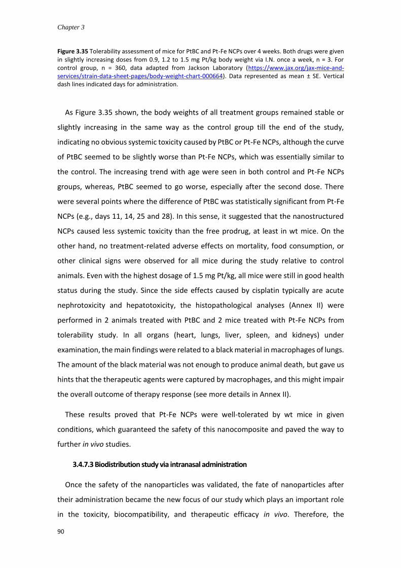
Chapter 3
90
Figure 3.35 Tolerability assessment of mice for PtBC and Pt-Fe NCPs over 4 weeks. Both drugs were given in slightly increasing doses from 0.9, 1.2 to 1.5 mg Pt/kg body weight via I.N. once a week, n = 3. For control group, n = 360, data adapted from Jackson Laboratory (https://www.jax.org/jax-mice-and-services/strain-data-sheet-pages/body-weight-chart-000664). Data represented as mean ± SE. Vertical dash lines indicated days for administration.
As Figure 3.35 shown, the body weights of all treatment groups remained stable or
slightly increasing in the same way as the control group till the end of the study,
indicating no obvious systemic toxicity caused by PtBC or Pt-Fe NCPs, although the curve
of PtBC seemed to be slightly worse than Pt-Fe NCPs, which was essentially similar to
the control. The increasing trend with age were seen in both control and Pt-Fe NCPs
groups, whereas, PtBC seemed to go worse, especially after the second dose. There
were several points where the difference of PtBC was statistically significant from Pt-Fe
NCPs (e.g., days 11, 14, 25 and 28). In this sense, it suggested that the nanostructured
NCPs caused less systemic toxicity than the free prodrug, at least in wt mice. On the
other hand, no treatment-related adverse effects on mortality, food consumption, or
other clinical signs were observed for all mice during the study relative to control
animals. Even with the highest dosage of 1.5 mg Pt/kg, all mice were still in good health
status during the study. Since the side effects caused by cisplatin typically are acute
nephrotoxicity and hepatotoxicity, the histopathological analyses (Annex II) were
performed in 2 animals treated with PtBC and 2 mice treated with Pt-Fe NCPs from
tolerability study. In all organs (heart, lungs, liver, spleen, and kidneys) under
examination, the main findings were related to a black material in macrophages of lungs.
The amount of the black material was not enough to produce animal death, but gave us
hints that the therapeutic agents were captured by macrophages, and this might impair
the overall outcome of therapy response (see more details in Annex II).
These results proved that Pt-Fe NCPs were well-tolerated by wt mice in given
conditions, which guaranteed the safety of this nanocomposite and paved the way to
further in vivo studies.
3.4.7.3 Biodistribution study via intranasal administration
Once the safety of the nanoparticles was validated, the fate of nanoparticles after
their administration became the new focus of our study which plays an important role
in the toxicity, biocompatibility, and therapeutic efficacy in vivo. Therefore, the

Development of nanoscale coordination polymers for glioblastoma treatment based on Pt(IV) prodrug
91
distribution of Pt-Fe NCPs in tumor and main organs was performed in GL261 GB-bearing
mice.
The reticuloendothelial system (RES) or mononuclear phagocyte system (MPS) is
responsible for the removal of colloidal stains in the blood circulation.102 Generally, liver
and spleen are the dominant organs to eliminate the NPs in bloodstream. However, they
may clear a large fraction of NPs from the blood circulation, and the excessive NPs could
accumulate in other macrophage-rich tissues such as lungs and adipose tissues.103 The
properties of the NPs affect considerably the biodistribution, while the size of NPs was
considered as one of the most important factors that determine the biodistribution
kinetics. 104 Figure 3.36 illustrated the size-dependent physiological barriers against NPs
blood circulation.
Figure 3.36 Scheme illustrating the size-dependent physiological barriers against NPs blood circulation.104
Unlike other administration routes, there are few reports about the biodistribution of
Pt agents delivered by intranasal administration. It is intriguing and valuable to evaluate
if Pt-Fe NCPs can gain access to the brain and GB tumor, the target sites, via intranasal
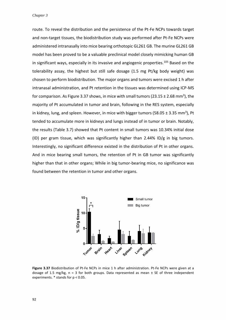
Chapter 3
92
route. To reveal the distribution and the persistence of the Pt-Fe NCPs towards target
and non-target tissues, the biodistribution study was performed after Pt-Fe NCPs were
administered intranasally into mice bearing orthotopic GL261 GB. The murine GL261 GB
model has been proved to be a valuable preclinical model closely mimicking human GB
in significant ways, especially in its invasive and angiogenic properties.105 Based on the
tolerability assay, the highest but still safe dosage (1.5 mg Pt/kg body weight) was
chosen to perform biodistribution. The major organs and tumors were excised 1 h after
intranasal administration, and Pt retention in the tissues was determined using ICP-MS
for comparison. As Figure 3.37 shows, in mice with small tumors (23.15 ± 2.68 mm3), the
majority of Pt accumulated in tumor and brain, following in the RES system, especially
in kidney, lung, and spleen. However, in mice with bigger tumors (58.05 ± 3.35 mm3), Pt
tended to accumulate more in kidneys and lungs instead of in tumor or brain. Notably,
the results (Table 3.7) showed that Pt content in small tumors was 10.34% initial dose
(ID) per gram tissue, which was significantly higher than 2.44% ID/g in big tumors.
Interestingly, no significant difference existed in the distribution of Pt in other organs.
And in mice bearing small tumors, the retention of Pt in GB tumor was significantly
higher than that in other organs; While in big tumor-bearing mice, no significance was
found between the retention in tumor and other organs.
Figure 3.37 Biodistribution of Pt-Fe NCPs in mice 1 h after administration. Pt-Fe NCPs were given at a dosage of 1.5 mg/kg, n = 3 for both groups. Data represented as mean ± SE of three independent experiments. * stands for p < 0.05.
Development of nanoscale coordination polymers for glioblastoma treatment based on Pt(IV) prodrug
93
Pt retention in mice
with small tumors
(% dose/g of tissue)
Pt retention in mice
with big tumors
(% dose/g of tissue)
Significance Average
(% dose/g of
tissue)
Tumor 10.34 ± 1.09 2.44 ± 0.76 Yes 6.39 ± 0.95
Brain 3.06 ± 1.40 0.85 ± 0.30 No 1.95 ± 1.10
Heart 1.75 ± 0.78 0.60 ± 0.13 No 1.18 ± 0.58
Liver 4.50 ± 2.89 3.09 ± 1.02 No 3.80 ± 0.71
Spleen 2.44 ± 1.68 1.19 ± 0.17 No 1.82 ± 0.63
Lung 4.06 ± 2.0 3.35 ± 1.45 No 3.70 ± 0.36
Kidney 3.58 ± 2.0 3.48 ± 0.21 No 3.53 ± 0.05
Table 3.7 Biodistribution study of Pt-Fe NCPs in mice. Dose = 1.5 mg Pt/kg, biodistribution time = 1 h, n = 3. The statistical difference was examined between mice group with small tumors and that with big tumors.
The BBB was reported to be disrupted at early stages after implantation of GL261
cells, within 2 weeks post implantation.105 The integrity of the BBB should be
compromised in both cases with tumors 2-3 weeks post implantation, and the
permeability of BBB/BBTB was supposed to be increased in mice with bigger tumors
than that with smaller tumors. However, the distribution results demonstrated that the
drug retention did not show positive correlation with tumor volume, which suggests the
permeability of the BBB is not the determinant contributor to the drug retention in CNS
herein. This might be explained partially by the angiogenic characteristics of GB, in which
heterogeneous vasculature features substantially, resulting in hypoxia and high
interstitial fluid pressure in the tumor core. These phenomena deteriorate considerably
along with the growth of tumors, making the drug delivery into bigger tumors even more
difficult.106
A recent paper reported blood-triggered Pt NPs with an average size of 86 nm and a
surface charge of -9 mV, which were similar to our Pt-Fe NCPs. The biodistribution of
this novel Pt NPs was also studied in C57BL/6 mice via IV injection which has been one
of the main administration routes for in vivo studies. However, the Pt retention in brain
was neglectable at both 4 h and 24 h post IV injection.107 Another report showed that IN
delivery resulted in significantly higher accumulation of Au NPs in cerebral cortex and
cerebellum, as well as in olfactory lobe, compared to the retention of 10-fold higher

Chapter 3
94
initial dose via intraperitoneal route 4 h post administration in C57BL/6 mice.108 These
results corroborated that intranasal administration could be an alternative route to
bypass the BBB and to effectively deliver drugs to the CNS directly.
3.4.7.4 In vivo anticancer efficacy for GB treatment
The in vivo efficacy of Pt-Fe NCPs was evaluated in orthotopic GL261 GB-bearing
C57BL/6J mice. GB is usually considered as an immunologically “cold” tumor, with
limited clinical response to the therapy.109 However, previous studies proved that it can
be converted to immunologically “hot” tumor when immunogenic cell death was
induced by TMZ treatment in “immune-enhancing metronomic schedule (IMS)”.110 This
study, conducted by GABRMN researchers, found that therapeutic administration
schedules respectful with the length described for mice immune cycle (6 days) produced
improved results in comparison with previously described schedules.111 The proposed
hypothesis for the events triggered in IMS-TMZ administration is schematically shown
in Figure 3.38.41,112 Therefore, therapeutic approaches triggering immunogenic cell
death/damage, when used in 6-day interval can trigger host immune response and be
associated with long-term, tumor-specific antitumor immune memory to greatly
increase the chemotherapy efficacy.113 Given that Pt agents were reported to be able to
trigger immunogenic effects,114-115 the first round therapy of Pt-Fe NCPs was given in
such IMS schedule starting on day 10 post implantation (p.i.) with a 6-day interval, and
the tumor evolution was monitored by high-resolution T2w MRI acquisition, as the
treatment schedule shown in Figure 3.39.

Development of nanoscale coordination polymers for glioblastoma treatment based on Pt(IV) prodrug
95
Figure 3.38 Hypothetic scheme of the cycle for immune response against a preclinical GL261 GB under IMS treatment.112 The whole cycle in mouse brain is assumed to take 6-7 days. DCs - dendric cells, CTLs – cytotoxic T lymphocyte cells.
Figure 3.39 Immune-enhancing metronomic schedule (IMS) used for GL261 GB therapy in mice. PtBC or Pt-Fe NCPs was administered every 6 days, tumor volumes acquired by high-resolution MRI T2W scanning twice per week.
In this in vivo study, in total 9 mice were randomly divided into three groups: control,
PtBC, and Pt-Fe NCPs. The mice from control group were untreated, and the mice from
Pt groups were administered intranasally at a dose of 1.5 mg Pt/kg body weight, which
was the highest dose in the tolerability assessment and proved to be well-tolerated and
safe for wt mice. The treatment initiated on day 10 p.i. when the GB volume was 6.07 ±
0.81 mm3, and there were no significant differences in tumor volumes between three
groups. As Figure 3.40 shown, both therapeutic drugs seemed to slightly slower down

Chapter 3
96
the tumor growth in comparison with the control group. The average curve may be tricky
to show a tumor growth arrest at day 21 p.i. but there was one case in both PtBC and
Pt-Fe NCPs groups (as the arrows indicated in Figure 3.40) responded to the therapy and
they showed transient growth shrinkage on day 23 p.i. but soon relapsed when scanned
on day 26 p.i. (no deviation bars on day 23 and 26 p.i., n=1). The separated curves
showed clearly that there was one case presenting more favorable evolution in both
groups treated with PtBC and Pt-Fe NCPs.
Figure 3.40 Tumor volume evolution of mice from control (untreated), treated with PtBC (1.5 mg Pt/kg), and Pt-Fe NCPs (1.5 mg Pt/kg), n = 3 for each group. Data represented as average ± SE. * stands for p < 0.05, all compared with control group. Vertical dash lines indicated the days for therapy administration.
Overall, the results suggested that both the prodrug and the NCPs can inhibit tumor
growth provided the tumor volume was small enough, though no differences between
themselves. This was confirmed by the increase rate in the tumor volume doubling time,
which is defined as time needed for a given tumor to duplicate its volume. Doubling
times were 2.1 ± 0.4 days, 2.5 ± 0.1 and 2.4 ± 0.3 days for control, PtBC and Pt-Fe NCPs,
respectively, although no significant difference was found.

Development of nanoscale coordination polymers for glioblastoma treatment based on Pt(IV) prodrug
97
Figure 3.41 Kaplan-Meier survival curves of three groups (where the green curve from Pt-Fe NCPs was superimposed with the red line from PtBC due to the same survival time).
As Figure 3.41 shown, both Pt agents displayed same survival time whose survival
curves were superimposed. The Log-rank test (typical for this type of data) did not show
statistically significant differences between groups, although the average of the treated
animals was slightly higher, driven by the single case surviving 26 days. The survival
averages of control and Pt agents-treated groups were 19.7 ± 2.1 days and 22.7 ± 2.9
days, respectively.
The body weights of the mice during therapy were recorded twice a week (Figure
3.42). The body weights of control group dropped very fast under 80% of the initial
weights on day 21 p.i., followed by other two groups. All animals had to be euthanized
after their body weights reduction was more than 20% due to the welfare consideration.
Figure 3.42 Evolution of body weights of mice in IMS schedule from control, PtBC (1.5 mg Pt/kg), and Pt-Fe NCPs (1.5 mg Pt/kg), n = 3 for each group. Data represented as average ± SE. Vertical dash lines indicated the days for administration. Horizontal dash line indicated the 20% weight reduction point, below which the mice had to be euthanized due to welfare consideration.
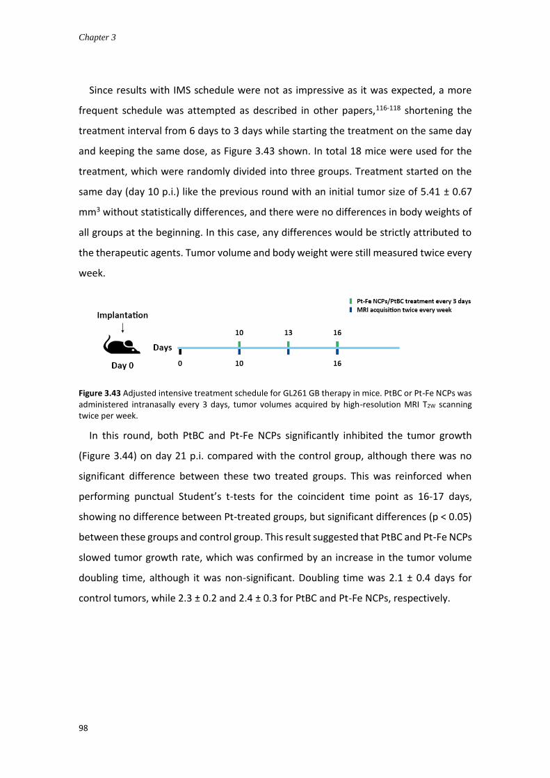
Chapter 3
98
Since results with IMS schedule were not as impressive as it was expected, a more
frequent schedule was attempted as described in other papers,116-118 shortening the
treatment interval from 6 days to 3 days while starting the treatment on the same day
and keeping the same dose, as Figure 3.43 shown. In total 18 mice were used for the
treatment, which were randomly divided into three groups. Treatment started on the
same day (day 10 p.i.) like the previous round with an initial tumor size of 5.41 ± 0.67
mm3 without statistically differences, and there were no differences in body weights of
all groups at the beginning. In this case, any differences would be strictly attributed to
the therapeutic agents. Tumor volume and body weight were still measured twice every
week.
Figure 3.43 Adjusted intensive treatment schedule for GL261 GB therapy in mice. PtBC or Pt-Fe NCPs was administered intranasally every 3 days, tumor volumes acquired by high-resolution MRI T2W scanning twice per week.
In this round, both PtBC and Pt-Fe NCPs significantly inhibited the tumor growth
(Figure 3.44) on day 21 p.i. compared with the control group, although there was no
significant difference between these two treated groups. This was reinforced when
performing punctual Student’s t-tests for the coincident time point as 16-17 days,
showing no difference between Pt-treated groups, but significant differences (p < 0.05)
between these groups and control group. This result suggested that PtBC and Pt-Fe NCPs
slowed tumor growth rate, which was confirmed by an increase in the tumor volume
doubling time, although it was non-significant. Doubling time was 2.1 ± 0.4 days for
control tumors, while 2.3 ± 0.2 and 2.4 ± 0.3 for PtBC and Pt-Fe NCPs, respectively.

Development of nanoscale coordination polymers for glioblastoma treatment based on Pt(IV) prodrug
99
Figure 3.44 Tumor volume evolution of mice from control (untreated), PtBC (1.5 mg Pt/kg), and Pt-Fe NCPs (1.5 mg Pt/kg), n = 6 for each group. Data represented as average ± SE. * stands for p < 0.05, all compared with control group. Vertical dash lines indicated the days for administration.
However, the survival time (Figure 3.45) seemed not beneficial from this intensive
schedule. The control cases had a survival of 19.7 ± 2.1 days, compared to 19 days’
survivals for both groups treated with Pt agents (all of them presented equal or less than
20% body weight decrease as Figure 3.46 indicated, signal of animal suffering and
euthanasia recommended). So, in this sense we can affirm that the tumor volumes were
smaller and far from the sizes that can kill animals (that may reach 150 - 200 mm3 easily
at final stages as the control), but animals showed signals of suffering. Therefore, the
intensive dosing significantly slowed down the growth rate of GB tumors, but failed to
prolong survival time.
Figure 3.45 Kaplan-Meier survival curves of three groups (where the green curve from Pt-Fe NCPs was superimposed with the red line from PtBC due to the same survival time).

Chapter 3
100
Figure 3.46 Body weight evolution of mice treated with vehicle, PtBC and Pt-Fe NCPs, n = 6 for each group. Data represented as average ± SE. Vertical dash lines indicated the days for administration. Horizontal dash line indicated the 20% weight reduction point, below which the mice had to be euthanized due to welfare consideration.
The survival results obtained with the dose-intense treatment schedule were not
encouraging, and indicated possible toxicity arisen from the intensive dosing. Previous
studies suggested that GB tumors with smaller volumes presented more favorable
responses,119 and it was in accordance to the results obtained in the first round of in vivo
therapy where the case with smallest volume ended with largest survival and smallest
tumor volume. So, the treatment schedule was adapted again, turning back to IMS
schedule but starting the treatment earlier on day 6 p.i. as Figure 3.47 shown. The
average tumor volume was 0.89 ± 0.26 mm3 on day 6 p.i., without significant differences
between groups, no difference of body weights as well.
Figure 3.47 IMS treatment schedule used for GL261 GMB therapy in mice starting on day 6 p.i. PtBC or Pt-Fe NCPs was administered every 6 days starting at day 6 p.i., tumor volumes acquired by MRI high-resolution T2W scanning twice per week.

Development of nanoscale coordination polymers for glioblastoma treatment based on Pt(IV) prodrug
101
Figure 3.48 Tumor volume evolution of mice of mice from control (untreated), PtBC (1.5 mg Pt/kg), and Pt-Fe NCPs (1.5 mg Pt/kg), n = 5 for each group. Data represented as average ± SE. * stands for p < 0.05, all compared with control group. Vertical dash lines indicated the days for administration.
The evolution of tumor volume (Figure 3.48) displayed that treatment with PtBC did
not slower down tumor growth rate, in which, in fact, the GB tumors grew even faster
than the control, and all mice lost more than 20% of body weights on day 15 p.i. (Figure
3.49). Nevertheless, the inflection point in Pt-Fe NCPs indicated that the growth of
tumor was successfully inhibited. Herein, the response to therapy of Pt-Fe NCPs was
variable. The evolution was fast at initial stages, then the tumor growth got arrested
after day 15 p.i. The separated figure showed clearly that one case in Pt-Fe NCPs group
got cured which reached the largest volume of 6.03 mm3 on day 15 p.i., then shrunk and
disappeared without relapse afterwards until it died on day 61 p.i., achieving the criteria
of cured mice. Commonly, when GL261 tumors shrunk until abnormal mass detection
by MRI became no longer possible, or the volume remained stable (usually less than 2
mm3), the treatment was halted. Then, MR monitoring kept twice a week. Whenever
the tumor volume of the residual/abnormal mass kept stable or non-detectable for 1
month, mice were transiently declared “cured” as in reference.41,120 To demonstrate
clearly the tumor evolution, two cases in Pt-Fe NCPs (Figure 3.50) were chosen to show
the follow-up MRI T2W images (Figure 3.51).
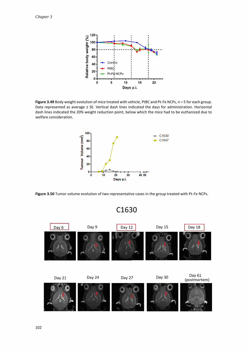
Chapter 3
102
Figure 3.49 Body weight evolution of mice treated with vehicle, PtBC and Pt-Fe NCPs, n = 5 for each group. Data represented as average ± SE. Vertical dash lines indicated the days for administration. Horizontal dash lines indicated the 20% weight reduction point, below which the mice had to be euthanized due to welfare consideration.
Figure 3.50 Tumor volume evolution of two representative cases in the group treated with Pt-Fe NCPs.

Development of nanoscale coordination polymers for glioblastoma treatment based on Pt(IV) prodrug
103
Figure 3.51 Axial T2w high-resolution MRI acquired at 7 T for animals C1630 and C1647 at the different time points assessed. The tumor was highlighted with a red arrow. From day 21-24 p.i. mouse C1630 was declared cured and tumor reduced to a visible but stable scar with no evolution in further follow-up. This animal was found dead at day 61 p.i. and a post-mortem fast MRI exploration was performed in order to discard presence of visible tumor.
Thus, with this round of Pt-Fe NCPs treatment, 20% of the cases responded to the
therapy with a curation rate of 20%, 20% of the cased presented growth arrest after the
second therapy cycle, while the remaining cased had the same behavior as the PtBC
group showing no therapeutic effects. Regarding the survival (Figure 3.52), Log-rank test
gave significant difference for PtBC which presented lower survival than other groups in
this case. Control cases had a survival of 19.7 ± 2.1 days, while PtBC had a survival of
14.8 ± 0.8 days and Pt-Fe NCPs in average 25 ± 19.8 days.
Figure 3.52 Kaplan-Meier survival curves of three groups for IMS treatment starting on day 6 p.i.
For GB treatment, current chemotherapies failed to provide substantial clinical
benefits and long-term control. The development of new chemotherapeutic agents is at
an early stage with uncertain output.121 Parallel efforts should be undertaken in

Chapter 3
104
optimizing/repurposing the delivery and efficacy of existing anticancer agents.122 There
were some clinical trials attempting to incorporate cisplatin into the GB treatment
regimen, commonly combined with concurrent TMZ chemotherapy in recurrent GB
patients. However, limited clinical benefits were obtained with an overall response rate
about 20%.123-125 Therefore, it is urgent and worthy to explore the potential efficacy of
Pt agents against GB with alternative delivery routes and treatment patterns, especially
in in-vivo preclinical models.
Our in vivo studies suggested that the therapy starting volume and treatment
schedule matter a lot for the GB therapy. Herein, 6 days-interval IMS and intense every-
3-days schedule were adopted during the in vivo studies. And the IMS schedule turned
out to be more effective and can prolong the survival time, even cure the experimental
animal when starting the therapy at earlier stage on day 6 p.i. On the other hand, the
intense dosing schedule successfully inhibited the GB tumor growth, but shortened the
survival time of treated animals due to the possible cumulative side toxicity. This was in
good accordance with other chemotherapeutic agents administered in such a
metronomic manner while receiving optimal therapeutic output.41,113,120 In addition, the
starting point for the therapy also played an important role for the efficacy of
chemotherapy. When comparing the in vivo therapies started on day 10 p.i. and on day
6 p.i., both in IMS schedule, the therapeutic outcome of therapy starting with small
tumors (0.89 ± 0.26 mm3) prevailed much compared to that of therapy starting with big
ones (6.07 ± 0.81 mm3). The therapy starting on day 6 p.i. resulted in significant
inhibition on tumor growth with 20% curation, 20% partial response, and prolonged
survival time; while the therapy starting with bigger tumor volume led to non-significant
difference in tumor growth, and close survival time as control. This was also in good
agreement with other studies.119,126-127
The presence of low-permeable BBB/BBTB has long been an obstacle to the successful
drug delivery for GB treatment, which compromises greatly the antitumor effects of
most chemotherapeutic agents.128-129 It is worth noting that the intranasal
administration can work as a great alternative which is a noninvasive, direct and more
effective route to deliver therapeutic drugs into CNS compared to other administration
routes. Our in vivo therapy proved that Pt-Fe NCPs administered intranasally could

Development of nanoscale coordination polymers for glioblastoma treatment based on Pt(IV) prodrug
105
inhibit the GB tumor growth and prolong the survival time of GL261 tumor-bearing mice,
given optimized treatment schedule. A previous study evaluated the in vivo anticancer
efficacy of cisplatin-loaded polybutylcyanoacrylate (PBCA) NPs in C6 GB bearing-rats via
intraperitoneal injection.130 The treatment started as early as 2 days p.i. at a at a dose
of 1 mg/kg (referred to cisplatin) and administered for 4 doses every 3 days. However,
the mean survival time of mice treated with cisplatin-PBCA NPs was 17.5 days compared
to 19.6 days of free cisplatin group, indicating the possible toxicity induced by the
nanoformulation, which was confirmed by hematoxylin and eosin staining of liver
tissues with acute tubular necrosis. In this case, the therapy with nanosized formulation
of cisplatin did not work even starting with extremely small tumor volumes and intense-
dosing schedule via intraperitoneal route.
Despite IN route, other delivery routes have been also investigated for GB treatment
in vivo. Among others, local administration has been widely explored due to its potential
effectiveness arisen from direct delivery into GB tumors. For instance, the anticancer
activity of Pt(IV) prodrug-loaded coiled nanotubes in GB cells and in U87MG xenograft-
bearing mice.131 After a single intratumoral injection with the drugs, the reduced
bioluminescence intensity indicated that Pt(IV) coiled nanotubes induced significant GB
cell death in the xenograft in comparison to the PBS group, though no significant
difference was found compared to the Pt(IV) prodrug. This result was confirmed by
TUNEL assay and histological examination. However, no further quantitative in vivo
studies were revealed in this report, which was insufficient to have insight into the in
vivo antitumor efficacy of the reported Pt(IV) prodrug-based nanotubes.
Our in vivo results displayed that synchronized IMS administration of Pt-Fe NCPs with
the cancer-immunity cycle brought certain chemotherapeutic effects in the preclinical
GL261 orthotopic models. The cured case treated with Pt-Fe NCPs was not detected any
signs of recurrence, which was an encouraging information. Together with other studies,
this implied that host immunity may play a pivotal role during the chemotherapy and
can be harnessed as an incredible comrade for the treatment of GB.41,132 Interestingly,
a recent report demonstrated that CED cisplatin induced cure in immunocompetent
GL261 models, but failed to cure immunocompromised NSG mice with intracranial
tumors, revealing that the therapeutic effects of CED cisplatin was immune-

Chapter 3
106
dependent.133 This provides an important hint that the administration schedule of
chemotherapeutic agents should be in harmony with the immune cycle to recruit the
host immune system into the battle against GB. Nevertheless, the role of host immunity
in our in vivo studies should be elucidated by further experiments in the future to
investigate the possible modulation of immunogenic effects.
3.4.7.5 in vivo imaging
The experimental results derived from in vitro relaxivity studies (see section 3.6)
suggested that Pt-Fe NCPs could have potential as a promising T2 contrast agent.
However, it was necessary to evaluate the contrast effect in vivo. The complexity of in
vivo environment can influence CA performances and in vitro results do not always
translate in suitable in vivo performances.134 Therefore, in vivo contrast enhancement
studies were carried out using orthotopic GL261 GB-bearing mice. Briefly, the
experimental mice were subjected to scanning with both T1 and T2 MRI and maps
acquisition prior to and after administration. The obtained images were compared as
the representative images shown in Figure 3.53.
Figure 3.53 A) T2 and B) T1 weighted coronal MRI, acquired at 7 T horizontal preclinical scanner, of C1639 GL61 GB bearing mouse, 15 days after tumor implantation. Images were acquired pre (left) and post (right) after intranasal administration of Pt-Fe NCPs at a dose of 1.5 mg Pt/kg body weight. Note that since

Development of nanoscale coordination polymers for glioblastoma treatment based on Pt(IV) prodrug
107
mice should be moved out from MR scanner for intranasal CA administration, the acquisition geometry is not fully coincident in pre- and post-administration MRI acquisitions. T1 and T2 maps were also acquired (not shown) and no significant differences were found. For this mouse, values found in tumor regions were 55.8ms and 2470.4ms (T2 and T1 respectively) pre-administration and 57.4ms and 2646.9ms (T2 and T1 respectively) post-administration. See section 3.5 for more acquisition parameter details.
The corresponding T1 and T2 values were calculated pre- and post-administration
(Table 3.8) for Pt-Fe NCPs. Variance homogeneity tests were non-significant; hence
Student’s t-tests were used for comparisons. The only significant one was T1 ipsilateral
(tumor) zone pre- and post-administration. However, it is also true that the decrease in
T1 was very limited, in average 4.7%, clearly not enough to produce a visible effect in
images. In T2, the values were non-significant pre- and post-administration, showing no
enhancement for contrast. In a previous work from our group, another type of Fe-NCPs
was synthesized and assessed its potential as a dual-mode CA.135 Notably, the maximal
contrast enhancement was observed at 9.4 ± 1.1 min for T1 and 5.3 ± 1.1 min for T2 after
IV injection of Fe-NCPs at a dose of 0.4 mmol Fe/kg in the GL261 tumor-bearing mice.
The iron amount injected from Fe-NCPs was 16-fold higher than that from Pt-Fe NCPs in
our case. This might explain the situation that such low concentration of Pt-Fe NCPs may
result in low intratumoral concentration of Fe, which may be not sufficient to enhance
contrast at the given conditions. To work as a theranostic agent, optimization need to
be further investigated in the future.
T1 pre-adm. T1 post-adm. T2 pre-adm. T2 post-adm.
Ipsilateral (tumor) zone
2686.92±84.4 2560.04±128.17 55.01±2.19 56.16±3.45
Contralateral zone 2229.75±69.34 2173.44±78.87 46.71±0.81 46.82±1.31
Observation: if there were some irregular zones suggestive of bleeding, these zones were avoided in calculation, since T2 values would be artificially reduced.
Table 3.8 T1 and T2 values pre- and post-administration (all values shown in ms)
3.5 Conclusion
Novel Pt(IV) prodrug-based nanoscale coordination polymers with an average size of
66.0 ± 2.3 nm, a PDI of 0.258 ± 0.013, and a slightly negative ζ-potential of -8.53 ± 0.418
mV, were designed and successfully synthesized and evaluated as drug delivery systems
for GB treatment. The obtained NCPs shows relevant advantages such as:

Chapter 3
108
• Chemical and colloidal stability, controlled drug release, and MRI relaxivity
properties compatible with its use as contrast agent for MRI.
• Pt-Fe NCPs exhibited comparable even superior cytotoxic effect compared to
cisplatin against HeLa, 1Br3G, and GL261 cell lines, while ROS generation was
notably dependent on the cell lines. Interestingly, Pt-Fe NCPs and PtBC did not
induce ROS formation in the GL261 glioblastoma cell line, which corroborated
that the cytotoxic effect was totally attributed to the chemotherapeutic effect of
the released Pt compounds.
• One of the factors that determine the notable cytotoxic effect against cancer
cells was the superior intracellular uptake of Pt-Fe NCPs in comparison to
cisplatin or PtBC. However, the DNA-bound Pt from Pt-Fe NCPs was significantly
lower than cisplatin after 24 h of exposure, indicating slower intracellular
activation process.
• Pt-Fe NCPs were proved to be well-tolerated and biosafe at a dose up to 1.5mg
Pt/kg body weight in wt mice, and displayed favorable accumulation of Pt in GB
tumor and brain, implying the effective delivery of intranasal administration.
• Optimized therapy with Pt-Fe NCPs in immune-enhancing metronomic schedule
starting at day 6 post implantation resulted in significant inhibition on tumor
growth in GL261 orthotopic models, with 20% curation rate, 20% partial
response, and prolonged survival time.
• These results indicated that Pt-Fe NCPs possess a potential anti-GB activity both
in vitro and in vivo. Intranasal administration has been established as a
noteworthy alternative to typical IV systemic administration what represents a
notable advance for the design of novel therapeutic methodology that could be
used in combination with other therapies.
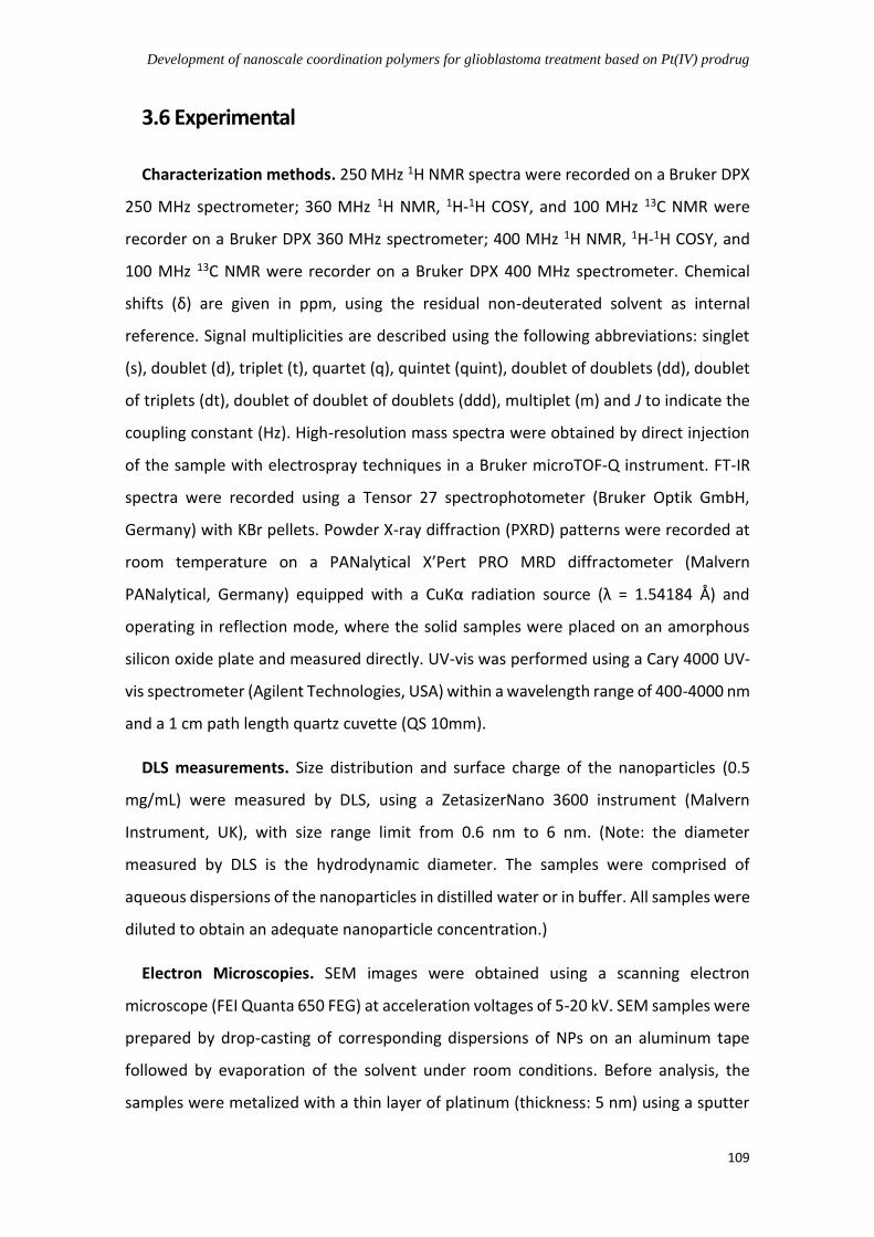
Development of nanoscale coordination polymers for glioblastoma treatment based on Pt(IV) prodrug
109
3.6 Experimental
Characterization methods. 250 MHz 1H NMR spectra were recorded on a Bruker DPX
250 MHz spectrometer; 360 MHz 1H NMR, 1H-1H COSY, and 100 MHz 13C NMR were
recorder on a Bruker DPX 360 MHz spectrometer; 400 MHz 1H NMR, 1H-1H COSY, and
100 MHz 13C NMR were recorder on a Bruker DPX 400 MHz spectrometer. Chemical
shifts (δ) are given in ppm, using the residual non-deuterated solvent as internal
reference. Signal multiplicities are described using the following abbreviations: singlet
(s), doublet (d), triplet (t), quartet (q), quintet (quint), doublet of doublets (dd), doublet
of triplets (dt), doublet of doublet of doublets (ddd), multiplet (m) and J to indicate the
coupling constant (Hz). High-resolution mass spectra were obtained by direct injection
of the sample with electrospray techniques in a Bruker microTOF-Q instrument. FT-IR
spectra were recorded using a Tensor 27 spectrophotometer (Bruker Optik GmbH,
Germany) with KBr pellets. Powder X-ray diffraction (PXRD) patterns were recorded at
room temperature on a PANalytical X’Pert PRO MRD diffractometer (Malvern
PANalytical, Germany) equipped with a CuKα radiation source (λ = 1.54184 Å) and
operating in reflection mode, where the solid samples were placed on an amorphous
silicon oxide plate and measured directly. UV-vis was performed using a Cary 4000 UV-
vis spectrometer (Agilent Technologies, USA) within a wavelength range of 400-4000 nm
and a 1 cm path length quartz cuvette (QS 10mm).
DLS measurements. Size distribution and surface charge of the nanoparticles (0.5
mg/mL) were measured by DLS, using a ZetasizerNano 3600 instrument (Malvern
Instrument, UK), with size range limit from 0.6 nm to 6 nm. (Note: the diameter
measured by DLS is the hydrodynamic diameter. The samples were comprised of
aqueous dispersions of the nanoparticles in distilled water or in buffer. All samples were
diluted to obtain an adequate nanoparticle concentration.)
Electron Microscopies. SEM images were obtained using a scanning electron
microscope (FEI Quanta 650 FEG) at acceleration voltages of 5-20 kV. SEM samples were
prepared by drop-casting of corresponding dispersions of NPs on an aluminum tape
followed by evaporation of the solvent under room conditions. Before analysis, the
samples were metalized with a thin layer of platinum (thickness: 5 nm) using a sputter

Chapter 3
110
coater (Emitech K550). STEM images were obtained using a scanning-transmission
electron microscope (Magellan 400L, FEI), while TEM images were obtained using a
transmission electron microscope (Tecnai G2 F20, FEI) at a voltage of 200 kV. Samples
for STEM and TEM were prepared by drop-casting corresponding dispersions on TEM
grids (ultrathin carbon type-A, 400 mesh Cu grid, Ted Pella Inc., Redding, USA) and drying
overnight under room conditions prior to examination.
Mössbauer spectroscopy measurements. 57Fe Mössbauer spectra were recorded in
transmission geometry with a conventional Mössbauer spectrometer equipped with a
57Co(Rh) radioactive source operating at room temperature 300 K and relatively low
temperature 80 K. The samples were sealed in aluminum foil and mounted on a nitrogen
bath cryostat (Oxford Instruments). The spectra were fitted to the sum of Lorentzians
by a least-squares refinement using Recoil 1.05 Mössbauer Analysis Software. All isomer
shifts refer to α-Fe at room temperature.
Quantitative magnetic resonance imaging phantoms (MRI-Phantoms). The
longitudinal r1 and transverse r2 relaxation rates for Pt-Fe NCPs at different
concentrations were measured in 1% agarose/PBS gelation under an external magnetic
field of 7 T (300 MHz) Bruker Biospec 70/30 SUR spectrometer (Bruker BioSpin GmbH,
Germany) with corresponding acquisition sequences. Specifically, the NCPs were
dispersed in 1.5 mL of 1% agarose/PBS solutions at 65 ℃ in an ultrasound bath to ensure
a good colloidal suspension, and solidified at room temperature, resulting in a series of
gel with different metal concentrations (3.94, 1.97, 1, 0.1, and 0 mM of Fe). The obtained
relaxation rate values were plotted versus the concentrations of iron.
Thermogravimetric analysis (TGA) and calorimetric measurements (DSC). Samples
were run on a Netzsch TG 209 F1 thermal gravimetric analyzer, holding in aluminium
crucibles at a heating rate of 10 K/min from 25 to 900 ℃ under air atmosphere.
Differential scanning calorimetry (DSC) were measured using the same equipment with
a cryostat under same conditions.
Inductively coupled plasma-Mass spectrometry (ICP-MS). Standard solutions were
prepared prior to the measurements at a range from 0 – 200 parts per billion (ppb) using

Development of nanoscale coordination polymers for glioblastoma treatment based on Pt(IV) prodrug
111
atomic spectroscopic analytical ICP-MS standards (PerkinElmer Inc., Germany). The
isotope 54Fe and 57Fe were selected as Fe tracers, while 194Pt, 195Pt and 196Pt as Pt tracers.
Digestion procedures for ICP-MS measurement. All glassware was immersed in 20%
HNO3 for 48 h, and plasticware in 5% HNO3 for 4 h prior to use for ICP-MS. All samples
were digested using wet digestion method. Depending on the materials to digest,
different procedures were adopted.
(1) Chemical samples: samples, 5.0 mg of NPs for instance, were weighed accurately
with an analytical balance, and transferred into a vial, then concentrated ultrapure HNO3
(69%, Ultratrace®, ppb-trace analysis grade, Scharlab, Spain) was added. The samples
were placed in the fume hood for 48 h to be fully digested, then were diluted with 0.5%
(v/v) ultrapure HNO3 for further measurements.
(2) Cell pellets: the cell pellets were immersed in 100 L of concentrated ultrapure
HNO3 (69%, Ultratrace®, ppb-trace analysis grade, Scharlab, Spain) and left to digest
overnight. Samples were then heated to 90 ℃ till the suspensions turned clear. The
samples were diluted with 0.5% (v/v) ultrapure HNO3 to appropriate volumes for later
measurements.
(3) Tissues: the tissues were added with T-PERTM buffer (Thermo Fisher Scientific, USA)
in a ratio of 10 mL : 1 g, then cut into small pieces, and homogenized with ultrasonication
microtip (Branson Digital Sonifier 450, Emerson, USA) in cycles of 10 seconds on and 15
seconds off with an amplitude of 30%. Afterwards, the tissue suspensions were added
with aqua regia (all ppb-trace analysis grade) and heated up to 300 ℃, while 30% H2O2
was added in the later digestion process till the suspensions turned clear. The clear
solutions were transferred and made up with 0.5% (v/v) ultrapure HNO3 to appropriate
volumes for the determination of metal contents using ICP-MS.
Synthesis of Pt (IV) prodrug PtBC
Synthesis of oxoplatin. The synthetic sequence started from the commercially
available cis-diamminedichloroplatinum(II), or cisplatin (CDDP). Into a suspension of
cisplatin (1 g, 3.33 mmol) in Milli-Q water (44 mL), hydrogen peroxide aqueous solution
(30%, 5.72 mL, 66.6 mmol) was added. The mixture was heated to 76℃ in darkness
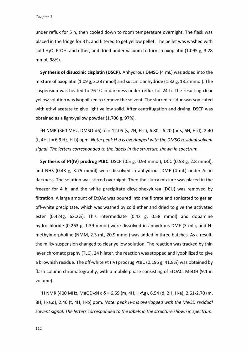
Chapter 3
112
under reflux for 5 h, then cooled down to room temperature overnight. The flask was
placed in the fridge for 3 h, and filtered to get yellow pellet. The pellet was washed with
cold H2O, EtOH, and ether, and dried under vacuum to furnish oxoplatin (1.095 g, 3.28
mmol, 98%).
Synthesis of disuccinic cisplatin (DSCP). Anhydrous DMSO (4 mL) was added into the
mixture of oxoplatin (1.09 g, 3.28 mmol) and succinic anhydride (1.32 g, 13.2 mmol). The
suspension was heated to 76 ℃ in darkness under reflux for 24 h. The resulting clear
yellow solution was lyophilized to remove the solvent. The slurred residue was sonicated
with ethyl acetate to give light yellow solid. After centrifugation and drying, DSCP was
obtained as a light-yellow powder (1.706 g, 97%).
1H NMR (360 MHz, DMSO-d6): δ = 12.05 (s, 2H, H-c), 6.80 - 6.20 (br s, 6H, H-d), 2.40
(t, 4H, J = 6.9 Hz, H-b) ppm. Note: peak H-a is overlapped with the DMSO residual solvent
signal. The letters corresponded to the labels in the structure shown in spectrum.
Synthesis of Pt(IV) prodrug PtBC. DSCP (0.5 g, 0.93 mmol), DCC (0.58 g, 2.8 mmol),
and NHS (0.43 g, 3.75 mmol) were dissolved in anhydrous DMF (4 mL) under Ar in
darkness. The solution was stirred overnight. Then the slurry mixture was placed in the
freezer for 4 h, and the white precipitate dicyclohexylurea (DCU) was removed by
filtration. A large amount of EtOAc was poured into the filtrate and sonicated to get an
off-white precipitate, which was washed by cold ether and dried to give the activated
ester (0.424g, 62.2%). This intermediate (0.42 g, 0.58 mmol) and dopamine
hydrochloride (0.263 g, 1.39 mmol) were dissolved in anhydrous DMF (3 mL), and N-
methylmorpholine (NMM, 2.3 mL, 20.9 mmol) was added in three batches. As a result,
the milky suspension changed to clear yellow solution. The reaction was tracked by thin
layer chromatography (TLC). 24 h later, the reaction was stopped and lyophilized to give
a brownish residue. The off-white Pt (IV) prodrug PtBC (0.195 g, 41.8%) was obtained by
flash column chromatography, with a mobile phase consisting of EtOAC: MeOH (9:1 in
volume).
1H NMR (400 MHz, MeOD-d4): δ = 6.69 (m, 4H, H-f,g), 6.54 (d, 2H, H-e), 2.61-2.70 (m,
8H, H-a,d), 2.46 (t, 4H, H-b) ppm. Note: peak H-c is overlapped with the MeOD residual
solvent signal. The letters corresponded to the labels in the structure shown in spectrum.

Development of nanoscale coordination polymers for glioblastoma treatment based on Pt(IV) prodrug
113
13C NMR (100MHz, MeOD-d4): δ 180.99, 173.85, 144.74, 143.26, 130.79, 119.71,
115.52, 114.96, 41.08, 34.45, 31.30, 31.00.
HRMS (EI): calcd 803.1300, found 803.1284.
Pt content by ICP-MS: calcd (%) 24.25, found (%) 24.12.
Synthesis of Pt-Fe NCPs. To prepare a material suitable for biological experiments,
synthesis of the nanoparticles was carried out inside a biosafety cabinet (Telstar
BioVanguard B green), and all materials and solvents were sterilized by autoclaving prior
to use. PtBC (50 mg, 0.06 mmol) was dissolved in 96% EtOH (15 mL), and Fe(OAc)2 (10.8
mg, 0.06 mmol) in 1 mL of H2O was added dropwise. A black precipitate formed
immediately with the addition of iron ions. After 30 min’s stirring at 700 rpm, the
precipitate was collected by centrifugation and then washed by EtOH and H2O several
times. The solid was then lyophilized to get black powder which was stored in the freezer
for later use. Elemental analysis: found (%) C 28.63, H 2.54, N 5.79.
Metal contents by ICP-MS: 18.87% Pt, 15.13% Fe.
Coating for Pt-Fe NCPs. The two-step coating was carried out according to our
previously reported method in our group. Specifically, NCPs (10 mg/mL) were dispersed
in carbonate buffer (pH 8.2) in vial covered with pierced parafilm. HMDA (10 mM) and
CAT (10 mM) were added into the NCP suspension slowly. The mixture was kept stirring
gently for 90 min to form the coating. During this process, the dark suspension turned
brownish while the colorless media got reddish. Afterwards, the suspension was
centrifuged and washed by Milli-Q® water four times. The coated NCP was checked by
FTIR, SEM and ICP-MS.
After primer coating with HMDA and CAT, a second coating layer with PEG-diamine
was performed in order to improve the colloidal stability of the NCPs. Specifically, the
NCPs with primer coating were redispersed in isopropanol, followed by the addition of
PEG-diamine in isopropanol. The vial was closed and kept stirring gently for 90 min.
Finally, the coated NPs were collected by centrifugation at (7000 rpm × 3 min), and
washed with Milli-Q® water several times, then dried under vacuum and lyophilized for
long-term storage. The coated NCP was checked by FTIR, SEM and ICP-MS.

Chapter 3
114
Colloidal stability improvement of Pt-Fe NCPs. In order to improve the colloidal
stability of Pt-Fe NCPs suspension, a range of stabilizers were used and compared,
including PVP, PVA, PEG, PEG-amine, PEG-COOH, and BSA. Generally, the stabilizers
were dissolved in the solvent at a concentration of 1% (BSA at 10 mg/mL) before the
synthesis of Pt-Fe NCPs started. Once the reaction of NCPs finished, the precipitate was
collected and washed. Then the NCPs were redispersed in PBS at a concentration of 0.5
mg/mL. And the colloidal dispersion was checked by DLS and STEM.
Drug release assay. Drug release from Pt-Fe NCPs was determined using dialysis
method. Concentrated Pt-Fe NCPs at 1 mg/mL were resuspended into 1 mL of PB buffer
in dialysis bags (MWCO = 6000 – 8000). The dialysis tubes were immersed into sealed
beakers with 40 mL of PB buffers at pH 7.4 and 5.5. The beakers were incubated at 37 ℃
with gentle stirring during the study. Aliquot of 500 L was taken from the dialysate at
predetermined time points (0.5, 1, 2, 4, 6, 8, 24, and 48 h), followed by supplementing
with fresh buffer in same volume immediately. The amount of released Pt was
determined by ICP-MS. To test the influence of GSH, PB buffers with 2 mM and 10 mM
GSH were used for dialysis, remaining same other conditions.
Cells. The cell lines were obtained from the Tumor Bank Repository at the National
Cancer Institute (Frederick/MD, USA), including human cervical cancer cell HeLa, non-
malignant human fibroblast cell 1Br3G, murine glioblastoma cell GL261, and a pair of
human ovarian cancer cell lines, A2780 and its cisplatin resistant counterpart A2780/cis.
Before starting any protocol, all solutions were prewarmed to 37 °C. All materials and
equipment that came in contact with cells were sterile. All cell-related performances
were carried out in Class II biosafety hoods in cell culture labs with a biosafety level 2
(BSL-2 with code A/ES/19/I-08).
Cell culture. All cell lines were grown in 75 cm2 cell-culture flask with corresponding
culture media. HeLa was cultured in MEM medium, 1Br3G in DMEM, GL261 in RPMI
1640, A2780 in RPMI 1640, and A2780/cis in RPMI 1640 containing 1 M of cisplatin. All
culture media were supplemented with 10% fetal bovine serum (FBS, Gibco®, Invitrogen,
UK), 0.285 g/L glutamine, 2.0 g/L sodium bicarbonate, and 1% penicillin-streptomycin.
Cells were cultured as adherent monolayers and maintained in an incubator (HERAcell,
150i, Thermo Scientific) at 37 °C in 5% CO2 and relative humidity of 95%, except 1Br3G
Development of nanoscale coordination polymers for glioblastoma treatment based on Pt(IV) prodrug
115
with 10% CO2. All cell culture media, FBS, supplements, antibiotics, trypsin, and Trypan
Blue were purchased from Fisher Scientific (Gibco®, Invitrogen, UK).
To subculture/harvest the cells at log phase around 75% confluence, 4 mL of sterile
PBS was added into the flask to rinse the cells after discarding spent cell culture media
by aspiration with a vacuum pump. After washing, 3 mL of 0.05 % trypsin-
ethylenediaminetetraacetic acid (EDTA, 0.2 g/L) were added and the flask was returned
to incubator for 2 minutes. Observing the cells under the microscope, if ≥ 90% of the
cells have detached, 6 mL of fresh cell media was added into the flask and detached the
cells from the bottom. The cell suspension was transferred into a 15 mL flask (Falcon,
Corning, USA) to centrifuge (Thermo Scientific, USA) at 1400 rpm for 5 min to collect
cells. The supernatant was discarded, and the pellet was resuspended in fresh media or
PBS for cell counting, then an appropriate number of cells was seeded in a new flask.
Cell counting. The cells were counted using a Neubauer hemocytometer cell counter
and Trypan Blue (Gibco®, Invitrogen, UK) exclusion to count the cells and determine the
viability. Trypan Blue is a dye used to distinguish the living cells from the dead, since the
dead cells cannot extrude Trypan Blue without required energy mechanisms. An aliquot
of 10 L of the cell suspension obtained in the subculture step was added into 10 L of
Trypan Blue. Then, 10 L of the resulting mixture was added onto the Neubauer
hemocytometer and the number of living cells was counted under an optical
microscopy, or counted using the TC10TM automatic cell counter (Biorad, Hercules,
California).
In vitro cytotoxicity assays. The cells (HeLa, 1Br3G, GL261, A2780 and A2780/cis) in
exponential growth were seeded in 96-well plate (Corning, USA) with proper density.
HeLa cell line was seeded at a density of 2000 cells/well, 1Br3G at 3000 cells/well, GL261
at 4000 cells/well, A2780 and A2780/cis at 3000 cells/well. Specifically, the cisplatin-
containing cell culture media was replaced with fresh RPMI media without drug 4 hours
before seeding. After 24 hours’ incubation, fresh media containing compounds (Pt-Fe
NCPs, PtBC, and CDDP) at different concentrations (0, 0.1, 1, 5, 10, 25, 50, 100, 200 M
referred to Pt concentration) were added, and the plates were incubated for 24 or 72
hours. Afterwards, 10 l of PrestoBlue® (0.15 mg/ml, Thermo Scientific, USA) was added

Chapter 3
116
into each well and the plates were incubated for another 4 hours before measuring the
fluorescence at 572 nm with excitation at 531 nm by the microplate reader Victor 3
(Perkin Elmer, USA). For 1Br3G, the incubation time with PrestoBlue® was prolonged to
7 h in order to achieve enough difference of fluorescence intensity between
concentrations. All experiments were carried out in triplicate, data were treated and
calculated to obtain IC50s by GraphPad Prism software (version 7.0).
Estimation of ROS formation. HeLa, 1Br3G and GL261 were seeded in black 96-well
plates (Corning, USA) at a density of 20,000 cells per well to achieve full confluence.
After 24 hours’ incubation, the spent medium was discarded and cells were washed out
from the medium serum, and added prewarmed PBS with the fluorescent probe 2’,7’-
dichlorofluorescin diacetate (DCFCDA, final working concentration 10 M), then
incubated for 30 minutes. DCFCDA is a non-fluorescent permeable compound which can
be hydrolyzed by esterase and oxidized by intracellular ROS and turn to fluorescent and
non-permeable 2’,7’-dichlorofluorescein (DCF). After the probe was internalized, the
buffer with probe was replaced by only medium or media with compounds (H2O2,
Fe(OAc)2, Pt-Fe NCPs, PtBC, and CDDP) at the concentration of IC50 for 24 hours. 0.1 mM
of H2O2 was added as positive control, while 0.1 mM of Fe(OAc)2 was added for
comparison. After 24 hours, the fluorescence of each well was examined at 530 nm after
excitation at 485 nm by a microplate reader Victor 3 (Perkin Elmer, USA). This
experiment was repeated in triplicate independently. The results were normalized
based on the negative control loaded with dye but without drug treatment.
Cellular internalization. HeLa, 1Br3G and GL261 cells were seeded in 6-well plates
(Corning, USA) at a density of 300,000 cells per well with 1.5 ml of media. After 24 h of
incubation, the media were replaced by 1 ml of fresh media w/wo CDDP, PtBC, and Pt-
Fe NCPs at 100 M referred to Pt concentration. The treated cells were allowed to
internalize the compounds for 6 h in the incubator. Another cellular uptake assay was
also performed with a decreased concentration of Pt agents to 10 M, and increased
time points of 2, 4, 8, and 24 h. The media were removed immediately and cells were
rinsed twice by cold PBS in order to remove excessive compounds. Then 0.5 ml of trypsin
was added to each well for 3 min and 2-fold volume of fresh media was added to
neutralize the trypsin. After an aliquot was taken from each well for cell counting, the

Development of nanoscale coordination polymers for glioblastoma treatment based on Pt(IV) prodrug
117
rest cell suspensions were collected into 1.5 ml Eppendorf tubes (Corning, USA) and
centrifuged at 12,000 rpm for 4 min. The supernatants were discarded while the cell
pellets were stored at -80 ℃ for further quantification by ICP-MS.
DNA-bound Pt. In order to clarify the action mechanism of our Pt NPs, the DNAs were
extracted and measured by ICP-MS to quantify the DNA-bound Pt after uptake for 24 h
in HeLa, 1Br3G and GL261 cells. The cells in exponential growth were seeded onto cell
culture dishes in optimal condition for each cell line to reach 50 - 60 % of confluence in
24 h before the treatment. Then the drugs (CDDP, PtBC, and Pt-Fe NCPs) were added at
100 M referred to Pt concentration, and the cells were allowed to incubate for another
24 h. Afterwards, media with drugs were removed and the cells were rinsed, trypsinized
and collected by centrifugation and rinsed with cold PBS twice to remove excessive
drugs. The resulting cell pellets were resuspended in lysis buffer (pH 8.0, 150 mM Tris-
HCl, 100 mM NaCl and 0.5 % (w/v) SDS). Pellets in buffer were incubated on ice for 15
min and centrifuged at 15000 rpm for 15 min. 0.1 volume of RNase A was added into
each supernatant at 200 g/mL and incubated for 1 h at 37 ℃. Afterwards, Proteinase
K was added at 100 g/mL and incubated for 3 h at 56 ℃. A volume of
Phenol/Chloroform/Isoamyl alcohol (25:24:1 in volume, Thermo Scientific®) was added
and mixed gently. And after a centrifugation of 3 min at 15000 rpm, aqueous phases
containing DNA were transferred into sterile tubes. DNA was precipitated with 0.1
volume of 3 M sodium acetate and 1 volume of absolute ethanol at -20 ℃ overnight.
Then, the DNA samples were centrifuged for 15 min at 15000 rpm, and finally DNA
samples were dried and resuspended in 100 L of elution buffer (pH 8.0, 10 mM Tris-
HCl, 1 mM EDTA). The concentration of isolated DNA was quantified by measuring the
absorbance at 260 nm using NanoDropTM 1000 spectrophotometer (Thermo Fisher
Scientific, USA). And remaining samples were frozen for further ICP-MS analyses.
Animals. Animal experiments and handling have been performed in collaboration
with properly accredited personnel (Dr. Shuang Wu or Ms. Mª del Pilar Calero). Healthy
female C57BL/6J mice (8 - 12 weeks, body weight 20 - 24 g) were used for all in vivo
studies in this work, which were obtained from Charles River Laboratories (Charles River
Laboratories Internacional, I’Abresle, France) and housed in the animal facility (Servei
d’Estabulari, https://estabulari.uab.cat/) of the Universitat Autònoma de Barcelona. All
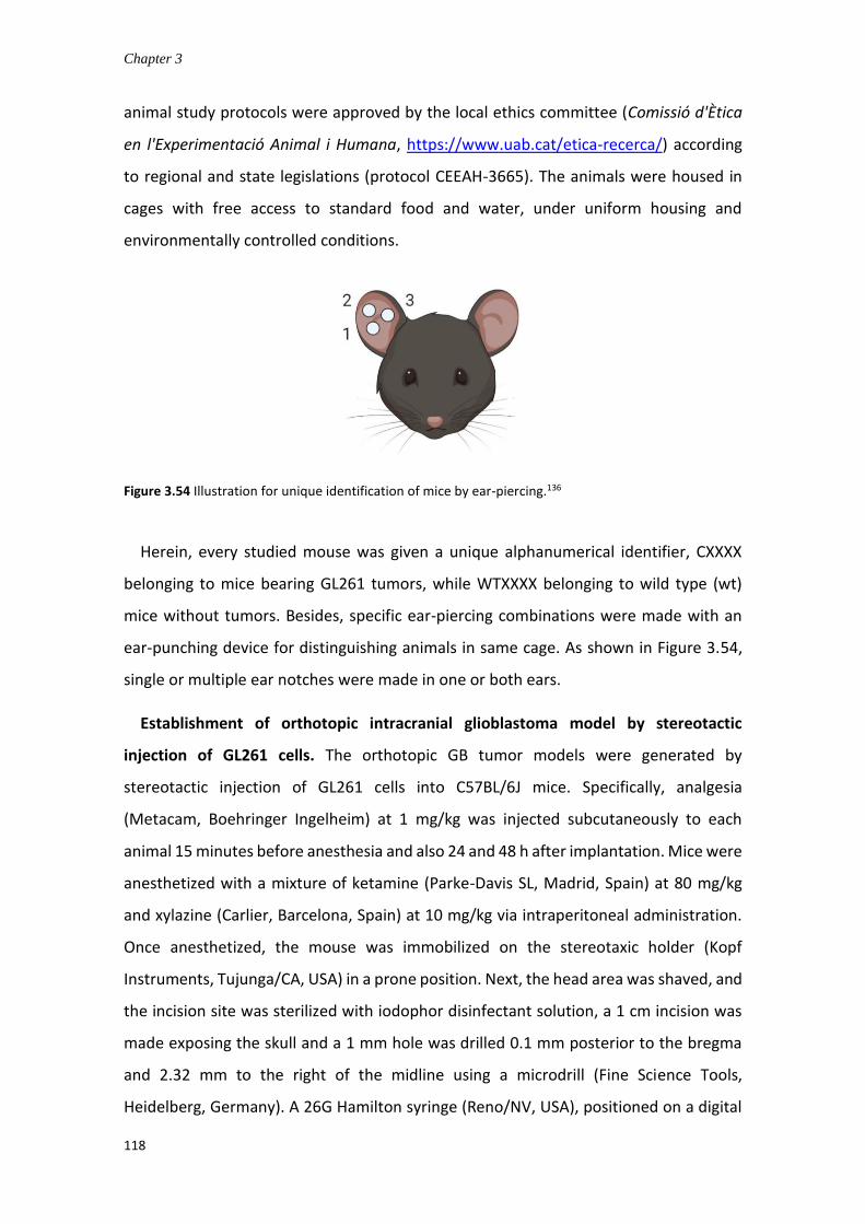
Chapter 3
118
animal study protocols were approved by the local ethics committee (Comissió d'Ètica
en l'Experimentació Animal i Humana, https://www.uab.cat/etica-recerca/) according
to regional and state legislations (protocol CEEAH-3665). The animals were housed in
cages with free access to standard food and water, under uniform housing and
environmentally controlled conditions.
Figure 3.54 Illustration for unique identification of mice by ear-piercing.136
Herein, every studied mouse was given a unique alphanumerical identifier, CXXXX
belonging to mice bearing GL261 tumors, while WTXXXX belonging to wild type (wt)
mice without tumors. Besides, specific ear-piercing combinations were made with an
ear-punching device for distinguishing animals in same cage. As shown in Figure 3.54,
single or multiple ear notches were made in one or both ears.
Establishment of orthotopic intracranial glioblastoma model by stereotactic
injection of GL261 cells. The orthotopic GB tumor models were generated by
stereotactic injection of GL261 cells into C57BL/6J mice. Specifically, analgesia
(Metacam, Boehringer Ingelheim) at 1 mg/kg was injected subcutaneously to each
animal 15 minutes before anesthesia and also 24 and 48 h after implantation. Mice were
anesthetized with a mixture of ketamine (Parke-Davis SL, Madrid, Spain) at 80 mg/kg
and xylazine (Carlier, Barcelona, Spain) at 10 mg/kg via intraperitoneal administration.
Once anesthetized, the mouse was immobilized on the stereotaxic holder (Kopf
Instruments, Tujunga/CA, USA) in a prone position. Next, the head area was shaved, and
the incision site was sterilized with iodophor disinfectant solution, a 1 cm incision was
made exposing the skull and a 1 mm hole was drilled 0.1 mm posterior to the bregma
and 2.32 mm to the right of the midline using a microdrill (Fine Science Tools,
Heidelberg, Germany). A 26G Hamilton syringe (Reno/NV, USA), positioned on a digital

Development of nanoscale coordination polymers for glioblastoma treatment based on Pt(IV) prodrug
119
push-pull microinjector (Harvard Apparatus, Holliston/MA, USA) was then used for
injection of 4 L of RPMI cell culture medium containing 100,000 GL261 cells (obtained
and counted as described in previous section) at a depth of 3.35 mm from the surface
of skull at a rate of 2 L/min.
Once the injection was completed, the Hamilton syringe was left untouched for 2
minutes more before its removal to prevent the cellular liquid leakage out of the skull.
Finally, the Hamilton syringe was gently and slowly withdrawn and the scission site was
closed with suture silk 6.0 (Braun, Barcelona, Spain). When the implantation was over,
the animal was left in a warm environment to recover from anesthesia.
The C57BL/6J immunocompetent mice were exposed to enriched environment (EE)
for 3 weeks before tumor implantation, since it was reported that it could significantly
reduce glioma growth and improve mice survival by increasing immunological
parameters in the brain of mice.137 Moreover, since this is the current protocol used at
GABRMN, the maintenance of basic parameters may help in further comparisons. Thus,
all mice in this thesis were allowed to endure 3 weeks of guarantee housing in EE-like
caging before tumor generation, and were maintained there from beginning to end
(Figure 3.55).
Figure 3.55 Illustration for “enriched environment”-like caging. Mice were allowed to endure 3 weeks of guarantee housing in EE-like caging before tumor generation, and EE was also maintained during the whole experiment and follow-up period. The ‘Shepherd Shack’ is cage insert composed of autoclavable paper, mice can shred it and use the paper for nesting.136

Chapter 3
120
For intranasal administration, the animals were anesthetized by isoflurane for 1 min
and kept in a horizontally supine position. The animals were administered with 2 L of
formulation for each nostril, with an interval about 2 minutes between each
administration. Treated animals were followed-up to evaluate tumour volume evolution
and the mice reaching endpoint criteria were euthanized for ethical considerations.
Tissue preservation procedures. When animals died or were euthanized by cervical
dislocation to prevent suffering, the brains and main organs (heart, lungs, spleen, liver
and kidneys) were excised and either frozen in liquid nitrogen or fixed in formalin,
depending on the purpose. In case of freezing, tumors were dissected and collected
from normal brain parenchyma, frozen in a liquid nitrogen container for further analysis.
In case of fixation, tissue was preserved in 4% formalin for further autopsy or
histopathological analysis. Tissues were resected after visual inspection of the whole
brain and tumor, avoiding as much as possible the cross contamination with non-
tumoral tissue.
In vivo MRI. The animals were subjected to non-invasive brain MRI scan to measure
the volumes, localization, and evolution of the GB. Each scan was performed at 7T
BioSpec 70/30 USR spectrometer (Bruker BioSpin GmbH, Ettlingen, Germany) at Servei
de Ressonància Magnètica from UAB (also part of Unit 25 of ICTS NANBIOSIS, NMR:
Biomedical Applications I https://www.nanbiosis.es/portfolio/u25-nmr-biomedical-
application-i/). Mice were positioned in a dedicated bed, which allowed the delivery of
anaesthesia (isoflurane, 1.5-2 % in O2 at 1 L/min), with an integrated heating circuit for
body temperature regulation. Respiratory frequency was monitored with a pressure
probe and kept between 60-80 breaths/min. GL261 tumor-bearing mice were scanned
to acquire high-resolution T2w images using a rapid acquisition with relaxation
enhancement (RARE) sequence to detect the brain tumors. The acquisition parameters
were as following: repetition time (TR)/effective echo time (TEeff) = 4200/36 ms; echo
train length (ETL) =8; field of view (FOV) = 19.2 × 19.2 mm; matrix size (MTX) = 256 ×256
(75 × 75 µm/pixel); number of slices (NS) = 10; slice thickness (ST) =0.5 mm; inter-ST =
0.1 mm; number of averages (NA) = 4; total acquisition time (TAT) = 6 min and 43 s. MRI
data were acquired and processed on a Linux computer using ParaVision 5.1 software
(Bruker BioSpin GmbH, Ettlingen, Germany).

Development of nanoscale coordination polymers for glioblastoma treatment based on Pt(IV) prodrug
121
Figure 3.56 MRI measurement for tumor volume assessment. High resolution axial T2W images (right)
were acquired for this purpose. The surface area (AS) of the tumour (white line contour at right) was
measured in each slice of the axial T2W images. The slice thickness (ST) and the inter-slice thickness (IT)
(represented by horizontal slices over a coronal image on the left) were considered for final volume
calculation. Figure adapted from reference 138.
To calculate the tumor volume from MRI acquisitions, ParaVision software was used
to generate regions of interest (ROIs) to measure the tumor area in each slice (Figure
3.56). And the tumor volume of the studied mice was calculated as following equation:
TV (mm3) = [(AS1 × ST) + [(AS2 + (. . .) + AS10) × (ST + IT)]] × 0.0752
Where TV is the tumor volume; AS is the number of pixels contained in the ROI
delimited by the tumor boundaries in each slice of the MRI sequence; ST is the slice
thickness (0.5 mm), while IT is the inter-slice thickness (0.1 mm), and 0.0752 is the
individual pixel surface area in mm2.
For in vivo contrast enhancement, GL261 tumor-bearing mice (n = 5, 16.5 ± 3.2 days
after tumor implantation, tumor volume = 39.4 ± 23.3 mm3) were placed in a mouse bed
and anesthetized with isoflurane 0.5 - 1.5% in O2. Breathing (60 - 80 breaths/min) and
temperature (37 - 38 °C) were monitored continuously (SA Instruments, Inc., New York,
USA). A first experiment was carried out prior to the administration of Pt-Fe NCPs. A
second experiment was performed 1 h post administration with Pt-Fe NCPs. The amount
of CA injected in tumor-bearing mice was 20 ± 4 μl (1.5mg Pt/kg). For T1 maps
acquisition, the parameters for the RARE-VTR sequence were set as the following: RARE

Chapter 3
122
factor = 2; FOV = 1.92 × 19.2 mm; MTX = 256 × 128 (75 × 150 m/pixel); NS = 3; ST = 1
mm; TEeff = 7.5 mg; TRs were set as following: 200, 400, 700, 1000, 1300, 1700, 2000,
2600, 3500 and 5000 ms; NA =1; TAT = 19 min 31 s. For T2 maps acquisition, a multi-
slice-multi-echo (MSME) sequence was used with parameters as: FOV = 1.92 × 19.2 mm,
MTX = 256 × 128 (75 × 150 m/pixel); NS = 3; ST = 1 mm; TR = 3000 ms; TEs were set as
following: 10, 20, 30, 40, 50, 60, 70, 80, 90, 100, 110, 120, 130, 140, 150, 160, 170, 180,
190 and 200 ms; NA = 4; TAT = 6 min 24 s.
Tolerability assessment. Healthy female C57BL/6 mice (8-12 weeks, 20-24 g) were
randomly divided into three groups, control, PtBC, and Pt-Fe, n = 3. After one min’s
anesthetized, the mice were administered with drugs intranasally using a micropipette
(Gilson, Limburg, Germany) in horizontally supine position. The dosage increased from
0.9, 1.2, to 1.5 mg Pt/kg body weight every week, scaling dosages based in work
described by reference139. The body weights of mice were recorded prior to the first
dose of treatment and were monitored 3 times a week thereafter. The animals were
inspected by veterinary and technicians from the animal facility, in particular for
abnormal behaviour, food and water consumption, and possible suffering clinical
signs.140 The whole study lasted for 4 weeks. Mice were euthanized under terminal
anesthesia, and bodies were preserved either in liquid nitrogen or in formalin for further
histological study.
Biodistribution study. Mice bearing GL261 gliomas were used for the biodistribution
study. According to the sizes of the GB, the mice were divided into two groups, one with
bigger tumors and the other with smaller tumors, as indicated in Table 3.9. Pt-Fe NCPs
were administrated intranasally at a dosage of 1.5 mg Pt/kg body weight. After 1 h, mice
were euthanized. Brain, tumor, heart, lungs, spleen, liver, and kidneys were excised and
weighed immediately. These tissues were then homogenized in T-PER buffer (10 mL/g
tissue) using ultrasound probe (Branson Digital Sonifier 450, Emerson, USA) at an
amplitude of 30%, with cycles of 10 seconds on and 15 seconds off. Afterwards, they
were centrifuged at 10000 rpm for 10 min. An aliquot was taken from each supernatant
for total protein quantification. The remaining part was digested as described in
previous procedure and prepared for ICP-MS measurement to determine the metal
concentration.

Development of nanoscale coordination polymers for glioblastoma treatment based on Pt(IV) prodrug
123
Tumor volumes & body weights of mice for biodistribution
Big tumors
Mice C1556 C1561 C1570 AV ± SE
Volume (mm3) 27.78 18.51 23.15 23.15 ± 2.68
Weight (g) 19.9 18.8 21.6 20.1 ± 0.81
Small tumors
Mice C1568 C1573 C1574 AV ± SE
Volume (mm3) 54.16 55.28 64.71 58.05 ± 3.35
Weight (g) 18.5 19.4 19.9 19.27 ± 0.41
Table 3.9 Average ± standard error (AV ± SE) for tumor volume (mm3) and body weights (g) of mice for biodistribution.
Assessment of antitumor efficacy in vivo. The antitumor efficacy was evaluated
against orthotopic intracerebral tumor-bearing C57BL/6J mice. A GL261 syngeneic
tumor-bearing model was established as described in previous part of section 3.6. All
animals included in the study were confirmed to have localized intracerebral tumors
with homogeneous volume using MRI T2 scanning as described in previous part of
section 3.6. They were randomly divided into 3 groups: Control (untreated), PtBC, and
Pt-Fe NCPs. All formulations were given at a dose of 1.5 mg Pt/kg weight via intranasal
administration. Therapeutic agents were administered with different schedules, as
follows (table 3.10):
Therapeutic Agent Starting day (p.i.) Frequency of administration
(days)
PtBC or Pt-Fe NCPs 10 Every 6 days (IMS)
PtBC or Pt-Fe NCPs 10 Every 3 days
PtBC or Pt-Fe NCPs 6 Every 6 days (IMS)
Table 3.10 Summary of the administration schedules used in therapeutic assessment used for GL261 GB in this thesis.
Histological examination. Necropsy of chosen tolerability Pt-administered mice was
performed by Unitat de Patologia Murina i Comparada (UPMiC,
https://sct.uab.cat/upmic/es) and main findings can be found in Annex II.

Chapter 3
124
3.7 References
(1) Chen, L.-J.; Yang, H.-B. Construction of Stimuli-Responsive Functional Materials Via Hierarchical Self-Assembly Involving Coordination Interactions. Acc. Chem. Res. 2018, 51 (11), 2699-2710. DOI: 10.1021/acs.accounts.8b00317. (2) Lin, W.; Rieter, W. J.; Taylor, K. M. L. Modular Synthesis of Functional Nanoscale Coordination Polymers. Angew. Chem. Int. Ed. 2009, 48 (4), 650-658. DOI: 10.1002/anie.200803387. (3) Spokoyny, A. M.; Kim, D.; Sumrein, A.; Mirkin, C. A. Infinite Coordination Polymer Nano- and Microparticle Structures. Chem. Soc. Rev. 2009, 38 (5), 1218-1227. DOI: 10.1039/B807085G. (4) Lee, H. J.; Cho, Y. J.; Cho, W.; Oh, M. Controlled Isotropic or Anisotropic Nanoscale Growth of Coordination Polymers: Formation of Hybrid Coordination Polymer Particles. ACS Nano 2013, 7 (1), 491-499. DOI: 10.1021/nn304597h. (5) Dang, S.; Zhu, Q.-L.; Xu, Q. Nanomaterials Derived from Metal–Organic Frameworks. Nature Reviews Materials 2017, 3 (1), 17075. DOI: 10.1038/natrevmats.2017.75. (6) Morsali, A.; Hashemi, L., Chapter Two - Nanoscale Coordination Polymers: Preparation, Function and Application. In Adv. Inorg. Chem., Ruiz-Molina, D.; van Eldik, R., Eds. Academic Press: 2020; Vol. 76, pp 33-72. ISBN: 0898-8838. (7) Sun, X.; Dong, S.; Wang, E. Coordination-Induced Formation of Submicrometer-Scale, Monodisperse, Spherical Colloids of Organic−Inorganic Hybrid Materials at Room Temperature. J. Am. Chem. Soc. 2005, 127 (38), 13102-13103. DOI: 10.1021/ja0534809. (8) Oh, M.; Mirkin, C. A. Chemically Tailorable Colloidal Particles from Infinite Coordination Polymers. Nature 2005, 438 (7068), 651-654. DOI: 10.1038/nature04191. (9) Zhu, W.; Zhao, J.; Chen, Q.; Liu, Z. Nanoscale Metal-Organic Frameworks and Coordination Polymers as Theranostic Platforms for Cancer Treatment. Coord. Chem. Rev. 2019, 398, 113009. DOI: 10.1016/j.ccr.2019.07.006. (10) Novio, F. Design of Targeted Nanostructured Coordination Polymers (Ncps) for Cancer Therapy. Molecules (Basel, Switzerland) 2020, 25 (15), 3449. DOI: 10.3390/molecules25153449. (11) Jin, Q.; Zhu, W.; Jiang, D.; Zhang, R.; Kutyreff, C. J.; Engle, J. W.; Huang, P.; Cai, W.; Liu, Z.; Cheng, L. Ultra-Small Iron-Gallic Acid Coordination Polymer Nanoparticles for Chelator-Free Labeling of 64cu and Multimodal Imaging-Guided Photothermal Therapy. Nanoscale 2017, 9 (34), 12609-12617. DOI: 10.1039/C7NR03086J. (12) Chao, Y.; Liang, C.; Yang, Y.; Wang, G.; Maiti, D.; Tian, L.; Wang, F.; Pan, W.; Wu, S.; Yang, K.; Liu, Z. Highly Effective Radioisotope Cancer Therapy with a Non-Therapeutic Isotope Delivered and Sensitized by Nanoscale Coordination Polymers. ACS Nano 2018, 12 (8), 7519-7528. DOI: 10.1021/acsnano.8b02400. (13) Zhao, J.; Yang, Y.; Han, X.; Liang, C.; Liu, J.; Song, X.; Ge, Z.; Liu, Z. Redox-Sensitive Nanoscale Coordination Polymers for Drug Delivery and Cancer Theranostics. ACS Appl. Mater. Interfaces 2017, 9 (28), 23555-23563. DOI: 10.1021/acsami.7b07535. (14) Liu, J.; Chen, Q.; Zhu, W.; Yi, X.; Yang, Y.; Dong, Z.; Liu, Z. Nanoscale-Coordination-Polymer-Shelled Manganese Dioxide Composite Nanoparticles: A Multistage Redox/Ph/H2o2-Responsive Cancer Theranostic Nanoplatform. Adv. Funct. Mater. 2017, 27 (10), 1605926. DOI: https://doi.org/10.1002/adfm.201605926. (15) Suárez-García, S.; Solórzano, R.; Novio, F.; Alibés, R.; Busqué, F.; Ruiz-Molina, D. Coordination Polymers Nanoparticles for Bioimaging. Coord. Chem. Rev. 2020, 213716. DOI: 10.1016/j.ccr.2020.213716. (16) Liu, D.; Huxford, R. C.; Lin, W. Phosphorescent Nanoscale Coordination Polymers as Contrast Agents for Optical Imaging. Angew. Chem. Int. Ed. 2011, 50 (16), 3696-3700. DOI: 10.1002/anie.201008277. (17) Sun, C.-Y.; Qin, C.; Wang, C.-G.; Su, Z.-M.; Wang, S.; Wang, X.-L.; Yang, G.-S.; Shao, K.-Z.; Lan, Y.-Q.; Wang, E.-B. Chiral Nanoporous Metal-Organic Frameworks with High Porosity as Materials

Development of nanoscale coordination polymers for glioblastoma treatment based on Pt(IV) prodrug
125
for Drug Delivery. Adv. Mater. 2011, 23 (47), 5629-5632. DOI: https://doi.org/10.1002/adma.201102538. (18) Xing, L.; Cao, Y.; Che, S. Synthesis of Core–Shell Coordination Polymer Nanoparticles (Cpns) for Ph-Responsive Controlled Drug Release. Chem. Commun. 2012, 48 (48), 5995-5997. DOI: 10.1039/C2CC30877K. (19) Yang, Y.; Xu, L.; Zhu, W.; Feng, L.; Liu, J.; Chen, Q.; Dong, Z.; Zhao, J.; Liu, Z.; Chen, M. One-Pot Synthesis of Ph-Responsive Charge-Switchable Pegylated Nanoscale Coordination Polymers for Improved Cancer Therapy. Biomaterials 2018, 156, 121-133. DOI: 10.1016/j.biomaterials.2017.11.038. (20) Hoang, V. L. T.; Foote, M. C.; Prow, T. W., Chapter 17 - the Potential for Metal Nanoparticle-Enhanced Radiotherapy in Dermatology. In Nanoscience in Dermatology, Hamblin, M. R.; Avci, P.; Prow, T. W., Eds. Academic Press: Boston, 2016; pp 217-227. ISBN: 978-0-12-802926-8. (21) Estève, F.; Adam, J. F.; Biston, M. C.; Joubert, A.; Corde, S.; Boudou, C.; Rousseau, J.; Gastaldo, J.; Bencokova, Z.; Rousseau, J.; Charvet, A. M.; Foray, N.; Le Bas, J. F.; Balosso, J.; Elleaume, H. Cmr 2005: 3.07: High-Z Compounds for Synchrotron Stereotactic Radiotherapy: Developments and Perspectives. Contrast Media & Molecular Imaging 2006, 1 (2), 60-60. DOI: https://doi.org/10.1002/cmmi.20. (22) He, C.; Duan, X.; Guo, N.; Chan, C.; Poon, C.; Weichselbaum, R. R.; Lin, W. Core-Shell Nanoscale Coordination Polymers Combine Chemotherapy and Photodynamic Therapy to Potentiate Checkpoint Blockade Cancer Immunotherapy. Nat. Commun. 2016, 7 (1), 12499. DOI: 10.1038/ncomms12499. (23) Amorín-Ferré, L.; Busqué, F.; Bourdelande, J. L.; Ruiz-Molina, D.; Hernando, J.; Novio, F. Encapsulation and Release Mechanisms in Coordination Polymer Nanoparticles. Chemistry – A European Journal 2013, 19 (51), 17508-17516. DOI: https://doi.org/10.1002/chem.201302662. (24) Solórzano, R.; Tort, O.; García-Pardo, J.; Escribà, T.; Lorenzo, J.; Arnedo, M.; Ruiz-Molina, D.; Alibés, R.; Busqué, F.; Novio, F. Versatile Iron–Catechol-Based Nanoscale Coordination Polymers with Antiretroviral Ligand Functionalization and Their Use as Efficient Carriers in Hiv/Aids Therapy. Biomaterials Science 2019, 7 (1), 178-186. DOI: 10.1039/C8BM01221K. (25) Nador, F.; Novio, F.; Ruiz-Molina, D. Coordination Polymer Particles with Ligand-Centred Ph-Responses and Spin Transition. Chem. Commun. 2014, 50 (93), 14570-14572. DOI: 10.1039/C4CC05299D. (26) Adarsh, N. N.; Frias, C.; Ponnoth Lohidakshan, T. M.; Lorenzo, J.; Novio, F.; Garcia-Pardo, J.; Ruiz-Molina, D. Pt(Iv)-Based Nanoscale Coordination Polymers: Antitumor Activity, Cellular Uptake and Interactions with Nuclear DNA. Chem. Eng. J. 2018, 340, 94-102. DOI: 10.1016/j.cej.2018.01.058. (27) Rieter, W. J.; Pott, K. M.; Taylor, K. M. L.; Lin, W. Nanoscale Coordination Polymers for Platinum-Based Anticancer Drug Delivery. J. Am. Chem. Soc. 2008, 130 (35), 11584-11585. DOI: 10.1021/ja803383k. (28) Huxford-Phillips, R. C.; Russell, S. R.; Liu, D.; Lin, W. Lipid-Coated Nanoscale Coordination Polymers for Targeted Cisplatin Delivery. RSC Advances 2013, 3 (34), 14438-14443. DOI: 10.1039/C3RA42033G. (29) Liu, D.; Poon, C.; Lu, K.; He, C.; Lin, W. Self-Assembled Nanoscale Coordination Polymers with Trigger Release Properties for Effective Anticancer Therapy. Nat. Commun. 2014, 5, 4182-4182. DOI: 10.1038/ncomms5182. (30) Poon, C.; He, C.; Liu, D.; Lu, K.; Lin, W. Self-Assembled Nanoscale Coordination Polymers Carrying Oxaliplatin and Gemcitabine for Synergistic Combination Therapy of Pancreatic Cancer. Journal of controlled release : official journal of the Controlled Release Society 2015, 201, 90-99. DOI: 10.1016/j.jconrel.2015.01.026. (31) Poon, C.; Duan, X.; Chan, C.; Han, W.; Lin, W. Nanoscale Coordination Polymers Codeliver Carboplatin and Gemcitabine for Highly Effective Treatment of Platinum-Resistant Ovarian Cancer. Mol. Pharm. 2016, 13 (11), 3665-3675. DOI: 10.1021/acs.molpharmaceut.6b00466.

Chapter 3
126
(32) He, C.; Liu, D.; Lin, W. Self-Assembled Nanoscale Coordination Polymers Carrying Sirnas and Cisplatin for Effective Treatment of Resistant Ovarian Cancer. Biomaterials 2015, 36, 124-133. DOI: 10.1016/j.biomaterials.2014.09.017. (33) He, C.; Poon, C.; Chan, C.; Yamada, S. D.; Lin, W. Nanoscale Coordination Polymers Codeliver Chemotherapeutics and Sirnas to Eradicate Tumors of Cisplatin-Resistant Ovarian Cancer. J. Am. Chem. Soc. 2016, 138 (18), 6010-6019. DOI: 10.1021/jacs.6b02486. (34) Chan, C.; Guo, N.; Duan, X.; Han, W.; Xue, L.; Bryan, D.; Wightman, S. C.; Khodarev, N. N.; Weichselbaum, R. R.; Lin, W. Systemic Mirna Delivery by Nontoxic Nanoscale Coordination Polymers Limits Epithelial-to-Mesenchymal Transition and Suppresses Liver Metastases of Colorectal Cancer. Biomaterials 2019, 210, 94-104. DOI: 10.1016/j.biomaterials.2019.04.028. (35) Duan, X.; Chan, C.; Han, W.; Guo, N.; Weichselbaum, R. R.; Lin, W. Immunostimulatory Nanomedicines Synergize with Checkpoint Blockade Immunotherapy to Eradicate Colorectal Tumors. Nat. Commun. 2019, 10 (1), 1899. DOI: 10.1038/s41467-019-09221-x. (36) Liu, J.; Wu, M.; Pan, Y.; Duan, Y.; Dong, Z.; Chao, Y.; Liu, Z.; Liu, B. Biodegradable Nanoscale Coordination Polymers for Targeted Tumor Combination Therapy with Oxidative Stress Amplification. Adv. Funct. Mater. 2020, 30 (13), 1908865. DOI: https://doi.org/10.1002/adfm.201908865. (37) Newcomb, E. W.; Zagzag, D., The Murine Gl261 Glioma Experimental Model to Assess Novel Brain Tumor Treatments. In Cns Cancer: Models, Markers, Prognostic Factors, Targets, and Therapeutic Approaches, Meir, E. G., Ed. Humana Press: Totowa, NJ, 2009; pp 227-241. ISBN: 978-1-60327-553-8. (38) Cha, S.; Johnson, G.; Wadghiri, Y. Z.; Jin, O.; Babb, J.; Zagzag, D.; Turnbull, D. H. Dynamic, Contrast-Enhanced Perfusion Mri in Mouse Gliomas: Correlation with Histopathology. Magn. Reson. Med. 2003, 49 (5), 848-855. DOI: https://doi.org/10.1002/mrm.10446. (39) Delgado-Goñi, T.; Ortega-Martorell, S.; Ciezka, M.; Olier, I.; Candiota, A. P.; Julià-Sapé, M.; Fernández, F.; Pumarola, M.; Lisboa, P. J.; Arús, C. Mrsi-Based Molecular Imaging of Therapy Response to Temozolomide in Preclinical Glioblastoma Using Source Analysis. NMR Biomed. 2016, 29 (6), 732-743. DOI: 10.1002/nbm.3521. (40) Simões, R. V.; García-Martín, M. L.; Cerdán, S.; Arús, C. Perturbation of Mouse Glioma Mrs Pattern by Induced Acute Hyperglycemia. NMR Biomed. 2008, 21 (3), 251-264. DOI: 10.1002/nbm.1188. (41) Wu, S.; Calero-Pérez, P.; Villamañan, L.; Arias-Ramos, N.; Pumarola, M.; Ortega-Martorell, S.; Julià-Sapé, M.; Arús, C.; Candiota, A. P. Anti-Tumour Immune Response in Gl261 Glioblastoma Generated by Temozolomide Immune-Enhancing Metronomic Schedule Monitored with Mrsi-Based Nosological Images. NMR Biomed. 2020, 33 (4), e4229. DOI: 10.1002/nbm.4229. (42) Johnstone, T. C.; Lippard, S. J. Reinterpretation of the Vibrational Spectroscopy of the Medicinal Bioinorganic Synthon C,C,T-[Pt(Nh3)2cl2(Oh)2]. J. Biol. Inorg. Chem. 2014, 19 (4-5), 667-674. DOI: 10.1007/s00775-014-1109-6. (43) He, C.; Hu, Y.; Yin, L.; Tang, C.; Yin, C. Effects of Particle Size and Surface Charge on Cellular Uptake and Biodistribution of Polymeric Nanoparticles. Biomaterials 2010, 31 (13), 3657-3666. DOI: 10.1016/j.biomaterials.2010.01.065. (44) Gaumet, M.; Vargas, A.; Gurny, R.; Delie, F. Nanoparticles for Drug Delivery: The Need for Precision in Reporting Particle Size Parameters. Eur. J. Pharm. Biopharm. 2008, 69 (1), 1-9. DOI: 10.1016/j.ejpb.2007.08.001. (45) Masserini, M. Nanoparticles for Brain Drug Delivery. ISRN Biochemistry 2013, 2013, 238428. DOI: 10.1155/2013/238428. (46) Abdel-Bar, H. M.; Abdel-Reheem, A. Y.; Awad, G. A.; Mortada, N. D. Evaluation of Brain Targeting and Mucosal Integrity of Nasally Administrated Nanostructured Carriers of a Cns Active Drug, Clonazepam. J. Pharm. Pharm. Sci. 2013, 16 (3), 456-469. DOI: 10.18433/j30s31. (47) Patel, R. B.; Patel, M. R.; Bhatt, K. K.; Patel, B. G.; Gaikwad, R. V. Evaluation of Brain Targeting Efficiency of Intranasal Microemulsion Containing Olanzapine: Pharmacodynamic and
Development of nanoscale coordination polymers for glioblastoma treatment based on Pt(IV) prodrug
127
Pharmacokinetic Consideration. Drug Deliv. 2016, 23 (1), 307-315. DOI: 10.3109/10717544.2014.912694. (48) Nour, S. A.; Abdelmalak, N. S.; Naguib, M. J.; Rashed, H. M.; Ibrahim, A. B. Intranasal Brain-Targeted Clonazepam Polymeric Micelles for Immediate Control of Status Epilepticus: In Vitro Optimization, ex Vivo Determination of Cytotoxicity, in Vivo Biodistribution and Pharmacodynamics Studies. Drug Deliv. 2016, 23 (9), 3681-3695. DOI: 10.1080/10717544.2016.1223216. (49) Zhang, F.; Sababi, M.; Brinck, T.; Persson, D.; Pan, J.; Claesson, P. M. In Situ Investigations of Fe3+ Induced Complexation of Adsorbed Mefp-1 Protein Film on Iron Substrate. J. Colloid Interface Sci. 2013, 404, 62-71. DOI: 10.1016/j.jcis.2013.05.016. (50) Jo, D.-H.; Chiou, Y.-M.; Que, L. Models for Extradiol Cleaving Catechol Dioxygenases: Syntheses, Structures, and Reactivities of Iron(Ii)−Monoanionic Catecholate Complexes. Inorg. Chem. 2001, 40 (13), 3181-3190. DOI: 10.1021/ic001185d. (51) Suarez-Garcia, S.; Sedo, J.; Saiz-Poseu, J.; Ruiz-Molina, D. Copolymerization of a Catechol and a Diamine as a Versatile Polydopamine-Like Platform for Surface Functionalization: The Case of a Hydrophobic Coating. Biomimetics (Basel, Switzerland) 2017, 2 (4). DOI: 10.3390/biomimetics2040022. (52) Suárez-García, S.; Sedó, J.; Saiz-Poseu, J.; Ruiz-Molina, D. Copolymerization of a Catechol and a Diamine as a Versatile Polydopamine-Like Platform for Surface Functionalization: The Case of a Hydrophobic Coating. Biomimetics 2017, 2 (4). DOI: 10.3390/biomimetics2040022. (53) Patel, R. B.; Patel, M. R.; Bhatt, K. K.; Patel, B. G.; Gaikwad, R. V. Microemulsion-Based Drug Delivery System for Transnasal Delivery of Carbamazepine: Preliminary Brain-Targeting Study. Drug Deliv. 2016, 23 (1), 207-213. DOI: 10.3109/10717544.2014.908980. (54) Kumar, M.; Misra, A.; Mishra, A. K.; Mishra, P.; Pathak, K. Mucoadhesive Nanoemulsion-Based Intranasal Drug Delivery System of Olanzapine for Brain Targeting. J. Drug Target. 2008, 16 (10), 806-814. DOI: 10.1080/10611860802476504. (55) Portakal, Z. G.; Shermer, S.; Jenkins, C.; Spezi, E.; Perrett, T.; Tuncel, N.; Phillips, J. Design and Characterization of Tissue-Mimicking Gel Phantoms for Diffusion Kurtosis Imaging. Med. Phys. 2018, 45 (6), 2476-2485. DOI: 10.1002/mp.12907. (56) Borges, M.; Yu, S.; Laromaine, A.; Roig, A.; Suárez-García, S.; Lorenzo, J.; Ruiz-Molina, D.; Novio, F. Dual T1/T2 Mri Contrast Agent Based on Hybrid Spion@Coordination Polymer Nanoparticles. RSC Advances 2015, 5 (105), 86779-86783. DOI: 10.1039/C5RA17661A. (57) He, Z.; Zhang, P.; Xiao, Y.; Li, J.; Yang, F.; Liu, Y.; Zhang, J.-R.; Zhu, J.-J. Acid-Degradable Gadolinium-Based Nanoscale Coordination Polymer: A Potential Platform for Targeted Drug Delivery and Potential Magnetic Resonance Imaging. Nano Research 2018, 11 (2), 929-939. DOI: 10.1007/s12274-017-1705-1. (58) Han, K.; Zhang, W. Y.; Zhang, J.; Ma, Z. Y.; Han, H. Y. Ph-Responsive Nanoscale Coordination Polymer for Efficient Drug Delivery and Real-Time Release Monitoring. Adv Healthc Mater 2017, 6 (19). DOI: 10.1002/adhm.201700470. (59) Besford, Q. A.; Ju, Y.; Wang, T. Y.; Yun, G.; Cherepanov, P.; Hagemeyer, C. E.; Cavalieri, F.; Caruso, F. Self-Assembled Metal-Phenolic Networks on Emulsions as Low-Fouling and Ph-Responsive Particles. Small 2018, 14 (39), e1802342. DOI: 10.1002/smll.201802342. (60) Feng, L.; Gao, M.; Tao, D.; Chen, Q.; Wang, H.; Dong, Z.; Chen, M.; Liu, Z. Cisplatin-Prodrug-Constructed Liposomes as a Versatile Theranostic Nanoplatform for Bimodal Imaging Guided Combination Cancer Therapy. Adv. Funct. Mater. 2016, 26 (13), 2207-2217. DOI: 10.1002/adfm.201504899. (61) Wu, G.; Fang, Y. Z.; Yang, S.; Lupton, J. R.; Turner, N. D. Glutathione Metabolism and Its Implications for Health. J. Nutr. 2004, 134 (3), 489-492. DOI: 10.1093/jn/134.3.489. (62) Jeong, E. M.; Yoon, J.-H.; Lim, J.; Shin, J.-W.; Cho, A. Y.; Heo, J.; Lee, K. B.; Lee, J.-H.; Lee, W. J.; Kim, H.-J.; Son, Y. H.; Lee, S.-J.; Cho, S.-Y.; Shin, D.-M.; Choi, K.; Kim, I.-G. Real-Time Monitoring of Glutathione in Living Cells Reveals That High Glutathione Levels Are Required to Maintain Stem Cell Function. Stem Cell Reports 2018, 10 (2), 600-614. DOI: 10.1016/j.stemcr.2017.12.007.

Chapter 3
128
(63) Traverso, N.; Ricciarelli, R.; Nitti, M.; Marengo, B.; Furfaro, A. L.; Pronzato, M. A.; Marinari, U. M.; Domenicotti, C. Role of Glutathione in Cancer Progression and Chemoresistance. Oxid. Med. Cell. Longev. 2013, 2013, 972913. DOI: 10.1155/2013/972913. (64) Shen, J.; Wang, Q.; Fang, J.; Shen, W.; Wu, D.; Tang, G.; Yang, J. Therapeutic Polymeric Nanomedicine: Gsh-Responsive Release Promotes Drug Release for Cancer Synergistic Chemotherapy. RSC Advances 2019, 9 (64), 37232-37240. DOI: 10.1039/C9RA07051F. (65) Gao, C.; Tang, F.; Zhang, J.; Lee, S. M. Y.; Wang, R. Glutathione-Responsive Nanoparticles Based on a Sodium Alginate Derivative for Selective Release of Doxorubicin in Tumor Cells. Journal of Materials Chemistry B 2017, 5 (12), 2337-2346. DOI: 10.1039/C6TB03032G. (66) Curcio, M.; Blanco-Fernández, B.; Costoya, A.; Concheiro, A.; Puoci, F.; Alvarez-Lorenzo, C. Glucose Cryoprotectant Affects Glutathione-Responsive Antitumor Drug Release from Polysaccharide Nanoparticles. Eur. J. Pharm. Biopharm. 2015, 93, 281-292. DOI: 10.1016/j.ejpb.2015.04.010. (67) Ling, X.; Tu, J.; Wang, J.; Shajii, A.; Kong, N.; Feng, C.; Zhang, Y.; Yu, M.; Xie, T.; Bharwani, Z.; Aljaeid, B. M.; Shi, B.; Tao, W.; Farokhzad, O. C. Glutathione-Responsive Prodrug Nanoparticles for Effective Drug Delivery and Cancer Therapy. ACS Nano 2019, 13 (1), 357-370. DOI: 10.1021/acsnano.8b06400. (68) Tan, S.; Wang, G. Lung Cancer Targeted Therapy: Folate and Transferrin Dual Targeted, Glutathione Responsive Nanocarriers for the Delivery of Cisplatin. Biomed. Pharmacother. 2018, 102, 55-63. DOI: 10.1016/j.biopha.2018.03.046. (69) Huang, H.; Dong, Y.; Zhang, Y.; Ru, D.; Wu, Z.; Zhang, J.; Shen, M.; Duan, Y.; Sun, Y. Gsh-Sensitive Pt(Iv) Prodrug-Loaded Phase-Transitional Nanoparticles with a Hybrid Lipid-Polymer Shell for Precise Theranostics against Ovarian Cancer. Theranostics 2019, 9 (4), 1047-1065. DOI: 10.7150/thno.29820. (70) Shukla, D.; Mandal, P. K.; Tripathi, M.; Vishwakarma, G.; Mishra, R.; Sandal, K. Quantitation of in Vivo Brain Glutathione Conformers in Cingulate Cortex among Age-Matched Control, Mci, and Ad Patients Using Mega-Press. Hum. Brain Mapp. 2020, 41 (1), 194-217. DOI: 10.1002/hbm.24799. (71) Mao, X. M.; Si, J. X.; Huang, Q.; Sun, X. R.; Zhang, Q. Z.; Shen, Y. Q.; Tang, J. B.; Liu, X. R.; Sui, M. H. Self-Assembling Doxorubicin Prodrug Forming Nanoparticles and Effectively Reversing Drug Resistance in Vitro and in Vivo. Adv. Healthc. Mater. 2016, 5 (19), 2517-2527. DOI: 10.1002/adhm.201600345. (72) Liu, Z.; Wang, M.; Wang, H.; Fang, L.; Gou, S. Platinum-Based Modification of Styrylbenzylsulfones as Multifunctional Antitumor Agents: Targeting the Ras/Raf Pathway, Enhancing Antitumor Activity, and Overcoming Multidrug Resistance. J. Med. Chem. 2020, 63 (1), 186-204. DOI: 10.1021/acs.jmedchem.9b01223. (73) Mahmoudi, M.; Laurent, S.; Shokrgozar, M. A.; Hosseinkhani, M. Toxicity Evaluations of Superparamagnetic Iron Oxide Nanoparticles: Cell "Vision" Versus Physicochemical Properties of Nanoparticles. Acs Nano 2011, 5 (9), 7263-7276. DOI: 10.1021/nn2021088. (74) Nair, P. K. R.; Melnick, S. J.; Wnuk, S. F.; Rapp, M.; Escalon, E.; Ramachandran, C. Isolation and Characterization of an Anticancer Catechol Compound from Semecarpus Anacardium. J. Ethnopharmacol. 2009, 122 (3), 450-456. DOI: 10.1016/j.jep.2009.02.001. (75) Bao, X.-Z.; Dai, F.; Wang, Q.; Jin, X.-L.; Zhou, B. Developing Glutathione-Activated Catechol-Type Diphenylpolyenes as Small Molecule-Based and Mitochondria-Targeted Prooxidative Anticancer Theranostic Prodrugs. Free Radic. Biol. Med. 2019, 134, 406-418. DOI: 10.1016/j.freeradbiomed.2019.01.033. (76) Kumar, N.; Goel, N. Phenolic Acids: Natural Versatile Molecules with Promising Therapeutic Applications. Biotechnology Reports 2019, 24, e00370. DOI: 10.1016/j.btre.2019.e00370. (77) Forooshani, P. K.; Meng, H.; Lee, B. P., Catechol Redox Reaction: Reactive Oxygen Species Generation, Regulation, and Biomedical Applications. In Advances in Bioinspired and Biomedical Materials Volume 1, American Chemical Society: 2017; Vol. 1252, pp 179-196. ISBN: 9780841232204.

Development of nanoscale coordination polymers for glioblastoma treatment based on Pt(IV) prodrug
129
(78) Zhang, Z.; He, X.; Zhou, C.; Reaume, M.; Wu, M.; Liu, B.; Lee, B. P. Iron Magnetic Nanoparticle-Induced Ros Generation from Catechol-Containing Microgel for Environmental and Biomedical Applications. ACS Appl. Mater. Interfaces 2020, 12 (19), 21210-21220. DOI: 10.1021/acsami.9b19726. (79) Aroui, S.; Dardevet, L.; Ajmia, W. B.; de Boisvilliers, M.; Perrin, F.; Laajimi, A.; Boumendjel, A.; Kenani, A.; Muller, J. M.; De Waard, M. A Novel Platinum–Maurocalcine Conjugate Induces Apoptosis of Human Glioblastoma Cells by Acting through the Ros-Erk/Akt-P53 Pathway. Mol. Pharm. 2015, 12 (12), 4336-4348. DOI: 10.1021/acs.molpharmaceut.5b00531. (80) Eruslanov, E.; Kusmartsev, S. Identification of Ros Using Oxidized Dcfda and Flow-Cytometry. Methods Mol. Biol. 2010, 594, 57-72. DOI: 10.1007/978-1-60761-411-1_4. (81) Montero, A. J.; Jassem, J. Cellular Redox Pathways as a Therapeutic Target in the Treatment of Cancer. Drugs 2011, 71 (11), 1385-1396. DOI: 10.2165/11592590-000000000-00000. (82) Sharma, S. S.; Dietz, K.-J. The Relationship between Metal Toxicity and Cellular Redox Imbalance. Trends Plant Sci. 2009, 14 (1), 43-50. DOI: 10.1016/j.tplants.2008.10.007. (83) Zhang, P.; Sadler, P. J. Redox-Active Metal Complexes for Anticancer Therapy. Eur. J. Inorg. Chem. 2017, 2017 (12), 1541-1548. DOI: 10.1002/ejic.201600908. (84) Calandrini, V.; Arnesano, F.; Galliani, A.; Nguyen, T. H.; Ippoliti, E.; Carloni, P.; Natile, G. Platination of the Copper Transporter Atp7a Involved in Anticancer Drug Resistance. Dalton Transactions 2014, 43 (31), 12085-12094. DOI: 10.1039/C4DT01339E. (85) Volckova, E.; Dudones, L. P.; Bose, R. N. Hplc Determination of Binding of Cisplatin to DNA in the Presence of Biological Thiols: Implications of Dominant Platinum-Thiol Binding to Its Anticancer Action. Pharm. Res. 2002, 19 (2), 124-131. DOI: 10.1023/a:1014268729658. (86) Knipp, M. Metallothioneins and Platinum(Ii) Anti-Tumor Compounds. Curr. Med. Chem. 2009, 16 (5), 522-537. DOI: 10.2174/092986709787458452. (87) Blanco, E.; Shen, H.; Ferrari, M. Principles of Nanoparticle Design for Overcoming Biological Barriers to Drug Delivery. Nat. Biotechnol. 2015, 33 (9), 941-951. DOI: 10.1038/nbt.3330. (88) Manz, D. H.; Blanchette, N. L.; Paul, B. T.; Torti, F. M.; Torti, S. V. Iron and Cancer: Recent Insights. Ann. N. Y. Acad. Sci. 2016, 1368 (1), 149-161. DOI: 10.1111/nyas.13008. (89) Voth, B.; Nagasawa, D. T.; Pelargos, P. E.; Chung, L. K.; Ung, N.; Gopen, Q.; Tenn, S.; Kamei, D. T.; Yang, I. Transferrin Receptors and Glioblastoma Multiforme: Current Findings and Potential for Treatment. J. Clin. Neurosci. 2015, 22 (7), 1071-1076. DOI: 10.1016/j.jocn.2015.02.002. (90) D’Angelo, M. A.; Hetzer, M. W. Structure, Dynamics and Function of Nuclear Pore Complexes. Trends Cell Biol. 2008, 18 (10), 456-466. DOI: 10.1016/j.tcb.2008.07.009. (91) Puskas, J. E.; Chen, Y.; Kulbaba, K.; Kaszas, G. Comparison of the Molecular Weight and Size Measurement of Polyisobutylenes by Size Exclusion Chromatography/Multi-Angle Laser Light Scattering and Viscometry. J. Polym. Sci., Part A: Polym. Chem. 2006, 44 (5), 1777-1783. DOI: https://doi.org/10.1002/pola.21272. (92) Paine, P. L.; Moore, L. C.; Horowitz, S. B. Nuclear Envelope Permeability. Nature 1975, 254 (5496), 109-114. DOI: 10.1038/254109a0. (93) Keminer, O.; Peters, R. Permeability of Single Nuclear Pores. Biophys. J. 1999, 77 (1), 217-228. DOI: 10.1016/S0006-3495(99)76883-9. (94) Kroemer, G.; El-Deiry, W. S.; Golstein, P.; Peter, M. E.; Vaux, D.; Vandenabeele, P.; Zhivotovsky, B.; Blagosklonny, M. V.; Malorni, W.; Knight, R. A.; Piacentini, M.; Nagata, S.; Melino, G. Classification of Cell Death: Recommendations of the Nomenclature Committee on Cell Death. Cell Death Differ. 2005, 12 (2), 1463-1467. DOI: 10.1038/sj.cdd.4401724. (95) Ganipineni, L. P.; Danhier, F.; Préat, V. Drug Delivery Challenges and Future of Chemotherapeutic Nanomedicine for Glioblastoma Treatment. J. Control. Release 2018, 281, 42-57. DOI: 10.1016/j.jconrel.2018.05.008. (96) Candolfi, M.; Curtin, J. F.; Nichols, W. S.; Muhammad, A. G.; King, G. D.; Pluhar, G. E.; McNiel, E. A.; Ohlfest, J. R.; Freese, A. B.; Moore, P. F.; Lerner, J.; Lowenstein, P. R.; Castro, M. G. Intracranial Glioblastoma Models in Preclinical Neuro-Oncology: Neuropathological

Chapter 3
130
Characterization and Tumor Progression. J. Neurooncol. 2007, 85 (2), 133-148. DOI: 10.1007/s11060-007-9400-9. (97) Robertson, F. L.; Marqués-Torrejón, M.-A.; Morrison, G. M.; Pollard, S. M. Experimental Models and Tools to Tackle Glioblastoma. Disease Models &amp; Mechanisms 2019, 12 (9), dmm040386. DOI: 10.1242/dmm.040386. (98) Huszthy, P. C.; Daphu, I.; Niclou, S. P.; Stieber, D.; Nigro, J. M.; Sakariassen, P. Ø.; Miletic, H.; Thorsen, F.; Bjerkvig, R. In Vivo Models of Primary Brain Tumors: Pitfalls and Perspectives. Neuro Oncol. 2012, 14 (8), 979-993. DOI: 10.1093/neuonc/nos135. (99) Kijima, N.; Kanemura, Y., Mouse Models of Glioblastoma. In Glioblastoma, De Vleeschouwer, S., Ed. Codon Publications
Copyright: The Authors.: Brisbane (AU), 2017. ISBN. (100) Shader, R. I. Safety Versus Tolerability. Clin. Ther. 2018, 40 (5), 672-673. DOI: 10.1016/j.clinthera.2018.04.003. (101) Fox, J.; Barthold, S.; Davisson, M.; Newcomer, C.; Quimby, F.; Smith, A., The Mouse in Biomedical Research. UK: Academic Press: 2007. ISBN: 9780123694546. (102) Gustafson, H. H.; Holt-Casper, D.; Grainger, D. W.; Ghandehari, H. Nanoparticle Uptake: The Phagocyte Problem. Nano Today 2015, 10 (4), 487-510. DOI: 10.1016/j.nantod.2015.06.006. (103) Feng, Q.; Liu, Y.; Huang, J.; Chen, K.; Huang, J.; Xiao, K. Uptake, Distribution, Clearance, and Toxicity of Iron Oxide Nanoparticles with Different Sizes and Coatings. Sci. Rep. 2018, 8 (1), 2082. DOI: 10.1038/s41598-018-19628-z. (104) Arami, H.; Khandhar, A.; Liggitt, D.; Krishnan, K. M. In Vivo Delivery, Pharmacokinetics, Biodistribution and Toxicity of Iron Oxide Nanoparticles. Chem. Soc. Rev. 2015, 44 (23), 8576-8607. DOI: 10.1039/C5CS00541H. (105) Oh, T.; Fakurnejad, S.; Sayegh, E. T.; Clark, A. J.; Ivan, M. E.; Sun, M. Z.; Safaee, M.; Bloch, O.; James, C. D.; Parsa, A. T. Immunocompetent Murine Models for the Study of Glioblastoma Immunotherapy. J. Transl. Med. 2014, 12 (1), 107. DOI: 10.1186/1479-5876-12-107. (106) Guerin, M. V.; Finisguerra, V.; Van den Eynde, B. J.; Bercovici, N.; Trautmann, A. Preclinical Murine Tumor Models: A Structural and Functional Perspective. eLife 2020, 9. DOI: 10.7554/eLife.50740. (107) Zeng, X.; Sun, J.; Li, S.; Shi, J.; Gao, H.; Sun Leong, W.; Wu, Y.; Li, M.; Liu, C.; Li, P.; Kong, J.; Wu, Y.-Z.; Nie, G.; Fu, Y.; Zhang, G. Blood-Triggered Generation of Platinum Nanoparticle Functions as an Anti-Cancer Agent. Nat. Commun. 2020, 11 (1), 567. DOI: 10.1038/s41467-019-14131-z. (108) Yin, P.; Li, H.; Ke, C.; Cao, G.; Xin, X.; Hu, J.; Cai, X.; Li, L.; Liu, X.; Du, B. Intranasal Delivery of Immunotherapeutic Nanoformulations for Treatment of Glioma through in Situ Activation of Immune Response. Int J Nanomedicine 2020, 15, 1499-1515. DOI: 10.2147/ijn.s240551. (109) Jackson, C. M.; Choi, J.; Lim, M. Mechanisms of Immunotherapy Resistance: Lessons from Glioblastoma. Nat. Immunol. 2019, 20 (9), 1100-1109. DOI: 10.1038/s41590-019-0433-y. (110) Wu, S.; Calero-Pérez, P.; Villamañan, L.; Arias-Ramos, N.; Pumarola, M.; Ortega-Martorell, S.; Julià-Sapé, M.; Arús, C.; Candiota, A. P. Anti-Tumour Immune Response in Gl261 Glioblastoma Generated by Temozolomide Immune-Enhancing Metronomic Schedule Monitored with Mrsi-Based Nosological Images. NMR Biomed. 2020, 33 (4), e4229. DOI: 10.1002/nbm.4229. (111) Karman, J.; Ling, C.; Sandor, M.; Fabry, Z. Initiation of Immune Responses in Brain Is Promoted by Local Dendritic Cells. J. Immunol. 2004, 173 (4), 2353-2361. DOI: 10.4049/jimmunol.173.4.2353. (112) Arias-Ramos, N.; Ferrer-Font, L.; Lope-Piedrafita, S.; Mocioiu, V.; Julià-Sapé, M.; Pumarola, M.; Arús, C.; Candiota, A. P. Metabolomics of Therapy Response in Preclinical Glioblastoma: A Multi-Slice Mrsi-Based Volumetric Analysis for Noninvasive Assessment of Temozolomide Treatment. Metabolites 2017, 7 (2). DOI: 10.3390/metabo7020020.
Development of nanoscale coordination polymers for glioblastoma treatment based on Pt(IV) prodrug
131
(113) Wu, J.; Waxman, D. J. Immunogenic Chemotherapy: Dose and Schedule Dependence and Combination with Immunotherapy. Cancer Lett. 2018, 419, 210-221. DOI: 10.1016/j.canlet.2018.01.050. (114) Hato, S. V.; Khong, A.; de Vries, I. J.; Lesterhuis, W. J. Molecular Pathways: The Immunogenic Effects of Platinum-Based Chemotherapeutics. Clin. Cancer Res. 2014, 20 (11), 2831-2837. DOI: 10.1158/1078-0432.ccr-13-3141. (115) Roberts, N. B.; Wadajkar, A. S.; Winkles, J. A.; Davila, E.; Kim, A. J.; Woodworth, G. F. Repurposing Platinum-Based Chemotherapies for Multi-Modal Treatment of Glioblastoma. Oncoimmunology 2016, 5 (9), e1208876. DOI: 10.1080/2162402x.2016.1208876. (116) Wang, Y.; Fan, X.; Wu, X. Ganoderma Lucidum Polysaccharide (Glp) Enhances Antitumor Immune Response by Regulating Differentiation and Inhibition of Mdscs Via a Card9-Nf-Κb-Ido Pathway. Biosci. Rep. 2020, 40 (6), BSR20201170. DOI: 10.1042/BSR20201170. (117) Qi, X.; Xu, W.; Xie, J.; Wang, Y.; Han, S.; Wei, Z.; Ni, Y.; Dong, Y.; Han, W. Metformin Sensitizes the Response of Oral Squamous Cell Carcinoma to Cisplatin Treatment through Inhibition of Nf-Κb/Hif-1α Signal Axis. Sci. Rep. 2016, 6, 35788-35788. DOI: 10.1038/srep35788. (118) Miura, Y.; Takenaka, T.; Toh, K.; Wu, S.; Nishihara, H.; Kano, M. R.; Ino, Y.; Nomoto, T.; Matsumoto, Y.; Koyama, H.; Cabral, H.; Nishiyama, N.; Kataoka, K. Cyclic Rgd-Linked Polymeric Micelles for Targeted Delivery of Platinum Anticancer Drugs to Glioblastoma through the Blood-Brain Tumor Barrier. Acs Nano 2013, 7 (10), 8583-8592. DOI: 10.1021/nn402662d. (119) Wu, S.; Calero-Pérez, P.; Arús, C.; Candiota, A. P. Anti-Pd-1 Immunotherapy in Preclinical Gl261 Glioblastoma: Influence of Therapeutic Parameters and Non-Invasive Response Biomarker Assessment with Mrsi-Based Approaches. Int J Mol Sci 2020, 21 (22). DOI: 10.3390/ijms21228775. (120) Wu, J.; Waxman, D. J. Metronomic Cyclophosphamide Eradicates Large Implanted Gl261 Gliomas by Activating Antitumor Cd8(+) T-Cell Responses and Immune Memory. Oncoimmunology 2015, 4 (4), e1005521. DOI: 10.1080/2162402x.2015.1005521. (121) Staedtke, V.; Bai, R.-Y.; Laterra, J. Investigational New Drugs for Brain Cancer. Expert Opin Investig Drugs 2016, 25 (8), 937-956. DOI: 10.1080/13543784.2016.1182497. (122) Yadavalli, S.; Yenugonda, V. M.; Kesari, S. Repurposed Drugs in Treating Glioblastoma Multiforme: Clinical Trials Update. Cancer J. 2019, 25 (2), 139-146. DOI: 10.1097/ppo.0000000000000365. (123) Wang, Y.; Kong, X.; Guo, Y.; Wang, R.; Ma, W. Continuous Dose-Intense Temozolomide and Cisplatin in Recurrent Glioblastoma Patients. Medicine (Baltimore) 2017, 96 (10), e6261. DOI: 10.1097/md.0000000000006261. (124) Silvani, A.; Eoli, M.; Salmaggi, A.; Lamperti, E.; Maccagnano, E.; Broggi, G.; Boiardi, A. Phase Ii Trial of Cisplatin Plus Temozolomide, in Recurrent and Progressive Malignant Glioma Patients. J. Neurooncol. 2004, 66 (1-2), 203-208. DOI: 10.1023/b:neon.0000013479.64348.69. (125) Brandes, A. A.; Basso, U.; Reni, M.; Vastola, F.; Tosoni, A.; Cavallo, G.; Scopece, L.; Ferreri, A. J.; Panucci, M. G.; Monfardini, S.; Ermani, M. First-Line Chemotherapy with Cisplatin Plus Fractionated Temozolomide in Recurrent Glioblastoma Multiforme: A Phase Ii Study of the Gruppo Italiano Cooperativo Di Neuro-Oncologia. J. Clin. Oncol. 2004, 22 (9), 1598-1604. DOI: 10.1200/jco.2004.11.019. (126) Koo, T. R.; Moon, S. H.; Lim, Y. J.; Kim, J. Y.; Kim, Y.; Kim, T. H.; Cho, K. H.; Han, J.-Y.; Lee, Y. J.; Yun, T.; Kim, H. T.; Lee, J. S. The Effect of Tumor Volume and Its Change on Survival in Stage Iii Non-Small Cell Lung Cancer Treated with Definitive Concurrent Chemoradiotherapy. Radiation Oncology 2014, 9 (1), 283. DOI: 10.1186/s13014-014-0283-6. (127) Ahlawat, P.; Rawat, S.; Kakria, A.; Pal, M.; Chauhan, D.; Tandon, S.; Jain, S. Tumour Volumes: Predictors of Early Treatment Response in Locally Advanced Head and Neck Cancers Treated with Definitive Chemoradiation. Rep Pract Oncol Radiother 2016, 21 (5), 419-426. DOI: 10.1016/j.rpor.2016.04.002. (128) van Tellingen, O.; Yetkin-Arik, B.; de Gooijer, M. C.; Wesseling, P.; Wurdinger, T.; de Vries, H. E. Overcoming the Blood-Brain Tumor Barrier for Effective Glioblastoma Treatment. Drug

Chapter 3
132
resistance updates : reviews and commentaries in antimicrobial and anticancer chemotherapy 2015, 19, 1-12. DOI: 10.1016/j.drup.2015.02.002. (129) Shergalis, A.; Bankhead, A.; Luesakul, U.; Muangsin, N.; Neamati, N. Current Challenges and Opportunities in Treating Glioblastoma. Pharmacol. Rev. 2018, 70 (3), 412. DOI: 10.1124/pr.117.014944. (130) Ebrahimi Shahmabadi, H.; Movahedi, F.; Koohi Moftakhari Esfahani, M.; Alavi, S. E.; Eslamifar, A.; Mohammadi Anaraki, G.; Akbarzadeh, A. Efficacy of Cisplatin-Loaded Polybutyl Cyanoacrylate Nanoparticles on the Glioblastoma. Tumour Biol. 2014, 35 (5), 4799-4806. DOI: 10.1007/s13277-014-1630-9. (131) Thanasupawat, T.; Bergen, H.; Hombach-Klonisch, S.; Krcek, J.; Ghavami, S.; Del Bigio, M. R.; Krawitz, S.; Stelmack, G.; Halayko, A.; McDougall, M.; Meier, M.; Stetefeld, J.; Klonisch, T. Platinum (Iv) Coiled Coil Nanotubes Selectively Kill Human Glioblastoma Cells. Nanomedicine-Nanotechnology Biology and Medicine 2015, 11 (4), 913-925. DOI: 10.1016/j.nano.2015.01.014. (132) Karachi, A.; Yang, C.; Dastmalchi, F.; Sayour, E. J.; Huang, J.; Azari, H.; Long, Y.; Flores, C.; Mitchell, D. A.; Rahman, M. Modulation of Temozolomide Dose Differentially Affects T-Cell Response to Immune Checkpoint Inhibition. Neuro Oncol. 2019, 21 (6), 730-741. DOI: 10.1093/neuonc/noz015. (133) Enríquez Pérez, J.; Fritzell, S.; Kopecky, J.; Visse, E.; Darabi, A.; Siesjö, P. The Effect of Locally Delivered Cisplatin Is Dependent on an Intact Immune Function in an Experimental Glioma Model. Sci. Rep. 2019, 9 (1), 5632. DOI: 10.1038/s41598-019-42001-7. (134) Candiota, A. P.; Acosta, M.; Simões, R. V.; Delgado-Goñi, T.; Lope-Piedrafita, S.; Irure, A.; Marradi, M.; Bomati-Miguel, O.; Miguel-Sancho, N.; Abasolo, I.; Schwartz, S., Jr.; Santamaria, J.; Penadés, S.; Arús, C. A New Ex Vivo Method to Evaluate the Performance of Candidate Mri Contrast Agents: A Proof-of-Concept Study. Journal of nanobiotechnology 2014, 12, 12. DOI: 10.1186/1477-3155-12-12. (135) Suárez-García, S.; Arias-Ramos, N.; Frias, C.; Candiota, A. P.; Arús, C.; Lorenzo, J.; Ruiz-Molina, D.; Novio, F. Dual T(1)/ T(2) Nanoscale Coordination Polymers as Novel Contrast Agents for Mri: A Preclinical Study for Brain Tumor. ACS Appl Mater Interfaces 2018, 10 (45), 38819-38832. DOI: 10.1021/acsami.8b15594. (136) Shuang, W. When Oncology Meets Immunology: Improving Gl261 Glioblastoma Treatment through Cancer-Related Immunity and Mrsi-Based Non-Invasive Follow-up of Response. Universitat Autònoma de Barcelona, 2020. ISBN: N/A. (137) Garofalo, S.; D’Alessandro, G.; Chece, G.; Brau, F.; Maggi, L.; Rosa, A.; Porzia, A.; Mainiero, F.; Esposito, V.; Lauro, C.; Benigni, G.; Bernardini, G.; Santoni, A.; Limatola, C. Enriched Environment Reduces Glioma Growth through Immune and Non-Immune Mechanisms in Mice. Nat. Commun. 2015, 6 (1), 6623. DOI: 10.1038/ncomms7623. (138) Ferrer-Font, L. Tuning Response to Therapy in Preclinical Gl261 Glioblastoma through Ck2 Targeting and Temozolomide Metronomic Approaches: Non-Invasive Assessment with Mri and Mrsi-Based Molecular Imaging Strategies. Universitat Autònoma de Barcelona, 2017. ISBN: 9788449069857. (139) Saldaña-Ruíz, S.; Soler-Martín, C.; Llorens, J. Role of Cyp2e1-Mediated Metabolism in the Acute and Vestibular Toxicities of Nineteen Nitriles in the Mouse. Toxicol. Lett. 2012, 208 (2), 125-132. DOI: 10.1016/j.toxlet.2011.10.016. (140) Robinson, S.; Chapman, K.; Hudson, S.; Sparrow, S.; Spencer-Briggs, D.; Danks, A.; Hill, R.; Everett, D.; Mulier, B.; Old, S. Guidance on Dose Level Selection for Regulatory General Toxicology Studies for Pharmaceuticals. London: NC3Rs/LASA 2009. DOI: N/A.

Development of nanoscale coordination polymers for glioblastoma treatment based on Pt(IV) prodrug
133

134
Chapter 4
Development of nanoscale polymeric
particles for glioblastoma treatment
based on Pt(IV) prodrug
In this chapter, the state of the art related to polymeric nanoparticles in biomedical
application is presented first (section 4.1). Specifically, in section 4.2 examples of
catechol-based polymeric NPs is demonstrated in terms of synthetic methodology and
therapeutic evaluation for glioblastoma treatment. The rest of this chapter is devoted
to develop a novel polymeric nanoparticle based on the Pt(IV) prodrug, and to evaluate
the anticancer activity of resulting polymeric NPs in vitro and in vivo.

Development of nanoscale polymeric particles for glioblastoma treatment based on Pt(IV) prodrug
135
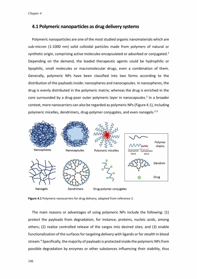
Chapter 4
136
4.1 Polymeric nanoparticles as drug delivery systems
Polymeric nanoparticles are one of the most studied organic nanomaterials which are
sub-micron (1-1000 nm) solid colloidal particles made from polymers of natural or
synthetic origin, comprising active molecules encapsulated or adsorbed or conjugated.1
Depending on the demand, the loaded therapeutic agents could be hydrophilic or
lipophilic, small molecules or macromolecular drugs, even a combination of them.
Generally, polymeric NPs have been classified into two forms according to the
distribution of the payloads inside: nanospheres and nanocapsules. In nanospheres, the
drug is evenly distributed in the polymeric matrix; whereas the drug is enriched in the
core surrounded by a drug-poor outer polymeric layer in nanocapsules.2 In a broader
context, more nanocarriers can also be regarded as polymeric NPs (Figure 4.1), including
polymeric micelles, dendrimers, drug-polymer conjugates, and even nanogels.2-3
Figure 4.1 Polymeric nanocarriers for drug delivery, adapted from reference 2.
The main reasons or advantages of using polymeric NPs include the following: (1)
protect the payloads from degradation, for instance, proteins, nucleic acids, among
others; (2) realize controlled release of the cargos into desired sites; and (3) enable
functionalization of the surfaces for targeting delivery with ligands or for stealth in blood
stream.4 Specifically, the majority of payloads is protected inside the polymeric NPs from
possible degradation by enzymes or other substances influencing their stability, thus

Development of nanoscale polymeric particles for glioblastoma treatment based on Pt(IV) prodrug
137
increasing the effective drug concentration at the action site.5 Due to the EPR effect, the
polymeric NPs can passively target the tumor tissues thus enhance the accumulation of
loaded drug in the tumors. Besides, the polymeric NPs are usually designed to provide
either controlled release or triggered release of the cargos to increase the specificity and
selectivity for targeting sites and reduce possible side effects.6 The polymeric NPs
commonly possess a relative longer residence in blood circulation by surface
modification of stealthy polymers (e.g., PEG), which improve the circulation half-lives of
the cargos in comparison to free molecules. To target the desired tissues, the surfaces
of polymeric NPs can be decorated with targeting ligands (peptide, aptamer,
antibody/antibody fragment, small molecules).7
A huge variety of polymers has been adopted for the fabrication of such polymeric
nanocarriers, including both natural or synthetic polymers. However, the biosafety of
these polymers is crucial for the drug delivery system, especially for drug delivery into
the CNS. Therefore, the polymers used should be biodegradable and biocompatible, and
the products from degradation must also be nontoxic and should be easily cleared from
the body.1,7 Currently, the frequently studied natural polymers include chitosan,
alginate, gelatin, dextran, and albumin. In contrary, the widely used synthetic polymers
include PEG, poly(lactide-co-glycolide) (PLGA), poly(lactide) (PLA), polyethyleneimine
(PEI), poly(cyanoacrylates) (PCA), and polycaprolactone (PCL).1,6-7 Although not all of
these polymers have been approved by FDA, they have been found to be nontoxic in
many studies.
Generally, there are two approaches to fabricate polymeric NPs: “top-down” and
“bottom-up” methods. In the top-down approach, polymeric NPs are produced from
preformed polymers, whereas in the bottom-up method the formation of polymeric NPs
result from the polymerization of monomers.8 The selection of preparation method
depends on the nature of the polymers used, the purpose of application, the control of
particle properties, and so on. On the methods, there are many excellent reviews
already which will not be stated more here.9-10

Chapter 4
138
4.2 Catechol-based polymeric nanoparticles for GB therapy
Catechols can be found in a vast host of natural compounds due to their
physicochemical versatility. Catechol derivatives feature the presence of the basic
catechol structure, o-dihydroxybenzene, comprising an aromatic moiety with two
neighboring (ortho-) hydroxyl groups. Catechol derivatives, together with polyphenols,
have become a hot research topic in recent decades, largely due to their antioxidant and
anti-inflammatory activities. For instance, (−)-epigallocatechin-3-gallate (EGCG),
quercetin, curcumin, resveratrol, chrysin, baicalein, luteolin, honokiol and silibinin are
typical catechol derivatives (Figure 4.2) being actively investigated for their bioactive
effects.
Figure 4.2 Chemical structures of typical catechol derivatives.
The potential therapeutic effects of catechol derivatives are constructed on their
structural properties, for example the presence of catechol rings, and variable
hydroxylation patterns, which endow them capability for metal-chelating, estrogen-like,
fibril-disassembly, enzyme-binding, pro- and anti-oxidative properties.11 Apart from the
structural benefits, the unique functions of catechol family greatly arise from their redox
chemical properties. Research implies that the cytotoxic and proapoptotic effects are
attributed to their pro-oxidant activity, while their antioxidant activity is likely
responsible for the protection effects. Thus, they can either provide protection for
normal cells from oxidative stress as antioxidant agents or exert pro-apoptotic effects in
cancer or malignant cells as cytotoxic pro-oxidants.12 The anticancer potential of these
catechol derivatives has greatly aroused scientists’ interest recently. Combining with

Development of nanoscale polymeric particles for glioblastoma treatment based on Pt(IV) prodrug
139
nanotechnology, the potentiality of catechol-based systems has increasing the interest
due to the combination of catechol properties with the nanostructuration advantages.
In this section we review some interesting examples of catechol-based polymeric
nanostructures with application in cancer therapy, and specifically in GB treatment.
One of the most used catechol in cancer therapy is quercetin. The bioavailability of
this molecule was greatly restricted by the its poor solubility what limits the persistence
in bloodstream and the BBB crossing. It is reported that Only 0.3% of quercetin reached
the brain via intraperitoneal administration.13 The encapsulation of quercetin into PLGA
NPs (215 nm) improved its bioavailability and anticancer efficacy in C6 glioma cells
compared to free drug. Moreover, this series of nanoparticles exhibited high cellular
uptake and antioxidant activity while effectively inhibiting cell proliferation.14 G. Wang
published a series of papers concerning the anti-glioma effects of liposomal and micelle
quercetin in vitro and in vivo.15-19 In a recent work, quercetin-loaded nano-micelles
(Figure 4.3) showed potent cytotoxicity in C6 glioma cells, accumulated in brain tissue
effectively and exhibited significant antitumor efficacy in vivo with significantly longer
survival.20
Figure 4.3 Preparation scheme for quercetin-loaded nanomicelles conjugated with Pluronic and Labrasol block copolymers, adapted from reference 20.
In another example (Figure 4.4), Pt-prodrug polyphenols and PEG-polyphenols were
employed to form coordination NPs with ferric ions and loaded an anticancer agent
Doxorubicin (DOX). DOX and Pt prodrug can both activate nicotinamide adenine
dinucleotide phosphate oxidases and generate ROS, while polyphenols acted as a
catalyzer to form highly toxic free radicals and to synergize the chemotherapy. This
system prolonged blood circulation and enhanced tumor accumulation, thus effectively
inhibited U87 glioma growth and mitigated side effects.21 This nanoparticle was also able
to encapsulate and deliver the phagocytic enzyme myeloperoxidase (MPO) in a

Chapter 4
140
controlled manner. In this case the therapeutic efficacy was related to the release of Pt-
drug and the in-situ production of HOCl. The results showed that the NPs induced a ROS
cascade that enhanced Pt therapy. Moreover, this nanosystem exhibited prolonged
circulation time in blood, high tumor accumulation, and effective inhibition of tumor
growth in vivo.22
Figure 4.4 DOX-loaded (a) and MPO-loaded (b) NPs based on the coordination of Pt-prodrug and PEG polyphenols with Fe(III) ions, adapted from references 21-22.
Other catechol derivative like luteolin was loaded in PEG-PCL micelles and its anti-
glioma effect was evaluated in vitro and in vivo. Luteolin-loaded micelles showed
a
b

Development of nanoscale polymeric particles for glioblastoma treatment based on Pt(IV) prodrug
141
stronger cytotoxicity in C6 and U87 GB cells than free luteolin. Within the U87 glioma-
bearing zebrafish model, the micelles induced more apoptosis and inhibited
neovascularization in tumor tissues, resulting significantly stronger inhibition on tumor
growth.23
4.3 Scope of this chapter
Catechol-based polymeric NPs incorporating Pt(IV) prodrug PtBC will be synthesized,
characterized and evaluated as drug delivery systems for GB chemotherapy. This
chapter will focus on the following objectives:
(i) Synthesis of the polymeric pPtBC NPs (Figure 4.5).
(ii) Physicochemical characterization of the resulting polymeric NPs.
(iii) In vitro studies of the anticancer activity of pPtBC NPs against GB compared to
CDDP and PtBC.
(iv) In vivo safety and biodistribution evaluation of pPtBC NPs in preclinical GB
murine model
Figure 4.5 Synthesis of polymeric pPtBC NPs based on oxidation polymerization of the catechol-bearing Pt(IV) prodrug.
4.4 Results and discussion
The Pt(IV) prodrug (PtBC) was synthesized and characterized in the same way as
described in previous section 3.1.

Chapter 4
142
4.4.1 Synthesis and characterization of polymeric pPtBC NPs
The polymeric pPtBC NPs were synthesized based on the oxidation of terminal
catechol moieties from Pt(IV) prodrug by sodium periodate. NaIO4 in aqueous solution
was added into the PtBC solution in EtOH/H2O (1:1 in volume) slowly, and brown NPs
formed after 4 h of reaction at rt. The pPtBC NPs were prepared as scheme shown in
Figure 4.6.
Figure 4.6 Schematic illustration of the synthesis of polymeric pPtBC NPs.
The properties of the nanocarriers play an important role for the performance of the
drug delivery system, especially the particle size, surface charge, and morphology,
among other factors. The physical properties will determine their circulation half-time,
and biodistribution, as well as the cellular uptake pathways and body clearance.
The hydrodynamic size and ζ-potential (Figure 4.6) of pPtBC NPs were measured using
DLS, showing an average size of 64.03 ± 6.58 nm, with a relatively low PDI of 0.143 ±
0.012, and a slightly negative ζ-potential of -6.18 ± 1.05 mV.

Development of nanoscale polymeric particles for glioblastoma treatment based on Pt(IV) prodrug
143
Figure 4.6 Representatively size distribution (a) and ζ-potential (b) of pPtBC NPs by DLS.
Figure 4.7 Representative morphologies of pPtBC NPs under SEM (a-c), and TEM (d-f).
The morphologies of the polymeric NPs were observed by SEM and TEM (Figure 4.7).
Relatively monodispersed and uniform spherical NPs were observed under SEM,
showing a size around 70 nm which was in good accordance with the result of DLS. Under
TEM, the NPs cannot be observed well due to their polymeric nature, whose
nanostructures probably collapsed because of the high voltage and presented as
irregular particles with blurry edges. As discussed in section 3.3, the size can influence
considerably the biodistribution of NPs in vivo. Nanoparticles with a size around 100 nm
a b
a b c
d e f

Chapter 4
144
are favorable as nanocarriers, which usually can offer longer circulation half-lives and
lead to improved therapeutic effects. And sizes less than 100 nm were reported to
facilitate the transport of nanoparticles across the BBB.24-25 The low PDI indicated the
uniform size distribution of the NPs and ensured the possibility of a more similar
behavior after administration and less pharmacokinetic irregularity and variability in
therapeutic outcome.26 The surface charge of the NPs influences mostly the interaction
of NPs and proteins in blood stream, thus affecting the pharmacokinetics and
biodistribution. And cationic NPs have been observed to be rapidly removed from
circulation due to their enhanced protein absorption, resulting in nonspecific uptake by
macrophages. Although positively-charged particles are reported to feature an
enhanced cellular uptake due to their interaction with the cell membrane, their rapid
clearance makes this meaningless without reaching target sites.27 In contrast, the almost
neutral or slightly negative ζ-potential avoids excessive interaction between the NPs and
proteins or non-target cell membranes before reaching the target sites, and thus
prolongs the half-lives and provide polymeric pPtBC NPs better stability during
transport.
Figure 4.8 Representative EDX spectrum of polymeric pPtBC NPs by TEM.
To confirm the chemical composition, a combination of characterization techniques
was used, including EDX and ICP-MS. All characterizations were performed with three
independent batches of pPtBC NPs, displaying comparable results between batches and
therefore proving the robustness and reproducibility of the synthetic procedure. EDX

Development of nanoscale polymeric particles for glioblastoma treatment based on Pt(IV) prodrug
145
was first used to check the elements qualitatively present in the polymeric pPtBC NPs
(Figure 4.8). All major elements in the prodrug PtBC were found in the NPs such as C, N,
O, Pt, and Cl, whereas Cu in the spectrum belonged to the copper grid for supporting
the TEM sample. ICP-MS measurements allowed the determination of Pt content in the
NPs, which also gave a reproducible percentage of Pt in the NPs in weight. Compared to
the pure prodrug, the Pt content decreased slightly from 24 .04% to 16.16%.
4.4.2 In vitro drug release
Numerous strategies have been developed to enhance the selectivity and targeting
capability of nanoscale drug delivery systems for tumor treatment. Among them, drug
delivery systems with stimuli-responsiveness have been increasingly investigated due to
their sensitivity to specific subtle changes in tumor or tumor microenvironment (TME)
from normal tissues, including mildly acidic pH, high GSH level, overexpressed proteins
or receptors, altered enzyme activity, etc.28-29 The unique features have been harnessed
to develop drug delivery systems which can actively target tumors or TME, thus increase
drug accumulation in tumor, improve therapeutic effects and reduce side toxicity.30
In our work, the Pt(IV) prodrug and resulting pPtBC NPs were designed to be pH-
responsive due to the presence of ester and amide bonds in the molecule which can
remain stable in physiological conditions and be cleaved in the weekly acidic
extracellular environment of tumors or in acidic endo/lysosomal compartments of
tumor cells. Besides, the linkage bonds also can be enzymatically cleaved by the
endogenous esterase and amidase/proteinase. Herein, drug release from polymeric
pPtBC NPs was studied in phosphate buffers at different pH, and released Pt was
determined using ICP-MS.
The release profiles (Figure 4.9) were obtained using dialysis method. pH 7.4 and pH
5.5 were chosen to mimic the physiological and mildly acidic conditions for the drug
release study in vitro. pPtBC NPs were incubated in the dialysis bags at 37 ℃ over 48 h
against phosphate buffers and released Pt was quantified by ICP-MS to yield the release
profiles. Both release curves are typically biphasic, while the release was faster in the
first few hours, and later in a more sustained and slower manner at both pH. However,
the release of Pt from pPtBC NPs was drastically accelerated under the acidic condition.

Chapter 4
146
As shown in Figure 4.9, 44.89 ± 4.64% of Pt was released in total after 48 h of incubation
in PB buffer at pH 7.4, while the released Pt increased to 70.98 ± 1.99% with incubation
for 48 h at pH 5.5. This substantiated that the pPtBC NPs are acid-sensitive, permitting
the possibility that pPtBC NPs can maintain stable in physiological conditions and release
the drug effectively in acidic tumoral tissues, thus realizing the controlled released of
the payloads.
Figure 4.9 a) Cumulative Pt release from pPtBC NPs at pH 7.4 and pH 5.5. b) Inset of release profiles in first 8 h. All data represented as mean ± SE with three independent experiments.
As discussed in section 3.6, GSH and its oxidized form glutathione disulfide (GSSG)
play a pivotal role in the redox hemostasis of cells. Generally, the intracellular
concentration of GSH is reported to be 1 - 10 mM in normal cells, while its concentration
decreased to 2 - 3 orders of magnitude lower levels in extracellular fluids. However, the
intracellular level of GSH in cancer cells are reported to increase up to fourfold more
than that in normal cells.31 The elevated levels of GSH protect tumor cells from the
anticancer action of numerous therapeutics, including Pt agents, contributing to the
multidrug resistance (MDR) of therapeutic agents.32 The different GSH levels in extra-
and intra-cellular circumstances and in normal and tumor tissues have prompted the
emergence of GSH-responsive nano-formulations to facilitate the preferential drug
release in tumor tissues and cells. 33-34 Commonly, this type of nanocarriers bear
sensitive bonds to GSH as structural features. The polymerization products from
catechol moieties have been reported to disassociate in acidic conditions and show GSH-
responsiveness.35-37 In this work, the catechol moieties presented in the prodrug and
a b

Development of nanoscale polymeric particles for glioblastoma treatment based on Pt(IV) prodrug
147
obtained pPtBC NPs may interplay with the redox of GSH and offer a GSH-responsive
nanodrug delivery platform.
Figure 4.10 a) Cumulative Pt release from pPtBC NPs in the presence of GSH (2 and 10 mM) at pH 7.4 (a) and pH 5.5 (b). All data represented as mean ± SE with three independent experiments.
To check this possibility, the release behavior of pPtBC NPs was furthermore studied
in the presence of GSH. The GSH level in brain was reported to be around 1 - 2 mM.38
Therefore, 2 mM and 10 mM were chosen as the concentrations of GSH to be tested,
using the same method described previously at pH 7.4 and 5.5. As Figure 4.10 shown,
indeed, the release of Pt was obviously promoted by GSH. Even with a concentration of
2 mM, GSH accelerated the release of Pt dramatically; 50.71 ± 0.26% of Pt was released
from the pPtBC NPs after 24 h of incubation at pH 7.4, which exceeded the amount of
released Pt (44.89 ± 4.64%) after 48 h in the absence of GSH. After 48 h, 57.95 ± 1.18%
of Pt was released in total in the presence of 2 mM GSH. Notably, the release was further
sped up while the concentration of GSH increased to 10 mM; 61.5 ± 0.24% of Pt was
released from the pPtBC NPs after 24 h of incubation, and the total release reached
80.89 ± 0.47% after 48 h. At pH 5.5, the release of Pt was accelerated in the same trend
as at pH 7.4 in the presence of GSH, and all Pt was released after incubation with 10 mM
GSH at pH 5.5 for 48 h.
In summary, the results demonstrated that pPtBC NPs are both pH- and GSH-
responsive, favorably activating and releasing the therapeutic drug in tumor tissues and
cells, providing selectivity of tumor tissues and cells over normal tissues and cells,
ensuring the maximal anticancer effectiveness and minimal off-target side effects.
a b

Chapter 4
148
4.4.3 Anticancer efficacy of pPtBC NPs in vitro
Molecules with catechol moieties were reported to have antibacterial, antiviral, and
other therapeutic effects mostly by modulating the redox balance.39 To elucidate the
origin of anticancer effect, a model compound nordihydroguaiaretic acid (NDGA), and
the corresponding polymeric material (pNDGA NPs) prepared with same methodology
as pPtBC NPs were included in the in vitro assays. As observed in Figure 4.11, NDGA
shares structural similarity with the prodrug PtBC, but without cisplatin core in the
structure.
Figure 4.11 Chemical structures of NDGA (a) and the prodrug PtBC (b).
4.4.3.1 Cytotoxicity assays
Similarly, the cytotoxic effects of PtBC, pPtBC NPs, NDGA, and pNDGA NPs were
evaluated in vitro and compared to cisplatin (CDDP) against a panel of cell lines,
including cancer cells of different origins, cisplatin-resistant cells, and non-malignant
cells. Human cervical cancer cell HeLa, murine glioma cell GL261, and non-malignant
human fibroblast cell 1Br3G were chosen for the cell viability assays using the well-
established PrestoBlue method for 24 h and 72 h (Figure 4.12). Except the three cell
lines, the cytotoxicity of these formulations was also tested against a pair of human
ovarian cancer cell lines, A2780 and its cisplatin resistant counterpart A2780/cis (Figure
4.12), hoping that the polymeric NPs may sensitize the resistant A2780/cis cells towards
cisplatin. It has been reported that nanocarriers with proper modifications can bypass
the efflux of resistant cells which the free drug is commonly subjected to, leading to
modulating or overcoming the resistance of cancer cells.40-41 The half maximal inhibitory
concentrations (IC50s) were calculated using Graphpad Prism 7 and summarized in Table
4.1 and 4.2.
a b

Development of nanoscale polymeric particles for glioblastoma treatment based on Pt(IV) prodrug
149
After 24 h of exposure (Table 4.1), PtBC and pPtBC NPs showed slightly lower
cytotoxicity against almost all cell lines compared to CDDP, while the cytotoxicity of
pPtBC NPs was weaker in GL261 cells line. Specifically, the cytotoxic effect of pPtBC NPs
was very similar to the prodrug in HeLa cells, with very close IC50s of 22.09 M and 29.94
M respectively. But CDDP showed higher cytotoxicity with an IC50 of 15.98 M in HeLa
cells. In 1Br3G cells, they demonstrated the same trend, and the IC50s of CDDP, PtBC,
and pPtBC NPs were 45.07, 56.09, and 56.3 M respectively.
After 72 h of exposure (Table 4.2), the cytotoxicity difference between the prodrug
PtBC, pPtBC NPs and CDDP was decreased to comparable levels. And pPtBC NPs showed
slightly higher cell killing ability than the prodrug in all cell lines. Specifically, in HeLa
cells, pPtBC NPs demonstrated a lower IC50 of 0.65 M than that of CDDP (2.34 M), and
PtBC also exhibited a slight stronger cytotoxicity than CDDP, with a IC50 of 1.85 M.
However, in the other two cell lines, the cytotoxicity of them was comparable, as CDDP >
pPtBC NPs ≥ PtBC with very close IC50s. For example, in GL261 cells, the IC50s of CDDP,
PtBC and pPtBC were 2.16, 4.17 and 4.64 M respectively.

Chapter 4
150
Figure 4.12 Cytotoxicity assays of CDDP, PtBC, pPtBC NPs, NDGA, and pNDGA NPs against a panel of cell lines for 24 h and 72 h. All data represented as mean ± SE of three independent experiments.
On the other hand, as the reference molecule, NDGA showed very low cytotoxicity in
these cell lines except in A2780 or A2780/cis, and its polymeric counterpart pNDGA NPs
was non-toxic in given concentration range with a cell viability close to 100% in all cell
lines either for 24 h or for 72 h. This interesting comparison excluded the possibility that
the catechol moieties may contribute to the cytotoxicity of PtBC and polymeric pPtBC in
HeLa, 1Br3G, or GL261 cells. Unfortunately, in the ovarian cancer cell lines resistant to
CDDP, neither the prodrug nor the NPs showed improvement in cytotoxicity compared
to CDDP (Figure 4.12). Such cross resistance of A2780/cis cells to PtBC and pPtBC NPs

Development of nanoscale polymeric particles for glioblastoma treatment based on Pt(IV) prodrug
151
suggested common elements in their mechanism of action as CDDP. In contrast, the
reference molecule NDGA exhibited slightly stronger cytotoxicity compared with CDDP
in both cell lines after 24 h, while it showed comparable but slightly weaker cytotoxic
effect as CDDP for 72 h. This indicated that NDGA could be a promising therapeutic agent
against the ovarian cell lines which might bear poor antioxidative capacities and be
hypersensitive to redox imbalance. However, we are well aware that the pathways for
uptake and detoxification of NPs are greatly influenced by the cell type, as well as the
intrinsic properties of NPs.42 These promising anticancer effects in vitro open new
avenues for the exploration of different biomedical applications.
IC50 (µM)a
Cell line
Compound HeLa 1Br3G GL261 A2780 A2780/cis
pPtBC NPs 22.09 ± 1.09 56.30 ± 1.32 33.56 ± 1.36 18.44 ± 2.19 31.40 ± 2.28
pNDGA NPs NTb NTb NTb NTb NTb
PtBC 29.94 ± 1.04 56.09 ± 1.18 17.40 ± 1.08 10.98 ± 0.56 18.39 ± 1.85
NDGA 164.15 ± 0.02 NTb 109.45 ± 1.95 8.94 ± 0.69 15.67 ± 2.14
CDDP 15.98 ± 1.04 45.07 ± 4.60 5.61 ± 0.28 13.95 ± 1.24 23.68 ± 1.74 a The IC50s of the compounds against different cell lines were determined by PrestoBlue assay. Each value
represented the mean ± SE of three independent experiments.
b NT represents non-toxic, which means the viability of cells is close to 100 % even at the maximum concentration.
Table 4.1 IC50s of different formulations against a panel of cell lines for 24 h.
IC50 (µM)a
Cell line
Compound HeLa 1Br3G GL261 A2780 A2780/cis
PPtBC NPs 0.65 ± 0.14 8.01 ± 0.72 4.64 ± 0.05 3.10 ± 0.32 10.50 ± 0.84
pNDGA NPs NTb NTb NTb NTb NTb
PtBC 1.85 ± 0.36 10.80 ± 0.60 4.17 ± 0.12 2.59 ± 0.33 9.12 ± 0.69
NDGA 55.65 ± 6.21 119.00 ± 14.20 81.73 ± 2.40 7.82 ± 0.39 12.96 ± 1.48
CDDP 2.34 ± 0.30 4.63 ± 0.42 2.16 ± 0.26 1.57 ± 0.08 5.22 ± 1.21 a The IC50s of the compounds against different cell lines were determined by PrestoBlue assay. Each value
represented the mean ± SE of three independent experiments.

Chapter 4
152
b NT represents non-toxic, which means the viability of cells is close to 100 % even at the maximum concentration.
Table 4.2 IC50s of different formulations against a panel of cell lines for 72 h.
4.4.3.2 ROS generation
As discussed in previous chapter, the chemical property of catechol moiety makes it
an interesting functional group in biomedical field. On one hand, its antioxidant and anti-
inflammatory activities endow it with multiple beneficial physiological effects.
Compounds with catechol moieties, generally polyphenols and flavonoids, were
reported to exhibit hepatoprotective, anti-inflammatory, antioxidant, and even
anticancer properties.43 On the other hand, ROS can be generated during the oxidation
process of the catechol moieties, which may induce excessive oxidative stress and cause
harmful effects, such as toxicity, damage, and inflammation in cells.4 Therefore, it’s
intriguing to check if the prodrug and the pPtBC NPs also generate ROS and possible
influence in their therapeutic effects.
Figure 4.13 ROS generation triggered by different compounds and pPtBC NPs in HeLa (a), 1Br3G (b) and GL261 (c) cells. The concentration of H2O2 was 0.1 mM, pNDGA NPs at 0.2 mM, other agents at their corresponding IC50s. Data represented as Mean ± SE of three independent experiments. * stands for p < 0.05, ** for p < 0.001, *** for p < 0.0001.
To assess if such oxidative stress also plays a role in the cytotoxicity of our prodrug
and nanoparticles, the ROS generation of these compounds/formulations was evaluated
using the fluorescent probe 2’,7’-dichlorofluorescin diacetate (DCFCDA) in HeLa, 1Br3G
and GL261 cells (Figure 4.13). DCFCDA has been proved that its fluorescence intensity is
correlated well with the intracellular ROS levels.44 After 24 h of exposure to the agents,
strong DCF fluorescence was only observed from NDGA and H2O2, the positive control,
a b c

Development of nanoscale polymeric particles for glioblastoma treatment based on Pt(IV) prodrug
153
in all cell lines. Neither the prodrug PtBC nor the polymeric pPtBC/pNDGA NPs triggered
the formation of ROS in all cell lines. This result proved that the cytotoxicity of these
agents mainly arises from the Pt core which can be activated intracellularly to cisplatin.
CDDP itself neither induced ROS generation. Thus, the possibility of oxidative stress
caused by catechol moieties was excluded from the mechanisms of action of these
agents, except NDGA where the oxidative stress might contribute partially to its
therapeutic effects.
4.4.3.3 Cellular uptake and DNA-bound Pt
The therapeutic efficacy of an anticancer agent greatly depends on its accumulation
in targeting cells, or its intracellular concentration, which in turn is directly related to its
uptake pathways and kinetics, in addition to other factors such as its metabolism and/or
cellular efflux. Increasing the Lipophilicity of NPs’ surface was reported to obtain higher
affinity to the cell membrane, even that of the BBB endothelial lining, compared to
hydrophilic NPs.45 The polymeric nature of the pPtBC NPs may facilitate their transport
across the BBB, and enhance the cellular uptake in target tumor cells.
Both the reference molecule NDGA and its polymeric NPs were excluded from further
studies since they did not show effective anticancer activity in previous cytotoxicity
assays. To assess the cellular uptake, HeLa, 1Br3G, and GL261 cells were used for the
study. All Pt-containing agents were co-incubated with the cells for 6 h and 24 h at a
concentration of 0.1 mM (referred to Pt). Then the cells were digested, and the amount
of intracellular Pt level was determined using ICP-MS.
As Figure 4.14a shown, with a relatively high extracellular concentration, the cellular
uptake of these Pt agents exhibited a trend as PtBC > pPtBC > CDDP after exposure to
the drugs for 6 h. And both the prodrug PtBC and the polymeric pPtBC NPs had a
significantly higher cellular uptake compared with CDDP in all cell lines. And the uptake
of the prodrug was generally higher than that of the polymeric NPs. CDDP and PtBC are
all small molecules which usually enter cells by passive diffusion. However, the cellular
entry pathway of CDDP is dominated by the carriers present on the cell membrane,
especially copper transporters (CTRs) and organic cation transporters (OCTs).46-47 The

Chapter 4
154
carrier-mediated endocytosis requires energy consumption, but passive diffusion does
not, which might be the reason why the cellular uptake of CDDP was less than that of
PtBC. NPs commonly cannot easily enter the cell by passive diffusion but by endocytosis
pathways, which is also energy-dependent. With a relatively high extracellular
concentration, PtBC may enter cells easier than the NPs given the high concentration
gradient.
Figure 4.14 Cellular uptake of CDDP, PtBC and pPtBC NPs in HeLa, 1Br3G and GL261 cells for 6 h (a) and
24 h (b). All drugs were incubated at a concentration of 100 M referred to Pt. Data represented as mean ± SE of three independent experiments. * stands for p < 0.05, ** for p < 0.001, *** for p < 0.0001.
After exposure for 24 h (Figure 4.14b), the cellular uptake of the agents remained
similar levels in HeLa cells which implied that the internalization of these agents was fast
and became dynamically stable after 6 h. However, the uptake in the other two cell lines
did not reach the dynamic equilibrium after 6 h, and the levels changed after incubation
for 24 h. In 1Br3G cells, the intracellular level of CDDP was comparable to that of 6 h,
but the levels of PtBC and pPtBC NPs decreased dramatically, though they were still
significantly higher than CDDP level. Interestingly, the trend in GL261 cells converted
totally. The cellular uptake of CDDP augmented to 8.5 times in comparison to the
intracellular concentration of 6 h, which turned significantly higher than that of PtBC or
pPtBC NPs whose intracellular level also increased than 6 h.
The cellular uptake in GL261 (Figure 4.15) was further studied along time with a
concentration decreased to 10 M which was lower than the IC50s of PtBC and pPtBC
NPs, but slightly higher than that of CDDP, in order to exclude the impact from high
concentration gradient of Pt agents which may be favorable for cell internalization. In
a b

Development of nanoscale polymeric particles for glioblastoma treatment based on Pt(IV) prodrug
155
general, the cellular uptake of all Pt agents increased with time, and the trend remained
similar to high initial concentration. At all time points (2, 4, 8 and 24 h), the intracellular
concentrations of CDDP kept the highest compared to the prodrug or the NPs. The
concentration difference at 2 h was not significant, whereas turned significantly higher
than that of PtBC and pPtBC NPs afterwards.
Figure 4.15 Cellular uptake of CDDP, PtBC and pPtBC NPs along with time at a concentration of 10 M referred to Pt. Data represented as mean ± SE of three independent experiments. * stands for p < 0.05, ** for p < 0.001, *** for p < 0.0001.
Since the anticancer effects of Pt drugs was mainly attributed to its interaction with
DNA, determination of DNA-bound Pt would be helpful to understand better the action
mechanisms of these Pt agents. Similarly, HeLa, 1Br3G, and GL261 cells were incubated
with CDDP, PtBC and pPtBC NPs at a Pt concentration of 100 M for 24 h. Then the
nuclear DNA of cells was further extracted and purified, and obtained DNA was
quantified by the absorbance, meanwhile, the amount of Pt bound to the DNA was
quantified using ICP-MS. As Figure 4.16 shown, the DNA-bound Pt concentration from
intrinsically active CDDP was significantly higher than that of PtBC and pPtBC NPs in all
cell lines. In HeLa and 1Br3G cells, the trends did not remain the same as cellular uptake,
which meant that not all Pt agents entering cells can enter the nucleus and bind to DNA.
In GL261, the trend was similar to that of cellular uptake, but the Pt concentration from
polymeric NPs became the lowest. This could be expected that the process for pPtBC
NPs was more difficult than CDDP or PtBC. After entering the cells, they had to dissociate
first, release the prodrug and get reduced to the active Pt form, then enter nucleus and
bind to DNA.

Chapter 4
156
Although the DNA-bound Pt , they exhibited comparable anticancer activities for 24 h
(see section 4.3.3.1). This suggested that the action mechanisms of PtBC and pPtBC NPs
probably were not limited to Pt-DNA adducts in this case. Increasing reports
demonstrated that Pt agents can also bind to other cellular macromolecules such as RNA
and proteins, and even induce immunogenic effects. 48-50 Bose and colleagues even
synthesized a novel Pt compound with high cytotoxicity similar to cisplatin, but not
binding to DNA at all.51
Figure 4.16 DNA-bound Pt after exposure to CDDP, PtBC, and pPtBC NPs for 24 h at a concentration of
100 M referred to Pt. Each value represented as mean ± SE of three independent experiments. * stands for p < 0.05, ** for p < 0.001, *** for p < 0.0001.
4.4.3.4 Cell morphology observation
Cell morphology is a direct indicator of cytotoxicity of therapeutic agents. Therefore,
the morphologies of GL261 cells incubated with CDDP, PtBC, and pPtBC NPs were
observed and recorded by optical microscopy.
The morphologies of GL261 cells changed a lot after incubation with those Pt agents
for 24 h (Annex I), which was more obvious with higher concentrations. Specifically,
exposure to PtBC at a concentration of 1 M for 24 h, obvious cell density decrease,
rounding-up, pseudopodes retraction, plasma membrane blebbing/budding, and cell
volume reduction were observed, but still the number of cells with normal morphology
accounted for the majority. When the concentration increased to 10 M, the proportion
of healthy cells decreased dramatically, and most of the cells got rounded and shrunken,
cellular fragments observed. At the concentration of 25 M, the shrunken cells
a b

Development of nanoscale polymeric particles for glioblastoma treatment based on Pt(IV) prodrug
157
disappeared, the fragments of cells occupied the vision, and little alive cell could be
found. The apoptotic morphologies were also observed in other two Pt agents to a more
severe degree, which was in good accordance of cytotoxic effects of these agents
indicated by their IC50s.
Figure 4.17 Representative morphologies of GL261 cells without/with exposure to pPtBC NPs for 72 h.

Chapter 4
158
After 72 h of exposure (Figure 4.17 and Annex I) to these agents, the morphology
changes showed a similar trend to that at 24 h, and the number of healthy cells
decreased drastically even at the lowest concentration of 1 M. the intracellular vesicle
swelling became extremely obvious in cells treated with 1 M CDDP, which implied that
probably the cells also underwent necrosis except apoptosis.52 When the concentration
went to 10 M, almost only fragments of cells were observed in the vision accompanied
with a few rounding-up cells for all agents. With agents at 25 M for 24 h, little alive
cells were observed but only floating dead cells or cell fragments.
4.4.4 In vivo studies of tolerability and biodistribution
The in vivo studies of Pt-Fe NCPs were performed in collaboration with the Biomedical
Applications of Nuclear Magnetic Resonance research group (GABRMN) led by Dr. Carles
Arús at Universitat Autònoma de Barcelona (UAB, Cerdanyola del Vallès, Spain) under
the supervision of Dr. Ana Paula Candiota with the contribution of Dr. Shuang Wu.
4.4.4.1 Safety and tolerability assessment
Tolerability assay is a fundamental assessment for the safety of new formulations.
Herein, intranasal administration was adopted in our animal studies to bypass the
impermeable BBB. However, there are few papers reporting about preclinical studies of
Pt drugs by nose-to-brain delivery for GB treatment. Therefore, the safety or tolerability
assay is extremely necessary prior to other in vivo studies. As discussed in previous
chapter, in a typical phase I study complying with US FDA regulations, clinical
therapeutics are often tested in either single-dose or limited-dose escalation
pharmacokinetic studies, or in studies where the exposure is limited to a small number
of doses or days, without regard to expected half-life of the drug.53 In this work, a
limited-dose escalation manner was chosen to assess the safety and tolerability of PtBC
and pPtBC NPs via intranasal route. To date, there has not been authorized justification
for the volume applied to IN administration, but commonly intranasal instillation in
volumes more than 20 mL is considered deleterious which can result in suffocation and
death of research mice.54 Although some researcher found that a larger volume of 50
mL or more could be safe for the experimental mice in specific conditions, herein smaller
and safer volumes no more than 20 mL were adopted for the in vivo study.55

Development of nanoscale polymeric particles for glioblastoma treatment based on Pt(IV) prodrug
159
Healthy C57BL/6J mice were used for the short-term safety and tolerability assay.
Three single slightly escalated doses were administered consecutively every week, from
0.9, 1.2 to 1.5 mg Pt/kg body weight. The body weights of mice were tracked three times
every week to monitor any weight changes which function as an important parameter
indicating systemic toxicity. Besides, food consumption, and any adverse physical or
behavioral effects concerning healthy conditions were closely observed by professional
personnel in the animal facility daily. The whole study lasted for 4 weeks.
Figure 4.18 Tolerability assessment of mice for PtBC and pPtBC NPs over 4 weeks. Both drugs were given in slightly increasing doses from 0.9, 1.2 to 1.5 mg Pt/kg body weight via I.N. every week, n = 3. For control group, n = 360, data adapted from Jackson Laboratory (https://www.jax.org/jax-mice-and-services/strain-data-sheet-pages/body-weight-chart-000664). Data represented as mean ± SE. Dash lines indicated days for administration.
As Figure 4.18 shown, the body weights of all treatment groups remained stable as
the control group till the end of the study, indicating no obvious systemic toxicity caused
by PtBC or pPtBC NPs. No treatment-related adverse effects such as mortality, body
weight, food consumption, or other clinical signs were observed for all mice during the
study relative to control animals. Even with the highest dosage of 1.5 mg Pt/kg body
weight, all mice were still in good status during the study. Since the side effects caused
by cisplatin typically are acute nephrotoxicity and hepatotoxicity, the major organs of
mice from tolerability study were harvested, fixed, and examined by histopathology to
assess the biocompatibility and impact on cellular structures.

Chapter 4
160
These results corroborated that PtBC and pPtBC NPs were well-tolerated by mice in
given conditions, which proved the safety of this nano-formulation and paved the way
to further in vivo studies.
4.4.4.2 Biodistribution in mice
As discussed in previous chapter, the biodistribution of NPs in vivo considerably
contributes to the fate of the administered NPs, and directly influences the
bioavailability of the formulations and therefore substantially affects the therapeutic
effects. Many factors have been considered key to the final distribution of NPs in vivo,
especially the size and surface properties of NPs, and interactions with biological
barriers.3,56 Currently, no report for the biodistribution of Pt nanosized agents via
intranasal delivery was available. Therefore, it is worthy to study the in vivo distribution
of polymeric pPtBC NPs via IN route, which could be valuable for related research in
future. Same syngeneic implanted orthotopic models were established and used in this
chapter as the previous by stereotactic injection of murine GB GL261 cells into C57BL/6J
mice.
In the case of Pt-Fe NCPs, the Pt tended to accumulate favorably in the GB tumor and
brain, especially in tumors with small volume (23.15 ± 2.68 mm3) reaching a high
retention of 10.34% ID/g tissue, which was significantly higher than that in other organs,
and significantly higher than the retention in bigger tumors (58.05 ± 3.35 mm3).
Therefore, three mice bearing small orthotopic GB tumors were used for the
biodistribution study of pPtBC NPs, with an average tumor volume of 24.96 ± 2.7 mm3
and an average body weight of 22.7 ± 0.2 g. The tolerability study proved that the pPtBC
NPs were relatively safe and well-tolerated by the mice, so the biodistribution of the
polymeric NPs was studied with the highest dose, 1.5 mg Pt/kg, in tolerability
assessment.
As Figure 4.19 and table 4.3 shown, 1 h post IN administration, the Pt mainly
accumulated in GB tumor, followed by kidneys, lungs, and brain. Few accumulated in
liver, spleen, and heart. The Pt retention in tumor was highest with 3.51 ± 0.36% ID/g,
and 2.53 ± 0.98%, 2.6 ± 0.69, 3.0 ± 0.43% ID/g in brain, lungs, and kidneys, respectively.
No significance was found in the Pt retention in tumor and in the aforementioned

Development of nanoscale polymeric particles for glioblastoma treatment based on Pt(IV) prodrug
161
organs, while the tumor accumulation of Pt was significantly higher than that in heart,
liver and spleen. Taken tumor and brain together, the Pt accumulation in CNS was
significantly higher than that in other organs.
Figure 4.19 Biodistribution of pPtBC NPs in mice bearing GL261 tumors 1 h after administration. Mice were intranasally administered with pPtBC NPs at a dose of 1.5 mg Pt/kg, and sacrificed 1 h post administration. Each value represented as mean ± SE, n = 3.
Tumor Brain Heart Liver Spleen Lungs Kidneys
Pt retention
(% ID/g of
tissue)
3.51 ± 0.36
2.53 ± 0.98
0.54 ± 0.11
1.41 ± 0.22
0.56 ± 0.10
2.60 ± 0.69
3.0 ± 0.43
Significance vs. tumor
N/A No Yes Yes Yes No No
Table 4.3 Pt biodistribution of pPtBC NPs in GL261 GB-bearing mice. Dose = 1.5 mg Pt/kg, biodistribution time = 1 h, n = 3. Comparison was made with Pt retention in tumor, with p < 0.05 considered significant by student’s t-test.
The BBB has long been a hindrance to the successful delivery into brain and brain
tumors, although the integrity of BBB could be disrupted and the permeability might
increase due to the compromised BBB.57 Still, the intra- and inter-tumoral heterogeneity
of GB make the drug delivery into CNS a big challenge for conventional therapeutic
agents. The nose-to-brain drug delivery by IN delivery was more effectively, compared
to other systemic administration routes, like IV injection and intraperitoneal injection,
which typically resulted in neglectable retention in brain.58-59 Conventional polymeric
NPs have been reported to favorably accumulate in RES/MPS organs via systemic
administration, commonly in liver and spleen.60-61 Notably, Pt accumulated more in
Chapter 4
162
lungs and kidneys, instead of the typical RES organs (liver and spleen). The
biodistribution of pPtBC NPs indicated distinctive pharmacokinetic pathways of IN
delivery different from other systemic routes.
Interestingly, the accumulation of Pt from pPtBC NPs in GB tumor was significantly
lower than the Pt retention from Pt-Fe NCPs in GB tumor, while there was no significance
in the tumor volumes. This difference could be due to the different
compositional/structural nature of these two nanoparticles. Although the fabrication of
these NPs was both based on PtBC, the same Pt(IV) prodrug, pPtBC NPs were polymeric
NPs formed from direct oxidation of catechol moieties, while Pt-Fe NCPs formed by
more labile coordination between the ligand and iron ions. The presence of Fe(III) in Pt-
Fe NCPs may make a contribution to the transport across the BBB and the internalization
into GB tumor cells due to the receptor-mediated endocytosis, for example, transferrin
receptors overexpressed in GB and closely related to Fe uptake.62-63 Generally, the
cancer cells have elevated demand of Fe(III) as a nutrient required for cell growth and
proliferation.64 And this is also common to high-grade glioma, resulting in enhanced
uptake of Fe(III) by upregulating activity of transferrin receptor.65 However, this
advantage did not exist in pPtBC NPs, probably leading to significantly lower Pt
accumulation than that of Pt-Fe NCPs.
4.5 Conclusions
Novel Pt(IV) prodrug-based polymeric nanoparticles with an average size of 64.03 ±
6.58 nm, a PDI of 0.143 ± 0.012, and a slightly negative ζ-potential of -6.18 ± 1.05 mV,
were designed, synthesized successfully, and evaluated as drug delivery systems for GB
treatment. The obtained polymeric pPtBC NPs showed relevant advantages such as:
• Chemical and colloidal stability, pH/GSH dual responsiveness and therefore
controlled drug release.
• The cellular uptake and DNA-bound Pt from pPtBC NPs were significantly
lower than that of cisplatin after 24 h of drug exposure, revealing a slower
intracellular activation process for the polymeric NPs. However, pPtBC NPs
exhibited comparable even superior cytotoxic effect in comparison to cisplatin
Development of nanoscale polymeric particles for glioblastoma treatment based on Pt(IV) prodrug
163
against HeLa, 1Br3G, and GL261 cell lines for 72 h, while no ROS generation
was triggered in any of these cell lines by pPtBC NPs. This corroborated that
the cytotoxic effect was totally attributed to the chemotherapeutic effect of
the released Pt compounds.
• pPtBC NPs were proved to be biosafe and well-tolerated at a dose of 1.5 mg
Pt/kg body weight in wt mice, and displayed favorable accumulation of Pt in
GB tumor and brain, implying the effective delivery into CNS of intranasal
administration.
• These results indicated that pPtBC NPs possess potential anti-GB activity in
vitro, while the in vivo efficacy should be further evaluated in the near future.
And intranasal administration could serve as an alternative to typical systemic
administration routes for CNS delivery.
4.6 Experimental
Characterization methods. 250 MHz 1H NMR spectra were recorded on a Bruker DPX
250 MHz spectrometer; 360 MHz 1H NMR, 1H-1H COSY, and 100 MHz 13C NMR were
recorder on a Bruker DPX 360 MHz spectrometer; 400 MHz 1H NMR, 1H-1H COSY, and
100 MHz 13C NMR were recorder on a Bruker DPX 400 MHz spectrometer. Chemical
shifts (δ) are given in ppm, using the residual non-deuterated solvent as internal
reference. Signal multiplicities are described using the following abbreviations: singlet
(s), doublet (d), triplet (t), quartet (q), quintet (quint), doublet of doublets (dd), doublet
of triplets (dt), doublet of doublet of doublets (ddd), multiplet (m) and J to indicate the
coupling constant (Hz). High-resolution mass spectra were obtained by direct injection
of the sample with electrospray techniques in a Bruker microTOF-Q instrument. FT-IR
spectra were recorded using a Tensor 27 spectrophotometer (Bruker Optik GmbH,
Germany) with KBr pellets. Powder X-ray diffraction (PXRD) patterns were recorded at
room temperature on a PANalytical X’Pert PRO MRD diffractometer (Malvern
PANalytical, Germany) equipped with a CuKα radiation source (λ = 1.54184 Å) and
operating in reflection mode, where the solid samples were placed on an amorphous

Chapter 4
164
silicon oxide plate and measured directly. UV-vis was performed using a Cary 4000 UV-
vis spectrometer (Agilent Technologies, USA) within a wavelength range of 400-4000 nm
and a 1 cm path length quartz cuvette (QS 10mm).
DLS measurements. Size distribution and surface charge of the nanoparticles were
measured by DLS, using a ZetasizerNano 3600 instrument (Malvern Instrument, UK),
whose size range limit is from 0.6 nm to 6 nm. (Note: the diameter measured by DLS is
the hydrodynamic diameter. The samples were comprised of aqueous dispersions of the
nanoparticles in distilled water or in buffer. All samples were diluted to obtain an
adequate nanoparticle concentration around 0.5 mg/mL.)
Electron Microscopies. SEM images were obtained using a scanning electron
microscope (FEI Quanta 650 FEG) at acceleration voltages of 5-20 kV. SEM samples were
prepared by drop-casting of corresponding dispersions of NPs on an aluminum tape
followed by evaporation of the solvent under room conditions. Before analysis, the
samples were metalized with a thin layer of platinum (thickness: 5 nm) using a sputter
coater (Emitech K550). STEM images were obtained using a scanning-transmission
electron microscope (Magellan 400L, FEI), while TEM images were obtained using a
transmission electron microscope (Tecnai G2 F20, FEI) at a voltage of 200 kV. Samples
for STEM and TEM were prepared by drop-casting corresponding dispersions on TEM
grids (ultrathin carbon type-A, 400 mesh Cu grid, Ted Pella Inc., Redding, USA) and drying
overnight under room conditions prior to examination.
Inductively coupled plasma-Mass spectrometry (ICP-MS). Standard solutions were
prepared prior to the measurements at a range from 0 - 200 parts per billion (ppb) using
atomic spectroscopic analytical ICP-MS standards (PerkinElmer Inc., Germany). The
isotope 54Fe and 57Fe were selected as Fe tracers, while 194Pt, 195Pt and 196Pt as Pt tracers.
Digestion procedures for ICP-MS measurement. All glassware was immersed in 20%
HNO3 for 48 h, and plasticware in 5% HNO3 for 4 h prior to use for ICP-MS. All samples
were digested using wet digestion method. Depending on the materials to digest,
different procedures were adopted.
(1) Chemical samples: the appropriate amount of samples, NPs for instance, was
weighed accurately with an analytical balance, and transferred into a vial, then

Development of nanoscale polymeric particles for glioblastoma treatment based on Pt(IV) prodrug
165
concentrated ultrapure HNO3 (69%, Ultratrace®, ppb-trace analysis grade, Scharlab,
Spain) was added. The samples were placed in the fume hood for 48 h to be fully
digested, then were diluted with 0.5% (v/v) ultrapure HNO3 for further measurements.
(2) Cell pellets: the cell pellets were resuspended in around 100 mL of concentrated
ultrapure HNO3 (69%, Ultratrace®, ppb-trace analysis grade, Scharlab, Spain) and left to
digest overnight. Samples were then heated to 90 ℃ till the suspensions turned clear.
The samples were diluted with 0.5% (v/v) ultrapure HNO3 to appropriate volumes for
later measurements.
(3) Tissues: the tissues were added with T-PERTM buffer (Thermo Fisher Scientific, USA)
in a ratio of 10 mL per gram, then cut into small pieces, and sonicated with
ultrasonication microtip (Branson Digital Sonifier 450, Emerson, USA) in cycles of 10
seconds on then 15 seconds off with an amplitude of 40%. Afterwards, the tissue
suspensions were added with aqua regia (all ppb-trace analysis grade) and heated up to
300 ℃, while 30% H2O2 was added in the later digestion process till the suspensions
turned clear. The clear solutions were transferred and made up with 0.5% (v/v) ultrapure
HNO3 to appropriate volumes for the determination of metal contents using ICP-MS.
Preparation of polymeric pPtBC/pNDGA NPs. PtBC (50 mg, 0.062 mmol) / NDGA (20
mg, 0.066 mmol) was dissolved in 30 mL of mixture solvent of EtOH: H2O (1:1). NaIO4 in
1 mL of water was added slowly at room temperature and the reaction was allowed to
continue for 4 h. Afterwards, the turbid solution was centrifuged and washed to furnish
yellow or brownish precipitate.
Drug release assay. Drug release from pPtBC NPs was determined using dialysis
method. Concentrated pPtBC NPs at 1 mg/mL were resuspended into 1 mL of Milli-Q
water in dialysis bags (MWCO = 6000 - 8000). The dialysis tubes were immersed into
sealed beakers with 40 mL of PB buffers at pH 7.4 and 5.5. The beakers were incubated
at 37 ℃ with gentle stirring during the study. 500 mL of aliquot was taken from the
dialysate at predetermined time point, followed by supplementing with fresh buffer in
same volume immediately. The amount of released Pt was determined by ICP-MS.
Cells. The cell lines were obtained from the Tumor Bank Repository at the National
Cancer Institute (Frederick/MD, USA), including human cervical cancer cell HeLa, non-

Chapter 4
166
malignant human fibroblast cell 1Br3G, murine glioblastoma cell GL261, and a pair of
human ovarian cancer cell lines, A2780 and its cisplatin resistant counterpart A2780/cis.
Before starting any protocol, all solutions were prewarmed to 37 °C. All materials and
equipment that came in contact with cells were sterile. All cell-related performance was
carried out in Class II biosafety hoods in cell culture labs with a biosafety level 2 (BSL-2).
Cell culture. All cell lines were grown in 75 cm2 cell-culture flask with corresponding
culture media. HeLa was cultured in MEM medium, 1Br3G in DMEM, GL261 in RPMI
1640, A2780 in RPMI 1640, and A2780/cis in RPMI 1640 containing 1 M of cisplatin. All
culture media were supplemented with 10% fetal bovine serum (FBS, Gibco®, Invitrogen,
UK), 0.285 g/L glutamine, 2.0 g/L sodium bicarbonate, and 1% penicillin-streptomycin.
Cells were cultured as adherent monolayers and maintained in an incubator (HERAcell,
150i, Thermo Scientific) at 37 °C in 5% CO2 and relative humidity of 95%, except 1Br3G
with 10% CO2. All cell culture media, FBS, supplements, antibiotics, trypsin, and Trypan
Blue are purchased from Fisher Scientific (Gibco®, Invitrogen, UK).
To subculture/harvest the cells at log phase around 75% confluence, 4 mL of sterile
PBS is added into the flask to rinse the cells after discarding spent cell culture media by
aspiration with a vacuum pump. After washing, 3 mL of 0.05 % trypsin-
ethylenediaminetetraacetic acid (EDTA, 0.2 g/L) was added and the flask was returned
to incubator for 2 minutes. Observing the cells under the microscope, if ≥ 90% of the
cells have detached, 6 mL of fresh cell media was added into the flask and dissociate the
cells from the surface. The cell suspension was transferred into a 15 mL conical (Falcon,
Corning, USA) to centrifuge (Thermo Scientific, USA) at 1400 rpm for 5 min to collect all
the cells. The supernatant was discarded, and the pellet was resuspended in fresh media
or PBS for cell counting, then an appropriate number of cells was seeded in a new flask.
Cell counting. The cells were counted using a Neubauer hemocytometer cell counter
and Trypan Blue (Gibco®, Invitrogen, UK) exclusion to count the cells and determine the
viability. Trypan Blue is a dye used to distinguish the living cells from the dead, since the
dead cells cannot extrude Trypan Blue without required energy mechanisms. An aliquot
of 10 mL of the cell suspension obtained in the subculture step was added into 10 mL of
Trypan Blue. Then, 10 mL of the resulting mixture was added onto the Neubauer

Development of nanoscale polymeric particles for glioblastoma treatment based on Pt(IV) prodrug
167
hemocytometer and the number of living cells was counted under an optical
microscopy.
In vitro cytotoxicity assays. The cells (HeLa, 1Br3G, GL261, A2780 and A2780/cis) in
exponential growth were seeded in 96-well plate (Corning, USA) in optimal condition.
HeLa was seeded at a density of 2000 cells/well, 1Br3G at 3000 cells/well, GL261 at 4000
cells/well, A2780 and A2780/cis at 3000 cells/well. Specifically, the cisplatin-containing
cell culture media was replaced with fresh RPMI media without drug 4 hours before
seeding. After 24 hours’ incubation, fresh media containing compounds (pPtBC NPs,
PtBC, and CDDP) at different concentrations (0, 0.1, 1, 5, 10, 25, 50, 100, 200 M
referred to Pt concentration) were added, and the plates were incubated for 24 or 72
hours. Afterwards, 10 l of PrestoBlue® (0.15 mg/ml, Thermo Scientific, USA) was added
into each well and the plates were incubated for another 4 hours before measuring the
fluorescence at 572 nm with excitation at 531 nm by the microplate reader Victor 3
(Perkin Elmer, USA). For 1Br3G, the incubation time with PrestoBlue® was prolonged to
7 hours in order to achieve enough difference of fluorescence intensity between
concentrations. All experiments were carried out in triplicate, and data were treated by
and Graphpad Prism (version 7.0). The calculated IC50s were obtained by Graphpad
Prism.
Estimation of ROS formation. HeLa, 1Br3G and GL261 were seeded in black 96-well
plates (Corning, USA) at a density of 20,000 cells per well to achieve full confluence.
After 24 hours’ incubation, the spent medium was discarded and cells were washed out
from the medium serum, and added prewarmed PBS with the fluorescent probe 2’,7’-
dichlorofluorescin diacetate (DCFCDA, final working concentration 10 mM), then
incubated for 30 minutes. DCFCDA is a non-fluorescent permeable compound which can
be hydrolyzed by esterase and oxidized by intracellular ROS and turn to fluorescent and
non-permeable 2’,7’-dichlorofluorescein (DCF). After the probe was internalized, the
buffer with probe was replaced by only medium or media with compounds (H2O2, pPtBC
NPs, pNDGA NPs, PtBC, NDGA, and CDDP) at the concentration of IC50 for 24 hours. 0.1
mM of H2O2 was added as positive control. After 24 hours, the fluorescence of each well
was examined at 530 nm after excitation at 485 nm by a microplate reader Victor 3
(Perkin Elmer, USA). This experiment was repeated in triplicate independently. The

Chapter 4
168
results were normalized based on the negative control loaded with dye but without drug
treatment.
Cellular internalization. HeLa, 1Br3G and GL261 cells were seeded in 6-well plates
(Corning, USA) at a density of 300,000 cells per well with 1.5 ml of media. After 24 hours’
incubation, the media were replaced by 1 ml of fresh media w/wo CDDP, PtBC, and
pPtBC NPs at 0.1 mM referred to Pt concentration. The treated cells were allowed to
internalize the compounds for 6 h and 24 in the incubator. Another cellular uptake assay
was also performed with a decreased concentration of Pt agents to 10 M, and
increased time points of 2, 4, 8, and 24 h. The media were removed immediately and
cells were rinsed twice by cold PBS in order to remove excessive compounds. Then 0.5
ml of trypsin was added to each well for 3 min and 2-fold volume of fresh media was
added to neutralize the trypsin. After an aliquot was taken from each well for cell
counting, the rest cell suspensions were collected into 1.5 ml Eppendorf tubes (Corning,
USA) and centrifuged at 12,000 rpm for 4 min. The supernatants were discarded while
the cell pellets were stored at -80 ℃ for further quantification by ICP-MS.
DNA-bound Pt. In order to clarify the action mechanism of our Pt NPs, the DNAs were
extracted and measured by ICP-MS to quantify the DNA-bound Pt after uptake for 24 h
in HeLa, 1Br3G and GL261 cells. The cells in exponential growth were seeded onto cell
culture dish in optimal condition for each cell line to reach 50-60 % of confluence in 24
h before the treatment. Then the drugs (CDDP, PtBC, and pPtBC NPs) were added at 0.1
mM referred to Pt concentration, and the cells were allowed to incubate for another 24
h. Afterwards, media with drugs were removed and the cells were rinsed, trypsinized
and collected by centrifugation and rinsed with cold PBS twice to remove excessive
drugs. The resulting cell pellets were resuspended in lysis buffer (pH 8.0, 150 mM Tris-
HCl, 100 mM NaCl and 0.5 % (w/v) SDS). Pellets in buffer were incubated on ice for 15
min and centrifuged at 15000 rpm for 15 min. 0.1 volume of RNase A was added into
each supernatant at 0.2 mg/mL and incubated for 1 h at 37 ℃. Afterwards, Proteinase K
was added at 0.1 mg/mL and incubated for 3 h at 56 ℃. A volume of
Phenol/Chloroform/Isoamyl alcohol (25:24:1 in volume, Thermo Scientific®) was added
and mixed gently. And after a centrifugation of 3 min at 15000 rpm, aqueous phases
containing DNA were transferred into sterile tubes. DNA was precipitated with 0.1
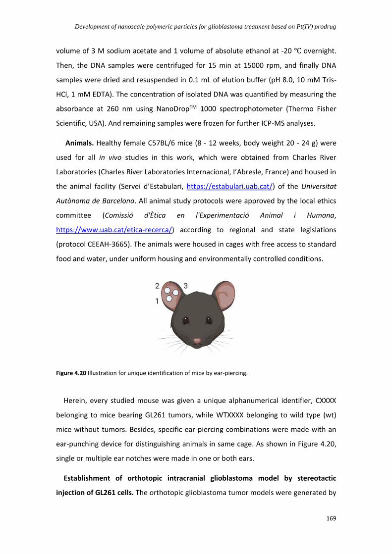
Development of nanoscale polymeric particles for glioblastoma treatment based on Pt(IV) prodrug
169
volume of 3 M sodium acetate and 1 volume of absolute ethanol at -20 ℃ overnight.
Then, the DNA samples were centrifuged for 15 min at 15000 rpm, and finally DNA
samples were dried and resuspended in 0.1 mL of elution buffer (pH 8.0, 10 mM Tris-
HCl, 1 mM EDTA). The concentration of isolated DNA was quantified by measuring the
absorbance at 260 nm using NanoDropTM 1000 spectrophotometer (Thermo Fisher
Scientific, USA). And remaining samples were frozen for further ICP-MS analyses.
Animals. Healthy female C57BL/6 mice (8 - 12 weeks, body weight 20 - 24 g) were
used for all in vivo studies in this work, which were obtained from Charles River
Laboratories (Charles River Laboratories Internacional, I’Abresle, France) and housed in
the animal facility (Servei d’Estabulari, https://estabulari.uab.cat/) of the Universitat
Autònoma de Barcelona. All animal study protocols were approved by the local ethics
committee (Comissió d'Ètica en l'Experimentació Animal i Humana,
https://www.uab.cat/etica-recerca/) according to regional and state legislations
(protocol CEEAH-3665). The animals were housed in cages with free access to standard
food and water, under uniform housing and environmentally controlled conditions.
Figure 4.20 Illustration for unique identification of mice by ear-piercing.
Herein, every studied mouse was given a unique alphanumerical identifier, CXXXX
belonging to mice bearing GL261 tumors, while WTXXXX belonging to wild type (wt)
mice without tumors. Besides, specific ear-piercing combinations were made with an
ear-punching device for distinguishing animals in same cage. As shown in Figure 4.20,
single or multiple ear notches were made in one or both ears.
Establishment of orthotopic intracranial glioblastoma model by stereotactic
injection of GL261 cells. The orthotopic glioblastoma tumor models were generated by

Chapter 4
170
stereotactic injection of GL261 cells into C57BL/6 mice. Specifically, analgesia (Metacam,
Boehringer Ingelheim) at 1 mg/kg was injected subcutaneously to each animal 15
minutes before anesthesia and also 24 and 48 h after implantation. Mice were
anesthetized with a mixture of ketamine (Parke-Davis SL, Madrid, Spain) at 80 mg/kg
and xylazine (Carlier, Barcelona, Spain) at 10 mg/kg via intraperitoneal administration.
Once anesthetized, the mouse was immobilized on the stereotaxic holder (Kopf
Instruments, Tujunga/CA, USA) in a prone position. Next, the head area was shaved, and
the incision site was sterilized with iodophor disinfectant solution, a 1 cm incision was
made exposing the skull and a 1 mm hole was drilled 0.1 mm posterior to the bregma
and 2.32 mm to the right of the midline using a microdrill (Fine Science Tools,
Heidelberg, Germany). A 26G Hamilton syringe (Reno/NV, USA), positioned on a digital
push-pull microinjector (Harvard Apparatus, Holliston/MA, USA) was then used for
injection of 4 mL of RPMI cell culture medium containing 100,000 GL261 cells (obtained
and counted as described in previous section) at a depth of 3.35 mm from the surface
of skull at a rate of 2 mL/min.
Once the injection was completed, the Hamilton syringe was left untouched for 2
minutes more before its removal to prevent the cellular liquid leakage out of the skull.
Finally, the Hamilton syringe was gently and slowly withdrawn and the scission site was
closed with suture silk 6.0 (Braun, Barcelona, Spain). When the implantation was over,
the animal was left in a warm environment to recover from anesthesia.
Once the injection was completed, the Hamilton syringe was left untouched for 2 min
more before its removal to prevent the cellular liquid leakage out of the skull. Finally,
the Hamilton syringe was gently and slowly withdrawn and the scission site was closed
with suture silk 6.0 (Braun, Barcelona, Spain). When the implantation was over, the
animal was left in a warm environment to recover from anesthesia.
The C57BL/6J immunocompetent mice were exposed to enriched environment (EE)
for 3 weeks before tumor implantation, since it was reported that it could significantly
reduce glioma growth and improve mice survival by increasing immunological
parameters in the brain of mice.66 Moreover, since this is the current protocol used at
GABRMN, the maintenance of basic parameters may help in further comparisons. Thus,
all mice in this thesis were allowed to endure 3 weeks of guarantee housing in EE-like

Development of nanoscale polymeric particles for glioblastoma treatment based on Pt(IV) prodrug
171
caging before tumor generation, and were maintained there from beginning to end
(Figure 4.21).
Figure 4.21 Illustration for “enriched environment”-like caging. Mice were allowed to endure 3 weeks of guarantee housing in EE-like caging before tumor generation, and EE was also maintained during the whole experiment and follow-up period. The ‘Shepherd Shack’ is cage insert composed of autoclavable paper, mice can shred it and use the paper for nesting.67
For intranasal administration, the animals were anesthetized by isoflurane for 1 min
and kept in a horizontally supine position. The animals were administered with 2 L of
formulation for each nostril, with an interval of 1 - 2 min between each administration.
Treated animals were observed for mortality and the moribund animals were sacrificed
for ethical considerations.
Tissue preservation procedures. When animals died or were euthanized by cervical
dislocation to prevent suffering, the brains and main organs were excised and either
frozen in liquid nitrogen or fixed in formalin, depending on the purpose. In case of
freezing, tumors were dissected and collected from normal brain parenchyma, frozen in
a liquid nitrogen container for further analysis. In case of fixation, tissue was preserved
in 4% formalin for further autopsy or histopathological analysis. Tissues were resected
after visual inspection of the whole brain and tumor, avoiding as much as possible the
cross contamination with non-tumoral tissue.
In vivo MRI. The animals were subjected to non-invasive brain MRI scan to measure
the volumes, localization, and evolution of the GB. Each scan was performed at 7 T
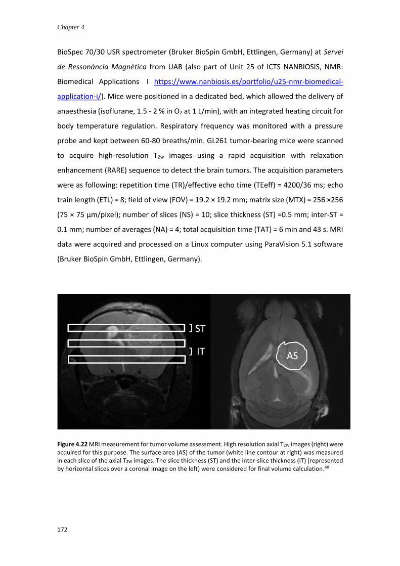
Chapter 4
172
BioSpec 70/30 USR spectrometer (Bruker BioSpin GmbH, Ettlingen, Germany) at Servei
de Ressonància Magnètica from UAB (also part of Unit 25 of ICTS NANBIOSIS, NMR:
Biomedical Applications I https://www.nanbiosis.es/portfolio/u25-nmr-biomedical-
application-i/). Mice were positioned in a dedicated bed, which allowed the delivery of
anaesthesia (isoflurane, 1.5 - 2 % in O2 at 1 L/min), with an integrated heating circuit for
body temperature regulation. Respiratory frequency was monitored with a pressure
probe and kept between 60-80 breaths/min. GL261 tumor-bearing mice were scanned
to acquire high-resolution T2w images using a rapid acquisition with relaxation
enhancement (RARE) sequence to detect the brain tumors. The acquisition parameters
were as following: repetition time (TR)/effective echo time (TEeff) = 4200/36 ms; echo
train length (ETL) = 8; field of view (FOV) = 19.2 × 19.2 mm; matrix size (MTX) = 256 ×256
(75 × 75 µm/pixel); number of slices (NS) = 10; slice thickness (ST) =0.5 mm; inter-ST =
0.1 mm; number of averages (NA) = 4; total acquisition time (TAT) = 6 min and 43 s. MRI
data were acquired and processed on a Linux computer using ParaVision 5.1 software
(Bruker BioSpin GmbH, Ettlingen, Germany).
Figure 4.22 MRI measurement for tumor volume assessment. High resolution axial T2W images (right) were acquired for this purpose. The surface area (AS) of the tumor (white line contour at right) was measured in each slice of the axial T2W images. The slice thickness (ST) and the inter-slice thickness (IT) (represented by horizontal slices over a coronal image on the left) were considered for final volume calculation.68

Development of nanoscale polymeric particles for glioblastoma treatment based on Pt(IV) prodrug
173
To calculate the tumor volume from MRI acquisitions, ParaVision software was used
to generate regions of interest (ROIs) to measure the tumor area in each slice (Figure
4.22). And the tumor volume of the studied mice was calculated as following equation:
TV (mm3) = [(AS1 × ST) + [(AS2 + (. . .) + AS10) × (ST + IT)]] × 0.0752
Where TV is the tumor volume; AS is the number of pixels contained in the ROI
delimited by the tumor boundaries in each slice of the MRI sequence; ST is the slice
thickness (0.5 mm), while IT is the inter-slice thickness (0.1 mm), and 0.0752 is the
individual pixel surface area in mm2.
Tolerability assessment. Healthy female C57BL/6 mice (8 - 12 weeks, 20-24 g) were
randomly divided into three groups, control, PtBC, and pPtBC NPs, n = 3. After lightly
anesthetized, the mice were administered with drugs intranasally using a micropipette
(Gilson, Limburg, Germany) in horizontally supine position. The dosage increased from
0.9, 1.2, to 1.5 mg Pt/kg body weight every week, 3 slightly-escalated dosages in total.
The body weights of mice were recorded prior to the first dose of treatment and were
monitored 3 times a week thereafter. The animals were closely observed by professional
personnel in the animal facility, in particular mortality, food and water consumption,
and possible clinical signs. The whole study lasted for 4 weeks. Mice were humanely
sacrificed under terminal anesthesia, and the opened mice bodies were preserved either
in liquid nitrogen or in formalin for further histological study.
To evaluate tolerability of the vaccine in infected mice, comparisons between three
or more groups were made by one-way analysis of variance followed by Bonferroni’s
multiple comparison’s test with comparison between PBS and vaccine treated groups.
Comparisons between two groups were made by an unpaired t-test assuming Gaussian
distribution (GraphPad Prism 6). P value < 0.05 was considered statistically significant.
Biodistribution study. Mice bearing GL261 gliomas were used for the biodistribution
study. According to the sizes of the GB, the mice were divided into two groups, one with
bigger tumors and the other with smaller tumors, as indicated in Table 4.4. Pt-Fe NCPs
were administrated intranasally at a dosage of 1.5 mg Pt/kg body weight. After 1 h, mice
were euthanized. Brain, tumor, heart, lungs, spleen, liver, and kidneys were excised and
weighed immediately. These tissues were then homogenized in T-PER buffer (10 mL/g

Chapter 4
174
tissue) using ultrasound probe (Branson Digital Sonifier 450, Emerson, USA) at an
amplitude of 30%, with cycles of 10 seconds on and 15 seconds off. Afterwards, they
were centrifuged at 10000 rpm for 10 min. The remaining part was digested as described
in previous procedure and prepared for ICP-MS measurement to determine the metal
concentration.
Tumor volumes & body weights of mice for biodistribution
Mice C1652 C1653 C1655 AV ± SE
Volume (mm3) 25.33 20.11 29.44 24.96 ± 2.7
Weight (g) 23.1 22.5 22.5 22.7 ± 0.2
Table 4.4 Average ± standard error of mean (AV ± SE) for tumor volume (mm3) and body weights (g) of mice for biodistribution.
4.7 References
(1) Karim, R.; Palazzo, C.; Evrard, B.; Piel, G. Nanocarriers for the Treatment of Glioblastoma Multiforme: Current State-of-the-Art. J. Control. Release 2016, 227, 23-37. DOI: 10.1016/j.jconrel.2016.02.026. (2) Bertoni, S.; Passerini, N.; Albertini, B., Chapter 3 - Nanomaterials for Oral Drug Administration. In Nanotechnology for Oral Drug Delivery, Martins, J. P.; Santos, H. A., Eds. Academic Press: 2020; pp 27-76. ISBN: 978-0-12-818038-9. (3) Alexis, F.; Pridgen, E.; Molnar, L. K.; Farokhzad, O. C. Factors Affecting the Clearance and Biodistribution of Polymeric Nanoparticles. Mol. Pharm. 2008, 5 (4), 505-515. DOI: 10.1021/mp800051m. (4) Forooshani, P. K.; Meng, H.; Lee, B. P., Catechol Redox Reaction: Reactive Oxygen Species Generation, Regulation, and Biomedical Applications. In Advances in Bioinspired and Biomedical Materials Volume 1, American Chemical Society: 2017; Vol. 1252, pp 179-196. ISBN: 9780841232204. (5) Tosi, G.; Costantino, L.; Ruozi, B.; Forni, F.; Vandelli, M. A. Polymeric Nanoparticles for the Drug Delivery to the Central Nervous System. Expert opinion on drug delivery 2008, 5 (2), 155-174. DOI: 10.1517/17425247.5.2.155. (6) Begines, B.; Ortiz, T.; Pérez-Aranda, M.; Martínez, G.; Merinero, M.; Argüelles-Arias, F.; Alcudia, A. Polymeric Nanoparticles for Drug Delivery: Recent Developments and Future Prospects. Nanomaterials (Basel) 2020, 10 (7), 1403. DOI: 10.3390/nano10071403. (7) Banik, B. L.; Fattahi, P.; Brown, J. L. Polymeric Nanoparticles: The Future of Nanomedicine. WIREs Nanomedicine and Nanobiotechnology 2016, 8 (2), 271-299. DOI: 10.1002/wnan.1364. (8) Krishnaswamy, K.; Orsat, V., Chapter 2 - Sustainable Delivery Systems through Green Nanotechnology. In Nano- and Microscale Drug Delivery Systems, Grumezescu, A. M., Ed. Elsevier: 2017; pp 17-32. ISBN: 978-0-323-52727-9.
Development of nanoscale polymeric particles for glioblastoma treatment based on Pt(IV) prodrug
175
(9) Crucho, C. I. C.; Barros, M. T. Polymeric Nanoparticles: A Study on the Preparation Variables and Characterization Methods. Materials Science and Engineering: C 2017, 80, 771-784. DOI: 10.1016/j.msec.2017.06.004. (10) Rao, J. P.; Geckeler, K. E. Polymer Nanoparticles: Preparation Techniques and Size-Control Parameters. Prog. Polym. Sci. 2011, 36 (7), 887-913. DOI: 10.1016/j.progpolymsci.2011.01.001. (11) Renaud, J.; Martinoli, M.-G. Considerations for the Use of Polyphenols as Therapies in Neurodegenerative Diseases. Int J Mol Sci 2019, 20 (8), 1883. DOI: 10.3390/ijms20081883. (12) Gorlach, S.; Fichna, J.; Lewandowska, U. Polyphenols as Mitochondria-Targeted Anticancer Drugs. Cancer Lett. 2015, 366 (2), 141-149. DOI: 10.1016/j.canlet.2015.07.004. (13) Zamin, L. L.; Filippi-Chiela, E. C.; Vargas, J.; Demartini, D. R.; Meurer, L.; Souza, A. P.; Bonorino, C.; Salbego, C.; Lenz, G. Quercetin Promotes Glioma Growth in a Rat Model. Food Chem. Toxicol. 2014, 63, 205-211. DOI: 10.1016/j.fct.2013.11.002. (14) Ersoz, M.; Erdemir, A.; Derman, S.; Arasoglu, T.; Mansuroglu, B. Quercetin-Loaded Nanoparticles Enhance Cytotoxicity and Antioxidant Activity on C6 Glioma Cells. Pharm. Dev. Technol. 2020, 25 (6), 757-766. DOI: 10.1080/10837450.2020.1740933. (15) Wang, G.; Jie, W. J.; Ping, Z. L.; Ming du, S.; Ying, L. J.; Lei, W.; Fang, Y. Liposomal Quercetin: Evaluating Drug Delivery in Vitro and Biodistribution in Vivo. Expert opinion on drug delivery 2012, 9 (6), 599-613. DOI: 10.1517/17425247.2012.679926. (16) Wang, G.; Wang, J. J.; Yang, G. Y.; Du, S. M.; Zeng, N.; Li, D. S.; Li, R. M.; Chen, J. Y.; Feng, J. B.; Yuan, S. H.; Ye, F. Effects of Quercetin Nanoliposomes on C6 Glioma Cells through Induction of Type Iii Programmed Cell Death. Int J Nanomedicine 2012, 7, 271-280. DOI: 10.2147/ijn.s26935. (17) Wang, G.; Wang, J. J.; Chen, X. L.; Du, S. M.; Li, D. S.; Pei, Z. J.; Lan, H.; Wu, L. B. The Jak2/Stat3 and Mitochondrial Pathways Are Essential for Quercetin Nanoliposome-Induced C6 Glioma Cell Death. Cell Death Dis. 2013, 4 (8), e746. DOI: 10.1038/cddis.2013.242. (18) Wang, G.; Wang, J.; Luo, J.; Wang, L.; Chen, X.; Zhang, L.; Jiang, S. Peg2000-Dpse-Coated Quercetin Nanoparticles Remarkably Enhanced Anticancer Effects through Induced Programed Cell Death on C6 Glioma Cells. J. Biomed. Mater. Res. A 2013, 101 (11), 3076-3085. DOI: 10.1002/jbm.a.34607. (19) Wang, G.; Wang, J. J.; To, T. S. S.; Zhao, H. F.; Wang, J. Role of Sirt1-Mediated Mitochondrial and Akt Pathways in Glioblastoma Cell Death Induced by Cotinus Coggygria Flavonoid Nanoliposomes. Int J Nanomedicine 2015, 10, 5005-5023. DOI: 10.2147/IJN.S82282. (20) Wang, G.; Wang, J. J.; Chen, X. L.; Du, L.; Li, F. Quercetin-Loaded Freeze-Dried Nanomicelles: Improving Absorption and Anti-Glioma Efficiency in Vitro and in Vivo. J. Control. Release 2016, 235, 276-290. DOI: 10.1016/j.jconrel.2016.05.045. (21) Dai, Y.; Yang, Z.; Cheng, S.; Wang, Z.; Zhang, R.; Zhu, G.; Wang, Z.; Yung, B. C.; Tian, R.; Jacobson, O.; Xu, C.; Ni, Q.; Song, J.; Sun, X.; Niu, G.; Chen, X. Toxic Reactive Oxygen Species Enhanced Synergistic Combination Therapy by Self-Assembled Metal-Phenolic Network Nanoparticles. Adv. Mater. 2018, 30 (8). DOI: 10.1002/adma.201704877. (22) Dai, Y.; Cheng, S.; Wang, Z.; Zhang, R.; Yang, Z.; Wang, J.; Yung, B. C.; Wang, Z.; Jacobson, O.; Xu, C.; Ni, Q.; Yu, G.; Zhou, Z.; Chen, X. Hypochlorous Acid Promoted Platinum Drug Chemotherapy by Myeloperoxidase-Encapsulated Therapeutic Metal Phenolic Nanoparticles. ACS Nano 2018, 12 (1), 455-463. DOI: 10.1021/acsnano.7b06852. (23) Zheng, S.; Cheng, Y.; Teng, Y.; Liu, X.; Yu, T.; Wang, Y.; Liu, J.; Hu, Y.; Wu, C.; Wang, X.; Liu, Y.; You, C.; Gao, X.; Wei, Y. Application of Luteolin Nanomicelles Anti-Glioma Effect with Improvement in Vitro and in Vivo. Oncotarget 2017, 8 (37), 61146-61162. DOI: 10.18632/oncotarget.18019. (24) Masserini, M. Nanoparticles for Brain Drug Delivery. ISRN Biochemistry 2013, 2013, 238428. DOI: 10.1155/2013/238428. (25) Abdel-Bar, H. M.; Abdel-Reheem, A. Y.; Awad, G. A.; Mortada, N. D. Evaluation of Brain Targeting and Mucosal Integrity of Nasally Administrated Nanostructured Carriers of a Cns Active Drug, Clonazepam. J. Pharm. Pharm. Sci. 2013, 16 (3), 456-469. DOI: 10.18433/j30s31.
Chapter 4
176
(26) Patel, R. B.; Patel, M. R.; Bhatt, K. K.; Patel, B. G.; Gaikwad, R. V. Evaluation of Brain Targeting Efficiency of Intranasal Microemulsion Containing Olanzapine: Pharmacodynamic and Pharmacokinetic Consideration. Drug Deliv. 2016, 23 (1), 307-315. DOI: 10.3109/10717544.2014.912694. (27) Arvizo, R. R.; Miranda, O. R.; Thompson, M. A.; Pabelick, C. M.; Bhattacharya, R.; Robertson, J. D.; Rotello, V. M.; Prakash, Y. S.; Mukherjee, P. Effect of Nanoparticle Surface Charge at the Plasma Membrane and Beyond. Nano Lett. 2010, 10 (7), 2543-2548. DOI: 10.1021/nl101140t. (28) Quail, D. F.; Joyce, J. A. The Microenvironmental Landscape of Brain Tumors. Cancer Cell 2017, 31 (3), 326-341. DOI: 10.1016/j.ccell.2017.02.009. (29) Klemm, F.; Maas, R. R.; Bowman, R. L.; Kornete, M.; Soukup, K.; Nassiri, S.; Brouland, J.-P.; Iacobuzio-Donahue, C. A.; Brennan, C.; Tabar, V.; Gutin, P. H.; Daniel, R. T.; Hegi, M. E.; Joyce, J. A. Interrogation of the Microenvironmental Landscape in Brain Tumors Reveals Disease-Specific Alterations of Immune Cells. Cell 2020. DOI: 10.1016/j.cell.2020.05.007. (30) Jin, M.-Z.; Jin, W.-L. The Updated Landscape of Tumor Microenvironment and Drug Repurposing. Signal Transduction and Targeted Therapy 2020, 5 (1), 166. DOI: 10.1038/s41392-020-00280-x. (31) Jeong, E. M.; Yoon, J.-H.; Lim, J.; Shin, J.-W.; Cho, A. Y.; Heo, J.; Lee, K. B.; Lee, J.-H.; Lee, W. J.; Kim, H.-J.; Son, Y. H.; Lee, S.-J.; Cho, S.-Y.; Shin, D.-M.; Choi, K.; Kim, I.-G. Real-Time Monitoring of Glutathione in Living Cells Reveals That High Glutathione Levels Are Required to Maintain Stem Cell Function. Stem Cell Reports 2018, 10 (2), 600-614. DOI: 10.1016/j.stemcr.2017.12.007. (32) Traverso, N.; Ricciarelli, R.; Nitti, M.; Marengo, B.; Furfaro, A. L.; Pronzato, M. A.; Marinari, U. M.; Domenicotti, C. Role of Glutathione in Cancer Progression and Chemoresistance. Oxid. Med. Cell. Longev. 2013, 2013, 972913. DOI: 10.1155/2013/972913. (33) Cai, X.; Dong, C.; Dong, H.; Wang, G.; Pauletti, G. M.; Pan, X.; Wen, H.; Mehl, I.; Li, Y.; Shi, D. Effective Gene Delivery Using Stimulus-Responsive Catiomer Designed with Redox-Sensitive Disulfide and Acid-Labile Imine Linkers. Biomacromolecules 2012, 13 (4), 1024-1034. DOI: 10.1021/bm2017355. (34) Zhang, J.; Ren, X.; Tian, X.; Zhang, P.; Chen, Z.; Hu, X.; Mei, X. Gsh and Enzyme Responsive Nanospheres Based on Self-Assembly of Green Tea Polyphenols and Bsa Used for Target Cancer Chemotherapy. Colloids Surf. B. Biointerfaces 2019, 173, 654-661. DOI: 10.1016/j.colsurfb.2018.10.037. (35) Zhang, Y.; Wu, X.; Hou, C.; Shang, K.; Yang, K.; Tian, Z.; Pei, Z.; Qu, Y.; Pei, Y. Dual-Responsive Dithio-Polydopamine Coated Porous Ceo(2) Nanorods for Targeted and Synergistic Drug Delivery. Int J Nanomedicine 2018, 13, 2161-2173. DOI: 10.2147/IJN.S152002. (36) Lyu, Q.; Zhang, J.; Neoh, K. G.; Li Lin Chai, C. A One Step Method for the Functional and Property Modification of Dopa Based Nanocoatings. Nanoscale 2017, 9 (34), 12409-12415. DOI: 10.1039/C7NR05293F. (37) Hao, Y.-N.; Zheng, A.-Q.; Guo, T.-T.; Shu, Y.; Wang, J.-H.; Johnson, O.; Chen, W. Glutathione Triggered Degradation of Polydopamine to Facilitate Controlled Drug Release for Synergic Combinational Cancer Treatment. Journal of Materials Chemistry B 2019, 7 (43), 6742-6750. DOI: 10.1039/C9TB01400D. (38) Shukla, D.; Mandal, P. K.; Tripathi, M.; Vishwakarma, G.; Mishra, R.; Sandal, K. Quantitation of in Vivo Brain Glutathione Conformers in Cingulate Cortex among Age-Matched Control, Mci, and Ad Patients Using Mega-Press. Hum. Brain Mapp. 2020, 41 (1), 194-217. DOI: 10.1002/hbm.24799. (39) Rady, I.; Mohamed, H.; Rady, M.; Siddiqui, I. A.; Mukhtar, H. Cancer Preventive and Therapeutic Effects of Egcg, the Major Polyphenol in Green Tea. Egyptian Journal of Basic and Applied Sciences 2018, 5 (1), 1-23. DOI: 10.1016/j.ejbas.2017.12.001. (40) Mao, X. M.; Si, J. X.; Huang, Q.; Sun, X. R.; Zhang, Q. Z.; Shen, Y. Q.; Tang, J. B.; Liu, X. R.; Sui, M. H. Self-Assembling Doxorubicin Prodrug Forming Nanoparticles and Effectively Reversing Drug Resistance in Vitro and in Vivo. Adv. Healthc. Mater. 2016, 5 (19), 2517-2527. DOI: 10.1002/adhm.201600345.
Development of nanoscale polymeric particles for glioblastoma treatment based on Pt(IV) prodrug
177
(41) Liu, Z.; Wang, M.; Wang, H.; Fang, L.; Gou, S. Platinum-Based Modification of Styrylbenzylsulfones as Multifunctional Antitumor Agents: Targeting the Ras/Raf Pathway, Enhancing Antitumor Activity, and Overcoming Multidrug Resistance. J. Med. Chem. 2020, 63 (1), 186-204. DOI: 10.1021/acs.jmedchem.9b01223. (42) Mahmoudi, M.; Laurent, S.; Shokrgozar, M. A.; Hosseinkhani, M. Toxicity Evaluations of Superparamagnetic Iron Oxide Nanoparticles: Cell "Vision" Versus Physicochemical Properties of Nanoparticles. Acs Nano 2011, 5 (9), 7263-7276. DOI: 10.1021/nn2021088. (43) Kumar, N.; Goel, N. Phenolic Acids: Natural Versatile Molecules with Promising Therapeutic Applications. Biotechnology Reports 2019, 24, e00370. DOI: 10.1016/j.btre.2019.e00370. (44) Eruslanov, E.; Kusmartsev, S. Identification of Ros Using Oxidized Dcfda and Flow-Cytometry. Methods Mol. Biol. 2010, 594, 57-72. DOI: 10.1007/978-1-60761-411-1_4. (45) Kreuter, J. Drug Delivery to the Central Nervous System by Polymeric Nanoparticles: What Do We Know? Adv. Drug Del. Rev. 2014, 71, 2-14. DOI: 10.1016/j.addr.2013.08.008. (46) Howell, S. B.; Safaei, R.; Larson, C. A.; Sailor, M. J. Copper Transporters and the Cellular Pharmacology of the Platinum-Containing Cancer Drugs. Mol. Pharmacol. 2010, 77 (6), 887-894. DOI: 10.1124/mol.109.063172. (47) Yonezawa, A.; Masuda, S.; Yokoo, S.; Katsura, T.; Inui, K. Cisplatin and Oxaliplatin, but Not Carboplatin and Nedaplatin, Are Substrates for Human Organic Cation Transporters (Slc22a1-3 and Multidrug and Toxin Extrusion Family). J. Pharmacol. Exp. Ther. 2006, 319 (2), 879-886. DOI: 10.1124/jpet.106.110346. (48) Chapman, E. G.; DeRose, V. J. Enzymatic Processing of Platinated Rnas. J. Am. Chem. Soc. 2010, 132 (6), 1946-1952. DOI: 10.1021/ja908419j. (49) Akaboshi, M.; Kawai, K.; Maki, H.; Akuta, K.; Ujeno, Y.; Miyahara, T. The Number of Platinum Atoms Binding to DNA, Rna and Protein Molecules of Hela Cells Treated with Cisplatin at Its Mean Lethal Concentration. Jpn. J. Cancer Res. 1992, 83 (5), 522-526. DOI: 10.1111/j.1349-7006.1992.tb01959.x. (50) Tesniere, A.; Schlemmer, F.; Boige, V.; Kepp, O.; Martins, I.; Ghiringhelli, F.; Aymeric, L.; Michaud, M.; Apetoh, L.; Barault, L.; Mendiboure, J.; Pignon, J. P.; Jooste, V.; van Endert, P.; Ducreux, M.; Zitvogel, L.; Piard, F.; Kroemer, G. Immunogenic Death of Colon Cancer Cells Treated with Oxaliplatin. Oncogene 2010, 29 (4), 482-491. DOI: 10.1038/onc.2009.356. (51) Bose, R. N.; Maurmann, L.; Mishur, R. J.; Yasui, L.; Gupta, S.; Grayburn, W. S.; Hofstetter, H.; Salley, T. Non-DNA-Binding Platinum Anticancer Agents: Cytotoxic Activities of Platinum–Phosphato Complexes Towards Human Ovarian Cancer Cells. Proceedings of the National Academy of Sciences 2008, 105 (47), 18314. DOI: 10.1073/pnas.0803094105. (52) Kroemer, G.; El-Deiry, W. S.; Golstein, P.; Peter, M. E.; Vaux, D.; Vandenabeele, P.; Zhivotovsky, B.; Blagosklonny, M. V.; Malorni, W.; Knight, R. A.; Piacentini, M.; Nagata, S.; Melino, G. Classification of Cell Death: Recommendations of the Nomenclature Committee on Cell Death. Cell Death Differ. 2005, 12 (2), 1463-1467. DOI: 10.1038/sj.cdd.4401724. (53) Shader, R. I. Safety Versus Tolerability. Clin. Ther. 2018, 40 (5), 672-673. DOI: 10.1016/j.clinthera.2018.04.003. (54) Fox, J.; Barthold, S.; Davisson, M.; Newcomer, C.; Quimby, F.; Smith, A., The Mouse in Biomedical Research. UK: Academic Press: 2007. ISBN: 9780123694546. (55) Miller, M. A.; Stabenow, J. M.; Parvathareddy, J.; Wodowski, A. J.; Fabrizio, T. P.; Bina, X. R.; Zalduondo, L.; Bina, J. E. Visualization of Murine Intranasal Dosing Efficiency Using Luminescent Francisella Tularensis: Effect of Instillation Volume and Form of Anesthesia. PLoS One 2012, 7 (2), e31359. DOI: 10.1371/journal.pone.0031359. (56) Kulkarni, S. A.; Feng, S.-S. Effects of Particle Size and Surface Modification on Cellular Uptake and Biodistribution of Polymeric Nanoparticles for Drug Delivery. Pharm. Res. 2013, 30 (10), 2512-2522. DOI: 10.1007/s11095-012-0958-3. (57) Oh, T.; Fakurnejad, S.; Sayegh, E. T.; Clark, A. J.; Ivan, M. E.; Sun, M. Z.; Safaee, M.; Bloch, O.; James, C. D.; Parsa, A. T. Immunocompetent Murine Models for the Study of Glioblastoma Immunotherapy. J. Transl. Med. 2014, 12 (1), 107. DOI: 10.1186/1479-5876-12-107.
Chapter 4
178
(58) Zeng, X.; Sun, J.; Li, S.; Shi, J.; Gao, H.; Sun Leong, W.; Wu, Y.; Li, M.; Liu, C.; Li, P.; Kong, J.; Wu, Y.-Z.; Nie, G.; Fu, Y.; Zhang, G. Blood-Triggered Generation of Platinum Nanoparticle Functions as an Anti-Cancer Agent. Nat. Commun. 2020, 11 (1), 567. DOI: 10.1038/s41467-019-14131-z. (59) Yin, P.; Li, H.; Ke, C.; Cao, G.; Xin, X.; Hu, J.; Cai, X.; Li, L.; Liu, X.; Du, B. Intranasal Delivery of Immunotherapeutic Nanoformulations for Treatment of Glioma through in Situ Activation of Immune Response. Int J Nanomedicine 2020, 15, 1499-1515. DOI: 10.2147/ijn.s240551. (60) Owens, D. E.; Peppas, N. A. Opsonization, Biodistribution, and Pharmacokinetics of Polymeric Nanoparticles. Int. J. Pharm. 2006, 307 (1), 93-102. DOI: 10.1016/j.ijpharm.2005.10.010. (61) Li, S.-D.; Huang, L. Pharmacokinetics and Biodistribution of Nanoparticles. Mol. Pharm. 2008, 5 (4), 496-504. DOI: 10.1021/mp800049w. (62) Legendre, C.; Garcion, E. Iron Metabolism: A Double-Edged Sword in the Resistance of Glioblastoma to Therapies. Trends Endocrinol. Metab. 2015, 26 (6), 322-331. DOI: 10.1016/j.tem.2015.03.008. (63) Voth, B.; Nagasawa, D. T.; Pelargos, P. E.; Chung, L. K.; Ung, N.; Gopen, Q.; Tenn, S.; Kamei, D. T.; Yang, I. Transferrin Receptors and Glioblastoma Multiforme: Current Findings and Potential for Treatment. J. Clin. Neurosci. 2015, 22 (7), 1071-1076. DOI: 10.1016/j.jocn.2015.02.002. (64) Manz, D. H.; Blanchette, N. L.; Paul, B. T.; Torti, F. M.; Torti, S. V. Iron and Cancer: Recent Insights. Ann. N. Y. Acad. Sci. 2016, 1368 (1), 149-161. DOI: 10.1111/nyas.13008. (65) Behr, S. C.; Villanueva-Meyer, J. E.; Li, Y.; Wang, Y. H.; Wei, J.; Moroz, A.; Lee, J. K.; Hsiao, J. C.; Gao, K. T.; Ma, W.; Cha, S.; Wilson, D. M.; Seo, Y.; Nelson, S. J.; Chang, S. M.; Evans, M. J. Targeting Iron Metabolism in High-Grade Glioma with 68ga-Citrate Pet/Mr. JCI insight 2018, 3 (21). DOI: 10.1172/jci.insight.93999. (66) Garofalo, S.; D’Alessandro, G.; Chece, G.; Brau, F.; Maggi, L.; Rosa, A.; Porzia, A.; Mainiero, F.; Esposito, V.; Lauro, C.; Benigni, G.; Bernardini, G.; Santoni, A.; Limatola, C. Enriched Environment Reduces Glioma Growth through Immune and Non-Immune Mechanisms in Mice. Nat. Commun. 2015, 6 (1), 6623. DOI: 10.1038/ncomms7623. (67) Shuang, W. When Oncology Meets Immunology: Improving Gl261 Glioblastoma Treatment through Cancer-Related Immunity and Mrsi-Based Non-Invasive Follow-up of Response. Universitat Autònoma de Barcelona, 2020. ISBN: N/A. (68) Ferrer-Font, L. Tuning Response to Therapy in Preclinical Gl261 Glioblastoma through Ck2 Targeting and Temozolomide Metronomic Approaches: Non-Invasive Assessment with Mri and Mrsi-Based Molecular Imaging Strategies. Universitat Autònoma de Barcelona, 2017. ISBN: 9788449069857.

179
Chapter 5
General conclusions

Chapter 5
180
General conclusions
181
In this thesis, the rational design in Pt(IV) prodrug and novel nanoscale drug delivery
systems have been stablished for the development and study of functional materials
with potential biomedical applications, specifically for glioblastoma treatment. To
achieve this challenging target, two types of nanocomposites were synthesized, fully
characterized, and evaluated for their anti-GB potential in vitro and in vivo using
intranasal administration as the administration route. The results were summarized as
following:
1. Catechol-bearing Pt(IV) prodrug (PtBC) has been synthesized successfully
starting from cisplatin with an overall yield of 25% and fully characterized with
a range of techniques.
2. Two novel nanoscale drug delivery systems have been fabricated and
characterized based on the Pt(IV) prodrug by forming a) coordination polymer
nanoparticles (Pt-Fe NCPs) or b) polymeric nanoparticles (pPtBC NPs). In both
cases the synthetic methodologies were based on one-pot simple and fast
reactions.
3. Both nanodrug delivery systems have been fully characterized concerning
their physicochemical properties and colloidal stability, while drug release
profiles in different possible physiological conditions (different pH and in the
presence/absence of GSH) have shown that both NPs are pH- and GSH-
responsive which allows controlled drug release in tumor tissues, making them
advantageous over cisplatin for in vivo biomedical applications.
4. Additionally, the in vitro relaxivity properties of Pt-Fe NCPs showed a high r2
value, and the high ratio of r2/r1 implied that these nanoparticles could serve
as a promising contrast agent for T2 MRI. Although in vivo MRI was performed,
no conclusive results were obtained due to the lack of contrast enhancement
observed at experimental conditions used.
5. The cytotoxicity, cellular uptake and DNA-bound Pt were studied in vitro. The
results demonstrated similar cytotoxicities of both NPs to cisplatin, although
the DNA-bound Pt from both NPs was significantly lower than CDDP. The
cellular uptake of Pt-Fe NCPs was significantly higher in comparison to the free
Chapter 5
182
prodrug, cisplatin, or pPtBC NPs. Thus, Pt-Fe NCP system was selected as a
candidate for further preclinical in vivo studies. Additionally, studies
concerning ROS generation and cell morphology observation were performed.
All indicated comparable in vitro anticancer effects of both NPs compared to
CDDP.
6. The in vivo safety and biodistribution of these two nanoformulations have
been studied in a preclinical murine model via intranasal administration,
showing significantly higher Pt accumulation in the GB tumor. These results
suggested the great benefit of IN route for drug delivery into brain and brain
tumors as alternative to the classical administration routes.
7. Additionally, the in vivo efficacy of Pt-Fe NCPs has displayed a 20% of cure rate
and a 20% of partial response rate with IMS administration schedule,
encouraging the use of these drug delivery systems as potential new
therapeutic platforms for GB treatment.
From these results, we can conclude that that the rational design of Pt-based
nanoparticles (NCPs or polymeric NPs) are feasible and versatile nanoplatforms for drug
delivery in the treatment of different cancers. Together with the utilization of nose-to-
brain delivery and immune-enhancing metronomic schedule, the NCPs can address
some existing challenges in GB chemotherapy, mainly reduction of side effects,
improvement of drug bioavailability, and enhancement of chemotherapeutic efficacy of
Pt agents.

Annex
183
Annex I
Cell morphological changes
Figure I.1 Representative morphologies of GL261 cells with exposure to Pt-Fe NCPs and other Pt agents for 24 h.
10 M 1 M 25 M
CD
DP
P
tBC
P
t-Fe
NC
Ps
Co
ntr
ol

Annex
184
Figure I.2 Representative morphologies of GL261 cells with exposure to Pt-Fe NCPs and other Pt agents for 72 h.
10 M 1 M 25 M
CD
DP
P
tBC
P
t-Fe
NC
Ps
Co
ntr
ol

Annex
185
Figure I.3 Representative morphologies of GL261 cells with exposure to polymeric pPtBC NPs and other Pt agents for 24 h.
10 M 1 M 25 M
CD
DP
P
tBC
p
PtB
C N
Ps
Co
ntr
ol

Annex
186
Figure I.3 Representative morphologies of GL261 cells with exposure to polymeric pPtBC NPs and other Pt agents for 72 h.
10 M 1 M 25 M
CD
DP
P
tBC
C
on
tro
l p
PtB
C N
Ps
Annex
187
Annex II
Histological analyses of mice from tolerability
DENTIFICATION: C1506; C1508; C1512; C1513
SPECIES: Murine
SEX: Females
STEM: Not indicated
AGE: between 19 and 24 weeks
OWNERS: Julia Lorenzo IBB
APPLICANT: Julia Lorenzo IBB
PATHOLOGIST: Rosa M. Rabanal
ASSISTANT: Ester Blasco
HISTORY:
Animals treated intranasally: 2 with one type of nanoparticle and 4 with another experiment (2 with the
Pt ligand and 2 with platinum nanoparticles).
UPM 117/20 (C1506) and UPM 118/20 (C1508): PtBC
UPM 119/20 (C1512) and UPM 120/20 (C1513): Pt-Fe NCPs
Euthanasia by cervical dislocation.
MACROSCOPIC INJURIES:
• UPM 117/20 (C1506): Focal hemorrhagic lesion in the oblong marrow. Massive hemorrhage in the thoracic cavity. Liver: has generalized pallor.
• UPM 118/20 (C1608): No apparent injuries. The thymus cannot be sampled.
• UPM 119/20 (C1512): Focal hemorrhagic lesion in the oblong marrow. Generalized pallor. Rest without apparent injuries.
• UPM 120/20 (C1513): Pale liver. Rest without apparent injuries.
A: brain; B: heart, lung, thymus, liver, spleen, pancreas and kidneys; C: stomach, intestines, ovaries, uterus, bladder; D: skin and skeletal muscle.
MICROSCOPIC INJURIES:
Annex
188
Central nervous system (CNS): scattered pycnosis of neural bodies, in the cerebellum, oblong marrow
and cerebral cortex; focal hemorrhages associated with parenchymal rupture (tissue manipulation). No
microscopic lesions were observed in the olfactory areas.
Liver: Blood vessels are empty of erythrocytes (corresponds to the pallor of the liver described
macroscopically in some animals). Various degrees of cytoplasm disintegration are observed, which may
correspond to the accumulation of physiological glycogen. In all the others isolated apoptosis of
hepatocytes and formation of small dispersed granulomas is observed (spontaneous injury that can
present/display in mice)
Lung: presence of a point-like material, more or less homogeneous in size, black in color, inside
macrophages scattered by the alveolar, subpleural and perivascular septa (mild pneumoconiosis), more
evident in the animal C1506 (UPM 117/20 ); it is a seemingly inert material that does not cause an
inflammatory response. Focal perivascular hemorrhages in animal C1512 (UPM 119/20).
Kidneys: maintain normal characteristics. C1513 (UPM 120/20) shows dilation of some renal tubes with
light hyaline cylinders (indicative of proteinuria), bilateral, focal and mild.
Spleen: Normal architecture is maintained in all animals. In C1506 (UPM 117/20) the red pulp is bloodless.
In all animals, more or less deposits of ocher pigment in macrophages, compatible with hemosiderin, are
observed.
The other organs evaluated: heart, thymus, digestive tract, pancreas, bladder, ovary, uterus, skeletal
muscle and skin are uninjured and maintain normal architecture.
DIAGNOSIS:
No microscopic lesions indicative of acute or chronic disease are observed.
COMMENTS:
Pneumoconiosis refers to the presence of inhaled and accumulated dust in the lungs, which in large
quantities can cause disease. In the animals studied, the amount observed is very small and it should be
determined, by other methods, whether it is related to inhaled nanoparticles or other more common
types of deposits such as coal (anthracosis).
The alterations described in the CNS are related to the opening of the skull and the manipulation of the
brain. Bleeding from animals may explain the macroscopically observed hepatic and generalized pallor.

Annex
189

Annex
190