
Alcohol-Induced Neurodegeneration: When, Where and Why
15
Alcohol-Induced Neurodegeneration: When, Where and Why? Fulton T. Crews, Michael A. Collins, Cynthia Dlugos, John Littleton, Lincoln Wilkins, Edward J. Neafsey, Roberta Pentney, Lawrence D. Snell, Boris Tabakoff, Jian Zou, and Antonio Noronha This manuscript reviews the proceedings of a symposium organized by Drs. Antonio Noronha and Fulton Crews presented at the 2003 Research Society on Alcoholism meeting. The purpose of the sympo- sium was to examine recent findings on when alcohol induced brain damage occurs, e.g., during intoxication and/or during alcohol withdrawal. Further studies investigate specific brain regions (where) and the mech- anisms (why) of alcoholic neurodegeneration. The presentations were (1) Characterization of Synaptic Loss in Cerebella of Mature and Senescent Rats after Lengthy Chronic Ethanol Consumption, (2) Ethanol Withdrawal Both Causes Neurotoxicity and Inhibits Neuronal Recovery Processes in Rat Organotypic Hippocampal Cultures, (3) Binge Drinking-Induced Brain Damage: Genetic and Age Related Effects, (4) Binge Ethanol-Induced Brain Damage: Involvement of Edema, Arachidonic Acid and Tissue Necrosis Factor (TNF), and (5) Cyclic AMP Cascade, Stem Cells and Ethanol. Taken together these studies suggest that alcoholic neurodegeneration occurs through multiple mechanisms and in multiple brain re- gions both during intoxication and withdrawal. Key Words: Ethanol, Brain Damage, Stem Cells, Withdrawal, TNF. I T HAS BEEN widely accepted that long-term alcohol use causes pathologic changes in the brain. Studies by Pfefferbaum using magnetic resonance imaging (MRI) techniques have documented thinner gyri, wider sulci and interhemispheric fissure in an alcoholic brain compared to that of an age matched healthy control brain. Further imaging studies have documented loss of both cortical gray and white matter and enlargement of the ventricles (Agartz et al., 1999; Pfefferbaum et al., 1992; Sullivan et al., 1995). Postmortem studies by Harper’s laboratory and others showing neuron and myelin loss have supported the imag- ing studies (Harding et al., 1997; Harper et al., 1987). In addition, morphological studies of Pentney, Walker and others have shown that cerebellar Purkinje neurons are sensitive to both acute and long term intake of intoxicating amounts of ethanol in rodents (Pentney, 1982; Walker et al., 1981). These effects included an increase in the indi- vidual lengths of terminal segments and a decrease in the total number of synapses on Purkinje neurons dendritic arbors (Pentney et al., 1989). In addition to the damage one sees with chronic expo- sure to alcohol, neurodegeneration has been demonstrated during ethanol intoxication (in vivo and in vitro) as well as during ethanol withdrawal (in vitro) and it appears that there is a difference in the pattern of degeneration one sees with these models perhaps involving different mechanisms. Studies from the Collins and Crews laboratories have shown that rodent models of binge drinking produce neu- rodegeneration in corticolimbic areas including the dentate gyrus and the olfactory bulb (Collins et al., 1996a; Collins et al., 1998; Crews, 2000; Crews et al., 2000). More recent work by the Herrera group shows involvement of the same regions with the exception of the olfactory bulb. On the other hand, in vitro slice and organotypic culture models of withdrawal show more involvement of the CA1 and CA3 region of the hippocampus (Prendergast et al., 2000). Another aspect of alcohol neurotoxicity is the disruption of neurogenesis. This disruption inhibits neurogenesis by retarding cell proliferation and increasing cell death. This has been found with both the binge drinking and the chronic drinking models. An exciting finding from these studies is that there appears to be protection with antioxi- dants (Herrera et al., 2003; Nixon and Crews, 2002). The symposium entitled “Alcohol-induced neurodegen- eration: When, Where and Why?” held in conjunction with the 26 th Annual Scientific Meeting of the Research Society on Alcoholism provided an opportunity to characterize the From the Bowles Center for Alcohol Studies University of North Carolina at Chapel Hill, NC (FTC, JZ); Loyola University Chicago, IL (MC, E.J.N); University at Buffalo, SUNY, NY (CD, RP); University of Kentucky, KY (JL, LW); University of Colorado, Denver, CO (LS, BT); and DBR/NIAAA/NIH (AN). September 16, 2003; October 2, 2003. The work within this manuscript was supported by grants from the NIAAA/ NIH and presented at the 2003 Research Society on Alcoholism meeting. Reprint requests: Dr. Fulton T. Crews, Director, Bowles Center for Alcohol Studies, University of North Carolina at Chapel Hill, CB # 7178, Thurston Bowles Building, Chapel Hill, NC 27599-7178; Fax: 919-966-5679; E-mail: [email protected] Copyright © 2004 by the Research Society on Alcoholism. DOI: 10.1097/01.ALC.0000113416.65546.01 0145-6008/04/2802-0350$03.00/0 ALCOHOLISM:CLINICAL AND EXPERIMENTAL RESEARCH Vol. 28, No. 2 February 2004 350 Alcohol Clin Exp Res, Vol 28, No 2, 2004: pp 350–364
Transcript of Alcohol-Induced Neurodegeneration: When, Where and Why

Alcohol-Induced Neurodegeneration:When, Where and Why?
Fulton T. Crews, Michael A. Collins, Cynthia Dlugos, John Littleton, Lincoln Wilkins, Edward J. Neafsey, Roberta Pentney,Lawrence D. Snell, Boris Tabakoff, Jian Zou, and Antonio Noronha
This manuscript reviews the proceedings of a symposium organized by Drs. Antonio Noronha andFulton Crews presented at the 2003 Research Society on Alcoholism meeting. The purpose of the sympo-sium was to examine recent findings on when alcohol induced brain damage occurs, e.g., during intoxicationand/or during alcohol withdrawal. Further studies investigate specific brain regions (where) and the mech-anisms (why) of alcoholic neurodegeneration. The presentations were (1) Characterization of SynapticLoss in Cerebella of Mature and Senescent Rats after Lengthy Chronic Ethanol Consumption, (2) EthanolWithdrawal Both Causes Neurotoxicity and Inhibits Neuronal Recovery Processes in Rat OrganotypicHippocampal Cultures, (3) Binge Drinking-Induced Brain Damage: Genetic and Age Related Effects, (4)Binge Ethanol-Induced Brain Damage: Involvement of Edema, Arachidonic Acid and Tissue NecrosisFactor � (TNF�), and (5) Cyclic AMP Cascade, Stem Cells and Ethanol. Taken together these studiessuggest that alcoholic neurodegeneration occurs through multiple mechanisms and in multiple brain re-gions both during intoxication and withdrawal.
Key Words: Ethanol, Brain Damage, Stem Cells, Withdrawal, TNF�.
IT HAS BEEN widely accepted that long-term alcoholuse causes pathologic changes in the brain. Studies by
Pfefferbaum using magnetic resonance imaging (MRI)techniques have documented thinner gyri, wider sulci andinterhemispheric fissure in an alcoholic brain compared tothat of an age matched healthy control brain. Furtherimaging studies have documented loss of both cortical grayand white matter and enlargement of the ventricles (Agartzet al., 1999; Pfefferbaum et al., 1992; Sullivan et al., 1995).Postmortem studies by Harper’s laboratory and othersshowing neuron and myelin loss have supported the imag-ing studies (Harding et al., 1997; Harper et al., 1987). Inaddition, morphological studies of Pentney, Walker andothers have shown that cerebellar Purkinje neurons aresensitive to both acute and long term intake of intoxicatingamounts of ethanol in rodents (Pentney, 1982; Walker etal., 1981). These effects included an increase in the indi-
vidual lengths of terminal segments and a decrease in thetotal number of synapses on Purkinje neurons dendriticarbors (Pentney et al., 1989).
In addition to the damage one sees with chronic expo-sure to alcohol, neurodegeneration has been demonstratedduring ethanol intoxication (in vivo and in vitro) as well asduring ethanol withdrawal (in vitro) and it appears thatthere is a difference in the pattern of degeneration one seeswith these models perhaps involving different mechanisms.Studies from the Collins and Crews laboratories haveshown that rodent models of binge drinking produce neu-rodegeneration in corticolimbic areas including the dentategyrus and the olfactory bulb (Collins et al., 1996a; Collins etal., 1998; Crews, 2000; Crews et al., 2000). More recentwork by the Herrera group shows involvement of the sameregions with the exception of the olfactory bulb. On theother hand, in vitro slice and organotypic culture models ofwithdrawal show more involvement of the CA1 and CA3region of the hippocampus (Prendergast et al., 2000).
Another aspect of alcohol neurotoxicity is the disruptionof neurogenesis. This disruption inhibits neurogenesis byretarding cell proliferation and increasing cell death. Thishas been found with both the binge drinking and thechronic drinking models. An exciting finding from thesestudies is that there appears to be protection with antioxi-dants (Herrera et al., 2003; Nixon and Crews, 2002).
The symposium entitled “Alcohol-induced neurodegen-eration: When, Where and Why?” held in conjunction withthe 26th Annual Scientific Meeting of the Research Societyon Alcoholism provided an opportunity to characterize the
From the Bowles Center for Alcohol Studies University of North Carolinaat Chapel Hill, NC (FTC, JZ); Loyola University Chicago, IL (MC, E.J.N);University at Buffalo, SUNY, NY (CD, RP); University of Kentucky, KY (JL,LW); University of Colorado, Denver, CO (LS, BT); and DBR/NIAAA/NIH(AN).
September 16, 2003; October 2, 2003.The work within this manuscript was supported by grants from the NIAAA/
NIH and presented at the 2003 Research Society on Alcoholism meeting.Reprint requests: Dr. Fulton T. Crews, Director, Bowles Center for Alcohol
Studies, University of North Carolina at Chapel Hill, CB # 7178, ThurstonBowles Building, Chapel Hill, NC 27599-7178; Fax: 919-966-5679; E-mail:[email protected]
Copyright © 2004 by the Research Society on Alcoholism.
DOI: 10.1097/01.ALC.0000113416.65546.01
0145-6008/04/2802-0350$03.00/0ALCOHOLISM: CLINICAL AND EXPERIMENTAL RESEARCH
Vol. 28, No. 2February 2004
350 Alcohol Clin Exp Res, Vol 28, No 2, 2004: pp 350–364

various in vivo and in vitro models of neurotoxicity. Forexample, where does damage occur in the brain with thesemodels and why is there selective toxicity in specific brainregions? What makes certain cell types more vulnerablethan others? Furthermore, what are the mechanisms un-derlying these different models? Is glutamate–mediatedexcitotoxicity a primary mechanism of the withdrawalmodel or is there evidence of multiple mechanisms includ-ing nonexcitotoxic pathways contributing to neurodegen-eration observed in other models such as intoxication. Thehighlights of this symposium as summarized below at-tempted to address these very issues.
CHARACTERIZATION OF SYNAPTIC LOSS IN CEREBELLAOF MATURE AND SENESCENT RATS AFTER LENGTHY
CHRONIC ETHANOL CONSUMPTION
Roberta Pentney, PhD, and Cynthia Dlugos, PhD,University at Buffalo, SUNY, Buffalo, NY 14214–3000 USA
Participation of a neuron in brain activity begins andends at a synapse, and the activity-associated changes in-duced in synapses strongly influence brain function (Bari-naga, 2000). It is also generally accepted that ethanol con-sumption has profound effects on brain function andbehavior and that these effects may stem from changesinduced in synapses.
Several studies in our laboratory have focused on theconsequences of chronic ethanol intake on synaptic struc-tures in rodents. For these studies 12 month old F344 ratswere treated chronically for 40–48 weeks with liquid dietscontaining sufficient ethanol to provide 35% of the dietarycalories. The ethanol-fed rats and two groups of controlrats, given a pellet diet (chow-fed controls) or an isocaloricethanol-free liquid diet (pair-fed controls), were adminis-tered the diets according to established protocols (Dlugosand Pentney, 1997). Ethanol-treated and control rats wereeuthanized for study immediately following the period ofethanol treatment, and additional groups of identicallytreated rats were euthanized after a subsequent period ofrecovery.
The cerebellar cortex was used as the model tissue fortwo reasons. The first was that the molecular layer of thecerebellar cortex is composed largely of neuronal axons anddendrites that establish exceedingly large numbers of syn-apses, and the second was that effects of acute and chronicethanol on functions of cerebellar neurons are well docu-mented (Pentney, 2001). The cerebellar neurons of primaryinterest in our studies were the Purkinje neurons (PN),arguably the most complex and spectacular neurons in thebrain, and the granule neurons (GN), the most numerousneurons in the brain (Voogd and Glickstein, 1998). Thebifurcated dendritic arbors of PN and the axonal parallelfibers of GN fill the molecular layer of the cerebellarcortex. Synapses in the molecular layer are predominatelyglutamatergic in nature with a very much smaller, but
nonetheless important, GABAergic synaptic component(De Schutter et al., 2000; Kistler and De Zeeuw, 2003).
The effect of ethanol on the total number of synapses incerebella of aged, ethanol-treated rats was shown in a studybased on a method designed by Black and colleagues forcombined light and electron microscopic study of the mo-lecular layer (Black et al., 1990). This method allowed us toestimate the total number of synapses per PN dendriticarbor from electron microscopic measurements of themean synaptic density in PN arbors and light microscopicmeasurements of the mean volume of PN arbors. Figure 1shows that the mean number of synapses per PN wassignificantly reduced in the ethanol-fed rats compared withthe chow-fed control rats (Dlugos and Pentney, 1997). Themean number of synapses per PN was also lower in thepair-fed control rats than in the chow-fed control rats, butthat reduction was not significant. It is important to notethat the results shown in Figure 1 are from measurementsin the vermis of the posterior lobe of the cerebellum only.When data from measurements in the vermis of the ante-rior lobe were included in the analysis, significance was stillpresent. When the same data from the vermis of the ante-rior lobe were analyzed separately, however, there was nosignificance. We do not know at present why ethanol had agreater effect on the number of synapses in the posteriorlobe. We suspect, however, that it relates to the fact thatPN in the posterior lobe characteristically have more ex-pansive dendritic arbors (Dlugos and Pentney, 1997) and,therefore, receive larger numbers of excitatory inputs fromGN than do PN in the anterior lobe. Alternatively, it mayrelate to the heterogeneous distribution of mossy fiberinputs to GN in the anterior and posterior cerebellar ver-mis (Ito, 1984).
An unexpected but interesting interaction between theprerecovery dietary treatment and recovery was also shown.We found that after a 20 week-recovery period the meannumber of synapses in the ethanol-fed rats was restored tothe level present in the chow-fed control rats before the 20week-recovery period (Fig. 1). The mean number of syn-apses in the pair-fed control rats was not altered duringrecovery, but there was a significant decrease in the meannumber of synapses in the chow-fed control rats. Given thatthe diet of the chow-fed control rats was not changedduring the recovery period, that decrease was attributed toaging.
Dendritic spines are contact sites for most excitatorysynapses in the brain (Harris and Kater, 1994). The smalldiameter segments of PN dendritic arbors are called spinybranchlets because they are heavily studded with spinesthat receive massive excitatory input from GN. Spines havea distinctive structure, commonly having a thin neck and anexpanded head region (Fig. 1). Each spine contains cy-toskeletal actin filaments that support dynamic changes inspine shape. The actin filaments are also closely associatedwith a postsynaptic density on the head of the spine thatprovides the structure for localizing neurotransmitter re-
ALCOHOL-INDUCED NEURODEGENERATION 351

ceptors, ion channels, and signaling proteins at the synapse(Matus, 2000).
The PN is an interesting model neuron because it has twodistinct spinous domains in its dendritic arbor, both ofwhich receive excitatory input. In the first domain, the largesmooth dendritic branches, proximal to the PN cell body,bear approximately 1300 short, stubby spines, all of whichare contacted by a single climbing fiber (Paley and Chan-Palay, 1974). We have not studied these spines. In thesecond domain, the spine-studded branchlets synapse withapproximately 150,000 axons of granule neurons that arecalled parallel fibers (Napper and Harvey, 1988). We havestudied these spines.
We have known for some time that deletion of variablenumbers of spiny branchlets occurred in PN of F344 ratsthat consumed ethanol chronically from 12 months of agefor 40–48 weeks (Pentney, 1995). We wondered whetherethanol might induce alterations in the cytoskeleton of thespines that led to a gradual loss of synapses on PN spinybranchlets and ultimately to deletion of those branchlets. Ifso, decreases in spine density on affected branchlets shouldprecede the actual deletion events. To determine whethersuch a sequence of events occurred in our model, we esti-mated spine densities on large random samples of spinybranchlets, expecting to find that a significant number ofbranchlets in ethanol-treated rats would bear reduced num-bers of spines.
We found that there was no visual evidence of reductionsin spine densities on spiny branchlets of PN in our ethanol-
treated rats. Quantitative measurements of spine densitieson over 3000 random samples of spiny branchlets in ratsfrom each treatment group confirmed our visual appraisals.There was no evidence that an ethanol-induced decrease inspine density led to a subsequent deletion of spinybranchlets in PN (Tabbaa et al., 1999).
At this point we knew that significant numbers of syn-apses were lost in aged F344 rats during chronic ethanoltreatment for lengthy periods of time. We also knew thatthe numbers of synapses were restored to control levels inethanol-treated rats that were allowed to recover from theethanol treatment. We had no information, however, con-cerning the neurotransmitter content or function of thenewly formed synapses. Each PN dendritic arbor normallyreceives excitatory (glutamatergic) stimulation at approxi-mately 150,000 spines on its spiny branchlets (Napper andHarvey, 1988). Each PN dendritic arbor also receives in-hibitory (GABAergic) stimulation at a smaller, but unde-termined, number of sites on its dendritic shafts. It isobvious that glutamatergic synapses far outnumberGABAergic synapses in PN dendritic arbors. Current the-ories of the function of the cerebellum, however, suggestthat the functional and regulatory roles of GABAergicsynapses may be of far greater importance than indicatedby their numbers. These theories suggest strongly thatGABAergic interneurons, i.e., the stellate and basket neu-rons in the molecular layer, control the activities of PN (DeSchutter et al., 2000; Kistler and De Zeeuw, 2003). A lossof excitatory synapses followed by the formation of new
Fig. 1. Left: A graph showing the mean totalnumber of synapses (�SEM) per PN dendriticarbor in chow-fed control, pair-fed control,and ethanol-fed rats. *Significant effect ofethanol treatment. ANOVA, F2,27 � 3.71,p � 0.05 (2). Right: Typical images of excita-tory parallel fiber (P) synapses on Purkinjeneuron dendritic spines (S). Note the largenumbers of synaptic vesicles in each parallelfiber varicosity and the irregular profiles ofsmooth endoplasmic reticulum in each spine.
352 CREWS ET AL.
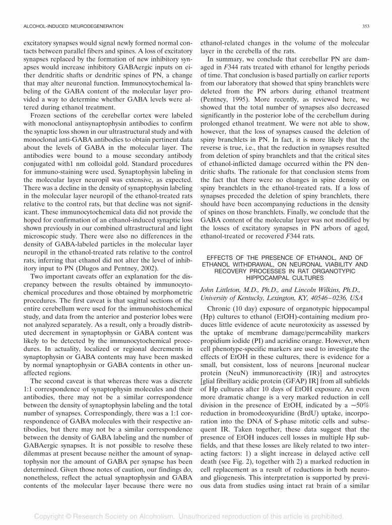
excitatory synapses would signal newly formed normal con-tacts between parallel fibers and spines. A loss of excitatorysynapses replaced by the formation of new inhibitory syn-apses would increase inhibitory GABAergic inputs on ei-ther dendritic shafts or dendritic spines of PN, a changethat may alter neuronal function. Immunocytochemical la-beling of the GABA content of the molecular layer pro-vided a way to determine whether GABA levels were al-tered during ethanol treatment.
Frozen sections of the cerebellar cortex were labeledwith monoclonal antisynaptophysin antibodies to confirmthe synaptic loss shown in our ultrastructural study and withmonoclonal anti-GABA antibodies to obtain pertinent dataabout the levels of GABA in the molecular layer. Theantibodies were bound to a mouse secondary antibodyconjugated with1 nm colloidal gold. Standard proceduresfor immuno-staining were used. Synaptophysin labeling inthe molecular layer neuropil was extensive, as expected.There was a decline in the density of synaptophysin labelingin the molecular layer neuropil of the ethanol-treated ratsrelative to the control rats, but that decline was not signif-icant. These immunocytochemical data did not provide thehoped for confirmation of an ethanol-induced synaptic lossshown previously in our combined ultrastructural and lightmicroscopic study. There were also no differences in thedensity of GABA-labeled particles in the molecular layerneuropil in the ethanol-treated rats relative to the controlrats, inferring that ethanol did not alter the level of inhib-itory input to PN (Dlugos and Pentney, 2002).
Two important caveats offer an explanation for the dis-crepancy between the results obtained by immunocyto-chemical procedures and those obtained by morphometricprocedures. The first caveat is that sagittal sections of theentire cerebellum were used for the immunohistochemicalstudy, and data from the anterior and posterior lobes werenot analyzed separately. As a result, only a broadly distrib-uted decrement in synaptophysin or GABA content waslikely to be detected by the immunocytochemical proce-dures. In actuality, localized or regional decrements insynaptophysin or GABA contents may have been maskedby normal synaptophysin or GABA contents in other un-affected regions.
The second caveat is that whereas there was a discrete1:1 correspondence of synaptophysin molecules and theirantibodies, there may not be a similar correspondencebetween the density of synaptophysin labeling and the totalnumber of synapses. Correspondingly, there was a 1:1 cor-respondence of GABA molecules with their respective an-tibodies, but there may not be a similar correspondencebetween the density of GABA labeling and the number ofGABAergic synapses. It is not possible to resolve thesedilemmas at present because neither the amount of synap-tophysin nor the amount of GABA per synapse has beendetermined. Given those notes of caution, our findings do,nonetheless, reflect the actual synaptophysin and GABAcontents of the molecular layer because there were no
ethanol-related changes in the volume of the molecularlayer in the cerebella of the rats.
In summary, we conclude that cerebellar PN are dam-aged in F344 rats treated with ethanol for lengthy periodsof time. That conclusion is based partially on earlier reportsfrom our laboratory that showed that spiny branchlets weredeleted from the PN arbors during ethanol treatment(Pentney, 1995). More recently, as reviewed here, weshowed that the total number of synapses also decreasedsignificantly in the posterior lobe of the cerebellum duringprolonged ethanol treatment. We were not able to show,however, that the loss of synapses caused the deletion ofspiny branchlets in PN. In fact, it is more likely that thereverse is true, i.e., that the reduction in synapses resultedfrom deletion of spiny branchlets and that the critical sitesof ethanol-inflicted damage occurred within the PN den-dritic shafts. The rationale for that conclusion stems fromthe fact that there were no changes in spine density onspiny branchlets in the ethanol-treated rats. If a loss ofsynapses preceded the deletion of spiny branchlets, thereshould have been accompanying reductions in the densityof spines on those branchlets. Finally, we conclude that theGABA content of the molecular layer was not modified bythe losses of excitatory synapses in PN arbors of aged,ethanol-treated or recovered F344 rats.
EFFECTS OF THE PRESENCE OF ETHANOL, AND OFETHANOL WITHDRAWAL, ON NEURONAL VIABILITY AND
RECOVERY PROCESSES IN RAT ORGANOTYPICHIPPOCAMPAL CULTURES
John Littleton, M.D., Ph.D., and Lincoln Wilkins, Ph.D.,University of Kentucky, Lexington, KY, 40546–0236, USA
Chronic (10 day) exposure of organotypic hippocampal(Hp) cultures to ethanol (EtOH)-containing medium pro-duces little evidence of acute neurotoxicity as assessed bythe uptake of membrane damage/permeability markerspropidium iodide (PI) and acridine orange. However, whencell phenotype-specific markers are used to investigate theeffects of EtOH in these cultures, there is evidence for asmall, but consistent, loss of neurons [neuronal nuclearprotein (NeuN) immunoreactivity (IR)] and astrocytes[glial fibrillary acidic protein (GFAP) IR] from all subfieldsof Hp cultures after 10 days of EtOH exposure. An evenmore dramatic change is a very marked reduction in celldivision in the presence of EtOH, indicated by a ~50%reduction in bromodeoxyuridine (BrdU) uptake, incorpo-ration into the DNA of S-phase mitotic cells and subse-quent IR. Taken together, these data suggest that thepresence of EtOH induces cell losses in multiple Hp sub-fields, and that these losses are likely related to two inter-acting factors: 1) a slight increase in delayed active celldeath (see Fig. 2), together with 2) a marked reduction incell replacement as a result of reductions in both neuro-and gliogenesis. This interpretation is supported by previ-ous data from studies using intact rat brain of a similar
ALCOHOL-INDUCED NEURODEGENERATION 353

developmental age (Miller, 1995). In contrast to the slowdevelopment of cytotoxicity during the presence of EtOH,its abrupt withdrawal from Hp cultures by changing themedium to one containing no EtOH causes significant andrapid toxicity as assessed by PI uptake (Mayer et al., 2002;Prendergast et al., 2000). Significant increases in PI uptakeare achieved within 24 hr of withdrawal, primarily in theCA1 subfield, suggesting a marked increase in rapid activecell death likely selective for pyramidal neurons in thissubfield (Fig. 2). The mechanism appears to be a classical“excitotoxic” cascade as originally suggested by severalworkers (Lovinger, 1993), and likely involves synaptic hy-perexcitability and ictal activity in recurrent circuits (Mul-holland and Prendergast, 2003), combined with excess re-lease of endogenous glutamate and polyamines (Gibson etal., 2003), culminating in excess activation of NMDARsand cytotoxic Ca2� entry (Mayer et al., 2002; Prendergastet al., 2001). In addition to evidence of membrane damageindicated by increased PI uptake, delayed reductions in cellphenotype-specific markers suggest that neuronal and as-troglial losses occur during the first several days of acutewithdrawal. Thus, NeuN IR declines steadily to an appar-ent maximal reduction at 3 days following EtOH with-drawal; GFAP IR declines more rapidly in most Hp sub-fields during the first 24 hr, but then recovers to or near tocontrol levels by 3–5 days following withdrawal. In addition,BrdU uptake/incorporation and IR, already markedly de-pressed by the prolonged presence of EtOH, are apparentlyreduced even further during the first 24 hr of acute with-drawal. Therefore, cell losses during acute withdrawal fromEtOH likely reflect the combined effects of a very activeprocess of rapid cell death, combined with detrimental
effects on cell proliferation, which are then manifest as areduction in cell replacement by neuro- and gliogenesis(Fig. 2). In addition to their suggested roles in the initialcytotoxicity associated with EtOH exposure and with-drawal, adverse effects on cell proliferation and astroglialdensities are also likely to be detrimental to neuronal re-covery processes. For example, following EtOH with-drawal, BrdU uptake/incorporation (cell proliferation) re-turns to levels observed in Hp cultures not previouslyexposed to EtOH, and then does not increase above theselevels. In contrast, acute exposure of Hp cultures to acytotoxic concentration of NMDA (200 �M) induces adelayed increase in cell proliferation in all Hp subfields tomore than 150% of control values. One interpretation ofthese data are that prior exposure to EtOH and subsequentwithdrawal impairs the reactive capacity of the cell prolif-erative aspect of the recovery process, an effect which maybe most detrimental in the dentate gyrus, since this area isknow to contain actively proliferating neural stem cells.Despite this effect, NeuN IR recovers slowly to aroundcontrol levels after several days, indicating that some re-covery of neurons following EtOH-induced losses is possi-ble. This may explain the previously conflicting results as towhether cell loss actually occurs in the Hp followingchronic EtOH administration. Our data would suggest thata determination of cell loss will depend exactly on whattime point following EtOH exposure at which measure-ments are taken. It has yet to be determined whetherturnover of neuronal and glial cell populations results inintact functional circuitry in Hp cultures. An answer to thisquestion will provide the basis for assessing if and whenfunctional recovery occurs postwithdrawal. Overall, the re-
Fig. 2. Theoretical positions in the dynamicequilibrium of neural cell turnover at whichethanol (EtOH) may exert stimulatory (�) orinhibitory (�) effects. Clockwise from top cen-ter: Convergent evidence suggests that thepresence of and withdrawal from prolongedEtOH exposure induces processes of concur-rent “delayed and rapid active neuronal celldeath,” respectively. The current studies indi-cate that EtOH may also exert similar directeffects on astrocytes. The inherent sensitivityof neural stem and or progenitor cells to thepresence and withdrawal of EtOH has yet tobe fully characterized. However, the currentreport, in conjunction with that of (Nixon andCrews, 2002), suggest that neural progenitorcell proliferation is significantly reduced byEtOH exposure and withdrawal. Reduced pro-genitor cell proliferation then likely leads toreduced genesis of both neurons and (astro-)glia, thus compounding the overt cytotoxiceffect of EtOH, and ultimately resulting in thereduced neuronal and astroglial densities ob-served in multiple hippocampal subfields.
354 CREWS ET AL.

sults support the concept that EtOH alters neuronal andglial densities both during its presence and during with-drawal. The mechanisms probably differ except that re-duced cell division and reduced cell replacement may becommon to both phases. In addition, EtOH exposure andwithdrawal also seem to disrupt the recovery of neuronsfollowing insult suggesting that they will increase the sever-ity of many other potentially neurotoxic stimuli in Hp.However, it remains to be assessed exactly which of thesemany processes is most important in the human alcoholic.
BINGE DRINKING-INDUCED BRAIN DAMAGE: GENETICAND AGE RELATED FACTORS EFFECTING SENSITIVITY
Fulton T. Crews, Ph.D., and Jian Zou, Ph.D., BowlesCenter for Alcohol Studies, University of North Carolina atChapel Hill, Chapel Hill, NC 27599 USA
Clinical and preclinical studies have established thatchronic alcohol consumption causes neuropathologicalchanges in brain (Agartz et al., 1999; Crews, 1999; Faddaand Rossetti, 1998; Pfefferbaum et al., 1992). The loss ofcortical gray and white matter, as well as synaptic and celldensity in alcoholic individuals is associated with behavioralneuropathology including cognitive and sensory deficits(Beatty et al., 2000; Bowden et al., 2001; Moselhy et al.,2001; Rupp et al., 2003). Further, these neuropathologicalchanges have been suggested to contribute to the progres-sion and chronicity of alcohol dependence (Bowden et al.,2001; Crews, 1999). These findings increase the importanceof understanding the mechanisms and processes mediatingalcoholic neuropathology.
The pattern of alcohol drinking has been suggested tocontribute to neuropathology, with binge drinking sug-gested to be the most neurotoxic (Crews et al., 1999; Hunt,1993). Binge drinking may represent an environmental fac-tor facilitated by genetic and other risk factors for alcohol-ism (Bates et al., 2002; Bowden et al., 2001; Hasin et al.,2001). A variety of evidence suggests that there are geneticrisk factors involved in the development of alcoholism(Begleiter and Porjesz, 1999; Prescott and Kendler, 1999).The selectively bred alcohol-preferring (P) rat line is agenetic animal model of alcoholism. The P rat will consumelarge amounts of alcohol with free choice drinking, willwork for alcohol, and will drink alcohol until both tolerantand physically dependent (Li and McBride, 1995). The Prat is most often compared to the alcohol-nonpreferring(NP) rat line. P rats are less sensitive to the aversive andsedative-hypnotic effects of ethanol than are NP rats(Froehlich et al., 1988; Waller et al., 1983), consistent withhuman studies showing that family-history-positive humanshave a lower sensitivity to alcohol (Schuckit and Smith,1996). Although P rats are less sensitive to the sedativeeffects of ethanol following the same binge ethanol treat-ment, P rats were found to have 2 to 3 fold more braindamage in perirhinal and entorhinal cortices (Crews andBraun, 2003) (Fig. 3). Adolescent drinking may represent
another risk factor for alcoholism. Although adolescentrats are less sensitive to the sedative effects of ethanol(Little et al., 1996), equivalent binge treatment of young-adolescent and adult rats indicated that the frontal regionsof adolescent brain are much more sensitive to binge in-duced ethanol neurotoxicity than the frontal regions ofadult brain (Crews et al., 2000)(Fig. 3). These findingssuggest that genetic and adolescent risk factors for bingeethanol induced neuropathology share some features withhuman risks for development of alcohol dependence.
Understanding when, where and why chronic alcoholconsumption leads to neuropathology will significantly ad-vance efforts to reduce alcohol induced brain damage andcould contribute to efforts to reduce alcohol dependence.Early studies in cultured cortical neurons indicated thatethanol could block NMDA mediated excitotoxicity andthat cortical cultures appeared to remain healthy in highconcentrations, e.g., 100 mM, of ethanol (Chandler et al.,1993b). Chronic ethanol treatment of cortical cultures ledto supersensitive NMDA receptors which remained inhib-ited during the presence of ethanol, but once ethanol wasremoved were supersensitive to NMDA stimulated nitric
Fig. 3. Binge Ethanol Induced Brain Damage is Greater in Adolescent andGenetic Models of Alcoholism. Shown are groups of animals all treated with a 4day binge ethanol protocol and sacrificed on day 4 while still intoxicated. Controlsshow no damage, whereas all binge ethanol treated animals show damage.Agyrophilic silver stain was used to identify dying neurons (Switzer, 2000). In theleft panel, damage in perirhinal cortex is compared between P and NP rats, withNP set at 100% (See Crews and Braun, 2003 for details). Note the P and NP ratswere both treated with identical 4 day binge treatments, yet P rats have 3 foldmore silver stain, e.g., more neurodegeneration. In the right panel, damage inperirhinal cortex is compared between adolescent and adult Sprague-Dawley ratstreated with identical 4 day binge treatments with adult anterior perirhinal cortexdamage set at 100%. Note that adolescents have 2–3 fold more silver stain, e.g.,dying neurons, than adults (See Crews et al., 2000 for details). These findingsuggest that genetic and age are factors that regulate sensitivity to binge inducedbrain damage.
ALCOHOL-INDUCED NEURODEGENERATION 355

oxide production (Chandler et al., 1997) and neurotoxicity(Chandler et al., 1993a). These findings and others sug-gested that ethanol neurotoxicity was related to glutamateexcitotoxicity, which occurred primarily during ethanolwithdrawal. (Crews and Chandler, 1993; Lovinger, 1993).More recent in vivo studies investigating ethanol-inducedneurotoxicity have indicated that ethanol is selectively toxicto certain brain regions that occur during ethanol intoxica-tion. Three studies using in vivo binge ethanol treatmentmodels have reported binge ethanol induced brain damageduring or at times close to intoxication with less damageapparent during ethanol withdrawal. In 80–90 day oldSprague-Dawley rats, entorhinal and perirhinal corticaldamage was greatest just after the last dose of a 4 day bingetreatment model, with less damage at 16 and 72 hrs ofwithdrawal and little to no apparent damage after 7 days ofwithdrawal (Crews et al., 2000; Obernier et al., 2002a).Obernier et al. (2002a) in a similar binge ethanol treatmentwith somewhat lower peak binge blood ethanol levels andyounger rats found necrotic dark cell degeneration after 2days of treatment in some brain regions and in agranularinsular, piriform and perirhinal, entorhinal cortex and den-tate gyrus after 4 days of treatment. Rats withdrawn for 3days showed little or no damage supporting the idea thatdamage occurs during intoxication (Obernier et al., 2002a).Collins et al. (1996a) found binge induced brain damage 8and 36 hrs after the last dose of a binge treatment with atrend toward reduced damage at the later time point (Col-lins et al., 1996a). Degeneration visualized by silver stainhas been estimated to appear 36–48 hrs after the insult asneuronal death proceeds (Switzer III, 2000). Taken to-gether, these studies suggest that binge drinking neurode-generation occurs during intoxication at high blood alcohollevels. Binge ethanol treatment also results in long termrelearning deficits (Obernier et al., 2002b). Since humanalcoholics have both structural and behavioral neuropa-
thology the finding that binge ethanol treatment results inassociated structural and relearning deficits supports thevalidity of binge ethanol induced neuropathology as amodel of human alcoholic brain damage.
As mentioned above, early hypothesis regarding ethanolinduced brain damage were based on in vitro preparationsand centered around glutamate excitotoxicity. Studies ofthe mechanisms of binge induced ethanol treatment haveindicated that glutamate excitotoxicity cannot explain theneurodegeneration. Glutamate antagonists have been triedby several groups and all agree that NMDA antagonists donot protect against binge ethanol induced brain damage(Table 1). Glutamate AMPA antagonists and calciumchannel antagonists also have little or no effect on bingeethanol induced brain damage. Taken together, these stud-ies suggest that glutamate NMDA excitotoxic delayed neu-ronal death cannot explain binge ethanol induced neuro-toxicity. The findings that binge ethanol induced damageoccurs during intoxication and does not involve NMDAexcitotoxicity is consistent with studies suggesting that theNMDA receptor is blocked by ethanol reducing excitatorytransmission during intoxication (Crews et al., 1996). Thus,in vivo binge drinking models establish that ethanol candamage the brain during intoxication and that the mecha-nism is not directly related to glutamate neurotoxicity.
Ethanol induced brain damage is an important area ofresearch with several laboratories focused on understand-ing the why and how ethanol damages the brain. Twooverlapping hypothesis related to binge induced damageare supported by studies using inhibitors. One hypothesissuggests that ethanol induced changes in cerebral pressureresult in brain damage (See Collins section this article andTable 2). Another suggests that oxidative stress or activa-tion of nuclear factor �-B (Nf�B) due to oxidative stressmay be a factor. Although inhibition of nitric oxide syn-thetase, which should reduce nitric oxide induced reactive
Table 1. Glutamate Receptor Antagonists and Calcium Channel Blockers Do NOT Block Binge-Induced Neurodegeneration.
Drug Class Hypothesis Effect Reference
Nimodipine Voltage-gatedCa � 2 channel
Chronic Etoh increases Ca � 2Channels in brain
Reduced damage in DGIncreased damage in Piri
Corso et al. (1998)
Nimodipine Voltage-gatedCa � 2 channel
Chronic Etoh increases Ca � 2Channels in brain
No neuroprotection Hamelink (1998, 1999)
DNQX AMPA antagonistNMDA antagonist at
glycine site
High densities of AMPAReceptors in the DG, CA fields, Ento, Piri,
FC
No effect Corso et al. (1998)
MK-801 NMDA antagonist Chronic Etoh causes supersensitiveNMDA excitotoxicity
Increased damage inEnto, orbital cortexAnd peri-amygdala
Corso et al. (1998)
MK-801 NMDA antagonist Chronic Etoh causes supersensitiveNMDA excitotoxicity
No effect Collins et al. (1998)
MK-801 NMDA antagonist Chronic Etoh causes supersensitiveNMDA excitotoxicity
No neuroprotection Hamelink (1999)
Memantine NMDA antagonist Chronic Etoh causes supersensitiveNMDA excitotoxicity
No neuroprotection Hamelink (1999)
Olfactory Bulbectomies Surgical procedure Chronic Etoh causes excitotoxic cascade No neuroprotection Hamelink (1999)
Studies listed address the hypothesis that ethanol induced neurodegeneration is related to glutamate-NMDA neurotoxicity. Glutamate-NMDA neurotoxicity isrelated to calcium influx which can occur through the glutamate-NMDA receptor and/or voltage gated calcium channels. Antagonists to these channels does notprotect against binge ethanol induced damage suggesting that glutamate-NMDA excitotoxicity is not the mechanism of neurodegeneration. Olfactory bulb hasglutamate projection neurons that innervate perirhinal, piriform and entorhinal corteces that are damaged (See Obernier et.al., 2002 for more discussion). Lessions ofglutamate input from olfactory bulbs also does not protect consistent with glutamate excitotoxicity not mediating binge ethanol induced brain damage.
356 CREWS ET AL.

oxygen species, or inhibition of cyclo-oxygenase 2, whichproduces reactive oxygen species, do not protect againstbinge ethanol induced brain damage, the antioxidant buty-lated hydroxytoluene does protect (Table 2). Some of thediuretics used to support the cerebral pressure hypothesishave antioxidant activity suggesting that might be the mech-anism of protection as apposed to reduced CSF pressure.Although the mechanisms of binge ethanol induced braindamage remain to be elucidated, these studies continue tosupport the occurrence of ethanol induced brain damageduring intoxication.
Neurotoxicity occurs as a balance between trophic sup-port of brain cells balanced with insults that damage braincells. Recent studies have indicated that the cAMP-response-element-binding protein (CREB) is a transcrip-tion factor important for inducing key genes that improve
neuronal vitality, growth and resistance to insults (Man-tamadiotis et al., 2002; Walton and Dragunow, 2000) (Fig.4). CREB is down stream of a variety of signaling kinasesand requires phosphorylation to initiate transcription ofprosurvival neuronal factors. Binge ethanol treatment hasbeen shown to reduce the amount of PCREB-immunoreactivity in the hippocampal dentate gyrus duringintoxication (Bison and Crews, 2003). Binge ethanol treat-ment increases neuronal death in the hippocampal dentategyrus (Crews et al., 1999) and reduces neurogenesis in thedentate gyrus (Nixon and Crews, 2002). The reduction inhippocampal dentate gyrus PCREB would be expected toreduce the transcription of prosurvival genes and makeneurons more susceptible to toxic insults (Fig. 4) as well asreducing neurogenesis. Ethanol has been found to increaseactivation of the transcription factor Nf�B due to upstream
Table 2. Anti-Oxidants Reduce Binge Induced Brain Damage NOS Inhibitors and Mannitol do NOT block brain damage
Drug Class Hypothesis Effect Reference
L-NAME Nitric oxideSynthase inhibitor
Chronic EtoH increases NO production No change/increased damage Zou (1996)
7-NI Nitric oxide synthase inhibitor Chronic EtoH increases No production No change/increased damage Zou (1996)Furosemide Diuretic (antioxidant*) Chronic ethanol
Increases CSF pressure*Chronic EtoH increases Oxidative stress
Protects in Ent and DG.(Collins, 1998)
*(Hamelick 1999, 2000)No protection (Crews, 2001)
Collins (1998)Hamelick (2000)*Crews (2001)
Nimesulide COX-2 Inhibitor COX-2 contributes to BIBD No neuroprotection Preliminary dataMannitol Osmolyte reduces CSF pressure Chronic ethanol increases CSF pressure No neuroprotection** Crews (2001)BHT Antioxidant Chronic EtoH
Increases Oxidative stressBlocks BIBD Crews & Zou
Hamelink (2000)
Listed are drugs that have been tested for effectiveness in reducing binge ethanol treatment induced brain damage. Nitric oxide synthesis and cyclo-oxygenase2 inhibitors were not protective suggesting these enzymes are not involved. Furosemide, a diuretic and anti-oxidant is protective as is butylated hydroxytoluene, ananti-oxidant. These findings suggest that oxidative stress and swelling may contribute. In an effort to further test increased swelling as a hypothesis mannitol was giventhroughout the binge treatment. It did not protect suggesting that oxidative stress may contribute to binge induced brain damage. See Collins, this article for morediscussion.
Fig. 4. Schematic representation ofethanol-induced imbalance of nuclear tran-scription signaling pathways leading to de-ceased expression of survival genes and in-creased expression of death genes. Ethanolinteracts with upstream initiators, such asTNF� and glutamate, leading to alterations intranscription factor nuclear signaling path-ways that regulate gene expression. The con-vergence of comprehensive signaling trans-duction in cytoplasm after ethanol influence isdirected toward a reduction in CREB signalingand an increase in NF-�B signaling. CREB isable to regulate the transcription of down-stream target genes, such as bcl2 and BrainDerived Neurotrophic factor, that support neu-ronal cell survival. NF-�B signaling pathwayregulates downstream target genes, such asTNF� and NO, that promote cell death. In ad-dition, ethanol could potentially increase AP-1signaling that is linked to cell death. All to-gether, ethanol exposure leads to imbalancebetween pro-death gene and pro-survivalgene transcription, making cells more vulner-able to toxic insults. Ethanol can act at the cellsurface and/or on nuclear transcriptionfactors.
ALCOHOL-INDUCED NEURODEGENERATION 357

signals such as TNF� or other processes in some cells.Although it is not clear exactly what the effects of ethanolare on transcription of pro-survival and pro-death genes, itis possible that the mechanism of ethanol induced neuro-toxicity is related to changes in transcription that increasepro-death gene transcription and reduce pro-survival genetranscription making neurons more vulnerable to oxidativestress, swelling and other insults that could occur duringethanol intoxication. Additional studies are needed to elu-cidate this important pathology associated with alcoholismand alcohol abuse.
BINGE ETHANOL-INDUCED BRAIN DAMAGE:INVOLVEMENT OF EDEMA, ARACHIDONIC ACID
AND TNF-�
Michael A. Collins, Ph.D. and Edward J. Neafsey,Department of Cell Biology, Neurobiology and Anatomy,Loyola University Stritch School of Medicine, Maywood, IL60153 USA.
Our studies with an in vivo rat model and with organo-typic brain slice cultures indicate that binge ethanol-induced neuronal degeneration is engendered, at least inpart, by trauma-like neuroinflammatory processes involvingbrain edema and the release of pro-inflammatory signalingmolecules such as arachidonic acid and TNF�. This inter-pretation is consistent with results suggestive of a neuroin-flammatory mechanism that have been obtained by otherlaboratories studying binge ethanol-dependent brain dam-age; namely, necrosis predominating over apoptosis(Obernier et al., 2002a), and an important role for oxidativestress (Hamelink et al., 2000).
Subchronic binge intoxication via gavage of adult rats, amodel first developed by Majchrowicz to induce alcoholwithdrawal signs (Majchrowicz, 1975), was subsequentlyshown to induce regionally selective neuronal degenera-tion, particularly in the entorhinal cortex (EC), hippocam-pal (HIP) dentate gyrus (see Fig. 5) and olfactory bulb,prior to the time of maximal induced seizures (Collins etal., 1996b; Switzer III et al., 1982). Despite indirect as wellas in vitro evidence for NMDA receptor-directed excito-toxicity in ethanol-dependent brain damage, we were un-able to demonstrate with pharmacological inhibitors thatan excitotoxic pathway was essential to neuronal degener-ation (Collins et al., 1998; Corso et al., 1998; Zou et al.,1996)(See Table 1). Rather, binge-intoxicated rats dis-played significant cortical edema—which is often a charac-teristic of brain trauma and neuroinflammation in gener-al—and by preventing the edema with a potent loopdiuretic, furosemide, most of the EC and HIP dentategranule cell neuronal degeneration could be prevented(Collins et al., 1998). Acetazolamide, another diuretic thatdiffers structurally and mechanistically from furosemide(and lacks furosemide’s antioxidant capabilities), was alsoantiedemic and neuroprotective. Figure 6 summarizesabove-mentioned neurotoxicological results.
When prepared from one week-old rats and cultured fora prolonged period (~2.5 weeks or longer), organotypicbrain slices are considered to be plausible in vitro models ofthe adolescent/young adult CNS (Diekmann et al., 1994;Gahwiler et al., 1997). Binge-treated ~15 hr/day for 7–10days with concentrations of ethanol (75–150 mM) attainedin alcoholics, our HIP-EC brain slice cultures replicatedsome, but not all, of the features of binge intoxication-dependent neuronal degeneration in adult rats (Collins etal., 1998). For example, binge ethanol-exposed culturesincurred appreciable neuronal degeneration that was sup-pressed by furosemide, acetazolamide, and other diuretics.However, whereas ethanol-dependent neurodamage pre-vailed in the EC, it also was evident in HIP CA1-CA3—aregion little affected in vivo (see Fig. 5). Although long-term organotypic cultures are more relevant, in our opin-ion, than isolated cell cultures for investigations of neuro-nal degenerative mechanisms that entail glial-neuronalinteractions, further studies are required to better under-stand their differences from the in vivo models with regardto specificity of binge ethanol-induced brain damage.
Fig. 5. Photomicrograph of section of temporal cortex of adult rat binge-intoxicated with ethanol via the Majchrowicz model (ref. 3), fixed, and stained withthe deOlmos cupric silver stain for degenerating neurons. Significant neuronaldegeneration is evident in the entorhinal cortex (lateral [Lat Ent] and intermedial[Int Ent]), the perirhinal cortex (PRh), and the dentate gyrus (DG). There are fewdegenerating cells in the hippocampal CA1-CA3 or medial entorhinal (Med Ent)cortex, but bands of degenerating axon terminals are found in the hippocampalCA2 region (larger open arrow). Sections from control rats showed no cupric silverstaining in any regions. Inset: larger power of Int Ent, showing relative specificityof degeneration of pyramidal neurons in layer 3, with degenerating neurites inlayers 1 and 2 subregions that create “degenerating islands.” Adapted from T.Corso, PhD thesis, Loyola University 1995, and reference 5.
358 CREWS ET AL.

Neuroinflammatory-associated signaling routes ostensi-bly triggered by edema and cell swelling have been inves-tigated with the organotypic HIP-EC slice cultures. Arachi-donic acid (AA) released by intracellular calciumelevations and phospholipase A2 (PLA2) activation, andincreased pro-inflammatory TNF, have been associatedwith astroglial swelling and inflammation under variousconditions (Allan and Rothwell, 2001; Winkler et al., 2000).Our studies indicate that AA may have a neurotoxic sig-naling role in the binge model, since inhibiting PLA2 with ageneral inhibitor greatly suppressed neuronal degenerationdue to ethanol (Kumar et al., 2000). Furthermore, cotreat-ment with an antibody to TNF� significantly reduced theneurodamage in slice cultures. Both of these mediators caninteract to potentiate each other, both have been associatedwith oxidative stress, and both have been frequently impli-cated in other forms of neuronal degeneration (Hernandezet al., 1999; Scorrano et al., 2001).
Figure 7 presents a working model for brain ethanol-induced brain damage that attempts to link neuroinflamma-tory sequelae such as arachidonic acid and TNF� with initialbrain edema events. How repetitively high ethanol levels cou-pled with abrupt withdrawals serves to induce brain (presum-ably cell or cytotoxic) swelling and hydration is a fundamentalquestion yet to be answered. Brain ionic imbalances are asso-ciated with the ethanol-induced edema (Collins et al., 1998);speculatively, it is possible that these imbalances underlie theinitial edema, and arise from alterations in glial ionic or water(e.g., aquaporin) transport mechanisms that are engenderedby ethanol and its withdrawal.
CYCLIC AMP CASCADE, STEM CELLS, AND ETHANOL
Lawrence D. Snell, Ph.D. and Boris Tabakoff, Ph.D. Dept.Of Pharmacology, University of Colorado Health SciencesCenter, School of Medicine, Denver, CO 80262, USA
Heavy alcohol consumption is associated with a numberof medical disorders, including alcohol-induced brain dam-
age. In general, such alcohol-induced brain damage is be-lieved to occur during periods of ethanol intoxicationand/or during periods of ethanol withdrawal. There are anumber of interrelated mechanisms postulated to accountfor ethanol-induced neurotoxicity, and many of these aredetailed in the other presentations of this symposia.
Regardless of the exact mechanisms, the dogma in neu-robiology has been that neurons, once lost, cannot be re-placed. However, recent studies indicate that neurogenesiscan occur throughout life, due to the presence of neuralprecursor cells in the adult mammalian brain (Armstrongand Barker, 2001). These “adult ”neural stem cells (NSCs)have a limited capacity for self-renewal and can give rise to
Fig. 6. Summary of protection studies against binge ethanol EC neurodegeneration. Quantitation of cupric silver staining of entorhinal cortex degenerating neuronsin adult male rats binge-intoxicated subchronically with ethanol, with and without potential inhibitors. Solid circles and open triangles indicate individual rat values. E4denotes the 4-day multiple ethanol dose model of Majchrowicz (Majchrowicz, 1975), whereas E8 is a modified model employing one ethanol dose daily for 8 days(Collins et al., 1998). The antagonists or inhibitors MK-801, Nimodipine, DNQX, L-NAME and 7-Nitroindazole (7-NI) did not significantly reduce the degeneration in the4-day model. The diuretics furosemide and acetazolamide were effective neuroprotectants in the 8-day model. Data summarized from (Collins et al., 1998; Corso etal., 1998; Zou et al., 1996) and (Kumar et al., 2002).
Fig. 7. Presents a working model for brain ethanol-induced brain damage thatattempts to link neuroinflammatory sequelae such as arachidonic acid and TNF�
with initial brain edema events. How repetitively high ethanol levels coupled withabrupt withdrawals serves to induce brain (presumably cell or cytotoxic) swellingand hydration is a fundamental question yet to be answered. Brain ionic imbal-ances are associated with the ethanol-induced edema (Collins et al., 1998);speculatively, it is possible that these imbalances underlie the initial edema, andarise from alterations in glial ionic or water (e.g., aquaporin) transport mechanismsthat are engendered by ethanol and its withdrawal.
ALCOHOL-INDUCED NEURODEGENERATION 359

differentiated cells (i.e., neurons, astrocytes and oligoden-drocytes) through asymmetric division. Adult neurogenesisis primarily confined to two discrete areas: the subventricu-lar zone of the lateral ventricle (SVZ) and the subgranularzone (SGZ) of the hippocampus (Doetsch et al., 1997;Johansson et al., 1999; Temple and Alvarez-Buylla, 1999).The finding that neuronal damage and loss generate asignal for NSCs to divide, migrate and differentiate (Arm-strong and Barker, 2001; Gage, 2000), has led to the hy-pothesis that NSCs can function as a “self-repair” mecha-nism, and that the failure of stem cells to respond normallyto cell loss may contribute to the progression of the neu-rodegenerative disease. Based on this hypothesis, one mayspeculate that ethanol-induced brain damage may reflect afailure of neuroregeneration (Armstrong and Barker,2001), possibly because ethanol itself affects the ability ofthe adult stem cells to proliferate, or migrate to the appro-priate locations in the brain, or differentiate.
Examination of recent literature using animals models ofethanol-induced brain injury, support this idea. In perhapsthe first study of this kind, (Nixon and Crews, 2002) dem-onstrated that BrdU labeling of neural progenitor cells wasreduced in the SGZ following both acute and chronic“binge” ethanol administration to rats. Similarly, (Herreraet al., 2003) found that a 6 week chronic ethanol dietadministration resulted in a decreased number and survivalof BrdU-labeled dividing cells in the SGZ. Using TUNELlabeling of apoptotic cells and PCNA (proliferating nuclearcell antigen) to label dividing cells, Pawlak et al. (2002)found that chronic ethanol administration to mice via aliquid diet produced a loss of hippocampal cells in the SGZthat was associated with an increased number of PCNA-labeled cells, suggesting an increase in neurogenesis (Paw-lak et al., 2002). This study caught our attention because itsuggested that increased neurogenesis can accompanyethanol-induced neurodegeneration.
cAMP is formed from the conversion of ATP to cAMPby adenylyl cyclases via G-protein-coupled receptors. ThecAMP cascade ultimately results in the phosphorylation ofcAMP responsive element binding protein (CREB), one ofthe key elements in controlling transcriptional activity incells. Currently, nine isoforms of adenylyl cyclase (AC)have been identified, which are differentially regulated byG proteins, Ca2� and protein kinases. ACs 2, 4 and 7 forma family of enzymes that are stimulated by G� (subunits,provided that G� is also present (Feinstein et al., 1991; Gaoand Gilman, 1991; Yoshimura et al., 1996). Our own workhas shown that, while ethanol can enhance agonist-stimulated activity of a subset of AC isoforms, AC7 is themost sensitive to this action of ethanol (Yoshimura andTabakoff, 1995). Following chronic exposure of animals orcells to ethanol, a reduction of agonist-stimulated AC ac-tivity occurs (Tabakoff and Hoffman, 1998). The acute andchronic actions of ethanol on AC, and particularly AC7,activity suggest a molecular mechanism by which ethanolexposure may affect neural progenitor cell proliferation
and differentiation. There is evidence that this cascade,including activation of CREB, can increase neurogenesis inthe adult mouse hippocampus (Nakagawa et al., 2002). Incontrast, cell proliferation is decreased in transgenic micethat conditionally overexpress a dominant negative mutantof CREB.
There are several markers that can be used to identifyand quantitate neural precursor cells. As already men-tioned, to study cellular proliferation and survival, cells canbe labeled with agents such as BrdU. However, precursorcells also express particular cell surface antigens that iden-tify them at different stages of development. One suchmarker, associated with mitotic and/or differentiating pre-cursor cells, is a protein that has been called AC133 (Mi-raglia et al., 1997), CD133 (Uchida et al., 2000) or, in themouse, prominin (Corbeil et al., 2001; Weigmann et al.,1997). The mouse prominin is present in the neuroepithe-lium of mouse embryos and in the ependymal layer of adultmouse brain (Weigmann et al., 1997). Of particular interestis a study (Sawamoto et al., 2001) in which embryonicmesencephalic ventricular zone precursor cells could beidentified based on their expression of prominin, and theseprominin-expressing cells were found near the ventricularwall, where mitogenic neurogenesis occurs. After sorting byfluorescence-activated cell sorting, the prominin-expressingcells were shown to have a high potential to form neuro-spheres in culture, and to generate neurons, as determinedby staining for the neuronal markers, MAP2 and �III-tubulin, as well as astrocytes (GFAP) and oligodendrocytes(O4). In contrast, the prominin-negative cells formed fewerand smaller neurospheres. These data suggest that promi-nin can be used as a marker for a population of mitogenic,multipotent precursor cells that have a high potential formigration and differentiation into neurons and glial cells.
We were particularly interested when our DNA microar-ray studies revealed that AC7 transgenic TG mice createdin our laboratories (Yoshimura et al., 2000) express ahigher level of mRNA for prominin in brain than theirwild-type controls. These mice, which were generated onthe FVB/N background, overexpress the human form ofType VII adenylyl cyclase (AC7) in brain. FVB/N wild-type(WT) mice are susceptible to brain damage induced byseizure activity (Schauwecker and Steward, 1997), as well asethanol-induced brain damage, reflected by gliosis.
The effect of ethanol treatment and withdrawal on braininsult/damage was assessed in the WT and AC7 transgenic(AC7 TG) mice in vivo using a liquid diet paradigm. Micewere subjected to two successive cycles of ethanol intoxi-cation, each followed by 3 days of ethanol withdrawal. BothWT and AC7 TG ethanol-fed mice exhibited an equivalentincrease in withdrawal severity during the second with-drawal period (kindling). We assessed glial fibrillary acidicprotein (GFAP) immunoreactivity 10 days after the secondwithdrawal as a marker of astrocytic proliferation, which ac-companies brain insult and injury. Ethanol feeding and with-drawal increased the number of GFAP-immunoreactive cells
360 CREWS ET AL.

in the hippocampus 30–40% compared to control diet-fedmice, but the increases in GFAP-positive cells were equivalentin WT and AC7 TG mice.
Using the same ethanol treatment/withdrawal paradigm,stem cell proliferation was assessed in the subventricularzone (SVZ) and the subgranular zone (SGZ) of the hip-pocampus, the sites of neurogenesis in the adult brain, bymeasuring the incorporation of bromodeoxyuridine (BrdU)and the expression of the stem cell antigen, prominin.Ethanol intoxication and withdrawal resulted in a decreasein BrdU-labeling of dividing cells in both the SVZ and SGZof WT mice (Fig. 8). In the AC7 TG mice, there was noethanol-induced decrease in BrdU labeling of dividing cellsin the SVZ and SGZ, suggesting that the presence of theAC7 transgene protects against ethanol-administration-induced cell loss. Immunohistochemical studies confirmedthat AC7 TG mice have an increased level of promininlabeling of cells in the SVZ compared to WT mice and thatethanol feeding/withdrawal did not result in any reductionin prominin-labeling in either genotype. A greater pool ofthese cells, which are also labeled by BrdU, in AC7 TGmice may compensate for the overall stem cell reductionseen WT mice.
The difference in prominin-expressing cells between theAC7 TG mice and the WT controls is presumably a con-
sequence of overexpression of human AC7. We hypothe-size that in AC7 TG mice, increased cAMP generationcapacity is associated with an increase in a subpopulation ofmultipotent stem cells (prominin-positive) which are resis-tant to the deleterious effects of ethanol administration/withdrawal. An even more interesting feature of stem cellsexpressing prominin is that prominin may be a marker ofcells preparing to undergo an asymmetric cell division.Huttner and Brand (1997) have reviewed evidence whichindicates that an asymmetric division of cells located at thesurface of the neuroepithelium (e.g., SVZ) results in dif-ferential distribution of constituents of the mother cell tothe two daughter cells (Huttner and Brand, 1997). One ofthe daughter cells remains a neuroepithelial cell while theother progresses to differentiation into a neuron or glialcell (Chenn and McConnell, 1995). The stem cells whichundergo such an asymmetric division are distinguished bythe differential location of certain proteins and lipids attheir basal and apical surfaces. Huttner and Brand (1997)noted that a “novel protein” could be used to identify theapical surface of neuroepithelial cells, and Corbeil et al.(2000) then identified this protein as CD133 or prominin(Corbeil et al., 2000; Huttner and Brand, 1997). Thus,prominin can be considered a marker of stem cells whichare undergoing (or preparing to undergo) an asymmetric
Fig. 8. Working model of the actions of ethanol on stem cell proliferation in FVB/N transgenic mice overexpressing human AC7 (AC7 TG) or their wild-type (WT)littermates. In the basal state (CONTROL-FED), both WT and AC7-TG animals have 2 pools of stem cells that can incorporate BrdU. One pool is distinguished by theexpression of prominin (P� stem cells; wavy lines) while the other does not (P� stem cells). BrdU (triangular circles) is taken up by the daughter cells of both P� stemcells after asymmetric division and P� stem cells after symmetric division, respectively. The AC7 TG animals have a bigger pool of prominin-labeled cells. Wehypothesize that this increased pool of prominin-labeled cells is being driven by higher levels of AC activity and cAMP. Upon chronic EtOH feeding and WD (EtOH-FED)there is a significant diminution of BrdU labeled cells in WT animals. On the other hand, the AC7 TG animals do not show this diminution. Our data suggests that theprominin-expressing cells are not (or not as) susceptible to EtOH-induced stem cell loss. Since both populations of stem cells (P� and P�) incorporate BrdU, this wouldexplain why a greater reduction in stem cells was observed in WT mice compared to TG mice. Another implication of our studies is that the AC7 TG mice display agreater number of precursor cells with a high potential for differentiation into neurons and glia, i.e., the AC7 TG mice may have greater neuroregenerative capacity thantheir WT litter mates.
ALCOHOL-INDUCED NEURODEGENERATION 361

cell division. The prominin-containing daughter cell wouldretain the phenotype of the parent stem cell while the otherdaughter cell would be destined to leave the ventricular zoneand differentiate into a neuron (Huttner and Brand, 1997).
Supporting this hypothesis are recent literature reportsthat stem cells expressing prominin also express ATF5, amember of the activating transcription factor/cAMP re-sponse element binding protein (ATF/CREB) family andthat ATF5 represses cAMP-induced transcription and differ-entiation. Our gene array studies indicate that ATF5 expres-sion levels are lower in the brains of AC7 TG mice. Therefore,the AC7 TG mice provide a very exciting model, in which thegenetic background for susceptibility to ethanol-induced braindamage is retained, but the AC7 TG mice display a greaternumber of precursor cells with a high potential for differen-tiation into neurons and glia, i.e., the AC7 TG mice may havegreater neuroregenerative capacity than their WT litter mates.
SUMMARY
The studies presented bring to the fore alcohol inducedbrain damage in specific brain regions during intoxicationas well as the concept that alcoholic degeneration involvesinhibition of progenetor cell brain regeneration. Bingedrinking models suggest that much greater brain damageoccurs in limbic cortical areas during intoxication, preced-ing withdrawal seizures. Oxidative stress and inflammatorycytokines are proposed as major contributors to binge in-duced brain damage that occurs at high blood ethanollevels. Ethanol withdrawal induced brain damage in hip-pocampal slice models involves changes in glutamate exci-totoxicity supporting earlier hypothesis that alcoholic neu-rodegeneration occurs during withdrawal. The findings alsosuggest that the amount of ethanol is related to the amountof damage. Oral self-administered alcohol by liquid dietover long periods can reduce synapses in cerebellum, butthe liquid diet contributes and the effects are not robustand dramatic. Interestingly, synapses return toward controllevels during extended withdrawal. Thus, synaptic and neu-ronal degeneration in cerebellar and limbic cortical areasappear to occur during intoxication, whereas componentsof hippocampal damage appear to involve withdrawal.
Neural cell proliferation is a newly recognized contribu-tor to brain structure. Studies indicating ethanol inhibitionof new cell formation during intoxication add an additionalelement to alcoholic degeneration. Additional studies areneeded to understand how the degeneration relates to lossof regeneration. Further, the effects of withdrawal are notclearly delineated. Human studies have indicated that al-coholic neurodegeneration begins to reverse in abstinence(Pfefferbaum et al., 1998). Although our understanding ofthe relationship between neurodegeneration and inhibitionof regeneration in alcohol abuse and withdrawal continueto make progress there continue to be more new andexciting discoveries regarding this alcoholic pathology.
ACKNOWLEDGMENTS
The authors thank Melissa Mann for her efforts in the prepa-ration of this manuscript, and Jennifer Obernier, for help with thepreparation of the tables.
REFERENCES
Agartz I, Momenan R, Rawlings RR, Kerich MJ, Hommer DW (1999)Hippocampal volume in patients with alcohol dependence. Arch GenPsychiatry 56(4):356–363.
Allan SM, Rothwell NJ (2001) Cytokines and acute neurodegeneration.Nat Rev Neurosci 2(10):734–744.
Armstrong RJ, Barker RA (2001) Neurodegeneration: a failure of neu-roregeneration? Lancet 358(9288):1174–1176.
Barinaga M (2000) Synapses call the shots. Science 290(5492):736–738.Bates ME, Bowden SC, Barry D (2002) Neurocognitive impairment asso-
ciated with alcohol use disorders: implications for treatment. Exp ClinPsychopharmacol 10(3):193–212.
Beatty WW, Tivis R, Stott HD, Nixon SJ, Parsons OA (2000) Neuropsy-chological deficits in sober alcoholics: influences of chronicity andrecent alcohol consumption Alcohol Clin Exp Res 24(2):149–154.
Begleiter H, Porjesz B (1999) What is inherited in the predispositiontoward alcoholism? A proposed model. Alcohol Clin Exp Res 23(7):1125–1135.
Bison S, Crews F (2003) Alcohol withdrawal increases neuropeptide Yimmunoreactivity in rat brain. Alcohol Clin Exp Res 27(7):1173–1183.
Black JE, Isaacs KR, Anderson BJ, Alcantara AA, Greenough WT (1990)Learning causes synaptogenesis, whereas motor activity causes angio-genesis, in cerebellar cortex of adult rats. Proc Natl Acad Sci USA87(14):5568–5572.
Bowden SC, Crews FT, Bates ME, Fals-Stewart W, Ambrose ML (2001)Neurotoxicity and neurocognitive impairments with alcohol and drug-use disorders: potential roles in addiction and recovery. Alcohol ClinExp Res 25(2):317–321.
Chandler LJ, Newsom H, Sumners C, Crews F (1993a) Chronic ethanolexposure potentiates NMDA excitotoxicity in cerebral cortical neurons.J Neurochem 60(4):1578–1581.
Chandler LJ, Sumners C, Crews FT (1993b) Ethanol inhibits NMDAreceptor-mediated excitotoxicity in rat primary neuronal cultures. Al-cohol Clin Exp Res 17(1):54–60.
Chandler LJ, Sutton G, Norwood D, Sumners C, Crews FT (1997)Chronic ethanol increases NMDA-stimulated nitric oxide formation butnot receptor density in cultured cortical neurons. Mol Pharmacol 51:733–740.
Chenn A, McConnell SK (1995) Cleavage orientation and the asymmetricinheritance of Notch1 immunoreactivity in mammalian neurogenesis.Cell 82(4):631–641.
Collins MA, Corso TD, Neafsey EJ (1996a) Neuronal degeneration in ratcerebrocortical and olfactory regions during subchronic “binge” intox-ication with ethanol: possible explanation for olfactory deficits in alco-holics. Alcohol Clin Exp Res 20(2):284–292.
Collins MA, Zou JY, Neafsey EJ (1998) Brain damage due to episodicalcohol exposure in vivo and in vitro: furosemide neuroprotection im-plicates edema-based mechanism. Faseb J 12(2):221–230.
Collins RL, Gollnisch G, Izzo CV (1996b) Drinking restraint and alcohol-related outcomes: exploring the contributions of beverage instructions,beverage content and self-monitoring. J Stud Alcohol 57(5):563–571.
Corbeil D, Roper K, Fargeas CA, Joester A, Huttner WB (2001) Promi-nin: a story of cholesterol, plasma membrane protrusions and humanpathology Traffic 2(2):82–91.
Corbeil D, Roper K, Hellwig A, Tavian M, Miraglia S, Watt SM, SimmonsPJ, Peault B, Buck DW, Huttner WB (2000) The human AC133 hema-topoietic stem cell antigen is also expressed in epithelial cells andtargeted to plasma membrane protrusions. J Biol Chem 275(8):5512–5520.
362 CREWS ET AL.

Corso TD, Mostafa HM, Collins MA, Neafsey EJ (1998) Brain neuronaldegeneration caused by episodic alcohol intoxication in rats: effects ofnimodipine, 6,7-dinitro-quinoxaline-2,3-dione, and MK-801. AlcoholClin Exp Res 22(1):217–224.
Crews FT (1999) Alcohol and neurodegeneration. CNS Drug Reviews5(4):379–394.
Crews FT (2000) Neurotoxicity of alcohol: Excitotoxicity, oxidative stress,neurotrophic factors, apoptosis, and cell adhesion molecules., in Reviewof NIAAA’s Neuroscience and Behavioral Research Portfolio, MonographNo. 34,(Noronha M, Eckardt M, Warren K eds), pp 189–206. NationalInstitutes of Health, Bethesda, MD.
Crews FT, Braun CJ (2003) Binge ethanol treatment causes greater braindamage in alcohol-preferring P rats than in alcohol nonpreferring NPrats. Alcohol Clin Exp Res 27:1075–1082.
Crews FT, Braun CJ, Hoplight B, Switzer RC, 3rd, Knapp DJ (2000)Binge ethanol consumption causes differential brain damage in youngadolescent rats compared with adult rats. Alcohol Clin Exp Res 24(11):1712–1723.
Crews FT, Chandler LJ (1993) Excitotoxicity and the neuropathology ofethanol, in Alcohol-Induced Brain Damage,(Hunt WA, Nixon SJ eds),vol 22, pp 355–371. NIAAA Monograph, Rockville.
Crews FT, Morrow AL, Criswell H, Breese G (1996) Effects of Ethanol onIon Channels, in International Review of Neurobiology,(Bradley R, Har-ris R, Jenner P eds), vol 39, pp 283–367. Academic Press, San Diego.
Crews FT, Waage HG, Wilkie MB, Lauder JM (1999) Ethanol pretreat-ment enhances NMDA excitotoxicity in biogenic amine neurons: pro-tection by brain derived neurotrophic factor. Alcoholism, Clinical andExperimental Research 23(11):1834–1842.
De Schutter E, Vos B, Maex R (2000) The function of cerebellar Golgicells revisited. Prog Brain Res 124:81–93.
Diekmann S, Nitsch R, Ohm TG (1994) The organotypic entorhinal-hippocampal complex slice culture of adolescent rats. A model to studytranscellular changes in a circuit particularly vulnerable in neurodegen-erative disorders. J Neural Transm Suppl 44:61–71.
Dlugos CA, Pentney RJ (1997) Morphometric evidence that the totalnumber of synapses on Purkinje neurons of old F344 rats is reducedafter long-term ethanol treatment and restored to control levels afterrecovery. Alcohol Alcohol 32(2):161–172.
Dlugos CA, Pentney RJ (2002) Quantitative immunocytochemistry ofGABA and synaptophysin in the cerebellar cortex of old ethanol-fedrats. Alcohol Clin Exp Res 26(11):1728–1733.
Doetsch F, Garcia-Verdugo JM, Alvarez-Buylla A (1997) Cellular com-position and three-dimensional organization of the subventricular ger-minal zone in the adult mammalian brain. J Neurosci 17(13):5046–5061.
Fadda F, Rossetti Z (1998) Chronic ethanol consumption: From neuro-adaptation to neurodegeneration. Progress in Neurobiology 56:385–431.
Feinstein PG, Schrader KA, Bakalyar HA, Tang WJ, Krupinski J, GilmanAG, Reed RR (1991) Molecular cloning and characterization of aCa2�/calmodulin-insensitive adenylyl cyclase from rat brain. Proc NatlAcad Sci USA 88(22):10173–10177.
Froehlich JC, Harts J, Lumeng L, Li TK (1988) Differences in response tothe aversive properties of ethanol in rats selectively bred for oralethanol preference. Pharmacol Biochem Behav 31(1):215–222.
Gage FH (2000) Mammalian neural stem cells. Science 287(5457):1433–1438.
Gahwiler BH, Capogna M, Debanne D, McKinney RA, Thompson SM(1997) Organotypic slice cultures: a technique has come of age. TrendsNeurosci 20(10):471–477.
Gao BN, Gilman AG (1991) Cloning and expression of a widely distrib-uted (type IV) adenylyl cyclase. Proc Natl Acad Sci USA 88(22):10178–10182.
Gibson DA, Harris BR, Prendergast MA, Hart SR, Blanchard JA, 2nd,Holley RC, Pedigo NW, Littleton JM (2003) Polyamines contribute toethanol withdrawal-induced neurotoxicity in rat hippocampal slice cul-tures through interactions with the NMDA receptor. Alcohol Clin ExpRes 27(7):1099–1106.
Hamelink C, Carpenter EP, Castonguay TW, Eskay RL (1998) NeitherNMDA nor L-type calcium channel blockade prevent alcohol-inducedneurodegeneration. Alcohol Clin Exp Res 22(3):64.A.
Hamelink C, Carpenter-Hyland E, Nikodemova M, Castonguay T, EskayR (1999) Unable to link glutamate excitotoxicity to alcohol inducedneurodegeneration, Winter Conference on Brain Research (Abstract83).
Hamelink C, Hampson A, Axelrod J, Eskay R (2000) Antioxidants preventneurotoxicity in a binge-drinking model of alcoholism. Soc NeurosciAbstr 26:1566.
Harding AJ, Wong A, Svoboda M, Kril JJ, Halliday GM (1997) Chronicalcohol consumption does not cause hippocampal neuron loss in hu-mans. Hippocampus 7(1):78–87.
Harper CG, Kril JJ, Daly J (1987) Are we drinking our neurons away?BMJ 294:534–536.
Harris KM, Kater SB (1994) Dendritic spines: cellular specializationsimparting both stability and flexibility to synaptic function. Annu RevNeurosci 17:341–371.
Hasin D, Paykin A, Endicott J (2001) Course of DSM-IV alcohol depen-dence in a community sample: effects of parental history and bingedrinking. Alcohol Clin Exp Res 25(3):411–414.
Hernandez M, Bayon Y, Sanchez Crespo M, Nieto ML (1999) Signalingmechanisms involved in the activation of arachidonic acid metabolismin human astrocytoma cells by tumor necrosis factor-alpha: phosphor-ylation of cytosolic phospholipase A2 and transactivation ofcyclooxygenase-2. J Neurochem 73(4):1641–1649.
Herrera DG, Yague AG, Johnsen-Soriano S, Bosch-Morell F, Collado-Morente L, Muriach M, Romero FJ, Garcia-Verdugo JM (2003) Selec-tive impairment of hippocampal neurogenesis by chronic alcoholism:protective effects of an antioxidant. Proc Natl Acad Sci USA 100(13):7919–7924.
Hunt WA (1993) Are binge drinkers more at risk of developing braindamage? Alcohol 10(6):559–561.
Huttner WB, Brand M (1997) Asymmetric division and polarity of neu-roepithelial cells. Curr Opin Neurobiol 7(1):29–39.
Ito M (1984) The Cerebellum and Neural Control. Raven Press, N.Y.Johansson CB, Momma S, Clarke DL, Risling M, Lendahl U, Frisen J
(1999) Identification of a neural stem cell in the adult mammaliancentral nervous system. Cell 96(1):25–34.
Kistler WM, De Zeeuw CI (2003) Time windows and reverberating loops:a reverse-engineering approach to cerebellar function. Cerebellum 2(1):44–54.
Kumar S, Neafsey EJ, Collins M (2002) Essentiality of brain edema inbinge ethanol-induced brain damage: protective effect of the diureticacetazolamide. Toxicol Sci 66:12.
Kumar S, Neafsey EJ, Collins MA (2000) Ethanol neurotoxicity in brainslice cultures: Prevention by phospholipase A2 inhibition. Abst SocNeurosci 26:1567.
Li TK, McBride WJ (1995) Pharmacogenetic models of alcoholism. ClinNeurosci 3(3):182–188.
Little PJ, Kuhn CM, Wilson WA, Swartzwelder HS (1996) Differentialeffects of ethanol in adolescent and adult rats. Alcohol Clin Exp Res20(8):1346–1351.
Lovinger DM (1993) Excitotoxicity and alcohol-related brain damage.Alcohol Clin Exp Res 17(1):19–27.
Majchrowicz E (1975) Induction of physical dependence upon ethanol andthe associated behavioral changes in rats. Psychopharmacologia 43(3):245–254.
Mantamadiotis T, Lemberger T, Bleckmann SC, Kern H, Kretz O, MartinVillalba A, Tronche F, Kellendonk C, Gau D, Kapfhammer J, Otto C,Schmid W, Schutz G (2002) Disruption of CREB function in brain leadsto neurodegeneration. Nature Genetics 31:47–54.
Matus A (2000) Actin-based plasticity in dendritic spines. Science290(5492):754–758.
Mayer S, Harris BR, Gibson DA, Blanchard JA, Prendergast MA, HolleyRC, Littleton J (2002) Acamprosate, MK-801, and ifenprodil inhibitneurotoxicity and calcium entry induced by ethanol withdrawal in or-
ALCOHOL-INDUCED NEURODEGENERATION 363

ganotypic slice cultures from neonatal rat hippocampus. Alcohol ClinExp Res 26(10):1468–1478.
Miller MW (1995) Generation of neurons in the rat dentate gyrus andhippocampus: effects of prenatal and postnatal treatment with ethanol.Alcohol Clin Exp Res 19(6):1500–1509.
Miraglia S, Godfrey W, Yin AH, Atkins K, Warnke R, Holden JT, BrayRA, Waller EK, Buck DW (1997) A novel five-transmembrane hema-topoietic stem cell antigen: isolation, characterization, and molecularcloning. Blood 90(12):5013–5021.
Moselhy HF, Georgiou G, Kahn A (2001) Frontal lobe changes in alco-holism: a review of the literature. Alcohol Alcohol 36(5):357–368.
Mulholland PJ, Prendergast MA (2003) Transection of intrinsic polysyn-aptic pathways reduces N-methyl-D-aspartate neurotoxicity in hip-pocampal slice cultures. Neurosci Res 46(3):369–376.
Nakagawa S, Kim JE, Lee R, Chen J, Fujioka T, Malberg J, Tsuji S,Duman RS (2002) Localization of phosphorylated cAMP responseelement-binding protein in immature neurons of adult hippocampus.J Neurosci 22(22):9868–9876.
Napper RM, Harvey RJ (1988) Quantitative study of the Purkinje celldendritic spines in the rat cerebellum. J Comp Neurol 274(2):158–167.
Nixon K, Crews FT (2002) Binge alcohol exposure decreases neurogenesisin adult rat hippocampus. J Neurochem 83:1087–1093.
Obernier JA, Bouldin TW, Crews FT (2002a) Binge ethanol exposure inadult rats causes necrotic cell death. Alcohol Clin Exp Res 26(4):547–557.
Obernier JA, White AM, Swartzwelder HS, Crews FT (2002b) Cognitivedeficits and CNS damage after a 4-day binge ethanol exposure in rats.Pharmacol Biochem Behav 72(3):521–532.
Paley SJ, Chan-Palay V (1974) Cerebellar cortex: cytology and organization.Berlin-Heidelberg-New York, Springer.
Pawlak R, Skrzypiec A, Sulkowski S, Buczko W (2002) Ethanol-inducedneurotoxicity is counterbalanced by increased cell proliferation inmouse dentate gyrus. Neurosci Lett 327(2):83–86.
Pentney R (2001) Fundamental aspects of alcohol toxicity in the cerebel-lum., in The Cerebellum and its Disorders, (Manto M-UaP, M. ed), pp327–335. Cambridge University Press.
Pentney RJ (1982) Quantitative analysis of ethanol effects on Purkinje celldendritic tree. Brain Res 249(2):397–401.
Pentney RJ (1995) Measurements of dendritic path lengths provide evi-dence that ethanol-induced lengthening of terminal dendritic segmentsmay result from dendritic regression. Alcohol Alcohol 30(1):87–96.
Pentney RJ, Quackenbush LJ, O’Neill M (1989) Length changes in den-dritic networks of cerebellar Purkinje cells of old rats after chronicethanol treatment. Alcohol Clin Exp Res 13(3):413–419.
Pfefferbaum A, Lim KO, Zipursky RB, Mathalon DH, Rosenbloom MJ,Lane B, Ha CN, Sullivan EV (1992) Brain gray and white matter volumeloss accelerates with aging in chronic alcoholics: a quantitative MRIstudy. Alcohol Clin Exp Res 16(6):1078–1089.
Pfefferbaum A, Sullivan EV, Rosenbloom MJ, Mathalon DH, Lim KO(1998) A controlled study of cortical gray matter and ventricularchanges in alcoholic men over a 5-year interval. Archives of GeneralPsychiatry 55:905–912.
Prendergast MA, Harris BR, Blanchard JA, 2nd, Mayer S, Gibson DA,Littleton JM (2000) In vitro effects of ethanol withdrawal and spermi-dine on viability of hippocampus from male and female rat. AlcoholClin Exp Res 24(12):1855–1861.
Prendergast MA, Harris BR, Mayer S, Holley RC, Hauser KF, LittletonJM (2001) Chronic nicotine exposure reduces N-methyl-D-aspartatereceptor-mediated damage in the hippocampus without altering calciumaccumulation or extrusion: evidence of calbindin-D28K overexpression.Neuroscience 102(1):75–85.
Prescott CA, Kendler KS (1999) Age at first drink and risk for alcoholism:a noncausal association. Alcohol Clin Exp Res 23(1):101–107.
Rupp CI, Kurz M, Kemmler G, Mair D, Hausmann A, Hinterhuber H,Fleischhacker WW (2003) Reduced olfactory sensitivity, discrimination,
and identification in patients with alcohol dependence. Alcohol ClinExp Res 27(3):432–439.
Sawamoto K, Nakao N, Kakishita K, Ogawa Y, Toyama Y, Yamamoto A,Yamaguchi M, Mori K, Goldman SA, Itakura T, Okano H (2001)Generation of dopaminergic neurons in the adult brain from mesence-phalic precursor cells labeled with a nestin-GFP transgene. J Neurosci21(11):3895–3903.
Schauwecker PE, Steward O (1997) Genetic determinants of susceptibilityto excitotoxic cell death: implications for gene targeting approaches.Proc Natl Acad Sci USA 94(8):4103–4108.
Schuckit MA, Smith TL (1996) An 8-year follow-up of 450 sons ofalcoholic and control subjects. Archives of General Psychiatry 53:202–210.
Scorrano L, Penzo D, Petronilli V, Pagano F, Bernardi P (2001) Arachi-donic acid causes cell death through the mitochondrial permeabilitytransition. Implications for tumor necrosis factor-alpha aopototic sig-naling. J Biol Chem 276(15):12035–12040.
Sullivan EV, Marsh L, Mathalon DH, Lim KO, Pfefferbaum A (1995)Anterior hippocampal volume deficits in nonamnesic, aging chronicalcoholics. Alcohol Clin Exp Res 19(1):110–122.
Switzer III RC (2000) Application of silver degeneration stains for neu-rotoxicity testing. Toxicologic Pathology 28(1):70–83.
Switzer III RC, Majchrowicz E, Weight FF (1982) Ethanol-induced argy-rophilia in entorhinal cortex of rat. Anat Rec 202:186.A.
Tabakoff B, Hoffman PL (1998) Adenylyl cyclases and alcohol. AdvSecond Messenger Phosphoprotein Res 32:173–193.
Tabbaa S, Dlugos C, Pentney R (1999) The number of granule cells andspine density on Purkinje cells in aged, ethanol-fed rats. Alcohol 17(3):253–260.
Temple S, Alvarez-Buylla A (1999) Stem cells in the adult mammaliancentral nervous system. Curr Opin Neurobiol 9(1):135–141.
Uchida N, Buck DW, He D, Reitsma MJ, Masek M, Phan TV, TsukamotoAS, Gage FH, Weissman IL (2000) Direct isolation of human centralnervous system stem cells. Proc Natl Acad Sci USA 97(26):14720–14725.
Voogd J, Glickstein M (1998) The anatomy of the cerebellum. TrendsNeurosci 21(9):370–375.
Walker DW, Hunter BE, Abraham WC (1981) Neuroanatomical andfunctional deficits subsequent to chronic ethanol administration in an-imals. Alcohol Clin Exp Res 5:267–282.
Waller MB, McBride WJ, Lumeng L, Li TK (1983) Initial sensitivity andacute tolerance to ethanol in the P and NP lines of rats. PharmacolBiochem Behav 19(4):683–686.
Walton MR, Dragunow M (2000) Is CREB a key to neuronal survival?Trends Neurosci 23:48–53.
Weigmann A, Corbeil D, Hellwig A, Huttner WB (1997) Prominin, anovel microvilli-specific polytopic membrane protein of the apical sur-face of epithelial cells, is targeted to plasmalemmal protrusions ofnon-epithelial cells. Proc Natl Acad Sci USA 94(23):12425–12430.
Winkler AS, Baethmann A, Peters J, Kempski O, Staub F (2000) Mech-anisms of arachidonic acid induced glial swelling. Brain Res Mol BrainRes 76(2):419–423.
Yoshimura M, Ikeda H, Tabakoff B (1996) mu-Opioid receptors inhibitdopamine-stimulated activity of type V adenylyl cyclase but enhancedopamine-stimulated activity of type VII adenylyl cyclase. Mol Phar-macol 50(1):43–51.
Yoshimura M, Tabakoff B (1995) Selective effects of ethanol on thegeneration of cAMP by particular members of the adenylyl cyclasefamily. Alcohol Clin Exp Res 19(6):1435–1440.
Yoshimura M, Wu PH, Hoffman PL, Tabakoff B (2000) Overexpressionof type 7 adenylyl cyclase in the mouse brain enhances acute andchronic actions of morphine. Mol Pharmacol 58(5):1011–1016.
Zou JY, Martinez DB, Neafsey EJ, Collins MA (1996) Binge ethanol-induced brain damage in rats: effect of inhibitors of nitric oxide syn-thase. Alcohol Clin Exp Res 20(8):1406–1411.
364 CREWS ET AL.