Venezuelan equine encephalomyelitis : the goat as a ... - CORE

Loss of AMPK exacerbates experimental autoimmuneencephalomyelitis disease severity
Narender Natha, Musfiquidin Khana, Ramandeep Rattanb, Ashutosh Mangalamc, Randhir S.Makkard, Carloe de Meestere, Luc Bertrande, Inderjit Singha, Yingjie Chenf, BenoitViolletg,h, and Shailendra Giria,b,*a Department of Pediatrics, Medical University of South Carolina, SC 29425, USAb Department of Experimental Pathology, Mayo Clinic, Rochester, MN 55902, USAc Department of Immunology, Mayo Clinic, Rochester, MN 55902, USAd Department of Microbiology and Immunology, Medical University of South Carolina, Charleston,SC 29425, USAe Université catholique de Louvain, Division of Cardiology, School of Medicine, Brussels, Belgiumf Division of Cardiovascular and Center for Vascular Biology, University of Minnesota, Minneapolis,MN 55455, USAg Institut Cochin, Université René Descartes, Centre National de la Recherche Scientifique (UMR8104), Paris, Franceh INSERM U567, Paris, France
AbstractAMP-activated protein kinase (AMPK) is an energy sensing metabolic switch in mammalian cells.Here, we report our novel finding that AMPK is lost in all immune cells of experimental autoimmuneencephalomyelitis (EAE), an inflammatory disease of Central Nervous System (CNS). AMPKα1 ispredominantly expressed in T cells and antigen presenting cells (APCs), which are primarily involvedin EAE disease progression. AMPK is lost at protein level in spleen macrophages, total T cells andtheir subsets (CD4, CD8 and regulatory T cells) isolated from EAE afflicted animals compared tocontrol, without affecting its mRNA levels suggesting that the loss of AMPK protein is the result ofposttranscriptional modification. To examine its pathological relevance in inflammatory disease,EAE was induced in wild type (+/+) and AMPKα1 null mice (−/−) using MOG35–55 peptide.AMPKα1−/− mice exhibited severe EAE disease with profound infiltration of mononuclear cellscompared to wild type mice however, AMPKα2 is not involved in enhancing the severity of thedisease. Spleen cells isolated from AMPKα1−/− immunized mice exhibited a significant inductionin the production of IFNγ. Our study identifies AMPK as a down regulated target during disease inall immune cells and possibly restoring AMPK may serve as a novel therapeutic target in autoimmunediseases like multiple sclerosis (MS).
KeywordsAMPK; EAE; Multiple sclerosis; T cells; Macrophage
*Corresponding author. Address: Department of Experimental Pathology, Mayo Clinic, College of Medicine, Rochester, MN 55905,USA. Fax: +1 (507) 284 1678. [email protected] (S. Giri).
NIH Public AccessAuthor ManuscriptBiochem Biophys Res Commun. Author manuscript; available in PMC 2010 November 9.
Published in final edited form as:Biochem Biophys Res Commun. 2009 August 14; 386(1): 16–20. doi:10.1016/j.bbrc.2009.05.106.
NIH
-PA Author Manuscript
NIH
-PA Author Manuscript
NIH
-PA Author Manuscript
IntroductionAMP-activated protein kinase (AMPK) is a phylogenetically conserved intracellular energysensor which plays a central role in the regulation of glucose and lipid metabolism [1]. It is aheterotrimeric complex enzyme comprising of a catalytic (α1 or α2), a regulatory (β1 or β2),and an AMP-binding regulatory (γ1, γ2, or γ3) subunits [1]. AMPK gets activated by alterationsin the AMP:ATP ratio in response to energetic stress and requires phosphorylation of Thr172
in the activation loop of the α catalytic subunit [2]. Once activated, AMPK induces ATP-generating catabolic pathways including glucose and fatty acid oxidation, while inhibitingATP-consuming anabolic pathways including cholesterol, fatty acid, and triacylglycerolsynthesis [1,3,2]. Three upstream kinases have been identified as activators of AMPK, thetumor suppressor LKB1, calcium/calmodulin-dependent protein kinase (CaMKK) and TGF-beta-activated kinase-1 (TAK1) [4–6].
MS is an inflammatory autoimmune demyelinating disease of CNS, in part, mediated bymyelin-specific CD4 T cells [7]. Different classes of immunomodulatory drugs with distinctmechanisms of action have been approved for MS treatment [8–10]. However, current MSmedications are either partially effective with significant side effects or less effective for longterm treatment [11,12]. Therefore, identification of novel targets for developing a new classof drugs is essential, which can be used for MS therapy alone or in combination with existingdrugs.
In the present work, we have investigated the status of AMPK in immune cells during EAEdisease and its possible role in disease pathology.
Materials and methodsAnimals
Female 6- to 8-wk-old C57BL/6 and SJL mice were obtained from National Cancer Institute(NCI), and were housed in pathogen free conditions. The generation of AMPKα1(−/−) and α2(−/−) mice has been described previously [13–15]. AMPKα1(−/−)(background of C57B6/129),α2(−/−) mice (background of C57B6) and their respective wild type (WT) mice weremaintained on a 12:12-h light–dark cycle and received standard rodent chow and water adlibitum. Genotyping was performed by PCR using DNA from a tail-piece as described before[13–15]. All animal protocols were approved by the Institutional Animal Care and UseCommittee (IACUC) of the Medical University of South Carolina, Charleston. Paralyzed micewere afforded facile access to food and water.
Peptide, reagents and cell cultureMyelin proteolipid protein peptide (PLP139–151) (HSLGKWLGHPDKF) and myelinoligodendrocytes protein peptide (MOG35–55) (MEVGWYRSPFSRVVHLYRNGK) werepurchased from Peptide International Inc. Louisville, KY USA. The anti-pAMPKα, pACC andAMPKα antibodies were purchased from Cell Signaling. The anti-AMPKα1, β1/2 and -γ1 and-γ2 Abs were purchased from Epitomics (Burlingame, CA).
EAE induction, Histology and recall responseAMPK null mice (α1−/− or α2−/−) and respective WT mice (6–8 wk old) were immunized onday 0 and 7 by subcutaneous (s.c.) injections in the flank region with total 100 μl of emulsioncontaining MOG35–55 peptide along with killed Mycobacterium tuberculosis H37Ra (400 μg).In case of EAE induction in SJL mice, PLP139–151 (100 μg/mouse) was used. Each mouseadditionally received 200 ng of pertussis toxin (Sigma) by intravenous (i.v.) injection in 300μl of PBS on day 0 and 7 of immunization and clinical disease was monitored as described
Nath et al. Page 2
Biochem Biophys Res Commun. Author manuscript; available in PMC 2010 November 9.
NIH
-PA Author Manuscript
NIH
-PA Author Manuscript
NIH
-PA Author Manuscript

earlier [16]. H&E staining of the lumbar region of the spinal cords were performed as describedearlier [16]. Myelin MOG35–55-immune spleen cells (2 × 10⁵/100 μl/well) isolated from WTand AMPKα1−/− mice were cultured in the presence of MOG35–55 (25 μg/ml). Cellproliferation and the production of cytokines (IFNγ and IL-17) were examined as before [16].
Separation of subpopulation of T cellsMice were killed at the peak of EAE disease (day 20) and spleens were removed, the singlecell suspension was prepared. Red blood cells (RBC) were lysed by 1× pharmalyse and thenwashed twice with RPMI-1640. Finally, the cells were resuspended in RPMI complete mediaand counted. Total CD3 T cells were enriched using T cells enrichment columns (R&DSystems) as per manufacturer’s instructions. Different subpopulation of T cells (CD4, CD8and CD4CD25) from control and EAE mice were enriched using Mag-Cellect CD4, CD8 andCD4CD25 regulatory T cells isolation kits, respectively (R&D Systems).
Immunoblot analysesCells were lysed in lysis buffer [50 mM Tris–HCl (pH 7.5), 250 mM NaCl, 5 mM EDTA, 50mM NaF, and 0.5% Nonidet P-40] containing a protease inhibitor cocktail (Sigma) and 50 μgof proteins was used for immunoblot analysis of pAMPKα1, pACC, AMPKα, AMPKα1 andβ actin using their specific antibodies as described before [16,17].
AMPK kinase assayAMPK activity was assayed by immunoprecipitation followed by kinase assay usingrecombinant ACC protein (Upstate Biotech) as a substrate as described before [16,17].
Nucleotide assayAdherent, non-adherent and T cells from control and EAE mice were lysed in perchloric acidas described [18]. These extracts were neutralized with 1.5 M KOH/KHCO3 and then separatedby high-performance liquid chromatography to measure ATP levels [18].
Statistical analysesGraphPad Prism software (GraphPad Software Inc.) was utilized throughout for statisticalanalysis. Kruskal–Wallis test and Student’s t-test were employed to analyze clinical diseasescore. Statistics for densitometric values comparison for proliferation and cytokine responseswere analyzed with one-way multiple-range ANOVA and Student’s t-test. A value of p < 0.05and above was considered significant.
Results and discussionExpression of AMPK subunits and their isoforms
We first examined the expression of different subunits of AMPK and their isoforms in T cells,antigen presenting cells (macrophages, dendritic and endothelial cells) and mouse brain (Suppl.Fig. S1A). The expression of α1, β1 and γ2 was found to be predominant in all the cellsexamined; however, expression of α2 was detected only in the brain (Suppl. Fig. S1A). Since,EAE disease is primarily mediated by T cells, therefore, we examined its expression in freshlyisolated subsets of CD3 T cells (CD4, CD8 and CD4CD25) without any stimulation. Theexpression of α1, β1, γ1 and γ2 was predominantly expressed in CD4, CD8 and in regulatory(CD4CD25) T cells (Suppl. Fig. S1B). These results were further supported by quantitativePCR using specific primers of AMPK subunits and their isoforms (Suppl. Fig. S1 C).
Nath et al. Page 3
Biochem Biophys Res Commun. Author manuscript; available in PMC 2010 November 9.
NIH
-PA Author Manuscript
NIH
-PA Author Manuscript
NIH
-PA Author Manuscript

AMPK activity was down regulated during EAE disease progressionTo examine the status of AMPK under inflammatory disease condition, we measured AMPKactivity and its phosphorylation in CNS at the peak of EAE disease (day 20). AMPK activitywas determined by immunoprecipitation with AMPKα antibodies followed by kinase assayusing recombinant Acetyl CoA Carboxylase (ACC) as a substrate. AMPK activity wassignificantly downregulated at the peak of EAE disease in brain compared to control mice (Fig.1A). Similarly, phosphorylation of both AMPK and its bona fide substrate ACC was decreasedin lymph nodes under EAE disease (Fig. 1B). AMPK activity examined by kinase assay intotal spleen cells was also found significantly reduced (Suppl. Fig. S2). We further analyzedthe expression of AMPK and observed that the protein levels of AMPKα1, -β1/2 and -γ1subunits were all significantly decreased in spleen cells isolated from EAE except -γ2 (Fig.1C). This data strongly suggests that AMPK activity gets downregulated during EAE diseasein lymphoid and non lymphoid organs. Interestingly, the decreased AMPK activity was due tothe loss of AMPKα1 and other subunits (β1/2 and γ1) at protein levels without any changes intheir mRNA expression (Data not shown) suggesting that AMPK is regulated at proteintranslation step or degradation. This also raises the question about the mechanism of regulationof AMPK protein levels during EAE disease. Whether inflammation is a causative effect or anupstream event in loss of AMPK or vice versa? Recent studies suggest that AMPK and AMPK-related kinases can be regulated by ubiquitin-dependent proteosome degradation [19,20],which may be one of the mechanisms of its regulation in immune cells during EAE.
To further investigate whether the inhibition of AMPK activity was confined to in vivoinflammatory condition or it could be mimicked in vitro, a macrophage cell line (Raw 267.4)was used and the inflammatory responses were stimulated with LPS/IFNγ (1 μg/50 ng/ml) for18 h followed by examination of phosphorylation of AMPK and ACC. The stimulus with LPS/IFNγ downregulated AMPK activity as documented by decreased phosphorylation ofAMPKα and ACC (Suppl. Fig. S3Ai). To mimic EAE disease, we generated Th1 conditionedmedia from in vitro stimulated CD4 T cells and macrophage cells were treated with Th1 media(1:20 dilution) for 18 h. Interestingly, phosphorylation of AMPKα and ACC was observed verylow compared to untreated cells (Suppl. Fig. S3Aii). Protein levels of AMPK-α1, -β1 and -γ1subunits were slightly decreased in Raw cells treated with LPS/IFNγ or Th1 media (Suppl. Fig.3 Ai&ii). Since microglia are resident APC in the CNS and further activates infiltrated T cellsduring disease, therefore, we examined AMPK in these cells under inflammatory condition.For this, microglial cell line (BV2) was treated with Th1 conditioned media at differentdilutions (from 5 to 100) for 24 h. The phosphorylation of AMPK and ACC was decreasedwith the increase of Th1 conditioned media (Suppl. Fig. S3B). Protein levels of AMPK-α1, -β1 and -γ1 subunits were not changed in BV2 treated with Th1 conditioned media. These datastrongly suggest that AMPK activity is down regulated under in vitro and in vivo inflammatorydisease conditions.
AMPK was down regulated in sub-fractions of spleen cells during EAE diseaseSpleen cells are comprised of mainly T cells, B cells, macrophages and dendritic cells. Sincewe observed the downregulation of AMPK in total spleen cells during EAE diseaseprogression, we further examined its activity in subpopulations of spleen cells. For this, weseparated spleen cells isolated from control and EAE mice in three different fractions: (1)adherent cells (mainly macrophages); (2) enriched CD3 positive T cells; and (3) non-adherentcells (total lymphocytes). We observed that phosphorylation of ACC and AMPKα was reducedin all fractionated cells isolated from total spleen cells including CD3 T cell, adherent and non-adherent cells as evident from immunoblot analysis of pACC and pAMPKα (Fig. 1D). Wefurther examined the phosphorylation of ACC and AMPKα in total T cells (CD3) and its subsets(CD4, CD8 and CD4CD25) from control and EAE diseased mice. As depicted in Fig. 1E, weobserved the inhibition of phosphorylation of ACC and AMPKα in total T cells (CD3) and
Nath et al. Page 4
Biochem Biophys Res Commun. Author manuscript; available in PMC 2010 November 9.
NIH
-PA Author Manuscript
NIH
-PA Author Manuscript
NIH
-PA Author Manuscript
their subsets, which was found due to the loss of AMPKα1 protein levels in EAE diseasecompared to control mice. These results indicate that reduced AMPK activity is a generalphenomenon in immune cells during EAE disease process. Our findings are in contrast to otherswhere they have shown the activation of AMPK by antigen receptor and Ca2 + in Tlymphocytes mediated by CaMKK [21]. The difference in the outcome of their and our studymay be due to the nature of stimulation as they have used PMA/ionomycin or TCR engagementby CD3 ligation (in vitro studies), whereas we have examined the AMPK in immune cells invivo under disease condition without any in vitro stimulation. Our results have direct relevanceto the inflammatory autoimmune disease like EAE/MS and may be implicated to otherinflammatory diseases.
A well characterized mechanism of AMPK activity modulation is the alteration of cellularenergy levels reflected by an altered AMP: ATP ratio [1]. Therefore, we investigated the energystatus in immune cells under disease by measuring the intracellular ATP and AMP levels intotal spleen cells, spleen macrophage and CD3 T cells isolated from control and EAE mice.Higher levels of ATP was detected in total spleen cells (10.3 vs. 6.9 nmol/106 cells, P < 0.001)and in spleen macrophage cells (10.8 vs. 5.46 nmol/106 cells, P < 0.001), however, no changewas found in ATP levels in T cells (7.7 vs. 8.6 nmol/106 cells, P < 0.089) isolated from EAEmice compared to the control (Suppl. Fig. S4). We were unable to detect AMP levels in totalspleen cells, CD3 and spleen macrophage cells suggesting these cells exhibit a normal energystatus in EAE mice.
AMPKα1 null mice (−/−) exhibited exacerbated EAE disease severity compared with WTlittermates
Since we observed loss of AMPKα1 at protein level in all immune cells, next we examined theeffect of AMPKα1−/− gene disruption on the development of EAE disease using AMPKα1 nullmice. EAE was induced in AMPKα1−/− and WT mice following active immunization withMOG35–55. As shown in Fig. 2 and in Table 1, the severity of disease was enhanced inAMPKα1−/− mice (3.37 ± 0.1; p < 0.001) compared with their WT littermates (2.59 ± 0.35)without any change in the onset of the disease (Table 1). The development of severe paralyticsymptoms was apparent in AMPKα1−/− by day 16 with the maximal disease severity score of3.37 being observed till end of the study (day 26). Although all mice in both groups succumbedto the disease, there was no mortality associated with the acute attack. The hallmark of EAEdisease is the infiltration of inflammatory cells into the CNS, leading to tissue damage [22].Therefore, we examined the status of infiltration of inflammatory cells into the CNS of WTand AMPKα1−/− mice. As shown in figure 2B, AMPKα1−/− EAE mice showed profoundinfiltration of immune cells in the CNS compare to WT mice. We also examined EAE diseaseinduction in AMPKα2−/− mice and did not observe any significant change in EAE clinicalscore in AMPKα2−/− mice compare with WT mice (Supp. Fig. S5), suggesting that α2 isoformdoes not play role in EAE pathology. Induction of EAE disease predominantly involved theparticipation of APCs and T cells to induce inflammatory cascade, which express mainly α1.Our novel observations, the loss of AMPKα1 during EAE disease and exacerbation of EAEclinical symptom in AMPKα1−/− null mice suggesting a critical role of AMPK in the regulationof inflammatory disease progression.
To confirm that AMPKα1−/− mice were sensitized to MOG35–55 peptide, we measured the Ag-induced T cell proliferation ex vivo. As shown in Fig. 2C, in vitro culture of spleen cells fromWT and AMPKα1−/− mice showed an antigen-dependent proliferation with no significantchange suggesting that the severity of disease in AMPKα1−/− mice is not due to the higherproliferation of MOG-specific T cell responses. Further, in response to Ag stimulation, spleencells from WT and AMPKα1−/− mice induced production of IFNγ and IL17 (Fig. 2 D&E),however, AMPKα1−/− cells produced higher amount of IFNγ with lesser IL17 compare with
Nath et al. Page 5
Biochem Biophys Res Commun. Author manuscript; available in PMC 2010 November 9.
NIH
-PA Author Manuscript
NIH
-PA Author Manuscript
NIH
-PA Author Manuscript

WT cells. Spleen cells isolated from AMPKα1−/− mice displayed normal cell proliferation andpro-inflammatory cytokines production suggesting that loss of AMPK did not affect the normalcellular functions which is consistent with a recent report [23]. However, loss of AMPKα1enhances the production of IFNγ with lesser production of IL17 compare with WT micerequired further detail study to establish the role of AMPKα1 in modulation of pro-inflammatory cytokines under inflammatory environment. Loss of AMPKα1 expression in allimmune cells and the higher production of proinflammatory cytokines during EAE diseaseindicate an inverse correlation. But, to establish this relationship, enzymatic active AMPKα1has to be restored in immune cells and examine if levels of cytokines and EAE disease coursecan be modulated.
Altogether, our study identified for the first time that energy sensor is lost during disease in allimmune cells and its genetically deficient mice exhibits an increased severity of EAE diseasesuggesting its critical role in inflammatory disease progression.
Supplementary MaterialRefer to Web version on PubMed Central for supplementary material.
AcknowledgmentsThe authors thank Ms. Joyce Bryan and Carrie Barnes for their technical assistance. Authors also acknowledgegenerous support of Prof. Inderjit Singh for providing infrastructure for conducting this study. This investigation (S.G.)was supported by Grants (RG 3810-A-1, PP1283) from the National Multiple Sclerosis Society. This work was alsosupported by Extramural Research Facilities Program of the National Center for Research Resources (Grants C06RR018823 and No C06 RR015455). C.d.M. is supported by the Fund for Scientific Research in Industry andAgriculture (F.R.I.A.), Belgium. L.B. is Research Associate of the Fonds National de la Recherche Scientifique,Belgium.
References1. Hardie DG. AMP-activated protein kinase as a drug target. Annu Rev Pharmacol Toxicol 2007;47:185–
210. [PubMed: 16879084]2. Hardie DG, Salt IP, Hawley SA, Davies SP. AMP-activated protein kinase: an ultrasensitive system
for monitoring cellular energy charge. Biochem J 1999;338(Pt 3):717–722. [PubMed: 10051444]3. Hardie DG, Scott JW, Pan DA, Hudson ER. Management of cellular energy by the AMP-activated
protein kinase system. FEBS Lett 2003;546:113–120. [PubMed: 12829246]4. Hong SP, Leiper FC, Woods A, Carling D, Carlson M. Activation of yeast Snf1 and mammalian AMP-
activated protein kinase by upstream kinases. Proc Natl Acad Sci USA 2003;100:8839–8843.[PubMed: 12847291]
5. Hong SP, Momcilovic M, Carlson M. Function of mammalian LKB1 and Ca2+/calmodulin-dependentprotein kinase kinase alpha as Snf1-activating kinases in yeast. J Biol Chem 2005;280:21804–21809.[PubMed: 15831494]
6. Momcilovic M, Hong SP, Carlson M. Mammalian TAK1 activates Snf1 protein kinase in yeast andphosphorylates AMP-activated protein kinase in vitro. J Biol Chem 2006;281:25336–25343.[PubMed: 16835226]
7. Sospedra M, Martin R. Immunology of multiple sclerosis. Annu Rev Immunol 2005;23:683–747.[PubMed: 15771584]
8. The IFNB Multiple Sclerosis Study Group. Interferon beta-1b is effective in relapsing-remittingmultiple sclerosis. I. Clinical results of a multicenter, randomized, double-blind, placebo-controlledtrial. Neurology 1993;43:655–661. [PubMed: 8469318]
9. Hartung HP, Gonsette R, Konig N, Kwiecinski H, Guseo A, Morrissey SP, Krapf H, Zwingers T.Mitoxantrone in progressive multiple sclerosis: a placebo-controlled, double-blind, randomised,multicentre trial. Lancet 2002;360:2018–2025. [PubMed: 12504397]
Nath et al. Page 6
Biochem Biophys Res Commun. Author manuscript; available in PMC 2010 November 9.
NIH
-PA Author Manuscript
NIH
-PA Author Manuscript
NIH
-PA Author Manuscript

10. Johnson KP, Brooks BR, Cohen JA, Ford CC, Goldstein J, Lisak RP, Myers LW, Panitch HS, RoseJW, Schiffer RB. Copolymer 1 reduces relapse rate and improves disability in relapsing-remittingmultiple sclerosis: results of a phase III multicenter, double-blind placebo-controlled trial, TheCopolymer 1 Multiple Sclerosis Study Group. Neurology 1995;45:1268–1276. [PubMed: 7617181]
11. Filippini G, Munari L, Incorvaia B, Ebers GC, Polman C, D’Amico R, Rice GP. Interferons inrelapsing remitting multiple sclerosis: a systematic review. Lancet 2003;361:545–552. [PubMed:12598138]
12. Munari LM, Filippini G. Lack of evidence for use of glatiramer acetate in multiple sclerosis. LancetNeurol 2004;3:641. [PubMed: 15488453]
13. Jorgensen SB, Wojtaszewski JF, Viollet B, Andreelli F, Birk JB, Hellsten Y, Schjerling P, VaulontS, Neufer PD, Richter EA, Pilegaard H. Effects of alpha- AMPK knockout on exercise-induced geneactivation in mouse skeletal muscle. FASEB J 2005;19:1146–1148. [PubMed: 15878932]
14. Mounier R, Lantier L, Leclerc J, Sotiropoulos A, Pende M, Daegelen D, Sakamoto K, Foretz M,Viollet B. Important role for AMPK{alpha}1 in limiting skeletal muscle cell hypertrophy. FASEBJ. 2009
15. Viollet B, Andreelli F, Jorgensen SB, Perrin C, Geloen A, Flamez D, Mu J, Lenzner C, Baud O,Bennoun M, Gomas E, Nicolas G, Wojtaszewski JF, Kahn A, Carling D, Schuit FC, Birnbaum MJ,Richter EA, Burcelin R, Vaulont S. The AMP-activated protein kinase alpha2 catalytic subunitcontrols whole-body insulin sensitivity. J Clin Invest 2003;111:91–98. [PubMed: 12511592]
16. Nath N, Khan M, Paintlia MK, Hoda MdN, Giri S. Metformin attenuated the autoimmune disease ofcentral nervous system in animal models of Multiple Sclerosis. J Immunol 2009;182:8005–8014.[PubMed: 19494326]
17. Giri S, Khan M, Nath N, Singh I, Singh AK. The role of AMPK in psychosine mediated effects onoligodendrocytes and astrocytes: implication for Krabbe disease. J Neurochem 2008;105:1820–1833.[PubMed: 18248608]
18. Vincent MF, Marangos PJ, Gruber HE, Van den Berghe G. Inhibition by AICA riboside ofgluconeogenesis in isolated rat hepatocytes. Diabetes 1991;40:1259–1266. [PubMed: 1657665]
19. Al-Hakim AK, Zagorska A, Chapman L, Deak M, Peggie M, Alessi DR. Control of AMPK-relatedkinases by USP9X and atypical Lys(29)/Lys(33)-linked polyubiquitin chains. Biochem J2008;411:249–260. [PubMed: 18254724]
20. Qi J, Gong J, Zhao T, Zhao J, Lam P, Ye J, Li JZ, Wu J, Zhou HM, Li P. Downregulation of AMP-activated protein kinase by Cidea-mediated ubiquitination and degradation in brown adipose tissue.EMBO J. 2008
21. Tamas P, Hawley SA, Clarke RG, Mustard KJ, Green K, Hardie DG, Cantrell DA. Regulation of theenergy sensor AMP-activated protein kinase by antigen receptor and Ca2+ in T. lymphocytes. J ExpMed 2006;203:1665–1670. [PubMed: 16818670]
22. Ziemssen T, Ziemssen F. The role of the humoral immune system in multiple sclerosis (MS) and itsanimal model experimental autoimmune encephalomyelitis (EAE). Autoimmun Rev 2005;4:460–467. [PubMed: 16137612]
23. Mayer A, Denanglaire S, Viollet B, Leo O, Andris F. AMP-activated protein kinase regulateslymphocyte responses to metabolic stress but is largely dispensable for immune cell developmentand function. Eur J Immunol 2008;38:948–956. [PubMed: 18350549]
Appendix A. Supplementary dataSupplementary data associated with this article can be found, in the online version, at doi:10.1016/j.bbrc.2009.05.106.
Nath et al. Page 7
Biochem Biophys Res Commun. Author manuscript; available in PMC 2010 November 9.
NIH
-PA Author Manuscript
NIH
-PA Author Manuscript
NIH
-PA Author Manuscript

Fig. 1.AMPK was down regulated at the peak of EAE disease. (A) EAE was induced in C57BL/6mice using MOG35–55 peptide and at the peak of disease, mice were killed and AMPK activitywas examined in brain homogenate using AMPKα antibody followed by kinase assay asdescribed in methods. *P < 0.05 compared to control mice (n = 3). (B) Lymph nodes wereisolated from control and EAE (SJL) mice at the peak of disease (Day 20) and phosphorylationof ACC and AMPKα was examined by immunoblot and densitometry analysis of pACC andpAMPKα were normalized with β actin. ***P < 0.001 compared to control mice (n = 3). (C)Immunoblot of various proteins including pAMPKα, -α1, -β1/2, -γ1, -γ2 and β actin wereperformed in the lysate isolated from spleen cells from control and EAE mice (C57B6) asdescribed above (A). Blots are representation of three independent experiments. (D) Spleencells were isolated from the control and EAE diseased mice (B6) at peak of disease (day 20)and T cells, adherent and non-adherent cells were fractioned as described in method.Phosphorylation of ACC and AMPKα was examined using immunoblot analysis. (E)Subpopulations of CD3 T cells (CD4, CD8 and CD4CD25) were isolated from control andEAE mice (B6) and processed for analysis of pACC, pAMPKα, α1 and β actin by immunoblotusing their specific antibodies as described in methods.
Nath et al. Page 8
Biochem Biophys Res Commun. Author manuscript; available in PMC 2010 November 9.
NIH
-PA Author Manuscript
NIH
-PA Author Manuscript
NIH
-PA Author Manuscript
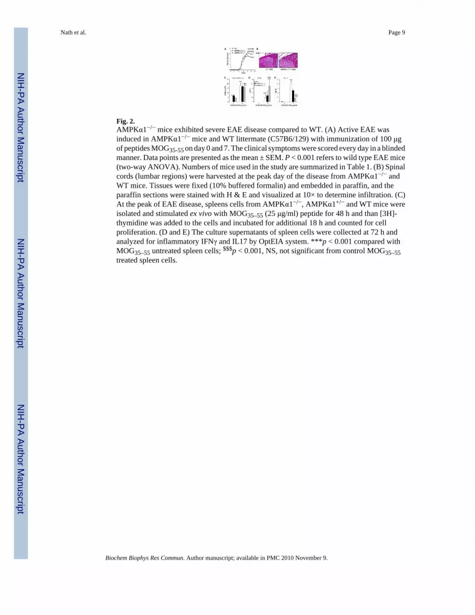
Fig. 2.AMPKα1−/− mice exhibited severe EAE disease compared to WT. (A) Active EAE wasinduced in AMPKα1−/− mice and WT littermate (C57B6/129) with immunization of 100 μgof peptides MOG35-55 on day 0 and 7. The clinical symptoms were scored every day in a blindedmanner. Data points are presented as the mean ± SEM. P < 0.001 refers to wild type EAE mice(two-way ANOVA). Numbers of mice used in the study are summarized in Table 1. (B) Spinalcords (lumbar regions) were harvested at the peak day of the disease from AMPKα1−/− andWT mice. Tissues were fixed (10% buffered formalin) and embedded in paraffin, and theparaffin sections were stained with H & E and visualized at 10× to determine infiltration. (C)At the peak of EAE disease, spleens cells from AMPKα1−/−, AMPKα1+/− and WT mice wereisolated and stimulated ex vivo with MOG35–55 (25 μg/ml) peptide for 48 h and than [3H]-thymidine was added to the cells and incubated for additional 18 h and counted for cellproliferation. (D and E) The culture supernatants of spleen cells were collected at 72 h andanalyzed for inflammatory IFNγ and IL17 by OptEIA system. ***p < 0.001 compared withMOG35–55 untreated spleen cells; $$$p < 0.001, NS, not significant from control MOG35–55treated spleen cells.
Nath et al. Page 9
Biochem Biophys Res Commun. Author manuscript; available in PMC 2010 November 9.
NIH
-PA Author Manuscript
NIH
-PA Author Manuscript
NIH
-PA Author Manuscript

NIH
-PA Author Manuscript
NIH
-PA Author Manuscript
NIH
-PA Author Manuscript
Nath et al. Page 10
Tabl
e 1
EAE
in A
MPK
α1 W
T, h
eter
ozyg
ote
and
in h
omoz
ygou
s litt
erm
ates
.
Num
ber
of m
ice
Inci
denc
e%
Inci
denc
eM
ean
max
imum
scor
e at
pea
kPe
ak d
ay
WT
1414
/14
100
2.59
± 0
.35
20
AM
PKα1
(+/−
)13
13/1
310
03.
05 ±
0.3
7**
21
AM
PKα1
(−/−
)7
7/7
100
3.37
± 0
.10*
**20
Act
ive
EAE
was
indu
ced
in A
MPK
α1 W
T, h
eter
ozyg
ote
and
in h
omoz
ygou
s mic
e w
ith M
OG
35–5
5 pe
ptid
e. T
otal
num
ber o
f mic
e us
ed in
stud
y, in
cide
nce,
clin
ical
dis
ease
scor
e an
d pe
ak d
ay a
re re
porte
d in
this
tabl
e. M
ean
max
imum
scor
e ar
e gi
ven
at th
e pe
ak o
f dis
ease
.
*** P
< 0.
001,
**P
< 0.
01 a
s com
pare
with
WT
EAE
(Stu
dent
t-te
st).
Biochem Biophys Res Commun. Author manuscript; available in PMC 2010 November 9.
Copyright © 2022 FDOKUMEN