Prolonged sleep fragmentation of mice exacerbates febrile responses to lipopolysaccharide
10
This article appeared in a journal published by Elsevier. The attached copy is furnished to the author for internal non-commercial research and education use, including for instruction at the authors institution and sharing with colleagues. Other uses, including reproduction and distribution, or selling or licensing copies, or posting to personal, institutional or third party websites are prohibited. In most cases authors are permitted to post their version of the article (e.g. in Word or Tex form) to their personal website or institutional repository. Authors requiring further information regarding Elsevier’s archiving and manuscript policies are encouraged to visit: http://www.elsevier.com/authorsrights
-
Upload
independent -
Category
Documents
-
view
2 -
download
0
Transcript of Prolonged sleep fragmentation of mice exacerbates febrile responses to lipopolysaccharide
This article appeared in a journal published by Elsevier. The attachedcopy is furnished to the author for internal non-commercial researchand education use, including for instruction at the authors institution
and sharing with colleagues.
Other uses, including reproduction and distribution, or selling orlicensing copies, or posting to personal, institutional or third party
websites are prohibited.
In most cases authors are permitted to post their version of thearticle (e.g. in Word or Tex form) to their personal website orinstitutional repository. Authors requiring further information
regarding Elsevier’s archiving and manuscript policies areencouraged to visit:
http://www.elsevier.com/authorsrights

Author's personal copy
Journal of Neuroscience Methods 219 (2013) 104– 112
Contents lists available at ScienceDirect
Journal of Neuroscience Methods
jou rn al h om epa ge : www.elsev ier .com/ locate / jneumeth
Basic Neuroscience
Prolonged sleep fragmentation of mice exacerbates febrile responsesto lipopolysaccharide
Kristyn M. Ringgolda,1, R. Paulien Barfa,1, Amrita Georgea, Blair C. Suttona,b,Mark R. Oppa,c,∗
a Department of Anesthesiology & Pain Medicine, University of Washington, Seattle, WA, USAb Neuroscience Graduate Program, University of Michigan, Ann Arbor, MI, USAc Program of Neurobiology and Behavior, University of Washington, Seattle, WA, USA
h i g h l i g h t s
• The method used is effective in fragmenting sleep of mice for prolonged periods without behavioral adaptation.• Although NREM sleep is disrupted and REM sleep is abolished by this method, there is no compensatory increase in sleep during periods without
fragmentation.• Water intake and body weight are unchanged by this protocol, and corticosterone is only modestly elevated.• After prolonged sleep fragmentation, febrile responses to immune challenge are exacerbated.
a r t i c l e i n f o
Article history:Received 31 May 2013Received in revised form 4 July 2013Accepted 8 July 2013
Keywords:RodentLPSStressInflammationFever
a b s t r a c t
Background: Sleep disruption is a frequent occurrence in modern society. Whereas many studies havefocused on the consequences of total sleep deprivation, few have investigated the condition of sleepdisruption.New method: We disrupted sleep of mice during the light period for 9 consecutive days using an inter-mittently rotating disc.Results: Electroencephalogram (EEG) data demonstrated that non-rapid eye movement (NREM) sleepwas severely fragmented and REM sleep was essentially abolished during the 12 h light period. Duringthe dark period, when sleep was not disrupted, neither NREM sleep nor REM sleep times differed fromcontrol values. Analysis of the EEG revealed a trend for increased power in the peak frequency of theNREM EEG spectra during the dark period. The fragmentation protocol was not overly stressful as bodyweights and water consumption remained unchanged, and plasma corticosterone did not differ betweenmice subjected to 3 or 9 days of sleep disruption and home cage controls. However, mice subjected to9 days of sleep disruption by this method responded to lipopolysaccharide with an exacerbated febrileresponse.Comparison with existing methods: Existing methods to disrupt sleep of laboratory rodents often subjectthe animal to excessive locomotion, vibration, or sudden movements. This method does not suffer fromany of these confounds.Conclusions: This study demonstrates that prolonged sleep disruption of mice exacerbates febrileresponses to lipopolysaccharide. This device provides a method to determine mechanisms by whichchronic insufficient sleep contributes to the etiology of many pathologies, particularly those with aninflammatory component.
© 2013 Elsevier B.V. All rights reserved.
∗ Corresponding author at: 325 9th Avenue, Box 359724, Seattle, WA 98104, USA.Tel.: +1 206 897 5987; fax: +1 206 897 6954.
E-mail addresses: [email protected], [email protected] (M.R. Opp).1 These authors contributed equally to this study.
1. Introduction
Sleep loss is a common problem in our modern society. Data sug-gest that sleep loss contributes to the development of many healthproblems, including, but not limited to, mood disorders (Breslauet al., 1996; Ford and Kamerow, 1989; Neckelmann et al., 2007),metabolic diseases (Chaput et al., 2006; Horne, 2011; Van Cauterand Knutson, 2008), immune dysfunction (Imeri and Opp, 2009;
0165-0270/$ – see front matter © 2013 Elsevier B.V. All rights reserved.http://dx.doi.org/10.1016/j.jneumeth.2013.07.008

Author's personal copy
K.M. Ringgold et al. / Journal of Neuroscience Methods 219 (2013) 104– 112 105
Fig. 1. The sleep disruption device and protocol are effective. Each sleep disruption device houses two mice and provides ad libitum access to food and water. Hypnogramsdepict consecutive 10 s epochs from one hour in the same mouse when the disc was not rotating (top panel) and during sleep fragmentation (SF) when the disc was rotating(SF Day 9, bottom panel). Both hypnograms are from the fifth hour after light onset. Red lines depict core body temperature. The disruption of sleep is readily apparent.WAKE, wakefulness; NREM, non-rapid eye movement sleep; REM, rapid eye movement sleep.
Irwin, 2002; Opp and Toth, 2003) and increased risk of cancer(Blask, 2009). Although the negative consequences of total sleepdeprivation have been clearly demonstrated in humans and rodents(Borbely and Neuhaus, 1979; Carskadon, 2004; Downey andBonnet, 1987; Everson, 1995; Everson et al., 1989; Knutson et al.,2007; Rechtschaffen et al., 1983; Soldatos and Paparrigopoulos,2005), it has only recently become apparent that chronic sleepdisruption or fragmentation is a common problem that can havean impact on recovery from illness (Irwin, 2002; Zager et al.,2007), alertness (Rosekind, 2005) and cognitive abilities (Cohen andAlbers, 1991; Stepanski, 2002).
There are many causes of sleep disruption, including but notlimited to social behavior (Cain and Gradisar, 2010), shift work(Akerstedt, 1998) and medical conditions such as obstructive sleepapnea (Bandla and Gozal, 2000; Svanborg and Guilleminault, 1996),chronic pain (Moldofsky, 2001) and narcolepsy (Tafti et al., 1992;Zorick et al., 1986). Sleep disruption is characterized by briefarousals that occur throughout the night, often without reducingthe total amount of time spent asleep (Bonnet and Arand, 2003).Sleep disruption results in decreased sleep efficiency, increaseddaytime sleepiness and cognitive impairment (Stepanski, 2002).For these and other reasons, recent efforts have been directedtoward determining the negative consequences of chronic sleepdisruption or fragmentation rather than total sleep deprivation,and several methods have been developed to mechanically disruptsleep of rodents for prolonged periods (Ramesh et al., 2009; Sintonet al., 2009).
As briefly mentioned, a relationship between sleep and immunefunction exists and pro-inflammatory cytokines have been impli-cated in sleep regulation (Imeri and Opp, 2009; Irwin, 2002;Opp and Toth, 2003). Interestingly, the previously mentionedpathologies linked to disturbed sleep, including metabolic diseases,immune disorders and increased cancer risk, are also character-ized by inflammation (Chung et al., 2011; Guess et al., 2009; Junand Polotsky, 2009; Palma et al., 2007). We report results obtained
using an instrumentation platform that disrupts sleep of mice forprolonged periods. Such a method provides the means to determinemechanisms by which prolonged sleep fragmentation negativelyimpacts immune function, possibly via inflammation. In this study,we determined the impact of chronic sleep fragmentation dur-ing the light phase on subsequent nighttime behavior and on thefebrile response to an immune challenge consisting of intraperi-toneal administration of lipopolysaccharide (LPS).
2. Methods
2.1. Animals
Adult male C57BL/6J mice (∼25 g at time of use; Jackson Labora-tories, Bar Harbor, ME) were group housed until surgery and/or thebeginning of the sleep fragmentation protocol. Mice that servedas home cage controls were then placed individually in standardshoeboxes, and experimental mice were housed individually insleep fragmentation devices. All mice were housed under a 12:12light:dark cycle at 29 ± 1 ◦C with food and water provided ad libi-tum. Water consumption and body weights were measured daily2 h after light onset. All procedures involving the use of animalswere approved by the University of Washington IACUC in accor-dance with the US Department of Agriculture Animal Welfare Actand the National Institutes of Health policy on Humane Care andthe Use of Laboratory Animals.
2.2. Sleep fragmentation device
The device consists of a cylindrical Plexiglas® chamber dividedinto two separate compartments (Fig. 1). The floor of the cham-ber is a disc that is programmed to rotate at specific intervals anddurations as selected by the investigator (see later). The timing anddirection of disc rotation is randomized to prevent behavioral adap-tation by a rodent to disc movements. The disc rotates more than

Author's personal copy
106 K.M. Ringgold et al. / Journal of Neuroscience Methods 219 (2013) 104– 112
180 degrees to ensure the mouse must move to avoid the centerdivider.
2.3. Experiment 1: effects of daytime sleep fragmentation onnighttime sleep, EEG power spectra, body weight and waterconsumption, and plasma corticosterone
This experiment consisted of two separate manipulations. Miceused to determine the impact of daytime sleep fragmentation onnighttime behavior, EEG spectra, and body weights and water con-sumption were used in a longitudinal protocol in which they servedas their own controls. This part of the study was longitudinal inthat data obtained during the habituation day (see later) were usedas device control data for subsequent analysis and compared todata obtained during sleep fragmentation from the same mouse.A separate group of mice was used to determine effects on plasmacorticosterone. This portion of the protocol was cross sectional, andanimals were sacrificed at selected time points as detailed later.
Surgical procedures, recording protocol, and sleep-wake deter-mination: Battery-operated biotelemetric transmitters [model #ETA-F10, Data Sciences International (DSI), St. Paul, MN] weresurgically implanted in the peritoneal cavity under isoflurane anes-thesia as previously described (Morrow and Opp, 2005a; Olivadotiet al., 2011). Briefly, insulated leads from the transmitter werepassed subcutaneously to the skull, where they were attachedto two small stainless steel screws implanted over the frontaland parietal brain cortices that served as electroencephalographic(EEG) recording electrodes. These telemetric devices also mea-sure core body temperature, which was collected for each mousethroughout the duration of the protocol. Drinking water containingibuprofen (0.2 mg/mL) was provided 24 h before through 48 h aftersurgery. Mice were placed in a warm chamber until ambulatory,and allowed to recover for 21 days prior to the start of the experi-ment. Buprenorphine (0.01–0.05 mg/kg) was administered once atthe time of surgery and one day following surgical procedures.
Transmitter signals were captured by a DSI receiver (RPC-1)located underneath each animal’s cage. Signals were sent to a DSIanalog converter (ART Analog-8 CM). Cage activity, in home cagesand on turntables, was detected using an infrared sensor housed inan observation unit that also contained a camera (BioBserve, GmbH,Bonn, Germany). All signals from the mice (EEG, body temperature,cage activity) were stored as binary files until later processing.
Undisturbed baseline recordings were obtained for 48 h whileeach animal was in its home cage. Mice were then placed in thesleep fragmentation device for two days of undisturbed recordings,followed by one day of habituation to the movement of the device(device control: DC). To habituate the mouse to the physical rota-tion of the disc, the disc was rotated with direction randomized for8 s once every 30 min for the 12 h light period. Following habitu-ation to the device, the sleep fragmentation protocol began. Sleepfragmentation (SF) in this protocol consisted of an 8-s disc rotationonce every 30 s, on average, during the 12 h light period for 9 con-secutive days. Animals were allowed to freely behave during theintervening dark periods.
Arousal states were determined as previously described(Morrow and Opp, 2005b; Olivadoti et al., 2011), and classifiedas non-rapid eye movement (NREM) sleep, rapid eye movement(REM) sleep or wakefulness (WAKE). Because the SF protocol useda disc rotation lasting 8 s, arousal state determinations were madeusing a 4 s epoch length. Artifact-free EEG epochs were subjectedto fast Fourier transformation (FFT) to yield power spectra between0.5 and 20 Hz in 0.5-Hz frequency bins. These artifact-free FFTspectra were matched to NREM, REM and WAKE epochs to obtainstate-specific power spectra (Baracchi and Opp, 2008). Power in thedelta (0.5–4.0 Hz) and theta (6.0–9.0) frequency bands within each
hour was determined by averaging power density values from eachepoch scored as NREMS, REMS or WAKE.
Body weights, water consumption and plasma corticosterone con-centrations: To determine the impact of sleep fragmentation ongeneralized stress responses, body weights, water consumptionand plasma corticosterone concentrations were determined froma total of 60 uninstrumented mice. Samples were analyzed frommice maintained in five different conditions: home cage control(n = 12), device control (n = 12), after 1 day of SF (n = 12), after 3days of SF (n = 12), and after 9 days of SF (n = 12). For all mice, bloodwas obtained via terminal orbital bleed, centrifuged (for 20 min at4 ◦C; 2000 × g) and plasma collected. Plasma samples were storedat −80 ◦C until assayed. Corticosterone concentrations from plasmasamples were determined using an EIA kit (Enzo Life Sciences, Cat-alog # ADI-900-097, Farmingdale, NY) according to manufacturerguidelines.
2.4. Experiment 2: impact of 9 days of sleep fragmentation onresponses to an immune challenge
Surgical procedures and recording protocol: Battery-operated dat-aloggers (Mini SubCue Dataloggers, SubCue Dataloggers, Alberta,Canada) were surgically implanted into the peritoneal cavity.These devices record only core body temperature. After surgery,mice were placed in a warmed cage until ambulatory. Drinkingwater containing ibuprofen (0.2 mg/mL) was provided 24 h beforethrough 48 h after surgery. Mice were allowed to recover 10–14days before the start of the experiments.
Mice were randomized into one of three conditions: homecage controls (n = 8), device controls (n = 6) or 9 days SF (n = 6).Home cage control mice were housed in standard shoeboxes, anddevice control animals were housed in the disruption devices forthe same duration, but the disc did not rotate. All animals wereinjected with 0.4 mg/kg lipopolysaccharide (LPS; Escherichia coliserotype 0111:B4, Sigma–Aldrich, St. Louis, MO) dissolved in 0.2 mLpyrogen-free saline (PFS, vehicle). Injections were given at lightonset of the day immediately following the last day of the proto-col, i.e., 10 days after the start of the protocol. As such, for the SFgroup, there was one intervening 12 h dark period during whichthe mice were able to freely behave. Body temperatures were con-tinuously recorded for 96 h following the LPS injection. To controlfor the effects of the injection per se on body temperature, after the96 h period all mice were injected with 0.2 mL vehicle, and bodytemperature recorded for an additional 24 h.
2.5. Statistical analyses
Where appropriate, repeated measures ANOVA was used todetermine the impact of manipulation on measures of interest(body temperature, NREM sleep, REM sleep and daily changes inbody weights and water intake). Post hoc evaluation was performedusing Tukey’s HSD test. All statistical values listed in the results sec-tion for repeated measures ANOVA reflect significant differences intime × group interaction effects, unless otherwise noted.
For EEG spectral analyses, paired T-tests were used to determineif the peak frequency for a given manipulation and behavioral state(0.5–4 Hz, NREM) differed between device control and SF condi-tions. For NREM delta power during the dark period, we requirethat each mouse spend at least a total of 5 min in NREM sleep beforeEEG spectral values for that animal are included for that hour insubsequent analysis. We use this criterion to reduce a potentialfor a relatively few epochs to exert a disproportionate effect onoutcome measures for EEG spectral analyses. Based on this crite-rion, some hours during the dark period did not contain enoughNREM sleep for analysis, and thus NREM delta power could not beanalyzed using repeated measures because data were lacking for
Author's personal copy
K.M. Ringgold et al. / Journal of Neuroscience Methods 219 (2013) 104– 112 107
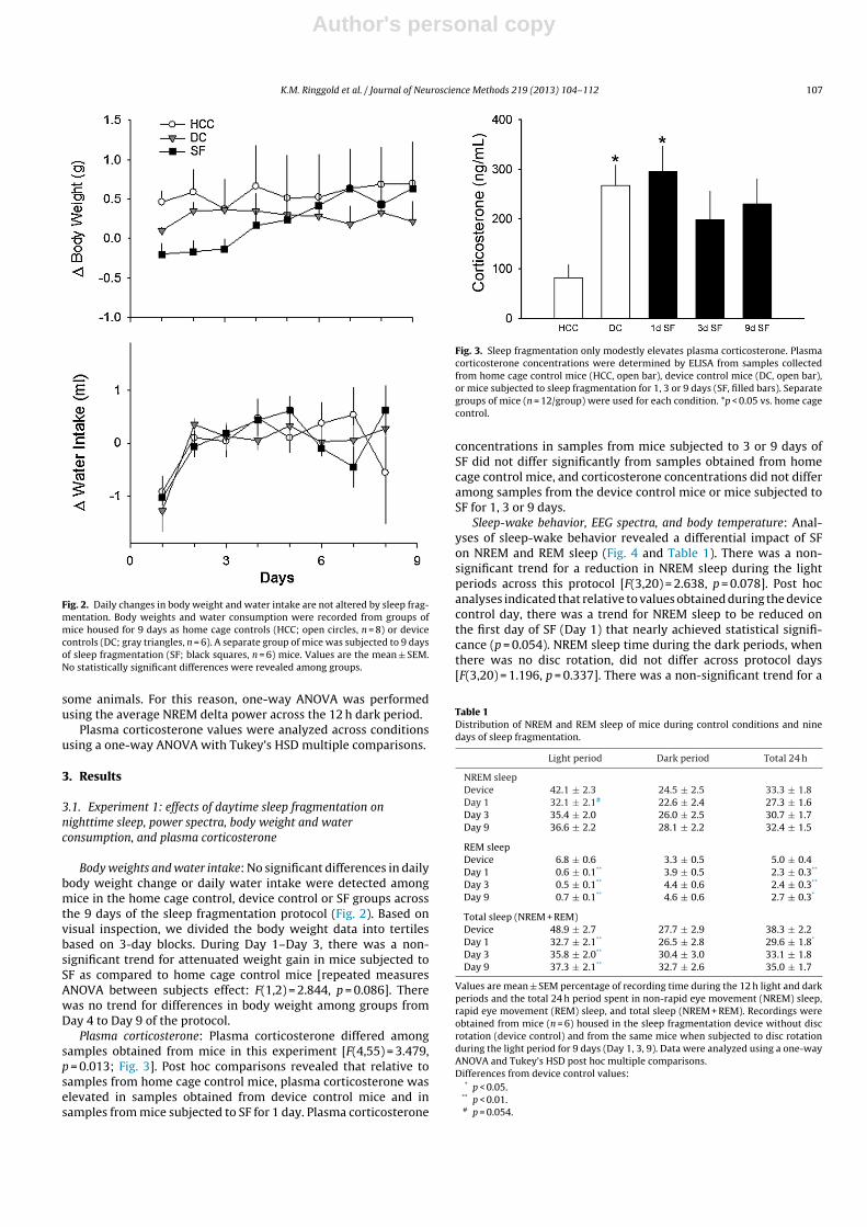
Fig. 2. Daily changes in body weight and water intake are not altered by sleep frag-mentation. Body weights and water consumption were recorded from groups ofmice housed for 9 days as home cage controls (HCC; open circles, n = 8) or devicecontrols (DC; gray triangles, n = 6). A separate group of mice was subjected to 9 daysof sleep fragmentation (SF; black squares, n = 6) mice. Values are the mean ± SEM.No statistically significant differences were revealed among groups.
some animals. For this reason, one-way ANOVA was performedusing the average NREM delta power across the 12 h dark period.
Plasma corticosterone values were analyzed across conditionsusing a one-way ANOVA with Tukey’s HSD multiple comparisons.
3. Results
3.1. Experiment 1: effects of daytime sleep fragmentation onnighttime sleep, power spectra, body weight and waterconsumption, and plasma corticosterone
Body weights and water intake: No significant differences in dailybody weight change or daily water intake were detected amongmice in the home cage control, device control or SF groups acrossthe 9 days of the sleep fragmentation protocol (Fig. 2). Based onvisual inspection, we divided the body weight data into tertilesbased on 3-day blocks. During Day 1–Day 3, there was a non-significant trend for attenuated weight gain in mice subjected toSF as compared to home cage control mice [repeated measuresANOVA between subjects effect: F(1,2) = 2.844, p = 0.086]. Therewas no trend for differences in body weight among groups fromDay 4 to Day 9 of the protocol.
Plasma corticosterone: Plasma corticosterone differed amongsamples obtained from mice in this experiment [F(4,55) = 3.479,p = 0.013; Fig. 3]. Post hoc comparisons revealed that relative tosamples from home cage control mice, plasma corticosterone waselevated in samples obtained from device control mice and insamples from mice subjected to SF for 1 day. Plasma corticosterone
Fig. 3. Sleep fragmentation only modestly elevates plasma corticosterone. Plasmacorticosterone concentrations were determined by ELISA from samples collectedfrom home cage control mice (HCC, open bar), device control mice (DC, open bar),or mice subjected to sleep fragmentation for 1, 3 or 9 days (SF, filled bars). Separategroups of mice (n = 12/group) were used for each condition. *p < 0.05 vs. home cagecontrol.
concentrations in samples from mice subjected to 3 or 9 days ofSF did not differ significantly from samples obtained from homecage control mice, and corticosterone concentrations did not differamong samples from the device control mice or mice subjected toSF for 1, 3 or 9 days.
Sleep-wake behavior, EEG spectra, and body temperature: Anal-yses of sleep-wake behavior revealed a differential impact of SFon NREM and REM sleep (Fig. 4 and Table 1). There was a non-significant trend for a reduction in NREM sleep during the lightperiods across this protocol [F(3,20) = 2.638, p = 0.078]. Post hocanalyses indicated that relative to values obtained during the devicecontrol day, there was a trend for NREM sleep to be reduced onthe first day of SF (Day 1) that nearly achieved statistical signifi-cance (p = 0.054). NREM sleep time during the dark periods, whenthere was no disc rotation, did not differ across protocol days[F(3,20) = 1.196, p = 0.337]. There was a non-significant trend for a
Table 1Distribution of NREM and REM sleep of mice during control conditions and ninedays of sleep fragmentation.
Light period Dark period Total 24 h
NREM sleepDevice 42.1 ± 2.3 24.5 ± 2.5 33.3 ± 1.8Day 1 32.1 ± 2.1# 22.6 ± 2.4 27.3 ± 1.6Day 3 35.4 ± 2.0 26.0 ± 2.5 30.7 ± 1.7Day 9 36.6 ± 2.2 28.1 ± 2.2 32.4 ± 1.5
REM sleepDevice 6.8 ± 0.6 3.3 ± 0.5 5.0 ± 0.4Day 1 0.6 ± 0.1** 3.9 ± 0.5 2.3 ± 0.3**
Day 3 0.5 ± 0.1** 4.4 ± 0.6 2.4 ± 0.3**
Day 9 0.7 ± 0.1** 4.6 ± 0.6 2.7 ± 0.3*
Total sleep (NREM + REM)Device 48.9 ± 2.7 27.7 ± 2.9 38.3 ± 2.2Day 1 32.7 ± 2.1** 26.5 ± 2.8 29.6 ± 1.8*
Day 3 35.8 ± 2.0** 30.4 ± 3.0 33.1 ± 1.8Day 9 37.3 ± 2.1** 32.7 ± 2.6 35.0 ± 1.7
Values are mean ± SEM percentage of recording time during the 12 h light and darkperiods and the total 24 h period spent in non-rapid eye movement (NREM) sleep,rapid eye movement (REM) sleep, and total sleep (NREM + REM). Recordings wereobtained from mice (n = 6) housed in the sleep fragmentation device without discrotation (device control) and from the same mice when subjected to disc rotationduring the light period for 9 days (Day 1, 3, 9). Data were analyzed using a one-wayANOVA and Tukey’s HSD post hoc multiple comparisons.Differences from device control values:
* p < 0.05.** p < 0.01.# p = 0.054.

Author's personal copy
108 K.M. Ringgold et al. / Journal of Neuroscience Methods 219 (2013) 104– 112
Fig. 4. Sleep fragmentation during the light period does not alter body temperature or sleep during the subsequent dark period. Body temperature of mice is not alteredduring the sleep disruption protocol (top panels). There is a tendency for non-rapid eye movements (NREM) sleep to be reduced during the light period fragmentation on day1 that did not achieve statistical significance (middle panels). Rapid eye movements (REM) sleep is essentially abolished by sleep fragmentation on this device and protocol(bottom panel). Depicted are the mean ± SEM values obtained from n = 6 mice during the device control day (DC), and from the same mice after 1, 3, and 9 days of sleepfragmentation (SF). Control values were obtained from mice housed in the sleep fragmentation devices without disc rotation, and as such each mouse served as its owncontrol. *p < 0.05 and **p < 0.01 vs. device control.
reduction in total 24 h NREM sleep time across the recording daysof this protocol [F(3,20) = 2.505, p = 0.088].
In contrast to NREM sleep, REM sleep was significantly reducedby this SF protocol (Fig. 4 and Table 1). REM sleep was essentiallyabolished during the light periods of the SF protocol, and was sig-nificantly reduced when compared to the device control condition[F(3,20) = 36.168, p = 0.000]. Post hoc comparisons indicate that rel-ative to device control values, REM sleep was significantly reducedduring each of the SF days analyzed. During the dark periods, whenthere was no disc rotation, time spent in REM sleep did not dif-fer from device control values [F(3,20) = 0.685, p = 0.572]. However,total 24 h REM sleep time was reduced during each of the SF daysrelative to device control values [F(3,20) = 6.393, p = 0.003].
We did not analyze EEG spectra during the light period becausethe SF protocol results in very short NREM sleep bouts. As a conse-quence, the build-up in delta power that normally occurs throughthe progression of a single NREM sleep bout (Franken et al., 1991b,2001a) is artificially truncated by the programmed disruption.During the intervening dark periods, when no sleep disruptionoccurred, spectral analyses of power in the peak frequency of thedelta frequency band (0.5–4.0 Hz) revealed a trend to increaseduring the 9-day protocol, but this effect did not reach statisti-cal significance (Fig. 5 Panels A–C: paired T-test: DC vs. Day 1 SF:t = −2.108, p = 0.089; DC vs. Day 3 SF: t = −2.361, p = 0.065; DC vs.Day 9 SF: t = −2.465, p = 0.057). During the 9 days of the protocol,NREM delta power during the 12 h dark period significantly
increased on Day 1 of SF and there is a tendency for an increaseduring Day 3 and Day 9 of SF compared to DC (Fig. 5 Panels D–F:paired T-test: DC vs. Day 1 SF: t = 2.697, p < 0.05; DC vs. Day 3 SF:t = −2.341, p = 0.066; DC vs. Day 9 SF: t = −2.524, p = 0.053).
Sleep fragmentation did not significantly impact core bodytemperatures of mice (Fig. 4). One-way ANOVA of average bodytemperatures during the 12 h light periods, 12 h dark periods, oracross 24 h periods did not reveal any differences among the fourconditions (device control, SF Days 1, 3 and 9). Activity-dependentchanges in body temperature are apparent during some timepoints, such as the light period of SF Day 9. However, these devi-ations in body temperature were not of sufficient magnitude orduration so as to result in a statistically significant alteration inbody temperatures during the SF protocol.
3.2. Experiment 2: impact of 9 days of sleep fragmentation onresponses to an immune challenge.
Intraperitoneal injection of LPS induced a febrile response in allmice, irrespective of group condition (Fig. 6). Data were analyzedand are presented as difference scores: core body temperature val-ues for each mouse obtained after injection of vehicle (PFS) weresubtracted from the body temperature values obtained from thesame mouse during the initial 24 h after injection of LPS. During the12-h light period after injections, there was a significant differencebetween groups [F(22,165) = 1.961, p = 0.009], as SF mice displayed

Author's personal copy
K.M. Ringgold et al. / Journal of Neuroscience Methods 219 (2013) 104– 112 109
Fig. 5. Sleep pressure may accumulate during prolonged sleep fragmentation even if sleep time is not altered. (Panels A–C) Spectral analyses conducted on artifact-freeelectroencephalogram epochs of non-rapid eye movements (NREM) sleep during the 12-h dark period, when there is no disc rotation, suggest accumulating sleep pressure.The magnitude of the peak spectral power in the delta frequency band (0.5–4.0 Hz) tends to increase across days of sleep fragmentation (SF). (Panels D–F) Hourly averagesfor NREM delta power after the first day of sleep fragmentation were significantly greater as compared to values from the same animals when housed as device controls(DC), without disc rotation. Values are means ± SEM for n = 6 mice.
a greater increase in body temperature after LPS challenge as com-pared to device and home cage control groups.
4. Discussion
This study demonstrates that the device and protocol used areeffective in disrupting sleep of mice in a manner that is apparentlynot physiologically stressful as determined by corticosterone con-centrations. Fragmenting daytime sleep of mice using this deviceand protocol has little impact on subsequent nighttime sleep-wake behavior, which is an unexpected observation. Many studiesdemonstrate that one 6 h period of total sleep deprivation duringthe light period results in significant increases in the amount oftime mice spend in NREM and REM sleep during the subsequentdark period (Baracchi and Opp, 2008; Franken et al., 1999; Huberet al., 2000; Morrow and Opp, 2005b). The amount of time spent inNREM sleep is only modestly impacted during the light period whenthe disc is rotating in this protocol. As such, NREM sleep is mini-mally reduced, which may explain the lack of a NREM sleep reboundduring the intervening undisturbed dark periods. It is possible thatsleep pressure increases during the course of this protocol, as sug-gested by a trend for increased EEG NREM delta power during thedark periods. However, as with time spent in NREM sleep, thechange in EEG NREM delta power during periods of spontaneousbehavior is not of the magnitude typically observed after a periodof total sleep deprivation (Easton et al., 2004; Huber et al., 2000;McKenna et al., 2007; Morrow and Opp, 2005b; Opp and Krueger,1994a).
In contrast to the minimal impact on NREM sleep, this protocolessentially eliminates REM sleep during light periods. REM sleepduring disc rotations is abolished, likely due to the fact that theinter-rotation interval is too short to allow mice to enter this sleepstage. Although daytime REM sleep is eliminated, there is no night-time REM sleep rebound and total REM sleep amounts across the24 h period remains less than during undisturbed baseline periods.Furthermore, there are no changes in the theta frequency bandof the EEG spectra (data not shown), such as would indicate anincrease in REM sleep pressure. Whether longer periods of day-time sleep fragmentation by this method would result in reboundsof NREM and/or REM sleep during subsequent dark periods remainsto be determined.
The duration of time freely behaving mice spend in sleep stagesduring the dark period is not altered dramatically, but other facetsof sleep are impacted by this protocol. Power in the delta frequencyband (0.5–4.0 Hz) during NREM sleep is generally accepted as anindex of sleep depth or sleep pressure (Benington and Heller, 1994;Brunner et al., 1993; Chemelli et al., 1999; Franken et al., 1991a;Machado et al., 2004). However, the duration of NREM sleep andthe mechanisms responsible for manifestation of the cortical EEGare regulated independently, and there are conditions under whichthese two parameters are dissociated [reviewed in Davis et al.,2011]. Although the trend for increases in NREM spectral poweracross the 9 days of sleep fragmentation in this protocol did notachieve statistical significance, these data suggest that sleep pres-sure accumulates, in spite of the fact that differences in time spentin NREM sleep does not statistically differ between manipulations.

Author's personal copy
110 K.M. Ringgold et al. / Journal of Neuroscience Methods 219 (2013) 104– 112
Fig. 6. Sleep fragmentation exacerbates febrile responses to immune challenge.Body temperature responses (◦C) of mice to lipopolysaccharide (LPS) administrationdiffer when housed as home cage controls (HCC; open circles, n = 7), device con-trols (DC; gray triangles, n = 5) or subject to sleep fragmentation (SF; filled squares,n = 6) mice. All mice were injected with LPS on day 10 of the protocol (see text),and recordings continued for 96 h. The same mice were then injected with vehi-cle (pyrogen-free saline) to control for the potentially confounding effects of theinjection procedure itself. As such, each mouse served as its own control. Differencescores (�) were calculated by subtracting values obtained after vehicle injectionfrom those obtained after LPS administration. Mice in the SF group were subjectedto disc rotation during the light period for a total of 9 consecutive days, whereasmice in the DC group were housed on non-rotating discs and HCC mice remained intheir home cages for the same amount of time. Values are the means ± SEM. *p < 0.05among groups.
Although REM sleep is reduced, and spectral properties of theEEG may be altered, sleep fragmentation by this device and protocoldoes not alter body weight or food intake. Changes in body weightof rodents during conditions of reduced sleep opportunity havepreviously been reported. However, previous studies either usedlong periods of total sleep deprivation [e.g., Bergmann et al., 1989],severe sleep restriction for up to 20 h per day [e.g., Barf et al., 2010]or selective REM sleep deprivation [e.g., Hipolide and Suchecki,2006; Koban et al., 2008]. In addition, the methods commonly usedto deprive rodents of sleep [gentle handling (Franken et al., 1991a,1993; Morrow and Opp, 2005b), disk-over-water (Bergmann et al.,1989), a continuously rotating drum (Barf et al., 2010), invertedflower pot (Hipolide and Suchecki, 2006; Koban et al., 2008)], aredesigned to eliminate all sleep or to selectively reduce REM sleep.Although mice are deprived of REM sleep during the light period,sleep disruption by the protocol used in this study does not affecttotal sleep time during the subsequent dark period. Furthermore,after the first day on the device, 24 h total sleep time is not reduced.The general lack of reduction in total sleep time may explain thelack of impact on body weight and water intake.
This protocol does not appear to be overly stressful based onthe lack of an effect on nighttime sleep, body weights, water con-sumption and body temperature. This conclusion is strengthenedby data demonstrating that plasma corticosterone is only modestlyaltered by sleep fragmentation using this technique. There is an ini-tial increase in plasma corticosterone the first day mice are placedon the device, and during the first day of disc rotation. These effectsmay be explained by the fact the mice are in a novel environment,which is known to elevate corticosterone in mice (Hennessy, 1991).However, after 3 and 9 days of sleep fragmentation, plasma corti-costerone does not statistically differ from values obtained frommice housed in their home cages.
Finally, our new data demonstrate that sleep fragmentationalters responses to immune challenge, even if the protocol doesnot dramatically alter sleep-wake behavior. We (Opp and Krueger,
1994a,b) and others (Everson, 1993, 2005; Everson and Toth,2000; Toth, 1995; Zager et al., 2007) have demonstrated adverseeffects of sleep loss on immune function of rodents. Studies alsodemonstrate that sleep loss impairs immune function in humanvolunteers [reviewed (Born et al., 1997; Marshall and Born, 2002;Opp et al., 2007)]. In this study, mice challenged with LPS after9 days of sleep fragmentation respond with a fever of greatermagnitude than control mice. The fever recorded after LPS inthis study do not differ among groups with respect to timingor duration as reported previously (Morrow and Opp, 2005a),but the increased magnitude of the peak temperature responsesuggests that prolonged sleep fragmentation by this methodexacerbates some aspects of responses to immune challenge. Theincrease in the magnitude of the peak febrile response is also ofinterest because the LPS challenge was administered 12 h afterthe end of sleep fragmentation. This observation suggests thatthe impact of sleep fragmentation by this device and protocolare not ameliorated by a short period (12 h) of undisturbed sleepopportunity. How long such an effect of sleep fragmentation onimmune challenge may last is not known, and requires additionalinvestigation. At present, mechanisms by which prolonged sleepfragmentation contributes to altered responsiveness to immunechallenge are not well understood. The device and protocol ofsleep fragmentation reported in this study allows us to begin todetermine the neuroanatomic and biochemical substrates thatlink sleep fragmentation to immune status under conditions whenamounts of sleep per se do not differ substantially from normal.
We consider several strengths of using this approach to frag-ment sleep of mice. Within the context of public health, manymedical conditions and societal constraints result in sleep disrup-tion without large decrements in total sleep time. For example,sleep disorders such as sleep apnea (Svanborg and Guilleminault,1996), narcolepsy (van den Hoed et al., 1981), and restless legssyndrome (Montplaisir et al., 1998), generally do not result inreductions in total sleep time. These medical conditions are char-acterized by inflammation (Kornum et al., 2011; Shamsuzzamanet al., 2003; Weinstock et al., 2012), which may be induced bysleep fragmentation per se (Bryant et al., 2004; Krueger et al.,2001; Mullington et al., 2009; Opp and Toth, 2003). Additionally,shift work (Akerstedt, 1998), caregiving (Happe and Berger, 2002;McCurry et al., 2007) or the care of a newborn (Hunter et al., 2009)disrupt sleep but rarely result in total sleep deprivation. Thesedemands are often associated with fragmented NREM sleep, as wellas a significant loss of REM sleep (McKenna et al., 2007; Stepanski,2002). Studies demonstrate that sleep restriction of human volun-teers for periods as short as four hours induces inflammation (Irwinet al., 2006). Although additional studies are required, the obser-vation that short periods of sleep restriction induce inflammationin humans provides a public health context within which futureresearch efforts should be directed.
While the protocol we used in this study lasted 9 days, the soft-ware controlling the disc allows the investigator complete freedomto program the timing and duration of disc rotation. For example,the duration of the sleep fragmentation protocol could be short-ened or lengthened, or the intensity of the fragmentation could bemade more or less severe by changing the duration and frequencyof disc rotations. The device and protocol have been designed todisrupt sleep of mice in an unpredictable fashion. Randomizationof rotation direction and inter-rotation interval prevent behavioralanticipation and adaptation of the mice to sleep fragmentation. Assuch, the device and protocol potentially remain effective in dis-rupting sleep for a protocol of any length. Because the disc rotatesonly slightly more than 180 degrees and then stops, there is noconfound of locomotor activity. Observations with a video moni-toring system reveal that mice awaken and move when the discrotation starts. When the disc is not rotating and mice are awake,
Author's personal copy
K.M. Ringgold et al. / Journal of Neuroscience Methods 219 (2013) 104– 112 111
they engage in general cage activity, such as grooming, eating anddrinking. Therefore, it is not necessary to include an additionalcontrol manipulation to account for locomotor activity, such as nec-essary when a treadmill or a continuously rotating disc or drum isused to deprive an animal of total sleep (Barf et al., 2010; Leenaarset al., 2011; McKenna et al., 2007). In addition to the software fea-tures that control the disc rotation schedule, the motor and drivesystem are extremely quiet, and the stepper motor provides a verysmooth disc rotation. After the first few rotations during habit-uation, there is no “startle response”, as would be indicated bythe mouse “flinching” when the disc rotation starts or stops. Thequiet and smooth nature of this device eliminates the potentialconfounds associated with methods that use laboratory shakers tophysically vibrate mice or sweeper bars that jostle mice (Rameshet al., 2009; Sinton et al., 2009).
Few, if any published studies of which we are aware includedisruption of rodent sleep for prolonged periods. Therefore, it isdifficult to assess other methods with respect to the impact of per-ceived stress in response to the sleep fragmentation method itself.Similarly, few studies using mechanical means to disrupt sleepof mice report the impact of the method on spectral propertiesof the EEG (Ramesh et al., 2009; Sinton et al., 2009). The devicewe use houses two mice simultaneously, and as such animals arenot isolated during the fragmentation protocol. This approach alsodoubles the number of animals that can be manipulated withinthe same apparatus and footprint. Finally, although a teleme-try system may be incorporated, as in this study, animals mayalso be implanted with cranial headpieces because the enclosureaccommodates tethers to connect the mouse to a recording system.
As with all methods and protocols to disturb or deprive labora-tory animals of sleep, there are some limitations to this approach.This system was designed only to disrupt sleep, and it is effective indoing so. No capabilities have been incorporated that would allowfeedback from the animal to the system such that a mouse could bedeprived of a specific stage of sleep, as in the classic rat studies ofRechtschaffen (Rechtschaffen and Bergmann, 1995; Rechtschaffenet al., 1983). We do not consider this a limitation due to the natureof our studies, although other investigators may desire this capabil-ity. There is an advantage to housing two mice on the same physicalapparatus in terms of animal density and elimination of social iso-lation. However, because the floor space is shared by two animals,it is not possible to determine food intake by an individual animal.During sleep fragmentation mice often “play” with their food, andthere is food waste, which gets mixed between mice when the discrotates. It is conceivable that a liquid diet could be used to allowassessment of caloric intake by each individual animal, but we havenot yet used this approach.
In summary, the device and protocol used are effective infragmenting sleep of mice. This initial study demonstrates bio-logic responses of mice to sleep fragmentation that validate thisapproach as a method by which sleep can be reliably disruptedfor prolonged periods. This system is capable of mimicking inrodents the sleep disruption or fragmentation that occurs in manyhuman conditions. Because the disc rotations are unpredictable,this system provides a means to conduct continuous long-termexperiments without behavioral adaptation by the subjects, despitepotentially increased sleep pressure. This initial study demon-strates that sleep disruption of mice exacerbates febrile responsesto LPS, an observation that provides impetus for future experimentsaimed at furthering our understanding of mechanisms by whichchronic insufficient sleep contributes to inflammatory disease.
Conflict of interest
The authors state they have no conflicts of interest.
Acknowledgments
We thank Ms. Ashley Ingiosi for her support and assistance withthis study. Dr. Tom Gardiner and Dr. Linda Toth, Southern Illi-nois University School of Medicine, designed and developed thesleep disruption devices and controlling software. This work wasfunded by National Institutes of Health grant AG041827, and by theDepartment of Anesthesiology & Pain Medicine of the University ofWashington.
References
Akerstedt T. Shift work and disturbed sleep/wakefulness. Sleep Med Rev1998;2:117–28.
Bandla HP, Gozal D. Dynamic changes in EEG spectra during obstructive apnea inchildren. Pediatr Pulmonol 2000;29:359–65.
Baracchi F, Opp MR. Sleep-wake behavior and responses to sleep deprivation ofmice lacking both interleukin-1 beta receptor 1 and tumor necrosis factor-alphareceptor 1. Brain Behav Immun 2008;22:982–93.
Barf RP, Meerlo P, Scheurink AJ. Chronic sleep disturbance impairs glucose homeo-stasis in rats. Int J Endocrinol 2010;2010:819414.
Benington JH, Heller HC. Does the function of REM sleep concern non-REM sleep orwaking? Prog Neurobiol 1994;44:433–49.
Bergmann BM, Kushida CA, Everson CA, Gilliland MA, Obermeyer W, RechtschaffenA. Sleep deprivation in the rat: II. Methodology. Sleep 1989;12:5–12.
Blask DE. Melatonin, sleep disturbance and cancer risk. Sleep Med Rev2009;13:257–64.
Bonnet MH, Arand DL. Clinical effects of sleep fragmentation versus sleep depriva-tion. Sleep Med Rev 2003;7:297–310.
Borbely AA, Neuhaus HU. Sleep-deprivation: effects on sleep and EEG in the rat. JComp Physiol Psychol 1979;133:71–87.
Born J, Lange T, Hansen K, Molle M, Fehm HL. Effects of sleep and circadian rhythmon human circulating immune cells. J Immunol 1997;158:4454–64.
Breslau N, Roth T, Rosenthal L, Andreski P. Sleep disturbance and psychiatric dis-orders: a longitudinal epidemiological study of young adults. Biol Psychiatry1996;39:411–8.
Brunner DP, Dijk DJ, Borbely AA. Repeated partial sleep deprivation progressivelychanges in EEG during sleep and wakefulness. Sleep 1993;16:100–13.
Bryant PA, Trinder J, Curtis N. Sick and tired: does sleep have a vital role in theimmune system? Nat Rev Immunol 2004;4:457–67.
Cain N, Gradisar M. Electronic media use and sleep in school-aged children andadolescents: a review. Sleep Med 2010;11:735–42.
Carskadon MA. Sleep deprivation: health consequences and societal impact. MedClin North Am 2004;88:767–76.
Chaput JP, Brunet M, Tremblay A. Relationship between short sleeping hours andchildhood overweight/obesity: results from the ‘Quebec en Forme’ Project. Int JObes (Lond) 2006;30:1080–5.
Chemelli RM, Willie JT, Sinton CM, Elmquist JK, Scammell T, Lee C, et al. Nar-colepsy in orexin knockout mice: molecular genetics of sleep regulation. Cell1999;98:437–51.
Chung HY, Lee EK, Choi YJ, Kim JM, Kim DH, Zou Y, et al. Molecular inflammation asan underlying mechanism of the aging process and age-related diseases. J DentRes 2011;90:830–40.
Cohen RA, Albers HE. Disruption of human circadian and cognitive regulation fol-lowing a discrete hypothalamic lesion: a case study. Neurology 1991;41:726–9.
Davis CJ, Clinton JM, Jewett KA, Zielinski MR, Krueger JM. Delta wave power: an inde-pendent sleep phenotype or epiphenomenon? J Clin Sleep Med 2011;7:S16–8.
Downey R, Bonnet MH. Performance during frequent sleep disruption. Sleep1987;10:354–63.
Easton A, Meerlo P, Bergmann B, Turek FW. The suprachiasmatic nucleus regulatessleep timing and amount in mice. Sleep 2004;27:1307–18.
Everson CA. Sustained sleep deprivation impairs host defense. Am J Physiol1993;265:R1148–54.
Everson CA. Functional consequences of sustained sleep deprivation in the rat. BehavBrain Res 1995;69:43–54.
Everson CA. Clinical assessment of blood leukocytes, serum cytokines, and serumimmunoglobulins as responses to sleep deprivation in laboratory rats. Am JPhysiol Regul Integr Comp Physiol 2005;289:R1054–63.
Everson CA, Bergmann BM, Rechtschaffen A. Sleep deprivation in the rat: III. Totalsleep deprivation. Sleep 1989;12:13–21.
Everson CA, Toth LA. Systemic bacterial invasion induced by sleep deprivation. AmJ Physiol Regul Integr Comp Physiol 2000;278:R905–16.
Ford DE, Kamerow DB. Epidemiologic study of sleep disturbances and psychiatricdisorders. An opportunity for prevention? JAMA 1989;262:1479–84.
Franken P, Chollet D, Tafti M. The homeostatic regulation of sleep need is undergenetic control. J Neurosci 2001a;21:2610–21.
Franken P, Dijk D-J, Tobler I, Borbély AA. Sleep deprivation in rats: effects onEEG power spectra, vigilance states, and cortical temperature. Am J Physiol1991a;261:R198–208.
Franken P, Malafosse A, Tafti M. Genetic determinants of sleep regulation in inbredmice. Sleep 1999;22:155–69.

Author's personal copy
112 K.M. Ringgold et al. / Journal of Neuroscience Methods 219 (2013) 104– 112
Franken P, Tobler I, Borbély AA. Sleep homeostasis in the rat: simulation of the timecourse of EEG slow-wave activity. Neurosci Lett 1991b;130:141–4.
Franken P, Tobler I, Borbely AA. Effects of 12-h sleep deprivation and of 12-h coldexposure on sleep regulation and cortical temperature in the rat. Physiol Behav1993;54:885–94.
Guess J, Burch JB, Ogoussan K, Armstead CA, Zhang H, Wagner S, et al. Circadian dis-ruption, Per3, and human cytokine secretion. Integr Cancer Ther 2009;8:329–36.
Happe S, Berger K. The association between caregiver burden and sleep disturbancesin partners of patients with Parkinson’s disease. Age Ageing 2002;31:349–54.
Hennessy MB. Sensitization of the plasma corticosterone response to novel environ-ments. Physiol Behav 1991;50:1175–9.
Hipolide DC, Suchecki D, Pimentel de Carvalho PA, Chiconelli FE, Tufik S, Luz J.Paradoxical sleep deprivation and sleep recovery: effects on the hypothalamic-pituitary-adrenal axis activity, energy balance and body composition of rats. JNeuroendocrinol 2006;18:231–8.
Horne J. Obesity and short sleep: unlikely bedfellows? Obes Rev 2011;12:e84–94.Huber R, Deboer T, Tobler I. Effects of sleep deprivation on sleep and sleep EEG in
three mouse strains: empirical data and simulations. Brain Res 2000;857:8–19.Hunter LP, Rychnovsky JD, Yount SM. A selective review of maternal sleep character-
istics in the postpartum period. J Obstet Gynecol Neonatal Nurs 2009;38:60–8.Imeri L, Opp MR. How (and why) the immune system makes us sleep. Nat Rev
Neurosci 2009;10:199–210.Irwin M. Effects of sleep and sleep loss on immunity and cytokines. Brain Behav
Immun 2002;16:503–12.Irwin MR, Wang M, Campomayor CO, Collado-Hidalgo A, Cole S. Sleep deprivation
and activation of morning levels of cellular and genomic markers of inflamma-tion. Arch Intern Med 2006;166:1756–62.
Jun J, Polotsky VY. Metabolic consequences of sleep-disordered breathing. ILAR J2009;50:289–306.
Knutson KL, Spiegel K, Penev P, Van Cauter E. The metabolic consequences of sleepdeprivation. Sleep Med Rev 2007;11:163–78.
Koban M, Sita LV, Le WW, Hoffman GE. Sleep deprivation of rats: the hyperphagicresponse is real. Sleep 2008;31:927–33.
Kornum BR, Kawashima M, Faraco J, Lin L, Rico TJ, Hesselson S, et al. Common variantsin P2RY11 are associated with narcolepsy. Nat Genet 2011;43:66–71.
Krueger JM, Obal FJ, Fang J, Kubota T, Taishi P. The role of cytokines in physiologicalsleep regulation. Ann NY Acad Sci 2001;933:211–21.
Leenaars CH, Dematteis M, Joosten RN, Eggels L, Sandberg H, Schirris M, et al. A newautomated method for rat sleep deprivation with minimal confounding effectson corticosterone and locomotor activity. J Neurosci Methods 2011;196:107–17.
Machado RB, Hipolide DC, Benedito-Silva AA, Tufik S. Sleep deprivation inducedby the modified multiple platform technique: quantification of sleep loss andrecovery. Brain Res 2004;1004:45–51.
Marshall L, Born J. Brain-immune interactions in sleep. Int Rev Neurobiol2002;52:93–131.
McCurry SM, Logsdon RG, Teri L, Vitiello MV. Sleep disturbances in caregivers ofpersons with dementia: contributing factors and treatment implications. SleepMed Rev 2007;11:143–53.
McKenna JT, Tartar JL, Ward CP, Thakkar MM, Cordeira JW, McCarley RW, et al. Sleepfragmentation elevates behavioral, electrographic and neurochemical measuresof sleepiness. Neuroscience 2007;146:1462–73.
Moldofsky H. Sleep and pain. Sleep Med Rev 2001;5:385–96.Montplaisir J, Boucher S, Nicolas A, Lesperance P, Gosselin A, Rompre P, et al. Immo-
bilization tests and periodic leg movements in sleep for the diagnosis of restlessleg syndrome. Mov Disord 1998;13:324–9.
Morrow JD, Opp MR. Diurnal variation of lipopolysaccharide-induced alterations insleep and body temperature of interleukin-6-deficient mice. Brain Behav Immun2005a;19:40–51.
Morrow JD, Opp MR. Sleep-wake behavior and responses of interleukin-6-deficientmice to sleep deprivation. Brain Behav Immun 2005b;19:28–39.
Mullington JM, Haack M, Toth M, Serrador JM, Meier-Ewert HK. Cardiovascular,inflammatory, and metabolic consequences of sleep deprivation. Prog Cardio-vasc Dis 2009;51:294–302.
Neckelmann D, Mykletun A, Dahl AA. Chronic insomnia as a risk factor for developinganxiety and depression. Sleep 2007;30:873–80.
Olivadoti MD, Weinberg JB, Toth LA, Opp MR. Sleep and fatigue in mice infected withmurine gammaherpesvirus 68. Brain Behav Immun 2011;25:696–705.
Opp MR, Krueger JM. Anti-interleukin-1� reduces sleep and sleep rebound aftersleep deprivation in rats. Am J Physiol 1994a;266:R688–95.
Opp MR, Krueger JM. Interleukin-1 is involved in responses to sleep deprivation inthe rabbit. Brain Res 1994b;639:57–65.
Opp MR, Toth LA. Neural-immune interactions in the regulation of sleep. Front Biosci2003;8:d768–79.
Opp M, Born J, Irwin M. Sleep and the immune system. In: Ader R, editor.Psychoneuroimmunology. Burlington, MA: Elseveir Academic Press; 2007.p. 579–618.
Palma BD, Tiba PA, Machado RB, Tufik S, Suchecki D. Immune outcomes of sleepdisorders: the hypothalamic-pituitary-adrenal axis as a modulatory factor. RevBras Psiquiatr 2007;29(Suppl. 1):S33–8.
Ramesh V, Kaushal N, Gozal D. Sleep fragmentation differentially modifies EEG deltapower during slow wave sleep in socially isolated and paired mice. Sleep Sci2009;2:64–75.
Rechtschaffen A, Bergmann BM. Sleep deprivation in the rat by the disk-over-watermethod. Behav Brain Res 1995;69:55–63.
Rechtschaffen A, Gilliland MA, Bergmann BM, Winter JB. Physiological correlates ofprolonged sleep deprivation in rats. Science 1983;221:182–4.
Rosekind MR. Underestimating the societal costs of impaired alertness: safety,health and productivity risks. Sleep Med 2005;6(Suppl. 1):S21–5.
Shamsuzzaman AS, Gersh BJ, Somers VK. Obstructive sleep apnea: implications forcardiac and vascular disease. JAMA 2003;290:1906–14.
Sinton CM, Kovakkattu D, Friese RS. Validation of a novel method to interrupt sleepin the mouse. J Neurosci Methods 2009;184:71–8.
Soldatos CR, Paparrigopoulos TJ. Sleep physiology and pathology: pertinence topsychiatry. Int Rev Psychiatry 2005;17:213–28.
Stepanski EJ. The effect of sleep fragmentation on daytime function. Sleep2002;25:268–76.
Svanborg E, Guilleminault C. EEG frequency changes during sleep apneas. Sleep1996;19:248–54.
Tafti M, Rondouin G, Besset A, Billiard M. Sleep deprivation in narcoleptic sub-jects: effect on sleep stages and EEG power density. Electroencephalogr ClinNeurophysiol 1992;83:339–49.
Toth LA. Sleep, sleep deprivation and infectious disease: studies in animals. AdvNeuroimmunol 1995;5:79–92.
Van Cauter E, Knutson KL. Sleep and the epidemic of obesity in children and adults.Eur J Endocrinol 2008;159(Suppl. 1):S59–66.
van den Hoed J, Kraemer H, Guilleminault C, Zarcone VP Jr, Miles LE, Dement WC,et al. Disorders of excessive daytime somnolence: polygraphic and clinical datafor 100 patients. Sleep 1981;4:23–37.
Weinstock LB, Walters AS, Paueksakon P. Restless legs syndrome—theoretical rolesof inflammatory and immune mechanisms. Sleep Med Rev 2012;16:341–54.
Zager A, Andersen ML, Ruiz FS, Antunes IB, Tufik S. Effects of acute and chronic sleeploss on immune modulation of rats. Am J Physiol Regul Integr Comp Physiol2007;293:R504–9.
Zorick F, Roehrs T, Wittig R, Lamphere J, Sicklesteel J, Roth T. Sleep–wake abnormal-ities in narcolepsy. Sleep 1986;9:189–93.