Bacterial lipopolysaccharide protects the retina from light-induced damage
12
Ischemia/reperfusion (I/R) injury, which plays an important role in the pathophysiology of several eye diseases, ultimately leads to retinal cell death. An option to increase the retina’s resistance to such injury is the phenomenon of ischemic preconditioning (IPC), which consists in the application of brief episodes of I/R before a prolonged ischemic event. It has been demonstrated that IPC induces robust tolerance against retinal ischemic damage (Roth et al. 1998). Preconditioning differs from neuroprotection in several aspects. The prophy- lactic approaches to neuroprotection in essence represent acute or chronic pretreatments in which the drug is present when damage occurs, whereas in preconditioning, the singular or final treatment precedes the deleterious event by many hours or days, and the obligatory genomic reprogramming that largely defines the tolerant phenotype is promoted. Moreover, the Received March 2, 2012; revised manuscript received April 20, 2012; accepted April 23, 2012. Address correspondence and reprint requests to Ruth E. Rosenstein, Laboratory of Retinal Neurochemistry and Experimental Ophthalmol- ogy, Department of Human Biochemistry, School of Medicine, University of Buenos Aires/CEFyBO, CONICET, Buenos Aires, Argentina. E-mail: [email protected] 1 Both authors contributed equally to this work. Abbreviations used: Akt/PKB, protein kinase B; AMG, aminoguani- dine; dexa, dexamethasone; ERG, electroretinogram; GCL, ganglion cell layer; 5HD, 5-hydroxydecanoic acid; INL, inner nuclear layer; IPC, ischemic preconditioning; IPL, inner plexiform layer; I/R, ischemia/ reperfusion; Jak, Janus-associated kinase; LPS, bacterial lipopolysac- charide; mK + /ATP channels, mitochondrial K + /ATP channels; NOS, nitric oxide synthase; ONL, outer nuclear layer; PI3K, phosphatidyl- inositol 3-kinase; PS, photoreceptor segments; Stat3, signal transducers and activators of transcription 3; TBARS, thiobarbituric acid reactive substances; WT, wortmannin. ,1 ,1 *Laboratory of Retinal Neurochemistry and Experimental Ophthalmology, Department of Human Biochemistry, School of Medicine, University of Buenos Aires/CEFyBO, CONICET, Buenos Aires, Argentina Cellular Biology and Neuroscience Institute ‘‘Prof. E. De Robertis’’, School of Medicine, University of Buenos Aires, CONICET, Buenos Aires, Argentina Abstract Light-induced damage is a widely used model to study retinal degeneration. We examined whether bacterial lipopolysac- charide (LPS) protects the retina against light-induced injury. One day before intense light exposure for 24 h, rats were intravitreally injected with LPS in one eye and vehicle in the contralateral eye. At several time points after light exposure, rats were subjected to electroretinography and histological analysis. Bax, Bcl-xL, p-Akt, and p-Stat3 levels were as- sessed by Western blotting, and retinal thiobarbituric acid reactive substances levels were measured as an index of lipid peroxidation. One group of animals received injections of dexamethasone, aminoguanidine (an inducible NOS inhibitor), 5-hydroxydecanoic acid (a mitochondrial K + /ATP channel blocker), or wortmannin [a phosphoinositide-3-kinase (PI3K) inhibitor] in order to analyze their effect on the protection in- duced by LPS. LPS afforded significant morphologic and functional protection in eyes exposed to intense light. Light damage induced an increase in mitochondrial Bax/cytoplas- mic Bax ratio, and lipid peroxidation which were prevented by LPS. Dexamethasone and wortmannin (but not aminoguani- dine or 5-hydroxydecanoic acid) prevented the effect of LPS. Moreover, wortmannin prevented the effect of LPS on p-Akt levels. These results indicate that LPS provides retinal pro- tection against light-induced stress, probably through a PI3K/ Akt-dependent mechanism. Keywords: light damage, LPS, preconditioning, retina. J. Neurochem. (2012) 122, 392–403. JOURNAL OF NEUROCHEMISTRY | 2012 | 122 | 392–403 doi: 10.1111/j.1471-4159.2012.07767.x 392 Journal of Neurochemistry ȑ 2012 International Society for Neurochemistry, J. Neurochem. (2012) 122, 392–403 ȑ 2012 The Authors
Transcript of Bacterial lipopolysaccharide protects the retina from light-induced damage

Ischemia/reperfusion (I/R) injury, which plays an importantrole in the pathophysiology of several eye diseases, ultimatelyleads to retinal cell death. An option to increase the retina’sresistance to such injury is the phenomenon of ischemicpreconditioning (IPC), which consists in the application ofbrief episodes of I/R before a prolonged ischemic event. It hasbeen demonstrated that IPC induces robust tolerance againstretinal ischemic damage (Roth et al. 1998). Preconditioningdiffers from neuroprotection in several aspects. The prophy-lactic approaches to neuroprotection in essence represent acuteor chronic pretreatments in which the drug is present whendamage occurs, whereas in preconditioning, the singular orfinal treatment precedes the deleterious event bymany hours ordays, and the obligatory genomic reprogramming that largelydefines the tolerant phenotype is promoted. Moreover, the
Received March 2, 2012; revised manuscript received April 20, 2012;accepted April 23, 2012.Address correspondence and reprint requests to Ruth E. Rosenstein,
Laboratory of Retinal Neurochemistry and Experimental Ophthalmol-ogy, Department of Human Biochemistry, School of Medicine,University of Buenos Aires/CEFyBO, CONICET, Buenos Aires,Argentina. E-mail: [email protected] authors contributed equally to this work.Abbreviations used: Akt/PKB, protein kinase B; AMG, aminoguani-
dine; dexa, dexamethasone; ERG, electroretinogram; GCL, ganglion celllayer; 5HD, 5-hydroxydecanoic acid; INL, inner nuclear layer; IPC,ischemic preconditioning; IPL, inner plexiform layer; I/R, ischemia/reperfusion; Jak, Janus-associated kinase; LPS, bacterial lipopolysac-charide; mK+/ATP channels, mitochondrial K+/ATP channels; NOS,nitric oxide synthase; ONL, outer nuclear layer; PI3K, phosphatidyl-inositol 3-kinase; PS, photoreceptor segments; Stat3, signal transducersand activators of transcription 3; TBARS, thiobarbituric acid reactivesubstances; WT, wortmannin.
,1 ,1
*Laboratory of Retinal Neurochemistry and Experimental Ophthalmology, Department of Human
Biochemistry, School of Medicine, University of Buenos Aires/CEFyBO, CONICET, Buenos Aires,
Argentina
�Cellular Biology and Neuroscience Institute ‘‘Prof. E. De Robertis’’, School of Medicine, University of
Buenos Aires, CONICET, Buenos Aires, Argentina
Abstract
Light-induced damage is a widely used model to study retinal
degeneration. We examined whether bacterial lipopolysac-
charide (LPS) protects the retina against light-induced injury.
One day before intense light exposure for 24 h, rats were
intravitreally injected with LPS in one eye and vehicle in the
contralateral eye. At several time points after light exposure,
rats were subjected to electroretinography and histological
analysis. Bax, Bcl-xL, p-Akt, and p-Stat3 levels were as-
sessed by Western blotting, and retinal thiobarbituric acid
reactive substances levels were measured as an index of lipid
peroxidation. One group of animals received injections of
dexamethasone, aminoguanidine (an inducible NOS inhibitor),
5-hydroxydecanoic acid (a mitochondrial K+/ATP channel
blocker), or wortmannin [a phosphoinositide-3-kinase (PI3K)
inhibitor] in order to analyze their effect on the protection in-
duced by LPS. LPS afforded significant morphologic and
functional protection in eyes exposed to intense light. Light
damage induced an increase in mitochondrial Bax/cytoplas-
mic Bax ratio, and lipid peroxidation which were prevented by
LPS. Dexamethasone and wortmannin (but not aminoguani-
dine or 5-hydroxydecanoic acid) prevented the effect of LPS.
Moreover, wortmannin prevented the effect of LPS on p-Akt
levels. These results indicate that LPS provides retinal pro-
tection against light-induced stress, probably through a PI3K/
Akt-dependent mechanism.
Keywords: light damage, LPS, preconditioning, retina.
J. Neurochem. (2012) 122, 392–403.
JOURNAL OF NEUROCHEMISTRY | 2012 | 122 | 392–403 doi: 10.1111/j.1471-4159.2012.07767.x
392 Journal of Neurochemistry � 2012 International Society for Neurochemistry, J. Neurochem. (2012) 122, 392–403� 2012 The Authors

concept of preconditioning involves the use of subtoxic stresslevels to provoke an endogenous protective response, which isnot the case for neuroprotection (Gidday 2010). It has beenpostulated that differences in the intensity, duration, and/orfrequency of a particular stress stimulus determine whetherthat stimulus is too weak to elicit a response, sufficient inmagnitude to serve as a preconditioning trigger, or too robustand therefore harmful (Gidday 2006). In that context, it wasdemonstrated that while toll-like receptor 4 is activated inresponse to cerebral ischemia leading to substantial braindamage, mild activation of toll-like receptor 4 by precondi-tioning with low dose exposure to toll-like receptor ligandssuch as bacterial lipopolysaccharide (LPS) prior to cerebralischemia, dramatically improves ischemia outcomes (Varta-nian et al.2011).Moreover, preconditioningwithLPS reducesmyocardial infarct size after I/R (Schober et al. 2008), and asingle intravitreal injection of 1 lg of LPS provides protectionagainst retinal I/R injury (Franco et al. 2008). The intracellularmechanisms related to the retinal effects of LPS involvedifferent signals such as protein kinase C, protein-tyrosinekinases, mitogen-activated protein kinases, Janus-associatedkinase (JAK), Signal transducers and activators of transcrip-tion (Stat), and NF-kB, among many others (Jang et al. 2007),but the mechanisms involved in the protection induced by lowdoses of LPS are not fully elucidated.
Ischemic preconditioning (IPC) and preconditioning withbright light protect the retina against light-induced damage(Liu et al. 1998; Casson et al. 2003). Retinal photoreceptorcells are uniquely adapted to function over a wide range ofambient light conditions. However, prolonged intense visiblelight exposure can lead to cell damage which can progress tocell death and loss of vision. Excessive light exposure leadsto photoreceptor degeneration (Noell et al. 1966; Shahinfaret al. 1991) and can be a risk factor for onset and progressionof age-related macular degeneration, and possibly someforms of retinitis pigmentosa (Cruickshanks et al. 1993).Notwithstanding, studies that support the hypothesis that sunexposure or excess light is a risk factor for age-relatedmacular degeneration are not conclusive. In that context,there are several studies showing that sun exposure is not a
risk factor (Darzins et al. 1997; Khan et al. 2006). Althoughischemic damage and light injury may share some commonmechanisms (such as calcium, NO, and oxidative stress),they seem to diverge in several aspects; for example, lightdamage depends on a functional visual cycle (reviewed byWenzel et al. 2005), which seems not to be the case for I/Rdamage, suggesting a particular damaging pathway inducedby light, but not by other deleterious stimuli. The exactmechanism of light-induced damage is still not fullyunderstood; however, photoreceptor degeneration inducedby light exposure has been widely used to study thecapability of photoreceptor protection by different strategies(LaVail et al. 1992; Organisciak and Winkler 1994; Wenet al. 1996). Since preconditioning could be a strategy toprotect the retina from light-induced degeneration, the aim ofthe present study was to examine whether an intravitrealinjection of LPS induces functional and histological protec-tion against light-induced retinal damage.
Materials and methods
AnimalsMale Wistar rats (average weight, 250 ± 40 g) were housed undercontrolled conditions of humidity and temperature (21 ± 2�C), andunder a 12 h light (200 lx) : 12 h dark lighting schedule (lights on at07.00 hours), in a standard animal room with food and water adlibitum. Animal use procedures were in strict accordance with theARVO Statement for the Use of Animals in Ophthalmic and VisionResearch.
LPS injectionsAnimals were anesthetized with ketamine hydrochloride (150 mg/kg) and xylazine hydrochloride (2 mg/kg) administered intraperito-neally. A drop of proparacaine (0.5%) was administered in each eyefor local anesthesia. Rats were submitted to different treatmentsdepicted in Fig. 1. With a Hamilton syringe and a 30-gauge needle,4 lL of sterile pyrogen-free saline containing 1 lg LPS fromSalmonella typhimurium (catalog # L-7261; Sigma Chemical Co.,St.Louis, MO, USA) was injected into one eye of anesthetized rats,while an equal volume of vehicle (saline solution, 0.9% NaCl insterile water, pH 7) was injected in the fellow eye. Injections were
Fig. 1 Experimental groups and protocols
for functional and histological studies. One
day after intravitreal injection of vehicle or
bacterial lipopolysaccharide (LPS), animals
were exposed to intense light for 24 h.
Electroretinograms were recorded at 1, 3, 7
and 14 days after light exposure. Retinal
histology examination was performed at
days 1, 3, and 14 after light treatment.
� 2012 The AuthorsJournal of Neurochemistry � 2012 International Society for Neurochemistry, J. Neurochem. (2012) 122, 392–403
LPS and retinal light damage | 393

applied at 1 mm of the limbus and the needle was left in the eye for60 s; this small volume prevented the increase in intraocularpressure and intravitreous volume loss.
Light damageOne day after intravitreal injections of vehicle or LPS, one group ofrats were placed in an open acrylic box of 60 cm · 60 cm · 60 cmwith 12 halogen lamps (12 V, 50 W each) located on top, with freeaccess to food and water, as previously described (Piehl et al. 2007),whereas control animals were maintained under a 12 h light (200lx) : 12 h dark lighting schedule. There were no areas of shadow inthe cages and pupils were not dilated. Light exposure commencedconsistently at 9.00 a.m., and was achieved using a light sourcepositioned above the cages, at an intensity of 10 000 lx at the cagefloor, as previously described (Piehl et al. 2007). Lighting level wasdetermined using an analog Gossen illuminance meter. Temperaturewas monitored and the air-conditioned room was maintained at24�C to avoid high temperature which may accelerate retinaldamage (Organisciak et al. 1995). Light exposure was maintainedover a period of 24 h, after which the animals were immediatelyreturned to dim cyclic conditions for the post-exposure period.Animals were kept in dim light conditions following intense lightexposure for a maximum period of 14 days. In the present study,age-matched vehicle-injected eyes served as the control groupbecause in preliminary studies we found that in comparison withintact animals, the injection of vehicle did not affect retinal functionand histology in non light-exposed eyes, or the effect of light onretinal function and histology (data not shown).
Preconditioning inhibitorsIn order to test the involvement of immune responses in the effect ofLPS, dexamethasone (3 mg/kg, catalog # D1756; Sigma ChemicalCo.,) was injected 30 min before the injection of vehicle or LPS, aspreviously described (Schober et al. 2008). To assess the involvementof inducible nitric oxide synthase (iNOS) in the retinal protectionafforded by LPS, aminoguanidine (AMG, catalog # 396494; SigmaChemical Co.,) was intraperitoneally administered (100 mg/kg)30 min before and 6 h after LPS. The timing and dose of AMGadministration was selected on the basis of a previous report (Francoet al. 2008). In order to examine the involvement of mitochondrialK+/ATP (mK+/ATP) channels, the mK+/ATP channel blocker 5-hydroxydecanoic acid (5HD, catalog # H-135; Sigma Chemical Co.,)was intraperitoneally injected (40 mg/kg) 15 min before LPS, aspreviously described (Roth et al. 2006). The involvement ofphosphatidylinositol 3-kinase (PI3K) was examined by injecting2 ll wortmannin (WT, catalog # W-1628; Sigma Chemical Co.,)(0.2 mM) which was intravitreally co-injected with LPS (2 lL,0.5 lg/lL) or vehicle, as previously described (Kermer et al. 2000).
ElectroretinographyElectroretinographic activity was assessed before, and at severaltime points after light exposure, as previously described (Francoet al. 2008; Fernandez et al. 2009). After 6 h of dark adaptation,rats were anesthetized under dim red illumination. Phenylephrinehydrochloride and tropicamide were used to dilate the pupils, andthe cornea was intermittently irrigated with balanced salt solution tomaintain the baseline recording and to prevent keratopathy. Ratswere placed facing the stimulus at a distance of 20 cm. A reference
electrode was placed through the ear, a grounding electrode wasattached to the tail, and a gold electrode was placed in contact withthe central cornea. A 15 W red light was used to enable accurateelectrode placement. This maneuver did not induce detectablechanges in dark adaptation and was switched off during theelectrophysiological recordings. Electroretinograms were recordedfrom both eyes simultaneously and 10 responses to flashes ofunattenuated white light (5 ms, 0.2 Hz) from a full-field/Ganzfeldstimulator (light-emitting diodes) set at maximum brightness (9 cds/m2 without filter) were amplified, filtered (1.5-Hz low-pass filter;1000-Hz high-pass filter, notch-activated) and averaged (AkonicBIO-PC, Buenos Aires, Argentina). The a- and b-wave amplitudeand latencies were measured as previously described (Franco et al.2008; Fernandez et al. 2009). Electrophysiological responses wereaveraged for each run. Runs were repeated three times with 5-minintervals to confirm consistency and the mean of these three runswas used for subsequent analysis.
Retinal histologyOne, 3 and 14 days after light exposure rats were killed and theireyes were immediately enucleated, immersed for 24 h in a fixativecontaining 4% formaldehyde in 0.1 M phosphate buffer (pH 7.2)and embedded in paraffin. The eyes were sectioned (5 lm) along thevertical meridian through the optic nerve head. Microscopic imageswere digitally captured (Eclipse E400 microscope with illumination:6-V halogen lamp, 20 W, equipped with a stabilized light source;Nikon, Tokyo, Japan, via a Coolpix s10 camera; Nikon). Thesections were stained with hematoxylin and eosin and analyzed bymasked observers. The average thickness (in lm) of the retina andretinal layers for each eye was measured. The number of cells in theganglion cell layer (GCL) was calculated by linear cell density (cellsper 100 lm). No attempt was made to distinguish cell types in theGCL for enumeration of the cells. Measurements (· 400) wereobtained at 1 mm dorsal and ventral of the optic disc. For each eye,results obtained from four separate sections were averaged, and themean of five eyes was recorded as the representative value for eachgroup. For histological studies of the effect of LPS per se, animalswere sacrificed at 3, 5, and 16 days after injection of vehicle or LPS,as shown in Fig. 1.
Tissue homogenization and subcellular fractionationOne day after 24-h light exposure rats were killed by decapitation,eyes were enucleated and retinas were dissected from the retinalpigment epithelium. For immunodetection of Bax and Bcl- xLproteins, tissues were gently homogenized in 150 lL of homoge-nization buffer (10 mM Tris-HCl, 1 mM MgCl2, 10% glycerol,0.5% Triton, 10 lg/mL leupeptin, 1 mM EDTA, 1 mM phenyl-methylsulfonyl fluoride, 1 lM pepstatin, 2 lg/mL aprotinin, pH7.4). The subcellular fractionation was preformed as described byWang et al. (2008) with minor modifications. Briefly, samples werecentrifuged at 800 g for 10 min at 4�C to remove unbroken cells andnuclear fraction. Supernatant A containing cytosolic and non-synaptosomal mitochondrial proteins was further centrifuged at13 400 g for 30 min at 4�C to separate supernatant B from pellet B.Supernatant B was used as the cytosolic fraction, and pellet B wasused as the non-synaptosomal enriched mitochondrial fraction, afterresuspension in 40 lL homogenization buffer. For immunodetectionof phosphorylated proteins (p-Akt/Thr308 and p-Stat3/Tyr705), the
Journal of Neurochemistry � 2012 International Society for Neurochemistry, J. Neurochem. (2012) 122, 392–403� 2012 The Authors
394 | M. P. Bordone et al.

homogenization buffer also contained a phosphatase inhibitorcocktail (2 mM imidazole, 1 mM sodium orthovanadate, 12 mMsodium fluoride, 1mM sodium molybdate, 4 mM sodium tartrate,dihydrate). Isolated fractions were stored at )80�C for furtheranalysis.
Western Blot analysisRetinal homogenates (supernatant A) or enriched mitochondrial andcytosolic fractions (30 lg protein/well) were denatured in samplebuffer (50 mM Tris-HCl, 10% glycerol, 2% SDS, 0.5% bromophe-nol blue, and 10% b-mercaptoethanol, pH 6.8) at 95�C for 5 minand then separated by 12% Tris-glycine SDS-PAGE. Proteins weretransferred to polyvinylidene difluoride (PVDF) membranes andblocked overnight at 4�C with 5% bovine serum albumin (BSA), orwith 5% non-fat dry milk Tris-buffered saline (TBS – 0.5% Tween)instead. Antibodies against Bax (1 : 1000, catalog # SC-493; SantaCruz Biotechnology), Blc-xL (1 : 2000, catalog # 610747; BDTransduction Laboratories, Franklin Lakes, NJ, USA) or phospho-Akt/Thr308 (1 : 500, catalog # 9275; Cell Signaling Technology,Beverly, MA, USA) and mouse anti-phospho-Stat3/Thy705(1 : 800, catalog # SC-8059; Santa Cruz Biotechnology, SantaCruz, CA, USA) were incubated overnight at 4�C. Membranes werewashed and incubated for 1 h at room temperature with ahorseradish peroxidase-conjugated goat anti-rabbit (1 : 2000, cata-log # 170-5046; Bio-Rad Laboratories, Hercules, CA, USA) or anti-mouse IgG antibody (1 : 10000, catalog # 115-035-146; JacksonImmunoResearch Laboratories, West Grove, PA, USA), visualizedby enzymatic chemiluminiscence (ECL; Western Blotting AnalysisSystem, Amersham Biosciences, Buenos Aires, Argentina), anddetected with Image Quant 350. Developed membranes werescanned and the intensity of bands was determined by using theImageJ program (National Institutes of Health, Bethesda, MD,USA). Values were expressed as arbitrary units of Bax, Bcl-xLp-Akt, and p-Stat3 relative to b-actin. Cytosolic–mitochondrial Baxtranslocation was determined as mitochondrial Bax/cytosolic Baxratio (normalized to b -actin).
Measurement of thiobarbituric acid reactive substances (TBARS)levelsRetinas were homogenized in 850 lL of 15 mM potassium bufferplus 60 mM KCl, pH 7.2 and TBARS levels were analyzed aspreviously described (Belforte et al. 2010). The reaction mixturecontained: retinal homogenate (200 lL), 50 lL 10% sodium dodecylsulfate (SDS), and 1.4 mL 0.8% thiobarbituric acid dissolved in 10%acetic acid (pH 3.5), and this solution was heated to 100�C for 60 min.After cooling, the precipitatewas removed by centrifugation at 3200 gfor 10 min. Then, 1.0 ml water and 5.0 mL of n-butanol-pyridinemixture (15 : 1, vol/vol) were added and the mixture was vigorouslyshaken and centrifuged at 2000 g for 15 min. The absorbance of theorganic layer was measured at an emission wavelength of 553 nm byusing an excitation wavelength of 515 nm with a Jasco FP 770fluorescence spectrophotometer (Japan Spectroscopic Co. Ltd.,Tokyo, Japan). The range of the standard curves of malondialdehydebis-dimethyl acetal (MDA) was 10–2000 pmol. Results wereexpressed as nanomol MDA/mg of protein.
Protein level assessmentProtein content was determined by the method of Lowry et al.(1951) using bovine serum albumin (BSA) as the standard.
Statistical analysisStatistical analysis of results was made by a two-way ANOVA
followed by Tukey¢s test.
Results
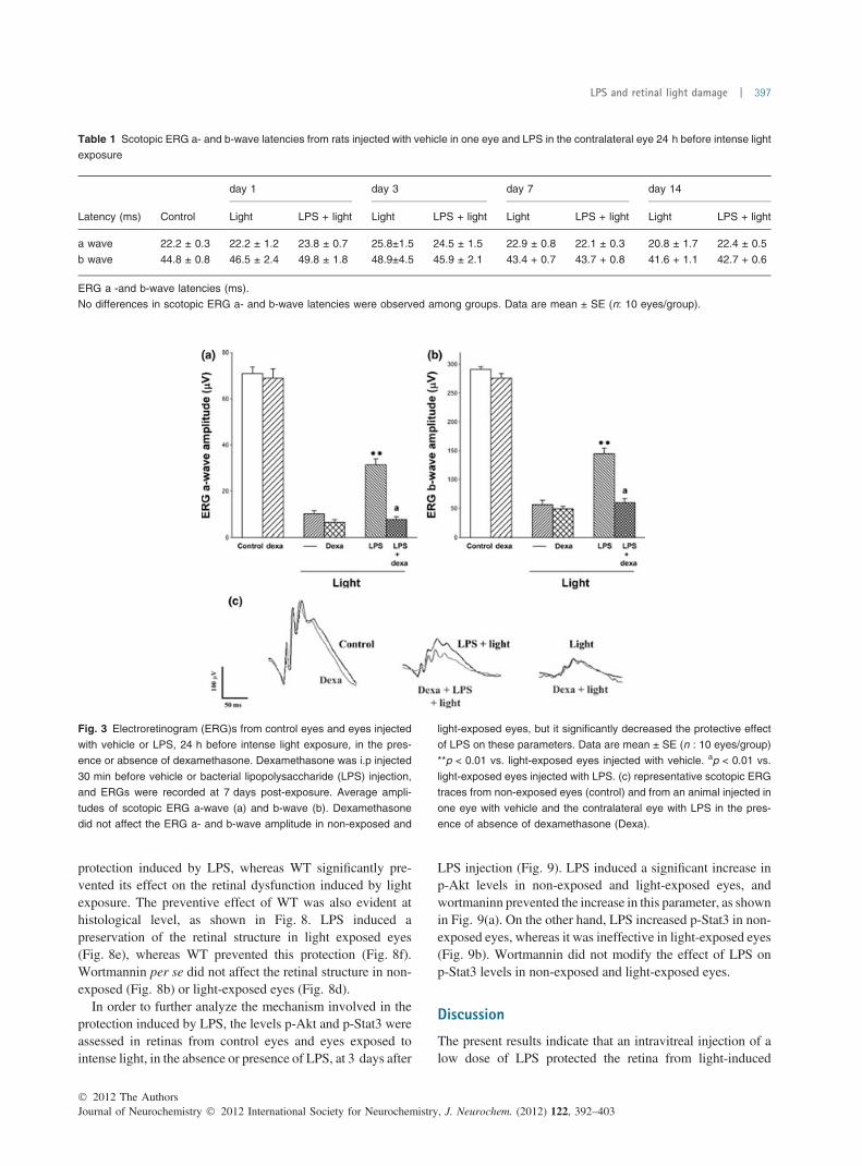
The temporal course of the electroretinographic protectioninduced by 1 lg LPS against retinal light damage wasstudied. One day before light exposure, rats were intravit-really injected with vehicle in one eye and LPS in thecontralateral eye, and ERGs were recorded at 1, 3, 7, and14 days after light exposure (Fig. 2). The average ERGa- and b-wave amplitude in vehicle-injected and non-exposedeyes (control) or at several time points after light exposure, aswell as representative scotopic ERG traces from rat eyessubmitted to these treatments are shown in Fig. 2. Lighttreatment caused a marked attenuation of the scotopic ERGa- and b-waves which was already evident one day after lightexposure, and persisted along the study, whereas theirlatencies remained unchanged (Table 1). LPS did not induceany significant protection on day 1 post-exposure, whereas atday 7 and 14, a significant prevention of the decrease in bothparameters was observed. At 3 days after light exposure, theb-wave amplitude significantly increased in LPS-injectedlight-exposed eyes, and there was a trend for improvementthe a-wave amplitude, but this trend was not significant. Nosignificant differences in the ERG were observed betweennon-injected and vehicle-injected eyes, and between non-exposed eyes injected with vehicle and LPS (data notshown). In order to analyze whether the inhibition of immuneresponses precludes the effect of LPS, dexamethasone wasintraperitoneally injected 30 min before the injection ofvehicle or LPS, and retinal function was assessed at 7 dayspost-light exposure (Fig. 3). Dexamethasone, which showedno effect per se in non-exposed and light-exposed eyes,significantly prevented the protective effect of LPS on retinalfunction. Figure 4 shows a representative photomicrographof a non-injected and non-exposed eye (Fig. 4a) a vehicle-injected and non-exposed eye (Fig. 4b) at 5 days aftervehicle injection. LPS per se (in non-exposed eyes) did notaffect the retinal structure at 3, 5, and 16 days after injection(Figs. 4c, 4d and 4e, Table 2). One day after 24-h lightexposure, no major alterations in the retinal structure wereobserved (Fig. 4f), whereas clear pathological changes wereevident in the retina from eyes injected with vehicle at day 3and 14 after light exposure (Figs. 4g and 4h, respectively).At 3 days post-exposure, a significant decrease in photore-ceptor outer and inner segment (PS) thickness was observed(Fig. 4g), whereas at 14 days post-exposure (Fig. 4h), adecrease in total retinal and ONL thickness and in ganglioncell layer (GCL) cell number, as well as a further decrease inPS thickness was evident in this group (Table 2). In LPS-injected and light-exposed eyes, the whole retinal structurewas significantly preserved at 3 and 14 days post-exposure
� 2012 The AuthorsJournal of Neurochemistry � 2012 International Society for Neurochemistry, J. Neurochem. (2012) 122, 392–403
LPS and retinal light damage | 395

(Fig. 4j and k, respectively, Table 2). The exposure to lightin the absence or presence of LPS did not affect outerplexiform layer (OPL), inner plexiform layer (IPL), and innernuclear layer (INL) thickness.
The effect of light exposure in vehicle- or LPS-injected eyeson the proapoptotic protein Bax and antiapoptotic protein Bcl-xL levels at 24 h after light exposure was assessed byWesternblotting. Figure 5(a) shows representative Western blots ofcytosolic andmitochondrial Bax levels in the retinas from eyesexposed to light in the absence or presence of LPS. Lightexposure induced a significant increase in mitochondrial Bax/cytosolic Bax ratio, which was completely abrogated by anintravitreal injection of LPS. No differences in cytosolic Bcl-xL levels were observed among groups (Fig. 5b). LPS per sedid not modify mitochondrial Bax/cytosolic Bax ratio orcytosolic Bcl-xL levels in non-exposed eyes.
Retinal lipid peroxidation was assessed at 1 or 3 days afterlight exposure, in the absence or presence of LPS. One dayafter light exposure, no differences in TBARS levels wereobserved among groups, whereas at 3 days after lightexposure, a significant increase of this parameter wasobserved in vehicle-injected and light-exposed eyes, whichwas partly prevented by LPS injection (Fig. 6). LPS per sedid not induce changes in TBARS levels at any of these timepoints in non-exposed rats.
The effect of AMG, 5HD, and WT on the retinal protectioninduced by LPS against light-induced damage was examinedby electroretinography at 7 days after light exposure. Theaverage ERG a- and b- wave amplitude of these groups areshown in the upper panel of Fig. 7, while representativescotopic ERG traces are shown in the lower panel of Fig. 7.Neither 5HD nor AMG had any significant effect on the
Fig. 2 Scotopic electroretinogram (ERG)s from rats injected with
vehicle in one eye and LPS in the contralateral eye 24 h before intense
light exposure. ERGs were recorded at 1, 3, 7 or 14 days post-
exposure. Average amplitudes of scotopic ERG a-wave (a) and
b-wave (b). A significant decrease in the amplitude (but not their
latencies) of both ERG a- and b-wave amplitude was observed in light-
exposed eyes injected with vehicle at all time points. In eyes injected
with LPS a significant preservation of the ERG a- and b-wave ampli-
tude was observed at 7 and 14 days after light exposure. At day 3 after
light exposure, LPS afforded a significant protection on the b- wave
(but not a-wave) amplitude. The ERG a- and b-wave amplitude from all
the eyes exposed to intense light in the presence or absence of LPS
significantly differ from that of non-exposed eyes (p < 0.01). Data are
mean ± SE (n : 10 eyes/group). *p < 0.05, **p < 0.01 vs. light-exposed
eyes injected with vehicle at the same time point; ap < 0.05 vs. LPS at
7 days after light exposure, by Tukey’s test. (c) representative
scotopic ERG traces from non-exposed eyes (control) and from an
animal injected in one eye with vehicle and the contralateral eye with
LPS whose ERGs were recorded at 1, 3, 7, and 14 days post-
exposure.
Journal of Neurochemistry � 2012 International Society for Neurochemistry, J. Neurochem. (2012) 122, 392–403� 2012 The Authors
396 | M. P. Bordone et al.
protection induced by LPS, whereas WT significantly pre-vented its effect on the retinal dysfunction induced by lightexposure. The preventive effect of WT was also evident athistological level, as shown in Fig. 8. LPS induced apreservation of the retinal structure in light exposed eyes(Fig. 8e), whereas WT prevented this protection (Fig. 8f).Wortmannin per se did not affect the retinal structure in non-exposed (Fig. 8b) or light-exposed eyes (Fig. 8d).
In order to further analyze the mechanism involved in theprotection induced by LPS, the levels p-Akt and p-Stat3 wereassessed in retinas from control eyes and eyes exposed tointense light, in the absence or presence of LPS, at 3 days after
LPS injection (Fig. 9). LPS induced a significant increase inp-Akt levels in non-exposed and light-exposed eyes, andwortmaninn prevented the increase in this parameter, as shownin Fig. 9(a). On the other hand, LPS increased p-Stat3 in non-exposed eyes, whereas it was ineffective in light-exposed eyes(Fig. 9b). Wortmannin did not modify the effect of LPS onp-Stat3 levels in non-exposed and light-exposed eyes.
Discussion
The present results indicate that an intravitreal injection of alow dose of LPS protected the retina from light-induced
Table 1 Scotopic ERG a- and b-wave latencies from rats injected with vehicle in one eye and LPS in the contralateral eye 24 h before intense light
exposure
Control
day 1 day 3 day 7 day 14
Latency (ms) Light LPS + light Light LPS + light Light LPS + light Light LPS + light
a wave 22.2 ± 0.3 22.2 ± 1.2 23.8 ± 0.7 25.8±1.5 24.5 ± 1.5 22.9 ± 0.8 22.1 ± 0.3 20.8 ± 1.7 22.4 ± 0.5
b wave 44.8 ± 0.8 46.5 ± 2.4 49.8 ± 1.8 48.9±4.5 45.9 ± 2.1 43.4 + 0.7 43.7 + 0.8 41.6 + 1.1 42.7 + 0.6
ERG a -and b-wave latencies (ms).
No differences in scotopic ERG a- and b-wave latencies were observed among groups. Data are mean ± SE (n: 10 eyes/group).
Fig. 3 Electroretinogram (ERG)s from control eyes and eyes injected
with vehicle or LPS, 24 h before intense light exposure, in the pres-
ence or absence of dexamethasone. Dexamethasone was i.p injected
30 min before vehicle or bacterial lipopolysaccharide (LPS) injection,
and ERGs were recorded at 7 days post-exposure. Average ampli-
tudes of scotopic ERG a-wave (a) and b-wave (b). Dexamethasone
did not affect the ERG a- and b-wave amplitude in non-exposed and
light-exposed eyes, but it significantly decreased the protective effect
of LPS on these parameters. Data are mean ± SE (n : 10 eyes/group)
**p < 0.01 vs. light-exposed eyes injected with vehicle. ap < 0.01 vs.
light-exposed eyes injected with LPS. (c) representative scotopic ERG
traces from non-exposed eyes (control) and from an animal injected in
one eye with vehicle and the contralateral eye with LPS in the pres-
ence of absence of dexamethasone (Dexa).
� 2012 The AuthorsJournal of Neurochemistry � 2012 International Society for Neurochemistry, J. Neurochem. (2012) 122, 392–403
LPS and retinal light damage | 397

damage. LPS preserved retinal function and histology,decreased Bax translocation from the cytoplasm to mito-chondria, and oxidative stress in light-exposed eyes. In aprevious report we showed that a single intravitreal injectionof 1 lg LPS per se does not affect the electroretinographic
activity or retinal histology at 1 or 7 days post-injection(Franco et al. 2008). It has been shown that LPS pretreat-ment induces myocardial and cerebral protection against I/Rinjury (Rosenzweig et al. 2004; Schober et al. 2008).Schober et al. (2008) have shown that dexamethasone
(a)
(c) (d) (e)
(f) (g) (h)
(i) (j) (k)
(b)
Fig. 4 Representative photomicrographs
showing histological appearance of non-in-
jected and non-exposed eyes (a), eyes in-
jected with vehicle at 5 days after injection
(b), eyes injected with LPS at 3, 5, and
16 days post-injection (c, d, and e, respec-
tively), and eyes exposed to light for 24 h in
the presence of vehicle (f, g, h) or LPS (i, j,
k) at 3, 5, and 16 after injection (1, 3, or
14 days after exposure, respectively). Se-
vere retinal damage is shown in the retina
from eyes exposed to light in the presence
of vehicle at 3 or 14 days after light expo-
sure, whereas in LPS-injected eyes, the
retinal structure was notably preserved.
Scale bar = 50 lm. GCL, ganglion cell lay-
er; IPL, inner plexiform layer; INL, inner
nuclear layer; OPL, outer plexiform layer;
ONL, outer nuclear layer, PS, photorecep-
tor outer and inner segment; PE, pigment
epithelium.
Table 2 Histological analysis of retinas from eyes exposed to light in the absence or presence of LPS
Total Thickness PS ONL OPL INL IPL cell number inGCL
Control 148.4 ± 1.4 31.8 ± 4.5 32.8 ± 2.7 8.1 ± 1.0 21.5 ± 3.7 33.4 ± 1.5 8.5 ± 0.4
Light
Day 1 141.0 ± 4.0 24.1 ± 1.2 32.3 ± 1.5 9.5 ± 0.3 26.6 ± 0.9 35.9 ± 0.6 8.4 ± 0.5
Day 3 135.6 ± 0.8 13.1 ± 0.3** 19.0 ± 0.4 8.9 ± 0.1 29.2 ± 2.8 47.5 ± 0.2 9.0 ± 1.3
Day 14 101.4 ± 15.1* 2.7 ± 2.6** 4.6 ± 3.1** 9.1 ± 1.5 25.5 ± 6.2 43.0 ± 8.7 5.6 ± 0.5*
LPS + Light
Day 1 142.2 ± 8.3 28.7 ± 2.6 35.3 ± 3.8 8.9 ± 0.7 29.4 ± 2.3 37.6 ± 1.3 8.8 ± 0.1
Day 3 141.4 ± 19.2 23.2 ± 4.4 36.8 ± 10.3 8.5 ± 0.4 28.8 ± 0.6 44.1 v 0.3 8.8 ± 0.4
Day 14 145.8 ± 9.9a 26.3 ± 3.lb 30.9 ± 4.6b 8.3 ± 0.8 22.0 ± 1.8 40.0 ± 2.0 8.3 ± 0.3a
Total retinal and retinal layer thicknesses (in lm) in control eyes, or eyes exposed to light in the absence or presence of LPS, at different time
points after light exposure. At 3 days after light exposure, a significant decrease in PS thickness was observed in vehicle-injected eyes, whereas at
14 days after light exposure a further decrease in PS and a decrease in total and ONL thickness and the number of GCL cells /100 lm was
observed. Data are mean ± SEM (n = 5 eyes/group). The presence of LPS significantly reduced the effect of light stress on these parameters.
*p < 0.05, **p < 0.01 vs. non-exposed eyes, ap < 0.05, bp < 0.01 vs. light exposed eyes injected with vehicle at the same time interval post-
exposure, by Tukey’s test. PS, photoreceptor outer and inner segment; OPL, outer plexiform layer; ONL, outer nuclear layer; IPL, inner plexiform
layer; INL, inner nuclear layer; GCL, ganglion cell layer.
Journal of Neurochemistry � 2012 International Society for Neurochemistry, J. Neurochem. (2012) 122, 392–403� 2012 The Authors
398 | M. P. Bordone et al.

inhibits the protective effect of LPS on acute myocardialinfarction. In agreement, dexamethasone prevented theprotective effect of LPS against light-induced damage,
supporting that the underlying mechanism in the protectioninduced by LPS is dependent upon induction of an inflam-matory response. Although Wenzel et al. (2001) havedemonstrated that dexamethasone completely protects theretina against light damage, in our experimental setting,dexamethasone alone was ineffective. The use of higherdoses of dexamethasone (20–52 mg/kg) as well as theexperimental model used in this report (BALB/c mice) couldaccount for this discrepancy.
Despite limitations of our method for ERG assessment dueto using a single intensity recordings (lack of intensityresponse plot, risk of artifact and error), light exposure induceda significant decrease in the scotopic ERG a- and b-waveamplitude which persisted along the study, while precondi-tioning induced by LPS did not merely delay but preventedretinal alterations, since the recovery in the ERG was asustained effect noted up to 14 days after light exposure.
Classically, it was assumed that prolonged light exposureexclusively damages retinal photoreceptors. However, morerecent findings show that excessive light in vivo (Sang et al.2011) and in vitro (Wood et al. 2007; Osborne et al. 2008)can negatively affect retinal ganglion cell survival. In thisregard, it should be noted that most of the observationsregarding intense light-induced photoreceptor degenerationhave been made immediately or shortly after cessation ofillumination, whereas long-term effects of light damage onthe retina have seldom been investigated. As shown herein,a highly significant decrease in photoreceptor segmentthickness was observed at 3 days, and with extended post-exposure survival times (i.e. 14 days), cell loss becomessignificant in the GCL. In agreement with these results,
Fig. 5 Effect of LPS and light exposure on
retinal mitochondrial and cytosolic levels of
Bax and cytosolic levels of Bcl-xL. Upper
Panel: one day after light exposure densi-
tometric analysis of the samples revealed a
significant increase in mitochondrial Bax/
cytosolic Bax ratio (a) in the absence of
LPS, whereas LPS which showed no effect
per se, prevented the effect of light expo-
sure. No differences in cytosolic Bcl-xL
levels (b) were observed among groups.
Lower panel: representative gel for cyto-
solic and mitochondrial Bax (left) and for
cytosolic Bcl-xL (right) for all the experi-
mental groups. Data are mean ± SEM
(n = 5 eyes per group). *p < 0.05 vs. non-
exposed eyes; ap < 0.05 vs. light-exposed
eyes injected with vehicle, by Tukey’s test.
Fig. 6 Retinal TBARS levels in the retina from non-exposed eyes or
eyes exposed to intense light in the presence of vehicle or LPS, at 1 or
3 days after light exposure. LPS per se did not affect TBARS levels at
4 days post-exposure. At 3 days after light exposure, this parameter
was significantly higher in light-exposed eyes injected with vehicle
than in those injected with LPS. At 1 day post-exposure, retinal lipid
peroxidation did not differ among groups. Data are mean ± SEM (n = 6
retinas/group), **p < 0.01, vs. non-exposed eyes, ap < 0.01 vs. vehi-
cle injected eyes at day 3 after light exposure, by Tukey’s test.
� 2012 The AuthorsJournal of Neurochemistry � 2012 International Society for Neurochemistry, J. Neurochem. (2012) 122, 392–403
LPS and retinal light damage | 399

some groups have shown a delayed ganglion cell loss afterdifferent intervals post-exposure to intense light (Wasowiczet al. 2002; Marco-Gomariz et al. 2006). Our resultsindicate that the decrease in total retinal, PS, and ONLthickness, as well as in GCL cell number in eyes exposed tointense light was prevented by the intravitreal injection ofLPS.
Exposure to intense light induces retinal apoptotic celldeath (Noell et al. 1966; ‘; Reme et al. 1998). Bax is a majoreffector of apoptotic cell death in the retina after ischemia,excitotoxicity, axotomy, and retinal degeneration (Isenmannet al. 1999; Tezel and Wax 1999; Podesta et al. 2000).Although light exposure increases Bax and induces apoptosisin other cell types such as lens epithelial cells, melanocytes,and retinal pigment epithelial cells (Waster and Ollinger2009; Kernt et al. 2010, 2011), to our knowledge, the presentresults are the first demonstration that light exposuretriggered retinal Bax translocation from the cytoplasm tomitochondria which was prevented by LPS, supporting therole of Bax in the cell death pathway provoked by intenselight, as well as in LPS-induced protection. In agreement, itwas previously shown that deficiency of Bax and Bakprotects the retina against light damage (Hahn et al. 2004),and that IPC prevents the increase in retinal Bax levelsinduced by I/R damage (Zhang et al. 2002). As previouslyshown by Grimm et al. (2000), no changes in retinal Bcl-xLlevels were observed after light exposure.
Compelling evidence exists that intense light leads toretinal oxidative stress. The effectiveness of a wide variety ofantioxidants indicates that reactive oxygen species areformed during intense light (Organisciak and Vaughan2010). In agreement with Chen and Zhang (1994), lightexposure induced a significant increase in retinal TBARSlevels (an index of lipid peroxidation) at 3 (but not 1) days,and this effect was prevented by LPS. These results suggestthat a low dose of LPS could behave as an antioxidanttherapy in the paradigm of retinal light-induced injury.
Several signaling pathways and/or cellular survival proteinsunderlie the neuroprotective effects of retinal IPC, such asadenosine (Li andRoth 1999), mK+/ATP channels (Roth et al.2006), and NO (Sakamoto et al. 2006), among many others(reviewed by Gidday 2006). We examined some mechanismswhose involvement in the protective effect of IPC against I/Rdamage has been previously demonstrated. Retinal protectioninduced by LPS was not influenced by an mK+/ATP channelblocker or by AMG, supporting that mK+/ATP channels andiNOS are not involved in the protection against light-inducedinjury triggered by LPS.We have previously shown that AMGprevents the protective effect of LPS against I/R injury (Francoet al. 2008). Since AMG did not affect the protection inducedby LPS against light damage, it seems that the mechanisminvolved in the protective effect of LPS could depend on thetype of damage. The protective effect of LPS, both atfunctional and histological level was completely prevented
Fig. 7 Effect of 5-hydroxydecanoic acid
(5HD), aminoguanidine (AMG), and wort-
mannin (WT) on the functional protection
induced by LPS against light damage. Eyes
were injected with 5HD, AMG or WT, as
described in Material and methods. Aver-
age amplitudes of scotopic ERG a-wave (a)
and b-wave (b) 7 days after light exposure.
Wortmannin significantly decreased the
protection induced by LPS on the retinal
dysfunction induced by light exposure,
whereas 5HD and AMG were ineffective.
Data are mean ± SE (n: 5 eyes/group).
**p < 0.01 vs. light-exposed eyes injected
with vehicle, ap < 0.01 vs. light-exposed
eyes injected with LPS, by Tukey’s test. (c)
Representative scotopic ERGs from all the
experimental groups.
Journal of Neurochemistry � 2012 International Society for Neurochemistry, J. Neurochem. (2012) 122, 392–403� 2012 The Authors
400 | M. P. Bordone et al.

(a) (b)
(c) (d)
(e) (f)
Fig. 8 Effect of wortmannin (WT) on the
histological protection against light damage
induced by bacterial lipopolysaccharide
(LPS) at 7 days after light exposure. Shown
are representative photomicrographs of
histological appearance of non-injected and
non-exposed eyes (a) and of light- exposed
eyes injected with vehicle (c), WT (d), LPS
(e), or LPS + WT (f). The preservation of the
retinal structure induced by the injection of
LPS was substantially decreased in the
presence of WT. Wormannin per se did not
affect the retinal structure in non-exposed
eyes (b) or in light-exposed eyes (d). Scale
bar = 50 lm.
Fig. 9 Effect of LPS and light exposure on
retinal p-Akt and p-Stat3 levels. (a) LPS
induced a significant increase in p-Akt lev-
els in non-exposed and light-exposed eyes
which was prevented by WT. (b) LPS
induced a significant increase in p-Stat3
levels in non-exposed eyes, and WT did not
affect the effect of LPS on this parameter.
No differences were observed in p-Stat3
levels in light-exposed eyes among groups.
Data are mean ± SEM (n = 5 eyes/group).
**p < 0.01 vs. eyes injected with vehicle
and under the same light regime, ap < 0.01
vs. eyes injected with LPS and under the
same light regime, by Tukey’s test.
� 2012 The AuthorsJournal of Neurochemistry � 2012 International Society for Neurochemistry, J. Neurochem. (2012) 122, 392–403
LPS and retinal light damage | 401

by WT. It was demonstrated that WT attenuates the neuropro-tective effect of IPC on I/R damage through an Akt (a serine/threonine kinase also known as protein kinase B)-dependentpathway (Dreixler et al. 2009) and a role for Akt in thepreservation of photoreceptors from light damage has beendemonstrated (Li et al. 2007; Ueki et al. 2008). On the otherhand, it was reported that Stat3 is the primary effector proteinof LPS-mediated JAK-STAT signaling (Jang et al. 2007). Asshown herein, LPS induced a significant increase in p-Akt andp-Stat3 levels, but WT only prevented the effect of LPS onp-Akt (but not p-Stat3) levels both in non-exposed and light-exposed eyes, supporting the involvement of PI3K/Aktpathway in the protective effect of LPS (Jang et al. 2007). Itwas demonstrated that dexamethasone inhibits PI3K-Aktsignaling in several systems (Fujita et al. 2004; Gupta et al.2007). Thus, this pathway could be involved in the inhibitoryeffect of dexamethasone on the retinal protection induced byLPS against intense light damage.
While all types of animal models can be criticized for theirrelevance to human physiology, people with inherited oculardiseases primarily live and work in light environments. Theuse of intense or prolonged light exposures and animalmodels can improve our understanding of basic mechanismsin retinal degeneration by exacerbating visual cell death withan acute insult superimposed on a chronic condition. At thistime, we need to extrapolate our findings in animal models tohuman disease, but it may turn out that simple treatmentsdeveloped to prevent retinal degeneration in animals may bethe basis of therapeutic interventions in people (Organisciakand Vaughan 2010). In that context, the demonstration that amoderate dose of LPS protected the retina against lightdamage could constitute a future fertile avenue for promotingthe survival of retinal cells.
Acknowledgments
The authors have no conflict of interest. This research was supportedby grants from the Agencia Nacional de Promocion Cientıfica yTecnologica (ANPCyT), the University of Buenos Aires, andCONICET, Argentina.
References
Belforte N. A., Moreno M. C., de Zavalıa N., Sande P. H., Chianelli M.S., Keller Sarmiento M. I. and Rosenstein R. E. (2010) Melatonin:a novel neuroprotectant for the treatment of glaucoma. J. PinealRes. 48, 353–364.
Casson R. J., Wood J. P., Melena J., Chidlow G. and Osborne N. N.(2003) The effect of ischemic preconditioning on light-inducedphotoreceptor injury. Invest. Ophthalmol. Vis. Sci. 44, 1348–1354.
Chen W. H. and Zhang H. R. (1994) Photogenic retinal damage and itsmedicinal prevention: lipid peroxide studies. Zhonghua Yan Ke ZaZhi. 30, 125–127.
Cruickshanks K. J., Klein R. and Klein B. E. (1993) Sunlight and age-related macular degeneration. The Beaver Dam Eye Study. Arch.Ophthalmol. 111, 514–518.
Darzins P., Mitchell P. and Heller R. F. (1997) Sun exposure and age-related macular degeneration. An Australian case–control study.Ophthalmology 104, 770–776.
Dreixler J. C., Hemmert J. W., Shenoy S. K., Shen Y., Lee H. T., ShaikhA. R., Rosenbaum D. M. and Roth S. (2009) The role of Akt/protein kinase B subtypes in retinal ischemic preconditioning. Exp.Eye Res. 88, 512–521.
FernandezD. C., BordoneM. P., ChianelliM. S. and Rosenstein R. E. (2009)Retinal neuroprotection against ischemia-reperfusion damage inducedby postconditioning. Invest. Ophthalmol. Vis. Sci. 50, 3922–3930.
Franco P. J., Fernandez D. C., Sande P. H., Keller Sarmiento M. I.,Chianelli M., Saenz D. A. and Rosenstein R. E. (2008) Effect ofbacterial lipopolysaccharide on ischemic damage in the rat retina.Invest. Ophthalmol. Vis. Sci. 49, 4604–4612.
Fujita T., Fukuyama R., Enomoto H. and Komori T. (2004) Dexa-methasone inhibits insulin-induced chondrogenesis of ATDC5cells by preventing PI3K-Akt signaling and DNA binding ofRunx2. J. Cell. Biochem. 93, 374–383.
Gidday J. M. (2006) Cerebral preconditioning and ischaemic tolerance.Nat. Rev. Neurosci. 7, 437–448.
Gidday J. M. (2010) Pharmacologic preconditioning: translating thepromise. Transl. Stroke Res. 1, 19–30.
GrimmC.,WenzelA.,Hafezi F. andRemeC. E. (2000)Gene expression inthe mouse retina: the effect of damaging light.Mol. Vis. 6, 252–260.
Gupta V., Awasthi N. and Wagner B. J. (2007) Specific activation of theglucocorticoid receptor and modulation of signal transductionpathways in human lens epithelial cells. Invest. Ophthalmol. Vis.Sci. 48, 1724–1734.
Hahn P., Lindsten T., Lyubarsky A., Ying G. S., Pugh Jr E. N. .,Thompson C. B. and Dunaief J. L. (2004) Deficiency of Bax andBak protects photoreceptors from light damage in vivo. Cell DeathDiffer. 11, 1192–1197.
Isenmann S., Engel S., Gillardon F. and Bahr M. (1999) Bax antisenseoligonucleotides reduce axotomy-induced retinal ganglion celldeath in vivo by reduction of Bax protein expression. Cell DeathDiff. 6, 673–682.
Jang S., Lee J. H., Choi K. R., Kim D., Yoo H. S. and Oh S. (2007)Cytochemical alterations in the rat retina by LPS administration.Neurochem. Res. 32, 1–10.
Kermer P., Klocker N., Labes M. and Bahr M. (2000) Insulin-likegrowth factor-I protects axotomized rat retinal ganglion cells fromsecondary death via PI3-K-dependent Akt phosphorylation andinhibition of caspase-3 in vivo. J. Neurosci. 20, 2–8.
Kernt M., Hirneiss C., Neubauer A. S., Ulbig M. W. and Kampik A.(2010) Coenzyme Q10 prevents human lens epithelial cells fromlight-induced apoptotic cell death by reducing oxidative stress andstabilizing BAX/Bcl-2 ratio. Acta Ophthalmol.. 88, e78–e86.
Kernt M., Thiele S., Hirneiss C., Neubauer A. S., Lackerbauer C. A.,Ulbig M. W. and Kampik A. (2011) The role of light in thedevelopement of RPE degeneration in AMD and potential cy-toprotection of minocycline. Klin. Monbl. Augenheilkd. 228,892–899.
Khan J.C., ShahidH., ThurlbyD.A., BradleyM.,ClaytonD.G.,MooreA.T., Bird A. C and Yates J. R. (2006) Genetic Factors in AMD Study.Age related macular degeneration and sun exposure, iris colour, andskin sensitivity to sunlight. Br. J. Ophthalmol. 90, 29–32.
LaVail M. M., Unoki K., Yasumura D., Matthes M. T., YancopoulosG. D. and Steinberg R. H. (1992) Multiple growth factors,cytokines, and neurotrophins rescue photoreceptors from thedamaging effects of constant light. Proc. Natl. Acad. Sci. USA.89, 11249–11253.
Li B. and Roth S. (1999) Retinal ischemic preconditioning in the rat:requirement for adenosine and repetitive induction. Invest. Oph-thalmol. Vis. Sci. 40, 200–216.
Journal of Neurochemistry � 2012 International Society for Neurochemistry, J. Neurochem. (2012) 122, 392–403� 2012 The Authors
402 | M. P. Bordone et al.
Li G., Anderson R. E., Tomita H., Adler R., Liu X., Zack D. J. andRajala R. V. (2007) Nonredundant role of Akt2 for neuroprotectionof rod photoreceptor cells from light-induced cell death. J. Neu-rosci. 27, 203–211.
Liu C., Peng M., Laties A. M. and Wen R. (1998) Preconditioning withbright light evokes a protective response against light damage inthe rat retina. J. Neurosci. 18, 1337–1344.
Lowry O. H., Rosebrough N. J., Farr A. L. and Randall R. J. (1951)Protein measurement with the Folin phenol reagent. J. Biol. Chem.193, 265–275.
Marco-Gomariz M. A., Hurtado-Montalban N., Vidal-Sanz M., Lund R.D. and Villegas-Perez M. P. (2006) Phototoxic-induced photore-ceptor degeneration causes retinal ganglion cell degeneration inpigmented rats. J Comp Neurol. 498, 163–179.
Noell W. K., Walker V. S., Kang B. S. and Berman S. (1966) Retinaldamage by light in rats. Invest. Ophthalmol. Vis. Sci. 5, 450–473.
Organisciak D. T. and Vaughan D. K. (2010) Retinal light damage:mechanisms and protection. Prog. Retin. Eye Res. 29, 113–134.
OrganisciakD. T. andWinkler B. S. (1994) Retinal light damage: practicaland theoretical considerations. Prog. Retin. Eye Res. 13, 1–29.
Organisciak D. T., Darrow R. M., Noell W. K. and Blanks J. C. (1995)Hyperthermia accelerates retinal light damage in rats. Invest.Ophthalmol. Vis. Sci. 36, 997–1008.
Osborne N. N., Li G. Y., Ji D., Mortiboys H. J. and Jackson S. (2008)Light affects mitochondria to cause apoptosis to cultured cells:possible relevance to ganglion cell death in certain optic neurop-athies. J. Neurochem. 105, 2013–2028.
Piehl L., Capani F., Facorro G., Lopez E. M., de Celis E. R., PustovrhC., Hager A., Coirini H. and Lopez-Costa J. J. (2007) Nitric oxideincreases in the rat retina after continuous illumination. Brain Res.1156, 112–119.
Podesta F., Romeo G., Liu W. H., Krajewski S., Reed J. C., Gerhar-dinger C. and Lorenzi M. (2000) Bax is increased in the retina ofdiabetic subjects and is associated with pericyte apoptosis in vivoand in vitro. Am. J. Pathol. 156, 1025–1032.
RemeC. E., GrimmC., Hafezi F.,Marti A. andWenzel A. (1998) Apoptoticcell death in retinal degenerations.Prog. Retin. Eye Res. 17, 443–464.
Rosenzweig H. L., Lessov N. S., Henshall D. C., Minami M., SimonR. P. and Stenzel-Poore M. P. (2004) Endotoxin preconditioningprevents cellular inflammatory response during ischemic neuro-protection in mice. Stroke 35, 2576–2581.
Roth S., Li B., Rosenbaum P. S., Gupta H., Goldstein I. M., Maxwell K.M. and Gidday J. M. (1998) Preconditioning provides completeprotection against retinal ischemic injury in rats. Invest. Ophthal-mol. Vis. Sci. 39, 777–785.
Roth S., Dreixler J. C., Shaikh A. R., Lee K. H. and Bindokas V. (2006)Mitochondrial potassium ATP channels and retinal ischemic pre-conditioning. Invest. Ophthalmol. Vis. Sci. 47, 2114–2124.
Sakamoto K., Yonoki Y., Kubota Y., Kuwagata M., Saito M., NakaharaT. and Ishii K. (2006) Inducible nitric oxide synthase inhibitors
abolished histological protection by late ischemic preconditioningin rat retina. Exp. Eye Res. 82, 512–518.
Sang A., Cheng Y., Lu H., Chen D., Gao R. and Shen A. (2011) Light-induced retinal ganglion cell damage in vivo involves Dexras1.Mol. Vis. 17, 134–143.
Schober P., Oprea G., Mersmann J., Nebert A., Zacharowski K. andZacharowski P. A. (2008) Lipoteichoic acid induces delayedmyocardial protection in isolated rat hearts: a comparison withendotoxin. Resuscitation 79, 311–315.
Shahinfar S., Edward D. P. and Tso M. O. (1991) A pathologic study ofphotoreceptor cell death in retinal photic injury.Curr.EyeRes.10, 47–59.
Tezel G. and Wax M. B. (1999) Inhibition of caspase activity in retinalcell apoptosis induced by various stimuli in vitro. Invest. Oph-thalmol. Vis. Sci. 40, 2660–2667.
Ueki Y., Wang J., Chollangi S. and Ash J. D. (2008) STAT3 activationin photoreceptors by leukemia inhibitory factor is associated withprotection from light damage. J. Neurochem. 105, 784–796.
Vartanian K. B., Stevens S. L., Marsh B. J., Williams-Karnesky R.,Lessov N. S. and Stenzel-Poore M. P (2011) LPS preconditioningredirects TLR signaling following stroke: TRIF-IRF3 plays aseminal role in mediating tolerance to ischemic injury. J. Neuro-inflammation 14, 140.
Wang Y., Luo J., Chen X., Chen H., Cramer S. W. and Sun D. (2008)Gene inactivation of Na+/H+ exchanger isoform 1 attenuatesapoptosis and mitochondrial damage following transient focalcerebral ischemia. Eur. J. Neurosci. 28, 51–61.
Wasowicz M., Morice C., Ferrari P., Callebert J. and Versaux-Botteri C.(2002) Long-term effects of light damage on the retina of albinoand pigmented rats. Invest. Ophthalmol. Vis. Sci. 43, 813–820.
Waster P. K. and Ollinger K. M. (2009) Redox-dependent translocationof p53 to mitochondria or nucleus in human melanocytes afterUVA- and UVB-induced apoptosis. J. Invest. Dermatol. 129,1769–1781.
Wen R., Cheng T., Li Y., Cao W. and Steinberg R. H. (1996) Alpha2-adrenergic agonists induce basic fibroblast growth factorexpression in photoreceptors in vivo and ameliorate light damage.J. Neurosci. 16, 5986–5992.
Wenzel A., Grimm C., Seeliger M. W., Jaissle G., Hafezi F., KretschmerR., Zrenner E. and Reme C. E. (2001) Prevention of photoreceptorapoptosis by activation of the glucocorticoid receptor. Invest.Ophthalmol. Vis. Sci. 42, 1653–1659.
Wenzel A., Grimm C., Samardzija M. and Reme C. E. (2005) Molecularmechanisms of light-induced photoreceptor apoptosis and neuropro-tection for retinal degeneration. Prog. Retin. Eye Res. 24, 275–306.
Wood J. P., Lascaratos G., Bron A. J. and Osborne N. N. (2007) Theinfluence of visible light exposure on cultured RGC-5 cells. Mol.Vis. 14, 334–344.
Zhang C., Rosenbaum D. M., Shaikh A. R., Li Q., Rosenbaum P. S.,Pelham D. J. and Roth S. (2002) Ischemic preconditioning atten-uates apoptotic cell death in the rat retina. Invest. Ophthalmol. Vis.Sci. 43, 3059–3066.
� 2012 The AuthorsJournal of Neurochemistry � 2012 International Society for Neurochemistry, J. Neurochem. (2012) 122, 392–403
LPS and retinal light damage | 403