Myofiber-specific inhibition of TGFb signaling protects skeletal ...
13
Myofiber-specific inhibition of TGFb signaling protects skeletal muscle from injury and dystrophic disease in mice Federica Accornero 1 , Onur Kanisicak 1 , Andoria Tjondrokoesoemo 1 , Aria C. Attia 1 , Elizabeth M. McNally 2 and Jeffery D. Molkentin 1,3,∗ 1 Department of Pediatrics, Cincinnati Children’s Hospital Medical Center, University of Cincinnati, Cincinnati, 240 Albert Sabin Way, Cincinnati, OH 45229, USA, 2 Department of Medicine, Section of Cardiology, 5841 S, Maryland, MC 6088, Chicago, IL 60637, USA and 3 Howard Hughes Medical Institute, Cincinnati Children’s Hospital Medical Center, 240 Albert Sabin Way, Cincinnati, OH 45229, USA Received April 17, 2014; Revised and Accepted August 5, 2014 Muscular dystrophy (MD) is a disease characterized by skeletal muscle necrosis and the progressive accumu- lation of fibrotic tissue. While transforming growth factor (TGF)-b has emerged as central effector of MD and fibrotic disease, the cell types in diseased muscle that underlie TGFb-dependent pathology have not been segregated. Here, we generated transgenic mice with myofiber-specific inhibition of TGFb signaling owing to expression of a TGFb type II receptor dominant-negative (dnTGFbRII) truncation mutant. Expression of dnTGFbRII in myofibers mitigated the dystrophic phenotype observed in d-sarcoglycan-null (Sgcd 2/ 2 ) mice through a mechanism involving reduced myofiber membrane fragility. The dnTGFbRII transgene also reduced muscle injury and improved muscle regeneration after cardiotoxin injury, as well as increased satellite cell numbers and activity. An unbiased global expression analysis revealed a number of potential mechanisms for dnTGFbRII-mediated protection, one of which was induction of the antioxidant protein metallothionein (Mt). Indeed, TGFb directly inhibited Mt gene expression in vitro, the dnTGFbRII transgene conferred protection against reactive oxygen species accumulation in dystrophic muscle and treatment with Mt mimetics protected skeletal muscle upon injury in vivo and improved the membrane stability of dystrophic myofibers. Hence, our results show that the myofibers are central mediators of the deleterious effects associated with TGFb signaling in MD. INTRODUCTION Muscular dystrophy (MD) is a disease characterized by skeletal muscle degeneration owing to myofiber necrosis followed by progressive accumulation of fibrotic tissue (1). Necrosis of myofibers in dystrophic skeletal muscle causes an inflammatory response resulting in secretion of multiple cytokines and growth factors, typically produced by macrophages, fibroblasts and sur- rounding viable myofibers (2). This enhanced cytokine profile is a two-edge sword, as it promotes resident satellite cell activation to repair the damaged fibers, while at the same time inducing fibroblast activation and fibrosis, as well as detrimental signaling in the remaining myofibers that likely leads to cell death (3 – 5). Transforming growth factor (TGF)-b has emerged as a primary secreted factor underlying the fibrotic response in MD. TGFb is upregulated in dystrophic muscles from human patients and animal models (6 – 9), and inhibition of systemic TGFb with a monoclonal blocking antibody reduces fibrosis and muscle path- ology in MD (10,11). These observations suggest that TGFb is a central effector of MD, although the role that TGFb plays in the disease process is not characterized at the level of individual cell types within the muscle (i.e. myofibers versus fibroblasts). Resident fibroblasts directly respond to TGFb to initiate fibro- sis, and their continual activation promotes the progressivity of MD over time (12). TGFb also negatively affects skeletal muscle regeneration by exerting direct inhibitory effects on the resident ∗ To whom correspondence should be addressed at: Cincinnati Children’s Hospital Medical Center, 240 Albert Sabin Way, MLC7020, Cincinnati, OH 45229, USA. Tel: +1 5136363557; Fax: +1 5136365958; Email: [email protected] # The Author 2014. Published by Oxford University Press. All rights reserved. For Permissions, please email: [email protected] Human Molecular Genetics, 2014 1–13 doi:10.1093/hmg/ddu413 Downloaded from https://academic.oup.com/hmg/article/23/25/6903/572343 by guest on 17 January 2022
-
Upload
khangminh22 -
Category
Documents
-
view
5 -
download
0
Transcript of Myofiber-specific inhibition of TGFb signaling protects skeletal ...

Myofiber-specific inhibition of TGFb signalingprotects skeletal muscle from injury and dystrophicdisease in mice
Federica Accornero1, Onur Kanisicak1, Andoria Tjondrokoesoemo1, Aria C. Attia1,
Elizabeth M. McNally2 and Jeffery D. Molkentin1,3,∗
1Department of Pediatrics, Cincinnati Children’s Hospital Medical Center, University of Cincinnati, Cincinnati,
240 Albert Sabin Way, Cincinnati, OH 45229, USA, 2Department of Medicine, Section of Cardiology, 5841 S, Maryland,
MC 6088, Chicago, IL 60637, USA and 3Howard Hughes Medical Institute, Cincinnati Children’s Hospital Medical Center,
240 Albert Sabin Way, Cincinnati, OH 45229, USA
Received April 17, 2014; Revised and Accepted August 5, 2014
Muscular dystrophy (MD) is a disease characterized by skeletal muscle necrosis and the progressive accumu-lation of fibrotic tissue. While transforming growth factor (TGF)-b has emerged as central effector of MD andfibrotic disease, the cell types in diseased muscle that underlie TGFb-dependent pathology have not beensegregated. Here, we generated transgenic mice with myofiber-specific inhibition of TGFb signaling owing toexpression of a TGFb type II receptor dominant-negative (dnTGFbRII) truncation mutant. Expression ofdnTGFbRII in myofibers mitigated the dystrophic phenotype observed in d-sarcoglycan-null (Sgcd2/2) micethrough a mechanism involving reduced myofiber membrane fragility. The dnTGFbRII transgene also reducedmuscle injury and improved muscle regeneration after cardiotoxin injury, as well as increased satellite cellnumbers and activity. An unbiased global expression analysis revealed a number of potential mechanismsfor dnTGFbRII-mediated protection, one of which was induction of the antioxidant protein metallothionein(Mt). Indeed, TGFbdirectly inhibited Mt gene expression in vitro, the dnTGFbRII transgene conferred protectionagainst reactive oxygen species accumulation in dystrophic muscle and treatment with Mt mimetics protectedskeletal muscle upon injury in vivo and improved the membrane stability of dystrophic myofibers. Hence, ourresults show that the myofibers are central mediators of the deleterious effects associated with TGFb signalingin MD.
INTRODUCTION
Muscular dystrophy (MD) is a disease characterized by skeletalmuscle degeneration owing to myofiber necrosis followed byprogressive accumulation of fibrotic tissue (1). Necrosis ofmyofibers in dystrophic skeletal muscle causes an inflammatoryresponse resulting in secretion of multiple cytokines and growthfactors, typically produced by macrophages, fibroblasts and sur-rounding viable myofibers (2). This enhanced cytokine profile isa two-edge sword, as it promotes resident satellite cell activationto repair the damaged fibers, while at the same time inducingfibroblast activation and fibrosis, as well as detrimental signalingin the remaining myofibers that likely leads to cell death (3–5).
Transforming growth factor (TGF)-b has emerged as a primarysecreted factor underlying the fibrotic response in MD. TGFb isupregulated in dystrophic muscles from human patients andanimal models (6–9), and inhibition of systemic TGFb with amonoclonal blocking antibody reduces fibrosis and muscle path-ology in MD (10,11). These observations suggest that TGFb is acentral effector of MD, although the role that TGFb plays in thedisease process is not characterized at the level of individual celltypes within the muscle (i.e. myofibers versus fibroblasts).
Resident fibroblasts directly respond to TGFb to initiate fibro-sis, and their continual activation promotes the progressivity ofMD over time (12). TGFbalso negatively affects skeletal muscleregeneration by exerting direct inhibitory effects on the resident
∗To whom correspondence should be addressed at: Cincinnati Children’s Hospital Medical Center, 240 Albert Sabin Way, MLC7020, Cincinnati,OH 45229, USA. Tel: +1 5136363557; Fax: +1 5136365958; Email: [email protected]
# The Author 2014. Published by Oxford University Press. All rights reserved.For Permissions, please email: [email protected]
Human Molecular Genetics, 2014 1–13doi:10.1093/hmg/ddu413
Dow
nloaded from https://academ
ic.oup.com/hm
g/article/23/25/6903/572343 by guest on 17 January 2022

stem cell pool (satellite cells) (13,14). TGFb can inhibit differ-entiation of myoblasts into myotubes in culture, and attenuationof TGFb activity improves the physiologic response of satellitecells following injury, leading to better regenerative capacityin vivo (2,15,16). However, TGFb signaling is also importantfor myoblast proliferation and formation from satellite cells,indicating important physiologic roles for this cytokine in propermuscle development (17,18). While skeletal muscle myofibersand myoblasts express the TGFb receptors and are direct targetsof TGFb signaling, the contribution that TGFb signaling playsat the level of the myofiber to the dystrophic disease phenotypehas not been directly analyzed. In the present study, we elucidatefor the first time the role of adult myofibers as primary respondersto TGFb signaling in the dystrophic disease process. We showthat specific inhibition of TGFb signaling only in myofibers ofadult skeletal muscle is sufficient to mitigate MD and improveskeletal muscle regeneration.
RESULTS
Generation of dominant-negative TGFb-receptor typeII-expressing transgenic mice
TGFb is produced and activated in skeletal muscle during injuryor in MD, where it is known to negatively affect aspects of heal-ing and regeneration as well as promote fibrosis and disease(9,19). Here, we generated transgenic (TG) mice expressingthe well-characterized TGFb type II receptor dominant-negative(dnTGFbRII) truncation mutant to block TGFb signaling spe-cifically at the level of the myofiber using the skeletal a-actinmuscle-specific promoter (Fig. 1A). This dominant-negativereceptor lacks the intracellular kinase domain, yet it still effect-ively dimerizes with TGFb type I receptors, thereby generatinginactive complexes (20–22). Two independent TG lines weregenerated with high (TG line 2) and medium (TG line 1) expres-sion (Fig. 1B). In line 1, which was selected for more in-depthanalysis, we observed dnTGFbRII expression in every skeletalmuscle analyzed but not the heart (Fig. 1C). H&E and Masson’strichrome-stained histological sections showed no obviousphenotype in themusclesofdnTGFbRIITGmicecomparedwithwild-type (WT) littermates (Fig. 1D). Also, we did not observechanges in muscle weights normalized to tibia length at 6 monthsof age or in fiber size owing to the transgene (data not shown).Analysis of myofiber central nucleation showed a small increasein the quadriceps and tibialis anterior (TA) from TG mice at 6months of age, although the other muscles analyzed showed nosignificant change (Fig. 1E). These subtle alterations in myofibercentral nucleationare likely non-pathologic andmaybe the resultof greater satellite cell activity, especially because dnTGFbRIITG mice had slightly but significantly better grip strength com-pared with WT littermates at both 2 and 6 months of age (Fig. 1F).Thus, expression of the dnTGFbRII on skeletal muscle fibers/myoblasts did not induce overt pathology although it did appearto slightly alter the physiology of the muscles in a more adaptiveprofile.
To address the in vivo effectiveness of the dnTGFbRII trans-gene, we isolated primary myofibers from the extensor digitorumlongus (EDL) muscle of TG versus WT mice and stimulated themwith TGFb for analysis of phospho-SMAD3 levels and nuclearaccumulation. The data show robust activation in WT myofibers,
but almost no activation in myofibers from the TG mice(Fig. 1G). Importantly, muscle interstitial cells (mostly fibro-blasts) and isolated myoblasts from these same two groups ofmice showed equal activation of SMAD3 phosphorylation in re-sponse to TGFb, indicating that the dnTGFbRII transgene hadno residual inhibitory effect on these cell types from muscle(Supplementary Material, Fig. S1A and B).
Myofiber-specific inhibition of TGFb signaling mitigatesMD histopathology
To assess how the myofibers contribute to TGFb-dependent MD,we crossed the dnTGFbRII transgene into the d-sarcoglycan-null (Sgcd2/2) genetic background, the latter of which is amouse model with fulminant dystrophic disease (23,24). Wefirst confirmed activation of TGFb in the Sgcd2/2 model of MDby ELISA (Supplementary Material, Fig. S2) as well as showedthat the dominant-negative receptor blocked SMAD3 phosphor-ylation in myofibers from these dystrophic mice, which isnormally highly activated with MD (Fig. 2A and B) (9,25). Inter-stitial cells showed equal SMAD3 phosphorylation levels frommuscleofWTversus TGmice, againsuggestingspecificity of thetransgene for TGFb signaling within the myofibers (Fig. 2B).Inhibition of TGFb signaling with this transgene reduced histo-pathology in Sgcd2/2 mice (Fig. 2C) with a normalization ofthe pseudohypertrophy typically observed in the quadriceps,gastrocnemius and TA (Fig. 2D), as well as a significant reduc-tion in central nucleation in every muscle analyzed except thediaphragm (Fig. 2E). The slightly less-potent effect in dia-phragm and soleus is likely due to the lower expression of thetransgene in those muscles owing to the greater content of slowfibers, where the skeletala-actin promoter is less active (Fig. 1C).Despite these protective histologic features associated with in-hibition of TGFb signaling within the myofibers, quantificationof total muscle fibrosis by analysis of hydroxyproline contentrevealed no significant changes in the Sgcd2/2 mice with orwithout the transgene (Fig. 1F). Analysis of collagen expressionby quantitative PCR confirmed equal collagen induction inSgcd2/2 and Sgcd2/2 TG muscles (Supplementary Material,Fig. S3). These observations suggest that the primary role ofTGFb signaling in the myofibers is independent from the well-established pro-fibrotic effect of TGFb, which is likely more de-pendent on fibroblasts that do not express the dominant-negativetransgene.
Myofiber-specific inhibition of TGFb improves MDby enhancing membrane integrity
Here, we subjected mice to forced treadmill running to assesswhether the observed improvement in histopathology conferredby the muscle-specific dnTGFbRII transgene correlated withfunctional benefits. Indeed, Sgcd2/2 mice with the dnTGFbRIItransgene showed a restoration of their ability to run on a tread-mill comparable with WT control mice at both 2 and 6 monthsof age, whereas Sgcd2/2 mice alone were severely compro-mised (Fig. 3A). In addition, Sgcd2/2 TG mice showed a signifi-cant reduction in total serum creatine kinase (CK) levels at 2 and6 months of ages compared with Sgcd2/2 mice, suggesting lessmyofiber membrane ruptures and ongoing myofiber necrosis(Fig. 3B).To further investigate membrane integrity, we employed
2 Human Molecular Genetics, 2014
Dow
nloaded from https://academ
ic.oup.com/hm
g/article/23/25/6903/572343 by guest on 17 January 2022
the Evans blue dye (EBD) uptake assay after forced treadmillrunning. Using this assay, Sgcd2/2 mice showed as muchas 30% fiber positivity, whereas WT mice showed no signal
(Fig. 3C and D and data not shown). However, the presence ofthe dnTGFbRII transgene dramatically reduced EBD uptakein the muscles of Sgcd2/2 mice, suggesting enhanced membrane
Figure 1. Generation of skeletal muscle-specific TG mice expressing a dominant-negative TGFb-receptor. (A) Schematic representation of the transgene construct.pA is the abbreviation for polyadenylation sequence. (B) Western blot analysis for dnTGFbRII protein expression in quadriceps isolated from WT and two differentTG mouse lines at 2 months of age. (C) Western blot analysis for dnTGFbRII protein expression in different muscle groups isolated from WT and TG mice at 2 monthsof age. Quad, quadriceps; Gast, gastrocnemius; TA, tibialis anterior; Diap, diaphragm. (D) H&E and Masson’s trichrome-stained histological sections of quadricepsfrom 6-month-old mice of the indicated genotypes. Original magnification is ×200. (E) Percentage of myofibers from histological sections of the indicated muscleswith centrally located nuclei from WT and TG mice at 6 months of age. (F) Measurement of grip strength in the indicated genotypes of mice at 2 and 6 months of age.∗P , 0.05 versus WT controls. The number of mice used is shown in each of the bars in each panel. (G) Immunocytochemistry for phospho-SMAD3 (top row) in WTor TG myofibers isolated from the EDL and left untreated or stimulated with TGFb for 20 min. The bottom row shows bright field images of the same fibers and nucleiin blue.
Human Molecular Genetics, 2014 3
Dow
nloaded from https://academ
ic.oup.com/hm
g/article/23/25/6903/572343 by guest on 17 January 2022

Figure 2. Myofiber-specific inhibition of TGFb signaling reduces MD histopathology. (A) Representative immunohistochemical images for phospho-SMAD3 inquadriceps (green, top panels) sections from 2-month-old Sgcd2/2 mice versus Sgcd2/2 mice crossed with the dnTGFbRII transgene (Sgcd2/2 TG). The lowerpanels show blue staining of nuclei (DAPI) and red staining of the myofiber outlines with WGA-TRITC. Original magnification is ×400. (B) Quantification ofphospho-SMAD3-positive fibers and interstitial cells from histological sections stained as shown in A, from the indicated genotypes. (C) Representative H&E-stainedhistological sections from quadriceps of 2-month-old mice of the indicated genotypes. Original magnification is ×200. (D) MW/TL from the indicated muscle groupsof mice at 2 months of age. (E) Percentage of myofibers with centrally located nuclei from mice at 2 months of age from the indicated muscles in Sgcd2/2 versusSgcd2/2 TG mice. (F) Measurement of hydroxyproline content for fibrosis from the indicated muscle groups of mice at 2 months of age of the indicated genotypes.∗P , 0.05 versus WT controls; #P , 0.05 versus Sgcd2/2 mice. The number of mice used is shown in the bars within each panel.
4 Human Molecular Genetics, 2014
Dow
nloaded from https://academ
ic.oup.com/hm
g/article/23/25/6903/572343 by guest on 17 January 2022

stability (Fig. 3C and D). To more directly and acutely evaluatemembrane integrity, we employed an assay in which FM1-43fluorescence dye entry was assessed in isolated myofibers from
the flexor digitorium brevis (FDB) after laser-induced damage.Compared with WT control, fibers from Sgcd2/2 mice showedsignificantly greater fluorescence dye entry, which is a typical
Figure 3. Myofiber-specific inhibition of TGFb signaling improves muscle performance and protects the sarcolemmal membrane in MD. (A) Time to fatigue inseconds with forced downhill treadmill running in the indicated groups of mice at 2 and 6 months of age. (B) Quantification of CK levels in the blood of the indicatedgroups of mice at 2 and 6 months of age. (C and D) Representative immunofluorescence images and quantitation of EBD (red) uptake in histological sections fromquadriceps of 2-month-old mice subjected to running. Membranes are stained with wheat germ agglutinin (WGA-FITC, green). Original magnification is ×400. (Eand F) Representative images and quantitation of FM1-43 dye entry (green fluorescence) in isolated FDB myofibers from the indicated genotypes of mice before andafter laser-induced injury. Eight or more fibers were analyzed from each genotype. ∗P , 0.05 versus WT controls; #P , 0.05 versus Sgcd2/2 mice. The number ofmice used is shown in the bars of each panel.
Human Molecular Genetics, 2014 5
Dow
nloaded from https://academ
ic.oup.com/hm
g/article/23/25/6903/572343 by guest on 17 January 2022

feature of dystrophic fibers and their greater membrane fragi-lity (Fig. 3E and F). However, the presence of the dnTGFbRIItransgene rescued the membrane fragility in Sgcd2/2 mice,bringing the levels back to WT control (Fig. 3E and F). Theseresults collectively suggest that inhibiting TGFb signaling spe-cifically in myofibers increased cell membrane stability in thecontext of MD.
Myofiber-specific inhibition of TGFb protects skeletalmuscle from acute injury
To more directly assess myofiber integrity and regenerativepotential associated with the dnTGFbRII transgene, we usedcardiotoxin (CTX) to acutely injure and cause focal necrosis ofthe TA muscle. H&E-stained histological sections showed im-proved muscle architecture with less myofiber death and morenewly forming nascent myofibers in TG muscles comparedwith WT controls 5 days after CTX injury, which also protectedagainst loss in muscle weights (Fig. 4A and B). By Day 14,WT muscle had fully recovered from the injury event, similarto TG muscle (Fig. 4A). New myofiber generation after CTXinjury is driven by resident satellite cells that ultimately differen-tiate into mature myofibers (26). Both newly formed and repairedmyofibers re-express developmental proteins such as embryonicmyosin heavy chain (eMHC), which is typically used as a markerfor this process. Evaluation of eMHC re-expression upon CTXinjury revealed higher levels in dnTGFbRII TG muscles com-pared with WT controls (Fig. 4C and D). The enhanced profilewas associated with increased satellite cell numbers in TG fibersfrom the EDL (Supplementary Material, Fig. S4A), which wasconfirmed by increased expression of the satellite cell markerPax7 from total muscle extracts (Supplementary Material,Fig. S4B). These results confirmed that inhibiting TGFb signal-ing in myofibers not only protects some fibers from degenerationin the first place (greater membrane stability) but it also enhancessatellite cell numbers and activity, leading to an improved regen-erative profile after acute injury.
Altered metallothionein expression underlies TGFbeffects on myofibers
To examine the mechanism whereby muscle-specific inhibitionof TGFb signaling is protective, we performed a genome-widetranscriptome analysis of dnTGFbRII TG and WT control mus-cles by RNA-seq (Supplementary Material, Table S1). This ana-lysis revealed metallothionein (Mt) family members (Mt1, Mt2and Mt3) as the most highly upregulated genes in muscle fromdnTGFbRII TG mice, which was confirmed by quantitativePCR (Fig. 5A–C). Mts are pro-regenerative and antioxidantproteins involved in protection against free radicals and heavymetals (27–29). Interleukin-6 (IL6) and zinc are known toinduce Mt expression (29), a feature that we utilized to betterevaluate the mechanistic linkage with TGFb signaling. C2C12myotubes were treated with recombinant TGFb, with or withoutzinc or IL6. TGFb was sufficient to inhibit expression of Mt1,Mt2 and Mt3 at baseline and following exposure to the inducerszinc and IL6 (Fig. 5D–I).
Inhibition of both canonical and non-canonical TGFb path-ways by adenoviral-mediated overexpression of the inhibitorySMAD6/7 protein or dominant-negative MKK3/6 adenoviruses
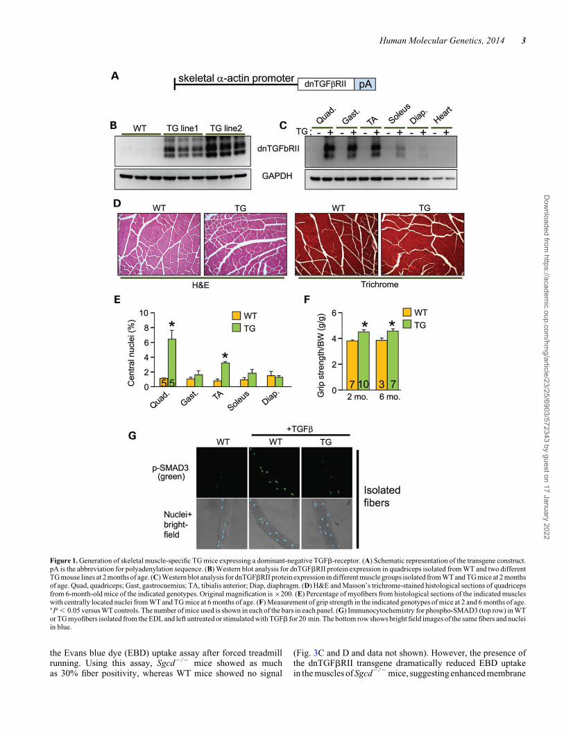
[inhibit p38 mitogen-activated protein kinase (MAPK)] producedan enhanced profile of Mt1-3 gene expression following Zn andZn + TGFb treatment (Fig.6A–C).However, inhibitionofextra-cellular signal-regulated kinases 1/2 (ERK1/2) with a dominant-negative MEK1 adenovirus was ineffective, whereas inhibitionof c-Jun N-terminal kinases 1/2 (JNK1/2) with adenovirus expres-sing dominant-negative mutant proteins was only partially effect-ive (Fig. 6A–C). The dnMKK3/6 adenoviruses also induced Mt1and Mt2 expression at baseline (Fig. 6A–C). Finally, consideringthe antioxidant properties of Mts, we examined whether TGFbaffects reactive oxygen species (ROS) levels in C2C12 cells. Tothis end, equal numbers of myotubes were treated with TGFb orvehicle control and ROS were measured by dihydroethidium(DHE) staining, which showed a significant increase with TGFbtreatment (Fig. 6D and E). Collectively, these results suggestthat TGFb signaling utilizes both canonical and non-canonicalpathways to regulate Mt expression in muscle cells, with an asso-ciated ROS component.
Treatment with Mt mimetics protects skeletal muscleupon injury in vivo
Increased ROS is a signature of MD that can contribute to mem-brane fragility and subsequent myofiber necrosis (1,30,31).Here, we quantified ROS by DHE staining and observed thatskeletal muscle from Sgcd2/2 mice had an increased ROS signa-ture that was significantly reduced by the dnTGFbRII transgene(Fig. 7A and B). Given the prominent induction of Mts asso-ciated with the dnTGFbRII transgene, we treated mice with anMt mimetic, EMTIN B after CTX injury (Fig. 7C) (32). Treat-ment with EMTIN B protected the injured muscles and improvedtheir regenerative capacity (Fig. 7D–G), reproducing many ofthe protective features of the dnTGFbRII transgene. We thenevaluated the efficacy of an EMTIN B-enriched antioxidantmixture (AO) to protect isolated dystrophic fibers upon laserinjury. The EMTIN/AO mix dramatically protected the Sgcd2/2
fibers from rupturing after laser injury suggesting increasedmembrane stability (Fig. 7H and I). Taken together, theseresults reveal a mechanism whereby TGFb-mediated pathologyat the level of the myofiber regulates membrane integrity througha ROS-dependent mechanism.
DISCUSSION
TGFb signaling underlies diverse cellular functions such as pro-liferation, differentiation, cell death and motility (33). The exactbiologic response to TGFb depends on the cell type and thecontext in which the signal is received (33). Although enhancedTGFb activity is associated with MD and inhibition of its signal-ing has been proposed as a therapeutic strategy in this disease(2,34,35), the known mechanisms of action for this cytokineare highly pleiotropic with a number of beneficial effects;hence, a more cell-type selective inhibitory strategy might benecessary. Our results provide the first evidence that myofibersare central mediators of the deleterious effects associated withTGFb signaling in skeletal muscle, both during acute injury ofskeletal muscle and during MD.
Unlike post-mitotic tissues with little regenerative capa-city, such as the heart or the brain, skeletal muscle robustly
6 Human Molecular Genetics, 2014
Dow
nloaded from https://academ
ic.oup.com/hm
g/article/23/25/6903/572343 by guest on 17 January 2022

regenerates after injury owing to the activity of resident satellitecells (26). However, during MD, skeletal muscle regenerationprogressively becomes impaired and excessive TGFb activation
and signaling is thought to be a primary mechanism underlyingthis effect (2). TGFb can inhibit the activity of myoblasts to dif-ferentiate in culture, as well as mediating a progressive fibrotic
Figure 4. Myofiber-specific inhibition of TGFb signaling protects skeletal muscle upon CTX injury. (A) Representative H&E-stained histological sections from CTXinjury to the TA muscle in WT or TG mice 5 and 14 days afterward. Original magnification is ×200. (B) TA muscle weights (MW) normalized to body weight (BW)from the indicated genotypes of mice with vehicle control or CTX injection. (C and D) Histological sections and quantification of eMHC-positive area (green) withinthe greater injury area (dark red) of the TA muscle 5 days after CTX injury. The white outline shows the regenerating area whereas the yellow outline is the entire area ofinjury. Original magnification is ×40. ∗P , 0.05 versus WT control; #P , 0.05 versus CTX-injured WT mice. The number of mice used is shown in the bars of eachpanel.
Human Molecular Genetics, 2014 7
Dow
nloaded from https://academ
ic.oup.com/hm
g/article/23/25/6903/572343 by guest on 17 January 2022

response that eventually restricts the activity of satellite cells toform nascent myofibers (12,13). However, we did not observedifferences in collagen deposition in our dnTGFbRII TG micecrossed to the Sgcd2/2 model of MD, in agreement with an un-changed profile of TGFb signaling in interstitial cells. ExcessiveTGFb signaling also appears to enhance the death of skeletalmuscle precursor cells and myoblasts, possibly through activa-tion of p38 MAPK and/or JNK1/2 (36–39). Our observationthat inhibition of TGFb signaling at the level of the myofiberenhances membrane stability, thus lessening myofiber degener-ation and even death with CTX injury, is consistent with pastobservations of less cell death or enhanced regenerationwhen TGFb signaling is dampened (2,40,41). Preservation of
membrane integrity is also crucial for skeletal muscle regener-ation. Indeed, interaction between integrins on myofiber mem-branes and laminins in the matrix is crucial for proper satellitecell adhesion to the myofiber and for fusion of myoblasts withinjured fibers (42).
Oxidative stress and ROS formation are important contribu-tors to membrane fragility in the pathology of several of theMDs (30,31). Moreover, TGFb signaling, which is enhancedin MD, is known to enhance ROS formation (43–46). Our un-biased expression analysis by RNA-seq revealed the Mts asbeing dramatically upregulated by the dnTGFbRII transgene,although many other candidate genes were also changed in ex-pression suggesting additional mechanisms at play. However,
Figure 5. Altered metallothionein mRNA expression underlies TGFb effects on myofibers. (A–C) Real-time PCR from quadriceps muscle of WT and TG mice formetallothionein 1, 2, 3 (Mt1, Mt2, Mt3) mRNA expression normalized to Rpl7 control mRNA. ∗P , 0.05 versus WT. (D–I) Real-time PCR from C2C12-derivedmyotubes with vehicle control (con) or treated with recombinant TGFb (TGF), recombinant interleukin-6 (IL6) or zinc (Zn) for Mt1, Mt2 and Mt3 mRNA expressionnormalized to Rpl7 control mRNA. ∗P , 0.05 versus control; #P , 0.05 versus Zn or IL6 treatment. The number of mice or biological replicates used is shown in eachof the graphs.
8 Human Molecular Genetics, 2014
Dow
nloaded from https://academ
ic.oup.com/hm
g/article/23/25/6903/572343 by guest on 17 January 2022
Mts appear to favor regeneration in both brain and heart (47–49),and their antioxidant properties appear to be a main determinantin protecting tissue from injury as well as enhancing regenerativeactivity by creating a more favorable environment for stem cells(50,51). Indeed, we observed that TGFb had a direct inhibitoryeffect on both baseline and induced Mt expression and that inhib-ition of TGFb signaling within the myofibers reduced ROSlevels and was protective. We also showed that TGFb treatmentwas sufficient to elevate ROS in C2C12 cells. Treatment of micewith EMTIM B promoted greater recovery after acute injurywith CTX, and an EMTIM B antioxidant cocktail directly pro-tected the degree of muscle membrane injury after acute laserdamage. These results suggest that a properly formulated anti-oxidant therapy might protect MD patients during the courseof their disease. However, clinical trials in humans have thusfar failed to conclusively show efficacy of an antioxidant ap-proach (52,53). These negative findings may be due to, in part,a lack of understanding of the precise mechanism responsiblefor ROS elevation and the ensuing cellular damage in skeletalmuscle or it is possibly due to an ineffective therapeutic regimento fully extinguish ROS in the proper locations where oxidativedamage is most central to the disease.
Our data show that the myofibers are central mediators of thedeleterious aspects of TGFb signaling in muscle. TGFb appearsto enhance the ROS status within skeletal muscle fibers, whichincreases membrane fragility, leading to greater myofiber necro-sis, and also reduced levels of effective satellite cell activity withregeneration. Hence, while global TGFb inhibition may not be adesirable therapeutic strategy for treating human MD, in partowing to adverse systemic side effects as well as owing to inhi-biting some beneficial aspects of TGFb signaling within muscle,our results suggest that targeted inhibition of TGFb at the level ofthe myofibers would be beneficial and protective against dys-trophic disease.
MATERIAL AND METHODS
Ethics statement
All animal procedures and usage were approved by the Institu-tional Animal Care and Use Committee of the Cincinnati Chil-dren’s Hospital Medical Center, protocol 2E11104. No humansubjects were used or human tissue or cells.
Animals
A modified human skeletal a-actin promoter (54) was used tocreate TG mice overexpressing a truncated TGFb type II recep-tor mutant (dnTGFbRII) (20). Two TG lines were generated andanalyzed for experiments. Sgcd– / – mice were described previ-ously (24).
Western blotting, ELISA and hydroxyl-proline assay
Western blot analysis of skeletal muscles homogenates wasperformed using standard procedures. TGFbRII was detectedusing an antibody purchased from Santa Cruz Biotechnology(sc-33929). ELISA assays were performed on muscle homoge-nates using an ELISA kit for TGF-b1 purchased from R&DSystems (SMB100B). Hydroxyproline content was performedas previously described (55).
Fiber isolation and cell culture
Muscle fibers were isolated by incubation of the EDL muscle in0.2% collagenase (Type I, Sigma–Aldrich, C-0130) for 1 h aftercareful removal of the fascia. Following collagenase treatment,muscles were transferred to DMEM (Gibco Invitrogen LifeTechnologies; 11995065) containing 10% horse serum and tritu-rated to release individual fibers. Interstitial cells were obtained
Figure 6. Signals involved in TGFb-mediated inhibition of Mt expression. (A–C) Real-time PCR for Mt1, 2, 3 mRNA expression from C2C12 cells with no treatment(control) or treated with recombinant TGFb (TGF) or zinc (Zn) with Adbgal control or AdSMAD6/7, Ad-dnJNK1/2, Ad-dnMEK1, Ad-dnMKK3/6 adenoviralinfection. ∗P , 0.05 versus Adbgal. Data were normalized to Rpl7 control mRNA. (D and E) Representative images and quantitation of the ROS marker DHE(red) from cultured C2C12 myotubes treated with vehicle (control) or recombinant TGFb. The same numbers of cells are present in each image. Original magnificationis ×200.
Human Molecular Genetics, 2014 9
Dow
nloaded from https://academ
ic.oup.com/hm
g/article/23/25/6903/572343 by guest on 17 January 2022

Figure 7. Treatment with Mt mimetics protects skeletal muscle upon injury. (A and B) Representative histological images and quantitation of the ROS marker DHE(red) from quadriceps sections of the indicated genotypes of mice. Original magnification is ×200. (C) Schematic of the protocol used for CTX injury to the TA alongwith EMTIN B treatment times. (D) Representative H&E-stained histological sections from the TA injured with CTX and treated with vehicle or EMTIN B, 5 dayspost-injury. Original magnification is ×200. (E) MW/BW from the indicated mouse treatment groups. (F and G) Representative immunohistochemical images andquantitation for eMHC expression (green) from CTX-injured TAs treated with vehicle or EMTIN B. (H and I) Time course and representative images of FM1-43 dyeentry into EDL myofibers from Sgcd2/2 mice with or without EMTIM B-enriched antioxidant mix (AO) following laser-induced injury. Eight or more fibers wereanalyzed each. ∗P , 0.05 versus WT or vehicle control; #P , 0.05 versus Sgcd2/2 or CTX-injured vehicle-treated mice. The number of mice used is shown in each ofthe graphs.
10 Human Molecular Genetics, 2014
Dow
nloaded from https://academ
ic.oup.com/hm
g/article/23/25/6903/572343 by guest on 17 January 2022

from the EDL fiber isolation procedure by plating the super-natant derived after trituration and seeding of the fibers. Myo-blasts were obtained by culturing isolated fibers in ECL CellAttachment Matrix (Millipore, 08-110)-coated dishes for 3 daysin F12 medium containing 10% bovine growth serum. Fibers,interstitial cells and myoblasts were treated with 10 ng/ml ofrecombinant TGFb (R&D Systems; 101-b1-010) for 20 minprior to being processed for immunostaining. C2C12 cells weredifferentiated into myotubes in DMEM containing 2% horseserum. Forty-eight hours post-induction of differentiation, cellswere treated with 10 ng/ml of recombinant TGFb or 40 ng/ml re-combinant IL6 (PeproTech; 216-16) for 15 h, and 100 mM ZnSO4
for 6 h. Overexpression of SMAD6, SMAD7, dnJNK1, dnJNK2,dnMKK3, dnMKK6, dnMEK1 or b-galactosidase was perfor-med by recombinant adenoviral infection in differentiatedC2C12 myotubes for 4 h the day before applying specific treat-ments. For ROS quantification, cells were incubated with DHEfor 10 min at 378C, and red fluorescence was visualized immedi-ately afterward.
Histological analysis and immunohistochemistry
Muscles were paraffin-embedded and 5-mm histological sec-tions were cut at the center of the muscle and stained with H&Eor Masson’s trichrome. Immunohistochemistry for eMHC wasperformed on cryosections within optimal cutting temperaturecompound (OCT)-embedded muscles using the antibody BF-G6from Developmental Studies Hybridoma Bank. Antibody forlaminin was purchased from Sigma–Aldrich (L9393). Antibodyfor vimentin (Sigma–Aldrich, V5255) was used to label intersti-tial cells whereas antibody for MyoD (BD Biosciences, 554130)was used to identify myoblasts. For ROS quantification, cryo-sections were incubated with DHE for 10 min at 378C. Satellitecells were visualized in isolated EDL fibers using the antibodiesagainst CD34 (rat anti-mouse CD34 monoclonal antibody;eBioscience, RAM34 14-0341-82) and Pax7 (mouse anti-chickenPax7 monoclonal antibody; Developmental Studies HybridomaBank). Quantitation was performed on fibers fixed in 4% paraf-ormaldehyde for 20 min at room temperature, washed threetimes for 20 min each in PBS, pH 7.4 and processed for immuno-histochemistry. Absence of endothelial contamination was con-firmed by staining with rat anti-mouse CD31 antibody (BDBiosciences; Clone MEC 13.3). Wheat germ agglutinin-TRITCwas also used to show fiber outlines in the Phospho-SMAD3immunohistochemistry experiments; nuclei are shown in bluewith DAPI.
mRNA expression analysis
RNA was extracted from muscles and C2C12 cells using theRNeasy Kit according to manufacturer’s instructions (Qiagen).Quadriceps-derived samples from two separate mice per groupwere submitted for RNAseq (University of Cincinnati sequencingand genome analysis core laboratory). For RNAseq data ana-lysis, P-values were calculated using a negative binomial statis-tical model as implemented in DESeq [Bioconductor version:Release (2.14)], meanwhile false discover rates were used toobtain the adjusted P-values. For qPCR assays, RNA sampleswere retrotranscribed using the High Capacity cDNA ReverseTranscriptionKit (AppliedBiosystems).Selectedgenedifferences
were analyzed by real-time qPCR using SYBR green (AppliedBiosystems). Quantified mRNA expression was normalized toRpl7 and expressed relative to WT or untreated C2C12 controls.
Forced treadmill running and grip strength evaluation
Mice were placed in individual lanes of an electrically drivenfour-lane treadmill (Omni-Pacer LC4/M; Columbus Instru-ments International) at the speed of 6 m/min for 3 min. Thetreadmill measured 19 inches wide by 20 inches in length, witha conveyor belt measuring 3 inches wide by 12 inches inlength. A training regimen was first instituted for 10 min to fa-miliarize the mice with the environment and shock grids (adjust-able from 0 to 2.0 mA). The speed was increased in increments of2 m/min every 3 min to a maximum speed of 22 m/min. Exhaus-tion was assessed as .5 consecutive seconds on the shock gridwithout attempting to reengage the treadmill. Time spent onthe treadmill before exhaustion or time to complete the protocolwas recorded as average maximum time spent in exercise. Thetreadmill regimen was performed downhill. Grip strength testwas performed using the Chatillon model DFIS-2 digital forcegauge, and each animal was assessed 5 times consecutively ona given day.
CTX injury
CTX derived from Naja mossambica (Sigma) was dissolved insterile saline to a final concentration of 10 mM, divided into ali-quots and stored at 2208C. Mouse legs were shaved and cleanedwith alcohol, and then, the TA muscle was injected with 50 ml ofthis CTX solution through a 26-gauge needle. EMTIN B (peptideSAGSCKCKESKSTS) was synthetized by Selleck Chemicals,dissolved in PBS and used intramuscular (50 ng/TA) once aday for 3 consecutive days post-CTX injury. PBS was usedwith the same regimen as vehicle control.
Laser-induced membrane injury assay
FDB muscles were excised from the foot and processed for laser-induced membrane injury assay as previously published method(56). The experiment was performed with and without addingEMTIN B at 10 ng/ml and an antioxidant supplement (A1345,from Sigma–Aldrich) on isolated fibers.
EBD uptake
Mice were injected with EBD (10 mg/ml, 0.1 ml/10 g bodyweight) and subjected to forced treadmill running 12 h later.Mice were then sacrificed, and quadriceps were embedded inOCT and snap-frozen in liquid nitrogen for viewing as previouslydescribed (57).
Statistics
All results are presented as mean+SEM. Statistical analysiswas performed with unpaired two-tailed t-test (for two groups)and one-way ANOVA with Bonferroni correction (for groupsof three or more). P-values of ,0.05 were considered significant.
Human Molecular Genetics, 2014 11
Dow
nloaded from https://academ
ic.oup.com/hm
g/article/23/25/6903/572343 by guest on 17 January 2022

SUPPLEMENTARY MATERIAL
Supplementary Material is available at HMG online.
Conflict of Interest statement. None declared.
FUNDING
This work was supported by grants from the NIH (J.D.M. andE.M.M). J.D.M was also supported by the Howard HughesMedical Institute.
REFERENCES
1. Wallace, G.Q. and McNally, E.M. (2009) Mechanisms of muscledegeneration, regeneration, and repair in the muscular dystrophies. Annu.Rev. Physiol., 71, 37–57.
2. Cohn, R.D., van Erp, C., Habashi, J.P., Soleimani, A.A., Klein, E.C., Lisi,M.T., Gamradt, M., ap Rhys, C.M., Holm, T.M., Loeys, B.L. et al. (2007)Angiotensin II type 1 receptor blockade attenuates TGF-beta-induced failureof muscle regeneration in multiple myopathic states. Nat. Med., 13, 204–210.
3. Merly, F., Lescaudron, L., Rouaud, T., Crossin, F. and Gardahaut, M.F.(1999) Macrophages enhance muscle satellite cell proliferation and delaytheir differentiation. Muscle Nerve, 22, 724–732.
4. Lescaudron, L., Peltekian, E., Fontaine-Perus, J., Paulin, D., Zampieri, M.,Garcia, L. and Parrish, E. (1999) Blood borne macrophages are essential forthe triggering of muscle regeneration following muscle transplant.Neuromuscul. Disord., 9, 72–80.
5. Brunelli, S. and Rovere-Querini, P. (2008) The immune system and therepair of skeletal muscle. Pharmacol. Res., 58, 117–121.
6. Iannaccone, S., Quattrini, A., Smirne, S., Sessa, M., de Rino, F.,Ferini-Strambi, L. and Nemni, R. (1995) Connective tissue proliferation andgrowth factors in animal modelsof Duchenne musculardystrophy.J. Neurol.Sci., 128, 36–44.
7. Murakami, N., McLennan, I.S., Nonaka, I., Koishi, K., Baker, C. andHammond-Tooke, G. (1999) Transforming growth factor-beta2 is elevatedin skeletal muscle disorders. Muscle Nerve, 22, 889–898.
8. Sun, G., Haginoya, K., Dai, H., Chiba, Y., Uematsu, M., Hino-Fukuyo, N.,Onuma, A., Iinuma, K. and Tsuchiya, S. (2009) Intramuscularrenin-angiotensin system is activated in human muscular dystrophy.J. Neurol. Sci., 280, 40–48.
9. Bernasconi, P., Torchiana, E., Confalonieri, P., Brugnoni, R., Barresi, R.,Mora, M., Cornelio, F., Morandi, L. and Mantegazza, R. (1995) Expressionof transforming growth factor-beta 1 in dystrophic patient muscles correlateswith fibrosis. Pathogenetic role of a fibrogenic cytokine. J. Clin. Invest., 96,1137–1144.
10. Brooke, B.S., Habashi, J.P., Judge, D.P., Patel, N., Loeys, B. and Dietz,H.C. 3rd. (2008) Angiotensin II blockade and aortic-root dilation in Marfan’ssyndrome. N. Engl. J. Med., 358, 2787–2795.
11. Andreetta, F., Bernasconi, P., Baggi, F., Ferro, P., Oliva, L., Arnoldi, E.,Cornelio, F., Mantegazza, R. and Confalonieri, P. (2006)Immunomodulation of TGF-beta 1 in mdx mouse inhibits connective tissueproliferation in diaphragm but increases inflammatory response:implications for antifibrotic therapy. J. Neuroimmunol., 175, 77–86.
12. Wynn, T.A. (2007) Common and unique mechanisms regulate fibrosis invarious fibroproliferative diseases. J. Clin. Invest., 117, 524–529.
13. Olson, E.N., Sternberg, E., Hu, J.S., Spizz, G. and Wilcox, C. (1986)Regulation of myogenic differentiation by type beta transforming growthfactor. J. Cell Biol., 103, 1799–1805.
14. Charge, S.B. and Rudnicki, M.A. (2004) Cellular and molecular regulationof muscle regeneration. Physiol. Rev., 84, 209–238.
15. Li, Y., Foster, W., Deasy, B.M., Chan, Y., Prisk, V., Tang, Y., Cummins, J.and Huard, J. (2004) Transforming growth factor-beta1 induces thedifferentiation of myogeniccells intofibroticcells in injured skeletal muscle:a key event in muscle fibrogenesis. Am. J. Pathol., 164, 1007–1019.
16. Allen, R.E. and Boxhorn, L.K. (1987) Inhibition of skeletal muscle satellitecell differentiation by transforming growth factor-beta. J. Cell Physiol., 133,567–572.
17. Kollias, H.D. and McDermott, J.C. (2008) Transforming growthfactor-beta and myostatin signaling in skeletal muscle. J. Appl. Physiol., 104,579–587.
18. Mu, X. and Li, Y. (2011) Conditional TGF-beta1 treatment increases stemcell-like cell population in myoblasts. J. Cell Mol. Med., 15, 679–690.
19. Yamazaki, M., Minota, S., Sakurai, H., Miyazono, K., Yamada, A.,Kanazawa, I. and Kawai, M. (1994) Expression of transforming growthfactor-beta 1 and its relation to endomysial fibrosis in progressive musculardystrophy. Am. J. Pathol., 144, 221–226.
20. Brand, T., MacLellan, W.R. and Schneider, M.D. (1993) Adominant-negative receptor for type beta transforming growth factorscreated by deletion of the kinase domain. J. Biol. Chem., 268, 11500–11503.
21. Yoo, B.M., Yeo, M., Oh, T.Y., Choi, J.H., Kim, W.W., Kim, J.H., Cho, S.W.,Kim, S.J. and Hahm, K.B. (2005) Amelioration of pancreatic fibrosis in micewith defective TGF-beta signaling. Pancreas, 30, e71–e79.
22. Chen, Y.F., Feng, J.A., Li, P., Xing, D., Zhang, Y., Serra, R., Ambalavanan,N., Majid-Hassan, E. and Oparil, S. (2006) Dominant negative mutation ofthe TGF-beta receptor blocks hypoxia-induced pulmonary vascularremodeling. J. Appl. Physiol., 100, 564–571.
23. Durbeej, M. and Campbell, K.P. (2002) Muscular dystrophies involving thedystrophin-glycoprotein complex: an overview of current mouse models.Curr. Opin. Genet. Dev., 12, 349–361.
24. Hack, A.A., Lam, M.Y., Cordier, L., Shoturma, D.I., Ly, C.T., Hadhazy,M.A., Hadhazy, M.R., Sweeney, H.L. and McNally, E.M. (2000)Differential requirement for individual sarcoglycans and dystrophin in theassembly and function of the dystrophin-glycoprotein complex. J. Cell Sci.,113(Pt 14), 2535–2544.
25. Burks, T.N. and Cohn, R.D. (2011) Role of TGF-beta signaling in inheritedand acquired myopathies. Skelet. Muscle, 1, 19.
26. Relaix, F. and Zammit, P.S. (2012) Satellite cells are essential for skeletalmuscle regeneration: the cell on the edge returns centre stage. Development,139, 2845–2856.
27. Leung, Y.K., Pankhurst, M., Dunlop, S.A., Ray, S., Dittmann, J., Eaton,E.D., Palumaa, P., Sillard, R., Chuah, M.I., West, A.K. et al. (2010)Metallothionein induces a regenerative reactive astrocyte phenotype viaJAK/STAT and RhoA signalling pathways. Exp. Neurol., 221, 98–106.
28. Leung, J.Y., Bennett, W.R., Herbert, R.P., West, A.K., Lee, P.R., Wake, H.,Fields, R.D., Chuah, M.I. and Chung, R.S. (2012) Metallothionein promotesregenerative axonal sprouting of dorsal root ganglion neurons after physicalaxotomy. Cell Mol. Life Sci., 69, 809–817.
29. Ruttkay-Nedecky, B., Nejdl, L., Gumulec, J., Zitka, O., Masarik, M.,Eckschlager, T., Stiborova, M., Adam, V. and Kizek, R. (2013) The role ofmetallothionein in oxidative stress. Int. J. Mol. Sci., 14, 6044–6066.
30. Terrill, J.R., Radley-Crabb, H.G., Iwasaki, T., Lemckert, F.A., Arthur, P.G.and Grounds, M.D. (2013) Oxidative stress and pathology in musculardystrophies: focus on protein thiol oxidation and dysferlinopathies. FEBS J.,280, 4149–4164.
31. Jung, C., Martins, A.S., Niggli, E. and Shirokova, N. (2008) Dystrophiccardiomyopathy: amplification of cellular damage by Ca2+ signalling andreactive oxygen species-generating pathways. Cardiovasc. Res., 77,766–773.
32. Ambjorn, M., Asmussen, J.W., Lindstam, M., Gotfryd, K., Jacobsen, C.,Kiselyov, V.V., Moestrup, S.K., Penkowa, M., Bock, E. and Berezin, V.(2008) Metallothionein and a peptide modeled after metallothionein,EmtinB, induce neuronal differentiation and survival through binding toreceptors of the low-density lipoprotein receptor family. J. Neurochem.,104, 21–37.
33. Ikushima, H. and Miyazono, K. (2011) Biology of transforming growthfactor-beta signaling. Curr. Pharm. Biotechnol., 12, 2099–2107.
34. Ceco, E. and McNally, E.M. (2013) Modifying muscular dystrophy throughtransforming growth factor-beta. FEBS J., 280, 4198–4209.
35. Heydemann, A., Ceco, E., Lim, J.E., Hadhazy, M., Ryder, P., Moran, J.L.,Beier, D.R., Palmer, A.A. and McNally, E.M. (2009) LatentTGF-beta-binding protein 4 modifies muscular dystrophy in mice. J. Clin.
Invest., 119, 3703–3712.36. Cencetti, F., Bernacchioni, C., Tonelli, F., Roberts, E., Donati, C. and Bruni,
P. (2013) TGFbeta1 evokes myoblast apoptotic response via a novelsignaling pathway involving S1P4 transactivation upstream of Rho-kinase-2activation. FASEB J., 27, 4532–4546.
37. Li, X., McFarland, D.C. and Velleman, S.G. (2009) Transforming growthfactor-beta1-induced satellite cell apoptosis in chickens is associated withbeta1 integrin-mediated focal adhesion kinase activation. Poult. Sci., 88,1725–1734.
38. Wissing, E.R., Boyer, J.G., Kwong, J.Q., Sargent, M.A., Karch, J., McNally,E.M., Otsu, K. and Molkentin, J.D. (2014) P38alpha MAPK underlies
12 Human Molecular Genetics, 2014
Dow
nloaded from https://academ
ic.oup.com/hm
g/article/23/25/6903/572343 by guest on 17 January 2022

muscular dystrophy and myofiber death through a Bax-dependentmechanism. Hum. Mol. Genet., doi: 10.1093/hmg/ddu270.
39. Sinha-Hikim, I., Braga, M., Shen, R. and Sinha Hikim, A.P. (2007)Involvement of c-Jun NH2-terminal kinase and nitric oxide-mediatedmitochondria-dependent intrinsic pathway signaling in cardiotoxin-inducedmuscle cell death: role of testosterone. Apoptosis, 12, 1965–1978.
40. Zimowska, M., Duchesnay, A., Dragun, P., Oberbek, A., Moraczewski, J.and Martelly, I. (2009) Immunoneutralization of TGFbeta1 improvesskeletal muscle regeneration: effects on myoblast differentiation andglycosaminoglycan content. Int. J. Cell Biol., 2009, 659372.
41. Fakhfakh, R., Lamarre, Y., Skuk, D. and Tremblay, J.P. (2012) Losartanenhances the success of myoblast transplantation. Cell Transplant, 21,139–152.
42. Schwander, M., Leu, M., Stumm, M., Dorchies, O.M., Ruegg, U.T.,Schittny, J. and Muller, U. (2003) Beta1 integrins regulate myoblast fusionand sarcomere assembly. Dev. Cell, 4, 673–685.
43. Sharma, K., Cook, A., Smith, M., Valancius, C. and Inscho, E.W. (2005)TGF-beta impairs renal autoregulation via generation of ROS.Am. J. Physiol. Renal Physiol., 288, F1069–F1077.
44. Hagler, M.A., Hadley, T.M., Zhang, H., Mehra, K., Roos, C.M., Schaff,H.V., Suri, R.M. and Miller, J.D. (2013) TGF-beta signalling and reactiveoxygen species drive fibrosis and matrix remodelling in myxomatous mitralvalves. Cardiovasc. Res., 99, 175–184.
45. Yang, L., Qu, M., Wang, Y., Duan, H., Chen, P., Wang, Y., Shi, W.,Danielson, P. and Zhou, Q. (2013) Trichostatin A inhibits transforminggrowth factor-beta-induced reactive oxygen species accumulation andmyofibroblast differentiation via enhanced NF-E2-related factor2-antioxidant response element signaling. Mol. Pharmacol., 83, 671–680.
46. Hsieh, H.L., Wang, H.H., Wu, W.B., Chu, P.J. and Yang, C.M. (2010)Transforming growth factor-beta1 induces matrix metalloproteinase-9 andcell migration in astrocytes: roles of ROS-dependent ERK- andJNK-NF-kappaB pathways. J. Neuroinflammation, 7, 88.
47. Santos, C.R., Martinho, A., Quintela, T. and Goncalves, I. (2012)Neuroprotective and neuroregenerative properties of metallothioneins.IUBMB Life, 64, 126–135.
48. Ceylan-Isik, A.F., Zhao, P., Zhang, B., Xiao, X., Su, G. and Ren, J. (2010)Cardiac overexpression of metallothionein rescues cardiac contractiledysfunction and endoplasmic reticulum stress but not autophagy in sepsis.J. Mol. Cell. Cardiol., 48, 367–378.
49. Oshima, Y., Fujio, Y., Nakanishi, T., Itoh, N., Yamamoto, Y., Negoro, S.,Tanaka, K., Kishimoto, T., Kawase, I. and Azuma, J. (2005) STAT3mediates cardioprotection against ischemia/reperfusion injury throughmetallothionein induction in the heart. Cardiovasc. Res., 65, 428–435.
50. Lim, K.S., Cha, M.J., Kim, J.K., Park, E.J., Chae, J.W., Rhim, T., Hwang,K.C. and Kim, Y.H. (2013) Protective effects of protein transductiondomain-metallothionein fusion proteins against hypoxia- and oxidativestress-induced apoptosis in an ischemia/reperfusion rat model. J. Control
Release, 169, 306–312.
51. Nielsen, A.E., Bohr, A. and Penkowa, M. (2007) The balance between lifeand death of cells: roles of metallothioneins. Biomark. Insights, 1, 99–111.
52. Malik, V., Rodino-Klapac, L.R. and Mendell, J.R. (2012) Emergingdrugs for Duchenne muscular dystrophy. Expert Opin. Emerg. Drugs, 17,261–277.
53. De Luca, A. (2012) Pre-clinical drug tests in the mdx mouse as a model ofdystrophinopathies: an overview. Acta Myol., 31, 40–47.
54. Tinsley, J.M., Potter, A.C., Phelps, S.R., Fisher, R., Trickett, J.I. and Davies,K.E. (1996) Amelioration of the dystrophic phenotype of mdx mice using atruncated utrophin transgene. Nature, 384, 349–353.
55. Accornero, F., van Berlo, J.H., Benard, M.J., Lorenz, J.N., Carmeliet, P. andMolkentin, J.D. (2011) Placental growth factor regulates cardiacadaptation and hypertrophy through a paracrine mechanism. Circ. Res., 109,272–280.
56. Cai, C., Masumiya, H., Weisleder, N., Matsuda, N., Nishi, M., Hwang, M.,Ko, J.K., Lin, P., Thornton, A., Zhao, X. et al. (2009) MG53 nucleatesassembly of cell membrane repair machinery. Nat. Cell Biol., 11, 56–64.
57. Millay, D.P., Sargent, M.A., Osinska, H., Baines, C.P., Barton, E.R.,Vuagniaux, G., Sweeney, H.L., Robbins, J. and Molkentin, J.D. (2008)Genetic and pharmacologic inhibition of mitochondrial-dependent necrosisattenuates muscular dystrophy. Nat. Med., 14, 442–447.
Human Molecular Genetics, 2014 13
Dow
nloaded from https://academ
ic.oup.com/hm
g/article/23/25/6903/572343 by guest on 17 January 2022