Bcl2 Protects Mice against Fatal Alphavirus Encephalitis
6
Proc. Natl. Acad. Sci. USA Vol. 93, pp. 4810-4815, May 1996 Microbiology Bcl-2 protects mice against fatal alphavirus encephalitis BETH LEVINE*t, JAMES E. GOLDMANt, Hui HuI JIANG*, DIANE E. GRIFFIN§II, AND J. MARIE HARDWICK§¶** Departments of *Medicine and tPathology, Columbia University College of Physicians and Surgeons, New York, NY 10032; and §Department of Molecular Microbiology and Immunology, Johns Hopkins University School of Hygiene and Public Health, and Departments of 1Neurology, ,Medicine, and **Pharmacology and Molecular Sciences, Johns Hopkins University School of Medicine, Baltimore, MD 21287 Communicated by Harold S. Ginsberg, National Institutes of Health, Rockville, MD, January 5, 1996 (received for review October 6, 1995) ABSTRACT Virus-induced apoptosis has been well char- acterized in vitro, but the role of apoptosis in viral pathogen- esis is not well understood. The suicide of a cell in response to viral infection is postulated to be an important host defense for the organism, leading to a reduction in its total viral burden. However, virus-induced death of nonregenerating cells in the central nervous system may be detrimental to the host. Therefore, to investigate the role of apoptosis in the pathogenesis of fatal encephalitis, we constructed a recombi- nant alphavirus chimera that expresses the antiapoptotic gene, bcl-2, in virally infected neural cells. Infection of neo- natal mice with the alphavirus chimera expressing human bcl-2 [Sindbis virus (SIN)/bcl-2] resulted in a significantly lower mortality rate (7.5%) as compared with infection with control chimeric viruses containing a chloramphenicol acetyl- transferase (CAT) reporter gene (SIN/CAT) (78.1%) or bcl-2 containing a premature stop codon (SIN/bcl-2stop) (72.1%) (P < 0.001). Viral titers were reduced 5-fold 1 day after infection and 10-fold 6 days after infection in the brains of SIN/bcl-2-infected mice as compared to SIN/CAT or SIN/bcl- 2stop-infected mice. In situ end labeling to detect apoptotic nuclei demonstrated a reduction in the number of foci of apoptotic cells in the brains of mice infected with SIN/bcl-2 as compared with SIN/bcl-2stop. The reduction in apoptosis was associated with a reduction in the number of foci of cells expressing alphavirus RNA. Thus, the antiapoptotic gene, bcl-2, suppresses viral replication and protects against a lethal viral disease, suggesting an interaction between cellular ge- netic control of viral replication and cell death. The relationship between a virus, its cellular target, and the host organism represents a complex, and often antagonistic, struggle for mutual survival. As obligate intracellular organ- isms, viruses have evolved strategies to infect, but not kill, their target cells. Such strategies may include encoding gene prod- ucts that either directly inhibit cell death pathways [e.g., adenovirus E1B 19K (1, 2), baculovirus p35 and OpIAP (3-5), cowpox CrmA (6, 7), and Epstein-Barr virus BHRF1 (8)] or that upregulate the expression of cellular "anti-death" genes [e.g., Epstein-Barr virus LMP (9)]. To promote survival of the host organism, cells may be motivated to commit "altruistic" suicide in response to viral infections. In support of this theory, baculovirus mutants that lack functional p35, a gene that inhibits cell death, replicate to reduced levels in insect cell lines and have attenuated virulence in insect larvae (10). However, depending upon the dispensability of the virus-infected cell, cellular suicide may be counterproductive not only for the virus but also for the host. Thus, virus-induced cell death may represent either an adaptive antiviral host defense strategy or, in the case of infections of vital, terminally differentiated cellular targets, a harmful event leading to host morbidity and mortality. Many viruses (11-23) and viral gene products (24-26) have been shown to kill cells by inducing programmed cell death, a genetically encoded suicide program that is important in the removal of superfluous or harmful cells from multicellular organisms. Programmed cell death occurs in a variety of physiologic conditions involving tissue homeostasis (reviewed in refs. 27 and 28) and often involves characteristic changes in cell morphology termed apoptosis. Several genes have been identified that function as either inducers or repressors of programmed cell death pathways. Among these is bcl-2, a protooncogene that was discovered by virtue of its involvement in t(14;18) chromosomal translocations in non-Hodgkins B- cell lymphomas (29). bcl-2 delays or blocks apoptosis induced by a wide variety of stimuli (reviewed in ref. 30) including at least two different RNA viruses, Sindbis (SIN; refs. 14 and 15) and influenza (19), indicating that it regulates a distal step in a final common pathway for apoptotic cell death. However, the mechanisms by which bcl-2 protects against apoptosis induced by viruses or other stimuli are poorly understood. Although virus-induced apoptosis and its prevention by bcl-2 have been described in vitro, very little is known about the implications of these observations for disease pathogenesis. To better understand the interrelationship between virus-induced apoptosis, antiapoptotic gene expression, and central nervous system (CNS) viral pathogenesis, we used SIN encephalitis as a model system. SIN, the prototype member of the alphavirus family, is a positive strand RNA virus that replicates predom- inantly in neurons (31, 32), produces fatal encephalitis in neonatal mice (33), and has been previously shown to induce apoptosis both in vitro (14, 15) and in vivo (23). In addition, SIN can be used as a vector to express high levels of heterologous genes in virally infected cells (reviewed in ref. 34). Therefore, to examine the effect of antiapoptotic gene expression on the natural history of fatal SIN encephalitis, we constructed a recombinant SIN chimera that expresses bcl-2 in virally in- fected neural cells. Our findings demonstrate that bcl-2 de- creases both SIN gene expression and SIN-induced apoptosis in the CNS, resulting in protection against lethal encephalitis. MATERIALS AND METHODS Plasmid Constructions. The neurovirulent SIN clone TE12 (35) was genetically modified to contain a duplicate sub- genomic mRNA promoter and a unique BstEII cloning site downstream of the SIN structural genes. A fragment from the double subgenomic clone TZJSINC (36) (gift of Henry Huang, Washington University School of Medicine) spanning from the BsiWI site in the El gene to the last untranslated nucleotide after the double subgenomic promoter was amplified by PCR, incorporating a BstEII restriction site into the downstream primer. A fragment from clone TE12 spanning the region from the beginning of the SIN 3' nontranslated region to the vector Abbreviations: SIN, Sindbis virus; ISEL, in situ end labeling; H&E, hematoxylin and eosin; CAT, chloramphenicol acetyltransferase; CNS, central nervous system; PFU, plaque-forming units. tTo whom reprint requests should be addressed. 4810 The publication costs of this article were defrayed in part by page charge payment. This article must therefore be hereby marked "advertisement" in accordance with 18 U.S.C. §1734 solely to indicate this fact.
Transcript of Bcl2 Protects Mice against Fatal Alphavirus Encephalitis

Proc. Natl. Acad. Sci. USAVol. 93, pp. 4810-4815, May 1996Microbiology
Bcl-2 protects mice against fatal alphavirus encephalitisBETH LEVINE*t, JAMES E. GOLDMANt, Hui HuI JIANG*, DIANE E. GRIFFIN§II, AND J. MARIE HARDWICK§¶**Departments of *Medicine and tPathology, Columbia University College of Physicians and Surgeons, New York, NY 10032; and §Department of MolecularMicrobiology and Immunology, Johns Hopkins University School of Hygiene and Public Health, and Departments of 1Neurology, ,Medicine, and**Pharmacology and Molecular Sciences, Johns Hopkins University School of Medicine, Baltimore, MD 21287
Communicated by Harold S. Ginsberg, National Institutes of Health, Rockville, MD, January 5, 1996 (received for review October 6, 1995)
ABSTRACT Virus-induced apoptosis has been well char-acterized in vitro, but the role of apoptosis in viral pathogen-esis is not well understood. The suicide of a cell in response toviral infection is postulated to be an important host defensefor the organism, leading to a reduction in its total viralburden. However, virus-induced death of nonregeneratingcells in the central nervous system may be detrimental to thehost. Therefore, to investigate the role of apoptosis in thepathogenesis of fatal encephalitis, we constructed a recombi-nant alphavirus chimera that expresses the antiapoptoticgene, bcl-2, in virally infected neural cells. Infection of neo-natal mice with the alphavirus chimera expressing humanbcl-2 [Sindbis virus (SIN)/bcl-2] resulted in a significantlylower mortality rate (7.5%) as compared with infection withcontrol chimeric viruses containing a chloramphenicol acetyl-transferase (CAT) reporter gene (SIN/CAT) (78.1%) or bcl-2containing a premature stop codon (SIN/bcl-2stop) (72.1%)(P < 0.001). Viral titers were reduced 5-fold 1 day afterinfection and 10-fold 6 days after infection in the brains ofSIN/bcl-2-infected mice as compared to SIN/CAT or SIN/bcl-2stop-infected mice. In situ end labeling to detect apoptoticnuclei demonstrated a reduction in the number of foci ofapoptotic cells in the brains of mice infected with SIN/bcl-2 ascompared with SIN/bcl-2stop. The reduction in apoptosis wasassociated with a reduction in the number of foci of cellsexpressing alphavirus RNA. Thus, the antiapoptotic gene,bcl-2, suppresses viral replication and protects against a lethalviral disease, suggesting an interaction between cellular ge-netic control of viral replication and cell death.
The relationship between a virus, its cellular target, and thehost organism represents a complex, and often antagonistic,struggle for mutual survival. As obligate intracellular organ-isms, viruses have evolved strategies to infect, but not kill, theirtarget cells. Such strategies may include encoding gene prod-ucts that either directly inhibit cell death pathways [e.g.,adenovirus E1B 19K (1, 2), baculovirus p35 and OpIAP (3-5),cowpox CrmA (6, 7), and Epstein-Barr virus BHRF1 (8)] orthat upregulate the expression of cellular "anti-death" genes[e.g., Epstein-Barr virus LMP (9)]. To promote survival of thehost organism, cells may be motivated to commit "altruistic"suicide in response to viral infections. In support of this theory,baculovirus mutants that lack functional p35, a gene thatinhibits cell death, replicate to reduced levels in insect cell linesand have attenuated virulence in insect larvae (10). However,depending upon the dispensability of the virus-infected cell,cellular suicide may be counterproductive not only for the virusbut also for the host. Thus, virus-induced cell death mayrepresent either an adaptive antiviral host defense strategy or,in the case of infections of vital, terminally differentiatedcellular targets, a harmful event leading to host morbidity andmortality.
Many viruses (11-23) and viral gene products (24-26) havebeen shown to kill cells by inducing programmed cell death, agenetically encoded suicide program that is important in theremoval of superfluous or harmful cells from multicellularorganisms. Programmed cell death occurs in a variety ofphysiologic conditions involving tissue homeostasis (reviewedin refs. 27 and 28) and often involves characteristic changes incell morphology termed apoptosis. Several genes have beenidentified that function as either inducers or repressors ofprogrammed cell death pathways. Among these is bcl-2, aprotooncogene that was discovered by virtue of its involvementin t(14;18) chromosomal translocations in non-Hodgkins B-cell lymphomas (29). bcl-2 delays or blocks apoptosis inducedby a wide variety of stimuli (reviewed in ref. 30) including atleast two different RNA viruses, Sindbis (SIN; refs. 14 and 15)and influenza (19), indicating that it regulates a distal step ina final common pathway for apoptotic cell death. However, themechanisms by which bcl-2 protects against apoptosis inducedby viruses or other stimuli are poorly understood.Although virus-induced apoptosis and its prevention by
bcl-2 have been described in vitro, very little is known about theimplications of these observations for disease pathogenesis. Tobetter understand the interrelationship between virus-inducedapoptosis, antiapoptotic gene expression, and central nervoussystem (CNS) viral pathogenesis, we used SIN encephalitis asa model system. SIN, the prototype member of the alphavirusfamily, is a positive strand RNA virus that replicates predom-inantly in neurons (31, 32), produces fatal encephalitis inneonatal mice (33), and has been previously shown to induceapoptosis both in vitro (14, 15) and in vivo (23). In addition, SINcan be used as a vector to express high levels of heterologousgenes in virally infected cells (reviewed in ref. 34). Therefore,to examine the effect of antiapoptotic gene expression on thenatural history of fatal SIN encephalitis, we constructed arecombinant SIN chimera that expresses bcl-2 in virally in-fected neural cells. Our findings demonstrate that bcl-2 de-creases both SIN gene expression and SIN-induced apoptosisin the CNS, resulting in protection against lethal encephalitis.
MATERIALS AND METHODSPlasmid Constructions. The neurovirulent SIN clone TE12
(35) was genetically modified to contain a duplicate sub-genomic mRNA promoter and a unique BstEII cloning sitedownstream of the SIN structural genes. A fragment from thedouble subgenomic clone TZJSINC (36) (gift of Henry Huang,Washington University School of Medicine) spanning from theBsiWI site in the El gene to the last untranslated nucleotideafter the double subgenomic promoter was amplified by PCR,incorporating a BstEII restriction site into the downstreamprimer. A fragment from clone TE12 spanning the region fromthe beginning of the SIN 3' nontranslated region to the vector
Abbreviations: SIN, Sindbis virus; ISEL, in situ end labeling; H&E,hematoxylin and eosin; CAT, chloramphenicol acetyltransferase;CNS, central nervous system; PFU, plaque-forming units.tTo whom reprint requests should be addressed.
4810
The publication costs of this article were defrayed in part by page chargepayment. This article must therefore be hereby marked "advertisement" inaccordance with 18 U.S.C. §1734 solely to indicate this fact.

Proc. Natl. Acad. Sci. USA 93 (1996) 4811
XhoI site was amplified by PCR, incorporating a BstEII siteinto the upstream primer. The BsiWI-BstEII TZJSINC andBstEII-XhoI TE12 fragments were ligated and inserted intothe BsiWI-XhoI sites of clone TE12 to construct a doublesubgenomic version of TE12, referred to as dsTE12. TheBsiWI-PvuI fragment from dsTEl2 was then ligated into theBsiWI-PvuI sites of SIN clone 633 (gift of Richard Kuhn,Purdue University) to generate double subgenomic 633 ords633.To construct SIN/bcl-2, a 798-bp fragment containing the
open reading frame of human bcl-2 was amplified by PCR fromthe template, pZIP/Bcl-2 (37), adding BstEII restriction sitesto the upstream and downstream primer, and cloned into theBstEII site of ds633. The correct sequence of the bcl-2 insertwas confirmed by the dideoxy chain termination sequencingmethod of Sanger et al. (38). To construct SIN/chloramphen-icol acetyltransferase (CAT), a 798-bp fragment containingthe open reading frame of CATwas amplified by PCR from thetemplate, pSV2/CAT, adding BstEII restriction sites to theupstream and downstream primer, and cloned into the BstEIIsite of ds633. SIN/bcl-2stop was constructed by inserting apolylinker containing a termination codon into the SmaI siteat nucleotide position 118 of the bcl-2 open reading frame.
Transcriptions and Transfections. 5'-Capped RNA tran-scripts were synthesized from cDNA clones linearized withXhoI and transcribed in vitro with SP6 DNA-dependent RNApolymerase at 38°C for 60 min. Baby hamster kidney (BHK)-21cells in 35-mm dishes (-5 x i05 cells) were transfected withRNA transcripts (m200 ng) mixed with 6 gg of Lipofectin(GIBCO/BRL) according to the manufacturer's instructions.At 24 h after transfection, virus particle-containing superna-tants of transfected cell monolayers were collected, frozen inaliquots at 70°C, and used for all subsequent experiments.Virus Titrations. Virus titers (typically 5 x 10⁷ to 1 X 10⁸
plaque-forming units (PFU)/ml) of stock viruses were deter-mined by plaque assay titration on BHK-21 cells. Because ofthe possibility that bcl-2 might protect against virus-inducedcell death and interfere with plaque formation, all stockchimeric viruses were also titered by immunofluorescence. Forimmunofluorescence titration, the plaque assay was modifiedin the following manner. Twenty-four hours after virus infec-tion, the agar overlay was removed, and cells were fixed with100% ethanol at -20°C for 20 min and stained with amonoclonal anti-SIN E2 antibody SV50 (1:50) (39) and fluo-rescein isothiocyanate-conjugated horse anti-mouse antibody(1:100) (Vector Laboratories). The number of immunofluo-rescent foci of cells per ml of virus was calculated.Western Blot Detection of Bcl-2. BHK-21 cells (5 x 10⁵)
were infected with SIN/CAT, SIN/bcl-2, and SIN/bcl-2stop ata multiplicity of infection of 1. Eighteen hours after infection,cell lysates were prepared, subjected to SDS/12% PAGE, andtransferred to nitrocellulose using the protocol described in ref.40. The nitrocellulose membrane was incubated for 1 h with amouse monoclonal anti-human Bcl-2 antibody (1:1000) (Dako),followed by 1 h with horseradish peroxidase-conjugated horseanti-mouse IgG (Amersham). The positive reaction was visual-ized by enhanced chemiluminescence according to the manufac-turer's instructions (Amersham).Animal Experiments. One-day-old litters of CD1 mice were
inoculated intracerebrally into the right cerebral hemispherewith 5000 PFU of SIN/CAT, SIN/bcl-2stop, and SIN/bcl-2 in0.03 ml of Hanks' balanced salt solution. For mortality exper-iments, five separate litters were inoculated with each virus,and mortality was determined by daily observation of the micefor 3 weeks after infection. For virus titration experiments, sixmice per experimental group were sacrificed at days 1, 2, 3, 6,and 10 after inoculation. Brains were dissected and stored at-70°C, and freeze-thawed tissues were used to prepare 10%homogenates in Hanks' balanced salt solution for plaque assaytitration on BHK-21 cells or for use in CAT assays. For
histopathological studies, mice were perfused intracardiallywith 3% paraformaldehyde before brain dissection on days 1,3, and 6 after infection.
Histopathology. Paraformaldehyde-perfused mouse brainswere embedded in paraffin and a series of 4-,um parasagittalsections were cut at the level of the olfactory bulb, extendingcaudally from the bulb to the cerebellum and medulla. Foreach brain, sequential sections were stained by hematoxylinand eosin (H&E), in situ end labeling (ISEL), in situ hybrid-ization, and immunoperoxidase. Immunoperoxidase stainingfor human Bcl-2 and SIN proteins was performed using amouse monoclonal anti-human bcl-2 (1:10) (Dako) and apolyclonal rabbit anti-SIN antibody (41), respectively, and theavidin-biotin peroxidase method (Vectastain ABC Kit, Vec-tor Laboratories) according to the manufacturer's instructions.ISEL staining to detect apoptotic nuclei was performed usingthe protocol described by Santarosa et al. (42). In situ hybrid-ization to detect positive strand SIN RNA was performed asdescribed (41) except for the substitution of digoxigenin-labeled oligonucleotide probes complementary to sequencesin the E2 (nucleotides 8893-8912) and El (nucleotides 10509-10528) envelope glycoproteins of SIN. Probes were preparedby incubating each oligonucleotide (5 ,uM) with 25 units ofterminal transferase, 5 mM cobalt chloride, 0.5mM dATP, and1 nM digoxigenin-11-dUTP. Hybridization products were vi-sualized using the immunodetection protocol described in theDig DNA labeling and detection kit (Boehringer Mannheim).H&E-, immunoperoxidase-, ISEL-, and in situ hybridization-stained sections were reviewed by a neuropathologist who wasblinded to the experimental treatment group.CAT Assays. CAT assays were performed according to a
standard protocol (43) using 40 mg of protein extract fromeach mouse brain.
RESULTSConstruction of Chimeric Alphaviruses. To investigate the
role of apoptosis in the pathogenesis of SIN encephalitis, weconstructed a chimeric SIN that expresses the human bcl-2protooncogene in virally infected cells (Fig. 1). We cloned a798-bp fragment containing the open reading frame of humanbcl-2 into a double subgenomic SIN expression vector toconstruct the full-length, nondefective, infectious clone, SIN/bcl-2. We used the background SIN strain 633 because it isneurovirulent in suckling mice, but susceptible to bcl-2-mediated protection against virus-induced apoptosis in vitro(16). The bcl-2 insert was positioned between a duplicatedinternal promoter located downstream of the SIN structuralgenes and upstream of the SIN 3' nontranslated region.Although the presence of the heterologous gene insert andduplicated subgenomic promoter alters the efficiency of SINreplication, the basic replication strategy of the double sub-genomic chimeric construct is similar to that ofwild-type virus.
A CAP j S e ji tNStrUCtUralGenes I AAAn
SG
B CAP SIN Replicase turalSGructuralGenes 4 3 AAA n
SG SG
C CAP SIN Replicase AAA n
SG SG
D CAP IN Replicase al Genes bcll2stop AAAAn
SG SG
replication promoter CD encapsidation signal jubgenomic promoter
SG
FIG. 1. Diagram of wild-type SIN (A) and recombinant SINchimeras SIN/bcl-2 (B), SIN/CAT (C), and SIN/bcl-2stop (D).
Microbiology: Levine et al.

Proc. Natl. Acad. Sci. USA 93 (1996)
Therefore, this type of chimeric construct can be used to studythe effect of bcl-2 expression on the natural history of SINinfection.To control for the effects of increased genome size and the
presence of the foreign gene itself (independent of proteinproduction) on neurovirulence, we constructed two controlchimeric viral cDNA clones, one containing a 798-bp fragmentencoding the CAT reporter gene, SIN/CAT, and one contain-ing a premature termination codon in the open reading frameof bcl-2, SIN/bcl-2stop. The cDNA clones were used astemplates for in vitro transcription, and infectious RNA wastransfected into BHK cells to yield infectious virus stocks forSIN/bcl-2, SIN/CAT, and SIN/bcl-2stop. Virus stock titerswere determined by plaque assay titration (range 107-108PFU/ml) and immunofluorescence focus assay titration(range 106_107 foci/ml). A 10-fold reduction in titer deter-mined by immunofluorescence focus assay versus plaqueassay was observed for each of the three chimeric viruses,most likely reflecting the decreased sensitivity of the immu-nofluorescence assay.
1 2 3A
d -26 kd
C 40
Chimeric Alphaviruses Express Heterologous Genes inMouse Brain. To detect expression of human Bcl-2 by SIN/bcl-2 in vitro, we performed immunoblot analyses of cell lysatesprepared from BHK cells 18 h after infection with SIN/bcl-2,SIN/CAT, and SIN/bcl-2stop. The 26-kDa human Bcl-2 pro-tein was detected in BHK cells infected with SIN/bcl-2, but notin cells infected with SIN/CAT or SIN/bcl-2stop (Fig. 24).After confirming that SIN/bcl-2 expresses human Bcl-2, weinfected 1-day-old CD1 mice intracerebrally with 5000 PFU ofSIN/bcl-2, SIN/CAT, and SIN/bcl-2stop. Human Bcl-2 pro-tein expression in vivo was detected by immunoperoxidasestaining of mouse brain sections from mice 1, 3, and 6 daysafter infection. Fig. 2B shows a representative field of neuronsin the central gray matter, which demonstrates human Bcl-2immunoreactivity. No human Bcl-2 immunoreactivity wasobserved in brains from uninfected control mice or miceinfected with SIN/CAT or SIN/bcl-2stop. Bcl-2 expression wasseen in the same regions of the brain that were positive for SINRNA and SIN protein (see below for details). The major celltype expressing Bcl-2 morphologically appeared to be neurons,with the notable exception that numerous progenitor cells inthe subventricular zone and immature cells in the corticalwhite matter also demonstrated immunoreactivity.To further evaluate the use of chimeric SIN constructs to
express heterologous proteins in virally infected neurons inmouse brain, we measured levels of expression of the CATreporter gene and analyzed the stability of the bcl-2 insertduring viral replication in vivo. Fig. 2C shows the results of aCAT assay performed on mouse brain homogenates 1, 2, 3, 6,and 10 days after infection with SIN/CAT. The temporalpattern of CAT expression is closely correlated with infectiousvirus production in mice infected with SIN/CAT (see Fig. 4),suggesting that CAT is expressed in vivo throughout the entirecourse of SIN infection.To assess the stability of the bcl-2 insert during viral replication
in vivo, we performed a double immunofluorescence focus assayto detect bcl-2 and SIN protein expression in Vero cells infectedwith brain homogenates obtained 1, 2,3, and 6 days after infectionwith SIN/bcl-2. The percentage of SIN-labeled immunofluores-cent foci that also were immunoreactive for Bcl-2 was deter-mined. Nearly 100% of all SIN-labeled immunofluorescent fociwere immunoreactive for Bcl-2 in Vero cells infected with brainhomogenates from 1, 2, 3, and 6 days after infection. Takentogether, these studies indicate that the heterologous gene insertremains stable in the SIN genome and is stably expressed for the6-day duration during which SIN productively replicates in suck-ling mouse brain.
30
0cocu
a)
10NU1
20
10
00 1 2 3 4 5 6 7
days after infection
8 9 10
FIG. 2. Expression of heterologous genes by recombinant SINchimeras. (A) Western blot detection of human Bcl-2 protein expres-sion in BHK cells infected with SIN/CAT (lane 1), SIN/bcl-2stop (lane2), or SIN/bcl-2 (lane 3). (B) Immunoperoxidase staining of neuronsexpressing human Bcl-2 (arrowheads) in central gray matter of mousebrain 3 days after infection. (x 150.) (C) CAT activity in 40-mg extractsfrom mouse brains infected with SIN/CAT. Each data point repre-sents mean value ± SEM of three mouse brains.
100'90
80'
70'
c 60'
e 50'(a 40'
o-0- 30'20'10'
0 5 10 15 20
days after infectionFIG. 3. Survival curve of mice infected with SIN chimeras. Data
represent combined survival probabilities for five independent litters.
4812 Microbiology: Levine et al.

Proc. Natl. Acad. Sci. USA 93 (1996) 4813
5
o 4
o 3
2
00 1 2 3 4 5 6 7 8 9 10
days after infectionFIG. 4. Viral growth of SIN chimeras in mouse brain. Each data
point represents geometric mean viral titer ± SEM of six mouse brains.
bcl-2 Expression Protects Mice Against Lethal AlphavirusEncephalitis. After demonstrating that the bcl-2 insert is ex-pressed in mouse brain and remains stable in the SIN/bcl-2chimera during replication in vivo, we investigated the effect ofbcl-2 expression on the natural history of fatal SIN encephalitis.We infected 1-day-old CD1 litters intracerebrally with 5000 PFUof SIN/bcl-2, SIN/CAT, and SIN/bcl-2stop and measured mor-tality. Mortality after infection with the control chimeric viruses,SIN/bcl-2stop and SIN/CAT, was less (72.1% and 78.1%, re-spectively) than mortality after infection with the parental strain633 (100%), indicating that the presence of a double subgenomicpromoter and heterologous gene insert resulted in neuroattenu-ation. However, compared with the control chimeric viruses,which resulted in mortality rates of 72.1% (SIN/bcl-2stop) and78.1% (SIN/CAT), the mortality of mice infected with SIN/bcl-2was significantly reduced (7.5%) (P <0.001 by life-table analysis)(Fig. 3). These data demonstrate that bcl-2 expression in virallyinfected neural cells results in marked protection against fatal SINencephalitis.
bcl-2 Reduces SIN Replication in Mouse Brain. To examinewhether bcl-2 affects SIN replication in vivo, we measured brainviral titers. Mean viral titers were reduced in SIN/bcl-2-infectedmice compared with SIN/bcl-2stop and SIN/CAT-infected miceby 5-fold at 1, 2, and 3 days after infection, and by 10-fold at 6 daysafter infection (Fig. 4); these differences were statistically signif-icant (P = 0.084 for day 1; P < 0.007 for day 2; P = 0.016 for day3; and P = 0.009 for day 6; Kruskal-Wallis test).To further study the effect of bcl-2 on SIN infection, we
examined brain sections by in situ hybridization (to detect SINRNA) and immunohistochemistry (to detect SIN protein). SINRNA-positive and SIN protein-positive cells were not distrib-uted randomly throughout the CNS, but were clustered incertain areas, notably the subventricular zone, posterior neo-cortex, colliculus, hippocampus, striatum, and olfactory bulb,and less commonly in the thalamus, hypothalamus, central graymatter, and cerebellum (see, for example, Fig. S C and D). Nodifferences were found with respect to the anatomic localiza-tion of SIN RNA or protein in mice infected with SIN/bcl-2and SIN/bcl-2stop. The number of virus-infected foci (definedas cluster of SIN RNA-positive cells), however, did differbetween mice infected with the two viruses. There were morevirus-infected foci in SIN/bcl-2stop-infected mice than inSIN/bcl-2-infected mice at 1, 3, and 6 days after infection(Table 1). Furthermore, fewer virus-positive cells per focuswere observed in SIN/bcl-2-infected mice than in SIN/bcl-2stop-infected mice (Fig. S C and D; Fig. 6 C and D). Theseobservations, together with the reduced viral titers in SIN/
A,@ tx ^*tij.^. *, tj~P..A~ ~ ~ r At
...... £' ....... .' ,
4:' ti;' ISI'P' Cl 1
- r TR eMt- \
lo MLs-."X.,<Jl
'4: f * s
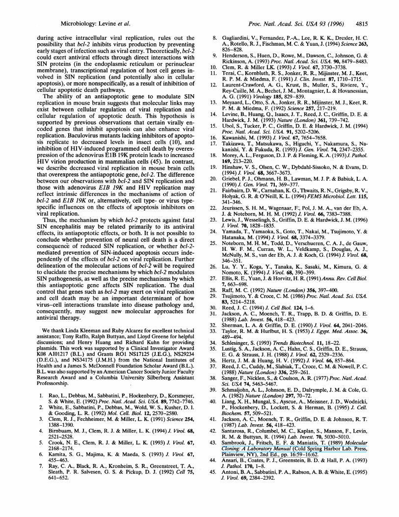
FIG. 5. Photomicrographs of mouse brains 3 days after infectionwith SIN/bcl-2 and SIN/bcl-2stop. Example of SIN/bcl-2stop-infectedfocus in the posterior neocortex with extensive cell death (A, C, andE) and SIN/bcl-2-infected focus in the posterior neocortex withminimal cell death (B, D, and F). (A and B) H&E-stained sections;arrowheads in A denote pyknotic nuclei. (X500.) (C and D) In situhybridization detection of SIN RNA. (x50.) (E and F) ISEL detectionof apoptotic nuclei. (XSO.)
bcl-2-infected mice, suggest that bcl-2 suppresses viral repli-cation in mouse brain.
bcl-2 Reduces SIN-Induced Cell Death in Mouse Brain. Wethen assessed the extent of cell death present in each virus-infected focus by looking for pyknotic nuclei by H&E stainingand for ISEL-positive cells. [ISEL detects chromosomal inter-nucleosomal cleavage that characteristically occurs in cellsdying by apoptosis (42, 44)]. The extent of cell death presentin each infectious focus was classified by a blinded observer asabsent, minimal, moderate, or extensive (see Table 1).
bcl-2 expression in mouse brain was associated with amarked reduction in SIN-induced cell death. The number ofvirus-infected foci demonstrating moderate or extensive celldeath was reduced in mice infected with SIN/bcl-2 as com-pared with SIN/bcl-2stop at all times (1, 3, and 6 days afterinfection), and no virus-infected foci with extensive cell deathwere detected at any time in any mice infected with SIN/bcl-2(Table 1). The extent of cell death assessed by numbers ofpyknotic nuclei was the same as that assessed by numbers ofISEL-positive cells. In addition to apoptotic cell death, miceinfected with SIN/bcl-2stop, but not SIN/bcl-2, also showedsevere tissue necrosis, inflammation, and vascular hyperplasiaby 6 days after infection (Fig. 6 A and B).
Representative photomicrographs illustrating the data inTable 1 are shown in Fig. 5 and Fig. 6. In Fig. 5, the posteriorneocortex of a brain from a SIN/bcl-2stop-infected mouse 3days after infection shows many degenerating cells (Fig. SA)and several foci of SIN RNA-positive (Fig. SC) and ISEL-positive (Fig. SE) cells. In contrast, the same region from amouse infected with SIN/bcl-2 shows only rare degeneratingcells (Fig. SB) and fewer SIN RNA-positive (Fig. SD) and
Microbiology: Levine et al.

Proc. Natl. Acad. Sci. USA 93 (1996)
Table 1. Number of virus-infected foci displaying apoptotic cell death in SIN-infected mouse brains
Days Total no.No. infected foci with absent, minimal moderate, or
Dayse Tofctal nextensive cell deathtafter Infected _ _ _ _ _ _ _ _ _ _ _ _ _ _ _ _ _ _ _ _ _ _ _ _ _
Virus infection foci* Absent Minimal Moderate Extensive
SIN/bcl-2 1 13 2 11 0 0SIN/bcl-2stop 1 17 6 8 2 1
SIN/bcl-2 3 12 5 4 3 0SIN/bcl-2stop 3 29 7 7 10 5
SIN/bcl-2 6 9 3 2 4 0SIN/bcl-2stop 6 28 1 2 9 16
Para-sagittal brain sections from five mice were examined for each virus group per time point; data shownrepresents combined total of infected foci for five mice per virus group per time point.*Focus defined as group of SIN RNA-positive cells.tExtent of cell death per virus-infected focus classified on basis of number of pyknotic nuclei observed on H&Esections and number of apoptotic nuclei observed on ISEL sections (e.g., minimal cell death refers to one totwo pyknotic nuclei and one to two ISEL-positive cells per X20 magnification field; extensive cell death refersto many pyknotic nuclei and ISEL-positive cells per X20 magnification field).
ISEL-positive (Fig. SF) cells. In Fig. 6, the colliculus from amouse infected with SIN/bcl-2stop 6 days after infection showsnumerous pyknotic nuclei and vascular hyperplasia (Fig. 6A)and many ISEL-positive cells (6E), compared with the col-liculus from a SIN/bcl-2-infected mouse, which has no pathol-ogy (Fig. 6B), or ISEL-positive cells (Fig. 6F).
DISCUSSIONOur findings indicate that bcl-2 can prevent virus-inducedneural cell death and fatal disease. Using recombinant SINchimeric constructs, we demonstrated that neonatal miceinfected with SIN/bcl-2, as compared with mice infected withSIN/bcl-2stop, have less neural cell apoptosis and a signifi-cantly reduced mortality rate. These observations extendprevious observations that bcl-2 prevents lytic SIN replicationin vitro (14, 15) and provide what is to our knowledge the firstevidence of a beneficial effect of an antiapoptotic gene on viralinfection in vivo. Although many viruses have been shown toinduce apoptosis in vitro (11-20), only a few examples ofvirus-associated apoptosis in vivo have been reported (21-23),and the pathogenetic importance of these observations re-mains uncertain. The ability of an antiapoptotic gene to exerta beneficial effect on the natural history of SIN encephalitissuggests that apoptosis may play an important role in thepathogenesis of fatal CNS viral infections.However, our findings suggest that the effects of bcl-2 on
SIN-induced mortality may be related to its effects on viralreplication rather than its effects on apoptosis. In this study, weobserved reduced brain viral titers, fewer CNS foci of infec-tion, and fewer virus-positive cells per infectious focus in miceinfected with SIN/bcl-2 as compared with mice infected withSIN/bcl-2stop. Although we did not directly investigate therelationship between viral replication and SIN-induced deathin individual cells, several observations suggest that bcl-2-mediated prevention of SIN-induced neural cell death isassociated with suppression of viral replication. The reductionin the number of virus-infected foci with moderate or extensivecell death in SIN/bcl-2-infected mice was associated with areduction in the total number of virus-infected foci. In addi-tion, the ratio of ISEL-positive cells to SIN RNA-positive cellsappeared similar in the brains of mice infected with SIN/bcl-2and SIN/bcl-2stop, but the brains of mice infected withSIN/bcl-2 had fewer SIN RNA-positive cells and, correspond-ingly, fewer ISEL-positive cells. Furthermore, apoptosis inSIN/bcl-2-infected mice occurred primarily in regions of thebrain that had higher levels of SIN protein expression (asdefined by positive immunohistochemical staining); regions ofISEL-positive cells usually demonstrate SIN protein immuno-reactivity, whereas almost no ISEL-positive cells were found in
regions that had detectable SIN RNA without detectable SINprotein (B.L. and J.E.G., unpublished data)The inhibitory effect of bcl-2 on SIN replication in vivo is
consistent with previous observations in vitro. Levine et al. (14)observed a 5-fold reduction in peak SIN viral titers in ratprostate adenocarcinoma AT3/bcl-2 cells compared withAT3/Neo cells (although this difference fell within the stan-dard error of the means and was not considered significant).In addition, using different strains of SIN, Ubol et al. (15)found a 10-fold reduction in SIN titers in AT3/bcl-2 cells. Thepresent study, using a SIN chimera that expresses bcl-2 only
S.W~~~~~~Wjs~~~,3. *>- - 'v',,"4 *st:
41 ............. -
i a. :
: .. .~~~ *4EE . . r . -
~~~~~~~~~~~~~~~~~~. .........
............ ... ::.
FIG. 6. Photomicrographs of mouse brains 6 days after infectionwith SIN/bcl-2 and SIN/bcl-2stop. Example of SIN/bcl-2stop-infectedfocus in the colliculus with extensive cell death and vascular hyper-plasia (A, C, and E) and SIN/bcl-2-infected focus in the colliculuswithout cell death (B, D, and F). (A and B) H&E-stained sections;arrowheads inA denote blood vessels. (C and D) In situ hybridizationof SIN RNA. (E and F) ISEL detection of apoptotic nuclei. (X250.)
4814 Microbiology: Levine et al.
A K:Zil: S.
t 'OPi....
...... ........ .. ... ........iM M4"M
.... .......
Proc. Natl. Acad. Sci. USA 93 (1996) 4815
during active intracellular viral replication, rules out thepossibility that bcl-2 inhibits virus production by preventingearly stages of infection such as viral entry. Theoretically, bcl-2could exert antiviral effects through direct interactions withSIN proteins (in the endoplasmic reticulum or perinuclearmembrane), transcriptional regulation of host cell genes in-volved in SIN replication (and potentially also in cellularapoptosis), or more nonspecifically, as a result of inhibition ofcellular apoptotic death pathways.The ability of an antiapoptotic gene to modulate SIN
replication in mouse brain suggests that molecular links mayexist between cellular regulation of viral replication andcellular regulation of apoptotic death. This hypothesis issupported by previous observations that certain virally en-coded genes that inhibit apoptosis can also enhance viralreplication. Baculovirus mutants lacking inhibitors of apopto-sis replicate to decreased levels in insect cells (10), andinhibition of HIV-induced programmed cell death by overex-pression of the adenovirus E1B 19K protein leads to increasedHIV virion production in mammalian cells (45). In contrast,we describe decreased viral replication in mouse brain cellsthat overexpress the antiapoptotic gene, bcl-2. The differencebetween our observations with bcl-2 and SIN replication andthose with adenovirus ElB 19K and HIV replication mayreflect intrinsic differences in the mechanisms of action ofbcl-2 and ElB 19K or, alternatively, cell type- or virus type-specific influences on the effects of apoptosis inhibitors onviral replication.
Thus, the mechanism by which bcl-2 protects against fatalSIN encephalitis may be related primarily to its antiviraleffects, its antiapoptotic effects, or both. It is not possible toconclude whether prevention of neural cell death is a directconsequence of reduced SIN replication, or whether bcl-2-mediated prevention of SIN-induced apoptosis occurs inde-pendently of the effects of bcl-2 on viral replication. Furtherdelineation of the molecular actions of bcl-2 will be requiredto elucidate the precise mechanisms by which bcl-2 modulatesSIN pathogenesis, as well as the precise mechanisms by whichthis antiapoptotic gene affects SIN replication. The dualcontrol that genes such as bcl-2 may exert on viral replicationand cell death may be an important determinant of howvirus-cell interactions translate into disease pathology and,consequently, may suggest new molecular approaches forantiviral therapy.
We thank Linda Kleeman and Ruby Alcarez for excellent technicalassistance; Tony Raffo, Ralph Buttyan, and Lloyd Greene for helpfuldiscussions; and Henry Huang and Richard Kuhn for providingplasmids. This work was supported by a Clinical Investigator AwardK08 AI01217 (B.L.) and Grants RO1 NS17125 (J.E.G.), NS29234(D.E.G.), and NS34175 (J.M.H.) from the National Institutes ofHealth and a James S. McDonnell Foundation Scholar Award (B.L.).B.L. was also supported by an American Cancer Society Junior FacultyResearch Award and a Columbia University Silberberg AssistantProfessorship.
1. Rao, L., Debbas, M., Sabbatini, P., Hockenbery, D., Korsmeyer,S. & White, E. (1992) Proc. Natl. Acad. Sci. USA. 89, 7742-7746.
2. White, E., Sabbatini, P., Debbas, M., Wold, W. S., Kusher, D. I.& Gooding, L. R. (1992) Mol. Cell. Biol. 12, 2570-2580.
3. Clem, R. J., Fechheimer, M. & Miller, L. K. (1991) Science 254,1388-1390.
4. Birnbuam, M. J., Clem, R. J. & Miller, L. K. (1994) J. Virol. 68,2521-2528.
5. Crook, N. E., Clem, R. J. & Miller, L. K. (1993) J. Virol. 67,2168-2174.
6. Kamita, S. G., Majima, K. & Maeda, S. (1993) J. Virol. 67,455-463.
7. Ray, C. A., Black, R. A., Kronheim, S. R., Greenstreet, T. A.,Sleath, P. R. Salvesen, G. S. & Pickup, D. J. (1992) Cell 75,641-652.
8. Gagliardini, V., Fernandez, P.-A., Lee, R. K. K., Drexler, H. C.A., Rotello, R. J., Fischman, M. C. & Yuan, J. (1994) Science 263,826-828.
9. Henderson, S., Huen, D., Rowe, M., Dawson, C., Johnson, G. &Rickinson, A. (1993) Proc. Natl. Acad. Sci. USA. 90, 8479-8483.
10. Clem, R. & Miller LK. (1993) J. Virol. 67, 3730-3738.11. Terai, C., Kornbluth, R. S., Jonker, R. R., Mijinster, M. J., Keet,
R. P. M. & Miedma, F. (1991) J. Clin. Invest. 87, 1710-1715.12. Laurent-Crawford, A. G., Krust, B., Muller, S., Riviere, Y.,
Rey-Cuille, M. A., Bechet, J. M., Montagnier, L. & Hovanessian,A. G. (1991) Virology 185, 829-839.
13. Meyaard, L., Otto, S. A., Jonker, R. R., Mijinster, M. J., Keet, R.P. M. & Miedma, F. (1992) Science 257, 217-219.
14. Levine, B., Huang, Q., Isaacs, J. T., Reed, J. C., Griffin, D. E. &Hardwick, J. M. (1993) Nature (London) 361, 739-742.
15. Ubol, S., Tucker, P. C., Griffin, D. E. & Hardwick, J. M. (1994)Proc. Natl. Acad. Sci. USA. 91, 5202-5206.
16. Kawanishi, M. (1993) J. Virol. 67, 7654-7658.17. Takizawa, T., Matsukawa, S., Higuchi, Y., Nakamura, S., Na-
kanishi, Y. & Fukuda, R. (1993) J. Gen. Virol. 74, 2347-2355.18. Morey, A. L., Ferguson, D. J. P. & Fleming, K. A. (1993)J. Pathol.
169, 213-220.19. Hinshaw, V. S., Olsen, C. W., Dybdahl-Sissoko, N. & Evans, D.
(1994) J. Virol. 68, 3667-3673.20. Griebel, P. J., Ohmann, H. B., Lawman, M. J. P. & Babiuk, L. A.
(1990) J. Gen. Virol. 71, 369-377.21. Fairbairn, D. W., Carnahan, K. G., Thwaits, R. N., Grigsby, R. V.,
Holyak, G. R. & O'Neill, K. L. (1994) FEMS Microbiol. Lett. 115,341-346.
22. Jeurissen, S. H. M., Wagenaar, F:, Pol, J. M. A., van der Eb, A.J. & Noteborn, M. H. M. (1992) J. Virol. 66, 7383-7388.
23. Lewis, J., Wesselingh, S., Griffin, D. E. & Hardwick, J. M. (1996)J. Virol. 70, 1828-1835.
24. Yamada, T., Yamaoka, S., Goto, T., Nakai, M., Tsujimoto, Y. &Hatanaka, M. (1994) J. Virol. 68, 3374-3379.
25. Noteborn, M. H. M., Todd, D., Verschueren, C. A. J., de Gauw,H. W. F. M., Curran, W. L., Veldkamp, S., Douglas, A. J.,McNully, M. S., van der Eb, A. J. & Koch, G. (1994) J. Virol. 68,346-351.
26. Lu, Y. Y., Koga, Y., Tanaka, K., Sasaki, M., Kimura, G. &Nomoto, K. (1994) J. Virol. 68, 390-399.
27. Ellis, R. E., Yuan, J. & Horvitz, H. R. (1991)Annu. Rev. CellBiol.7, 663-698.
28. Raff, M. C. (1992) Nature (London) 356, 397-400.29. Tsujimoto, Y. & Croce, C. M. (1986) Proc. Natl. Acad. Sci. USA.
83, 5214-5218.30. Reed, J. C. (1994) J. Cell Bio. 124, 1-6.31. Jackson, A. C., Moench, T. R., Trapp, B. D. & Griffin, D. E.
(1988) Lab. Invest. 56, 418-423.32. Sherman, L. A. & Griffin, D. E. (1990) J. Virol. 64, 2041-2046.33. Taylor, R. M. & Hurlbut, H. S. (1953) J. Egypt. Med. Assoc. 36,
489-494.34. Schlesinger, S. (1993) Trends Biotechnol. 11, 18-22.35. Lustig, S. A., Jackson, A. C., Hahn, C. S., Griffin, D. E., Strauss,
E. G. & Strauss, J. H. (1988) J. Virol. 62, 2329-2336.36. Hertz, J. M. & Huang, H. V. (1992) J. Virol. 66, 857-864.37. Reed, J. C., Cuddy, M., Slabiak, T., Croce, C. M. & Nowell, P. C.
(1988) Nature (London) 336, 259-261.38. Sanger, F., Nicklen, S., & Coulson, A. R. (1977) Proc. Natl. Acad.
Sci. USA 74, 5463-5467.39. Schmaljohn, A. L., Johnson, E. D., Dalrymple, J. M. & Cole, G.
A. (1982) Nature (London) 297, 70-72.40. Liang, X. H., Mungal, S., Ayscue, A., Meissner, J. D., Wodnicki,
P., Hockenbery, D., Lockett, S. & Herman, B. (1995) J. Cell.Biochem. 57, 509-521.
41. Jackson, A. C., Moench, T. R., Griffin, D. E. & Johnson, R. T.(1987) Lab. Invest. 56, 418-423.
42. Santarosa, R., Columbel, M. C., Kaplan, S., Manson, F., Levin,R. M. & Buttyan, R. (1994) Lab. Invest. 70, 5030-5010.
43. Sambrook, J., Fritsch, E. F. & Maniatis, T. (1989) MolecularCloning: A Laboratory Manual (Cold Spring Harbor Lab. Press,Plainview, NY), 2nd Ed., pp. 16:59-16:62.
44. Ansari, B., Coates, P. J., Greenstein, B. D. & Hall, P. A. (1993)J. Pathol. 170, 1-8.
45. Antoni, B. A., Sabbatini, P. A., Rabson, A. B. & White, E. (1995)J. Virol. 69, 2384-2392.
Microbiology: Levine et al.