Early and Late Pathogenic Events of Newborn Mice Encephalitis Experimentally Induced by Itacaiunas...
12
Early and Late Pathogenic Events of Newborn Mice Encephalitis Experimentally Induced by Itacaiunas and Curiono ´ polis Bracorhabdoviruses Infection Jose ´ Antonio Picanc ¸o Diniz 1 , Zaire Alves dos Santos 2 , Marcio Augusto Galva ˜ o Braga 2 ,A ´ dila Liliane Barros Dias 1 , Daisy Elaine Andrade da Silva 1 , Daniele Barbosa de Almeida Medeiros 1 , Vera Lucia Reis de Souza Barros 1 , Jannifer Oliveira Chiang 1 , Kendra Eyllen de Freitas Zoghbi 1 , Juarez Anto ˆ nio Simo ˜ es Quaresma 2 , Christina Maeda Takiya 3 , Vivaldo Moura Neto 3 , Wanderley de Souza 3 , Pedro Fernando da Costa Vasconcelos 1 *, Cristovam Wanderley Picanc ¸o Diniz 2 1 Instituto Evandro Chagas, Bele ´m, Brazil, 2 Universidade Federal do Para ´, Bele ´m, Brazil, 3 Universidade Federal do Rio de Janeiro, Rio de Janeiro, Brazil Abstract In previous reports we proposed a new genus for Rhabdoviridae and described neurotropic preference and gross neuropathology in newborn albino Swiss mice after Curionopolis and Itacaiunas infections. In the present report a time- course study of experimental encephalitis induced by Itacaiunas and Curionopolis virus was conducted both in vivo and in vitro to investigate cellular targets and the sequence of neuroinvasion. We also investigate, after intranasal inoculation, clinical signs, histopathology and apoptosis in correlation with viral immunolabeling at different time points. Curionopolis and Itacaiunas viral antigens were first detected in the parenchyma of olfactory pathways at 2 and 3 days post-inoculation (dpi) and the first clinical signs were observed at 4 and 8 dpi, respectively. After Curionopolis infection, the mortality rate was 100% between 5 and 6 dpi, and 35% between 8 and 15 dpi after Itacaiunas infection. We identified CNS mice cell types both in vivo and in vitro and the temporal sequence of neuroanatomical olfactory areas infected by Itacaiunas and Curionopolis virus. Distinct virulences were reflected in the neuropathological changes including TUNEL immunolabeling and cytopathic effects, more intense and precocious after intracerebral or in vitro inoculations of Curionopolis than after Itacaiunas virus. In vitro studies revealed neuronal but not astrocyte or microglial cytopathic effects at 2 dpi, with monolayer destruction occurring at 5 and 7 dpi with Curionopolis and Itacaiunas virus, respectively. Ultrastructural changes included virus budding associated with interstitial and perivascular edema, endothelial hypertrophy, a reduced and/or collapsed small vessel luminal area, thickening of the capillary basement membrane, and presence of phagocytosed apoptotic bodies. Glial cells with viral budding similar to oligodendrocytes were infected with Itacaiunas virus but not with Curionopolis virus. Thus, Curionopolis and Itacaiunas viruses share many pathological and clinical features present in other rhabdoviruses but distinct virulence and glial targets in newborn albino Swiss mice brain. Citation: Diniz JAP, dos Santos ZA, Braga MAG, Dias ALB, da Silva DEA, et al (2008) Early and Late Pathogenic Events of Newborn Mice Encephalitis Experimentally Induced by Itacaiunas and Curiono ´polis Bracorhabdoviruses Infection. PLoS ONE 3(3): e1733. doi:10.1371/journal.pone.0001733 Editor: Philip G. Stevenson, Cambridge University, United Kingdom Received October 1, 2007; Accepted December 30, 2007; Published March 5, 2008 Copyright: ß 2008 Diniz et al. This is an open-access article distributed under the terms of the Creative Commons Attribution License, which permits unrestricted use, distribution, and reproduction in any medium, provided the original author and source are credited. Funding: This work was partially supported by CNPq (processes 300460/2005-8 and 484511/2007-6). Competing Interests: The authors have declared that no competing interests exist. * E-mail: [email protected] Introduction Many efforts have been dedicated to understand the neuro- pathogenesis of experimental viral encephalitis [1,2,3,4,5]. How- ever, few studies have investigated the experimental neuropathol- ogy of arboviruses [6,7,8,9,10], and virtually no studies have addressed the neuropathology of emerging Amazonian rhabdovi- ruses [11,12]. The family Rhabdoviridae is characterized by its unique morphology of virus particles and consists of six genera: Vesiculovirus, Lyssavirus, Ephemerovirus, Novirhabdovirus, Cytorhabdovirus, and Nucleorhabdovirus [13]. More recently, a new genus, called Bracorhabdovirus, has been proposed for that family based on the nucleotide sequencing, serological reactions and ultrastructural morphology of two virus species, Itacaiunas and Curionopolis viruses [14]. These species were isolated from Culicoides midges in the municipality of Parauapebas, State of Para ´, Brazil, in 1984 and 1985, respectively. A previous report described the neurotropic preference and neuropathological outcomes of these rhabdoviruses in newborn mice with severe experimental encephalitis [11]. The pathogenesis of viral invasion of the central nervous system (CNS) involves many distinct steps, including replication at the primary site of infection, entry into and spread within the CNS, replication in neural tissue, host immune response, and tissue injury [15,2,4]. Many species of the family Rhabdoviridae have been used successfully to induce experimental encephalitis in mice [11,16,17,18]. The genus Vesiculovirus has been used as a model in vitro and in vivo studies investigating viral adaptive and host immune responses [19]. When Vesicular stomatitis Indiana virus (VSIV) was inoculated intranasally into 5–7-week-old male BALB/c mice, olfactory receptor neurons were the first cells to be infected [10,20], followed by neurons of the olfactory bulb and, finally, acute infection of other brain areas [21,8,22]. Complete PLoS ONE | www.plosone.org 1 March 2008 | Volume 3 | Issue 3 | e1733
-
Upload
independent -
Category
Documents
-
view
4 -
download
0
Transcript of Early and Late Pathogenic Events of Newborn Mice Encephalitis Experimentally Induced by Itacaiunas...
Early and Late Pathogenic Events of Newborn MiceEncephalitis Experimentally Induced by Itacaiunas andCurionopolis Bracorhabdoviruses InfectionJose Antonio Picanco Diniz1, Zaire Alves dos Santos2, Marcio Augusto Galvao Braga2, Adila Liliane
Barros Dias1, Daisy Elaine Andrade da Silva1, Daniele Barbosa de Almeida Medeiros1, Vera Lucia Reis de
Souza Barros1, Jannifer Oliveira Chiang1, Kendra Eyllen de Freitas Zoghbi1, Juarez Antonio Simoes
Quaresma2, Christina Maeda Takiya3, Vivaldo Moura Neto3, Wanderley de Souza3, Pedro Fernando da
Costa Vasconcelos1*, Cristovam Wanderley Picanco Diniz2
1 Instituto Evandro Chagas, Belem, Brazil, 2 Universidade Federal do Para, Belem, Brazil, 3 Universidade Federal do Rio de Janeiro, Rio de Janeiro, Brazil
Abstract
In previous reports we proposed a new genus for Rhabdoviridae and described neurotropic preference and grossneuropathology in newborn albino Swiss mice after Curionopolis and Itacaiunas infections. In the present report a time-course study of experimental encephalitis induced by Itacaiunas and Curionopolis virus was conducted both in vivo and invitro to investigate cellular targets and the sequence of neuroinvasion. We also investigate, after intranasal inoculation,clinical signs, histopathology and apoptosis in correlation with viral immunolabeling at different time points. Curionopolisand Itacaiunas viral antigens were first detected in the parenchyma of olfactory pathways at 2 and 3 days post-inoculation(dpi) and the first clinical signs were observed at 4 and 8 dpi, respectively. After Curionopolis infection, the mortality ratewas 100% between 5 and 6 dpi, and 35% between 8 and 15 dpi after Itacaiunas infection. We identified CNS mice cell typesboth in vivo and in vitro and the temporal sequence of neuroanatomical olfactory areas infected by Itacaiunas andCurionopolis virus. Distinct virulences were reflected in the neuropathological changes including TUNEL immunolabelingand cytopathic effects, more intense and precocious after intracerebral or in vitro inoculations of Curionopolis than afterItacaiunas virus. In vitro studies revealed neuronal but not astrocyte or microglial cytopathic effects at 2 dpi, with monolayerdestruction occurring at 5 and 7 dpi with Curionopolis and Itacaiunas virus, respectively. Ultrastructural changes includedvirus budding associated with interstitial and perivascular edema, endothelial hypertrophy, a reduced and/or collapsedsmall vessel luminal area, thickening of the capillary basement membrane, and presence of phagocytosed apoptotic bodies.Glial cells with viral budding similar to oligodendrocytes were infected with Itacaiunas virus but not with Curionopolis virus.Thus, Curionopolis and Itacaiunas viruses share many pathological and clinical features present in other rhabdoviruses butdistinct virulence and glial targets in newborn albino Swiss mice brain.
Citation: Diniz JAP, dos Santos ZA, Braga MAG, Dias ALB, da Silva DEA, et al (2008) Early and Late Pathogenic Events of Newborn Mice Encephalitis ExperimentallyInduced by Itacaiunas and Curionopolis Bracorhabdoviruses Infection. PLoS ONE 3(3): e1733. doi:10.1371/journal.pone.0001733
Editor: Philip G. Stevenson, Cambridge University, United Kingdom
Received October 1, 2007; Accepted December 30, 2007; Published March 5, 2008
Copyright: � 2008 Diniz et al. This is an open-access article distributed under the terms of the Creative Commons Attribution License, which permitsunrestricted use, distribution, and reproduction in any medium, provided the original author and source are credited.
Funding: This work was partially supported by CNPq (processes 300460/2005-8 and 484511/2007-6).
Competing Interests: The authors have declared that no competing interests exist.
* E-mail: [email protected]
Introduction
Many efforts have been dedicated to understand the neuro-
pathogenesis of experimental viral encephalitis [1,2,3,4,5]. How-
ever, few studies have investigated the experimental neuropathol-
ogy of arboviruses [6,7,8,9,10], and virtually no studies have
addressed the neuropathology of emerging Amazonian rhabdovi-
ruses [11,12]. The family Rhabdoviridae is characterized by its
unique morphology of virus particles and consists of six genera:
Vesiculovirus, Lyssavirus, Ephemerovirus, Novirhabdovirus, Cytorhabdovirus,
and Nucleorhabdovirus [13]. More recently, a new genus, called
Bracorhabdovirus, has been proposed for that family based on the
nucleotide sequencing, serological reactions and ultrastructural
morphology of two virus species, Itacaiunas and Curionopolis
viruses [14]. These species were isolated from Culicoides midges in
the municipality of Parauapebas, State of Para, Brazil, in 1984 and
1985, respectively. A previous report described the neurotropic
preference and neuropathological outcomes of these rhabdoviruses
in newborn mice with severe experimental encephalitis [11].
The pathogenesis of viral invasion of the central nervous system
(CNS) involves many distinct steps, including replication at the
primary site of infection, entry into and spread within the CNS,
replication in neural tissue, host immune response, and tissue
injury [15,2,4]. Many species of the family Rhabdoviridae have
been used successfully to induce experimental encephalitis in mice
[11,16,17,18]. The genus Vesiculovirus has been used as a model in
vitro and in vivo studies investigating viral adaptive and host
immune responses [19]. When Vesicular stomatitis Indiana virus
(VSIV) was inoculated intranasally into 5–7-week-old male
BALB/c mice, olfactory receptor neurons were the first cells to
be infected [10,20], followed by neurons of the olfactory bulb and,
finally, acute infection of other brain areas [21,8,22]. Complete
PLoS ONE | www.plosone.org 1 March 2008 | Volume 3 | Issue 3 | e1733

clearance of viral antigens from the brain parenchyma was
observed in surviving mice within 12 days post-inoculation (dpi),
without long-term damage to the brain [21,8].
At present, little is known about the interactions of Itacaiunas
and Curionopolis viruses with neuronal or glial cells possibly
affected during the temporal course of encephalitis. In previous
reports we proposed a new genus for Rhabdoviridae [14] and
described neurotropic preference and gross neuropathology in
newborn albino Swiss mice after Curionopolis and Itacaiunas
infections [11]. In the present report a time-course study of
experimental encephalitis induced by Itacaiunas and Curionopolis
virus was conducted both in vivo and in vitro to investigate cellular
targets and the sequence of neuroinvasion. We also investigate,
after intranasal inoculation, clinical signs, histopathology and
apoptosis in correlation with viral immunolabeling at different
time points.
Thus, the main objective of the present study was to investigate
details of the pathogenesis of these neurotropic viruses in a time-
course experiment after in vivo and in vitro inoculation, to answer
the following questions in the newborn albino Swiss mice:
1) What are the cell targets and the sequence of neuroinvasion?
2) Does a correlation exist between the severity of clinical signs
and intensity of viral antigen immunolabeling in the
parenchyma?
3) Is there any specific difference in the ultrastructural damage
induced by each virus that may distinguish possible
neuropathological and clinical outcomes?
Methods
1. Animals, Viral Species and InfectionThe suckling Swiss mice used in our study were obtained from
the animal care facility at Instituto Evandro Chagas (IEC) and
maintained in standard mouse cages. All animal procedures were
in accordance with the National Institutes of Health Guide for the
Care and Use of Laboratory Animals (2nd Edition, 2002) and
Brazilian laws, and were performed in biosafety level 2 facilities. A
different subset of adult animals (n = ?) were also infected by
intranasal inoculation but did not present detectable clinical signs
despite the presence of viral antigenic immunolabeling in the
brain. The study was approved by the Ethics Committee of IEC.
The selected viral strains were characterized as members of the
family Rhabdoviridae and of the proposed new genus, Bracorhab-
dovirus, based on their biological, antigenic, serological, and genetic
characteristics according to protocols described elsewhere [11,22].
Virus-containing brain homogenates were obtained as follows:
0.02 ml of each viral suspension was first inoculated intracerebrally
into newborn mice and the animals were observed daily. When
presenting clinical signs, the animals were sacrificed and immediately
stored at 270uC. Later, brain tissue (0.2 g/animal) was macerated
and mixed with 1.8 ml phosphate-buffered saline (PBS) containing
100 U/ml penicillin and 100 mg/ml streptomycin. The suspension
was cleared by centrifugation at 10,000 g for 15 min at 4uC. Virus
titration was carried out by intracerebral inoculation of newborn
mice with 0.02 ml of serial 10-fold dilutions of the viral suspensions
in PBS, and LD50 values were calculated [24].
2. In Vivo Assays2.1 Experimental groups and inoculation. Infected groups:
Supernatants of the viral suspensions were inoculated intranasally
(5 ml of each virus suspension into the two nostrils of newborn
mice using a 10-ml micropipette). After inoculation, the animals
were observed daily and sacrificed and perfused at the survival
times chosen according to virus species (Curionopolis: 1, 2, 4 and 6
dpi; Itacaiunas: 1, 2, 4, 6, 8, 12, 15, 23 and 30 dpi). A total of 156
(2–3 days old) animals were inoculated intranasally with 0.005 ml
(1:2, v/v) of one of the two viruses (Itacaiunas virus: n = 108;
Curionopolis virus: n = 48).
Intracerebral inoculation
Newborn mice (2–3 days old) were also inoculated intracere-
brally with 0.02 ml of a viral suspension containing homogenized
infected mouse brains diluted 1:10 (v/v) in PBS containing 0.75%
bovine serum albumin and antibiotics. Newborn mice infected
with Curionopolis virus became sick and died within 3–4 dpi,
while Itacaiunas virus killed newborn mice at 4–5 dpi. The
Itacaiunas and Curionopolis virus titers were 4.7 log10 and 5.6
log10, respectively.
Control groups
Another set of animals (n = 78) were used as uninfected controls
and observed under the same conditions as infected animals.
2.2 Perfusion and microtomy. Animals were anesthetized
by hypothermia (up to 4 days of age) or with Avertin (6 days of age
or older) and perfused intracardially through the left ventricle with
0.9% saline and 4% formaldehyde in 0.1 M PBS, pH 7.2. After
craniotomy, the brains were post-fixed in the primary fixative
solution for 48 h and cut into 150-mm thick sections with a
Vibratome (Micron International GmbH, Walldorf, Germany).
Alternatively, fixed tissue was processed for gross histopathology
after paraffin embedding. Blocks were cut into 10-mm thick sections
with a microtome (Leica Microsystems, Inc., Bannockburn, IL,
USA), mounted on electrostatic slides and stained with
hematoxylin-eosin.
2.3 Immunocytochemistry
Viral antigen detection
In order to assess the distribution of Curionopolis and
Itacaiunas viral antigens in newborn mouse brain at different
time points, immunohistochemistry was performed on all infected
brains. Specific antibodies against each virus species were
produced at IEC according to protocols published elsewhere
[14,23]. Free-floating sections were rinsed once in 0.05 M PBS for
5 min. The sections were incubated under constant shaking in 1%
hydrogen peroxide solution in methanol for 20 min, rinsed in a
0.05% Tris-buffer saline (TBS)/Tween (Sigma, St. Louis, MO,
USA) solution for 5 min (4 times), and incubated with 10% normal
horse serum in PBS for 30 min. The sections were then Ig blocked
according to the instructions of the Mouse-on-Mouse Immunode-
tection kit (M.O.M. kit, Vector Laboratories, Burlingame, CA,
USA) and incubated with the primary antibody diluted in protein-
concentrated solution (M.O.M. kit) overnight. Next, the sections
were washed 4 times in PBS/Tween solution for 5 min and
incubated for 1 h with the biotinylated horse anti-mouse
secondary antibody (M.O.M. kit) diluted 1:100 in PBS. As a
negative control, normal horse serum rather than the primary
antibody was added to some slides for each antibody used for each
virus. The slides were rinsed again for 5 min (4 times) and
incubated with the avidin/biotin/peroxidase complex (M.O.M.
kit) according to manufacturer instructions. The sections were
then rinsed 4 times (5 min each rinse) and the reaction was
developed with 3, 39-diaminobenzidine tetrahydrochloride dihy-
drate (DAB buffer tablets, Merck, Darmstadt, Germany) accord-
ing to manufacturer instructions. Color was detected and sections
were rinsed 4 times (5 min each) in 0.1 M PBS, embedded in n-
propyl gallate, and coverslipped.
Encephalitis of Rhabdoviruses
PLoS ONE | www.plosone.org 2 March 2008 | Volume 3 | Issue 3 | e1733
Apoptosis detection
In order to investigate cell death by apoptosis induced by
Curionopolis and Itacaiunas rhabdoviruses in infected newborn
mice at different time points, immunohistochemistry for caspase-3
and terminal deoxynucleotidyl transferase-mediated dUTP nick
end-labeling (TUNEL) assay for specific apoptosis markers were
performed [25], [26]. The primary antibody for caspase-3 (Promega,
Madison, WI, USA) was diluted 1:250 in PBS. Alternatively, sections
were submitted to the TUNEL assay at various time points to
determine DNA fragmentation according to manufacturer instruc-
tions (In Situ Cell Death Detection kit, POD, Roche Diagnostics,
Mannheim, Germany). In brief, antigen retrieval was performed by
incubating the sections in 0.1 M citrate buffer, pH 6.0, for 15 min.
Endogenous peroxidase was blocked by incubation with 3% H2O2
in 100% methanol for 5 min. Nonspecific epitopes were blocked
with a mixture of 0.1 M Tris-buffer, pH 7.5, 3% bovine serum
albumin and 20% normal bovine serum for 1 h. More permeable
tissue was obtained by incubating the sections in 0.2% Triton X-100
in 0.1 M sodium citrate buffer, pH 6.0, for 30 min. The sections
were then incubated in the labeling solution for 2 h and washed 3
times in 0.1 M PBS for 5 min each according to manufacturer
instructions. Next, the sections were incubated with the converter
reagent (POD kit) for 30 min and washed in 0.1 M PBS (3 times,
5 min each) and the reaction was developed with DAB/peroxidase.
The remaining immunohistochemical steps for apoptosis
detection were similar to those described above. Some sections
were also counterstained with cresyl violet.
3. In Vitro AssaysImmunofluorescence assays were performed on all types of cell
cultures used in this study according to a previously described
protocol [27]. Analysis of cross-reactivity in the immunofluores-
cence assay using Curionopolis and Itacaiunas antigens and
hyperimmune mouse serum showed that these viruses only reacted
with their respective antiserum [14].
3.1 Preparation of primary mouse neuron and glial
cultures. Primary cultures of neurons were prepared from
mouse embryos using a previously published protocol, with slight
modifications [28]. In brief, a pregnant (16–18 days of gestation)
mouse was killed under halothane anesthesia. The uterus with
embryos was immediately extracted under sterile conditions and
placed in a 100-mm Petri dish containing cold, sterile Hanks’
balanced salt solution (HBSS; Gibco BRL, Grand Island, NY,
USA), supplemented with 1.0 mM sodium pyruvate (Sigma, St.
Louis, MO, USA) and 10 mM HEPES (Gibco BRL, Grand
Island, NY, USA), pH 7.4. The brains were removed from the
embryos, transferred to a new dish containing cold HBSS, and
meninges and blood vessels were carefully removed. Individual
cells were isolated by trituration in cold HBSS using a fire-polished
Pasteur pipette. After non-dispersed tissue was allowed to settle for
35 min, the supernatant was centrifuged for 5 min at 350 g. The
cell pellet was resuspended in Neurobasal medium (Gibco BRL,
Grand Island, NY, USA) supplemented with 1 mM Glutamax
(Gibco BRL, Grand Island, NY, USA), 25 mM glutamate (Sigma),
and B27 (2 ml of a 506 concentrate; Gibco BRL, Grand Island,
NY, USA). The cells were either plated on glass coverslips in
multi-well dishes (24 wells) (TPP, Switzerland) or transferred to 25-
cm2 plastic flasks (TPP) at a concentration of 26105 cells/ml in
Neurobasal medium. The cultures were usually inoculated 3 to
4 days after plating of the cells. Mixed primary glial cultures were
prepared as described previously [29], with slight modifications.
Briefly, the brains of anesthetized 2-day-old mouse pups were
removed and disrupted by mechanical trituration in sterile PBS
supplemented with 0.6% D-glucose (Sigma, St. Louis, MO, USA).
After non-dissociated tissue was allowed to settle for 3–5 min, the
cell supernatant was centrifuged for 5 min at 350 g. The pellet was
resuspended in Dulbecco’s modified Eagles medium/nutrient
mixture F-12 (Gibco BRL, Grand Island, NY, USA) supplemented
with 10% fetal bovine serum (Gibco BRL, Grand Island, NY,
USA), 2 mM glutamine (Sigma, St. Louis, MO, USA), 0.6% D-
glucose (Gibco BRL, Grand Island, NY, USA), 3 mM sodium
bicarbonate (Gibco BRL, Grand Island, NY, USA), and 0.5 mg/
ml penicillin-streptomycin (Gibco BRL, Grand Island, NY, USA),
henceforth referred to as ‘‘glial medium’’. The dissociated cells
were transferred to 25- or 75-cm2 plastic culture flasks at a
concentration of 26105 cells/ml in glial medium. These cultures
usually reached confluence within 10–14 days.
3.2 Primary astrocyte cultures. The procedures for
preparation of primary mouse astrocyte cultures were adapted
from a previously described protocol [29]. Briefly, 10–14 days after
plating of glial cells, 75-cm2 flasks containing confluent cells were
rinsed in glial medium and mechanically shaken at 250 rpm for 18 h
in an orbital shaker incubator (combi-SV 12 hybridization
incubator, Finemould Precision Ind. Co.; Yang-Chun, Seoul,
Korea) at 37uC. After shaking, the supernatant mainly consisted of
oligodendrocytes and microglia detached during shaking. The glial
medium was replaced onto a predominantly astrocyte monolayer.
After 3 days, the flask-adherent astrocytes were trypsinized,
collected, and either plated onto glass coverslips in multi-well
dishes (24 wells; TPP) or transferred to 25-cm2 flasks at a
concentration of 26105 cells/ml in glial medium.
3.3 Primary microglial cultures. Microglial cultures were
obtained following a protocol described elsewhere [30], with
minor modifications. Briefly, isolated bright cells located over the
culture monolayer of culture flasks were obtained after rinsing and
manual shaking, followed by centrifugation of the supernatant at
350 g for 5 min. After resuspension, microglial cells were plated
and maintained in glial medium as described previously.
Virus inoculation of cell cultures was performed using a viral
suspension containing homogenized infected mouse brains at a
final concentration of 1:100 (v/v) in culture medium.
All glass coverslips and culture flasks were treated previously
with a solution of 6.25 mg/ml cold poly-L-ornithine (0.16 ml/cm2
surface area) overnight. All cultures were incubated at 37uC in a
humidified 5% CO2 atmosphere and cells were observed daily
with an Axiovert S100 (Zeiss) microscope.
4. Transmission electron microscopy analysisPrimary cultures of neurons and mouse brains infected or not with
Curionopolis or Itacaiunas virus were processed to obtain ultrathin
sections and analyzed with a Zeiss EM 900 transmission electron
microscope as described elsewhere [14]. In brief, immediately after
removal of the medium, cell monolayers (infected and noninfected)
were fixed for 2 h in a mixture of 4% formaldehyde and 2.5%
glutaraldehyde in 0.1 M sodium cacodylate buffer, pH 7.2,
containing 5 mM CaCl2 at room temperature. After primary
fixation, the monolayers were washed in 0.1 M cacodylate buffer.
The cells were scraped off the plastic, pelleted by centrifugation
(5500 g for 5 min) in buffer, and post-fixed in a solution containing
1% osmium tetroxide, 0.8% potassium ferrocyanide and 5 mM
CaCl2 [31] at room temperature in the same buffer. Cells were
stained en bloc with 2% uranyl acetate in 25% acetone, dehydrated in
graded acetone concentrations, and embedded in EMbed-812
(Electron Microscopy Sciences, Fort Washington, PA, USA).
Samples obtained from mouse brains after perfusion and craniotomy
were cut into small fragments and immersed for 2 h in a fixative
solution containing 2.5% glutaraldehyde in 0.1 M PBS, pH 7.2, at
room temperature. All other technical steps following fixation were
Encephalitis of Rhabdoviruses
PLoS ONE | www.plosone.org 3 March 2008 | Volume 3 | Issue 3 | e1733

similar to those described for in vitro samples. Ultrathin sections were
obtained with a Reichert/Leica Ultracut S ultramicrotome (Leica
Microsystems, Bannockburn, IL, USA) and stained with aqueous
uranyl acetate and lead citrate [32] before examination.
5. Photomicrographs and Image ProcessingPhotomicrographs were obtained with a digital camera coupled
to a Zeiss Microscope (AxioCam HRc, Zeiss, Jena, Germany). The
brightness and contrast of the images were adjusted with the
Adobe Photoshop 7.0.1 cs2 software (San Jose, CA, USA). Bright-
field, interferential contrast and fluorescence photomicrographs
were obtained with a light microscope (Zeiss-Axiophot), whereas
phase-contrast images were taken with an inverted microscope
(Zeiss-Axiovert S100).
Results
After Itacaiunas and Curionopolis virus infections, the cellular
targets both in vivo and in vitro and the sequence of neuroinvasion
are described in association with clinical symptoms, in a time
course study in newborn mice. These viruses did not induce
encephalitis in mature albino Swiss mice but exhibit antigenic
immunolabeling in olfactory pathways in non-symptomatic
subjects (not illustrated).
In Vitro AssaysFigure 1 illustrates the indirect immunofluorescent staining for
neurofilaments of neurons in primary cell cultures. The typical
morphology of neurons can be compared with the altered features
of infected cultures. Infected and normal neurons were detected in
vitro by indirect immunofluorescence and cytopathic effects of
Curionopolis and Itacaiunas viruses were observed after 3 and 4
dpi, respectively. Cytopathic effects were evident and consisted of
neurite fragmentation and refringent points probably indicating
apoptotic nuclei. At 5 and 7 dpi, the Curionopolis and Itacaiunas
viruses had completely destroyed the neuronal monolayer,
respectively. Primary astrocyte and microglial cell cultures were
examined by indirect immunocytochemistry for glial fibrillary acid
protein and fluorescein B4 isolectin, respectively, and no
cytopathic effect was observed (data not shown).
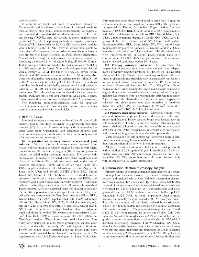
Figure 2 illustrates the TUNEL immunocytochemical reactions
for UV exposed noninfected and infected neuronal cultures.
Infected cultures corresponded to 4 and 5 dpi with Curionopolis
and Itacaiunas virus, respectively. As expected, positive control
cultures exhibited a massive amount of TUNEL-positive cells after
UV exposure when compared to non-exposed control cultures.
Inoculation with the two viral species induced neuronal TUNEL
immunolabeling, but at an earlier stage for Curionopolis virus
compared to Itacaiunas virus infection.
Figure 1. Phase-contrast (A, C, E) and fluorescence photomicrographs (B, D, F) of primary neuronal cultures to illustrate normalnoninfected cell morphology (A, B) and cytopathic effects 3 days after inoculation of Curionopolis virus (C, D) and 4 days afterinoculation of Itacaiunas virus (E, F). Arrows point to neurite fragmentation and circles indicate refringent points probably corresponding toapoptotic nuclei (A, C). Arrows and arrowheads indicate infected soma and dendrites, respectively (D, F). Neurons were stained by indirectimmunofluorescence for anti-neurofilament antibodies. Secondary antibodies were conjugated with fluorescein isothiocyanate.doi:10.1371/journal.pone.0001733.g001
Encephalitis of Rhabdoviruses
PLoS ONE | www.plosone.org 4 March 2008 | Volume 3 | Issue 3 | e1733
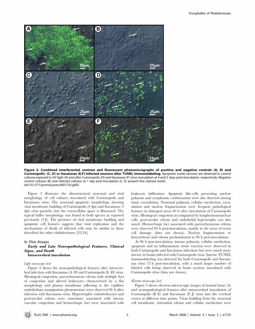
Figure 3 illustrates the ultrastructural neuronal and viral
morphology of cell cultures inoculated with Curionopolis and
Itacaiunas virus. The neuronal apoptotic morphology showing
viral membrane budding of Curionopolis (4 dpi) and Itacaiunas (5
dpi) virus particles into the extracellular space is illustrated. The
typical bullet morphology was found in both species as reported
previously [14]. The presence of viral membrane budding and
apoptotic cell features suggests that viral replication and the
mechanisms of death of infected cells may be similar to those
described for other rhabdoviruses [33,34].
In Vivo AssaysEarly and Late Neuropathological Features, Clinical
Signs, and DeathIntracerebral inoculation
Light microscopy level
Figure 4 shows the neuropathological features after intracere-
bral infection with Itacaiunas (A–D) and Curionopolis (E–H) virus.
Meningeal congestion, parenchymatous edema with multiple foci
of congestion and altered leukocytes characterized by a flat
morphology and plasma membrane adhering to the capillary
endothelium (margination phenomenon) were observed 96 h after
infection with Itacaiunas virus. Hypertrophic endotheliocytes and
perivascular edema were sometimes associated with intense
vascular congestion and hemorrhagic foci were associated with
leukocyte infiltration. Apoptotic like-cells presenting nuclear
pyknosis and cytoplasmic condensation were also detected among
tissue vacuolation. Neuronal pyknosis, cellular rarefaction, vacu-
olation and nuclear fragmentation were frequent pathological
features in damaged areas 60 h after inoculation of Curionopolis
virus. Meningeal congestion accompanied by lymphomononuclear
cells, perivascular edema and endothelial hypertrophy was also
noted. Hemorrhagic foci associated with parenchymatous edema
were observed 84 h post-inoculation, mainly in the areas of worst
cell damage (data not shown). Nuclear fragmentation or
karyorrhexis and edema predominated at 96 h post-inoculation.
At 96 h post-inoculation, intense pyknosis, cellular rarefaction,
apoptosis and an inflammatory tissue reaction were observed in
both Curionopolis and Itacaiunas infections but were much more
intense in brains infected with Curionopolis virus. Intense TUNEL
immunolabeling was detected for both Curionopolis and Itacaiu-
nas virus 72 h post-inoculation, with a much larger number of
labeled cells being observed in brain sections inoculated with
Curionopolis virus (data not shown).
Electron microscopy level
Figure 5 shows electron microscopic images of normal tissue (A)
and neuropathological features after intracerebral inoculation of
Curionopolis (B–E) and Itacaiunas (F–J) virus into the cerebral
cortex at different time points. Virus budding from the neuronal
cell membrane, interstitial edema and cellular rarefaction were
Figure 2. Combined interferential contrast and fluorescent photomicrographs of positive and negative controls (A, B) andCurionopolis- (C, D) or Itacaiunas (E,F)-infected neurons after TUNEL immunolabeling. Apoptotic nuclei (arrows) are observed in controlcultures exposed to UV light (A) and after Curionopolis (D) and Itacaiunas (F) virus inoculation at 4 and 5 days post-inoculation, respectively. Negativecontrol cultures (B) and infected cultures at 1 day post-inoculation (C, E) present few stained nuclei.doi:10.1371/journal.pone.0001733.g002
Encephalitis of Rhabdoviruses
PLoS ONE | www.plosone.org 5 March 2008 | Volume 3 | Issue 3 | e1733
observed 36 h after Curionopolis inoculation. Necrotic cells were
first detected at 60 h post-inoculation. After 96 h, intense
perivascular edema and endothelial hyperplasia accompanied by
a marked reduction in the luminal area of blood vessels were noted
(data not shown). There were no glial cells infected with
Curionopolis virus. Electron microscopy analysis of the cerebral
cortex (F–J) revealed no change 24 h after intracerebral infection
with Itacaiunas virus. Viral replication at an early stage was detected
60 h post-inoculation. After 72 h, glial cells containing abundant
cytoplasmic polyribosomes, similar to oligodendrocytes, presented
dilatations of the rough endoplasmic reticulum and viral budding. At
84 h post-inoculation, Itacaiunas virus-infected tissue was charac-
terized by interstitial edema and cellular rarefaction around the
capillary bed, pericytes with phagosomes, mitochondrial membrane
rupture, dilatation of the rough endoplasmic reticulum of endothelial
cells, and presence of phagosomes (data not shown). At 96 h post-
inoculation, large numbers of viral particles were detected in brain
parenchyma, and intense cellular rarefaction and interstitial edema
were observed in large areas. Apoptotic neurons were the
predominant finding at 108 h post-inoculation.
Intranasal inoculation. Figure 6 shows the distribution of
Curionopolis viral antigens in the brain parenchyma at different
time points after intranasal inoculation of the virus. The cytoplasm
of infected cells stained positive for virus proteins and small
unstained nuclei were apparent. This feature is consistent with the
known fact that viral proteins associated with RNA viruses are
located in the cytoplasm. At 1 dpi, Curionopolis antigens were
detected in meningeal cells and in a few neurons located in the
cortical parenchyma near the pial surface (data not shown). No
clinical signs of infection were observed at this stage. At 2 dpi,
clusters of immunostained neurons were clearly visible in ventral
sections of the olfactory bulb, ventral prefrontal cortex and
meninges. Neuronal soma, dendrites and the initial part of the
axon were labeled. At 4 dpi, hypomotility and absence of milk in the
stomach were frequent clinical signs, and immunostaining for virus
proteins revealed a large number of infected cells. Immunolabeled
primary dendrites and cell soma with altered cell appendages
showing small dots of dense accumulation of viral antigens were
detected in the meninges and on the pial surface of the olfactory
bulb, cerebral cortex, hippocampus, inferior colliculus, thalamus and
pons. Death and an agonic state were the dominant scenario in the
colony at 6 dpi. Clinical signs included complete absence of milk in
the stomach, weight loss, impaired body growth, meningeal irritative
signs such as nuchal and thoracic rigidity, motor incoordination,
spasticity and, in some cases, permanent lateral decubitus with
opisthotonus after stimulation. Immunolabeling at this time point
revealed viral antigens throughout the cerebral parenchyma, with
intense staining of the meninges, cerebral cortex (near pial surface),
Figure 3. Transmission electron photomicrographs of ultrathinsections obtained from primary neuronal cultures. Controlcultures of normal cells (A) and after Itacaiunas (B) and Curionopolis(C, D) infection at 4 and 5 days post-inoculation, respectively. Apoptoticcells (C) and virus budding (rectangle and arrows) (C, D) afterCurionopolis infection. Itacaiunas virus particles (arrows) in culture at5 days post-inoculation (B). N: nucleus; AN: apoptotic nucleus.doi:10.1371/journal.pone.0001733.g003
Figure 4. Photomicrographs of hematoxylin-eosin-stainedsections from control (A) and infected brain sections at 96 hpost-inoculation with Itacaiunas virus (B–D) and 60 h post-inoculation with Curionopolis virus (E–H). Multiple foci ofcongestion with sparse distribution of leukocytes characterized by themargination phenomenon (leukocytes becoming flat and the plasmamembrane sticking to the capillary endothelium) (B); hypertrophicendotheliocytes (ellipse) and perivascular edema (stars) (C). Apoptoticlike-cells with nuclear pyknosis and cytoplasmic condensation weredetected among tissue vacuolation (arrows) (D). Meningeal congestionand edema with lymphomononuclear cells (arrows) (E). Groups ofpyknotic cells presenting cytoplasmic condensation (circles), mixed withnormal cells and vacuolated parenchyma (stars) (F). Recent hemorrhagicpoints (presence of red blood cells) presenting parenchymatous edema(lozenge), mainly in the areas of worst cell damage (84 h post-inoculation) (G). Nuclear fragmentation or karyorrhexis (arrows) andedema (lozenge) (96 h post-inoculation) (H).doi:10.1371/journal.pone.0001733.g004
Encephalitis of Rhabdoviruses
PLoS ONE | www.plosone.org 6 March 2008 | Volume 3 | Issue 3 | e1733

CA1, dentate gyrus, inferior colliculus, pons and thalamus, and a
reduced staining intensity in the cerebellum. Necrotic tissue was
always detected by this histological procedure at a later stage during
the disease (data not shown).
Figure 7 shows the distribution of Itacaiunas viral antigens in
the brain parenchyma at different time point after intranasal
inoculation of the virus. Viral antigens were only detected at 3 dpi.
At this time point, a few clusters of immunolabeled neurons were
found in the frontal, temporal and parietal cortices near the pial
surface, but mainly in the olfactory bulb. Most labeled cells were
pyramidal neurons showing frequent immunolabeling of the
proximal dendrites. Axonal segments of pyramidal neurons were
also visible to varying extents. The findings observed at 6 dpi were
similar to those found on day 4, with the absence of clinical signs
and diffuse immunolabeling, which was slightly more intense than
on day 4. The full spectrum of clinical signs was observed on day
8, ranging from healthy to moribund animals, and included
hypomotility, absence of suckling, weight loss, impaired body
growth, nuchal rigidity, motor incoordination, and in some
terminal animals, permanent lateral decubitus with opisthotonus
after stimulation. Intense and diffuse CNS immunolabeling
occurred, with a large number of labeled neurons in the cortical,
hippocampal and pontine regions. Round immunolabeled dots
were frequently detected in the cytoplasm of neuronal soma in all
stained areas. These dots corresponded to the local accumulation
of viral antigenic proteins.
Figure 6 (G, H) and Figure 7 (F, G) illustrate neuronal TUNEL
immunolabeling on infected brain sections after inoculation of
Curionopolis virus (6 dpi) and Itacaiunas virus (12 dpi). Massive
TUNEL-labeled neuronal nuclei were found at this stage spread
Figure 5. Transmission electron photomicrographs of ultrathin sections obtained from control (A) and mouse brain infectedintracerebrally with Curionopolis for 36 (B,C), 60 (D) and 96 h (E), and with Itacaiunas for 24 (F), 60 (G), 72 (H), 96 (I) and 108 h (J).Normal tissue with intact neuronal soma and appendages (A); viral particles (arrow), interstitial edema (stars) and cellular rarefaction (lozenge) areseen 36 h post-inoculation (p.i.) (B, C); necrotic cells were observed at 60 h p.i. (D); intense perivascular edema (stars), hyperplastic endotheliocytesand reduced vessel luminal area (E); well-preserved brain parenchyma and vessels at 24 h p.i. (F); viral particles, endotheliocyte hyperplasia, and mildinterstitial edema (stars) at 60 h p.i. (G); membrane viral budding in rich polyribosomes oligodendrocyte-like cell at 72 h p.i. (H); brain parenchyma at96 h p.i. presenting a large number of viral particles (I); apoptotic features were more marked at 108 h p.i. (J). AC = apoptotic cell, M = mitochondria,OL = oligodendrocyte, EC = endothelial cells, VL = vascular lumen, N = cell nucleus, NC = necrotic cells.doi:10.1371/journal.pone.0001733.g005
Encephalitis of Rhabdoviruses
PLoS ONE | www.plosone.org 7 March 2008 | Volume 3 | Issue 3 | e1733
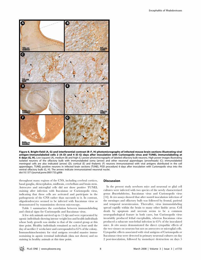
throughout many regions of the CNS, including cerebral cortices,
basal ganglia, diencephalon, midbrain, cerebellum and brain stem.
Astrocytes and microglial cells did not show positive TUNEL
staining after infection with Itacaiunas or Curionopolis virus,
indicating that these cells are activated and participate in the
pathogenesis of the CNS rather than succumb to it. In contrast,
oligodendrocytes seemed to be infected with Itacaiunas virus as
demonstrated by transmission electron microscopy.
Table 1 summarizes the correlation between immunolabeling
and clinical signs for Curionopolis and Itacaiunas virus.
A few sick animals survived up to 15 dpi and were represented by
agonic individuals showing intense weight loss and health individuals
whose body growth was similar to that of the control group at this
time point. Healthy individuals remained as such at least until the
day of sacrifice 2 weeks later and corresponded to 65% of the colony.
Immunohistochemistry for viral antigens revealed massive immu-
nostaining in agonic terminal individuals (data not shown) and no
staining in healthy animals at this time point.
Discussion
In the present study newborn mice and neuronal or glial cell
cultures were infected with two species of the newly characterized
genus Bracorhabdovirus, Itacaiunas virus and Curionopolis virus
[14]. In vivo assays showed that after nostril inoculation infection of
the meninges and olfactory bulb was followed by frontal, parietal
and temporal neuroinvasion. Thereafter, virus immunolabeling
spread rapidly within the brain to many other limbic areas. Cell
death by apoptosis and necrosis seems to be a common
neuropathological feature in both cases, but Curionopolis virus
invariably produced lethal encephalitis, whereas Itacaiunas virus
produced a subacute non-lethal infection in 65% of the inoculated
mice. In vitro assays demonstrated the direct cytopathic effects of
the two viruses on neurons but not on astrocytes or microglial cells.
Cytopathic effects associated with viral antigens of Curionopolis or
Itacaiunas virus were detected in primary neuronal cultures on day
2 post-inoculation, followed by monolayer destruction on days 5
Figure 6. Bright-field (A, G) and interferential contrast (B–F, H) photomicrographs of infected mouse brain sections illustrating viralantigen-immunolabeled cells 2 (A–D) and 4 (E–G) days after inoculation with Curionopolis virus and TUNEL immunolabeling at6 days (G, H). Low (square) (A), medium (B) and high (C) power photomicrographs of labeled olfactory bulb neurons. High power images illustratingisolated neurons of the olfactory bulb with immunolabeled soma (arrow) and other neuronal appendages (arrowheads) (C); immunolabeledmeningeal cells are also indicated (arrow) (D); cortical (E) and thalamic (F) neurons immunostained with viral antigens distributed in the cellappendages. TUNEL-positive neurons in infected brain sections (TUNEL POD procedure) 6 days after inoculation with Curionopolis virus into theventral olfactory bulb (G, H). The arrows indicate immunostained neuronal nuclei.doi:10.1371/journal.pone.0001733.g006
Encephalitis of Rhabdoviruses
PLoS ONE | www.plosone.org 8 March 2008 | Volume 3 | Issue 3 | e1733

and 7, respectively. Ultrastructural analysis revealed frequent
bullet-shaped budding from the neuronal membrane for both
Curionopolis and Itacaiunas viruses, a typical feature of members
of the Rhabdoviridae family. Viral budding from glial cells was
observed in brain tissue infected with Itacaiunas virus but not with
Curionopolis virus, with the cells exhibiting a morphology similar
to that of oligodendrocytes.
A variety of neurotrophic viruses can use the olfactory system as a
route for neuroinvasion of the mammalian brain, e.g. herpes simplex
virus 1, mouse hepatitis viruses, pseudorabies virus, Venezuelan
Figure 7. Bright-field (A, D, F) and interferential contrast (B, C, E, G) photomicrographs of Itacaiunas virus-infected mouse brain at 4 (A–C), 6 (D, E) and 8 (F, G) days post-inoculation. Low (A) and medium (B) power photomicrographs of the olfactory neuronal group (smaller rectangle).Details of immunolabeled neurons of the frontal cortex (C) (arrows and arrowheads). Low (D) (rectangle) and medium (E) power photomicrographs of agroup (arrows) of hippocampal neurons showing low viral antigen condensation (arrowhead). TUNEL-positive midbrain neurons of infected brain sections(TUNEL POD procedure) 12 days after inoculation with Itacaiunas virus (F, G). The arrows indicate immunostained neuronal nuclei.doi:10.1371/journal.pone.0001733.g007
Encephalitis of Rhabdoviruses
PLoS ONE | www.plosone.org 9 March 2008 | Volume 3 | Issue 3 | e1733

Equine Encephalitis virus, and the challenge virus standard (CVS)
strain of the rabies virus [35,36,1,37,38] but the sequence of
neuroinvasion observed for Curionopolis and Itacaiunas viruses
remember that of VSIV. After nasal administration, both Curiono-
polis and Itacaiunas antigens were observed in the meninges and
olfactory bulb at 2 and 4 dpi, respectively. Six days after inoculation,
strong signs of Curionopolis virus infection were found both inside,
and outside the olfactory system, including the frontal cortex,
hippocampus and thalamus. Itacaiunas immunolabeling was
detected at 4 dpi in the olfactory bulb and in small groups of
neurons of the frontal, parietal and temporal cortices near the pial
surface. After Itacaiunas infection, surviving mice were immunone-
gative for virus antigens, suggesting a complete clearance of the virus
from the brain parenchyma within 15 dpi. When VSIV is inoculated
intranasally into 5–7-week-old male BALB/c mice, olfactory
receptors are the first cells to be infected [10], followed by neurons
of the olfactory bulb and, finally, acute infection of other brain areas
[21,8,22]. A peak in viral antigens in the brain parenchyma,
accompanied by the highest mortality rate, is observed within 7 to 10
dpi. In surviving mice, complete clearance of the virus from the
parenchyma occurs within 12 dpi, without long-term damage to the
brain [21,8]. Thus, the temporal course of immunolabeling after
intranasal inoculation revealing the sequence of neuroinvasion by
Itacaiunas and Curionopolis viruses through the olfactory route in
neonate albino Swiss mice, seem to be different of the BALB/cByJ
and SJL/J neonate mice after mouse hepatitis virus (MHV)-JHM
infection where MHV lesions are diffusely distributed throughout
the brain suggesting blood-borne infection [39]. These data may
suggest that neonate albino Swiss mice would present an effective
blood-brain barrier earlier than BALB/c mice. However, since
intranasal inoculation of these viruses in mature albino Swiss mice
did not induce encephalitis but exhibit antigenic immunolabeling in
non-symptomatic subjects (not illustrated), it remains an open
question whether or not vascular route remain an alternative
pathway for neuroinvasion in neonatal period. Since neurotropic
viruses that enter the CNS through the vascular route may damage
the blood-brain barrier it would be interesting to investigate in
neonate mice, early and late in the disease, the integrity of the blood
brain barrier in order to clarify this possibility. Blood brain barrier
(BBB) disruption is associated with activating sentinel macrophages
[40,41,42] and nitric oxide synthases type III in astrocytes and in
endothelial or ependymal cells [43]. These macrophages may
activate matrix metalloproteinases which have been associated with
breakdown of the blood-brain barrier and tissue destruction [41].
Some viruses are also known to infect mouse brain endothelial cells,
increasing CNS vascular permeability and resulting in edema and an
increased local viral load [44,45]. Lymphocytic infiltration and
gliosis are common pathological findings in viral panencephalitis,
with CD4+ T lymphocytes being observed in perivascular areas and
CD8+ lymphocytes in the parenchyma. B lymphocytes are usually
located in large perivascular cuffs associated with a longer and slower
disease [46]. A time course study of the infection in the neonate
albino Swiss mice for cellular immune response would be important
to understand pathophysiological mechanisms of Itacaiunas and
Curionopolis neuroinvasion.
Intense pyknosis in the cerebral cortex demonstrated by
hematoxylin staining was not always accompanied by intense
TUNEL immunolabeling in Curionopolis or Itacaiunas virus.
Recent studies have shown an apoptosis-necrosis continuum for
these two major forms of cell death [47]. However, a larger number
of apoptotic cortical neurons were observed for Curionopolis virus at
72 h post-inoculation when compared to Itacaiunas virus-infected
brain. This fact might be associated with the earlier cytopathic
changes induced by precocious Curionopolis neuroinvasion, acti-
vating first cell death mechanisms. In addition, a previous report [11]
showed that, despite intense pyknosis observed by hematoxylin
staining, anti-caspase-3 labeling was consistently negative after
Itacaiunas virus infection. This finding demonstrates that not all
pyknotic profiles represent cells undergoing apoptosis that depends
on caspase-3 activation and that apoptosis determined by TUNEL-
positive DNA damage can occur irrespective of caspase-3 activation
in some circumstances such as hypoxia [48]. Both necrosis and
apoptosis can also be a consequence of the inflammatory response
induced by the virus as previously suggested [49,4,50]. As reported
for humans and mice, capillary and endothelial inflammation of
cortical vessels is also a striking pathological feature after viral
encephalitis and may contribute to blood-brain barrier damage and
viral neuroinvasion [51,52].
Ultrastructural studies have shown that the morphology of the
Itacaiunas and Curionopolis viruses resembles that of animal-
infecting members of the family Rhabdoviridae. Lyssaviruses have
been demonstrated to frequently become enveloped virions within
the endoplasmic reticulum and on the plasma membrane of
infected neurons [53]. In contrast, vesiculoviruses bud from the
plasma membrane of infected cells, but the process of budding into
cytoplasmic vesicles was rarely observed [54,55]. In the present
study, ultrastructural analysis showed frequent bullet-shaped
budding from neuronal membranes, indicating that Curionopolis
and Itacaiunas viruses formed enveloped virions on the plasma
membrane but not within the endoplasmic reticulum, a finding
suggesting a replication process similar to that of vesiculoviruses.
Overall, our results provide at the first time fresh insights into
the cell targets and the pathogenesis of two species of the newly
characterized genus Bracorhabdovirus. We identified CNS mice cell
types both in vivo and in vitro and the temporal sequence of
neuroanatomical olfactory areas infected by Itacaiunas and
Curionopolis virus, and our next aim is to further characterize
how these viruses interact wit these cells. It will be of interest to
determine the cellular associated immune response in a time
course study to investigate the molecular mechanisms associated
Table 1. Clinical Signs Intensity and Virus Immunolabelling after Inoculation
Virus Events Days of post-inoculation survival and intensity of reported events
1 2 4 6 8 12 15
Itacaiunas Clinical signs (2) (2) (2) (2) (++++) (2) (++++) (2) (++++) (2)
Viral antigens (2) (2) (++) (++++) (++++) (++++) (2) (++++) (2)
Curionopolis Clinical signs (2) (2) (+) (++++)
Viral antigens (+) (++) (+++) (++++)
(2) Absence of clinical signs or viral immunolabelling; (+), (++), (+++) and (++++) distinguish progressive intensities of clinical signs and immunolabellingdoi:10.1371/journal.pone.0001733.t001
Encephalitis of Rhabdoviruses
PLoS ONE | www.plosone.org 10 March 2008 | Volume 3 | Issue 3 | e1733
with the higher virulence of Curionopolis virus infection. It will be
also useful to investigate the molecular mechanisms that made glial
cells, similar to oligodendrocytes, sensitive to Itacaiunas virus, but
not to Curionopolis virus infections.
Author Contributions
Conceived and designed the experiments: JD PV VM Wd CD. Performed
the experiments: JD Zd MB AD Dd DM VB JC KZ. Analyzed the data:
JD PV Zd MB DM VB JC JQ CT VM Wd CD. Contributed reagents/
materials/analysis tools: PV JC JQ CD. Wrote the paper: JD PV JQ CD.
References
1. Charles PC, Walters E, Margolis F, Johnston RE (1995) Mechanism ofneuroinvasion of Venezuelan equine encephalitis virus in the mouse. Virology.
208: 662–71.
2. Fazakerley JK (2004) Semliki forest virus infection of laboratory mice: a model to
study the pathogenesis of viral encephalitis. Arch Virol Suppl. pp 179–90.
3. Jackson AC, Ye H, Ridaura-Sanz C, Lopez-Corella E (2001) Quantitative studyof the infection in brain neurons in human rabies. J Med Virol. 65: 614–8.
4. Rempel JD, Murray SJ, Meisner J, Buchmeier MJ (2004) Differential regulation
of innate and adaptive immune responses in viral encephalitis. Virology. 318:381–92.
5. Saha S, Sugumar P, Bhandari P, Rangarajan PN (2006) Identification ofJapanese encephalitis virus-inducible genes in mouse brain and characterization
of GARG39/IFIT2 as a microtubule-associated protein. J Gen Virol. 87:3285–9.
6. Dobler G (1996) Arboviruses causing neurological disorders in the central
nervous system. Arch Virol Suppl. 11: 33–40.
7. Griffin DE (1995) Arboviruses and the central nervous system. Springer Semin
Immunopathol. 17: 121–32.
8. Huneycutt BS, Plakhov IV, Shusterman Z, Bartido SM, Huang A, Reiss CS,Aoki C (1994) Distribution of vesicular stomatitis virus proteins in the brains of
BALB/c mice following intranasal inoculation: an immunohistochemicalanalysis. Brain Res. 635: 81–95.
9. Hunsperger EA, Roehrig JT (2006) Temporal analyses of the neuropathogenesisof a West Nile virus infection in mice. J Neurovirol. 12: 129–39.
10. Plakhov IV, Arlund EE, Aoki C, Reiss CS (1995) The earliest events in vesicular
stomatitis virus infection of the murine olfactory neuroepithelium and entry ofthe central nervous system. Virology. 209: 257–62.
11. Gomes-Leal W, Martins LC, Diniz JA, Dos Santos ZA, Borges JA, et al. (2006)
Neurotropism and neuropathological effects of selected rhabdoviruses on
intranasally-infected newborn mice. Acta Trop. 97: 126–39.
12. Vasconcelos PF, Travassos da Rosa AP, Rodrigues SG, Travassos da Rosa ES,Degallier N, et al. (2001) Inadequate management of natural ecosystem in the
Brazilian Amazon region results in the emergence and reemergence ofarboviruses. Cad Saude Publica. 17 Suppl: 155–64.
13. Fu ZF (2005) Genetic comparison of the rhabdoviruses from animals and plants.Curr. Top Microbiol. Immunol. 292: 1–24.
14. Diniz JA, Nunes MR, Travassos da Rosa AP, Cruz AC, de Souza W, et al.
(2006) Characterization of two new rhabdoviruses isolated from midges(Culicoides SPP) in the Brazilian Amazon: proposed members of a new genus,
Bracorhabdovirus. Arch Virol. 151: 2519–27.
15. Crotty S, Hix L, Sigal LJ, Andino R (2002) Poliovirus pathogenesis in a new
poliovirus receptor transgenic mouse model: age-dependent paralysis and amucosal route of infection. J Gen Virol. 83: 1707–20.
16. Iwasaki T, Inoue S, Tanaka K, Sato Y, Morikawa S, et al. (2004)
Characterization of Oita virus 296/1972 of Rhabdoviridae isolated from a
horseshoe bat bearing characteristics of both lyssavirus and vesiculovirus. ArchVirol. 149: 1139–54.
17. Travassos da Rosa AP, Mather TN, Takeda T, Whitehouse CA, Shope RE, et
al. (2002) Two new rhabdoviruses (Rhabdoviridae) isolated from birds duringsurveillance for arboviral encephalitis, northeastern United States. Emerg Infect
Dis. 8: 614–8.
18. Van der Poel WH, Van der Heide R, Van Amerongen G, Van Keulen LJ,
Wellenberg GJ, et al. (2000) Characterization of a recently isolated lyssavirus infrugivorous zoo bats. Arch Virol. 145: 1919–31.
19. Elena SF, Sanjuan R (2005) Adaptive value of high mutation rates of RNA
viruses: separating causes from consequences. J Virol. 79: 11555–8.
20. Reiss CS, Plakhov IV, Komatsu T (1998) Viral replication in olfactory receptor
neurons and entry into the olfactory bulb and brain. Ann N Y Acad Sci. 855:751–61.
21. Forger JM 3rd, Bronson RT, Huang AS, Reiss CS (1991) Murine infection by
vesicular stomatitis virus: initial characterization of the H-2d system. J Virol. 65:4950–8.
22. Lundh B, Kristensson K, Norrby E (1987) Selective infections of olfactory and
respiratory epithelium by vesicular stomatitis and Sendai viruses. Neuropathol
Appl Neurobiol. 13: 111–22.
23. Travassos da Rosa AP, Turell MJ, Watts DM, Powers AM, Vasconcelos PF, etal. (2001) Trocara virus: a newly recognized Alphavirus (Togaviridae) isolated
from mosquitoes in the Amazon Basin. Am J Trop Med Hyg. 64: 93–7.
24. Beaty BJ, Calisher CH, Shope RE (1989) Arboviruses. In: Schmidt NJ,
Emmons RW, eds. Diagnostic Procedures for Viral Rickettsial and ChlamydialInfections, 6th ed. Washington, USA: American Public Health Association. pp
797–855.
25. Buki A, Okonkwo DO, Wang KK, Povlishock JT (2000) Cytochrome C releaseand caspase activation in traumatic axonal injury. J Neurosci. 20: 2825–34.
26. Fazakerley JK, Cotterill CL, Lee G, Graham A (2006) Virus tropism,
distribution, persistence and pathology in the corpus callosum of the Semliki
Forest virus-infected mouse brain: a novel system to study virus-oligodendrocyte
interactions. Neuropathol Appl. Neurobiol. 32: 397–409.
27. Gubler DJ, Kuno G, Sather GE, Velez M, Oliver A (1984) Mosquito cell
cultures and specific monoclonal antibodies in surveillance for dengue viruses.
Am J Trop Med Hyg. 33: 158–65.
28. Brewer GJ (1997) Effects of acidosis on the distribution of processing of the beta-
amyloid precursor protein in cultured hippocampal neurons. Mol Chem
Neuropathol. 31: 171–86.
29. McCarthy KD, de Vellis J (1980) Preparation of separate astroglial and
oligodendroglial cell cultures from rat cerebral tissue. J Cell Biol. 85: 890–902.
30. Lima FR, Gervais A, Colin C, Izembart M, Neto VM, et al. (2001) Regulation of
microglial development: a novel role for thyroid hormone. J Neurosci. 21:
2028–38.
31. Hepler PK (1980) Membranes in the mitotic apparatus of barley cells. J Cell
Biol. 86: 490–9.
32. Reynolds ES (1963) The use of lead citrate at high pH as an electron-opaque
stain in electron microscopy. J Cell Biol. 17: 208–12.
33. Barber GN (2005) VSV-tumor selective replication and protein translation.
Oncogene. 24: 7710–9.
34. Fu ZF, Jackson AC (2005) Neuronal dysfunction and death in rabies virus
infection. J Neurovirol. 11: 101–6.
35. Babic N, Mettenleiter TC, Ugolini G, Flamand A, Coulon P (1994) Propagation
of pseudorabies virus in the nervous system of the mouse after intranasal
inoculation. Virology. 204: 616–25.
36. Barnett EM, Perlman S (1993) The olfactory nerve and not the trigeminal nerve
is the major site of CNS entry for mouse hepatitis virus, strain JHM. Virology.
194: 185–91.
37. Esiri MM, Tomlinson AH (1984) Herpes simplex encephalitis. Immunohisto-
logical demonstration of spread of virus via olfactory and trigeminal pathways
after infection of facial skin in mice. J Neurol Sci. 64: 213–7.
38. Lafay F, Coulon P, Astic L, Saucier D, Riche D, et al. (1991) Spread of the CVS
strain of rabies virus and of the avirulent mutant AvO1 along the olfactory
pathways of the mouse after intranasal inoculation. Virology. 183: 320–30.
39. Barthold SW, Smith AL (1987) Response of genetically susceptible and resistant
mice to intranasal inoculation with mouse hepatitis virus JHM. Virus Res. 7:
225–39.
40. Chan WL, Javanovic T, Lukic ML (1989) Infiltration of immune T cells in the
brain of mice with herpes simplex virus-induced encephalitis. J Neuroimmunol.
23: 195–201.
41. Khuth ST, Akaoka H, Pagenstecher A, Verlaeten O, Belin MF, et al. (2001)
Morbillivirus infection of the mouse central nervous system induces region-
specific upregulation of MMPs and TIMPs correlated to inflammatory cytokine
expression. J Virol. 75: 8268–82.
42. Khuth ST, Strazielle N, Giraudon P, Belin MF, Ghersi-Egea JF (2005)
Impairment of blood-cerebrospinal fluid barrier properties by retrovirus-
activated T lymphocytes: reduction in cerebrospinal fluid-to-blood efflux of
prostaglandin E2. J Neurochem. 94: 1580–93.
43. Komatsu T, Ireland DD, Chung N, Dore A, Yoder M, et al. (1999) Regulation
of the BBB during viral encephalitis: roles of IL-12 and NOS. Nitric Oxide. 3:
327–39.
44. Soilu-Hanninen M, Eralinna JP, Hukkanen V, Roytta M, Salmi AA, Salonen R
(1994) Semliki Forest virus infects mouse brain endothelial cells and causes
blood-brain barrier damage. J Virol. 68: 6291–8.
45. Soilu-Hanninen M, Roytta M, Salmi AA, Salonen R (1997) Semliki Forest virus
infection leads to increased expression of adhesion molecules on splenic T-cells
and on brain vascular endothelium. J Neurovirol. 3: 350–60.
46. Anlar B, Soylemezoglu F, Aysun S, Kose G, Belen D, et al. (2001) Tissue
inflammatory response in subacute sclerosing panencephalitis (SSPE). J Child
Neurol. 16: 895–900.
47. Martin LJ, Al-Abdulla NA, Brambrink AM, Kirsch JR, Sieber FE, et al. (1998)
Neurodegeneration in excitotoxicity, global cerebral ischemia, and target
deprivation: A perspective on the contributions of apoptosis and necrosis. Brain
Res Bull. 46: 281–309.
48. Vila M, Przedborski S (2003) Targeting programmed cell death in neurode-
generative diseases. Nat Rev Neurosci. 4: 365–75.
49. Julkunen I, Melen K, Nyqvist M, Pirhonen J, Sareneva T, et al. (2000)
Inflammatory responses in influenza A virus infection. Vaccine. 19 Suppl 1:
S32–7.
50. Roulston A, Marcellus RC, Branton PE (1999) Viruses and apoptosis. Annu Rev
Microbiol. 53: 577–628.
Encephalitis of Rhabdoviruses
PLoS ONE | www.plosone.org 11 March 2008 | Volume 3 | Issue 3 | e1733
51. German AC, Myint KS, Mai NT, Pomeroy I, Phu NH, et al. (2006) A
preliminary neuropathological study of Japanese encephalitis in humans and a
mouse model. Trans R Soc Trop Med Hyg. 100: 1135–45.
52. Maslin CL, Kedzierska K, Webster NL, Muller WA, Crowe SM (2005)
Transendothelial migration of monocytes: the underlying molecular mechanisms
and consequences of HIV-1 infection. Curr HIV Res. 3: 303–17.
53. Murphy FA, Bauer SP, Harrison AK, Winn WC Jr (1973) Comparative
pathogenesis of rabies and rabies-like viruses. Viral infection and transit frominoculation site to the central nervous system. Lab Invest. 28: 361–76.
54. Dal Canto MC, Rabinowitz SG, Johnson TC (1976) In vivo assembly and
maturation of vesicular stomatitis virus. Lab Invest. 35: 515–24.55. Le Blanc I, Luyet PP, Pons V, Ferguson C, Emans N, et al. (2005) Endosome-to-
cytosol transport of viral nucleocapsids. Nat Cell Biol. 7: 653–64.
Encephalitis of Rhabdoviruses
PLoS ONE | www.plosone.org 12 March 2008 | Volume 3 | Issue 3 | e1733