Generation of Synthetic FRP Microstructures Based on Experimentally Observed Microstructures

The Effects of Experimentally Induced Adelphophagy inGastropod EmbryosOlaf Thomsen1,2, Rachel Collin1*, Allan Carrillo-Baltodano1¤
1 Smithsonian Tropical Research Institute, Balboa, Ancon, Republic of Panama, 2 Universitat Oldenburg, Oldenburg, Germany
Abstract
Adelphophagy, development where embryos grow large by consuming morphologically distinct nutritive embryos or theirown normal siblings is widespread but uncommon among animal phyla. Among invertebrates it is particularly common insome families of marine gastropods and segmented worms, but rare or unknown in other closely related families. Incalyptraeid gastropods phylogenetic analysis indicates that adelphophagy has arisen at least 9 times from species withplanktotrophic larval development. This pattern of frequent parallel evolution of adelphophagy suggests that the embryosof planktotrophic species might be predisposed to evolve adelphophagy. Here we used embryos of three species ofplanktotrophic calyptraeids, one from each of three major genera in the family (Bostrycapulus, Crucibulum, and Crepidula), toanswer the following 3 questions: (1) Can embryos of species with planktotrophic development benefit, in terms of pre-hatching growth, from the ingestion of yolk and tissue from experimentally damaged siblings? (2) Does ingestion of thismaterial from damaged siblings increase variation in pre-hatching size? and (3) Does this experimentally inducedadelphophagy alter the allometry between the velum and the shell, increasing morphological similarity to embryos ofnormally adelphophagic species? We found an overall increase in shell length and velum diameter when embryos feed ondamaged siblings within their capsules. There was no detectable increase in variation in shell length or velum diameter, orchanges in allometry. The overall effect of our treatment was small compared to the embryonic growth observed innaturally adelphophagic development. However each embryo in our experiment probably consumed less than one siblingon average, whereas natural adelphophages often each consume 10–30 or more siblings. These results suggest that theability to consume, assimilate, and benefit from yolk and tissue of their siblings is widespread across calyptraeids.
Citation: Thomsen O, Collin R, Carrillo-Baltodano A (2014) The Effects of Experimentally Induced Adelphophagy in Gastropod Embryos. PLoS ONE 9(7): e103366.doi:10.1371/journal.pone.0103366
Editor: Donald James Colgan, Australian Museum, Australia
Received October 11, 2013; Accepted June 30, 2014; Published July 29, 2014
This is an open-access article, free of all copyright, and may be freely reproduced, distributed, transmitted, modified, built upon, or otherwise used by anyone forany lawful purpose. The work is made available under the Creative Commons CC0 public domain dedication.
Funding: This research was supported by United States National Science Foundation grant IOS 1019727 to R. Collin and the Smithsonian Tropical ResearchInstitute. The funders had no role in study design, data collection and analysis, decision to publish, or preparation of the manuscript.
Competing Interests: The authors have declared that no competing interests exist.
* Email: [email protected]
¤ Current address: Biology Department, Clark University, Worcester, MA, United States of America
Introduction
Marine invertebrates exhibit a remarkable variety of modes of
development [1–3]. There are species with free-living larvae that
swim and feed in the water column before they become competent
to settle and metamorphose. There are species where large eggs
develop directly into fully formed juveniles without the need for
exogenous nutrition. Perhaps the most poorly understood mode of
development is oophagic or adelphophagic development where
embryos consume other eggs (oophagy) or embryos (adelpho-
phagy), which provide exogenous nutrition prior to hatching
(hereafter referred to simply as adelphophagy). Adelphophagic
embryos usually develop directly into juveniles, bypassing the free-
living larval stage. This results in large benthic hatchlings with low
dispersal ability compared to those species with planktotrophic
larvae. In this respect adelphophagic species are ecologically
similar to species with direct development from large eggs. Why
some species evolve direct development via large eggs and others
evolve direct development via small eggs with adelphogphy is not
clearly understood [4–5].
Encapsulated or brooded development where embryos develop
in close proximity to their siblings is a prerequisite for the
evolution of adelphophagy. Such protected development of early
embryonic stages is common in many invertebrate groups, but the
phylogenetic distribution of adelphophagy is patchy; being
particularly common in some groups and absent in others. For
example, among caenogastropods it is common in calyptraeids,
muricids, vermetids, and buccinids but unknown in littorinids and
conids, which also have encapsulated development [4,6–10]. It is
also particularly common in spionid polychaetes [11]. This
suggests that the embryos of some groups may be predisposed to
evolve adelphophagy.
Various consequences of adelphophagy have been demonstrat-
ed primarily in spionid worms and calyptraeid and muricid
gastropods. In gastropods adelphophagy increases variation in
hatching size [5,7–8]. Several cases of poecilogony (an extreme
case of variation in hatching size where females of a single species
can produce more than one kind of development) result from
adelphophagic development [12]. It remains to be seen if the
increased variation in hatching size is a bet-hedging strategy or
simply the by-product of a mechanism to produce large hatchlings
[5]. In calyptraeid gastropods, adelphophagic development can,
but does not always, produce hatchlings larger than those from
large eggs [4]. Adelphophagic muricids may develop more quickly
than do those with large eggs [6–7], however there is no evidence
PLOS ONE | www.plosone.org 1 July 2014 | Volume 9 | Issue 7 | e103366
of rapid development in adelphophagic calyptraeids [5], and no
other groups have been examined. In the adelphophagic worm
Boccardia proboscidia females can actively control the time of
hatching altering the size and developmental stage of hatchlings
[13]. Genetic analyses have shown that adelphophagy increases
the average relatedness of offspring within each capsule in one
buccinid and one calyptraeid species [14–15]. Early in develop-
ment the embryos within a capsule have more genetic diversity
and represent offspring from more fathers than they do later in
development. Despite these demonstrable consequences of adel-
phophagy, the factors that select for adelphophagic development
remain unclear.
The developmental mechanisms by which nutritive eggs or
embryos are specified, and the manner in which they are
consumed by their siblings are still poorly understood. Nurse
embryos or nurse eggs span a range of developmental potentials.
In some cases they seem to have no potential to develop. For
example the nurse eggs in the calyptraeid Crepipatella dilatata and
the nassariid Buccinanops globulosus do not appear to be fertilized
and do not initiate development [16–17]. In other species of
calyptraeids, vermetids, and some spionid polychaetes the nurse
embryos initiate development but become arrested at some point
prior to the development of the definitive juvenile body plan [18–
23]. Finally, in the polychaete Boccardia proboscidia, the worms-
nail Vermetus triquetrus, and another unidentified vermetid some
well-developed, fully-functional embryos are consumed by their
siblings [13,23–24]. In none of these species is it known if the
arrested embryos have the potential to develop into normal
hatchlings if some environmental factor did not arrest their
development, or, alternately if their developmental fate is specified
prior to ovulation or oviposition.
The mechanisms by which developing embryos consume
nutritive embryos vary as widely as their appearance. In some
species the nutritive eggs or embryos are swallowed whole by their
siblings. This is often the case in species where the nutritive eggs
do not cleave, like in Buccinum undatum [25]. Consumption of
entire embryos has also been observed in this species [25]. In other
cases blobs of yolk are detached from the surface or sucked from
the interior of the nutritive embryos [8–9,17,19–22]. This seems to
be particularly common in calyptraeids where species of
Crepidula, Calyptraea, Crepipatella, and Crucibulum have all
been described has having nutritive embryos that either disinte-
grate or become hollowed out as development progresses
[19,20,26]. In these species nutritive embryos may follow
developmental pathways that produce yolk vesicles that are easily
detached and consumed by their normal siblings [26]. The role of
apoptosis in producing such vesicles has been demonstrated in the
polychaete Polydora cornuta [21] but has not been looked for in
adelphophagic gastropods.
In families were adelphophagy is common it appears to have
arisen multiple times. For example in calyptraeid gastropods
phylogenetic analysis indicates that adelphophagic or oophagic
development has arisen at least 9 times, generally from species
with planktotrophic larval development [18,27]. The developing
embryos in many adelphophagic calyptraeid species do not appear
to be modified from the typical development of their plankto-
trophic relatives, other than having a relatively larger shell and
visceral mass [18–20,26,28–30]. This suggests that the evolution of
adelphophagy is relatively simple in this family, and that the
embryos of related planktotrophic species might already express
features that enable them to take advantage of dead or damaged
siblings. Such ‘‘pre-adaptations’’ for adelphophagy could involve
both the ability to capture and ingest yolk particles within the egg
capsule and the digestion, absorption, and utilization of the
material for growth and development.
Here we used three species of planktotrophic calyptraeids, one
from each of three major genera in the family (Crepidula,
Crucibulum, and Bostrycapulus) to answer the following 3
questions: (1) Can embryos of species with planktotrophic
development benefit, in terms of pre-hatching growth, from the
ingestion of yolk and tissue from experimentally damaged siblings?
(2) Does ingestion of yolk and tissue from siblings increase
variation in pre-hatching size? and (3) Does this experimentally
induced adelphophagy alter the allometry between the velum and
the shell, increasing morphological similarity to embryos of
normally adelphophagic species? Anecdotal accounts report that
in species with planktotrophic development the occasional empty
shell is found in egg capsules containing otherwise normally
developing embryos [27,31]. This suggests that embryos occa-
sionally die naturally during development and that their siblings
may consume the tissue. Such consumption of dead siblings could
even be selected for if it prevented contamination of the egg
capsule. In addition, embryos of Crepipatella peruviana (previ-
ously referred to by the name Crepidula fecunda or Crepipatellafecunda see [32] for a discussion of taxonomic revisions in
Crepipatella), a species that is thought to have recently re-evolved
planktotrophic development from oophagy can consume damaged
siblings prior to hatching, and that this can lead to increased
hatching size [31,28]. Our study was designed to determine if this
capacity is common among primary planktotrophs across the
family, thus setting the stage for the evolution of adelphophagy.
Materials and Methods
Snails were collected under permits issued by the Autoridad de
Recursos Acuaticos de Panama (ARAP). Crepidula cf. marginaliswere collected at Chumical Beach (8u 539 N, 79u 389 W) and
Bostrycapulus calyptraeformis (Deshayes 1830) and Crucibulumspinosum (Sowerby 1824) were collected at Venado Beach (8u 539
N, 79u 359 W), near Panama City, Panama. All 3 species occur in
the intertidal with a mix of soft bottom and rocky-rubble, and were
collected between June and August 2012. All three species have
planktotrophic development with similar ranges of hatching sizes;
Bostrycapulus calyptraeformis hatches at 300–345 mm shell length;
Crucibulum spinosum hatches at 280–325 mm shell length and
Crepidula cf. marginalis hatches at 290–330 mm shell length
[4,33]. In all 3 species the female produces clutches of multiple
transparent thin-walled capsules, which she protects between the
substrate, her propodium, and her neck. Each capsule contains
numerous (50–150) small eggs, each of which normally develops
into a veliger larva. The embryos are free within the capsule, and
shortly after gastrulation they can be seen to move actively around
the capsule, often bumping into and pushing past each other.
When each mature veliger no longer has visible yolk, the larvae
hatch with the help of the mother. In calyptraeid species where
this has been observed, the female appears to nudge the capsules
out from under the shell, sometimes pulling at them with her
mouth, while pumping the shell up and down [30]. This
presumably acts to disperse the larvae away from the substrate.
Capsules raised in still culture away from the mother do not hatch.
Instead the larvae continue to swim actively inside the capsule well
past their due date, and eventually they appear to starve [26,30].
Large females were collected and kept individually at room
temperature, approximately 22–25uC, in 350 ml transparent
plastic cups filled with filtered, UV-sterilized seawater. The water
was changed three times a week, and the snails were fed
approximately 38.66106 cells of Isochrysis galbana daily (following
Experimentally Induced Adelphophagy in Gastropod Embryos
PLOS ONE | www.plosone.org 2 July 2014 | Volume 9 | Issue 7 | e103366
[34]). Females were monitored and the date of egg deposition was
recorded. Egg capsules were collected from brooding females after
7 or 8 days of incubation, when they were at a stage capable of
ingesting exogenous particles. This ‘‘head vesicle stage’’ is
characterized by the extension of the velum away from the body
wall and the presence of a transparent, ciliated head vesicle (see
[35]). It is at this stage that many naturally adelphophagic
calyptraeid species begin the consumption of nurse eggs.
Preliminary attempts to apply our experimental manipulations to
embryos prior to the head-vesicle stage generally resulted in the
fouling of the egg capsules and infection with microorganisms. The
head vesicle stage is approximately 4–5 days before hatching
would normally be induced by the female. Once this stage was
reached capsules were taken from the female, and placed in
40612 mm Petri dishes containing 0.22 mm-filtered, UV-sterilized
seawater and antibiotics (5.261025 g/ml Penicillin G potassium
salt and 9.261024 g/ml streptomycin sulfate salt) to reduce
contamination from microorganisms [36].
For the Encapsulated Experiment 6 capsules were removed from
the female and each was placed individually in a Petri dish. Three
capsules were assigned to the treatment and 3 were assigned to the
control (Figure 1). For treated capsules 20–40% of the embryos
within a single capsule were killed by applying pressure to the
intact capsule with thin flat forceps (Figure 2). Care was taken not
to rupture the capsule wall, and any capsules that appeared
damaged were discarded. Observation of the treated capsules
showed that the small yolk particles were cleared from the capsule
fluid within 2–3 hours and that larger fragments took longer to
clear (Figure 2). Because the capsules are a closed system, and
because undamaged embryos were observed to capture and ingest
yolk particles (Figures 3 & 4, Video S1), it seems likely that this
material was ingested. For the control group, the intact capsules
were each left in a Petri dish without killing any of the embryos.
Broods from 9 females were used for C. cf. marginalis, 11 for C.spinosum, and 16 for B. calyptraeformis.
For the Excapsulated Experiment three replicates were used
from each of 12 mothers for C. cf. marginalis only. In the ‘treated’
group, two capsules were placed into the Petri dish (Figure 1). The
contents of one capsule was killed completely and emptied into the
Petri dish, while the other capsule was opened so that the embryos
swam freely and could feed on their dead siblings. The embryos
were allowed to feed for one hour (Figures 3 & 4), and were then
transferred to a new Petri dish with fresh filtered seawater. In the
control group, the embryos of one capsule were released into the
Petri dish without an additional food source.
All dishes were maintained in an incubator at 24uC and the
water was exchanged daily, with new filtered seawater and
antibiotics. To measure the velum and shell length, embryos were
removed from the capsules and photographed three days after the
treatment was imposed, slightly before the date on which the
embryos were due to hatch. We have never observed capsules of
these species to hatch naturally when they have been removed
from the mother, so it was not possible to determine if the
treatments had an effect on natural hatching. Lateral views of the
embryos were used to measure the shell length, and anterior views
were used to measure the diameter across the two extended velar
lobes using ImageJ 1.46r [37]. Each of these measurements was
taken for at least 10 embryos from each replicate dish. A two-level
nested ANOVA was used to test for effects of treatment, female,
and treatment 6 female on larval morphology, using JMP 9.0.2.
The replicate dishes were included as a random effect nested
within female. To test for changes in the allometry between shell
length and velum diameter we used an ANCOVA with average
velum diameter for each replicate as the dependent variable,
treatment as a factor, and average shell length for the same
replicate as a covariate. We used average values for each replicate
because the shell length and velum size measurements often could
not be obtained for the same individual embryo. To test for
differences in variation between treated and control embryos the
coefficient of variation (CV) was calculated for each replicate dish.
ANOVA was used to test for the effect of female, treatment, and
the interaction between female and treatment on the CV of shell
length and CV of velum diameter.
Results
Encapsulated ExperimentIn general capsules from Crepidula cf. marginalis responded the
best to the experimental treatment. They were seldom infected
with microorganisms and the development of most embryos
appeared normal. Crucibulum spinosum and Bostrycapuluscalpytraeformis suffered from infections somewhat more frequently
and capsules occasionally contained unusual yolky blobs (C.spinosum) or poorly developed embryos. This was true for both
treated and control capsules. We therefore, limited the excapsu-
lated treatment, which seemed more likely to suffer from infection,
to C. cf. marginalis. Overall 847 embryos were measured from
encapsulated C. cf. marginalis, 1228 from Crucibulum spinosumand 1928 from Bostrycapulus calyptraeformis.
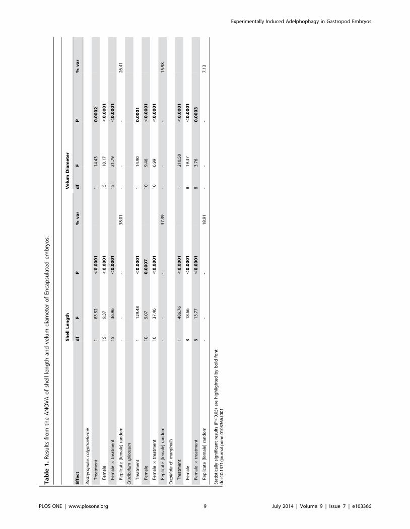
Shell length and velum diameter were both significantly larger
in treated embryos compared to control embryos in all three
species (Figure 5; Table 1). Mean shell lengths were between 3%
and 7% larger in treated embryos than in controls and velum
diameters were between 2% and 7% larger in treated embryos
than in controls (Figure 5). In all three there were significant
effects of female and a significant interaction between female and
treatment (Table 1; Figure 6). The random effect of replicate
nested within female explained 19–38% of the variation in shell
length, and 7–26% of the variation in velum diameter. This is
most likely due to a combination of slight differences in egg size
between capsules and differences in the number of embryos that
were successfully smashed in our experimental treatment.
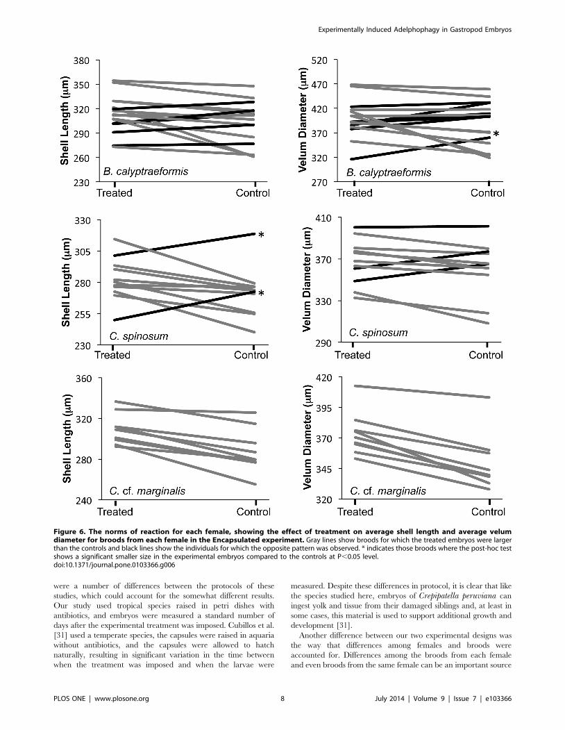
In Crepidula cf. marginalis the significant interaction between
female and treatment was due to differences in slopes in the
reaction norms for each female; but the response to the treatment
was in the same direction for all females (Figure 6). For C.spinosum two females had a significantly larger shell length in the
controls compared to the treated embryos (black lines in Figure 6;
post-hoc Tukey’s HSD test P,0.05). This response was evident in
all of the replicates for these females. However, it was observed
that the treated capsules were full of a yolky emulsion when they
were opened. For some reason the embryos from these females
may not have been able to clear the capsules of the particles of
dead siblings which may have inhibited their natural growth
compared to controls. Alternately the remaining embryos them-
selves may have been disintegrating for some reason. This
emulsion was not seen in any of the treated capsules that
produced embryos with larger shell lengths or velar diameters
compared to their controls. Three C. spinosum females, including
the two with unusual yolky capsular fluid, also had larger average
velum diameters in the controls than the treated embryos
(Figure 6). None of these comparisons were significant in the post
hoc test (P.0.05). For Bostrycapulus calyptraeformis the response
of each female to the treatment was less consistent than in the
other two species. Here, despite an overall significant increase in
shell length and velum diameter in the treated embryos, 5 females
showed smaller shell lengths in treated capsules (none of which
were significant at P,0.05 in Tukey’s HSD post-hoc tests) and 7
Experimentally Induced Adelphophagy in Gastropod Embryos
PLOS ONE | www.plosone.org 3 July 2014 | Volume 9 | Issue 7 | e103366

females showed smaller velum diameters (one of which was
significant in Tukey’s HSD post-hoc tests). These non-significant
post-hoc comparisons suggest that the significant interaction term
is due to a lack of a significant response to the treatment in a
minority of the females, rather than differences between females in
the direction of significant responses.
The experimental treatment did not alter the overall allometry
between the average velum diameter and average shell length in
Bostrycapulus calyptraeformis or Crepidula cf. marginalis (Fig-
ure 7). ANCOVA showed that in all three species velum diameter
was positively correlated with shell length (P,0.0001) but was not
affected by treatment (P = 0.75; P = 0.95; P = 0.16; B. calyptrae-formis, C. spinosum and C. cf. marginalis respectively). The lack of
an effect of treatment on velum size in this ANCOVA is due to
smaller sample sizes and low power when individuals are averaged
within each replicate dish, in contrast to the significant effect
recovered by the nested ANOVA above. The ANCOVA
recovered no effect of the interaction between treatment and shell
length on velum size for B. calyptraeformis (P = 0.66) or C. cf.
marginalis (P = 0.47). The interaction was significant for C.spinosum (P = 0.042), where the velum size increased more slowly
with increasing shell length in treated embryos than in control
embryos (Figure 7). This relationship disappeared when the two
females with yolky emulsion in the treated capsules were removed
from the analysis.
The experimental treatment did not increase the variability of
shell length or velum diameter for B. calyptraeformis or C.spinosum (Table 2). There was no significant effect of treatment
nor a significant interaction between female and treatment on the
coefficient of variation in shell size or velum diameter. One female
B. calyptraeformis had unusually variable embryos and her 3
replicate control capsules were identified as outliers from visual
inspection of the residuals plot. When she was included in the
analysis there was a significant interaction between female and
treatment for that species (P = 0.015). When her broods were
removed from the analysis this effect was no longer significant
(Table 2). In C. cf. marginalis there was an unexpected significant
effect of treatment on variation in shell length. Treated embryos
had significantly less variation (CV = 3.64) in shell length than
control embryos (CV = 4.34).
Excapsulated ExperimentEmbryos subjected to the excapsulated treatment often became
contaminated with microorganisms, resulting in the abnormal
Figure 1. Experimental protocol for the Encapsulated and Excapsulated experiments. Capsules were placed into 6 replicate dishes, 3assigned to the experimental treatment and 3 to the control. Solid black circles indicate embryos and hashed circles indicate embryos that weresmashed.doi:10.1371/journal.pone.0103366.g001
Experimentally Induced Adelphophagy in Gastropod Embryos
PLOS ONE | www.plosone.org 4 July 2014 | Volume 9 | Issue 7 | e103366

development of the embryos. However replicate broods with
normal-appearing embryos were measured for 12 females. For
these there was no significant effect of treatment on shell length,
but velum diameter was significantly larger in treated than in
control embryos (Table 3; Figure 5). For both velum diameter and
shell length there were significant effects of female or the
interaction between female and treatment. The random effect of
replicate dish accounted for 11% of the variation in velum
diameter and 34% in shell length. Treatment did not alter the
allometry between shell length and velum diameter (ANCOVA:
treatment 6 shell length P.0.05). Again the power to detect a
difference was reduced by the use of means for each dish.
There was a significant effect of treatment (P,0.005) and
female (P,0.001), and of the interaction between female and
treatment (P,0.005) on CV in shell length. Treated embryos were
more variable (CV = 4.08) than control embryos (CV = 3.15).
The significant effect of treatment was due entirely to the broods
from two females (with CV = 9.75 and 7.13 for treated embryos).
One of these had significantly smaller and less well-developed
embryos than any other female. It is possible that the treatment
was applied at a slightly earlier developmental stage for this brood.
Treatment had no significant effect on the CV of velum diameter.
Discussion
Our results and previously published data on Crepipatellaperuviana [31] indicate that the ability to consume, absorb, and
utilize material derived from dead and damaged siblings occurs in
Figure 2. Yolk consumption of Crepidula cf. marginalis in the Encapsulated experiment. A. Control embryos in an untreated capsule. B.Embryos in the capsule immediately after the manipulation that smashed some of the embryos. C. and D. Experimental capsules 1.5 hours after thetreatment. The fine yolk particles have been cleared from the capsule and only some larger particles, most of which will disintegrate over the nextfew days, remain.doi:10.1371/journal.pone.0103366.g002
Experimentally Induced Adelphophagy in Gastropod Embryos
PLOS ONE | www.plosone.org 5 July 2014 | Volume 9 | Issue 7 | e103366
embryos of planktotrophic calyptraeids from 4 genera. Destruction
of 20–40% of the embryos within a capsule resulted in a
statistically significant increase in shell length and velum diameter
in the three species studied here. A similar result was also obtained
for some embryos of Crepipatella peruviana, but the results of this
previous study were mixed [31]. Embryos in that study which took
a particularly long time to hatch showed a significant increase in
shell length and velum area. Those that hatched particularly
quickly showed the opposite effect, and those that hatched after
the average duration showed no effect of the treatment. There
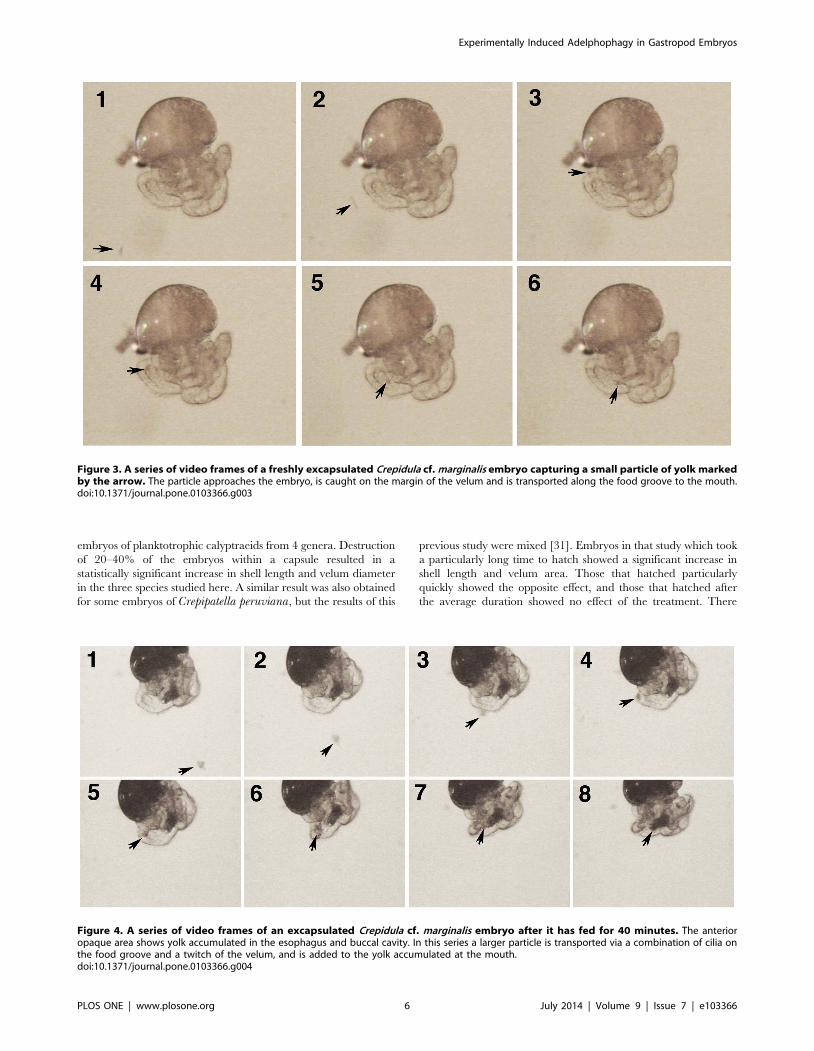
Figure 3. A series of video frames of a freshly excapsulated Crepidula cf. marginalis embryo capturing a small particle of yolk markedby the arrow. The particle approaches the embryo, is caught on the margin of the velum and is transported along the food groove to the mouth.doi:10.1371/journal.pone.0103366.g003
Figure 4. A series of video frames of an excapsulated Crepidula cf. marginalis embryo after it has fed for 40 minutes. The anterioropaque area shows yolk accumulated in the esophagus and buccal cavity. In this series a larger particle is transported via a combination of cilia onthe food groove and a twitch of the velum, and is added to the yolk accumulated at the mouth.doi:10.1371/journal.pone.0103366.g004
Experimentally Induced Adelphophagy in Gastropod Embryos
PLOS ONE | www.plosone.org 6 July 2014 | Volume 9 | Issue 7 | e103366

Figure 5. Bar graph showing the effect of the Encapsulated and Excapsulated experiments on shell length and velum diameter ofencapsulated Bostrycapulus calyptraeformis (B. calyp), encapsulated Crucibulum spinosum (C. spin), encapsulated Crepidula cf. marginalis(Encap marg), and excapsulated Crepidula cf. marginalis (Excap marg). * indicates statistical significance at the P,0.05 level (see Tables 1 & 3for details). Mean shell lengths for encapsulated embryos (treated vs. controls): B. calyptraeformis: 312.70 mm (s.e. = 1.98) versus 302.37 mm (s.e.= 2.07); C. spinosum: 282.55 mm (s.e. = 2.19) versus 272.04 mm (s.e. = 2.18); C. cf. marginalis: 309.42 mm (s.e. = 1.46) versus 287.93 mm (s.e. = 1.46).Mean velum diameters for encapsulated embryos (treated vs. controls): B. calyptraeformis: 398.78 mm (s.e. = 7.78) versus 380.80 mm (s.e. = 7.81); C.spinosum: 367.24 mm (s.e. = 2.44) versus 360.66 mm (s.e. = 2.44); C. cf. marginalis: 373.44 mm (s.e. = 1.72) versus 349.10 mm (s.e. = 1.74).doi:10.1371/journal.pone.0103366.g005
Experimentally Induced Adelphophagy in Gastropod Embryos
PLOS ONE | www.plosone.org 7 July 2014 | Volume 9 | Issue 7 | e103366
were a number of differences between the protocols of these
studies, which could account for the somewhat different results.
Our study used tropical species raised in petri dishes with
antibiotics, and embryos were measured a standard number of
days after the experimental treatment was imposed. Cubillos et al.
[31] used a temperate species, the capsules were raised in aquaria
without antibiotics, and the capsules were allowed to hatch
naturally, resulting in significant variation in the time between
when the treatment was imposed and when the larvae were
measured. Despite these differences in protocol, it is clear that like
the species studied here, embryos of Crepipatella peruviana can
ingest yolk and tissue from their damaged siblings and, at least in
some cases, this material is used to support additional growth and
development [31].
Another difference between our two experimental designs was
the way that differences among females and broods were
accounted for. Differences among the broods from each female
and even broods from the same female can be an important source
Figure 6. The norms of reaction for each female, showing the effect of treatment on average shell length and average velumdiameter for broods from each female in the Encapsulated experiment. Gray lines show broods for which the treated embryos were largerthan the controls and black lines show the individuals for which the opposite pattern was observed. * indicates those broods where the post-hoc testshows a significant smaller size in the experimental embryos compared to the controls at P,0.05 level.doi:10.1371/journal.pone.0103366.g006
Experimentally Induced Adelphophagy in Gastropod Embryos
PLOS ONE | www.plosone.org 8 July 2014 | Volume 9 | Issue 7 | e103366
Ta
ble
1.
Re
sult
sfr
om
the
AN
OV
Ao
fsh
ell
len
gth
and
velu
md
iam
ete
ro
fEn
cap
sula
ted
em
bry
os.
Sh
ell
Le
ng
thV
elu
mD
iam
ete
r
Eff
ect
df
FP
%v
ar
df
FP
%v
ar
Bo
stry
cap
ulu
sca
lyp
tra
efo
rmis
Tre
atm
en
t1
83
.52
,0
.00
01
11
4.4
30
.00
02
Fem
ale
15
9.3
7,
0.0
00
11
51
0.1
7,
0.0
00
1
Fem
ale6
tre
atm
en
t1
53
6.9
6,
0.0
00
11
52
1.7
9,
0.0
00
1
Re
plic
ate
[fe
mal
e]
ran
do
m-
--
38
.01
--
-2
6.4
1
Cru
cib
ulu
msp
ino
sum
Tre
atm
en
t1
12
9.4
8,
0.0
00
11
14
.90
0.0
00
1
Fem
ale
10
5.0
70
.00
07
10
9.4
6,
0.0
00
1
Fem
ale6
tre
atm
en
t1
03
7.4
6,
0.0
00
11
06
.99
,0
.00
01
Re
plic
ate
[fe
mal
e]
ran
do
m-
--
37
.39
--
-1
5.9
8
Cre
pid
ula
cf.
ma
rgin
alis
Tre
atm
en
t1
48
6.7
6,
0.0
00
11
21
0.5
0,
0.0
00
1
Fem
ale
81
8.6
6,
0.0
00
18
19
.37
,0
.00
01
Fem
ale6
tre
atm
en
t8
13
.77
,0
.00
01
83
.76
0.0
00
3
Re
plic
ate
[fe
mal
e]
ran
do
m-
--
18
.91
--
-7
.13
Stat
isti
cally
sig
nif
ican
tre
sult
s(P
,0
.05
)ar
eh
igh
ligh
ted
by
bo
ldfo
nt.
do
i:10
.13
71
/jo
urn
al.p
on
e.0
10
33
66
.t0
01
Experimentally Induced Adelphophagy in Gastropod Embryos
PLOS ONE | www.plosone.org 9 July 2014 | Volume 9 | Issue 7 | e103366

of variation in offspring size in calyptraeids, often accounting for
almost half of the observed variation [5,33,38]. For all three
species studied here, a statistically significant effect of ‘‘female’’ was
recovered in virtually all of the tests. We used only a single brood
from each female and all of the broods were not collected from
them at exactly the same moment in development. Therefore this
‘‘female’’ effect incorporates a combination of variation among
females, among broods from the same female which may differ in
egg size or quality, as well as any differences in the exact stage at
which the experimental treatment was imposed. Differences
between capsules from the same clutch, variation due to our
inability to exactly replicate the smashing treatment, as well as any
dish effects were accounted for by the random effect of replicate.
The experimental design of Cubillos et al. [31], which did not
explicitly take into account this variation, may have had reduced
power to detect the significant effects of the treatments if ‘‘female’’
effects account for a large proportion of the variance in C.peruviana. An intriguing suggestion of their results is that embryos
exposed to the treatment earlier (i.e., longer time to hatch) respond
differently to the availability of yolky material than do embryos
that are exposed later in development. It would be interesting to
test this possibility explicitly.
The magnitude of the effects of consuming siblings detected
here were modest. In C. peruviana a 16% average increase in shell
length was detected in broods that showed an increase in size in
treated embryos. In the present study, however, the average shell
lengths of experimental embryos only exceeded those of controls
by 3–7%. In neither study did the shell length of artificially
adelphophagic embryos approach the length at hatching of species
that are naturally adelphophagic, nor did we expect them to.
Because 20%–40% of the embryos in each capsule were killed, we
expected each remaining embryo to consume less than the
material from a single sibling. In normally adelphophagic species
the number of nurse embryos usually exceeds the number of
embryos that will complete development by an order of
magnitude. For example in Crepipatella dilatata, the sister species
to C. peruviana, each developing embryo consumes between 15
and 35 uncleaved nurse eggs to produce a 1–1.8 mm hatchling
[39]. In addition, in our experiment, we opened the capsules 3
days after the experimental treatment was imposed, while the
embryos still retained some little yolk. It is possible that embryos
that had ingested material from siblings could have continued
growth and development for longer than controls and ultimately
demonstrated a larger effect. Our standardized protocol could not
detect this possible greater potential for growth of the treated
embryos, because capsules of these species do not hatch naturally
away from the mother. Even if the conversion of ingested material
to embryonic growth is not as efficient in normally planktotrophic
embryos as in embryos that normally utilize nurse eggs or
embryos, providing a greater number of damaged siblings, over a
longer span of development than used here might produce a larger
effect similar to Cubillos et al. [31]. If a larger effect could be
produced it would be possible to examine the effects of changes in
size induced by this treatment on larval growth and survival.
We did not observe changes in the variation or morphology of
the embryos that indicated increased similarity to species with
adelphophagic development. It is well documented that one of the
consequences of adelphophagy is an increased variation in
hatchling size within and among females [5]. Our treated embryos
did not generally show a significant increase in variation in either
shell length or velum diameter. This could be due to the overall
small effect of the treatment. A treatment with a larger absolute
effect on embryo size might have produced a more easily
detectable change in variation. We also failed to detect significant
differences in shell-velum allometry in treated and control
embryos. We had predicted that increased energy from consump-
tion of siblings would reduce the velum size relative to shell length
in treated embryos. This prediction was based on two observa-
tions. It is common for embryos of both adelphophagic species and
species that develop from large yolky eggs to have velar lobes that
are smaller relative to the shell length than in planktotrophic
species ([18]; Collin per. obs.). Also, changes in shell-velum
allometry have been induced by different food rations in
planktotrophic mollusc larvae, with well-fed larvae having
relatively smaller velar lobes than less well-fed larvae [40,41].
We failed to find a change in allometry in response to consumption
of dead siblings, instead it appeared that the increase in resources
resulted in a uniform increase in size. The small overall effect of
our treatment and the use of dish averages make it difficult to
strongly demonstrate that there was no change in allometry
Our results suggest that planktotrophic calyptraeid embryos
typically express features that enable them to take advantage of
Figure 7. Effect of treatment on allometry between averageshell length and average velum diameter in treated andcontrol dishes. There were no significant effects of treatment onallometry in any species when the two females with yolky emulsion intheir capsules were removed from the analysis of C. spinosum (see textfor details).doi:10.1371/journal.pone.0103366.g007
Experimentally Induced Adelphophagy in Gastropod Embryos
PLOS ONE | www.plosone.org 10 July 2014 | Volume 9 | Issue 7 | e103366

abnormally developing embryos, and that embryologically the
evolution of adelphophagy might be fairly easy. Head vesicle stage
embryos of most planktotrophic species have ciliation on the
velum and head vesicle (e.g., [42]) that can be used to capture
small particles [28,41]. Assays have shown that head vesicle stage
embryos of Crepipatella peruviana and diverse other calyptraeids
can ingest 2 mm plastic beads [28] (R. Collin unpublished data).
The cilia of the velum and sometimes the uniform ciliation of the
head vesicle can sweep various kinds of debris into the mouth in
embryos of several planktotrophic Crepidula and Bostrycapulusspecies (Figures 3 & 4; R. Collin pers. obs.). It seems likely from
these observations that the ability to capture particles in pre-
hatching stages of development is widespread among plankto-
trophic calyptraeids. What is less well known is the ability of these
embryos to absorb and utilize the material they capture. The
results of the present study and [31] demonstrate that all species
that have been examined to date can grow in response to ingesting
particles of their siblings. This ability may be widespread in
calyptraeids, and may mean that the evolution of adelphophagy
does not require any special embryological modifications,
explaining the frequent parallel evolution of adelphophagy in this
family. It would be interesting to conduct similar experiments in
vermetids and muricids, where adelphophagy is common, to
determine if the ability to consume and utilize damaged siblings is
common across families. Comparisons with a group that lacks
adelphophagy entirely could be used to determine if constraints on
the ability of normal embryos to benefit from the ingestion of
siblings inhibit the evolution of adelphophagy in those groups.
Table 2. Results of ANOVA on coefficient of variation of shell length and velum diameter for each replicate dish of encapsulatedembryos.
Shell Length Velum Diameter
Effect df F P df F P
Bostrycapulus calyptraeformis
Treatment 1 0.01 0.26 1 0.62 0.44
Female 13 4.21 ,0.005 14 5.89 ,0.0001
Female 6 treatment 13 1.55 0.65 14 0.76 0.54
Crucibulum spinosum
Treatment 1 0.76 0.38 1 0.25 0.62
Female 10 2.01 0.06* 10 1.79 0.09*
Female 6 treatment 10 1.57 0.15 10 0.69 0.73
Crepidula cf. marginalis
Treatment 1 3.73 0.06# 1 1.32 0.26
Female 8 2.81 0.02 8 2.71 0.02
Female 6 treatment 8 0.63 0.74 8 0.67 0.72
Statistically significant results (P,0.05) are highlighted by bold font.*These factors do not become significant with the removal of the non-significant interaction term.#This factor becomes significant with the removal of the non-significant interaction (P = 0.009).doi:10.1371/journal.pone.0103366.t002
Table 3. Results from the ANOVA on shell length, velum diameter, and coefficient of variation of each from Excapsulated embryosof Crepidula cf. marginalis.
Shell Length Velum Diameter
Effect df F P % var df F P % var
Treatment 1 2.51 0.11 1 9.21 0.0025
Female 11 18.50 ,0.0001 15 22.48 ,0.0001
Female 6 treatment 11 24.34 ,0.0001 15 7.47 ,0.0001
Replicate [female] random - - - 33.56 - - - 11.24
CV Shell Length CV Velum Diameter
Effect df F P df F P
Treatment 1 11.73 0.001 1 0.05 0.82
Female 11 10.39 ,0.0001 11 4.05 0.0004
Female 6 treatment 11 3.22 0.003 11 0.55 0.86
Statistically significant results (P,0.05) are highlighted by bold font.doi:10.1371/journal.pone.0103366.t003
Experimentally Induced Adelphophagy in Gastropod Embryos
PLOS ONE | www.plosone.org 11 July 2014 | Volume 9 | Issue 7 | e103366

Supporting Information
Video S1 Crepidula cf. marginalis embryos capturing particles.
This video shows several sequences of embryos using the cilia on
the velum to capture yolk particles from damaged siblings.
(M4V)
Acknowledgments
This work was conducted in partial fulfillment of the requirements for a
Master’s degree program at the Universitat Oldenburg, Germany. We wish
to thank members of the Collin lab for logistical support and comments on
the manuscript and Autoridad de Recursos Acuaticos de Panama (ARAP)
for permission to collect animals in Panama.
Author Contributions
Conceived and designed the experiments: RC OT. Performed the
experiments: OT ACB. Analyzed the data: RC OT ACB. Contributed
reagents/materials/analysis tools: RC. Wrote the paper: RC.
References
1. Thorson G (1946) Reproduction and larval development of Danish marinebottom invertebrates with special reference to the planktonic larvae in the Sound
(Oresund). Medd Komm Dan Fisk- Havunders (Ser Plancton) 4: 1–523.
2. Thorson G (1950) Reproductive and larval ecology of marine bottom
invertebrates. Biol Rev 25: 1–45.
3. Strathmann RR (1985) Feeding and nonfeeding larval development and life-
history evolution in marine invertebrates. Ann Rev Ecol Syst 16: 339–
4. Collin R (2003) World-wide patterns of development in calyptraeid gastropods.
Mar Ecol Prog Ser 247: 103–122.
5. Collin R, Spangler A (2012) Does adelphophagic development decouple the
effects of temperature on egg size and hatching size? Biol Bull 223: 268–277.
6. Spight TM (1975) Factors extending gastropod embryonic development and
their selective cost. Oecologia 21: 1–16.
7. Spight TM (1976) Hatching size and the distribution of nurse eggs among
prosobranch embryos. Biol Bull 150: 491–499.
8. Rivest BR (1983) Development and the influence of nurse egg allotment on
hatching size in Searlesia dira (Reeve, 1846) (Prosobranchia: Buccinidae). J ExpMar Biol Ecol 69: 217–241.
9. Smith KE, Thatje S (2013) Nurse egg consumption and intracapsulardevelopment in the common whelk Buccinum undatum (Linnaeus 1758).
Helgoland Marine Research, 1–12.
10. Kohn AJ Perron FE (1994) Life history and biogeography: patterns in Conus.
Oxford: Clarendon Press.
11. Blake JA, Arnofsky PL (1999) Reproduction and larval development of the
spioniform Polychaeta with application to systematics and phylogeny. Hydro-
biologia 402: 57–106.
12. Collin R (2012a) Non-traditional life history choices: What can ‘‘intermediates’’
tell us about evolutionary transitions between modes of invertebrate develop-ment? Integr Comp Biol 52: 128–137.
13. Oyarzun FX, Strathmann RR (2011) Plasticity of hatching and the duration ofplanktonic development in marine invertebrates. Integr Comp Biol 51: 81–90.
14. Kamel SJ, Oyarzun FX, Grosberg RK (2010) Reproductive biology, familyconflict, and size of offspring in marine invertebrates. Integr Comp Biol 50: 619–
629.
15. Brante A, Fernandez M, Viard F (2013) Non-random sibling cannibalism in the
marine gastropod Crepidula coquimbensis. PLoS One
16. Gallardo CS, Garrido OA (1987) Nutritive egg formation in the marine snails
Crepidula dilatata and Nucella crassilabrum. Int J Invert Reprod Dev 11: 239–
254.
17. Averbuj A, Rocha MN, Zabala S (2014) Embryonic development and
reproductive seasonality of Buccinanops globulosus (Nassariidae) (Kiener,1834) in Patagonia, Argentina. Invert Reprod Dev 58: 138–147.
18. Collin R (2004) The loss of complex characters, phylogenetic effects, and theevolution of development in a family of marine gastropods. Evolution. 58: 1488–
1502.
19. Veliz D, Guisado C, Winkler F (2001) Morphological, reproductive, and genetic
variability among three populations of Crucibulum quiriquinae (Gastropoda:Calyptraeidae) in northern Chile. Mar Biol 139: 527–534.
20. Veliz D, Winkler FM, Guisado C (2003) Developmental and genetic evidencefor the existence of three morphologically cryptic species of Crepidula in
northern Chile. Mar Biol 143: 131–142.
21. Gibson G, Hart C, Coulter C, Xu H (2012) Nurse eggs form through an active
process of apoptosis in the spionid Polydora cornuta (Annelida). Integr Comp
Biol 52: 151–160.
22. Fioroni P (1982) Larval organs, larvae, metamorphosis and types of development
of Mollusca: A comprehensive review. Zool Jb Anat 108: 375–42023. Calvo M, Templado J (2004) Reproduction and development in a vermetid
gastropod, Vermetus triquetrus. Invert Biol 123: 289–303.24. Strathmann MF, Strathmann RR (2006) A vermetid gastropod with complex
intracapsular cannibalism of nurse eggs and sibling larvae and a high potential
for invasion. Pacific Sci 60: 97–108.25. Smith KE, Thatje S (2013) The subtle intracapsular survival of the fittest:
maternal investment, sibling conflict or environmental effects? Ecology 94:2263–2274.
26. Lesoway MP, Abouheif E, Collin R (2014) Development of Crepidula cf. onyx a
model system for adelphophagy in gastropods. International J Develop Biol. Inpress.
27. Hoagland KE (1986) Patterns of encapsulation and brooding in theCalyptraeidae (Prosobranchia: Mesogastropoda). Am Malacol Bull 4: 173–183.
28. Collin R, Chaparro OR, Winkler F, Veliz D (2007) Molecular phylogenetic andembryological evidence that feeding larvae have been reacquired in a marine
gastropod. Biol Bull 212: 83–92.
29. Chaparro OR, Charpentier JL, Collin R (2002) Embryonic velar structure andfunction of two sibling species of Crepidula with different modes of development
Biol Bull 203: 80–86.30. McDonald KA, Collin R, Lesoway MP (2014) Poecilogony in the caenogas-
tropod Calyptraea lichen. Invert Biol doi:10.1111/ivb.12057
31. Cubillos VM, Chaparro OR, Montiel YA, Veliz D (2008) Unusual source offood: impact of dead siblings on encapsulated embryo development of
Crepipatella fecunda (Gastropoda: Calyptraeidae). Mar Freshw Res 58: 1152–1162.
32. Veliz D, Winkler FM, Guisado C, Collin R (2012) A new species of Crepipatella(Gastropoda: Calyptraeidae) from northern Chile. Moll Res. 32: 145–153.
33. Collin R (2012) Temperature-mediated trade-offs in the life histories of two
slipper limpets (Gastropoda: Calyptraeidae) with planktotrophic development.Biol J Linn Soc 106: 763–775.
34. Merot C Collin R (2012) Effects of food availability on sex change in two speciesof Crepidula (Gastropoda: Calyptraeidae). Mar Ecol Prog Ser 449: 173–181.
35. Collin R, Starr MJ (2013) Comparative ontogenetic changes in enzyme activity
during embryonic development of calyptraeid gastropods. Biol Bull 225: 8–17.36. Henry JJ, Collin R, Perry KJ (2010) The slipper snail, Crepidula: an emerging
lophotrochozoan model system. Biol Bull 218: 211–229.37. Rasband WS (1887–2012) ImageJ, U.S. National Institutes of Health, Bethesda,
Maryland, USA, imagej.nih.gov/ij/
38. Collin R (2010) Repeatability of egg size in two marine gastropods: brood orderand female size do not contribute to intraspecific variation. Mar Ecol Prog Ser
410: 89–96.39. Chaparro OR, Oyarzun RF, Vergara AM, Thompson RJ (1999) Energy
investment in nurse eggs and egg capsules in Crepidula dilatata Lamarck(Gastropoda, Calyptraeidae) and its influence on the hatching size of the
juvenile. J Exp Mar Biol Ecol 232: 261–274.
40. Strathmann RR, Fenaux L, Sewell AT, Strathmann MF (1993) Abundance offood affects relative size of larval and postlarval structures of a molluscan veliger.
Biol Bull 185: 232–239.41. Klinzing MSE, Pechenik JA (2000) Evaluating whether velar lobe size indicates
food limitation among larvae of the marine gastropod Crepidula fornicata. J Exp
Mar Biol Ecol 252: 255–279.42. Collin R (2000) Sex change, reproduction and development of Crepidula adunca
and C. lingulata (Gastropoda: Calyptraeidae). Veliger 43: 24–33.
Experimentally Induced Adelphophagy in Gastropod Embryos
PLOS ONE | www.plosone.org 12 July 2014 | Volume 9 | Issue 7 | e103366
Copyright © 2022 FDOKUMEN