Cadmium contaminated soil affects retinogenesis in lizard embryos
Upload
independentCategory
view
2download
0
Acta Zoologica
(Stockholm)
89
: 231–251 (July 2008) doi: 10.1111/j.1463-6395.2007.00311.x
© 2007 The AuthorsJournal compilation © 2007 The Royal Swedish Academy of Sciences
231
Abstract
Kundrát, M., Cruickshank, A. R. I., Manning, T. W. and Nudds, J. 2008.Embryos of therizinosauroid theropods from the Upper Cretaceous of China:diagnosis and analysis of ossification patterns. —
Acta Zoologica
(Stockholm)
89
: 231–251
Exceptionally complete,
in ovo
dinosaur embryos from the Upper Cretaceousof China are analysed. Ossification patterns of these embryos suggest that theydied during the final third of their development. The therizinosauroid identity ofthe embryos follows from: (1) an edentulous premaxilla with a sharp downturnededge; (2) dentary with a lateral shelf; (3) teeth with fan-shaped crowns, with afew marginal cusps; (4) humerus with a massive deltopectoral crest extendingproximally, with a pointed proximomedial tuberosity; (5) ilium with an expandedand hooked preacetabular process; (6) strongly curved hypertrophied manualunguals tapering to sharp points. These embryos are closest to two Chinesetherizinosauroids,
Neimongosaurus yangi
Zhang
et al
. 2001 and
Erliansaurusbellamanus
Xu
et al
. 2002. An elongated narial opening, reduced basipterygoidprocess, low cervical neural spines, a transversely narrow pubic apron, and a pubicfoot expanded anteriorly are found in these embryos and are synapomorphiesuniting the Therizinosauroidea and the Oviraptorosauria. Fusion of cervicaland caudal neural arches and centra, complete ossification of thoracic ribs andilium, possible co-ossification of tibia and fibula, fused pubes, complete meta-and acropodial elements, together with small portions of unossified epiphysesof long bones suggest an advanced precociality of these embryos.
M. Kundrát, Geological Institute, Slovak Academy of Sciences, Severná 5, SK-97401, Banská Bystrica, Slovak Republic. E-mail: [email protected]
(Correction added after online publication 10 June 2008: Corresponding author’s address and e-mail have been changed.)
Blackwell Publishing Ltd
Embryos of therizinosauroid theropods from the Upper Cretaceous of China: diagnosis and analysis of ossification patterns
Martin Kundrát,
1
Arthur R. I. Cruickshank,
2
Terry W. Manning
3
and John Nudds
4
1
Redpath Museum – Biology Department, McGill University, 859 Sherbrooke Street West, Montreal, Quebec, Canada, H3A 2K6;
2
Department of Geology, University of Leicester, Leicester LE1 7RH, UK;
3
Rock Art, 4 Gipsy Lane, Leicester LE4 6RB, UK;
4
School of Earth, Atmospheric and Environmental Sciences, University of Manchester, Oxford Road, Manchester M13 9PL, UK
Keywords:
Theropoda, Therizinosauroidea, embryo, systematics, phylogeny, ontogeny, development, ossification, precociality
Accepted for publication:
28 September 2007
Introduction
Therizinosauroids form one of the rarest and most enigmaticgroups of theropod dinosaurs. They had a bizarre skeletalconstruction, walked on four digits of their hindfeet, andhave often been interpreted as herbivores (Barsbold andMaryañska 1990; Clark
et al
. 2004). Embryos within theeggs of this species (Fig. 1A,B), from the Upper CretaceousNanchao Formation of Henan Province, China, have beenrevealed by acid etching (Cohen
et al
. 1995; Manning
et al
.1997). Extraordinary preservation of almost completespecimens represents the most valuable specimens of
dinosaur embryos ever found. The co-occurrence of eggs andembryos is still relatively rare so the material available for thisstudy provided a significant and novel opportunity to studyvarious enigmatic aspects of the embryology of an extinctnon-avian dinosaur. Of particular interest is the opportunityto examine new skeletal characteristics of therizinosauroidsand analyse the developmental changes that occurred inthese specimens throughout a late period of embryogenesis.This research has significant potential to elucidate poorlyunderstood aspects of dinosaur skeletal embryogenesis andprovides new sources of data to contribute to the under-standing of the evolutionary history of these animals.

Therizinosauroid embryos from China
•
Kundrát
et al.
Acta Zoologica
(Stockholm)
89
: 231–251 (July 2008)
© 2007 The Authors
232
Journal compilation © 2007 The Royal Swedish Academy of Sciences
The objectives of the present study are: (1) to provide abroad basis of anatomical information on dinosaur embryonicskeletons and to diagnose their theropodan identity; (2) toanalyse the phylogenetic relationship of the embryos bycomparing their skeletal characters with those of other
theropods; (3) to compare the embryos with other relevantdinosaur embryos and corresponding developmental stagesof recent crocodilians and precocial birds for the presence orabsence of ossifications in their skeletal elements; (4) tospeculate on the functional design of the skeletons of thetherizinosauroid neonates from the perspective of differentontogenetic traits such as incubation period, precociality vs.altriciality, parent–offspring relation, hatchling locomotoractivity, and postnatal growth.
We provide descriptions of well-preserved craniofacial,neurocranial and postcranial characters of therizinosauroidembryos, and compare these with corresponding features inother non-avian and avian theropods regardless of ontogeneticstage. We then discuss the following topics: (1) diagnosticskeletal features that allocate the embryos to the Theropodaand Therizinosauroidea, respectively; (2) classification ofdevelopmental patterns of the embryos within the precocialspectrum of recent archosaurs, including birds; and (3)evidence from the fossil record of theropodan embryos topropose specific characters related to the evolution oftheropodan prenatal ontogenies.
Materials and Methods
Source of embryos
The collection of eggs analysed here is reported to have comefrom the Late Cretaceous (about 75–85 million years ago;Campanian–Santonian) Nanchao Formation of theNanyang Valley near Xinye, Henan Province, China. Embryonicremains were prepared by one of us (T.W.M.) and display themost remarkable ontogenetic details among all knownembryonic dinosaurs. The specimens are currently depositedin New Walk Museum, Leicester, UK. The primary numberingsystem ‘RAP’ as originally applied (Manning
et al
. 2000) iscombined with temporary accession numbers ‘CAGS’provided by the Chinese Academy of Geological Sciencesin Beijing, China, pending their permanent repository in amuseum in China. Because of the belief that the eggs(approximately 70
×
90 mm in size) and embryos belong toa small-to-medium size theropod, ‘RAP’ has been chosen toindicate a raptorial affinity. The prefix ‘RAP’ is followed by acombination of serial number and year. The ‘D’ suffix isapplied when preparation was completed. Thus for exampleRAP 194D is applied to the first therizinosauroid embryonicspecimen completely prepared in 1994. The following eggshave been used in this study: RAP 194D: CAGS-01-IG-1;RAP 3495D: CAGS-01-IG-2; RAP 3494D: CAGS-01-IG-4; RAP 10495D: CAGS-01-IG-5; RAP 1496D: CAGS-01-IG-6; RAP 7294D: CAGS-01-IG-7; RAP 1294D: CAGS-01-IG-11; RAP 294D: CAGS-01-IG-12 (Cohen
et al
. 1995).Seven eggs were found in a single clutch [CAGS-01-IG-13].Five of these were prepared (RAP 796D, RAP 1496D,RAP 1196D, 596D, 696D) and embryonic remains werefound inside the first three.
Fig. 1—Therizinosauroid embryos from Nanchao Formation, Santonian-Campanian, Upper Cretaceous, Nanyang Valley, Henan Province, China. —A. Skeletal remains in ovo, CAGS-01-IG-1. —B. Reconstruction of a therizinosauroid embryo made by Pavel Ríha under supervision of Martin Kundrát, 2003. (Corrections added after online publication 10 June 2008: Duplicate text was deleted.)

Acta Zoologica
(Stockholm)
89
: 231–251 (July 2008)
Kundrát
et al.
•
Therizinosauroid embryos from China
© 2007 The AuthorsJournal compilation © 2007 The Royal Swedish Academy of Sciences
233
Embryo preparation
Early efforts to use conventional and computed tomographyscanning techniques to investigate the eggs for the detectionof embryonic remains were frustrated by the presence ofbarytes, calcite and various grades of sediment in the enclosingmatrix. Eventually, one of us (T.W.M.) adapted a chemicalmethod that etches away only 10
μ
m of the rock a day.Manning
et al
. (1997) found that the usual concentration of5% acetic acid was too strong, being prone to erode the verydelicate bones of the preserved embryos. At less aggressiveconcentrations of 0.5–2.0%, however, the acid solutionbecomes locally stratified, with saturated acetate solutionnext to the fossils inhibiting progressive action of the acid.Manning
et al
. (1997) overcame this problem by agitatingthe solution, which allowed the active acid to remain in directcontact with the exposed bone. A small quantity of wettingagent and an algal inhibitor were added to the etchingsolution to maintain the rate of reaction with the specimenand prevent subsequent appearance of microorganisms. Atintervals during the acid treatment, digested matrix wascarefully washed away, using weak, precisely controlled jetsof distilled water; even fine needles were too coarse for thisstage of the process (Manning
et al
. 1997). Then thespecimens were washed in running tap water, air-dried andfinally the exposed bone was protected with dilute ParaloidB72 in acetone (Manning
et al
. 1997, 2000).
Whole-mount skeletal preparation
For comparative purposes
Alligator mississippiensis
embryoswere provided by Dr James Perran Ross from the Depart-ment of Wildilfe Ecology and Conservation, University ofFlorida, Gainesville, FL, USA. Cleared whole-mount speci-mens stained for bone with Alizarin Red and for cartilagewith Alcian Blue were prepared following the enzyme clear-ing procedure of Dingerkus and Uhler (1977). Photographswere taken using a digital camera Nikon Coolpix 995 andprocessed with A
dobe
P
hotoshop
.
Results
Craniofacial anatomy
The facial skeleton of the therizinosauroid embryo can berestored from an almost complete set of craniofacial bones ofthe single specimen (CAGS-01-IG-1; Fig. 2A). Individualcraniofacial bones, however, have also been identified inother examined specimens. Along with the accompanyingdescriptions below, we compare craniofacial bones ofthe therizinosauroid embryo (CAGS-01-IG-1) with thosepreserved in adult specimens of
Alxasaurus elesitaiensis
(mandible; China) (Russell and Dong 1993),
Beipiaosaurusinexpectus
(dentary; China) (Xu
et al
. 1999),
Erlikosaurusandrewsi
(skull; Mongolia) (Clark
et al
. 1994),
Eshanosaurus
deguchiianus
(mandible; China) (Xu
et al
. 2001),
Falcariusutahensis
(mandible, maxilla, frontals; USA) (Kirkland
et al
.2005a), and
Segnosaurus galbinensis
(mandible; Mongolia)(Perle 1979).
The skull is well ossified with craniofacial bones exposed onits left side. The original articulation of the bones is more orless disturbed by postmortem processes. While the surangular–articular complex is well co-ossified, suturation of otherbones is disturbed. A more extreme case represents adislocation of nasal, prefrontal, lacrimal, postorbital andquadratojugal bones in a posterior direction, and angular inan anterior direction from their original location. However,the articular edges of the bones involved in suturation arewell differentiated, suggesting that a compact cranialskeleton developed in the embryo before it hatched.
The premaxilla forms a toothless beak with a sharp andventrolateral edge at the anterior end of the skull (Fig. 2B).The external surface of the premaxilla shows numerousvascular pits, suggesting that a keratinous covering, such asrhamphotheca, may have been present. The anterior edge ofthe bony beak is considerably rugose, which may indicate amore expressed anteroventral projection of the rhamphothecain front of the premaxillary bone. The nasal process of thepremaxilla projects towards the nasofrontal region at anangle around 45
°
to the horizontal axis of the bone. The angledeclines with continued postnatal growth of the snout asexemplified by an adult phenotype in
Erlikosaurus
. As in theoviraptorid embryo (Norell
et al
. 2001), a small fossa ispresent just posterior to the base of the nasal process of thepremaxilla in the therizinosauroid embryo. It probablyaccommodated that part of the nasal soft tissue, whichincludes the fleshy nostril (white dash line ellipse in Fig. 2A).The bony narial opening is considerably enlarged in thetherizinosauroid embryo. It is larger than the antorbitalfenestra, as it expanded into the anterodorsal rim of the maxilla.
Posteriorly, the premaxilla meets the maxilla in a straightsuture that is almost transverse. Although the maxilla bearsteeth (see also Figs 5A and 6A), most of its external surfaceoverlying alveoli is highly vascularized. This suggests that thekeratinous rhamphotheca extended as far posteriorly as themost distal tooth positions in the upper jaw in the therizino-sauroid embryo. In the adult
Falcarius
, nutrient foramina arealso present above the distalmost maxillary teeth. Posteriorto the maxillary rim, a region of the antorbital fenestra isconfluent with the orbit as the result of the absence of thelacrimal in its original position. The medial maxillary wall ofthe antorbital cavity is partially preserved and shows thepresence of a premaxillary fenestra (Fig. 2A).
The lacrimal is displaced into the hindmost half of theorbit, rotated clockwise, but completely preserved. In itsarticulated position, the T-shaped lacrimal separates theantorbital fenestra from the orbit. The shaft of the lacrimal(la-1 in Fig. 2A) descends posteroventrally to contact abroadened part of the jugal process of the maxilla, exactlyanterior to the V-shaped suture with the jugal in the embryos.

Therizinosauroid embryos from China
•
Kundrát
et al.
Acta Zoologica
(Stockholm)
89
: 231–251 (July 2008)
© 2007 The Authors
234
Journal compilation © 2007 The Royal Swedish Academy of Sciences
In adult
Erlikosaurus
, the shaft contact projects behind thesuture. Dorsally, the anterior arm of the lacrimal (la-2 inFig. 2A) fits into a gap between the dorsal projection of themaxilla and the frontal in the embryo. The posterior arm ofthe lacrimal (la-3 in Fig. 2A) communicates with the S-shaped prefrontal. As well as sharply projecting anteriorly,the upper part of the lacrimal also expands posterodorsallyinto the prefrontal region, as in maniraptorans.
Following the maxilla posteriorly, the jugal is a long andslender bone that expands behind the orbit and forms twoprocesses: the postorbital process (j-1 in Fig. 2A), and thequadratojugal process (j-2 in Fig. 2A). A depression betweenthe processes indicates the ventral extension of the infra-temporal fenestra. The embryonic jugal is similar to the adultphenotype. However, the slender part of the jugal has a sinu-soidal appearance, and the quadratojugal process is directedposterodorsally in
Erlikosaurus
. Ventral to the quadratojugalprocess is an incisure that might represent an articulationwith the anterior quadratojugal process. This process,however, is broken away in the embryonic quadratojugal thatshows only two preserved processes: the dorsal (squamosal)
process (qj-1 in Fig. 2A) and the posterior (quadrate)process (qj-2 in Fig. 2A).
The postorbital is preserved with all three delicateprocesses: the jugal process (po-1 in Fig. 2A), the frontalprocess (po-2 in Fig. 2A), and the squamosal process (po-3in Fig. 2A). The embryonic and adult postorbitals are iden-tical, except in the latter the bone is more trimmed awaybetween the squamosal and jugal processes that form theanterior border of the infratemporal fenestra.
The nasal is slightly dislocated, anteroposteriorly rotatedand exposing the inner surface. Differences in the nasal-to-frontal length ratio, which is approximately 1 : 3 in thetherizinosauroid embryo and 1 : 1 in the adult
Erlikosaurus
,argues for an accelerated growth of the nasal after hatching.An opposite situation concerns the prefrontal, which isalmost as long as the nasal in the embryo, but represents asubstantially reduced element in the adult condition of
Erlikosaurus
. These differences indicate that craniofacialbones, such as the maxilla and nasal, undergo positiveallometric growth associated with a rostral prolongation ofthe snout region during postnatal ontogeny. This process
Fig. 2—Craniofacial bones of therizinosauroid embryos from Nanchao Formation, Santonian-Campanian, Upper Cretaceous, Nanyang Valley, Henan Province, China. —A. Embryonic skull in left lateral view, CAGS-01-IG-1. Note strongly curved hypertrophied manual unguals. —B. Magnified perspective of upper and lower jaw. —C. Detailed view of in ovo erupted tooth, CAGS-01-IG-14. Abbreviations: af, antorbital fenestra; an, angular; art, articular; a1, first alveolus; ce, part of the cerebral endoneurocranial cavity; cth, crown of a tooth; d, dentary; emf, external mandibular fenestra; f, frontal; fn, position of fleshy nostril; ft, flexor tubercle; ip, interdental plate; j, jugal; j-1, postorbital process; j-2, quadratojugal process; la, lacrimal; l-1, lacrimal shaft; l-2, anterior lacrimal arm; l-3, posterior lacrimal arm; mx, maxilla; n, nasal; ob, part of passage transmitting the olfactory bulbs; or, orbit; ors, inner surface of the orbital shelf; pf, prefrontal; pmx, premaxilla; pmxf, premaxillary fenestra; po, postorbital; po-1, jugal process; po-2, frontal process; po-3, squamosal process; qj, quadratojugal; qj-1, squamosal process; qj-2, quadrate process; rp, retroarticular process; rth, root of a tooth; san, surangular; sh, dentary shelf; th, tooth; u, manual ungual.
Acta Zoologica
(Stockholm)
89
: 231–251 (July 2008)
Kundrát
et al.
•
Therizinosauroid embryos from China
© 2007 The AuthorsJournal compilation © 2007 The Royal Swedish Academy of Sciences
235
considerably affects the proportions of the antorbitalfenestra, which is higher than it is long in the embryo butlonger than it is high in adult
Erlikosaurus
.The frontal represents the most massive craniofacial bone
in the embryo. The left frontal is complete and exposes thedorsal face, while its right counterpart is preserved only by ananterior half. It seems that both frontals joined each other viaa dentate suture along the midline. Dorsally, the anterior halfof the frontal is convex, while the posterior half is concave inthe embryo. The adult frontal of
Erlikosaurus
is characterizedby a longitudinal depression throughout the external surfaceof the bone. The ventral aspect of the embryonic right frontalshows a longitudinal ridge separating the orbital shelf fromthe cerebral endoneurocranial cavity. The orbital margin onthe ventral side is lined by a row of vascular foramina. As in
Falcarius
, the dorsal orbital shelf was massive in the embryo,as it occupied half of the frontal width. Such proportions arguefor the presence of a large eye. On the other hand, the reliefof the exposed endoneurocranium provides the information togive approximate cerebral hemisphere proportions. Theolfactory tract projected through a relatively large and longcanal in the anterior frontal in the embryo. Similar charactersare also found in the adult
Falcarius
. The size of the olfactorypassages is relatively large in comparison with the size of thespace occupied by the cerebrum. Such proportions allowspeculation that therizinosauroids possessed a well-developed sense of smell. Comparing the cerebral region,which projected into the interior of the medial frontal of thetherizinosauroid embryo, and the adult
Falcarius
, this showsdifferent phenotypes that might be explained by differentialgrowth of the cerebral hemispheres in therizinosauroids,after
in ovo
development.The mandible in the therizinosauroid embryo consists of
the slender dentary and more robust complex of postdentarybones (Fig. 2A). The dentary is shallow but starts to deepenposteriorly from the fourth tooth position. The externalsurface of the posterior dentary shows a single row ofvascular foramina, suggesting that the keratinous rhamphothecawas enveloping whole bone, and extended as far posteriorlyas the rhamphotheca on the upper jaw. A single row ofvascular pits is well-documented on the external surface ofthe dentary of
Alxasaurus
. A horny beak covering the dentaryhas been suggested for the oviraptorid embryo (Norell
et al
.2001) based on the crenulation of the external surface of thebone in the symphyseal area.
Downward bending of the dentary that is typical for theadult phenotype must be the result of postnatal remodellingbecause the dentary is a straight bone in the therizinosauroidembryo, and similar to the dentary of the oldest therizinosauroid
Eshanosaurus
. Closer to the symphyseal region, the embryonicdentary is slightly expanded laterally. As in
Erlikosaurus
, themost anterior region of the dentary does not show alveolarpits and is therefore interpreted here as being toothless.However, with respect to further growth of the dentary, itseems that the adult pattern of the embryo may be similar to
that known in
Alxasaurus
and
Beipiaosaurus
, in whichdentary teeth begin more mesially than in
Segnosaurus
and
Erlikosaurus
.Eight tooth positions are identified in the dentaries of the
present therizinosauroid embryos; the first four alveoli arefilled by the roots of teeth, and the seventh and eighthpositions by complete teeth (Fig. 2B). It appears thatinterdental plates were present to separate dentary teeth. Acomplete tooth is restored below the eighth dentary tooth,and an isolated, but intact, crown of another tooth is overlyingthe external surface of the maxilla. The embryonic dentarytooth (Fig. 2C) shows the symmetrical fan-shaped crownwith cusps resembling those in
Falcarius
. The crown is basallyconstricted, and followed below by the root with a slightlygreater anteroposterior diameter.
The tooth row is bordered laterally by a shelf starting at thefifth tooth position in the therizinosauroid embryo (Fig. 2A),a character also shared with the adult
Alxasaurus
and
Erlikosaurus
.However, unlike in the latter species, but similar to
Segnosaurus
,the shelf starts in the middle of the dentary length in theembryo. Because new tooth positions are added from a distaldirection, it is proposed that the shelf maintained its anteriorposition around the fifth tooth during postnatal growth of thedentary of the therizinosauroid embryo, by differentialgrowth.
Among postdentary bones, surangular, angular andarticular were identified in the embryo (Fig. 2A). Themassive surangular slightly overlaps the posterior part ofthe dentary, but the contact here is damaged. Posteriorly, thesurangular is well co-ossified with the articular. As in adulttherizinosauroids, the jaw articulation is positioned markedlybelow the line of the embryonic tooth row. The retroarticularprocess is more pronounced in the embryo than in the adultcondition. The last postdentary bone identified in theembryo is the angular. It is relocated in a position underthe surangular. Ventral bowing of the surangular and dorsalmargin of the angular indicates that the bones participated inenclosure of the external mandibular fenestra (black dashedellipse in Fig. 2A) below the posterior half of the surangular.
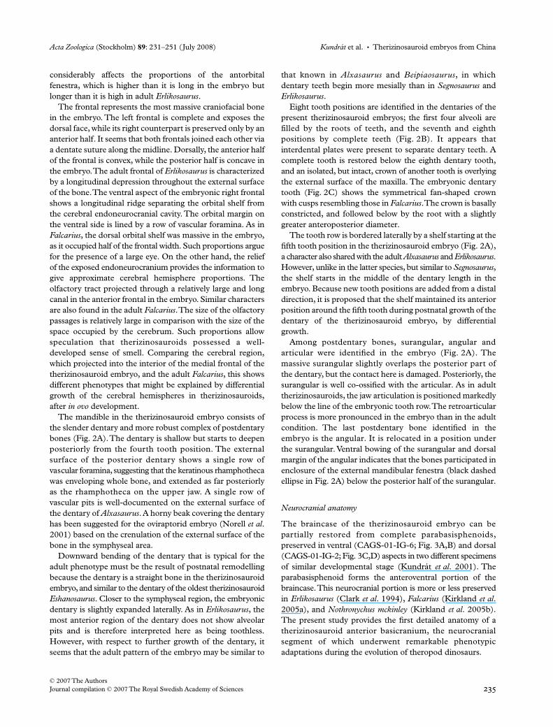
Neurocranial anatomy
The braincase of the therizinosauroid embryo can bepartially restored from complete parabasisphenoids,preserved in ventral (CAGS-01-IG-6; Fig. 3A,B) and dorsal(CAGS-01-IG-2; Fig. 3C,D) aspects in two different specimensof similar developmental stage (Kundrát
et al
. 2001). Theparabasisphenoid forms the anteroventral portion of thebraincase. This neurocranial portion is more or less preservedin
Erlikosaurus
(Clark
et al
. 1994),
Falcarius
(Kirkland
et al
.2005a), and
Nothronychus mckinley
(Kirkland
et al
. 2005b).The present study provides the first detailed anatomy of atherizinosauroid anterior basicranium, the neurocranialsegment of which underwent remarkable phenotypicadaptations during the evolution of theropod dinosaurs.

Therizinosauroid embryos from China
•
Kundrát
et al.
Acta Zoologica
(Stockholm)
89
: 231–251 (July 2008)
© 2007 The Authors
236
Journal compilation © 2007 The Royal Swedish Academy of Sciences
Fig. 3—Basicranium of therizinosauroid embryos from Nanchao Formation, Santonian-Campanian, Upper Cretaceous, Nanyang Valley, Henan Province, China. —A, B. Location of the parabasisphenoid inside embryonic bony assemblage CAGS-01-IG-6, and magnified view on ventral aspect of the bone. —C, D. Location of the parabasisphenoid inside embryonic bony assemblage CAGS-01-IG-2, and magnified view on dorsal aspect of the bone. Abbreviations: athv, anterior thoracic vertebra; atr, anterior tympanic recess; avcp, anteroventral coracoid process; bpp, basipterygoid process; btp, basitemporal process; c, capitulum; cav, caudal vertebra; cc, carotid canal; cf, coracoid foramen for supracoracoid nerve; cnab, canalis for abducens nerve; co, coracoid; cp, cultriform process; cv, cervical vertebra; ds, dorsum sellae; fcca, foramen for cerebral carotid artery; fe, femur; fnab, foramen for abducens nerve; fnoc, foramen for oculomotor nerve; fsa, foramen for sphenoid artery; fu, furcula; gf, glenoid fossa; hp, hypophyseal fossa; ihr, infrahypophyseal recess; ihs, intrahypophyseal septum; il, ilium; isr, infrasellar recess; mc, metacarpal; nc, notochordal canal; pbs, parabasisphenoid; plc, pleurocoel; pp, parapophysis; sca, scapular articulation; st, sella turcica; sv, sacral vertebra; t, tarsal; thr, thoracic rib; ti, tibia; tu, tuberculum; u, pedal ungual; vpbsr, ventral parabasisphenoid recess; vr, ventral ridge. Arrowhead shows the hypocleideum of the furcula.

Acta Zoologica (Stockholm) 89: 231–251 (July 2008) Kundrát et al. • Therizinosauroid embryos from China
© 2007 The AuthorsJournal compilation © 2007 The Royal Swedish Academy of Sciences 237
The parabasisphenoid (CAGS-01-IG-2; Fig. 3C,D) oftherizinosauroid embryos consists of a massive cultriformprocess, a deep and laterally expanded parabasisphenoidcorpus, substantially reduced basipterygoid processes,and short basitemporal processes. The corpus itself (notincluding the cultriform process) of the parabasisphenoidexpands laterally to such an extent that it appears to be twiceas broad as it is long.
The parabasisphenoid of the embryonic therizinosauroidwas highly pervaded by an epithelial system of pneumaticspaces derived from the anterior tympanic recess, extendingfrom the middle ear cavity. After the anterior tympanicrecess enters the parabasisphenoid interior it forms apneumatic sinus inside the bone. First, it invades thespace under the dorsum sellae by the infrasellar recess,and then it extends further anteriorly as the infrahypophysealrecess inside bones situated ventral and lateral to the hypo-physeal fossa (CAGS-01-IG-2; Fig. 4A–C). An additionalextension of the intrahypophyseal recess is hollowed into thebase of the cultriform process. The left and right anteriortympanic recesses are contralaterally connected through abroad interaural pathway that includes separate passagesthrough both the infrahypophyseal and infrasellar recess(Fig. 4B). A single opening visible inside the divided bonyprojections posterior to the dorsum sellae is interpreted hereas providing a pneumatic communication between theinfrasellar recess and the endoneurocranial space (Fig. 4C).We further assume that a large pneumatic connection existedbetween the infrasellar pneumatic recess and a basioccipitalrecess through a pair of openings located laterally on theposterior wall of ossified basisphenoid (Fig. 4A,C).
The cultriform process in the therizinosauroid embryo isslightly inflated at the base, unlike the bulbous appearance ofthe base of the cultriform process in the ornithomimosaurGallimimus (Osmólska et al. 1972), and troodontids such asSaurornithoides (Barsbold 1974), and Troodon (Currie andZhao 1994). From the base, the cultriform process in theembryo tapers slightly rostrally into a broad projection withU-shaped dorsal surface showing paired arterial foraminaand grooves extending anteriorly. Broken in its rostral exten-sion, a cavernous structure has been exposed (asterisk inFig. 4A), suggesting that the cultriform process might havebeen pneumatized further rostrally in the therizinosauroidembryo, as occurs in the adult Gallimimus (Osmólska et al.1972), Troodon (Currie and Zhao 1994), and Sinornithosaurus(Xu and Wu 2001).
The hypophyseal fossa is relatively shallow with theintrahypophyseal septum separating the exits of the cerebralcarotid arteries at the bottom (Figs 3D and 4B). Posterodorsallyto each carotid opening is a small foramen for the oculomotornerve. A shallow pituitary fossa with separate exits for thecerebral carotid arteries was reported for Gallimimus (Fig. 6Bin Osmólska et al. 1972). In troodontids, such as Troodon andByronosaurus, each internal carotid artery meets its counterpartimmediately before entering the hypophyseal fossa itself and
Fig. 4—Parabasisphenoid bone of a therizinosauroid embryo from Nanchao Formation, Santonian-Campanian, Upper Cretaceous, Nanyang Valley, Henan Province, China. Location of the parabasisphenoid inside embryonic bony assemblage CAGS-01-IG-2. —A. Left lateral view. —B. Right lateral view. —C. Posterior view. Abbreviations: atr, anterior tympanic recess; bpp, basipterygoid process; btp, basitemporal process; cc, carotid canal; cnab, canalis for abducens nerve; cp, cultriform process; ds, dorsum sellae; fcca, foramen for cerebral carotid artery; fnab, foramen for abducens nerve; fnoc, foramen for oculomotor nerve; fsa, foramen for sphenoid artery; gpbfn, groove for palatal branch of the facial nerve; hf, hypophyseal fossa; iap, interaural pathway; ihr, infrahypophyseal recess; ihs, intrahypophyseal septum; isr, infrasellar recess; pbr, passage to a basioccipital recess; pf, pneumatic foramen. Asterisk indicates exposure of cavernous bone tissue of the cultriform process.

Therizinosauroid embryos from China • Kundrát et al. Acta Zoologica (Stockholm) 89: 231–251 (July 2008)
© 2007 The Authors238 Journal compilation © 2007 The Royal Swedish Academy of Sciences
descends through a common carotid opening, the ostiumcanalium caroticorum (Currie and Zhao 1994; Makovickyet al. 2003). A similar single carotid opening at the bottom ofthe hypophyseal fossa in the adult Nothronychus may indicatethat the intrahypophyseal septum may be reduced by postnatalenlargement of the carotid arteries.
The internal carotid artery entered the parabasisphenoidat its posteroventral corner, within the middle ear recession(= lateral depression sensu Barsbold 1974; Currie 1985) andcontinued anteromedially inside the osseous carotid canal asthe cerebral carotid artery (CAGS-01-IG-2; Fig. 4A). Amore posterior entrance of the internal carotid arterythrough a foramen in the ventrolateral part of the occiput wassuggested by Clark et al. (1994; Fig. 7) for Erlikosaurus. Thetherizinosauroid embryo also indicates a more posteriorlyprolonged pathway of the internal carotid artery. However,here we suggest that the artery entered the braincase throughan opening inside the middle ear recession directly. Moreanteriorly it ran inside the tube-like canal directed into theinfrahypophyseal recess. This condition has not previouslybeen described in theropods, but is similar to that seen inpalaeognathous birds (personal observation by M.K.). Beforepassing through two separate openings in the hypophysealfossa, each cerebral carotid artery forms an anterior branchexiting through an ipsilateral foramen on the dorsal U-shaped surface of the cultriform process. The transmittedarterial branch might represent a homologue to the aviansphenoid artery (sensu internal ophthalmic artery), whichsprings from that part of the cerebral carotid artery thatcourses within the carotid canal. The sphenoid artery in birdsleaves the base of the skull via the orbital foramen at the sideof the parabasisphenoid rostrum (Baumel 1979).
Posteroventral to the dorsum sellae, a pair of passages forthe abducens nerves are preserved like an opening on the leftside, and an exposed canal on the partly damaged right side(CAGS-01-IG-2; Figs 3D and 4B,C). The position of theforamen for the abducens nerve has been misinterpreted inthe adult Nothronychus, as the nerve, after it enters theparabasisphenoid at the lateral side of the sella turcica, runsinside the bone medial to the trigeminal ganglion and itsophthalmic branch, exiting the bone anteriorly. Finally, thediameter of the foramen for the abducens nerve is commonlymuch smaller than the opening for the trigeminal nerve,unlike the labelled openings for these structures in Nothronychus.
Short basipterygoid processes in therizinosauroid embryos(Figs 3B and 4A) are similar to those of the adult phenotypeof Falcarius. In both therizinosauroids, the basipterygoidprocess is closely associated with that part of the middle earrecession (= subotic recess in Falcarius), through which theanterior tympanic recess enters the parabasisphenoid.Furthermore, the basipterygoid process in the therizinosauroidembryos recalls the more reduced conditions in oviraptorosaurs(Barsbold et al. 1990; Sues 1997; Clark et al. 2002; Xu et al.2002b) than those in the ornithomimid Gallimimus (Osmólskaet al. 1972). They are rugose at their distal points, which
indicates their firm sinusoid-like suturing with the adjacentpterygoids. The pterygoid–basipterygoid suture is obliteratedwith age, and basipterygoid extensions become even unrecog-nizable in adult conditions as documented by Erlikosaurusand Nothronychus. From the embryonic conditions, it wouldseem that the therizinosauroid basipterygoid process mighthave become reduced during the early evolution of theirgroup by a process of distal abbreviation of the process, com-bined with lateral expansion of the parabasisphenoid corpus,which engulfs the base of the process. A groove transmittingthe palatal branch of the facial nerve makes a markeddemarcation line between the basipterygoid process and theposterolaterally expanded parabasisphenoid wings (Fig. 4A).
The ventral side of the parabasisphenoid forms a slightlyconvex and smooth area in the adult Erlikosaurus andNothronychus. In the therizinosauroid embryos, this area hasa similar shape, but is flattened and hollowed sagittally by theventral parabasisphenoid recess (Fig. 3B), as in the adultFalcarius (basisphenoidal recess). It starts behind the level ofthe basipterygoid processes as a shallow V-shaped notchslightly deepening and broadening posteriorly, with a probableextension into the basioccipital between the basicranialtubera. A broad rectangular platform between the basipterygoidprocesses was originally described for Gallimimus (Osmólskaet al. 1972) and Saurornithoides (Barsbold 1974). In Gallimimusa deep recess invades and penetrates the platform (Osmólskaet al. 1972), while in troodontids the ventral parabasisphenoidrecess is missing (Barsbold 1974; Currie 1985; Xu et al.2002c; Makovicky et al. 2003). In Dromaeosaurus (Colbertand Russell 1969; Currie 1995) and Velociraptor (Barsboldand Osmólska 1999), the ventral side of the parabasisphe-noid is deeply hollowed by the pneumatic recess and moreanteriorly positioned depression (subsellar recess; Witmer1997a) between the basipterygoid processes. In Velociraptorspecimens, the variably deep parabasisphenoid recess isseparated by a low median ridge (Kundrát 2004). Also, theoviraptorosaurs display variability in both the extension andconfiguration of the parabasisphenoid recess. In the oviraptoridCitipati a large vacuity is present on the ventral midlinebetween the basioccipital and parabasisphenoid, with aforamen lying mainly within the latter (Clark et al. 2002),whereas there is only a shallow parabasisphenoid recessdivided by a longitudinal crest in Chirostenotes (Sues 1997)and Incisivosaurus (Xu et al. 2002b).
In the therizinosauroid embryos the basitemporal processesare situated medial to the expanded lateral parabasisphenoidwings (Figs 3B and 4C). Their dorsal aspect shows anarticulation surface for the anteroventral part of the basi-occipital. It does not show any indication for the presence ofthe craniopharyngeal canal.
Postcranial anatomy
Both axial and appendicular skeletal elements are preservedin the examined therizinosauroid embryos, although different

Acta Zoologica (Stockholm) 89: 231–251 (July 2008) Kundrát et al. • Therizinosauroid embryos from China
© 2007 The AuthorsJournal compilation © 2007 The Royal Swedish Academy of Sciences 239
bones are present in different specimens. Postcranial bonesof single individuals are referred to more or less differentdevelopmental stages based on different patterns of boneporosity and extension of ossified portions of the bones. Ourdescription of the postcranial anatomy of the therizinosau-roid embryos is based mainly on the specimen that is themost advanced embryo within the studied collection and hasthe most complete selection of postcranial bones (CAGS-01-IG-5; Fig. 5A). We complete our description by usingcharacters of postcranial bones from other specimens(CAGS-01-IG-6 and 2, Fig. 3A,C; CAGS-01-IG-12, Fig. 6A;CAGS-01-IG-4, Fig. 6B).
The vertebrae of the therizinosauroid embryos are mostlyfound as individual centra or neural arches throughout thespecimens. However, the anterior half of a vertebral columnis preserved, including cervical and thoracic vertebrae inarticulated positions, especially the axis and the atlas(Fig. 5B). Cervical vertebrae consist of large and elongatedcentra and robust neural arches.
The atlas represents two dissociated elements, a smalldisc-like centrum and a much larger neural arch. The atlantalneural arch is an L-shaped element viewed from the lateralside. Apically, it forms a convex arch with a groove on itsdorsal surface. It slightly expands basally and projects aposterior process that extends as far as the axial diapophysis.The atlantal neural arch is slightly dislocated at about 30°anticlockwise rotation from its original position. The atlantalneural arch articulates with the axial prezygapophysis insideits posterior angle. The neural arch of the axis is massive,having well-developed diapophysis and posteriorly directedvery low spinal process.
The low and narrow neural spines are known in cervicalvertebrae of Alxasaurus (Russell and Dong 1993), Beipiaosaurus(Xu et al. 1999), Nothronychus (Kirkland and Wolfe 2001),Neimongosaurus yangi (Zhang et al. 2001) and Falcarius(Kirkland et al. 2005a). While it is difficult to compare themorphology of the atlas from our description in thetherizinosauroid embryo and the adult Nanshiungosaurusbohlini (Dong and Yu 1997), it is possible to compare theaxis, suggesting numerous similarities with the exception ofthe long prezygapophysis in the adult Nanshiungosaurus. Ithas reduced prezygapophyses but expanded postzygapophysespointed more dorsally than in the following cervical vertebra.There is a swollen portion on the lateral side of the atlantalneural centrum with a crystal-filled depression on the uppersurface. The parapophysis, as we intepret this structure, ispositioned on the posterior half of the atlantal centrum, whileit is found in the middle of the following cervical vertebra andin the anterior half of the other cervical vertebra. The para-pophysis is positioned posterior to a deep pneumatic recess,the pleurocoel. It differs from the condition in Erliansaurus(Xu et al. 2002a) where robust parapophyses project fromthe centrum ventral to the pleurocoel. A deep-pocket-likepleurocoel develops on the lateral side in the cervical centraof Nanshiungosaurus, unlike Eshanosaurus, in which it
appears, to occupy much of the lateral surface at axial levelof posterior cervicals. Broad pleurocoels occupying muchof the sides of the cervical vertebrae are also reported forNeimongosaurus.
The cervical vertebrae following the axis have longprezygapophyses, shorter but more robust postzygapophyses,very low spinal processes, and well-developed diapophyses.Diapophyses of the third and fourth cervical vertebrae showco-ossification with the cervical ribs. The suture between theneural arch and centrum is obliterated completely in thethird cervical vertebra. A partial cleft between the neural archof the axis and the fourth cervical vertebra may be apreservational artefact caused by postmortem crushing. Thecervical centra are amphiplatyan with a shallow concentricconcavity around the notochordal canal (Fig. 3A,B). Itappears that a ventral ridge on each side of cervical centrumis present in the therizinosauroid embryo. This character isalso known in the third cervical of Nanshiungosaurus.
The thoracic vertebrae of the therizinosauroid embryosare preserved as individual centra (Figs 3A,B and 6A,B) orneural arches (Figs 5A and 6A,B), as well as seriallyarticulated elements (Fig. 5A). The articulated centra aremore or less covered by sediment or other skeletal elementswith damaged neural arches, and it is therefore difficult toreveal details. However, it appears that the anterior thoraciccentra (Figs 3B and 5A) are more amphicelous having largernotochordal canals, being rounded in cross-section, shorterthan the cervical centra, and possessing deep socket-likepleurocoels that are located posteroventrally to the capitularfacet of the parapophysis. The posterior thoracic centra(Fig. 6A,B) are very narrow with a slightly expanded ventralpart. There are large concavities on the lateral surfaces of theposterior thoracic vertebrae in therizinosauroid embryos.The thoracic neural arches are broad and massive skeletalelements with considerably reduced neural spines, androbust diapophyses. We have recognized other phenotypes ofvertebral centra in therizinosauroid embryos. However, theirprecise identification is not definite because of theirindividual preservation and phenotypic difference atdifferent ontogenetic stages.
The last axial skeletal element to be described is the caudalvertebra (Fig. 3A) that is three times as long as high. It showsa complete co-ossification of the neural arch and thecentrum. The centrum is slightly bent bearing a robusttransverse process that expands distally and posteriorly, thecharacter known in the anterior caudal of Alxasaurus andNothronychus, and in the first and second caudal vertebrae ofErliansaurus, and the first caudal vertebra of Neimongosaurus.Both prezygapophyses and postzygapophyses of this caudalvertebra are distinctly developed. No neural spine has beenobserved in the embryonic specimen.
Cervical ribs have been found associated with the thirdthrough fifth cervical vertebrae (Fig. 5B). The third cervicalrib is a triangular bone and shorter than that of succeedingcentra. It projects posteroventrally but does not reach the

Therizinosauroid embryos from China • Kundrát et al. Acta Zoologica (Stockholm) 89: 231–251 (July 2008)
© 2007 The Authors240 Journal compilation © 2007 The Royal Swedish Academy of Sciences
Fig. 5—Postcranial skeleton of a therizinosauroid embryo from Nanchao Formation, Santonian-Campanian, Upper Cretaceous, Nanyang Valley, Henan Province, China, CAGS-01-IG-5. —A. Axial and appendicular bones. —B. Magnified view of first four cervical vertebrae. —C. Magnified view of the pubic bone. Abbreviations: acr, acromion; atc, atlantal centrum; athr, anterior thoracic rib; atn, atlantal neural arch; ax, axis; bt, biceps tuberosity; cer, cervical rib; cv, cervical vertebra; dp, diapophysis; dpc, deltopectoral crest; fi, fibula; hpp, hypopubic process; hu, humerus; ich, ischium; mtb, medial tuberosity; mx, maxilla; nathv; neural arch of thoracic vertebra; obp, obturator process; olp, olecranon process; par, pubic apron; pb, pubis; plc, pleurocoel; poz, postzygapophysis; pp, parapophysis; prz, prezygapophysis; pthr, posterior thoracic rib; ra, radius; sc, scapula; sp, spinal process; mt, metatarsal; ti, tibia; tu, tuberculum; ul, ulna.
Acta Zoologica (Stockholm) 89: 231–251 (July 2008) Kundrát et al. • Therizinosauroid embryos from China
© 2007 The AuthorsJournal compilation © 2007 The Royal Swedish Academy of Sciences 241
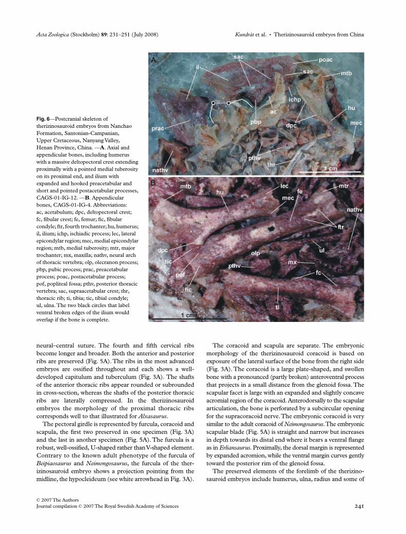
neural–central suture. The fourth and fifth cervical ribsbecome longer and broader. Both the anterior and posteriorribs are preserved (Fig. 5A). The ribs in the most advancedembryos are ossified throughout and each shows a well-developed capitulum and tuberculum (Fig. 3A). The shaftsof the anterior thoracic ribs appear rounded or subroundedin cross-section, whereas the shafts of the posterior thoracicribs are laterally compressed. In the therizinosauroidembryos the morphology of the proximal thoracic ribscorresponds well to that illustrated for Alxasaurus.
The pectoral girdle is represented by furcula, coracoid andscapula, the first two preserved in one specimen (Fig. 3A)and the last in another specimen (Fig. 5A). The furcula is arobust, well-ossified, U-shaped rather than V-shaped element.Contrary to the known adult phenotype of the furcula ofBeipiaosaurus and Neimongosaurus, the furcula of the ther-izinosauroid embryo shows a projection pointing from themidline, the hypocleideum (see white arrowhead in Fig. 3A).
The coracoid and scapula are separate. The embryonicmorphology of the therizinosauroid coracoid is based onexposure of the lateral surface of the bone from the right side(Fig. 3A). The coracoid is a large plate-shaped, and swollenbone with a pronounced (partly broken) anteroventral processthat projects in a small distance from the glenoid fossa. Thescapular facet is large with an expanded and slightly concaveacromial region of the coracoid. Anterodorsally to the scapulararticulation, the bone is perforated by a subcircular openingfor the supracoracoid nerve. The embryonic coracoid is verysimilar to the adult coracoid of Neimongosaurus. The embryonicscapular blade (Fig. 5A) is straight and narrow but increasesin depth towards its distal end where it bears a ventral flangeas in Erliansaurus. Proximally, the dorsal margin is representedby expanded acromion, while the ventral margin curves gentlytoward the posterior rim of the glenoid fossa.
The preserved elements of the forelimb of the therizino-sauroid embryos include humerus, ulna, radius and some of
Fig. 6—Postcranial skeleton of therizinosauroid embryos from Nanchao Formation, Santonian-Campanian, Upper Cretaceous, Nanyang Valley, Henan Province, China. —A. Axial and appendicular bones, including humerus with a massive deltopectoral crest extending proximally with a pointed medial tuberosity on its proximal end, and ilium with expanded and hooked preacetabular and short and pointed postacetabular processes, CAGS-01-IG-12. —B. Appendicular bones, CAGS-01-IG-4. Abbreviations: ac, acetabulum; dpc, deltopectoral crest; fc, fibular crest; fe, femur; fic, fibular condyle; ftr, fourth trochanter; hu, humerus; il, ilium; ichp, ischiadic process; lec, lateral epicondylar region; mec, medial epicondylar region; mtb, medial tuberosity; mtr, major trochanter; mx, maxilla; nathv, neural arch of thoracic vertebra; olp, olecranon process; pbp, pubic process; prac, preacetabular process; poac, postacetabular process; pof, popliteal fossa; pthv, posterior thoracic vertebra; sac, supraacetabular crest; thr, thoracic rib; ti, tibia; tic, tibial condyle; ul, ulna. The two black circles that label ventral broken edges of the ilium would overlap if the bone is complete.

Therizinosauroid embryos from China • Kundrát et al. Acta Zoologica (Stockholm) 89: 231–251 (July 2008)
© 2007 The Authors242 Journal compilation © 2007 The Royal Swedish Academy of Sciences
autopodial elements. The embryonic humerus is a relativelylarge bone with a strongly expanded proximal and less expandeddistal end. It shows derived features common to other ther-izinosauroids, such as a massive deltopectoral crest, a pointedmedial tuberosity and hypertrophied lateral and medialepicondylar regions (Figs 5A and 6A,B). However, it closelyresembles the adult humerus of Neimongosaurus andErliansaurus. The deltopectoral crest extends for approxi-mately half of the humeral length in the embryo. Whereas theinitial growth of the deltopectoral crest is observed in thetherizinosauroid embryo (Fig. 6A), the rotation of the distalcondyles onto the anterior aspect is not pronounced and thisis therefore suggested to be part of postnatal development.
The identification of the radius (Fig. 5A) is based on itsslightly S-shaped pattern, with a more expanded proximalend, having the biceps tubercule in its proximity. The radiusis well ossified and represents an almost identical miniatur-ization of the adult Neimongosaurus radius. The ulna isassociated with the radius in the embryo (Figs 5A and 6B)and has been identified by the presence of the olecranonprocess on its more expanded proximal end. The embryoniculna is most similar to that of Erliansaurus, among alltherizinosauroids for which this bone is preserved.
The unguals are the only elements of the manus to beidentified with certainty. The three manual unguals overlyingthe head of the therizinosauroid embryo (Fig. 2A) arecompletely hypertrophied and well ossified, including theflexor tubercle near the proximal articulation. The largestungual is here interpreted to pertain to the most anteriordigit. The unguals of consecutive digits are smaller in size.The manual unguals are laterally compressed and stronglycurved. As in other therizinosauroids, the proximal ends ofthe manual unguals are deep but taper to needle-sharppoints. Putative metacarpals (Fig. 3C) are well ossifiedincluding their articular facets.
The pelvic girdle of therizinosauroid embryo is representedby the ilium, ischium and pubis. The left preacetabularprocess (Fig. 3A) and the complete left ilium (Fig. 6A) havebeen identified inside two therizinosauroid eggs. UnlikeBeipiaosaurus, but as in all other therizinosauroids, theembryonic ilium has a substantially longer preacetabularprocess than postacetabular process. The preacetabularprocess is expanded and hooked as reconstructed forAlxasaurus. Contrary to the most adult phenotypes, theembryonic preacetabular process, however, is not flaredlaterally as in Beipiaosaurus. The postacetabular process islong as in Alxasaurus, Beipiaosaurus, Neimongosaurus andFalcarius. In the therizinosauroid embryo, the supra-acetabular crest projects somewhat beyond the lateralmargin of the blade, as has been described for the adultNeimongosaurus. Comparable with dromaeosaurids,Archaeopteryx (Norell and Makovicky 1997), and othertherizinosauroids (Barsbold and Maryañska 1990; Clarket al. 2004), the pubic peduncle is longer than the ischiadicpeduncle of the ilium.
Right and left ischia are preserved in the therizinosauroidembryo (Fig. 5A). However, crushing along the anterior partdoes not allow for unambiguous proximodistal orientation ofthe bone. The ischial shafts are flattened. Based on theknown anatomy of adult therizinosauroids, the ischium isshorter than the pubis (Barsbold and Maryañska 1990). Ifso, then the embryonic ischia should be complete as for thelength, because they are as long as the pubic bones in thesame specimen. The obturator process develops distally onthe adult ischium therefore the flat bone expansion at thedistal end of the prenatal ischium is here interpreted as theobturator process in the therizinosauroid embryo. It is similarto the adult ischial phenotype of Nanshiungosaurus (Clarket al. 2004).
Both pubic bones co-ossified along the distal quarter oftheir length in the therizinosauroid embryo (Fig. 5C). Aridge projects about the pubic symphysis. Each pubic isU-shaped for most of its distal length. The pubic apron istransversely narrow, restricted to the medial part of the shaft,and relatively short, extending for less than one-third of thetotal length of the pubis; a character shared by oviraptorids(Osmólska et al. 2004). The distal end does not expand butpoints anteroventrally as the hypopubic process.
The femur of the therizinosauroid embryo (Figs 3C and6B) is straight with the diaphyseal part oval in cross-sectionas in the adult Beipiaosaurus, Erliansaurus and Neimongosaurus.Dorsal parts of the femoral head and major trochanter arenot ossified. The fourth trochanter (Fig. 6B), in the form ofa low longitudinal swelling, is positioned close to the femoralhead. The fourth trochanter is just proximal to mid shaftin adult therizinosauroids, suggesting that the embryonicfourth trochanter undergoes a positional shift and elongationafter hatching. The fibular and tibial condyles are wellseparated by an elongated depression, the popliteal fossa.
The tibia (Figs 3C, 5A and 6B) is nearly equal in length tothe femur, while the fibula (Fig. 5A) is shorter than the tibiain the therizinosauroid embryo. The tibia is well ossifiedexcept for the epiphyseal caps. It also has a distinct andwell-ossified fibular crest, a theropodan character (Gauthier1986), extending distally to the level of the mid-shaft, as itdoes in Erliansaurus. However, as suggested for the fourthtrochanter, the embryonic fibular crest might have extendedmore distally because of postnatal growth. If so, thanextension of the fibular crest would be similar to the adultcondition of Neimongosaurus, in which it extends more thanhalf the length of the tibia. If the preservation of the tibia andfibula reflects an original state in Fig. 5(A) and is not acondition caused by fossilization, then both bones mighthave been co-ossified or firmly attached before hatching. Thefibula is considerably more slender compared to the tibia inthe therizinosauroid embryo. It is also well ossified with theexception of the most dorsal part on the proximal epiphysis.The tibia has a flattened and widened proximal end. Themedial face of the proximal part of the fibula is slightly con-cave, an intermediate feature between the medial fossa of

Acta Zoologica (Stockholm) 89: 231–251 (July 2008) Kundrát et al. • Therizinosauroid embryos from China
© 2007 The AuthorsJournal compilation © 2007 The Royal Swedish Academy of Sciences 243
some theropods and a flat surface of the medial facet of theadult therizinosauroids, such as Alxasaurus, Beipiaosaurus,Erliansaurus, Nothronychus and the Avialae (Chiappe et al.1996). The distal end of the embryonic fibula tapers to apointed process.
The pes of the therizinosauroid embryos is represented bydisarticulated unguals (Fig. 3A,B) and putative metatarsalelements (Figs 3C and 5A), all well ossified. The pedalunguals are shorter than all manual unguals, and pedaltarsals are broader than any manual metacarpals.
Discussion
Identity of the embryos
The discovery and subsequent description of the firsttherizinosauroid was the beginning of the most mysterioussaga in the history of dinosaur systematics. Originally identifiedas a giant turtle (Maleev 1954), the claws of Therizinosauruscheloniformis were recognized by Barsbold (1976) to belongto a theropod. However, the systematic position of therizino-sauroid dinosaurs has generated a diversity of opinions.From a historical point of view, therizinosauroid dinosaurs(Perle 1979; Barsbold and Perle 1980; Barsbold andMaryañska 1990) have been hypothesized to be the sistergroup of Ornithischia (Paul 1984), or relatives of Sauropodo-morpha (Gauthier 1986), the sister group of a monophyleticProsauropoda (Sereno 1989), or even as a new dinosaurorder, Segnosaurischia (Dong 1992). Although the firstdescribed species, Therizinosaurus cheloniformis (Maleev1954), was confidently assigned to the Theropoda (Barsbold1976), the systematic position of these dinosaurs hasremained controversial. This was the result not only of theirvery rare and incomplete fossil record, but also of theunusual mixture of skeletal characters of therizinosauroids,which converge more or less on the morphologies of manydinosaur groups (Barsbold and Maryañska 1990). It was thedescription of Alxasaurus in 1993 (Russell and Dong 1993)that revived Barsbold’s original conclusion of a theropodaffinity.
The embryos described above have provided multipleevidence about the development of skeletal morphologiesthat are recognized as theropodan diagnostic features(Benton 1990; Currie 1997), such as: (1) the premaxillaryfenestra on the medial wall of the antorbital cavity, a featurepresent also in Archaeopteryx (Witmer 1997b); (2) thelacrimal extended on the top of the skull; (3) highly pneuma-tized parabasisphenoid; (4) the strap-like scapular blade; (5)manual unguals enlarged, laterally compressed, stronglyrecurved, sharply pointed, and with prominent flexortubercles; (6) an enlarged preacetabular process on the ilium;(7) the strap-like fibula appressed to the tibial crest; (8)thin-walled long bones.
Among theropods, remarkable similarities with the adultoviraptorosaurs (Osmólska et al. 2004) have been identified
on the examined embryonic skeletons, including: (1) theelongated narial opening; (2) the short basipterygoidprocess; (3) the low cervical neural spines; (4) the distal endof the pubis expanded anteriorly; (5) transversely narrowpubic apron, restricted to the medial part of the craniallyconcave shaft.
Further comparative analysis revealed that the examinedembryos show considerable affinity to the therizinosauroidphenotype, and represent the most complete fossil assem-blage of any therizinosauroid dinosaur ever found. Theembryos are assigned to the Therizinosauroidea with confi-dence on account of the following cranial and postcranialautapomorphies (Barsbold and Maryañska 1990; Clarket al. 2004): (1) an edentulous premaxilla with a sharpdown-turned edge lying below the subhorizontally elongatedexternal naris (Fig. 2A,B); (2) a dentary with the lateralsurface forming a horizontal shelf (Fig. 2B); (3) teeth withfan-shaped crowns that have a few marginal cusps and arelabio-lingually compressed, basally constricted, and followedbelow by the root with a larger anteroposterior diameter(Fig. 2B,C); (4) a humerus with a massive deltopectoralcrest extending proximally and with a pointed medialtuberosity on its proximal end (Figs 5A and 6A,B); (5) anilium with an expanded and hooked preacetabular andshorter and pointed postacetabular processes (Fig. 6A);(6) strongly curved hypertrophied manual unguals that aredeep proximally, but taper to sharp points (Fig. 2A).
The use of bones to identify embryos at the generic levelis not without problems because the skeleton was stillgrowing. However, exceptional preservation of well-ossifiedskeletal features in the therizinosauroid embryos, as well asrecent discoveries of new therizinosauroids in China, allowus to make further comparisons of both embryos and adults.Previously, embryos have been identified as therizinosauridswithout making suggestions of their affinity to known generaof that time (Cohen et al. 1995). It was later suggested(Manning et al. 1997, 2000) that the unguals of the hands inthe embryos are similar to those of Erlikosaurus (Barsboldand Perle 1980), and unlike the long, trenchant unguals ofTheirizinosaurus (Barsbold 1976). However, allowance has tobe made for allometric growth in the unguals, which is notpossible to elucidate at present. We can proceed with the issuebecause of the recent discoveries of new therizinosauroidspecies that substantially expanded our knowledge of thisgroup, and provide the well-preserved diagnostic featurescomparable to those in the therizinosauroid embryos.Therefore, referral of the unhatched therizinosauroids to theknown genera, particularly those from Asia, is possible withreasonable confidence.
Among known Asian therizinosauroids, the therizinosauroidembryos show skeletal similarities closest to the twotherizinosauroids discovered recently in the Upper Creta-ceous beds of the Iren Dabasu Formation of Nei Mongol,Neimongosaurus (Zhang et al. 2001) and Erliansaurus (Xuet al. 2002a); these include: (1) a ventral flange on the distal

Therizinosauroid embryos from China • Kundrát et al. Acta Zoologica (Stockholm) 89: 231–251 (July 2008)
© 2007 The Authors244 Journal compilation © 2007 The Royal Swedish Academy of Sciences
end of the scapular blade that is found in Erliansaurus, thispart is missing in Neimongosaurus; (2) overall morphology ofthe coracoid that resembles the adult one in Neimongosaurus,this bone has not been preserved in Erliansaurus; (3) expansionof the deltopectoral crest and the pointed medial tuberosityof the humerus that is almost identical to correspondingconditions in the both Neimongosaurus and Erliansaurus; (4)the radius phenotype that represents almost identicalminiaturization of the bone in Neimongosaurus, while itdiffers from the Erliansaurus radius by morphology of theproximal region; (5) long postacetabular process as inNeimongosaurus but contrary to a short one in Erliansaurus;(6) the supra-acetabular crest projecting somewhat beyondthe lateral margin of the blade, as described for the adultNeimongosaurus and Erliansaurus; (7) the straight femur as inthe both Neimongosaurus and Erliansaurus; and (8) the tibiawith the crest extending distally to the level of the mid-shaftas in Neimongosaurus and Erliansaurus.
Incubation period of the therizinosauroid embryos
The embryonic bones lie within their eggs in differentembryonic positions. They are either preserved as disarticu-lated and crushed skeletal remains clustered at the equatoriallevel (Stage A and B in Fig. 7), or disarticulated but lesscrushed and more ossified remains concentrated in the lowerspherical part of the egg (Stage C in Fig. 7), or preserved assemiarticulated and intact bones concentrated on the innersurface of an egg bottom (Stage D in Fig. 7).
Two distinct taphonomic patterns of bone distribution areobserved within the eggs, which we here call E1 (Stages Aand B) and E2 (Stage C and D) preservational styles. Theseare correlated with the amount of yolk present at the time ofdeath, and suggest that the E1 group of embryos are at amarkedly earlier developmental stage than the E2 embryos,as supported by ossification patterns of the vertebral centra.
Embryos of Alligator mississippiensis (right-hand column,Fig. 7) represent the first precocial model of a living archosaur,of which ossification patterns have been compared in detailwith those in these therizinosauroid specimens. Stage Arepresents the earliest-known developmental stage oftherizinosauroid embryos, with an apparently porousstructure of the vertebral centra, whereas by Stage D, whichis the latest stage so far determined, these had completelyossified vertebral centra and a partially obliterated neuro-central suture in their cervical vertebrae (Fig. 7, left andcentral columns). Using ossification rates of vertebral centra
in extant species, Stages A, B and C may correspond approx-imately with developmental levels of 45-, 50- and 64-day-oldembryos of Alligator mississippiensis. Compared to the almosthatched embryo of Alligator mississippiensis, similar patternsin postcranial ossification had already been reached bytherizinosauroid embryos of Stage C, which are not embryosimmediately prior to hatching. Although the therizinosauroidembryo of Stage D (CAGS-01-IG-5) developed ossificationpatterns that are more advanced than those in Alligatorhatchlings, proportions of the therizinosauroid embryonicbones indicate that the Stage D therizinosauroid embryo hadnot yet reached an appropriate size to hatch.
Although the incubation rate of dinosaurs will probablynever be known with certainty, some inference can be madefrom closely related extant taxa. Crocodilians and birds arethe only living representatives of the Archosauria, andtherefore can offer developmental parameters with whichdinosaur embryos might be compared. As shown later,therizinosauroid embryos were precocial; however, theperiod of incubation varies considerably even amongdifferent precocial species of both crocodilians and birds.
Among modern crocodilians, the incubation period aver-ages 65 days in the medium-sized Alligator mississippiensis(depending on temperature). While a large-size Crocodylusporosus hatches after 90–100 days, small to medium-sizeCrocodylus cataphractus may hatch after about 110 days, andOsteolaemus tetraspis, one of the smallest living crocodilians,hatches after 85–105 days (Britton 1995). In precocial birds,incubation may take as little as 12 days in Turnix tanki,through 42 days in Struthio camelus, to as long as 80 days insome Megapodidae (Starck 1993).
Even if the body size is diverse among precocial birds, theyall have relatively large eggs, whereas altricial species havesmaller eggs (Heinroth 1922). Presently we do not knowwhat body size these therizinosauroids achieved in adult-hood. Mongolian therizinosauroids from the Gobi Desertwere quite large animals (4–8.5 m long; Russell 1997). How-ever, the two Chinese therizinosauroids, Neimongosaurus(Zhang et al. 2001) and Erliansaurus (Xu et al. 2002a), thatshare the most skeletal similarities with the therizinosauroidembryos described here, are ranked among small-size ther-izinosauroids (2–3 m overall length). Compared to othersmall-size theropods, therizinosauroid eggs (9 × 7 cm) areslightly smaller than Troodon eggs (12–16 cm × 5–6 cm;Varricchio et al. 2002) and substantially smaller than ovirap-torid eggs (18–19 cm × 6.5–7.2 cm; Clark et al. 1999). ForNeimongosaurus to be considered as the most potential layer
Fig. 7—Correlation of developmental stages between therizinosauroid embryos and the Alligator mississippiensis embryos based on ossification patterns of vertebral centra. Abbreviations: Stage A and B showing E1 taphonomic pattern of disarticulated and crushed skeletal remains clustered at the equatorial level; Stage C showing E2 taphonomic pattern of disarticulated but more ossified remains preserved in a lower spherical part of the egg; and Stage D, showing E2 taphonomic pattern of more articulated and intact bones concentrated on the inner surface of an egg bottom. Two distinct E1 and E2 taphonomic patterns of bone distribution are correlated with the amount of yolk present at the time of death, and suggest that the E1 group of embryos died at a substantially earlier developmental stage than the E2 embryos, as supported by ossification patterns of the vertebral centra.

Acta Zoologica (Stockholm) 89: 231–251 (July 2008) Kundrát et al. • Therizinosauroid embryos from China
© 2007 The AuthorsJournal compilation © 2007 The Royal Swedish Academy of Sciences 245

Therizinosauroid embryos from China • Kundrát et al. Acta Zoologica (Stockholm) 89: 231–251 (July 2008)
© 2007 The Authors246 Journal compilation © 2007 The Royal Swedish Academy of Sciences
(for reasons see above) of the eggs with embryos analysedhere, the size of these eggs to body mass would be relativelylarge and would suggest a longer incubation period. A longerincubation period of the therizinosauroid embryos is alsosuggested by the development of their advanced ossificationpatterns (analysed below).
Aside from the absolute time of the incubation period, itcould be expected, based on Alligator ossification patterns,that the therizinosauroid embryos included in this study diedsometime during the final third of their prehatching develop-ment. However, such a comparison is oversimplistic becausethe therizinosauroid embryos developed crocodilianposthatching ossification patterns in ovo and might havestayed inside their eggs for a much longer time. Based on thediscussion above, we could speculate that the incubationperiod of the therizinosauroid embryos might have been aslong as 1.5–3 months.
Precocial developmental patterns of the therizinosauroid embryos
Variation in rates of development among avian taxa has ledneontologists to separate birds into altricial and precocial ornidicolous and nidifugous developmental types. Starck andRicklefs (1998) pointed out that altricial and precocial referto the developmental stage, whereas nidicolous andnidifugous refer to length of nest presence.
Various classifications (Nice 1962; Ricklefs 1983;O’Connor 1984; Starck 1993) used different features todescribe the developmental mode of avian hatchlings.However, they mostly include such characteristics as (1) nestpresence – staying in nest vs. locomotor activity, (2) parent–offspring relation – no parental care vs. any dependence onparents, (3) feeding behaviours – fed by parents vs. searchingfor food and feeding alone independently, (4) developmentalstage of sense organs – eyes open or closed at hatching, and(5) anatomical traits – presence or absence of downy hatchlingplumage. These traits do not strictly align themselves in acontinuous sequence, particularly nest leaving and self-feeding, but depend on nest site and food supply and maytherefore be independent of development (Starck and Rick-lefs 1998).
Using this heterogeneous set of characters, Starck (1993)classified avian hatchlings into eight different categoriesarranged along a precocial–altricial gradient. In his classification,absence of parental care and prolonged incubation time differ-entiates a superprecocial from a precocial level. Feedingstrategy divides the precocial spectrum into three levelswhere hatchlings search for food and feed alone (precocial1), or food is shown by parents (precocial 2), or young are fedby parents (precocial 3 through altricial extreme). Most ofthese characters cannot be described from the fossil recorddirectly and with certainty, and palaeontologists are thus veryoften left only with deductions of behavioural characteristicsbased on the relative degree of skeletal maturation ofpreserved specimens.
In contrast to altricial birds, the skeletons of precocialneonates, which are active soon after hatching, have achievedan advanced degree of ossification, providing them with sup-port for resistance to the mechanical forces of locomotion(Starck 1993). As a consequence of substantial reduction ofcartilaginous skeleton, the postnatal growth of the precocialbirds is limited by the relatively small uncalcified or unossi-fied portions of skeletal elements (Starck 1994; Ricklefs andStarck 1998). Thus a relatively slow postnatal growth of bodymass in the precocial birds is hypothesized related to a highdegree of ossification, the closure of cranial clefts, andresistance against mechanical stress (Starck 1993).
An interesting phenomenon referred to in the developmentof the avian skeleton is that the sequence and timing ofossifications do not change during evolution from precocialto altricial, and are therefore constant in all avian species(Ricklefs and Starck 1998). Early embryonic stages are ofapproximately the same duration and the succession ofembryonic stages seems to be almost invariant among birds(Ricklefs and Starck 1998). This suggests that only deviationsin late stages cause diverging maturation patterns in altricialand precocial birds.
Distinct differences that are independent of time and canbe related to the different developmental modes are dif-ferences in the extension of ossification (Starck 1993). Inprecocial species of birds, the osseous areas are extendedthrough the skeleton and only small cartilaginous areasremain after hatching. Designation of non-avian dinosaursinto categories along the altricial–precocial gradient is usu-ally based on maturation of ossification patterns of variouspreserved skeletal fragments of a single embryo of unknowndevelopmental age. The collection of therizinosauroid embryosexamined here, however, has provided an opportunity tocompare successional ossification patterns of the same typebones in the same species over a longer incubation period.Despite there being no therizinosauroid hatchling availablein the collection, we assume that the most advancedtherizinosauroid embryo (CAGS-01-IG-5; Fig. 5A)represents a substantial integration of skeletal developmentduring incubation and may be used to approximate thephenotypic and behavioural characteristics of therizinosau-roid hatchlings, and thus compare with other embryos orneonates of theropods, birds and crocodiles.
With regard to advanced ossification patterns of the ther-izinosauroid embryo (CAGS-01-IG-5), further skeletalcomparisons can be made with the oviraptorid embryo fromthe Late Cretaceous Djadokhta Formation of Ukhaa Tolgodin Mongolia (IGM100/971; Norell et al. 1994, 2001),embryonic remains of Troodon formosus from the Late Creta-ceous Two Medicine Formation of western Montana in theUSA (Varricchio et al. 2002), avian embryonic bones ofGobipteryx minuta from the locality Hermiin Tsav I inMongolia (Elzanowski 1981); an embryo of an enantiornithinebird from the Early Cretaceous of Liaoning in China(V14238; Zhou and Zhang 2004), embryos and hatchlings

Acta Zoologica (Stockholm) 89: 231–251 (July 2008) Kundrát et al. • Therizinosauroid embryos from China
© 2007 The AuthorsJournal compilation © 2007 The Royal Swedish Academy of Sciences 247
of recent birds such as Dromaius novae-hollandiae (emu –precocial 1; Starck 1996) and Turnix suscitator (buttonquail– precocial 3; Starck 1993), and embryos of Alligator mississipp-iensis. The following comparisons are approximate, as theprecise embryonic stage of the fossil embryos is unknown.With regard to the position of the oviraptorid embryo (IGM100/971), and an enantiornithine embryo (V14238) insidethe egg, these specimens died before hatching. This mightalso be the case of the Gobipteryx embryonic specimens thatwere suggested by Elzanowski (1981) to possess advancedossification patterns indicating an extreme precociality. TheTroodon embryo (MOR 246-11) seems to represent an earlierstage, as various cranial elements are still incompletelyossified. Although extension of ossification areas in theskeletal elements of the latest therizinosauroid embryo(Fig. 5A) is comparable with all the specimens mentionedabove, the preserved bones do not appear to be large enoughto consider the specimen to have reached the size of ahatching stage. We assume that therizinosauroid embryos(CAGS-01-IG-5) stayed within the egg for a longer period toenlarge their proportions regardless of the apparent advancedossification of the skeleton.
In the therizinosauroid embryo (CAGS-01-IG-1; Fig. 2A),the dermal bones of the jaws and the articular are completelyossified and well sutured to each other to form a rigid bonysupport for the feeding apparatus. The same degree ofossification is achieved in the jaw apparatus of prehatchingembryos of Alligator, as well as the bill skeleton of theGobipteryx embryo (ZPAL MgR-I-88), an enantiornithineembryo (V14238), and of the recent superprecocial andprecocial avian hatchlings mentioned above. The suturebetween the angular and the surangular seems to be obliteratedin the oviraptorid embryo (IGM 100/971), suggesting anadvanced ossification of the posterior portion of the mandible.Similarly in the therizinosauroid embryo (CAGS-01-IG-1),the surangular is co-ossified with the articular, providing jawarticulation of a therizinosauroid hatchling with resistanceagainst mechanical stress caused by initial biting or rippingof food. As in the Troodon embryonic specimen (MOR 246-11), teeth were erupted in therizinosauroid embryos (CAGS-01-IG-1 and 5), although crenulation of the crown of teethin the latter suggests a broader trophic range of omnivory.
The edges of the dermal bones of the neurocranium aresutured together, with the exception of relatively large fronto-parietal window in the precocial 3-hatchling of Turnix. Incontrast, the vertex is complete in the precocial 1-neonate ofDromaius, as it is in a prehatching stage of Alligator. Althoughthe parietal is not exposed in the therizinosauroid embryo(CAGS-01-IG-1), the posteromedial edge of the preservedfrontal does not indicate that this bone participated in anopening on the top of the skull. As in a prehatching stage ofAlligator, and the oviraptorid embryo (IGM 100/971), theossification of the parabasisphenoid in therizinosauroidembryos (CAGS-01-IG-2 and 6) is complete, although thebasioccipital is not yet firmly sutured with the parabasisphenoid.
A separate basioccipital has also been identified in theGobipteryx embryo (ZPAL MgR-I/33) and the Troodon embryo(MOR 246–1). Well-developed sutural scars on the dorsalsurface of the basitemporal processes indicate that theseparation of the parabasisphenoid and the basioccipital inthe therizinosauroid embryo may simply reflect a preservationalartefact.
The neural arches and vertebral centra are fully ossified inthe latest therizinosauroid embryo (Stage D; CAGS-01-IG-5). Because the neurocentral suture is obliterated in the thirdcervical vertebra, we interpret a cleft that appears betweenthe neural arch and centrum of the axis and the fourthcervical vertebra as being a postmortem effect (Fig. 5B).Crushing of the neural arches is obvious in the thoracicvertebral column and does not allow us to discover whetherthese were fused with their corresponding centra. On theother hand, we identified a proximal caudal vertebra in theStage C-specimen (CAGS-01-IG-6) that shows completeobliteration of the neurocentral suture. Starck (1993)concluded that in all precocial neonates, the ossified centresof vertebrae and neural arches are fused and form continuousosseous vertebrae. If so, then fusion of both vertebral com-ponents must precede the completion of vertebral ossifica-tion itself, as Starck’s illustration of the Turnix hatchlingdisplays the substantial presence of cartilaginous areas in thevertebral column of this precocial 3 bird. In contrast to thetherizinosauroid embryo (CAGS-01-IG-5), cartilaginousareas are present in the cervical vertebrae during the latestperiod of the Alligator embryogenesis (Fig. 8). It is not earlierthan at the hatching stage of Alligator that the axial skeletonis fully ossified. However, the neurocentral sutures are stilldistinct in the cervical, thoracic and proximal caudal regions,but has become closed in the distal caudal region (Rieppel1993). Complete ossification and fusion of the vertebrae isachieved by the precocial 1-neonate of Dromaius. It seemsthat distinct neurocentral sutures are present in cervical andthoracic vertebrae of an enantiornithine embryo (V14238),whereas their obliteration progressed in caudal vertebrae.The neurocentral sutures are distinct in cervical and thoracicvertebrae of the Gobipteryx embryo (ZPAL MgG-I/34).Unlike the therizinosauroid embryo (CAGS-01-IG-5) andthe enantiornithine birds above, the oviraptorid embryo(IGM 100/971) possesses a neural arch separated from itsaccompanying centrum in at least one single cervical vertebra.
Unlike those in the oviraptorid embryo (IGM 100/971),the cervical ribs fused with corresponding vertebrae in thetherizinosauroid embryo (CAGS-01-IG-5). On the otherhand, anterior and posterior thoracic ribs are well ossifiedwith clearly differentiated capitulum and tuberculum in bothembryos. In contrast to the prehatching specimens of Alligator(Fig. 8) and Turnix, the thoracic ribs are completely ossifiedin the therizinosauroid embryo (CAGS-01-IG-5). Suchconditions are accomplished after hatching in Dromaius,while distal regions of the thoracic ribs remain cartilaginousafter hatching in Turnix.

Therizinosauroid embryos from China • Kundrát et al. Acta Zoologica (Stockholm) 89: 231–251 (July 2008)
© 2007 The Authors248 Journal compilation © 2007 The Royal Swedish Academy of Sciences
The pectoral and pelvic girdle elements are more ossifiedin the therizinosauroid embryos than in Alligator and most ofthe recent precocial birds. Significant differences in exten-sion of osseous areas are found in the distal portions of thescapula that remains cartilaginous in the prehatching stage ofAlligator (Fig. 8), and the precocial 3-neonate of Turnix. Theossification of the scapula is complete in the oviraptoridembryo (IGM 100/971) and in Dromaius (precocial 1) afterhatching. Unlike Turnix, distal regions of the postacetabularprocess of the ilium are fully ossified in the therizinosauroidembryos (CAGS-01-IG-5 and -12), as it is accomplished byDromaius after hatching. In contrast to both the precocialbirds, Dromaius and Turnix, in which the distal portion of thepubis remains cartilaginous after hatching, the pubes areossified along their full extension and fused together distallyin the therizinosauroid embryo (CAGS-01-IG-5).
The osseous areas of skeletal elements are extended andonly small cartilaginous caps remain at the proximal anddistal ends of appendicular bones. The humerus andantebrachial bones are almost equal in length to the femurand crural bones, respectively. These proportions suggest
that the therizinosauroid hatchlings might have preferredquadrupedal locomotion.
All precocial species compared here have extensive ossi-fications of all leg bones. However, comparing the therizino-sauroid embryo (CAGS-01-IG-5) with the prehatchingspecimen of Alligator (Fig. 8), it is evident that ossification ofthe humerus and tibia is more advanced in the fossil. As inthe oviraptorid embryo (IGM 100/971), the femoral head isabsent in the therizinosauroid embryos (CAGS-01-IG-2 and-4). The most probable explanation of such preservation isincomplete ossification, as seen in the late embryos andhatching of precocial birds, with the exception of the Dromaiusneonate, in which the femoral head is illustrated by Starck(1996) as completely ossified. On the other hand it is moredifficult to interpret preservation of the fibula that is firmlyappressed to the tibia in the therizinosauroid embryo(CAGS-01-IG-5). There is a well-developed tibial crest sup-porting close communication of the bones. However, theyare preserved in an unusual position that might be explainedby an assumption that these embryonic bones were at leastslightly co-ossified at the time of fossilization. Probably the
Fig. 8—Different ossification of postcranial structures. —A. In the most advanced therizinosauroid embryo (Stage D, E2 preservational style; CAGS-01-IG-5). —B. In the almost hatched embryo of Alligator mississippiensis. Labelled structures in the therizinosauroid embryo are ossified, while the same structures in the alligator embryo remain cartilaginous: cervical spinal process (black arrow), neural arch/vertebral centrum contact (red arrow), distal part of scapula (orange arrow), proximal epiphysis of humerus (dark blue arrow), distal epiphysis of humerus (light blue arrow), ventral part of anterior thoracic rib (green arrow), ventral part of posterior thoracic rib (white arrow), and proximal epiphysis of tibia (yellow arrow).

Acta Zoologica (Stockholm) 89: 231–251 (July 2008) Kundrát et al. • Therizinosauroid embryos from China
© 2007 The AuthorsJournal compilation © 2007 The Royal Swedish Academy of Sciences 249
most distinct difference between the therizinosauroidembryos (CAGS-01-IG-5) and the precocial neonates ofDromaius and Turnix, is maturation of the metacarpal,metatarsal and phalangeal elements. These are fully ossifiedincluding distal epiphyses in the former, whereas epiphysealcaps remain cartilaginous in the precocial birds afterhatching. Well-ossified metacarpals and completely ossifiedphalanges are also present in the Gobipteryx embryo (ZPALMgR-I/33 and I/88). An unfinished ossification of theautopodial elements is apparent in the oviraptorid (IGM100/971) and Troodon (MOR 246-11) embryos.
In conclusion, it is unknown whether the therizinosauroidembryos hatched with downy plumage, despite the presenceof the feathers being reported associated with therizinosauroidfossils (Xu et al. 1999). Developmental patterns of maturationof the postcranial skeleton, including a fusion of cervical andcaudal neural arches and centra, cervical ribs and vertebrae,a complete ossification of thoracic ribs and ilium, a putativeco-ossification of tibia and fibula, fully ossified and fusedpubes, completely ossified meta- and acropodial elements,together with small portions of unossified epiphyses of longbones all argue for a precociality of the therizinosauroidembryos, which are advanced in comparison with theoviraptorid embryo, the Troodon embryo, the Gobipteryxembryos, as well as the pre- and posthatching specimensof Alligator, Dromaius and Turnix. In numerous cases, thetherizinosauroid embryos achieved the same extension ofthe skeletal ossification present in the comparable prehatchingembryos or neonates. This suggests that therizinosauroidembryos remained in the eggs for a prolonged incubationtime.
Considering these advanced precocial traits of thetherizinosauroid embryos, we assume that correspondingtherizinosauroid hatchlings were capable of locomotoractivity, allowing them to leave the nest shortly after hatchingand begin to search for food and feed alone. In contrast to theoviraptorids (Clark et al. 1999), a brooding behaviour ofoviraptorid parents has not been reported for therizinosauroids,neither skeletal remains of juvenile or adult specimens werefound associated with therizinosauroid nests. Furthermore,a strategy to construct hidden subterranean nests may indicatea possible lack of parental care of the eggs during the incubationperiod. This might lead to speculation that therizinosauroidhatchlings were independent of their parents and, togetherwith the ossification patterns they developed, they could beclassified into Starck’s category of superprecocial (Starck1993). The proposed scenario of independence from theirparents may be supported by the fact that the therizinosauroidhatchlings possessed dentition compatible with an omnivoroustype of diet, and their skeleton was considerably adapted tomechanical resistance associated with immediate posthatchinglocomotion. The findings above support the notion that,if ignored by their parents, therizinosauroid hatchlingsmight have been more precocial, nidifugous, and trophicallyindependent from the moment they hatched.
Acknowledgements
We thank Mark Evans (New Walk Museum, Leicester, UK)and Richard Forrest (CBRP Ltd, Leicester, UK) for theirassistance with photography, J. M. Clark and T. Maryañskafor their comments on previous versions of manuscript. Theauthors are grateful to the anonymous referees for theirconstructive reviews. This study was supported by fundingprovided to M.K. from The Jurassic Foundation (2000),Grant Agency of Charles University, Prague, Czech Republic(136/2000/B BIO/PrF), Field Museum of Natural History(2002), and Office of Grants & Fellowships of AmericanMuseum of Natural History (2002).
References
Barsbold, R. 1974. Saurornithidae, a new family of small theropoddinosaurs from Central Asia and North America. In Kielan-Jaworowska, Z. (Ed.): Results of the Polish-Mongolian PalaeontologicalExpeditions-Part V. – Palaeontologia Polonica 30: 5–22.
Barsbold, R. 1976. New data on Therizinosaurus (Therizinosauri-dae, Theropoda). – Trudy, Sovmestnaa Sovetsko-Mongol’skaaPaleontologiceskaa Ekspedicia. 3: 76–92. (in Russian).
Barsbold, R. and Maryañska, T. 1990. Segnosauria. In Weishampel,D. B., Dodson, P. and Osmólska, H. (Eds): The Dinosauria,pp. 408–415. University of California Press, Berkeley.
Barsbold, R. and Osmólska, H. 1999. The skull of Velociraptor(Theropoda) from the Late Cretaceous of Mongolia. – ActaPalaeontologica Polonica 32: 121–132.
Barsbold, R. and Perle, A. 1980. Segnosauria, a new infraorder ofcarnivorous dinosaurs. – Acta Palaeontologica Polonica 25: 185–195.
Barsbold, R., Maryañska, T. and Osmólska, H. 1990. Oviraptoro-sauria. In Weishampel, D. B., Dodson, P. and Osmólska, H.(Eds): The Dinosauria, pp. 249–258. University of CaliforniaPress, Berkeley.
Baumel, J. J. 1979. Nomina Anatomica Avium. Academic Press,London.
Benton, M. J. 1990. Origin and interrelationships of dinosaurs. InWeishampel, D. B., Dodson, P. and Osmólska, H. (Eds): TheDinosauria, pp. 11–30. University of California Press, Berkeley.
Britton, A. 1995. www.flmnh.ufl.edu/natsci/herpetology/brittoncrocs/csl.html
Chiappe, L. M., Norell, M. A. and Clark, J. M. 1996. Phylogeneticposition of Mononykus (Aves: Alvarezsauridae) from the LateCretaceous of the Gobi Desert. – Memoirs of the QueenslandMuseum 39: 557–582.
Clark, J. M., Perle, A. and Norell, M. A. 1994. The skull of Erlico-saurus andrewsi, a Late Cretaceous ‘segnosaur’ (Theropoda:Therizinosauridae) from Mongolia. – American Museum Novitates3113: 1–39.
Clark, J. M., Norell, M. A. and Chiappe, L. M. 1999. An oviraptoridskeleton from the Late Cretaceous of Ukhaa Tolgod, Mongolia,preserved in an avianlike brooding position over an oviraptoridnest. – American Museum Novitates 3265: 1–36.
Clark, J. M., Norell, M. A. and Rowe, T. 2002. Cranial anatomy ofCitipati osmolskae (Theropoda, Oviraptorosauria), and a reinter-pretation of the holotype of Oviraptor philoceratops. – AmericanMuseum Novitates 3364: 1–24.
Clark, J. M., Maryañska, T. and Barsbold, R. 2004. Therizinosau-roidea. In Weishampel, D. B., Dodson, P. and Osmólska, H.

Therizinosauroid embryos from China • Kundrát et al. Acta Zoologica (Stockholm) 89: 231–251 (July 2008)
© 2007 The Authors250 Journal compilation © 2007 The Royal Swedish Academy of Sciences
(Eds): The Dinosauria, 2nd edn, pp. 151–164. University ofCalifornia Press, Berkeley.
Cohen, S., Cruickshank, A. R. I., Joysey, K. A., Manning, T. W.and Upchurch, P. 1995. The Dinosaur Egg and Embryo Project.Exhibition Guide. Rock Art, Leicester.
Colbert, P. J. and Russell, D. A. 1969. The small Cretaceous dinosaurDromaeosaurus. – American Museum Novitates 2380: 1–49.
Currie, P. J. 1985. Cranial anatomy of Stenonychosaurus inequalis(Saurischia, Theropoda) and its bearing on the origin of birds. – Canadian Journal of Earth Sciences 22: 1643–1658.
Currie, P. J. 1995. New information on the anatomy and relation-ships of Dromaeosaurus albertensis (Dinosauria: Theropoda). – Journal of Vertebrate Paleontology 15: 576–591.
Currie, P. J. 1997. Theropoda. In Currie, P. J. and Padian, K. (Eds):Encyclopedia of Dinosaurs, pp. 731–737. Academic Press, SanDiego.
Currie, P. J. and Zhao, X.-J. 1994. A new troodontid (Dinosauria,Theropoda) braincase from the Dinosaur Park Formation(Campanian) of Alberta. In Currie, P. J. (Ed.): Results from theSino-Canadian Dinosaur Project. – Canadian Journal of Earth Sciences30: 2231–2247.
Dingerkus, G. and Uhler, D. L. 1977. Enzyme clearing of alcianblue stained whole small vertebrates for demonstration ofcartilage. – Stain Technology 32: 229–231.
Dong, Z.-M. 1992. Dinosaur Faunas of China. Ocean Press, Beijing.Dong, Z.-M. and Yu H.-L. 1997. A new segnosaur from Mazong-
shan area, Gansu Province, China. In Dong, Z.-M. (Ed.):Sino-Japanese Silk Road Dinosaur Expedition, pp. 90–95. ChinaOcean Press, Beijing.
Elzanowski, A. 1981. Embryonic bird skeletons from the LateCretaceous of Mongolia. – Palaeontologia Polonica 42: 147–179.
Gauthier, J. A. 1986. Saurischian monophyly and the origin of birds.In Padian, K. (Ed.): The Origin of Birds and the Evolution of Flight.Memoirs of the California Academy of Sciences, Vol. 8, pp. 1–55.California Academy of Sciences, San Francisco.
Heinroth, O. 1922. Die Beziehungen zwischen Vogelgewicht,Eigewicht, Gelegegewicht und Brutdauer. – Journal of Ornithology70: 172–285.
Kirkland, J. I. and Wolfe, D. G. 2001. First definitive therizinosaurid(Dinosauria; Theropoda) from North America. – Journal ofVertebrate Paleontology 21: 410–414.
Kirkland, J. I., Zanno, L. E., Sampson, S. D., Clark, J. M. and De-Blieux, D. D. 2005a. A primitive therizinosauroid dinosaur fromthe Early Cretaceous of Utah. – Nature 435: 84–87.
Kirkland, J. I., Smith, D. K. and Wolfe, D. G. 2005b. Holotypebraincase of Nothronychus mckinleyi Kirkland and Wolfe 2001(Theropoda: Therizinosauridae) from the Upper Cretaceous(Turonian) of West-Central New Mexico. In Carpenter, K. (Ed.):The Carnivorous Dinosaurs, pp. 87–96. Indiana University Press,Bloomington, Indianapolis.
Kundrát, M. 2004. Two morphotypes of the Velociraptorneurocranium. – Journal of Morphology 260: 305.
Kundrát, M., Cruickshank, A. R. I., Manning, T. W. and Joysey, K.A. 2001. Structure of the embryonic parabasisphenoid in atherizinosauroid dinosaur. – Journal of Morphology 248: 251–252.
Makovicky, P., Norell, M. A., Clark, J. M. and Rowe, T. 2003.Osteology and relationships of Byronosaurus jaffei (Theropoda:Troodontidae). – American Museum Novitates 3402: 1–32.
Maleev, E. A. 1954. New turtle-like reptile in Mongolia. – Priroda1954: 106–108. (in Russian).
Manning, T. W., Joysey, K. A. and Cruickshank, A. R. I. 1997.Observations of microstructures within dinosaur eggs from HenanProvince, People´s Republic of China. In Wolberg, D. L., Stump, E.
and Rosenberg, R. D. (Eds): Dinofest International: Proceedings ofa Symposium held at Arizona State University, pp. 287–290.Academy of Natural Sciences, Philadelphia.
Manning, T. W., Joysey, K. A. and Cruickshank, A. R. I. 2000. In ovotooth replacement in a therizinosauroid dinosaur. In Bravo, A.M. and Reyes, T. (Eds): Extended Abstracts, First InternationalSymposium on Dinosaur Eggs and Babies, Spain, pp. 129–134.Impremta Provincial de la Diputació Lleida, Isona i Conca Dellà.
Nice, M. M. 1962. Development of behavior in precocial birds.– Transactions of the Linnean Society of New York 8: 1–211.
Norell, M. A. and Makovicky, P. J. 1997. Important features of thedromaeosaur skeleton: information from a new specimen. – American Museum Novitates 3215: 1–28.
Norell, M. A., Clark, J. M., Dashzeveg, D., Barsbold, R., Chiappe,L. M., Davidson, A. R., McKenna, M. C. and Novacek, M. J.1994. A theropod dinosaur embryo, and the affinities of theFlaming Cliffs dinosaur eggs. – Science 266: 779–782.
Norell, M. A., Clark, J. M. and Chiappe, L. M. 2001. An embryonicoviraptorid (Dinosauria: Theropoda) from the Upper Cretaceousof Mongolia. – American Museum Novitates 3315: 1–17.
O’Connor, R. J. 1984. The Growth and Development of Birds. Wiley,Chichester.
Osmólska, H., Currie, P. J. and Barsbold, R. 2004. Oviraptorosauria.In Weishampel, D. B., Dodson, P. and Osmólska, H. (Eds): TheDinosauria, 2nd edn, pp. 165–183. University of California Press,Berkeley.
Osmólska, H., Roniewicz, E. and Barsbold, R. 1972. A new dinosaur,Gallimimus bullatus n. gen., n. sp. (Ornithomimidae) from the UpperCretaceous of Mongolia. – Palaeontologica Polonica 27: 103–143.
Paul, G. S. 1984. The segnosaurian dinosaurs: relics of the prosau-ropodo-ornithischian transition? Journal of Vertebrate Paleontology4: 507–515.
Perle, A. 1979. Segnosauridae – a new family of theropods from theLate Cretaceous of Mongolia. – Trudy, Sovmestnaa Sovetsko-Mongol’skaa Paleontologiceskaa Ekspedicia. 8: 45–55 (in Russian).
Ricklefs, R. E. 1983. Avian postnatal development. In Farner, D. S.,King, J. R. and Parkes, K. C. (Eds): Avian Biology, Vol. 7, pp. 1–83. Academic Press, New York.
Ricklefs, R. E. and Starck, J. M. 1998. Embryonic growth and devel-opment. In Starck, J. M. and Ricklefs, R. E. (Eds): Avian Growthand Development, pp. 31–58. Oxford University Press, New York,Oxford.
Rieppel, O. 1993. Studies on skeleton formation in reptiles. V. Pat-terns of ossification in the skeleton of Alligator missisippiensisDAUDIN (Reptilia, Crocodylia). – Zoological Journal of the Lin-nean Society, London 109: 301–325.
Russell, D. A. 1997. Therizinosauroidea. In Currie, P. J. and Padian,K. (Eds): Encyclopedia of Dinosaurs, pp. 729–730. AcademicPress, San Diego.
Russell, D. A. and Dong, Z.-M. 1993. The affinities of a new thero-pod from the Alxa Desert, Inner Mongolia, People’s Republic ofChina. – Canadian Journal of Earth Sciences 30: 2107–2127.
Sereno, P. C. 1989. Prosauropod monophyly and basal sauropodo-morph phylogeny. – Journal of Vertebrate Paleontology 9: 38A.
Starck, J. M. 1993. Evolution of avian ontogenies. In Power, D. M.(Ed.): Current Ornithology, Vol. 10, pp. 275–366. Plenum Press,New York.
Starck, J. M. 1994. Quantitative design of the skeleton in bird hatchlings:does tissue compartmentalization limit posthatching growth rates?Journal of Morphology 222: 113–131.
Starck, J. M. 1996. Comparative morphology and cytokinetics ofskeletal growth in hatchlings of altricial and precocial birds. – Zoologischer Anzeiger 235: 53–75.

Acta Zoologica (Stockholm) 89: 231–251 (July 2008) Kundrát et al. • Therizinosauroid embryos from China
© 2007 The AuthorsJournal compilation © 2007 The Royal Swedish Academy of Sciences 251
Starck, J. M. and Ricklefs, R. E. 1998. Patterns of development: thealtricial–precocial spectrum. In Starck, J. M. and Ricklefs, R. E.(Eds): Avian Growth and Development, pp. 3–30. Oxford UniversityPress, New York, Oxford.
Sues, H.-D. 1997. On Chirostenotes, a Late Cretaceous oviraptoro-saur (Dinosauria: Theropoda) from Western North America. –Journal of Vertebrate Paleontology 17: 698–716.
Varricchio, D. J., Horner, J. R. and Jackson, F. D. 2002. Embryosand eggs for the Cretaceous theropod dinosaur Troodon formosus. – Journal of Vertebrate Paleontology 22: 564–576.
Witmer, L. M. 1997a. Craniofacial air sinus system. In Currie, P. J.and Padian, K. (Eds): Encyclopedia of Dinosaurs, pp. 151–159.Academic Press, San Diego.
Witmer, L. M. 1997b. The evolution of the antorbital cavity ofarchosaurs: a study in soft-tissue reconstruction in the fossilrecord with an analysis of the function of pneumaticity. – Societyof Vertebrate Paleontology, Memoir 3, Journal of Vertebrate Paleontology17: 1–73.
Xu, X. and Wu, X.-C. 2001. Cranial morphology of Sinornithosaurusmillenii Xu et al. 1999 (Dinosauria: Theropoda: Dromaeosauridae)from the Yixian Formation of Liaoning, China. – Canadian Journalof Earth Sciences 38: 1739–1752.
Xu, X., Tang, Z.-I. and Wang, X.-L. 1999. A therizinosauroid dino-saur with integumentary structures from China. – Nature 399:350–354.
Xu, X., Zhao, C. and Clark, J. M. 2001. A new therizinosaur fromthe Lower Jurassic Lower Lufeng Formation of Yunnan, China. –Journal of Vertebrate Paleontology 21: 477–483.
Xu, X., Zhang, X.-H., Sereno, P. C., Zhao, X.-J., Kuang, X.-W.,Han, J. and Tan, L. 2002a. A new therizinosauroid (Dinosauria,Theropoda) from the Upper Cretaceous Iren Dabasu Formationof Nei Mongol. – Vertebrata Palasiatica 40: 228–240.
Xu, X., Cheng, Y.-N., Wang, X.-L. and Chang, C.-H. 2002b. Anunusual oviraptorosaurian dinosaur from China. – Nature 419:291–293.
Xu, X., Norell, M. A., Wang, X.-L., Makovicky, P. and Wu, X.-C.2002c. A basal troodontid from the Early Cretaceous of China. –Nature 415: 780–784.
Zhang, X.-H., Xu, X., Zhao, X.-J., Sereno, P. C., Kuang, X.-W. andTan, L. 2001. A long-necked therizinosauroid from the UpperCretaceous Iren Dabasu of Nei Mongol, People’s Republic ofChina. – Vertebrata Palasiatica 39: 282–290.
Zhou, Z. and Zhang, F. 2004. A precocial avian embryo from theLower Cretaceous of China. – Nature 306: 653.
Copyright © 2022 FDOKUMEN