Role of Reconstructive Microsurgery in Tubal Infertility in ...
Upload
khangminh22Category
view
0download
0
IRAQI JOURNAL OF EMBRYOS AND INFERTILITY RESEARCHES (IJEIR) Abunayla, et al, Vol. 9, Issue 2, Pp. 78-94, (2019) Original Research DOI: http://doi.org/10.28969/IJEIR.v9.i2.r6 E-ISSN: 2616-6984 P-ISSN: 2218-0265
6.r2.i9IJEIR.v/10.28969http://doi.org/et al. ,layanAbu 78
Using Uterine Biophysical Profile as a Predictor of
Endometrial Receptivity and Pregnancy in
Stimulated IUI Cycles Rawaa Saad Hasan Abunayla *1, Lubna Amer Al-Anbari *, Muayad S, Abood *, Huda A. R.
Hussaini* *High Institute of Infertility Diagnosis and Assisted Reproductive Technologies, Al Nahrain University,
Baghdad, Iraq. [email protected]
Abstract
Implantation failure and disorders of endometrial receptivity represent an essential
cause of infertility; multiple parameters were needed to predict the uterine receptivity
understanding that no sole parameter could predict the same. A score was termed as
(Uterine Biophysical Profile) could be utilized as a predictor of endometrial
receptivity. To evaluate the predictive potential of Uterine biophysical profile of both
endometrial receptivity and pregnancy outcome in infertile women undergoing
Intrauterine Insemination (IUI). The current cross-sectional study was conducted in
the High Institute for Infertility Diagnosis and Assisted Reproductive Technologies in
Al Nahrain University, Baghdad, Iraq from the 1st of Oct. 2018 till 1st of May 2019
involving seventy women of infertile couples with the same inclusion and exclusion
criteria. Uterine biophysical profile was evaluated using a doppler ultrasound
examination and then a score was calculated and correlated to pregnancy outcome.
The mean Uterine Artery Pulsatility Index (UAPI) was significantly lower in women
with positive pregnancy in comparison to women with negative pregnancy, 2.10±0.19
versus 2.47±0.65, respectively (P=0.032). Moreover, no women with Pulsatility Index
(PI) score (0) succeeded to get pregnant and the higher the score, the higher the rate
of pregnancy (P=0.006). Furthermore, Spearman correlation showed significant
positive correlation between positive pregnancy outcome and UAPI (r=0.365; P=
0.002). The mean total score was significantly higher for pregnant women than in
women with negative pregnancy, 18.27±1.33 versus 16.35±2.47, respectively (P=
0.005). The cutoff value was >17 with an acceptable accuracy level of 74.2. The
sensitivity of that cutoff vale was 80 % and the specificity was 65.5%. Uterine artery
pulsatility index and total uterine biophysical score are the principal predictors of
positive pregnancy outcomes in infertile women undergoing IUI.
Keywords: Uterine Biophysical Profile, Endometrial Receptivity, Pregnancy rate,
Intrauterine Insemination.
Re
ce
ive
d: 1
0-N
ov
-20
19
Acce
pte
d: 1
6-M
ay
-20
20
Pu
bli
she
d: 1
7-M
ay
-20
20

http://doi.org/10.28969/IJEIR.v9.i2.r6, et al. Abunayla 79
1. Introduction
Infertility is a relatively common problem
in developed as well as developing
countries in which there is the failure of
pregnancy by couples despite regular
uninterrupted natural intercourse for at
least 12 months (Vander Borght and Wyns
[1]). The prevalence rate of infertility
worldwide shows considerable variation;
however, it is estimated to affect 15 % of
couples globally (Agarwal, et al. [2]); and
the regional variation among countries is a
function of variation in predominant
causes of infertility in a particular
geographic area (Datta, et al. [3]). The
opportunity of having live birth by infertile
couples have been made very like
following the invention and practicing
assisted reproductive techniques (Almasi-
Hashiani, et al. [4]). Intrauterine
insemination (IUI) was one of the earliest
methods in the field of assisted
reproductive techniques (Allahbadia, GN
[5]; Ombelet and Van Robays, [6]). Human
implantation is an organized complex
process involves an interaction between a
receptive endometrium and a competent
embryo. Diagnosis of endometrial
receptivity (ER) has posed a task and so
far, most offered tests have been subjective
and lack of accuracy and significancy.
Ultrasound is a non-invasive technique
that can assess changes in the
endometrium during stimulated cycles
(Silva Martins, et al. [7]). Evaluation of
uterine receptivity using a score
determined on the basis of assessment of
endometrial thickness, pattern, blood flow,
myometrial contractions, pulsatility index
of uterine arteries, myometrial
echogenicity, and myometrial blood flow.
This was termed as (Uterine Scoring
System for Reproduction) or (Uterine
Biophysical Profile) may be used as a
predictor of implantation. To date, data
regarding the validity of assessing each
parameter as an indicative value for
endometrial receptivity persist
inconclusively and the impact of various
ovarian stimulation protocols on
endometrial receptivity has not been
assessed in detail (Tiwary, et al. [8]).
http://doi.org/10.28969/IJEIR.v9.i2.r6, et al. Abunayla 80
2. Materials and Methods
The current cross-sectional study was
conducted in the High Institute for
Infertility Diagnosis and Assisted
Reproductive Technologies in Al Nahrain
University, Baghdad, Iraq from the 1st of
October 2018 till the 1st of May 2019. The
study was approved by institutional ethical
approval committee and written consent
was acquired from every couple
participating in the study, involving
seventy women of infertile couples with
the same inclusion and exclusion criteria,
received minimal ovarian stimulation
(using Clomiphene Citrate and Human
Menopausal Gonadotropin) protocol with
IUI. Inclusion Criteria: Age 19-39 years;
accepted seminal fluid analysis of male
partners; with one or both patent tubes and
no uterine anomalies. Exclusion Criteria:
History of ovarian cyst or surgery;
endometriosis; bilateral tubal disease;
untreated endocrine, a medical or
psychological disease that contrary to
pregnancy. Uterine biophysical profile
was evaluated using the Doppler
ultrasound examination and then a score
was calculated and correlated to pregnancy
outcome. Cycle monitoring was done
using Honda ultrasound to monitor
follicular size and number and endometrial
thickness and pattern by 2D transvaginal
ultrasound probe done at CD2, CD7, and
till the day of ovulation trigger every two
days or daily. On the day of the trigger
before the administration of hCG,
transvaginal ultrasound (TVUS) was done
measuring: number of follicles of 18-
24mm in average diameter and
endometrial thickness and pattern. On the
day of IUI: Doppler ultrasound was
performed in Ultrasound Clinic by
radiology specialist using Voluson P8
from GE Healthcare USA, by 5-9 Mhz
transducer for B mode and color imaging
as well as pulsed Doppler spectral analysis
to check ultrasound findings of ovulation,
to detect the position of the uterus and
simplify access to the cavity during IUI
and to apply uterine biophysical profile
(UBP) evaluation according to the UBP by
Michael Applebaum (Navinchandra, et al.
http://doi.org/10.28969/IJEIR.v9.i2.r6, et al. Abunayla 81
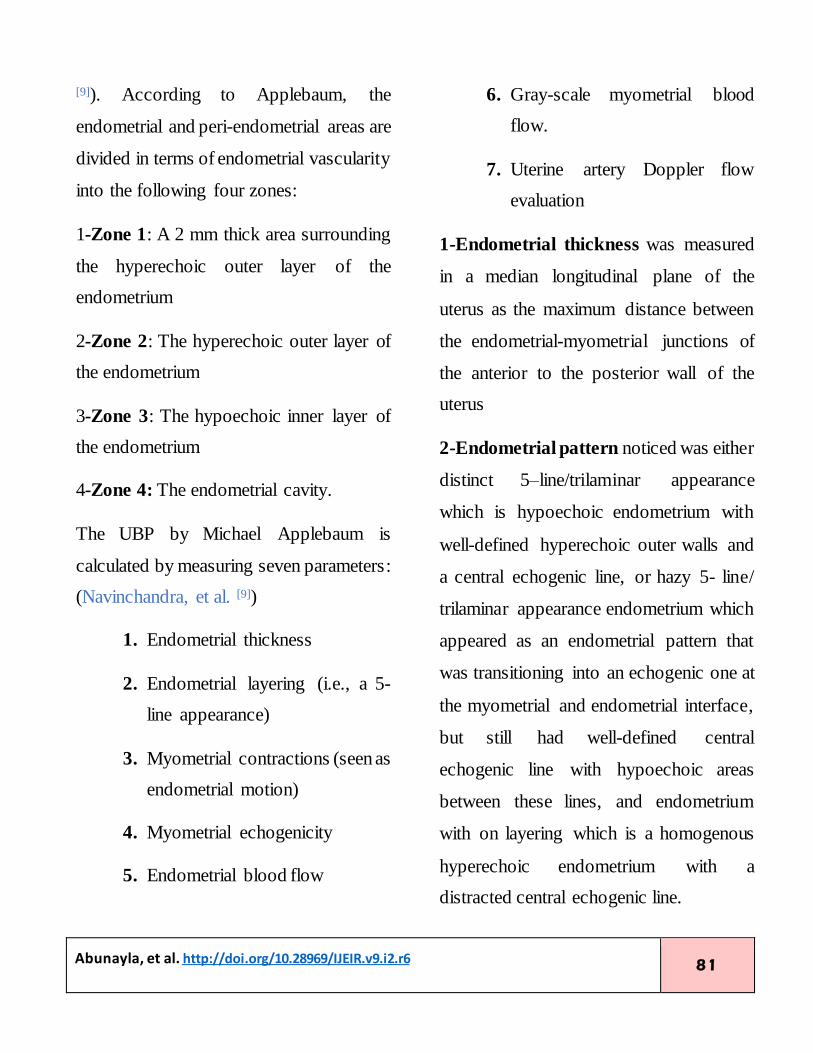
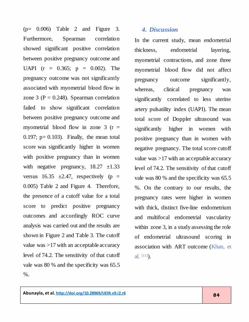
[9]). According to Applebaum, the
endometrial and peri-endometrial areas are
divided in terms of endometrial vascularity
into the following four zones:
1-Zone 1: A 2 mm thick area surrounding
the hyperechoic outer layer of the
endometrium
2-Zone 2: The hyperechoic outer layer of
the endometrium
3-Zone 3: The hypoechoic inner layer of
the endometrium
4-Zone 4: The endometrial cavity.
The UBP by Michael Applebaum is
calculated by measuring seven parameters:
(Navinchandra, et al. [9])
1. Endometrial thickness
2. Endometrial layering (i.e., a 5-
line appearance)
3. Myometrial contractions (seen as
endometrial motion)
4. Myometrial echogenicity
5. Endometrial blood flow
6. Gray-scale myometrial blood
flow.
7. Uterine artery Doppler flow
evaluation
1-Endometrial thickness was measured
in a median longitudinal plane of the
uterus as the maximum distance between
the endometrial-myometrial junctions of
the anterior to the posterior wall of the
uterus
2-Endometrial pattern noticed was either
distinct 5–line/trilaminar appearance
which is hypoechoic endometrium with
well-defined hyperechoic outer walls and
a central echogenic line, or hazy 5- line/
trilaminar appearance endometrium which
appeared as an endometrial pattern that
was transitioning into an echogenic one at
the myometrial and endometrial interface,
but still had well-defined central
echogenic line with hypoechoic areas
between these lines, and endometrium
with on layering which is a homogenous
hyperechoic endometrium with a
distracted central echogenic line.
http://doi.org/10.28969/IJEIR.v9.i2.r6, et al. Abunayla 82
3-Myometrial contraction: Myometrial
contractions were monitored by two
observers. Wave frequency was
considered as the number of waves during
2 minutes period. Concentration on
moderate and strong movement rather than
weak one and those directed cranially was
concerned.
4-Subendometrial vessels were usually
visualized at the periphery of the
endometrium. Sometimes they penetrated
the hyper-echogenic endometrial edge or
even reached the endometrial cavity. The
blood flow velocity waveforms from the
sub endometrial vessels were obtained by
placing the Doppler gate over the color
area and activating the pulsed Doppler
function (El-Zenneni, et al. [10]).
5-Pulsatility Index of Uterine Artery:
The Doppler gate was then placed over
both uterine arteries lateral to the cervix
and the same examination was repeated.
(PI) were measured automatically by using
the software program in the equipment, the
average of both sides results was recorded.
PI= (peak systolic velocity - end diastolic
velocity)/mean velocity during the cycle.
6-Uterine myometrial echogenicity was
assessed as uniform which is relatively
(homogeneous) or non-uniform (coarse
inhomogeneous).
7-Myometrial vascularity to the arcuate
arteries can be seen power Doppler rather
than color Doppler because power Doppler
is more sensitive to detect flow in smaller
vessels and low velocity flows,
myometrial vascularity is seen in the outer
part of the myometrium running parallel to
the serosa. For each patient scoring, all
parameters and calculation of the total
score number which is out of twenty were
done. Data were summarized, analyzed,
and presented using statistical package for
social sciences (SPSS) version 23 and
Microsoft Office Excel 2010. Quantitative
variables were expressed as mean and
standard deviation (SD); whereas,
categorical variables were expressed as
number and percentage. Independent
samples t-test was used to compare mean
http://doi.org/10.28969/IJEIR.v9.i2.r6, et al. Abunayla 83
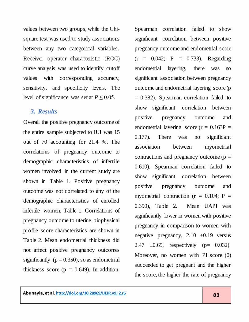
values between two groups, while the Chi-
square test was used to study associations
between any two categorical variables.
Receiver operator characteristic (ROC)
curve analysis was used to identify cutoff
values with corresponding accuracy,
sensitivity, and specificity levels. The
level of significance was set at P ≤ 0.05.
3. Results
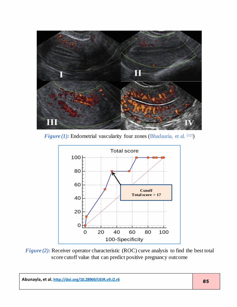
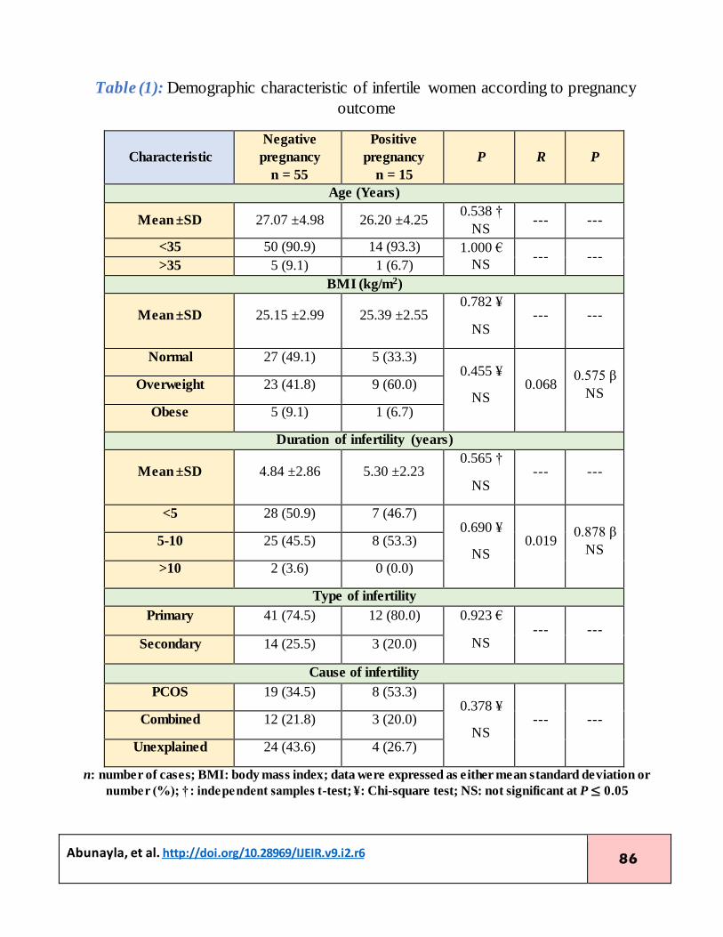
Overall the positive pregnancy outcome of
the entire sample subjected to IUI was 15
out of 70 accounting for 21.4 %. The
correlations of pregnancy outcome to
demographic characteristics of infertile
women involved in the current study are
shown in Table 1. Positive pregnancy
outcome was not correlated to any of the
demographic characteristics of enrolled
infertile women, Table 1. Correlations of
pregnancy outcome to uterine biophysical
profile score characteristics are shown in
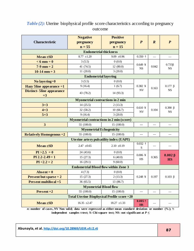
Table 2. Mean endometrial thickness did
not affect positive pregnancy outcomes
significantly (p = 0.350), so as endometrial
thickness score (p = 0.649). In addition,
Spearman correlation failed to show
significant correlation between positive
pregnancy outcome and endometrial score
(r = 0.042; P = 0.733). Regarding
endometrial layering, there was no
significant association between pregnancy
outcome and endometrial layering score (p
= 0,382). Spearman correlation failed to
show significant correlation between
positive pregnancy outcome and
endometrial layering score (r = 0.163P =
0.177). There was no significant
association between myometrial
contractions and pregnancy outcome (p =
0.610). Spearman correlation failed to
show significant correlation between
positive pregnancy outcome and
myometrial contraction (r = 0.104; P =
0.390), Table 2. Mean UAPI was
significantly lower in women with positive
pregnancy in comparison to women with
negative pregnancy, 2.10 ±0.19 versus
2.47 ±0.65, respectively (p= 0.032).
Moreover, no women with PI score (0)
succeeded to get pregnant and the higher
the score, the higher the rate of pregnancy
http://doi.org/10.28969/IJEIR.v9.i2.r6, et al. Abunayla 84
(p= 0.006) Table 2 and Figure 3.
Furthermore, Spearman correlation
showed significant positive correlation
between positive pregnancy outcome and
UAPI (r = 0.365; p = 0.002). The
pregnancy outcome was not significantly
associated with myometrial blood flow in
zone 3 (P = 0.248). Spearman correlation
failed to show significant correlation
between positive pregnancy outcome and
myometrial blood flow in zone 3 (r =
0.197; p= 0.103). Finally, the mean total
score was significantly higher in women
with positive pregnancy than in women
with negative pregnancy, 18.27 ±1.33
versus 16.35 ±2.47, respectively (p =
0.005) Table 2 and Figure 4. Therefore,
the presence of a cutoff value for a total
score to predict positive pregnancy
outcomes and accordingly ROC curve
analysis was carried out and the results are
shown in Figure 2 and Table 3. The cutoff
value was >17 with an acceptable accuracy
level of 74.2. The sensitivity of that cutoff
vale was 80 % and the specificity was 65.5
%.
4. Discussion
In the current study, mean endometrial
thickness, endometrial layering,
myometrial contractions, and zone three
myometrial blood flow did not affect
pregnancy outcome significantly,
whereas, clinical pregnancy was
significantly correlated to less uterine
artery pulsatility index (UAPI). The mean
total score of Doppler ultrasound was
significantly higher in women with
positive pregnancy than in women with
negative pregnancy. The total score cutoff
value was >17 with an acceptable accuracy
level of 74.2. The sensitivity of that cutoff
vale was 80 % and the specificity was 65.5
%. On the contrary to our results, the
pregnancy rates were higher in women
with thick, distinct five-line endometrium
and multifocal endometrial vascularity
within zone 3, in a study assessing the role
of endometrial ultrasound scoring in
association with ART outcome (Khan, et
al. [11]).
http://doi.org/10.28969/IJEIR.v9.i2.r6, et al. Abunayla 85
shown in (table 2). Regarding non-
Figure (1): Endometrial vascularity four zones (Bhadauria, et al. [12])
Figure (2): Receiver operator characteristic (ROC) curve analysis to find the best total
score cutoff value that can predict positive pregnancy outcome
Heparin binding epidermal growth factor (HBEGF)
0
20
40
60
80
100
Total score
0 20 40 60 80 100
100-Specificity
Se
nsi
tivity
Cutoff
Total score > 17
http://doi.org/10.28969/IJEIR.v9.i2.r6, et al. Abunayla 86
pregnant and pregnant groups, embryo Table (1): Demographic characteristic of infertile women according to pregnancy
outcome
Characteristic
Negative
pregnancy
n = 55
Positive
pregnancy
n = 15
P R P
Age (Years)
Mean ±SD 27.07 ±4.98 26.20 ±4.25 0.538 †
NS --- ---
<35 50 (90.9) 14 (93.3) 1.000 €
NS --- ---
>35 5 (9.1) 1 (6.7)
)2BMI (kg/m
Mean ±SD 25.15 ±2.99 25.39 ±2.55 0.782 ¥
NS --- ---
Normal 27 (49.1) 5 (33.3)
0.455 ¥
NS 0.068
0.575 β
NS Overweight 23 (41.8) 9 (60.0)
Obese 5 (9.1) 1 (6.7)
Duration of infertility (years)
Mean ±SD 4.84 ±2.86 5.30 ±2.23 0.565 †
NS --- ---
<5 28 (50.9) 7 (46.7) 0.690 ¥
NS 0.019
0.878 β
NS 5-10 25 (45.5) 8 (53.3)
>10 2 (3.6) 0 (0.0)
Type of infertility
Primary 41 (74.5) 12 (80.0) 0.923 €
NS --- ---
Secondary 14 (25.5) 3 (20.0)
Cause of infertility
PCOS 19 (34.5) 8 (53.3) 0.378 ¥
NS --- --- Combined 12 (21.8) 3 (20.0)
Unexplained 24 (43.6) 4 (26.7)
n: number of cases; BMI: body mass index; data were expressed as either mean standard deviation or
number (%); †: independent samples t-test; ¥: Chi-square test; NS: not significant at P ≤ 0.05
http://doi.org/10.28969/IJEIR.v9.i2.r6, et al. Abunayla 87
Table (2): Uterine biophysical profile score characteristics according to pregnancy
outcome
Characteristic
Negative
pregnancy
n = 55
Positive
pregnancy
n = 15
P R P
Endometrial thickness
Mean ±SD 8.77 ±1.20 9.09 ±0.96 0.350 † --- ---
< 6 mm = 0 3 (5.5) 0 (0.0) 0.649 ¥
NS 0.042
0.733β
NS 7-9 mm = 2 41 (74.5) 12 (80.0)
10-14 mm = 3 11 (20.0) 3 (20.0)
Endometrial layering
No layering=0 3 (5.5) 0 (0.0)
0.382 ¥
NV 0.163
0.177 β
NS
Hazy 5line appearance =1 9 (16.4) 1 (6.7)
Distinct- 5line appearance
=3 43 (78.2) 14 (93.3)
Myometrial contractions in 2 min
3=3 14 (25.5) 2 (13.3) 0.610 ¥
NV 0.104
0.390 β
NS 4=3 32 (58.2) 10 (66.7)
5=3 9 (16.4) 3 (20.0)
Myometrial contractions in 2 min (score)
3 55 (100.0) 15 (100.0) --- --- ---
Myometrial Echogenicity
Relatively Homogenous =2 55 (100.0) 15 (100.0) --- --- ---
Uterine artery pulsatility index (UAPI)
Mean ±SD 2.47 ±0.65 2.10 ±0.19 0.032 †
S --- ---
PI >2.5 = 0 24 (43.6) 0 (0.0) 0.006 ¥
HS 0.365
0.002 β
HS PI 2.2-2.49 = 1 15 (27.3) 6 (40.0)
PI <2.2 = 2 16 (29.1) 9 (60.0)
Endometrial Blood flow within Zone 3
Absent = 0 4 (7.3) 0 (0.0)
0.248 ¥ 0.197 0.103 β Present but sparse = 2 15 (27.3) 2 (13.3)
Present multifocal =5 36 (65.5) 13 (86.7)
Myometrial Blood flow
Present =2 55 (100.0) 15 (100.0) --- --- ---
Total Uterine Biophysical Profile score =20
Mean ±SD 16.35 ±2.47 18.27 ±1.33 0.005 †
HS --- ---
n: number of cases, NV Non valid; data were expressed as either mean standard deviation or number (% ); †:
independent samples t-test; ¥: Chi-square test; NS: not significant at P ≤

http://doi.org/10.28969/IJEIR.v9.i2.r6, et al. Abunayla 88
Table (3): Total score ROC curve characteristics
Characteristic Value
Cutoff >17
AUC 0.742
95% CI 0.624 - 0.840
Accuracy 74.2
P <0.001
HS
Sensitivity 80 %
Specificity 65.5 %
AUC: area under curve; CI: confidence interval; HS: highly significant at P ≤ 0.01
Figure (3): Mean PI index according to pregnancy outcome
Figure (4): Mean UBP index according to pregnancy outcome
Negative pregnancy Positive pregnancy
1.9
2
2.1
2.2
2.3
2.4
2.5
Mean
PI
ind
ex ±
SD
16.3518.27
Negative pregnancy Positive pregnancy
0
5
10
15
20
25
Me
an
to
tal U
BP
score
±
SD
http://doi.org/10.28969/IJEIR.v9.i2.r6, et al. Abunayla 89
However, in agreement with our study, the
latter study (Khan, et al. [11]) and other
studies (Baruffi, et al. [13]; Aghahoseini, et
al. [14]) found no significant association
between myometrial blood flow and
pregnancy outcome. (Khan, et al. [11]) also
found a significant association between
endometrial layering and pregnancy
outcome, again in clear contradiction to
our findings. The lack of a significant
association between endometrial thickness
and pregnancy outcome is in agreement
with several other authors (Kolibiianahis,
et al. [15]; Habibzadeh, et al. [16]) stated that
women with endometrial thickness less
than 6 mm have significantly lower
pregnancy rate than women with an
endometrial thickness higher than 6 mm;
therefore, the lack of significant effect of
endometrial thickness on the rate of
pregnancy in our study may be explained
by that most of women enrolled in the
current study had an endometrial thickness
of > 6 mm. Therefore, the overall score of
endometrial and myometrial assessment
should be considered rather than
individual sonographic criteria when
dealing with the prediction of positive
pregnancy outcome as was the case in the
current study in which the total ultrasound
score was the main determinant with a
cutoff value that had acceptable accuracy
level. The current study result agreed by
(Navinchandra, et al. [9]) study that found
no significant effect of vascularity within
zone 3 and pregnancy outcome. On the
contrary to the study results, pregnancy
rates were higher in women with thick,
distinct five-line endometrium and
multifocal endometrial vascularity within
zone 3, in a study assessing the role of
endometrial ultrasound scoring in
association with ART outcome (Khan, et
al. [11]). However, in agreement with the
study, (Khan, et al. [11]; Baruffi, et al. [13];
Aghahoseini, et al. [14]) found no
significant association between
myometrial blood flow and pregnancy
outcome. (Kim, et al. [17]; Khan, et al. [11])
found a significant association between
endometrial layering and pregnancy
outcome, again in clear contradiction to
http://doi.org/10.28969/IJEIR.v9.i2.r6, et al. Abunayla 90
present findings. (Masrour, et al. [18])
demonstrated that the endometrial blood
flow was significantly greater in pregnant
women and that the endometrial thickness
and pattern of sonography did not have any
predictive values for endometrial
receptivity these results are similar to the
current study concerning endometrial
thickness and dissimilar to current results
regarding sub endometrial vascularity and
pregnancy. There was a negative
correlation between pregnancy outcome
and Uterine Artery Pulsatility Index
(UAPI). Some authors have studied the
difference in PI between pregnant and
nonpregnant women following ARTs and
have found, on the contrary to this study,
no significant difference (Prasad, et al.
[19]). However, other authors concluded
that lower PI was correlated with positive
biochemical pregnancy outcome in line
with the findings of the current study
(Navinchandra, et al. [9]; Martins, et al. [7]).
These findings may relate to the hormonal
status during ovarian controlled
stimulation and the effects of relatively
higher serum. Oestrogen promotes
vascular smooth muscle relaxation and
reduces sensitivity to adrenergic
stimulation (Guedes-Martins, et al. [20]).
Acknowledgment
We would like to acknowledge the High
Institute of Infertility Diagnosis and
Assisted Reproductive Technologies, Al
Nahrain University.
Funding
This work received no funding.
Author Contribution
Abunayla RSH, performed the study, Al-
Anbari LA, Abood MS, and Husaini
HAR, supervised the work.
Conflict of Interest
The authors declare no conflict of
interest.
Ethical Clearance
The study was approved by the Ethical Approval Committee.
References
[1] Vander Borght M, Wyns C. Fertility
and infertility: Definition and
epidemiology. Clinical Biochemistry.
Elsevier BV; 2018;62:2–10. Doi:
http://dx.doi.org/10.1016/j.clinbioche
m.2018.03.012 [PubMed][Elsevier]
http://doi.org/10.28969/IJEIR.v9.i2.r6, et al. Abunayla 91
[2] Agarwal A, Mulgund A, Hamada A,
Chyatte MR. A unique view on male
infertility around the globe.
Reproductive Biology and
Endocrinology. Springer Science and
Business Media LLC; 2015;13(1). Doi:
http://dx.doi.org/10.1186/s12958-015-
0032-1 [PubMed][PMC][BMC]
[3] Datta J, Palmer MJ, Tanton C, Gibson
LJ, Jones KG, Macdowall W, et al.
Prevalence of infertility and help
seeking among 15 000 women and
men. Human Reproduction. Oxford
University Press (OUP);
2016;31(9):2108–18. Doi:
http://dx.doi.org/10.1093/humrep/dew
123 [PubMed][PMC][OxFord]
[4] Almasi-Hashiani A, Omani-Samani R,
Mohammadi M, Amini P, Navid B,
Alizadeh A, et al. Assisted
reproductive technology and the risk of
preeclampsia: an updated systematic
review and meta-analysis. BMC
Pregnancy and Childbirth. Springer
Science and Business Media LLC;
2019;19(1). Doi:
http://dx.doi.org/10.1186/s12884-019-
2291-x [PubMed][PMC][BMC]
[5] Allahbadia GN. Intrauterine
Insemination: Fundamentals
Revisited. The Journal of Obstetrics
and Gynecology of India. Springer
Science and Business Media LLC;
2017;67(6):385–92. Doi:
http://dx.doi.org/10.1007/s13224-017-
1060-x [PubMed][PMC]
[6] Ombelet W, Van Robays J. Artificial
insemination history: hurdles and
milestones. Facts Views Vis Obgyn,
2015;7(2):137-143. [EuroPMC]
[7] Silva Martins R, Helio Oliani A, Vaz
Oliani D, Martinez de Oliveira J.
Subendometrial resistence and
pulsatility index assessment of
endometrial receptivity in assisted
reproductive technology cycles.
Reproductive Biology and
Endocrinology. Springer Science and
Business Media LLC; 2019;17(1). Doi:
http://dx.doi.org/10.1186/s12958-019-
0507-6 [BMC]
[8] Tiwary B., Dhaliwal L. Gainder S.
Effect of GnRH antagonist on
follicular development and uterine
biophysical profile in controlled
ovarian stimulation. International
Journal of Reproduction,
Contraception, Obstetrics and
Gynecology. 2015;4(1):157-163.
[ijrcog]
[9] Navinchandra Rn, Shankar S, Kavitha
D, Kamath M, Devdas S, Vineela P.
Relationship between uterine scoring
system for reproduction and pregnancy
in controlled ovarian stimulation-
intrauterine insemination cycles. IVF
Lite. Medknow; 2016;3(3):115. Doi:
http://dx.doi.org/10.4103/2348-
2907.204669 [ivflite]
[10] El-Zenneni H, Moustafa R, Abdel-
Hafeez M, El-Salally H, Abdel-Kader
A, Elnaggar A. Assessment of uterine,
http://doi.org/10.28969/IJEIR.v9.i2.r6, et al. Abunayla 92
subendometrial blood flows and
endometrial gland vascular endothelial
growth factor (EG-VEGF) in women
with unexplained infertility. Middle
East Fertility Society Journal. Springer
Science and Business Media LLC;
2015;20(2):119–26. Doi:
http://dx.doi.org/10.1016/j.mefs.2014.
07.002 [Mendeley][ResearchGate]
[11] Khan MS, Shaikh A, Ratnani R.
Ultrasonography and Doppler Study to
Predict Uterine Receptivity in Infertile
Patients Undergoing Embryo Transfer.
The Journal of Obstetrics and
Gynecology of India. Springer Science
and Business Media LLC;
2015;66(S1):377–82. Doi:
http://dx.doi.org/10.1007/s13224-015-
0742-5 [PMC][PubMed]
[12] Bhadauria K, Sahni R, Verma S,
Khatri PQ. Colour Doppler ultrasound
in controlled ovarian stimulation with
intrauterine insemination. Apollo
Medicine. Medknow; 2012;9(3):252–
62. Doi:
http://dx.doi.org/10.1016/j.apme.2012.
07.014 [Elsevier]
[13] Baruffi RL, Contart P, Mauri AL,
Peterson C, Felipe V, Franco JG. A
uterine ultrasonographic scoring
system as a method for the prognosis
of embryo implantation in an ICSI
program. Journal of Assisted
Reproduction and Geneics.
2002;19(3):99-102. [EuroPMC]
[14] Aghahoseini M, Tuba K, Marsousi
V, Aleyasin A. Assessment of
endometrial-subendometrial blood
flow detected by color Doppler
sonography and uterine receptivity in
infertile women. Acta Med
Iran. 2015;46:461–6.
[ResearchGate][ActaMedIr]
[15] Kolibianakis E, Zikopoulos K,
Fatemi H, Osmanagaoglu K, Evenpoel
J, Van Steirteghem A, et al.
Endometrial thickness cannot predict
ongoing pregnancy achievement in
cycles stimulated with clomiphene
citrate for intrauterine insemination.
Reproductive BioMedicine Online.
Elsevier BV; 2004;8(1):115–8. Doi:
http://dx.doi.org/10.1016/s1472-
6483(10)60505-6 [PubMed][Elsevier]
[16] Habibzadeh V, Nematolahi Mahani
S, Kamyab H. The correlation of
factors affecting the endometrial
thickness with pregnancy outcome in
the IUI cycles. International Journal of
Reproductive BioMedicine. 2011;9(1):
41-46. [PMC]
[17] Kim A, Lee J, Ji Y, Lee H, Lee E,
Kim H, Oh Y. Do Endometrial
Movements Affect. The Achievement
of Pregnancy during Intrauterine
Insemination? International Journal of
Fertility and Sterility. 2015;8(4):399-
408. [PMC][EuroPMC]
[18] Masrour M, Yoonesi L,
Aerabsheibani H. The effect of
endometrial thickness and endometrial
http://doi.org/10.28969/IJEIR.v9.i2.r6, et al. Abunayla 93
blood flow on pregnancy outcome in
intrauterine insemination cycles.
Journal of Family Medicine and
Primary Care. Medknow;
2019;8(9):2845. Doi:
http://dx.doi.org/10.4103/jfmpc.jfmpc
_212_19 [jfmpc]
[19] Prasad S, Goyal R, Kumar Y, Nayar
P, Hajela S, Kumaran A, Vairagi R,
Chauhan S. The Relationship Between
Uterine Artery two-dimensional Color
Doppler Measurement and Pregnancy
Outcome: A Prospective
Observational Study. Journal of
Reproduction and Infertility.
2017;18(2):251-256. [PubMed][PMC]
[20] Guedes-Martins L, Gaio R, Saraiva
J, Cerdeira S, Matos L, Silva E, et al.
Reference Ranges for Uterine Artery
Pulsatility Index during the Menstrual
Cycle: A Cross-Sectional Study.
Crispi-Brillas F, editor. PLOS ONE.
Public Library of Science (PLoS);
2015;10(3):e0119103. Doi:
http://dx.doi.org/10.1371/journal.pone
.0119103 [PMC][PlosOne]
Biography
Dr. Rawaa Saad Hasan
Abunayla
She graduated from Tikrit
University, College of Medicine
in 1996. Completed permanency
in gynecology and Obstetrics in
2002. Received the Diploma in
Obstetrics and Gynaecology in 2007 from Baghdad
University, College of Medicine. She worked as a
senior at Al Rumaitha General Hospital. Currently she
is a student in the High Diploma in Clinical Infertility
and ARTs in High institute for Infertility Diagnosis
and ARTs Al Nahrain University, 2020.
Dr. Lubna Amer Al-Anbari
She received the M.B.CH.B., the
Higher diploma from the college
of medicine, University of
Baghdad, in 2004, and 2011,
respectively. She received the
board in infertility from the Iraqi
society of medical specialists in
2012. She published many kinds of research both local
and international. She supervised many students. She held many academic positions.
Dr. Muayad S. Abood
He received the M.B.CH.B.
from Al-Kufa University, the
M.Sc., and the Ph.D. in
Pharmacology from Al Nahrain
University, in 1998, 2008, and
2014, respectively. He occupied
several academic positions
including the dean assistant for scientific affairs at Al
Nahrain University. He has more than 10 published papers both local and international.
Dr. Huda A. R. Hussaini
She received the M.B.Ch.B:
Babylon University, college of
medicine, in 2000. The diploma
in Radiology from the
University of Baghdad in 2007.
She is a fellow of the Iraqi
Board for Medical
Specialization, Council of Radiology since 2008.
Currently, she is an assistant professor. Al Nahrain
University, College of Medicine, department of
surgery ( specialist radiologist ). She is a Member of
staff in the high Institute of infertility diagnosis and
ARTS. Including the ultrasound department in the
institute since 2018. She has Participations in a lot of
annual meetings, medical conferences. Seminars,

http://doi.org/10.28969/IJEIR.v9.i2.r6, et al. Abunayla 94
continuous learning, and thesis discussion. She is a
member of the Iraqi medical association, the Iraqi
Society of radiologist and medical imaging, Iraqi
Journal of Embryos infertility researches, European
Society for Hybrid, Molecular, and translation
imaging. She published more than articles and has supervised many students.
© 2019 Author(s)
This article is licensed under a Creative Commons
Attribution 4.0 International License, which permits
use, sharing, adaptation, distribution and reproduction
in any medium or format, as long as you give
appropriate credit to the original author(s) and the
source, provide a link to the Creative Commons
license, and indicate if changes were made.
http://creativecommons.org/licenses/by/4.0/.
How to cite:
Abunayla RSH, Al-Anbari LA, Abood MS,
Hussaini HAR. Using Uterine Biophysical Profile
as a Predictor of Endometrial Receptivity and
Pregnancy in Stimulated IUI cycles; Iraqi Journal of
Embryos and Infertility Researches (IJEIR), (2019);
9(2): 78-94.
Doi: http://doi.org/10.28969/IJEIR.v9.i2.r6
Copyright © 2022 FDOKUMEN