Proteomics, oxidative stress and male infertility
27
REVIEW Proteomics, oxidative stress and male infertility Ashok Agarwal a, * , Damayanthi Durairajanayagam a,b , Jacques Halabi a , Jason Peng a , Monica Vazquez-Levin c a Center for Reproductive Medicine, Glickman Urological and Kidney Institute, Cleveland, OH, USA; b Faculty of Medicine, MARA University of Technology, Sungai Buloh, Selangor, Malaysia; c Institute of Biology and Experimental Medicine, National Research Council of Argentina, CONICET, Buenos Aires, Argentina * Corresponding author. E-mail address: [email protected] (A Agarwal). Ashok Agarwal is a Professor at the Lerner College of Medicine, Case Western Reserve University and the Head of the Andrology Center and Director of Research at the Center for Reproductive Medicine, Cleveland Clinic, USA. He has researched extensively on oxidative stress and its implications on human fertility. He serves on the editorial boards of several key journals in human reproduction. Ashok’s current research interests are the study of molecular markers of oxidative stress, DNA fragmentation and apoptosis using proteomics and bioinformatics tools, as well as fertility preservation in patients with cancer, and the efficacy of certain antioxidants in improving male fertility. Abstract Oxidative stress has been established as one of the main causes of male infertility and has been implicated in many dis- eases associated with infertile men. It results from high concentrations of free radicals and suppressed antioxidant potential, which may alter protein expression in seminal plasma and/or spermatozoa. In recent years, proteomic analyses have been performed to characterize the protein profiles of seminal ejaculate from men with different clinical conditions, such as high oxidative stress. The aim of the present review is to summarize current findings on proteomic studies performed in men with high oxidative stress com- pared with those with physiological concentrations of free radicals, to better understand the aetiology of oxidative stress-induced male infertility. Each of these studies has suggested candidate biomarkers of oxidative stress, among them are DJ-1, PIP, lactotrans- ferrin and peroxiredoxin. Changes in protein concentrations in seminal plasma samples with oxidative stress conditions were related to stress responses and to regulatory pathways, while alterations in sperm proteins were mostly associated to metabolic responses (carbohydrate metabolism) and stress responses. Future studies should include assessment of post-translational modifications in the spermatozoa as well as in seminal plasma proteomes of men diagnosed with idiopathic infertility. RBMOnline ª 2014, Reproductive Healthcare Ltd. Published by Elsevier Ltd. All rights reserved. KEYWORDS: biomarkers, male infertility, proteomics, oxidative stress, seminal plasma, spermatozoa http://dx.doi.org/10.1016/j.rbmo.2014.02.013 1472-6483/ª 2014, Reproductive Healthcare Ltd. Published by Elsevier Ltd. All rights reserved. Reproductive BioMedicine Online (2014) 29, 32– 58 www.sciencedirect.com www.rbmonline.com
Transcript of Proteomics, oxidative stress and male infertility

Reproductive BioMedicine Online (2014) 29, 32–58
www.sc iencedi rec t .comwww.rbmonl ine .com
REVIEW
Proteomics, oxidative stress and maleinfertility
http://dx.doi.org/10.1016/j.rbmo.2014.02.0131472-6483/ª 2014, Reproductive Healthcare Ltd. Published by Elsevier Ltd. All rights reserved.
Ashok Agarwal a,*, Damayanthi Durairajanayagam a,b, Jacques Halabi a,Jason Peng a, Monica Vazquez-Levin c
a Center for Reproductive Medicine, Glickman Urological and Kidney Institute, Cleveland, OH, USA; b Faculty of Medicine,MARA University of Technology, Sungai Buloh, Selangor, Malaysia; c Institute of Biology and Experimental Medicine,National Research Council of Argentina, CONICET, Buenos Aires, Argentina* Corresponding author. E-mail address: [email protected] (A Agarwal).
Abstract Oxidative stress
Ashok Agarwal is a Professor at the Lerner College of Medicine, Case Western Reserve University and the Head ofthe Andrology Center and Director of Research at the Center for Reproductive Medicine, Cleveland Clinic, USA.He has researched extensively on oxidative stress and its implications on human fertility. He serves on theeditorial boards of several key journals in human reproduction. Ashok’s current research interests are the studyof molecular markers of oxidative stress, DNA fragmentation and apoptosis using proteomics and bioinformaticstools, as well as fertility preservation in patients with cancer, and the efficacy of certain antioxidants inimproving male fertility.
has been established as one of the main causes of male infertility and has been implicated in many dis-eases associated with infertile men. It results from high concentrations of free radicals and suppressed antioxidant potential, whichmay alter protein expression in seminal plasma and/or spermatozoa. In recent years, proteomic analyses have been performed tocharacterize the protein profiles of seminal ejaculate from men with different clinical conditions, such as high oxidative stress. Theaim of the present review is to summarize current findings on proteomic studies performed in men with high oxidative stress com-pared with those with physiological concentrations of free radicals, to better understand the aetiology of oxidative stress-inducedmale infertility. Each of these studies has suggested candidate biomarkers of oxidative stress, among them are DJ-1, PIP, lactotrans-ferrin and peroxiredoxin. Changes in protein concentrations in seminal plasma samples with oxidative stress conditions were relatedto stress responses and to regulatory pathways, while alterations in sperm proteins were mostly associated to metabolic responses(carbohydrate metabolism) and stress responses. Future studies should include assessment of post-translational modifications in the
spermatozoa as well as in seminal plasma proteomes of men diagnosed with idiopathic infertility. RBMOnline
ª 2014, Reproductive Healthcare Ltd. Published by Elsevier Ltd. All rights reserved.
KEYWORDS: biomarkers, male infertility, proteomics, oxidative stress, seminal plasma, spermatozoa

Proteomics, oxidative stress and male infertility 33
Introduction
Infertility is defined as the inability to achieve a clinicalpregnancy after 12 months or more of regular, unprotectedand well-timed intercourse (Practice Committee ofAmerican Society for Reproductive Medicine, 2013). Infertil-ity affects around 15% of all couples of reproductive age,with about 50% being associated with abnormalities in themale, called male factor infertility (Sabanegh andAgarwal, 2012). A recent study using the current durationapproach to assess the prevalence of infertility estimatedthat 9 to 14% of American men within reproductive age (i.e.15 to 44 years old) will probably experience difficulties toconceive (Louis et al., 2013). Male infertility could resultfrom dysfunction at various levels along the hypothalamic-pituitary-gonadal axis: pre-testicular (damage at thehypothalamus or pituitary level), testicular (failure of thetestis), post-testicular (normal testicular function but withobstruction or inflammation that leads to infertility) or acombination of these. Causes of male infertility includehypogonadotrophic hypogonadism and Kallmann syndrome,direct trauma, inflammation or infection of the testis, var-icocele, cryptorchidism, Y-chromosome microdeletions,testicular cancer and chemotherapy, erectile dysfunction,infrequent or retrograde ejaculation, epididymitis, congen-ital bilateral absence of the vas deferens, Klinefelter’ssyndrome (47,XXY), and Sertoli-cell only syndrome (Wiseret al., 2012).
The aetiology of male factor infertility, although multi-factorial, remains largely idiopathic (Sabanegh andAgarwal, 2012). Reactive oxygen species (ROS)-induced oxi-dative stress is well-known to play a major role in male fac-tor infertility (Hamada et al., 2012; Tremellen, 2008).Excess ROS concentrations and oxidative stress in the malereproductive tract are detrimental to spermatozoa (Azizet al., 2004) and have been associated with negativechanges in sperm concentration, motility and morphology,leading to poor semen parameters and eventually to infertil-ity (Khosrowbeygi and Zarghami, 2007). In fact, oxidativestress has been implicated in several male infertility-associated pathologies, including leukocytospermia andvaricocele as well as idiopathic infertility (Pasqualottoet al., 2000).
The diagnosis of male infertility routinely begins with abasic semen analysis, which measures various semen param-eters including semen volume, colour, pH, liquefactiontime, viscosity, sperm count and motility, sperm morphol-ogy, concentration of round cells and polymorphonucleo-cytes, sperm agglutination and sperm viability (ifrequired). Two or more of these basic semen analyses areused to identify abnormalities in: sperm concentration (oli-gozoospermia or azoospermia), motility (asthenozoosper-mia) and morphology (teratozoospermia), based onreference values established by the World Health Organiza-tion (WHO, 1999, 2010). In addition to the routine evalua-tion, several advanced tests can be performed to establish
the causes(s) of infertility, among them are the assessment
of ROS levels, total antioxidant capacity and sperm DNA
fragmentation level, DNA compaction and apoptosis, as well
as presence and localization of antisperm antibodies andgenetic testing (Kovac et al., 2013). However, results of
these tests typically either fall within the normal range ordo not help determine an exact aetiology of infertility, lead-ing to a classification of ‘idiopathic infertility’ (Kovac et al.,2013). While assisted reproductive technology (ART) mayincrease the chances of conception, it does not ensure thegenomic integrity of the embryo (Tremellen, 2008). In fact,high ROS concentrations in infertile men have been associ-ated with DNA fragmentation and poor chromatin packing(Tremellen, 2008). Damaged DNA in spermatozoa is indica-tive of poor cellular health. Sperm DNA damage reducessemen quality and is the cause of infertility in many men(Lewis et al., 2013; Simon et al., 2013). In assisted repro-duction, spermatozoa with damaged DNA lower fertilizationand pregnancy rates, impair embryo development and qual-ity and increase the risk of spontaneous miscarriage, birthdefects and childhood diseases such as cancer (Aitkenet al., 2013; Lewis, 2014). The level of DNA damage is sug-gestive of clinical outcome in assisted reproduction: idio-pathically infertile couples with higher levels of spermDNA fragmentation were found to have lower live-birthrates following IVF (Simon et al., 2013).
Highly specialized techniques such as proteomics allowcharacterization of the semen profile at a molecular level,proving useful in the assessment of proteins and the under-standing of biological pathways that play a key role in maleinfertility (du Plessis et al., 2011). Advances in thisrapidly-evolving field have allowed researchers to betteridentify seminal plasma and sperm proteins and todetermine how their presence or concentration may differin fertile versus infertile patients (Mitulovic and Mechtler,2006). Studies looking at the sperm and seminal plasma pro-tein profiles of men with oxidative stress-induced infertilitywould help in identifying alterations in the proteinexpression and/or translational modifications that mayoccur during sperm maturation and functions of proteinsinvolved. Moreover, these studies may be extended to thecharacterization of other pathologies associated with maleinfertility at the molecular level.
Despite the established role of oxidative stress in theaetiology of male infertility, there are, as of yet, relativelyfew studies that have investigated the correlation betweenROS-induced oxidative stress and a differential proteinexpression profile in the human ejaculate using proteomicanalysis. Our laboratory has recently published a series ofstudies on patients diagnosed with primary and secondaryinfertility and elevated ROS concentrations using proteomicapproaches (Hamada et al., 2013; Sharma et al., 2013a,2013b). Using similar strategies, other laboratories havealso studied the proteomic profile of infertile patients withpoor semen quality who were also affected with oxidativestress (Herwig et al., 2013; Wang et al., 2009).
Thus, in this review, we aim to summarize and comparethe findings of these initial studies that have utilized pro-teomic analysis to look into the differential expression ofproteins in the seminal ejaculate of infertile men with highoxidative stress and fertile men with physiological levels ofROS. In this review, we only included proteomic studies inwhich the oxidative status of infertile men was measured.Our review begins with an overview of oxidative stress andits impact on male infertility as well as the methodologiesand general work flow utilized in proteomic studies, in orderto provide some basis to readers less familiar with the field.

34 A Agarwal et al.
In addition, this review highlights seminal plasma and sper-matozoa proteins identified using proteomic analysis thatare likely to play a major role in oxidative stress-inducedmale infertility and subsequently it lists proteins that havethe potential to serve as diagnostic biomarkers of maleinfertility. To conclude, current limitations of theseresearch studies as well as some perspectives in this areaof research are highlighted. It is hoped that proteomic stud-ies in men with different diagnosis of infertility will eventu-ally lead to the discovery of biomarkers for idiopathic maleinfertility, which would help with the diagnosis and bettermanagement of male factor infertility.
Oxidative stress and male infertility
Oxidative stress occurs when there is an imbalance betweenROS and the antioxidants that scavenge surplus free radicals(Hwang and Lamb, 2012; Sharma and Agarwal, 1996; Sikka,2001; Sikka et al., 1995). ROS are natural products of cellu-lar metabolism which, in physiological amounts, are essen-tial requirements of spermatozoa for sperm processesleading to successful fertilization, such as capacitation,hyperactivated motility and acrosomal reaction (Agarwalet al., 2004). However, studies have shown that 30–80%of male factor infertility cases are due to ROS–mediatedsperm damage (Iwasaki and Gagnon, 1992; Ochsendorfet al., 1994; Shekarriz et al., 1995a, 1995b; Tremellen,2008; Zini et al., 1993).
There are two principal methods in which ROS can causemale infertility: through damage of the sperm membraneand damage of the sperm DNA. Sperm membranes havelarge amounts of polyunsaturated fatty acids, making themsusceptible to oxidative stress. This can then affect spermmotility as well as their ability to fertilize oocytes. Further-more, DNA fragmentation may harm the paternal geneticcontribution to the developing embryo (Tremellen, 2008).
ROS are considered a class of free radicals because theycontain oxygen molecules with one or more unpaired elec-trons. This makes them highly reactive and susceptible toradical formation, potentially altering cellular function
* Not a free radical
ROS
SeconPrimary
Superoxide anion radical Hydrogen pHydroxylPeroxyl r
Figure 1 Primary, secondary and tertiary forms of ROS and diffe
and ultimately endangering cell survival (de Lamirandeand Gagnon, 1993; Hwang and Lamb, 2012). In turn, reac-tions such as oxidation of membrane lipids, carbohydratesrelated to DNA and amino acids may occur (Ochsendorf,1999). However, not all ROS are free radicals (Cheesemanand Slater, 1993). There are three different general formsof the ROS (Figure 1): (i) the primary form of ROS, thesuperoxide anion radical from which secondary ROS can bederived either directly or indirectly (Hwang and Lamb,2012; Tremellen, 2008); (ii) the secondary form of ROS,hydrogen peroxide (an example of a ROS that is not a freeradical), hydroxyl radical and peroxyl radical; and (iii) thetertiary form of ROS, a class of free radicals that are nitrog-enous compounds: peroxynitrous acid, nitroxyl anion, per-oxynitrile and nitrous oxide (Hwang and Lamb, 2012).
Sources of oxidative stress
The origins of oxidative stress may vary from lifestylechoices and environmental factors (exogenous), to testicu-lar (endogenous) sources, in addition to idiopathic causes.
Lifestyle, for example, is a major contributor to ROS pro-duction; smokers see a 107% increase in ROS concentrationsin the semen, increased leukocytes and a greater likelihoodof DNA fragmentation compared with non-smokers (Salehand Agarwal, 2002; Sepaniak et al., 2004; Trummer et al.,2002). Alcohol abuse induces systemic oxidative stress andreduces antioxidant defences, which is likely further exac-erbated by an antioxidant deficient diet (Hwang andLamb, 2012; Mostafa et al., 2006; Villalta et al., 1997). Dietand exercise are also important factors in oxidative stress:improper diet and sedentary lifestyles can lead to obesity.Obesity in general increases the risk of co-morbidities, suchas hypertension, dyslipidaemia, type 2 diabetes, coronaryheart disease, stroke, non-alcoholic fatty liver disease,osteoarthritis, sleep apnoea and several types of cancers(Savini et al., 2013). These obesity-linked systemic inflam-matory conditions upset the redox balance and contributeto seminal oxidative stress, which causes detriment tosperm function (Palmer et al., 2012). The accumulation of
dary Tertiary
Nitrogenous compoundseroxide* radical adical
Peroxynitrous acidNitroxyl anion Peroxynitrile Nitrous oxide
rent types of free radicals.

Proteomics, oxidative stress and male infertility 35
adipose tissue, which causes obesity, may increase ROS lev-els through the release of pro-inflammatory cytokines,increased ROS production in leukocytes and heating of thetesticles (Banks et al., 2005; Ishii et al., 2005;Perez-Crespo et al., 2008; Singer and Granger, 2007). Con-versely, intensive exercise has been linked to increasedROS concentrations, regardless of the type of exercise,because of an increased demand for energy in the muscles(Peake et al., 2007).
Environmental factors also have an impact in ROS pro-duction. Phthalates are chemicals that are added to plasticsto increase its flexibility (plasticizers), and are used in foodpackaging, medical devices and personal care products.They have been linked to increased generation of ROS andreduction in antioxidants, leading to testicular oxidativestress (Agarwal et al., 1985; Hauser et al., 2007; Kasaharaet al., 2002). Pesticide and heavy metal exposure are asso-ciated with diminished antioxidant levels and elevated8-hydroxy-20-deoxyguanosine levels in sperm DNA, indica-tive of increased oxidative DNA damage in spermatozoa(Chitra et al., 2001; Latchoumycandane et al., 2003; Xuet al., 2003). Other studies have detailed how both drugadministration (such as aspirin and acetaminophen) andeven assisted reproduction treatment (such as IVF and intra-uterine insemination) increase oxidative stress (Agarwal andSaid, 2005; Iwasaki and Gagnon, 1992; Shekarriz et al.,1995a, 1995b). This is either through increasing enzymicactivity in the case of drugs or through assisted reproduc-tion practices such as centrifugation or removal of antioxi-dants from semen.
ROS can also be produced as a result of infection andinflammation. Generally, pathogens will elicit a naturalimmune response such as an acute inflammatory responsefrom leukocytes and macrophages (Mazzilli et al., 1994;Roos, 1991). For example, genitourinary tract infectionscause elevated levels of sperm oxidative stress (Brackettet al., 2008; Padron et al., 1997), while a history of or a cur-rent Chlamydia infection is linked to an increase in spermoxidative damage (Segnini et al., 2003).
In terms of testicular sources of oxidative stress, varico-cele is believed to be the principal underlying pathology forthe infertile male (Agarwal et al., 2006a; Barbieri et al.,1999; Hendin et al., 1999; Nallella et al., 2004; Salehet al., 2003; Smith et al., 2007). Varicocele is the abnormaldilation and tortuosity of the pampiniform plexus veinswithin the spermatic cord and is the cause for nearly 35%of male factor infertility. Clinical varicocele can be foundin 35% of men with primary infertility and 80% of men withsecondary infertility. Infertile men with varicocele havehigh levels of seminal ROS and oxidative stress, whichcauses significant sperm DNA damage (Agarwal et al.,2006a, 2009; Pasqualotto et al., 2008).
Cryptorchidism is also a common testicular cause of oxi-dative stress; this pathology is the result of a deficient mat-uration of gonocytes into type A spermatogonia, whichcauses hypospermatogenesis (Huff et al., 1991). If there isprolonged ischaemia followed by restoration of the bloodflow (spontaneous or surgical), an influx of activated leuko-cytes follows into both testicles (Turner et al., 2004). Thiscauses an increase in ROS production, leading to necrosisof germinal cells and ultimately subfertility or infertility(Filho et al., 2004; Tremellen, 2008).
In the seminal ejaculate, the principal sources of freeradical production come from either the leukocytes or thespermatozoa themselves (Tremellen, 2008). However, it isimplied that leukocytes contribute the most to oxidativestress because compared with spermatozoa, the rate ofROS production in leukocytes is 1000-times greater (Planteet al., 1994). This is considered an ‘extrinsic source’ ofROS as opposed to the ‘intrinsic’ sources from sperm. Incases of idiopathic causes of male infertility, patients mayhave normozoospermia and yet are infertile, showing highROS production and reduced antioxidant levels when com-pared with fertile men (Agarwal et al., 2006b; Pasqualottoet al., 2001).
Effects of oxidative stress on semen parameters
Low levels of ROS are generated naturally during processessuch as spermatogenesis. Indeed, ROS are necessary, in bal-ance with antioxidants, for proper spermatogenic mecha-nisms to occur. For example, hydrogen peroxide, asecondary form of ROS, stimulates acrosomal reaction,hyperactivation, and tyrosine phosphorylation in the sperm(Aitken et al., 1995; de Lamirande and Gagnon, 1993). Even-tually, hydrogen peroxide leads to sperm binding to thezona pellucida (Hwang and Lamb, 2012). The antioxidantcatalase, on the other hand, decomposes hydrogen peroxideand is also produced to preserve sperm motility (Tremellen,2008). This delicate balance between hydrogen peroxideand its antioxidant, catalase, illustrates the subtle equilib-rium that both facilitates proper function and prevents oxi-dative stress.
ROS are generally produced as a byproduct of enzymicreactions in oxidative phosphorylation, which is used to pro-duce energy in the form of ATP. These reactions thatinvolve the reduction of oxygen usually take place in themitochondria (Tremellen, 2008; Valko et al., 2007). In thesperm cell, mitochondria are located on the midpiece. Stud-ies show that mitochondrial DNA is more susceptible tomutations than nuclear DNA, increasing the production ofROS during this process (Bogenhagen, 1999; Liu et al.,2004; Taylor and Turnbull, 2005). In fact, elevated ROS lev-els have been linked to release of cytochrome C, a proteinthat activates apoptotic reactions, which is increased inpatients with male factor infertility (Wang et al., 2003).
While mutations are less likely in nuclear DNA than inmitochondrial DNA, prior studies have shown that intrinsicROS production is highly related to DNA fragmentation (Hen-kel et al., 2005). Free radicals can attack the purine andpyrimidine bases and the deoxyribose backbone. DNA dam-age may ultimately lead to poor blastocyst formationin vitro (Meseguer et al., 2008; Zorn et al., 2003).
Finally, sperm membranes contain large amounts ofunsaturated fatty acids which provide fluidity, a processthat is necessary for membrane fusion (Hwang and Lamb,2012). However, this also makes spermatozoa vulnerableto ROS attack. Seminal fluid is an important source of anti-oxidants in semen, as the lack of cytoplasm and DNA com-paction in spermatozoa leaves very little room fortranslation or for antioxidant defenses (Jeulin et al., 1989;Zini et al., 1993). Lipid peroxidation has also been associ-ated with a decrease in sperm motility (Agarwal et al.,

Antioxidants
Lifestyle(Smoking, Alcohol, Poor dietary intake, Lack of exercise)
Environment (Phthalates, Pesticides, Heavy metals, Drugs, Infections)
Testicular/Semen Sources (Varicocele, Cryptorchidism/Leukocytes, Spermatozoa)
Oxidative Stress
Damage to: Mitochondrial DNA
Nuclear DNA Lipid Peroxidation
Reactive Oxygen Species
CatalaseGlutathione
Superoxide dismutase Peroxidase
Vitamins A, C, E
Figure 2 Model of the build up of oxidative stress in the semen. The model highlights the imbalance caused by accumulating ROSand depleting antioxidant, which brings about a state of oxidative stress. Various lifestyle and environmental factors along withtesticular and seminal sources cause the generation of ROS. Antioxidants comprise both enzymatic and non-enzymatic types.
36 A Agarwal et al.
2008). The main concepts of oxidative stress are summa-rized in Figure 2.
Assessment of oxidative stress
Using the seminal ejaculate of a patient consulting for infer-tility, a clinical diagnosis of oxidative stress can be madeusing two alternative approaches: (i) measurement of ROSgenerated by spermatozoa; or (ii) measurement of eitherthe amount of protein that is oxidized due to the presenceof ROS (such as protein carbonyl) or the concentrations ofantioxidant enzymes present (such as gluthathione peroxi-dase, superoxide dismutase and catalase).
Measurement of intra- and extracellular ROS generatedby spermatozoa in a semen sample is performed using aluminol-mediated chemiluminescence assay. This methodmeasures ROS concentrations in a sperm suspension (Kobay-ashi et al., 2001), where horseradish peroxidase is added inorder to sensitize the assay to hydrogen peroxide. A chem-ical called luminol is then added, which is extremely sensi-tive to oxidation by a number of ROS at normal pH,producing luminescence and this signal is then measuredby a luminometer (Saleh and Agarwal, 2002). This methodis used in our clinical laboratory as part of the advancedtests for seminal ejaculates of infertile patients and we
have previously published reference levels used as cut-off
values to determine levels of seminal ROS in patients: phys-
iological ROS concentrations are <20 relative light units
(RLU)/s/10 million spermatozoa while pathological ROS
concentrations (i.e. oxidative stress) are �20 RLU/s/10 mil-lion spermatozoa (Benjamin et al., 2012).
For the measurement of ROS levels in a semen sample,products of oxidation such as protein carbonyls, which arechemically stable, are useful for detection purposes (Dal-le-Donne et al., 2003) and are a more reliable and com-monly used marker of protein oxidation (Stadtman andBerlett, 1991). Protein carbonyl content in the seminalplasma can be quantitated by enzyme-linked immunosor-bent assay (ELISA; El-Taieb et al., 2009) by using a colouri-metric assay. In contrast, measurement of unstableantioxidants may be subjected to variability handling andprocessing (Herwig et al., 2013).
Total antioxidant capacity measures the overall antioxi-dant capacity present in the seminal plasma. This includes:enzymatic antioxidants such as superoxide dismutase, cata-lase, glutathione peroxidase; non-enzymatic antioxidantssuch as ascorbic acid (vitamin C) and alpha-tocotrienol(vitamin E); and molecules such as albumin, ceruloplasmin,ferritin, bilirubin, uric acid and reduced glutathione.Together, these antioxidants represent the cumulative

Proteomics, oxidative stress and male infertility 37
effect of the antioxidants present in the seminal plasma(Kashou et al., 2013).
Proteomics as a tool to study protein functions
For a long time, it was thought that sequencing of thehuman genome would be the ultimate strategy for unravel-ling the different diseases expressed by the human body.Along with advances in technology, this strategy culminatedin a complete sequencing of the human genome, which wasthen placed on a database for public consultation (Levyet al., 2007). As many as 31,000 genes were identified (Bal-timore, 2001); however, this number did not seem to accu-rately account for the total number of proteins in the body(Anderson and Anderson, 1998). The one-gene-one-polypep-tide theory, dominant in the past, was found to be far toosimplistic to explain the relationship between the genotypeand phenotype. This discrepancy in explaining the pheno-type by solely examining the genotype further increasedwith studies that claimed the presence of around a millionproteins in the human body (Wilkins et al., 1996). Thesefindings then brought about the idea of a single gene encod-ing multiple proteins with common or even differentfunctions.
Proteins aremore dynamic and intricate than the genome,varying with the state of the cell rather than remaining rela-tively fixed (May et al., 2011). Protein diversity may bederived through threemain processes: at the DNA level (genepolymorphisms), the pre-mRNA or mRNA level (alternativesplicing) or the protein level subsequent to RNA translation(post-translational modification and specific proteolyticcleavages) (Casado-Vela et al., 2011). Post-translationalmodifications may involve glycosylation, phosphorylationand ubiquitination. Translation usually occurs in the cyto-plasm and involves activation, initiation, elongation and ter-mination of the polypeptide chain. After translation, proteinsundergo chemical modification, by the addition of a func-tional chemical group (glycosylation or phosphorylation) orother proteins (ubiquitination), or undergo structuralchanges (proteolytic cleavage, protein folding), which modi-fies the immature protein before it turns into a mature pro-tein product. This important aspect of protein developmentshows how sequencing the human genome alone is notenough. Proteins play a major role in the understanding ofthe body and the diseases that affect it.
In the 1990s, researchers came to realize that the bio-chemical role of proteins needed to be explored to explainwhat genomics could not, which pushed researchers to worktowards filling this gap in scientific knowledge (du Plessiset al., 2011). This endeavour was supplemented with theadvancement in techniques such as 2D-differential in-gelelectrophoresis (2D-DIGE) and mass spectroscopy. Thesetechnical advancements helped molecular biologists to iden-tify protein–protein interactions and gain understanding ofthe cell phenotype. Proteomic approaches, such as labellingand fractioning techniques, are powerful tools for the quan-titative and qualitative measurement of the total proteins ina cell, where different techniques can be used to elucidatedifferentially expressed proteins when comparing varioustissue types or the organism in different states (Wilkinset al., 1996).
Proteomics continues to rapidly evolve and it is currentlyconsidered a promising field with many applications in thefuture. While basic proteomic analysis helps in the identifi-cation of proteins present in a particular tissue, quantita-tive proteomics deals with relative quantification ofproteins present in different physiological or pathologicalconditions to identify differentially expressed genes in orderto unravel the cellular processes and their biological signif-icance (Zhou et al., 2013). Bioinformatics helps connectthese initial protein lists to its biological significance in var-ious states of disease, which is resourceful in the discoveryof biomarkers of fertility (Zhou et al., 2013).
Specific proteins that are differentially expressed in dis-eased states may be used as biomarkers, which can act as ahighly important non-invasive diagnostic tool (Milardi et al.,2013). The development of proteomics-based therapy mayalso prove more effective than therapies presently used.However, leaps in the field are not made without chal-lenges. Factors such as lifestyle and nutrition, environment,race and population differences cause diversity in proteinexpression. Further, factors such as ageing may affectpost-translational modifications in a cell, making measure-ments less precise and difficult to combine (Kashou et al.,2011). Advancements in technology and an increase inknowledge of post-transcriptional modification may, in thefuture, be able to eliminate such drawbacks.
Methods used in proteomics
The evolution of proteomics parallels the development ofthe techniques involved. Currently, several methods areemployed to identify thousands of differentially expressedproteins.
Differences in amino acid content dictate variation infunctional and chemical properties of proteins. Characteris-tics such as size and charge help molecular biology studies,as many separation techniques rely on these properties. Forexample, 2D-gel electrophoresis (2D-GE) is a method whereproteins from cell lysates or fluids are run on an immobilizedpH gradient that facilitates their separation in the firstdimension based on their isoelectric point. Proteinsresolved after the first run are then separated on the seconddimension based on their apparent molecular weight, usingclassic polyacrylamide gel electrophoresis in the presenceof sodium dodecyl sulphate (SDS–PAGE; May et al., 2011).SDS is a denaturing agent (detergent) that imposes a nega-tive charge on all proteins, overriding the original chargewhich no longer affects protein separation. At the end ofSDS–PAGE, proteins have migrated at different distancesdepending on their molecular size. In order to determinethe proteins that are differentially expressed, their expres-sion is compared between treated and control conditionsand a cut-off value is established to take into account thepresence of artefacts.
One disadvantage of 2D-GE is its inability to resolvehydrophobic molecules and its low load–separation capac-ity (Bunai and Yamane, 2005). In addition, 2D-GE is insensi-tive to proteins present in low abundance and therefore it isnot a completely reliable technique. However, it doesprovide a general overview of protein expression andconcentrations in the cell (Wilkins et al., 1998). More
Sample collec�on
(seminal plasma/spermatozoa)
Removal of dead cells/ leukocytes/ cells debris
2 dimensional gel electrophoresis (2D-GE)
2 dimensional differen�al in gel electrophoresis (2D-DIGE)
Protein Trypsin diges�on
MALDI-TOF
Separa�on using liquid chromatography
Protein iden�fica�on using MASCOT or
SEQUEST
Protein Valida�on using Western
Immunoblo�ng
Spectral counts
Bioinforma�cs / Gene Ontology
ROS
TAC
OS levels
Isoelectric point
LC-MS-MS
Molecular weight
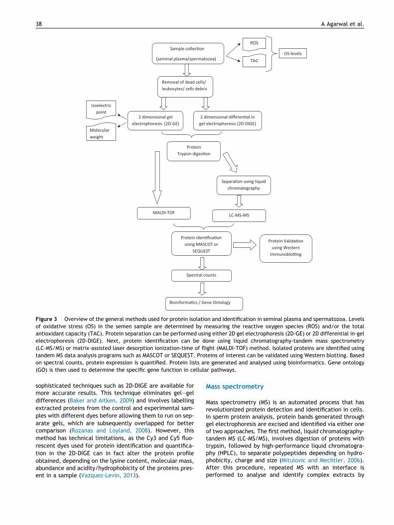
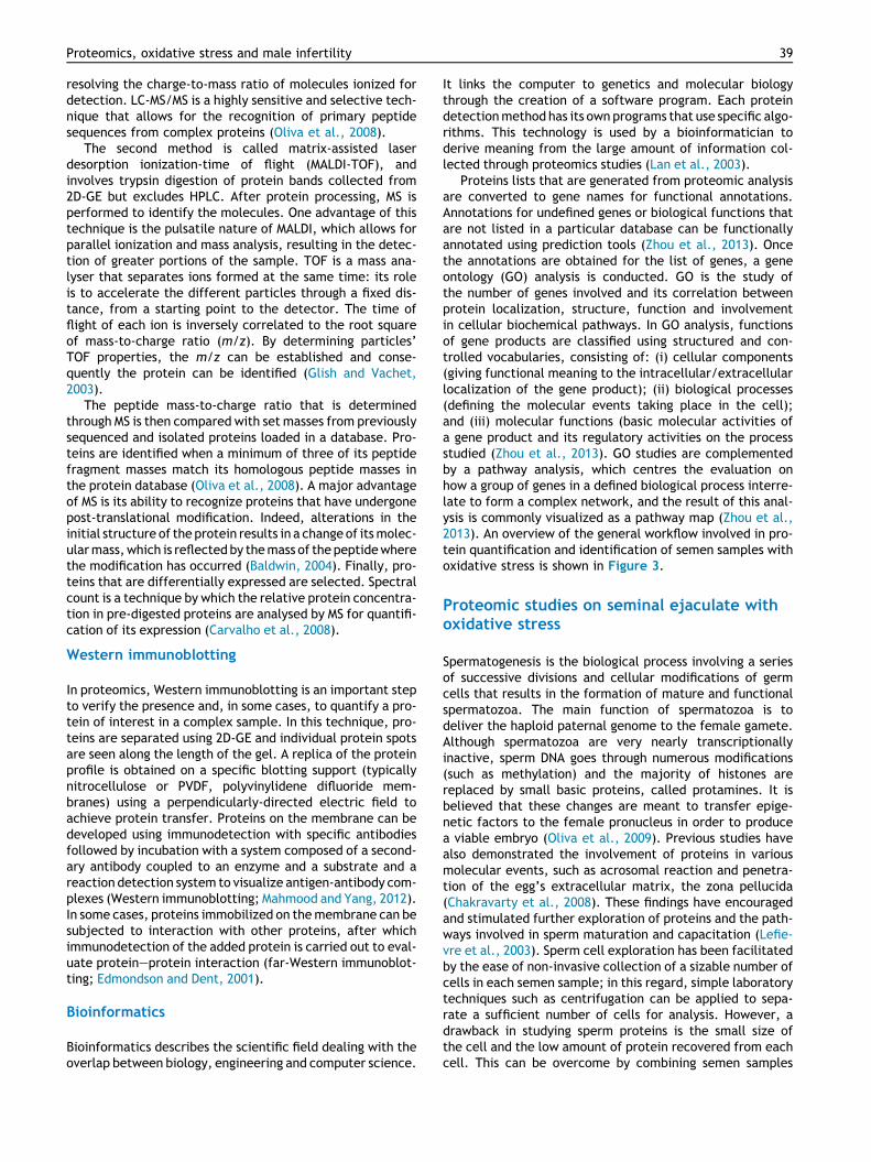
Figure 3 Overview of the general methods used for protein isolation and identification in seminal plasma and spermatozoa. Levelsof oxidative stress (OS) in the semen sample are determined by measuring the reactive oxygen species (ROS) and/or the totalantioxidant capacity (TAC). Protein separation can be performed using either 2D gel electrophoresis (2D-GE) or 2D differential in-gelelectrophoresis (2D-DIGE). Next, protein identification can be done using liquid chromatography-tandem mass spectrometry(LC-MS/MS) or matrix-assisted laser desorption ionization-time of flight (MALDI-TOF) method. Isolated proteins are identified usingtandem MS data analysis programs such as MASCOT or SEQUEST. Proteins of interest can be validated using Western blotting. Basedon spectral counts, protein expression is quantified. Protein lists are generated and analysed using bioinformatics. Gene ontology(GO) is then used to determine the specific gene function in cellular pathways.
38 A Agarwal et al.
sophisticated techniques such as 2D-DIGE are available formore accurate results. This technique eliminates gel–geldifferences (Baker and Aitken, 2009) and involves labellingextracted proteins from the control and experimental sam-ples with different dyes before allowing them to run on sep-arate gels, which are subsequently overlapped for bettercomparison (Rozanas and Loyland, 2008). However, thismethod has technical limitations, as the Cy3 and Cy5 fluo-rescent dyes used for protein identification and quantifica-tion in the 2D-DIGE can in fact alter the protein profileobtained, depending on the lysine content, molecular mass,abundance and acidity/hydrophobicity of the proteins pres-ent in a sample (Vazquez-Levin, 2013).
Mass spectrometry
Mass spectrometry (MS) is an automated process that hasrevolutionized protein detection and identification in cells.In sperm protein analysis, protein bands generated throughgel electrophoresis are excised and identified via either oneof two approaches. The first method, liquid chromatography-tandem MS (LC-MS/MS), involves digestion of proteins withtrypsin, followed by high-performance liquid chromatogra-phy (HPLC), to separate polypeptides depending on hydro-phobicity, charge and size (Mitulovic and Mechtler, 2006).After this procedure, repeated MS with an interface isperformed to analyse and identify complex extracts by
Proteomics, oxidative stress and male infertility 39
resolving the charge-to-mass ratio of molecules ionized fordetection. LC-MS/MS is a highly sensitive and selective tech-nique that allows for the recognition of primary peptidesequences from complex proteins (Oliva et al., 2008).
The second method is called matrix-assisted laserdesorption ionization-time of flight (MALDI-TOF), andinvolves trypsin digestion of protein bands collected from2D-GE but excludes HPLC. After protein processing, MS isperformed to identify the molecules. One advantage of thistechnique is the pulsatile nature of MALDI, which allows forparallel ionization and mass analysis, resulting in the detec-tion of greater portions of the sample. TOF is a mass ana-lyser that separates ions formed at the same time: its roleis to accelerate the different particles through a fixed dis-tance, from a starting point to the detector. The time offlight of each ion is inversely correlated to the root squareof mass-to-charge ratio (m/z). By determining particles’TOF properties, the m/z can be established and conse-quently the protein can be identified (Glish and Vachet,2003).
The peptide mass-to-charge ratio that is determinedthrough MS is then compared with set masses from previouslysequenced and isolated proteins loaded in a database. Pro-teins are identified when a minimum of three of its peptidefragment masses match its homologous peptide masses inthe protein database (Oliva et al., 2008). A major advantageof MS is its ability to recognize proteins that have undergonepost-translational modification. Indeed, alterations in theinitial structure of the protein results in a change of itsmolec-ularmass,which is reflected by themass of the peptidewherethe modification has occurred (Baldwin, 2004). Finally, pro-teins that are differentially expressed are selected. Spectralcount is a technique by which the relative protein concentra-tion in pre-digested proteins are analysed by MS for quantifi-cation of its expression (Carvalho et al., 2008).
Western immunoblotting
In proteomics, Western immunoblotting is an important stepto verify the presence and, in some cases, to quantify a pro-tein of interest in a complex sample. In this technique, pro-teins are separated using 2D-GE and individual protein spotsare seen along the length of the gel. A replica of the proteinprofile is obtained on a specific blotting support (typicallynitrocellulose or PVDF, polyvinylidene difluoride mem-branes) using a perpendicularly-directed electric field toachieve protein transfer. Proteins on the membrane can bedeveloped using immunodetection with specific antibodiesfollowed by incubation with a system composed of a second-ary antibody coupled to an enzyme and a substrate and areaction detection system to visualize antigen-antibody com-plexes (Western immunoblotting; Mahmood and Yang, 2012).In some cases, proteins immobilized on themembrane can besubjected to interaction with other proteins, after whichimmunodetection of the added protein is carried out to eval-uate protein–protein interaction (far-Western immunoblot-ting; Edmondson and Dent, 2001).
Bioinformatics
Bioinformatics describes the scientific field dealing with theoverlap between biology, engineering and computer science.
It links the computer to genetics and molecular biologythrough the creation of a software program. Each proteindetectionmethod has its own programs that use specific algo-rithms. This technology is used by a bioinformatician toderive meaning from the large amount of information col-lected through proteomics studies (Lan et al., 2003).
Proteins lists that are generated from proteomic analysisare converted to gene names for functional annotations.Annotations for undefined genes or biological functions thatare not listed in a particular database can be functionallyannotated using prediction tools (Zhou et al., 2013). Oncethe annotations are obtained for the list of genes, a geneontology (GO) analysis is conducted. GO is the study ofthe number of genes involved and its correlation betweenprotein localization, structure, function and involvementin cellular biochemical pathways. In GO analysis, functionsof gene products are classified using structured and con-trolled vocabularies, consisting of: (i) cellular components(giving functional meaning to the intracellular/extracellularlocalization of the gene product); (ii) biological processes(defining the molecular events taking place in the cell);and (iii) molecular functions (basic molecular activities ofa gene product and its regulatory activities on the processstudied (Zhou et al., 2013). GO studies are complementedby a pathway analysis, which centres the evaluation onhow a group of genes in a defined biological process interre-late to form a complex network, and the result of this anal-ysis is commonly visualized as a pathway map (Zhou et al.,2013). An overview of the general workflow involved in pro-tein quantification and identification of semen samples withoxidative stress is shown in Figure 3.
Proteomic studies on seminal ejaculate withoxidative stress
Spermatogenesis is the biological process involving a seriesof successive divisions and cellular modifications of germcells that results in the formation of mature and functionalspermatozoa. The main function of spermatozoa is todeliver the haploid paternal genome to the female gamete.Although spermatozoa are very nearly transcriptionallyinactive, sperm DNA goes through numerous modifications(such as methylation) and the majority of histones arereplaced by small basic proteins, called protamines. It isbelieved that these changes are meant to transfer epige-netic factors to the female pronucleus in order to producea viable embryo (Oliva et al., 2009). Previous studies havealso demonstrated the involvement of proteins in variousmolecular events, such as acrosomal reaction and penetra-tion of the egg’s extracellular matrix, the zona pellucida(Chakravarty et al., 2008). These findings have encouragedand stimulated further exploration of proteins and the path-ways involved in sperm maturation and capacitation (Lefie-vre et al., 2003). Sperm cell exploration has been facilitatedby the ease of non-invasive collection of a sizable number ofcells in each semen sample; in this regard, simple laboratorytechniques such as centrifugation can be applied to sepa-rate a sufficient number of cells for analysis. However, adrawback in studying sperm proteins is the small size ofthe cell and the low amount of protein recovered from eachcell. This can be overcome by combining semen samples

40 A Agarwal et al.
from men with a similar diagnosis and then analysing thesample.
Today, thousands of proteins have been profiled inhuman semen, and scientists have been working on compar-ing protein expression in fertile and infertile men using dif-ferent approaches. However, notwithstanding its relevance,until the present time, there are only a few studies thathave focused on oxidative stress and its ability to alterthe protein expression in the semen of patients with highROS levels. In those studies, researchers have identified sev-eral proteins that are differentially expressed, which mayplay a role in the regulation and response of cells with highROS levels.
This review includes a set of proteomic studies thatassessed oxidative stress levels in their subjects as quantita-tive proof of oxidative stress status in seminal ejaculate.The studies selected in this review are three by our groupfrom the Centre for Reproductive Medicine, Glickman Uro-logical and Kidney Institute, Cleveland Clinic (Hamadaet al., 2013; Sharma et al., 2013a, 2013b) and two by otherresearch teams (Herwig et al., 2013; Wang et al., 2009(Table 1). Among these five studies in men who were mostlyinfertile with high levels of oxidative stress, three lookedinto seminal plasma proteins (Herwig et al., 2013; Sharmaet al., 2013a; Wang et al., 2009) whereas two of our studiesanalysed sperm protein mixtures (Hamada et al., 2013;Sharma et al., 2013b). Patient populations in these studiesare characterized as males with asthenozoospermia, idio-pathic oligoasthenoteratozoospermia (OAT) or primary orsecondary infertility (a majority of which have varicocele).The donors used as controls were normozoospermic(according to WHO, 1999 criteria) males with either provenor unproven fertility. Donors with proven fertility are menwho have established a clinical pregnancy that resulted ina live birth.
The studies in this review pertain to men with clinicaloxidative stress versus those with physiological ROS levels.To support our discussion in this review, references areoccasionally made to studies performed on a similar groupof subjects (i.e. infertile men with various semen parame-ters), although there was no assessment of oxidative stresslevels in that study group (Sharma et al., 2013c). ROS levelsin the seminal ejaculate are independent of semen parame-ters (Agarwal et al., 2006b), as the seminal constituents aremainly added to the spermatozoa at ejaculation. Semensamples used in studies from our team as well as from Her-wig’s group were negative for leukocytospermia (negativeEndtz test), to rule out oxidative stress originating fromROS produced by leukocytes (Hamada et al., 2013; Herwiget al., 2013; Sharma et al., 2013a, 2013b). Other exclusioncriteria observed in each study are mentioned in Table 1.
Oxidative stress was measured in these studies either bychemiluminescence assay (for ROS; Hamada et al., 2013;Sharma et al., 2013a, 2013b; Wang et al., 2009) or by ELISA(for carbonyl proteins; Herwig et al., 2013). In our lateststudy (Sharma et al., 2013a), total antioxidant capacityand sperm DNA fragmentation were also measured as a fur-ther indication of the oxidative status of the sample. Oxida-tive stress is likely to result in an abundance of immatureproteins in their precursor or preprotein forms, whichentails a deficiency of mature functional proteins (Martinez-Heredia et al., 2008). At the same time, presence of
the immature proteins may be indicative of insufficientpost-translational processing (Martinez-Heredia et al.,2008) that could result from high ROS levels. Protein oxida-tion may inhibit enzymatic and protein-binding activities aswell as increase molecular weight, aggregation or proteoly-sis (Shacter, 2000), which may affect the number of identi-fied proteins.
The methodology employed in these studies played apart in the number of proteins that were identified(Table 1). Protein separation in these studies was per-formed by SDS-PAGE, except for the study by Hamadaet al. (2013), which used the 2D-DIGE method. Most of thestudies ran samples in replicates. All the studies used theLC-MS/MS method (either the linear trap quadrupole Orbi-trap (Hamada et al., 2013; Sharma et al., 2013a, 2013b;Wang et al., 2009), or the Hybrid Velos (Herwig et al.,2013). The largest pool of proteins identified was 2489 pro-teins in the seminal plasma (Herwig et al., 2013) and 1343proteins in spermatozoa (Hamada et al., 2013).
The database used for protein identification searcheswas comparable and most studies employed more thanone database search to reduce false positives. As is com-monly used in quantitative proteomics, the relative proteinabundance of the proteins expressed was measured by theintensity of the spectral count. As an overall, the cut-offvalues for over- and under-expressed proteins were 1.5 to3-fold, respectively. The protein(s) identified were vali-dated by Western immunoblotting in studies reported byWang et al. (2009). To determine whether the pooled sam-ple groups used in their study were truly representative ofthe infertile/fertile individual, the study by Herwig et al.(2013) evaluated individual samples alongside the pooledsamples. Based on the results of the study, the authorsreported a lower number of unique proteins in an individualidiopathic OAT sample compared with the pooled idiopathicOAT group. All the studies had performed enrichment anal-ysis in the GO categories of the genes expressed except forour first paper on sperm proteins (Hamada et al., 2013).This was followed by pathway and network analysis andprotein-protein interaction analysis to determine theprocesses involved when the seminal plasma or the sperma-tozoa is in a state of oxidative stress. In the next sections, abrief summary of reported findings of proteomics studies onseminal ejaculate with oxidative stress is presented.
Seminal plasma proteins and oxidative stress
Seminal plasma is the fluid in the semen that contains secre-tions from the testis, epididymis, prostate, seminal vesiclesand Cowper’s glands. Seminal plasma plays an important rolein providing nourishment and protection to spermatozoa andacts as a buffer as well as a medium for sperm motility.Human semen is composed of lipids, ions (such as citrate, cal-cium, magnesium, potassium, sodium, zinc and chloride),fructose, ascorbic acid, proteins (such as semenogelin andfibronectin), albumin and globulins, amino acids and amines,cytokines and hormones. It also contains numerous enzy-matic (gluthathione peroxidase, superoxide dismutase, cata-lase) and non-enzymatic antioxidants (vitamins C and E, zinc)that protect spermatozoa from oxidative stress (Pahuneet al., 2013). Protein concentration in human seminal plasmahas been estimated to be 45 mg/ml (Tomar et al., 2012a).

Table 1 Summary of proteomic studies in patients with oxidative stress.
StudySource ofproteins
Subjects Exclusion criteria Indication of oxidativestress
Proteomic analysisstrategy
Main findings Potentialbiomarkers ofoxidative stress
Wanget al.(2009)
Seminalplasma
38 infertile patients withAS (motility a + b:8.7 ± 20.4%)
Liquefaction time>30 min
Chemiluminescentmeasurement of ROS:patients had 3.3-timeshigher ROS than controls(P < 0.01)
SDS–PAGE, LC-MS/MS, LTQOrbitrap, Peptide-Prophet,Protein-Prophet (P > 0.5),SEQUEST, Western blot(SDS–PAGE, DJ-1 antibody)
741 proteins identified DJ-1 (about 50% lowerin seminal plasma ofAS than controls;P < 0.05)
x x 20 normal donors(normozoospermic;motility a + b 57.4 ± 9.7%)
Sperm density <20 · 106
spermatozoa/mlx x 101 differentially expressed
proteins: 45 over-expressed(�3-fold) in AS samples; 56underexpressed (�3-fold) in ASsamples
x
x x x Abnormal morphology>10% of total in ASsamples
x x DJ-1 the most down-regulatedprotein in AS
x
Hamadaet al.(2013)
Spermatozoa 32 infertile patients (25primary infertility + 7secondary infertility; 21/32 had clinical varicoceleof grades 1–2)
Leukocytospermia (Endtzpositive >0.1 million/ml)
Chemiluminescentmeasurement of ROS: ROS+(31 samples) �20 RLU/s/106
spermatozoa; ROS– (21samples) <20 RLU/s/106
spermatozoa
2D-DIGE, LC-MS/MS,Finnigan, LTQ Orbitrap,NCBI, MASCOT, SEQUEST,BLAST
1343 proteins identified inROS– gel
Lactotransferrin-2and peroxiredoxin-1(increased in ROS–group)
x x 20 healthy donors withunproven fertility(normozoospermic)
x x x 1265 proteins identified in ROS+gel
x
Sharmaet al.(2013b)
Spermatozoa 32 infertile patients (25primary infertility + 7secondary infertility; 21/32 had clinical varicoceleof grades 1–2)
Leukocytospermia (Endtzpositive >0.1 million/ml)
Chemiluminescentmeasurement of ROS: ROS+(31 samples) �20 RLU/s/106
spermatozoa; ROS– (21samples)<20 RLU/s/106
spermatozoa
In-solution digestion, LC-MS/MS, Finnigan, LTQ Orbitrap,NCBI, MASCOT, SEQUEST
74 proteins identified HIST1H2BA, MDH2,TGM4, GPX4, GLUL,HSP90B1, HSPA5 (allhigher in seminalplasma of ROS+ versusROS–)
x x 20 healthy donors withunproven fertility(normozoospermic)
x x x 20 differentially expressedproteins: 15 over-expressed(>2-fold) in ROS+ samples; 5underexpressed (<0.5-fold) inROS+ samples
x
x x x x x x ODF1 and ACE1underexpressed; LDHC over-expressed; all modulated byCREM activators
x
(continued on next page)
Proteomics,
oxid
ativestre
ssan
dmale
infertility
41
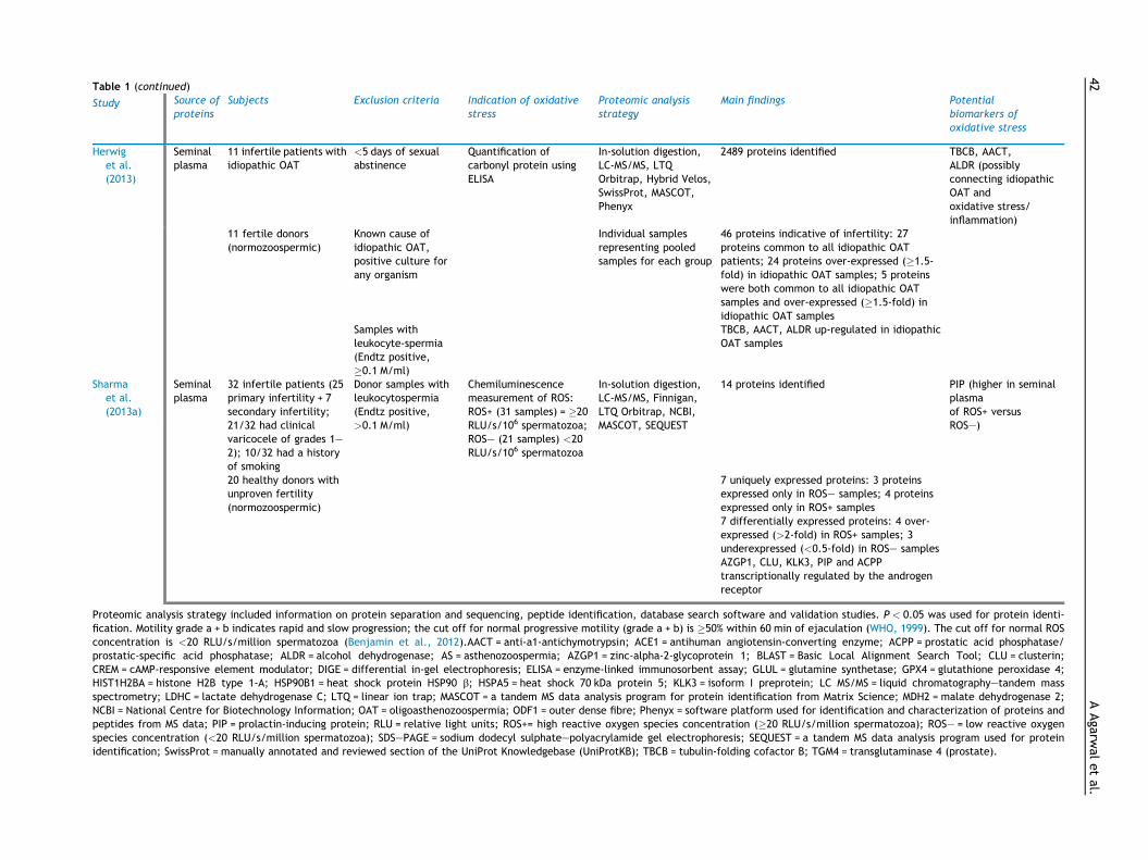
Table 1 (continued)
Study Source ofproteins
Subjects Exclusion criteria Indication of oxidativestress
Proteomic analysisstrategy
Main findings Potentialbiomarkers ofoxidative stress
Herwiget al.(2013)
Seminalplasma
11 infertile patients withidiopathic OAT
<5 days of sexualabstinence
Quantification ofcarbonyl protein usingELISA
In-solution digestion,LC-MS/MS, LTQOrbitrap, Hybrid Velos,SwissProt, MASCOT,Phenyx
2489 proteins identified TBCB, AACT,ALDR (possiblyconnecting idiopathicOAT andoxidative stress/inflammation)
x x 11 fertile donors(normozoospermic)
Known cause ofidiopathic OAT,positive culture forany organism
x Individual samplesrepresenting pooledsamples for each group
46 proteins indicative of infertility: 27proteins common to all idiopathic OATpatients; 24 proteins over-expressed (�1.5-fold) in idiopathic OAT samples; 5 proteinswere both common to all idiopathic OATsamples and over-expressed (�1.5-fold) inidiopathic OAT samples
x
x x x Samples withleukocyte-spermia(Endtz positive,�0.1 M/ml)
x x TBCB, AACT, ALDR up-regulated in idiopathicOAT samples
x
Sharmaet al.(2013a)
Seminalplasma
32 infertile patients (25primary infertility + 7secondary infertility;21/32 had clinicalvaricocele of grades 1–2); 10/32 had a historyof smoking
Donor samples withleukocytospermia(Endtz positive,>0.1 M/ml)
Chemiluminescencemeasurement of ROS:ROS+ (31 samples) = �20RLU/s/106 spermatozoa;ROS– (21 samples) <20RLU/s/106 spermatozoa
In-solution digestion,LC-MS/MS, Finnigan,LTQ Orbitrap, NCBI,MASCOT, SEQUEST
14 proteins identified PIP (higher in seminalplasmaof ROS+ versusROS–)
x x 20 healthy donors withunproven fertility(normozoospermic)
x x x 7 uniquely expressed proteins: 3 proteinsexpressed only in ROS– samples; 4 proteinsexpressed only in ROS+ samples
x
7 differentially expressed proteins: 4 over-expressed (>2-fold) in ROS+ samples; 3underexpressed (<0.5-fold) in ROS– samplesAZGP1, CLU, KLK3, PIP and ACPPtranscriptionally regulated by the androgenreceptor
Proteomic analysis strategy included information on protein separation and sequencing, peptide identification, database search software and validation studies. P < 0.05 was used for protein identi-fication. Motility grade a + b indicates rapid and slow progression; the cut off for normal progressive motility (grade a + b) is �50% within 60 min of ejaculation (WHO, 1999). The cut off for normal ROSconcentration is <20 RLU/s/million spermatozoa (Benjamin et al., 2012).AACT = anti-a1-antichymotrypsin; ACE1 = antihuman angiotensin-converting enzyme; ACPP = prostatic acid phosphatase/prostatic-specific acid phosphatase; ALDR = alcohol dehydrogenase; AS = asthenozoospermia; AZGP1 = zinc-alpha-2-glycoprotein 1; BLAST = Basic Local Alignment Search Tool; CLU = clusterin;CREM = cAMP-responsive element modulator; DIGE = differential in-gel electrophoresis; ELISA = enzyme-linked immunosorbent assay; GLUL = glutamine synthetase; GPX4 = glutathione peroxidase 4;HIST1H2BA = histone H2B type 1-A; HSP90B1 = heat shock protein HSP90 b; HSPA5 = heat shock 70 kDa protein 5; KLK3 = isoform I preprotein; LC MS/MS = liquid chromatography–tandem massspectrometry; LDHC = lactate dehydrogenase C; LTQ = linear ion trap; MASCOT = a tandem MS data analysis program for protein identification from Matrix Science; MDH2 = malate dehydrogenase 2;NCBI = National Centre for Biotechnology Information; OAT = oligoasthenozoospermia; ODF1 = outer dense fibre; Phenyx = software platform used for identification and characterization of proteins andpeptides from MS data; PIP = prolactin-inducing protein; RLU = relative light units; ROS+= high reactive oxygen species concentration (�20 RLU/s/million spermatozoa); ROS– = low reactive oxygenspecies concentration (<20 RLU/s/million spermatozoa); SDS–PAGE = sodium dodecyl sulphate–polyacrylamide gel electrophoresis; SEQUEST = a tandem MS data analysis program used for proteinidentification; SwissProt = manually annotated and reviewed section of the UniProt Knowledgebase (UniProtKB); TBCB = tubulin-folding cofactor B; TGM4 = transglutaminase 4 (prostate).
42AAgarw
aletal.

Proteomics, oxidative stress and male infertility 43
The study of these proteins can provide a basis for the identi-fication of biomarkers for the assessment of male infertilitydisorders. Tables 2–5 give an overview of seminal fluid pro-teins that have been associatedwith elevated levels of oxida-tive stress.
Seminal plasma protein expression was analysed inpatients with primary and secondary infertility who pre-sented high levels of oxidative stress (ROS+;�20 RLU/s/10 million spermatozoa) versus those withphysiological levels of oxidative stress (ROS�;<20 RLU/s/10 mil-lion spermatozoa) (Sharma et al., 2013a). Using LC-MS/MS,the study resulted in the identification of 14 proteins: sevencommonly present in both ROS+ and ROS� samples, threeexclusively present in ROS� samples (fibronectin I isoform3 preprotein (FN1), macrophage migration inhibitory fac-tor-1 peptide (MIF) and galectin 3 binding protein (G3BP))and four expressed solely in ROS+ samples (cystatin S pre-cursor, albumin preprotein, lactotransferrin precursor 1peptide and prostate-specific antigen isoform 4 preprotein)(Sharma et al., 2013a).
Semenogelin 2 precursor was found to be 2-fold up-regu-lated in the ROS+ group, whereas semenogelin 1 isoform awas found to be down-regulated in the ROS+ group (Sharmaet al., 2013a). Both semenogelin 1 (50 kDa) and semenogelin2 (63 kDa, present in lesser abundance) are secreted fromthe seminal vesicle and represent the most abundant com-ponents (about 20–40%) of the human semen coagulum(de Lamirande and Lamothe, 2010). Semenogelin areresponsible for the coagulation of the gel matrix thatencloses the spermatozoa and it helps prevent the capacita-tion process (the initial part of sperm activation) by inhibit-ing the formation of ROS (de Lamirande and Lamothe,
Table 2 Seminal plasma proteins over-expressed in semen sampoxidative stress.
Protein UniProtID
Function
Semenogelin-2 (SEMG2) Q02383 CoagulationProlactin-induced protein (PIP) P12273 Breakdown o
liquefactionProstatic acid phosphatase (PAP or ACPP) P15309 LiquefactionEpididymal secretory protein E1 (NPC2) P61916 Transport of
x x Egress of chocompartmen
Epididymal secretory protein E4 (WFDC2) Q14508 Serine proteadebated
Immunoglobulin kappa chain C protein(IGKC)
P01834 Immune resp
Pyruvate kinase M2 (PKM2) P14618 PhosphotyrosCathepsin H (CTSH) P09668 Lysosomal cyL-lactate dehydrogenase B chain (LDHB) P07195 Catalyses con
pyruvate andLegumain precursor (LGMN) Q99538 Hydrolysis ofDelta aminolevulinic acid dehydratase
(ALAD or ALADH)P13716 Catalyse the
form porphoAlpha-1-antichymotrypsin (AACT) P01011 Inactivate se
x x Control oxidaand is involv
Aldose reductase (ALDR or AKR1B1) P15121 Catalyse the
2010). A small amount of ROS, generated by the spermato-zoa itself, is required to facilitate the initiation of thecapacitation process and the subsequent hyperactivatedmotility of the spermatozoa. However, a premature onsetof capacitation leads to poor fertility outcomes (deLamirande and Gagnon, 1993).
Semenogelin decreases the formation of ROS throughseveral pathways: (i) by reducing sperm motility and energyconsumption (de Lamirande et al., 2001); (ii) by indirectlyinteracting with sperm NADH-oxidase to block superoxideradical generation (Bonilha et al., 2008); and (iii) by bindingthe antioxidant zinc ion (Zn2+) and allowing it to functioninside the spermatozoa (Bonilha et al., 2008). In the reportby Wang et al. (2009), the quantity of semenogelin in theseminal plasma of fertile donors (sperm motility gradesA + B i.e. rapid + slow progression of 48–67%) versusasthenozoospermic patients (sperm motility 6–11%) withhigh levels of oxidative stress was not significantly differ-ent. The results suggested that seminal vesicle proteinssuch as semenogelin were less likely to be associated withthe regulation of sperm motility in asthenozoospermicpatients compared with proteins of the epididymis and pros-tate, such as DJ-1 (Wang et al., 2009).
Seminal plasma motility inhibitors (SPMI) are degradatoryproducts or proteinase-resistant fragments of semenogelin 1and 2 (Terai et al., 2010). Terai et al. (2010) studied theassociation of SPMI and spermatozoa in asthenozoospermicinfertile patients (sperm motility <50%) by labelling washedsperm cells with anti-SPMI antibody, followed by flowcytometry analysis and Western immunoblotting. Althoughspermatozoa from both asthenozoospermic patients andnormal subjects showed similar labelling patterns, both
les with oxidative stress compared to semen samples without
Reference
of semen Sharma et al. (2013a)f fibronectin during semen Sharma et al. (2013a)
of semen Sharma et al. (2013a)intracellular cholesterol Wang et al. (2009)lesterol from endosomal/lysosomalt
x
se inhibitor, though function is still Wang et al. (2009)
onse Wang et al. (2009)
ine-binding protein Wang et al. (2009)steine proteinase Wang et al. (2009)version of lactate and NAD toNADH (glycolysis)
Wang et al. (2009)
asparaginyl bonds Wang et al. (2009)second step in haeme synthesis tobilinogen
Wang et al. (2009)
rine proteases Wang et al. (2009)tive damage, anti-inflammatoryed in defence mechanism
x
reduction of aldehydes and carbonyl Herwig et al. (2013)

Table 3 Seminal plasma proteins underexpressed in semen samples with oxidative stress compared to semen samples withoutoxidative stress.
Protein UniProtID
Function Reference(s)
Semenogelin 1 isoform (SEMG1) P04279 Coagulation of semen Sharma et al. (2013a)Prostate specific antigen isoform 1
preprotein (KLK3)P07288 Hydrolysis of semenogelin 1 and liquefaction of
seminal coagulumSharma et al. (2013a)
x x Remodelling of extracellular matrix xx x Degradation of connective tissue xZinc alpha 2 glycoprotein 1 (AZGP1) P25311 Plays a role in signal transduction Sharma et al. (2013a)Clusterin preprotein (CLU) P10909 Protection against oxidative reactions, protein
denaturation and aggregation of abnormalspermatozoa and controls complement-inducedsperm lysis
Sharma et al. (2013a),Wang et al. (2009)
Alpha-2-macroglobulin (A2 M) P01023 Antiprotease activity Wang et al. (2009)Fructose-biphosphate aldolase A
(ALDOA)P04075 Glycolytic enzyme converting fructose 1,6
biphosphate to GAPH and DHAPWang et al. (2009)
Glyceraldehyde-3-phosphatedehydrogenase (GAPDH)
P04406 Enzyme in glycolysis pathway Wang et al. (2009)
Dipeptidyl peptidase 4 (DPP4) P27487 Fused to spermatozoa, induce sperm motility andprevent premature capacitation
Wang et al. (2009)
DJ-1 (PARK 7) Q99497 Antioxidant protein that protects cells againstoxidative stress and cell death
Sharma et al. (2013a),Wang et al. (2009)
Laminin subunit alpha-5 (LAMA5)precursor
O15230 Binds to cells, acting as part of laminin, which isfound in basement membranes
Wang et al. (2009)
Keratin, type 1 cytoskeletal 9 (KRT9) P35527 Part of keratin filament assembly; function in skintissue
Wang et al. (2009)
Rab GDP dissociation inhibitor beta(GDI2)
P50395 Inhibits dissociation of GDP from Rab proteins andthe subsequent binding of GTP to them
Wang et al. (2009)
Annexin 6 isoform 2 (ANXA6) P08133 It may associate with CD21 and regulate calciumrelease from intracellular stores, cell receptor forchondroitin sulphate chain
Wang et al. (2009)
Attractin precursor isoform 2 (ATRN) O75882 Inflammatory response; involved in immune cellclustering and possibly regulates the chemotacticactivity of chemokines
Wang et al. (2009)
Alpha-actinin-4 (ACTN4) O43707 Actin-binding protein (a bundling protein) Wang et al. (2009)Nesprin-2 isoform 1 (SYNE2) Q8WXH0 Links organelles to actin cytoskeleton for
organization in the cellWang et al. (2009)
Transitional endoplasmic reticulumATPase (VCP)
P55072 Involved in fragmenting the Golgi stacks duringmitosis and reassembling afterwards
Wang et al. (2009)
SNC66 protein Q8WY24 Involved in membranes; function is not well known Wang et al. (2009)Alpha-N-acetyl glucosaminidase
precursor (NAGLU)P54802 Located in lysosomes, breaks down
glycosaminoglycansWang et al. (2009)
44 A Agarwal et al.
labelling intensity and the number of labelled spermatozoawere higher in patient samples compared with normal sub-jects. Further, a marked negative correlation was foundbetween labelled sperm cells and gamete motility and via-bility. Based on these findings, Terai’s group postulated thatthe presence of membrane surface-bound SPMI on thesperm head and tail was the basis for poor motility inasthenozoospermic patients rather than the presence ofsemenogelin in their seminal plasma (Terai et al., 2010).
Prostate-specific antigen (PSA, or human kallikrein 3(hK3)) is a serine protease that is synthesized in prostate tis-sue and involved in semenogelin breakdown, causing lique-faction of the semen coagulum (Jansen et al., 2009;Robert and Gagnon, 1999). PSA isoforms were found to bedifferentially expressed between patients with high ROS
levels and donors with physiological ROS levels: PSA isoform1 preprotein was down-regulated in ROS+ patients, whilePSA isoform 4 preprotein was unique to ROS+ samples (Shar-ma et al., 2013a). The identification of these precursorforms of incompletely modified proteins can be explainedby faulty post-translational modifications, which result ina decrease in the presence of the mature form of the pro-tein and thereby the lack of its function (Martinez-Herediaet al., 2008).
Prolactin-induced protein (PIP; �17 kDa) is secretedfrom the prostate gland, seminal vesicles and testis (Yama-kawa et al., 2007) and constitutes about 1% of seminalplasma (Lilja, 1993). PIP has attracted much attention whenit comes to seminal fluid studies. This protein plays variousroles, such as fibronectin degradation during semen
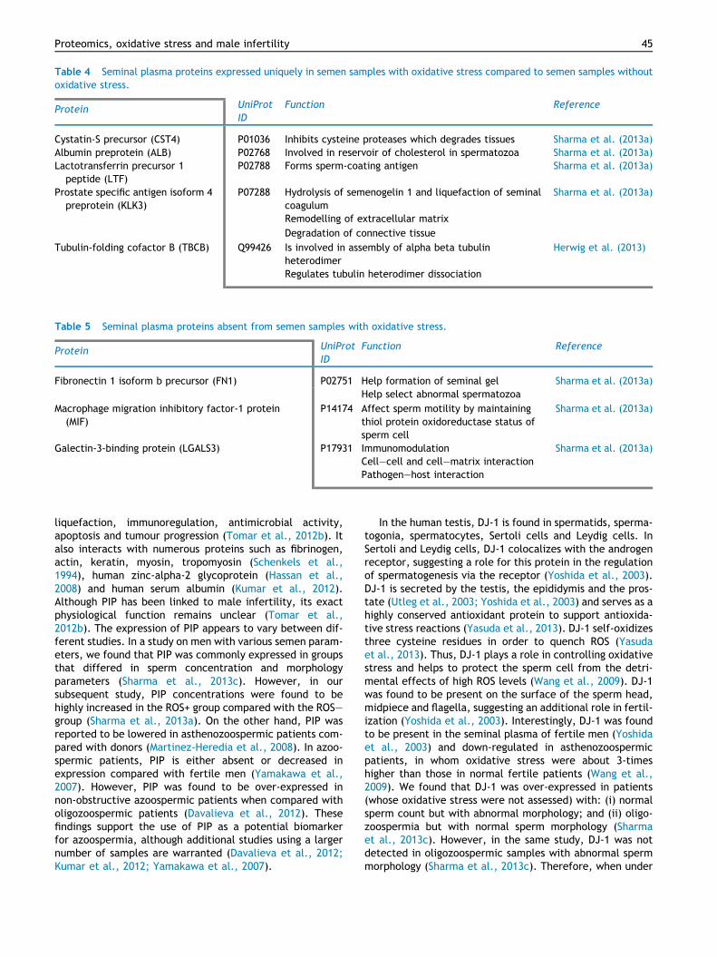
Table 4 Seminal plasma proteins expressed uniquely in semen samples with oxidative stress compared to semen samples withoutoxidative stress.
Protein UniProtID
Function Reference
Cystatin-S precursor (CST4) P01036 Inhibits cysteine proteases which degrades tissues Sharma et al. (2013a)Albumin preprotein (ALB) P02768 Involved in reservoir of cholesterol in spermatozoa Sharma et al. (2013a)Lactotransferrin precursor 1
peptide (LTF)P02788 Forms sperm-coating antigen Sharma et al. (2013a)
Prostate specific antigen isoform 4preprotein (KLK3)
P07288 Hydrolysis of semenogelin 1 and liquefaction of seminalcoagulum
Sharma et al. (2013a)
x x Remodelling of extracellular matrix xx x Degradation of connective tissue xTubulin-folding cofactor B (TBCB) Q99426 Is involved in assembly of alpha beta tubulin
heterodimerHerwig et al. (2013)
Regulates tubulin heterodimer dissociation x
Table 5 Seminal plasma proteins absent from semen samples with oxidative stress.
Protein UniProtID
Function Reference
Fibronectin 1 isoform b precursor (FN1) P02751 Help formation of seminal gel Sharma et al. (2013a)
x x Help select abnormal spermatozoa xMacrophage migration inhibitory factor-1 protein
(MIF)P14174 Affect sperm motility by maintaining
thiol protein oxidoreductase status ofsperm cell
Sharma et al. (2013a)
Galectin-3-binding protein (LGALS3) P17931 Immunomodulation Sharma et al. (2013a)Cell–cell and cell–matrix interactionPathogen–host interaction x
Proteomics, oxidative stress and male infertility 45
liquefaction, immunoregulation, antimicrobial activity,apoptosis and tumour progression (Tomar et al., 2012b). Italso interacts with numerous proteins such as fibrinogen,actin, keratin, myosin, tropomyosin (Schenkels et al.,1994), human zinc-alpha-2 glycoprotein (Hassan et al.,2008) and human serum albumin (Kumar et al., 2012).Although PIP has been linked to male infertility, its exactphysiological function remains unclear (Tomar et al.,2012b). The expression of PIP appears to vary between dif-ferent studies. In a study on men with various semen param-eters, we found that PIP was commonly expressed in groupsthat differed in sperm concentration and morphologyparameters (Sharma et al., 2013c). However, in oursubsequent study, PIP concentrations were found to behighly increased in the ROS+ group compared with the ROS–group (Sharma et al., 2013a). On the other hand, PIP wasreported to be lowered in asthenozoospermic patients com-pared with donors (Martinez-Heredia et al., 2008). In azoo-spermic patients, PIP is either absent or decreased inexpression compared with fertile men (Yamakawa et al.,2007). However, PIP was found to be over-expressed innon-obstructive azoospermic patients when compared witholigozoospermic patients (Davalieva et al., 2012). Thesefindings support the use of PIP as a potential biomarkerfor azoospermia, although additional studies using a largernumber of samples are warranted (Davalieva et al., 2012;Kumar et al., 2012; Yamakawa et al., 2007).
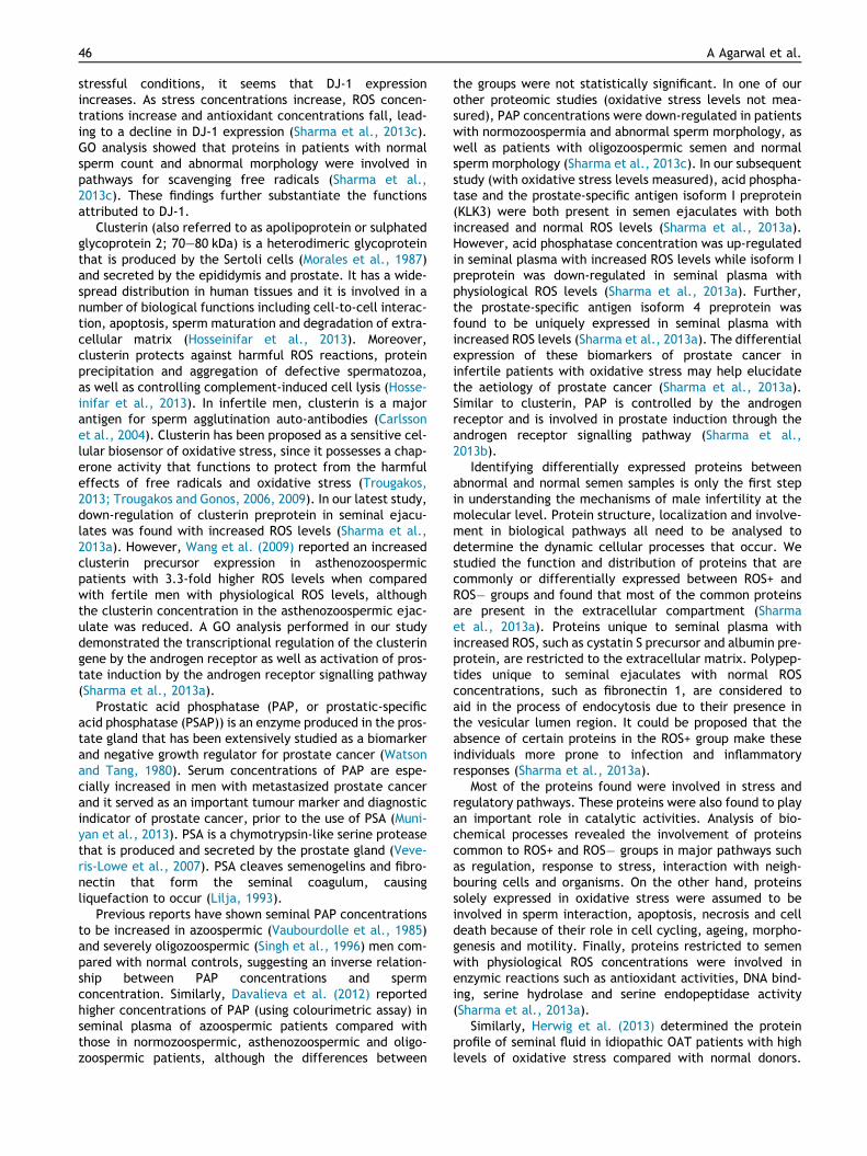
In the human testis, DJ-1 is found in spermatids, sperma-togonia, spermatocytes, Sertoli cells and Leydig cells. InSertoli and Leydig cells, DJ-1 colocalizes with the androgenreceptor, suggesting a role for this protein in the regulationof spermatogenesis via the receptor (Yoshida et al., 2003).DJ-1 is secreted by the testis, the epididymis and the pros-tate (Utleg et al., 2003; Yoshida et al., 2003) and serves as ahighly conserved antioxidant protein to support antioxida-tive stress reactions (Yasuda et al., 2013). DJ-1 self-oxidizesthree cysteine residues in order to quench ROS (Yasudaet al., 2013). Thus, DJ-1 plays a role in controlling oxidativestress and helps to protect the sperm cell from the detri-mental effects of high ROS levels (Wang et al., 2009). DJ-1was found to be present on the surface of the sperm head,midpiece and flagella, suggesting an additional role in fertil-ization (Yoshida et al., 2003). Interestingly, DJ-1 was foundto be present in the seminal plasma of fertile men (Yoshidaet al., 2003) and down-regulated in asthenozoospermicpatients, in whom oxidative stress were about 3-timeshigher than those in normal fertile patients (Wang et al.,2009). We found that DJ-1 was over-expressed in patients(whose oxidative stress were not assessed) with: (i) normalsperm count but with abnormal morphology; and (ii) oligo-zoospermia but with normal sperm morphology (Sharmaet al., 2013c). However, in the same study, DJ-1 was notdetected in oligozoospermic samples with abnormal spermmorphology (Sharma et al., 2013c). Therefore, when under
46 A Agarwal et al.
stressful conditions, it seems that DJ-1 expressionincreases. As stress concentrations increase, ROS concen-trations increase and antioxidant concentrations fall, lead-ing to a decline in DJ-1 expression (Sharma et al., 2013c).GO analysis showed that proteins in patients with normalsperm count and abnormal morphology were involved inpathways for scavenging free radicals (Sharma et al.,2013c). These findings further substantiate the functionsattributed to DJ-1.
Clusterin (also referred to as apolipoprotein or sulphatedglycoprotein 2; 70–80 kDa) is a heterodimeric glycoproteinthat is produced by the Sertoli cells (Morales et al., 1987)and secreted by the epididymis and prostate. It has a wide-spread distribution in human tissues and it is involved in anumber of biological functions including cell-to-cell interac-tion, apoptosis, sperm maturation and degradation of extra-cellular matrix (Hosseinifar et al., 2013). Moreover,clusterin protects against harmful ROS reactions, proteinprecipitation and aggregation of defective spermatozoa,as well as controlling complement-induced cell lysis (Hosse-inifar et al., 2013). In infertile men, clusterin is a majorantigen for sperm agglutination auto-antibodies (Carlssonet al., 2004). Clusterin has been proposed as a sensitive cel-lular biosensor of oxidative stress, since it possesses a chap-erone activity that functions to protect from the harmfuleffects of free radicals and oxidative stress (Trougakos,2013; Trougakos and Gonos, 2006, 2009). In our latest study,down-regulation of clusterin preprotein in seminal ejacu-lates was found with increased ROS levels (Sharma et al.,2013a). However, Wang et al. (2009) reported an increasedclusterin precursor expression in asthenozoospermicpatients with 3.3-fold higher ROS levels when comparedwith fertile men with physiological ROS levels, althoughthe clusterin concentration in the asthenozoospermic ejac-ulate was reduced. A GO analysis performed in our studydemonstrated the transcriptional regulation of the clusteringene by the androgen receptor as well as activation of pros-tate induction by the androgen receptor signalling pathway(Sharma et al., 2013a).
Prostatic acid phosphatase (PAP, or prostatic-specificacid phosphatase (PSAP)) is an enzyme produced in the pros-tate gland that has been extensively studied as a biomarkerand negative growth regulator for prostate cancer (Watsonand Tang, 1980). Serum concentrations of PAP are espe-cially increased in men with metastasized prostate cancerand it served as an important tumour marker and diagnosticindicator of prostate cancer, prior to the use of PSA (Muni-yan et al., 2013). PSA is a chymotrypsin-like serine proteasethat is produced and secreted by the prostate gland (Veve-ris-Lowe et al., 2007). PSA cleaves semenogelins and fibro-nectin that form the seminal coagulum, causingliquefaction to occur (Lilja, 1993).
Previous reports have shown seminal PAP concentrationsto be increased in azoospermic (Vaubourdolle et al., 1985)and severely oligozoospermic (Singh et al., 1996) men com-pared with normal controls, suggesting an inverse relation-ship between PAP concentrations and spermconcentration. Similarly, Davalieva et al. (2012) reportedhigher concentrations of PAP (using colourimetric assay) inseminal plasma of azoospermic patients compared withthose in normozoospermic, asthenozoospermic and oligo-zoospermic patients, although the differences between
the groups were not statistically significant. In one of ourother proteomic studies (oxidative stress levels not mea-sured), PAP concentrations were down-regulated in patientswith normozoospermia and abnormal sperm morphology, aswell as patients with oligozoospermic semen and normalsperm morphology (Sharma et al., 2013c). In our subsequentstudy (with oxidative stress levels measured), acid phospha-tase and the prostate-specific antigen isoform I preprotein(KLK3) were both present in semen ejaculates with bothincreased and normal ROS levels (Sharma et al., 2013a).However, acid phosphatase concentration was up-regulatedin seminal plasma with increased ROS levels while isoform Ipreprotein was down-regulated in seminal plasma withphysiological ROS levels (Sharma et al., 2013a). Further,the prostate-specific antigen isoform 4 preprotein wasfound to be uniquely expressed in seminal plasma withincreased ROS levels (Sharma et al., 2013a). The differentialexpression of these biomarkers of prostate cancer ininfertile patients with oxidative stress may help elucidatethe aetiology of prostate cancer (Sharma et al., 2013a).Similar to clusterin, PAP is controlled by the androgenreceptor and is involved in prostate induction through theandrogen receptor signalling pathway (Sharma et al.,2013b).
Identifying differentially expressed proteins betweenabnormal and normal semen samples is only the first stepin understanding the mechanisms of male infertility at themolecular level. Protein structure, localization and involve-ment in biological pathways all need to be analysed todetermine the dynamic cellular processes that occur. Westudied the function and distribution of proteins that arecommonly or differentially expressed between ROS+ andROS� groups and found that most of the common proteinsare present in the extracellular compartment (Sharmaet al., 2013a). Proteins unique to seminal plasma withincreased ROS, such as cystatin S precursor and albumin pre-protein, are restricted to the extracellular matrix. Polypep-tides unique to seminal ejaculates with normal ROSconcentrations, such as fibronectin 1, are considered toaid in the process of endocytosis due to their presence inthe vesicular lumen region. It could be proposed that theabsence of certain proteins in the ROS+ group make theseindividuals more prone to infection and inflammatoryresponses (Sharma et al., 2013a).
Most of the proteins found were involved in stress andregulatory pathways. These proteins were also found to playan important role in catalytic activities. Analysis of bio-chemical processes revealed the involvement of proteinscommon to ROS+ and ROS� groups in major pathways suchas regulation, response to stress, interaction with neigh-bouring cells and organisms. On the other hand, proteinssolely expressed in oxidative stress were assumed to beinvolved in sperm interaction, apoptosis, necrosis and celldeath because of their role in cell cycling, ageing, morpho-genesis and motility. Finally, proteins restricted to semenwith physiological ROS concentrations were involved inenzymic reactions such as antioxidant activities, DNA bind-ing, serine hydrolase and serine endopeptidase activity(Sharma et al., 2013a).
Similarly, Herwig et al. (2013) determined the proteinprofile of seminal fluid in idiopathic OAT patients with highlevels of oxidative stress compared with normal donors.

Proteomics, oxidative stress and male infertility 47
Their proteomics analysis identified 46 proteins related toinfertility. GO analysis determined that the protein pro-cesses of the 27 proteins common in all idiopathic OATpatients are focused on cellular organization and modifica-tion. Our results in samples with elevated levels of oxidativestress concur with their findings (Sharma et al., 2013a). Inthe study by Herwig et al. (2013), the 24 proteins highlyexpressed in ROS+ idiopathic OAT patients were involvedin biological processes that centre on metabolism, inflam-mation, immunity and stress response. Pathway analysis ofthe proteins identified uniquely in idiopathic OAT had anenrichment of the glycerolipid metabolism pathway only(Herwig et al., 2013).
Sperm proteins and oxidative stress
Seminal proteins are not the only proteins that are of inter-est as potential biomarkers of oxidative stress. Proteinsexpressed by the spermatozoa themselves are equally asimportant, as they help scientists identify their roles inthe spermatozoon’s various metabolic processes, capacita-tion reactions and oocyte fertilization. Alterations in testic-ular ROS can disrupt the internal milieu of the cell, resultingin sperm dysfunction and impaired viability, motility andfertilization capacity (Aitken and Clarkson, 1987; Joneset al., 1979; Tremellen, 2008). Few studies have beenreported regarding protein expression in spermatozoa andhow they correlate with oxidative stress. Some of theimportant sperm proteins in relation to oxidative stress thathave been isolated from infertile patients are summarizedin Tables 6–8.
Our earlier paper (Hamada et al., 2013), we studied pro-teins expressed in spermatozoa from semen with and with-out oxidative stress and found that a total of 1343 proteinspots were identified in ROS� samples and 1265 proteinspots were identified in ROS+ samples. The majority of pro-tein spots did not show any differential expression betweenROS+ and ROS� samples, although when bands were com-pared in a gel, six spots were found to be decreased and25 spots were increased.
Among the differentially expressed proteins, A kinaseanchor protein kinase A 4 (AKAP4) was found to beover-expressed in ROS+ patients (Hamada et al., 2013). Thiscytoskeletal glycoprotein is the major protein in the fibroussheath of the spermatozoon. AKAP4 plays a role in spermmotility because it anchors cAMP-dependent protein kinase,which phosphorylates neighbouring proteins in the presenceof a threshold concentration of cAMP in the cell (Brownet al., 2003; Edwards and Scott, 2000). Elevated ROS con-centrations increase cAMP concentrations, inhibiting tyro-sine phosphatase and activating tyrosine kinase. Thisresults in an increase in tyrosine phosphorylation of AKAP4(Ford, 2004), which might explain the overexpression ofAKAP4 in ROS+ patients. Furthermore, Ficarro et al. (2003)reported an increase in AKAP4 phosphorylation due topremature capacitation induced by elevated ROSconcentrations.
In the same study (Hamada et al., 2013), AKAP3, anothermember of the A kinase anchoring protein family, was sim-ilarly found to be over-expressed. AKAP3 regulates spermmotility by forming complexes with different proteins. Itanchors testis-specific protein ropporin (a sperm-specific
binding protein of rhophilin localized in the sperm fibroussheath) and AKAP-associated sperm protein and also inter-acts with AKAP4. Premature capacitation resulting from oxi-dative stress increases, in the same fashion as AKAP4, theconcentrations of AKAP3 (Ficarro et al., 2003). However,our following study did not find any differential expressionof both AKAP3 and AKAP4 proteins in spermatozoa frommen with and without oxidative stress (Sharma et al.,2013b).
Heat shock proteins (HSP) play an important role in pro-tein assembly, stabilization, folding and translocation ofoligomeric proteins in physiological cellular conditions(Pockley, 2003). HSP are present in different cellular com-partments. Under stressful conditions that cause proteinunfolding and aggregation, HSP expression increases consid-erably (Pockley, 2003), which may explain the increase ofHSP90 b and endoplasmin HSP90 b1 expression in spermato-zoa of ROS+ patients (Hamada et al., 2013). In oursubsequent study (Sharma et al., 2013b), we found thatheat shock proteins (heat shock 70 kDa protein 5 (HSPA5)and heat shock protein 90 kDa b member 1 (HSP90B1)) wereover-expressed in ROS+ samples. HSPA5, known also as glu-cose-regulated protein 78 or binding immunoglobulin pro-tein, is a chaperone protein that is present in thecytoplasmic droplet of abnormal spermatozoa (Marin-Brigg-iler et al., 2010). In normozoospermic men, HSPA5 is local-ized either in the whole flagellum or confined to themidpiece or neck region in both ejaculated and capacitatedspermatozoa (Marin-Briggiler et al., 2010). HSPA5 isassociated to the sperm acrosomal cap and modulatesthe spermatozoon–zona pellucida interaction in acalcium-dependent manner (Marin-Briggiler et al., 2010).HSP90 b takes part in the reduction of cytochrome C (whichserves as an apoptotic signal) through its sulphydryl groupsand defends the cell against apoptosis (Nardai et al.,2000). However, increased oxidation of the chaperone’sthiol group following the elevation of ROS concentrations,resulted in the inactivation of HSP90 b activity (Burtonand Jauniaux, 2011; Gao et al., 2005).
Oxidative stress is detrimental to sperm DNA becauseoxygen radicals react with the nucleic content during sper-miogenesis, an important step in sperm development andmaturation. During this process, histones (which anchorDNA in somatic cells) are replaced by protamines (majornuclear sperm proteins) for greater condensation of thesperm genetic material (Oliva, 2006). Oxidativestress-induced defects in this pathway lead to an increasein histone concentrations inside the cell, which has beenassociated with infertility (Boissonnas et al., 2013). Wefound histone cluster 1 to be over-expressed in ROS+ sam-ples (Sharma et al., 2013b).
Antioxidants play an essential role in maintaining anappropriate intracellular concentration of ROS. Suppressionof antioxidants present in the cell result in oxidative stress.Peroxiredoxins are antioxidant enzymes that scavenge ROSand act as regulators of redox homeostasis and ROS-dependent signalling (O’Flaherty and de Souza, 2011). Eachof our studies that peroxiredoxin 1 (PRDX1; Hamada et al.,2013) and peroxiredoxin 6 (PRDX6; Sharma et al., 2013b)have reduced expresssion in samples with oxidative stresscompared with those without oxidative stress. O’Flahertyand de Souza (2011) reported that members of the

Table 6 Sperm proteins overexpressed in semen samples with oxidative stress compared to semen samples without oxidative stress.
Protein UniProtID
Subcellular location inspermatozoa
Function Reference(s)
A-kinase anchor protein 4 (AKAP4)a Q5JQC9 Fibrous sheath that providesmechanical support of theflagellum
Structural protein, involved in sperm motility: anchors proteinkinase A which can phosphorylate (under high cAMPstimulation) other functional proteins
Hamada et al. (2013)
x x Testis-specific; only expressed inspermatid cells; post-meioticphase of spermatogenesis
x x
Heat shock protein HSP 90-beta(HSP90AB1 or HSP90B)a
P08238 Endoplasmic reticulum Molecular chaperone, reduces cytochrome C (apoptotic signal)and protects against apoptosis
Hamada et al. (2013)
Endoplasmin (heat shock protein 90 kDa bmember 1 (HSP90B1))
P14625 Lumen of endoplasmic reticulum Molecular chaperone, ATP binding, helps in translocation ofsecreted proteins, protein folding and assembly under normalconditions
Hamada et al. (2013),Sharma et al. (2013b)
x x x Increases in response to cellular stressors (e.g. free radicalattack, heat and infection)
x
Glyceraldehyde-3-phosphatedehydrogenase (GAPDH or GAPD)b
P04406 Cytoplasm Oxidoreductase, key enzyme in glycolytic pathway Sharma et al. (2013b)
x x x Binds to and activates the androgen receptor xGlyceraldehyde-3-phosphate
dehydrogenase, testis-specific (GAPDHSor GAPD2)
O14556 Cytoplasm Oxidoreductase, essential enzyme for male fertility – requiredfor sperm motility and male fertility
Sharma et al. (2013b)
Fructose-biphosphate aldolase A (ALDOA) P04075 Cytosol Lyase, plays an important role in glycolysis andgluconeogenesis
Sharma et al. (2013b)
Malate dehydrogenase, mitochondrial(MDH2) precursor
P40926 Mitochondrial matrix Oxidoreductase; produces ROS; catalyses the last step of theKrebs cycle
Sharma et al. (2013b)
Triosephosphate isomerase (isoform 1(TPI1))
P60174 Cytosol Isomerase, catalyses the isomerization of G3P and DHAP inglycolysis
Sharma et al. (2013b)
Histone H2B type 1-A (testis-specifichistone H2B) (HIST1H2BA)
Q96A08 Nucleus, chromosome Transcribed exclusively in testis, corresponding proteinpresent in mature spermatozoa; core part of nucleosomes thatbind and compact DNA into chromatin
Sharma et al. (2013b)
Heat shock 70 kDa protein 5 (HSPA5) Q24JP5 Lumen of endoplasmic reticulum Under normal conditions, plays a role in protein folding andassembly in the endoplasmic reticulum
Sharma et al. (2013b)
Glutamine synthetase (glutamate-ammonia ligase (GLUL))
P15104 Exists as a transcript Ligase, catalyses the synthesis of glutamine from glutamateand ammonia
Sharma et al. (2013b)
Olfactomedin-4 (OLFM4) precursor Q6UX06 Extracellular matrix Strongly expressed in the prostate; facilitates cell adhesion Sharma et al. (2013b)Sperm acrosome membrane-associated
protein 4 (sperm acrosomal membraneprotein 14 (SPACA4))
Q8TDM5 Sperm acrosomal matrix and innerand outer acrosome membranes
Sperm surface membrane protein with a possible role insperm–egg plasma membrane adhesion and fusion duringfertilization
Sharma et al. (2013b)
x x Testis-specific x xAdipocyte plasma membrane-associated
protein (APMAP) (chromosome 20 openreading frame 3 (C20orf3))
Q9HDC9 Membrane, also found on the cellsurface of monocytes
Possible role in adipocyte differentiation Sharma et al. (2013b)
48AAgarw
aletal.
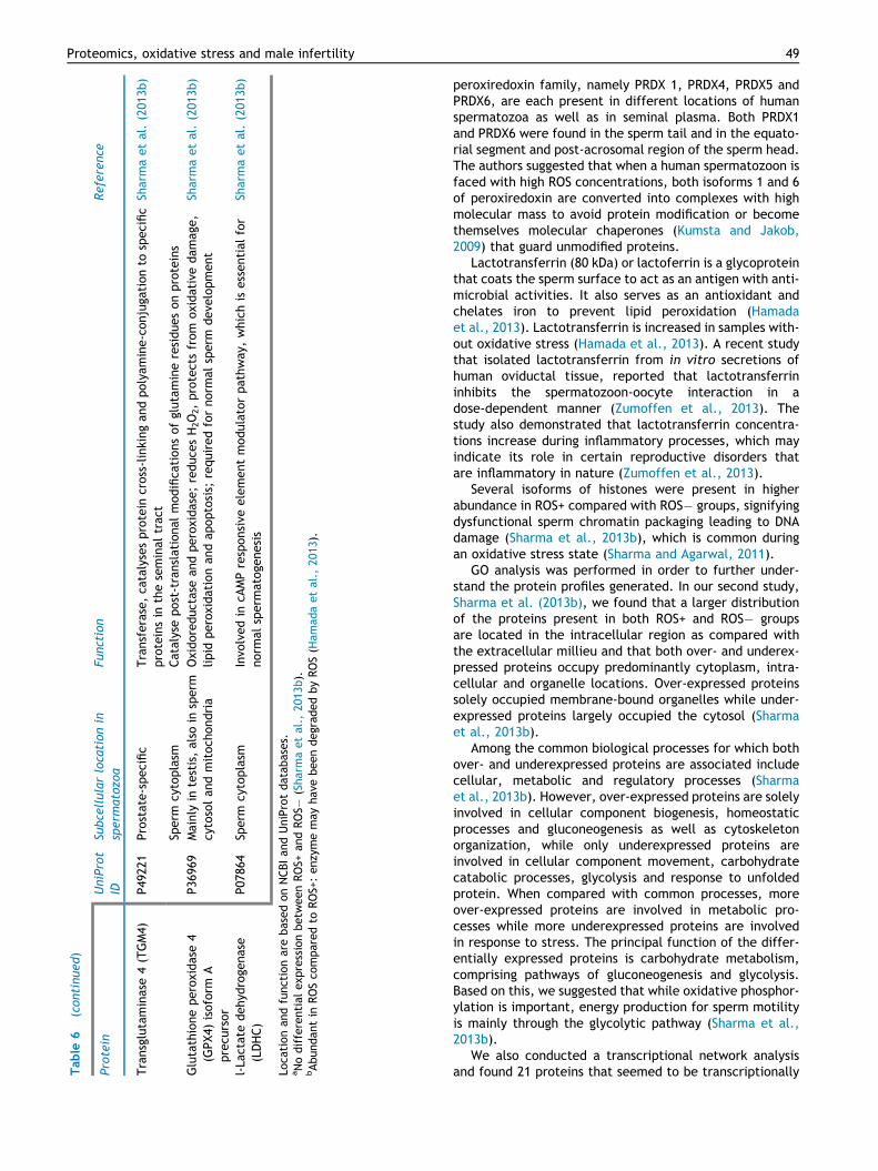
Table
6(continued)
Protein
UniProt
IDSu
bcellularlocationin
sperm
atozo
aFu
nction
Reference
Transglutaminase
4(TGM4)
P492
21
Prostate-specific
Transferase,ca
talysesprotein
cross-linkingan
dpolyam
ine-conjuga
tionto
specific
proteinsin
theseminal
trac
tSh
armaetal.(201
3b)
xx
Sperm
cytoplasm
Catalysepost-translational
modifica
tionsofglutamineresiduesonproteins
xGlutathioneperoxidase4
(GPX4)
isoform
Aprecu
rsor
P36
969
Mainly
intestis,also
insperm
cytosolandmitoch
ondria
Oxidoreductasean
dperoxidase;reduce
sH2O2,protectsfrom
oxidativedam
age,
lipid
peroxidationan
dap
optosis;
requiredfornorm
alsperm
deve
lopment
Sharmaetal.(201
3b)
l-La
ctatedehyd
roge
nase
(LDHC)
P07
864
Sperm
cytoplasm
Invo
lvedin
cAMPresponsive
elementmodulatorpathway
,whichis
essential
for
norm
alsperm
atoge
nesis
Sharmaetal.(201
3b)
Loca
tionan
dfunctionarebasedonNCBIan
dUniProtdatab
ases.
aNodifferential
exp
ressionbetw
eenROS+
andROS�
(Sharmaetal.,
2013
b).
bAbundan
tin
ROSco
mparedto
ROS+:enzymemay
hav
ebeendegrad
edbyROS(H
amad
aetal.,
2013
).
Proteomics, oxidative stress and male infertility 49
peroxiredoxin family, namely PRDX 1, PRDX4, PRDX5 andPRDX6, are each present in different locations of humanspermatozoa as well as in seminal plasma. Both PRDX1and PRDX6 were found in the sperm tail and in the equato-rial segment and post-acrosomal region of the sperm head.The authors suggested that when a human spermatozoon isfaced with high ROS concentrations, both isoforms 1 and 6of peroxiredoxin are converted into complexes with highmolecular mass to avoid protein modification or becomethemselves molecular chaperones (Kumsta and Jakob,2009) that guard unmodified proteins.
Lactotransferrin (80 kDa) or lactoferrin is a glycoproteinthat coats the sperm surface to act as an antigen with anti-microbial activities. It also serves as an antioxidant andchelates iron to prevent lipid peroxidation (Hamadaet al., 2013). Lactotransferrin is increased in samples with-out oxidative stress (Hamada et al., 2013). A recent studythat isolated lactotransferrin from in vitro secretions ofhuman oviductal tissue, reported that lactotransferrininhibits the spermatozoon-oocyte interaction in adose-dependent manner (Zumoffen et al., 2013). Thestudy also demonstrated that lactotransferrin concentra-tions increase during inflammatory processes, which mayindicate its role in certain reproductive disorders thatare inflammatory in nature (Zumoffen et al., 2013).
Several isoforms of histones were present in higherabundance in ROS+ compared with ROS� groups, signifyingdysfunctional sperm chromatin packaging leading to DNAdamage (Sharma et al., 2013b), which is common duringan oxidative stress state (Sharma and Agarwal, 2011).
GO analysis was performed in order to further under-stand the protein profiles generated. In our second study,Sharma et al. (2013b), we found that a larger distributionof the proteins present in both ROS+ and ROS� groupsare located in the intracellular region as compared withthe extracellular millieu and that both over- and underex-pressed proteins occupy predominantly cytoplasm, intra-cellular and organelle locations. Over-expressed proteinssolely occupied membrane-bound organelles while under-expressed proteins largely occupied the cytosol (Sharmaet al., 2013b).
Among the common biological processes for which bothover- and underexpressed proteins are associated includecellular, metabolic and regulatory processes (Sharmaet al., 2013b). However, over-expressed proteins are solelyinvolved in cellular component biogenesis, homeostaticprocesses and gluconeogenesis as well as cytoskeletonorganization, while only underexpressed proteins areinvolved in cellular component movement, carbohydratecatabolic processes, glycolysis and response to unfoldedprotein. When compared with common processes, moreover-expressed proteins are involved in metabolic pro-cesses while more underexpressed proteins are involvedin response to stress. The principal function of the differ-entially expressed proteins is carbohydrate metabolism,comprising pathways of gluconeogenesis and glycolysis.Based on this, we suggested that while oxidative phosphor-ylation is important, energy production for sperm motilityis mainly through the glycolytic pathway (Sharma et al.,2013b).
We also conducted a transcriptional network analysisand found 21 proteins that seemed to be transcriptionally

Table 7 Sperm proteins underexpressed in semen samples with oxidative stress compared to semen samples without oxidativestress.
Protein UniProtID
Subcellular location inspermatozoa
Function Reference
Semenogelin II precursor(SEMG2)
Q02383 Main protein in semen Forms the semen coagulum and isdegraded by prostate-specific antigen
Sharma et al. (2013b)
Peroxiredoxin-6 (PRDX6) P30041 Post-acrosomal regionand equatorial segmentand sperm tail
Antioxidant, detoxifies H2O2 Sharma et al. (2013b)
Peroxiredoxin-1 (PRDX1) Q06830 Post-acrosomal regionand equatorial segmentand sperm tail
Antioxidant, detoxifies H2O2 Hamada et al. (2013)
Clathrin heavy chain 1(CLTC)
Q00610 Cytoplasmic face ofintracellular organelles
Binds and activates androgen receptor(required for normal spermatogenesis)
Sharma et al. (2013b)
Eukaryotic translationelongation factor 2(EEF2)
Q6PK56 Cytoplasm Plays an important role in protein synthesis Sharma et al. (2013b)
x x x Allows translocation of the nascent proteinchain from the A site to the P site of theRibosome
x
Enolase 1 (ENO1) P06733 Cytosol Involved in energy production Sharma et al. (2013b)
x x x Enzyme of the glycolytic pathway xAngiotensin 1 converting
enzyme 1 isoform 1precursor (ACE)
P12821 Membrane Involved in cAMP responsive elementmodulator pathway, which is essential fornormal spermatogenesis
Sharma et al. (2013b)
Outer dense fibre ofsperm tails 1 (ODF1)
Q5BJF6 Cytoskeleton Involved in cAMP responsive elementmodulator pathway, which is essential fornormal spermatogenesis
Sharma et al. (2013b)
Phosphoglycerate kinase2 (PGK2)
P07205 Cytoplasm Involved in energy production Sharma et al. (2013b)
x x x Enzyme of the glycolytic pathway xHeat shock protein beta-
1 (HSPB1)P04792 Mainly in cytosol Binds and activates androgen receptor
(required for normal spermatogenesis)Sharma et al. (2013b)
Location and function are based on NCBI and UniProt databases.
Table 8 Sperm proteins over-expressed in semen samples without oxidative stress compared to semen samples with oxidativestress.
Protein UniProtID
Subcellular location inspermatozoa
Function Reference(s)
Lactotransferrin isoforms 2and 1
P02788 Sperm membrane Antioxidant, antibacterial,immune modulating agent
Hamada et al. (2013),Sharma et al. (2013b)
Peroxiredoxin-1 (PRDX1) Q06830 Post-acrosomal region andequatorial segment and spermtail
Antioxidant, detoxifies H2O2 Hamada et al. (2013),Sharma et al. (2013b)
Mitochondrial superoxidedismutase (SOD)
P04179 Spermatozoal mitochondria Scavenges superoxide ionsand converts it to H2O2
Hamada et al. (2013)
Glyceraldehyde-3-phosphate dehydrogenase(GADH)
P04406 Testis specific Plays a role in the glycolysispathway
Hamada et al. (2013)
Binds to androgen receptorand activates it
x
Location and function are based on NCBI and UniProt databases.
50 A Agarwal et al.
activated by the androgen receptor (Sharma et al., 2013b).Androgen receptors are present in spermatids and Sertoliand Leydig cells, through which androgens mediate various
developmental processes during spermatogenesis (Mostafaet al., 2012; Zhou et al., 2002). Infertile men have beenreported to have decreased androgen receptor expression

Proteomics, oxidative stress and male infertility 51
compared with fertile normozoospermic men (Zalata et al.,2013). Androgen receptor expression has a positive correla-tion with serum testosterone, sperm count and normalsperm morphology (Zalata et al., 2013). Oxidativestress-induced DNA damage has been shown to occur inspermatocytes when androgen is suppressed (Stantonet al., 2012).
The spermatogenic process is dependent on thecAMP-responsive element modulator (CREM) signalling path-way in the testis, which is regulated by repressors and acti-vators (Tamai et al., 1997). In that regard, our study(Sharma et al., 2013b) revealed underexpression of ODF1(outer dense fibre) and ACE1 (antihuman angiotensin-converting enzyme) and overexpression of LDHC (lactatedehydrogenase C) in the ROS+ group, and these threeproteins were regulated by CREM activators.
Biomarkers of oxidative stress
An ideal biomarker can be detected with ease and is able toaccurately indicate an abnormal state at early onset. Inaddition, its evaluation should have minimal associated sideeffects and it should be available at an affordable price(Kovac et al., 2013). In general clinical terms, biomarkerswould serve to screen for, diagnose or monitor diseaseactivity, while therapeutically, they would serve as a guideto targeted therapy or would assess therapeutic response(Etzioni et al., 2003). In the studies reviewed in this paper,several proteins may be selected as putative biomarkersthat are indicative of a state of oxidative stress. A summaryof possible biomarkers that are indicative of oxidativestress-induced male infertility are provided in Table 9.
From the seminal plasma studies, six proteins have beensuggested: i.e. DJ-1, TBCB (tubulin-folding cofactor B),AACT (anti-a1-antichymotrypsin), ALDR (alcohol dehydroge-nase), DGK (diacylglycerol kinase) and PIP. All of these pro-teins are highly expressed in seminal plasma with highoxidative stress except for DJ-1, which decreases in sampleswith oxidative stress. PIP has also been suggested as a bio-marker of azoospermia and asthenozoospermia and its con-centrations are decreased in both conditions;Martinez-Heredia et al., 2008; Yamakawa et al., 2007;Tomar et al., 2012b). Sperm proteins such as MDH2 (malatedehydrogenase 2), TGM4 (transglutaminase 4 (prostate)),GPX4 (glutathione peroxidase 4), GLUL (glutamine synthe-tase), HSP90B1 and HSPA5 are suggested as possible bio-markers due to their increased expression in ROS+ samplescompared with those without oxidative stress. Heat shockproteins such as HSP90B1 and HSPA5 are reported to behighly related to varicocele (Ferlin et al., 2010), whichwas a cause of oxidative stress and infertility in ourpatient population. Most of these candidate proteins orenzymes seem to have a general function of reducing oxida-tion or to play a role in anti-apoptosis and regulatorymechanisms.
Challenges faced in proteomic studies
The proteomics road ahead is long, windy and strewn withobstacles. Studies characterizing protein profiles result inthe generation of enormous amounts of information,
complex protein compositions and a large number of iso-lated proteins whose actions or functions are not yet discov-ered. These limiting factors could potentially restrict theuse of proteomic approaches to elucidate biological markerssolely for the purpose of research (Sanchez et al., 2013).
Proteomics is a highly technical and user-based field thatis of growing interest. In order to apply this technologytowards the understanding of the molecular basis of malefactor infertility associated with high oxidative stress,researchers and clinicians are bound to be challenged in var-ious areas. To begin with, patient selection will requireon-site advanced tests for the measurement of oxidativestress in freshly collected semen samples – a facility thatmany clinical laboratories may not currently have.Moreover, in order to conduct proteomic analysis in thesesamples, laboratories will need to have access to proteomicresearch facilities or services and, along with that, ade-quate funding to support the expense and the technicalknow-how to adequately process the samples. Statisticalevaluations and interpretation of the protein lists generatedby these studies are key elements in making sense of thedata generated. In this regard, the role of bioinformaticiansis vital in describing a meaningful and valid protein pathwayand protein–protein interactions for completion of theproject.
In addition to these aspects, researchers and clinicianswould have to take in to account that proteomic profilesfrom patients with a particular infertility diagnosis aresubjected to a great deal of variability, not only betweenexperimental settings but also amongst individuals them-selves. Differences in the proteomic strategy employed,such as the LC-MS/MS analysis, and the database searchesused add to the variability of the proteins that are identi-fied (Macleod and Varmuza, 2013). Further, infertile indi-viduals may have multiple underlying conditions orcontributing factors, which could add to a great variationin protein expression. In that regard, fertile individualshave been found to share very little common proteome(Milardi et al., 2012). Specifically in this study, the authorsreported a common pool of only 83 seminal plasma pro-teins between five healthy fertile men, who each gener-ated between 919–1487 unique proteins per sample(Milardi et al., 2012).
Regarding data interpretation, researchers would have tokeep in mind that if any particular protein does not turn upon the list of proteins identified from the particular tissue,cell or fluid being studied, it cannot be taken for grantedthat the protein of interest is not represented in that sam-ple. Instead, this absence could be attributed to that theprotein expression may have been too low to be detected,that the proteomic coverage may have been incomplete orthat the protein was inadvertently missed (Baker andAitken, 2009).
The dynamic nature of the proteomic profile is furthercomplicated by post-translational modifications that mayoccur in the cell or tissue of interest. As technologyadvances and MS techniques develop further, hopefullythe complexity when dealing with translated proteins thatcontinue to be intracellularly modified can be toned down(Macleod and Varmuza, 2013).
All these factors should be kept in mind, as we continueto apply proteomic techniques are applied to clinical

Table 9 Candidate protein biomarkers that may be indicative of oxidative stress-induced male infertility.
Suggested proteinbiomarker
UniProtID
Function Expression withpathologicalROS or oxidativestress
Expression withphysiological ROSor without oxidativestress
Reference
Seminal plasma xxxDJ-1 Q99497 Protects against oxidative stress and apoptosis, eliminates H2O2,
positive regulator of androgen receptor-dependent transcriptionDecreased Identified Wang et al. (2009)
Tubulin-folding cofactorB (TBCB)
Q99426 Aids in the regulation of tubulin heterodimer dissociation Increased Not stated Herwig et al. (2013)
Alpha-1-antichymotrypsin(AACT)
P01011 Physiological role undefined, may be involved in controlling oxidativedamage, highly anti-inflammatory, significant role in defencemechanism against pathological processes
Increased Identified Herwig et al. (2013)
Aldose reductase (ALDR) P15121 Catalyses the reduction of aldehydes and carbonyls Increased Identified Herwig et al. (2013)Diacylglycerol kinase(DGK) eta
Q86XP1 Upstream regulator of oxidative stress-induced activation of PKDsignalling pathway
Increased Not identified Herwig et al. (2013)
Prolactin-inducibleprotein (PIP)
P12273 Involved in immunoregulation, antimicrobial activity, apoptosis andtumour progression
Increased Identified (very lowamount)
Sharma et al. (2013a)
x xxxxSpermatozoa xxx
Lactotransferrin-2 P02788 Antioxidant Not identified Increased Hamada et al. (2013)
x x Chelates iron (an essential catalyst for ROS production) x x xPeroxiredoxin-1 (PRDX1) Q06830 Detoxifies H2O2 Decreased Increased Hamada et al. (2013)Histone cluster 1, H2ba(HIST1H2BA)
Q96A08 Core component of nucleosome which wrap and compact DNA intochromatin
Increased Identified Sharma et al. (2013b)
Malate dehydrogenase2, NAD (mitochondrial)(MDH2) precursor
P40926 Catalyses the reversible oxidation of malate to oxaloacetate (citricacid cycle)
Increased Not identified Sharma et al. (2013b)
Transglutaminase 4(TGM4)
P49221 Catalyses post-translational cross-linking of proteins, part of acalcium-dependent enzyme
Increased Identified Sharma et al. (2013b)
Glutathione peroxidase4 (GPX4) isoform Aprecursor
P36969 Catalyses the reduction of hydrogen peroxide, protects againstoxidative damage
Increased Identified Sharma et al. (2013b)
Glutamate-ammonialigase (GLUL)
P15104 Catalyses the synthesis of glutamine (from glutamate and ammonia),which plays a role in inhibition of apoptosis, cell proliferation andsignalling, aids in control of body pH
Increased Identified Sharma et al. (2013b)
Heat shock protein90 kDa beta (HSP90B1)
P14625 ATP-metabolizing molecular chaperone involved in stabilizing andfolding other proteins, expressed in formation of tumours and otherpathogenic states
Increased Identified Sharma et al. (2013b)
Heat shock protein70 kDa protein 5(HSPA5)
Q24JP5 Plays a role in the folding and assembly of proteins in the endoplasmicreticulum; possible important role in monitoring protein transportthrough the cell
Increased Not identified Sharma et al. (2013b)
Function is based on NCBI and UniProt databases.
52AAgarw
aletal.

Proteomics, oxidative stress and male infertility 53
samples in the search for plausible male infertility bio-markers of clinical relevance. In future, if and when anassay is successfully developed and adopted in andrologylaboratories for use in detecting biomarkers of male infer-tility, other considerations come into play. For example,the cost of the new ‘test’ should be maintained at anaffordable rate in line with the battery of other tests inthe diagnosis of male infertility. This would allow for itswider use among patients from different socioeconomicbackgrounds. This new test will have to be carried outaccording to a standardized measurement methodologyand test procedural protocols across different laboratoriesin order to maintain uniformity and minimize variability.The use of internal and external controls is required toprovide the quality control measure to ensure that the testis valid, highly repeatable and robust.
The way forward
The goal of studying the proteomic profiles of infertilepatients with signs of oxidative stress would be to identifyfrom the lengthy protein lists generated, several compo-nents of interest that may represent a particular diseaseprocess, a ‘protein signature’ that could be used in theclinic to help in the diagnosis of male factor infertility.As researchers delve deeper into the proteome, experi-mental techniques should look into deciphering thepost-translational modifications that would better explainthe dynamic nature and function of the proteins of inter-est. This is particularly important when characterizingthe proteome of mature ejaculated spermatozoa, as themale gamete does not undergo further transcription ortranslation (Macleod and Varmuza, 2013). As previouslyreported (Kamath et al., 2011), post-translational modifi-cations may be reversible or irreversible, which adds com-plexity and diversity to the proteins in the tissue studiedand warrants further studies to look into uncovering pro-tein functions, activities and networks in a given physio-logical or pathological status.
Conclusion
In this review, we have compared findings from studies onprotein samples from seminal plasma and spermatozoa thatevaluated the protein profile of seminal ejaculate withoxidative stress and with physiological oxidative status.Proteins from both spermatozoa and seminal plasmacomponents that may play a role in the response to preventoxidative stress have been listed and putative biomarkersindicative of oxidative stress have been identified.
Considering all these studies altogether, pathway analy-ses reveal proteins involved in stress response and cellular,metabolic and regulatory pathways. Studies such as theones compiled in this report will contribute towards eluci-dating the main causes of idiopathic male infertility. Ulti-mately, it is hoped that once the aetiology of maleinfertility is known at a molecular level, its findings will con-tribute towards an improved diagnosis, treatment and pre-vention of male infertility.
Acknowledgement
The research reported in this study was supported by finan-cial assistance from the Center for Reproductive Medicine,Cleveland Clinic, United States.
References
Agarwal, A., Said, T.M., 2005. Oxidative stress, DNA damage andapoptosis in male infertility: a clinical approach. BJU Int. 95,503–507.
Agarwal, A., Allamaneni, S.S., Said, T.M., 2004. Chemilumines-cence technique for measuring reactive oxygen species. Reprod.Biomed. Online 9, 466–468.
Agarwal, A., Prabakaran, S., Allamaneni, S.S., 2006a. Relationshipbetween oxidative stress, varicocele and infertility: ameta-analysis. Reprod. Biomed. Online 12, 630–633.
Agarwal, A., Sharma, R.K., Nallella, K.P., Thomas Jr., A.J.,Alvarez, J.G., Sikka, S.C., 2006b. Reactive oxygen species as anindependent marker of male factor infertility. Fertil. Steril. 86,878–885.
Agarwal, A., Makker, K., Sharma, R., 2008. Clinical relevance ofoxidative stress in male factor infertility: an update. Am. J.Reprod. Immunol. 59, 2–11.
Agarwal, A., Sharma, R.K., Desai, N.R., Prabakaran, S., Tavares,A., Sabanegh, E., 2009. Role of oxidative stress in pathogenesisof varicocele and infertility. Urology 73, 461–469.
Agarwal, D.K., Maronpot, R.R., Lamb, J.C.T., Kluwe, W.M., 1985.Adverse effects of butyl benzyl phthalate on the reproductiveand hematopoietic systems of male rats. Toxicology 35,189–206.
Aitken, R.J., Clarkson, J.S., 1987. Cellular basis of defectivesperm function and its association with the genesis of reactiveoxygen species by human spermatozoa. J. Reprod. Fertil. 81,459–469.
Aitken, R.J., Buckingham, D.W., Brindle, J., Gomez, E., Baker,H.W., Irvine, D.S., 1995. Analysis of sperm movement in relationto the oxidative stress created by leukocytes in washed spermpreparations and seminal plasma. Hum. Reprod. 10, 2061–2071.
Aitken, R.J., Bronson, R., Smith, T.B., de Iuliis, G.N., 2013. Thesource and significance of DNA damage in human spermatozoa; acommentary on diagnostic strategies and straw man fallacies.Mol. Hum. Reprod. 19, 475–485.
Anderson, N.L., Anderson, N.G., 1998. Proteome and proteomics:new technologies, new concepts, and new words. Electropho-resis 19, 1853–1861.
Aziz, N., Saleh, R.A., Sharma, R.K., Lewis-Jones, I., Esfandiari, N.,Thomas, A.J. (Jr.), Agarwal, A., 2004. Novel associationbetween sperm reactive oxygen species production, spermmorphological defects, and the sperm deformity index. Fertil.Steril. 81, 349–354.
Baker, M.A., Aitken, R.J., 2009. Proteomic insights into sperma-tozoa: critiques, comments and concerns. Expert Rev. Proteo-mics 6, 691–705.
Baldwin, M.A., 2004. Protein identification by mass spectrometry:issues to be considered. Mol. Cell. Proteomics 3, 1–9.
Baltimore, D., 2001. Our genome unveiled. Nature 409, 814–816.Banks, S., King, S.A., Irvine, D.S., Saunders, P.T., 2005. Impact ofa mild scrotal heat stress on DNA integrity in murine sperma-tozoa. Reproduction 129, 505–514.
Barbieri, E.R., Hidalgo, M.E., Venegas, A., Smith, R., Lissi, E.A.,1999. Varicocele-associated decrease in antioxidant defenses. J.Androl. 20, 713–717.
Benjamin, D.J., Sharma, R.K., Moazzam, A., Agarwal, A., 2012.Methods for the detection of ROS in human sperm samples. In:
54 A Agarwal et al.
Agarwal, A., Aitken, R.J., Alvarez, J.G. (Eds.), Studies in Men’sHealth and Fertility. Springer Science+Business Media, NewYork.
Bogenhagen, D.F., 1999. Repair of mtDNA in vertebrates. Am. J.Hum. Genet. 64, 1276–1281.
Boissonnas, C.C., Jouannet, P., Jammes, H., 2013. Epigeneticdisorders and male subfertility. Fertil. Steril. 99, 624–631.
Bonilha, V.L., Rayborn, M.E., Shadrach, K.G., Li, Y., Lundwall, A.,Malm, J., Hollyfield, J.G., 2008. Semenogelins in the humanretina: differences in distribution and content between AMD andnormal donor tissues. Exp. Eye Res. 86, 150–156.
Brackett, N.L., Ibrahim, E., Grotas, J.A., Aballa, T.C., Lynne,C.M., 2008. Higher sperm DNA damage in semen from men withspinal cord injuries compared with controls. J. Androl. 29,93–99 (discussion 100–1).
Brown, P.R., Miki, K., Harper, D.B., Eddy, E.M., 2003. A-kinaseanchoring protein 4 binding proteins in the fibrous sheath of thesperm flagellum. Biol. Reprod. 68, 2241–2248.
Bunai, K., Yamane, K., 2005. Effectiveness and limitation oftwo-dimensional gel electrophoresis in bacterial membraneprotein proteomics and perspectives. J. Chromatogr. B Analyt.Technol. Biomed. Life Sci. 815, 227–236.
Burton, G.J., Jauniaux, E., 2011. Oxidative stress. Best Pract. Res.Clin. Obstet. Gynaecol. 25, 287–299.
Carlsson, L., Ronquist, G., Nilsson, B.O., Larsson, A., 2004.Dominant prostasome immunogens for sperm-agglutinatingautoantibodies of infertile men. J. Androl. 25, 699–705.
Carvalho, P.C., Hewel, J., Barbosa, V.C., Yates 3rd, J.R., 2008.Identifying differences in protein expression levels by spectralcounting and feature selection. Genet. Mol. Res. 7, 342–356.
Casado-Vela, J., Cebrian, A., Gomez del Pulgar, M.T., San-chez-Lopez, E., Vilaseca, M., Menchen, L., Diema, C.,Selles-Marchart, S., Martinez-Esteso, M.J., Yubero, N., Bru-Mar-tinez, R., Lacal, J.C., 2011. Lights and shadows of proteomictechnologies for the study of protein species including isoforms,splicing variants and protein post-translational modifications.Proteomics 11, 590–603.
Chakravarty, S., Bansal, P., Sutovsky, P., Gupta, S.K., 2008. Roleof proteasomal activity in the induction of acrosomal exocytosisin human spermatozoa. Reprod. Biomed. Online 16, 391–400.
Cheeseman, K.H., Slater, T.F., 1993. An introduction to freeradical biochemistry. Br. Med. Bull. 49, 481–493.
Chitra, K.C., Sujatha, R., Latchoumycandane, C., Mathur, P.P.,2001. Effect of lindane on antioxidant enzymes in epididymisand epididymal sperm of adult rats. Asian J. Androl. 3, 205–208.
Dalle-Donne, I., Rossi, R., Giustarini, D., Milzani, A., Colombo, R.,2003. Protein carbonyl groups as biomarkers of oxidative stress.Clin. Chim. Acta 329, 23–38.
Davalieva, K., Kiprijanovska, S., Noveski, P., Plaseski, T.,Kocevska, B., Broussard, C., Plaseska-Karanfilska, D., 2012.Proteomic analysis of seminal plasma in men with differentspermatogenic impairment. Andrologia 44, 256–264.
de Lamirande, E., Gagnon, C., 1993. Human sperm hyperactivationin whole semen and its association with low superoxidescavenging capacity in seminal plasma. Fertil. Steril. 59,1291–1295.
de Lamirande, E., Lamothe, G., 2010. Levels of semenogelin inhuman spermatozoa decrease during capacitation: involvementof reactive oxygen species and zinc. Hum. Reprod. 25,1619–1630.
de Lamirande, E., Yoshida, K., Yoshiike, T.M., Iwamoto, T.,Gagnon, C., 2001. Semenogelin, the main protein of semencoagulum, inhibits human sperm capacitation by interfering withthe superoxide anion generated during this process. J. Androl.22, 672–679.
du Plessis, S.S., Kashou, A.H., Benjamin, D.J., Yadav, S.P.,Agarwal, A., 2011. Proteomics: a subcellular look at spermato-zoa. Reprod. Biol. Endocrinol. 9, 36.
Edmondson, D.G., Dent, S.Y., 2001. Identification of proteininteractions by far western analysis. Curr. Protoc. Protein Sci.,Chapter 19, Unit 19.7.
Edwards, A.S., Scott, J.D., 2000. A-kinase anchoringproteins: protein kinase A and beyond. Curr. Opin. Cell Biol.12, 217–221.
El-Taieb, M.A., Herwig, R., Nada, E.A., Greilberger, J., Marberger,M., 2009. Oxidative stress and epididymal sperm transport,motility and morphological defects. Eur. J. Obstet. Gynecol.Reprod. Biol. 144, S199–S203.
Etzioni, R., Urban, N., Ramsey, S., McIntosh, M., Schwartz, S.,Reid, B., Radich, J., Anderson, G., Hartwell, L., 2003. The casefor early detection. Nat. Rev. Cancer 3, 243–252.
Ferlin, A., Speltra, E., Patassini, C., Pati, M.A., Garolla, A.,Caretta, N., Foresta, C., 2010. Heat shock protein and heatshock factor expression in sperm: relation to oligozoospermiaand varicocele. J. Urol. 183, 1248–1252.
Ficarro, S., Chertihin, O., Westbrook, V.A., White, F., Jayes, F.,Kalab, P., Marto, J.A., Shabanowitz, J., Herr, J.C., Hunt, D.F.,Visconti, P.E., 2003. Phosphoproteome analysis of capacitatedhuman sperm. Evidence of tyrosine phosphorylation of akinase-anchoring protein 3 and valosin-containing protein/p97during capacitation. J. Biol. Chem. 278, 11579–11589.
Filho, D.W., Torres, M.A., Bordin, A.L., Crezcynski-Pasa, T.B.,Boveris, A., 2004. Spermatic cord torsion, reactive oxygen andnitrogen species and ischemia-reperfusion injury. Mol. AspectsMed. 25, 199–210.
Ford, W.C., 2004. Regulation of sperm function by reactive oxygenspecies. Hum. Reprod. Update 10, 387–399.
Gao, C., Guo, H., Wei, J., Mi, Z., Wai, P.Y., Kuo, P.C., 2005.Identification of S-nitrosylated proteins in endotoxin-stimulatedRAW264.7 murine macrophages. Nitric Oxide 12, 121–126.
Glish, G.L., Vachet, R.W., 2003. The basics of mass spectrometryin the twenty-first century. Nat. Rev. Drug Discov. 2, 140–150.
Hamada, A.J., Montgomery, B., Agarwal, A., 2012. Male infertility:a critical review of pharmacologic management. Expert Opin.Pharmacother. 13, 2511–2531.
Hamada, A., Sharma, R., du Plessis, S.S., Willard, B., Yadav, S.P.,Sabanegh, E., Agarwal, A., 2013. Two-dimensional differentialin-gel electrophoresis-based proteomics of male gametes inrelation to oxidative stress. Fertil. Steril. 99, 1216–1226, e2.
Hassan, M.I., Waheed, A., Yadav, S., Singh, T.P., Ahmad, F., 2008.Zinc alpha 2-glycoprotein: a multidisciplinary protein. Mol.Cancer Res. 6, 892–906.
Hauser, R., Meeker, J.D., Singh, N.P., Silva, M.J., Ryan, L., Duty,S., Calafat, A.M., 2007. DNA damage in human sperm is relatedto urinary levels of phthalate monoester and oxidative metab-olites. Hum. Reprod. 22, 688–695.
Hendin, B.N., Kolettis, P.N., Sharma, R.K., Thomas Jr., A.J.,Agarwal, A., 1999. Varicocele is associated with elevatedspermatozoal reactive oxygen species production and dimin-ished seminal plasma antioxidant capacity. J. Urol. 161,1831–1834.
Henkel, R., Kierspel, E., Stalf, T., Mehnert, C., Menkveld, R.,Tinneberg, H.R., Schill, W.B., Kruger, T.F., 2005. Effect ofreactive oxygen species produced by spermatozoa and leuko-cytes on sperm functions in non-leukocytospermic patients.Fertil. Steril. 83, 635–642.
Herwig, R., Knoll, C., Planyavsky, M., Pourbiabany, A., Greilber-ger, J., Bennett, K.L., 2013. Proteomic analysis of seminalplasma from infertile patients with oligoasthenoteratozoosper-mia due to oxidative stress and comparison with fertile volun-teers. Fertil. Steril. 100, 355–366, e2.
Hosseinifar, H., Gourabi, H., Salekdeh, G.H., Alikhani, M.,Mirshahvaladi, S., Sabbaghian, M., Modarresi, T., Gilani, M.A.,2013. Study of sperm protein profile in men with and withoutvaricocele using two-dimensional gel electrophoresis. Urology81, 293–300.

Proteomics, oxidative stress and male infertility 55
Huff, D.S., Hadziselimovic, F., Snyder 3rd, H.M., Blyth, B, Duckett,J.W., 1991. Early postnatal testicular maldevelopment in crypt-orchidism. J. Urol. 146, 624–626.
Hwang, K., Lamb, D.J., 2012. Molecular mechanisms of maleinfertility. In: Parekattil, S.J., Agarwal, A. (Eds.), Male Infertil-ity. Springer, New York.
Ishii, T., Matsuki, S., Iuchi, Y., Okada, F., Toyosaki, S., Tomita, Y.,Ikeda, Y., Fujii, J., 2005. Accelerated impairment of spermato-genic cells in SOD1-knockout mice under heat stress. Free Radic.Res. 39, 697–705.
Iwasaki, A., Gagnon, C., 1992. Formation of reactive oxygenspecies in spermatozoa of infertile patients. Fertil. Steril. 57,409–416.
Jansen, F.H., Roobol, M., Jenster, G., Schroder, F.H., Bangma,C.H., 2009. Screening for prostate cancer in 2008 II: theimportance of molecular subforms of prostate-specific antigenand tissue kallikreins. Eur. Urol. 55, 563–574.
Jeulin, C., Soufir, J.C., Weber, P., Laval-Martin, D., Calvayrac, R.,1989. Catalase activity in human spermatozoa and seminalplasma. Gamete Res. 24, 185–196.
Jones, R., Mann, T., Sherins, R., 1979. Peroxidative breakdown ofphospholipids in human spermatozoa, spermicidal properties offatty acid peroxides, and protective action of seminal plasma.Fertil. Steril. 31, 531–537.
Kamath, K.S., Vasavada, M.S., Srivastava, S., 2011. Proteomicdatabases and tools to decipher post-translational modifica-tions. J. Proteomics 75, 127–144.
Kasahara, E., Sato, E.F., Miyoshi, M., Konaka, R., Hiramoto, K.,Sasaki, J., Tokuda, M., Nakano, Y., Inoue, M., 2002. Role ofoxidative stress in germ cell apoptosis induced by di(2-ethyl-hexyl)phthalate. Biochem. J. 365, 849–856.
Kashou, A.H., Benjamin, D.J., Agarwal, A., du Plessis, S.S., 2011.The advent of sperm proteomics has arrived. Open Reprod. Sci.J. 3, 92–97.
Kashou, A.H., Sharma, R., Agarwal, A., 2013. Assessment ofoxidative stress in sperm and semen. Methods Mol. Biol. 927,351–361.
Khosrowbeygi, A., Zarghami, N., 2007. Levels of oxidative stressbiomarkers in seminal plasma and their relationship with seminalparameters. BMC Clin. Pathol. 7, 6.
Kobayashi, H., Gil-Guzman, E., Mahran, A.M., Sharma, R., Nelson,D.R., Thomas Jr., A.J., Agarwal, A., 2001. Quality control ofreactive oxygen species measurement by luminol-dependentchemiluminescence assay. J. Androl. 22, 568–574.
Kovac, J.R., Pastuszak, A.W., Lamb, D.J., 2013. The use ofgenomics, proteomics, and metabolomics in identifying bio-markers of male infertility. Fertil. Steril. 99, 998–1007.
Kumar, S., Tomar, A.K., Singh, S., Saraswat, M., Singh, T.P.,Yadav, S., 2012. Human serum albumin as a new interactingpartner of prolactin inducible protein in human seminal plasma.Int. J. Biol. Macromol. 50, 317–322.
Kumsta, C., Jakob, U., 2009. Redox-regulated chaperones. Bio-chemistry 48, 4666–4676.
Lan, N., Montelione, G.T., Gerstein, M., 2003. Ontologies forproteomics: towards a systematic definition of structure andfunction that scales to the genome level. Curr. Opin. Chem. Biol.7, 44–54.
Latchoumycandane, C., Chitra, K.C., Mathur, P.P., 2003.2,3,7,8-tetrachlorodibenzo- p-dioxin (TCDD) induces oxidativestress in the epididymis and epididymal sperm of adult rats.Arch. Toxicol. 77, 280–284.
Lefievre, L., Barratt, C.L., Harper, C.V., Conner, S.J., Flesch,F.M., Deeks, E., Moseley, F.L., Pixton, K.L., Brewis, I.A.,Publicover, S.J., 2003. Physiological and proteomic approachesto studying prefertilization events in the human. Reprod.Biomed. Online 7, 419–427.
Levy, S., Sutton, G., Ng, P.C., Feuk, L., Halpern, A.L., Walenz,B.P., Axelrod, N., Huang, J., Kirkness, E.F., Denisov, G., Lin, Y.,
Macdonald, J.R., Pang, A.W., Shago, M., Stockwell, T.B.,Tsiamouri, A., Bafna, V., Bansal, V., Kravitz, S.A., Busam, D.A.,Beeson, K.Y., McIntosh, T.C., Remington, K.A., Abril, J.F., Gill,J., Borman, J., Rogers, Y.H., Frazier, M.E., Scherer, S.W.,Strausberg, R.L., Venter, J.C., 2007. The diploid genomesequence of an individual human. PLoS Biol. 5, e254.
Lewis, S.E., 2014. Sperm DNA fragmentation and base oxidation.Adv. Exp. Med. Biol. 791, 103–116.
Lewis, S.E., John Aitken, R., Conner, S.J., Iuliis, G.D., Evenson,D.P., Henkel, R., Giwercman, A., Gharagozloo, P., 2013. Theimpact of sperm DNA damage in assisted conception and beyond:recent advances in diagnosis and treatment. Reprod. Biomed.Online 27, 325–337.
Lilja, H., 1993. Structure, function, and regulation of the enzymeactivity of prostate-specific antigen. World J. Urol. 11, 188–191.
Liu, C.Y., Lee, C.F., Hong, C.H., Wei, Y.H., 2004. MitochondrialDNA mutation and depletion increase the susceptibility of humancells to apoptosis. Ann. N. Y. Acad. Sci. 1011, 133–145.
Louis, J.F., Thoma, M.E., Sorensen, D.N., McLain, A.C., King, R.B.,Sundaram, R., Keiding, N., Buck Louis, G.M., 2013. The preva-lence of couple infertility in the United States from a maleperspective: evidence from a nationally representative sample.Andrology 1, 741–748.
Macleod, G., Varmuza, S., 2013. The application of proteomicapproaches to the study of mammalian spermatogenesis andsperm function. FEBS J. 280, 5635–5651.
Mahmood, T., Yang, P.C., 2012. Western blot: technique, theory,and trouble shooting. N. Am. J. Med. Sci. 4, 429–434.
Marin-Briggiler, C.I., Gonzalez-Echeverria, M.F., Munuce, M.J.,Ghersevich, S., Caille, A.M., Hellman, U., Corrigall, V.M.,Vazquez-Levin, M.H., 2010. Glucose-regulated protein 78(Grp78/BiP) is secreted by human oviduct epithelial cells andthe recombinant protein modulates sperm-zona pellucida bind-ing. Fertil. Steril. 93, 1574–1584.
Martinez-Heredia, J., de Mateo, S., Vidal-Taboada, J.M., Ballesca,J.L., Oliva, R., 2008. Identification of proteomic differences inasthenozoospermic sperm samples. Hum. Reprod. 23, 783–791.
May, C., Brosseron, F., Chartowski, P., Schumbrutzki, C., Scho-enebeck, B., Marcus, K., 2011. Instruments and methods inproteomics. In: Hamacher, M., Eisenacher, M., Stephan, C.(Eds.), Data Mining in Proteomics. In: Methods in MolecularBiology, vol. 696, pp. 3–26.
Mazzilli, F., Rossi, T., Marchesini, M., Ronconi, C., Dondero, F.,1994. Superoxide anion in human semen related to seminalparameters and clinical aspects. Fertil. Steril. 62, 862–868.
Meseguer, M., Martinez-Conejero, J.A., O’Connor, J.E., Pellicer,A., Remohi, J., Garrido, N., 2008. The significance of sperm DNAoxidation in embryo development and reproductive outcome inan oocyte donation program: a new model to study a maleinfertility prognostic factor. Fertil. Steril. 89, 1191–1199.
Milardi, D., Grande, G., Vincenzoni, F., Messana, I., Pontecorvi,A., de Marinis, L., Castagnola, M., Marana, R., 2012. Proteomicapproach in the identification of fertility pattern in seminalplasma of fertile men. Fertil. Steril. 97, 67–73, e1.
Milardi, D., Grande, G., Vincenzoni, F., Castagnola, M., Marana,R., 2013. Proteomics of human seminal plasma: identification ofbiomarker candidates for fertility and infertility and the evolu-tion of technology. Mol. Reprod. Dev. 80, 350–357.
Mitulovic, G., Mechtler, K., 2006. HPLC techniques for proteomicsanalysis – a short overview of latest developments. Brief. Funct.Genomic. Proteomic. 5, 249–260.
Morales, C., Hugly, S., Griswold, M.D., 1987. Stage-dependentlevels of specific mRNA transcripts in Sertoli cells. Biol. Reprod.36, 1035–1046.
Mostafa, T., Tawadrous, G., Roaia, M.M., Amer, M.K., Kader, R.A.,Aziz, A., 2006. Effect of smoking on seminal plasmaascorbic acid in infertile and fertile males. Andrologia 38,221–224.

56 A Agarwal et al.
Mostafa, T., El-Shahid, L.H., el Azeem, A.A., Shaker, O., Gomaa,H., Abd El Hamid, H.M., 2012. Androgen receptor-CAG repeats ininfertile Egyptian men. Andrologia 44, 147–151.
Muniyan, S., Chaturvedi, N.K., Dwyer, J.G., Lagrange, C.A.,Chaney, W.G., Lin, M.F., 2013. Human prostatic acid phospha-tase: structure, function and regulation. Int. J. Mol. Sci. 14,10438–10464.
Nallella, K.P., Allamaneni, S.S., Pasqualotto, F.F., Sharma, R.K.,Thomas Jr., A.J., Agarwal, A., 2004. Relationship of interleu-kin-6 with semen characteristics and oxidative stress in patientswith varicocele. Urology 64, 1010–1013.
Nardai, G., Sass, B., Eber, J., Orosz, G., Csermely, P., 2000.Reactive cysteines of the 90-kDa heat shock protein, Hsp90.Arch. Biochem. Biophys. 384, 59–67.
Ochsendorf, F.R., 1999. Infections in the male genital tract andreactive oxygen species. Hum. Reprod. Update 5, 399–420.
Ochsendorf, F.R., Thiele, J., Fuchs, J., Schuttau, H., Freisleben,H.J., Buslau, M., Milbradt, R., 1994. Chemiluminescence insemen of infertile men. Andrologia 26, 289–293.
O’Flaherty, C., de Souza, A.R., 2011. Hydrogen peroxide modifieshuman sperm peroxiredoxins in a dose-dependent manner. Biol.Reprod. 84, 238–247.
Oliva, R., 2006. Protamines and male infertility. Hum. Reprod.Update 12, 417–435.
Oliva, R., Martinez-Heredia, J., Estanyol, J.M., 2008. Proteomicsin the study of the sperm cell composition, differentiation andfunction. Syst. Biol. Reprod. Med. 54, 23–36.
Oliva, R., de Mateo, S., Estanyol, J.M., 2009. Sperm cell proteo-mics. Proteomics 9, 1004–1017.
Padron, O.F., Brackett, N.L., Sharma, R.K., Lynne, C.M., ThomasJr., A.J., Agarwal, A., 1997. Seminal reactive oxygen speciesand sperm motility and morphology in men with spinal cordinjury. Fertil. Steril. 67, 1115–1120.
Pahune, P.P., Choudhari, A.R., Muley, P.A., 2013. The totalantioxidant power of semen and its correlation with the fertilitypotential of human male subjects. J. Clin. Diagn. Res. 7,991–995.
Palmer, N.O., Bakos, H.W., Fullston, T., Lane, M., 2012. Impact ofobesity on male fertility, sperm function and molecular compo-sition. Spermatogenesis 2, 253–263.
Pasqualotto, F.F., Sharma, R.K., Nelson, D.R., Thomas, A.J.,Agarwal, A., 2000. Relationship between oxidative stress, semencharacteristics, and clinical diagnosis in men undergoing infer-tility investigation. Fertil. Steril. 73, 459–464.
Pasqualotto, F.F., Sharma, R.K., Kobayashi, H., Nelson, D.R.,Thomas Jr., A.J., Agarwal, A., 2001. Oxidative stress innormospermic men undergoing infertility evaluation. J. Androl.22, 316–322.
Pasqualotto, F.F., Sundaram, A., Sharma, R.K., Borges Jr., E.,Pasqualotto, E.B., Agarwal, A., 2008. Semen quality and oxida-tive stress scores in fertile and infertile patients with varicocele.Fertil. Steril. 89, 602–607.
Peake, J.M., Suzuki, K., Coombes, J.S., 2007. The influence ofantioxidant supplementation on markers of inflammation andthe relationship to oxidative stress after exercise. J. Nutr.Biochem. 18, 357–371.
Perez-Crespo, M., Pintado, B., Gutierrez-Adan, A., 2008. Scrotalheat stress effects on sperm viability, sperm DNA integrity, andthe offspring sex ratio in mice. Mol. Reprod. Dev. 75, 40–47.
Plante, M., de Lamirande, E., Gagnon, C., 1994. Reactive oxygenspecies released by activated neutrophils, but not by deficientspermatozoa, are sufficient to affect normal sperm motility.Fertil. Steril. 62, 387–393.
Pockley, A.G., 2003. Heat shock proteins as regulators of theimmune response. Lancet 362, 469–476.
Practice Committee of American Society for Reproductive Medi-cine, 2013. Definitions of infertility and recurrent pregnancyloss: a committee opinion. Fertil. Steril. 99, 63.
Robert, M., Gagnon, C., 1999. Semenogelin I: a coagulum forming,multifunctional seminal vesicle protein. Cell. Mol. Life Sci. 55,944–960.
Roos, D., 1991. The involvement of oxygen radicals in microbicidalmechanisms of leukocytes and macrophages. Klin. Wochenschr.69, 975–980.
Rozanas, C.R., Loyland, S.M., 2008. Capabilities using 2-D DIGE inproteomics research: the new gold standard for 2-D gelelectrophoresis. Methods Mol. Biol. 441, 1–18.
Sabanegh, E., Agarwal, A., 2012. Male infertility. In: Wein, A.(Ed.), Campbell’s urology. Elsevier Saunders, Philadelphia, PA.
Saleh, R.A., Agarwal, A., 2002. Oxidative stress and male infer-tility: from research bench to clinical practice. J. Androl. 23,737–752.
Saleh, R.A., Agarwal, A., Sharma, R.K., Said, T.M., Sikka, S.C.,Thomas Jr., A.J., 2003. Evaluation of nuclear DNA damage inspermatozoa from infertile men with varicocele. Fertil. Steril.80, 1431–1436.
Sanchez, V., Wistuba, J., Mallidis, C., 2013. Semen analysis:update on clinical value, current needs and future perspectives.Reproduction 146, R249–R258.
Savini, I., Catani, M.V., Evangelista, D., Gasper, I.V., Avigliano, L.,2013. Obesity-associated oxidative stress: strategies finalized toimprove redox state. Int. J. Mol. Sci. 14, 10497–10538.
Schenkels, L.C., Schaller, J., Walgreen-Weterings, E., Scha-dee-Eestermans, I.L., Veerman, E.C., Nieuw Amerongen, A.V.,1994. Identity of human extra parotid glycoprotein (EP-GP) withsecretory actin binding protein (SABP) and its biological prop-erties. Biol. Chem. Hoppe Seyler 375, 609–615.
Segnini, A., Camejo, M.I., Proverbio, F., 2003. Chlamydia tracho-matis and sperm lipid peroxidation in infertile men. Asian J.Androl. 5, 47–49.
Sepaniak, S., Forges, T., Fontaine, B., Gerard, H., Foliguet, B.,Guillet-May, F., Zaccabri, A., Monnier-Barbarino, P., 2004.Negative impact of cigarette smoking on male fertility: fromspermatozoa to the offspring. J. Gynecol. Obstet. Biol. Reprod.33, 384–390.
Shacter, E., 2000. Quantification and significance of proteinoxidation in biological samples. Drug Metab. Rev. 32, 307–326.
Sharma, R.K., Agarwal, A., 1996. Role of reactive oxygen speciesin male infertility. Urology 48, 835–850.
Sharma, R., Agarwal, A., 2011. Spermatogenesis: an overview. In:Zini, A., Agarwal, A. (Eds.), Sperm Chromatin: Biological andClinical Applications in Male. Springer Science+Bussiness Media.
Sharma, R., Agarwal, A., Mohanty, G., du Plessis, S.S., Gopalan,B., Willard, B., Yadav, S.P., Sabanegh, E., 2013a. Proteomicanalysis of seminal fluid from men exhibiting oxidative stress.Reprod. Biol. Endocrinol. 11, 85.
Sharma, R., Agarwal, A., Mohanty, G., Hamada, A.J., Gopalan, B.,Willard, B., Yadav, S., du Plessis, S., 2013b. Proteomic analysisof human spermatozoa proteins with oxidative stress. Reprod.Biol. Endocrinol. 11, 48.
Sharma, R., Agarwal, A., Mohanty, G., Jesudasan, R., Gopalan, B.,Willard, B., Yadav, S.P., Sabanegh, E., 2013c. Functionalproteomic analysis of seminal plasma proteins in men withvarious semen parameters. Reprod. Biol. Endocrinol. 11, 38.
Shekarriz, M., Dewire, D.M., Thomas Jr., A.J., Agarwal, A., 1995a.A method of human semen centrifugation to minimize theiatrogenic sperm injuries caused by reactive oxygen species.Eur. Urol. 28, 31–35.
Shekarriz, M., Thomas Jr., A.J., Agarwal, A., 1995b. Incidence andlevel of seminal reactive oxygen species in normal men. Urology45, 103–107.
Sikka, S.C., 2001. Relative impact of oxidative stress on malereproductive function. Curr. Med. Chem. 8, 851–862.
Sikka, S.C., Rajasekaran, M., Hellstrom, W.J., 1995. Role ofoxidative stress and antioxidants in male infertility. J. Androl.16, 464–468.

Proteomics, oxidative stress and male infertility 57
Simon, L., Proutski, I., Stevenson, M., Jennings, D., McManus, J.,Lutton, D., Lewis, S.E., 2013. Sperm DNA damage has a negativeassociation with live-birth rates after IVF. Reprod. Biomed.Online 26, 68–78.
Singer, G., Granger, D.N., 2007. Inflammatory responses underly-ing the microvascular dysfunction associated with obesity andinsulin resistance. Microcirculation 14, 375–387.
Singh, G., Adaikan, P.G., Ng, Y.K., 1996. Is seminal prostatic acidphosphatase a reliable marker for male infertility? SingaporeMed. J. 37, 598–599.
Smith, R., Kaune, H., Parodi, D., Madariaga, M., Morales, I., Rios,R., Castro, A., 2007. Extent of sperm DNA damage in sperma-tozoa from men examined for infertility. Relationship withoxidative stress. Rev. Med. Chil. 135, 279–286.
Stadtman, E.R., Berlett, B.S., 1991. Fenton chemistry. Amino acidoxidation. J. Biol. Chem. 266, 17201–17211.
Stanton, P.G., Sluka, P., Foo, C.F., Stephens, A.N., Smith, A.I.,McLachlan, R.I., O’Donnell, L., 2012. Proteomic changes in ratspermatogenesis in response to in vivo androgen manipulation;impact on meiotic cells. PLoS ONE 7, e41718.
Tamai, K.T., Monaco, L., Nantel, F., Zazopoulos, E., Sas-sone-Corsi, P., 1997. Coupling signalling pathways to transcrip-tional control: nuclear factors responsive to cAMP. Recent Prog.Horm. Res. 52, 121–139 (discussion 139–40).
Taylor, R.W., Turnbull, D.M., 2005. Mitochondrial DNA mutationsin human disease. Nat. Rev. Genet. 6, 389–402.
Terai, K., Yoshida, K., Yoshiike, M., Fujime, M., Iwamoto, T.,2010. Association of seminal plasma motility inhibitors/seme-nogelins with sperm in asthenozoospermia-infertile men. Urol.Int. 85, 209–215.
Tomar, A.K., Sooch, B.S., Singh, S., Yadav, S., 2012a. Differentialproteomics of human seminal plasma: a potential target forsearching male infertility marker proteins. Proteomics Clin.Appl. 6, 147–151.
Tomar, A.K., Sooch, B.S., Singh, S., Yadav, S., 2012b. Quantifica-tion studies in human seminal plasma samples identify prolactininducible protein as a plausible marker of azoospermia. Bio-markers 17, 545–551.
Tremellen, K., 2008. Oxidative stress and male infertility – aclinical perspective. Hum. Reprod. Update 14, 243–258.
Trougakos, I.P., 2013. The molecular chaperone apolipoproteinJ/clusterin as a sensor of oxidative stress: implications intherapeutic approaches – a mini-review. Gerontology 59,514–523.
Trougakos, I.P., Gonos, E.S., 2006. Regulation of clusterin/apoli-poprotein J, a functional homologue to the small heat shockproteins, by oxidative stress in ageing and age-related diseases.Free Radic. Res. 40, 1324–1334.
Trougakos, I., Gonos, E., 2009. Chapter 9: oxidative stress inmalignant progression: the role of clusterin, a sensitive cellularbiosensor of free radicals. Adv. Cancer Res. 104, 171–210.
Trummer, H., Habermann, H., Haas, J., Pummer, K., 2002. Theimpact of cigarette smoking on human semen parameters andhormones. Hum. Reprod. 17, 1554–1559.
Turner, T.T., Bang, H.J., Lysiak, J.L., 2004. The molecularpathology of experimental testicular torsion suggests adjuncttherapy to surgical repair. J. Urol. 172, 2574–2578.
Utleg, A.G., Yi, E.C., Xie, T., Shannon, P., White, J.T., Goodlett,D.R., Hood, L., Lin, B., 2003. Proteomic analysis of humanprostasomes. Prostate 56, 150–161.
Valko, M., Leibfritz, D., Moncol, J., Cronin, M.T., Mazur, M.,Telser, J., 2007. Free radicals and antioxidants in normalphysiological functions and human disease. Int. J. Biochem.Cell Biol. 39, 44–84.
Vaubourdolle, M., Clavel, J.P., Gonzales, J., Galli, A., 1985.Evaluation of acid phosphatase isoenzymes in seminal fluid fromnormozoospermic, oligozoospermic, azoospermic and asthe-noteratozoospermic men. Andrologia 17, 598–604.
Vazquez-Levin, M.H., 2013. Proteomic analysis and sperm physio-pathology: the two-dimensional difference in gel electrophore-sis approach. Fertil. Steril. 99, 1199–1200.
Veveris-Lowe, T.L., Kruger, S.J., Walsh, T., Gardiner, R.A.,Clements, J.A., 2007. Seminal fluid characterization for malefertility and prostate cancer: kallikrein-related serine proteasesand whole proteome approaches. Semin. Thromb. Hemost. 33,87–99.
Villalta, J., Ballesca, J.L., Nicolas, J.M., Martinez de Osaba, M.J.,Antunez, E., Pimentel, C., 1997. Testicular function in asymp-tomatic chronic alcoholics: relation to ethanol intake. Alcohol.Clin. Exp. Res. 21, 128–133.
Wang, J., Zhang, H.R., Shi, H.J., Ma, D., Zhao, H.X., Lin, B., Li,R.S., 2009. Proteomic analysis of seminal plasma from astheno-zoospermia patients reveals proteins that affect oxidative stressresponses and semen quality. Asian J. Androl. 11, 484–491.
Wang, X., Sharma, R.K., Gupta, A., George, V., Thomas, A.J.,Falcone, T., Agarwal, A., 2003. Alterations in mitochondriamembrane potential and oxidative stress in infertile men: aprospective observational study. Fertil. Steril. 80, 844–850.
Watson, R.A., Tang, D.B., 1980. The predictive value of prostaticacid phosphatase as a screening test for prostatic cancer. N.Engl. J. Med. 303, 497–499.
WHO, 1999. WHO laboratory manual for the examination of humansemen and sperm-cervical mucus interaction, fourth ed. Cam-bridge University Press.
WHO, 2010. WHO laboratory manual for the examination andprocessing of human semen, fifth ed. Cambridge UniversityPress.
Wilkins, M.R., Sanchez, J.-C., Gooley, A.A., Appel, R.D., Humph-ery-Smith, I., Hochstrasser, D.F., Williams, K.L., 1996. Progresswith proteome projects: why all proteins expressed by a genomeshould be identified and how to do it. Biotechnol. Genet. Eng.Rev. 13, 19–50.
Wilkins, M.R., Gasteiger, E., Sanchez, J.C., Bairoch, A., Hochst-rasser, D.F., 1998. Two-dimensional gel electrophoresis forproteome projects: the effects of protein hydrophobicity andcopy number. Electrophoresis 19, 1501–1505.
Wiser, H.J., Sandlow, J., Kohler, T.S., 2012. Causes of maleinfertility. In: Parekattil, S.J., Agarwal, A. (Eds.), Male Infertil-ity: Contemporary Clinical Approaches, Andrology, ART andAntioxidants. Springer Science+Business Media, New York.
Xu, D.X., Shen, H.M., Zhu, Q.X., Chua, L., Wang, Q.N., Chia, S.E.,Ong, C.N., 2003. The associations among semen quality, oxida-tive DNA damage in human spermatozoa and concentrations ofcadmium, lead and selenium in seminal plasma. Mutat. Res. 534,155–163.
Yamakawa, K., Yoshida, K., Nishikawa, H., Kato, T., Iwamoto, T.,2007. Comparative analysis of interindividual variations in theseminal plasma proteome of fertile men with identification ofpotential markers for azoospermia in infertile patients. J.Androl. 28, 858–865.
Yasuda, T., Kaji, Y., Agatsuma, T., Niki, T., Arisawa, M., Shuto, S.,Ariga, H., Iguchi-Ariga, S.M., 2013. DJ-1 co-operates with PYCR1in cell protection against oxidative stress. Biochem. Biophys.Res. Commun. 436, 289–294.
Yoshida, K., Sato, Y., Yoshiike, M., Nozawa, S., Ariga, H.,Iwamoto, T., 2003. Immunocytochemical localization of DJ-1in human male reproductive tissue. Mol. Reprod. Dev. 66,391–397.
Zalata, A.A., Mokhtar, N., Badawy Ael, N., Othman, G., Alghobary,M., Mostafa, T., 2013. Androgen receptor expression relation-ship with semen variables in infertile men with varicocele. J.Urol. 189, 2243–2247.
Zhou, Q., Nie, R., Prins, G.S., Saunders, P.T., Katzenellenbogen,B.S., Hess, R.A., 2002. Localization of androgen and estrogenreceptors in adult male mouse reproductive tract. J. Androl. 23,870–881.

58 A Agarwal et al.
Zhou, T., Zhou, Z.M., Guo, X.J., 2013. Bioinformatics forspermatogenesis: annotation of male reproduction based onproteomics. Asian J. Androl. 15, 594–602.
Zini, A., de Lamirande, E., Gagnon, C., 1993. Reactive oxygenspecies in semen of infertile patients: levels of superoxidedismutase- and catalase-like activities in seminal plasma andspermatozoa. Int. J. Androl. 16, 183–188.
Zorn, B., Vidmar, G., Meden-Vrtovec, H., 2003. Seminal reactiveoxygen species as predictors of fertilization, embryo quality andpregnancy rates after conventional in vitro fertilization andintracytoplasmic sperm injection. Int. J. Androl. 26, 279–285.
Zumoffen, C.M., Gil, R., Caille, A.M., Morente, C., Munuce, M.J.,Ghersevich, S.A., 2013. A protein isolated from human oviductaltissue in vitro secretion, identified as human lactoferrin,interacts with spermatozoa and oocytes and modulates gameteinteraction. Hum. Reprod. 28, 1297–1308.
Declaration: The authors report no financial or commercialconflicts of interest.
Received 15 October 2013; refereed 16 February 2014; accepted 17February 2014.