Proteomics in neurosciences
19
PROTEOMICS IN NEUROSCIENCES Anna Drabik, 1,2 Anna Bierczynska-Krzysik, 1 Anna Bodzon-Kulakowska, 1 Piotr Suder, 1 Jolanta Kotlinska, 3 and Jerzy Silberring 1,4 * 1 Faculty of Chemistry and Regional Laboratory, Jagiellonian University, Krakow, Poland 2 Institute of Medical Biochemistry, Medical School, Jagiellonian University, Krakow, Poland 3 Department of Pharmacodynamics, Medical Academy of Lublin, Lublin, Poland 4 Centerof Polymer Chemistry, Polish Academy of Sciences, Zabrze, Poland Received 30 July 2006; received (revised) 21 December 2006; accepted 02 January 2007 Published online 2 April 2007 in Wiley InterScience (www.interscience.wiley.com) DOI 10.1002/mas.20131 This review provides an outline of the most important proteomic applications in the study of neurodegenerative disorders including Alzheimer’s (AD), Parkinson’s (PD), Huntington’s (HD), and prion diseases, and also discusses advances in cancer and addiction. One of the scopes is to illustrate the potential of proteomics in the biomarkers discovery of these diseases. Finally, this article comments the advantages and drawbacks of the most commonly used techniques and methods for samples preparation. # 2007 Wiley Periodicals, Inc., Mass Spec Rev 26:432–450, 2007 Keywords: neurodegenerative disorders; cancer; addiction; sample preparation; proteomic; techniques I. INTRODUCTION Proteomics centers on the analysis of the whole protein contents in the cell or organism, but this task is only one of the major goals, because the proteins of interest should be identified, but also their function(s) and interactions should be determined. This part of biomedical sciences arose as a consequence of in-depth genome studies. The genome investigations, particularly the human genome project (HGP), provided complete and detailed data about structure, expression, and function of genes, but failed to demonstrate how the organism (or the cell) uses all the information implicated in the genome. In the ‘‘post-genomic era,’’ proteomics might be the key to understand systems biology, that is, how the organism works. The term ‘‘proteome’’ was introduced in 1995 by Marc Wilkins from the University of Sydney during the protein mapping of Mycoplasma genitalium (Wasinger et al., 1995). The term was explained as ‘‘proteins expressed by a particular genome.’’ During the last few years of proteomic investigations, various ‘‘-omics’’ were developed, like synaptomics (proteome in the synapse), metabolomics or metabonomics (studies of the metabolites in the cells), ribonomics (proteins binding to mRNA), dependomics (proteome of the dependent organism), peptidomics (peptide pool in the tissue), and some others. Moreover, there is a widely accepted division of proteomics into three main subgroups: . Clinical proteomics: Analysis of protein biomarkers of disease (Fig. 1), . Structural proteomics: Determination of the three-dimen- sional structure of a protein to understand mechanisms and properties of its action in the cell, . Functional proteomics: Investigations of protein–protein(s) and protein-other molecule(s) interactions to understand complex physiological processes. Judging by publications in PubMed (almost 500,000 articles in the last three years including ‘‘protein’’ as a keyword in comparison to 200,000 matches to ‘‘gene’’) proteomics is one of the fastest growing branches of biomedical sciences. Proteomics is one of the ways to fully understand the key processes in growing, differentiation, and regulation of phenomena that occur at the cellular and intercellular levels. To define ‘‘proteomics’’ precisely, we might ask questions that can be answered only thanks to proteome studies; for example, . what are the functions of proteins in the context of their presence in a particular tissue . what is the role of post-translational modifications (PTMs) and how do they influence the activity of proteins, . in what way a protein function can be changed because of interaction with other molecules, in particular with other proteins, . what is the relationship between a protein function and its three-dimensional structure, and . what is the influence of transcriptional and translational regulation, RNA alternative splicing, compartmentaliza- tion, proteolysis, and the PTMs on the final protein expression and activity. It should be noted that these questions cannot be answered by genomic studies. It is common knowledge that the level of Mass Spectrometry Reviews, 2007, 26, 432– 450 # 2007 by Wiley Periodicals, Inc. ———— Contract grant sponsor: International Centre for Genetic Engineering and Biotechnology (ICGEB, Trieste, Italy); Contract grant number: CRP/POL05-02. *Correspondence to: Jerzy Silberring, Ph.D., Department Neurobio- chemistry Faculty of Chemistry, Jagiellonian University, Ingardena 3 Str. 30-060 Krakow, Poland. E-mail: [email protected]
-
Upload
independent -
Category
Documents
-
view
0 -
download
0
Transcript of Proteomics in neurosciences

PROTEOMICS IN NEUROSCIENCES
Anna Drabik,1,2 Anna Bierczynska-Krzysik,1 Anna Bodzon-Kulakowska,1
Piotr Suder,1 Jolanta Kotlinska,3 and Jerzy Silberring1,4*1Faculty of Chemistry and Regional Laboratory, Jagiellonian University,Krakow, Poland2Institute of Medical Biochemistry, Medical School, Jagiellonian University,Krakow, Poland3Department of Pharmacodynamics, Medical Academy of Lublin,Lublin, Poland4Center of Polymer Chemistry, Polish Academy of Sciences, Zabrze, Poland
Received 30 July 2006; received (revised) 21 December 2006; accepted 02 January 2007
Published online 2 April 2007 in Wiley InterScience (www.interscience.wiley.com) DOI 10.1002/mas.20131
This review provides an outline of the most important proteomicapplications in the study of neurodegenerative disordersincluding Alzheimer’s (AD), Parkinson’s (PD), Huntington’s(HD), and prion diseases, and also discusses advances incancer and addiction. One of the scopes is to illustrate thepotential of proteomics in the biomarkers discovery of thesediseases. Finally, this article comments the advantages anddrawbacks of the most commonly used techniques and methodsfor samples preparation. # 2007 Wiley Periodicals, Inc., MassSpec Rev 26:432–450, 2007Keywords: neurodegenerative disorders; cancer; addiction;sample preparation; proteomic; techniques
I. INTRODUCTION
Proteomics centers on the analysis of the whole protein contentsin the cell or organism, but this task is only one of themajor goals,because the proteins of interest should be identified, but also theirfunction(s) and interactions should be determined. This part ofbiomedical sciences arose as a consequence of in-depth genomestudies. The genome investigations, particularly the humangenome project (HGP), provided complete and detailed dataabout structure, expression, and function of genes, but failed todemonstrate how the organism (or the cell) uses all theinformation implicated in the genome. In the ‘‘post-genomicera,’’ proteomicsmight be the key to understand systems biology,that is, how the organism works.
The term ‘‘proteome’’ was introduced in 1995 by MarcWilkins from the University of Sydney during the proteinmapping ofMycoplasma genitalium (Wasinger et al., 1995). Theterm was explained as ‘‘proteins expressed by a particulargenome.’’
During the last few years of proteomic investigations,various ‘‘-omics’’ were developed, like synaptomics (proteome
in the synapse), metabolomics or metabonomics (studies of themetabolites in the cells), ribonomics (proteins binding tomRNA), dependomics (proteome of the dependent organism),peptidomics (peptide pool in the tissue), and some others.Moreover, there is a widely accepted division of proteomics intothree main subgroups:
. Clinical proteomics: Analysis of protein biomarkers ofdisease (Fig. 1),
. Structural proteomics: Determination of the three-dimen-sional structure of a protein to understand mechanisms andproperties of its action in the cell,
. Functional proteomics: Investigations of protein–protein(s)and protein-other molecule(s) interactions to understandcomplex physiological processes.
Judging by publications in PubMed (almost 500,000 articlesin the last three years including ‘‘protein’’ as a keyword incomparison to 200,000 matches to ‘‘gene’’) proteomics is one ofthe fastest growing branches of biomedical sciences. Proteomicsis one of the ways to fully understand the key processes ingrowing, differentiation, and regulation of phenomena that occurat the cellular and intercellular levels.
To define ‘‘proteomics’’ precisely, we might ask questionsthat can be answered only thanks to proteome studies; forexample,
. what are the functions of proteins in the context of theirpresence in a particular tissue
. what is the role of post-translational modifications (PTMs)and how do they influence the activity of proteins,
. in what way a protein function can be changed because ofinteraction with other molecules, in particular with otherproteins,
. what is the relationship between a protein function and itsthree-dimensional structure, and
. what is the influence of transcriptional and translationalregulation, RNA alternative splicing, compartmentaliza-tion, proteolysis, and the PTMs on the final proteinexpression and activity.
It should be noted that these questions cannot be answeredby genomic studies. It is common knowledge that the level of
Mass Spectrometry Reviews, 2007, 26, 432– 450# 2007 by Wiley Periodicals, Inc.
————Contract grant sponsor: International Centre for Genetic Engineering
and Biotechnology (ICGEB, Trieste, Italy); Contract grant number:
CRP/POL05-02.
*Correspondence to: Jerzy Silberring, Ph.D., Department Neurobio-
chemistry Faculty of Chemistry, Jagiellonian University, Ingardena
3 Str. 30-060 Krakow, Poland. E-mail: [email protected]

gene expression is not related to the quantity of protein, its PTMs,and finally, its function. So, based only on genomic data wecannot explain changes in quantity/state of proteins that usuallyresult from external conditions. For example, globins containstrongly bactericidal sequences (hemocidins) and, in case of skindamage or during menstruation, peptides released from globinsmay prevent bacterial infection. Similarly, proteins, not genes,play a key role in various neurodegenerative disorders; forexample, Alzheimer’s (AD) and Parkinson’s diseases (PD),amyotrophic lateral sclerosis (ALS), or prion diseases. Proteo-mics might help not only in diagnostics or treatment of diseases,but also in the treatment of various non-physiological states suchas drug dependence. We might treat drug or alcohol dependenceas a process initiated and developed by abnormal protein balanceor activity, especially in brain tissue. The results of experimentsthat compared proteomes of healthy and alcohol-addictedorganisms might point out that some proteins are up- or down-regulated in the addicted population. Moreover, because manyproteins may be post-translationally modified in a different waythan under normal physiological condition, they might beimportant markers of drug dependence or therapy targets thatcould open new perspectives for modern pharmacology.
One should remember that proteomic science is relativelynew, and that we are still working on development of sufficienttechniques, methods of quantification and identification.
This review focuses on the proteomic studies applied inneuroscience.
II. DIFFERENT ASPECTS OF PROTEOMICS
The emergence of proteomics was inspired by assuming that thefinal product of a gene is essentiallymore complex and closer to aspecific function than the gene itself. The aimof proteomics is notonly to identify all proteins in a cell, but also to create a complete
three-dimensional map of the cell that indicates where proteinsare located, and to describe their functions and interactions. In thequest to characterize the proteome of given cell or organism, itshould be remembered that the proteome is dynamic and willimmediately reflect the environment in which it is studied. Inresponse to internal or external signals, a protein can be modifiedby PTMs, undergo translocations within the cell, and also besynthesized or degraded at various stages of cell development.Considering all the possibilities, it is likely that any genome canpotentially give rise to a very large number of proteomes.
Many types of information cannot be obtained only from thestudy of gene. For example, proteins, not genes, are responsiblefor the phenotypes of cells. In addition, it has been estimated thatonly 2% of human diseases are because of a single gene defect(Choe et al., 2002a). Throughout the study of protein modifica-tions a disease can be characterized, and drug targets identified. Itis expected that up to 300 different types of PTMs exist (Husi &Grant, 2001). The average number of protein forms for each genewas predicted to be one or two in bacteria, three in yeast, and threeor more in humans. For instance, the human body containsapproximately 26,000 genes and over 700,000 proteins.
An additional difficulty stems from the cellular hetero-geneity of the central nervous system (CNS) with its complexneuronal morphologies and unique subcellular compartments,such as neuronal densities, post-synaptic dendritic spines, axons,and pre-synaptic terminals. Other cells such as glia are also ofimportance. Changes in a protein set of this heterogenic tissuecould lead to various neurodegenerative disorders and otherdiseases.
III. GENERAL GUIDELINES FORSAMPLES PREPARATION
Nowadays, two-dimensional gel electrophoresis (2-DGE), liquidchromatography (LC), and mass spectrometry (MS) are themajor platforms for proteomic analysis. It is not the purpose ofthis review to focus on sample preparation for each technique.Instead, we would like to point out major principles for handlingthe biological material. Tissues, cell lines, and biological fluidsconstitute the most valuable sources of information on neurolo-gical disorders.
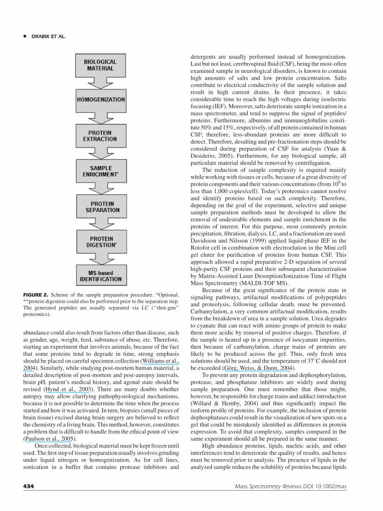
In any experiment, the quality of the results strongly dependson the condition of the starting material. Therefore, proteinextraction from cells and tissues is regarded as a critical step insample preparation protocol (Fig. 2). As the matter point is ofgreat importance in proteomics, where a comparison of case andcontrol groups that result in an identification of even minorvariations in protein profilesmight lead to the comprehension anddetermination of the disorders’ mechanisms. For years, animals,mainly rats and mice, have served as valuable models forproteomic studies in neuroscience. High similarity between therats/mice and human proteins enables an advanced study onmechanisms and hypothesis concerning various diseases. How-ever, it should be remembered that, although animal modelsallow on understanding of neurological processes and mightbe referred to human illness, they cannot be perceived asequivalents. It should be noted that alterations in a protein state or
FIGURE 1. Scheme of the clinical feature of proteomics.
PROTEOMICS IN NEUROSCIENCES &
Mass Spectrometry Reviews DOI 10.1002/mas 433
abundance could also result from factors other than disease, suchas gender, age, weight, feed, substance of abuse, etc. Therefore,starting an experiment that involves animals, because of the factthat some proteins tend to degrade in time, strong emphasisshould be placed on careful specimen collection (Williams et al.,2004). Similarly, while studying post-mortem human material, adetailed description of post-mortem and post-autopsy intervals,brain pH, patient’s medical history, and agonal state should berevised (Hynd et al., 2003). There are many doubts whetherautopsy may allow clarifying pathophysiological mechanisms,because it is not possible to determine the time when the processstarted and how it was activated. In turn, biopsies (small pieces ofbrain tissue) excised during brain surgery are believed to reflectthe chemistry of a living brain. Thismethod, however, constitutesa problem that is difficult to handle from the ethical point of view(Paulson et al., 2005).
Once collected, biologicalmaterialmust be kept frozen untilused. The first step of tissue preparation usually involves grindingunder liquid nitrogen or homogenization. As for cell lines,sonication in a buffer that contains protease inhibitors and
detergents are usually performed instead of homogenization.Last but not least, cerebrospinal fluid (CSF), being themost-oftenexamined sample in neurological disorders, is known to containhigh amounts of salts and low protein concentration. Saltscontribute to electrical conductivity of the sample solution andresult in high current drains. In their presence, it takesconsiderable time to reach the high voltages during isoelectricfocusing (IEF).Moreover, salts deteriorate sample ionization in amass spectrometer, and tend to suppress the signal of peptides/proteins. Furthermore, albumins and immunoglobulins consti-tute 50%and 15%, respectively, of all protein contained in humanCSF; therefore, less-abundant proteins are more difficult todetect. Therefore, desalting and pre-fractionation steps should beconsidered during preparation of CSF for analysis (Yuan &Desiderio, 2005). Furthermore, for any biological sample, allparticulate material should be removed by centrifugation.
The reduction of sample complexity is required mainlywhileworkingwith tissues or cells, because of a great diversity ofprotein components and their various concentrations (from 106 toless than 1,000 copies/cell). Today’s proteomics cannot resolveand identify proteins based on such complexity. Therefore,depending on the goal of the experiment, selective and uniquesample preparation methods must be developed to allow theremoval of undesirable elements and sample enrichment in theproteins of interest. For this purpose, most commonly proteinprecipitation, filtration, dialysis, LC, and a fractionation are used.Davidsson and Nilsson (1999) applied liquid-phase IEF in theRotofor cell in combination with electroelution in the Mini cellgel eluter for purification of proteins from human CSF. Thisapproach allowed a rapid preparative 2-D separation of severalhigh-purity CSF proteins and their subsequent characterizationby Matrix-Assisted Laser Desorption/Ionization-Time of FlightMass Spectrometry (MALDI-TOF MS).
Because of the great significance of the protein state insignaling pathways, artifactual modifications of polypeptidesand proteolysis, following cellular death, must be prevented.Carbamylation, a very common artifactual modification, resultsfrom the breakdown of urea in a sample solution. Urea degradesto cyanate that can react with amino groups of protein to makethem more acidic by removal of positive charges. Therefore, ifthe sample is heated up in a presence of isocyanate impurities,then because of carbamylation, charge trains of proteins arelikely to be produced across the gel. Thus, only fresh ureasolutions should be used, and the temperature of 378C should notbe exceeded (Gorg, Weiss, & Dunn, 2004).
To prevent any protein degradation and dephosphorylation,protease, and phosphatase inhibitors are widely used duringsample preparation. One must remember that those might,however, be responsible for charge trains and adduct introduction(Willard & Hemby, 2004) and thus significantly impact theisoform profile of proteins. For example, the inclusion of proteindephosphatases could result in the visualization of new spots on agel that could be mistakenly identified as differences in proteinexpression. To avoid that complexity, samples compared in thesame experiment should all be prepared in the same manner.
High abundance proteins, lipids, nucleic acids, and otherinterferences tend to deteriorate the quality of results, and hencemust be removed prior to analysis. The presence of lipids in theanalyzed sample reduces the solubility of proteins because lipids
FIGURE 2. Scheme of the sample preparation procedure. *Optional,
**protein digestion could also be performed prior to the separation step.
The generated peptides are usually separated via LC (‘‘shot-gun’’
proteomics).
& DRABIK ET AL.
434 Mass Spectrometry Reviews DOI 10.1002/mas

form complexeswith commonly used detergents and reduce theirefficacy. It might affect the observed pI and molecular weight(MW) of proteins. Frequently, sodium dodecyl sulfate (SDS) andTween are used to remove lipid components. Optionally, proteinprecipitation with trichloroacetic acid (TCA) or acetone isperformed (Berkelman& Stenstedt, 2002; Lubec, Krapfenbauer,& Fountoulakis, 2003).
Nucleic acids tend to bind to proteins through electrostaticinteractions that increase sample viscosity and prevent focusing;therefore, nucleases are introduced to degrade them (Gorg,Weiss, & Dunn, 2004). Polysaccharides are usually ignored,because most often they do not disturb the analysis of proteins. Ifnecessary, precipitation with TCA or ammonium sulfate andultracentrifugation are applied.
The proteins of interest must be isolated from a complexmixture. To ensure that a single spot on a gel will represent anindividual protein, they must be previously denatured, disag-gregated, reduced, and solubilized. Similarly, the efficacy ofprotein extraction, digestion, and creation of representativepeptidemaps for LC analysis depends on the same steps as above.Dithiothreitol (DTT), a widely known reducing agent, preventsoxidation of free sulfhydryl groups present in cysteine residues.Oxidationmight lead to non-specific aggregation of proteins and,hence its heterogeneity, inactivity or denaturation. Detergents forprotein solubilization (CHAPS, NP-40, ASB-14) are a matter ofchoice, and should be selected according to the properties ofanalyzed sample and to the main goal of the experiment. Untilnow, there is no single detergent that would allow solubilizing allproteins simultaneously.
Many laboratories start samples preparation (for peptides)with the use of extraction at neutral pH. This should be avoidedwhenworkingwith peptides/neuropeptides as they undergo rapiddegradation in crude homogenates. Only acidic pH and hightemperature (not even an inhibitory cocktail) can partiallyeliminate such problems. Another option is application ofmicrowaves for efficient extraction of neuropeptides (Nylanderet al., 1997). Adsorption of the minute amounts of material,causing further problems with recovery should also be taken intoconsideration. This has been described, for example, by Speicheret al. (2000) and is out of scope of this review.
One must consider the fact that protein content decreasessignificantly when the sample is handled and stored inappropri-ately, because of protein/peptide degradation or losses whiletransferring and diluting. Protein/peptide adsorption on workingsurfaces, that is, falcon or Eppendorf tubes, if not coated withsilicone; or pipette tips, is also of great importance.
Therefore, when planning the experiment, one should keepin mind that the sample-preparation step should be compatiblewith the techniques applied for further analysis (Grafin, 2003).Moreover, the sample preparation strategy should be as simple aspossible, because of protein loss in each step.
IV. FROM GENES THROUGH PROTEINS ANDPEPTIDES TO DRUGS
The general flow-path fromDNA to protein synthesis and proteinfunction is a basic dogma in systems biology that involves
computation, genomics, and various aspects of proteomics,including expression, structure, and function of proteins. Thestrategies for each approach are described in part in this review,and also in details in the references. Sometimes, it is also valuableto leave high-throughput methodologies and focus on singlemolecule discovery, which in certain cases, might be a goodalternative for large-scale studies. Here, we would focus on theapproaches for a small-scale identification of molecules derivedfrom the nervous system.
Identification of a single type of molecule often requires aspecific method for sample preparation, which is distinct from ageneral protocols applied in proteomics. Usually, the informationthat indicates a molecule derives from other studies, such as anincreased level as determined by, for example, radioimmunoas-say (RIA), radioreceptor assay (RRA), or enzyme-linkedimmunosorbent assay (ELISA). An antibody-based method is,however, not sufficient for a thorough identification of a proteinsequence, its eventual modifications, or mutations.
Nervous tissue contains a high amount of lipids that must beremoved prior to further processing of the sample. In addition, thehuge excess of proteolytic enzymes might severely influenceprotein/peptide content during isolation procedure. More detailscan be found inmany reviews; for example,Meunier et al. (1995).
The standard procedure for the identification of a neuropep-tide from a tissue includes the following steps shown in Figure 3.
The protocol for CSF samples differs slightly from the aboveprocedure, and is presented in Figure 4.
The above methodologies are often modified according tothe requirements for each different sample.
An example of a mass spectrum of Neuropeptide Y (NPY)isolated from rat hippocampus (Stenfors, Hellman, & Silberring,1997) is shown in Figure 5. The major component of the peptidecorresponds to the authentic amidated form of NPY (1–36).
Several spectacular discoveries of novel peptides werepublished over past years. One includes cortistatin, whoseaminoacid sequence was predicted from the characterization ofbrain mRNAs, followed by a sequence similarity search (deLecea et al., 1996).
The authors found a peptide sequence and, using thisinformation, a synthetic peptide that possessed biological activityhas been prepared. Another example describes Nociceptin/Orphanin FQ (Meunier et al., 1995; Reinscheid et al., 1995),which was extracted, purified, and identified through its bindingto an orphan receptor, followed by sequencing and pharmaco-logical studies.
The final step in the characterization of bioactivesequence(s) derived from the native protein/peptide involvespharmacological studies, as well as molecular biologyapproaches. The proposed consecutive steps in the discovery ofbioactive sequences are shown in Figure 6.
Elucidation of the optimal sequence can also be performedby using combinatorial chemistry for a search, for example, amost potent enzyme inhibitor or receptor ligand. The selectedcompounds are further studied for their pharmacological/biological activity.
An idea to discover a novel sequence is always attractive fora scientist. The above procedures are only examples of theapproaches that are alternative to proteomics strategies. It should,however, always be a matter of workload, possible advantages
PROTEOMICS IN NEUROSCIENCES &
Mass Spectrometry Reviews DOI 10.1002/mas 435

and drawbacks, and costs to select optimal strategy foridentification of novel neuropeptides or proteins.
V. MAJOR ADVANTAGES AND LIMITATIONS OFPROTEOMIC TECHNIQUES
Currently, there are numerous potential applications of proteo-mics in the neurosciences, including the determination of aneuroproteome, profiling of PTMs, comparative analysis ofprotein expression, mapping of protein–protein interactions, etc.As its main achievement, one can take the identification ofnumerous brain proteins of different types, such as enzymes,cytoskeleton, synaptosomal of antioxidant proteins, heat shockproteins/chaperones, and proteins of the transcription andtranslation machinery (Lubec, Krapfenbauer, & Fountoulakis,2003).
Particular challenges in neuroproteomics concern thecomplexity and heterogeneity of the brain, and the limitednumber of databases and informatic tools (Grafin, 2003).Moreover, the spatial and temporal dynamic nature of aproteome, various properties of proteins depending on theiramino acid composition, wide range of concentrations within thecells, and a large number of samples that must be analyzed,
require high-throughput, automated, and comparable proce-dures.
Neuroproteomic research might be preceded based ontwo main approaches, the traditional proteomic approach, andshotgun proteomics (Fig. 7). Shotgun proteomics refers to thedirect analysis of complex protein mixture that is digested beforeseparation to generate a global profile of the protein components.
Another aspect associated with quantitative proteomicsconcerns a question, how much the difference in proteinexpression must be to assign such molecule as a potentialmarker. Various programs for 2-DGE evaluation have an optionto set certain threshold of differences between gels, from whichthe software detects such proteins. As we learn from clinicalchemistry and ‘‘classical’’ measurement of various bloodparameters, it is very difficult or even impossible to predict fromsuch data, which protein might serve as a marker. Sometimes, atiny difference in protein expression between normalcy andpathophysiology may be a cause (or a reason) of a disease. Inother cases, even large differences do not indicate a disease.Further studies are, therefore necessary to reveal function(s) ofparticular molecules in the organism and under conditions testedto select a correct set of markers.
A. Gel Electrophoresis
The majority of the proteomic methods applied in neuroscienceinvolve MS as a highly sensitive and versatile tool to identify
FIGURE 3. Protocol for identification of neuropeptides from brain
tissue.
FIGURE 4. Protocol for identification of neuropeptides from CSF.
& DRABIK ET AL.
436 Mass Spectrometry Reviews DOI 10.1002/mas

FIG
URE5.MassspectrumofNPY(1–36)analyzedwithnanosprayMS.L
eftpanel(A
):accumulatedmass
spectrum
showsthemultiply
charged
speciesofendogenousNPY;rightpanel
(B):
deconvolutedESI
spectrum
reveals
themolecularweightofintact
amidated
NPY.Reproducedfrom
(Stenfors,Hellm
an,&
Silberring,1997).
PROTEOMICS IN NEUROSCIENCES &
Mass Spectrometry Reviews DOI 10.1002/mas 437

proteins from complex biological samples that additionallyrequires minute amounts of sample (femtomole to attomole)(Godovac-Zimmermann & Brown, 2001; Aebersold & Mann,2003). Its combination with the unique resolving power of 2-
DGE and in-gel trypsin digestion enables quantitative expressionprofiling of large sets of complex protein mixtures, based onlarge-scale gel analysis (Gorg, Weiss, & Dunn, 2004). 2-DGE,the most popular tool to explore neurodegenerative disorders,remains an efficient technique that allows not only a screening forabundant-protein changes in various diseases, but also foralterations in metabolic pathways. The major advantage of2-DGE lies in its potential to simultaneously resolve thousands ofproteins, at the same time revealing their MW, pI, and reflectingchanges in protein expression and isoforms. However, one shouldremember that a significant number of spots on a gel probablyconsist of more than one protein. The notable strength of thesystem is the capability to identify PTMs and to omit thelimitations of immunochemical detection methods (Castegnaet al., 2002). Approaches that took advantage of 2-DGE profilingandMS analysis were applied to identify oxidized proteins in AD(Castegna et al., 2002, 2003 Korolainen et al., 2002) and to findalterations in the expression of proteins linked to apoptosis andneuronal death that could contribute to neurodegenerativedisorders (Tilleman et al., 2002). 2-DGE was also applied toidentify alterations in proteins associated with Down syndrome(Engidawork & Lubec, 2003), Huntington’s disease (HD) (Zabel
FIGURE 6. Consecutive steps during discovery of bioactive sequences.
FIGURE 7. Different strategies for proteomic studies.
& DRABIK ET AL.
438 Mass Spectrometry Reviews DOI 10.1002/mas

et al., 2002), and various psychiatric disorders (Johnston-Wilsonet al., 2000) as well as PTMs in AD brain (Castegna et al., 2003;Kanninien et al., 2004). A further advantage of 2-DGE is that itpermits the isolation of proteins in milligram amounts to enabletheir structural analyses by MALDI-TOF MS, electrosprayionization-mass spectrometry (ESI-MS), or Edman microse-quencing (Celis &Gromov, 1999; Fey&Larsen, 2001; Graves&Haystead, 2002; Lilley, Razzaq, &Dupree, 2002; Ong&Pandey,2002; Rabilloud, 2002; Beranova-Giorgianni, 2003). Moreover,2-DGE allows one to detect and quantify less than sub-ng amountof a protein (Gorg, Weiss, & Dunn, 2004).
Apart from these advantages, 2-DGE has also somedrawbacks. One of its limitations includes the difficult identifica-tion of low (<15 kDa) and high (>150 kDa) MW proteins(Wildgruber et al., 2000; Westbrook et al., 2001). Furthermore,because the variations in concentrations (dynamic range) ofdifferent proteins in a single eukaryotic cell range from severalmillion (e.g., glycolytic enzymes) to just a few copies per cell(e.g., receptor molecules or transcription factors), the possibilityof detection of these rare proteins is still limited. Consequently,there is high interest in development of new pre-fractionation,enrichment, and detection methods that would increase thedynamic range of 2-DGE and could contribute to a more efficientanalysis of proteomes. The total protein content of themajority ofCNS samples is dominated by albumins, immunoglobulins,serine protease inhibitors, cytoskeleton proteins, etc. Otherproteins of potential diagnostic significance are often present in10–1,000 fmol/mL amounts (Davidsson et al., 2003). Such lowprotein levels are detectable nowadays with the use of advancedMS techniques, provided that most common serum proteins,which suppress the less abundant ones, are removed.
One of the most serious problems encountered whenworking with proteomes is the under-representation of numerousclasses of proteins because of their limited solubilization,because commonly used IEF is not compatible with introductionof charged molecules, that is, ionic detergents (Sagi & Zabel,2003). The introduction of immobilized pH gradients (IPGs) forthe first dimension allowed one to partially overcome manyformer problems connected with the use of carrier ampholytes(CA)-based 2-DGE, such as poor resolution, poor separation ofproteins with extreme pIs, and low sample loading capacity(Gorg et al., 1985; Gorg, Postel, & Gunther, 1988; Wildgruberet al., 2000;Westbrook et al., 2001; Gorg,Weiss, &Dunn, 2004).Nevertheless, separation is generally limited to proteins that areneither too acidic/basic, nor too hydrophobic. Despite recentdevelopments in new electrophoretic methods (Hartinger et al.,1996; Lin et al., 1999; Neff &Dencher, 1999; Dreger et al., 2001;Coughenour, Spaulding, & Thompson, 2004; Rais, Karas, &Schagger, 2004), and the introduction of strong detergents,various studies still show a significant under-representation ofmembrane proteins, and that the choice of detergent remainslargely empirical (Friso &Wikstrom, 1999; Santoni et al., 1999;Fountoulakis&Takacs, 2001;Carboni et al., 2002;Navarre et al.,2002; Kashino, 2003; Luche, Santoni, & Rabilloud, 2003; Babuet al., 2004; Rais, Karas, & Schagger, 2004; Seddon, Curnow, &Booth, 2004). These problems are attributed mainly to their lowsolubility and tendency to aggregate and precipitate in aqueoussolutions. Bierczynska-Krzysik et al. (2006b) described the MSidentification of highly insoluble and membrane rat brain
proteins. Although 2-DGE/SDS–PAGE was able to resolvemembrane proteinswith a single transmembrane domain (TMD),the use of benzyldimethyl-n-hexadecylammonium chloride(16-BAC/SDS–PAGE) led to the identification of multipleproteins with strongly basic pI and integral membrane proteinswith up to eight TMD.
The reproducibility and quantitation of 2-DGE results,especially between different laboratories, still constitutes a greatchallenge for applications such as expression profiling. Conven-tional 2-DGE that relies on comparison of (individual, frequentlynot fully superimposable) gel images is very laborious andtime-consuming. Commercially available 2-D image analysisprograms (continuously ameliorated in terms of focusing onautomation and faster algorithms with lesser manual inter-vention) still require a great deal of attention and manualintervention.
To address some of the above-mentioned problems, a newmethod in neuroproteomics difference gel electrophoresis(DIGE) was developed by Unlu, Morgan, and Minden (1997).DIGE was used to analyze human prefrontal cortex (PFC) incontrol and schizophrenic individuals (Swatton et al., 2004).DIGE separates two or more different samples, labeled withdifferent fluorescent dyes (comparable in sensitivity to silverstaining), on a single gel, to provide accurate spot matching andquantification. Once each dye molecule is excited with eachappropriate wavelength, images are overlaid and normalized tovisualize only differences in protein expression between twosamples. The major drawback is the high cost of the equipmentand software. The application of DIGE is also restrictedto proteins that have long half-lives and are expressed at highlevels.
Furthermore, the most commonly used staining methods,Coomassie Brilliant Blue (CBB) and silver, do not detect low-abundance proteins because of the dyes’ limited sensitivity,reproducibility or narrow dynamic range (Nishihara & Cham-pion, 2002). The popularity of silver staining decreased mainlybecause of complications with background (increased back-ground signal results from the presence of DNA and liposachar-ides that are also stained by silver), quantitation, and MScompatibility. Nowadays, some modified procedures are beingdeveloped to avoid the problems with the presence ofglutaraldehyde in silver-staining protocols and to enable acqui-sition ofMS spectra (Yan et al., 2000). Another visible stain, zincimidazole, has limited quantitation abilities (Fernandez-Patronet al., 1998). One alternative for protein visualization is theapplication of easy-to-use, quantitative, and MS-compatiblefluorescent dyes, such as SYPRO Ruby. However, the inventionof a single stain that would fulfill all the requirements forsensitivity, quantitation, broad dynamic range, and compatibilitywith MS still remains a challenge.
B. Liquid Chromatography Combinedwith Mass Spectrometry
Liquid chromatography and reversed-phase high pressure liquidchromatography (RP HPLC) are other complementary methodsthat are widely used in neuroproteomics and are applied foreffective separation of proteins/peptides. The online hyphenation
PROTEOMICS IN NEUROSCIENCES &
Mass Spectrometry Reviews DOI 10.1002/mas 439

of nanoscale LC with MS allows an automated and highlysensitive analysis. Furthermore, the use of these techniquestogether with tandem mass spectrometry (MS/MS) (i.e.,MudPIT—the multi-dimensional protein identification techni-que) results in the identification of peptides in mixtures in asingle analysis and offers an increased potential to detect low-abundance proteins. That approach was successfully applied toidentify endogenous peptides from the brain extracellular fluid ofliving rats that represented candidates for neurotransmitters,neuromodulators, andmarkers of synaptic activity or brain tissuedamage (Haskins et al., 2004). Capillary electrophoresis (CE)offers a significant enhancement in resolution, although it can beused to analyze only one sample at a time (Freeman & Hemby,2004). A significant improvement in the reliability of identifica-tion of peptidemapswas recently described byNoga et al. (2005)and Noga, Asperger, and Silberring (2006). This highly sensitivemethod combines ESI and nano-scale liquid chromatography(nanoLC) coupled online to a tandem MS to determine theprimary structure of unknown peptides modified by acetylation/deuteroacetylation (Morris et al., 1981). The acceleration andfacilitation of mass spectra interpretation, as well as its potentialapplications for the analysis of PTMs, constitute its majoradvantages. The major limitation results from the necessarydivision of analyzed sample into two parts prior to derivatization.Therefore, a decrease in sensitivity is observed. In contrast to2-DGE followed by MS, LC–MS/MS, by which peptides areanalyzed, does not directly reveal the MW and pI of proteinswithout the use of specific databases. Also, protein’s isoformsmight be overlooked with the use of 2D LC–MS approach.
When quantitative analysis is required, stable isotope-labeling must be performed. The combination of LC–MS andisotope-coded affinity tag (ICAT) established the differencesbetween neuropeptides from pituary extracts of wild-type andmutant mice (Che & Fricker, 2002). It was also effective in thedifferential analysis of plasma-membrane proteins from fore-and hindbrain (Che & Fricker, 2002; Olsen et al., 2004), and wasable to identify and quantify changes in protein expressionbetween control and camptothecin-treated mouse corticalneurons (Yu et al., 2002). An alternative approach, based onaffinity capture and LC–MS, was also implemented for large-scale profiling of carbamylation in samples from aged mousebrains (Soreghan et al., 2003). The ICAT methodology, superiorin examining high MW proteins, is a very promising tool inneuroproteomics, although its certain limitations must beconsidered, such as detection of only cysteine-containingproteins, and high requirements for MS/MS instrumentationand operators (Choudhary & Grant, 2004).
Currently, there exist a number of different MS platformsand each has great capabilities but also limitations. ESI andMALDI are the ionization techniques of choice. These highlysensitive, versatile, and high-throughput methods can be used toderive sequences de novo and obtain structural information.Modern MS distinguishes between the residues of similar mass,like Gln/Lys or Phe/Met sulfoxide, excluding identification ofLeu/Ile because their mass is the same. A limitation ofMS is thatquantitative analyses are restricted to techniques like ICAT,isobaric tags for relative and absolute quantitation (iTRAQ),or stable isotopic labeling using amino acids in cell culture(SILAC).
More recently, a variation on the MALDI concept wasintroduced (i.e., SELDI or ClinProt) that possesses a quantitativeability without having to use an additional tag (Hutchens & Yip,1993;Merchant&Weinberger, 2000). Detection limits of SELDIreach attomole level; however, with this technique the identifica-tion of proteins of interest is difficult. SELDI was successfullyapplied in analysis of potential biomarkers in the CSF of healthyversus AD patients (Carrette et al., 2003; Solassol et al., 2006).
Peptide mass fingerprinting and tandem mass sequencingconstitute powerful tools to identify proteins in complex bio-logical samples. The major concerns of protein identificationbased on fragmentation involve the inability of the automationand the considerable time required for mass spectra interpretat-ion (Reinders et al., 2004). Additional weakness involves theapplication of database searches in protein identification. Thesesearches are still not complete, especially in terms of unknown orrare modifications. Furthermore, individually chosen searchparameters have great influence on the results and subsequentprotein identification. However, de novo sequencing of peptidesconstitutes a promising tool, even for organisms with incompletegenomic or error-containing databases (Reinders et al., 2004).Another challenge concerns the management of the enormousand still growing volume of data, for example, translationalmedicine applications that become more difficult to handle, andthe sharing of data among laboratories. The last, but not least,challenge that poses limitations to the routine application ofproteomics is the high cost of research.
Summary of different proteomics methods used in neuro-science is shown in Figure 8.
VI. PROTEOMICS IN NEURODEGENERATIVEDISORDERS
There are many articles regarding methods of protein analysisand identification by MS (Schrader & Schultz-Knappe, 2001;Davidsson et al., 2002; Dunckley, Coon, & Stephan, 2005;Paulson et al., 2005), as well as ones concerning disease specific
FIGURE 8. Scheme of different MS platforms used in proteomics.
& DRABIK ET AL.
440 Mass Spectrometry Reviews DOI 10.1002/mas

biomarkers discovery. Many neurodegenerative diseases share acommon feature—aggregation and deposition of abnormalproteins. Revealed patterns of disease-specific cellular markerscould be classified according to:
. senile plaques,
. neurofibrillary tangles,
. Lewy bodies,
. protein aggregates (e.g., amyloid fibrils),
. cellular inclusions and swollen motor neuron axons,
. neostriatum g-aminobutric acid-containing neurons, andothers.
These symptoms can increase oxidative and metabolicstress, and finally initiate apoptosis in neurons.
Aggregates are a widespread theme in the molecularpathology of AD, PD, ALS, HD, Creutzfeldt–Jacob (CJD),Gerstmann-Straussler-Scheinker syndrome (GSS), Kuru, fatalfamilial insomnia (FFI), and fatal sporadic insomnia (FSI). Self-aggregation of a-synuclein, incorrect folding of the prion protein,defective cleavage of the amyloid peptide, and hyperpho-sphorylation of the tau protein all lead to changes in thedynamics of cytosolic proteins, and eventually to the formation ofmacroscopic aggregates. The advance of our knowledge aboutthe physiological functions of these proteins and the neuronalsystems, in which they are expressed, might help identifytherapeutical targets to prevent neuronal death in any neurode-generative disease.
The collection of CSF via lumbar puncture is much lessinvasive than performing biopsies of neural tissue, and is themainreason why CSF is the material of choice for proteomics analysisin neurodegenerative diseases, as it is in a direct contact withneural tissue and may be considered as reflecting dynamicchanges in the CNS function. CSF protects the brain fromchanges in blood pressure and trauma. CSF is also responsible fortransport of neurosecreted, biosynthesized, and metabolizedcellular products. Using 2-DGE, Yuan and Desiderio (2005)detected potential biomarkers in CSF in a variety of neurologicalconditions such as AD, dementia, cancer, schizophrenia, CJD,stroke, and traumatic brain injury. The alterations of various CSFcomponents were found, including apolipoproteins, a1-antitryp-sin, transthyretin, retinal-binding protein, prostaglandin D2
synthase, b2-microglobulin, ubiquitin, kininogen, a1b-glycopro-tein, 14-3-3 protein isoforms, and haptoglobulin. Furthermore,Romeo et al. (2005) described proteins responsible for themechanism of neurodegeneration; that is, gelsolin, hemopexin,fibrinogen, kallikrein-6, amyloid fragments, cystatin C, andplasminogen.Moreover, changes in protein expression in CSF ofpatients with various neurodegenerative disorders including AD,PD, and HD, were detected by Rohlff by using a classicalproteomic approach (Rohlff, 2001, 2006). Alterations of tauprotein, amyloid fragments, apolipoproteins, interleukin-1receptor type II, transthyretin, dopamine-releasing protein,14-3-3 protein, neuronal-specific enolase, S-100 protein, soma-tostatin, angiotensin-converting enzyme, neuronal cell adhesionmolecule, inositol monophosphatate, synaptosome-associatedprotein 25 kDa (SNAP-25), and growth-associated protein43 kDa (GAP-43) have been described. Additionally, 2-DGEwas performed by Sheta, Appel, and Goldknopf (2006) to reveal
CSF, serum, and brain tissue biomarkers in patients withneuropathological symptoms. Modifications of cathepsin Bprecursor, prion precursor protein, collapsing response mediatorprotein 2, glial fibrillary acidic protein (GFAP), heat shockcognate 71 (HSC71), and peroxidoredoxins (I, II, and IV) werealso observed.
Brain tissue was also investigated by using a proteomicapproach to identify possible disease-associated proteinmarkers,to assist in diagnosis or prognosis, and to select potential targetsfor specific drug therapy (Chambers et al., 2000). Kaytor andWarren (1999) examined the most aberrant proteins depositionduring neurological disorders (see above). Additionally, changesin proteome expression were illustrated by McNaught, throughmultiplexed protein studies of PD, AD, HD, CJD patients(McNaught, 2004). Ubiquitin–proteasome system, heat shockprotein 70 (HSP70), heat shock protein (HSP90), heat shockprotein (HSP27), ubiquitin C-terminal hydrolase-1 (UCH-1),4 hydroxynonenal protein, cholesterol transport protein, a, b,g-secretases, superoxide dismutase-1 (SOD-1), atrophin, andandrogen receptor were found to be differentially regulated.Proteomic studies in human post-mortem brain obtained frompatients with AD, PD, and schizophrenia, resulted in therapeutictargets information (Morrison et al., 2002; Kim et al., 2004;Jain, 2005; Schmidt et al., 2005).
The antioxidant status is essential for the recognitionof neurodegenerative mechanisms. In particular, ubiquitin C-terminal hydrolase L-1 (UCHL-1), creatine kinase BB (CK-BB),GS, a-enolase, dihydropyrimidinase-related protein-2 (DRP-2),3-nitrotyrosine, neuropolypeptide h3, triosophospate isomerase,SOD-1, superoxide dismutase-2 (SOD-2), catalase, glutathionereductases, transferases, and peroxiredoxins are major determi-nants in the control of oxidative stress (Knight, 2001; Sagi &Zabel, 2003).
In addition, the cellular distribution of Ca2þ-bindingproteins was studied during the past decade (Heizmann&Braun,1992). These proteins proved to be useful neuronal markers for avariety of functional brain systems. Because massive neuronaldegeneration takes place in several human brain diseases,changes in the expression of Ca2þ-binding proteins occur.Excessive concentration of extracellular glutamate overactivatesionotropic glutamate receptors, and results in an intracellularcalcium overload and a cascade of events that leads to neuronalcell death. These factsmay be useful to estimate, for example, thelocation and extent of brain damage in various neurologicaldisorders. In the future, if we deepen our knowledge about thephysiological functions of these proteins and the neuronalsystems in which they are expressed, Ca2þ-binding proteinsmight become important therapeutical targets for preventingneuronal death in any array of neurodegenerative diseases.
A. Alzheimer’s Disease
The proteins, whose expression is altered in the AD brain, help toelucidate possible mechanisms of the disease. In addition topathophysiological symptoms, which are well-documented,AD is associated with synapse loss, oxidative stress, decreasedglucose metabolism, mitochondrial deficit, increased proteinmisfolding, and decreased protein turnover (Butterfield, Boyd-Kimball, & Castegna, 2003). Modifications observed in many
PROTEOMICS IN NEUROSCIENCES &
Mass Spectrometry Reviews DOI 10.1002/mas 441

proteins involved in numerous pathways allowed one to develop ahypothesis of neurodegenerative mechanisms in AD brain. Theneuropathologic properties of AD are connected with formationof extracellular plaques and intracellular neurofibrillary tangles.A component of senile plaques in AD is b-amyloid, ahydrophobic peptide fragment of the amyloid precursor protein(APP), which can be metabolized along at least two pathways;one involves the generation of an insoluble b-amyloid peptide byan enzyme named a-secretase (Fig. 9). It has been demonstratedthat b-amyloid peptide, generally associated with the ADplaques, can also be found in the brains of survivors of headinjury (Korolainen et al., 2002). Processing of APP to produceamyloid peptides occurs in the synaptic terminal field of axonsand illustrates the utility of protein activity as a general markerfor axonal injury. Measurements of a-secretase-cleaved APPrepresent a new and promising diagnostic marker. Manyresearchers agree that amyloid peptide is central to thepathogenesis of AD (Berlett & Stadtman, 1997). For example,persons who inherit AD because of mutations in the presenilin-1,presenilin-2, and APP genes have excessive amounts of amyloidpeptide. In addition, persons with Down’s syndrome alwaysdevelop AD if they live long enough, what can be related to thelocation of APP gene on chromosome 21. Comparison of 2-DGEpatterns of human brain proteins in AD, Down’s syndrome,and controls revealed proteins that differed between control anddisease groups. Those proteins were identified byMS, and servedto create a potential marker list for these two common diseases(Butterfield, 2004).
Classes of specifically oxidatively modified proteinswere identified by using proteomic techniques in AD brain(Vercauteren et al., 2004). Moreover, some of the pathologicalcharacteristics of AD brain, such as accumulation of aggregated,damaged proteins, increased ubiquination, excitotoxicity, apo-ptosis, and shortened dendritic lengths are consistent with adysfunction of some of these specifically oxidized proteins. Theoxidative modifications affect protein functions. An increase inthe expression of several antioxidant proteins, to name a few:carbonyl reductase (CBR), alcohol dehydrogenase (ADH),
peroxiredoxin-I (Prx-I), peroxiredoxin-II (Prx-II), antioxidantprotein 2, and Cu/Zn superoxide dismutase, was also observed inthe AD brains, as a result of oxidative stress (Heizmann&Braun,1992; Berlett & Stadtman, 1997; Butterfield, 2004). Moreover,a decrease in peroxiredoxin-III (Prx-III), metallothionein-1(MT-1), and metallothionein-3 (MT-3) expression was found.
Furthermore, apoptosis plays a major role in the neuronalloss revealed in AD (Berlett & Stadtman, 1997; Tilleman et al.,2002). The pro-apoptotic proteins: zipper-interacting proteinkinase (ZIPK), Bcl-2-interacting mediator of cell death, andthe receptor-interacting protein (RIP)-like interacting CLARPkinase (RICK) were found to be increased in AD brain, whereasthe anti-apoptotic protein p21 and the apoptosis repressor withcaspase recruitment domain (ARC) were up-regulated inresponse to oxidative stress and apoptotic signals such asb-amyloid peptide. Conversely, the level of Fas-associated deathdomain (FADD)-like interleukin-1b-converting enzyme inhibi-tory protein (FLIP) was decreased as a reason of attending asa substrate for caspases and forming an inactive complex.Additionally, down-regulation of FLIP and DNA fragmentationfactor (DFF45) provides supporting evidence for the activation ofcaspases, aswell as the down-regulation of procaspases-3, -8, and-9. Finally, GFAP, amarker of glia, was elevated in ADbrain, andsupported the hypothesis of neuronal cell death. AD is alsoassociated with protein misfolding and decreased protein turn-over. Protein misfolding can be related to the dysregulation ofmolecular chaperone proteins in the AD brain such as heat shockprotein (HSP60),HSC71, glucose-regulated protein 75 (GRP75),and aB-crystallin (ABC), all of which are down-regulated.
The major pathological signs of AD are neurofibrillarytangles, and in particular their important constituent, tau protein.Hyperphosphorylation of tau is responsible for its dissociationfrom the microtubule and further precipitation (Gotz, Ittner, &David, 2004; Vercauteren et al., 2004). It was proposed thathyperphosphorylation of tau protein is responsible for AD. Thesefindings established the relationship between pathologicalsymptoms of the disease and metabolic or structural abnormal-ities that are strictly dependent on significant proteins. The
FIGURE 9. Generation of the b-amyloid peptide.
& DRABIK ET AL.
442 Mass Spectrometry Reviews DOI 10.1002/mas

analysis of PTMs is a new approach to gain insight intomolecularmechanisms involved in this dementing disorder. Moreover,glycosylation (Kanninien et al., 2004) and nitration (Castegnaet al., 2003) play very important roles during neurodegenerativeprocesses.
Information obtained by proteomic analysis offers thepossibility of establishing new hypotheses for the mechanismsof neurodegeneration in AD brain. It is important to note thatvariations in protein expression that were detected in AD are notglobal, but correspond to specific brain regions. Consequently,protein expression changes provide only a clue to the altered stateof the protein that causes the disease.
In summary, the expression ofmany proteins is altered in theAD brain and results in a cascade of alterations on multiplepathways within the brain. Perhaps there is a relationship amongthese pathways that sustain the balance between neuronalsurvival and neuronal death.
B. Parkinson’s Disease
Parkinson’s disease is caused by a selective neuron loss in thesubstantia nigra and striatum, that results in a unique set ofextrapyramidal signs, such as tremor, muscle rigidity, akinesia,and postural and gait disturbance. The symptoms are suggested toresult from abnormal dopamine levels or from the degenerationof dopaminergic neurons in affected regions of the CNS (Bassoet al., 2004). In addition to neuronal degeneration, in numerousbrain regions ubiquitinated intracytoplasmic bodies (Lewybodies) have been found, the major component of which isa-synuclein.
Significant changes in protein expression levels wereobserved in PD post-mortem brain tissue (Basso et al., 2004),including Prx-II, mitochondrial complexes I, II, III (ubiquinolcytochrome c reductase), vacuolar ATP synthase (VATP-ase),complexin I, and L-type calcium channel. These findings couldsuggest the involvement of oxidative stress in PD pathogenesis.Using quantitative proteomics with ICAT labeling, a largenumber of potential biomarkers were identified (Jin et al., 2005).
It was shown that inactivation of UCH L-1 and parkin,components of the ubiquitin–proteasome system, might leadto protein degradation (De Iuliis et al., 2005). Parkin could beinvolved in the polyubiquitination of substrate proteins byallowing the transfer of ubiquitin from the enzyme to the targetprotein. Polyubiquitination is a priming event for proteindegradation via the ubiquitin/proteasome pathway. The toxicbuild of parkin leads to parkinsonism, but on the other hand, thelack of parkin function leads to the progressive accumulation ofaggregates, and finally cell stress, degeneration, and eventuallydeath of dopaminergic neurons. In addition to ubiquitinationof its substrates, parkin is also known to catalyze its ownubiquitination and proteasome-mediated degradation; it thusregulates its own cellular level.
By means of proteomic techniques, oxidation and nitrationof a-synuclein, ubiquitin E3-ligase, parkin, possibly leading toPD development, were identified (Zhang, Dawson, & Dawson,2006). However, most common in PD research is the analysis ofgenemutations (Michell et al., 2004; Corti et al., 2005; Gandhi &Wood, 2005).
C. Huntington’s Disease
Huntington’s disease is themost common in a family of inheritedneurodegenerative disorders that are caused by an expansion ofpolyglutamine tract-associated disease proteins. In HD, thedegeneration occurs in the striatum and deep layers of the cortex.During the late stages of the disease, degeneration extendsto a variety of brain regions, including the hypothalmus andhippocampus. As a result, HD is characterized by cognitivedefects, psychiatric abnormalities, and movement disorders.Individuals with HD are unable to select an angry look from fear,but they have fewer less difficulties in distinguishing betweenmale and female or happiness from sadness. The symptomsusually appear in middle age, and progressively become worseduring the next 10–15 years until the patient dies.
Although the huntingtin protein (htt) was discovered 10 yearsago, its function is still not completely understood (Li & Li,2004). Htt-interacting proteins provide valuable information toexplain the function of htt and the pathogenesis of HD. Theubiquitous expression of htt in nearly all tissues and itswidespread localization at subcellular level also make it difficultto determine its function. Htt is associated with a variety oforganelles, including the nucleus, endoplasmic reticulum, Golgicomplex, synaptic vesicles, and mitochondria. Researcherssuggest that htt is required for cell survival, and that loss offunction of htt can be involved in neurodegeneration (Zabel et al.,2002). Wild-type htt is responsible for cytotoxicity, fibrillogen-esis, disrupts a number of vital cellular processes includingenergy metabolism, gene transcription, intraneuronal trafficking,and post-synaptic signaling. Drugs and chemicals that improvethe function of htt could be beneficial for the treatment of HD.
Studies in HD and normal aging suggest a relationshipbetween central dopaminergic neurotransmission and cognitivefunctions (Deyer & McMurray, 1995). A decrease in dopaminefunction was revealed in HD and aging. Moreover, HD patientsand older adults show deficits across episodic memory, speed ofprocessing, and executive functioning. Although few studies areavailable at present, there is emerging evidence that pre- andpost-synaptic dopamine levels are highly unstable, and arestrongly associated with the cognitive deficits that accompanyHD and aging.
During HD, development of a polyglutamine pathway isobserved, that is caused by htt [44]. Pathogenesis in HD includesthe cytoplasmic cleavage of htt and the release of an amino-terminal fragment that is capable of nuclear localization.Researchers investigated ravages of htt including aggregation,protein–protein interactions, and transcription. These resultsraise the possibility that httmight cause neuronal dysfunction andcell dead.
Inhibition of proteolysis of mutant htt protein leads toaggregation and toxicity through the sequestering of importanttargets, including normal htt.
Several proteinswere identified as potentialHDmarkers: a1-antitrypsin (AAT), contraspin (CTS), serine proteinase inhibitors(SERPINs), ABC, major urinary proteins (MUPs), and chaper-ones, which have already been implicated in other neurodegen-erative diseases (Steffan et al., 2000).
The formation of insoluble protein aggregates is a mainsymptom of HD and other neurodegenerative disorders such as
PROTEOMICS IN NEUROSCIENCES &
Mass Spectrometry Reviews DOI 10.1002/mas 443
bulbar muscular atrophy (SBMA) and ataxia (Bates, 2001). Inpost-mortembrain specimens fromHDpatients, it was found thatthe number of neostriatum neurons that contain the Ca2þ-bindingprotein, calbindin, is lowered. Therefore, an understanding of themolecular mechanisms of protein aggregation and its effects onneuronal cell death could open new opportunities for therapy.
Similar to PD, Huntington’s patients are examined mostwidely, by using the genomic approach (DiFiglia et al., 1997).
D. Prion Diseases
The term ‘‘prions’’ is used to describe the proteins involved incertain neurodegenerative diseases such as CJD, GSS, and Kuru.A prion is an infectious protein, a transmissible b-amyloid formof a cellular protein that replicates by converting the nativeprotein into the same abnormal prion form. In the primarysequence of the prion protein, the PHGGGWGQ sequence isrepeated up to 10 times in certain pathologies, and that sequenceis responsible for the pathological forms of the prions (Ironside,Ritchie, & Head, 2005). CJD was the subject of extensiveproteomic analyses that led to the identification of two membersof the 14-3-3 family of proteins in the CSF of CJD patients. Thepresence of these proteins was used to differentiate CJD fromother kinds of dementia (Knight, 2001; Choe et al., 2002b; VanEvenbroeck, Boons, & Cras, 2005). The antioxidant status ofSOD-1 and SOD-2, catalase, glutathione reductases, trans-ferases, and peroxiredoxins plays important role in the controlof oxidative stress (Prusiner et al., 1996). Activation of microgliaand macrophages was implicated in the pathogenesis of a varietyof neurodegenerative diseases, including multiple sclerosis, AD,CJD, and human immunodeficiency virus dementia (Ridley,1994).
In addition, multiple mutations in the PRNP gene areassociated with prion diseases; mutations are the reason why agenomic examination is of great importance for diagnosticpurposes.
VII. THE PROBLEM OF CANCER INPROTEOMIC RESEARCH
Cancer is characterized by an abnormal and uncontrolled growthof cells that can invade and destroy surrounding normal tissues.These abnormal cells can also spread through the bloodstream orlymph system to start new tumors in other parts of the body. Thedisease is a great challenge to clinicians and scientists. First,tumor demands early detection—the main condition of beingcured. Second, treatment could be difficult because of drugresistance that very often accompanies the disease. Theaggressiveness of cancer, closely related to lifespan prognosis,is also hard to predict. Therefore, it is crucial to find new targetsfor more effective therapies. At the end, there is still a lot to learnabout the mechanism underlying the development of cancer(Lawrie, Fothergill, & Murray, 2001; Simpson & Dorow, 2001;Mocellin et al., 2004).
Although cancer is often classified as a genetic disease, in afunctional sense it is a proteomic disease. Variations in proteinexpression between the normal and tumor stages are thought to be
the cause of abnormal cell proliferation, and the proteomicapproach might be very helpful in discovering those variations.Identification of proteins that are characteristic for cancerdevelopment might uncover diagnostic or prognostic markersor novel drug targets, and could also help understand themechanisms of tumor formation.
The application of proteomics in cancer treatment can beexemplified by the studies on human pituitary macroadenomas.To reveal the molecular basis of this early form of a tumor, acomparative analysis was performed between pituitary adenomaand control tissue; 39 differently regulated proteins wereidentified (Desiderio & Zhan, 2003). Such investigations mightfacilitate further studies focused on particular proteins and theirroles in tumor development (Zhan et al., 2003).
Finding biomarkers for certain tumors is a special challenge.In a clinical approach, it is important to analyze biomarkers inavailable biological fluids to make the diagnosis easier and lessharmful for the patient than by biopsy. Asmentioned above, earlydiagnosis is extremely important for successful treatment, andthe analysis of biological fluids is an easyway of screening.Whatis more, the identified biomarkers could be useful in monitoringthe patient response to treatment (Fig. 1). There are two mainstrategies in which the problem of finding useful biomarkerscould be handled.
One strategy is called the biomarker—discovery approach.Here, researchers utilize traditional methods of comparativeproteomics, and usually global protein profiles are created fornormal and tumor cells by 2-DGE.These profiles are compared tofind the differences in protein expression (Lawrie, Fothergill, &Murray, 2001). For instance, CSF is in continuous contact withbrain tissue and could be an excellent source of biomarkers. Twomarkers, low-molecular weight caldesmon (l-CaD) and N-myconcoprotein were found using 2-DGE and MALDI-MS analysis.The practical value of these two proteins as primary brain tumorbiomarkers is currently being investigated (Zheng et al., 2003).
Another strategy is the diagnostic-proteomics approach(also named: proteomic-pattern diagnostic). Here, the samples ofbiological fluids from healthy and cancer-affected patients areusually examined by surface-enhanced laser desorption/ioniza-tion mass spectrometry (SELDI MS) or other techniques toacquire the protein pattern. In these methods serum is usuallyused because it can be easily obtained and because it containsproteins that diffused into blood during circulation throughoutthe body.
Protein patterns are composed of many individual proteins,any one of which independently is not discriminatory for thedisease, but which together make the pattern characteristic for agiven state. Using artificial-intelligence-based algorithms, thesystem is taught to distinguish between samples characteristic forsick and healthy patients (Petricoin & Liotta, 2002). Thistechnique was used in a pilot study to distinguish betweenpatients with glioblastoma—a malignant brain tumor of glialorigin, and the control group, on the basis of serum proteomicpattern. Majorities (96.4%) of samples were classified correctly;however, despite its promising potential, this technology stilldemands further validation (Veenstra, Prieto, & Conrads, 2004).
Avery important feature of cancer is its aggressiveness thatis currently assessed based on cell morphology. However, thisapproach could be unreliable in predicting the clinical course and
& DRABIK ET AL.
444 Mass Spectrometry Reviews DOI 10.1002/mas

response to the treatment mainly because cancers of the samegrade and stage could have very different clinical outcomes.Therefore, a reliable way of estimation of aggressiveness couldhelp predict survival time and choose the best therapy (Lawrie,Fothergill, & Murray, 2001).
Glioma is the most common brain tumor; it begins in glialtissue. It is characterized by a highly heterogeneous biologicalaggressiveness, even in the same histological category. Analysisof proteomic patterns in astrocytomas of different aggressiveness(like glioblastoma, anaplastic astrocytomas, astrocytomas, andnormal brain tissues) led to the identification of 37 proteins thatwere differentially expressed. This finding shows that proteomicscould be effective in estimating the aggressiveness of gliomasand could help choose the best treatment (Iwadate et al., 2004).
As already mentioned, prediction of tumor response to acertain therapy is crucial. Therapy resistance is the main cause ofcure failure and death of patients who suffer from malignancies.In particular, various histological types of brain tumor are usuallyvery resistant. Proteomics, together with genomics, could behelpful to understand and predict anti-tumor drug response(Bredel & Zentner, 2002).
Recently, it was found that certain proteins connected withmultidrug-resistant phenotype of cancer, such as P-glycoprotein(PGP), multidrug resistance protein (MRP1), lung resistanceprotein (LRP), and O6-methylguanine-DNA-methyltransferase(MGMT), exhibit marked heterogeneity in the expression anddistribution across different types of brain tumor. Therefore,analysis of above-mentioned proteins could be important in theselection of chemotherapy, regardless of tumor type (Anderssonet al., 2004).
As could be seen from the previous examples, proteomictechniques are widely used in brain tumor research. They offerthe possibility to identify disease-associated protein markers toassist in diagnosis or prognosis, and to select potential targets forspecific therapy. Proteomics application in the clinical approachraises the hope for better treatment and effective therapy ofcancer patients (Fig. 1).
VIII. PROTEOMICS IN ADDICTIONS
Addiction is assumed to be a disease of the brain; it is a verycomplex process that involves physiological changes in specificbrain structures, such as the ventral tegmental area (VTA)(Koeltzow & White, 2003), nucleus accumbens (Nac) (Dead-wyler et al., 2004), and PFC. These structures together form thereward systemwhere dopamine plays an important role. Drugs ofabuse, including alcohol and nicotine, interfere with normalprocesses in this circuit, and impose significant changes inbehavior, including loss of control over drug taking, despiteadverse health, social, or legal consequences. Addiction involvesthe development of drug dependence, tolerance, and withdrawalsymptoms after removal of the addictive substance (Roberts &Knoob, 2000; Wise, 2000).
It is obvious that such complex phenomena cannot occurwithout modifications of protein patterns in brain-rewardstructures. Therefore, recently much research was done to findproteins or peptides that could serve as markers of addiction or
therapy targets, and that could clarify those complex biologicalmechanisms, that underlie this illness (Williams et al., 2004). It isalso worth noting that various drugs (narcotics) act via differentmechanisms. For instance, morphine binds to m-opioid receptor,cocaine blocks dopamine reuptake to the pre-synaptic neuron,LSD binds to serotonin receptors, and Ecstasy (MDMA) causesmassive release of serotonin. This picturizes complexicity ofdrug misuse, addiction, and further pathophysiological effects.
The proteomics approach was used to compare changes inproteins between autopsy samples of the superior frontal cortex(SFC) taken from healthy and long-term alcohol-abusingsubjects. The expression of 182 proteins was found to bedifferent, 132 showed a lower level in alcoholics (Lewohl et al.,2004). This approach was also used to compare the striatum ofnicotine-treated rat brains with controls. Seven differentlyexpressed proteins were discovered (Yeom et al., 2005).
Recently, studies connected with drugs of abuse wereperformed by using protein-profiling techniques. Prokai andcolleagues used ICATs and LC/MS to examine the subproteomeof the synaptic plasma membrane to discover changes in theprotein abundance between morphine-treated and control rats(De Vry, Donselaar, & Van Ree, 1989). Bierczynska-Krzysikinvestigated the changes that occurred in morphine addiction inthe whole rat brain, using 2-DGE and liquid chromatography/MS. Eleven potential drug dependence markers were identified,mainly cytoplasmic and mitochondrial enzymes (Roberts &Knoob, 2000; Wise, 2000; Bierczynska-Krzysik, 2006; Bierc-zynska-Krzysik et al., 2006a,b). Several publications describe thechanges of certain proteins during development of addiction.Some changes in the level of opioid peptides were also found indifferent phases of drug addiction (Sweep, Van Ree, &Wiegant,1988; Gerrits, Lesscher, & van Ree, 2003).
The changes in behavior that are connected with addictioncan persist for a very long time, and can suggest changes in neuralgene expression. DeltaFosB, a transcription factor, could beinvolved in this process because its level increased in the Nac anddorsal striatum (brain regions important for addiction) afterrepeated administration ofmany different kinds of drugs of abuse(Nestler, Barrot, & Self, 2001). Other proteins whose expressionlevel changed during addiction are cyclin-dependent kinase 5(Cdk5), a downstream target gene of DeltaFosB (Bibb et al.,2001), the GluR1 subunit of alpha-amino-3-hydroxy-5-methyl-4-isoxazolepropionate (AMPA) glutamate receptors, probablyconnectedwith sensitization, whose level is increased in theVTA(Carlezon & Nestler, 2002), and a synaptic scaffolding protein:post-synaptic density (PSD)-95 down-regulated in animalmodels of drug addiction (Roche, 2004).
PTMs of proteins could also be crucial for drug dependenceand it was suggested that phosphorylation of certain proteins is animportant event during development of drug dependence. Theproteomic study of Kim et al. revealed changes in thephosphorylation of tyrosine residues in proteins from the PFCof morphine-dependent rats. Nineteen phosphorylated proteinswere found only in the sample taken from morphine-dependentrats, and 26 additional proteins were up-regulated at this stage(Kim et al., 2005).
Although the knowledge about proteins that are affected inaddiction is increasing,many aspects still remain unclear becauseof the complexity of this phenomenon (Elkashef & Vocci, 2003)
PROTEOMICS IN NEUROSCIENCES &
Mass Spectrometry Reviews DOI 10.1002/mas 445
and require further investigation. Proteomics seems to be apromising tool in these studies, and raises the hope for findingtreatment against addiction.
IX. FUTURE PERSPECTIVES
Rapidly developing techniques that considerably enhancedinformation gained from proteomes, integrate proteomics withother disciplines such as cell biology, biochemistry, moleculargenetics, and chemistry. This consolidation does and willcertainly demonstrate proteomics incredible power and possibi-lities for further applications despite immense challenges. Thegoals for the future concern the achieving, integration, andhandling of vast amounts of data, establishment of criteria forprotein identification by MS, and a wide access to proteomicresults. It is necessary to cross the barriers of limited resolution,mass range, detection level, and other reasons of protein under-representation in analyzed proteomes. Once achieved, the doorthat allows complete identification of specific protein markersand the comprehension of complex networks of protein/peptideinteractions involved in particular brain-related disorders will beopened.
X. ABBREVIATIONS
2-DGE two-dimensional gel electrophoresisAAT a1-antitrypsinABC aB-crystallinAD Alzheimer’s diseaseADH alcohol dehydrogenaseALS amyotrophic lateral sclerosisAMPA alpha-amino-3-hydroxy-5-methyl-4-
isoxazolepropionateAPP amyloid precursor proteinARC caspase recruitment domainASB-14 amidosulfobetaine-14BAC/SDS–PAGE benzyldimethyl-n-hexadecylammonium
chloride sodium dodecyl sulfate polyacry-lamide gel electrophoresis
CA carrier ampholytesCBB Coomassie Brilliant BlueCBR carbonyl reductaseCdk5 cyclin-dependent kinase 5CE capillary electrophoresisCHAPS 3-[(3-cholamidopropyl)dimethylammonio]-
1-propanesulfonateCJD Creutzfeldt–Jacob diseaseCK-BB creatine kinase BBCNS central nervous systemCSF cerebrospinal fluidCTS contraspinDFF45 DNA fragmentation factor 45DIGE difference gel electrophoresisDRP-2 dihydropyrimidinase-related protein-2DTT dithiothreitolELISA enzyme-linked immunosorbent assay
ESI electrospray IonizationFFI fatal familial insomniaFLIP fas-associated death domain (FADD)-like
interleukin-1b-converting enzyme inhibitoryprotein
FSI fatal sporadic insomniaGAP-43 growth-associated protein 43 kDaGFAP glial fibrillary acidic proteinGRP75 glucose-regulated protein 75GSS Gerstmann-Straussler-Scheinker syndromeHSC71 heat shock cognate 71HSP60 heat shock protein 60HSP70 heat shock protein 70HSP90 heat shock protein 90HSP27 heat shock protein 27HGP human Genome ProjectHtt huntingtinHD Huntington’s diseaseICAT isotope-coded affinity tagiTRAQ isobaric tags for relative and absolute
quantitationIEF isoelectric focusingIPGs immobilized pH gradientsLC liquid chromatographyl-CaD light-type caldesmonLRP lung-resistance proteinMALDI MS matrix-assisted laser desorption/ionization
mass spectrometryMGMT O6-methylguanine-DNA-methyltransferaseMRP1 multidrug resistance proteinMS mass spectrometryMS/MS tandem mass spectrometryMT-1 metallothionein-1MT-3 metallothionein-3MuDPIT the multi-dimensional protein identification
techniqueMUPs major urinary proteinsMW molecular weightNac nucleus accumbensnanoLC nano-scale liquid chromatographyNPY neuropeptide YPD Parkinson’s diseasePFC prefrontal cortexPGP P-glycoproteinPrx-I peroxiredoxin-IPrx-II peroxiredoxin-IIPrx-III peroxiredoxin-IIIPSD-95 post-synaptic densityPTMs post-translational modificationsRIA radio immunoassayRICK receptor-interacting protein (RIP)-like
interacting CLARP kinaseRP-HPLC reversed phase-high pressure liquid
chromatographyRRA radio receptor assaySBMA bulbar muscular atrophySDS sodium dodecyl sulfateSELDI MS surface-enhanced laser desorption/ionization
mass spectrometry
& DRABIK ET AL.
446 Mass Spectrometry Reviews DOI 10.1002/mas

SERPINs serine proteinase inhibitorsSFC superior frontal cortexSILAC stable isotopic labeling using amino acids in
cell cultureSNAP-25 synaptosome-associated protein 25 kDaSOD-1 superoxide dismutase-1SOD-2 superoxide dismutase-2TCA trichloroacetic acidTMD transmembrane domainUCH-1 ubiquitin C-terminal hydrolase-1UCH L-1 ubiquitin C-terminal hydrolase L-1VATP-ase vacuolar ATP synthaseVTA ventral tegmental areaZIPK zipper-interacting protein kinase
ACKNOWLEDGMENTS
We thank Tomasz Dylag, Ph.D., for inspiring discussionsduring the preparation of the manuscript. The research discussedin this article was supported by a grant from the InternationalCentre for Genetic Engineering and Biotechnology (ICGEB,Trieste, Italy) CRP/POL05-02.
REFERENCES
Aebersold R, Mann M. 2003. Mass spectrometry-based proteomics. Nature
(Lond) 422:198–207.
AnderssonU,MalmerB,BergenheimAT,BrannstromT,HenrikssonR. 2004.
Heterogeneity in the expression of markers for drug resistance in brain
tumors. Clin Neuropathol 23:21–27.
Babu GJ, Wheeler D, Alzate O, Periasamy M. 2004. Solubilization of
membrane proteins for two-dimensional gel electrophoresis: Identifica-
tion of sarcoplasmic reticulummembrane proteins. Anal Biochem 325:
121–125.
BassoM, Giraudo S, Corpillo D, Bergamasco B, Lopiano L, FasanoM. 2004.
Proteome analysis of human substantia nigra in Parkinson’s disease.
Proteomics 4:3943–3952.
Bates GP. 2001. Huntington’s disease: Exploiting expression. Nature 413:
691–694.
Beranova-Giorgianni S. 2003. Proteome analysis by two-dimensional gel
electrophoresis and mass spectrometry: Strengths and limitations.
Trends Anal Chem 22:273–289.
Berkelman T, Stenstedt T, editors. 2002. 2-D Electrophoresis using
immobilized pH gradients. Principles and methods. Uppsala, Sweden:
Amersham Biosciences.
Berlett BS, Stadtman ER. 1997. Protein oxdation in aging, disease, and
oxidative stress. J Biol Chem 272:20313–20316.
Bibb JA, Chen J, Taylor JR, Svenningsson P, Nishi A, Snyder GL, Yan Z,
Sagawa ZK, Ouimet CC, Nairn AC, Nestler EJ, Greengard P. 2001.
Effects of chronic exposure to cocaine are regulated by the neuronal
protein Cdk5. Nature 410:376–380.
Bierczynska-Krzysik A. 2006. Proteomic identification of brain proteins
including markers evoked by exposure to morphine, highly insoluble-
,and transmembrane proteins. In: Chemistry department. Krakow:
Jagiellonian University. Doctoral dissertation.
Bierczynska-KrzysikA,BonarE,DrabikA,NogaM,Suder P,DylagT,Dubin
A, Kotlinska J, Silberring J. 2006a. Rat brain proteome in morphine
dependence. Neurochem Int 48:401–406.
Bierczynska-Krzysik A, Kang SU, Silberring J, Lubec G. 2006b. Mass
spectrometrical identification of brain proteins including highly
insoluble and transmembrane proteins. Neurochem Int 49:245–255.
Bredel M, Zentner J. 2002. Brain-tumor drug resistance: The bare essentials.
Lancet Oncol 3:397–406.
Butterfield DA. 2004. Proteomics: A new approach to investigate oxidative
stress in Alzheimer’s disease brain. Brain Res 1000:1–7.
Butterfield DA, Boyd-Kimball D, Castegna A. 2003. Proteomics in
Alzheimer’s disease: Insight into potential mechanisms of neurode-
generation. J Neurochem 86:1313–1327.
Carboni L, Piubelli C, Righetti PG, Jansson B, Domenici E. 2002. Proteomic
analysis of rat brain tissue: Comparison of protocols for two-
dimensional gel electrophoresis analysis based on different solubilizing
agents. Electrophoresis 24:4132–4141.
CarlezonWAJ, Nestler EJ. 2002. Elevated levels of GluR1 in the midbrain: A
trigger for sensitization to drugs of abuse? Trends Neurosci 25:610–
615.
Carrette O, Demalte I, Scherl A, Yalkinoglu O, Corthals G, Burkhard P,
Hochstrasser DF, Sanchez JC. 2003. A panel of cerebrospinal fluid
potential biomarkers for the diagnosis of Alzheimer’s disease.
Proteomics 8:1486–1494.
Castegna A, Aksenov M, Aksenova M, Thongboonkerd V, Klein JB, Pierce
WM, Booze R, Markesbery WR, Butterfield DA. 2002. Proteomic
identification of oxidatively modified proteins in Alzheimer’s disease
brain. Part I: creatine kinase BB, glutamine synthase, and ubiquitin
carboxy-terminal hydrolase L-1. Free Radic Biol Med 33:562–571.
Castegna A, Thongbookerd V, Klein JB, Lynn B,MarkeberyWR, Butterfield
DA. 2003. Proteomic identification of nitrated proteins in Alzheimer’s
disease brain. J Neurochem 85:1394–1401.
Celis JE, Gromov P. 1999. 2D protein electrophoresis: Can it be perfected?
Curr Opin Biotechnol 10:16–21.
ChambersG, Lawrie L, Cash P,MurryGI. 2000. Proteomics: A new approach
to the study of disease. J Pathol 192:280–288.
Che FY, Fricker LD. 2002.Quantitation of neuropeptides inCpe(fat)/Cpe(fat)
mice using differential isotopic tags andmass spectrometry. Anal Chem
74:3190–3198.
Choe LH, Dutt MJ, Relkin N, Lee KH. 2002a. Studies of potential
cerebrospinal fluid molecular markers for Alzheimer’s disease.
Electrophoresis 23:2247–2251.
Choe LH, Green A, Knight RSG, Thompson EJ, Lee KH. 2002b.
Apolipoprotein E and other cerebrospinal fluid proteins differentiate
ante mortem variant Creutzfeldt-Jakob disease from ante mortem
sporadic Creutzfeldt-Jakob disease. Electrophoresis 23:2242–2246.
Choudhary J, Grant SGN. 2004. Proteomics in postgenomic neuroscience:
The end of the beginning. Nat Neurosci 7:440–445.
Corti O, Hampe C, Darios F, Ibanez P, Ruberg M, Brice A. 2005. Parkinson’s
disease: From causes to mechanisms. CR Biol 328:131–142.
Coughenour HD, Spaulding RS, Thompson CM. 2004. The synaptic vescile
proteome: A comparative study in membrane protein identification.
Proteomics 4:3141–3155.
Davidsson P, Nilsson CL. 1999. Peptidemapping of proteins in cerebrospinal
fluid utilizing a rapid preparative two-dimensional electrophoretic
procedure andmatrix-assisted laser desorption/ionizationmass spectro-
metry. Biochim Biophys Acta 1473:391–399.
Davidsson P, Folkesson S, Christiansson M, Lindbjer M, Dellheden B,
BlennowK,Westman-BrinkmalmA. 2002. Identification of proteins in
human cerebrospinal fluid using liquid-phase isoelectric focusing as a
prefractionation step followed by two-dimensional gel electrophoresis
and matrix-assisted laser desorption/ionization mass spectrometry.
Rapid Commun Mass Spectrom 16:2083–2088.
Davidsson P, BrinkmalmA,KarlssonG, PerssonR, LindbjerM, PuchadesM,
Folkesson S, Paulson L, Dahl A, Rymo L, Silberring J, Ekman R,
Blennow K. 2003. Clinical mass spectrometry in neuroscience.
Proteomics and peptidomics. Cell Mol Biol 49:681–688.
PROTEOMICS IN NEUROSCIENCES &
Mass Spectrometry Reviews DOI 10.1002/mas 447

De Iuliis A, Grigoletto J, Recchia A, Giusti P, Arslan P. 2005. A proteomic
approach in the study of an animal model of Parkinson’s disease. Clin
Chim Acta 357:202–209.
de Lecea L, Criado JR, Prospero-Garcia O, Gautvik KM, Schweitzer P,
Danielson PE, Dunlop CL, Siggins GR, Henriksen SJ, Sutcliffe JG.
1996. A cortical neuropeptide with neuronal depressant and sleep-
modulating properties. Nature (Lond) 6579:242–245.
DeVry J, Donselaar I, VanRee JM. 1989. Food deprivation and acquisition of
intravenous cocaine self-administration in rats: Effect of naltrexone and
haloperidol. J Pharmacol Exp Ther 251:735–740.
Deadwyler SA, Hayashizaki S, Cheer J, Hampson RE. 2004. Reward,
memory and substance abuse: Functional neuronal circuits in the
nucleus accumbens. Neurosci Biobehav Rev 27:703–711.
Desiderio DM, Zhan X. 2003. The human pituitary proteome: The
characterization of differentially expressed proteins in an adenoma
compared to a control. Cell Mol Biol 49:689–712.
Deyer RB, McMurray CT. 1995. Mutant protein in Huntington’s disease is
resistant to proteolysis in affected brain. Nat Genet 10:104–110.
DiFigliaM, SappE, ChaseKO,Davies SW,Bates GP, Vonsattel JP, AroninN.
1997. Aggregation of Huntingtin in neuronal intraclear inclusions and
dystrophic neuritis in brain. Science 277:1990–1993.
Dreger M, Bengtsson L, Schoneberg T, Otto H, Hucho F. 2001. Nuclear
envelope proteomics: Novel integral membrane proteins of the inner
nuclear membrane. Proc Natl Acad Sci USA 98:11943–11948.
Dunckley T, Coon KD, Stephan DA. 2005. Discovery and development of
biomarkers of neurological disease. Drug Discov Today 20:326–333.
Elkashef A, Vocci F. 2003. Biological markers of cocaine addiction:
Implications for medications development. Addict Biol 8:123–139.
Engidawork E, Lubec G. 2003. Molecular changes in fetal down syndrome
brain. J Neurochem 84:895–904.
Fernandez-Patron C, Castellanos-Serra L, Hardy E, Guerra M, Estevez E,
Mehl E, FrankRW. 1998. Understanding themechanism of the zinc-ion
stains of biomacromolecules in electrophoresis gels: Generalization of
the reverse-staining technique. Electrophoresis 14:2398–2406.
FeySJ, LarsenPM. 2001. 2Dor not 2D.Two-dimensional gel electrophoresis.
Curr Opin Chem Biol 6:46–50.
Fountoulakis M, Takacs B. 2001. Effect of strong detergents and chaotropes
on the detection of proteins in two-dimensional gels. Electrophoresis
122:1593–1602.
FreemanWM, Hemby SE. 2004. Proteomics for protein expression profiling
in neuroscience. Neurochem Res 29:1065–1081.
Friso G, Wikstrom L. 1999. Analysis of proteins from membrane-enriched
cerebellar preparations by two-dimensional gel electrophoresis and
mass spectrometry. Electrophoresis 20:917–927.
Gandhi S, Wood NW. 2005. Molecular pathogenesis of Parkinson’s disease.
Hum Mol Genet 18:2749–2755.
Gerrits MA, Lesscher HB, van Ree JM. 2003. Drug dependence and the
endogenous opioid system. Eur Neuropsychopharmacol 13:424–434.
Godovac-Zimmermann J, Brown LR. 2001. Perspectives for mass spectro-
metry and functional proteomics. Mass Spectrom Rev 10:1–57.
Gorg A, Postel W, Weser J, Patutschnick W, Cleve H. 1985. Improved
resolution of PI (alpha 1-antitrypsin) phenotypes by a large-scale
immobilized pH gradient. Am. J Hum Genet 37:922–930.
Gorg A, Postel W, Gunther S. 1988. The current state of two-dimensional
electrophoresis with immobilized pH gradients. Electrophoresis
9:531–546.
Gorg A, Weiss W, Dunn MJ. 2004. Current two-dimensional electrophoresis
technology for proteomics. Proteomics 12:3665–3685.
Gotz J, Ittner LM, David DC. 2004. Role of the protein tau in Alzheimer’s
disease. Nerv Syst 4:395–399.
Grafin DE. 2003. Two-dimensional gel electrophoresis: An overview. Trends
Anal Chem 22:263–272.
Graves PR, Haystead TA. 2002. Molecular biologist’s guide to proteomics.
Microbiol Mol Biol Rev 66:39–63.
Hartinger J, Stenius K, Hogemann D, Jahn R. 1996. 16-BAC/SDS–PAGE: A
two-dimensional gel electrophoresis system suitable for the separation
of integral membrane proteins. Anal Biochem 240:126–133.
Haskins WE, Watson CJ, Cellar NA, Powell DH, Kennedy RT. 2004.
Discovery and neurochemical screening of peptides in brain extra-
cellular fluid by chemical analysis of in vivo microdialysis samples.
Anal Chem 76:5523–5533.
Heizmann CW, Braun K. 1992. Changes in Caþ2-binding protein in human
neurodegenerative disorders. Trends Neurosci 15:259–264.
HusiH,Grant SGN. 2001. Proteomics of the nervous system. TrendsNeurosci
24:259–266.
Hutchens W, Yip TT. 1993. New Desorption Strategies for the Mass
Spectrometric Analysis of Macromolecules. Rapid Commun Mass
Spectrom 7:576–580.
HyndMR,Lewohl JM, ScottHL,DoddPR. 2003.Biochemical andmolecular
studies using human autopsy brain tissue. J Neurochem 85:543–562.
Ironside JW, Ritchie DL, Head MW. 2005. Phenotypic variability in human
prion diseases. Neuropathol Appl 31:565–579.
Iwadate Y, Sakaida T, Hiwasa T, Nagai Y, Ishikura H, Takiguchi M, Yamaura
A. 2004. Molecular classification and survival prediction in human
gliomas based on proteome analysis. Cancer Res 64:2496–2501.
Jain KK. 2005. Personalized medicine. Future Med 2:15–21.
Jin J,MeredithGE,ChenL, ZhouY,Xu J, Shie FS, Lockhart P, Zhang J. 2005.
Quantitative proteomic analysis of mitochondrial proteins: Relevance
to Lewy body formation and Parkinson’s disease. Mol Brain Res
134:119–138.
Johnston-Wilson NL, Sims CD, Hofmann JP, Anderson L, Shore AD, Torrey
EF,YolkenRH. 2000.Disease-specific alterations in frontal cortex brain
proteins in schizophrenia, bipolar disorder, and major depressive
disorder. The Stanley Neuropathology Consortium. Mol Psychiatry
5:142–149.
Kanninien K, Goldstains G, Auriol S, Alafuzoff I, Koistinaho J. 2004.
Glycosylation changes in Alzheimer’s disease revealed by a proteomic
approach. Neurosci Lett 367:235–240.
Kashino Y. 2003. Separation methods in the analysis of protein membrane
complexes. J Chromatogr BAnalyt Technol Biomed Life Sci 797:191–
216.
Kaytor M, Warren ST. 1999. Aberrant protein deposition and neurological
disease. J Biol Chem 274:37507–37510.
Kim SI, Voshol H, van Oostrum J, Hastings TG, Cascio M, Glucksman MJ.
2004. Neuroproteomics: Expression profiling of the brain proteomes in
health and disease. Neurochem Res 29:1317–1331.
Kim SY, Chudapongse N, Lee SM, Levin MC, Oh JT, Park HJ, Ho IK. 2005.
Proteomic analysis of phosphotyrosyl proteins in morphine-dependent
rat brains. Brain Res 133:58–70.
Knight RSG. 2001. Creutzfeld-Jakob disease: A protein disease. Proteomics
1:763–766.
Koeltzow TE, White FJ. 2003. Behavioral depression during cocaine
withdrawal is associated with decreased spontaneous activity of ventral
tegmental area dopamine neurons. Behav Res 117:860–865.
Korolainen MA, Goldsteins G, Alafuzoff I, Koistinaho J, Pirttila T. 2002.
Proteomic analysis of protein oxidation in Alzheimer’s disease brain.
Electrophoresis 23:3428–3433.
Lawrie LC, Fothergill JE, Murray GI. 2001. Spot the differences: Proteomics
in cancer research. Lancet Oncol 2:270–277.
Lewohl JM, van Dyk DD, Craft GE, Innes DJ, Mayfield RD, CobonG, Harris
RA, Dodd PR. 2004. The application of proteomics to the human
alcoholic brain. Ann N YAcad Sci 1025:14–26.
Li SH, Li XJ. 2004. Huntington-protein interactions and the pathogenesis of
Huntington’s disease. Trends Genet 20:146–154.
& DRABIK ET AL.
448 Mass Spectrometry Reviews DOI 10.1002/mas

Lilley KS, Razzaq A, Dupree P. 2002. Two-dimensional gel electrophoresis:
Recent advances in sample preparation, detection andquantitation. Curr
Opin Chem Biol 6:46–50.
LinSX,GangloffA,HuangYW,XieB. 1999.Electrophoresis of hydrophobic
proteins. Anal Chim Acta 383:101–107.
Lubec G, Krapfenbauer K, Fountoulakis M. 2003. Proteomics in brain
research: Potentials and limitations. Prog Neurobiol 69:193–211.
Luche S, Santoni V, Rabilloud T. 2003. Evaluation of nonionic and
zwitterionic detergents as membrane protein solubilizers in two-
dimensional electrophoresis. Proteomics 3:249–253.
McNaught KSP. 2004. Proteolytic dysfunction in neurodegenerative
disorders. Int Rev Neurobiol 62:95–114.
Merchant M, Weinberger SR. 2000. Recent advancements in surface-
enhanced laser desorption/ionization-time of flight-mass spectrometry.
Electrophoresis 21:1164–1177.
Meunier JC, Mollereau C, Toll L, Suaudeau C, Moisand C, Alvinerie P,
Butour JL, Guillemot JC, Ferrara P, Monsarrat B, Mazarguil H, Vassart
G, Parmentier M, Costentin J. 1995. Isolation and structure of the
endogenous agonist of opioid receptor like ORL1 receptor. Nature
377:532–535.
Michell AW, Lewis SJG, Foltynie T, Barker RA. 2004. Biomarkers and
Parkinson’s disease. Brain 127:1693–1705.
Mocellin S, Rossi CR, Traldi P, Nitti D, LiseM. 2004.Molecular oncology in
the post-genomic era: The challenge of proteomics. Trends Mol Med
10:24–32.
Morris H, Panico M, Barber M, Bordoli RS, Sedgwick RD, Tyler A. 1981.
Fast atom bombardment: A newmass spectrometric method for peptide
sequence analysis. Biochem Biophys Res Commun 101:623–631.
Morrison RS, Kinoshita Y, JohnsonMD, Uo T, Ho JT,McBee JK, Conras TP,
Veenstra TD. 2002. Proteomic analysis in the neurosciences. Mol Cell
Proteomics 8:553–560.
Navarre C, Degand H, Bennett KL, Crawford JS, Mortz E, Boutry M. 2002.
Subproteomics: Identification of plasma membrane proteins from the
yeast Saccharomyces cerevisiae. Proteomics 12:1706–1714.
Neff D, Dencher NA. 1999. Purification of multisubunit membrane protein
complexes: Isolation of chloroplast FoF1-ATP synthase, CFo and CF1
by blue native electrophoresis. Biochem Biophys Res Commun 259:
569–575.
Nestler EJ, Barrot M, Self DW. 2001. DeltaFosB: A sustained molecular
switch for addiction. Proc Natl Acad Sci USA 98:11042–11046.
Nishihara JC, Champion KM. 2002. Quantitative evaluation of proteins in
one- and two-dimensional polyacrylamide gels using a fluorescent
stain. Electrophoresis 14:2203–2215.
Noga MJ, Lewandowski JJ, Suder P, Silberring J. 2005. An enhanced
method for peptides sequencing by N-terminal derivatization and MS.
Proteomics 17:4367–4375.
Noga MJ, Asperger A, Silberring J. 2006. N-terminal H3/D3-acetylation for
improved high-throughput peptide sequencing by matrix-assisted laser
desorption/ionization mass spectrometry with a time-of-flight/time-of-
flight analyzer. Rapid Commun Mass Spectrom 20:1823–1827.
Nylander I, Stenfors C, Tan-No K, Mathe AA, Terenius L. 1997.
A comparison between microwave irradiation and decapitation:
Basal levels of dynorphin and enkephalin and the effect of chronic
morphine treatment on dynorphin peptides. Neuropeptides 31:357–
365.
Olsen JV, Andersen JR, Nielsen PA, Nielsen ML, Figeys D, Mann M,
Wisniewski JR. 2004. HysTag—a novel proteomic quantification tool
applied to differential display analysis of membrane proteins from
distinct areas of mouse brain. Mol Cell Proteomics 3:82–92.
Ong SE, Pandey A. 2002. An evaluation of the use of two-dimensional gel
electrophoresis in proteomics. Biomol Eng 18:195–205.
Paulson L, Persson R, Karlsson G, Silberring J, Bierczynska-Krzysik A,
Ekman R, Westman-Brinkmalm A. 2005. Proteomics and peptidomics
in neuroscience. Experience of capabilities and limitations in a
neurochemical laboratory. J Mass Spectrom 40:202–213.
Petricoin EF, Liotta LA. 2002. Proteomic analysis at the bedside: Early
detection of cancer, a trends guide to proteomics. Trends Biotechnol
20:S30–S34.
Prusiner SB, Teliing G, Cohen FE, DeArmond SJ. 1996. Prion diseases of
human and animals. Virology 7:159–173.
Rabilloud T. 2002. Two-dimensional gel electrophoresis in proteomics: Old,
old fashioned, but it still climbs up the mountains. Proteomics 2:3–10.
Rais I, Karas M, Schagger H. 2004. Two-dimensional electrophoresis for
the isolation of integral membrane proteins and mass spectrometric
identification. Proteomics 9:2567–2571.
Reinders J, Lewandrowski U, Moebius J, Wagner Y, Sickmann A. 2004.
Challenges in mass spectrometry-based proteomics. Proteomics 4:
3686–3703.
Reinscheid RK, Nothacker HP, Bourson A, Ardati A, Henningsen RA,
Bunzow JR, Grandy DK, Langen H, Monsma FJ, Jr, Civelli O. 1995.
Orphanin FQ: A neuropeptide that activates an opioidlike G protein-
cupled receptor. Science 5237:792–794.
Ridley RM. 1994. Perceptions of prion disease. J Clin Pathol 47:876–879.
Roberts J, Knoob GF. 2000. The Neurobiology of Addiction: An Overview.
Alcohol Health Res World 21:101–106.
Roche KW. 2004. The expanding role of PSD-95: A new link to addiction.
Trends Neurosci 27:699–700.
Rohlff C. 2001. Proteomics in neuropsychoatric disorders. Int J Neuropsy-
chopharmacol 4:93–102.
Rohlff C. 2006. Proteomics in molecular medicine: Applications in central
nervous systems disorders. Electrophoresis 6:1227–1234.
Romeo MJ, Espina V, Lowenthal M, Espina BH, Petricoin EF, Liotta LA.
2005. CSF proteome: A protein repository for potential biomarker
identification. Proteomics 2:57–70.
Sagi D, Zabel C. 2003. Proteomics in neurodegenerative diseases.
J Neurochem 85:19.
SantoniV, RabilloudT,Doumas P, RouquieD,MansionM,Kieffer S, Garin J,
Rossignol M. 1999. Towards the recovery of hydrophobic proteins on
two-dimensional electrophoresis. Electrophoresis 20:705–711
Schmidt O, Schulenborg T, Meyer HE, Marcus K, Hamacher M. 2005. How
proteomics reveals potential biomarkers in brain diseases. Expert Rev
Proteomics 6:901–913.
Schrader M, Schultz-Knappe P. 2001. Peptidomics technologies for human
body fluids. Trends Biotechnol 19:S55-. S59.
Seddon AM, Curnow P, Booth PJ. 2004. Membrane proteins, lipids and
detergents: Not just a soap opera. Biochim Biophys Acta 1666:105–117.
Sheta EA, Appel SH, Goldknopf IL. 2006. 2D gel blood serum biomarkers
reveal differential clinical proteomics of the neurodegenerative diseases.
Expert Rev Proteomics 3:45–62.
Simpson RJ, Dorow DS. 2001. Cancer proteomics: From signaling networks
to tumor markers. Trends Biotechnol 19:40–48.
Solassol J, Jacot W, Lhermitte L, Boulle N, Maudelonde T, Mange A. 2006.
Clinical proteomics and mass spectrometry profiling for cancer
detection. Expert Rev Proteomics 3:311–320.
Soreghan BA, Yang F, Thomas SN, Hsu J, Yang AJ. 2003. High-throughput
proteomic-based identification of oxidatively induced protein carbo-
nylation in mouse brain. Pharm Res 20:1713–1720.
Speicher KD, Kabas O, Harper S, Speicher DW. 2000. Systematic analysis of
peptide reciveries from in-gel digestions for protein identifications in
proteome studies. J Biomol Tech 11:74–86.
Steffan JS,KazantsevA, Spascic-BoskovicO,GreenwaldM,ZhuYZ,Gohler
H, Wanker EE, Bates GP, Housman DE, Thopmson LM. 2000. The
Huntington’s disease protein interacts with p53 and CREB-binding
protein and represses transcription. Proc Natl Acad Sci USA 97:6763–
6768.
PROTEOMICS IN NEUROSCIENCES &
Mass Spectrometry Reviews DOI 10.1002/mas 449

Stenfors C, Hellman U, Silberring J. 1997. Characterization of endogenous
neuropeptide Y in rat hippocampus and its metabolism by nanospray
mass spectrometry. J Biol Chem 272:5747–5751.
Swatton JE, Prabakaran S, Karp NA, Lilley KS, Bahn S. 2004. Protein
profiling of human postmortem brain using 2-dimensional fluorescence
difference gel electrophoresis (2-D DIGE).Mol Psychiatry 2:128–143.
Sweep CG, Van Ree JM, Wiegant VM. 1988. Characterization of beta-
endorphin-immunoreactivity in limbic brain structures of rats self-
administering heroin or cocaine. Neuropeptides 12:229–236.
Tilleman K, Van der Haute C, Geerts H,Moens L, Esmans EL, van Leuven F.
2002. Proteomic analysis in the neurosciences. Proteomics 2:656–665.
Unlu M, Morgan ME, Minden JS. 1997. Difference gel electrophoresis: A
single gel method for detecting changes in protein extracts. Electro-
phoresis 11:2071–2077.
Van Evenbroeck B, Boons J, Cras P. 2005. Cerebrospinal fluid biomarkers in
Creutzfeldt-Jakob disease. Clin Neurol Neurosurg 107:355–360.
Veenstra TD, Prieto DA, Conrads TP. 2004. Proteomic patterns for early
cancer detection. Drug Discov Today 20:889–897.
Vercauteren FGG,Bergeron JJM,Vandesande F, Arckens L, QuirionR. 2004.
Proteomic approaches in brain research and neuropharmacology. Eur J
Pharmacol 50:385–398.
Wasinger V, Cordwell S, Cerpa-Poljak A, Yan J, Gooley A, Wilkins M,
Duncan M, Harris R, Williams K, Humphery-Smith I. 1995. Progress
with gene-product mapping of theMollicutes:Mycoplasma genitalium.
Electrophoresis 16:1090–1094.
Westbrook JA, Yan JX, Wait R, Welson SY, Dunn MJ. 2001. Zooming-in on
the proteome: Very narrow-range immobilised pH gradients reveal
more protein species and isoforms. Electrophoresis 22:2865–2871.
Wildgruber R, Harder A, Obermaier C, Boguth G, Weiss W, Fey SJ, Larsen
PM, Gorg A. 2000. Towards higher resolution: Two-dimensional
electrophoresis of Saccharomyces cerevisiae proteins using overlapping
narrow immobilized pH gradients. Electrophoresis 21:2610–2616.
WillardWM,HembySE. 2004. Proteomics for protein expression profiling in
neuroscience. Neurochemistry Research 29:1065–1081.
Williams K, Wu T, Colangelo C, Nairn AC. 2004. Recent advances in
neuroproteomics and potential application to studies of drug addiction.
Neuropharmacology 47:S148-. S166.
Wise RA. 2000. Addiction becomes a brain disease. Neuron 26:27–33.
Yan JX, Wait R, Berkelman T, Harry RA, Westbrook JA, Wheeler CH, Dunn
MJ. 2000. A modified silver staining protocol for visualization of
proteins compatible with matrix-assisted laser desorption/ionization
and electrospray ionization-mass spectrometry. Electrophoresis 17:
3666–3672.
Yeom M, Shim I, Lee HJ, Hahm DH. 2005. Proteomic analysis of
nicotine-associated protein expression in the striatum of repeated
nicotine-treated rats. Biochem Biophys Res Commun 126:321–
328.
Yu LR, Johnson MD, Conrads TP, Smith RD, Morrison RS, Veenstra TD.
2002. Proteome analysis of camptothecin-treated cortical neurons using
isotope-coded affinity tags. Electrophoresis 23:3252.
Yuan X, Desiderio DM. 2005. Proteomics analysis of human cerebrospinal
fluid. J Chromatogr B Anal Technol Biomed Life Sci 815:179–189.
Zabel C, Charmard DC, Priller J, Woodman B,Meyer HE, Bates GP, Klose J.
2002. Alterations in the mouse and human proteome caused by
Huntington’s disease. Mol Cell Proteomics 1:66–72.
Zhan X, Evans CO, Oyesiku NM, Desiderio DM. 2003. Proteomics and
transcriptomics analyses of secretagogin down-regulation in human
non-functional pituitary adenomas. Pituitary 6:189–202.
Zhang J, Dawson VL, Dawson TD. 2006. Role of nitric oxide in Parkinson’s
disease. Pharmacol Ther 109:33–41.
ZhengPP,LuiderTM,PietersR,AvezaatCJ, van denBentMJ, Smitt PA,Kros
JM. 2003. Identification of tumor-related proteins byproteomic analysis
of cerebrospinal fluid from patients with primary brain tumors.
J Neuropathol Exp Neurol 62:855–862.
AnnaDrabik is the Ph.D. student at the Institute ofMedicinal Biochemistry and Faculty of
Chemistry. Her main topic is glycoproteome in cancer.
AnnaBierczynska-Krzysik received her Ph.D. in chemistry and her interest is proteome in
drug dependence and analysis of ‘‘extreme’’ proteins.
AnnaBodzon-Kulakowska is a graduate student at the Department of Neurobiochemistry,
Jagiellonian University. Her work is focused on cellular models of drug dependence and
molecular mechanisms of drug action.
Piotr Suder is a senior research assistant (chemistry and biochemistry) at theDepartment of
Neurobiochemistry, Jagiellonian University. His main research concerns proteinases
converting neuropeptides.
Jolanta Kotlinska is associate professor at the Medical School of Lublin, Poland. Her
research is focused on pharmacology and biochemistry of drug dependence and pain
transmission.
Jerzy Silberring is professor of biochemistry and head of Neurobiochemistry Department,
Jagiellonian University. His interests cover proteomics (morphinome) in drug dependence,
neuropeptide peptidases, and their inhibitors in pathophysiology of CNS, and design of
neuropeptide analogs for pharmacological studies of drug dependence.
& DRABIK ET AL.
450 Mass Spectrometry Reviews DOI 10.1002/mas