Quantitative proteomics to decipher ubiquitin signaling
19
Quantitative proteomics to decipher ubiquitin signaling Ping-Chung Chen 1,2 and Junmin Peng 1,2,3 1 Department of Human Genetics, Center for Neurodegenerative Disease, Emory University, Atlanta, GA 30322, USA Abstract Ubiquitin signaling plays an essential role in controlling cellular processes in eukaryotes, and the impairment of ubiquitin regulation contributes to the pathogenesis of a wide range of human diseases. During the last decade, mass spectrometry–based proteomics has emerged as an indispensable approach for identifying ubiquitinated proteins, ubiquitin modification sites, the structure of complex ubiquitin chains, as well as the interactome of ubiquitin enzymes. In particular, implementation of quantitative strategies allows the detection of dynamic changes in the ubiquitinated proteome, enhancing the ability to differentiate between function-relevant protein targets and false positives arising from biological and experimental variations. The profiling of total cell lysate and ubiquitinated proteome in the same sets of samples has become a powerful tool, revealing a subset of substrates that are modulated by specific physiological and pathological conditions, such as gene mutations in ubiquitin signaling. This strategy is equally useful for dissecting the pathways of ubiquitin-like proteins. Keywords ubiquitin; proteasome; E3; DUB; mass spectrometry; proteomics; SILAC Introduction Ubiquitin (Ub) is a highly conserved essential protein of 76 amino acids that regulates almost all cellular events in eukaryotes. Although the purification of Ub was described more than 30 years ago, novel regulatory functions of Ub pathways are continuously being discovered. Ubiquitin, originally known as the ubiquitous immunopoietic polypeptide (UBIP), was first purified from the bovine thymus and then from all tested living cells (Goldstein et al., 1975; Schlesinger and Goldstein, 1975; Schlesinger et al., 1975), and it was found to form an isopeptide bond with a lysine residue in histone 2A (Goldknopf and Busch, 1977). The biological function of Ub, however, remained a mystery until 1980, in part because the monoubiquitination of histone 2A does not promote protein degradation. By accident, another protein, the ATP-dependent proteolysis factor 1 (APF-1), which facilitates protein degradation in an ATP-dependent manner, was shown to be identical to Ub (Ciehanover et al., 1978; Wilkinson et al., 1980); the physiological role of Ub in protein degradation was later shown in cells (Ciechanover et al., 1984; Finley et al., 1984). Finally, a protease complex (the 26S proteasome) was found to degrade ubiquitinated species (Hough et al., 1987). The 26S proteasome is a 2.5-Mda complex composed of a 19S regulatory particle and a 20S core particle. This 19S particle recognizes the Ub tag on substrates and then deubiquitinates, unfolds, and translocates the substrates into the 20S 3 Correspondence to Junmin Peng ([email protected]). 2 Current address: Departments of Structural Biology and Developmental Neurobiology, St. Jude Proteomics Facility, St. Jude Children’s Research Hospital, Memphis, TN 38105, USA NIH Public Access Author Manuscript Amino Acids. Author manuscript; available in PMC 2012 November 15. Published in final edited form as: Amino Acids. 2012 September ; 43(3): 1049–1060. doi:10.1007/s00726-012-1286-y. $watermark-text $watermark-text $watermark-text
-
Upload
independent -
Category
Documents
-
view
0 -
download
0
Transcript of Quantitative proteomics to decipher ubiquitin signaling
Quantitative proteomics to decipher ubiquitin signaling
Ping-Chung Chen1,2 and Junmin Peng1,2,3
1Department of Human Genetics, Center for Neurodegenerative Disease, Emory University,Atlanta, GA 30322, USA
AbstractUbiquitin signaling plays an essential role in controlling cellular processes in eukaryotes, and theimpairment of ubiquitin regulation contributes to the pathogenesis of a wide range of humandiseases. During the last decade, mass spectrometry–based proteomics has emerged as anindispensable approach for identifying ubiquitinated proteins, ubiquitin modification sites, thestructure of complex ubiquitin chains, as well as the interactome of ubiquitin enzymes. Inparticular, implementation of quantitative strategies allows the detection of dynamic changes inthe ubiquitinated proteome, enhancing the ability to differentiate between function-relevantprotein targets and false positives arising from biological and experimental variations. Theprofiling of total cell lysate and ubiquitinated proteome in the same sets of samples has become apowerful tool, revealing a subset of substrates that are modulated by specific physiological andpathological conditions, such as gene mutations in ubiquitin signaling. This strategy is equallyuseful for dissecting the pathways of ubiquitin-like proteins.
Keywordsubiquitin; proteasome; E3; DUB; mass spectrometry; proteomics; SILAC
IntroductionUbiquitin (Ub) is a highly conserved essential protein of 76 amino acids that regulatesalmost all cellular events in eukaryotes. Although the purification of Ub was described morethan 30 years ago, novel regulatory functions of Ub pathways are continuously beingdiscovered. Ubiquitin, originally known as the ubiquitous immunopoietic polypeptide(UBIP), was first purified from the bovine thymus and then from all tested living cells(Goldstein et al., 1975; Schlesinger and Goldstein, 1975; Schlesinger et al., 1975), and itwas found to form an isopeptide bond with a lysine residue in histone 2A (Goldknopf andBusch, 1977). The biological function of Ub, however, remained a mystery until 1980, inpart because the monoubiquitination of histone 2A does not promote protein degradation. Byaccident, another protein, the ATP-dependent proteolysis factor 1 (APF-1), which facilitatesprotein degradation in an ATP-dependent manner, was shown to be identical to Ub(Ciehanover et al., 1978; Wilkinson et al., 1980); the physiological role of Ub in proteindegradation was later shown in cells (Ciechanover et al., 1984; Finley et al., 1984). Finally,a protease complex (the 26S proteasome) was found to degrade ubiquitinated species(Hough et al., 1987). The 26S proteasome is a 2.5-Mda complex composed of a 19Sregulatory particle and a 20S core particle. This 19S particle recognizes the Ub tag onsubstrates and then deubiquitinates, unfolds, and translocates the substrates into the 20S
3Correspondence to Junmin Peng ([email protected]).2Current address: Departments of Structural Biology and Developmental Neurobiology, St. Jude Proteomics Facility, St. JudeChildren’s Research Hospital, Memphis, TN 38105, USA
NIH Public AccessAuthor ManuscriptAmino Acids. Author manuscript; available in PMC 2012 November 15.
Published in final edited form as:Amino Acids. 2012 September ; 43(3): 1049–1060. doi:10.1007/s00726-012-1286-y.
$waterm
ark-text$w
atermark-text
$waterm
ark-text
particle for proteolysis, whereas the 20S particle has multiple protease activities to digest theprotein substrates into small peptides (Finley, 2009). These landmark findings establishedthe concept of the ubiquitin proteasome system (UPS) in protein degradation (Hershko andCiechanover, 1998); further studies revealed the nonproteolytic role of Ub in protein sorting,interactions, and activity modulation for a host of protein targets (Mukhopadhyay andRiezman, 2007) (Fig. 1).
Protein ubiquitination is a reversible posttranslational process catalyzed by several enzymes.Ub molecules are mainly conjugated to the lysine residues of substrates by the catalyticaction of E1 activating enzymes, E2 conjugating enzymes, and E3 ligases (Ciechanover,2005). In some cases, non-lysine residues (e.g., N-terminal amine group and cysteineresidues) can also be alternative sites for Ub conjugation (Cadwell and Coscoy, 2005;Ciechanover and Ben-Saadon, 2004). In addition to monoubiquitination, substrates can befurther modified by additional Ub molecules, resulting in multi-monoubiquitination orpolyUb chains. The polyUb chains are assembled through any of the 8 amine groups in theUb sequence: the N-terminus, K6, K11, K27, K29, K33, K48, and K63 (Kirisako et al.,2006; Peng et al., 2003; Xu et al., 2009c). As Ub signaling is transduced through theinteraction of Ub moieties (monoUb or polyUb) with numerous Ub receptors containing Ub-binding domains (Dikic et al., 2009; Pickart and Fushman, 2004), the 8 Ub chain linkagesallow the formation of diverse chain structures (Bremm et al., 2010; Varadan et al., 2004;Virdee et al., 2010), potentially providing a basis for mediating specific downstreamsignaling events. Ubiquitin conjugated to substrates can be cleaved by 5 families ofdeubiquitinating enzymes (DUBs) that are either metalloproteases or cysteine proteases(Komander et al., 2009; Nijman et al., 2005). The DUBs function to produce Ub monomersfrom Ub precursors, edit polyUb chains on substrates, recycle Ub from substrates, andrescue substrates from proteasomal degradation (Reyes-Turcu et al., 2009). The specificityof Ub pathways is primarily mediated by the E3–substrate interaction, the recognition of Ubmoieties (monoUb and polyUb with diverse linkages) by Ub receptors, and the selectiveremoval of Ub modifications by DUBs. The scope of protein ubiquitination is as vast as thatof protein phosphorylation. The human genome encodes 2 Ub E1 enzymes, approximately40 different E2 enzymes, at least 600 E3 ligases, and approximately 100 DUBs (Li et al.,2008; Nijman et al., 2005; Semple, 2003), which are proposed to regulate thousands ofprotein substrates in cells.
Considering ubiquitin’s versatile role in cells, a link between ubiquitination dysregulationand the development of human diseases such as cancer (Voorhees and Orlowski, 2006) andneurodegenerative disorders (Ciechanover and Brundin, 2003; Goldberg, 2007) should comeas no surprise. For example, the localization and degradation of the well-known tumorsuppressor p53 protein are exquisitely regulated by numerous E3 ligases as well as by atleast 1 known DUB (Lee and Gu, 2010). Tumorigenesis is directly associated withmutations in multiple E3 ligases, including the breast cancer protein BRCA1 (Wang, 2007),Fanconi anaemia protein complex (Wang, 2007), and Von Hippel-Lindau disease proteinVHL (Kaelin, 2008). Deubiquitinating enzymes, such as the cylindromatosis protein CYLD(Bignell et al., 2000) and TNFAIP3/A20 (Musone et al., 2008), are also known as tumorsuppressors. As regards to neurodegenerative disorders, some cases of early-onsetParkinson’s disease are associated with mutations in the Parkin E3 ligase (Bandopadhyayand de Belleroche, 2010). Missense mutations in a BTB-Kelch protein (KLHL7, which mayfunction in ubiquitination through Cullin E3 ligases) cause retinitis pigmentosa, aprogressive degenerative disease of rod and cone photoreceptors in the retina; mutations inanother BTB-Kelch protein (gigaxonin) lead to giant axonal neuropathy (Friedman et al.,2009); and UBE3A mutations are linked to the onset of Angelman syndrome (Yi and Ehlers,2007). Two DUB genes, UCHL1 and ATXN3, are tied genetically to Parkinson’s diseaseand spinal cerebral ataxia, respectively (Yi and Ehlers, 2007). In addition to genetic
Chen and Peng Page 2
Amino Acids. Author manuscript; available in PMC 2012 November 15.
$waterm
ark-text$w
atermark-text
$waterm
ark-text
evidence, Ub-positive inclusion staining is a hallmark of pathology in variousneurodegenerative disorders, suggesting the role of Ub in disease development (Ciechanoverand Brundin, 2003; Yi and Ehlers, 2007).
In addition to Ub, a growing family of proteins referred to as ubiquitin-like proteins (Ubls)shares similar protein modification mechanisms and regulates a broad range of cellularfunctions in eukaryotes and even in prokaryotes (Hochstrasser, 2009; Kerscher et al., 2006).Of the more than 10 Ubls identified so far, 9 can covalently modify other proteins, such assmall ubiquitin-like modifier (SUMO), neural precursor cell-expressed developmentallydownregulated protein 8 (Nedd8), and interferon-stimulated 15-kDa protein (ISG15).Despite limited sequence similarity, Ubls exhibit a common globular 3D structure of the β-grasp fold or ubiquitin fold (Hochstrasser, 2000). Ubl modifications alter the chemicalproperties and tertiary structure of substrates and are able to compete with modification ofUb and other molecules (Jeram et al., 2009).
In summary, Ub and Ubls are key regulators of cellular events, and failure of the Ub and Ublpathways plays a significant role in the pathogenesis of human diseases. In most cases,however, the molecular mechanisms underlying pathogenesis are far from clear, partlybecause of the limitation in analytic tools. Current developments in modern massspectrometry (MS) have enabled biochemical characterization of proteins in the femtomolaror even sub-femtomolar range (Choudhary and Mann, 2010; Cravatt et al., 2007; Gstaigerand Aebersold, 2009; Peng and Gygi, 2001), providing unprecedented opportunities fordissecting ubiquitin pathways. In this review, we present the strategies for applying MS tounravel the functions of Ub or Ubl modifications, with a focus on protein ubiquitination.
Mass spectrometry (MS) analysis of the proteome modified by ubiquitin(Ub) or ubiquitin-like proteins (Ubls)
Ubiquitin-modified proteins are usually analyzed by capillary liquid chromatography-tandem mass spectrometry (LC-MS/MS) (Choudhary and Mann, 2010; Cravatt et al., 2007;Gstaiger and Aebersold, 2009). The protein sample is first digested with a protease (usuallytrypsin) and the resulting peptides are then fractionated by liquid chromatography (e.g.,using a C18 reverse-phase column). These peptides are ionized and transferred to a massspectrometer, where they are further separated on the basis of mass-to-charge (m/z) ratio. Itshould be emphasized that tandem mass spectrometry itself is a high-resolution separationtool to isolate one ion among many coeluting ions. The isolated peptide ion is thenfragmented to generate a specific MS/MS spectrum containing its sequence information.Next, a bioinformatics software such as SEQUEST (Eng et al., 1994) or Mascot (Perkins etal., 1999) is used to match the experimental MS/MS spectrum with theoretical (computer-generated) peptide spectra from a database. As current MS instruments can acquire MS/MSspectra at a rate of up to 10 Hz, this procedure allows the identification of a large number ofpeptide or protein sequences from complex mixtures. For example, a single run on anoptimized LC-MS/MS platform identified approximately 1000 proteins from a total yeastlysate (Xu et al., 2009b), and with further modifications, identified more than 2400 proteinsfrom mouse pancreatic islets (Waanders et al., 2009). To increase the separation powerbefore LC-MS/MS analysis, it is common to resolve protein samples by one-dimensionalSDS gel electrophoresis or fractionate peptide mixtures by strong cation exchangechromatography or isoelectric focusing (de Godoy et al., 2008). Details of these techniquesare available in other review papers (Motoyama and Yates, 2008; Peng and Gygi, 2001;Steen and Mann, 2004).
As ubiquitinated proteins are often sparse in cells, it is recommended that Ub-conjugates bepre-enriched by affinity approaches (Fig. 2) such as Ub antibodies (Matsumoto et al., 2005;
Chen and Peng Page 3
Amino Acids. Author manuscript; available in PMC 2012 November 15.
$waterm
ark-text$w
atermark-text
$waterm
ark-text

Vasilescu et al., 2005), Ub-binding proteins (Bennett et al., 2007; Layfield et al., 2001; Maoret al., 2007; Weekes et al., 2003), or epitope-tagged Ub (e.g., FLAG, HA, myc, His, andbiotin tag) (Kirkpatrick et al., 2005; Wang et al., 2007; Xu and Peng, 2006). Isolated Ub-conjugates are then analyzed by MS for protein identification and quantification. Mostanalyses have been compared in previous reviews (Wang et al., 2007; Xu and Peng, 2006);in this review, we focus on a few key experiments and recent developments. The first large-scale study used a His-tagged Ub purification method to isolate ubiquitinated proteins,followed by LC/LC-MS/MS to identify these ubiquitinated proteins and modification sitesfrom yeast (Peng et al., 2003). In this study, 1075 proteins were identified as ubiquitinatedcandidates and 110 ubiquitinated sites were detected. A critical step was the use of a highlydenatured condition (8M urea) to minimize associated proteins and inhibit Ub proteaseactivities that rapidly disassemble Ub-conjugates during purification. When applied tomammalian cell cultures and even animal models (Jeon et al., 2007), however, the His tagstrategy has had limited success due at least in part to the large number of His-rich nativeproteins in mammalian cells and the low-level expression of tagged Ub (Peng J, unpublisheddata). Further development of a tandem His-biotin tag strategy enabled the analysis of 669potential ubiquitinated proteins and 44 ubiquitinated sites in HeLa cells (Meierhofer et al.,2008). More recently, a shortened biotin-tagged Ub was expressed under a neuron-specificpromoter in fly, which allows the study of cell type–specific protein ubiquitination in vivo(Franco et al., 2010). To improve the expression of epitope-tagged ubiquitin in higherorganisms, suppression of endogenous Ub by tetracycline-inducible RNAi strategy (Xu etal., 2009a) may be useful. In contrast to tagged Ub methods that require genetic engineering,other high-affinity reagents have been tested for their ability to capture endogenousubiquitinated species. One promising approach is the use of tandem-repeated Ub-bindingentities to increase Ub binding and to protect Ub-conjugates from degradation by DUBs(Hjerpe et al., 2009; Hjerpe and Rodriguez, 2008). A recent study used a specific antibody toenrich GG-tagged peptides and reveal 236 ubiquitinated proteins and 374 modified sitesfrom HEK293 cells (Xu et al., 2010).
Regardless of the method employed, nonubiquitinated species are always copurified indifferent concentrations, depending on experimental conditions and personal expertise (Fig.2). Lower concentrations of contaminants are expected under denaturing conditions thanunder native conditions. Therefore, it is essential to confirm whether proteins identified inUb-conjugate–enriched fractions are truly modified by Ub. Three approaches may be used toconfirm modification by Ub: (i) identification of ubiquitinated sites in some of theseubiquitinated proteins (Peng et al., 2003); (ii) quantitative comparison of samples withdifferent levels of ubiquitination (Meierhofer et al., 2008); and (iii) immunoprecipitation andWestern blotting to validate large mass shift caused by ubiquitination. As it is impractical toapply this method to every identified candidate, a virtual Western blotting approach wasdeveloped based on one-dimensional SDS gel and LC-MS/MS, enabling the large-scaleexamination of mass shift in these proteins (Seyfried et al., 2008).
The concept of using MS to identify ubiquitinated sites relies on the specificity of trypsin,which cleaves proteins at the carboxyl side of lysine or arginine unless followed by prolineresidues. Trypsinization of the C-terminal part of the Ub moiety produces a di-glycine (GG)that is still attached to the modified residues in ubiquitinated substrates, and the GG-modified lysine residues are resistant to trypsin digestion (Goldknopf and Busch, 1977). TheGG-tagged, miscleaved lysine residues ensure a monoisotopic mass addition of 114.0429 Daand allow identification by MS (Marotti et al., 2002; Peng and Gygi, 2001; Peng et al.,2003). The same principle is applied to determine amino acid residues modified by Ubls(Table 1). As Nedd8 or ISG15 modification leads to the generation of the same GG tag asubiquitination does, a purification procedure before MS analysis may be needed todifferentiate these conjugates. Alternatively, digestion of the Ub-conjugates with other
Chen and Peng Page 4
Amino Acids. Author manuscript; available in PMC 2012 November 15.
$waterm
ark-text$w
atermark-text
$waterm
ark-text

proteases such as LysC can produce specific, but long, signature peptides. A long peptidetag is also produced when trypsinizing SUMO on modified proteins. These long peptidetags, however, greatly increase the size of modified peptides, lower the detection signal on atypical LC-MS/MS platform, and increase the complexity of MS/MS spectra duringfragmentation. These spectra are often difficult to interpret in database searches, but aspecial software such as “SUMmOn” (Pedrioli et al., 2006) or ChopNSpice (Hsiao et al.,2009) may facilitate data analysis. A combination of digestion by different proteases,database searching, and specific software analysis may improve the method of determiningUbl-conjugates (Jeram et al., 2009). Genetically shortening the tryptic SUMO peptide tag to2 or 5 residues can enhance the mapping of SUMOylated sites (Knuesel et al., 2005; Maticet al., 2010).
During the database search of MS/MS spectra, false-positive identification of ubiquitinationsites often occurs (Peng, 2008; Seyfried et al., 2008; Shi et al., 2011). For example, GGmodification could be assigned to the C-terminal lysine in matched peptides by computeralgorithms (Denis et al., 2007; Xu et al., 2010), resulting in a questionable conclusion thattrypsin may cleave at the C-terminus of GG-modified Lys residues. However, an experimentto trypsinize a reported synthetic GG-peptide was not successful (Seyfried et al., 2008).None of the C-terminal ubiquitination events survive during manual interpretation of MS/MS matches (Seyfried et al., 2008; Shi et al., 2011). Another issue is the reliability of areported longer tag (LRGG) due to miscleavage at Arg74 in Ub (Warren et al., 2005). TheLRGG tag may be generated under certain digestion conditions but can rarely be identified.The Arg74 residue is usually cleaved at high efficiency under native or denaturingconditions (Wang et al., 2006; Xu et al., 2006). Thus, acceptance of either C-terminalubiquitination or LRGG-modified peptides is not recommended. In addition, iodoacetamideused during purification to inhibit DUB activities by alkylating cysteine residues may alsomodify lysine residues twice to produce a monoisotopic mass tag of 114.0429 Da, the samemass as the GG tag (Nielsen et al., 2008). The suggested chloroacetamide (Nielsen et al.,2008) might also produce this artifact tag at high temperature, although at a much lowerlevel than iodoacetamide (Xu et al., 2009c). However, at a low temperature (roomtemperature or lower) or low dosage, this side reaction is essentially eliminated (Xu et al.,2009c). As the most abundant ubiquitinated peptide in cells (K48-GG Ub peptide) can bedistinguished from its iodoacetamide-modified artifact on the basis of LC retention time anda specific neutral loss in MS/MS pattern, checking for the presence of this artifact peptide insamples can be a quality control measure. Alternatively, other alkylation reagents (e.g., N-ethylmaleimide) may be used to avoid the formation of such an artifact tag. The peptidecoverage of identified proteins is another parameter to evaluate false-positive matches (Shiet al., 2011). Ubiquitination sites identified on proteins with low peptide coverage need to beexamined with caution. Finally, recent developments in MS such as faster scanning rates(Olsen et al., 2009) and the acquisition of high-resolution MS/MS spectra (Olsen et al.,2007) have substantially improved the analysis of protein ubiquitination (Danielsen et al.,2011; Shi et al., 2010). For example, the Nielsen group used Strep-HA tagged Ub to identify753 ubiquitination lysine residues and more than 5700 putative Ub-conjugates, and found anapproximately 20% overlap between protein ubiquitination and acetylation sites in theproteins analyzed, indicating a cross-talk between the 2 modifications (Danielsen et al.,2011).
Quantitative profiling of Ub-regulated proteomeQuantitative proteomics analysis of wild-type versus mutant cells may be an effectivestrategy to uncover specific substrates of Ub enzymes (e.g., E3 ligases or DUBs)(Kirkpatrick et al., 2005). The quantity of proteins can be evaluated by a number of MSapproaches such as label-free quantification based on spectral counting (SC) and extracted
Chen and Peng Page 5
Amino Acids. Author manuscript; available in PMC 2012 November 15.
$waterm
ark-text$w
atermark-text
$waterm
ark-text

ion current (XIC) or stable isotope labeling methods. Spectral counting uses the totalnumber of MS/MS spectra identifying a single protein, which increases almost linearly withprotein abundance after normalizing for protein size (Liu et al., 2004). This method worksreasonably well for proteins with high spectral counts, but its reliability decreasessignificantly for proteins with low spectral counts (Zhang et al., 2006; Zhou et al., 2010).The abundance of peptides in samples can also be compared by the extracted ion current ofcorresponding ions (Radulovic et al., 2004; Wang et al., 2003). Because the ionizationefficiency of peptides may vary among different LC runs, largely due to fluctuations of theLC system and ion suppression, considerable variations need to be normalized in this label-free method. Intrinsic LC-MS/MS variations can be effectively reduced by using stableisotope-labeled peptides as internal standards (Choudhary and Mann, 2010). As internalstandards and counterparts are eluted and ionized simultaneously during the LC-MS/MSruns, relative quantification can be achieved by comparing the peptide pairs beforefragmentation or comparing derived product ion peaks after fragmentation. Stable isotopescan be introduced into samples by in vitro labeling methods such as iTRAQ (Ross et al.,2004). iTRAQ reagents differentially label the amine group at the N-termini and Lysresidues of peptides after protein digestion; eight iTRAQ reagents are currently available formultiplex comparisons in a single experiment. Moreover, the strategy of stable isotopelabeling with amino acids in cell culture (SILAC) (Ong et al., 2002) has emerged as a highlyaccurate in vivo labeling method for large-scale proteomics. Many of these advancedquantitative methods and even the traditional 2D gel approach have been used to investigateprotein changes in Ub pathways.
Assuming that a single E3 ligase mediates the degradation of substrates, the substrates areexpected to accumulate in mutated cells. For example, to identify plasma membranereceptors modified by the transmembrane E3 ligase MARCH9, the SILAC method was usedto monitor protein changes in the plasma membrane isolated from wild-type and MARCH9mutant B cells (Hor et al., 2009). The label-free quantitative method was used to identify E3ligase (ASB2) substrates, in which proteins from inducible cell lines expressing wild-type orASB2 mutant were compared (Burande et al., 2009). The same method was also adapted tostudy the parkin-related pathway underlying the loss of parkin in a Drosophila model (Xunet al., 2009). Two-dimensional fluorescence difference gel electrophoresis (2-D DIGE) wasemployed to compare protein lysates from lung carcinoma cells with and without expressionof viral E3 ligase (E1B55K and E40rf6) (Dallaire et al., 2009). It should be noted thatproteins whose levels are altered in E3 mutants may not be genuine substrates of the relatedE3s, but instead may represent the adaptation of cells to mutants. Additional confirmationexperiments, such as mRNA-level analysis, half-time measurement, protein–proteininteraction, and in vitro ubiquitination, are required to validate the enzyme–substraterelationship.
Profiling of isolated ubiquitinated proteome is more informative than analysis of the totalcell lysate, indicating direct changes caused by perturbation of Ub signaling. Meierhofer etal. studied the dynamics of ubiquitinated proteins upon protease inhibition in HeLa cells bythe SILAC strategy (Meierhofer et al., 2008). Ideally, both total cell lysate and ubiquitinatedproteome should be profiled from the same set of cells to reveal protein targets. For instance,Xu et al. used the SILAC method to compare 2 sets of the proteome in wild-type and UbK11R mutant yeast strains to identify protein substrates modified by K11 polyUb chains (Xuet al., 2009c). As K11 linkage modification directs proteins to proteasomal degradation, 2substrate candidates were identified on the basis of their enrichment in the total cell lysateand reduction in the ubiquitinated proteome. The same strategy was also used to probe asubset of Ub-conjugates recognized by Rpn10, a Ub receptor of the yeast proteasome(Mayor et al., 2007; Mayor et al., 2005), and a similar method has been used to search forSUMOylated substrates in response to heat shock (Golebiowski et al., 2009).
Chen and Peng Page 6
Amino Acids. Author manuscript; available in PMC 2012 November 15.
$waterm
ark-text$w
atermark-text
$waterm
ark-text

Quantitative analysis of polyubiquitin chainsAs the linkages of polyubiquitin chains may determine the functional consequences ofmodified substrates (Fig. 1), analyzing the type of linkages on protein targets is of greatimportance. Classical K48 polyUb linkages direct substrates to the proteasome fordegradation (Chau et al., 1989); the functions of newly discovered polyUb linkages (K6,K11, K27, K29, and K33), however, are much less understood, and these linkages may alsocontribute to proteasomal targeting (Johnson et al., 1995; Kirkpatrick et al., 2006; Xu et al.,2009c). In contrast, K63 linkages and monoUb modification mainly play roles in proteinsorting (Hicke and Dunn, 2003), DNA repair (Bergink and Jentsch, 2009), and inflammation(Bhoj and Chen, 2009). Finally, linear polyUb chains are formed via the Ub N-terminalalpha amino group, but whether the linear chains function in proteolysis remainscontroversial (Kirisako et al., 2006; Rahighi et al., 2009; Zhao and Ulrich, 2010).
To measure all the polyUb linkages, stable isotope labeling peptides have been synthesizedfor all 8 linkages corresponding to ubiquitinated GG peptides (Kirkpatrick et al., 2006; Xu etal., 2006). The labeled peptides that are used as internal standards are spiked into a proteinmixture. The mixture is then digested with trypsin to generate native GG peptides frompolyUb chains. The pairs of native peptides and internal standards are indistinguishableduring reverse-phase chromatography, but are separated by a mass spectrometer. These pairsare detected by the mass spectrometer in the setting of selected reaction monitoring [SRM,also termed MRM (multiple reaction monitoring)] for quantification. By this method,Kirkpatrick et al. detected mixed chain topologies (K11, K48, and K63) on ubiquitinatedcyclin B1 catalyzed by the anaphase-promoting complex in vitro, and the heterogeneouschains were capable of mediating the degradation of cyclin B1 in a reconstituted system invitro, suggesting a more broad involvement of linkages in substrate degradation (Kirkpatricket al., 2006). The Kirkpatrick group has refined this approach recently and compared thedetection sensitivity on different MS platforms (Phu et al., 2010). Xu et al. extended themeasurement to yeast and uncovered a surprisingly high level of unconventional polyUblinkages from His-tag affinity-purified Ub-conjugates: K6, 10.9%; K11, 28%; K27, 9%;K29, 3.2%; K33, 3.5%; K48, 29%; K63, 16% (Xu et al., 2009c); the measurements inmammalian cells are slightly different (Dammer et al., 2011). The analysis of linkagechanges under proteasomal inhibition in cell culture and in animals (Bedford et al., 2011)suggests that all non-K63 linkages (not including linear linkage in N-terminal-tagged Ubchains) contribute to proteasomal degradation. Other studies have also implicated the non-degradation role of unconventional linkages such as K11 (Boname et al., 2010; Goto et al.,2010) and K33 (Huang et al., 2010).
During fully tryptic digestion of Ub-conjugates, some structural information on polyUbchains is lost. For example, the current Ub-SRM method does not account for forked polyUbchains (i.e., 1 Ub molecule simultaneously modified by 2 other Ub molecules at Lys29 andLys33) (Peng et al., 2003). To address this issue, a “middle-down” technology (Garciaa etal., 2007; Wu et al., 2006) has been used to partially digest Ub polymers under nativeconditions and enable analysis of the forked structure (Xu and Peng, 2008). Alternatively,the development of antibodies recognizing specific ubiquitin linkages, including K-11,K-48, and K63, makes it possible to immunoprecipitate specific chains and to perform cellimaging analysis (Matsumoto et al., 2010; Newton et al., 2008). These complementary toolswill permit a more comprehensive study of Ub chain structure and function.
Interactome studies of ubiquitin enzymesThrough physical interactions, protein function is regulated by associated partners duringcellular signaling in response to physiological conditions. To reveal the regulatory
Chen and Peng Page 7
Amino Acids. Author manuscript; available in PMC 2012 November 15.
$waterm
ark-text$w
atermark-text
$waterm
ark-text
mechanism of Ub signaling, it is necessary to identify the interactome of core Ub enzymessuch as E3 ligases, DUBs, and proteasome. Interactome studies consists of 2 main steps: (i)purification of the targeted protein complex and (ii) MS analysis to identify all componentsin the complex. Purification of the protein complex is the most critical step and requiresoptimization to increase yield and reduce nonspecific binding proteins. A commonly usedapproach to improve specificity is tandem-affinity purification (TAP) (Li, 2010; Puig et al.,2001), although a single step of affinity purification is also widely used in laboratories. Assignaling proteins often bind to targeted proteins weakly and transiently, the quantitativeanalysis of tandem-affinity purified cross-linked protein complexes (QTAX) was developedto increase the recovery of these interacting proteins (Guerrero et al., 2008). A protein lysatecan be made from formaldehyde-fixed cells by the QTAX method, followed by TAP underdenaturing conditions. QTAX can be further coupled with SILAC to distinguish specificbinding proteins from contaminants. Another strategy is to use the cleavable cross-linkerdisuccinimidyl sulfoxide to improve the purification of weak interacting proteins (Kao et al.,2010). Alternatively, affinity purification by GST fusion proteins is also frequently used.
A few representative examples of interactome studies are discussed here. Multiple E3ligases ( RNF8, BRCA1, and RNF168) and the E2 enzyme Ubc13 are critical regulatingproteins in DNA repair. A strep tag used to identify the RNF8 complex revealed thatHERC2 interacts with RNF8 upon DNA damage–inducible phosphorylation of HERC2(Bekker-Jensen et al., 2010). The TAP tag strategy was used to define the interaction ofBRCA1 and NBA1, which is required to maintain the BRCA1 complex and to recruitBRCA1 to chromosomal damage sites (Wang et al., 2009). A recent immunoprecipitation–MS study found that OTUB1, a DUB associated with Ubc13, suppresses RNF168-dependentubiquitination and inhibits the DNA damage response (Nakada et al., 2010). Alternatively,GST affinity purification has been used to pull down interacting proteins of mind bomb 1, amembrane-associated E3 ligase required for Notch signaling. MS analysis revealed thatMib1 primarily interacts with membrane trafficking proteins, cell adhesion components,several DUBs, and a number of kinases. Further study showed that the p35/CDK5 kinasedownregulates the protein level of Mib1 and antagonizes Mib1 activity during neuronaldevelopment (Choe et al., 2007). Sowa et al. also used a global proteomic analysis strategyto identify the proteins associated with DUBs. They developed an unbiased comparativeapproach that integrates parallel epitope-tagged purification with LC-MS/MS analysis(Sowa et al., 2009). With this platform, 774 high-confidence candidate proteins were foundto interact with 75 human DUBs. Many of the DUBs were associated with proteincomplexes, and protein network analysis linked the DUBs to diverse functional processes.Recently, a similar study of DUB interactome and localization was performed in fissionyeast (Kouranti et al., 2010).
In addition to E3s and DUBs, the interactome of a proteasome has been under intensescrutiny in recent years. Traditional purification of a functional proteasome uses high-saltbuffer for washing, making the detection of weakly associated protein impossible. Incontrast, by using epitope tag and near-physiological washing conditions during purification,a large number of proteasome-interacting proteins have been found (Finley, 2009). Guerreroet al. used the QTAX method to identify transient interacting components of the proteasome,disclosing 42 novel proteins from mammalian cells (Guerrero et al., 2008). So far, proteomicanalyses have characterized proteasome complexes from different tissues such as brain,heart, kidney, liver, lung, thymus, spleen, and intestines (Bingol et al., 2010; Ducoux-Petit etal., 2008; Gorbea et al.; Tai et al. 2010). These studies identified a large number ofproteasome-associated proteins, many of which play important regulatory roles. Forexample, the proteasome can regulate Ub chains of substrates dynamically through Hul5 (aproteasome-associated E3) and Ubp6/Usp14 protease. It is of clinical importance that drugtargeting to proteasome-associated proteins may represent a new way to modulate
Chen and Peng Page 8
Amino Acids. Author manuscript; available in PMC 2012 November 15.
$waterm
ark-text$w
atermark-text
$waterm
ark-text
proteasomal function for disease therapy. Along with this line, Lee et al. identified a small-molecule inhibitor of Usp14 and showed that inhibition of Usp14 catalytic activity increasesthe turnover of several proteins such as tau, ataxin-3, and TDP-43 (Lee et al., 2010), whichare involved in neurodegenerative diseases. Furthermore, the localization and activity of theproteasome can be mediated through associated proteins in cell signaling pathways. Forexample, phosphorylation of CaMKIIα enhances both its association to and recruitment ofthe proteasome to the dendritic spine in hippocampal neurons (Bingol et al. 2010). Thetranslocation of proteasome to the spine may play an important role in synapse function andmemory. Therefore, proteasome-associated proteins regulate proteasome activity andlocalization, and are potential targets for disease treatment.
ConclusionsAlthough advances in MS have enabled the sensitive analysis of the ubiquitinated proteomeand partners that interact with Ub pathway enzymes, Ub proteomics still lags far behindphosphoproteomics. The development of affinity capture methods as well as MS technologyis crucial for biological and medical research on Ub signaling. Nevertheless, existingtechnologies have played a major role in deepening our understanding of proteinubiquitination, especially in the discovery of complex Ub chain structures and in theexploration of proteins associated with Ub enzymes. With the continuous development ofMS and Ub affinity capture reagents, the future holds more extensive applications of MS-based proteomics for this field.
AcknowledgmentsThis work was partially supported by National Institutes of Health grants (RR025822, and NS055077) and anAmerican Cancer Society grant (RSG-09-181). We thank C. Strauss for editing the manuscript.
ReferencesBandopadhyay R, de Belleroche J. Pathogenesis of Parkinson’s disease: emerging role of molecular
chaperones. Trends Mol Med. 2010; 16:27–36. [PubMed: 20036196]
Bedford L, Layfield R, Mayer RJ, Peng J, Xu P. Diverse polyubiquitin chains accumulate following26S proteasomal dysfunction in mammalian neurones. Neuroscience letters. 2011; 491:44–47.[PubMed: 21215295]
Bekker-Jensen S, Rendtlew Danielsen J, Fugger K, Gromova I, Nerstedt A, Lukas C, Bartek J, LukasJ, Mailand N. HERC2 coordinates ubiquitin-dependent assembly of DNA repair factors on damagedchromosomes. Nature cell biology. 2010; 12:80–86. sup pp 81–12.
Bennett EJ, Shaler TA, Woodman B, Ryu KY, Zaitseva TS, Becker CH, Bates GP, Schulman H,Kopito RR. Global changes to the ubiquitin system in Huntington’s disease. Nature. 2007; 448:704–708. [PubMed: 17687326]
Bergink S, Jentsch S. Principles of ubiquitin and SUMO modifications in DNA repair. Nature. 2009;458:461–467. [PubMed: 19325626]
Bhoj VG, Chen ZJ. Ubiquitylation in innate and adaptive immunity. Nature. 2009; 458:430–437.[PubMed: 19325622]
Bignell GR, Warren W, Seal S, Takahashi M, Rapley E, Barfoot R, Green H, Brown C, Biggs PJ,Lakhani SR, et al. Identification of the familial cylindromatosis tumour-suppressor gene. Naturegenetics. 2000; 25:160–165. [PubMed: 10835629]
Bingol B, Wang CF, Arnott D, Cheng D, Peng J, Sheng M. Autophosphorylated CaMKIIalpha acts asa scaffold to recruit proteasomes to dendritic spines. Cell. 2010; 140:567–578. [PubMed: 20178748]
Boname JM, Thomas M, Stagg HR, Xu P, Peng J, Lehner PJ. Efficient internalization of MHC Irequires lysine-11 and lysine-63 mixed linkage polyubiquitin chains. Traffic. 2010; 11:210–220.[PubMed: 19948006]
Chen and Peng Page 9
Amino Acids. Author manuscript; available in PMC 2012 November 15.
$waterm
ark-text$w
atermark-text
$waterm
ark-text
Bremm A, Freund SM, Komander D. Lys11-linked ubiquitin chains adopt compact conformations andare preferentially hydrolyzed by the deubiquitinase Cezanne. Nature structural & molecularbiology. 2010; 17:939–947.
Burande CF, Heuze ML, Lamsoul I, Monsarrat B, Uttenweiler-Joseph S, Lutz PG. A label-freequantitative proteomics strategy to identify E3 ubiquitin ligase substrates targeted to proteasomedegradation. Mol Cell Proteomics. 2009; 8:1719–1727. [PubMed: 19376791]
Cadwell K, Coscoy L. Ubiquitination on nonlysine residues by a viral E3 ubiquitin ligase. Science.2005; 309:127–130. [PubMed: 15994556]
Chau V, Tobias JW, Bachmair A, Marriott D, Ecker DJ, Gonda DK, Varshavsky A. A multiubiquitinchain is confined to specific lysine in a targeted short-lived protein. Science. 1989; 243:1576–1583. [PubMed: 2538923]
Choe EA, Liao L, Zhou JY, Cheng D, Duong DM, Jin P, Tsai LH, Peng J. Neuronal morphogenesis isregulated by the interplay between cyclin-dependent kinase 5 and the ubiquitin ligase mind bomb1. J Neurosci. 2007; 27:9503–9512. [PubMed: 17728463]
Choudhary C, Mann M. Decoding signalling networks by mass spectrometry-based proteomics. NatRev Mol Cell Biol. 2010; 11:427–439. [PubMed: 20461098]
Ciechanover A. Proteolysis: from the lysosome to ubiquitin and the proteasome. Nat Rev Mol CellBiol. 2005; 6:79–87. [PubMed: 15688069]
Ciechanover A, Ben-Saadon R. N-terminal ubiquitination: more protein substrates join in. Trends incell biology. 2004; 14:103–106. [PubMed: 15055197]
Ciechanover A, Brundin P. The ubiquitin proteasome system in neurodegenerative diseases:sometimes the chicken, sometimes the egg. Neuron. 2003; 40:427–446. [PubMed: 14556719]
Ciechanover A, Finley D, Varshavsky A. Ubiquitin dependence of selective protein degradationdemonstrated in the mammalian cell cycle mutant ts85. Cell. 1984; 37:57–66. [PubMed: 6327060]
Ciehanover A, Hod Y, Hershko A. A heat-stable polypeptide component of an ATP-dependentproteolytic system from reticulocytes. Biochemical and biophysical research communications.1978; 81:1100–1105. [PubMed: 666810]
Cravatt BF, Simon GM, Yates JR 3rd . The biological impact of mass-spectrometry-based proteomics.Nature. 2007; 450:991–1000. [PubMed: 18075578]
Dallaire F, Blanchette P, Branton PE. A proteomic approach to identify candidate substrates of humanadenovirus E4orf6-E1B55K and other viral cullin-based E3 ubiquitin ligases. Journal of virology.2009; 83:12172–12184. [PubMed: 19759146]
Dammer EB, Na CH, Xu P, Seyfried NT, Duong DM, Cheng D, Gearing M, Rees H, Lah JJ, LeveyAI, et al. Polyubiquitin linkage profiles in three models of proteolytic stress suggest etiology ofAlzheimer disease. Journal of biological chemistry. 2011; 286:10457–10465. [PubMed:21278249]
Danielsen JM, Sylvestersen KB, Bekker-Jensen S, Szklarczyk D, Poulsen JW, Horn H, Jensen LJ,Mailand N, Nielsen ML. Mass spectrometric analysis of lysine ubiquitylation reveals promiscuityat site level. Mol Cell Proteomics. 2011; 10:M110 003590. [PubMed: 21139048]
de Godoy LM, Olsen JV, Cox J, Nielsen ML, Hubner NC, Frohlich F, Walther TC, Mann M.Comprehensive mass-spectrometry-based proteome quantification of haploid versus diploid yeast.Nature. 2008; 455:1251–1254. [PubMed: 18820680]
Denis NJ, Vasilescu J, Lambert JP, Smith JC, Figeys D. Tryptic digestion of ubiquitin standardsreveals an improved strategy for identifying ubiquitinated proteins by mass spectrometry.Proteomics. 2007; 7:868–874. [PubMed: 17370265]
Dikic I, Wakatsuki S, Walters KJ. Ubiquitin-binding domains - from structures to functions. Nat RevMol Cell Biol. 2009; 10:659–671. [PubMed: 19773779]
Ducoux-Petit M, Uttenweiler-Joseph S, Brichory F, Bousquet-Dubouch MP, Burlet-Schiltz O, HaeuwJF, Monsarrat B. Scaled-down purification protocol to access proteomic analysis of 20Sproteasome from human tissue samples: comparison of normal and tumor colorectal cells. Journalof proteome research. 2008; 7:2852–2859. [PubMed: 18510353]
Eng J, McCormack AL, Yates JR 3rd . An approach to correlate tandem mass spectral data of peptideswith amino acid sequences in a protein database. J Am Soc Mass Spectrom. 1994; 5:976–989.
Chen and Peng Page 10
Amino Acids. Author manuscript; available in PMC 2012 November 15.
$waterm
ark-text$w
atermark-text
$waterm
ark-text
Finley D. Recognition and processing of ubiquitin-protein conjugates by the proteasome. Annualreview of biochemistry. 2009; 78:477–513.
Finley D, Ciechanover A, Varshavsky A. Thermolability of ubiquitin-activating enzyme from themammalian cell cycle mutant ts85. Cell. 1984; 37:43–55. [PubMed: 6327059]
Franco M, Seyfried NT, Brand AH, Peng J, Mayor U. A novel strategy to isolate ubiquitin conjugatesreveals wide role of ubiquitination during neural development. Mol Cell Proteomics. 2010 [Epub].
Friedman JS, Ray JW, Waseem N, Johnson K, Brooks MJ, Hugosson T, Breuer D, Branham KE,Krauth DS, Bowne SJ, et al. Mutations in a BTB-Kelch protein, KLHL7, cause autosomal-dominant retinitis pigmentosa. Am J Hum Genet. 2009; 84:792–800. [PubMed: 19520207]
Garciaa BA, Siutib N, Thomasb CE, Mizzena CA, Kelleher NL. Characterization of neurohistonevariants and post-translational modifications by electron capture dissociation mass spectrometry.Int J Mass Spectr. 2007; 259:184–196.
Goldberg AL. Functions of the proteasome: from protein degradation and immune surveillance tocancer therapy. Biochem Soc Trans. 2007; 35:12–17. [PubMed: 17212580]
Goldknopf IL, Busch H. Isopeptide linkage between nonhistone and histone 2A polypeptides ofchromosomal conjugate-protein A24. Proceedings of the National Academy of Sciences of theUnited States of America. 1977; 74:864–868. [PubMed: 265581]
Goldstein G, Scheid M, Hammerling U, Schlesinger DH, Niall HD, Boyse EA. Isolation of apolypeptide that has lymphocyte-differentiating properties and is probably represented universallyin living cells. Proceedings of the National Academy of Sciences of the United States of America.1975; 72:11–15. [PubMed: 1078892]
Golebiowski F, Matic I, Tatham MH, Cole C, Yin Y, Nakamura A, Cox J, Barton GJ, Mann M, HayRT. System-wide changes to SUMO modifications in response to heat shock. Sci Signal. 2009;2:ra24. [PubMed: 19471022]
Gorbea C, Pratt G, Ustrell V, Bell R, Sahasrabudhe S, Hughes RE, Rechsteiner M. A proteininteraction network for ECM29 links the 26S proteasome to molecular motors and endosomalcomponents. Journal of biological chemistry. 2010; 285:31616–31633. [PubMed: 20682791]
Goto E, Yamanaka Y, Ishikawa A, Aoki-Kawasumi M, Mito-Yoshida M, Ohmura-Hoshino M,Matsuki Y, Kajikawa M, Hirano H, Ishido S. Contribution of lysine 11-linked ubiquitination toMIR2-mediated major histocompatibility complex class I internalization. Journal of biologicalchemistry. 2010; 285:35311–35319. [PubMed: 20833710]
Gstaiger M, Aebersold R. Applying mass spectrometry-based proteomics to genetics, genomics andnetwork biology. Nat Rev Genet. 2009; 10:617–627. [PubMed: 19687803]
Guerrero C, Milenkovic T, Przulj N, Kaiser P, Huang L. Characterization of the proteasomeinteraction network using a QTAX-based tag-team strategy and protein interaction networkanalysis. Proceedings of the National Academy of Sciences of the United States of America. 2008;105:13333–13338. [PubMed: 18757749]
Hershko A, Ciechanover A. The ubiquitin system. Annual review of biochemistry. 1998; 67:425–479.
Hicke L, Dunn R. Regulation of membrane protein transport by ubiquitin and ubiquitin-bindingproteins. Annual review of cell and developmental biology. 2003; 19:141–172.
Hjerpe R, Aillet F, Lopitz-Otsoa F, Lang V, England P, Rodriguez MS. Efficient protection andisolation of ubiquitylated proteins using tandem ubiquitin-binding entities. EMBO Rep. 2009;10:1250–1258. [PubMed: 19798103]
Hjerpe R, Rodriguez MS. Efficient approaches for characterizing ubiquitinated proteins. Biochem SocTrans. 2008; 36:823–827. [PubMed: 18793144]
Hochstrasser M. Evolution and function of ubiquitin-like protein-conjugation systems. Nature cellbiology. 2000; 2:E153–157.
Hochstrasser M. Origin and function of ubiquitin-like proteins. Nature. 2009; 458:422–429. [PubMed:19325621]
Hor S, Ziv T, Admon A, Lehner PJ. Stable isotope labeling by amino acids in cell culture anddifferential plasma membrane proteome quantitation identify new substrates for the MARCH9transmembrane E3 ligase. Mol Cell Proteomics. 2009; 8:1959–1971. [PubMed: 19457934]
Chen and Peng Page 11
Amino Acids. Author manuscript; available in PMC 2012 November 15.
$waterm
ark-text$w
atermark-text
$waterm
ark-text
Hough R, Pratt G, Rechsteiner M. Purification of two high molecular weight proteases from rabbitreticulocyte lysate. The Journal of biological chemistry. 1987; 262:8303–8313. [PubMed:3298229]
Hsiao HH, Meulmeester E, Frank BTC, Melchior F, Urlaub H. “ChopNSpice,” a Mass SpectrometricApproach That Allows Identification of Endogenous Small Ubiquitin-like Modifier-conjugatedPeptides. Molecular & Cellular Proteomics. 2009; 8:2664–2675. [PubMed: 19721078]
Huang H, Jeon MS, Liao L, Yang C, Elly C, Yates JR 3rd, Liu YC. K33-linked polyubiquitination ofT cell receptor-zeta regulates proteolysis-independent T cell signaling. Immunity. 2010; 33:60–70.[PubMed: 20637659]
Jeon HB, Choi ES, Yoon JH, Hwang JH, Chang JW, Lee EK, Choi HW, Park ZY, Yoo YJ. Aproteomics approach to identify the ubiquitinated proteins in mouse heart. Biochemical andbiophysical research communications. 2007; 357:731–736. [PubMed: 17451654]
Jeram SM, Srikumar T, Pedrioli PG, Raught B. Using mass spectrometry to identify ubiquitin andubiquitin-like protein conjugation sites. Proteomics. 2009; 9:922–934. [PubMed: 19180541]
Johnson ES, Ma PC, Ota IM, Varshavsky A. A proteolytic pathway that recognizes ubiquitin as adegradation signal. J Biol Chem. 1995; 270:17442–17456. [PubMed: 7615550]
Kaelin WG Jr. The von Hippel-Lindau tumour suppressor protein: O2 sensing and cancer. Nat RevCancer. 2008; 8:865–873. [PubMed: 18923434]
Kao A, Chiu CL, Vellucci D, Yang Y, Patel VR, Guan S, Randall A, Baldi P, Rychnovsky SD, HuangL. Development of a novel cross-linking strategy for fast and accurate identification of cross-linked peptides of protein complexes. Mol Cell Proteomics. 2011; 10:M110.002212. [PubMed:20736410]
Kerscher O, Felberbaum R, Hochstrasser M. Modification of proteins by ubiquitin and ubiquitin-likeproteins. Annu Rev Cell Dev Biol. 2006; 22:159–180. [PubMed: 16753028]
Kirisako T, Kamei K, Murata S, Kato M, Fukumoto H, Kanie M, Sano S, Tokunaga F, Tanaka K, IwaiK. A ubiquitin ligase complex assembles linear polyubiquitin chains. EMBO J. 2006; 25:4877–4887. [PubMed: 17006537]
Kirkpatrick DS, Denison C, Gygi SP. Weighing in on ubiquitin: the expanding role of mass-spectrometry-based proteomics. Nature cell biology. 2005; 7:750–757.
Kirkpatrick DS, Hathaway NA, Hanna J, Elsasser S, Rush J, Finley D, King RW, Gygi SP.Quantitative analysis of in vitro ubiquitinated cyclin B1 reveals complex chain topology. Nat CellBiol. 2006; 8:700–710. [PubMed: 16799550]
Knuesel M, Cheung HT, Hamady M, Barthel KK, Liu X. A method of mapping protein sumoylationsites by mass spectrometry using a modified small ubiquitin-like modifier 1 (SUMO-1) and acomputational program. Mol Cell Proteomics. 2005; 4:1626–1636. [PubMed: 16020427]
Komander D, Clague MJ, Urbe S. Breaking the chains: structure and function of the deubiquitinases.Nat Rev Mol Cell Biol. 2009; 10:550–563. [PubMed: 19626045]
Kouranti I, McLean JR, Feoktistova A, Liang P, Johnson AE, Roberts-Galbraith RH, Gould KL. Aglobal census of fission yeast deubiquitinating enzyme localization and interaction networksreveals distinct compartmentalization profiles and overlapping functions in endocytosis andpolarity. PLoS Biol. 2010; 8:e1000471. [PubMed: 20838651]
Layfield R, Tooth D, Landon M, Dawson S, Mayer J, Alban A. Purification of poly-ubiquitinatedproteins by S5a-affinity chromatography. Proteomics. 2001; 1:773–777. [PubMed: 11677784]
Lee JT, Gu W. The multiple levels of regulation by p53 ubiquitination. Cell Death Differ. 2010;17:86–92. [PubMed: 19543236]
Lee MJ, Lee BH, Hanna J, King RW, Finley D. Trimming of ubiquitin chains by proteasome-associated deubiquitinating enzymes. Mol Cell Proteomics. 2010; 10:R110.003871. [PubMed:20823120]
Li W, Bengtson MH, Ulbrich A, Matsuda A, Reddy VA, Orth A, Chanda SK, Batalov S, Joazeiro CA.Genome-wide and functional annotation of human E3 ubiquitin ligases identifies MULAN, amitochondrial E3 that regulates the organelle’s dynamics and signaling. PLoS ONE. 2008;3:e1487. [PubMed: 18213395]
Li Y. Commonly used tag combinations for tandem affinity purification. Biotechnology and appliedbiochemistry. 2010; 55:73–83. [PubMed: 20156193]
Chen and Peng Page 12
Amino Acids. Author manuscript; available in PMC 2012 November 15.
$waterm
ark-text$w
atermark-text
$waterm
ark-text
Liu H, Sadygov RG, Yates JR 3rd . A model for random sampling and estimation of relative proteinabundance in shotgun proteomics. Anal Chem. 2004; 76:4193–4201. [PubMed: 15253663]
Maor R, Jones A, Nuhse TS, Studholme DJ, Peck SC, Shirasu K. Multidimensional proteinidentification technology (MudPIT) analysis of ubiquitinated proteins in plants. Mol CellProteomics. 2007; 6:601–610. [PubMed: 17272265]
Marotti LA Jr, Newitt R, Wang Y, Aebersold R, Dohlman HG. Direct identification of a G proteinubiquitination site by mass spectrometry. Biochemistry. 2002; 41:5067–5074. [PubMed:11955054]
Matic I, Schimmel J, Hendriks IA, van Santen MA, van de Rijke F, van Dam H, Gnad F, Mann M,Vertegaal AC. Site-specific identification of SUMO-2 targets in cells reveals an invertedSUMOylation motif and a hydrophobic cluster SUMOylation motif. Molecular cell. 2010; 39:641–652. [PubMed: 20797634]
Matsumoto M, Hatakeyama S, Oyamada K, Oda Y, Nishimura T, Nakayama KI. Large-scale analysisof the human ubiquitin-related proteome. Proteomics. 2005; 5:4145–4151. [PubMed: 16196087]
Matsumoto ML, Wickliffe KE, Dong KC, Yu C, Bosanac I, Bustos D, Phu L, Kirkpatrick DS,Hymowitz SG, Rape M, et al. K11-linked polyubiquitination in cell cycle control revealed by aK11 linkage-specific antibody. Molecular cell. 2010; 39:477–484. [PubMed: 20655260]
Mayor T, Graumann J, Bryan J, MacCoss MJ, Deshaies RJ. Quantitative profiling of ubiquitylatedproteins reveals proteasome substrates and the substrate repertoire influenced by the Rpn10receptor pathway. Mol Cell Proteomics. 2007; 6:1885–1895. [PubMed: 17644757]
Mayor T, Lipford JR, Graumann J, Smith GT, Deshaies RJ. Analysis of polyubiquitin conjugatesreveals that the Rpn10 substrate receptor contributes to the turnover of multiple proteasometargets. Mol Cell Proteomics. 2005; 4:741–751. [PubMed: 15699485]
Meierhofer D, Wang X, Huang L, Kaiser P. Quantitative analysis of global ubiquitination in HeLacells by mass spectrometry. J Proteome Res. 2008; 7:4566–4576. [PubMed: 18781797]
Motoyama A, Yates JR 3rd . Multidimensional LC separations in shotgun proteomics. Anal Chem.2008; 80:7187–7193. [PubMed: 18826178]
Mukhopadhyay D, Riezman H. Proteasome-independent functions of ubiquitin in endocytosis andsignaling. Science. 2007; 315:201–205. [PubMed: 17218518]
Musone SL, Taylor KE, Lu TT, Nititham J, Ferreira RC, Ortmann W, Shifrin N, Petri MA, KambohMI, Manzi S, et al. Multiple polymorphisms in the TNFAIP3 region are independently associatedwith systemic lupus erythematosus. Nature genetics. 2008; 40:1062–1064. [PubMed: 19165919]
Nakada S, Tai I, Panier S, Al-Hakim A, Iemura S, Juang YC, O’Donnell L, Kumakubo A, Munro M,Sicheri F, et al. Non-canonical inhibition of DNA damage-dependent ubiquitination by OTUB1.Nature. 2010; 466:941–946. [PubMed: 20725033]
Newton K, Matsumoto ML, Wertz IE, Kirkpatrick DS, Lill JR, Tan J, Dugger D, Gordon N, Sidhu SS,Fellouse FA, et al. Ubiquitin chain editing revealed by polyubiquitin linkage-specific antibodies.Cell. 2008; 134:668–678. [PubMed: 18724939]
Nielsen ML, Vermeulen M, Bonaldi T, Cox J, Moroder L, Mann M. Iodoacetamide-induced artifactmimics ubiquitination in mass spectrometry. Nat Methods. 2008; 5:459–460. [PubMed: 18511913]
Nijman SM, Luna-Vargas MP, Velds A, Brummelkamp TR, Dirac AM, Sixma TK, Bernards R. Agenomic and functional inventory of deubiquitinating enzymes. Cell. 2005; 123:773–786.[PubMed: 16325574]
Olsen JV, Macek B, Lange O, Makarov A, Horning S, Mann M. Higher-energy C-trap dissociation forpeptide modification analysis. Nature methods. 2007; 4:709–712. [PubMed: 17721543]
Olsen JV, Schwartz JC, Griep-Raming J, Nielsen ML, Damoc E, Denisov E, Lange O, Remes P,Taylor D, Splendore M, et al. A dual pressure linear ion trap Orbitrap instrument with very highsequencing speed. Mol Cell Proteomics. 2009; 8:2759–2769. [PubMed: 19828875]
Ong SE, Blagoev B, Kratchmarova I, Kristensen DB, Steen H, Pandey A, Mann M. Stable isotopelabeling by amino acids in cell culture, SILAC, as a simple and accurate approach to expressionproteomics. Mol Cell Proteomics. 2002; 1:376–386. [PubMed: 12118079]
Pedrioli PG, Raught B, Zhang XD, Rogers R, Aitchison J, Matunis M, Aebersold R. Automatedidentification of SUMOylation sites using mass spectrometry and SUMmOn pattern recognitionsoftware. Nature methods. 2006; 3:533–539. [PubMed: 16791211]
Chen and Peng Page 13
Amino Acids. Author manuscript; available in PMC 2012 November 15.
$waterm
ark-text$w
atermark-text
$waterm
ark-text
Peng J. Evaluation of proteomic strategies for analyzing ubiquitinated proteins. BMB Rep. 2008;41:177–183. [PubMed: 18377720]
Peng J, Gygi SP. Proteomics: the move to mixtures. J Mass Spectrom. 2001; 36:1083–1091. [PubMed:11747101]
Peng J, Schwartz D, Elias JE, Thoreen CC, Cheng D, Marsischky G, Roelofs J, Finley D, Gygi SP. Aproteomics approach to understanding protein ubiquitination. Nat Biotechnol. 2003; 21:921–926.[PubMed: 12872131]
Perkins DN, Pappin DJ, Creasy DM, Cottrell JS. Probability-based protein identification by searchingsequence databases using mass spectrometry data. Electrophoresis. 1999; 20:3551–3567.[PubMed: 10612281]
Phu L, Izrael-Tomasevic A, Matsumoto ML, Bustos DJ, Dynek JN, Fedorova AV, Bakalarski CE,Arnott D, Deshayes K, Dixit VM, et al. Improved quantitative mass spectrometry methods forcharacterizing complex ubiquitin signals. Mol Cell Proteomics. 2010; 10:M110.003756. [PubMed:21048196]
Pickart CM, Fushman D. Polyubiquitin chains: polymeric protein signals. Curr Opin Chem Biol. 2004;8:610–616. [PubMed: 15556404]
Puig O, Caspary F, Rigaut G, Rutz B, Bouveret E, Bragado-Nilsson E, Wilm M, Seraphin B. Thetandem affinity purification (TAP) method: a general procedure of protein complex purification.Methods. 2001; 24:218–229. [PubMed: 11403571]
Radulovic D, Jelveh S, Ryu S, Hamilton TG, Foss E, Mao Y, Emili A. Informatics platform for globalproteomic profiling and biomarker discovery using liquid chromatography-tandem massspectrometry. Mol Cell Proteomics. 2004; 3:984–997. [PubMed: 15269249]
Rahighi S, Ikeda F, Kawasaki M, Akutsu M, Suzuki N, Kato R, Kensche T, Uejima T, Bloor S,Komander D, et al. Specific recognition of linear ubiquitin chains by NEMO is important for NF-kappaB activation. Cell. 2009; 136:1098–1109. [PubMed: 19303852]
Reyes-Turcu FE, Ventii KH, Wilkinson KD. Regulation and cellular roles of ubiquitin-specificdeubiquitinating enzymes. Annual review of biochemistry. 2009; 78:363–397.
Ross PL, Huang YN, Marchese JN, Williamson B, Parker K, Hattan S, Khainovski N, Pillai S, Dey S,Daniels S, et al. Multiplexed protein quantitation in Saccharomyces cerevisiae using amine-reactive isobaric tagging reagents. Mol Cell Proteomics. 2004; 3:1154–1169. [PubMed:15385600]
Schlesinger DH, Goldstein G. Molecular conservation of 74 amino acid sequence of ubiquitin betweencattle and man. Nature. 1975; 255:42304. [PubMed: 1128706]
Schlesinger DH, Goldstein G, Niall HD. The complete amino acid sequence of ubiquitin, an adenylatecyclase stimulating polypeptide probably universal in living cells. Biochemistry. 1975; 14:2214–2218. [PubMed: 1170880]
Semple CA. The comparative proteomics of ubiquitination in mouse. Genome Res. 2003; 13:1389–1394. [PubMed: 12819137]
Seyfried NT, Xu P, Duong DM, Cheng D, Hanfelt J, Peng J. Systematic approach for validating theubiquitinated proteome. Anal Chem. 2008; 80:4161–4169. [PubMed: 18433149]
Shi Y, Chan DW, Jung SY, Malovannaya A, Wang Y, Qin J. A dataset of human endogenousubiquitination sites. Mol Cell Proteomics. 2010; 10:M110.002089. [PubMed: 20972266]
Shi Y, Xu P, Qin J. Ubiquitinated proteome: Ready for global? Mol Cell Proteomics. 2011
Sowa ME, Bennett EJ, Gygi SP, Harper JW. Defining the human deubiquitinating enzyme interactionlandscape. Cell. 2009; 138:389–403. [PubMed: 19615732]
Steen H, Mann M. The abc’s (and xyz’s) of peptide sequencing. Nat Rev Mol Cell Biol. 2004; 5:699–711. [PubMed: 15340378]
Tai HC, Besche H, Goldberg AL, Schuman EM. Characterization of the Brain 26S Proteasome and itsInteracting Proteins. Frontiers in molecular neuroscience. 2010; 3:12. pii. [PubMed: 20717473]
Varadan R, Assfalg M, Haririnia A, Raasi S, Pickart C, Fushman D. Solution conformation of Lys63-linked di-ubiqutin chain provides clues to functional diversity of polyubiquitin signaling. Journalof biological chemistry. 2004; 279:7055–7063. [PubMed: 14645257]
Chen and Peng Page 14
Amino Acids. Author manuscript; available in PMC 2012 November 15.
$waterm
ark-text$w
atermark-text
$waterm
ark-text
Vasilescu J, Smith JC, Ethier M, Figeys D. Proteomic analysis of ubiquitinated proteins from humanMCF-7 breast cancer cells by immunoaffinity purification and mass spectrometry. Journal ofproteome research. 2005; 4:2192–2200. [PubMed: 16335966]
Virdee S, Ye Y, Nguyen DP, Komander D, Chin JW. Engineered diubiquitin synthesis reveals Lys29-isopeptide specificity of an OTU deubiquitinase. Nature chemical biology. 2010; 6:750–757.
Voorhees PM, Orlowski RZ. The proteasome and proteasome inhibitors in cancer therapy. Annu RevPharmacol Toxicol. 2006; 46:189–213. [PubMed: 16402903]
Waanders LF, Chwalek K, Monetti M, Kumar C, Lammert E, Mann M. Quantitative proteomicanalysis of single pancreatic islets. Proceedings of the National Academy of Sciences of theUnited States of America. 2009; 106:18902–18907. [PubMed: 19846766]
Wang B, Hurov K, Hofmann K, Elledge SJ. NBA1, a new player in the Brca1 A complex, is requiredfor DNA damage resistance and checkpoint control. Genes & development. 2009; 23:729–739.[PubMed: 19261749]
Wang M, Cheng D, Peng J, Pickart CM. Molecular determinants of polyubiquitin linkage selection byan HECT ubiquitin ligase. EMBO J. 2006; 25:1710–1719. [PubMed: 16601690]
Wang W. Emergence of a DNA-damage response network consisting of Fanconi anaemia and BRCAproteins. Nat Rev Genet. 2007; 8:735–748. [PubMed: 17768402]
Wang W, Zhou H, Lin H, Roy S, Shaler TA, Hill LR, Norton S, Kumar P, Anderle M, Becker CH.Quantification of proteins and metabolites by mass spectrometry without isotopic labeling orspiked standards. Anal Chem. 2003; 75:4818–4826. [PubMed: 14674459]
Wang X, Guerrero C, Kaiser P, Huang L. Proteomics of proteasome complexes and ubiquitinatedproteins. Expert review of proteomics. 2007; 4:649–665. [PubMed: 17941820]
Warren MR, Parker CE, Mocanu V, Klapper D, Borchers CH. Electrospray ionization tandem massspectrometry of model peptides reveals diagnostic fragment ions for protein ubiquitination. RapidCommun Mass Spectrom. 2005; 19:429–437. [PubMed: 15655800]
Weekes J, Morrison K, Mullen A, Wait R, Barton P, Dunn MJ. Hyperubiquitination of proteins indilated cardiomyopathy. Proteomics. 2003; 3:208–216. [PubMed: 12601813]
Wilkinson KD, Urban MK, Haas AL. Ubiquitin is the ATP-dependent proteolysis factor I of rabbitreticulocytes. The Journal of biological chemistry. 1980; 255:7529–7532. [PubMed: 6249803]
Wu SL, Kim J, Bandle RW, Liotta L, Petricoin E, Karger BL. Dynamic profiling of the post-translational modifications and interaction partners of epidermal growth factor receptor signalingafter stimulation by epidermal growth factor using Extended Range Proteomic Analysis (ERPA).Mol Cell Proteomics. 2006; 5:1610–1627. [PubMed: 16799092]
Xu G, Paige JS, Jaffrey SR. Global analysis of lysine ubiquitination by ubiquitin remnantimmunoaffinity profiling. Nature biotechnology. 2010; 28:868–873.
Xu M, Skaug B, Zeng W, Chen ZJ. A ubiquitin replacement strategy in human cells reveals distinctmechanisms of IKK activation by TNFalpha and IL-1beta. Molecular cell. 2009a; 36:302–314.[PubMed: 19854138]
Xu P, Cheng D, Duong DM, Rush J, Roelofs J, Finley D, Peng J. A proteomic strategy for quantifyingpolyubiquitin chain topologies. Israel J Chem. 2006; 46:171–182.
Xu P, Duong DM, Peng J. Systematical optimization of reverse-phase chromatography for shotgunproteomics. Journal of proteome research. 2009b; 8:3944–3950. [PubMed: 19566079]
Xu P, Duong DM, Seyfried NT, Cheng D, Xie Y, Robert J, Rush J, Hochstrasser M, Finley D, Peng J.Quantitative proteomics reveals the function of unconventional ubiquitin chains in proteasomaldegradation. Cell. 2009c; 137:133–145. [PubMed: 19345192]
Xu P, Peng J. Dissecting the ubiquitin pathway by mass spectrometry. Biochimica et biophysica acta.2006; 1764:1940–1947. [PubMed: 17055348]
Xu P, Peng J. Characterization of polyubiquitin chain structure by middle-down mass spectrometry.Anal Chem. 2008; 80:3438–3444. [PubMed: 18351785]
Xun Z, Kaufman TC, Clemmer DE. Stable isotope labeling and label-free proteomics of Drosophilaparkin null mutants. Journal of proteome research. 2009; 8:4500–4510. [PubMed: 19705877]
Yi JJ, Ehlers MD. Emerging roles for ubiquitin and protein degradation in neuronal function.Pharmacological reviews. 2007; 59:14–39. [PubMed: 17329546]
Chen and Peng Page 15
Amino Acids. Author manuscript; available in PMC 2012 November 15.
$waterm
ark-text$w
atermark-text
$waterm
ark-text

Zhang B, VerBerkmoes NC, Langston MA, Uberbacher E, Hettich RL, Samatova NF. Detectingdifferential and correlated protein expression in label-free shotgun proteomics. Journal ofproteome research. 2006; 5:2909–2918. [PubMed: 17081042]
Zhao S, Ulrich HD. Distinct consequences of posttranslational modification by linear versus K63-linked polyubiquitin chains. Proceedings of the National Academy of Sciences of the UnitedStates of America. 2010; 107:7704–7709. [PubMed: 20385835]
Zhou JY, Afjehi-Sadat L, Asress S, Duong DM, Cudkowicz M, Glass JD, Peng J. Galectin-3 Is aCandidate Biomarker for Amyotrophic Lateral Sclerosis: Discovery by a Proteomics Approach.Journal of proteome research. 2010; 9:5133–5141. [PubMed: 20698585]
Chen and Peng Page 16
Amino Acids. Author manuscript; available in PMC 2012 November 15.
$waterm
ark-text$w
atermark-text
$waterm
ark-text

Figure 1.The chemistry and function of protein ubiquitination.
Chen and Peng Page 17
Amino Acids. Author manuscript; available in PMC 2012 November 15.
$waterm
ark-text$w
atermark-text
$waterm
ark-text
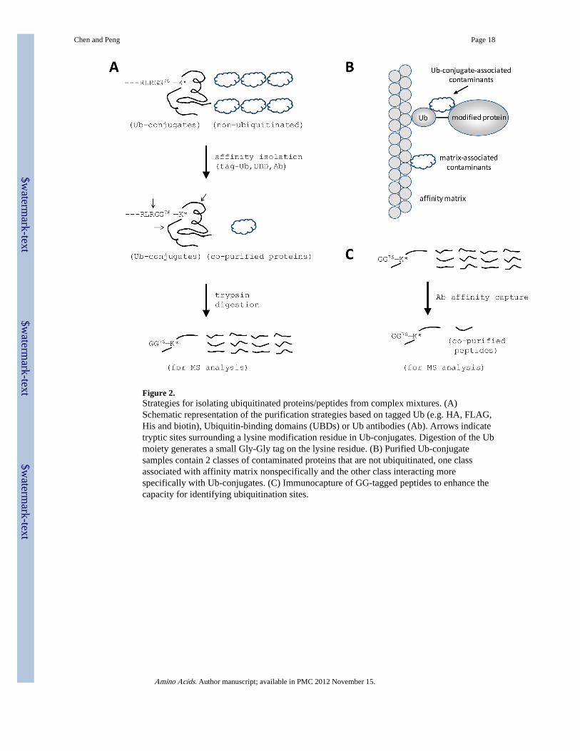
Figure 2.Strategies for isolating ubiquitinated proteins/peptides from complex mixtures. (A)Schematic representation of the purification strategies based on tagged Ub (e.g. HA, FLAG,His and biotin), Ubiquitin-binding domains (UBDs) or Ub antibodies (Ab). Arrows indicatetryptic sites surrounding a lysine modification residue in Ub-conjugates. Digestion of the Ubmoiety generates a small Gly-Gly tag on the lysine residue. (B) Purified Ub-conjugatesamples contain 2 classes of contaminated proteins that are not ubiquitinated, one classassociated with affinity matrix nonspecifically and the other class interacting morespecifically with Ub-conjugates. (C) Immunocapture of GG-tagged peptides to enhance thecapacity for identifying ubiquitination sites.
Chen and Peng Page 18
Amino Acids. Author manuscript; available in PMC 2012 November 15.
$waterm
ark-text$w
atermark-text
$waterm
ark-text
$waterm
ark-text$w
atermark-text
$waterm
ark-text
Chen and Peng Page 19
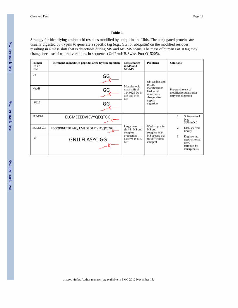
Table 1
Strategy for identifying amino acid residues modified by ubiquitin and Ubls. The conjugated proteins areusually digested by trypsin to generate a specific tag (e.g., GG for ubiquitin) on the modified residues,resulting in a mass shift that is detectable during MS and MS/MS scans. The mass of human Fat10 tag maychange because of natural variations in sequence (UniProtKB/Swiss-Prot O15205).
HumanUb orUBL
Remnant on modified peptides after trypsin digestion Mass changein MS andMS/MS
Problems Solutions
Ub
Monoisotopicmass shift of114.0429 Da inMS and MS/MS
Ub, Nedd8, andISG15modificationslead to thesame masschange aftertrypsindigestion
Pre-enrichment ofmodified proteins priortotrypsin digestion
Nedd8
ISG15
SUMO-1
Large massshift in MS andcomplexproductionpatterns in MS/MS
Weak signal inMS andcomplex MS/MS spectra thatare difficult tointerpret
1 Software tool(e.g.SUMmOn)
2 UBL spectrallibrary
3 Engineeringtryptic sites atthe C-terminus bymutagenesis
SUMO-2/3
Fat10
Amino Acids. Author manuscript; available in PMC 2012 November 15.