Ubiquitin Chains Are Remodeled at the Proteasome by Opposing Ubiquitin Ligase and Deubiquitinating...
13
Ubiquitin Chains Are Remodeled at the Proteasome by Opposing Ubiquitin Ligase and Deubiquitinating Activities Bernat Crosas, 1,2 John Hanna, 1 Donald S. Kirkpatrick, 1,3 Dan Phoebe Zhang, 1,3 Yoshiko Tone, 1 Nathaniel A. Hathaway, 1 Christa Buecker, 1 David S. Leggett, 1,4 Marion Schmidt, 1,5 Randall W. King, 1 Steven P. Gygi, 1 and Daniel Finley 1, * 1 Department of Cell Biology, Harvard Medical School, 240 Longwood Avenue, Boston, MA 02115, USA 2 Institut de Biologia Molecular de Barcelona, CSIC, Jordi Girona 18–26, Barcelona 08034, Spain 3 These authors contributed equally to this work. 4 Present address: ArQule Incorporated, 19 Presidential Way, Woburn, MA 01801, USA. 5 Present address: Department of Biochemistry, Albert Einstein College of Medicine, 1300 Morris Park Avenue, Bronx, NY 10461, USA. *Contact: daniel_fi[email protected] DOI 10.1016/j.cell.2006.09.051 SUMMARY The ubiquitin ligase Hul5 was recently identified as a component of the proteasome, a multi- subunit protease that degrades ubiquitin-pro- tein conjugates. We report here a proteasome- dependent conjugating activity of Hul5 that endows proteasomes with the capacity to ex- tend ubiquitin chains. hul5 mutants show re- duced degradation of multiple proteasome substrates in vivo, suggesting that the polyubi- quitin signal that targets substrates to the pro- teasome can be productively amplified at the proteasome. However, the products of Hul5 conjugation are subject to disassembly by a proteasome-bound deubiquitinating enzyme, Ubp6. A hul5 null mutation suppresses a ubp6 null mutation, suggesting that a balance of chain-extending and chain-trimming activities is required for proper proteasome function. As the association of Hul5 with proteasomes was found to be strongly stabilized by Ubp6, these enzymes may be situated in proximity to one another. We propose that through dy- namic remodeling of ubiquitin chains, protea- somes actively regulate substrate commitment to degradation. INTRODUCTION The ubiquitin-proteasome system mediates the break- down of most short-lived regulatory proteins, and in this way it controls many aspects of cell physiology. A central element of this system, the proteasome, is also its most complex, with over 30 distinct integral subunits (Pickart and Cohen, 2004). The proteasome degrades substrates within its core particle (CP or 20S particle), which is a bar- rel-shaped assembly of 670 kDa whose hollow interior houses multiple proteolytic sites. Protein substrates reach this interior chamber through a gated channel, guided by the regulatory particle (RP, 19S, or PA700), which is a com- plex of 1 MDa. The RP recognizes the substrate-bound ubiquitin chain, unfolds the substrate, and directs its translocation into the CP. The need for an unfolding step owes to the narrow cross-section of the channel leading to the CP (Pickart and Cohen, 2004). Critical components of the RP include ubiquitin receptors, deubiquitinating enzymes, and six ATPases that have been implicated in substrate unfolding and translocation. Recent work has shown that factors associated with the proteasome via loose, often salt-labile interactions play important roles in regulating its function (Schmidt et al., 2005). UBL-UBA proteins, for example, help to deliver specific ubiquitin conjugates to the proteasome (Verma et al., 2004; Elsasser et al., 2004; Madura, 2004). Among the most abundant proteasome-associated proteins of S. cerevisiae are two enzymatic factors: Ubp6, a deubiqui- tinating enzyme, and Hul5, a ubiquitin-protein ligase (Leg- gett et al., 2002). Ubp6 is one of two deubiquitinating en- zymes within the proteasome; in contrast, a much larger number of ubiquitin ligases (nine) have been suggested to interact with the proteasome, including Ubr1, Ufd4, the APC, and the SCF (Xie and Varshavsky, 2000, 2002; Verma et al., 2000; You and Pickart, 2001; Leggett et al., 2002; Seeger et al., 2003; reviewed in Schmidt et al., 2005). However, Hul5 is the only ligase observed on pro- teasomes in amounts high enough to be visible by con- ventional protein staining (Leggett et al., 2002). Hul5 is evolutionarily conserved, and its likely mammalian ortho- log, KIAA10, also associates with proteasomes (You and Pickart, 2001; Wang and Pickart, 2005). Cell 127, 1401–1413, December 29, 2006 ª2006 Elsevier Inc. 1401
Transcript of Ubiquitin Chains Are Remodeled at the Proteasome by Opposing Ubiquitin Ligase and Deubiquitinating...
Ubiquitin Chains Are Remodeled at theProteasome by Opposing UbiquitinLigase and Deubiquitinating ActivitiesBernat Crosas,1,2 John Hanna,1 Donald S. Kirkpatrick,1,3 Dan Phoebe Zhang,1,3 Yoshiko Tone,1
Nathaniel A. Hathaway,1 Christa Buecker,1 David S. Leggett,1,4 Marion Schmidt,1,5 Randall W. King,1
Steven P. Gygi,1 and Daniel Finley1,*1Department of Cell Biology, Harvard Medical School, 240 Longwood Avenue, Boston, MA 02115, USA2 Institut de Biologia Molecular de Barcelona, CSIC, Jordi Girona 18–26, Barcelona 08034, Spain3These authors contributed equally to this work.4Present address: ArQule Incorporated, 19 Presidential Way, Woburn, MA 01801, USA.5Present address: Department of Biochemistry, Albert Einstein College of Medicine, 1300 Morris Park Avenue, Bronx,NY 10461, USA.*Contact: [email protected] 10.1016/j.cell.2006.09.051
SUMMARY
The ubiquitin ligase Hul5 was recently identifiedas a component of the proteasome, a multi-subunit protease that degrades ubiquitin-pro-tein conjugates. We report here a proteasome-dependent conjugating activity of Hul5 thatendows proteasomes with the capacity to ex-tend ubiquitin chains. hul5 mutants show re-duced degradation of multiple proteasomesubstrates in vivo, suggesting that the polyubi-quitin signal that targets substrates to the pro-teasome can be productively amplified at theproteasome. However, the products of Hul5conjugation are subject to disassembly by aproteasome-bound deubiquitinating enzyme,Ubp6. A hul5 null mutation suppresses a ubp6null mutation, suggesting that a balance ofchain-extending and chain-trimming activitiesis required for proper proteasome function.As the association of Hul5 with proteasomeswas found to be strongly stabilized by Ubp6,these enzymes may be situated in proximityto one another. We propose that through dy-namic remodeling of ubiquitin chains, protea-somes actively regulate substrate commitmentto degradation.
INTRODUCTION
The ubiquitin-proteasome system mediates the break-down of most short-lived regulatory proteins, and in thisway it controls many aspects of cell physiology. A centralelement of this system, the proteasome, is also its most
complex, with over 30 distinct integral subunits (Pickartand Cohen, 2004). The proteasome degrades substrateswithin its core particle (CP or 20S particle), which is a bar-rel-shaped assembly of 670 kDa whose hollow interiorhouses multiple proteolytic sites. Protein substrates reachthis interior chamber through a gated channel, guided bythe regulatory particle (RP, 19S, or PA700), which is a com-plex of !1 MDa. The RP recognizes the substrate-boundubiquitin chain, unfolds the substrate, and directs itstranslocation into the CP. The need for an unfolding stepowes to the narrow cross-section of the channel leadingto the CP (Pickart and Cohen, 2004). Critical componentsof the RP include ubiquitin receptors, deubiquitinatingenzymes, and six ATPases that have been implicated insubstrate unfolding and translocation.
Recent work has shown that factors associated with theproteasome via loose, often salt-labile interactions playimportant roles in regulating its function (Schmidt et al.,2005). UBL-UBA proteins, for example, help to deliverspecific ubiquitin conjugates to the proteasome (Vermaet al., 2004; Elsasser et al., 2004; Madura, 2004). Amongthe most abundant proteasome-associated proteins ofS. cerevisiae are two enzymatic factors: Ubp6, a deubiqui-tinating enzyme, and Hul5, a ubiquitin-protein ligase (Leg-gett et al., 2002). Ubp6 is one of two deubiquitinating en-zymes within the proteasome; in contrast, a much largernumber of ubiquitin ligases (nine) have been suggestedto interact with the proteasome, including Ubr1, Ufd4,the APC, and the SCF (Xie and Varshavsky, 2000, 2002;Verma et al., 2000; You and Pickart, 2001; Leggett et al.,2002; Seeger et al., 2003; reviewed in Schmidt et al.,2005). However, Hul5 is the only ligase observed on pro-teasomes in amounts high enough to be visible by con-ventional protein staining (Leggett et al., 2002). Hul5 isevolutionarily conserved, and its likely mammalian ortho-log, KIAA10, also associates with proteasomes (You andPickart, 2001; Wang and Pickart, 2005).
Cell 127, 1401–1413, December 29, 2006 ª2006 Elsevier Inc. 1401
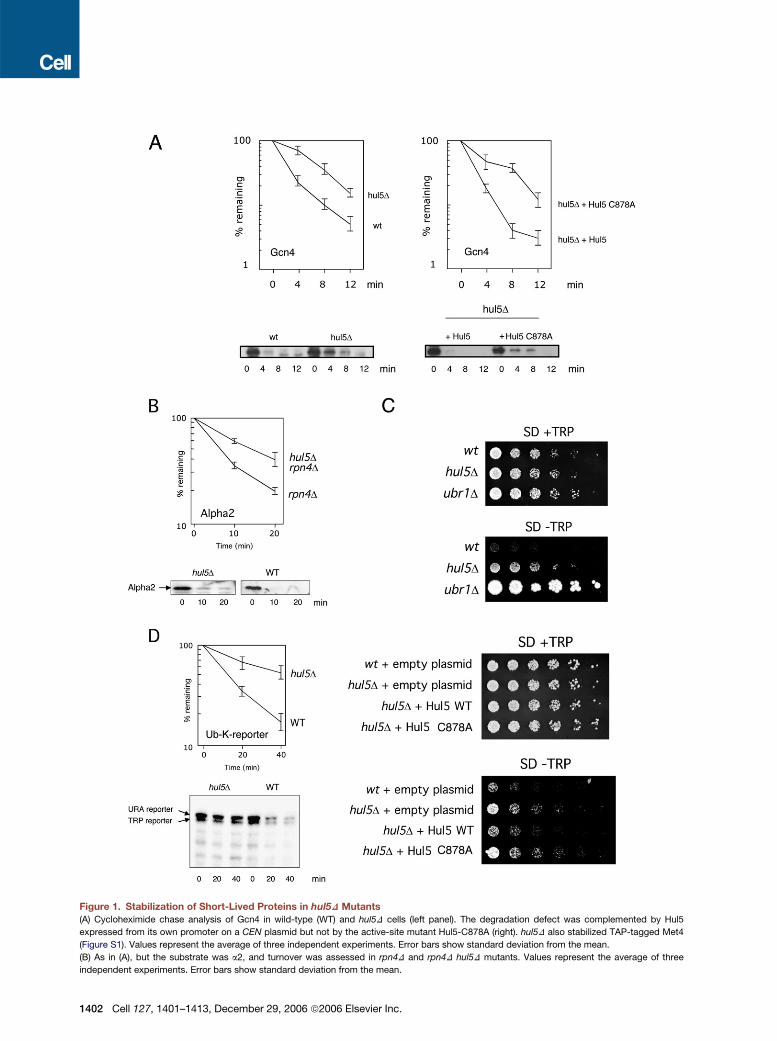
Figure 1. Stabilization of Short-Lived Proteins in hul5D Mutants(A) Cycloheximide chase analysis of Gcn4 in wild-type (WT) and hul5D cells (left panel). The degradation defect was complemented by Hul5
expressed from its own promoter on a CEN plasmid but not by the active-site mutant Hul5-C878A (right). hul5D also stabilized TAP-tagged Met4
(Figure S1). Values represent the average of three independent experiments. Error bars show standard deviation from the mean.
(B) As in (A), but the substrate was a2, and turnover was assessed in rpn4D and rpn4D hul5D mutants. Values represent the average of three
independent experiments. Error bars show standard deviation from the mean.
1402 Cell 127, 1401–1413, December 29, 2006 ª2006 Elsevier Inc.

In this study we report genetic and biochemical charac-terization of Hul5. hul5 null mutants are deficient in proteindegradation, but, unlike typical ubiquitin ligase mutants,they do not show highly substrate-specific defects in deg-radation. Rather, the hul5 phenotype more closely resem-bles that of a partial proteasome loss-of-function mutant.In vitro, Hul5 endows proteasomeswith a potent ubiquitin-ligase activity, which can extend unanchored multiubiqui-tin chains or ubiquitin chains bound to a proteolytic sub-strate such as cyclin B. Ubiquitin chain extension byHul5 is strongly stimulated by proteasomes, suggestingthat proteasomes are a principal site of ubiquitination byHul5. Ubiquitin groups added to proteasome-bound tar-get proteins by Hul5 can be efficiently removed byUbp6. These and other observations imply that Hul5and Ubp6 function in opposition with one another. Wesuggest that by maintaining and extending ubiquitinchains on proteasome-bound substrates, Hul5 stabilizesproteasome-substrate interactions and promotes proteinbreakdown.
RESULTS
hul5 Mutants Exhibit Reduced Proteolytic CapacityTo test for a role of Hul5 in proteasome function, we exam-ined the effects of deleting the HUL5 gene on the rates ofturnover of several short-lived proteins. Substantial stabi-lization of the transcriptional activator Gcn4 was observedin hul5Dmutants (Figure 1A). This effect could be comple-mented by a HUL5 gene carried on a low copy-numberCEN plasmid. The HECT-domain ligases, of which Hul5is a member, form covalent thiolester adducts with ubiqui-tin as a required step in conjugation. When the adduct-forming cysteine 878 of Hul5 was substituted with alanine,complementation was no longer observed in the Gcn4degradation assay (Figure 1A).In contrast to Gcn4, the transcriptional repressor a2
was not noticeably stabilized in the hul5D mutant (datanot shown). However, defects in protein degradation canbe compensated for by upregulation of proteasome levels(London et al., 2004; Ju et al., 2004). Rpn4 is a transcriptionfactor that is degraded by the proteasome and that acti-vates transcription of proteasome subunit genes (Xieand Varshavsky, 2001; Leggett et al., 2002; Ju et al.,2004). rpn4D mutants show reduced proteasome levelsand an inability to upregulate proteasome subunit synthe-sis when the ubiquitin-proteasome system is challenged.In strains lacking Rpn4, we found that turnover of a2was significantly affected by deletion of HUL5 (Figure 1B).It is possible that for a2, ubiquitin conjugation is normallyrate limiting fordegradation; bydeletingRPN4,proteasomefunction is attenuated and becomes partially rate limiting,
so that underlying defects in proteasome function becomemore apparent.
Targets of the N-end rule pathway are well-studiedmodel substrates of the proteasome (Varshavsky, 1996).The Trp1 protein was converted into an N-end rule sub-strate by fusing it to ubiquitin, with a lysine placed adja-cent to the ubiquitin C terminus to allow recognition bythe ubiquitin ligaseUbr1 (Hanna et al., 2006). A strain bear-ing this construct grew slowly in the absence of exoge-nous tryptophan but was Trp+ when UBR1 was deleted(Figure 1C). Hul5 appeared to promote degradation ofthe Ub-K-Trp1 fusion protein, because deletion of theHUL5 gene conferred a growth advantage to this strainin the absence of tryptophan (Figure 1C). As seen withGcn4, this function of Hul5 required its active site cysteine(Figure 1C). Cycloheximide chase analysis confirmed thatUb-K-Trp1 and a comparable reporter, Ub-K-Ura3, werestabilized by deletion of HUL5 (Figure 1D). In summary,the stabilization of a range of substrates in hul5Dmutantsimplies that Hul5 serves a general function at the protea-some. The defects in protein degradation in hul5D mu-tants appear to be more general than expected for a ubiq-uitin-protein ligase since ubiquitin ligases tend to be highlysubstrate specific.
Hul5-Dependent Ubiquitination on the ProteasomeThe results above, taken together with our earlier findingthat Hul5 associates with proteasomes (Leggett et al.,2002), suggested that Hul5 may stimulate the breakdownof multiple proteins by promoting their ubiquitination atthe proteasome. To test whether Hul5 canmediate ubiqui-tination of proteasome-bound proteins, we incubatedaffinity-purified proteasomes with biotinylated ubiquitin,ubiquitin-activating enzyme (E1), and ATP. Proteasomesconverted free ubiquitin into conjugates with surprising ef-ficiency (Figure 2A). When purified from hul5D mutants,however, proteasomes were virtually devoid of this activ-ity. Thus, although previous studies have indicated thatother ligases can bind proteasomes with some affinity,this simple assay for ligase activity is specific for Hul5. Fur-thermore, the role of Hul5 in the proteasome’s ubiquitin li-gase activity is direct since proteasomesbearing a catalyt-ically inactive formof Hul5, Hul5-C878A, were comparableto those of hul5D in their conjugation deficiency (Fig-ure 2A). The selective detection of Hul5 activity may reflectthat it is present on proteasomes at higher levels thanother ligases in our preparations (Leggett et al., 2002). Inaddition, we suggest that the assay preferentially detectsHul5 because it is unique among proteasome-associatedligases in the nature or breadth of its substrate specificity.
Proteasomes are associated with deubiquitinating ac-tivity as well as ubiquitin ligase activity. An integral subunit
(C) Effect of deleting HUL5 on growth of a strain expressing a Ub-K-Trp1 fusion protein. Plates were incubated for 3 (+Trp) or 7 ("Trp) days. As in (A),
the degradation defect was complemented by plasmid-borne HUL5 but not by hul5-C878A.
(D) As in (A), but the test substrates were Ub-K-Trp1 andUb-K-Ura3. These two bandswere quantitated together. All chase experiments were initiated
by the addition of cycloheximide to 100 mg/ml, except in (A) where 20 mg/ml was used. Values represent the average of three independent experi-
ments. Error bars show standard deviation from the mean.
Cell 127, 1401–1413, December 29, 2006 ª2006 Elsevier Inc. 1403

of the proteasome, Rpn11, removes ubiquitin chains enbloc from conjugates, apparently cutting at the sub-strate-proximal end of the chain (Verma et al., 2002; Yaoand Cohen, 2002). A second proteasomal deubiquitinat-ing enzyme is Ubp6, a proteasome-associated protein(Verma et al., 2000; Leggett et al., 2002) that progressivelytrims chains (Hanna et al., 2006).While cleavage by Rpn11is coupled to substrate degradation (Verma et al., 2002;Yao and Cohen, 2002), the activity of Ubp6 is not; on thecontrary Ubp6 can inhibit degradation (Hanna et al., 2006).
When the conjugate-synthesis assay was carried outusing ubp6D proteasomes, both the yield of conjugatesand their average molecular weight increased significantly(Figure 2A), suggesting that conjugates synthesized byHul5 can be substrates of Ubp6. To test this possibility,we prepared conjugates using ubp6D proteasomes,then quenched further conjugate synthesis by depletingATP. Upon addition of purified Ubp6, we observed nearlycomplete elimination of high molecular weight conjugatesby 30 min, while in the absence of Ubp6 these conjugatespersisted for at least 90 min (Figure 2B, right panel). Thus,conjugates formed by Hul5 on the proteasome are favor-able substrates for Ubp6.
For the model substrates shown above to be stabilizedin a hul5D mutant (Figure 1), E3 requirements have previ-ously been defined, so it is unlikely that Hul5 could serveas a major E3 for these proteins (Meimoun et al., 2000;Swanson et al., 2001; Varshavsky, 1996). If not an E3,Hul5 could function as a member of a rarer and more re-cently recognized group of ubiquitin ligases known asE4 enzymes, which are defined as specialized mediatorsof ubiquitin chain elongation (Koegl et al., 1999; Richlyet al., 2005). Thus, an E4 enzyme should not modify a pro-tein unless it has been previously ubiquitinated throughthe action of an E3. To test whether Hul5 can elongate pre-formed ubiquitin chains, we purified Hul5 (Figure S2) andassayed its ubiquitin-ligase activity using tetraubiquitinchains (Ub4) as substrate. These chains were efficiently
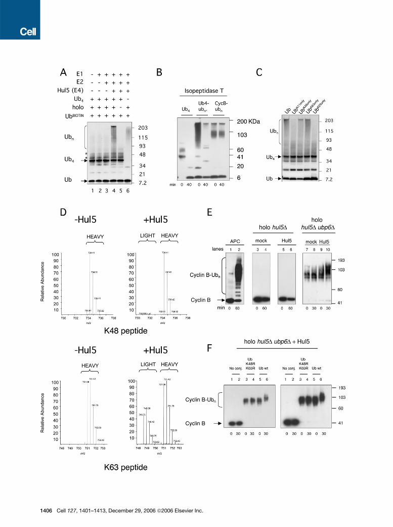
elongated in a reaction mixture that included E1, E2,Hul5, and proteasomes, but not when Hul5 was omitted(Figure 3A). Also required for this reaction was the protea-some holoenzyme. These data suggest that Hul5 canserve as an E4, and that its enzymatic activity is stimulatedby proteasomes, just as for Ubp6 (Leggett et al., 2002).The E4 model assumes that tetraubiquitin is being used
as a ubiquitin acceptor rather than a donor in these conju-gation reactions. This assumption was confirmed throughbiochemical analysis of the product conjugates. Tetraubi-quitin carries a free C-terminal carboxylate group andtherefore can, like ubiquitin, be activated by E1 and sub-sequently be donated to substrates. The ‘‘tetraubiquitindonor’’ model, an alternative to the E4 model, predictsthat the conjugates produced by Hul5 lack a ubiquitin-derived C-terminal carboxylate; that is, they are conven-tional ubiquitin-protein conjugates rather than free chains.However, isopeptidase T, which is specific for free ubiqui-tin chains, digested the Hul5 reaction products readily(Figure 3B). Thus, tetraubiquitin plays an acceptor role inthese conjugation reactions, consistent with the E4 modelin which Hul5 extends chains. The predominant donor inthese reactions appears to be free ubiquitin, which is sup-plied in excess (Figure S3).Proteins are thought to be targeted to the proteasome
principally by Lys48-linked ubiquitin chains (Pickart andCohen, 2004). However, when tested with single-lysineubiquitin derivatives, Hul5 formed chains more efficientlyon Lys63 than on Lys48 (Figure 3C). To confirm the linkagepreference of Hul5, we used mass spectrometry to mapthe linkage site on chains formedwith wild-type (WT) ubiq-uitin. This analysis revealed a strong preference for the for-mation of Lys63 linkages (Figure 3D). Thus, Hul5 has thecapacity to build chains with heterogeneous ubiquitin-ubiquitin linkages. Hul5 can build off Lys48-linked chains,but in vitro it producesmainly Lys63 linkages.Whether thisapparent chain specificity represents a functionally signif-icant parameter for Hul5 in vivo remains to be assessed.
Figure 2. Hul5-Dependent Ubiquitin-Ligase Activity of Purified Proteasomes(A) Proteasomes fromWT and mutant strains were incubated for 1 hr with biotinylated ubiquitin, E1, and ATP. UbBIOT indicates biotinylated ubiquitin,
Ub!conj indicates ubiquitin-protein conjugates, and * indicates self-ubiquitinated E1 (data not shown). Samples were analyzed by SDS-PAGE. After
blotting, ubiquitin conjugates were visualized with HRP-Streptavidin. Left and central panels show different exposures of the same blot.
(B) Ubiquitin-protein conjugation assays were performed with proteasomes purified from ubp6D and hul5D ubp6D strains as in (A). The leftmost panel
shows a time course of the conjugation reaction. The 120 min sample was ATP depleted, then incubated with or without stoichiometric amounts of
recombinant Ubp6. Time points were taken, and the samples were analyzed as in (A).
1404 Cell 127, 1401–1413, December 29, 2006 ª2006 Elsevier Inc.
The ability of Hul5 to promote protein degradation in vivosuggests that Hul5 should be able to extend ubiquitinchains on defined degradative substrates. We tested thisusing cyclin B, a highly unstable protein. Cyclin B was ex-pressed in insect cells, complexed to Cdc2, purified, andubiquitinated by the APC ligase (Kirkpatrick et al., 2006).The resulting conjugates are degraded almost to comple-tion within 5 min by ubp6D proteasomes (Hanna et al.,2006). Ubp6, when added to such proteasomes, progres-sively trims the ubiquitin chains of cyclin B. Since the APCmodifies cyclin B extensively, the conjugates do not runfar into the gel, making it difficult to detect further chain ex-tension. We therefore used Ubp6 to reduce the extent ofcyclin B ubiquitination (Figure 3E, compare lanes 2 and 7)while suppressing cyclin B degradation with the protea-some inhibitor PS341 and blocking Rpn11-mediated deu-biquitinating activity with o-phenanthroline. After 20min ofUbp6-dependent processing,Hul5wasadded, resulting inefficient addition of ubiquitin groups to the cyclin B conju-gate (Figure 3E). Importantly, Hul5 was inactive on cyclinB that had not been ubiquitinated by the APC (Figure 3E,lane 6), indicating that Hul5 does not function as an E3for cyclin B, but rather as an E4.Although Figure 3E shows that Hul5 acts preferentially
on previously ubiquitinated cyclin B, it does not showthat Hul5 elongates pre-existing ubiquitin chains on cyclinB. Alternatively, Hul5 might conjugate ubiquitin to lysineresidues of cyclin B. According to the E4 model, the ubiq-uitin chains on cyclin B should both target the conjugateto Hul5 and provide the acceptor lysine residues for fur-ther ubiquitination. The latter possibility was tested bypreparing ubiquitin cyclin B conjugates with mutant ubiq-uitin lacking Lys48 and Lys63, the two lysines that Hul5preferentially utilizes for chain extension. Unlike Hul5,the APC can utilize Lys11 in addition to Lys48 andLys63, and Lys48 is not required for in vitro degradationof ubiquitinated cyclin B by ubp6D proteasomes (Kirkpa-trick et al., 2006). Conjugates lacking Lys48- and Lys63-linked ubiquitin chains were also degraded by purifiedyeast proteasomes (ubp6D) with an efficiency close tothat of conjugates synthesized with WT ubiquitin (datanot shown). However, when Hul5 was challenged withsuch conjugates in the presence of WT free ubiquitinas a donor, it failed to catalyze significant ubiquitination(Figure 3F). These results imply that Hul5 elongates pre-formed ubiquitin chains on cyclin B, as expected of anE4 enzyme.
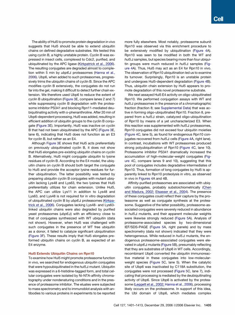
Hul5 Extends Ubiquitin Chains on Rpn10To examine howHul5might promote proteasome functionin vivo, we searched for endogenous ubiquitin conjugatesthat were hypoubiquitinated in the hul5Dmutant. Ubiquitinwas expressed in a 6-histidine-tagged form, and total cel-lular conjugates were isolated by Ni-NTA-affinity chroma-tography under nondenaturing conditions and in the pres-ence of proteasome inhibitor. The eluates were subjectedtomass spectrometry and to immunoblot analysis with an-tibodies to various proteins in experiments to be reported
more fully elsewhere. Most notably, proteasome subunitRpn10 was observed via this enrichment procedure tobe extensively modified by ubiquitination (Figure 4A).Rpn10 was seen to be modified in both the WT andhul5D samples, but species bearingmore than four ubiqui-tin groups were much reduced in hul5D samples (Fig-ure 4A). Thus, Hul5 may act as an E4 for Rpn10 in vivo.The observation of Rpn10 ubiquitination led us to examineits turnover. Surprisingly, Rpn10 is an unstable proteinand undergoes Hul5-dependent degradation (Figure 4B).Thus, ubiquitin chain extension by Hul5 appears to pro-mote degradation of this novel proteasome substrate.
We next assayed Hul5 E4 activity on oligo-ubiquitinatedRpn10. We performed conjugation assays with WT andhul5D proteasomes in the presence of a chromatographicfraction (fraction 8; see Supplemental Data) that was ac-tive in forming oligo-ubiquitinated Rpn10. Fraction 8, pre-pared from a hul5D strain, catalyzed oligo-ubiquitinationof Rpn10 by means of a yet uncharacterized E3. Whenthis reaction was supplemented with hul5D proteasomes,Rpn10 conjugates did not exceed four ubiquitin moieties(Figure 4C, lane 5), as found for endogenous Rpn10 con-jugates recovered from hul5D cells (Figure 4A, right lane).In contrast, incubations with WT proteasomes producedstrong polyubiquitination of Rpn10 (Figure 4C, lane 10).Proteasome inhibitor PS341 dramatically increased theaccumulation of high-molecular-weight conjugates (Fig-ure 4C, compare lanes 9 and 10), suggesting that thispool of conjugates includes degradative intermediates ofRpn10. Thus, formation of long conjugates by Hul5 is ap-parently linked to Rpn10 proteolysis in vitro, as observedin vivo in Figures 4A and 4B.
Affinity-purified proteasomes are associated with ubiq-uitin conjugates, probably substoichiometrically (Chenand Madura, 2002; Elsasser et al., 2004). The presenceof these conjugates could reflect their capture by the pro-teasome as well as conjugate synthesis at the protea-some. Suggestive of the latter possibility, proteasome-as-sociated conjugates were severely reduced in abundancein hul5D mutants, and their apparent molecular weightswere likewise strongly reduced (Figure 5A). Analysis ofproteasome-associated species by two-dimensionalIEF/SDS-PAGE (Figure 5A, right panels) and by massspectrometry (data not shown) indicated that they wereheterogeneous. While reduced in hul5D mutants, the en-dogenous proteasome-associated conjugates were ele-vated in ubp6Dmutants (Figure 5B), presumably reflectingthat they are substrates of Ubp6 in WT cells. Accordingly,recombinant Ubp6 converted the ubiquitin immunoreac-tive material in these conjugates into low-molecular-weight species (Figure 5C, lane 5). When the catalyticsite of Ubp6 was inactivated by C118A substitution, theconjugates were not processed (Figure 5C, lane 7), indi-cating that processing is mediated by the deubiquitinatingactivity of Ubp6. Since Ubp6 is activated by the protea-some (Leggett et al., 2002; Hanna et al., 2006), processinglikely occurs on the proteasome. In support of this idea,the Ubl domain of Ubp6, which mediates complex
Cell 127, 1401–1413, December 29, 2006 ª2006 Elsevier Inc. 1405
1406 Cell 127, 1401–1413, December 29, 2006 ª2006 Elsevier Inc.

formation with the proteasome, was also required for thisreaction (Figure 5C, lane 6).Interestingly, in cells lacking Hul5, deletion of UBP6 did
not enhance the levels of proteasome-associated ubiqui-tin conjugates (Figure 5B), suggesting that the disassem-bly of conjugates synthesized by Hul5 is an important roleof Ubp6. We suggest that Hul5 mediates the formation ofthese proteasome-bound conjugates and that addition ofUbp6 to the isolated complex disassembles the endoge-nous conjugates as seen with conjugates synthesized byHul5 in vitro (Figure 2B). Moreover, the endogenous con-jugates observed in Figure 5may serve as the ubiquitin ac-ceptors in the in vitro experiments of Figure 2A. If true, thiswould imply that Hul5 serves to enhance the recovery onproteasomes of ubiquitin conjugates that it can furtherubiquitinate in vitro and that in this assay the products ofHul5 conjugation are themselves the major substrates ofHul5.
Hul5 and Ubp6 Function in OppositionThe analyses of endogenous and in vitro synthesizedubiquitin conjugates in Figures 2–5 suggest that ubiquitinchains bound to the proteasome exist in a dynamic state,with both elongation and attrition of chains occurring. Theprincipal mediators of these reactions appear to be Hul5and Ubp6. These observations led us to test for functionalopposition between these enzymes in vivo. Figure 6Ashows that deletion of HUL5 results in substantial sup-pression of the previously reported (Amerik et al., 2000;Leggett et al., 2002) canavanine sensitivity of ubp6D mu-tants. This suggests that the phenotypic defect in ubp6Dmutants is partly attributable to the presence of Hul5, inthat Hul5 activity is unopposed by Ubp6 in themutant. An-other ubiquitin ligase known to bind proteasomes, the E3enzyme Ufd4 (Xie and Varshavsky, 2000, 2002), was inac-tive in this suppression assay (Figure 6A). It is currently un-clear whether the suppressive effect of HUL5 deletion is
mediated through increased ubiquitin levels or some othermechanism.
Deletion of the gene encoding Ubc4, an E2 enzyme,was found to suppress ubp6D to approximately thesame extent as HUL5, possibly because Ubc4 collabo-rates with Hul5 in ubiquitin conjugation (Figure 6A). Nullmutations in genes encoding six other E2 enzymes failedto suppress, indicating that the assay is specific (Fig-ure 6A). Ubc4 is involved in the degradation of short-livedproteins (Seufert and Jentsch, 1990), and significantly it is,like Hul5, found in association with the proteasome (Ton-gaonkar et al., 2000; Chuang and Madura, 2005). More-over, our in vitro ubiquitin conjugation assays show thatHul5 and Ubc4 productively interact (Figure 3A). There-fore, Ubc4 is a good candidate for the physiological part-ner of Hul5. The ability of hul5D and ubc4D mutations toenhance growth of ubp6D mutants in the presence ofcanavanine is striking, because both hul5D and ubc4Dmutants exhibit canavanine sensitivity in other contexts(Seufert and Jentsch, 1990; data not shown).
The specific relationship between Hul5 and Ubp6 wasalso apparent from compositional analysis of protea-somes isolated from the corresponding null mutants. Un-der relatively stringent conditions of proteasome prepara-tion, the recovery of Hul5 on ubp6D proteasomes wassignificantly reduced (Figure 6B). However, it was unclearhow hul5D could suppress ubp6D if Hul5 were not boundto proteasomes in vivo in the ubp6Dmutant. We thereforeisolated proteasomes again from ubp6D mutants, but inthe absence of salt. These proteasomes were immobilizedon beads, and the column was step-eluted with NaCl in25 mM increments. Hul5 dissociated from WT protea-some primarily at 175–200 mM NaCl but from the ubp6Dproteasome at 50–75 mM (Figure 6C). Substitution of theactive site cysteine of Ubp6 had little or no effect on thesalt dependence of theHul5-proteasome association (Fig-ure 6C). Thus, the presence of Ubp6, whether in an active
Figure 3. Ubiquitin Chain-Extending Activity of Hul5(A) Hul5 can extend unanchored multiubiquitin chains in a proteasome-dependent reaction. Ubiquitin-conjugation assays were performed in reac-
tions supplied with ubiquitin, E1, E2 (UbcH5), Hul5, proteasome holoenzyme (holo), and tetraubiquitin (Ub4) as shown. Reaction products were an-
alyzed by SDS-PAGE and antiubiquitin immunoblotting. Proteasomes were derived from a hul5D ubp6D strain and had been previously incubated
with Ubp6 to reduce antiubiquitin immunoreactive material (see Supplemental Data). The two asterisked bands just above Ub4 are minor contami-
nants present in the input Ub4, presumably Ub5 and Ub6 (data not shown).
(B) Tetraubiquitin (1 mg), tetra-Ub (1 mg) extended by Hul5 as in (A), and APC-generated cyclin B conjugates were incubated with isopeptidase T (0.1
mg) for 40 min at 30#C. Cyclin B-ubiquitin conjugates, a negative control, are not expected to be digested with isopeptidase T.
(C) As in (A), but the added free ubiquitin contained in each case only a single lysine residue, as indicated, to support chain formation.
(D) Full MS scan spectra corresponding to either the K48 and K63 -GG signature peptides as measured in high-molecular-mass gel regions from
complete reactions with or without addition of Hul5 (lanes 3–4 from [A]). The m/z range shown includes both the digested native peptide as well
as its corresponding isotopically labeled internal standard peptide. The spectra shown represent an average of all MS spectra collected during
peak elution.
(E) E4 activity of Hul5 on cyclin B conjugates. Cyclin B conjugates were formed by the APC in 1 hr incubations (lanes 1 and 2). Conjugation assays
performed with Hul5 and proteasomes but without prior APC treatment did not show detectable ubiquitination (lanes 3–6). Cyclin B conjugates were
partially trimmed by adding purified Ubp6 then incubating for 20 min. Ubp6 was then inhibited by adding ubiquitin-aldehyde. Hul5-dependent ubiq-
uitination was observed on cyclin B conjugates partially trimmed by Ubp6 (lanes 7–10). Proteasomes were incubated with 10 mM PS341 and 10 mM
o-phenanthroline prior to reactions, and conjugation reactionswere suppliedwith E1 andUbc4.Mock reactionswere supplementedwith BSA instead
of Hul5. Here and in (F), conjugates were visualized with antibody to cyclin B (Kirkpatrick et al., 2006).
(F) Cyclin B (lanes 1 and 2) and cyclin B conjugates generated by the APC using either ubiquitin carrying K48R and K63R mutations (lanes 3 and 4) or
WT ubiquitin (lanes 5 and 6) were washed and subsequently incubated with proteasomes, Hul5, and WT ubiquitin as in (E). Different exposures of the
same gel are shown.
Cell 127, 1401–1413, December 29, 2006 ª2006 Elsevier Inc. 1407

or an inactive form, stabilizes the association of Hul5 withproteasomes. A possible consequence of this would bethat Hul5 partitions predominantly onto Ubp6-containingproteasomes in vivo. However, Ubp6 is not essential forrecognition of Hul5 by proteasomes. Therefore, to betterunderstand the association of Hul5 with proteasomes,we attempted to identify the primary Hul5 receptor.
We first examined subassemblies of the proteasome,which are stable and can be purified in large amounts.These consisted of the 28-subunit CP, the 19-subunit
RP, and the two complexes that make up the RP: thenine-subunit base, which is proximal to the CP, and thenine-subunit lid, which is distal to the CP (Glickmanet al., 1998; Schmidt et al., 2005). Hul5 was recognizedby the RP and its constituent, the base, but it was not rec-ognized by the CP or the lid (Figure 7A). We also testedeight proteasome subunits individually for Hul5 binding,and among these Rpn2 and Rpn10 were positive(Figure 7B). However, Rpn2 bound Hul5 in an approxi-mately stoichiometric complex, while Rpn10 capturedHul5 inefficiently, according to Coomassie blue stainingintensities (Figure 7C). Rpn10may not be a physiologicallysignificant mediator of Hul5 binding because protea-somes isolated from rpn10D strains have WT levels ofHul5 (Figure 7D), whereas the assignment of Rpn2 asthe physiological Hul5-binding subunit is supported byHul50s binding to the base, which contains Rpn2. In sum-mary, these data suggest that Rpn2 is the principal pro-teasomal receptor for Hul5. Rpn2 and the related
Figure 4. Hul5 Promotes Rpn10 Ubiquitination and Turnover(A) Ubiquitination of endogenous Rpn10. Purification of whole-cell
6HIS-ubiquitin-protein conjugates under nondenaturing conditions,
from SJR125 (WT) and SBC9 (hul5D) strains (see Supplemental Data-
for details). Conjugates were eluted with imidazole and analyzed by
SDS-PAGE and anti-Rpn10 immunoblotting.
(B) Cycloheximide chase analysis of Rpn10 in WT and hul5D cells.
Rpn5 was used as a loading control. Values represent the average of
three independent experiments. Error bars show standard deviation
from the mean.
(C) In vitro analysis of Hul5-dependent Rpn10 ubiquitination. WT and
hul5D proteasomes were incubated with ATP, 6HIS-ubiquitin, PS-
341, and hul5D extract (fraction 8) containing Rpn10 oligo-ubiquitinat-
ing activity. The lower panel shows a longer exposure.
Figure 5. Antagonistic Effects of Hul5 and Ubp6 on theRecovery of Endogenous Conjugates with Affinity-PurifiedProteasomes(A) Proteasomes (2 mg) from WT and hul5D strains were analyzed by
SDS-PAGE (left panel) or by two-dimensional isoelectric focusing/
SDS-PAGE followed by antiubiquitin immunoblotting (center and right
panels). Rpt1 was used as a loading control. Ub!conj indicates ubiq-
uitin-protein conjugates.
(B) Proteasomes from WT and mutant strains were analyzed by SDS-
PAGE and antiubiquitin immunoblotting.
(C) WT or ubp6D proteasomes (12 nM) were incubated with ATP (1
mM) and a 4-fold molar excess of the indicated recombinant Ubp6
construct. Reactions were stopped by the addition of 53 SDS-loading
buffer and analyzed by SDS-PAGE and immunoblotting. Note that
a second deubiquitinating enzyme, Rpn11, appears to be largely inac-
tive against the ubiquitin immunoreactive material.
1408 Cell 127, 1401–1413, December 29, 2006 ª2006 Elsevier Inc.

proteasome subunit Rpn1, which is apparently the majorproteasomal binding site for Ubp6 (Leggett et al., 2002;Stone et al., 2004), may together provide a scaffold thatbrings Hul5 and Ubp6 into proximity.
DISCUSSION
A diverse set of reversibly bound proteins participates inand regulates the function of the proteasome. Affinity pu-rification, mass spectrometry, and other methods haveallowed for the identification of hundreds of proteasome-associated proteins (see for example Verma et al., 2000;Cagney et al., 2001; Leggett et al., 2002; Guerrero et al.,2006). These proteins remain poorly characterized onthe whole and probably represent a mixture of protea-some cofactors, ubiquitin-protein conjugates, and non-
specifically associated proteins. However, only a few pro-teins are associated with proteasomes in high amounts(Verma et al., 2000; Leggett et al., 2002), and each of thesetested so far modulates proteasome function and asso-ciates with proteasomes in both yeast and mammals(reviewed by Schmidt et al., 2005).
Among the abundant proteasome-associated proteinsis a ubiquitin ligase, Hul5. The ligase activity of Hul5 hasthus far not shown discriminatory capacity for the accep-tor protein of the ubiquitin conjugate that it takes as a sub-strate. Hul5 is active on bulk protein associated with theproteasome, on synthetic tetraubiquitin chains, on ubiqui-tin-Rpn10 conjugates, and on ubiquitin-cyclin B conju-gates. The relaxed in vitro specificity of Hul5 parallels itseffects on protein turnover in vivo, where it was seen tostabilize a variety of substrates. Since all substrates exam-ined are ubiquitinated by known E3 enzymes, it is difficultto envisage Hul5 functioning as an E3 for these degrada-tive events. In contrast to E3 enzymes, E4 enzymes arespecialized in the elongation of ubiquitin chains and maycharacteristically lack the capacity to recognize specificdeterminants in the target protein (Koegl et al., 1999;Richly et al., 2005). Instead, our data and those of Richlyet al. (2005) suggest that their specificity of action maybe determined principally by the E4’s association witha particular protein complex. The E4-like activity of Hul5is indicated not only by its specificity for ubiquitinatedforms of cyclin B but also by its ability to elongate tetrau-biquitin chains. These engineered chains are not attached
Figure 6. Genetic andBiochemical Interactions betweenHul5and Ubp6(A) Cells were spotted onto selective plates with or without 0.8 mg/ml
canavanine sulfate and grown at 30#C for 3 days (above). ubc (E2)
and ufd4 (E3) mutants were also screened for suppression of
ubp6D-associated canavanine sensitivity.
(B) Proteasomes fromWT andmutant strains were extensively washed
(500 bed vol.) prior to elution. Equal protein amounts were analyzed by
SDS-PAGE and immunoblotting.
(C) Proteasomes from WT, ubp6D, and ubp6"C118A mutants were
bound to resin and washed with a NaCl gradient. Washes were col-
lected and analyzed by SDS-PAGE and immunoblotting.
Figure 7. Proteasome Subunit Rpn2 Binds Hul5(A) Immobilized RP, CP, base (B), and lid (L) were prepared as de-
scribed (Leggett et al., 2002). A control sample (C) was from a con-
genic, untagged strain. Hul5 was bound to resin, and, after washing,
protein remaining bound to the column was analyzed by SDS-PAGE
and Hul5 immunoblotting. The leftmost lane represents 5% of input
Hul5.
(B) GST fusions of proteasome subunits and a GST control (C) were
bound to glutathione Sepharose and tested for Hul5 binding. Numeri-
cal lane designations indicate the identity of the Rpn subunit tested:
Rpn1, Rpn2, Rpn10, and so forth. Beads were treated as in (A). Equal
amounts of beads were analyzed by SDS-PAGE and immunoblotting.
(C) GST-Rpn2 and GST-Rpn10 beads were loaded with Hul5. Proteins
were then eluted with glutathione and visualized by Coomassie stain-
ing after SDS-PAGE.
(D) Equal amounts of ProA-TEV-Rpt1-tagged proteasomes, affinity-
purified from WT and rpn10D mutants, were analyzed by SDS-PAGE
and immunoblotting.
Cell 127, 1401–1413, December 29, 2006 ª2006 Elsevier Inc. 1409
to a target protein, so an E3mode of conjugate recognitionis not possible in this case.
A Hul5 substrate of particular interest is Rpn10, a pro-teasomal ubiquitin receptor. Rpn10 is the first proteasomesubunit to be identified as a proteasome substrate and asa major target of ubiquitination, although it should benoted that a nonproteasomal pool of Rpn10 exists (vanNocker et al., 1996). Hul5 appears to function as an E4for Rpn10 because its presence is not required for thegeneration of mono- or diubiquitinated forms of Rpn10but is required to form extensively modified species ofRpn10 in vivo and in vitro. Many ubiquitin receptors areubiquitinated (Kumar et al., 1999; Shih et al., 2003; Di Fioreet al., 2003; Hoeller et al., 2006), and these modificationsare thought to be regulatory in nature. Conjugated ubiqui-tin often blocks receptor function, apparently by occupy-ing the ubiquitin-binding site of the receptor. In contrast,chain extension by Hul5 appears to be critical for efficientRpn10 degradation in vivo and in vitro. An interestingquestion is why ubiquitination targets Rpn10 for degrada-tion, given that Rpn10 can associate with the proteasomein the absence of this modification.
We have thus far not found Hul5 to fully stabilize anysubstrate in vivo under standard growth conditions. Theseobservations seem consistent with our in vitro data; sinceHul5 does not seem to function efficiently unless the con-jugate is already bound to the proteasome, Hul5 may beinherently unable to promote the initial recognition ofa substrate by the proteasome. These properties of Hul5would predict a facilitative rather than obligatory role indegradation. If Hul5 is, as we propose, irrelevant to the ini-tial recognition event, it could promote substrate turnoverby slowing dissociation of the substrate from the protea-some, thus increasing the probability of degradation asthe outcome of a particular substrate-proteasome-bind-ing event.
Opposing Activities of Hul5 and Ubp6Our data suggest that the chain-elongating activity of Hul5functions specifically in opposition to deubiquitination byUbp6. That is, Hul5 serves not simply to elongate chainsbut to preserve chains against attrition by Ubp6. Althoughopposing pairs of ligases and deubiquitinating enzymesare probably rare, several have been described (Wertzet al., 2004; Rumpf and Jentsch, 2006; Kee et al., 2005;and references therein). Hul5 and Ubp6 appear to providethe first example of an E4 and a deubiquitinating enzymethat reside simultaneously on the same protein complex.
We recently found that Ubp6 can, for at least some sub-strates, act noncatalytically to inhibit the proteasome(Hanna et al., 2006). The degradation delay resultingfrom this activity of Ubp6 may be significant for the func-tion of Hul5 because it provides a window of time neces-sary for refashioning of ubiquitin chains by Hul5 and Ubp6.When Ubp6 inhibits the proteasome, it also prevents chainremoval by Rpn11, directly or indirectly. Thus, Ubp6 op-poses Rpn11 via its noncatalytic activity and Hul5 via itscatalytic activity.
The chain-extending activity of Hul5 may promote deg-radation of a protein by producing a longer chain thatbinds the proteasome more tightly. The greater effective-ness of long chains in targeting degradation (Throweret al., 2000) may reflect their ability to engage multipleproteasomal ubiquitin receptors. A second possible viewis that Hul50s chain-extending activity rarely affects netchain growth but serves to prevent Ubp6 from shorteningthe chain to such an extent that it can no longer tether thesubstrate to the proteasome. In either scenario, the com-bined effect of Hul5 and Ubp6 in regulating the lengths ofchains on proteasome-bound conjugates may functionprincipally to modulate the dissociation rate of substratefrom the proteasome (Varshavsky, 1996) and, as a conse-quence, the probability of the substrate’s degradation.ATP-dependent proteases from eukaryotes and pro-
karyotes share many properties but differ in that only eu-karyotic systems show ubiquitin dependence. An impor-tant aspect of this difference that, to our knowledge,has not been commented upon is that the reversibility ofthe ubiquitin targeting signal allows eukaryotic protea-somes in particular to play an active role in modulatingthe strength of the targeting signal. Our studies suggestthat Hul5 and Ubp6 are key factors that provide thiscapacity.
A Scaffold on the ProteasomeMay Organize Factorsthat Act on Ubiquitin ChainsMany of the proteins that mediate the dynamics of ubiqui-tin chains on the proteasome are not true proteasomesubunits but are weakly associated factors. This appliesto Hul5, Ubp6, and Ubl-Uba proteins, such as Rad23,that are major mediators of ubiquitin conjugate deliveryto the proteasome. Thus, chain delivery, elongation, andattrition are all mediated at least in part through pro-teasome-associated proteins. Interestingly, proteasomesubunits Rpn1 and Rpn2 appear to serve together asa scaffold that organizes these factors. Rpn1 is a receptorfor both Rad23 and Ubp6 (Elsasser et al., 2002; Leggettet al., 2002; Saeki et al., 2002; Seeger et al., 2003; Elsasseret al., 2004; Stone et al., 2004), whereas Rpn2 binds Hul5avidly (Figure 7). In addition, both Rpn1 and Rpn2 cross-link to Rad23 (Saeki et al., 2002), suggesting that Rpn1and Rpn2 are close neighbors. Rpn1 and Rpn2 are bothcomponents of the base, have similar structural motifsand size (Kajava, 2002), may bind one another (Gorbeaet al., 2000), and are thought to have evolved from a com-mon ancestor. Thus we propose that Rpn1 and Rpn2 im-pose a close alignment of Rad23, Ubp6, and Hul5 on theproteasome. The stabilization of Hul5 binding to the pro-teasome by Ubp6 supports this view. The detailed organi-zation of these factors may be significant since both Hul5and Ubp6 appear to work efficiently only in the context ofthe proteasome (Leggett et al., 2002; Hanna et al., 2006;this work). This may be due in part to the proteasome play-ing a role in the presentation of ubiquitin chains to theseenzymes.
1410 Cell 127, 1401–1413, December 29, 2006 ª2006 Elsevier Inc.

Convergence of Cdc48 and the ProteasomeCdc48, also known as p97 and VCP, is a homohexamericATPase ring complex. It falls into the AAA family of AT-Pases, which also includes the Rpt subunits of the protea-some. Interestingly, many AAA proteins exhibit protein-unfolding activity. Recent studies have revealed thatCdc48 is an intricate assembly for the processing of ubiq-uitin-protein conjugates (Richly et al., 2005; Rumpf andJentsch, 2006; reviewed in Elsasser and Finley, 2005).Some proteins that are ubiquitinated and destined fordegradation are shuttled first to Cdc48, where their ubiq-uitin chains are extended to a length required for asso-ciation with the proteasome (Richly et al., 2005). Theseremarkable activities of Cdc48 are not intrinsic to itsATPase rings but reflect the properties of associated co-factors: ubiquitin receptors such as Ubx-Uba proteins,an E4 enzyme known as Ufd2, and the Otu1 deubiquitinat-ing enzyme (Rumpf and Jentsch, 2006). Based in part onthe present results, it can be seen that the ensemble ofCdc48 cofactors is deeply similar to that of the protea-some. Thus, Ubx-Uba proteins are cognates of Ubl-Ubaproteins, Otu1 is a cognate of Ubp6, andUfd2 is a cognateof Hul5. This represents a dramatic evolutionary conver-gence, especially because for the most part, these factorscannot have been produced by a recent evolutionary bi-furcation. For example, while Hul5 is a HECT-domainubiquitin ligase, Ufd2 is a member of the U-box family ofligases. Similarly, Ubp6 falls into the DUB family of ligases,and Otu1 falls into the OTU family. Even Cdc48 apparentlydiverged from its sister subunits in the proteasome far be-fore the eukaryotic radiation and thus prior to the evolutionof ubiquitination.Despite these similarities, key properties of these com-
plexes differ, and in particular Hul5 differs functionallyfrom Ufd2. For example, we have found that the bindingof Hul5 to proteasomes is promoted by Ubp6. In contrast,the binding of Ufd2 and Otu1 to the same Cdc48 complexis thought to be disfavored (Rumpf and Jentsch, 2006),so that Ufd2 and Otu1 presumably act on distinct conju-gates while positioned on different complexes. The Ufd2reaction, as presently understood, stereotypically takessubstrates with chains of one or two ubiquitin groupsand extends them to approximately six groups (Richlyet al., 2005). The Hul5 reaction seems to embrace agreater range of chain lengths and to be more dynamic,in that it involves a competition with chain attrition byUbp6.Although it is the final arbiter of protein degradation, the
proteasome has traditionally been viewed as having a pas-sive role. It has been assumed that the fate of individualsubstrates has effectively been determined before theirengagement by the proteasome, primarily through the ac-tivity of E3 enzymes. The discovery that Hul5 and Ubp6mediate powerful ubiquitin-chain-remodeling activitieson the proteasome suggests that this view may be toosimplistic. In a multistep pathway such as the ubiquitin-proteasome system, the capacity to control the final andonly irreversible step in protein breakdown should be ad-
vantageous. Our data suggest a new discriminatory stepin protein breakdown, in which substrates are activelyscrutinized by the proteasome.
EXPERIMENTAL PROCEDURES
Yeast Strains and Media
Media are described in the Supplemental Data. Strain genotypes are
given in Table S1.
Purification of Hul5, Proteasomes, and Proteasome
Subcomplexes
Hul5 was purified using Hul5-TEV-ProA (SBC8) following the proce-
dure described for proteasome purification (Leggett et al., 2002), ex-
cept that binding and washing buffers were 50 mM Tris-HCl (pH 7.5),
1 mM EDTA, and 300 mM NaCl. Procedures for proteasome purifica-
tion are given in the Supplemental Data.
Assays of Ubiquitin-Protein Ligase Activity
Incubations were performed in 25 mM Tris-HCl (pH 7.5), 1 mM DTT,
0.5 mM EDTA, 5 mM MgCl2, and 5 mM ATP buffer in 100 ml reactions
at 30#C supplied with 2 mg of holoenzyme, 0.1 mg of E1, and 1 mg of ei-
ther biotinylated, 6-histidine-tagged ubiquitin or mutated ubiquitins.
Assays with WT ubiquitin contained 21 mg of this molecule per reac-
tion. Depending on the reaction, additional reagents included 0.1 mg
Hul5, 0.1 mg E2 (Ubc4 or UbcH5), 10 mMPS341, 10 mM o-phenanthro-
line, 0.1 mg tetraubiquitin, 0.5 mg ubiquitin chains, or 1 mg ubiquitin-
aldehyde. When required, ATP was depleted by adding hexokinase
(0.3 units/ml) and glucose (10 mM) for 15 min at 30#C. A description
of preincubations of holoenzyme and conjugate substrates is in the
Supplemental Data.
Protein Turnover Analysis
Cells were grown in YPD media. Logarithmically growing cells were
treated with 100 mg/ml cycloheximide, and time points were taken.
Samples were analyzed by immunoblot, using HRP-conjugated sec-
ondary antibodies and ECL detection or 125I-protein A followed by
quantitation with a PhosphorImager.
Mass Spectrometry
Gel slices were incubated with trypsin. Digest products were mixed
with isotope-labeled internal standard peptides for both K48- and
K63-linked polyubiquitin chains. A fraction of the sample correspond-
ing to 0.2 pmol of each internal standard peptide was analyzed by
LC-MS/MS using an LTQ-FT mass spectrometer. The MS spectra
shown were obtained as an average across the eluted peak.
Supplemental Data
Supplemental Data include three figures, two tables, Experimental
Procedures, and References and can be found with this article online
at http://www.cell.com/cgi/content/full/127/7/1401/DC1/.
ACKNOWLEDGMENTS
This work was supported by NIH grants GM65592 (D.F.), GM67945
(S.P.G.), and GM66492 (R.W.K.); Spanish Government (MEC) grant
RyC3582 (B.C.); and a JSPS Postdoctoral Fellowship for Research
Abroad (Y.T.). We thank Millennium Pharmaceuticals for the gift of
PS341 and R. Baker, M. Hochstrasser, R. Vierstra, D. Kornitzer, R. Li,
and C. Mann for antibodies. We also thank S. Elsasser and J. Roelofs
for yeast strains.
Received: March 8, 2006
Revised: August 25, 2006
Accepted: September 29, 2006
Published: December 28, 2006
Cell 127, 1401–1413, December 29, 2006 ª2006 Elsevier Inc. 1411
REFERENCES
Amerik, A.Y., Nowak, J., Swaminathan, S., and Hochstrasser, M.
(2000). The Doa4 deubiquitinating enzyme is functionally linked to
the vacuolar protein-sorting and endocytic pathways. Mol. Biol. Cell
11, 3365–3380.
Cagney, G., Uetz, P., and Fields, S. (2001). Two-hybrid analysis of the
Saccharomyces cerevisiae 26S proteasome. Physiol. Genomics 7,
27–34.
Chen, L., and Madura, K. (2002). Rad23 promotes the targeting of pro-
teolytic substrates to the proteasome. Mol. Cell. Biol. 22, 4902–4913.
Chuang, S.M., and Madura, K. (2005). Saccharomyces cerevisiae
Ub-conjugating enzyme Ubc4 binds the proteasome in the presence
of translationally damaged proteins. Genetics 171, 1477–1484.
Di Fiore, P.P., Polo, S., and Hofmann, K. (2003). When ubiquitin meets
ubiquitin receptors: a signalling connection. Nat. Rev. Mol. Cell Biol. 4,
491–497.
Elsasser, S., and Finley, D. (2005). Delivery of ubiquitinated substrates
to protein-unfolding machines. Nat. Cell Biol. 7, 742–749.
Elsasser, S., Gali, R.R., Schwickart, M., Larsen, C.N., Leggett, D.S.,
Muller, B., Feng, M.T., Tubing, F., Dittmar, G.A.G., and Finley, D.
(2002). Proteasome subunit Rpn1 binds ubiquitin-like protein domains.
Nat. Cell Biol. 4, 725–730.
Elsasser, S., Chandler-Militello, D., Mueller, B., Hanna, J., and Finley,
D. (2004). Rad23 and Rpn10 serve as alternative ubiquitin receptors
for the proteasome. J. Biol. Chem. 279, 26817–26822.
Glickman, M.H., Rubin, D.M., Coux, O., Wefes, I., Pfeifer, G., Cjeka, Z.,
Baumeister, W., Fried, V.A., and Finley, D. (1998). A subcomplex of the
proteasome regulatory particle required for ubiquitin-conjugate degra-
dation and related to the COP9-signalosome and eIF3. Cell 94, 615–
623.
Gorbea, C., Taillandier, D., and Rechsteiner, M. (2000). Mapping sub-
unit contacts in the regulatory complex of the 26S proteasome. J. Biol.
Chem. 275, 875–882.
Guerrero, C., Tagwerker, C., Kaiser, P., and Huang, L. (2006). An inte-
grated mass spectrometry-based proteomic approach: quantitative
analysis of tandem affinity-purified in vivo cross-linked protein com-
plexes (QTAX) to decipher the 26 S proteasome-interacting network.
Mol. Cell. Proteomics 5, 366–378.
Hanna, J., Hathaway, N.A., Tone, Y., Crosas, B., Elsasser, S., Kirkpa-
trick, D.S., Leggett, D.S., Gygi, S.P., King, R.W., and Finley, D. (2006).
Deubiquitinating enzyme Ubp6 functions noncatalytically to delay
proteasomal degradation. Cell 127, 99–111.
Hoeller, D., Crosetto, N., Blagoev, B., Raiborg, C., Tikkanen, R.,
Wagner, S., Kowanetz, K., Breitling, R., Mann, M., Stenmark, H.,
and Dikic, I. (2006). Regulation of ubiquitin-binding proteins by
monoubiquitination. Nat. Cell Biol. 8, 163–169.
Ju, D., Wang, L., Mao, X., and Xie, Y. (2004). Homeostatic regulation of
the proteasome via an Rpn4-dependent feedback circuit. Biochem.
Biophys. Res. Commun. 321, 51–57.
Kajava, A.V. (2002). What curves alpha-solenoids? Evidence for an
alpha-helical toroid structure of Rpn1 and Rpn2 proteins of the 26 S
proteasome. J. Biol. Chem. 277, 49791–49798.
Kee, Y., Lyon, N., and Huibregtse, J.M. (2005). The Rsp5 ubiquitin
ligase is coupled to and antagonized by the Ubp2 deubiquitinating
enzyme. EMBO J. 24, 2414–2424.
Kirkpatrick, D.S., Hathaway, N.A., Hanna, J., Elsasser, S., Rush, J.,
Finley, D., King, R.W., and Gygi, S.P. (2006). Quantitative analysis of
in vitro ubiquitinated cyclin B1 reveals complex chain topology. Nat.
Cell Biol. 8, 700–710.
Koegl, M., Hoppe, T., Schlenker, S., Ulrich, H.D., Mayer, T.U., and
Jentsch, S. (1999). A novel ubiquitination factor, E4, is involved in mul-
tiubiquitin chain assembly. Cell 96, 635–644.
Kumar, S., Talis, A., and Howley, P. (1999). Identification of HHR23A as
a substrate for E6-associated protein-mediated ubiquitination. J. Biol.
Chem. 274, 18785–18792.
Leggett, D.S., Hanna, J., Borodovsky, A., Crosas, B., Schmidt, M.,
Baker, R.T., Walz, T., Ploegh, H., and Finley, D. (2002). Multiple asso-
ciated proteins regulate proteasome structure and function. Mol. Cell
10, 495–507.
London, M.K., Keck, B.I., Ramos, P.C., and Dohmen, J.R. (2004). Reg-
ulatory mechanisms controlling biogenesis of ubiquitin and the protea-
some. FEBS Lett. 567, 259–264.
Madura, K. (2004). Rad23 and Rpn10: perennial wallflowers join the
melee. Trends Biochem. Sci. 29, 637–640.
Meimoun, A., Holtzman, T., Weissman, Z., McBride, H.J., Stillman,
D.J., Fink, G.R., and Kornitzer, D. (2000). Degradation of the transcrip-
tion factor Gcn4 requires the kinase Pho85 and the SCF(CDC4) ubiq-
uitin-ligase complex. Mol. Biol. Cell 11, 915–927.
Pickart, C.M., and Cohen, R.E. (2004). Proteasomes and their kin: pro-
teases in the machine age. Nat. Rev. Mol. Cell Biol. 5, 177–187.
Richly, H., Rape, M., Braun, S., Rumpf, S., Hoege, C., and Jentsch, S.
(2005). A series of ubiquitin binding factors connects CDC48/p97 to
substrate multiubiquitylation and proteasomal targeting. Cell 120,
73–84.
Rumpf, S., and Jentsch, S. (2006). Functional division of substrate pro-
cessing cofactors of the ubiquitin-selective cdc48 chaperone. Mol.
Cell 21, 261–269.
Saeki, Y., Sone, T., Toh-e, A., and Yokosawa, H. (2002). Identification
of ubiquitin-like protein-binding subunits of the 26S proteasome. Bio-
chem. Biophys. Res. Commun. 296, 813–819.
Schmidt, M., Hanna, J., Elsasser, S., and Finley, D. (2005). Protea-
some-associated proteins: regulation of a proteolytic machine. Biol.
Chem. 386, 725–737.
Seeger, M., Hartmann-Petersen, R., Wilkinson, C.R., Wallace, M.,
Samejima, I., Taylor, M.S., and Gordon, C. (2003). Interaction of the
anaphase-promoting complex/cyclosome and proteasome protein
complexes with multiubiquitin chain-binding proteins. J. Biol. Chem.
278, 16791–16796.
Seufert, W., and Jentsch, S. (1990). Ubiquitin-conjugating enzymes
UBC4 and UBC5 mediate selective degradation of short-lived and ab-
normal proteins. EMBO J. 9, 543–550.
Shih, S.C., Prag, G., Francis, S.A., Sutanto, M.A., Hurley, J.H., and
Hicke, L. (2003). A ubiquitin-binding motif required for intramolecular
monoubiquitylation, the CUE domain. EMBO J. 22, 1273–1281.
Stone, M., Hartmann-Petersen, R., Seeger, M., Bech-Otschir, D.,
Wallace, M., and Gordon, C. (2004). Uch2/Uch37 is the major deubi-
quitinating enzyme associated with the 26S proteasome in fission
yeast. J. Mol. Biol. 344, 697–706.
Swanson, R., Locher, M., and Hochstrasser, M. (2001). A conserved
ubiquitin ligase of the nuclear envelope/endoplasmic reticulum that
functions in both ER-associated and Mata2 repressor degradation.
Genes Dev. 15, 2660–2674.
Thrower, J.S., Hoffman, L., Rechsteiner, M., and Pickart, C.M. (2000).
Recognition of the polyubquitin proteolytic signal. EMBO J. 19, 94–
102.
Tongaonkar, P., Chen, L., Lambertson, D., Ko, B., and Madura, K.
(2000). Evidence for an interaction between ubiquitin-conjugating en-
zymes and the 26S proteasome. Mol. Cell. Biol. 20, 4691–4698.
van Nocker, S., Sadis, S., Rubin, D.M., Glickman, M., Fu, H., Coux, O.,
Wefes, I., Finley, D., and Vierstra, R.D. (1996). The multiubiquitin-
chain-binding protein Mcb1 is a component of the 26S proteasome
in Saccharomyces cerevisiae and plays a nonessential, substrate-
specific role in protein turnover. Mol. Cell. Biol. 16, 6020–6028.
Varshavsky, A. (1996). The N-end rule: functions, mysteries, uses.
Proc. Natl. Acad. Sci. USA 93, 12142–12149.
1412 Cell 127, 1401–1413, December 29, 2006 ª2006 Elsevier Inc.
Verma, R., Chen, S., Feldman, R., Schieltz, D., Yates, J., Dohmen, J.,
and Deshaies, R.J. (2000). Proteasomal proteomics: identification
of nucleotide-sensitive proteasome-interacting proteins by mass
spectrometric analysis of affinity-purified proteasomes. Mol. Biol.
Cell 11, 3425–3439.
Verma, R., Aravind, L., Oania, R., McDonald, W.H., Yates, J.R., 3rd,
Koonin, E.V., and Deshaies, R.J. (2002). Role of Rpn11 metallopro-
tease in deubiquitination and degradation by the 26S proteasome. Sci-
ence 298, 611–615.
Verma, R., Oania, R., Graumann, J., and Deshaies, R.J. (2004). Multi-
ubiquitin chain receptors define a layer of substrate selectivity in the
ubiquitin-proteasome system. Cell 118, 99–110.
Wang, M., and Pickart, C.M. (2005). Different HECT domain ubiquitin
ligases employ distinct mechanism of polyubiquitin chain synthesis.
EMBO J. 24, 4324–4333.
Wertz, I.E., O’Rourke, K.M., Zhou, H., Eby, M., Aravind, L., Seshagiri,
S., Wu, P., Wiesmann, C., Baker, R., Boone, D.L., et al. (2004).
De-ubiquitination and ubiquitin ligase domains of A20 downregulate
NF-KB signaling. Nature 430, 694–699.
Xie, Y., and Varshavsky, A. (2000). Physical association of ubiquitin li-
gases and the 26S proteasome. Proc. Natl. Acad. Sci. USA 97, 2497–
2502.
Xie, Y., and Varshavsky, A. (2001). RPN4 is a ligand, substrate, and
transcriptional regulator of the 26S proteasome: a negative feedback
circuit. Proc. Natl. Acad. Sci. USA 98, 3056–3061.
Xie, Y., and Varshavsky, A. (2002). UFD4 lacking the proteasome-bind-
ing region catalyses ubiquitination but is impaired in proteolysis. Nat.
Cell Biol. 4, 1003–1007.
Yao, T., and Cohen, R.E. (2002). A cryptic protease couples deubi-
quitination and degradation by the proteasome. Nature 419, 403–
407.
You, J., and Pickart, C.M. (2001). A HECT domain E3 enzyme as-
sembles novel polyubiquitin chains. J. Biol. Chem. 276, 19871–
19878.
Cell 127, 1401–1413, December 29, 2006 ª2006 Elsevier Inc. 1413