Ubiquitin-mediated proteolysis of HuR by heat shock
12
Ubiquitin-mediated proteolysis of HuR by heat shock Kotb Abdelmohsen 1,2, *, Subramanya Srikantan 1 , Xiaoling Yang 1 , Ashish Lal 1,4 , Hyeon Ho Kim 1 , Yuki Kuwano 1 , Stefanie Galban 1 , Kevin G Becker 2 , Davida Kamara 3 , Rafael de Cabo 3 and Myriam Gorospe 1, * 1 Laboratory of Cellular and Molecular Biology, NIA-IRP, NIH, Baltimore, MD, USA, 2 Research Resources Branch, NIA-IRP, NIH, Baltimore, MD, USA, 3 Laboratory of Experimental Gerontology, NIA-IRP, NIH, Baltimore, MD, USA and 4 Immune Disease Institute and Department of Pediatrics, Harvard Medical School, Boston, MA, USA The RNA-binding protein HuR regulates the stability and translation of numerous mRNAs encoding stress-response and proliferative proteins. Although its post-transcrip- tional influence has been linked primarily to its cytoplas- mic translocation, here we report that moderate heat shock (HS) potently reduces HuR levels, thereby altering the expression of HuR target mRNAs. HS did not change HuR mRNA levels or de novo translation, but instead reduced HuR protein stability. Supporting the involvement of the ubiquitin–proteasome system in this process were results showing that (1) HuR was ubiquitinated in vitro and in intact cells, (2) proteasome inhibition increased HuR abundance after HS, and (3) the HuR kinase check- point kinase 2 protected against the loss of HuR by HS. Within a central, HS-labile B110-amino-acid region, K182 was found to be essential for HuR ubiquitination and proteolysis as mutant HuR(K182R) was left virtually un- ubiquitinated and was refractory to HS-triggered degrada- tion. Our findings reveal that HS transiently lowers HuR by proteolysis linked to K182 ubiquitination and that HuR reduction enhances cell survival following HS. The EMBO Journal (2009) 28, 1271–1282. doi:10.1038/ emboj.2009.67; Published online 26 March 2009 Subject Categories: RNA; proteins Keywords: post-transcriptional gene regulation; proteasome; protein stability; ribonucleoprotein complex; ubiquitination Introduction In response to environmental changes, mammalian cells maintain homeostasis by altering the collections of expressed proteins. Protein expression patterns can be modified tran- scriptionally, but are also tightly controlled through post- transcriptional mechanisms (Moore, 2005). Following cell damage, changes in mRNA turnover and translation are particularly influential towards altering the subsets of expressed cellular proteins (Keene, 2007). These events are governed by factors that associate with the mRNA, most prominently RNA-binding proteins (RBPs) and microRNAs (miRNAs). Although the effect of stress on miRNA function is still poorly understood, RBPs are solidly established regula- tors of mRNA stability and translation following cellular damage. Acting upon specific RNA sequences (typically pre- sent in the 5 0 - or 3 0 -untranslated regions (UTRs)), RBPs can modulate mRNA turnover and translation after stress. Some stress-regulated RBPs are specialized in one post-transcrip- tional function; for instance, RBPs tristetraprolin (TTP), KSRP, and BRF1 promote target mRNA decay (reviewed in Abdelmohsen et al, 2008). However, many RBPs influence both the stability and translation of target mRNAs. For example, AUF1 primarily promotes mRNA degradation, but can also stabilize and promote the translation of target mRNAs (Xu et al, 2001; Liao et al, 2007); TIA-1 primarily inhibits target mRNA translation, but was also shown to inhibit mRNA decay (Kedersha and Anderson, 2002; Yamasaki et al, 2007); and NF90 can modulate both the stability and the translation of target mRNAs (Kuwano et al, 2008). The RBP HuR can also promote mRNA stability and influence translation (Brennan and Steitz, 2001; Gorospe, 2003). The ubiquitous member of the Hu family (which also comprises the primarily neuronal RBPs HuB, HuC, and HuD), HuR, has three RNA recognition motifs (RRMs) through which it binds to target mRNAs, many of them bearing U-rich sequences (Chen et al, 2002; Lo ´pez de Silanes et al, 2003). In stress-treated and untreated cells HuR was shown to stabilize many target mRNAs, including those that encode stress-response and proliferative proteins such as p21, c-fos, cyclins A2, B1, D1, inducible nitric oxide synthase (iNOS), vascular endothelial growth factor (VEGF), sirtuin 1 (SIRT1), tumor necrosis factor (TNF)-a, bcl-2, mcl-1, cyclooxygenase (COX)-2, g-glutamylcysteine synthetase h (g-GCSh), MAP kinase phosphatase-1 (MKP-1), interleukin- 3, and urokinase plasminogen activator (uPA) and its recep- tor (uPAR) (Fan and Steitz, 1998; Peng et al, 1998; Wang et al, 2000a, b; Ming et al, 2001; Chen et al, 2002; Tran et al, 2003; Lal et al, 2004; Abdelmohsen et al, 2007a, b and 2008; Kuwano et al, 2008). In addition, stress conditions also promoted the translation of HuR target mRNAs encoding prothymosin a, p53, hypoxia-inducible factor-1a (HIF-1a), cationic amino acid transporter 1 (CAT-1), and MKP-1 (Mazan-Mamczarz et al, 2003; Lal et al, 2005; Bhattacharyya et al, 2006; Kuwano et al, 2008; Galba ´n et al, 2008). In response to stress-causing agents, the post-transcrip- tional effect of HuR on its target mRNAs has been tightly correlated to HuR’s translocation to the cytoplasm. This process implicates several transport machinery components [CRM1, transportins 1 and 2, and importin-1a (Gallouzi and Steitz, 2001; Guttinger et al, 2004; Rebane et al, 2004)], and is Received: 11 October 2008; accepted: 23 February 2009; published online: 26 March 2009 *Corresponding author. K Abdelmohsen or M Gorospe, Laboratory of Cellular and Molecular Biology, NIA-IRP, NIH, 251 Bayview Blvd, Baltimore, MD 21224, USA. Tel.: þ 1 410 558 8443; Fax: þ 1 410 558 8386; E-mail: [email protected] or [email protected] The EMBO Journal (2009) 28, 1271–1282 | & 2009 European Molecular Biology Organization | All Rights Reserved 0261-4189/09 www.embojournal.org & 2009 European Molecular Biology Organization The EMBO Journal VOL 28 | NO 9 | 2009 EMBO THE EMBO JOURNAL THE EMBO JOURNAL 1271
-
Upload
independent -
Category
Documents
-
view
1 -
download
0
Transcript of Ubiquitin-mediated proteolysis of HuR by heat shock

Ubiquitin-mediated proteolysis of HuRby heat shock
Kotb Abdelmohsen1,2,*, SubramanyaSrikantan1, Xiaoling Yang1, Ashish Lal1,4,Hyeon Ho Kim1, Yuki Kuwano1, StefanieGalban1, Kevin G Becker2, Davida Kamara3,Rafael de Cabo3 and Myriam Gorospe1,*1Laboratory of Cellular and Molecular Biology, NIA-IRP, NIH, Baltimore,MD, USA, 2Research Resources Branch, NIA-IRP, NIH, Baltimore, MD,USA, 3Laboratory of Experimental Gerontology, NIA-IRP, NIH,Baltimore, MD, USA and 4Immune Disease Institute and Department ofPediatrics, Harvard Medical School, Boston, MA, USA
The RNA-binding protein HuR regulates the stability and
translation of numerous mRNAs encoding stress-response
and proliferative proteins. Although its post-transcrip-
tional influence has been linked primarily to its cytoplas-
mic translocation, here we report that moderate heat
shock (HS) potently reduces HuR levels, thereby altering
the expression of HuR target mRNAs. HS did not change
HuR mRNA levels or de novo translation, but instead
reduced HuR protein stability. Supporting the involvement
of the ubiquitin–proteasome system in this process were
results showing that (1) HuR was ubiquitinated in vitro
and in intact cells, (2) proteasome inhibition increased
HuR abundance after HS, and (3) the HuR kinase check-
point kinase 2 protected against the loss of HuR by HS.
Within a central, HS-labile B110-amino-acid region, K182
was found to be essential for HuR ubiquitination and
proteolysis as mutant HuR(K182R) was left virtually un-
ubiquitinated and was refractory to HS-triggered degrada-
tion. Our findings reveal that HS transiently lowers HuR
by proteolysis linked to K182 ubiquitination and that HuR
reduction enhances cell survival following HS.
The EMBO Journal (2009) 28, 1271–1282. doi:10.1038/
emboj.2009.67; Published online 26 March 2009
Subject Categories: RNA; proteins
Keywords: post-transcriptional gene regulation; proteasome;
protein stability; ribonucleoprotein complex; ubiquitination
Introduction
In response to environmental changes, mammalian cells
maintain homeostasis by altering the collections of expressed
proteins. Protein expression patterns can be modified tran-
scriptionally, but are also tightly controlled through post-
transcriptional mechanisms (Moore, 2005). Following cell
damage, changes in mRNA turnover and translation are
particularly influential towards altering the subsets of
expressed cellular proteins (Keene, 2007). These events are
governed by factors that associate with the mRNA, most
prominently RNA-binding proteins (RBPs) and microRNAs
(miRNAs). Although the effect of stress on miRNA function is
still poorly understood, RBPs are solidly established regula-
tors of mRNA stability and translation following cellular
damage. Acting upon specific RNA sequences (typically pre-
sent in the 50- or 30-untranslated regions (UTRs)), RBPs can
modulate mRNA turnover and translation after stress. Some
stress-regulated RBPs are specialized in one post-transcrip-
tional function; for instance, RBPs tristetraprolin (TTP),
KSRP, and BRF1 promote target mRNA decay (reviewed in
Abdelmohsen et al, 2008). However, many RBPs influence
both the stability and translation of target mRNAs. For
example, AUF1 primarily promotes mRNA degradation, but
can also stabilize and promote the translation of target
mRNAs (Xu et al, 2001; Liao et al, 2007); TIA-1 primarily
inhibits target mRNA translation, but was also shown to
inhibit mRNA decay (Kedersha and Anderson, 2002;
Yamasaki et al, 2007); and NF90 can modulate both the
stability and the translation of target mRNAs (Kuwano
et al, 2008).
The RBP HuR can also promote mRNA stability and
influence translation (Brennan and Steitz, 2001; Gorospe,
2003). The ubiquitous member of the Hu family (which
also comprises the primarily neuronal RBPs HuB, HuC, and
HuD), HuR, has three RNA recognition motifs (RRMs)
through which it binds to target mRNAs, many of them
bearing U-rich sequences (Chen et al, 2002; Lopez de
Silanes et al, 2003). In stress-treated and untreated cells
HuR was shown to stabilize many target mRNAs, including
those that encode stress-response and proliferative proteins
such as p21, c-fos, cyclins A2, B1, D1, inducible nitric oxide
synthase (iNOS), vascular endothelial growth factor (VEGF),
sirtuin 1 (SIRT1), tumor necrosis factor (TNF)-a, bcl-2, mcl-1,
cyclooxygenase (COX)-2, g-glutamylcysteine synthetase h
(g-GCSh), MAP kinase phosphatase-1 (MKP-1), interleukin-
3, and urokinase plasminogen activator (uPA) and its recep-
tor (uPAR) (Fan and Steitz, 1998; Peng et al, 1998; Wang et al,
2000a, b; Ming et al, 2001; Chen et al, 2002; Tran et al, 2003;
Lal et al, 2004; Abdelmohsen et al, 2007a, b and 2008;
Kuwano et al, 2008). In addition, stress conditions also
promoted the translation of HuR target mRNAs encoding
prothymosin a, p53, hypoxia-inducible factor-1a (HIF-1a),
cationic amino acid transporter 1 (CAT-1), and MKP-1
(Mazan-Mamczarz et al, 2003; Lal et al, 2005;
Bhattacharyya et al, 2006; Kuwano et al, 2008; Galban
et al, 2008).
In response to stress-causing agents, the post-transcrip-
tional effect of HuR on its target mRNAs has been tightly
correlated to HuR’s translocation to the cytoplasm. This
process implicates several transport machinery components
[CRM1, transportins 1 and 2, and importin-1a (Gallouzi and
Steitz, 2001; Guttinger et al, 2004; Rebane et al, 2004)], and isReceived: 11 October 2008; accepted: 23 February 2009; publishedonline: 26 March 2009
*Corresponding author. K Abdelmohsen or M Gorospe, Laboratory ofCellular and Molecular Biology, NIA-IRP, NIH, 251 Bayview Blvd,Baltimore, MD 21224, USA. Tel.: þ 1 410 558 8443;Fax: þ 1 410 558 8386; E-mail: [email protected] [email protected]
The EMBO Journal (2009) 28, 1271–1282 | & 2009 European Molecular Biology Organization | All Rights Reserved 0261-4189/09
www.embojournal.org
&2009 European Molecular Biology Organization The EMBO Journal VOL 28 | NO 9 | 2009
EMBO
THE
EMBOJOURNAL
THE
EMBOJOURNAL
1271

influenced by HuR phosphorylation (Doller et al, 2007; Kim
et al, 2008). HuR’s post-transcriptional influence has also
been linked to its modification, including its phosphorylation
at S88, S100, and T118 by the checkpoint kinase 2 (CHK2),
which modulated HuR’s association with target mRNAs
(Abdelmohsen et al, 2007a), and methylation at R217 by
CARM1 (Li et al, 2002). In addition, a few instances of altered
whole-cell HuR levels have been reported. For example, HuR
was elevated in early-passage, proliferating fibroblasts, but
was expressed at very low levels in senescent, terminally
growth-arrested fibroblasts (Wang et al, 2001). Similarly, HuR
abundance increased during muscle differentiation (Figueroa
et al, 2003; van der Giessen et al, 2003), and was found to be
markedly higher in cancer specimens compared with healthy
control tissues (Lopez de Silanes et al, 2003). However, the
mechanisms that influence the steady-state levels of HuR
have not been explored. The transcriptional regulation of
HuR expression remains virtually unknown; on the post-
transcriptional level, HuR binds the HuR mRNA (Pullmann
et al, 2007), but little else is known regarding the regulation
of HuR expression post-transcriptionally.
A recent survey of stress agents revealed that mild heat
shock (HS), but not other stresses, transiently and potently
decreased HuR abundance. Whole-cell HuR levels were
not previously found to fluctuate in response to an acute
stimulus. In earlier studies, HS (451C) was shown to trigger
the cytoplasmic accumulation of HuR into stress granules
(SGs), disrupted HuR’s association with mRNAs on poly-
somes, and increased HuR’s interaction with polyadenylated
mRNA in the nucleus (Gallouzi et al, 2000; Kedersha and
Anderson 2002), but HS-induced alterations in total HuR
abundance were not formally reported. Here, we present
evidence that HS lowers whole-cell HuR protein levels by
promoting the degradation of HuR through HuR ubiquitina-
tion. We map this post-translational modification to HuR
residue K182 and find that cells expressing the mutant
HuR(K182R) show impaired HuR ubiquitination and en-
hanced cell death following HS. This is the first report to
implicate the ubiquitin (Ub)–proteasome pathway in the
regulation of HuR abundance, HuR function, and HuR-en-
hanced cell survival.
Results
HS markedly reduces HuR levels
Exposure of HeLa (human cervical carcinoma) cells to stres-
ses such as actinomycin D (ActD), hydrogen peroxide (H2O2),
or short-wavelength ultraviolet light (UVC) increased the
levels of cytoplasmic (Cytopl.) HuR, but had no measurable
influence on whole-cell (Total) HuR levels (Figure 1A). These
findings agree with previous reports showing that HuR
translocates to the cytoplasm in response to various stimuli;
as reported earlier, HuR is predominantly nuclear and there-
fore these treatments did not affect nuclear HuR levels
significantly (Keene, 1999; Wang et al, 2000b; and data not
shown). By contrast, exposure to moderate HS (431C for 1 h)
potently lowered total HuR levels, with the nuclear and
cytoplasmic HuR levels showing comparable reductions
(Figure 1B). Despite slight variation among experiments,
this decrease was dependent on the length and magnitude
of HS, as HuR was progressively reduced and the effect was
more pronounced at 431C than at milder temperatures (391C)
(Figure 1C). HuR was specifically reduced by HS, as other
RBPs remained unchanged following the same intervention
(Figure 1D). HS also decreased HuR in other cell types,
indicating that the effect was not limited to HeLa cells
(Figure 1E).
Consequences of lowering HuR levels on mRNA
expression programs
In order to assess the importance of the HS-mediated reduc-
tion in HuR levels, we compared the collections of HuR-
associated mRNAs and mRNAs whose levels were reduced by
HS. HuR-associated mRNAs were identified by ribonucleo-
protein immunoprecipitation followed by microarray analysis
(RNP IP, Supplementary Table I). In parallel, the transcripts
− HS
120
80
60
40
20
Total
Cytopl.
− UVC
HuR
α-Tubulin
α-Tubulin
α-Tubulin
α-Tubulin
0
0 1 2 3 4
15 30 45 60
− ActD − H2O2
HuR
− HSWI-38 HCT116 B16F10
− HS
HuR
− HS
AUF1
TIA-1
TIAR
NF90
β-Actin
β-Actin
β-Actin
HuR
HuR
− HS − HS − HS
Time (min) at 43°C
Time (h) at 39°C
100
0400 15 30 35
Time (min) at 43°C
% R
emai
nin
g H
uR
Cytopl. Nuclear Total
HuR
hnRNP C1/C2
100 31 100 26 100 21
100 90 100 110 100 11(%HuR)
(%HuR)
100 30 100 30 100 50(%HuR)
p45p42p40p37
Figure 1 Decline in HuR levels following heat shock. (A) HeLacells were treated with UVC (20 J/m2, collected 4 h later), withactinomycin D (ActD, 2mg/ml for 1 h), or with H2O2 (1 mM, 3 h).The levels of HuR and loading control a-Tubulin in whole-cell(Total, 2.5mg) and cytoplasmic (Cytopl., 5mg) lysates were assessedby western blot analysis. (B) Cells were treated with heat shock (HS,431C) for 1 h, whereupon Cytopl. (5mg), Nuclear (2.5mg), and Total(2.5mg) lysates were prepared and the levels of HuR and thecytoplasmic (a-Tubulin) and nuclear (hnRNP C1/C2) markerswere tested by western blot analysis. (C) Kinetics of HuR reductionafter HS at 431C (top) and 391C (bottom); graph, means±s.d.(n43) of HuR signals after HS at 431C. (D) Effect of HS (431C,1 h) on the levels of the RNA-binding proteins shown (whole-celllysates); four AUF1 isoforms are indicated; b-actin was included asloading control. (E) Effect of HS (431C, 1 h) on the levels of HuR andloading control a-Tubulin in human and mouse cell lines. % HuR,densitometric analysis of signals from HuR and loading controlproteins.
HuR ubiquitination and proteolysisK Abdelmohsen et al
The EMBO Journal VOL 28 | NO 9 | 2009 &2009 European Molecular Biology Organization1272

present in total cellular RNA showing altered levels after HS
were also studied using microarrays (Supplementary Tables II
and III). Joint analysis of these data revealed 57 mRNAs that
both were associated with HuR and showed reduced levels
following HS. Of these transcripts, 29 were studied further
using specific primer pairs. This mRNA subset was validated
by comparing the concentration in HuR IP samples with that
in IgG IP samples (‘Fold Enrichment’ column) (Figure 2A)
and by comparing their abundance in total RNA (‘% mRNA’
column) in HS-treated cells relative to untreated cells
(hatched bars) and in cells with HuR silenced by transfection
with HuR-directed siRNA relative to cells transfected with
control siRNAs (solid bars) (Figure 2B). As shown, 16 target
mRNAs were reduced both after HS and after HuR silencing;
analysis of the 13 other mRNAs is shown in Supplementary
Figure S1.
The effect of HS was verified for several known HS-
inducible mRNAs (Figure 2C). To validate further the data
in Figure 2B, we also analyzed the levels of proteins encoded
by HuR-associated mRNAs whose levels declined after HS. As
shown, the concentrations of the proteins exportin1/CRM1,
nucleolin (an RBP), and pVHL (a tumor suppressor), all
encoded by HuR-associated mRNAs, decreased after HS
(and in some cases later increased), with different kinetics
after a period of incubation at 371C (‘Recovery’, Figure 2D
left). A similar downregulation was seen after HuR silencing
(Figure 2D right). The levels of b-actin were also tested;
although b-actin mRNA is a target of HuR (Dormoy-Raclet
et al, 2007), it showed no reduction under the conditions
employed here (48 h after HuR silencing or 1 h HS at
431C). Together, HuR downregulation after HS correlated
with a transient and potent decrease in the levels of
HuR target mRNAs and the proteins they encode, that was
recapitulated by silencing HuR. Thus, we set out to elucidate
the molecular details underlying the transient lowering of
HuR by HS.
VHL
GNL3L
RAB11A
GOLPH4
HDAC4
NCL
ZMAT3
LMNB1
PKN2
KRAS
H2AFV
XPO1
RPP14
BCL9
CA5B
MCM4
1 h HS vs Untr
HuR siRNA vs Ctrl siRNA
Symbol Accession # Gene name Fold
enrichment
% mRNA
mR
NA
leve
ls a
fter
HS
(fo
ld)
0
5
10
15
20
25
30
35
40
EG
R1
GA
PD
H
HS
PA
1A
DN
AJB
1
HS
PA
1B
CtrlsiRNA
HuRsiRNA
HuR
CRM1
Nucleolin
pVHL
β-Actin
VHL NM_000551 von Hippel-Lindau tumor suppressor (VHL)
GNL3L NM_019067 Guanine nucleotide binding protein-like 3 (nucleolar)-like
RAB11A NM_004663 Member RAS oncogene family (RAB11A)
GOLPH4 NM_014498 Golgi phosphoprotein 4
HDAC4 NM_006037 Histone deacetylase 4 (HDAC4)
NCL NM_005381 Nucleolin
ZMAT3 NM_152240 Zinc finger, matrin type 3
LMNB1 NM_005573 Lamin B1 (LMNB1)
PKN2 NM_006256 Protein kinase N2
KRAS NM_004985 v-Ki-ras2 Kirsten rat sarcoma viral oncogene homolog
H2AFV NM_138635 H2A histone family, member V
XPO1 NM_003400 Exportin 1 (CRM1)
RPP14 NM_007042 Ribonuclease P 14 kDa subunit (RPP14)
BCL9 NM_004326 B-cell CLL/lymphoma 9 (BCL9)
CA5B NM_007220 Carbonic anhydrase VB, mitochondrial
MCM4 NM_005914 Minichromosome maintenance complex component 4
15.4
4.9
3.0
4.1
2.3
5.7
6.5
6.3
2.4
8.6
9.8
3.0
10.2
9.6
4.6
2.5
HS (43°C, h)Recovery (37°C, h)
00
10
11
12
14
16
16
110
0 20 40 60 80 100
Figure 2 Analysis of HuR target mRNAs after HS. Microarray analyses were performed to identify HuR-associated mRNAs (SupplementaryTable I) and mRNAs showing altered levels after HS (1 h, 431C) (Supplementary Tables II and III). (A, B) The mRNAs listed (shown to beassociated with HuR and to decrease after HS by array analysis) were validated using RT-qPCR and specific primer pairs; the individualenrichment of each mRNA in anti-HuR IPs compared with IgG IPs is indicated [Fold Enrichment, (A)], and the influence of HS (relative to notreatment) and the influence of HuR silencing by siRNA transfection (relative to control Ctrl siRNA transfection) were quantified [% mRNA,(B)]. (C) The levels of control HS-inducible mRNAs were tested by RT-qPCR to monitor the effectiveness of the HS conditions. (D) Left, Westernblot analysis of the levels of proteins encoded by HuR target mRNAs showing reduction after HS (CRM1, Nucleolin, pVHL) and loading markerb-actin. Protein levels were monitored after 1 h at 431C and several time points after HS as at 371C (Recovery). Right, protein levels by 48 h aftertransfecting cells with control (Ctrl) or HuR-directed siRNAs.
HuR ubiquitination and proteolysisK Abdelmohsen et al
&2009 European Molecular Biology Organization The EMBO Journal VOL 28 | NO 9 | 2009 1273
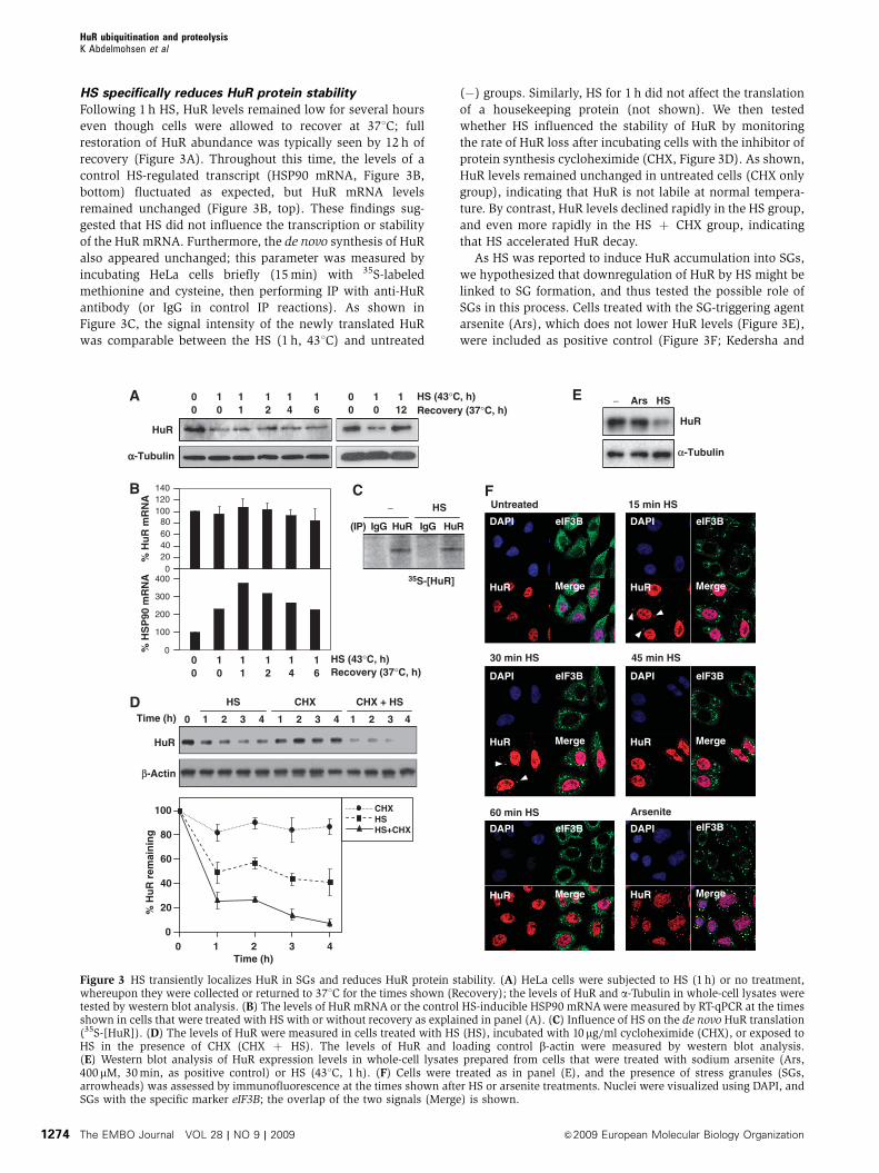
HS specifically reduces HuR protein stability
Following 1 h HS, HuR levels remained low for several hours
even though cells were allowed to recover at 371C; full
restoration of HuR abundance was typically seen by 12 h of
recovery (Figure 3A). Throughout this time, the levels of a
control HS-regulated transcript (HSP90 mRNA, Figure 3B,
bottom) fluctuated as expected, but HuR mRNA levels
remained unchanged (Figure 3B, top). These findings sug-
gested that HS did not influence the transcription or stability
of the HuR mRNA. Furthermore, the de novo synthesis of HuR
also appeared unchanged; this parameter was measured by
incubating HeLa cells briefly (15 min) with 35S-labeled
methionine and cysteine, then performing IP with anti-HuR
antibody (or IgG in control IP reactions). As shown in
Figure 3C, the signal intensity of the newly translated HuR
was comparable between the HS (1 h, 431C) and untreated
(�) groups. Similarly, HS for 1 h did not affect the translation
of a housekeeping protein (not shown). We then tested
whether HS influenced the stability of HuR by monitoring
the rate of HuR loss after incubating cells with the inhibitor of
protein synthesis cycloheximide (CHX, Figure 3D). As shown,
HuR levels remained unchanged in untreated cells (CHX only
group), indicating that HuR is not labile at normal tempera-
ture. By contrast, HuR levels declined rapidly in the HS group,
and even more rapidly in the HS þ CHX group, indicating
that HS accelerated HuR decay.
As HS was reported to induce HuR accumulation into SGs,
we hypothesized that downregulation of HuR by HS might be
linked to SG formation, and thus tested the possible role of
SGs in this process. Cells treated with the SG-triggering agent
arsenite (Ars), which does not lower HuR levels (Figure 3E),
were included as positive control (Figure 3F; Kedersha and
10
% H
uR
mR
NA
(IP) IgG HuR IgG HuR
35S-[HuR]
HuR
αα-Tubulin α-Tubulin
020406080
100120140
0
100
200
300
400
% H
SP
90 m
RN
A
00
10
11
12
14
16
00
112
HS (43°C, h)Recovery (37°C, h)
00
10
11
12
14
16
− HS
0 1 2 3 4 1 2 3 4 1 2 3 4
HuR
β-Actin
HS CHX CHX + HS
Time (h)
HS (43°C, h)Recovery (37°C, h)
HuR
− Ars HS
43210Time (h)
% H
uR
rem
ain
ing
0
20
40
60
80
100 CHXHSHS+CHX
DAPI eIF3B
HuR Merge
DAPI eIF3B
HuR Merge
DAPI eIF3B
HuR Merge
DAPI eIF3B
HuR Merge
DAPI eIF3B
HuR Merge
DAPI eIF3B
HuR Merge
15 min HS
45 min HS
Arsenite
Untreated
30 min HS
60 min HS
Figure 3 HS transiently localizes HuR in SGs and reduces HuR protein stability. (A) HeLa cells were subjected to HS (1 h) or no treatment,whereupon they were collected or returned to 371C for the times shown (Recovery); the levels of HuR and a-Tubulin in whole-cell lysates weretested by western blot analysis. (B) The levels of HuR mRNA or the control HS-inducible HSP90 mRNA were measured by RT-qPCR at the timesshown in cells that were treated with HS with or without recovery as explained in panel (A). (C) Influence of HS on the de novo HuR translation(35S-[HuR]). (D) The levels of HuR were measured in cells treated with HS (HS), incubated with 10 mg/ml cycloheximide (CHX), or exposed toHS in the presence of CHX (CHX þ HS). The levels of HuR and loading control b-actin were measured by western blot analysis.(E) Western blot analysis of HuR expression levels in whole-cell lysates prepared from cells that were treated with sodium arsenite (Ars,400mM, 30 min, as positive control) or HS (431C, 1 h). (F) Cells were treated as in panel (E), and the presence of stress granules (SGs,arrowheads) was assessed by immunofluorescence at the times shown after HS or arsenite treatments. Nuclei were visualized using DAPI, andSGs with the specific marker eIF3B; the overlap of the two signals (Merge) is shown.
HuR ubiquitination and proteolysisK Abdelmohsen et al
The EMBO Journal VOL 28 | NO 9 | 2009 &2009 European Molecular Biology Organization1274

Anderson, 2002). HuR localized in SGs by 15 and 30 min
(arrowheads), but disappeared by 60 min (Figure 3F). CHX
treatment blocked the formation of SGs (Supplementary
Figure S2), but HuR was still reduced in this group
(Figure 3D), suggesting that HuR degradation was indepen-
dent of its localization in SGs. Together, these findings reveal
that HS did not affect HuR biosynthesis and instead influ-
enced HuR protein stability and subcytoplasmic localization.
HuR kinase CHK2 protects against HuR decay after HS
While screening cell types to study the reduction in HuR
levels by HS (Figure 1E), we observed that HuR was more
efficiently reduced in HCT116 cells carrying somatic deletions
of the HuR kinase CHK2 (Jallepalli et al, 2003; Abdelmohsen
et al, 2007a) than in the parental HCT116 counterparts
(Figure 4A and B). Supporting the notion that CHK2 might
ameliorate HuR degradation, HeLa cells treated with a CHK2
inhibitor showed a similarly accelerated loss of HuR after HS
(Figure 4B). Although CHK2 was phosphorylated (and hence
activated) by HS, HuR degradation preceded the increase in
CHK2 phosphorylation above basal levels (Figure 4C). In
keeping with a role for CHK2 in preventing HuR degradation
during HS, the decline in HuR by HS was accelerated when
CHK2 was silenced by siRNA (Figure 4D).
Ctrl siRNA
0
0
100 100 84 81 43100
15 30 0 15 30
1 2 0 1 2
HuR
HCT116 (parental)
HCT116 (CHK2KO)
Paren
tal
CHK2KO
CHK2
β-Actin
β-Actin
β-Actin
β-Actin
β-Actin
β-Actin
β-ActinHS (min)
HuR
DMSO CHK2 inhibitor
(% HuR)
0
0 15 30 60 0 15 30 60
5 10 15 20 25 30 45 60 90 120 180
HuR
p-CHK2
CHK2
HS(min) 0 15 30 60
0 15 30 60 0 15 30 60 0 15 30 60 0 15 30 60
0 15 30 60 0 15 30 60 0 15 30 60
HuR
HuR(WT)-TAP HuR(S88A)-TAP HuR(S100A)-TAP
HuR(WT)-TAP HuR(S88D)-TAP HuR(S100D)-TAP
HuR(T118A)-TAP
HuR(T118D)-TAP
HuR(WT)-TAPHuR(S88A)-TAPHuR(S100A)-TAPHuR(T118A)-TAP
0 15 30 45 60HS (min)
0 15 30 45 60HS (min)
% H
uR
-TA
P r
emai
nin
g
HS
0
20
40
60
80
100
HuR(WT)-TAP
HuR(S88D)-TAP
HuR(S100D)-TAP
HuR(T118D)-TAP% H
uR
-TA
P r
emai
nin
g
HuR
Ctrl siRNACHK2 siRNA
HS (min)
% H
uR
rem
ain
ing
HuR
CHK2
0
20
40
60
80
100
00 15 30 45 60
20
40
60
80
100
HS (min)
CHK2 siRNA
RRM3RRM2
HuR
RRM1 HNS
S88
S10
0T
118
TAPHS (h)
HS (min)
Figure 4 CHK2 reduces HuR loss by HS. (A) Western blot analysis of the levels of CHK2, HuR, and loading control in untreated (left) orHS-treated (431C, right) colon carcinoma HCT116 cells expressing CHK2 (Parental) or CHK2-null through somatic knockout (CHK2KO).(B) Western blot analysis of HuR and b-actin expression levels in HeLa cells pre-treated with a CHK2 inhibitor (10mM, 1 h), then exposed to HSfor the times indicated; signal intensities were quantified by densitometry. (C) Western blot analysis of the levels of HuR, phosphorylated(p-)CHK2, total CHK2, and b-actin in HeLa cells treated with HS for the times indicated. (D) At 48 h after transfection of CHK2-directed orcontrol (Ctrl) siRNAs, HeLa cells were treated with HS for the times shown and the levels of HuR, CHK2, and loading control b-actin wereassessed by western blot analysis. In panels (D–F), the western blotting signals were quantified by densitometry and the means ±s.d. fromthree independent experiments are shown. (E) Top, schematic of the sites of CHK2 phosphorylation on HuR. By 48 h after transfection ofplasmids to express HuR–TAP fusion proteins [WT or bearing mutations in putative CHK2 phosphorylation sites (S88A, S100A, T118A)], thelevels of HuR–TAP fusion proteins were assessed by western blotting. (F) By 48 h after transfection of plasmids to express HuR–TAP fusionproteins [WT or bearing phosphomimic mutations (S88D, S100D, T118D)], HuR–TAP fusion protein levels were assessed as explained in (E).
HuR ubiquitination and proteolysisK Abdelmohsen et al
&2009 European Molecular Biology Organization The EMBO Journal VOL 28 | NO 9 | 2009 1275

CHK2 phosphorylates HuR at three residues (S88, S100,
T118; Figure 4E), and through these modifications CHK2 was
proposed to modulate HuR binding to target mRNAs
(Abdelmohsen et al, 2007a, b). Preliminary evidence sug-
gested that HuR is phosphorylated by HS in a CHK2-depen-
dent manner (Supplementary Figure S3), but in-depth
analysis of this process awaits the availability of specific
antibodies recognizing phosphorylated S88, S100, and T118.
We hypothesized that if CHK2 slows down the loss of HuR
after HS, HuR mutants refractory to CHK2 phosphorylation
could be more labile. Indeed, comparison of non-phosphor-
ylatable and wild-type HuR–TAP fusion proteins revealed that
HuR(S88A)–TAP and HuR(T118A)–TAP decreased more
rapidly after HS (Figure 4E). To further test the notion that
HuR phosphorylation at CHK2 target sites reduced HuR decay
by HS, we generated plasmids to express HuR–TAP fusion
proteins mimicking phosphorylated residues S88, S100, and
T118. As shown in Figure 4F, fusion proteins HuR(S88D)–
TAP, HuR(S100D)–TAP, and HuR(T118D)–TAP were markedly
protected against HS-triggered loss. These observations
further strengthen the view that phosphorylation of HuR by
CHK2 slows down HuR degradation by HS.
Ubiquitination linked to HuR stability after HS
To identify the mechanisms whereby HS accelerates the
degradation of HuR, a number of inhibitors of proteases
[e.g., inhibitors of calpains, caspases (Z-VAD-FMK), and
lysosomal proteases (chloroquine and ammonium chloride)]
were tested and found not to block the degradation of HuR by
HS (not shown). Only inhibition of the proteasome using
MG132 (20mM) enhanced HuR levels, increasing HuR signals
relative to those in the control (DMSO) group (Figure 5A). It
should be noted that pretreatment with MG132 before HS as
well as joint MG132 and HS treatments were severely toxic
for HeLa cells (unlike for WI-38 cells (Bonelli et al, 2004)),
and therefore the effects of these treatments could not be
studied (not shown). Similarly, when the Ub–proteasome
degradation system was suppressed by silencing Ub (Ub
siRNA group), we observed a stabilization of HuR after HS
(Figure 5B). As anticipated, this intervention not only re-
duced the levels of Ub (an 8.5-kDa protein), but also lowered
the subset of ubiquitinated proteins in HeLa cells (Ub con-
jugates, Figure 5C). Proteasome activity remained elevated
during HS and during recovery at 371C (Supplementary
Figure S4). Collectively, this evidence suggested that HuR
degradation by HS was linked to HuR ubiquitination and
prompted us to test directly whether HuR was ubiquitinated.
In vitro evidence of HuR ubiquitination was obtained using
a cell-free assay (Materials and methods). As shown in this
system, Ub residues were readily incorporated onto GST-HuR
(and not onto GST), but this reaction only occurred in the
presence of ATP, in agreement with the energy dependence of
the ubiquitination process (Figure 5D); ubiquitination of p53
was included as positive control (Figure 5D).
In vivo evidence that endogenous HuR was ubiquitinated
was first sought using standard western blotting conditions,
without success. Using a modified western blotting protocol
(which included lysis under strong inhibition of anti-deubi-
quitinating enzymes, use of greater amounts of lysate, longer
transferring times, and concentrated primary antibody;
details in Supplementary data), we could detect endogenous
ubiquitinated HuR (Figure 5E) in cells that were untreated,
HS-treated, and recovering from HS. Silencing of HuR
revealed that these bands indeed corresponded to polyubi-
quitinated HuR (Supplementary Figure S5). As the routine
use of this cumbersome procedure posed major limitations to
the molecular characterization of HuR ubiquitination, addi-
tional experiments were conducted using cells cotransfected
with plasmids to express HA-tagged Ub (Ub-HA) and tandem
affinity purification-tagged HuR (HuR–TAP); the correspond-
ing control vector-transfected groups are V and TAP, respec-
tively. As shown, ubiquitinated HuR–TAP was readily
observed in the presence of Ub-HA, but not in the presence
of HA alone, whereas no Ub-HA was incorporated into TAP
alone (Figure 5F, left). Similarly, no Ub-HA was incorporated
when using the mutant Ub(K48R) that could not form poly-
ubiquitin chains (Figure 5F, right). HS treatment was found to
decrease ubiquitinated endogenous HuR and HuR–TAP
(Figure 5E and G); this was observed at all times studied,
including at the earliest times when the analysis was possible
(by 15 min of HS, not shown). Further characterization of this
effect in intact cells included treatment with MG132 (4 h),
which enhanced the levels of total ubiquitinated (Ub-HA-
modified) proteins (Figure 5H, left) ubiquitinated HuR–TAP
specifically (Figure 5H, right), and ubiquitinated endo-
genous HuR (Supplementary Figures S5 and S6). Moreover,
Figure 5 Analysis of HuR ubiquitination in vitro and in vivo. (A) The levels of HuR, ubiquitinated proteins, and loading control b-actin werestudied by western blot analysis in cells that were treated (temperatures and times) as shown, in the presence of MG132 (20mM) or vehiclecontrol (DMSO). (B) At 48 h after transfecting either control (Ctrl) siRNA or a ubiquitin-targeting (Ub) siRNA, HuR and loading control b-actinwere assessed in cells incubated as shown. (C) Western blot analysis of the levels of ubiquitin and total ubiquitin-conjugated proteins 48 h aftertransfection with Ctrl or Ub siRNAs. (D) Left, in vitro polyubiquitination of HuR was measured using a control protein (GST) and a GST-HuRfusion protein in the absence or presence of ATP; Right, in vitro polyubiquitination of purified p53; kDa, sizes of molecular weight markers.(E) Western blot analysis (modified as detailed in the Supplementary data) of endogenous ubiquitinated HuR after treatment of HeLa cells withHS (left) and during recovery from HS (right). (F) Left, HeLa cells were cotransfected with a plasmid expressing an HA-tagged ubiquitin (Ub-HA) or the corresponding control vector (V), together with a plasmid expressing either HuR–TAP or the vector control (TAP); polyubiquitinatedHuR–TAP was assessed 48 h later by HA IP, followed by HuR western blot (WB) analysis. Right, cells were processed as shown on the left ofpanel (F), but a mutant variant of ubiquitin that cannot oligomerize [Ub(K48R)-HA] was also tested; polyubiquitinated HuR–TAP was assessed48 h later by HA IP, followed by HuR WB analysis. (G) Cells cotransfected with HuR–TAP along with either Ub-HA-expressing or V plasmidswere left without further treatment or were treated with HS (431C for 15 min), whereupon the levels of ubiquitinated HuR–TAP were detectedby TAP IP (using Rabbit IgG) and WB analysis using an anti-HA antibody. (H) Left, in cells transfected with Ub-HA-expressing plasmid, Ub-HAlevels were tested in the absence (DMSO) or presence of MG132 (20 mM for 4 h) by IP using an anti-HA antibody, followed by HA detection byWB. Right, cells co-transfected with pHuR–TAP and Ub-HA were used to detect the levels of ubiquitinated HuR–TAP in the absence (DMSO) orpresence of MG132 (20mM for 4 h) after TAP IP (using rabbit IgG), and then to detect Ub using an anti-HA antibody. (I) HuR abundance in cellsexpressing normal ubiquitin levels or overexpressing ubiquitin as HA-Ub; protein levels were studied at the times shown following HS. (J) Theubiquitination of HuR–TAP fusion proteins bearing point mutations S88A and S118A (left) or S100A (right) was tested by cotransfection ofplasmids as explained for panel (F), except that the HuR mutants tested were those described in Figure 4E. Protein signals were visualized byTAP IP, followed by WB analysis using an anti-HA antibody.
HuR ubiquitination and proteolysisK Abdelmohsen et al
The EMBO Journal VOL 28 | NO 9 | 2009 &2009 European Molecular Biology Organization1276

experiments that complemented the increase in HuR seen
after silencing Ub (Figure 5B and C) revealed that Ub over-
expression actually reduced the levels of HuR–TAP
(Figure 5I). Given that non-phosphorylatable HuR–TAP mu-
tants had distinct stabilities (Figure 4E), we studied whether
their ubiquitination levels differed. As shown in Figure 5J, the
ubiquitination of HuR(S88A)–TAP was similar to that of
HuR(WT)–TAP, in agreement with the similar stabilities of
both proteins (Figure 4E). By contrast, the least stable
mutant, HuR(T118A)–TAP, was more extensively ubiquiti-
nated (left), whereas the more stable mutant, HuR(S100A)–
TAP, was less ubiquitinated (right). Together, these results
show that HuR is ubiquitinated in vitro and in vivo and link
HuR ubiquitination to its degradation in response to HS.
Ub
Ub
iqu
itin co
nju
gates
CtrlsiRNA
UbsiRNA
HuR
HS (43°C, h)Recovery (37°C, h)
00
10
12
14
00
10
12
14
CtrlsiRNA
UbsiRNA
β-Actin β-Actin
β-Actin
β-Actin
HuR
HS (43°C, h)Recovery (37°C, h)
11
12
14
11
12
14
DMSO MG132
10
00
00
(DMSO)
Ub
iqu
itin co
nju
gates
250
150
100
75
TAPHuR-TAP
V Ub
-HA
V Ub
-HA
Hu
R-T
AP
-[U
b-H
A]n
HuR-TAP
IP: HA WB: HuR
V Ub
(WT
)-H
A
Ub
(K48
R)-
HA
IP: HA WB: HuR
250
150
100
75
HuR-TAP
(kDa)(kDa)
HuR-TAP
42104210
V Ub-HA
HS (h)
HuR-TAP
250
150
100
75
DMSO MG132
V Ub
-HA
V Ub
-HA
Hu
R-T
AP
-[U
b-H
A]n
V Ub
-HA
V Ub
-HA
IP: TAP WB: HA
IP: HA WB: HA
DMSO MG132
(kDa)
Hu
R-T
AP
-[U
b-H
A]n
V Ub
-HA
V Ub
-HA
IP: TAP WB: HA
− HS
HuR-TAP
250
150
100
75
(kDa)
HuR-TAP
250
150
100
75
HuR(WT)TAP
V Ub
-HA
V Ub
-HA
V Ub
-HA
(kDa)
Hu
R-T
AP
-[U
b-H
A]n
HuR-TAP
IP: TAP WB: HA
IP: TAP WB: HA
HuR(S88A)TAP
HuR(T118A)
TAP
HuR(WT)TAP
V Ub
-HA
V Ub
-HA
HuR(S100A)
TAP
GS
T-H
uR
-[Ub
]n
+ − ATP
GST-HuR
250
150
100
75
p53-[U
b]n
ATP+ −
p53
250
150
100
75
+ −
GST
(kDa) (kDa)
30
50
60
80
110U
biq
uit
inat
ed H
uR
HuR
(kDa)
Recovery (h, 37°C) after HS
604530150 0 0 2 4 6 12
HS (min)
HuR ubiquitination and proteolysisK Abdelmohsen et al
&2009 European Molecular Biology Organization The EMBO Journal VOL 28 | NO 9 | 2009 1277

Ubiquitination at Lys-182 promotes HuR decay
and enhances cell survival after HS
In order to map the residue(s) implicated in Ub-mediated HuR
proteolysis, we first narrowed down the region of HuR
involved in HS-mediated degradation. Depicted in Figure 6A
are three TAP-linked segments of HuR that were tested
initially. After transfection of the corresponding expression
vectors, we assessed the stability of the resulting fusion
proteins in response to HS. A segment spanning HuR amino
acids 1–110 was stable after HS, but addition of amino acids
111–220 rendered it labile in response to HS (Figure 6B),
indicating that the 111–220 region of HuR conferred instability
after HS. This middle region contains five lysines, the amino
acids where Ub moieties are ligated. After systematically
mutating each lysine to an arginine (Figure 6C), we tested
the stability of the full-length HuR–TAP carrying each of the
resulting point mutations (K120R, K156R, K171R, K182R or
K191R). As shown, in each transfection group, both the
HuR(WT)–TAP and the endogenous HuR were labile after
HS (Figure 6D). In addition, each of the HuR–TAP point
mutants was also labile, with the exception of mutant
K182R, which was refractory to degradation by HS
RRM3RRM2
3261
1101
220
326111
HuR(WT)-TAP
HuR(1-110)-TAP
HuR(1-220)-TAP
HuR(111-326)-TAP
HuR
1
HuR-TAP
ββ-Actin
– HS – HS – HS – HS
HuR(WT)-TAP
HuR(1-110)-TAP
HuR(1-220)-TAP
HuR(111-326)-TAP
111 220
K171RK120RK156RK191R K182R
∗ ∗ ∗ ∗ ∗
HuR(WT)-TAP
HuR(K120R)-TAP∗
HuR(K156R)-TAP∗
HuR(K171R)-TAP∗
HuR(K182R)-TAP∗
HuR(K191R)-TAP∗
HuR-TAP
HuR
ββ-Actin
K120RWT
– HS – HS
HuR-TAP
HuR
ββ-Actin
K156RWT
– HS – HS
HuR-TAP
HuR
ββ-Actin
K171RWT
– HS – HS
HuR-TAP
HuR
ββ-Actin
K182RWT
– HS – HS
HuR-TAP
HuR
ββ-Actin
K191RWT
– HS – HS
RRM1 HNS
0HuR
(K191R)TAP
HuR(K182R)
TAP
HuR(K120R)
TAP
HuR(K156R)
TAP
HuR(K171R)
TAP
HuR(WT)TAP
20
40
60
80
100 –
HS
% H
uR
-TA
P r
emai
nin
g
Figure 6 HuR ubiquitination and degradation by HS requires Lys-182. (A) Schematic of truncated HuR segments (generated from HuR–TAP).(B) The effect of HS on the levels of the HuR segments shown in (A) were tested by measuring HuR–TAP signals on western blots using an anti-IgG antibody to recognize the TAP segments of the fusion proteins. (C) Schematic of the point mutations [Lys-Arg (K-R)] introduced into thefive lysine residues within the labile region of HuR–TAP. (D) The relative stability of each HuR–TAP (K-R) mutant was tested by western blotanalysis; the levels of endogenous HuR, HuR–TAP (WT relative to K120R, K156R, K171R, K182R or K191R), and loading control b-actin weretested by monitoring HuR signals in untreated (�) compared with HS-treated (HS) cells. (E) Quantification of the western blotting data shownin (D); data represent the means±s.d. from three independent experiments.
HuR ubiquitination and proteolysisK Abdelmohsen et al
The EMBO Journal VOL 28 | NO 9 | 2009 &2009 European Molecular Biology Organization1278

(Figure 6D). The extent of degradation of each HuR–TAP
variant following HS was quantified (Figure 6E). Ub-HA co-
expression in HeLa cells transfected with each mutant verified
that all HuR–TAP mutants were efficiently ubiquitinated [as
shown for HuR(K171R)–TAP], but not HuR(K182R)–TAP,
which was found virtually without ubiquitination (Figure 7A).
Importantly, while the HS employed throughout this study
(431C) was not cytotoxic, expression of K182R significantly
reduced cell survival, causing a significant loss in cell viabi-
lity (B38% by the MTT assay, 32% by direct cell counts) by
24 h following HS in this population; in contrast, only o8%
(by both assays) of loss in cell viability was seen in the WT
HuR population (Figure 7B and C). The other transfection
groups (K120R, K156R, K171R, K191R) showed significantly
less toxicity than the K182R group, with viabilities closer to
that seen for WT (Figure 7B and C). In keeping with its
relatively higher stability, the K182R mutant remained asso-
ciated to numerous HuR target mRNAs tested (Figure 7D and
data not shown). Collectively, these results indicate that the
transient HS-mediated degradation of HuR is prevented by
CHK2, involves ubiquitination of K182, and confers a survi-
val advantage against HS cytotoxicity.
Hu
R-T
AP
-[U
b-H
A]n
V Ub
-HA
V Ub
-HA
IP: TAP WB: HA
IP: TAP WB: HA
K171RWT
Hu
R-T
AP
-[U
b-H
A]n
V Ub
-HA
V Ub
-HA
K182RWT
HuR-TAP
250
150
100
75
250
150
100
75HuR-TAP
WT K191R
% L
oss
in c
ell v
iab
ility
(M
TT
ass
ay, 2
4 h
aft
er H
S)
50
45
40
35
30
25
20
15
10
5
0K182RK120R K171RK156R
WT K191R
% L
oss
in c
ell n
um
ber
s (2
4 h
aft
er H
S)
50
45
40
35
30
25
20
15
10
5
0K182RK120R K171RK156R
0
369
12
1518
05
101520253035
VHL
NCL
CRM1
WTAP
TUBB
ACTB
BCL9
KRAS
−HS
0369
12151821
0369
12151821
0369
12151821
0369
121518
Fo
ld e
nri
chm
ent
in m
RN
A (
Hu
R-T
AP
IP c
om
par
ed w
ith
TA
P IP
)
0
24
6
8
1012
0
36
9
12
1518
WT
K19
1R
K18
2R
K12
0R
K17
1R
K15
6R
Figure 7 In vivo ubiquitination of HuR–TAP and mutant TAP and influence on cell survival after HS. (A) Cells were transfected with HuR–TAP(WT), HuR–TAP (K171R) (left), or HuR–TAP (K182R) (right) along with Vor Ub-HA-expressing plasmids. The levels of ubiquitinated HuR–TAPwere detected by IP (using Rabbit IgG) followed by WB analysis using anti-HA antibody. (B, C) Cells were transfected with plasmids tooverexpress the HuR–TAP fusion proteins shown [WT (black) and K-R mutants (gray)]; 48 h later, cells were subjected to HS (431C, 1 h), andafter an additional 24 h the surviving fraction was quantified using the MTTassay (B) or by direct cell counts (C). The loss in cell viability andnumbers was represented as a percentage of untreated cells in each transfection group (means±s.d. from three independent experiments).(D) In cells transfected as described in panels (B, C), the association of each HuR–TAP chimeric protein with HuR target transcripts was tested byRNP IP. The fold enrichment represents mRNA association to each HuR–TAP relative to TAP; the average of two similar experiments is shown.
HuR ubiquitination and proteolysisK Abdelmohsen et al
&2009 European Molecular Biology Organization The EMBO Journal VOL 28 | NO 9 | 2009 1279
Discussion
Despite extensive evidence that stress-regulated HuR function
is linked to its nucleocytoplasmic shuttling in the absence of
net changes in HuR levels, here we document the rapid and
potent degradation of HuR in response to an acute damaging
stimulus. HS (431C) triggered a fast and robust reduction in
HuR stability and the effect was reversed if cells were
returned to 371C. Given HuR’s central role in the post-
transcriptional regulation of many critical proteins (e.g., cell
cycle control factors, stress-response proteins, and cyto-
kines), we investigated in depth the mechanism of HuR
downregulation by HS. Our results show that HuR is a
ubiquitination substrate in vitro and in vivo, that CHK2
ameliorates HuR loss by HS, that residue K182 is particularly
important for HuR ubiquitination and HS-mediated decay,
and that the HS-triggered transient downregulation of HuR
protects against cell death.
Post-translational modification of HuR by HS
Earlier reports showed that HuR associated with cytoplasmic
poly(A)þ RNA in unstressed cells, displaying a prominent
presence in the polysomal component (Gallouzi et al, 2000).
HS altered the subcellular localization and function of HuR,
mobilizing it to SGs, enhancing its association with
poly(A)þ RNA in the nucleus, and promoting the nuclear
export of HuR linked to the cytoplasmic localization of hsp70
mRNA (Gallouzi et al, 2000, Gallouzi and Steitz, 2001). Our
analysis did not directly test the association of HuR with
poly(A)þ RNA, but did show that HuR colocalized in SGs
shortly after commencing HS (Figure 3F). Additionally, we
did not observe specific decreases in cytoplasmic or nuclear
HuR, but an overall reduction in both compartments.
Although there appeared to be a reduction in whole-cell
HuR levels after HS in the studies by Gallouzi and coworkers,
they did not formally report overt decreases in HuR levels
following HS. It is likely that these differences collectively
arise from variations between the two experimental systems,
including the fact that we employed milder HS without serum
(431C, 1 h) whereas Gallouzi and coworkers used 451C with
serum.
Our results also agree with those of an earlier study in
which inhibition of the proteasome elevated HuR levels in
human diploid fibroblasts, although the mechanism respon-
sible for this effect was not examined (Bonelli et al, 2004). We
initially tested other post-translational modifications of HuR,
including sumoylation, but did not find that HS influenced
the levels of sumoylated HuR (Supplementary Figure S7).
Instead, we found that HuR was ubiquitinated and was
degraded through a process dependent on Ub and the protea-
some. The Ub–proteasome system degrades many cellular
proteins through the action of three enzymes: E1 activates
Ub, triggering its transfer onto the Ub carrier enzyme E2,
which in turn is transferred onto a substrate protein by an E3
Ub protein ligase and Ub becomes covalently linked. The
repeated addition of Ub moieties results in the formation of a
polyubiquitinated substrate protein, which is recognized by a
large proteolytic complex, the 26S proteasome (Hershkom
and Ciechanover, 1998). Here, we were able to detect
endogenous as well ectopically expressed tagged HuR migrat-
ing with slower electrophoretic mobilities (Figures 5 and 7,
Supplementary Figure S6), but it is unclear why HS did not
appear to elevate the levels of polyubiquitinated moieties.
It is possible that HS does not increase HuR ubiquitination
per se, but instead signals the decay of already-ubiquitinated
HuR protein.
Consequences of ubiquitination of RBPs AUF1 and HuR
Among the RBPs influencing the stability/translation of spe-
cific mRNA subsets, only AUF1 was previously shown to be
ubiquitinated (Laroia et al, 1999). Laroia and colleagues
showed that AUF1-associated AU-rich mRNAs were degraded
after eukaryotic initiation factor (eIF)4G dissociated from
AUF1, and that AUF1 was ubiquitinated and degraded
(along with associated mRNAs, as proposed by the authors)
via the proteasome. Interestingly, in this model system, hsp70
(which was elevated in response to HS) sequestered AUF1 in
the nuclear–perinuclear region and blocked the decay of
AUF1 and AUF1-bound mRNAs (Laroia et al, 1999). These
investigators further showed that overexpression of a deubi-
quitinating protein (DUB) or inhibition of the proteasome
prevented the decay of labile mRNAs (Laroia et al, 2002).
Together with these two reports, our findings suggest
a functional interplay between HuR and AUF1 upon the levels
of many predictedly shared target mRNAs: following HS, the
AUF1-bound hsp70 would be unable to trigger the degrada-
tion of AUF1-bound mRNAs, while the loss of HuR through
Ub-mediated proteolysis would preclude the mRNA-stabiliz-
ing influence of HuR. Thus, the balance between decay and
stability for an mRNA that is the target of HuR and AUF1
could depend directly on its relative affinity for each RBP. It is
as yet unclear whether the proteasomal degradation of HuR
also degrades HuR-bound mRNAs, although this possibility
seems unlikely, given the wealth of evidence supporting
HuR’s mRNA-stabilizing influence (Brennan and Steitz,
2001). Nonetheless, as HS affects each protein differently
(prevents AUF1 degradation and promotes HuR degradation),
perhaps HuR proteolysis does not itself degrade target
mRNAs, but the very decrease of HuR facilitates the binding
of AUF1 to shared target mRNAs for which both RBPs
compete. Such mRNAs could initially be protected from
degradation while AUF1 is nuclear/perinuclear and bound
to hsp70, but may later be degraded through an AUF1-
dependent process.
It is also worth noting that the lysine residue implicated in
HuR ubiquitination (K182) lies within the RRM2 (spanning
amino acid residues 106–186). The post-translational modi-
fication of HuR at this site raises the possibility that ubiqui-
tination at K182 interferes with HuR’s association with a
target mRNA and may also help to ‘release’ it, in conjunction
with triggering HuR degradation. Moreover, the findings that
RRM2 mutants S88A and T118A are significantly more heat-
labile than HuR(WT) and that all phosphomimic mutants
(S88D, S100D, T118D) are markedly more heat-resistant than
WT (Figure 4E and F) further support the notion that CHK2
phosphorylation of HuR protects against HS-triggered HuR
decay. In this regard, the ubiquitination-deficient mutant
K182R showed enhanced binding to target mRNAs after HS
(Figure 7D). Although this mutant is logically expected to
continue forming RNP complexes after HS (as it is refractory
to HS-triggered decay), the influence of post-translational
modifications at S88, S100, T118, and K182 upon binding to
target mRNAs deserves further experimental scrutiny.
HuR ubiquitination and proteolysisK Abdelmohsen et al
The EMBO Journal VOL 28 | NO 9 | 2009 &2009 European Molecular Biology Organization1280

RBPs mediating post-transcriptional gene
expression by HS
Heat stress induces the misfolding and denaturation of cellular
proteins, causing cytotoxicity and triggering the HS response
(De Maio, 1999; Voellmy, 2006). A prominent component of the
HS response is the activation of a family of HS factors (HSFs),
which bind to HS elements (HSEs) and direct the transcription
of HS proteins (HSPs) and other stress and immune response
proteins (recently reviewed by Wheeler and Wong, 2007). In
addition, hyperthermia has long been recognized to regulate
gene expression through post-transcriptional mechanisms (Yost
et al, 1990). Global studies of the influence of HS on mRNA
stability have revealed that large subsets of mRNAs are stabi-
lized or destabilized in response to HS in both mammalian and
yeast cells (Fan et al, 2002; Grigull et al, 2004). Global analyses
have also shown that HS broadly represses the translation of
subsets of mRNAs (Lindquist, 1981), and that hyperthermia
can act upon the RNA itself, modifying its secondary structure
and thereby altering its translational status (Narberhaus et al,
2006). At the same time, both the stability and translation of
HSPs are favored in response to HS (Panniers, 1994;
Kaarniranta et al, 2000).
The post-transcriptional changes in gene expression follow-
ing HS are controlled through the action of various RBPs
interacting with HS-regulated mRNAs. For example, HS inhibits
the translation of capped mRNAs by dephosphorylating both
eIF4E and the 4E-inactivating protein BP-1, thereby reducing
eIF4E activity and availability (Feigenblum and Schneider,
1996). RBPs associating with specific regions of the 50 and
30UTRs, such as AUF1, TIAR, TIA-1, and TTP, have also been
linked to HS-regulated gene expression (Laroia et al, 1999;
Gallouzi et al, 2000; Kedersha and Anderson, 2002). However,
HS did not cause dramatic changes in the levels of TIAR, TIA-1,
or TTP, instead triggering changes in their subcellular distribu-
tion and their aggregation in SGs (Kedersha and Anderson,
2002). In this regard, the lower HuR abundance after HS is a
novel mechanism whereby an RBP can regulate subsets of
target mRNAs. The changes in both RBP localization and RBP
levels are in agreement with the post-transcriptional operon
model (Keene and Tenenbaum, 2002). According to this model,
collections of functionally related genes can be coordinately
regulated through the presence of shared RNA elements within
the transcripts, which render them targets of a given RBP. Thus,
by reducing HuR expression levels, HS can effectively reduce
the stability and/or alter the translation of HuR target mRNAs.
Given the protective influence of a timely reduction of HuR
following HS (Figure 7B and C), the corresponding decreases in
proliferative proteins encoded by HuR target mRNAs (e.g.,
c-Fos, prothymosin-a, and Cyclins A2, B1, and D1) are likely
to be advantageous, allowing cells to channel its resources and
energy towards the repair of heat-damaged components. As we
gain a deeper understanding of HuR’s influence on cell survival
after HS, we propose that the transient decrease in HuR
abundance is a key adaptive mechanism whereby mammalian
cells maintain homeostasis in response to thermal stress.
Materials and methods
Cell culture, treatments, and transfectionsHuman cervical carcinoma HeLa cells and mouse melanomaB16F10 cells were cultured in Dulbecco’s modified essentialmedium (DMEM, Invitrogen) supplemented with 10% fetal bovineserum (FBS) and antibiotics; WI-38 cells were further supplementedwith non-essential amino acids (Gibco). HCT116 colon cancer cells(WT, CHK2KO; Jallepalli et al, 2003) were cultured in McCoy’s 5Amedium supplemented with 10% FBS and antibiotics. For HStreatments, cells were washed with PBS, given preheated medium(DMEM without FBS), and further incubated for 1 h at 431C unlessindicated otherwise. Control cells were also washed with PBS andgiven pre-warmed medium (DMEM without FBS) for 1 h at 371C. Allsmall interfering (si)RNAs (Qiagen, listed in the Supplementarydata) were used at a final concentration of 10 nM. The plasmidsused are described in the Supplementary data. CHX and the CHK2inhibitor were from Calbiochem; sodium arsenite was from Sigma.Cell survival was measured by the MTTassay (Sigma) and by directcell counts using a hemocytometer.
mRNA and RNP analysisEndogenous mRNA–protein complexes were precipitated as de-scribed previously (Abdelmohsen et al, 2007a). Total RNA as wellas RNA in RNP IP reactions was reverse transcribed (RT) usingrandom hexamers, oligo(dT) primer, and Superscript II RT(Invitrogen). The levels of specific mRNAs were measured byreal-time quantitative (q)PCR using the QuantiTect SYBR green PCRkit (Qiagen) and analyzed on an ABI Prism 7000 detection system;transcript-specific primers are listed as Supplementary data.
Protein analysisWestern blot analysis was performed using standard procedures, asdescribed (Abdelmohsen et al, 2007a and Supplementary data).Nascent (de novo) translation of HuR was studied as described(Galban et al, 2008). Briefly, following incubation for 1 h at 431C(HS) or 371C (control), HeLa cells were incubated with 1 mCiL-[35S]methionine and L-[35S]cysteine (EasyTag TMEXPRESS, NEN/Perkin Elmer) per 60-mm plate for 15 min. Cells were then lysed inRIPA buffer and the IP reactions carried out in 1 ml TNN buffer(50 mM Tris-HCl (pH 7.5), 250 mM NaCl, 5 mM EDTA, and 0.5%NP-40) for 16 h at 41C using IgG (BD Pharmingen) or anti-HuRantibodies. Following extensive washes in TNN buffer, the IPsamples were resolved by SDS-PAGE, transferred onto PVDF filters,and visualized with a PhosphorImager (Molecular Dynamics). Theanalysis of ubiquitinated HuR is detailed in the Supplementary data.
Supplementary dataSupplementary data are available at The EMBO Journal Online(http://www.embojournal.org).
Acknowledgements
We thank MJ Pazin, V Dixit, WH Wood 3rd, B Frank, S Subaran, andFE Indig for their assistance with these studies. HCT116 cells werekindly provided by F Bunz. This research was supported entirely bythe NIA-IRP Program Z01-AG000511-10 and by set-aside funds of theNIA-IRP, NIH.
References
Abdelmohsen K, Kuwano Y, Kim HH, Gorospe M (2008) Posttrans-criptional gene regulation by RNA-binding proteins during oxidativestress: implications for cellular senescence. Biol Chem 389: 243–255
Abdelmohsen K, Lal A, Kim HH, Gorospe M (2007b) Posttranscrip-tional orchestration of an anti-apoptotic program by HuR. CellCycle 6: 1288–1292
Abdelmohsen K, Pullmann Jr R, Lal A, Kim HH, Galban S, Yang X,Blethrow JD, Walker M, Shubert J, Gillespie DA, Furneaux H,
Gorospe M (2007a) Phosphorylation of HuR by Chk2 regulatesSIRT1 expression. Mol Cell 25: 543–557
Bhattacharyya SN, Habermacher R, Martine U, Closs EI,Filipowicz W (2006) Relief of microRNA-mediated translationalrepression in human cells subjected to stress. Cell 125:1111–1124
Bonelli MA, Alfieri RR, Desenzani S, Petronini PG, Borghetti AF(2004) Proteasome inhibition increases HuR level, restores
HuR ubiquitination and proteolysisK Abdelmohsen et al
&2009 European Molecular Biology Organization The EMBO Journal VOL 28 | NO 9 | 2009 1281

heat-inducible HSP72 expression and thermotolerance in WI-38senescent human fibroblasts. Exp Gerontol 39: 423–432
Brennan CM, Steitz JA (2001) HuR and mRNA stability. Cell Mol LifeSci 58: 266–277
Chen CY, Xu N, Shyu AB (2002) Highly selective actions of HuR inantagonizing AU-rich element-mediated mRNA destabilization.Mol Cell Biol 22: 7268–7278
De Maio A (1999) Heat shock proteins: facts, thoughts, and dreams.Shock 11: 1–12
Doller A, Huwiler A, Muller R, Radeke HH, Pfeilschifter J, EberhardtW (2007) Protein kinase C alpha-dependent phosphorylation ofthe mRNA-stabilizing factor HuR: implications for posttranscrip-tional regulation of cyclooxygenase-2. Mol Biol Cell 18: 2137–2148
Dormoy-Raclet V, Menard I, Clair E, Kurban G, Mazroui R, Di MarcoS, von Roretz C, Pause A, Gallouzi IE (2007) The RNA-bindingprotein HuR promotes cell migration and cell invasion by stabi-lizing the beta-actin mRNA in a U-rich-element-dependent man-ner. Mol Cell Biol 27: 5365–5380
Fan J, Yang X, Wang W, Wood III WH, Becker KG, Gorospe M(2002) Global analysis of stress-regulated mRNA turnover byusing cDNA arrays. Proc Natl Acad Sci USA 99: 10611–10616
Fan XC, Steitz JA (1998) Overexpression of HuR, a nuclear-cyto-plasmic shuttling protein, increases the in vivo stability of ARE-containing mRNAs. EMBO J 17: 3448–3460
Feigenblum D, Schneider RJ (1996) Cap-binding protein (eukaryoticinitiation factor 4E) and 4E-inactivating protein BP-1independently regulate cap-dependent translation. Mol Cell Biol16: 5450–5457
Figueroa A, Cuadrado A, Fan J, Atasoy U, Muscat GE, Munoz-Canoves P, Gorospe M, Munoz A (2003) Role of HuR in skeletalmyogenesis through coordinate regulation of muscle differentia-tion genes. Mol Cell Biol 23: 4991–5004
Galban S, Kuwano Y, Pullmann Jr R, Martindale JL, Kim HH, Lal A,Abdelmohsen K, Yang X, Dang Y, Liu JO, Lewis SM, Holcik M,Gorospe M (2008) RNA-binding proteins HuR and PTB promotethe translation of hypoxia-inducible factor 1alpha. Mol Cell Biol28: 93–107
Gallouzi IE, Brennan CM, Stenberg MG, Swanson MS, Eversole A,Maizels N, Steitz JA (2000) HuR binding to cytoplasmic mRNA isperturbed by heat shock. Proc Natl Acad Sci USA 97: 3073–3078
Gallouzi IE, Steitz JA (2001) Delineation of mRNA export pathwaysby the use of cell-permeable peptides. Science 294: 1895–1901
Gorospe M (2003) HuR in the mammalian genotoxic response: post-transcriptional multitasking. Cell Cycle 2: 412–414
Grigull J, Mnaimneh S, Pootoolal J, Robinson MD, Hughes TR(2004) Genome-wide analysis of mRNA stability using transcrip-tion inhibitors and microarrays reveals posttranscriptional con-trol of ribosome biogenesis factors. Mol Cell Biol 24: 5534–5547
Guttinger S, Muhlhausser P, Koller-Eichhorn R, Brennecke J, KutayU (2004) Transportin2 functions as importin and mediates nucle-ar import of HuR. Proc Natl Acad Sci USA 101: 2918–2923
Hershkom A, Ciechanover A (1998) The ubiquitin system. AnnuRev Biochem 67: 425–479
Jallepalli PV, Lengauer C, Vogelstein B, Bunz F (2003) The Chk2tumor suppressor is not required for p53 responses in humancancer cells. J Biol Chem 278: 20475–20479
Kaarniranta K, Holmberg CI, Helminen HJ, Eriksson JE, Sistonen L,Lammi MJ (2000) Protein synthesis is required for stabilization ofhsp70 mRNA upon exposure to both hydrostatic pressurizationand elevated temperature. FEBS Lett 475: 283–286
Kedersha N, Anderson P (2002) Stress granules: sites of mRNAtriage that regulate mRNA stability and translatability. BiochemSoc Trans 30: 963–969
Keene JD (1999) Why is Hu where? Shuttling of early-response-genemessenger RNA subsets. Proc Natl Acad Sci USA 96: 5–7
Keene JD (2007) RNA regulons: coordination of post-transcriptionalevents. Nat Rev Genet 8: 533–543
Keene JD, Tenenbaum SA (2002) Eukaryotic mRNPs may representposttranscriptional operons. Mol Cell 9: 1161–1167
Kim HH, Abdelmohsen K, Lal A, Pullmann Jr R, Yang X, Galban S,Srikantan S, Martindale JL, Blethrow J, Shokat KM, Gorospe M(2008) Nuclear HuR accumulation through phosphorylation byCdk1. Genes Dev 22: 1804–1815
Kuwano Y, Kim HH, Abdelmohsen K, Pullmann Jr R, Martindale JL,Yang X, Gorospe M (2008) MKP-1 mRNA stabilization andtranslational control by RNA-binding proteins HuR and NF90.Mol Cell Biol 28: 4562–4575
Lal A, Kawai T, Yang X, Mazan-Mamczarz K, Gorospe M (2005)Antiapoptotic function of RNA-binding protein HuR effectedthrough prothymosin alpha. EMBO J 24: 1852–1862
Lal A, Mazan-Mamczarz K, Kawai T, Yang X, Martindale JL, GorospeM (2004) Concurrent versus individual binding of HuR and AUF1to common labile target mRNAs. EMBO J 23: 3092–3102
Laroia G, Cuesta R, Brewer G, Schneider RJ (1999) Control of mRNAdecay by heat shock-ubiquitin-proteasome pathway. Science 284:499–502
Laroia G, Sarkar B, Schneider RJ (2002) Ubiquitin-dependentmechanism regulates rapid turnover of AU-rich cytokinemRNAs. Proc Natl Acad Sci USA 99: 1842–1846
Li H, Park S, Kilburn B, Jelinek MA, Henschen-Edman A, AswadDW, Stallcup MR, Laird-Offringa IA (2002) Lipopolysaccharide-induced methylation of HuR, an mRNA-stabilizing protein, byCARM1. Coactivator-associated arginine methyltransferase. J BiolChem 277: 44623–44630
Liao B, Hu Y, Brewer G (2007) Competitive binding of AUF1 and TIAR toMYC mRNA controls its translation. Nat Struct Mol Biol 14: 511–518
Lindquist S (1981) Regulation of protein synthesis during heatshock. Nature 293: 311–314
Lopez de Silanes I, Fan J, Yang X, Zonderman AB, Potapova O, PizerES, Gorospe M (2003) Role of the RNA-binding protein HuR incolon carcinogenesis. Oncogene 22: 7146–7154
Mazan-Mamczarz K, Galban S, Lopez de Silanes I, Martindale JL,Atasoy U, Keene JD, Gorospe M (2003) RNA-binding protein HuRenhances p53 translation in response to ultraviolet light irradia-tion. Proc Natl Acad Sci USA 100: 8354–8359
Ming XF, Stoecklin G, Lu M, Looser R, Moroni C (2001) Paralleland independent regulation of interleukin-3 mRNA turnover byphosphatidylinositol 3-kinase and p38 mitogen-activated proteinkinase. Mol Cell Biol 21: 5778–5789
Moore MJ (2005) From birth to death: the complex lives ofeukaryotic mRNAs. Science 309: 1514–1518
Narberhaus F, Waldminghaus T, Chowdhury S (2006) RNA thermo-meters. FEMS Microbiol Rev 30: 3–16
Panniers R (1994) Translational control during heat shock.Biochimie 76: 737–747
Peng SS, Chen CY, Xu N, Shyu A-B (1998) RNA stabilization by theAU-rich element binding protein, HuR, an ELAV protein. EMBO J17: 3461–3470
Pullmann Jr R, Kim HH, Abdelmohsen K, Lal A, Martindale JL, YangX, Gorospe M (2007) Analysis of turnover and translationregulatory RNA-binding protein expression through binding tocognate mRNAs. Mol Cell Biol 27: 6265–6278
Rebane A, Aab A, Steitz JA (2004) Transportins 1 and 2 are redundantnuclear import factors for hnRNP A1 and HuR. RNA 10: 590–599
Tran H, Maurer F, Nagamine Y (2003) Stabilization of urokinase andurokinase receptor mRNAs by HuR is linked to its cytoplasmicaccumulation induced by activated mitogen-activated proteinkinase-activated protein kinase 2. Mol Cell Biol 23: 7177–7188
van der Giessen K, Di-Marco S, Clair E, Gallouzi IE (2003) RNAi-mediated HuR depletion leads to the inhibition of muscle celldifferentiation. J Biol Chem 278: 47119–47128
Voellmy R (2006) Feedback regulation of the heat shock response.Handbook Exp Pharmacol 172: 43–68
Wang W, Caldwell MC, Lin S, Furneaux H, Gorospe M (2000a) HuRregulates cyclin A and cyclin B1 mRNA stability during cellproliferation. EMBO J 19: 2340–2350
Wang W, Furneaux H, Cheng H, Caldwell MC, Hutter D, Liu Y,Holbrook NJ, Gorospe M (2000b) HuR Regulates p21 mRNAstabilization by ultraviolet light. Mol Cell Biol 20: 760–769
Wang W, Yang X, Cristofalo VJ, Holbrook NJ, Gorospe M (2001) Lossof HuR is linked to reduced expression of proliferative genesduring replicative senescence. Mol Cell Biol 21: 5889–5898
Wheeler DS, Wong HR (2007) Heat shock response and acute lunginjury. Free Radic Biol Med 42: 1–14
Xu N, Chen C, Shyu A-B (2001) Versatile role for hnRNPD isoformsin the differential regulation of cytoplasmic mRNA turnover. MolCell Biol 21: 6960–6971
Yamasaki S, Stoecklin G, Kedersha N, Simarro M, Anderson P(2007) T-cell intracellular antigen-1 (TIA-1)-induced translationalsilencing promotes the decay of selected mRNAs. J Biol Chem282: 30070–30077
Yost HJ, Petersen RB, Lindquist S (1990) RNA metabolism: strate-gies for regulation in the heat shock response. Trends Genet 6:223–227
HuR ubiquitination and proteolysisK Abdelmohsen et al
The EMBO Journal VOL 28 | NO 9 | 2009 &2009 European Molecular Biology Organization1282