Deubiquitylases in developmental ubiquitin signaling and ...
19
Cell Death & Differentiation (2021) 28:538–556 https://doi.org/10.1038/s41418-020-00697-5 REVIEW ARTICLE Deubiquitylases in developmental ubiquitin signaling and congenital diseases Mohammed A. Basar 1 ● David B. Beck 1,2 ● Achim Werner 1 Received: 16 October 2020 / Revised: 20 November 2020 / Accepted: 24 November 2020 / Published online: 17 December 2020 This is a U.S. government work and not under copyright protection in the U.S.; foreign copyright protection may apply 2020 Abstract Metazoan development from a one-cell zygote to a fully formed organism requires complex cellular differentiation and communication pathways. To coordinate these processes, embryos frequently encode signaling information with the small protein modifier ubiquitin, which is typically attached to lysine residues within substrates. During ubiquitin signaling, a three-step enzymatic cascade modifies specific substrates with topologically unique ubiquitin modifications, which mediate changes in the substrate’s stability, activity, localization, or interacting proteins. Ubiquitin signaling is critically regulated by deubiquitylases (DUBs), a class of ~100 human enzymes that oppose the conjugation of ubiquitin. DUBs control many essential cellular functions and various aspects of human physiology and development. Recent genetic studies have identified mutations in several DUBs that cause developmental disorders. Here we review principles controlling DUB activity and substrate recruitment that allow these enzymes to regulate ubiquitin signaling during development. We summarize key mechanisms of how DUBs control embryonic and postnatal differentiation processes, highlight developmental disorders that are caused by mutations in particular DUB members, and describe our current understanding of how these mutations disrupt development. Finally, we discuss how emerging tools from human disease genetics will enable the identification and study of novel congenital disease-causing DUBs. Facts ● Deubiquitylases (DUBs) are a class of ~100 human enzymes that regulate ubiquitin signaling by proces- sing ubiquitin precursors, hydrolyzing ubiquitin chains, and cleaving ubiquitin modifications from substrates. ● Intricate regulatory mechanisms ensure spatial and temporal regulation of DUB activity and substrate recruitment to allow DUBs to integrate signals during development and coordinate developmental cell-fate decision. ● DUBs regulate gene expression (through deubiquitylat- ing histones and modulating the stability of chromatin regulators/transcription factors) and signaling pathways to control metazoan development. ● Mutations in particular DUBs cause developmental disorders, but the molecular mechanisms and cognate substrates or E3 ligases are often unknown. ● Many DUBs are intolerant to haploinsufficiency and missense mutations in the general human population, suggesting that their dysregulation likely causes devel- opmental diseases. Open questions ● How are specific DUBs regulated during embryonic and postnatal development to achieve their functions in cell- fate determination? ● What are the mechanisms and substrates of DUBs whose mutations underlie developmental diseases? Edited by G. Melino * Achim Werner [email protected] 1 Stem Cell Biochemistry Unit, National Institute of Dental and Craniofacial Research, National Institutes of Health, Bethesda, MD 20892, USA 2 Metabolic, Cardiovascular and Inflammatory Disease Genomics Branch, National Human Genome Research Institute, National Institutes of Health, Bethesda, MD 20892, USA 1234567890();,: 1234567890();,:
-
Upload
khangminh22 -
Category
Documents
-
view
0 -
download
0
Transcript of Deubiquitylases in developmental ubiquitin signaling and ...

Cell Death & Differentiation (2021) 28:538–556https://doi.org/10.1038/s41418-020-00697-5
REVIEW ARTICLE
Deubiquitylases in developmental ubiquitin signaling andcongenital diseases
Mohammed A. Basar 1● David B. Beck1,2 ● Achim Werner 1
Received: 16 October 2020 / Revised: 20 November 2020 / Accepted: 24 November 2020 / Published online: 17 December 2020This is a U.S. government work and not under copyright protection in the U.S.; foreign copyright protection may apply 2020
AbstractMetazoan development from a one-cell zygote to a fully formed organism requires complex cellular differentiation andcommunication pathways. To coordinate these processes, embryos frequently encode signaling information with the smallprotein modifier ubiquitin, which is typically attached to lysine residues within substrates. During ubiquitin signaling, athree-step enzymatic cascade modifies specific substrates with topologically unique ubiquitin modifications, which mediatechanges in the substrate’s stability, activity, localization, or interacting proteins. Ubiquitin signaling is critically regulated bydeubiquitylases (DUBs), a class of ~100 human enzymes that oppose the conjugation of ubiquitin. DUBs control manyessential cellular functions and various aspects of human physiology and development. Recent genetic studies haveidentified mutations in several DUBs that cause developmental disorders. Here we review principles controlling DUBactivity and substrate recruitment that allow these enzymes to regulate ubiquitin signaling during development. Wesummarize key mechanisms of how DUBs control embryonic and postnatal differentiation processes, highlightdevelopmental disorders that are caused by mutations in particular DUB members, and describe our current understandingof how these mutations disrupt development. Finally, we discuss how emerging tools from human disease genetics willenable the identification and study of novel congenital disease-causing DUBs.
Facts
● Deubiquitylases (DUBs) are a class of ~100 humanenzymes that regulate ubiquitin signaling by proces-sing ubiquitin precursors, hydrolyzing ubiquitinchains, and cleaving ubiquitin modifications fromsubstrates.
● Intricate regulatory mechanisms ensure spatial andtemporal regulation of DUB activity and substrate
recruitment to allow DUBs to integrate signals duringdevelopment and coordinate developmental cell-fatedecision.
● DUBs regulate gene expression (through deubiquitylat-ing histones and modulating the stability of chromatinregulators/transcription factors) and signaling pathwaysto control metazoan development.
● Mutations in particular DUBs cause developmentaldisorders, but the molecular mechanisms and cognatesubstrates or E3 ligases are often unknown.
● Many DUBs are intolerant to haploinsufficiency andmissense mutations in the general human population,suggesting that their dysregulation likely causes devel-opmental diseases.
Open questions
● How are specific DUBs regulated during embryonic andpostnatal development to achieve their functions in cell-fate determination?
● What are the mechanisms and substrates of DUBswhose mutations underlie developmental diseases?
Edited by G. Melino
* Achim [email protected]
1 Stem Cell Biochemistry Unit, National Institute of Dental andCraniofacial Research, National Institutes of Health,Bethesda, MD 20892, USA
2 Metabolic, Cardiovascular and Inflammatory Disease GenomicsBranch, National Human Genome Research Institute, NationalInstitutes of Health, Bethesda, MD 20892, USA
1234
5678
90();,:
1234567890();,:

● Which other DUBs cause developmental diseases? Canwe utilize tools from human genetics to identify theseDUBs and study their (patho-)physiological functionsand mechanisms?
Introduction: the ubiquitin code and DUBs inearly development
During metazoan development, stem cells of the embryoundergo self-renewal, commit to differentiation programs,and produce and react to signaling molecules to ensureproper formation of specialized cell types, tissues, andorgans. The precise execution of these processes is oftencontrolled by ubiquitylation, an essential posttranslationalmodification (PTM) that regulates the stability, activity,localization, or interaction landscape of substrates [1–3].The differential outcomes of ubiquitylation are accom-plished by elaborate enzymatic cascades that synthesize
ubiquitin signals, which are covalently linked to substratesand recognized and interpreted by various effector proteins(Fig. 1A) [4–6]. Research in recent decades has elucidatedkey principles of this ubiquitin code [4]. Substrates caneither be modified with ubiquitin monomers or with struc-turally distinct ubiquitin polymers that are linked via the Nterminus or one of the seven internal lysine residues (K6,K11, K27, K29, K33, K48, K63). These ubiquitin polymerscan be homotypic (chains with only one linkage type) orheterotypic (chains with at least 2 different linkage types)[7]. Mono- or multi-monoubiquitylation of substrates oftenresult in changes in the interaction landscape of the mod-ified protein and play important roles during e.g., tran-scription, translation, and endosomal sorting [8–11].Modification of substrates with homotypic and heterotypicubiquitin polymers elicits various downstream effects,which depend on the linkage type(s) and architecture of theubiquitin chain. Well established examples with relevanceto this review include homotypic K11- or K48-linked chains
Fig. 1 Overview of how ubiquitin signaling regulates develop-mental cell-fate decisions and cleavage modes of DUBs. A Toinitiate ubiquitin signaling, an enzymatic cascade, consisting of ubi-quitin E1 activating, E2 conjugating, and E3 ligating enzymes, dec-orates substrates with topologically different ubiquitin modifications.Effector proteins containing various ubiquitin-binding domains(UBDs) interpret the ubiquitin signals and mediate changes in thesubstrate activity, stability, localization or interacting proteins. This
controls cellular behavior during many physiological processes,including development. DUBs are important regulators of this ubi-quitin code by reversing ubiquitin modifications, thus modulating orterminating signaling. B Cartoons depicting different position- andlinkage-specific cleavage modes by which DUBs can act on theirsubstrates. The yellow arrow indicates the peptide/isopeptide bond thatis hydrolyzed in each example.
Deubiquitylases in developmental ubiquitin signaling and congenital diseases 539

that mediate degradation via the 26S proteasome [12, 13],homotypic M1- or K63-linked chains that allow for for-mation of signaling complexes during NF-kB activation[14, 15] and DNA repair [16–18], and homotypic K63-linked chains that mediate autophagic degradation of pro-tein complexes, aggregates, and damaged organelles [19].
Ubiquitylation regulates biological pathways in a highlyspecific and reversible manner, which enables ubiquitinsignaling to control cellular behavior and decision-makingduring embryonic development [1, 3]. Specificity isachieved by more than 600 ubiquitin E3 ligases, which binddistinct sets of substrates and cooperate with 2 E1 and ~40E2 enzymes to mediate transfer of ubiquitin monomers orchains to thousands of cellular substrates [4, 20, 21].Reversibility is ensured by ~100 human deubiquitylases(DUBs), a family of enzymes that processes ubiquitin pre-cursors, edits chain architecture, or cleaves ubiquitin signalsfrom substrates. Through these activities, DUBs maintain afunctional ubiquitin pool for conjugation and modulate orterminate signaling responses [1, 22–24].
DUBs can deubiquitylate a broad range of substrates infundamental cellular processes including transcription,translation, cell cycle progression, vesicular trafficking,autophagy, proteasomal degradation, and intracellularsignaling to control various aspects of stem cell main-tenance, differentiation, and development [1, 3, 25–30].Consistent with these essential functions, genetic deletionof a number of DUBs are embryonic, early postnatal, orperinatal lethal in mice (examples discussed in thisreview include USP7 [31], USP8 [32], USP9X [33, 34],USP16 [35], USP22 [36], BAP1 [37], OTUB1 [38],AMSH [39], OTUD6B [40], and OTUD5 [41, 42]) andkockdown of tens of DUBs has been shown to be lethal orto cause severe defects during zebrafish development[43, 44]. It is therefore not surprising that dysregulationof DUBs is linked to many human diseases, includingcancer, neurodegeneration, and inflammatory syndromes[1, 25–28, 45–47]. A growing number of studies has alsoimplicated aberrant activities of several DUBs as driversof distinct congenital diseases (Table 1), providing fur-ther evidence for essential roles for these enzymes incontrolling ubiquitin signaling during embryonic andpostnatal development.
Here, we review structural, functional, and regulatoryfeatures of DUBs that allow this class of enzymes to fulfilltheir roles during development. We summarize generalmechanisms of how DUBs regulate stem cell self-renewaland differentiation processes and our current understandingof how mutations in particular DUBs cause congenitaldiseases. Finally, we discuss how recently developedgenetic resources can help identify candidate DUBs criticalfor development.
Structural and functional features of DUBs
DUB families
Since the discovery of DUBs in the mid-1980s [48–50],extensive studies have defined them as a structurally diverseset of ~100 human proteases that can be divided into twogroups according to their enzymatic mechanisms. First,zinc-dependent JAB1/MPN/MOV34 (JAMM) metallopro-teases (12 human members) and second papain-like cysteineproteases that, based on their catalytic domain, are furthersubclassified into six families: ubiquitin-specific proteases(USPs, 56 members), ovarian tumor proteases (OTUs,17 human members), ubiquitin carboxy-terminal hydrolases(UCHs, 4 human members), the Machado–Joseph diseaseproteases (MJDs, 4 human members), and two morerecently identified families, the motif interacting withubiquitin-containing novel DUB family (MINDYs [51], 5human members) and zinc finger containing ubiquitinpeptidase 1 family (ZUP1, 1 human member [52–55]).Eleven of these 99 DUBs have lost critical catalytic residuesand are thought to be catalytically inactive [56].
DUBs can remove ubiquitin from substrates orcleave ubiquitin-linkages
Several in-depth discussions on DUB enzymology, struc-ture, and substrate specificity have recently been publishedand we refer interested readers to these seminal reviews[22, 27, 57–60]. To provide the mechanistic framework forthe role of DUBs in development, we highlight a fewgeneral structural and functional properties of DUBs.
To maintain free ubiquitin pools for conjugation and toregulate ubiquitin signaling, DUBs hydrolyze peptide orisopeptide bonds between ubiquitin and a substrate or withinubiquitin chains. In this process, DUBs generally utilizetheir catalytic domain to recognize and remove the distal(C-terminal glycine-contributing) ubiquitin from the prox-imal (lysine- or methionine-contributing) ubiquitin or thesubstrate. By utilizing additional ubiquitin-binding sites and/or substrate interaction motifs, DUBs have evolved specifi-cities for cleavage at particular positions in the ubiquitinchain or linkage types [22, 27, 57–59] (Fig. 1B). Forinstance, many DUBs of the USP family encode substrateinteraction motifs and cleave ubiquitin chains from sub-strates (base cleavage) [22], while other DUBs prefer tocleave ubiquitin chains from the middle (endo-cleavage,e.g., most OTU DUBs [61]) or from the distal or proximalend of the chain (distal exo cleavage, e.g., MINDY [51, 62],and proximal exo-cleavage, e.g., USP5 [63], respectively).In addition, some DUBs display exquisite linkage specificity(e.g., some members of the OTUs [61], JAMMs [64–66],
540 M. A. Basar et al.

Table1DUBswho
semutations
underlie
developm
entaldiseases.
Gene
Disease
Pheno
types
Mutations
Propo
sedmolecular
diseasemechanism
/sub
strates
AMSH
MIC-CAPsynd
rome
(MIM
:614
261)
Microceph
alywith
prog
ressivecortical
atroph
y,intractableepilepsy,
profou
nddevelopm
ental
delay,
andmultip
lesm
allcapillary
malform
ations
ontheskin
[146]
Loss-of-fun
ction;
recessivemutations
that
redu
celevels,proteininteractions,and,
catalytic
activ
ity[146,18
0–18
3]
IncreasedRASandPI3K
sign
alingthroug
hdy
sregulationof
endo
somal
sorting[146]
USP
7Hao-Fou
ntainsynd
rome
(MIM
:616
863)
Seizures,behavioral
abno
rmalities,h
ypog
onadism,
andhy
potonia[141
]Loss-of-fun
ction;
heterozygo
usdeletio
nsor
nonsense
mutations
[141]
Impaired
endo
somal
proteinrecyclingandactin
dynamicsthroug
hloss
ofUSP7-mediated
regu
latio
nof
MAGE-L2-TRIM
27andWASH
[141]
USP
9XMentalretardation,
X-linked
99(M
IM:30
0919)
Globaldevelopm
entaldelay,
intellectualdisability,
brainabno
rmalities,hy
potonia,
motor
andspeech
delay[195,19
9]
Loss-of-fun
ction;
hemizyg
ousmutations
[195,19
9]Decreased
TGF-β
sign
aling(SMAD4/SMURF1)
[199];im
paired
centrioledu
plication(STIL)and
cilia
form
ation(N
PHP5)
[201
,20
4];im
paired
dend
ritic
spineform
ationandmaintenance
(ank
yrin-G
)[144,14
5]
USP
9XMentalretardation,
X-linked
99,syn
drom
ic(M
IM:30
0968
)Fem
ale-restricted,intellectualdisabilityassociated
with
characteristic
facial
features,shortstature,
cardiac,
andstructural
brainabno
rmalities
[197,19
8]
Loss-of-fun
ction;
heterozygo
usmissense
andno
nsense
mutations
[197,19
8]Decreased
TGF-β
sign
aling(SMAD4/SMURF1)
[199];im
paired
centrioledu
plication(STIL)and
cilia
form
ation(N
PHP5)
[201
,20
4];im
paired
dend
ritic
spineform
ationandmaintenance
(ank
yrin-G
)[144,14
5]
OTUD5
LIN
KED
synd
rome
Globaldevelopm
entaldelay,
intellectualdisability,
centralnervou
ssystem
,craniofacial,cardiac,
skeletal,andgenitourinaryanom
alies[41]
Loss-of-fun
ction;
hemizyg
ousdeletio
nand
missensemutations
that
affect
protein
levels,localization,
andcatalytic
activ
ity[41]
Impaired
chromatin
remod
elingatneuroectod
ermal
enhancersdu
eto
aberrant
degradationof
chromatin
regu
lators
(e.g.,ARID
1A/B,HDAC2,
HCF1)
[41]
OTUD6B
Multip
lecong
enitalanom
aly
disorder
(MIM
:617
452)
Globaldevelopm
entaldelay,
feedingdifficulties,
structural
brainabno
rmalities,andcong
enitalheart
disease[40,
214]
Loss-of-fun
ction;
recessiveno
nsense
and
missensemutations
[40,
214]
Unclear;po
ssible
functio
nsin
proteintranslation
[215]andproteasomeassembly[40]
OTUD7A
15q1
3.3microdeletio
nsynd
rome(M
IM:612
001)
Intellectualdisability,
seizures,lang
uage
impairment,dy
smorph
icfeatures,neurop
sychiatric
disorders[216–21
9]
Loss-of-fun
ction;
deletio
nmutations
[216–
219],on
erepo
rtof
biallelic
missense
mutation[221]
Unclear;po
ssible
functio
nsin
proteasome
assembly[221]andDNA
damagerespon
ses[235
]
USP
27X
Mentalretardation,
X-linked
105(M
IM:30
0984
)Intellectualdisability,
lang
uage
impairment,
behavioral
prob
lems[236]
Loss-of-fun
ction;
hemizyg
ousno
nsense
and
missensemutations
[236]
Unclear;po
orly
characterizedhiston
e-directed
DUB
[107]
ALG13
Epileptic
enceph
alop
athy
(MIM
:30
0884
)Seizures,delayedpsycho
motor
developm
ent,
dysm
orph
icfeatures
[237,23
8]Loss-of-fun
ction;
Hem
izyg
ousand
heterozygo
usmissenseanddeletio
nmutations
[237]
Reductio
nin
N-linkedglycosylationdu
eto
redu
ctionof
UDP-G
lcNActransferaseactiv
ityof
ALG13
[237,23
8];un
clearwhether
theOTU
domainin
ALG13
isactiv
eandfunctio
nal
Table
summarizingDUBsthat
have
been
show
nto
causecong
enitaldisorderswhenmutated.Disease
name,
patient
phenotyp
es,type
ofmutations,andtheprop
osed
molecular
mechanism
ofpathog
enesisareshow
n.
Deubiquitylases in developmental ubiquitin signaling and congenital diseases 541

MINDYs [51], ZUP1 [52–55] and the USPs USP30 [67–69]and CYLD [70, 71]) and only cleave one or a distinct set ofubiquitin-linkage types, while other DUBs (e.g., most USPs[72]) are more promiscuous.
Regulatory principles impinging on DUBs
Ubiquitylation frequently orchestrates core signaling net-works essential for stem cell maintenance and differentia-tion. During these processes, it is important that ubiquitinsignals are tightly controlled. Similar to their E3-ligasecounterparts [1, 3, 73, 74], DUBs are subject to a plethora ofregulatory principles that impinge on their abundance,localization, activity, and substrate recruitment, thusallowing temporal and spatial regulation of deubiquitylation[26–28, 59, 75]. Here, we will briefly summarize keymechanisms of DUB regulation highlighting examples thatare recent and have particular relevance for development.
Regulation of DUB abundance
During development, similar to other signaling proteins,DUBs are commonly controlled at the level of theirsynthesis and degradation [11, 76–78]. For instance, twohistone-directed DUBs, USP44 and USP22, are antag-onistically regulated in their mRNA expression to ensurefaithful stem cell differentiation [79–81]. In addition, tran-scription of other DUBs is upregulated at stages of differ-entiation or in specialized cell types when they arefunctionally required (e.g., ATXN3, UCHL1, andOTUD7A in neuronal cells and in the brain [77, 82–90]).Besides transcriptional control, DUBs are also frequentlysubject to regulated ubiquitin-dependent degradation. Thisprocess can be induced by stimulus-dependent proteolyticprocessing (as e.g., shown for CYLD [91, 92], A20 [93],and USP1 [94, 95]) and some DUBs can counteract theirown degradation via auto-deubiquitylation. For example,phosphorylation of USP4 by AKT activates and thus sta-bilizes this DUB, a process required for proper regulation ofTGF-β signaling during embryonic development (furtherdiscussed below) [96, 97]. Taken together, transcriptionaland posttranslational mechanisms cooperate to enableadjustment of the cellular DUB repertoire required for aparticular developmental process or tissue function.
Regulation of DUB localization
Another frequent mode of regulation in eukaryotic cells istargeted localization [98]. Experiments in mammalian tissueculture cells analyzing GFP-tagged DUBs by fluorescencemicroscopy have revealed that at steady-state conditions,specific DUBs are localized to distinct sites such as the
cytoplasm, nucleus, select organelles, or cellular mem-branes [99]. These subcellular localizations can be modu-lated through various mechanisms. First, a number of DUBsare expressed as multiple splice variants, which can encodedomains that allow for isoform-specific subcellular locali-zation and function. Examples include USP19 (cytosolicand ER [100, 101]), USP33 (ER and Golgi [102]), andUSP35 (cytosolic, ER, and lipid droplets [103]). Second,several DUBs are shuttled between the nucleus and cyto-plasm via reversible PTMs [96, 97, 104, 105]. For instance,AKT-mediated phosphorylation relocates nuclear USP4 tothe cytoplasm and membranes to regulate TGFβ signalingduring embryonic stem cell differentiation [96, 97]. Inaddition, UBE2O-mediated multi-monoubiquitylation ofBAP1 sequesters this DUB to the cytoplasm during adi-pocyte differentiation [105]. Third, many DUBs arerecruited to their substrates with the help of adapter pro-teins. This regulatory concept is frequently applied byhistone-directed DUBs. For example, USP44 (through theN-CoR complex [106]), USP51, USP27X, and USP22(through ATXN7L3 and ENY2 [107]), and BAP1 (throughFOXK1/2 and ASXL1/2/3 [37, 108, 109]) are recruited tospecific regions on chromatin, where they counteractmonoubiquitylation of H2A and H2B to regulate geneexpression changes required for various aspects of stem cellmaintenance and differentiation (see below). In addition,DUB recruitment to substrates via adapter proteins can alsobe utilized to stabilize transcription factors. This is exem-plified by USP7, which has recently been shown to betargeted to stemness factors SOX2, NANOG, and OCT4 viaBACH1 to counteract their degradative ubiquitylation, thusensuring hESC self-renewal [110]. Taken together, variousmechanisms control the dynamic localization of DUBs toenable spatial restriction of ubiquitin signaling duringdevelopment.
Regulation of DUB activity and substraterecruitment
In addition to control of abundance and localization, DUBsare also subject to regulation at the level of their activity andsubstrate recruitment (reviewed in detail in [22, 28, 59, 75]).We will briefly outline these principles in the followingsection by describing their relevance in ubiquitin signalingduring developmental cell-fate decisions.
Regulation of DUB activity through interactions in cis ortrans
Catalytic activities of DUBs can be modified through inter-action with accessory domains or proteins. For instance,ubiquitin-binding and activity of BAP1, an essential, histone-directed DUB that regulates gene expression networks during
542 M. A. Basar et al.

development [37, 111], is stimulated by binding to thetranscription regulators ASXL1, ASXL2, or ASXL3 [111–113]. This activation is critically controlled by mono-ubiquitylation of ASXL proteins [114]. In a different exam-ple, USP7, a DUB with pivotal roles in stem cell self-renewaland differentiation (see below), requires its C-terminal ubi-quitin-like domains to fold onto the catalytic USP domain[115, 116] resulting in increased intrinsic USP7 activity,which can be further stimulated by binding of an interactingprotein in the form of GMP-synthase [115, 117]. Similarly,the interaction of UAF1 and WD-repeat-containing proteinswith specific DUBs regulates their catalytic activity [118–120]. One example is given by USP1, an important negativeregulator of osteoblastic differentiation [121]. Such allostericregulation is also well described for DUBs that are incor-porated into large macromolecular complexes such as theproteasome [122–126] and the SAGA histone acetyl-transferase complex [127–129] (i.e., USP22, an essentialregulator of stem cell differentiation [36, 81]). Finally, alsoself-association of DUBs has been shown to regulate DUBactivity. USP25, a regulator of the WNT signaling pathway[130], forms active dimers and autoinhibited tetramersin vitro and in cells [131–133]. Thus, various types ofinteractions in cis or trans can activate or inhibit the activityof DUBs that regulate important aspects of differentiation.However, in most cases, how these mechanisms are imple-mented to control ubiquitin signaling during embryonic andpostnatal development remain unclear and will require fur-ther investigation.
DUB interactions with E3s
In addition to interacting with allosteric regulators, DUBsalso frequently associate with ubiquitin E3 ligases in cells[134]. This coupling of opposing enzymatic activities hasemerged as a functionally important feature that can reg-ulate ubiquitin signaling in diverse and complex ways[135]. DUB-E3 interactions are used for mutual ubiquitin-dependent regulation (e.g., to control each other’s stability,see above) or for editing ubiquitin chain architecture onparticular substrates (as shown for the hybrid DUB/E3enzyme A20 and CYLD-ITCH complexes during inflam-matory signaling [136, 137]). Moreover, DUB-E3 com-plexes can work in direct opposition on shared substrates,thus fine-tuning responses during cell-fate decisions. Forinstance, USP9X associates with the ubiquitin E3-ligaseWWP1 to modulate DVL2 ubiquitylation to specify cano-nical and noncanonical responses of WNT signaling, whichcontrols various aspects of stem cell self-renewal and dif-ferentiation [138–140]. Similarly, USP7, an integral part ofthe ubiquitin E3-ligase complex MAGE-L2-TRIM27, actsas a molecular rheostat to control the activity of the actin
nucleating protein WASH during neurodevelopment [141](further discussed below), illustrating the exquisite regula-tion afforded by coupling opposing DUB and E3 activityduring differentiation processes.
Regulation by PTMs
DUB catalytic activity can be further controlled by rever-sible PTMs such as phosphorylation, ubiquitylation,SUMOylation, or oxidation [28, 59]. Regulation of DUBactivity by PTMs has been shown to control physiologicalprocesses such as DNA damage responses, cell cycle pro-gression, and innate immune signaling [26, 59]; however,little is known how this regulatory principle is employed tocontrol development. In contrast, multiple recent studieshave demonstrated that substrate recruitment to DUBs isfrequently regulated by phosphorylation to ensure faithfuldifferentiation (Fig. 2). For instance, during osteoblast dif-ferentiation, USP15 recognizes and deubiquitylates its tar-get, the transcription factor β-catenin, only upon β-cateninphosphorylation by MEKK2 [142]. Conversely, ERK1-mediated phosphorylation of the pluripotency factorNANOG inhibits interactions with USP21, which results inproteasomal degradation of NANOG, thus facilitatingrewiring of transcriptional networks during mESC differ-entiation [143]. In another example, USP9X undergoesTGF-β-induced phosphorylation, which does not affect itsDUB activity but increases binding to its substrate ankyrin-G, resulting in ankyrin-G stabilization required for main-taining dendritic spines during neuronal differentiation[144, 145] (see further details below). Thus, during devel-opment, DUB-substrate interactions are frequently modu-lated by signal-induced phosphorylation, which allowsDUBs to convert a particular signaling input into down-stream cellular responses.
Mechanisms how DUBs control development
Several DUBs have been shown to control different aspectsof embryonic development by diverse mechanisms. Thisincludes AMSH and USP7, which regulate endosomal sort-ing and membrane trafficking required for faithful neuronaldifferentiation [141, 146] (see below) and USP8, whichmaintains high levels of autophagy in mESCs required forself-renewal and pluripotency [147]. Most commonly how-ever, DUBs regulate stem cell maintenance and differentia-tion by controlling gene expression (through deubiquitylatinghistones or through stabilizing chromatin regulators and cell-identity-defining transcription factors) or by modulatingdevelopmental signaling pathways (Fig. 3). In the following,we will outline examples for each of these mechanisms.
Deubiquitylases in developmental ubiquitin signaling and congenital diseases 543

DUBs regulating development throughdeubiquitylating histones
Dynamic changes in chromatin architecture are required fordriving developmental gene expression programs. Thesechanges are brought about by reversible histone PTMs, whichalter the physical properties of chromatin and/or recruit effectorproteins to alter transcription. Monoubiquitylation of H2A andH2B are an abundant and critical means for ensuring accurategene expression during metazoan development [1, 27]. Cata-lyzed by a family of multi-subunit E3 ligases known asPolycomb repressive complexes (PRC1), monoubiquitylationof H2A at K119 is generally thought to silence downstreamgenes [148–151]. Conversely, RNF20/RNF40-mediatedmonoubiquitylation of H2B at K120 is generally associatedwith activation of gene expression through recruiting enzymesthat decorate H3 with activating methylation marks [152, 153].Several DUBs (including USP7, MYSM1, USP21, USP22,USP44, USP16, and BAP1 [79, 81, 111, 117, 143, 154–158])have been proposed to reverse H2A and/or H2B ubiquitylationto control transcriptional networks during development(Fig. 3A). In this context, the mechanistic details of histonedeubiquitylation and recruitment to chromatin have been well-characterized for only a subset of these DUBs (e.g., USP22[107, 129, 159] and BAP1 [37, 108, 109, 111–114]). DUBsthat have been reported to control developmental processesthrough deubiquitylating H2B include USP44, which repressesgenes involved in lineage commitment during mESC main-tenance [79], and USP22, which specifically inhibits expression
of the pluripotency factor SOX2 during hESC differentiation[81]. Examples of DUBs that are thought to elicit their func-tions through H2A deubiquitylation include BAP1, USP21,and USP16 [35, 37, 108, 111, 143, 160]. BAP1 and USP21activity are required for stem cell self-renewal by ensuring theexpression of genes that are involved in basic cellular functionsand that are under the control of the pluripotency factorNANOG, respectively [108, 143]. In contrast, the DownSyndrome-associated USP16 is not essential for stem cellmaintenance, but its activity was proposed to alleviate H2Aubiquitylation-imposed repression of lineage-specific genesduring differentiation [35, 154, 160]. Thus, multiple DUBslikely cooperate to modulate chromatin accessibility and geneexpression during development through counteracting H2A/H2B monoubiquitylation. In most cases, how such interplay isspatially and temporally regulated, remains to be determined.
DUBs regulating development through controllingchromatin regulator and transcription factorstability
In addition to controlling gene expression at the level ofhistone deubiquitylation, DUBs also frequently targetchromatin regulators and transcription factors to modulatetheir stability and function during stem cell maintenanceand differentiation [27, 41, 161] (Fig. 3A). For example,results from somatic reprogramming studies suggest thatUSP26 cleaves K48-linked ubiquitin chains from thechromobox-containing proteins CBX4 and CBX6 during
Fig. 2 Stimulus-induced phosphorylation regulates DUB-substraterecruitment during differentiation. Stimulus-induced phosphoryla-tion is frequently used to regulate DUB-substrate recruitment duringdevelopmental cell-fate decisions. This can occur through interaction-promoting or -inhibiting substrate modification (upper panel).
Examples in which substrate phosphorylation promotes and inhibitsDUB-substrate interaction include USP15-β-catenin and USP21-NANOG, respectively. Stimulus-induced phosphorylation can alsooccur on DUBs to promote interactions with substrates (e.g., USP9X-ankyrin G, lower panel).
544 M. A. Basar et al.
mESC differentiation [162]. This was proposed to stabilizethese proteins and promote their function in the context ofthe PRC1 complex to repress the expression of pluripotencygenes, ensuring faithful lineage commitment. In addition,
recent reports have shown that both, USP21 and USP7counteract degradative ubiquitylation of NANOG to ensureself-renewal of ESCs [110, 143]. This example showcaseshow several DUBs can target the same transcription factorand it will be interesting to further explore how suchinterplay regulates ESC maintenance (e.g., through target-ing differently localized pools of NANOG). Conversely, thesame DUB can also target several distinct transcriptionfactors in a cell-type-specific manner. For instance, USP7,in addition to its function in maintaining hESCs [110], hasbeen shown to control the stability of several other cell-identity-defining and lineage-promoting transcription fac-tors, including (1) REST in neural progenitor cells to pro-mote their maintenance [163, 164], (2) c-MYC in neuralstem cells to promote their self-renewal [165], and (3)RUNX in skeletal stem cells to promote differentiation intoosteoblasts [166]. Taken together, DUBs frequently targetchromatin regulators or transcription factors in cell-type andtissue-specific contexts to control developmental cell-fatedecisions.
DUBs regulating development through modulatingsignaling pathways
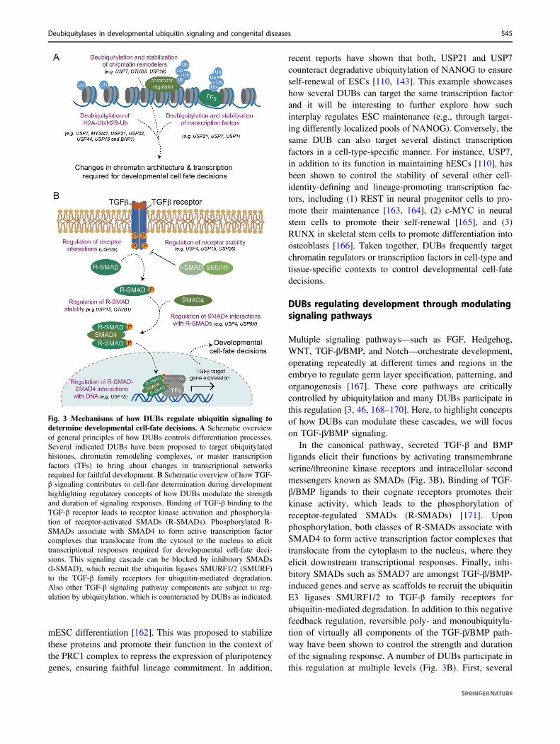
Multiple signaling pathways—such as FGF, Hedgehog,WNT, TGF-β/BMP, and Notch—orchestrate development,operating repeatedly at different times and regions in theembryo to regulate germ layer specification, patterning, andorganogenesis [167]. These core pathways are criticallycontrolled by ubiquitylation and many DUBs participate inthis regulation [3, 46, 168–170]. Here, to highlight conceptsof how DUBs can modulate these cascades, we will focuson TGF-β/BMP signaling.
In the canonical pathway, secreted TGF-β and BMPligands elicit their functions by activating transmembraneserine/threonine kinase receptors and intracellular secondmessengers known as SMADs (Fig. 3B). Binding of TGF-β/BMP ligands to their cognate receptors promotes theirkinase activity, which leads to the phosphorylation ofreceptor-regulated SMADs (R-SMADs) [171]. Uponphosphorylation, both classes of R-SMADs associate withSMAD4 to form active transcription factor complexes thattranslocate from the cytoplasm to the nucleus, where theyelicit downstream transcriptional responses. Finally, inhi-bitory SMADs such as SMAD7 are amongst TGF-β/BMP-induced genes and serve as scaffolds to recruit the ubiquitinE3 ligases SMURF1/2 to TGF-β family receptors forubiquitin-mediated degradation. In addition to this negativefeedback regulation, reversible poly- and monoubiquityla-tion of virtually all components of the TGF-β/BMP path-way have been shown to control the strength and durationof the signaling response. A number of DUBs participate inthis regulation at multiple levels (Fig. 3B). First, several
Fig. 3 Mechanisms of how DUBs regulate ubiquitin signaling todetermine developmental cell-fate decisions. A Schematic overviewof general principles of how DUBs controls differentiation processes.Several indicated DUBs have been proposed to target ubiquitylatedhistones, chromatin remodeling complexes, or master transcriptionfactors (TFs) to bring about changes in transcriptional networksrequired for faithful development. B Schematic overview of how TGF-β signaling contributes to cell-fate determination during developmenthighlighting regulatory concepts of how DUBs modulate the strengthand duration of signaling responses. Binding of TGF-β binding to theTGF-β receptor leads to receptor kinase activation and phosphoryla-tion of receptor-activated SMADs (R-SMADs). Phosphorylated R-SMADs associate with SMAD4 to form active transcription factorcomplexes that translocate from the cytosol to the nucleus to elicittranscriptional responses required for developmental cell-fate deci-sions. This signaling cascade can be blocked by inhibitory SMADs(I-SMAD), which recruit the ubiquitin ligases SMURF1/2 (SMURF)to the TGF-β family receptors for ubiquitin-mediated degradation.Also other TGF-β signaling pathway components are subject to reg-ulation by ubiquitylation, which is counteracted by DUBs as indicated.
Deubiquitylases in developmental ubiquitin signaling and congenital diseases 545

DUBs have been shown to regulate turnover of the TGF-βreceptor using diverse mode of actions. This includesDUBs that stabilize the receptor by deubiquitylation of thereceptor (USP4 [96] and USP15 [172]) and bydeubiquitylation-dependent inactivation of SMURF2(USP15 [173]) or DUBs that promote receptor degradationthrough deubiquitylating and stabilizing SMAD7 (USP26[174]). Second, DUBs regulate protein interactions of theTGF-β receptor. This is exemplified by USP2a, whichassociates with the TGF-β type I and II receptors to cleaveK33-linked ubiquitin chains from them, thus promotinginteractions with R-SMADs and enhancing downstreamsignaling [175]. Third, DUBs target ubiquitylated R-SMADs to regulate their stability and interactions. Forexample, both, USP15 and OTUB1 counteract degradativepolyubiquitylation of activated R-SMADs to promotetranscriptional downstream responses [176, 177]. Thisoccurs by different molecular mechanisms and requirescatalytic activity of USP15, but not that of OTUB1, whichrather binds to and inhibits the ubiquitin-conjugatingactivity of the cognate E2 enzyme [176, 178]. In addi-tion, USP15 also promotes TGF-β/BMP signaling byopposing monoubiquitylation of R-SMADs, therebyallowing activated R-SMAD-SMAD4 complexes torecognize target promoters [177]. Fourth, DUBs targetmonoubiquitylated SMAD4 to regulate its interactions.Both, USP9X and USP4 have been shown to catalyze thisreaction to promote activated R-SMAD/SMAD4 complexformation, nuclear translocation, and TGF-β-induced tran-scriptional activation required for zebrafish developmentand mESC differentiation (in case of USP4 [97]) orXenopus development (in case of USP9X [179]).
Thus, as outlined in the above examples, multiple DUBsmodulate TGF-β/BMP signaling at the receptor or effectorlevel through prevention of degradation or control ofprotein–protein interactions. In this context, to achieve acertain biological outcome, the same DUB can regulate thepathway at different levels (e.g., USP15 and USP4 promotesignaling by targeting the TGF-β receptor and the effectorSMADs) or multiple DUBs can act on the same target (e.g.,USP15 and OTUB1 promote signaling through stabilizingR-SMADs). Future experiments are required to furtherexamine how DUB interplay at these different levels isspatially and temporally regulated to ensure proper TGF-β/BMP signaling responses during embryonic development.
Dysregulation of DUBs results indevelopmental diseases
Extensive studies over the last decades have established thatdysregulation of DUBs leads to human diseases, in particular
cancer, neurodegeneration, and inflammation [1, 25–28, 45, 46]. In addition, mutations in DUBs frequently causesevere developmental disorders (summarized in Table 1). Ingeneral, these disorders are characterized by early-onsetneurologic deficits and are thought to be caused by loss-of-function mechanisms. In the following, we will discuss selectexamples of DUBs that have been directly linked to mono-genic developmental disorders and the proposed mechanismsof pathogenesis.
Microcephaly-capillary malformation (MIC-CAP)syndrome (MIM:614261) caused by mutations inSTAMBP/AMSH
Recessive loss-of-function mutations in STAMBP, alsoknown as AMSH, causes MIC-CAP syndrome [146]. Thesepatients have severe microcephaly with progressive corticalatrophy, intractable epilepsy, profound developmentaldelay, and multiple small capillary malformations on theskin. A variety of disease-causing mutations have beenidentified including frameshift, nonsense, splicing, andmissense mutations, implicating loss-of-function as amechanism of disease [146, 180–183]. Indeed, Amsh-defi-cient mice exhibit defects in cortical development similar tothose in patients [39]. AMSH is a DUB that, through itsK63-specific ubiquitin cleavage activity [184], controls thefate of endosomal cargos that undergo ubiquitin-dependentsorting into degradation or recycling compartments by theESCRT pathway [65, 185]. The reported disease-causingmissense mutations in AMSH are located either in thecatalytic domain reducing its K63-cleavage activity [186] orin the MIT domain potentially affecting binding to com-ponents of the ESCRT pathway [146, 185]. During thepathogenesis of MIC-CAP, dysregulation of endosomalsorting likely interferes with appropriate responses todownregulate RAS/PI3K signaling, ultimately leading tothe congenital anomalies observed in patients. In support ofthis, phenotypes of MIC-CAP syndrome closely resemblethose of RASopathies, developmental disorders caused byactivating mutations in the RAS-ERK signaling pathway[187]. However, the molecular details of how the loss ofAMSH activity results increased RAS/PI3K signaling andthe key substrates involved remain to be determined.
Hao-Fountain syndrome (MIM:616863) caused byheterozygous mutations in USP7
USP7 encodes an essential DUB for which disruption of oneallele, whether via heterozygous deletions or nonsense/mis-sense mutations, results in Hao-Foutain syndrome, a devel-opmental disorder with seizures, behavioral abnormalities,hypogonadism, and hypotonia [141, 188]. Surprisingly, the
546 M. A. Basar et al.

molecular origin of this disease was not primarily linked todysregulation of the many essential functions of USP7 inDNA repair, transcription, immune responses, or viral repli-cation [189, 190], but rather to an aberrant role in cellularprotein trafficking [141]. Elegant cell biological and bio-chemical studies demonstrated that USP7 is a component ofthe MAGE-L2-TRIM27 complex, a multi-subunit ubiquitinE3 ligase with well-established roles in retromer-dependentendosomal recycling of membrane proteins. MAGE-L2-TRIM27 regulates endosomal sorting through conjugationof K63-linked ubiquitin chains to WASH, thereby activatingthis actin nucleation promoting factor and facilitating endo-somal actin assembly [191]. USP7 acts as a rheostat forthis reaction by (1) deubiquitylating TRIM27 to protect itfrom auto-degradation and (2) by deubiquitylating WASH tolimit its activity, thus fine-tuning endosomal actin assembly[141]. MAGE-L2 is located within the Prader–Williimprinting region [192] and was identified as the causativegene in Schaaf-Yang syndrome [193, 194]. These two dis-orders share many disease manifestations with Hao-Fountainsyndrome, further suggesting that the USP7-deficiency-induced patient phenotypes are caused by aberrant endoso-mal sorting.
Mental retardation, X-linked 99 (MRX99,MIM:300919, 300968) caused by mutations inUSP9X
Mutations in USP9X, encoding an X-linked DUB, causesyndromic and non-syndromic intellectual disability. Initialstudies reported three male individuals with non-syndromicX-linked intellectual disability, all carrying missense var-iants in USP9X [195]. Consistent with this, brain-specificknockout of Usp9x causes aberrant cortical architecturesimilar to that found in patients [196]. Reijinders et al.showed that heterozygous loss-of-function alleles present infemales, as opposed to males, lead to a syndromic form ofX-linked intellectual disability associated with characteristicfacial features, short stature, cardiac, and structural brainabnormalities [197]. Together with more recent studies, thissolidified a spectrum of neurodevelopmental disease in maleand females with variable phenotypes, decreased pene-trance, and likely variant-specific mechanisms of disease,contributing to the different sex-specific manifestations[198, 199]. USP9X is an essential DUB that, throughcounteracting mono- and polyubiquitylation of specificsubstrates, has been implicated in a plethora of cellularprocesses [200]. Dysregulation of several of these functionshave been proposed to lead to the phenotypes observed inpatients. First, as described above, USP9X regulates TGF-βsignaling through deubiquitylating SMAD4 and this path-way is defective in patient fibroblasts [199]. Second,
USP9X has been shown to control centriole duplication andcentrosome biogenesis through e.g., deubiquitylating andstabilizing the centriole duplication factor STIL [201–203]as well as cilia assembly through regulating the localizationand stability of the ciliogenesis-promoting factor NPHP5[204]. Mutations in genes regulating these processes(including STIL and ICQB1 encoding for NPHP5) fre-quently result in primary microcephaly [205, 206] andciliopathies [207, 208], respectively, with considerablephenotypic overlap with USP9X patients, thus suggestingthat aberrant centrosome duplication and cilia assemblycould contribute to MRX99. Third, USP9X has been shownto regulate dendritic spine development and maintenance[144]. This occurs through deubiquitylation and stabiliza-tion of ankyrin-G, a scaffold protein that links plasmamembrane proteins to the actin/β-spectrin cytoskeleton andthereby regulates multiple neurobiological processes suchas synaptogenesis and synaptic plasticity [209–211]. Var-iants in ANK, encoding for ankyrin-G are associated withneurodevelopmental disorders [212] and USP9X patientmutations were shown to reduce interaction with ankyrin-G,strongly suggesting that abnormal ankyrin-G degradation isa pathogenic mechanism in MRX99. Consistent with this,Usp9X knockout mice exhibit synaptic abnormalities,ankyrin-G aggregates, and hyperactivity [144].
It is interesting to note that TGF-β promotes cortical spinedevelopment through promoting USP9X-dependent stabili-zation of ankyrin-G [145] and that TGF-β signaling can relyon primary cilia [213]. This raises the intriguing possibilitythat the aforementioned pathogenic mechanisms may beinterconnected and that USP9X orchestrates neurodevelop-ment by acting on several distinct substrates in differentpathways. Future research should focus on such interplayand test the relative contributions of different substrates andfunctions to the sex-specific MRX99 manifestations.
Intellectual developmental disorder withdysmorphic faces, seizures, and distal limbanomalies (MIM:617452) caused by recessivemutations in OTUD6B
Bi-allelic loss-of-function of OTUD6B causes globaldevelopmental delay, feeding difficulties, structural brainabnormalities, and congenital heart disease [40, 214].OTUD6B is a poorly characterized OTU DUB with noclearly assigned in vitro deubiquitylation activity orubiquitin-linkage preference [61]. It has been connected toprotein translation [215] and may regulate proteasome sta-bility [40]; however, further mechanistic studies arerequired to establish whether loss of these or other functionsof OTUD6B drive the aberrant differentiation processesobserved in OTUD6B patients.
Deubiquitylases in developmental ubiquitin signaling and congenital diseases 547

15q13.3 microdeletion syndrome (MIM:612001)caused in part by haploinsufficiency of OTUD7A
OTUD7A, encoding a poorly studied K11-specfic OTUDUB [61], is located in the 15q13.3 locus, which whendeleted causes a wide spectrum of neurodevelopmental andpsychiatric disorders [216–219]. 15q13.3 microdeletionsyndrome is the most common genetic cause of epilepsy[220]. Recent studies have shown that out of the six protein-coding genes that are typically encompassed in the dele-tions, OTUD7A is the most likely candidate to cause asso-ciated epilepsy. First, studies in mice have shown thatOTUD7A controls dendritic branching of cortical neurons[86]. Second, knockout of Otud7a recapitulated neurode-velopmental deficits including abnormal EEGs [87]. Third,an individual with neurodevelopmental phenotypes andepilepsy carrying biallelic OTUD7A missense variants hasbeen reported [221]. These findings highlight an importantrole in OTUD7A in controlling neurodevelopment; yet, themolecular underpinnings of this regulation, including cel-lular mechanisms and cognate E3 ligases and substrates,have remained largely unclear. Their identification will haveimportant implications for understanding distinct forms ofepilepsy.
Linkage-specific deubiquitylation deficiency-induced embryonic defect (LINKED) syndromecaused by mutations in OTUD5
Hemizygous missense and deletion variants in OTUD5,encoding an X-linked OTU DUB that prefers cleavage ofK48- and K63-linked ubiquitin chains [61, 222–224], haverecently been shown to cause a male-specific multiple con-genital disorder [41]. Affected patients suffer from a spectrumof central nervous system, craniofacial, cardiac, skeletal, andgenitourinary anomalies. OTUD5 has previously been impli-cated in regulating innate and adaptive immune signaling[224–226]; however, the reported patient phenotypes suggestan additional role of this enzyme during embryonic cell-fatedetermination. Indeed, knockout of Otud5 is embryonic lethalin mice and OTUD5-depleted hESCs are defective in neu-roectodermal differentiation, which can be rescued by re-expression of wild-type OTUD5 [41]. Interestingly, a patientvariant that affects K48- but not K63-ubiquitin chain cleavageactivity, is not able to rescue the differentiation defects, sug-gesting that the disease originates from loss of OTUD5’sactivity towards degradative K48-linked ubiquitin chains.Corroborating this notion, OTUD5 prevents the degradation ofmultiple chromatin remodelers to coordinate enhancer acti-vation during neuroectodermal differentiation. Amongst theseOTUD5 substrates are ARID1A/B, HDAC2, and HCF1,mutations of which underlie different developmental disorders(Coffin–Siris and Cornelia de Lange syndromes [227, 228],
X-linked mental retardation 3 [229]) that exhibit considerablephenotypic overlap with LINKED patients. Thus, this workreveals K48-ubiquitin chain cleavage of functionally relatedsubstrates as an essential signaling mode coordinating chro-matin remodeling during early human development. Addi-tional experiments are required to determine the moleculardetails of this regulation in the broader context ofembryogenesis.
Conclusion and perspectives
Since the initial discovery of DUBs almost 40 years ago,numerous studies have provided insights into their struc-tures, substrate/cleavage specificities, and regulatorymechanisms that allow this versatile enzyme family tocontribute to diverse cellular processes. In particular, wehere highlight principles of how DUBs modulate ubiquitinsignaling during embryonic and postnatal development andthe emerging roles of their dysregulation in congenitaldisorders. Despite many recent advances in our under-standing of DUBs in these (patho-)physiological processes,many open questions remain. First, for more than half ofthe human DUBs, substrates and linkage specificities haveremained unclear [26]. Moreover, as several DUBs arerelatively large proteins challenging to produce in bacteria,many biochemical activities have been determined withtruncation variants, which could lack important specificitydeterminants encoded in the full-length protein. Similarly,as detailed in this review, PTMs and co-factors have beenshown to regulate DUB activity and linkage-specificities incells and those contributions are not captured duringin vitro activity assays using bacterial proteins. Therefore,characterizing DUB mechanisms and specificities byin vitro and cell-based assays, particularly focusing on full-length proteins, will be important to further define phy-siological roles of DUBs and elucidate their role in disease.Second, as alluded to throughout this review, it is oftenunclear how the intricate regulatory mechanisms that canregulate DUB localization, activity, and substrate recruit-ment in vitro are implemented to ensure faithful embryonicand postnatal development in vivo. Third, while mutationsin ~10 DUBs have been convincingly demonstrated tocause developmental disease (Fig. 4), the underlyingmechanisms, E3 ligases, and/or substrates are often ill-defined (e.g., OTUD6B and OTUD7A). Fourth, knockout orknockdown of tens of DUBs has been shown to be lethal orto cause severe defects during embryogenesis of modelorganisms such as zebrafish and mice [43, 44]. In manycases, these DUBs have not yet been associated withcongenital disorders and/or their precise functions andunderlying mechanisms in early human development arenot known (e.g., OTUD4, USP25).
548 M. A. Basar et al.

With the rapid increase in databases of exome and gen-ome sequences from healthy individuals, it has now becomepossible to quantify the tolerance of genes to loss-of-function and missense mutations in control populations.[230–233] Genes that are highly restricted in such variationare likely to be essential and, when mutated, either result inembryonic lethality or developmental disease. As recentlydemonstrated for OTUD5 and LINKED syndrome [41],such genomic constraint metrics can be used to prioritizecandidate disease variants and, combined with mechanisticstudies, facilitate the discovery of novel developmentalpathways. Intriguingly, there are many DUBs, not yetassociated with congenital disorders, but that are likely to bedisease-causing based on how constrained they are frommutations in the healthy population (highlighted in violet inFig. 4). We propose that systematic search for missensevariants in these genes in patients with undiagnosed dis-eases, will likely allow identification of novel develop-mental disorders and may yield variants that can be used todissect functions and mechanisms of these DUBs duringembryogenesis. Even if such patients are not readily iden-tified, these tools provide clues about enzymes important forhuman health to prioritize for mechanistic studies. Suchgenomic constraint-based genotype-first approaches wouldbe especially interesting for poorly characterized DUBssuch as USP24, USP48, and USP32. It would be equallyattractive to apply this methodology to the linkage-specific
OTU DUBs OTUD4, OTUB1, VCPIP, and ZRANB1 [61] touncover potentially novel roles of particular ubiquitin chaintypes during early development. Finally, such methodolo-gies could provide important mechanistic insights to helpimprove disease diagnosis and patient management and,given the growing ability to target the activity of specificDUBs with small molecules [26, 45, 234], potentially opennew avenues for therapeutic intervention to amelioratedisease symptoms.
Acknowledgements We apologize to all scientists whose work couldnot be discussed within the space confines of this review. We thankmembers of our lab for continuous help and discussions, and we aregrateful to Drs Nadine Samara and Shireen Sarraf for comments onthis manuscript. MAB and AW are funded by the intramural programof the NIDCR and DBB by the intramural program of NHGRI.
Author contributions MAB and DBB helped conceptualize and for-mulate the design of the article, interpreted relevant literature, wrotethe original draft of the manuscript. AW conceptualized and for-mulated the design of the article, interpreted relevant literature, editedthe draft of the manuscript, designed figures, and acquired funding.
Compliance with ethical standards
Conflict of interest The authors declare that they have no conflict ofinterest.
Publisher’s note Springer Nature remains neutral with regard tojurisdictional claims in published maps and institutional affiliations.
Fig. 4 Many DUBs are intolerant to genomic variation in humansand are likely to cause developmental disease when mutated.Graph depicting a plot of missense (Z) and loss-of-function intolerance(pLI) scores of all human DUBs (as determined using gnomAD [233]).Highlighted in color are DUBs whose mutations have been demon-strated to cause monogenic diseases that are inherited in an autosomaldominant manner (orange), autosomal recessive manner (green), or X-linked dominant/recessive manner (blue). Mutations in USP8 (high-lighted in black) cause corticotroph adenomas and Cushing’s disease
in the somatic state. Note that DUBs associated with autosomaldominant and X-linked disease are constrained in their genomic var-iation within the healthy human population (pLI ≈ 1, Z-score > 1).Many other DUBs, previously not linked to monogenic diseases, arealso highly intolerant to missense and loss-of-function mutations andthus likely cause embryonic lethality or developmental disease whenmutated. The strongest of these candidates are highlighted in violet inthe zoomed-in panel of the plot on the right.
Deubiquitylases in developmental ubiquitin signaling and congenital diseases 549

Open Access This article is licensed under a Creative CommonsAttribution 4.0 International License, which permits use, sharing,adaptation, distribution and reproduction in any medium or format, aslong as you give appropriate credit to the original author(s) and thesource, provide a link to the Creative Commons license, and indicate ifchanges were made. The images or other third party material in thisarticle are included in the article’s Creative Commons license, unlessindicated otherwise in a credit line to the material. If material is notincluded in the article’s Creative Commons license and your intendeduse is not permitted by statutory regulation or exceeds the permitteduse, you will need to obtain permission directly from the copyrightholder. To view a copy of this license, visit http://creativecommons.org/licenses/by/4.0/.
References
1. Oh E, Akopian D, Rape M. Principles of ubiquitin-dependentsignaling. Annu Rev Cell Dev Biol. 2018;34:137–62.
2. Strikoudis A, Guillamot M, Aifantis I. Regulation of stem cellfunction by protein ubiquitylation. EMBO Rep. 2014;15:365–82.
3. Werner A, Manford AG, Rape M. Ubiquitin-dependent regula-tion of stem cell biology. Trends Cell Biol. 2017;27:568–79.
4. Komander D, Rape M. The ubiquitin code. Annu Rev Biochem.2012;81:203–29.
5. Dikic I, Wakatsuki S, Walters KJ. Ubiquitin-binding domains—from structures to functions. Nat Rev Mol Cell Biol.2009;10:659–71.
6. Husnjak K, Dikic I. Ubiquitin-binding proteins: decoders ofubiquitin-mediated cellular functions. Annu Rev Biochem.2012;81:291–322.
7. Haakonsen DL, Rape M. Branching out: improved signaling byheterotypic ubiquitin chains. Trends Cell Biol. 2019;29:704–16.
8. Di Fiore PP, Polo S, Hofmann K. When ubiquitin meets ubi-quitin receptors: a signalling connection. Nat Rev Mol Cell Biol.2003;4:491–7.
9. Haglund K, Dikic I. Ubiquitylation and cell signaling. EMBO J.2005;24:3353–9.
10. Morgan MT, Wolberger C. Recognition of ubiquitinatednucleosomes. Curr Opin Struct Biol. 2017;42:75–82.
11. Werner A, Iwasaki S, McGourty CA, Medina-Ruiz S, TeerikorpiN, Fedrigo I, et al. Cell-fate determination by ubiquitin-dependent regulation of translation. Nature. 2015;525:523–7.
12. Chau V, Tobias JW, Bachmair A, Marriott D, Ecker DJ, GondaDK, et al. A multiubiquitin chain is confined to specific lysine ina targeted short-lived protein. Science. 1989;243:1576–83.
13. Jin L, Williamson A, Banerjee S, Philipp I, Rape M. Mechanismof ubiquitin-chain formation by the human anaphase-promotingcomplex. Cell. 2008;133:653–65.
14. Tokunaga F, Sakata S, Saeki Y, Satomi Y, Kirisako T, Kamei K,et al. Involvement of linear polyubiquitylation of NEMO in NF-kappaB activation. Nat Cell Biol. 2009;11:123–32.
15. Wang C, Deng L, Hong M, Akkaraju GR, Inoue J, Chen ZJ.TAK1 is a ubiquitin-dependent kinase of MKK and IKK. Nature.2001;412:346–51.
16. Doil C, Mailand N, Bekker-Jensen S, Menard P, Larsen DH,Pepperkok R, et al. RNF168 binds and amplifies ubiquitin con-jugates on damaged chromosomes to allow accumulation ofrepair proteins. Cell. 2009;136:435–46.
17. Hoege C, Pfander B, Moldovan GL, Pyrowolakis G, Jentsch S.RAD6-dependent DNA repair is linked to modification of PCNAby ubiquitin and SUMO. Nature. 2002;419:135–41.
18. Stewart GS, Panier S, Townsend K, Al-Hakim AK, Kolas NK,Miller ES, et al. The RIDDLE syndrome protein mediates a
ubiquitin-dependent signaling cascade at sites of DNA damage.Cell. 2009;136:420–34.
19. Grumati P, Dikic I. Ubiquitin signaling and autophagy. J BiolChem. 2018;293:5404–13.
20. Kim W, Bennett EJ, Huttlin EL, Guo A, Li J, Possemato A, et al.Systematic and quantitative assessment of the ubiquitin-modifiedproteome. Mol Cell. 2011;44:325–40.
21. Pickart CM. Mechanisms underlying ubiquitination. Annu RevBiochem. 2001;70:503–33.
22. Komander D, Clague MJ, Urbe S. Breaking the chains: structureand function of the deubiquitinases. Nat Rev Mol Cell Biol.2009;10:550–63.
23. Newton K, Matsumoto ML, Wertz IE, Kirkpatrick DS, Lill JR,Tan J, et al. Ubiquitin chain editing revealed by polyubiquitinlinkage-specific antibodies. Cell. 2008;134:668–78.
24. Ronau JA, Beckmann JF, Hochstrasser M. Substrate specificityof the ubiquitin and Ubl proteases. Cell Res. 2016;26:441–56.
25. Bonacci T, Emanuele MJ. Dissenting degradation: deubiquiti-nases in cell cycle and cancer. Semin Cancer Biol. 2020;67:145–58.
26. Clague MJ, Urbe S, Komander D. Breaking the chains: deubi-quitylating enzyme specificity begets function. Nat Rev Mol CellBiol. 2019;20:338–52.
27. Heideker J, Wertz IE. DUBs, the regulation of cell identity anddisease. Biochem J. 2015;465:1–26.
28. Leznicki P, Kulathu Y. Mechanisms of regulation and diversi-fication of deubiquitylating enzyme function. J Cell Sci.2017;130:1997–2006.
29. Gomez-Diaz C, Ikeda F. Roles of ubiquitin in autophagy and celldeath. Semin Cell Dev Biol. 2019;93:125–35.
30. Vucic D, Dixit VM, Wertz IE. Ubiquitylation in apoptosis: apost-translational modification at the edge of life and death. NatRev Mol Cell Biol. 2011;12:439–52.
31. Kon N, Kobayashi Y, Li M, Brooks CL, Ludwig T, Gu W.Inactivation of HAUSP in vivo modulates p53 function. Onco-gene. 2010;29:1270–9.
32. Niendorf S, Oksche A, Kisser A, Lohler J, Prinz M, Schorle H,et al. Essential role of ubiquitin-specific protease 8 for receptortyrosine kinase stability and endocytic trafficking in vivo. MolCell Biol. 2007;27:5029–39.
33. Naik E, Webster JD, DeVoss J, Liu J, Suriben R, Dixit VM. Reg-ulation of proximal T cell receptor signaling and tolerance inductionby deubiquitinase Usp9X. J Exp Med. 2014;211:1947–55.
34. Pantaleon M, Kanai-Azuma M, Mattick JS, Kaibuchi K, KayePL, Wood SA. FAM deubiquitylating enzyme is essential forpreimplantation mouse embryo development. Mech Dev.2001;109:151–60.
35. Yang W, Lee YH, Jones AE, Woolnough JL, Zhou D, Dai Q,et al. The histone H2A deubiquitinase Usp16 regulatesembryonic stem cell gene expression and lineage commitment.Nat Commun. 2014;5:3818.
36. Lin Z, Yang H, Kong Q, Li J, Lee SM, Gao B, et al. USP22antagonizes p53 transcriptional activation by deubiquitinatingSirt1 to suppress cell apoptosis and is required for mouseembryonic development. Mol Cell. 2012;46:484–94.
37. Dey A, Seshasayee D, Noubade R, French DM, Liu J, Chaur-ushiya MS, et al. Loss of the tumor suppressor BAP1 causesmyeloid transformation. Science. 2012;337:1541–6.
38. Pasupala N, Morrow ME, Que LT, Malynn BA, Ma A, Wol-berger C. OTUB1 non-catalytically stabilizes the E2 ubiquitin-conjugating enzyme UBE2E1 by preventing its autoubiquitina-tion. J Biol Chem. 2018;293:18285–95.
39. Ishii N, Owada Y, Yamada M, Miura S, Murata K, Asao H, et al.Loss of neurons in the hippocampus and cerebral cortex ofAMSH-deficient mice. Mol Cell Biol. 2001;21:8626–37.
550 M. A. Basar et al.

40. Santiago-Sim T, Burrage LC, Ebstein F, Tokita MJ, Miller M, BiW, et al. Biallelic variants in OTUD6B cause an intellectualdisability syndrome associated with seizures and dysmorphicfeatures. Am J Hum Genet. 2017;100:676–88.
41. Beck DB, Basar MA, Asmar AJ, Thompson J, Oda H, UeharaDT, et al. Regulation of human development by ubiquitin chainediting of chromatin remodelers. Sci. Adv. 2021. In Press.
42. Cox BJ, Vollmer M, Tamplin O, Lu M, Biechele S, GertsensteinM, et al. Phenotypic annotation of the mouse X chromosome.Genome Res. 2010;20:1154–64.
43. Cheng J, Guo J, North BJ, Wang B, Cui CP, Li H, et al.Functional analysis of deubiquitylating enzymes in tumorigen-esis and development. Biochim Biophys Acta Rev Cancer.2019;1872:188312.
44. Tse WK, Eisenhaber B, Ho SH, Ng Q, Eisenhaber F, Jiang YJ.Genome-wide loss-of-function analysis of deubiquitylatingenzymes for zebrafish development. BMC Genomics. 2009;10:637.
45. Harrigan JA, Jacq X, Martin NM, Jackson SP. Deubiquitylatingenzymes and drug discovery: emerging opportunities. Nat RevDrug Discov. 2018;17:57–78.
46. Rape M. Ubiquitylation at the crossroads of development anddisease. Nat Rev Mol Cell Biol. 2018;19:59–70.
47. Beck DB, Aksentijevich I. Biochemistry of autoinflammatorydiseases: catalyzing monogenic disease. Front Immunol. 2019;10:101.
48. Hershko A, Ciechanover A, Heller H, Haas AL, Rose IA. Pro-posed role of ATP in protein breakdown: conjugation of proteinwith multiple chains of the polypeptide of ATP-dependent pro-teolysis. Proc Natl Acad Sci USA. 1980;77:1783–6.
49. Pickart CM, Rose IA. Ubiquitin carboxyl-terminal hydrolase actson ubiquitin carboxyl-terminal amides. J Biol Chem. 1985;260:7903–10.
50. Pickart CM, Rose IA. Mechanism of ubiquitin carboxyl-terminalhydrolase. Borohydride and hydroxylamine inactivate in thepresence of ubiquitin. J Biol Chem. 1986;261:10210–7.
51. Abdul Rehman SA, Kristariyanto YA, Choi SY, Nkosi PJ,Weidlich S, Labib K, et al. MINDY-1 is a member of an evo-lutionarily conserved and structurally distinct new family ofdeubiquitinating enzymes. Mol Cell. 2016;63:146–55.
52. Haahr P, Borgermann N, Guo X, Typas D, Achuthankutty D,Hoffmann S, et al. ZUFSP deubiquitylates K63-linked poly-ubiquitin chains to promote genome stability. Mol Cell.2018;70:165–74 e166.
53. Hermanns T, Pichlo C, Woiwode I, Klopffleisch K, Witting KF,Ovaa H, et al. A family of unconventional deubiquitinases withmodular chain specificity determinants. Nat Commun. 2018;9:799.
54. Hewings DS, Heideker J, Ma TP, AhYoung AP, El Oualid F,Amore A, et al. Reactive-site-centric chemoproteomics identifiesa distinct class of deubiquitinase enzymes. Nat Commun.2018;9:1162.
55. Kwasna D, Abdul Rehman SA, Natarajan J, Matthews S, Mad-den R, De Cesare V, et al. Discovery and characterization ofZUFSP/ZUP1, a distinct deubiquitinase class important forgenome stability. Mol Cell. 2018;70:150–64 e156.
56. Walden M, Masandi SK, Pawlowski K, Zeqiraj E. Pseudo-DUBsas allosteric activators and molecular scaffolds of protein com-plexes. Biochem Soc Trans. 2018;46:453–66.
57. Komander D. Mechanism, specificity and structure of the deu-biquitinases. Subcell Biochem. 2010;54:69–87.
58. Reyes-Turcu FE, Ventii KH, Wilkinson KD. Regulation andcellular roles of ubiquitin-specific deubiquitinating enzymes.Annu Rev Biochem. 2009;78:363–97.
59. Mevissen TET, Komander D. Mechanisms of deubiquitinasespecificity and regulation. Annu Rev Biochem. 2017;86:159–92.
60. Wolberger C. Mechanisms for regulating deubiquitinatingenzymes. Protein Sci. 2014;23:344–53.
61. Mevissen TE, Hospenthal MK, Geurink PP, Elliott PR, AkutsuM, Arnaudo N, et al. OTU deubiquitinases reveal mechanisms oflinkage specificity and enable ubiquitin chain restriction analysis.Cell. 2013;154:169–84.
62. Kristariyanto YA, Abdul Rehman SA, Weidlich S, Knebel A,Kulathu Y. A single MIU motif of MINDY-1 recognizes K48-linked polyubiquitin chains. EMBO Rep. 2017;18:392–402.
63. Reyes-Turcu FE, Horton JR, Mullally JE, Heroux A, Cheng X,Wilkinson KD. The ubiquitin binding domain ZnF UBP recog-nizes the C-terminal diglycine motif of unanchored ubiquitin.Cell. 2006;124:1197–208.
64. Cooper EM, Boeke JD, Cohen RE. Specificity of the BRISCdeubiquitinating enzyme is not due to selective binding toLys63-linked polyubiquitin. J Biol Chem. 2010;285:10344–52.
65. McCullough J, Row PE, Lorenzo O, Doherty M, Beynon R,Clague MJ, et al. Activation of the endosome-associated ubi-quitin isopeptidase AMSH by STAM, a component of the mul-tivesicular body-sorting machinery. Curr Biol. 2006;16:160–5.
66. Sato Y, Yoshikawa A, Yamagata A, Mimura H, Yamashita M,Ookata K, et al. Structural basis for specific cleavage of Lys 63-linked polyubiquitin chains. Nature. 2008;455:358–62.
67. Cunningham CN, Baughman JM, Phu L, Tea JS, Yu C, CoonsM, et al. USP30 and parkin homeostatically regulate atypicalubiquitin chains on mitochondria. Nat Cell Biol. 2015;17:160–9.
68. Gersch M, Gladkova C, Schubert AF, Michel MA, Maslen S,Komander D. Mechanism and regulation of the Lys6-selectivedeubiquitinase USP30. Nat Struct Mol Biol. 2017;24:920–30.
69. Sato Y, Okatsu K, Saeki Y, Yamano K, Matsuda N, Kaiho A,et al. Structural basis for specific cleavage of Lys6-linked poly-ubiquitin chains by USP30. Nat Struct Mol Biol. 2017;24:911–9.
70. Komander D, Reyes-Turcu F, Licchesi JD, Odenwaelder P,Wilkinson KD, Barford D. Molecular discrimination of structu-rally equivalent Lys 63-linked and linear polyubiquitin chains.EMBO Rep. 2009;10:466–73.
71. Sato Y, Goto E, Shibata Y, Kubota Y, Yamagata A, Goto-Ito S,et al. Structures of CYLD USP with Met1- or Lys63-linkeddiubiquitin reveal mechanisms for dual specificity. Nat StructMol Biol. 2015;22:222–9.
72. Faesen AC, Luna-Vargas MP, Geurink PP, Clerici M, Merkx R,van Dijk WJ, et al. The differential modulation of USP activityby internal regulatory domains, interactors and eight ubiquitinchain types. Chem Biol. 2011;18:1550–61.
73. Deshaies RJ, Joazeiro CA. RING domain E3 ubiquitin ligases.Annu Rev Biochem. 2009;78:399–434.
74. Asmar AJ, Beck DB, Werner A. Control of craniofacial andbrain development by Cullin3-RING ubiquitin ligases: lessonsfrom human disease genetics. Exp Cell Res. 2020;396:112300.
75. Sahtoe DD, Sixma TK. Layers of DUB regulation. TrendsBiochem Sci. 2015;40:456–67.
76. Lee MJ, Yaffe MB. Protein regulation in signal transduction.Cold Spring Harb Perspect Biol. 2016;7:a005918.
77. Clague MJ, Barsukov I, Coulson JM, Liu H, Rigden DJ, Urbe S.Deubiquitylases from genes to organism. Physiol Rev. 2013;93:1289–315.
78. Clague MJ, Heride C, Urbe S. The demographics of the ubiquitinsystem. Trends Cell Biol. 2015;25:417–26.
79. Fuchs G, Shema E, Vesterman R, Kotler E, Wolchinsky Z,Wilder S, et al. RNF20 and USP44 regulate stem cell differ-entiation by modulating H2B monoubiquitylation. Mol Cell.2012;46:662–73.
80. Boyer LA, Lee TI, Cole MF, Johnstone SE, Levine SS, ZuckerJP, et al. Core transcriptional regulatory circuitry in humanembryonic stem cells. Cell. 2005;122:947–56.
Deubiquitylases in developmental ubiquitin signaling and congenital diseases 551

81. Sussman RT, Stanek TJ, Esteso P, Gearhart JD, Knudsen KE,McMahon SB. The epigenetic modifier ubiquitin-specific pro-tease 22 (USP22) regulates embryonic stem cell differentiationvia transcriptional repression of sex-determining region Y-box 2(SOX2). J Biol Chem. 2013;288:24234–46.
82. Kapushesky M, Adamusiak T, Burdett T, Culhane A, Farne A,Filippov A, et al. Gene Expression Atlas update–a value-addeddatabase of microarray and sequencing-based functional geno-mics experiments. Nucleic Acids Res. 2012;40:D1077–1081.
83. Lowe J, McDermott H, Landon M, Mayer RJ, Wilkinson KD.Ubiquitin carboxyl-terminal hydrolase (PGP 9.5) is selectivelypresent in ubiquitinated inclusion bodies characteristic of humanneurodegenerative diseases. J Pathol. 1990;161:153–60.
84. Burnett B, Li F, Pittman RN. The polyglutamine neurodegen-erative protein ataxin-3 binds polyubiquitylated proteins and hasubiquitin protease activity. Hum Mol Genet. 2003;12:3195–205.
85. Kawaguchi Y, Okamoto T, Taniwaki M, Aizawa M, Inoue M,Katayama S, et al. CAG expansions in a novel gene forMachado-Joseph disease at chromosome 14q32.1. Nat Genet.1994;8:221–8.
86. Uddin M, Unda BK, Kwan V, Holzapfel NT, White SH, ChalilL, et al. OTUD7A regulates neurodevelopmental phenotypes inthe 15q13.3 microdeletion syndrome. Am J Hum Genet.2018;102:278–95.
87. Yin J, Chen W, Chao ES, Soriano S, Wang L, Wang W, et al.Otud7a knockout mice recapitulate many neurological featuresof 15q13.3 microdeletion syndrome. Am J Hum Genet.2018;102:296–308.
88. Bilguvar K, Tyagi NK, Ozkara C, Tuysuz B, Bakircioglu M,Choi M, et al. Recessive loss of function of the neuronal ubi-quitin hydrolase UCHL1 leads to early-onset progressive neu-rodegeneration. Proc Natl Acad Sci USA. 2013;110:3489–94.
89. Gong B, Cao Z, Zheng P, Vitolo OV, Liu S, Staniszewski A,et al. Ubiquitin hydrolase Uch-L1 rescues beta-amyloid-induceddecreases in synaptic function and contextual memory. Cell.2006;126:775–88.
90. Leroy E, Boyer R, Auburger G, Leube B, Ulm G, Mezey E, et al.The ubiquitin pathway in Parkinson’s disease. Nature. 1998;395:451–2.
91. Legarda D, Justus SJ, Ang RL, Rikhi N, Li W, Moran TM, et al.CYLD proteolysis protects macrophages from TNF-mediatedauto-necroptosis induced by LPS and licensed by type I IFN.Cell Rep. 2016;15:2449–61.
92. O’Donnell MA, Perez-Jimenez E, Oberst A, Ng A, Massoumi R,Xavier R, et al. Caspase 8 inhibits programmed necrosis byprocessing CYLD. Nat Cell Biol. 2011;13:1437–42.
93. Coornaert B, Baens M, Heyninck K, Bekaert T, Haegman M,Staal J, et al. T cell antigen receptor stimulation induces MALT1paracaspase-mediated cleavage of the NF-kappaB inhibitor A20.Nat Immunol. 2008;9:263–71.
94. Huang TT, Nijman SM, Mirchandani KD, Galardy PJ, CohnMA, Haas W, et al. Regulation of monoubiquitinated PCNA byDUB autocleavage. Nat Cell Biol. 2006;8:339–47.
95. Piatkov KI, Colnaghi L, Bekes M, Varshavsky A, Huang TT.The auto-generated fragment of the Usp1 deubiquitylase is aphysiological substrate of the N-end rule pathway. Mol Cell.2012;48:926–33.
96. Zhang L, Zhou F, Drabsch Y, Gao R, Snaar-Jagalska BE,Mickanin C, et al. USP4 is regulated by AKT phosphorylationand directly deubiquitylates TGF-beta type I receptor. Nat CellBiol. 2012;14:717–26.
97. Zhou F, Xie F, Jin K, Zhang Z, Clerici M, Gao R, et al. USP4inhibits SMAD4 monoubiquitination and promotes activin andBMP signaling. EMBO J. 2017;36:1623–39.
98. Bauer NC, Doetsch PW, Corbett AH. Mechanisms regulatingprotein localization. Traffic. 2015;16:1039–61.
99. Urbe S, Liu H, Hayes SD, Heride C, Rigden DJ, Clague MJ.Systematic survey of deubiquitinase localization identifiesUSP21 as a regulator of centrosome- and microtubule-associatedfunctions. Mol Biol Cell. 2012;23:1095–103.
100. Hassink GC, Zhao B, Sompallae R, Altun M, Gastaldello S,Zinin NV, et al. The ER-resident ubiquitin-specific protease 19participates in the UPR and rescues ERAD substrates. EMBORep. 2009;10:755–61.
101. Lee JG, Takahama S, Zhang G, Tomarev SI, Ye Y. Unconven-tional secretion of misfolded proteins promotes adaptation toproteasome dysfunction in mammalian cells. Nat Cell Biol.2016;18:765–76.
102. Thorne C, Eccles RL, Coulson JM, Urbe S, Clague MJ. Isoform-specific localization of the deubiquitinase USP33 to the Golgiapparatus. Traffic. 2011;12:1563–74.
103. Leznicki P, Natarajan J, Bader G, Spevak W, Schlattl A, AbdulRehman SA, et al. Expansion of DUB functionality generated byalternative isoforms - USP35, a case study. J Cell Sci. 2018;131:jcs212753.
104. Herhaus L, Perez-Oliva AB, Cozza G, Gourlay R, Weidlich S,Campbell DG, et al. Casein kinase 2 (CK2) phosphorylates thedeubiquitylase OTUB1 at Ser16 to trigger its nuclear localiza-tion. Sci Signal. 2015;8:ra35.
105. Mashtalir N, Daou S, Barbour H, Sen NN, Gagnon J, Hammond-Martel I, et al. Autodeubiquitination protects the tumor sup-pressor BAP1 from cytoplasmic sequestration mediated by theatypical ubiquitin ligase UBE2O. Mol Cell. 2014;54:392–406.
106. Lan X, Atanassov BS, Li W, Zhang Y, Florens L, Mohan RD,et al. USP44 is an integral component of N-CoR that contributesto gene repression by deubiquitinating histone H2B. Cell Rep.2016;17:2382–93.
107. Atanassov BS, Mohan RD, Lan X, Kuang X, Lu Y, Lin K, et al.ATXN7L3 and ENY2 coordinate activity of multiple H2Bdeubiquitinases important for cellular proliferation and tumorgrowth. Mol Cell. 2016;62:558–71.
108. Kolovos P, Nishimura K, Sankar A, Sidoli S, Cloos PA, Helin K,et al. PR-DUB maintains the expression of critical genes throughFOXK1/2- and ASXL1/2/3-dependent recruitment to chromatinand H2AK119ub1 deubiquitination. Genome Res. 2020;30:1119–30.
109. Campagne A, Lee MK, Zielinski D, Michaud A, Le Corre S,Dingli F, et al. BAP1 complex promotes transcription byopposing PRC1-mediated H2A ubiquitylation. Nat Commun.2019;10:348.
110. Wei X, Guo J, Li Q, Jia Q, Jing Q, Li Y, et al. Bach1 regulatesself-renewal and impedes mesendodermal differentiation ofhuman embryonic stem cells. Sci Adv. 2019;5:eaau7887.
111. Scheuermann JC, de Ayala Alonso AG, Oktaba K, Ly-Hartig N,McGinty RK, Fraterman S, et al. Histone H2A deubiquitinaseactivity of the Polycomb repressive complex PR-DUB. Nature.2010;465:243–7.
112. Daou S, Hammond-Martel I, Mashtalir N, Barbour H, Gagnon J,Iannantuono NV, et al. The BAP1/ASXL2 histone H2A deubi-quitinase complex regulates cell proliferation and is disrupted incancer. J Biol Chem. 2015;290:28643–63.
113. Sahtoe DD, van Dijk WJ, Ekkebus R, Ovaa H, Sixma TK.BAP1/ASXL1 recruitment and activation for H2A deubiquiti-nation. Nat Commun. 2016;7:10292.
114. Daou S, Barbour H, Ahmed O, Masclef L, Baril C, Sen Nkwe N,et al. Monoubiquitination of ASXLs controls the deubiquitinaseactivity of the tumor suppressor BAP1. Nat Commun.2018;9:4385.
115. Faesen AC, Dirac AM, Shanmugham A, Ovaa H, Perrakis A,Sixma TK. Mechanism of USP7/HAUSP activation by its C-terminal ubiquitin-like domain and allosteric regulation by GMP-synthetase. Mol Cell. 2011;44:147–59.
552 M. A. Basar et al.

116. Kim RQ, Geurink PP, Mulder MPC, Fish A, Ekkebus R, ElOualid F, et al. Kinetic analysis of multistep USP7 mechanismshows critical role for target protein in activity. Nat Commun.2019;10:231.
117. van der Knaap JA, Kumar BR, Moshkin YM, Langenberg K,Krijgsveld J, Heck AJ, et al. GMP synthetase stimulates histoneH2B deubiquitylation by the epigenetic silencer USP7. Mol Cell.2005;17:695–707.
118. Cohn MA, Kee Y, Haas W, Gygi SP, D’Andrea AD. UAF1 is asubunit of multiple deubiquitinating enzyme complexes. J BiolChem. 2009;284:5343–51.
119. Kee Y, Yang K, Cohn MA, Haas W, Gygi SP, D’Andrea AD.WDR20 regulates activity of the USP12 x UAF1 deubiquiti-nating enzyme complex. J Biol Chem. 2010;285:11252–7.
120. Li H, Lim KS, Kim H, Hinds TR, Jo U, Mao H, et al. Allostericactivation of ubiquitin-specific proteases by beta-propeller pro-teins UAF1 and WDR20. Mol Cell. 2016;63:249–60.
121. Williams SA, Maecker HL, French DM, Liu J, Gregg A, Sil-verstein LB, et al. USP1 deubiquitinates ID proteins to preserve amesenchymal stem cell program in osteosarcoma. Cell.2011;146:918–30.
122. Borodovsky A, Kessler BM, Casagrande R, Overkleeft HS,Wilkinson KD, Ploegh HL. A novel active site-directed probespecific for deubiquitylating enzymes reveals proteasome asso-ciation of USP14. EMBO J. 2001;20:5187–96.
123. Qiu XB, Ouyang SY, Li CJ, Miao S, Wang L, Goldberg AL.hRpn13/ADRM1/GP110 is a novel proteasome subunit that bindsthe deubiquitinating enzyme, UCH37. EMBO J. 2006;25:5742–53.
124. Worden EJ, Padovani C, Martin A. Structure of the Rpn11-Rpn8dimer reveals mechanisms of substrate deubiquitination duringproteasomal degradation. Nat Struct Mol Biol. 2014;21:220–7.
125. Yao T, Song L, Xu W, DeMartino GN, Florens L, Swanson SK,et al. Proteasome recruitment and activation of the Uch37 deubi-quitinating enzyme by Adrm1. Nat Cell Biol. 2006;8:994–1002.
126. Buckley SM, Aranda-Orgilles B, Strikoudis A, Apostolou E,Loizou E, Moran-Crusio K, et al. Regulation of pluripotency andcellular reprogramming by the ubiquitin-proteasome system. CellStem Cell. 2012;11:783–98.
127. Kohler A, Zimmerman E, Schneider M, Hurt E, Zheng N.Structural basis for assembly and activation of the hetero-tetrameric SAGA histone H2B deubiquitinase module. Cell.2010;141:606–17.
128. Koutelou E, Hirsch CL, Dent SY. Multiple faces of the SAGAcomplex. Curr Opin Cell Biol. 2010;22:374–82.
129. Samara NL, Datta AB, Berndsen CE, Zhang X, Yao T, CohenRE, et al. Structural insights into the assembly and function ofthe SAGA deubiquitinating module. Science. 2010;328:1025–9.
130. Xu D, Liu J, Fu T, Shan B, Qian L, Pan L, et al. USP25 regulatesWnt signaling by controlling the stability of tankyrases. GenesDev. 2017;31:1024–35.
131. Gersch M, Wagstaff JL, Toms AV, Graves B, Freund SMV,Komander D. Distinct USP25 and USP28 oligomerization statesregulate deubiquitinating activity. Mol Cell. 2019;74:436–51 e437.
132. Liu B, Sureda-Gomez M, Zhen Y, Amador V, Reverter D. Aquaternary tetramer assembly inhibits the deubiquitinatingactivity of USP25. Nat Commun. 2018;9:4973.
133. Sauer F, Klemm T, Kollampally RB, Tessmer I, Nair RK, PopovN, et al. Differential oligomerization of the deubiquitinasesUSP25 and USP28 regulates their activities. Mol Cell.2019;74:421–35 e410.
134. Sowa ME, Bennett EJ, Gygi SP, Harper JW. Defining the humandeubiquitinating enzyme interaction landscape. Cell. 2009;138:389–403.
135. Nielsen CP, MacGurn JA. Coupling conjugation and deconju-gation activities to achieve cellular ubiquitin dynamics. TrendsBiochem Sci. 2020;45:427–39.
136. Ahmed N, Zeng M, Sinha I, Polin L, Wei WZ, Rathinam C, et al.The E3 ligase Itch and deubiquitinase Cyld act together to reg-ulate Tak1 and inflammation. Nat Immunol. 2011;12:1176–83.
137. Wertz IE, O’Rourke KM, Zhou H, Eby M, Aravind L, SeshagiriS, et al. De-ubiquitination and ubiquitin ligase domains of A20downregulate NF-kappaB signalling. Nature. 2004;430:694–9.
138. Nielsen CP, Jernigan KK, Diggins NL, Webb DJ, MacGurn JA.USP9X deubiquitylates DVL2 to regulate WNT pathway spe-cification. Cell Rep. 2019;28:1074–89 e1075.
139. Nusse R, Clevers H. Wnt/beta-catenin signaling, disease, andemerging therapeutic modalities. Cell. 2017;169:985–99.
140. Steinhart Z, Angers S. Wnt signaling in development and tissuehomeostasis. Development. 2018;145:dev146589.
141. Hao YH, Fountain MD Jr., Fon Tacer K, Xia F, Bi W, Kang SH,et al. USP7 acts as a molecular rheostat to promote WASH-dependent endosomal protein recycling and is mutated in ahuman neurodevelopmental disorder. Mol Cell. 2015;59:956–69.
142. Greenblatt MB, Shin DY, Oh H, Lee KY, Zhai B, Gygi SP, et al.MEKK2 mediates an alternative beta-catenin pathway that pro-motes bone formation. Proc Natl Acad Sci USA. 2016;113:E1226–1235.
143. Jin J, Liu J, Chen C, Liu Z, Jiang C, Chu H, et al. The deubi-quitinase USP21 maintains the stemness of mouse embryonic stemcells via stabilization of Nanog. Nat Commun. 2016;7:13594.
144. Yoon S, Parnell E, Kasherman M, Forrest MP, Myczek K,Premarathne S, et al. Usp9X controls ankyrin-repeat domainprotein homeostasis during dendritic spine development. Neuron.2020;105:506–21 e507.
145. Yoon S, Parnell E, Penzes P. TGF-beta-induced phosphorylationof Usp9X stabilizes ankyrin-G and regulates dendritic spinedevelopment and maintenance. Cell Rep. 2020;31:107685.
146. McDonell LM, Mirzaa GM, Alcantara D, Schwartzentruber J,Carter MT, Lee LJ, et al. Mutations in STAMBP, encoding adeubiquitinating enzyme, cause microcephaly-capillary mal-formation syndrome. Nat Genet. 2013;45:556–62.
147. Gu H, Shi X, Liu C, Wang C, Sui N, Zhao Y, et al. USP8maintains embryonic stem cell stemness via deubiquitination ofEPG5. Nat Commun. 2019;10:1465.
148. Blackledge NP, Farcas AM, Kondo T, King HW, McGouran JF,Hanssen LLP, et al. Variant PRC1 complex-dependent H2Aubiquitylation drives PRC2 recruitment and Polycomb domainformation. Cell. 2014;157:1445–59.
149. Cao R, Wang L, Wang H, Xia L, Erdjument-Bromage H, TempstP, et al. Role of histone H3 lysine 27 methylation in Polycomb-group silencing. Science. 2002;298:1039–43.
150. Cooper S, Dienstbier M, Hassan R, Schermelleh L, Sharif J,Blackledge NP, et al. Targeting Polycomb to pericentric het-erochromatin in embryonic stem cells reveals a role forH2AK119u1 in PRC2 recruitment. Cell Rep. 2014;7:1456–70.
151. Kalb R, Latwiel S, Baymaz HI, Jansen PW, Muller CW, Ver-meulen M, et al. Histone H2A monoubiquitination promoteshistone H3 methylation in Polycomb repression. Nat Struct MolBiol. 2014;21:569–71.
152. Kim J, Hake SB, Roeder RG. The human homolog of yeastBRE1 functions as a transcriptional coactivator through directactivator interactions. Mol Cell. 2005;20:759–70.
153. Pavri R, Zhu B, Li G, Trojer P, Mandal S, Shilatifard A, et al.Histone H2B monoubiquitination functions cooperatively withFACT to regulate elongation by RNA polymerase II. Cell.2006;125:703–17.
154. Adorno M, Sikandar S, Mitra SS, Kuo A, Nicolis Di Robilant B,Haro-Acosta V, et al. Usp16 contributes to somatic stem-celldefects in Down’s syndrome. Nature. 2013;501:380–4.
155. Jiang XX, Nguyen Q, Chou Y, Wang T, Nandakumar V, YatesP, et al. Control of B cell development by the histone H2Adeubiquitinase MYSM1. Immunity. 2011;35:883–96.
Deubiquitylases in developmental ubiquitin signaling and congenital diseases 553

156. Li P, Yang YM, Sanchez S, Cui DC, Dang RJ, Wang XY, et al.Deubiquitinase MYSM1 is essential for normal bone formationand mesenchymal stem cell differentiation. Sci Rep. 2016;6:22211.
157. Zhu P, Zhou W, Wang J, Puc J, Ohgi KA, Erdjument-BromageH, et al. A histone H2A deubiquitinase complex coordinatinghistone acetylation and H1 dissociation in transcriptional reg-ulation. Mol Cell. 2007;27:609–21.
158. Liefke R, Karwacki-Neisius V, Shi Y. EPOP interacts withelongin BC and USP7 to modulate the chromatin landscape. MolCell. 2016;64:659–72.
159. Morgan MT, Haj-Yahya M, Ringel AE, Bandi P, Brik A, Wol-berger C. Structural basis for histone H2B deubiquitination bythe SAGA DUB module. Science. 2016;351:725–8.
160. Gu Y, Jones AE, Yang W, Liu S, Dai Q, Liu Y, et al. The histoneH2A deubiquitinase Usp16 regulates hematopoiesis and hema-topoietic stem cell function. Proc Natl Acad Sci USA. 2016;113:E51–60.
161. Lecona E, Narendra V, Reinberg D. USP7 cooperates withSCML2 to regulate the activity of PRC1. Mol Cell Biol. 2015;35:1157–68.
162. Ning B, Zhao W, Qian C, Liu P, Li Q, Li W, et al. USP26functions as a negative regulator of cellular reprogramming bystabilising PRC1 complex components. Nat Commun. 2017;8:349.
163. Huang Z, Wu Q, Guryanova OA, Cheng L, Shou W, Rich JN, et al.Deubiquitylase HAUSP stabilizes REST and promotes maintenanceof neural progenitor cells. Nat Cell Biol. 2011;13:142–52.
164. Westbrook TF, Hu G, Ang XL, Mulligan P, Pavlova NN, LiangA, et al. SCFbeta-TRCP controls oncogenic transformation andneural differentiation through REST degradation. Nature.2008;452:370–4.
165. Nicklas S, Hillje AL, Okawa S, Rudolph IM, Collmann FM, vanWuellen T, et al. A complex of the ubiquitin ligase TRIM32 andthe deubiquitinase USP7 balances the level of c-Myc ubiquiti-nation and thereby determines neural stem cell fate specification.Cell Death Differ. 2019;26:728–40.
166. Kim JM, Yang YS, Park KH, Ge X, Xu R, Li N, et al. ARUNX2 stabilization pathway mediates physiologic and patho-logic bone formation. Nat Commun. 2020;11:2289.
167. Sanz-Ezquerro JJ, Munsterberg AE, Stricker S. Editorial: sig-naling pathways in embryonic development. Front Cell DevBiol. 2017;5:76.
168. Le Bras S, Loyer N, Le, Borgne R. The multiple facets of ubi-quitination in the regulation of notch signaling pathway. Traffic.2011;12:149–61.
169. Clague MJ, Coulson JM, Urbe S. Cellular functions of theDUBs. J Cell Sci. 2012;125:277–86.
170. Herhaus L, Sapkota GP. The emerging roles of deubiquitylatingenzymes (DUBs) in the TGFbeta and BMP pathways. CellSignal. 2014;26:2186–92.
171. Gaarenstroom T, Hill CS. TGF-beta signaling to chromatin: howSmads regulate transcription during self-renewal and differ-entiation. Semin Cell Dev Biol. 2014;32:107–18.
172. Eichhorn PJ, Rodon L, Gonzalez-Junca A, Dirac A, Gili M,Martinez-Saez E, et al. USP15 stabilizes TGF-beta receptor I andpromotes oncogenesis through the activation of TGF-beta sig-naling in glioblastoma. Nat Med. 2012;18:429–35.
173. Iyengar PV, Jaynes P, Rodon L, Lama D, Law KP, Lim YP,et al. USP15 regulates SMURF2 kinetics through C-lobe medi-ated deubiquitination. Sci Rep. 2015;5:14733.
174. Kit Leng Lui S, Iyengar PV, Jaynes P, Isa Z, Pang B, Tan TZ,et al. USP26 regulates TGF-beta signaling by deubiquitinatingand stabilizing SMAD7. EMBO reports. 2017;18:797–808.
175. Zhao Y, Wang X, Wang Q, Deng Y, Li K, Zhang M, et al.USP2a Supports Metastasis by Tuning TGF-beta Signaling. CellRep. 2018;22:2442–54.
176. Herhaus L, Al-Salihi M, Macartney T, Weidlich S, Sapkota GP.OTUB1 enhances TGFbeta signalling by inhibiting the ubiqui-tylation and degradation of active SMAD2/3. Nat Commun.2013;4:2519.
177. Inui M, Manfrin A, Mamidi A, Martello G, Morsut L, Soligo S,et al. USP15 is a deubiquitylating enzyme for receptor-activatedSMADs. Nat Cell Biol. 2011;13:1368–75.
178. Wiener R, Zhang X, Wang T, Wolberger C. The mechanism ofOTUB1-mediated inhibition of ubiquitination. Nature. 2012;483:618–22.
179. Dupont S, Mamidi A, Cordenonsi M, Montagner M, ZacchignaL, Adorno M, et al. FAM/USP9x, a deubiquitinating enzymeessential for TGFbeta signaling, controls Smad4 mono-ubiquitination. Cell. 2009;136:123–35.
180. Carter MT, Mirzaa G, McDonell LM, Boycott KM.Microcephaly-capillary malformation syndrome. In: Adam MP,Ardinger HH, Pagon RA, Wallace SE, Bean LJH, Stephens K,et al. editors. Seattle (WA): GeneReviews((R)); 1993.
181. Faqeih EA, Bastaki L, Rosti RO, Spencer EG, Zada AP, SalehMA, et al. Novel STAMBP mutation and additional findings inan Arabic family. Am J Med Genet A. 2015;167A:805–9.
182. Naseer MI, Sogaty S, Rasool M, Chaudhary AG, Abutalib YA,Walker S, et al. Microcephaly-capillary malformation syndrome:Brothers with a homozygous STAMBP mutation, uncovered byexome sequencing. Am J Med Genet A. 2016;170:3018–22.
183. Wu F, Dai Y, Wang J, Cheng M, Wang Y, Li X, et al. Earlyonsetepilepsy and microcephalycapillary malformation syndromecaused by a novel STAMBP mutation in a Chinese boy. MolMed Rep. 2019;20:5145–51.
184. McCullough J, Clague MJ, Urbe S. AMSH is an endosome-associated ubiquitin isopeptidase. J Cell Biol. 2004;166:487–92.
185. Clague MJ, Urbe S. Endocytosis: the DUB version. Trends CellBiol. 2006;16:551–9.
186. Shrestha RK, Ronau JA, Davies CW, Guenette RG, Strieter ER,Paul LN, et al. Insights into the mechanism of deubiquitinationby JAMM deubiquitinases from cocrystal structures of theenzyme with the substrate and product. Biochemistry.2014;53:3199–217.
187. Tidyman WE, Rauen KA. The RASopathies: developmentalsyndromes of Ras/MAPK pathway dysregulation. Curr OpinGenet Dev. 2009;19:230–6.
188. Tacer KF, Potts PR. Cellular and disease functions of the Prader-Willi Syndrome gene MAGEL2. Biochem J. 2017;474:2177–90.
189. Li M, Chen D, Shiloh A, Luo J, Nikolaev AY, Qin J, et al.Deubiquitination of p53 by HAUSP is an important pathway forp53 stabilization. Nature. 2002;416:648–53.
190. Nicholson B, Suresh, Kumar KG. The multifaceted roles ofUSP7: new therapeutic opportunities. Cell Biochem Biophys.2011;60:61–68.
191. Hao YH, Doyle JM, Ramanathan S, Gomez TS, Jia D, Xu M,et al. Regulation of WASH-dependent actin polymerization andprotein trafficking by ubiquitination. Cell. 2013;152:1051–64.
192. Chibuk TK, Bischof JM, Wevrick R. A necdin/MAGE-like genein the chromosome 15 autism susceptibility region: expression,imprinting, and mapping of the human and mouse orthologues.BMC Genet. 2001;2:22.
193. Boccaccio I, Glatt-Deeley H, Watrin F, Roeckel N, Lalande M,Muscatelli F. The human MAGEL2 gene and its mouse homo-logue are paternally expressed and mapped to the Prader-Williregion. Hum Mol Genet. 1999;8:2497–505.
194. Schaaf CP, Gonzalez-Garay ML, Xia F, Potocki L, Gripp KW,Zhang B, et al. Truncating mutations of MAGEL2 cause Prader-Willi phenotypes and autism. Nat Genet. 2013;45:1405–8.
195. Homan CC, Kumar R, Nguyen LS, Haan E, Raymond FL, AbidiF, et al. Mutations in USP9X are associated with X-linked
554 M. A. Basar et al.

intellectual disability and disrupt neuronal cell migration andgrowth. Am J Hum Genet. 2014;94:470–8.
196. Stegeman S, Jolly LA, Premarathne S, Gecz J, Richards LJ,Mackay-Sim A, et al. Loss of Usp9x disrupts cortical archi-tecture, hippocampal development and TGFbeta-mediated axo-nogenesis. PLoS One. 2013;8:e68287.
197. Reijnders MR, Zachariadis V, Latour B, Jolly L, Mancini GM,Pfundt R, et al. De novo loss-of-function mutations in USP9Xcause a female-specific recognizable syndrome with develop-mental delay and congenital malformations. Am J Hum Genet.2016;98:373–81.
198. Au PYB, Huang L, Broley S, Gallagher L, Creede E, Lahey D,et al. Two females with mutations in USP9X highlight thevariable expressivity of the intellectual disability syndrome. EurJ Med Genet. 2017;60:359–64.
199. Johnson BV, Kumar R, Oishi S, Alexander S, Kasherman M,Vega MS, et al. Partial loss of USP9X function leads to a maleneurodevelopmental and behavioral disorder converging ontransforming growth factor beta signaling. Biol Psychiatry.2020;87:100–12.
200. Murtaza M, Jolly LA, Gecz J, Wood SA. La FAM fatale: USP9Xin development and disease. Cell Mol Life Sci. 2015;72:2075–89.
201. Kodani A, Moyer T, Chen A, Holland A, Walsh CA, Reiter JF.SFI1 promotes centriole duplication by recruiting USP9X tostabilize the microcephaly protein STIL. J Cell Biol.2019;218:2185–97.
202. Han KJ, Wu Z, Pearson CG, Peng J, Song K, Liu CW. Deubi-quitylase USP9X maintains centriolar satellite integrity by sta-bilizing pericentriolar material 1 protein. J Cell Sci. 2019;132:jcs221663.
203. Wang Q, Tang Y, Xu Y, Xu S, Jiang Y, Dong Q, et al. The X-linked deubiquitinase USP9X is an integral component of cen-trosome. J Biol Chem. 2017;292:12874–84.
204. Das A, Qian J, Tsang WY. USP9X counteracts differentialubiquitination of NPHP5 by MARCH7 and BBS11 to regulateciliogenesis. PLoS Genet. 2017;13:e1006791.
205. Jayaraman D, Bae BI, Walsh CA. The genetics of primarymicrocephaly. Annu Rev Genomics Hum Genet. 2018;19:177–200.
206. Kumar A, Girimaji SC, Duvvari MR, Blanton SH. Mutations inSTIL, encoding a pericentriolar and centrosomal protein, causeprimary microcephaly. Am J Hum Genet. 2009;84:286–90.
207. Logan CV, Abdel-Hamed Z, Johnson CA. Molecular geneticsand pathogenic mechanisms for the severe ciliopathies: insightsinto neurodevelopment and pathogenesis of neural tube defects.Mol Neurobiol. 2011;43:12–26.
208. Suciu SK, Caspary T. Cilia, neural development and disease.Semin Cell Dev Biol. 2020. In press.
209. Bennett V, Healy J. Organizing the fluid membrane bilayer: diseaseslinked to spectrin and ankyrin. Trends Mol Med. 2008;14:28–36.
210. Smith KR, Kopeikina KJ, Fawcett-Patel JM, Leaderbrand K,Gao R, Schurmann B, et al. Psychiatric risk factor ANK3/ankyrin-G nanodomains regulate the structure and function ofglutamatergic synapses. Neuron. 2014;84:399–415.
211. Tseng WC, Jenkins PM, Tanaka M, Mooney R, Bennett V. Giantankyrin-G stabilizes somatodendritic GABAergic synapsesthrough opposing endocytosis of GABAA receptors. Proc NatlAcad Sci USA. 2015;112:1214–9.
212. Iqbal Z, Vandeweyer G, van der Voet M, Waryah AM, ZahoorMY, Besseling JA, et al. Homozygous and heterozygous dis-ruptions of ANK3: at the crossroads of neurodevelopmental andpsychiatric disorders. Hum Mol Genet. 2013;22:1960–70.
213. Christensen ST, Morthorst SK, Mogensen JB, Pedersen LB.Primary cilia and coordination of receptor tyrosine kinase (RTK)
and transforming growth factor beta (TGF-beta) signaling. ColdSpring Harb Perspect Biol. 2017;9:a028167.
214. Straniero L, Rimoldi V, Solda G, Bellini M, Biasucci G, AsseltaR, et al. First replication of the involvement of OTUD6B inintellectual disability syndrome with seizures and dysmorphicfeatures. Front Genet. 2018;9:464.
215. Sobol A, Askonas C, Alani S, Weber MJ, Ananthanarayanan V,Osipo C, et al. Deubiquitinase OTUD6B isoforms are importantregulators of growth and proliferation. Mol Cancer Res.2017;15:117–27.
216. Ben-Shachar S, Lanpher B, German JR, Qasaymeh M, PotockiL, Nagamani SC, et al. Microdeletion 15q13.3: a locus withincomplete penetrance for autism, mental retardation, and psy-chiatric disorders. J Med Genet. 2009;46:382–8.
217. Lowther C, Costain G, Stavropoulos DJ, Melvin R, SilversidesCK, Andrade DM, et al. Delineating the 15q13.3 microdeletionphenotype: a case series and comprehensive review of the lit-erature. Genet Med. 2015;17:149–57.
218. Masurel-Paulet A, Andrieux J, Callier P, Cuisset JM, Le CaignecC, Holder M, et al. Delineation of 15q13.3 microdeletions. ClinGenet. 2010;78:149–61.
219. Miller DT, Shen Y, Weiss LA, Korn J, Anselm I, BridgemohanC, et al. Microdeletion/duplication at 15q13.2q13.3 amongindividuals with features of autism and other neuropsychiatricdisorders. J Med Genet. 2009;46:242–8.
220. Sharp AJ, Mefford HC, Li K, Baker C, Skinner C, StevensonRE, et al. A recurrent 15q13.3 microdeletion syndrome asso-ciated with mental retardation and seizures. Nat Genet. 2008;40:322–8.
221. Garret P, Ebstein F, Delplancq G, Dozieres-Puyravel B,Boughalem A, Auvin S, et al. Report of the first patient with ahomozygous OTUD7A variant responsible for epileptic ence-phalopathy and related proteasome dysfunction. Clin Genet.2020;97:567–75.
222. de Vivo A, Sanchez A, Yegres J, Kim J, Emly S, Kee Y. TheOTUD5-UBR5 complex regulates FACT-mediated transcriptionat damaged chromatin. Nucleic Acids Res. 2019;47:729–46.
223. Huang OW, Ma X, Yin J, Flinders J, Maurer T, Kayagaki N,et al. Phosphorylation-dependent activity of the deubiquitinaseDUBA. Nat Struct Mol Biol. 2012;19:171–5.
224. Kayagaki N, Phung Q, Chan S, Chaudhari R, Quan C, O’RourkeKM, et al. DUBA: a deubiquitinase that regulates type I inter-feron production. Science. 2007;318:1628–32.
225. Rutz S, Kayagaki N, Phung QT, Eidenschenk C, Noubade R,Wang X, et al. Deubiquitinase DUBA is a post-translational brakeon interleukin-17 production in T cells. Nature. 2015;518:417–21.
226. Guo Y, Jiang F, Kong L, Wu H, Zhang H, Chen X, et al. OTUD5promotes innate antiviral and antitumor immunity through deu-biquitinating and stabilizing STING. Cell Mol Immunol. 2020.In press.
227. Tsurusaki Y, Okamoto N, Ohashi H, Kosho T, Imai Y, Hibi-KoY, et al. Mutations affecting components of the SWI/SNFcomplex cause Coffin-Siris syndrome. Nat Genet. 2012;44:376–8.
228. Wagner VF, Hillman PR, Britt AD, Ray JW, Farach LS. A Denovo HDAC2 variant in a patient with features consistent withCornelia de Lange syndrome phenotype. Am J Med Genet A.2019;179:852–6.
229. Huang L, Jolly LA, Willis-Owen S, Gardner A, Kumar R,Douglas E, et al. A noncoding, regulatory mutation implicatesHCFC1 in nonsyndromic intellectual disability. Am J HumGenet. 2012;91:694–702.
230. Deciphering Developmental Disorders S. Large-scale discoveryof novel genetic causes of developmental disorders. Nature.2015;519:223–8.
Deubiquitylases in developmental ubiquitin signaling and congenital diseases 555

231. Lek M, Karczewski KJ, Minikel EV, Samocha KE, Banks E,Fennell T, et al. Analysis of protein-coding genetic variation in60,706 humans. Nature. 2016;536:285–91.
232. Martin HC, Jones WD, McIntyre R, Sanchez-Andrade G, San-derson M, Stephenson JD, et al. Quantifying the contribution ofrecessive coding variation to developmental disorders. Science.2018;362:1161–4.
233. Karczewski KJ, Francioli LC, Tiao G, Cummings BB, Alfoldi J,Wang Q, et al. The mutational constraint spectrum quantifiedfrom variation in 141,456 humans. Nature. 2020;581:434–43.
234. Wertz IE, Murray JM. Structurally-defined deubiquitinase inhi-bitors provide opportunities to investigate disease mechanisms.Drug Discov Today Technol. 2019;31:109–23.
235. Wu X, Liu S, Sagum C, Chen J, Singh R, Chaturvedi A, et al.Crosstalk between Lys63- and Lys11-polyubiquitin signaling at
DNA damage sites is driven by Cezanne. Genes Dev. 2019;33:1702–17.
236. Hu H, Haas SA, Chelly J, Van Esch H, Raynaud M, de BrouwerAP, et al. X-exome sequencing of 405 unresolved familiesidentifies seven novel intellectual disability genes. Mol Psy-chiatry. 2016;21:133–48.
237. Ng BG, Eklund EA, Shiryaev SA, Dong YY, Abbott MA,Asteggiano C, et al. Predominant and novel de novo variants in29 individuals with ALG13 deficiency: Clinical description,biomarker status, biochemical analysis, and treatment sugges-tions. J Inherit Metab Dis. 2020;43:1333–48.
238. Timal S, Hoischen A, Lehle L, Adamowicz M, Huijben K,Sykut-Cegielska J, et al. Gene identification in the congenitaldisorders of glycosylation type I by whole-exome sequencing.Hum Mol Genet. 2012;21:4151–61.
556 M. A. Basar et al.