signaling pathway control gene
40
T he development of all organisms requires execution of a complex program whereby specific genes are acti- vated and repressed in specific sets of cells and in a pre- cise time sequence. Many developmental changes in gene expression are generated by extracellular signaling molecules that act on cell-surface receptors. Most of these signals are soluble, secreted factors that act in a paracrine fashion on re- ceiving (target) cells near the releasing cell. However, some signaling proteins are themselves attached to the cell surface, where they interact with cell-surface receptors on adjacent cells to alter the receiving cell’s pattern of gene expression. Even mature cells that are part of a differentiated tissue constantly change their patterns of gene expression. In large measure this occurs because of many different cell-surface re- ceptors that continually receive information from extracellu- lar signals and transduce this information into activation of specific transcription factors that stimulate or repress ex- pression of specific target genes. Many such signaling path- ways lead to alterations in the cell’s metabolic activities. Liver, for example, responds to fluctuations in the levels of many hormones (e.g., insulin, glucagon, and epinephrine) by altering expression of many genes encoding enzymes of glu- cose and fat metabolism. Other signaling pathways influence the levels of proteins that affect the ability of cells to progress through the cell cycle and divide. A typical mammalian cell often expresses cell-surface re- ceptors for more than 100 different types of extracellular sig- naling molecules that function primarily to regulate the activity of transcription factors (see Figure 13-1). The signal- induced activation of transcription factors occurs by several mechanisms. In the last chapter, for instance, we saw that stimulation of some G protein–coupled receptors leads to a rise in cAMP and the cAMP-dependent activation of protein kinase A. After translocating to the nucleus, protein kinase A phosphorylates and thereby activates the CREB transcription factor. In this chapter, we focus on five other classes of cell- surface receptors that illustrate additional signal-induced mechanisms of activating transcription factors. Stimulation of transforming growth factor (TGF) receptors and cy- tokine receptors leads directly to activation of cytosolic tran- scription factors as the result of phosphorylation by a kinase that is part of the receptor or associated with it. The acti- vated transcription factors then translocate into the nucleus and act on specific target genes. In the case of receptor 14 high low Fluorescence resonance energy transfer (FRET) detects time and location of activation of Ras protein in live cells triggered by epidermal growth factor. [Michiyuki Matsuda, Research Institute for Microbial Diseases, Osaka University.] SIGNALING PATHWAYS THAT CONTROL GENE ACTIVITY 571 OUTLINE 14.1 TGF Receptors and the Direct Activation of Smads 14.2 Cytokine Receptors and the JAK-STAT Pathway 14.3 Receptor Tyrosine Kinases and Activation of Ras 14.4 MAP Kinase Pathways 14.5 Phosphoinositides as Signal Transducers 14.6 Pathways That Involve Signal-Induced Protein Cleavage 14.7 Down-Modulation of Receptor Signaling
Transcript of signaling pathway control gene

The development of all organisms requires execution ofa complex program whereby specific genes are acti-vated and repressed in specific sets of cells and in a pre-
cise time sequence. Many developmental changes in geneexpression are generated by extracellular signaling moleculesthat act on cell-surface receptors. Most of these signals aresoluble, secreted factors that act in a paracrine fashion on re-ceiving (target) cells near the releasing cell. However, somesignaling proteins are themselves attached to the cell surface,where they interact with cell-surface receptors on adjacentcells to alter the receiving cell’s pattern of gene expression.
Even mature cells that are part of a differentiated tissueconstantly change their patterns of gene expression. In largemeasure this occurs because of many different cell-surface re-ceptors that continually receive information from extracellu-lar signals and transduce this information into activation ofspecific transcription factors that stimulate or repress ex-pression of specific target genes. Many such signaling path-ways lead to alterations in the cell’s metabolic activities.Liver, for example, responds to fluctuations in the levels ofmany hormones (e.g., insulin, glucagon, and epinephrine) byaltering expression of many genes encoding enzymes of glu-cose and fat metabolism. Other signaling pathways influencethe levels of proteins that affect the ability of cells to progressthrough the cell cycle and divide.
A typical mammalian cell often expresses cell-surface re-ceptors for more than 100 different types of extracellular sig-naling molecules that function primarily to regulate theactivity of transcription factors (see Figure 13-1). The signal-induced activation of transcription factors occurs by severalmechanisms. In the last chapter, for instance, we saw that
stimulation of some G protein–coupled receptors leads to arise in cAMP and the cAMP-dependent activation of proteinkinase A. After translocating to the nucleus, protein kinase Aphosphorylates and thereby activates the CREB transcriptionfactor.
In this chapter, we focus on five other classes of cell-surface receptors that illustrate additional signal-inducedmechanisms of activating transcription factors. Stimulationof transforming growth factor � (TGF�) receptors and cy-tokine receptors leads directly to activation of cytosolic tran-scription factors as the result of phosphorylation by a kinasethat is part of the receptor or associated with it. The acti-vated transcription factors then translocate into the nucleusand act on specific target genes. In the case of receptor
14high
low
Fluorescence resonance energy transfer (FRET) detects time
and location of activation of Ras protein in live cells triggered
by epidermal growth factor. [Michiyuki Matsuda, Research Institutefor Microbial Diseases, Osaka University.]
SIGNALINGPATHWAYS THATCONTROL GENE ACTIVITY
571
O U T L I N E
14.1 TGF� Receptors and the Direct Activation of Smads
14.2 Cytokine Receptors and the JAK-STAT Pathway14.3 Receptor Tyrosine Kinases and Activation
of Ras14.4 MAP Kinase Pathways14.5 Phosphoinositides as Signal Transducers14.6 Pathways That Involve Signal-Induced
Protein Cleavage14.7 Down-Modulation of Receptor Signaling

572 CHAPTER 14 • Signaling Pathways That Control Gene Activity
TABLE 14-1 Overview of Major Receptor Classes and Signaling Pathways
Receptor Class/Pathway* Distinguishing Characteristics
RECEPTORS LINKED TO TRIMERIC G PROTEINS
G protein–coupled receptors (13) Ligands: Epinephrine, glucagon, serotonin, vasopressin, ACTH, adenosine, and many others (mammals); odorant molecules, light; mating factors (yeast)Receptors: Seven transmembrane � helices; cytosolic domain associated with a membrane-tethered trimeric G proteinSignal transduction: (1) Second-messenger pathways involving cAMP or IP3/DAG;(2) linked ion channels; (3) MAP kinase pathway
RECEPTORS WITH INTRINSIC OR ASSOCIATED ENZYMATIC ACTIVITY
TGF� receptors (14, 15) Ligands: Transforming growth factor � superfamily (TGF�, BMPs), activin, inhibins (mammals); Dpp (Drosophila)Receptors: Intrinsic protein serine/threonine kinase activity in cytosolic domain (type I and II)Signal transduction: Direct activation of cytosolic Smad transcription factors
Cytokine receptors (14, 15) Ligands: Interferons, erythropoietin, growth hormone, some interleukins (IL-2, IL-4), other cytokinesReceptors: Single transmembrane � helix; conserved multi-� strand fold in extracellular domain; JAK kinase associated with intracellular domainSignal transduction: (1) Direct activation of cytosolic STAT transcription factors; (2) PI-3 kinase pathway; (3) IP3/DAG pathway; (4) Ras-MAP kinase pathway
Receptor tyrosine kinases (14) Ligands: Insulin, epidermal growth factor (EGF), fibroblast growth factor (FGF), neurotrophins, other growth factorsReceptor: Single transmembrane � helix; intrinsic protein tyrosine kinase activity in cytosolic domainSignal transduction: (1) Ras–MAP kinase pathway; (2) IP3/DAG pathway; (3) PI-3 kinase pathway
Receptor guanylyl cyclases (13) Ligands: Atrial natriuretic factor and related peptide hormonesReceptor: Single transmembrane � helix; intrinsic guanylate cyclase activity in cytosolic domainSignal transduction: Generation of cGMP
Receptor phosphotyrosine Ligands: Pleiotrophins, other protein hormonesphosphatases Receptors: Intrinsic phosphotyrosine phosphatase activity in cytosolic domain
inhibited by ligand bindingSignal transduction: Hydrolysis of activating phosphotyrosine residue on cytosolic protein tyrosine kinases
T-cell receptors Ligands: Small peptides associated with major histocompatability (MHC) proteins in the plasma membrane of macrophages and other antigen-presenting cellsReceptors: Single transmembrane � helix; several protein kinases associated with cytosolic domain; found only on T lymphocytesSignal transduction: (1) Activation of cytosolic protein tyrosine kinases; (2) PI-3 kinase pathway; (3) IP3/DAG pathway; (4) Ras–MAP kinase pathway

14.1 • TGFb Receptors and the Direct Activation of Smads 573
TABLE 14-1 Overview of Major Receptor Classes and Signaling Pathways
Receptor Class/Pathway* Distinguishing Characteristics
RECEPTORS THAT ARE ION CHANNELS
Ligand-gated ion channels (7, 13) Ligands: Neurotransmitters (e.g., acetylcholine, glutamate), cGMP, physical stimuli (e.g., touch, stretching), IP3 (receptor in ER membrane)Receptors: Four or five subunits with a homologous segment in each subunit lining the ion channelSignal transduction: (1) Localized change in membrane potential due to ion influx, (2) elevation of cytosolic Ca2+
PATHWAYS INVOLVING PROTEOLYSIS
Wnt pathway (15) Ligands: Secreted Wnt (mammals); Wg (Drosophila)Receptors: Frizzled (Fz) with seven transmembrane � helices; associated membrane-bound LDL receptor–related protein (Lrp) required for receptor activitySignal transduction: Assembly of multiprotein complex at membrane that inhibits the proteasome-mediated proteolysis of cytosolic �-catenin transcription factor, resulting in its accumulation
Hedgehog (Hh) pathway (15) Ligands: Cell-tethered HedgehogReceptors: Binding of Hh to Patched (Ptc), which has 12 transmembrane � helices;activation of signaling from Smoothened (Smo), with 7 transmembrane � helicesSignal transduction: Proteolytic release of a transcriptional activator from multiprotein complex in the cytosol
Notch/Delta pathway (14, 15) Ligands: Membrane-bound Delta or Serrate proteinReceptors: Extracellular subunit of Notch receptor noncovalently associated with transmembrane-cytosolic subunitSignal transduction: Intramembrane proteolytic cleavage of receptor transmembrane domain with release of cytosolic segment that functions as co-activator for nuclear trascription factors
NF-�B pathways (14, 15) Ligands: Tumor necrosis factor � (TNF-�), interleukin 1 (mammals); Spätzle (Drosophila)Receptors: Various in mammals; Toll and Toll-like receptors in DrosophilaSignal transduction: Phosphorylation-dependent degradation of inhibitor protein with release of active NF-�B transcription factor (Dorsal in Drosophila) in the cytosol
INTRACELLULAR RECEPTORS PATHWAYS
Nitric oxide pathway (13) Ligands: Nitric oxide (NO)Receptor: Cytosolic guanylyl cyclaseSignal transduction: Generation of cGMP
Nuclear receptor pathways (11) Ligands: Lipophilic molecules including steroid hormones, thyroxine, retinoids, and fatty acids in mammals and ecdysone in DrosophilaReceptors: Highly conserved DNA-binding domain, somewhat conserved hormone-binding domain, and a variable domain; located within nucleus or cytosolSignal transduction: Activation of receptor’s transcription factor activity by ligand binding
*Unless indicated otherwise, receptors are located in the plasma membrane. Numbers in parentheses indicate chapters in which a receptor/pathway is discussed in depth.
SOURCES: J. Gerhart, 1999, Teratology 60:226, and A. Brivanlou and J. E. Darnell, 2002, Science 295:813.

tyrosine kinases, binding of a ligand to its receptor sets intomotion a cascade of intracellular events leading to activa-tion of a cytosolic kinase that moves into the nucleus andactivates one or more transcription factors by phosphoryla-tion. Signaling from tumor necrosis factor � (TNF-�) recep-tors generates an active NF-�B transcription factor byproteolytic cleavage of a cytosolic inhibitor protein, and pro-teolytic cleavage of a Notch receptor releases the receptor’scytosolic domain that then functions as a co-activator fortranscription factors in the nucleus. Proteolysis also plays arole in the signaling pathways triggered by binding of proteinligands called Wnt and Hedgehog (Hh) to their receptors. Wecover these two pathways, which play a major role duringdevelopment and differentiation, in Chapter 15.
For simplicity, we often describe the various receptorclasses independently, concentrating on the major pathwayof signal transduction initiated by each class of receptor.However, as shown in Table 14-1, several classes of receptorscan transduce signals by more than one pathway. Moreover,many genes are regulated by multiple transcription factors,each of which can be activated by one or more extracellularsignals. Especially during early development, such cross talkbetween signaling pathways and the resultant sequential al-terations in the pattern of gene expression eventually can become so extensive that the cell assumes a different devel-opmental fate.
Researchers have employed a variety of experimental ap-proaches and systems to identify and study the function of extracellular signaling molecules, receptors, and intracellularsignal-transduction proteins. For instance, the secreted sig-naling protein Hedgehog (Hh) and its receptor were first iden-tified in Drosophila mutants with developmental defects.Subsequently the human and mouse homologs of these pro-teins were cloned and shown to participate in a number of important signaling events during differentiation. Some signal-transduction proteins were first identified when gain-of-function mutations in the genes encoding them or overexpres-sion of the normal protein caused abnormal cell proliferationleading to malignancy. A mutant Ras protein exhibiting un-regulated (i.e., constitutive) activity was identified in this way;wild-type Ras later was found to be a key player in many sig-naling pathways. Numerous extracellular signaling moleculesinitially were purified from cell extracts based on their abilityto stimulate growth and proliferation of specific cell types.These few examples illustrate the importance of studying sig-naling pathways both genetically—in flies, mice, worms,yeasts, and other organisms—and biochemically.
TGF� Receptors and the DirectActivation of SmadsA number of related extracellular signaling molecules thatplay widespread roles in regulating development in both in-vertebrates and vertebrates constitute the transforming
14.1
growth factor � (TGF�) superfamily. One member of this su-perfamily, bone morphogenetic protein (BMP), initially wasidentified by its ability to induce bone formation in culturedcells. Now called BMP7, it is used clinically to strengthenbone after severe fractures. Of the numerous BMP proteinssubsequently recognized, many help induce key steps in de-velopment, including formation of mesoderm and the earli-est blood-forming cells.
Another member of the TGF� superfamily, now calledTGF�-1, was identified on the basis of its ability to induce atransformed phenotype of certain cells in culture. However,the three human TGF� isoforms that are known all have po-tent antiproliferative effects on many types of mammaliancells. Loss of TGF� receptors or certain intracellular signal-transduction proteins in the TGF� pathway, thereby releas-ing cells from this growth inhibition, frequently occurs in human tumors. TGF� proteins also promote expression ofcell-adhesion molecules and extracellular-matrix molecules.TGF� signals certain types of cells to synthesize and secretegrowth factors that can, on balance, overcome the normalTFG�-induced growth inhibition; this explains why TGF�was originally detected as a growth factor. A Drosophilahomolog of TGF�, called Dpp protein, controls dorsoventralpatterning in fly embryos, as we detail in Chapter 15. Othermammalian members of the TGF� superfamily, the activinsand inhibins, affect early development of the genital tract.
Despite the complexity of cellular effects induced by var-ious members of the TGF� superfamily, the signaling path-way is basically a simple one. Once activated, receptors forthese ligands directly phosphorylate and activate a particulartype of transcription factor. The response of a given cell tothis activated transcription factor depends on the constella-tion of other transcription factors it already contains.
TGF� Is Formed by Cleavage of a SecretedInactive PrecursorIn humans TGF� consists of three protein isoforms, TGF�-1,TGF�-2, and TGF�-3, each encoded by a unique gene andexpressed in both a tissue-specific and developmentally reg-ulated fashion. Each TGF� isoform is synthesized as part ofa larger precursor that contains a pro-domain. This domainis cleaved from but remains noncovalently associated withthe mature domain after the protein is secreted. Most se-creted TGF� is stored in the extracellular matrix as a latent,inactive complex containing the cleaved TGF� precursor anda covalently bound TGF�-binding protein called LatentTGF� Binding Protein, or LTBP. Binding of LTBP by thematrix protein thrombospondin or by certain cell-surface in-tegrins triggers a conformational change in LTBP that causesrelease of the mature, active dimeric TGF�. Alternatively, di-gestion of the binding proteins by matrix metalloproteasescan result in activation of TGF� (Figure 14-1a).
The monomeric form of TGF� growth factors contains110–140 amino acids and has a compact structure with fourantiparallel � strands and three conserved intramolecular
574 CHAPTER 14 • Signaling Pathways That Control Gene Activity

disulfide linkages (Figure 14-1b). These form a structure,called a cystine knot, that is relatively resistant to denatura-tion. An additional N-terminal cysteine in each monomerlinks TGF� monomers into functional homodimers and het-erodimers. Much of the sequence variation among differentTGF� proteins is observed in the N-terminal regions, theloops joining the � strands, and the � helices. Different het-erodimeric combinations may increase the functional diver-sity of these proteins beyond that generated by differencesin the primary sequence of the monomer.
TGF� Signaling Receptors Have Serine/Threonine Kinase ActivityTo identify the cell-surface TGF� receptors, investigatorsfirst reacted the purified growth factor with the radioisotopeiodine-125 (125I) under conditions such that the radioisotopecovalently binds to exposed tyrosine residues. The 125I-labeled TGF� protein was incubated with cultured cells, andthe incubation mixture then was treated with a chemicalagent that covalently cross-linked the labeled TGF� to its re-ceptors on the cell surface. Purification of the labeled recep-tors revealed three different polypeptides with apparentmolecular weights of 55, 85, and 280 kDa, referred to astypes RI, RII, and RIII TGF� receptors, respectively.
The most abundant TGF� receptor, RIII, is a cell-surfaceproteoglycan, also called �-glycan, which binds and concen-trates TGF� near the cell surface. The type I and type II receptors are dimeric transmembrane proteins with serine/threonine kinases as part of their cytosolic domains. RII is aconstitutively active kinase that phosphorylates itself in theabsence of TGF�. Binding of TGF� induces the formationof complexes containing two copies each of RI and RII. AnRII subunit then phosphorylates serine and threonineresidues in a highly conserved sequence of the RI subunit ad-jacent to the cytosolic face of the plasma membrane, therebyactivating the RI kinase activity.
Activated Type I TGF� Receptors PhosphorylateSmad Transcription FactorsResearchers identified the transcription factors downstreamfrom TGF� receptors in Drosophila from genetic studies sim-ilar to those used to dissect receptor tyrosine kinase pathways(see Section 14.3). These transcription factors in Drosophilaand the related vertebrate proteins are now called Smads.Three types of Smad proteins function in the TGF� signalingpathway: receptor-regulated Smads (R-Smads), co-Smads,and inhibitory or antagonistic Smads (I-Smads).
14.1 • TGFb Receptors and the Direct Activation of Smads 575
S
S
110–140 aa
SH Mature domain
COO−
Secreted TGFβ precursor
Mature form(homo- or hetero-dimer)
50–375 aa
Proteolytic cleavage
(a) Formation of mature, dimeric TGFβ
Binding by LTBP
+H3N
Latent complex
Conformational change orproteolysis of LTBP;release of mature TGFβ
Pro-domain
Pro-domain
Pro-domain
(b) Dimeric TGFβ
LTBP
SS
SS
S S
+H3N
+H3N
COO−
COO−
� FIGURE 14-1 Formation and structure of TGF�
superfamily of signaling molecules. (a) TGF� precursors arecleaved soon after being secreted. The pro-domain and maturedomain are stored in the extracellular matrix in a complex thatalso contains latent TGF�-binding protein (LTBP). The maturedomain contains six conserved cysteine residues (yellow circles),which form three intrachain disulfide bonds and also a singledisulfide bond connecting two monomers. Following proteolysisor a conformational change in LTBP, the active homo- orheterodimeric protein is released. (b) In this ribbon diagram of mature TGF� dimer, the two subunits are shown in green and blue. Disulfide-linked cysteine residues are shown in ball-and-stick form. The three intrachain disulfide linkages (red) ineach monomer form a cystine-knot domain, which is resistant todegradation. [Part (a) see J. Massagué and Y.-G. Chen, 2000, Genes andDevel. 14:627; part (b) from S. Daopin et al., 1992, Science 257:369.]
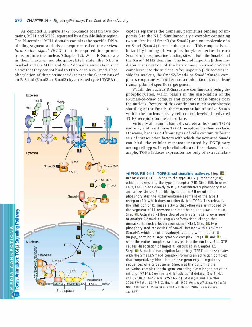
As depicted in Figure 14-2, R-Smads contain two do-mains, MH1 and MH2, separated by a flexible linker region.The N-terminal MH1 domain contains the specific DNA-binding segment and also a sequence called the nuclear-localization signal (NLS) that is required for proteintransport into the nucleus (Chapter 12). When R-Smads arein their inactive, nonphosphorylated state, the NLS ismasked and the MH1 and MH2 domains associate in sucha way that they cannot bind to DNA or to a co-Smad. Phos-phorylation of three serine residues near the C-terminus ofan R-Smad (Smad2 or Smad3) by activated type I TGF� re-
ceptors separates the domains, permitting binding of im-portin � to the NLS. Simultaneously a complex containingtwo molecules of Smad3 (or Smad2) and one molecule of aco-Smad (Smad4) forms in the cytosol. This complex is sta-bilized by binding of two phosphorylated serines in eachSmad3 to phosphoserine-binding sites in both the Smad3 andthe Smad4 MH2 domains. The bound importin � then me-diates translocation of the heteromeric R-Smad/co-Smadcomplexes into the nucleus. After importin � dissociates in-side the nucleus, the Smad2/Smad4 or Smad3/Smad4 com-plexes cooperate with other transcription factors to activatetranscription of specific target genes.
Within the nucleus R-Smads are continuously being de-phosphorylated, which results in the dissociation of the R-Smad/co-Smad complex and export of these Smads fromthe nucleus. Because of this continuous nucleocytoplasmicshuttling of the Smads, the concentration of active Smadswithin the nucleus closely reflects the levels of activatedTGF� receptors on the cell surface.
Virtually all mammalian cells secrete at least one TGF�isoform, and most have TGF� receptors on their surface.However, because different types of cells contain differentsets of transcription factors with which the activated Smadscan bind, the cellular responses induced by TGF� varyamong cell types. In epithelial cells and fibroblasts, for ex-ample, TGF� induces expression not only of extracellular-
576 CHAPTER 14 • Signaling Pathways That Control Gene Activity
PP
TGFβ
RIIIRII
1a 1b
2
3
4
5
6
7
RI
Smad3
Smad4
TFE3
TFE3
Exterior
Smad4Smad3-P
Imp-β
Smad4
Smad3-P
TGFβ
PP PP
NLS
MH2
MH1
NLS
P
NLS
NLS
RanGTP
RanGTP
3-bp spacer
Transcription
PP
Cytosol
Nucleus
+
P P
AG
AC
AG
AC
CACGTGAGAC PAI-1
P
� FIGURE 14-2 TGF�-Smad signaling pathway. Step : In some cells, TGF� binds to the type III TGF� receptor (RIII),which presents it to the type II receptor (RII). Step : In othercells, TGF� binds directly to RII, a constitutively phosphorylatedand active kinase. Step : Ligand-bound RII recruits andphosphorylates the juxtamembrane segment of the type Ireceptor (RI), which does not directly bind TGF�. This releasesthe inhibition of RI kinase activity that otherwise is imposed bythe segment of RI between the membrane and kinase domain.Step : Activated RI then phosphorylates Smad3 (shown here)or another R-Smad, causing a conformational change thatunmasks its nuclear-localization signal (NLS). Step : Twophosphorylated molecules of Smad3 interact with a co-Smad(Smad4), which is not phosphorylated, and with importin �(Imp-�), forming a large cytosolic complex. Steps and : After the entire complex translocates into the nucleus, Ran�GTPcauses dissociation of Imp-� as discussed in Chapter 12. Step : A nuclear transcription factor (e.g., TFE3) then associateswith the Smad3/Smad4 complex, forming an activation complexthat cooperatively binds in a precise geometry to regulatorysequences of a target gene. Shown at the bottom is theactivation complex for the gene encoding plasminogen activatorinhibitor (PAI-1). See the text for additional details. [See Z. Xiao et al., 2000, J. Biol. Chem. 275:23425; J. Massagué and D. Wotton, 2000, EMBO J. 19:1745; X. Hua et al., 1999, Proc. Nat'l. Acad. Sci. USA96:13130; and A. Moustakas and C.-H. Heldin, 2002, Genes Devel.16:1867.]
7
65
4
3
2
1b
1a
ME
DIA
CO
NN
EC
TIO
NS
Focu
s Ani
mat
ion:
TGF
�Si
gnali
ng P
athw
ay

matrix proteins (e.g., collagens) but also of proteins that in-hibit serum proteases, which otherwise would degrade thematrix. The latter category includes plasminogen activatorinhibitor 1 (PAI-1). Transcription of the PAI-1 gene requiresformation of a complex of the transcription factor TFE3with the Smad3/Smad4 complex and binding of all these pro-teins to specific sequences within the regulatory region of thePAI-1 gene (see Figure 14-2, bottom). By partnering withother transcription factors, Smad2/Smad4 and Smad3/Smad4 complexes induce expression of proteins such as p15,which arrests the cell cycle at the G1 stage and thus blockscell proliferation (Chapter 21). These Smad complexes alsorepress transcription of the myc gene, thereby reducing ex-pression of many growth-promoting genes whose transcrip-tion normally is activated by Myc.
The various growth factors in the TGF� superfamily bindto their own receptors and activate different sets of Smadproteins, resulting in different cellular responses. The speci-ficity exhibited by these related receptors is a common phe-nomenon in intercellular signaling, and the TGF� signalingpathway provides an excellent example of one strategy forachieving such response specificity. As just discussed, for in-stance, binding of any one TGF� isoform to its specific re-ceptors leads to phosphorylation of Smad2 or Smad3,formation of Smad2/Smad4 or Smad3/Smad4 complexes,and eventually transcriptional activation of specific targetgenes (e.g., the PAI-1 gene). On the other hand, BMP pro-teins, which also belong to the TGF� superfamily, bind toand activate a different set of receptors, leading to phospho-rylation of Smad1, its dimerization with Smad4, and activa-tion of specific transcriptional responses by Smad1/Smad4.These responses are distinct from those induced by Smad2/Smad4 or Smad3/Smad4.
Oncoproteins and I-Smads Regulate SmadSignaling via Negative Feedback LoopsSmad signaling is regulated by additional intracellular pro-teins, including two cytosolic proteins called SnoN and Ski(Ski stands for “Sloan-Kettering Cancer Institute”). Theseproteins were originally identified as oncoproteins becausethey cause abnormal cell proliferation when overexpressed incultured fibroblasts. How they accomplish this was not un-derstood until years later when SnoN and Ski were found tobind to the Smad2/Smad4 or Smad3/Smad4 complexesformed after TGF� stimulation. SnoN and Ski do not affectthe ability of the Smad complexes to bind to DNA control re-gions. Rather, they block transcription activation by thebound Smad complexes, thereby rendering cells resistant tothe growth-inhibitory actions normally induced by TGF�(Figure 14-3). Interestingly, stimulation by TGF� causes therapid degradation of Ski and SnoN, but after a few hours,expression of both Ski and SnoN becomes strongly induced.The increased levels of these proteins are thought to dampenlong-term signaling effects due to continued exposure toTGF�.
Among the proteins induced after TGF� stimulation arethe I-Smads, especially Smad7. Smad7 blocks the ability ofactivated type I receptors to phosphorylate R-Smad pro-teins. In this way Smad7, like Ski and SnoN, participatesin a negative feedback loop; its induction serves to inhibitintracellular signaling by long-term exposure to the stimu-lating hormone. In later sections we see how signaling byother cell-surface receptors is also controlled by negativefeedback loops.
Loss of TGF� Signaling Contributes to AbnormalCell Proliferation and Malignancy
Many human tumors contain inactivating muta-tions in either TGF� receptors or Smad proteins,and thus are resistant to growth inhibition by
TGF� (see Figure 23-20). Most human pancreatic cancers,for instance, contain a deletion in the gene encoding Smad4and thus cannot induce p15 and other cell-cycle inhibitorsin response to TGF�. This mutation-defined gene originallywas called DPC (deleted in pancreatic cancer). Retinoblas-toma, colon and gastric cancer, hepatoma, and some T- andB-cell malignancies are also unresponsive to TGF� growthinhibition. This loss of responsiveness correlates with loss oftype I or type II TGF� receptors; responsiveness to TGF� canbe restored by recombinant expression of the “missing” pro-tein. Mutations in Smad2 also commonly occur in severaltypes of human tumors. Not only is TGF� signaling essential
14.1 • TGFb Receptors and the Direct Activation of Smads 577
N-CoR
AGAC
Smad4
Smad3-P
PAI-1Transcription
AG
AC
AG
AC
mSin3A
HDAC
Histonedeacetylation
Ski
PP
−
CACGTG
3-bp spacer
▲ FIGURE 14-3 Schematic model of Ski-mediated down-regulation of the response to TGF� stimulation. Skibinds to Smad4 in Smad3/Smad4 or Smad2/Smad4 (not shown)signaling complexes and may partially disrupt interactionsbetween the Smad proteins. Ski also recruits a protein termed N-CoR that binds directly to mSin3A, which in turn interacts withhistone deacetylase (HDAC), an enzyme that promotes histonedeacetylation (Chapter 11). As a result, transcription activationinduced by TGF� and mediated by Smad complexes is shutdown. [See S. Stroschein et al., 1999, Science 286:771; X. Liu et al.,2001, Cytokine and Growth Factor Rev. 12:1; and J.-W. Wu et al., 2002,Cell 111:357.]

for controlling cell proliferation, as these examples show, butit also causes some cells to differentiate along specific path-ways, as discussed in Chapter 15. ❚
KEY CONCEPTS OF SECTION 14.1
TGF� Receptors and the Direct Activation of Smads■ TGF� is produced as an inactive precursor that is stored inthe extracellular matrix. Several mechanisms can release theactive, mature dimeric growth factor (see Figure 14-1).■ Stimulation by TGF� leads to activation of the intrinsicserine/threonine kinase activity in the cytosolic domain ofthe type I (RI) receptor, which then phosphorylates an R-Smad, exposing a nuclear-localization signal.■ After phosphorylated R-Smad binds a co-Smad, the re-sulting complex translocates into the nucleus, where it in-teracts with various transcription factors to induce ex-pression of target genes (see Figure 14-2).■ Oncoproteins (e.g., Ski and SnoN) and I-Smads (e.g.,Smad7) act as negative regulators of TGF� signaling.■ TGF� signaling generally inhibits cell proliferation. Lossof various components of the signaling pathway con-tributes to abnormal cell proliferation and malignancy.
Cytokine Receptors and the JAK-STAT PathwayWe turn now to a second important class of cell-surface re-ceptors, the cytokine receptors, whose cytosolic domainsare closely associated with a member of a family of cy-tosolic protein tyrosine kinases, the JAK kinases. A thirdclass of receptors, the receptor tyrosine kinases (RTKs),contain intrinsic protein tyrosine kinase activity in theircytosolic domains. The mechanisms by which cytokine re-ceptors and receptor tyrosine kinases become activated byligands are very similar, and there is considerable overlapin the intracellular signal-transduction pathways triggeredby activation of receptors in both classes. In this section,we first describe some similarities in signaling from thesetwo receptor classes. We then discuss the JAK-STAT path-way, which is initiated mainly by activation of cytokine receptors.
Cytokine Receptors and Receptor TyrosineKinases Share Many Signaling FeaturesLigand binding to both cytokine receptors and RTKs trig-gers formation of functional dimeric receptors. In somecases, the ligand induces association of two monomeric
14.2
receptor subunits diffusing in the plane of the plasmamembrane (Figure 14-4). In others, the receptor is a dimerin the absence of ligand, and ligand binding alters the con-formation of the extracellular domains of the two sub-units. In either case, formation of a functional dimericreceptor causes one of the poorly active cytosolic kinasesto phosphorylate a particular tyrosine residue in the acti-vation lip of the second kinase. This phosphorylation ac-tivates kinase activity and leads to phosphorylation of thesecond kinase in the dimer, as well as several tyrosine
578 CHAPTER 14 • Signaling Pathways That Control Gene Activity
EGF
(a)
(b)
EGF-bindingdomains
Membrane
Exterior
Membrane surface
COO−−OOC
▲ FIGURE 14-4 Dimerization of the receptor for epidermalgrowth factor (EGF), a receptor tyrosine kinase. (a) Schematicdepiction of the extracellular and transmembrane domains of the EGF receptor. Binding of one EGF molecule to a monomericreceptor causes an alteration in the structure of a loop locatedbetween the two EGF-binding domains. Dimerization of twoidentical ligand-bound receptor monomers in the plane of themembrane occurs primarily through interactions between thetwo “activated” loop segments. (b) Structure of the dimeric EGFreceptor’s extracellular domain bound to transforming growthfactor � (TGF�), a homolog of EGF. The EGF receptor extracellulardomains are shown in white (left) and blue (right). The twosmaller TGF� molecules are colored green. Note the interactionbetween the “activated” loop segments in the two receptors.[Part (a) adapted from J. Schlessinger, 2002, Cell 110:669; part (b) from T. Garrett et al., 2002, Cell 110:763.]

14.2 • Cytokine Receptors and the JAK-STAT Pathway 579
residues in the cytosolic domain of the receptor (Figure 14-5). As we see later, phosphorylation of residues in the ac-tivation loop is a general mechanism by which many kinasesare activated.
Certain phosphotyrosine residues formed in activated cy-tokine receptors and RTKs serve as binding, or “docking,”sites for SH2 domains or PTB domains, which are presentin a large array of intracellular signal-transduction proteins.
Once they are bound to an activated receptor, some signal-transduction proteins are phosphorylated by the receptor’sintrinsic or associated kinase to achieve their active form.Binding of other signal-transduction proteins, present in thecytosol in unstimulated cells, positions them near their sub-strates localized in the plasma membrane. Both mechanismscan trigger downstream signaling. Several cytokine receptors(e.g., the IL-4 receptor) and RTKs (e.g., the insulin receptor)
Transmembraneα helix
Poorly activeprotein tyrosinekinase
1 2 3
Exterior
Cytosol
Activationlip
Ligand
Active proteintyrosine kinase
Dimerization andphosphorylation ofactivation lip tyrosines
Bound ligand
Phosphorylationof additional tyrosine residues
Receptor tyrosinekinases (RTKs)
P
PP
P
PP
Ligand-binding sites
Transmembraneα helix
Kinase
1 2 3
Exterior
Cytosol
Lip
Ligand
Dimerization andphosphorylation ofactivation lip tyrosines
Bound ligand
Phosphorylationof additional tyrosine residues
Cytokine receptors
ATPADP
ATPADPJAK
ActiveJAK
PPPP
P
P
PP
P
P
PP
ATP
ADP
ATP
ADPPP
ATP
ADP
ATP
ADP
Ligand-binding sites
ATPADP
ATPADP
▲ FIGURE 14-5 General structure and ligand-inducedactivation of receptor tyrosine kinases (RTKs) and cytokinereceptors. The cytosolic domain of RTKs contains a proteintyrosine kinase catalytic site, whereas the cytosolic domain ofcytokine receptors associates with a separate JAK kinase (step ). In both types of receptor, ligand binding causes aconformational change that promotes formation of a functionaldimeric receptor, bringing together two intrinsic or associated
1
kinases, which then phosphorylate each other on a tyrosineresidue in the activation lip (step ). Phosphorylation causes the lip to move out of the kinase catalytic site, thus allowing ATP or a protein substrate to bind. The activated kinase thenphosphorylates other tyrosine residues in the receptor’s cytosolicdomain (step ). The resulting phosphotyrosines function asdocking sites for various signal-transduction proteins (see Figure 14-6).
3
2

580 CHAPTER 14 • Signaling Pathways That Control Gene Activity
EpoNo Epo
Erythroid progenitor (CFU-E)
Hematopoietic stem cell
Progenitors of othertypes of blood cells
Eporeceptors
Apoptosis(cell death)
Mature red cells
� FIGURE 14-7 Role of erythropoietin information of red blood cells (erythrocytes). Erythroidprogenitor cells, called colony-forming units erythroid(CFU-E), are derived from hematopoietic stem cells,which also give rise to progenitors of other blood celltypes. In the absence of erythropoietin (Epo), CFU-Ecells undergo apoptosis. Binding of erythropoietin to itsreceptors on a CFU-E induces transcription of severalgenes whose encoded proteins prevent programmedcell death (apoptosis), allowing the cell to survive andundergo a program of three to five terminal celldivisions. Epo stimulation also induces expression oferythrocyte-specific proteins such as the globins, whichform hemoglobin, and the membrane proteinsglycophorin and anion-exchange protein. The Eporeceptor and other membrane proteins are lost fromthese cells as they undergo differentiation. If CFU-Ecells are cultured with erythropoietin in a semisolidmedium (e.g., containing methylcellulose), daughtercells cannot move away, and thus each CFU-E producesa colony of 30–100 erythroid cells, hence its name. [See M. Socolovsky et al., 2001, Blood 98:3261.]
bind IRS1 or other multidocking proteins via a PTB domainin the docking protein (Figure 14-6).The activated receptorthen phosphorylates the bound docking protein, formingmany phosphotyrosines that in turn serve as docking sites forSH2-containing signaling proteins. Some of these proteinsin turn may also be phosphorylated by the activated receptor.
Cytokines Influence Development of Many Cell TypesThe cytokines form a family of relatively small, secretedproteins (generally containing about 160 amino acids) thatcontrol many aspects of growth and differentiation of spe-cific types of cells. During pregnancy prolactin, for example,induces epithelial cells lining the immature ductules of themammary gland to differentiate into the acinar cells thatproduce milk proteins and secrete them into the ducts. An-other cytokine, interleukin 2 (IL-2), is essential for prolifer-ation and functioning of the T cells of the immune system;its close relative IL-4 is essential for formation of functional antibody-producing B cells. Some cytokines, such as inter-feron �, are produced and secreted by many types of cells fol-
P
P
PP
P
P
PP
Exterior
Cytosol
PP P
IRS-1
PTBdomain
Signaling proteins
SH2domain
Activated RTK
PP
P
Kinase
▲ FIGURE 14-6 Recruitment of signal-transduction proteinsto the cell membrane by binding to phosphotyrosineresidues in activated receptors. Cytosolic proteins with SH2 (purple) or PTB (maroon) domains can bind to specificphosphotyrosine residues in activated RTKs (shown here) orcytokine receptors. In some cases, these signal-transductionproteins then are phosphorylated by the receptor’s intrinsic orassociated protein tyrosine kinase, enhancing their activity.Certain RTKs and cytokine receptors utilize multidocking proteinssuch as IRS-1 to increase the number of signaling proteins thatare recruited and activated. Subsequent phosphorylation of theIRS-1 by receptor kinase activity creates additional docking sitesfor SH2-containing signaling proteins.

lowing virus infection. The secreted interferons act on nearbycells to induce enzymes that render these cells more resist-ant to virus infection.
Many cytokines induce formation of importanttypes of blood cells. For instance, granulocytecolony stimulating factor (G-CSF) induces a par-
ticular type of progenitor cell in the bone marrow to divideseveral times and then differentiate into granulocytes, thetype of white blood cell that inactivates bacteria and otherpathogens. Because many cancer therapies reduce granulo-cyte formation by the body, G-CSF often is administered topatients to stimulate proliferation and differentiation ofgranulocyte progenitor cells, thus restoring the normal levelof granulocytes in the blood. Thrombopoietin, a “cousin”of G-CSF, similarly acts on megakaryocyte progenitors to di-vide and differentiate into megakaryocytes. These then frag-ment into the cell pieces called platelets, which are critical forblood clotting. ❚
Another related cytokine, erythropoietin (Epo), triggersproduction of red blood cells by inducing the proliferationand differentiation of erythroid progenitor cells in the bonemarrow (Figure 14-7). Erythropoietin is synthesized by kid-ney cells that monitor the concentration of oxygen in theblood. A drop in blood oxygen signifies a lower than optimallevel of erythrocytes (red blood cells), whose major functionis to transport oxygen complexed to hemoglobin. By meansof the oxygen-sensitive transcription factor HIF-1�, the kid-ney cells respond to low oxygen by synthesizing more ery-thropoietin and secreting it into the blood (see Figure 15-9).As the level of erythropoietin rises, more and more erythroidprogenitors are saved from death, allowing each to produce≈50 or so red blood cells in a period of only two days. In thisway, the body can respond to the loss of blood by accelerat-ing the production of red blood cells.
All Cytokines and Their Receptors Have SimilarStructures and Activate Similar SignalingPathwaysStrikingly, all cytokines have a similar tertiary structure, con-sisting of four long conserved � helices folded together in aspecific orientation. Similarly, the structures of all cytokinereceptors are quite similar, with their extracellular domainsconstructed of two subdomains, each of which containsseven conserved � strands folded together in a characteristicfashion. The interaction of erythropoietin with the dimericerythropoietin receptor (EpoR), depicted in Figure 14-8, ex-emplifies the binding of a cytokine to its receptor. The struc-tural homology among cytokines is evidence that they allevolved from a common ancestral protein. Likewise, the var-ious receptors undoubtedly evolved from a single commonancestor.
Whether or not a cell responds to a particular cytokinedepends simply on whether or not it expresses the corre-sponding (cognate) receptor. Although all cytokine receptorsactivate similar intracellular signaling pathways, the re-sponse of any particular cell to a cytokine signal depends onthe cell’s constellation of transcription factors, chromatinstructures, and other proteins relating to the developmentalhistory of the cell. If receptors for prolactin or thrombopoi-etin, for example, are expressed experimentally in an ery-throid progenitor cell, the cell will respond to these cytokinesby dividing and differentiating into red blood cells, not intomammary cells or megakaryocytes.
Figure 14-9 summarizes the intracellular signaling path-ways activated when the EpoR binds erythropoietin. Stimu-lation of other cytokine receptors by their specific ligandsactivates similar pathways. All these pathways eventuallylead to activation of transcription factors, causing an in-crease or decrease in expression of particular target genes.Here we focus on the JAK-STAT pathway; the other path-ways are discussed in later sections.
14.2 • Cytokine Receptors and the JAK-STAT Pathway 581
Erythropoietin
EpoR(monomer)
EpoR(monomer)
Membrane surface
COO− COO−
▲ FIGURE 14-8 Structure of erythropoietin bound to theextracellular domains of a dimeric erythropoietin receptor(EpoR). Erythropoietin contains four conserved long � helicesthat are folded in a particular arrangement. The extracellulardomain of an EpoR monomer is constructed of two subdomains,each of which contains seven conserved � strands folded in acharacteristic fashion. Side chains of residues on two of thehelices in erythropoietin contact loops on one EpoR monomer,while residues on the two other Epo helices bind to the sameloop segments in a second receptor monomer, thereby stabilizingthe dimeric receptor. The structures of other cytokines and theirreceptors are similar to erythropoietin and EpoR. [Adapted from R. S. Syed et al., 1998, Nature 395:511.]

Somatic Cell Genetics Revealed JAKs and STATsas Essential Signal-Transduction ProteinsSoon after the discovery and cloning of cytokines, most oftheir receptors were isolated by expression cloning or otherstrategies. Elucidation of the essential components of their in-tracellular signaling pathways, however, awaited develop-
ment of new types of genetic approaches using cultured mam-malian cells. In these studies, a bacterial reporter gene encod-ing guanine phosphoribosyl transferase (GPRT) was linked toan upstream interferon-responsive promoter. The resultingconstruct was introduced into cultured mammalian cells thatwere genetically deficient in the human homolog HGPRT.GPRT or HGPRT is necessary for incorporation of purines
582 CHAPTER 14 • Signaling Pathways That Control Gene Activity
Epo EpoR JAK2
(b) GRB2 or Shc Ras Transcriptional activation or repressionMAP kinase
(d) PI-3 kinase Transcriptional activation or repression;modification of other cellular proteins
Protein kinase B
(c) Phospholipase Cγ Transcriptional activation or repression; modification of other cellular proteinsElevation of Ca2+
(a) STAT5 Transcriptional activation
▲ FIGURE 14-9 Overview of signal-transduction pathwaystriggered by ligand binding to the erythropoietin receptor(EpoR), a typical cytokine receptor. Four major pathways cantransduce a signal from the activated, phosphorylated EpoR-JAKcomplex (see Figure 14-5, bottom). Each pathway ultimatelyregulates transcription of different sets of genes. (a) In the mostdirect pathway, the transcription factor STAT5 is phosphorylated
and activated directly in the cytosol. (b) Binding of linker proteins(GRB2 or Shc) to an activated EpoR leads to activation of theRas–MAP kinase pathway. (c, d) Two phosphoinositide pathwaysare triggered by recruitment of phospholipase C� and PI-3 kinaseto the membrane following activation of EpoR. Elevated levels ofCa2� and activated protein kinase B also modulate the activity ofcytosolic proteins that are not involved in control of transcription.
Interferon-responsivepromoter
Reporter gene construct
(a)HGPRT− cells (+ reporter gene)
(b)HGPRT− cells (+ reporter gene)defective for interferonsignaling
Growthin HAT medium
Killed by6-thioguanine
− Interferon
+ Interferon
Mutagen
Yes Yes
No No
+ Interferon
+ Wild-type gene that restores interferon responsiveness
Yes Yes
No No
GPRTexpressed
Yes
No
Yes
No
GPRT
+ Interferon
▲ EXPERIMENTAL FIGURE 14-10 Mutagenized cellscarrying an interferon-responsive reporter gene were used toidentify JAKs and STATs as essential signal-transductionproteins. A reporter gene was constructed consisting of aninterferon-responsive promoter upstream of the bacterial geneencoding GPRT, a key enzyme in the purine salvage pathway (seeFigure 6-39). (a) Introduction of this construct into mammaliancells lacking the mammalian homolog HGPRT yielded reportercells that grew in HAT medium and were killed by 6-thioguaninein the presence but not the absence of interferon. (b) Following
treatment of reporter cells with a mutagen, cells with defects inthe signaling pathway initiated by interferon do not induce GPRTin response to interferon and thus cannot incorporate the toxicpurine 6-thioguanine. Restoration of interferon responsiveness byfunctional complementation with wild-type DNA clones identifiedgenes encoding JAKs and STATs. See the text for details. [SeeR. McKendry et al., 1991, Proc. Nat’l. Acad. Sci. USA 88:11455; D. Watlinget al., 1993, Nature 366:166; and G. Stark and A. Gudkov, 1999, HumanMol. Genet. 8:1925.]

in the culture medium into ribonucleotides and then intoDNA or RNA. As shown in Figure 14-10a, HGPRT-negativecells carrying the reporter gene responded to interferon treat-ment by expressing GPRT and thus acquiring the ability togrow in HAT medium. This medium does not allow growthof cells lacking GPRT or HGPRT, since synthesis of purinesby the cells is blocked by aminopterin (the A in HAT), andthus DNA synthesis is dependent on incorporation of purinesfrom the culture medium (see Figure 6-39). Simultaneouslythe cells acquired sensitivity to killing by the purine analog6-thioguanine, which is converted into the corresponding ribonucleotide by GPRT; incorporation of this purine intoDNA in place of guanosine eventually causes cell death.
The reporter cells were then heavily treated with muta-gens in an attempt to inactivate both alleles of the genes en-coding critical signal-transduction proteins in the interferonsignaling pathway. Researchers looked for mutant cells thatexpressed the interferon receptor (as evidenced by the cell’sability to bind radioactive interferon) but did not expressGPRT in response to interferon and thus survived killing by 6-thioguanine when cells were cultured in the presenceof interferon (Figure 14-10b). After many such interferon-nonresponding mutant cell lines were obtained, they wereused to screen a genomic or cDNA library for the wild-typegenes that complemented the mutated genes in nonrespond-ing cells, a technique called functional complementation (seeFigure 9-20). In this case, mutant cells expressing the corre-sponding recombinant wild-type gene grew on HAT mediumand were sensitive to killing by 6-thioguanine in the presenceof interferon. That is, they acted like wild-type cells.
Cloning of the genes identified by this procedure led torecognition of two key signal-transduction proteins: a JAKtyrosine kinase and a STAT transcription factor. Subsequentwork showed that one (sometimes two) of the four human
JAK proteins and at least one of several STAT proteins areinvolved in signaling downstream from all cytokine recep-tors. To understand how JAK and STAT proteins function,we examine one of the best-understood cytokine signalingpathways, that downstream of the erythropoietin receptor.
Receptor-Associated JAK Kinases Activate STATTranscription Factors Bound to a CytokineReceptorThe JAK2 kinase is tightly bound to the cytosolic domain ofthe erythropoietin receptor (EpoR). Like the three othermembers of the JAK family of kinases, JAK2 contains an N-terminal receptor-binding domain, a C-terminal kinase do-main that is normally poorly active catalytically, and amiddle domain of unknown function. JAK2, erythropoietin,and the EpoR are all required for formation of adult-typeerythrocytes, which normally begins at day 12 of embryonicdevelopment in mice. As Figure 14-11 shows, embryonicmice lacking functional genes encoding either the EpoR orJAK2 cannot form adult-type erythrocytes and eventually dieowing to the inability to transport oxygen to the fetal organs.
As already noted, erythropoietin binds simultaneously tothe extracellular domains of two EpoR monomers on the cellsurface (see Figure 14-8). As a result, the associated JAKs arebrought close enough together that one can phosphorylatethe other on a critical tyrosine in the activation lip. As withother kinases, phosphorylation of the activation lip leads toa conformational change that reduces the Km for ATP or thesubstrate to be phosphorylated, thus increasing the kinaseactivity. One piece of evidence for this activation mechanismcomes from study of a mutant JAK2 in which the critical ty-rosine is mutated to phenylalanine. The mutant JAK2 bindsnormally to the EpoR but cannot be phosphorylated.
14.2 • Cytokine Receptors and the JAK-STAT Pathway 583
EpoR JAK2
+/+ –/– +/+ –/–
▲ EXPERIMENTAL FIGURE 14-11 Studies with mutantmice reveal that both the erythropoietin receptor (EpoR) andJAK2 are essential for development of erythrocytes. Mice inwhich both alleles of the EpoR or JAK2 gene are “knocked out”develop normally until embryonic day 13, at which time theybegin to die of anemia due to the lack of erythrocyte-mediatedtransport of oxygen to the fetal organs. The red organ in the wild-type embryos (�/�) is the fetal liver, the major site of
erythrocyte production at this developmental stage. The absenceof color in the mutant embryos (�/�) indicates the absence oferythrocytes containing hemoglobin. Otherwise the mutantembryos appear normal, indicating that the main function of theEpoR and JAK2 in early mouse development is to supportproduction of erythrocytes. [EpoR images from H. Wu et al., 1995,Cell 83:59; JAK2 images from H. Neubauer et al., 1998, Cell 93:307.]
Expression of this mutant JAK2 in erythroid cells in greaterthan normal amounts totally blocks EpoR signaling, as themutant JAK2 blocks the function of the wild-type protein.This type of mutation, referred to as a dominant negative,causes loss of function even in cells that carry copies of thewild-type gene (Chapter 9).
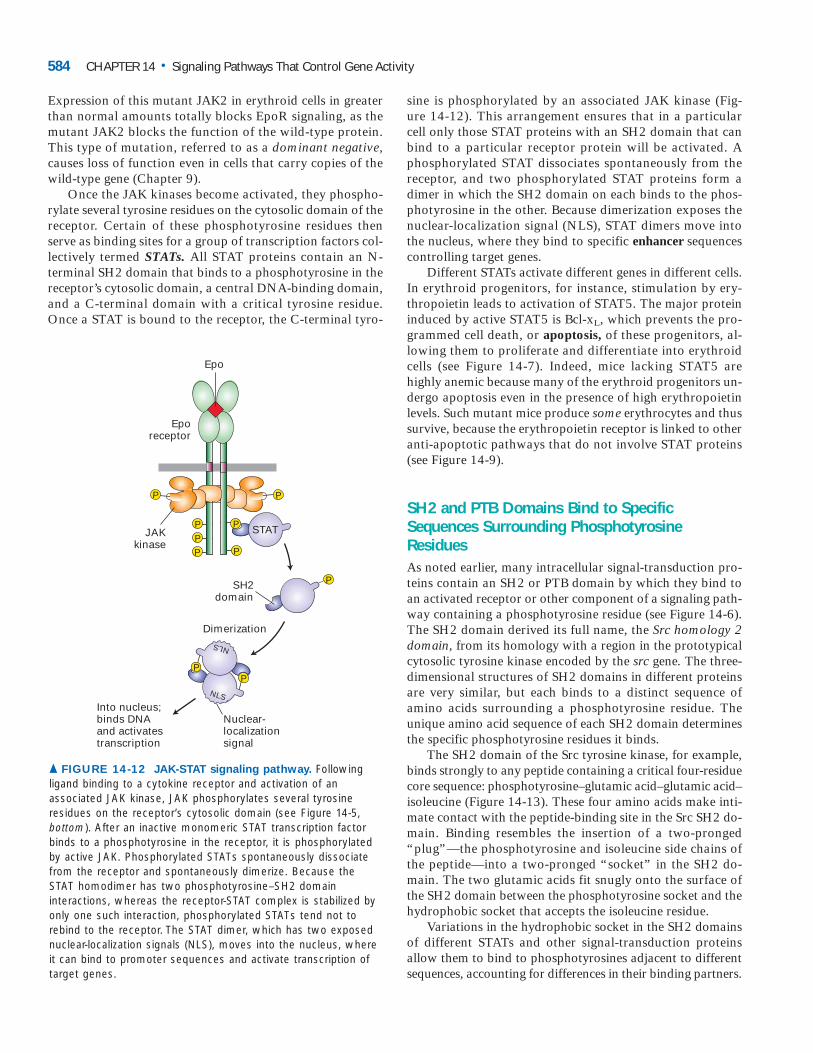
Once the JAK kinases become activated, they phospho-rylate several tyrosine residues on the cytosolic domain of thereceptor. Certain of these phosphotyrosine residues thenserve as binding sites for a group of transcription factors col-lectively termed STATs. All STAT proteins contain an N-terminal SH2 domain that binds to a phosphotyrosine in thereceptor’s cytosolic domain, a central DNA-binding domain,and a C-terminal domain with a critical tyrosine residue.Once a STAT is bound to the receptor, the C-terminal tyro-
sine is phosphorylated by an associated JAK kinase (Fig-ure 14-12). This arrangement ensures that in a particular cell only those STAT proteins with an SH2 domain that can bind to a particular receptor protein will be activated. A phosphorylated STAT dissociates spontaneously from the receptor, and two phosphorylated STAT proteins form a dimer in which the SH2 domain on each binds to the phos-photyrosine in the other. Because dimerization exposes the nuclear-localization signal (NLS), STAT dimers move into the nucleus, where they bind to specific enhancer sequencescontrolling target genes.
Different STATs activate different genes in different cells.In erythroid progenitors, for instance, stimulation by ery-thropoietin leads to activation of STAT5. The major proteininduced by active STAT5 is Bcl-xL, which prevents the pro-grammed cell death, or apoptosis, of these progenitors, al-lowing them to proliferate and differentiate into erythroidcells (see Figure 14-7). Indeed, mice lacking STAT5 arehighly anemic because many of the erythroid progenitors un-dergo apoptosis even in the presence of high erythropoietinlevels. Such mutant mice produce some erythrocytes and thussurvive, because the erythropoietin receptor is linked to otheranti-apoptotic pathways that do not involve STAT proteins(see Figure 14-9).
SH2 and PTB Domains Bind to SpecificSequences Surrounding PhosphotyrosineResiduesAs noted earlier, many intracellular signal-transduction pro-teins contain an SH2 or PTB domain by which they bind toan activated receptor or other component of a signaling path-way containing a phosphotyrosine residue (see Figure 14-6).The SH2 domain derived its full name, the Src homology 2domain, from its homology with a region in the prototypicalcytosolic tyrosine kinase encoded by the src gene. The three-dimensional structures of SH2 domains in different proteinsare very similar, but each binds to a distinct sequence ofamino acids surrounding a phosphotyrosine residue. Theunique amino acid sequence of each SH2 domain determinesthe specific phosphotyrosine residues it binds.
The SH2 domain of the Src tyrosine kinase, for example,binds strongly to any peptide containing a critical four-residuecore sequence: phosphotyrosine–glutamic acid–glutamic acid–isoleucine (Figure 14-13). These four amino acids make inti-mate contact with the peptide-binding site in the Src SH2 do-main. Binding resembles the insertion of a two-pronged“plug”—the phosphotyrosine and isoleucine side chains ofthe peptide—into a two-pronged “socket” in the SH2 do-main. The two glutamic acids fit snugly onto the surface ofthe SH2 domain between the phosphotyrosine socket and thehydrophobic socket that accepts the isoleucine residue.
Variations in the hydrophobic socket in the SH2 domainsof different STATs and other signal-transduction proteinsallow them to bind to phosphotyrosines adjacent to differentsequences, accounting for differences in their binding partners.
584 CHAPTER 14 • Signaling Pathways That Control Gene Activity
P
P
P
P
PP
SH2domain
P
JAKkinase
Eporeceptor
Epo
PP
Into nucleus;binds DNA and activates transcription
P
Dimerization
STAT
NLS
NLS
Nuclear-localizationsignal
▲ FIGURE 14-12 JAK-STAT signaling pathway. Followingligand binding to a cytokine receptor and activation of anassociated JAK kinase, JAK phosphorylates several tyrosineresidues on the receptor’s cytosolic domain (see Figure 14-5,bottom). After an inactive monomeric STAT transcription factorbinds to a phosphotyrosine in the receptor, it is phosphorylatedby active JAK. Phosphorylated STATs spontaneously dissociatefrom the receptor and spontaneously dimerize. Because theSTAT homodimer has two phosphotyrosine–SH2 domaininteractions, whereas the receptor-STAT complex is stabilized byonly one such interaction, phosphorylated STATs tend not torebind to the receptor. The STAT dimer, which has two exposednuclear-localization signals (NLS), moves into the nucleus, whereit can bind to promoter sequences and activate transcription oftarget genes.

The binding specificity of SH2 domains is largely determinedby residues C-terminal to the phosphotyrosine in a target pep-tide. In contrast, the binding specificity of PTB domains is de-termined by specific residues five to eight residues N-terminalto a phosphotyrosine residue. Sometimes a PTB domain bindsto a target peptide even if the tyrosine is not phosphorylated.
Signaling from Cytokine Receptors Is Modulatedby Negative SignalsSignal-induced transcription of target genes for too long a pe-riod can be as dangerous for the cell as too little induction.Thus cells must be able to turn off a signaling pathway quicklyunless the extracellular signal remains continuously present. Invarious progenitor cells, two classes of proteins serve todampen signaling from cytokine receptors, one over the shortterm (minutes) and the other over longer periods of time.
Short-Term Regulation by SHP1 Phosphatase Mutant micelacking SHP1 phosphatase die because of excess productionof erythrocytes and several other types of blood cells. Analy-sis of these mutant mice offered the first suggestion thatSHP1, a phosphotyrosine phosphatase, negatively regulatessignaling from several types of cytokine receptors in severaltypes of progenitor cells.
How SHP1 dampens cytokine signaling is depicted in Fig-ure 14-14a. In addition to a phosphatase catalytic domain,SHP1 has two SH2 domains. When cells are not stimulated
14.2 • Cytokine Receptors and the JAK-STAT Pathway 585
Tyr0Ile3 Glu1
SH2 domain
OPO3−Glu2
▲ FIGURE 14-13 Surface model of the SH2 domain from Srckinase bound to a phosphotyrosine-containing peptide. Thepeptide bound by this SH2 domain (gray) is shown in spacefill. Thephosphotyrosine (Tyr0 and OPO3
�, orange) and isoleucine (Ile3,orange) residues fit into a two-pronged socket on the surface of theSH2 domain; the two glutamate residues (Glu1, dark blue; Glu2,light blue) are bound to sites on the surface of the SH2 domainbetween the two sockets. Nonbinding residues on the targetpeptide are colored green. [See G. Waksman et al., 1993, Cell 72:779.]
P
P
P
P
P
P
P
P
Recruitmentof E3 ubiquitinligase
SH2domain
SOCSbox
SOCSprotein
(b) Signal blocking and protein degradation induced by SOCS proteins
▲ FIGURE 14-14 Two mechanisms for terminating signaltransduction from the erythropoietin receptor (EpoR).(a) SHP1, a protein tyrosine phosphatase, is present in aninactive form in unstimulated cells. Binding of an SH2 domain inSHP1 to a particular phosphotyrosine in the activated receptorunmasks its phosphatase catalytic site and positions it near thephosphorylated tyrosine in the lip region of JAK2. Removal of the phosphate from this tyrosine inactivates the JAK kinase. (b) SOCS proteins, whose expression is induced in erythropoietin-stimulated erythroid cells, inhibit or permanentlyterminate signaling over longer time periods. Binding of SOCS tophosphotyrosine residues on the EpoR or JAK2 blocks binding ofother signaling proteins (left). The SOCS box can also targetproteins such as JAK2 for degradation by the ubiquitin-proteasome pathway (right). Similar mechanisms regulatesignaling from other cytokine receptors. [Part (a) adapted from S. Constantinescu et al., 1999, Trends Endocrin. Metabol. 10:18; part (b) adapted from B. T. Kile and W. S. Alexander, 2001, Cell. Mol. LifeSci. 58:1.]
P
P
P
P
Active JAK2kinase
EpoR
Epo
InactiveSHP1
P
Inactive JAK2kinase
ActiveSHP1
SH2domains
Phosphatasedomain
(a) JAK2 deactivation induced by SHP1 phosphatase

by a cytokine (are in the resting state), one of the SH2 do-mains physically binds to and inactivates the catalytic site inSHP1. In the stimulated state, however, this blocking SH2domain binds to a specific phosphotyrosine residue in the ac-tivated receptor. The conformational change that accompa-nies this binding unmasks the SHP1 catalytic site and alsobrings it adjacent to the phosphotyrosine residue in the acti-vation lip of the JAK associated with the receptor. By re-moving this phosphate, SHP1 inactivates the JAK, so that itcan no longer phosphorylate the receptor or other substrates(e.g., STATs) unless additional cytokine molecules bind tocell-surface receptors, initiating a new round of signaling.
Long-Term Regulation by SOCS Proteins Among the geneswhose transcription is induced by STAT proteins are thoseencoding a class of small proteins, termed SOCS proteins,that terminate signaling from cytokine receptors. These neg-ative regulators, also known as CIS proteins, act in two ways(Figure 14-14b). First, the SH2 domain in several SOCS pro-teins binds to phosphotyrosines on an activated receptor,preventing binding of other SH2-containing signaling pro-teins (e.g., STATs) and thus inhibiting receptor signaling.One SOCS protein, SOCS-1, also binds to the critical phos-photyrosine in the activation lip of activated JAK2 kinase,thereby inhibiting its catalytic activity. Second, all SOCS pro-teins contain a domain, called the SOCS box, that recruitscomponents of E3 ubiquitin ligases (see Figure 3-13). As a re-sult of binding SOCS-1, for instance, JAK2 becomes polyu-biquitinated and then degraded in proteasomes, thuspermanently turning off all JAK2-mediated signaling path-ways. The observation that proteasome inhibitors prolongJAK2 signal transduction supports this mechanism.
Studies with cultured mammalian cells have shown thatthe receptor for growth hormone, which belongs to the cy-tokine receptor superfamily, is down-regulated by anotherSOCS protein, SOCS-2. Strikingly, mice deficient in thisSOCS protein grow significantly larger than their wild-typecounterparts and have long bone lengths and proportionateenlargement of most organs. Thus SOCS proteins play anessential negative role in regulating intracellular signalingfrom the receptors for erythropoietin, growth hormone, andother cytokines.
Mutant Erythropoietin Receptor That Cannot BeDown-Regulated Leads to Increased HematocritIn normal adult men and women, the percentage of erythro-cytes in the blood (the hematocrit) is maintained very close to45– 47 percent. A drop in the hematocrit results in increasedproduction of erythropoietin by the kidney. The elevated ery-thropoietin level causes more erythroid progenitors to un-dergo terminal proliferation and differentiation into matureerythrocytes, soon restoring the hematocrit to its normallevel. In endurance sports, such as cross-country skiing,where oxygen transport to the muscles may become limiting,
an excess of red blood cells may confer a competitive advan-tage. For this reason, use of supplemental erythropoietin toincrease the hematocrit above the normal level is banned inmany athletic competitions, and athletes are regularly testedfor the presence of commercial recombinant erythropoietinin their blood and urine.
Supplemental erythropoietin not only confers apossible competitive advantage but also can bedangerous. Too many red cells can cause the blood
to become sluggish and clot in small blood vessels, especiallyin the brain. Several athletes who doped themselves with ery-thropoietin have died of a stroke while exercising.
Discovery of a mutant, unregulated erythropoietin recep-tor (EpoR) explained a suspicious situation in which a win-ner of three gold medals in Olympic cross-country skiing wasfound to have a hematocrit above 60 percent. Testing for ery-thropoietin in his blood and urine, however, revealed lower-than-normal amounts. Subsequent DNA analysis showedthat the athlete was heterozygous for a mutation in the geneencoding the erythropoietin receptor. The mutant allele en-coded a truncated receptor missing several of the tyrosinesthat normally become phosphorylated after stimulation byerythropoietin. As a consequence, the mutant receptor wasable to activate STAT5 and other signaling proteins nor-mally, but was unable to bind the negatively acting SHP1phosphatase, which usually terminates signaling (see Figure14-14a). Thus the very low level of erythropoietin producedby this athlete induced prolonged intracellular signaling inhis erythroid progenitor cells, resulting in production ofhigher-than-normal numbers of erythrocytes. This examplevividly illustrates the fine level of control over signaling fromthe erythropoietin receptor in the human body. ❚
KEY CONCEPTS OF SECTION 14.2
Cytokine Receptors and the JAK-STAT Pathway■ Two receptor classes, cytokine receptors and receptortyrosine kinases, transduce signals via their associated orintrinsic protein tyrosine kinases. Ligand binding triggersformation of functional dimeric receptors and phosphory-lation of the activation lip in the kinases, enhancing theircatalytic activity (see Figure 14-5).■ All cytokines are constructed of four � helices that arefolded in a characteristic arrangement.■ Erythropoietin, a cytokine secreted by kidney cells, pre-vents apoptosis and promotes proliferation and differenti-ation of erythroid progenitor cells in the bone marrow. Anexcess of erythropoietin or mutations in its receptor thatprevent down-regulation result in production of elevatednumbers of red blood cells.■ All cytokine receptors are closely associated with a JAKprotein tyrosine kinase, which can activate several down-
586 CHAPTER 14 • Signaling Pathways That Control Gene Activity

stream signaling pathways leading to changes in tran-scription of target genes or in the activity of proteins thatdo not regulate transcription (see Figure 14-9).■ The JAK-STAT pathway operates downstream of all cy-tokine receptors. STAT monomers bound to receptors arephosphorylated by receptor-associated JAKs, then dimer-ize and move to the nucleus, where they activate tran-scription (see Figure 14-12).■ Short peptide sequences containing phosphotyrosineresidues are bound by SH2 and PTB domains, which are found in many signal-transducing proteins. Such protein-protein interactions are important in many signal-ing pathways.■ Signaling from cytokine receptors is terminated by thephosphotyrosine phosphatase SHP1 and several SOCS pro-teins (see Figure 14-14).
Receptor Tyrosine Kinases and Activation of RasWe return now to the receptor tyrosine kinases (RTKs),which have intrinsic protein tyrosine kinase activity in their cytosolic domains. The ligands for RTKs are soluble or membrane-bound peptide or protein hormones includingnerve growth factor (NGF), platelet-derived growth factor(PDGF), fibroblast growth factor (FGF), epidermal growthfactor (EGF), and insulin. Ligand-induced activation of anRTK stimulates its tyrosine kinase activity, which subse-quently stimulates the Ras–MAP kinase pathway and severalother signal-transduction pathways. RTK signaling path-ways have a wide spectrum of functions including regula-tion of cell proliferation and differentiation, promotion ofcell survival, and modulation of cellular metabolism.
Some RTKs have been identified in studies onhuman cancers associated with mutant forms ofgrowth-factor receptors, which send a proliferative
signal to cells even in the absence of growth factor. For ex-ample, a constitutively active mutant form of Her2, a recep-tor for EGF-like proteins, enables uncontrolled proliferationof cancer cells even in the absence of EGF, which is requiredfor proliferation of normal cells (see Figure 23-14). Alterna-tively, overproduction of the wild-type receptor for EGF incertain human breast cancers results in proliferation at lowEGF levels that do not stimulate normal cells; monoclonalantibodies targeted to the EGF receptor have proved thera-peutically useful in these patients. Other RTKs have been un-covered during analysis of developmental mutations thatlead to blocks in differentiation of certain cell types in C. elegans, Drosophila, and the mouse. ❚
Here we discuss how ligand binding leads to activation ofRTKs and how activated receptors transmit a signal to the
14.3
Ras protein, the GTPase switch protein that functions intransducing signals from many different RTKs. The trans-duction of signals downstream from Ras to a common cascade of serine/threonine kinases, leading ultimately to activation of MAP kinase and certain transcription factors, iscovered in the following section.
Ligand Binding Leads to Transphosphorylation of Receptor Tyrosine KinasesAll RTKs constitute an extracellular domain containing a ligand-binding site, a single hydrophobic transmembrane� helix, and a cytosolic domain that includes a region withprotein tyrosine kinase activity. Most RTKs are mono-meric, and ligand binding to the extracellular domain in-duces formation of receptor dimers, as depicted in Figure14-4 for the EGF receptor. Some monomeric ligands, in-cluding FGF, bind tightly to heparan sulfate, a negativelycharged polysaccharide component of the extracellular matrix (Chapter 6); this association enhances ligand bind-ing to the monomeric receptor and formation of a dimeric receptor-ligand complex (Figure 14-15). The ligands forsome RTKs are dimeric; their binding brings two receptormonomers together directly. Yet other RTKs, such as the in-sulin receptor, form disulfide-linked dimers in the absenceof hormone; binding of ligand to this type of RTK altersits conformation in such a way that the receptor becomesactivated.
Regardless of the mechanism by which ligand binds andlocks an RTK into a functional dimeric state, the next stepis universal. In the resting, unstimulated state, the intrinsickinase activity of an RTK is very low. In the dimeric receptor,however, the kinase in one subunit can phosphorylate oneor more tyrosine residues in the activation lip near the cat-alytic site in the other subunit. This leads to a conforma-tional change that facilitates binding of ATP in somereceptors (e.g., insulin receptor) and binding of protein sub-strates in other receptors (e.g., FGF receptor). The resultingenhanced kinase activity then phosphorylates other sites inthe cytosolic domain of the receptor. This ligand-induced ac-tivation of RTK kinase activity is analogous to the activationof the JAK kinases associated with cytokine receptors (seeFigure 14-5). The difference resides in the location of the kinase catalytic site, which is within the cytosolic domain ofRTKs, but within a separate JAK kinase in the case of cytokine receptors.
As in signaling by cytokine receptors, phosphotyrosineresidues in activated RTKs serve as docking sites for proteinsinvolved in downstream signal transduction. Many phos-photyrosine residues in activated RTKs interact with adapterproteins, small proteins that contain SH2, PTB, or SH3 do-mains but have no intrinsic enzymatic or signaling activities(see Figure 14-6). These proteins couple activated RTKs toother components of signal-transduction pathways such asthe one involving Ras activation.
14.3 • Receptor Tyrosine Kinases and Activation of Ras 587

Ras, a GTPase Switch Protein, Cycles BetweenActive and Inactive StatesRas is a monomeric GTP-binding switch protein that, likethe G� subunits in trimeric G proteins, alternates between anactive on state with a bound GTP and an inactive off state
with a bound GDP. As discussed in Chapter 13, trimeric Gproteins are directly linked to cell-surface receptors andtransduce signals, via the G� subunit, to various effectorssuch as adenylyl cyclase. In contrast, Ras is not directlylinked to cell-surface receptors.
Ras activation is accelerated by a guanine nucleotide–exchange factor (GEF), which binds to the Ras�GDP complex,causing dissociation of the bound GDP (see Figure 3-29). Because GTP is present in cells at a higher concentration thanGDP, GTP binds spontaneously to “empty” Ras molecules,with release of GEF and formation of the active Ras�GTP. Sub-sequent hydrolysis of the bound GTP to GDP deactivates Ras.Unlike the deactivation of G��GTP, deactivation of Ras�GTPrequires the assistance of another protein, a GTPase-activatingprotein (GAP) that binds to Ras�GTP and accelerates its in-trinsic GTPase activity by more than a hundredfold. Thus theaverage lifetime of a GTP bound to Ras is about 1 minute,which is much longer than the average lifetime of G��GTP. Incells, GAP binds to specific phosphotyrosines in activatedRTKs, bringing it close enough to membrane-bound Ras�GTPto exert its accelerating effect on GTP hydrolysis. The actualhydrolysis of GTP is catalyzed by amino acids from both Rasand GAP. In particular, insertion of an arginine side chain onGAP into the Ras active site stabilizes an intermediate in thehydrolysis reaction.
The differences in the cycling mechanisms of Ras and G�
are reflected in their structures. Ras (≈170 amino acids) issmaller than G� proteins (≈300 amino acids), but its three-dimensional structure is similar to that of the GTPase domainof G� (see Figure 13-8). Recent structural and biochemicalstudies show that G� also contains another domain that ap-parently functions like GAP to increase the rate of GTP hy-drolysis by G�. In addition, the direct interaction between anactivated receptor and inactive G protein promotes release ofGDP and binding of GTP, so that a separate nucleotide ex-change factor is not required.
Both the trimeric G proteins and Ras are members of afamily of intracellular GTP-binding switch proteins collec-tively referred to as the GTPase superfamily, which we in-troduced in Chapter 3. The many similarities between thestructure and function of Ras and G� and the identificationof both proteins in all eukaryotic cells indicate that a singletype of signal-transducing GTPase originated very early inevolution. In fact, their structures are similar to those of theGTP-binding factors involved in protein synthesis, which arefound in all prokaryotic and eukaryotic cells. The gene en-coding this ancestral protein subsequently duplicated andevolved to the extent that the human genome encodes a su-perfamily of such GTPases, comprising perhaps a hundreddifferent intracellular switch proteins. These related proteinscontrol many aspects of cellular growth and metabolism.
Mammalian Ras proteins have been studied ingreat detail because mutant Ras proteins are as-sociated with many types of human cancer. These
mutant proteins, which bind but cannot hydrolyze GTP, are
588 CHAPTER 14 • Signaling Pathways That Control Gene Activity
Heparansulfate
FGF
FGFR FGFR
Heparansulfate
FGF
Membrane surface
Heparansulfate
Heparansulfate
▲ FIGURE 14-15 Structure of the dimerized ligand-boundreceptor for fibroblast growth factor (FGF), which isstabilized by heparan sulfate. Shown here are side and topviews of the complex comprising the extracellular domains oftwo FGF receptor (FGFR) monomers (green and blue), twobound FGF molecules (white), and two short heparan sulfatechains (purple), which bind tightly to FGF. In the side view, theupper domain of one receptor (blue) is situated behind that ofthe other (green). In the top view, the heparan sulfate chainsthread between and make numerous contacts with the upperdomains of both receptor monomers. These interactions promotebinding of the ligand to the receptor and receptor dimerization.[Adapted from J. Schlessinger et al., 2000, Mol. Cell 6:743.]

permanently in the “on” state and contribute to neoplastictransformation (Chapter 23). Determination of the three-dimensional structure of the Ras-GAP complex explainedthe puzzling observation that most oncogenic, constitu-tively active Ras proteins (RasD) contain a mutation at po-sition 12. Replacement of the normal glycine-12 with anyother amino acid (except proline) blocks the functionalbinding of GAP, and in essence “locks” Ras in the activeGTP-bound state. ❚
An Adapter Protein and GuanineNucleotide–Exchange Factor Link Most Activated Receptor Tyrosine Kinases to RasThe first indication that Ras functions downstream fromRTKs in a common signaling pathway came from experi-ments in which cultured fibroblast cells were induced to pro-liferate by treatment with a mixture of PDGF and EGF.Microinjection of anti-Ras antibodies into these cells blockedcell proliferation. Conversely, injection of RasD, a constitu-tively active mutant Ras protein that hydrolyzes GTP very in-efficiently and thus persists in the active state, caused thecells to proliferate in the absence of the growth factors. Thesefindings are consistent with studies showing that addition ofFGF to fibroblasts leads to a rapid increase in the proportionof Ras present in the GTP-bound active form.
How does binding of a growth factor (e.g., EGF) to anRTK (e.g., the EGF receptor) lead to activation of Ras? Twocytosolic proteins—GRB2 and Sos—provide the key links(Figure 14-16). An SH2 domain in GRB2 binds to a specificphosphotyrosine residue in the activated receptor. GRB2 alsocontains two SH3 domains, which bind to and activate Sos.GRB2 thus functions as an adapter protein for the EGF re-ceptor. Sos is a guanine nucleotide–exchange protein (GEF),which catalyzes conversion of inactive GDP-bound Ras tothe active GTP-bound form. Genetic analyses of mutants inthe worm C. elegans and in the fly Drosophila blocked atparticular stages of differentiation were critical in elucidatingthe roles of these two proteins in linking an RTK to Ras ac-tivation. To illustrate the power of this experimental ap-proach, we consider development of a particular type of cellin the compound eye of Drosophila.
14.3 • Receptor Tyrosine Kinases and Activation of Ras 589
Inactive Ras
Sos
Sos
GDP
GDP
GDP
GTP
P
EGF
Exterior
ActiveEGF
dimer
Binding of hormone causes dimer-ization and phosphorylation of cytosolic receptor tyrosine residues
Binding of GRB2 and Sos couples receptor to inactive Ras
EGFmonomers
GRB2
Sos promotes dissociation of GDP from Ras; GTP binds and active Ras dissociates from Sos
Signaling
GDP
GTP
ActiveRas
SH3
3
2
1
Cytosol
P
P
P
P
P
P
P
P
P
P
P
P
P
P
P
P
P
SH2
GRB2
SH3
SH2
Sos
SH3
SH2GRB2
� FIGURE 14-16 Activation of Ras following ligandbinding to receptor tyrosine kinases (RTKs). The receptors forepidermal growth factor (EGF) and many other growth factors areRTKs. The cytosolic adapter protein GRB2 binds to a specificphosphotyrosine on an activated, ligand-bound receptor and tothe cytosolic Sos protein, bringing it near its substrate, theinactive Ras�GDP. The guanine nucleotide–exchange factor (GEF)activity of Sos then promotes formation of active Ras�GTP. Notethat Ras is tethered to the membrane by a hydrophobic farnesylanchor (see Figure 5-15). [See J. Schlessinger, 2000, Cell 103:211, andM. A. Simon, 2000, Cell 103:13.]
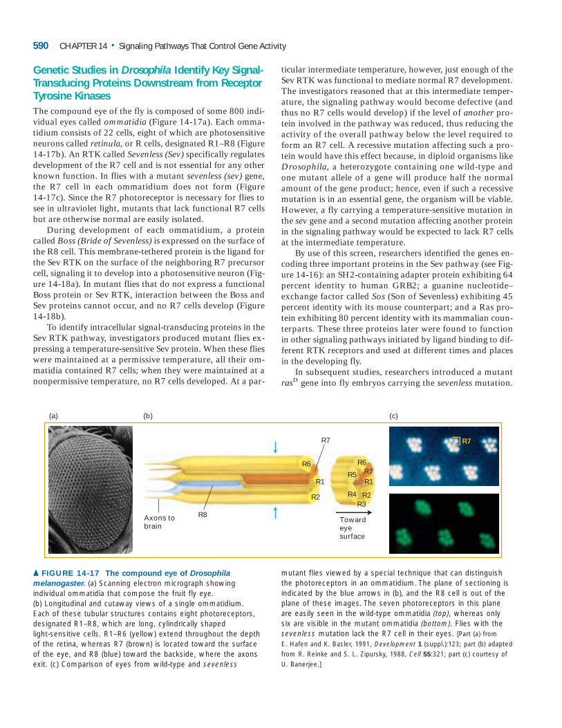
Genetic Studies in Drosophila Identify Key Signal-Transducing Proteins Downstream from ReceptorTyrosine KinasesThe compound eye of the fly is composed of some 800 indi-vidual eyes called ommatidia (Figure 14-17a). Each omma-tidium consists of 22 cells, eight of which are photosensitiveneurons called retinula, or R cells, designated R1–R8 (Figure14-17b). An RTK called Sevenless (Sev) specifically regulatesdevelopment of the R7 cell and is not essential for any otherknown function. In flies with a mutant sevenless (sev) gene,the R7 cell in each ommatidium does not form (Figure 14-17c). Since the R7 photoreceptor is necessary for flies tosee in ultraviolet light, mutants that lack functional R7 cellsbut are otherwise normal are easily isolated.
During development of each ommatidium, a proteincalled Boss (Bride of Sevenless) is expressed on the surface ofthe R8 cell. This membrane-tethered protein is the ligand forthe Sev RTK on the surface of the neighboring R7 precursorcell, signaling it to develop into a photosensitive neuron (Fig-ure 14-18a). In mutant flies that do not express a functionalBoss protein or Sev RTK, interaction between the Boss andSev proteins cannot occur, and no R7 cells develop (Figure14-18b).
To identify intracellular signal-transducing proteins in theSev RTK pathway, investigators produced mutant flies ex-pressing a temperature-sensitive Sev protein. When these flieswere maintained at a permissive temperature, all their om-matidia contained R7 cells; when they were maintained at anonpermissive temperature, no R7 cells developed. At a par-
ticular intermediate temperature, however, just enough of theSev RTK was functional to mediate normal R7 development.The investigators reasoned that at this intermediate temper-ature, the signaling pathway would become defective (andthus no R7 cells would develop) if the level of another pro-tein involved in the pathway was reduced, thus reducing theactivity of the overall pathway below the level required toform an R7 cell. A recessive mutation affecting such a pro-tein would have this effect because, in diploid organisms likeDrosophila, a heterozygote containing one wild-type andone mutant allele of a gene will produce half the normalamount of the gene product; hence, even if such a recessivemutation is in an essential gene, the organism will be viable.However, a fly carrying a temperature-sensitive mutation inthe sev gene and a second mutation affecting another proteinin the signaling pathway would be expected to lack R7 cellsat the intermediate temperature.
By use of this screen, researchers identified the genes en-coding three important proteins in the Sev pathway (see Fig-ure 14-16): an SH2-containing adapter protein exhibiting 64percent identity to human GRB2; a guanine nucleotide–exchange factor called Sos (Son of Sevenless) exhibiting 45percent identity with its mouse counterpart; and a Ras pro-tein exhibiting 80 percent identity with its mammalian coun-terparts. These three proteins later were found to functionin other signaling pathways initiated by ligand binding to dif-ferent RTK receptors and used at different times and placesin the developing fly.
In subsequent studies, researchers introduced a mutantrasD gene into fly embryos carrying the sevenless mutation.
590 CHAPTER 14 • Signaling Pathways That Control Gene Activity
(a) (b)
R6
R7
R1
R6R7R1
R2R3
R4
R5
R2
Axons to brain
R8 Towardeyesurface
(c)
R7
▲ FIGURE 14-17 The compound eye of Drosophila
melanogaster. (a) Scanning electron micrograph showingindividual ommatidia that compose the fruit fly eye. (b) Longitudinal and cutaway views of a single ommatidium. Each of these tubular structures contains eight photoreceptors,designated R1–R8, which are long, cylindrically shaped light-sensitive cells. R1–R6 (yellow) extend throughout the depthof the retina, whereas R7 (brown) is located toward the surfaceof the eye, and R8 (blue) toward the backside, where the axonsexit. (c) Comparison of eyes from wild-type and sevenless
mutant flies viewed by a special technique that can distinguishthe photoreceptors in an ommatidium. The plane of sectioning isindicated by the blue arrows in (b), and the R8 cell is out of theplane of these images. The seven photoreceptors in this planeare easily seen in the wild-type ommatidia (top), whereas onlysix are visible in the mutant ommatidia (bottom). Flies with thesevenless mutation lack the R7 cell in their eyes. [Part (a) from E. Hafen and K. Basler, 1991, Development 1 (suppl.):123; part (b) adaptedfrom R. Reinke and S. L. Zipursky, 1988, Cell 55:321; part (c) courtesy ofU. Banerjee.]

As noted earlier, the rasD gene encodes a constitutive Rasprotein that is present in the active GTP-bound form evenin the absence of a hormone signal. Although no functionalSev RTK was expressed in these double-mutants (sev�; rasD),R7 cells formed normally, indicating that activation of Ras issufficient for induction of R7-cell development (Figure 14-18c). This finding, which is consistent with the resultswith cultured fibroblasts described earlier, supports the con-clusion that activation of Ras is a principal step in intracel-lular signaling by most if not all RTKs.
Binding of Sos Protein to Inactive Ras Causes a Conformational Change That Activates RasThe adapter protein GRB2 contains two SH3 domains,which bind to Sos, a guanine nucleotide–exchange factor, in
addition to an SH2 domain, which binds to phosphotyrosineresidues in RTKs. Like phosphotyrosine-binding SH2 andPTB domains, SH3 domains are present in a large numberof proteins involved in intracellular signaling. Although thethree-dimensional structures of various SH3 domains aresimilar, their specific amino acid sequences differ. The SH3domains in GRB2 selectively bind to proline-rich sequencesin Sos; different SH3 domains in other proteins bind to proline-rich sequences distinct from those in Sos.
Proline residues play two roles in the interaction betweenan SH3 domain in an adapter protein (e.g., GRB2) and a pro-line-rich sequence in another protein (e.g., Sos). First, the pro-line-rich sequence assumes an extended conformation thatpermits extensive contacts with the SH3 domain, thereby fa-cilitating interaction. Second, a subset of these prolines fit intobinding “pockets” on the surface of the SH3 domain (Figure14-19). Several nonproline residues also interact with the SH3domain and are responsible for determining the binding speci-ficity. Hence the binding of proteins to SH3 and to SH2 do-mains follows a similar strategy: certain residues provide theoverall structural motif necessary for binding, and neighbor-ing residues confer specificity to the binding.
Following activation of an RTK (e.g., Sevenless or theEGF receptor), a complex containing the activated receptor,GRB2, and Sos is formed on the cytosolic face of the plasmamembrane (see Figure 14-16). Formation of this complex depends on the ability of GRB2 to bind simultaneously to the receptor and to Sos. Thus receptor activation leads to
14.3 • Receptor Tyrosine Kinases and Activation of Ras 591
Boss
(a) Wild type
Sev
R7 precursor
R8 cell
R7 neuron
(b) Single mutant (sev−)
Noinduction
(c) Double mutant (sev−; RasD)
ActiveRas
Induction
Cone cell R7 neuron
▲ EXPERIMENTAL FIGURE 14-18 Genetic studies revealthat activation of Ras induces development of R7photoreceptors in the Drosophila eye. (a) During larvaldevelopment of wild-type flies, the R8 cell in each developingommatidium expresses a cell-surface protein, called Boss, thatbinds to the Sev RTK on the surface of its neighboring R7precursor cell. This interaction induces changes in geneexpression that result in differentiation of the precursor cell into afunctional R7 neuron. (b) In fly embryos with a mutation in thesevenless (sev) gene, R7 precursor cells cannot bind Boss andtherefore do not differentiate normally into R7 cells. Rather theprecursor cell enters an alternative developmental pathway andeventually becomes a cone cell. (c) Double-mutant larvae (sev�;RasD) express a constitutively active Ras (RasD) in the R7precursor cell, which induces differentiation of R7 precursor cellsin the absence of the Boss-mediated signal. This finding showsthat activated Ras is sufficient to mediate induction of an R7 cell.[See M. A. Simon et al., 1991, Cell 67:701, and M. E. Fortini et al., 1992,Nature 355:559.]
Arg1
Pro4Pro7
SH3 domain
▲ FIGURE 14-19 Surface model of an SH3 domain boundto a short, proline-rich target peptide. The target peptide isshown as a space-filling model. In this target peptide, twoprolines (Pro4 and Pro7, dark blue) fit into binding pockets on thesurface of the SH3 domain. Interactions involving an arginine(Arg1, red), two other prolines (light blue), and other residues inthe target peptide (green) determine the specificity of binding.[After H. Yu et al., 1994, Cell 76:933.]

relocalization of Sos from the cytosol to the membrane,bringing Sos near to its substrate, namely, membrane-boundRas�GDP. Biochemical and genetic studies indicate that theC-terminus of Sos inhibits its nucleotide-exchange activityand that GRB2 binding relieves this inhibition.
Binding of Sos to Ras�GDP leads to conformationalchanges in the Switch I and Switch II segments of Ras,thereby opening the binding pocket for GDP so it can diffuseout (Figure 14-20). Because GTP is present in cells at a con-centration some 10 times higher than GDP, GTP binding occurs preferentially, leading to activation of Ras. The acti-vation of Ras and G� thus occurs by similar mechanisms: aconformational change induced by binding of a protein—Sosand an activated G protein–coupled receptor, respectively—that opens the protein structure so bound GDP is releasedto be replaced by GTP. Binding of GTP to Ras, in turn, in-duces a specific conformation of Switch I and Switch II thatallows Ras�GTP to activate downstream effector molecules,as we discuss in the next section.
KEY CONCEPTS OF SECTION 14.3
Receptor Tyrosine Kinases and Activation of Ras■ Receptor tyrosine kinases (RTKs), which bind to pep-tide and protein hormones, may exist as preformed dimersor dimerize during binding to ligands.■ Ligand binding leads to activation of the intrinsic pro-tein tyrosine kinase activity of the receptor and phospho-rylation of tyrosine residues in its cytosolic domain (seeFigure 14-5, top). The activated receptor also can phos-phorylate other protein substrates.
■ Ras is an intracellular GTPase switch protein that actsdownstream from most RTKs. Like G�, Ras cycles betweenan inactive GDP-bound form and an active GTP-boundform. Ras cycling requires the assistance of two proteins,a guanine nucleotide–exchange factor (GEF) and aGTPase-activating protein (GAP).■ RTKs are linked indirectly to Ras via two proteins:GRB2, an adapter protein, and Sos, which has GEF activ-ity (see Figure 14-16).■ The SH2 domain in GRB2 binds to a phosphotyrosinein activated RTKs, while its two SH3 domains bind Sos,thereby bringing Sos close to membrane-bound Ras�GDPand activating its nucleotide exchange activity.■ Binding of Sos to inactive Ras causes a large conforma-tional change that permits release of GDP and binding ofGTP, forming active Ras (see Figure 14-20). GAP, whichaccelerates GTP hydrolysis, is localized near Ras�GTP bybinding to activated RTKs.■ Normally, Ras activation and the subsequent cellular re-sponse require ligand binding to an RTK or a cytokine re-ceptor. In cells that contain a constitutively active Ras, thecellular response occurs in the absence of ligand binding.
MAP Kinase PathwaysIn mammalian cells all receptor tyrosine kinases (RTKs), aswell as most cytokine receptors, appear to utilize a highlyconserved signal-transduction pathway in which the signal in-duced by ligand binding is carried via GRB2 and Sos to Ras,leading to its activation (see Figure 14-16). Activated Ras pro-
14.4
592 CHAPTER 14 • Signaling Pathways That Control Gene Activity
(a) Ras.GDP (b) Ras-Sos (c) Ras.GTP
GDP
Sos αhelix
GTP
Switch I
Switch II
GDP
GTP
GTP γphosphate
GTP α, βphosphates
▲ FIGURE 14-20 Structures of Ras bound to GDP, Sosprotein, and GTP. (a) In Ras�GDP, the Switch I and Switch IIsegments do not directly interact with GDP (see Figure 13-8). (b) One � helix (orange) in Sos binds to both switch regions ofRas�GDP, leading to a massive conformational change in Ras. Ineffect, Sos pries Ras open by displacing the Switch I region,thereby allowing GDP to diffuse out. (c) GTP is thought to bind to
Ras-Sos first through its base; subsequent binding of the GTPphosphates completes the interaction. The resultingconformational change in Switch I and Switch II segments ofRas, allowing both to bind to the GTP � phosphate (see Figure 13-8), displaces Sos and promotes interaction of Ras�GTP withits effectors (discussed later). [Adapted from P. A. Boriack-Sjodin andJ. Kuriyan, 1998, Nature 394:341.]

motes formation at the membrane of signaling complexescontaining three sequentially acting protein kinases that areassociated with a scaffold protein. This kinase cascade cul-minates in activation of MAP kinase, a serine/threonine ki-nase also known as ERK. After translocating into the nucleus,MAP kinase can phosphorylate many different proteins, in-cluding transcription factors that regulate expression of im-portant cell-cycle and differentiation-specific proteins.Activation of MAP kinase in two different cells can lead tosimilar or different cellular responses, as can its activation inthe same cell following stimulation by different hormones.
In this section, we first examine the components of thekinase cascade downstream from Ras in RTK-Ras signalingpathways in mammalian cells. Then we discuss the linkage ofother signaling pathways to similar kinase cascades, and weexamine recent studies indicating that both yeasts and cellsof higher eukaryotes contain multiple MAP kinase pathways.
Signals Pass from Activated Ras to a Cascade of Protein Kinases A remarkable convergence of biochemical and genetic studiesin yeast, C. elegans, Drosophila, and mammals has revealed a highly conserved cascade of protein kinases that operates
in sequential fashion downstream from activated Ras (Figure14-21). Active Ras�GTP binds to the N-terminal regulatory do-main of Raf, a serine/threonine kinase, thereby activating it(step 2 ). Hydrolysis of Ras�GTP to Ras�GDP releases activeRaf (step 3 ), which phosphorylates and thereby activates MEK(step 4 ). Active MEK then phosphorylates and activates MAPkinase, another serine/threonine kinase (step 5 ). (A dual-specificity protein kinase, MEK phosphorylates its target pro-teins on both tyrosine and serine or threonine residues.) MAP ki-nase phosphorylates many different proteins, including nucleartranscription factors, that mediate cellular responses (step 6 ).
Several types of experiments have demonstrated that Raf,MEK, and MAP kinase lie downstream from Ras and haverevealed the sequential order of these proteins in the path-way. For example, mutant Raf proteins missing the N-terminal regulatory domain are constitutively active andinduce quiescent cultured cells to proliferate in the absence ofstimulation by growth factors. These mutant Raf proteinswere initially identified in tumor cells; like the constitutivelyactive RasD protein, such mutant Raf proteins are said to beencoded by oncogenes (Chapter 23). Conversely, culturedmammalian cells that express a mutant, nonfunctional Rafprotein cannot be stimulated to proliferate uncontrollably bya constitutively active RasD protein. This finding established
14.4 • MAP Kinase Pathways 593
P
PP
GDP GTP
Inactive Raf
C-terminalkinasedomain
Ras activated by exchange of GDP for GTP
Inactive Ras Active RasPi
ActiveRaf
Active Ras recruits,binds, and activatesRaf
MEK
MAP kinase
ATP
ADP
ATP
ADP
P
P14-3-3
GTP hydrolysis leads to dissociationof Ras from Raf
Rafactivates
MEK
MEKactivates
MAPK
Dimeric form of active MAP kinasetranslocates to nucleus; activatesmany transcription factors
1 2 3
4
5
6
Cytosol
Exterior
GDP
GTP
N-terminalregulatory
domain
GDP
GTP
PP
P P PP
▲ FIGURE 14-21 Kinase cascade that transmits signals downstream fromactivated Ras protein to MAP kinase. In unstimulated cells, most Ras is in the inactiveform with bound GDP; binding of a ligand to its RTK or cytokine receptor leads toformation of the active Ras�GTP complex (step ; see also Figure 14-16). Activated Ras triggers the downstream kinase cascade depicted in steps – , culminating inactivation of MAP kinase (MAPK). In unstimulated cells, binding of the 14-3-3 protein toRaf stabilizes it in an inactive conformation. Interaction of the Raf N-terminal regulatorydomain with Ras�GTP relieves this inhibition, results in dephosphorylation of one of theserines that bind Raf to 14-3-3, and leads to activation of Raf kinase activity (steps and ). Note that in contrast to many other protein kinases, activation of Raf does notdepend on phosphorylation of the activation lip. After inactive Ras�GDP dissociates from Raf, it presumably can be reactivated by signals from activated receptors, therebyrecruiting additional Raf molecules to the membrane. See the text for details. [SeeE. Kerkhoff and U. Rapp, 2001, Adv. Enzyme Regul. 41:261; J. Avruch et al., 2001, Recent Prog.Hormone Res. 56:127; and M. Yip-Schneider et al., 2000, Biochem. J. 351:151.]
32
621

a link between the Raf and Ras proteins. In vitro bindingstudies further showed that the purified Ras�GTP complexbinds directly to the N-terminal regulatory domain of Rafand activates its catalytic activity. An interaction between themammalian Ras and Raf proteins also was demonstrated inthe yeast two-hybrid system, a genetic system in yeast used toselect cDNAs encoding proteins that bind to target, or“bait,” proteins (see Figure 11-39).
That MAP kinase is activated in response to Ras activa-tion was demonstrated in quiescent cultured cells expressinga constitutively active RasD protein. In these cells activatedMAP kinase is generated in the absence of stimulation bygrowth-promoting hormones. More importantly, R7 pho-toreceptors develop normally in the developing eye ofDrosophila mutants that lack a functional Ras or Raf proteinbut express a constitutively active MAP kinase. This findingindicates that activation of MAP kinase is sufficient to trans-mit a proliferation or differentiation signal normally initiatedby ligand binding to a receptor tyrosine kinase such as Sev-enless (see Figure 14-18). Biochemical studies showed, how-ever, that Raf cannot directly phosphorylate MAP kinase orotherwise activate its activity.
The final link in the kinase cascade activated by Ras�GTPemerged from studies in which scientists fractionated ex-tracts of cultured cells searching for a kinase activity thatcould phosphorylate MAP kinase and that was present onlyin cells stimulated with growth factors, not quiescent cells.This work led to identification of MEK, a kinase that specif-ically phosphorylates one threonine and one tyrosine residueon MAP kinase, thereby activating its catalytic activity. (Theacronym MEK comes from MAP and ERK kinase.) Laterstudies showed that MEK binds to the C-terminal catalyticdomain of Raf and is phosphorylated by the Raf serine/threonine kinase; this phosphorylation activates the catalyticactivity of MEK. Hence, activation of Ras induces a kinasecascade that includes Raf, MEK, and MAP kinase: activatedRTK → Ras → Raf → MEK → MAP kinase.
Activation of Raf Kinase The mechanism for activating Rafdiffers from that used to activate many other protein kinasesincluding MEK and MAP kinase. In a resting cell prior tohormonal stimulation, Raf is present in the cytosol in a con-formation in which the N-terminal regulatory domain isbound to the kinase domain, thereby inhibiting its activity.This inactive conformation is stabilized by a dimer of the 14-3-3 protein, which binds phosphoserine residues in a numberof important signaling proteins. Each 14-3-3 monomer bindsto a phosphoserine residue in Raf, one to phosphoserine-259in the N-terminal domain and the other to phosphoserine-621 (see Figure 14-21). These interactions are thought to beessential for Raf to achieve a conformational state such thatit can bind to activated Ras.
The binding of Ras�GTP, which is anchored to themembrane, to the N-terminal domain of Raf relieves the in-hibition of Raf’s kinase activity and also induces a confor-mational change in Raf that disrupts its association with
14-3-3. Raf phosphoserine-259 then is dephosphorylated (byan unknown phosphatase) and other serine or threonineresidues on Raf become phosphorylated by yet other kinases.These reactions incrementally increase the Raf kinase activ-ity by mechanisms that are not fully understood.
Activation of MAP Kinase Biochemical and x-ray crystallo-graphic studies have provided a detailed picture of howphosphorylation activates MAP kinase. As in JAK kinasesand the cytosolic domain of receptor tyrosine kinases, thecatalytic site in the inactive, unphosphorylated form of MAPkinase is blocked by a stretch of amino acids, the activationlip (Figure 14-22a). Binding of MEK to MAP kinase desta-bilizes the lip structure, resulting in exposure of tyrosine-185, which is buried in the inactive conformation. Followingphosphorylation of this critical tyrosine, MEK phosphory-lates the neighboring threonine-183 (Figure 14-22b).
Both the phosphorylated tyrosine and the phosphory-lated threonine residues in MAP kinase interact with additional amino acids, thereby conferring an altered con-formation to the lip region, which in turn permits bindingof ATP to the catalytic site. The phosphotyrosine residue(pY185) also plays a key role in binding specific substrateproteins to the surface of MAP kinase. Phosphorylation pro-motes not only the catalytic activity of MAP kinase but also
594 CHAPTER 14 • Signaling Pathways That Control Gene Activity
PP
(a) Inactive MAP kinase (b) Active MAP kinase
Y185T183
pY185
pT183
Activationlip
▲ EXPERIMENTAL FIGURE 14-22 Molecular structures of MAP kinase in its inactive, unphosphorylated form (a) andactive, phosphorylated form (b). Phosphorylation of MAP kinaseby MEK at tyrosine-185 (Y185) and threonine-183 (T183) leads toa marked conformational change in the activation lip. This changepromotes dimerization of MAP kinase and binding of itssubstrates, ATP and certain proteins. A similar phosphorylation-dependent mechanism activates JAK kinases, the intrinsic kinaseactivity of RTKs, and MEK. [After B. J. Canagarajah et al., 1997, Cell 90:859.]
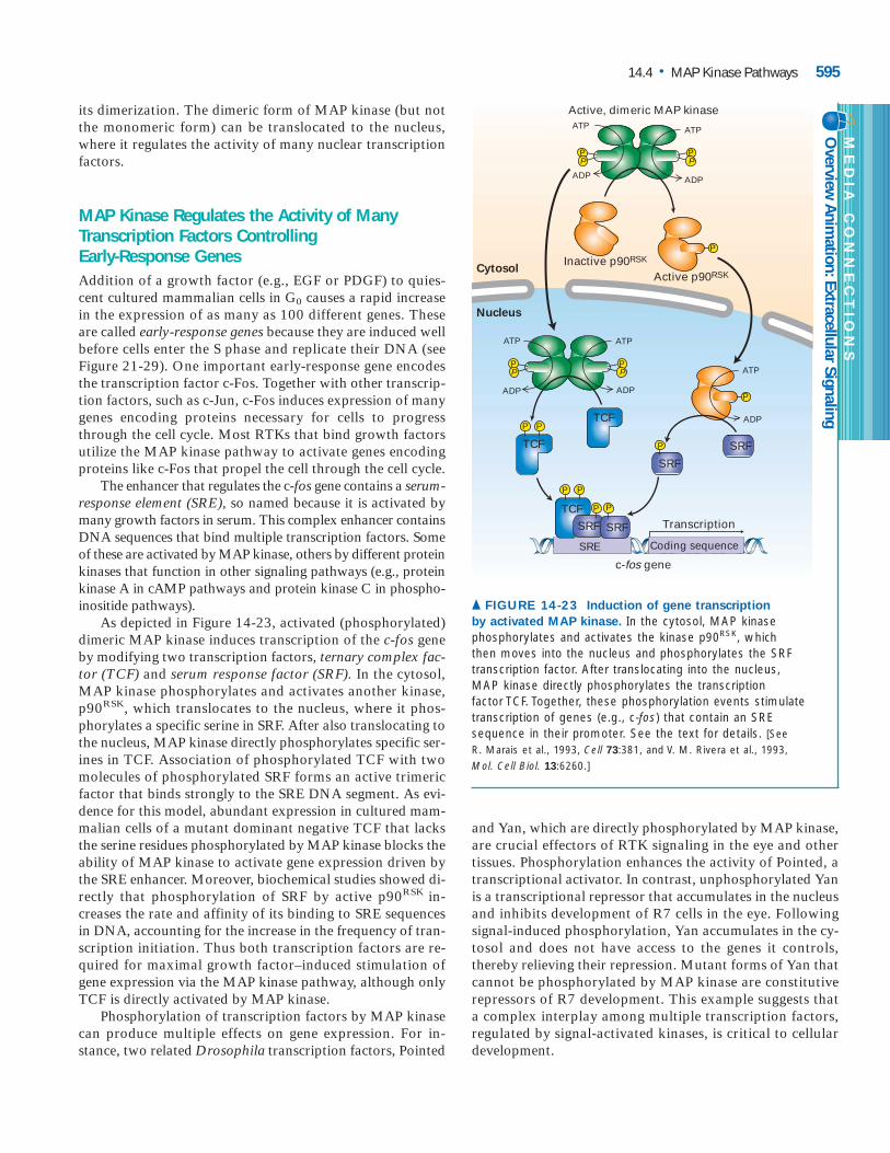
its dimerization. The dimeric form of MAP kinase (but notthe monomeric form) can be translocated to the nucleus,where it regulates the activity of many nuclear transcriptionfactors.
MAP Kinase Regulates the Activity of ManyTranscription Factors Controlling Early-Response GenesAddition of a growth factor (e.g., EGF or PDGF) to quies-cent cultured mammalian cells in G0 causes a rapid increasein the expression of as many as 100 different genes. Theseare called early-response genes because they are induced wellbefore cells enter the S phase and replicate their DNA (seeFigure 21-29). One important early-response gene encodesthe transcription factor c-Fos. Together with other transcrip-tion factors, such as c-Jun, c-Fos induces expression of manygenes encoding proteins necessary for cells to progressthrough the cell cycle. Most RTKs that bind growth factorsutilize the MAP kinase pathway to activate genes encodingproteins like c-Fos that propel the cell through the cell cycle.
The enhancer that regulates the c-fos gene contains a serum-response element (SRE), so named because it is activated bymany growth factors in serum. This complex enhancer containsDNA sequences that bind multiple transcription factors. Someof these are activated by MAP kinase, others by different proteinkinases that function in other signaling pathways (e.g., proteinkinase A in cAMP pathways and protein kinase C in phospho-inositide pathways).
As depicted in Figure 14-23, activated (phosphorylated)dimeric MAP kinase induces transcription of the c-fos geneby modifying two transcription factors, ternary complex fac-tor (TCF) and serum response factor (SRF). In the cytosol,MAP kinase phosphorylates and activates another kinase,p90RSK, which translocates to the nucleus, where it phos-phorylates a specific serine in SRF. After also translocating tothe nucleus, MAP kinase directly phosphorylates specific ser-ines in TCF. Association of phosphorylated TCF with twomolecules of phosphorylated SRF forms an active trimericfactor that binds strongly to the SRE DNA segment. As evi-dence for this model, abundant expression in cultured mam-malian cells of a mutant dominant negative TCF that lacksthe serine residues phosphorylated by MAP kinase blocks theability of MAP kinase to activate gene expression driven bythe SRE enhancer. Moreover, biochemical studies showed di-rectly that phosphorylation of SRF by active p90RSK in-creases the rate and affinity of its binding to SRE sequencesin DNA, accounting for the increase in the frequency of tran-scription initiation. Thus both transcription factors are re-quired for maximal growth factor–induced stimulation ofgene expression via the MAP kinase pathway, although onlyTCF is directly activated by MAP kinase.
Phosphorylation of transcription factors by MAP kinasecan produce multiple effects on gene expression. For in-stance, two related Drosophila transcription factors, Pointed
and Yan, which are directly phosphorylated by MAP kinase,are crucial effectors of RTK signaling in the eye and othertissues. Phosphorylation enhances the activity of Pointed, atranscriptional activator. In contrast, unphosphorylated Yanis a transcriptional repressor that accumulates in the nucleusand inhibits development of R7 cells in the eye. Followingsignal-induced phosphorylation, Yan accumulates in the cy-tosol and does not have access to the genes it controls,thereby relieving their repression. Mutant forms of Yan thatcannot be phosphorylated by MAP kinase are constitutiverepressors of R7 development. This example suggests thata complex interplay among multiple transcription factors,regulated by signal-activated kinases, is critical to cellulardevelopment.
14.4 • MAP Kinase Pathways 595
PP
PP
P
ATP
ADP
Cytosol
Nucleus
SRE Coding sequence
PP
PP
ATP
ATP
ADP
c-fos gene
P
ATP
ADP
TranscriptionTCF
SRF SRF
TCF
SRF
ADP
ATP
ADP
SRFTCF
Inactive p90RSK
Active p90RSK
Active, dimeric MAP kinase
P
PP
PP
PP
▲ FIGURE 14-23 Induction of gene transcription by activated MAP kinase. In the cytosol, MAP kinasephosphorylates and activates the kinase p90RSK, which then moves into the nucleus and phosphorylates the SRFtranscription factor. After translocating into the nucleus, MAP kinase directly phosphorylates the transcription factor TCF. Together, these phosphorylation events stimulatetranscription of genes (e.g., c-fos) that contain an SREsequence in their promoter. See the text for details. [SeeR. Marais et al., 1993, Cell 73:381, and V. M. Rivera et al., 1993, Mol. Cell Biol. 13:6260.]
ME
DIA
CO
NN
EC
TIO
NS
Overview Anim
ation: Extracellular Signaling

G Protein–Coupled Receptors Transmit Signals to MAP Kinase in Yeast Mating PathwaysAlthough many MAP kinase pathways are initiated by RTKsor cytokine receptors, signaling from other receptors can ac-tivate MAP kinase in different cell types of higher eukary-otes. Moreover, yeasts and other single-celled eukaryotes,which lack cytokine receptors or RTKs, do possess severalMAP kinase pathways. To illustrate, we consider the matingpathway in S. cerevisiae, a well-studied example of a MAPkinase cascade linked to G protein–coupled receptors(GPCRs), in this case for two secreted peptide pheromones,the a and � factors.
As discussed in Chapter 22, these pheromones controlmating between haploid yeast cells of the opposite matingtype, a or �. An a haploid cell secretes the a mating factorand has cell-surface receptors for the � factor; an � cell se-cretes the � factor and has cell-surface receptors for the a fac-tor (see Figure 22-13). Thus each type of cell recognizes themating factor produced by the opposite type. Activation ofthe MAP kinase pathway by either the a or � receptors in-
duces transcription of genes that inhibit progression of thecell cycle and others that enable cells of opposite mating typeto fuse together and ultimately form a diploid cell.
Ligand binding to either of the two yeast pheromone re-ceptors triggers the exchange of GTP for GDP on the singleG� subunit and dissociation of G��GTP from the G�� com-plex. This activation process is identical to that for theGPCRs discussed in the previous chapter (see Figure 13-11).In most, but not all, mammalian GPCR-initiated pathways,the active G� transduces the signal. In contrast, mutant stud-ies have shown that the dissociated G�� complex mediates allthe physiological responses induced by activation of the yeastpheromone receptors. For instance, in yeast cells that lackG�, the G�� subunit is always free. Such cells can mate in theabsence of mating factors; that is, the mating response is con-stitutively on. However, in cells defective for the G� or G�
subunit, the mating pathway cannot be induced at all. If dis-sociated G� were the transducer, the pathway would be ex-pected to be constitutively active in these mutant cells.
In yeast mating pathways, G�� functions by triggering akinase cascade that is analogous to the one downstream from
596 CHAPTER 14 • Signaling Pathways That Control Gene Activity
Serine/threoninekinase
Activation of genesrequired for mating
Exterior
Cytosol
MEKK,serine/threoninekinase
MAPK,serine/threoninekinase
Ste 12
MEK,threonine/tyrosinedual-specificity kinase
Ste5scaffoldprotein
Matingfactor
Receptor
Ste20
Transcription factor
Fus3 to nucleus
PP
Gαγ
Gα
Gβγ β
Activationof G protein
P
P
P
Ste11
Ste7
Fus3
GDP
GTP
P
� FIGURE 14-24 Kinase cascade that transmits signals downstream frommating factor receptors in S. cerevisiae.
The receptors for yeast a and � matingfactors are coupled to the same trimeric G protein. Ligand binding leads toactivation and dissociation of the G protein(see Figure 13-10). In the yeast matingpathway, the dissociated G�� activates aprotein kinase cascade analogous to thecascade downstream of Ras that leads to activation of MAP kinase (see Figure 14-21). The final component, Fus3, isfunctionally equivalent to MAP kinase(MAPK) in higher eukaryotes. Associationof several kinases with the Ste5 scaffoldcontributes to specificity of the signalingpathway by preventing phosphorylation ofother substrates. [See A. Whitmarsh and R. Davis, 1998, Trends Biochem. Sci. 23:481,and H. Dohlman and J. Thorner, 2001, Ann. Rev.Biochem. 70:703.]

Ras. The components of this cascade were uncovered mainlythrough analyses of mutants that possess functional a and �receptors and G proteins but are sterile (Ste), or defective inmating responses. The physical interactions between the components were assessed through immunoprecipitation experiments with extracts of yeast cells and other types ofstudies. Based on these studies, scientists have proposed the ki-nase cascade depicted in Figure 14-24. G��, which is tetheredto the membrane via the � subunit, binds to and activatesSte20, a protein kinase that in turn phosphorylates and acti-vates Ste11, a serine/threonine kinase analogous to Raf andother mammalian MEKK proteins. Activated Ste11 then phos-phorylates Ste7, a dual-specificity MEK that then phosphory-lates and activates Fus3, a serine/threonine kinase equivalentto MAP kinase. After translocation to the nucleus, Fus3 pro-motes expression of target genes by phosphorylating and thusactivating nuclear transcription factors (e.g., Ste12) that con-trol expression of proteins involved in mating-specific cellularresponses. The other component of the yeast mating cascade,Ste5, interacts with G�� as well as Ste11, Ste7, and Fus3. Ste5has no obvious catalytic function and acts as a scaffold for as-sembling other components in the cascade.
Scaffold Proteins Isolate Multiple MAP KinasePathways in Eukaryotic CellsIn addition to the MAP kinases discussed above, both yeastsand higher eukaryotic cells contain other members of theMAP kinase superfamily. These include mammalian Jun N-terminal kinases (JNKs) and p38 kinases, which become ac-tivated by various types of stresses, and six yeast kinasesdescribed below. Collectively referred to as MAP kinases, allthese proteins are serine/threonine kinases that are activatedin the cytosol in response to specific extracellular signals andthen translocate to the nucleus. Activation of all known MAPkinases requires phosphorylation of both a tyrosine and a
threonine residue in the lip region (see Figure 14-22). Simi-larly, all eukaryotic cells contain several members of the dual-specificity MEK kinase superfamily that phosphorylatedifferent members of the MAP kinase superfamily. Thus in alleukaryotic cells, binding of a wide variety of extracellular sig-naling molecules triggers highly conserved kinase cascadesculminating in activation of a particular MAP kinase. The dif-ferent MAP kinases mediate specific cellular responses, in-cluding morphogenesis, cell death, and stress responses.
Current genetic and biochemical studies in the mouse andDrosophila are aimed at determining which MAP kinases arerequired for mediating the response to which signals inhigher eukaryotes. This has already been accomplished inlarge part for the simpler organism S. cerevisiae. Each of thesix MAP kinases encoded in the S. cerevisiae genome hasbeen assigned by genetic analyses to specific signaling path-ways triggered by various extracellular signals, such aspheromones, starvation, high osmolarity, hypotonic shock,and carbon/nitrogen deprivation. Each of these MAP kinasesmediates very specific cellular responses (Figure 14-25).
In both yeasts and higher eukaryotic cells, different MAPkinase cascades share some common components. For in-stance, Ste11 functions in the yeast signaling pathways thatregulate mating, filamentous growth, and osmoregulation.Nevertheless, each pathway activates its own MAP kinase:Fus3 in the mating pathway, Kss1 in the filamentation path-way, and Hog1 in the osmoregulation pathway. Similarly, inmammalian cells, common upstream signal-transducing pro-teins participate in activating multiple JNK kinases.
Once the sharing of components among different MAPkinase pathways was recognized, researchers wondered howthe specificity of the cellular responses to particular signalscould be achieved. Studies with yeast provided the initial evidence that pathway-specific scaffold proteins enable thesignal-transducing kinases in a particular pathway to interactwith one another but not with kinases in other pathways.
14.4 • MAP Kinase Pathways 597
Pheromone Starvation
Filamentation
Highosmolarity
Osmolytesynthesis
Hypotonicshock
Cell-wallremodeling
Carbon andnitrogen
deprivationExtracellularsignal
MAP kinase
Cell response
Fus3 Kss1 Hog1 Mpk1 Smk1
Mating Sporulation
▲ FIGURE 14-25 Overview of five MAP kinase pathways inS. cerevisiae. Each pathway is triggered by a specificextracellular signal and leads to activation of a single differentMAP kinase, which mediates characteristic cellular responses.Formation of pathway-specific complexes of MAP kinases and
scaffold proteins prevents “cross talk” between pathwayscontaining a common component such as the MEKK Ste11,which occurs in the mating, filamentation, and osmoregulatorypathways (see Figure 14-24). [Adapted from H. D. Madhani and G. R. Fink, 1998, Trends Genet. 14(4):152.]

For example, the scaffold protein Ste5 stabilizes a large com-plex that includes Ste11 and other kinases in the matingpathway (see Figure 14-24). Different Ste11-binding scaffoldproteins, however, stabilize signaling complexes containingthe components of the filamentation and osmoregulationpathways. In each pathway in which Ste11 participates, it isconstrained within a large complex that forms in responseto a specific extracellular signal, and signaling downstreamfrom Ste11 is restricted to the complex in which it is local-ized. As a result, exposure of yeast cells to mating factors in-duces activation of a single MAP kinase, Fus3, whereasexposure to a high osmolarity or starvation induces activa-tion of different MAP kinases (see Figure 14-25).
Scaffolds for MAP kinase pathways are well documentedin yeast, fly, and worm cells, but their presence in mam-malian cells has been difficult to demonstrate. Perhaps thebest documented scaffold protein is Ksr (kinase suppressor ofRas), which binds both MEK and MAP kinase. Loss of theDrosophila Ksr homolog blocks signaling by a constitutivelyactive Ras protein, suggesting a positive role for Ksr inRas–MAP kinase signaling in fly cells. Although knockoutmice that lack Ksr are grossly normal, activation of MAP ki-nase by growth factors or cytokines is lower than normal inseveral types of cells in these animals. This finding suggeststhat Ksr functions as a scaffold that enhances but is not es-sential for Ras–MAP kinase signaling in mammalian cells.Other proteins also have been found to bind to specific mam-malian MAP kinases. Thus the signal specificity of differentMAP kinases in animal cells may arise from their associa-tion with various scaffold-like proteins, but much additionalresearch is needed to test this possibility.
KEY CONCEPTS OF SECTION 14.4MAP Kinase Pathways■ Activated Ras triggers a kinase cascade in which Raf,MEK, and MAP kinase are sequentially phosphorylatedand thus activated. Activated MAP kinase dimerizes andtranslocates to the nucleus (see Figure 14-21).■ Phosphorylation of one or more residues in a conservedlip region activates MAP kinases and many other proteinkinases involved in signal-transduction pathways.■ Activation of MAP kinase following stimulation of agrowth factor receptor leads to phosphorylation and acti-vation of two transcription factors, TCF and SRF. Theseassociate into a trimeric complex that promotes transcrip-tion of various early-response genes (see Figure 14-23).■ Yeast and higher eukaryotes contain multiple MAP ki-nase pathways that are triggered by activation of variousreceptor classes including G protein–coupled receptors.■ Different extracellular signals induce activation of dif-ferent MAP kinases, which regulate diverse cellularprocesses (see Figure 14-25).
■ The upstream components of MAP kinase cascades as-semble into large pathway-specific complexes stabilized byscaffold proteins (see Figure 14-24). This assures that ac-tivation of one pathway by a particular extracellular sig-nal does not lead to activation of other pathways con-taining shared components.
Phosphoinositides as SignalTransducersIn previous sections, we have seen how signal transduction from cytokine receptors and receptor tyrosine kinases (RTKs) begins with formation of multiprotein complexes associated with the plasma membrane. Here we discuss how these receptors initiate signaling pathways that involve membrane-bound phosphorylated inositol lipids, collec-tively referred to as phosphoinositides. We begin with the branch of the phosphoinositide pathway that also is medi-ated by G protein–coupled receptors and then consider an-other branch that is not shared with these receptors.
Phospholipase C� Is Activated by Some RTKs and Cytokine ReceptorsAs discussed in Chapter 13, hormonal stimulation of some Gprotein–coupled receptors leads to activation of the � iso-form of phospholipase C (PLC�). This membrane-associatedenzyme then cleaves phosphatidylinositol 4,5-bisphosphate(PIP2) to generate two important second messengers, 1,2-diacylglycerol (DAG) and inositol 1,4,5-trisphosphate (IP3).Signaling via the IP3/DAG pathway leads to an increase incytosolic Ca2� and to activation of protein kinase C (see Fig-ure 13-29).
Many RTKs and cytokine receptors also can initiate theIP3/DAG pathway by activating another isoform of phos-pholipase C, the � isoform (PLC�). The SH2 domains ofPLC� bind to specific phosphotyrosines of the activated re-ceptors, thus positioning the enzyme close to its membrane-bound substrate PIP2 (see Figure 13-28). In addition, thereceptor kinase activity phosphorylates tyrosine residues onthe bound PLC�, enhancing its hydrolase activity. Thus acti-vated RTKs and cytokine receptors promote PLC� activity intwo ways: by localizing the enzyme to the membrane and byphosphorylating it.
Recruitment of PI-3 Kinase to Hormone-Stimulated Receptors Leads to Activation of Protein Kinase BIn addition to initiating the IP3/DAG pathway, some acti-vated RTKs and cytokine receptors can initiate anotherphosphoinositide pathway, the PI-3 kinase pathway, by re-
14.5
598 CHAPTER 14 • Signaling Pathways That Control Gene Activity

cruiting the enzyme phosphatidylinositol-3 kinase to themembrane. PI-3 kinase was first identified as a kinase thatcopurifies with several viral oncoproteins such as the “mid-dle T” protein encoded by polyoma virus. When inactive,dominant negative, versions of PI-3 kinase are expressed invirus-transformed cells, they inhibit the uncontrolled cellproliferation characteristic of virus-transformed cells. Thisfinding suggested that the normal kinase is important in cer-tain signaling pathways essential for cell proliferation or forthe prevention of apoptosis. Subsequent work showed thatPI-3 kinases participate in many signaling pathways relatedto cell growth and apoptosis. Of the nine PI-3 kinase ho-mologs encoded by the human genome, the best character-ized contains a p110 subunit with catalytic activity and ap85 subunit with an SH2 domain.
The SH2 domain in PI-3 kinase binds to phosphotyro-sine residues in the cytosolic domain of many activatedRTKs and cytokine receptors. The recruitment of PI-3 kinase to the plasma membrane by activated receptors po-sitions its catalytic domain near its phosphoinositide sub-strates on the cytosolic face of the plasma membrane,leading to formation of PI 3,4-bisphosphate or PI 3,4,5-trisphosphate (Figure 14-26). By acting as docking sites forvarious signal-transducing proteins, these membrane-boundPI 3-phosphates in turn transduce signals downstream inseveral important pathways.
A primary binding target of PI 3-phosphates is proteinkinase B (PKB), a serine/threonine kinase. Besides its kinasedomain, PKB contains a PH domain that tightly binds the3-phosphate in both PI 3,4-bisphosphate and PI 3,4,5-trisphosphate. In unstimulated, resting cells, the level ofboth these compounds is low, and protein kinase B is pres-ent in the cytosol in an inactive form. Following hormonestimulation and the resulting rise in PI 3-phosphates, pro-tein kinase B binds to them and is localized at the cell sur-face membrane.
Binding of protein kinase B to PI 3-phosphates not onlyrecruits the enzyme to the plasma membrane but also re-leases inhibition of the catalytic site by the PH domain in thecytosol. Maximal activation of protein kinase B, however,depends on recruitment of another kinase, PDK1, to theplasma membrane via binding of its PH domain to PI 3-phosphates. Both membrane-associated protein kinase Band PDK1 can diffuse in the plane of the membrane, bring-ing them close enough so that PDK1 can phosphorylate pro-tein kinase B (Figure 14-27). PDK1 phosphorylates oneserine residue in the activation lip of protein kinase B, pro-viding yet another example of kinase activation by phos-phorylation in this segment. Phosphorylation of a secondserine, not in the lip segment, is necessary for maximal pro-tein kinase B activity. Thus, as with Raf, an inhibitory do-main and phosphorylation by other kinases regulate theactivity of protein kinase B. Once fully activated, protein ki-nase B can dissociate from the plasma membrane and phos-phorylate its many target proteins.
14.5 • Phosphoinositides as Signal Transducers 599
ATP ADP
ATP ADP
PIP-5 kinase
OC OC
O O
CH2CH
4OH HOOH
OH
CH2
P
PI 4-phosphate(PIP)
−O O
O
P
O
OC OC
O O
CH
4
5
OH HOOH
CH2
P
P
PI 4,5-bisphosphate(PIP2)
CH2
−O O
O
P
O
PIP-5 kinase
OC OC
O O
CH2CH
4OHOH
OH
CH2
P
PI 3,4-bisphosphate
−O O
O
P
O
OC OC
O O
CH
4
5
OHOH
CH2
P
P
PI 3,4,5-trisphosphate
CH2
−O O
O
P
O
1
2 3
56
P1
2 3
56
P
3
ATP
ADP
ATP
ADP
PI-3 kinase PI-3 kinase
Cyto
solic
leaf
let
Inos
itol
▲ FIGURE 14-26 Generation of phosphatidylinositol 3-phosphates. The enzyme phosphatidylinositol-3 kinase (PI-3kinase) is recruited to the membrane by many activated receptortyrosine kinases (RTKs) and cytokine receptors. The 3-phosphateadded by this enzyme is a binding site for various signal-transduction proteins. [See L. Rameh and L. C. Cantley, 1999, J. Biol.Chem. 274:8347.]

The Insulin Receptor Acts Through the PI-3Kinase Pathway to Lower Blood GlucoseThe insulin receptor is a dimeric receptor tyrosine kinase that can initiate the Ras–MAP kinase pathway, leading tochanges in gene expression. Insulin stimulation also can ini-tiate the PI-3 kinase pathway just described, leading to acti-vation of protein kinase B. In insulin-stimulated liver, muscle,and fat cells, activated protein kinase B acts in several waysto lower blood glucose and promote glycogen synthesis.
The principal mechanism by which insulin causes a re-duction of the blood glucose level is by increasing import ofglucose by fat and muscle cells. This effect is mediated byprotein kinase B, which through mechanisms that are notfully understood causes movement of the GLUT4 glucosetransporter from intracellular membranes to the cell surface(Chapter 15). The resulting increased influx of glucose intothese cells lowers blood glucose levels.
In both liver and muscle, insulin stimulation also leadsto activation of glycogen synthase (GS), which synthesizesglycogen from UDP-glucose (see Figure 13-16). This repre-sents another mechanism for reducing glucose concentrationin the circulation. In resting cells (i.e., in the absence of in-sulin), glycogen synthase kinase 3 (GSK3) is active and phos-phorylates glycogen synthase, thereby blocking its activity.Activated protein kinase B phosphorylates and thereby in-activates GSK3. As a result, GSK3-mediated inhibition ofglycogen synthase is relieved, promoting glycogen synthesis.
Activated Protein Kinase B Promotes Cell Survivalby Several PathwaysIn many cells activated protein kinase B directly phosphory-lates pro-apoptotic proteins such as Bad, thereby preventing
activation of an apoptotic pathway leading to cell death(Chapter 22). Activated protein kinase B also promotes survival of many cultured cells by phosphorylating the transcription factor Forkhead-1 on as many as three serine or threonine residues. In the absence of growth factors, Forkhead-1 is unphosphorylated and localizes to the nucleus,where it activates transcription of several genes encoding pro-apoptotic proteins. When growth factors are added to thecells, protein kinase B becomes active and phosphorylatesForkhead-1. This allows the cytosolic phosphoserine-binding protein 14-3-3 to bind Forkhead-1 and thus sequesterit in the cytosol. (14-3-3 is the same protein that retains phos-phorylated Raf protein in the cytosol; see Figure 14-21.)Withdrawal of growth factor leads to inactivation of proteinkinase B and dephosphorylation of Forkhead-1, thus favoringapoptosis. A Forkhead-1 mutant in which the three serine tar-get residues for protein kinase B are mutated is “constitutivelyactive” and initiates apoptosis even in the presence of acti-vated protein kinase B. This finding demonstrates the impor-tance of Forkhead-1 in controlling apoptosis of cultured cells.
PTEN Phosphatase Terminates Signaling via the PI-3 Kinase PathwayLike virtually all intracellular signaling events, phosphoryla-tion by PI-3 kinase is reversible. The relevant phosphatase,termed PTEN phosphatase, has an unusually broad specificity.Although PTEN can remove phosphate groups attached toserine, threonine, and tyrosine residues in proteins, its abilityto remove the 3-phosphate from PI 3,4,5-trisphosphate isthought to be its major function in cells. Overexpression ofPTEN in cultured mammalian cells promotes apoptosis by re-ducing the level of PI 3,4,5-trisphosphate and hence the acti-vation and anti-apoptotic effect of protein kinase B.
600 CHAPTER 14 • Signaling Pathways That Control Gene Activity
P
P
P43
Inactive PKB
Kinasedomain
PH domain
PI 3,4-bisphosphateP
P43
Partiallyactive PKB
Activationlip
P
P43
P
P43
Fully active PKBP PDK1
Exterior
Cytosol
▲ FIGURE 14-27 Recruitment and activation ofprotein kinase B (PKB) in PI-3 kinase pathways. Inunstimulated cells, PKB is in the cytosol with its PHdomain bound to the catalytic domain, inhibiting itsactivity. Hormone stimulation leads to activation of PI-3kinase and subsequent formation of phosphatidylinositol
ME
DIA
CO
NN
EC
TIO
NS
Vide
o: P
rote
in D
ynam
ics in
Res
pons
e to
cAM
PSt
imul
atio
n of
a D
ictyo
steliu
mCe
ll
(PI) 3-phosphates (see Figure 14-26). The 3-phosphate groupsserve as docking sites on the plasma membrane for the PHdomain of PKB and another kinase, PDK1. Full activation of PKBrequires phosphorylation both in the activation lip and at the C-terminus by PDK1. [Adapted from A. Toker and A. Newton, 2000, Cell103:185, and M. Scheid et al., 2002, Mol. Cell Biol. 22:6247.]

The gene encoding PTEN is deleted in multiple typesof advanced human cancers, and its loss is thoughtto lead to uncontrolled growth. Indeed, cells lacking
PTEN have elevated levels of PI 3,4,5-trisphosphate and PKBactivity. Since protein kinase B exerts an anti-apoptotic effect,loss of PTEN indirectly reduces the programmed cell deaththat is the normal fate of abnormally controlled cells. In certain cells, such as neuronal stem cells, absence of PTEN not only prevents apoptosis but also leads to stimulation ofcell-cycle progression and an enhanced rate of proliferation.Thus knockout mice that cannot express PTEN have bigbrains with excess numbers of neurons, attesting to PTEN’simportance in control of normal development. ❚
The Receptor for a Particular Growth FactorOften Is Linked to Multiple Signaling PathwaysInteraction of different signaling pathways permits the fine-tuning of cellular activities required to carry out complex de-velopmental and physiological processes. As we have notedpreviously, both RTKs and cytokine receptors can initiatesignaling via the Ras–MAP kinase pathway, DAG/IP3 path-way, and PI-3 kinase pathway (see Table 14-1). In addition,cytokine receptors can act through their associated JAK kinases to directly activate STAT transcription factors.
Activation of multiple signal-transduction pathways bymany receptors allows different sets of genes to be inde-pendently controlled by the same or different receptors. Oc-casionally these pathways can induce opposite effects. Forexample, genetic manipulation of the Ras–MAP kinase andPI-3 kinase pathways during muscle differentiation indicatesthat these pathways have opposite phenotypic effects: acti-vation of the Ras–MAP kinase pathway inhibits myocyte dif-ferentiation into myotubes, whereas activation of the PI-3kinase pathway promotes it.
The initiation of tissue-specific signaling pathways by stim-ulation of the same receptor in different cells is exemplifiedby the EGF receptor. Genetic studies analogous to those de-scribed earlier for development of R7 cells in Drosophilademonstrated the central importance of EGF-stimulated sig-naling via the Ras–MAP pathway in development of the vulvain C. elegans. Other genetic studies, however, showed thatstimulation of the EGF receptor triggers a Ras-independentpathway in some tissues. For example, one of the many func-tions of EGF in C. elegans is to control contractility of smoothmuscle, which in turn regulates the extrusion of oocytes fromone compartment of the hermaphrodite gonad to another,where they are fertilized. Coupling of the EGF receptor to Rasis not required for the EGF-induced contractions of the gonad.Analysis of several different types of mutations led researchersto conclude that in C. elegans smooth muscle, the EGF recep-tor is linked to the IP3/DAG pathway. Ligand binding to thereceptor leads to activation of PLC� activity, an increase in IP3,and release of intracellular Ca2� stores. The increased cytoso-lic Ca2� level then promotes muscle contraction.
In Chapter 15 we will encounter several other examplesof how stimulation of the same receptor in different cell typesactivates different signaling pathways that produce very di-verse effects on the metabolism and fate of the cell.
KEY CONCEPTS OF SECTION 14.5
Phosphoinositides as Signal Transducers■ Many RTKs and cytokine receptors can initiate theIP3/DAG signaling pathway by activating phospholipaseC� (PLC�), a different PLC isoform than the one activatedby G protein–coupled receptors.■ Activated RTKs and cytokine receptors can initiate an-other phosphoinositide pathway by binding PI-3 kinases,thereby allowing the catalytic subunit access to its membrane-bound phosphoinositide (PI) substrates, which are phospho-rylated at the 3 position (see Figure 14-26).■ The PH domain in various proteins binds to PI 3-phosphates,forming signaling complexes associated with the plasmamembrane.■ Protein kinase B (PKB) becomes partially activated bybinding to PI 3-phosphates. Its full activation requiresphosphorylation by another kinase (PDK1), which also isrecruited to the membrane by binding to PI 3-phosphates(see Figure 14-27).■ Activated protein kinase B promotes survival of manycells by directly inactivating several pro-apoptotic proteinsand down-regulating expression of others.■ Signaling via the PI-3 kinase pathway is terminated bythe PTEN phosphatase, which hydrolyzes the 3-phosphatein PI 3-phosphates. Loss of PTEN, a common occurrencein human tumors, promotes cell survival and proliferation.■ A single RTK or cytokine receptor often initiates differ-ent signaling pathways in multiple cell types. Differentpathways may be essential in certain cell signaling eventsbut not in others.
Pathways That Involve Signal-Induced Protein CleavageUp to now we have discussed reversible signaling pathways,where inactivation is as important as the initial activation. Incontrast are essentially irreversible pathways in which a com-ponent is proteolytically cleaved. Here we consider two suchpathways: the NF-�B pathway, which enables cells to respondimmediately and vigorously to a number of stress-inducingconditions, and the Notch/Delta pathway, which determinesthe fates of many types of cells during development. Prote-olytic activation of the cell-surface receptor Notch is facili-tated by presenilin 1, a membrane protein that also has beenimplicated in the pathology of Alzheimer’s disease.
14.6
14.6 • Pathways That Involve Signal-Induced Protein Cleavage 601

Signal-Induced Degradation of a CytosolicInhibitor Protein Activates the NF-�BTranscription FactorThe examples in previous sections have demonstrated the im-portance of signal-induced phosphorylation in modulatingthe activity of many transcription factors. Another mecha-nism for regulating transcription factor activity in response toextracellular signals was revealed in studies with both mam-malian cells and Drosophila. This mechanism, which involvesphosphorylation and subsequent ubiquitin-mediated degra-dation of an inhibitor protein, is exemplified by the NF-�Btranscription factor.
Originally discovered on the basis of its transcriptional ac-tivation of the gene encoding the � light-chain of antibodies(immunoglobulins) in B cells, NF-�B is now thought to be themaster transcriptional regulator of the immune system in mam-mals. Although flies do not make antibodies, NF-�B homologsin Drosophila mediate the immune response to bacterial andviral infection by inducing synthesis of a large number of an-timicrobial peptides that are secreted from cells. This indicatesthat the NF-�B regulatory system is more than half a billionyears old. NF-�B is rapidly activated in mammalian immune-system cells in response to infection, inflammation, and a num-ber of other stressful situations, such as ionizing radiation. Italso is activated by so-called inflammatory cytokines such astumor necrosis factor � (TNF-�) and interleukin 1 (IL-1),which are released by nearby cells in response to infection.
Biochemical studies in mammalian cells and genetic stud-ies in flies have provided important insights into the operationof the NF-�B pathway (Figure 14-28). The two subunits of het-erodimeric NF-�B (p65 and p50) share a region of homologyat their N-termini that is required for their dimerization andbinding to DNA. In resting cells, NF-�B is sequestered in aninactive state in the cytosol by direct binding to an inhibitorcalled I-�B. A single molecule of I-�B binds to the N-terminaldomains of each subunit in the p50/p65 heterodimer, therebymasking the nuclear-localization signals. A protein kinasecomplex termed I-�B kinase is the point of convergence of allof the extracellular signals that activate NF-�B. Within min-utes of stimulation, I-�B kinase becomes activated and phos-phorylates two N-terminal serine residues on I-�B. An E3ubiquitin ligase then binds to these phosphoserines andpolyubiquitinates I-�B, triggering its immediate degradationby a proteasome (see Figure 3-13). In cells expressing mutantforms of I-�B in which these two serines have been changedto alanine, and thus cannot be phosphorylated, NF-�B is per-manently repressed, demonstrating that phosphorylation ofI-�B is essential for pathway activation.
The degradation of I-�B exposes the nuclear-localizationsignals on NF-�B, which then translocates into the nucleusand activates transcription of a multitude of target genes.Despite its activation by proteolysis, NF-�B signaling even-tually is turned off by a negative feedback loop, since oneof the genes whose transcription is immediately induced byNF-�B encodes I-�B. The resulting increased levels of the
602 CHAPTER 14 • Signaling Pathways That Control Gene Activity
p65 p50
Nuclear-localization
signals
I-κBα
I-κBkinase
SequesteredNF-κB
p65 p50
E3 ligase
TAK1
13
4
5
6
7
2a2b
p65 p50
p65 p50
P Pp65 p50
P P
Ionizingradiation
Poly-ubiquitin
TNF-αreceptor
TNF-α
IL-1 receptor
IL-1
Cytosol
Nucleus
Free NF-κB
Induces transcriptionof target genes
Unknownmechanism
Proteasomaldegradationof I-κBα
αβ
γ
I-κBα
I-κBα
Exterior
� FIGURE 14-28 NF-�B signalingpathway. In resting cells, the dimerictranscription factor NF-�B, composed ofp50 and p65, is sequestered in thecytosol, bound to the inhibitor I-�B.Stimulation by TNF-� or IL-1 inducesactivation of TAK1 kinase (step ), leadingto activation of the trimeric I-�B kinase(step ). Ionizing radiation and otherstresses can directly activate I-�B kinaseby an unknown mechanism (step ).Following phosphorylation of I-�B by I-�Bkinase and binding of E3 ubiquitin ligase(step ), polyubiquitination of I-�B (step
) targets it for degradation byproteasomes (step ). The removal of I-�B unmasks the nuclear-localizationsignals (NLS) in both subunits of NF-�B,allowing their translocation to the nucleus(step ). Here NF-�B activatestranscription of numerous target genes(step ), including the gene encoding the � subunit of I-�B, which acts toterminate signaling. [See M. Karin and Y. Ben-Neriah, 2000, Ann. Rev. Immunol.18:621, and R. Khush, F. Leulier, and B. Lemaitre, 2001, Trends Immunol. 22:260.]
7
6
54
3
2b
2a
1

I-�B protein bind active NF-�B in the nucleus and return itto the cytosol.
NF-�B stimulates transcription of more than 150 genes,including those encoding cytokines and chemokines that at-tract other immune-system cells and fibroblasts to sites of in-fection. It also promotes expression of receptor proteins thatenable neutrophils (a type of white blood cell) to migratefrom the blood into the underlying tissue (see Figure 6-30).In addition, NF-�B stimulates expression of iNOS, the in-ducible isoform of the enzyme that produces nitric oxide,which is toxic to bacterial cells, and of several anti-apoptoticproteins, which prevent cell death. Thus this single tran-scription factor coordinates and activates the body’s defenseeither directly by responding to pathogens and stress or in-directly by responding to signaling molecules released fromother infected or wounded tissues and cells.
Besides its roles in inflammation and immunity, NF-�Bplays a key role during mammalian development. For in-stance, mouse embryos that cannot express one of the I-�Bkinase subunits die at mid-gestation of liver degeneration caused by excessive apoptosis of cells that would normally survive; thus NF-�B is essential for normal development of this tissue. As we will see in Chapter 21, phosphorylation-dependent degradation of a cyclin kinase–dependent in-hibitor plays a central role in regulating progression through the cell cycle in S. cerevisiae. It seems likely that phosphorylation-dependent protein degradation may emerge as a common regulatory mechanism in many different cellu-lar processes.
Regulated Intramembrane Proteolysis Catalyzedby Presenilin 1 Activates Notch ReceptorBoth Notch and its ligand Delta are transmembrane proteinswith numerous EGF-like repeats in their extracellular do-mains. They participate in a highly conserved and importanttype of cell differentiation in both invertebrates and verte-brates, called lateral inhibition, in which adjacent and devel-opmentally equivalent cells assume completely different fates.This process, discussed in detail in Chapter 15, is particularlyimportant in preventing too many nerve precursor cells form-ing from an undifferentiated layer of epithelial cells.
Notch protein is synthesized as a monomeric membraneprotein in the endoplasmic reticulum, where it binds prese-nilin 1, a multispanning membrane protein; the complex trav-els first to the Golgi and then on to the plasma membrane.In the Golgi, Notch undergoes a proteolytic cleavage thatgenerates an extracellular subunit and a transmembrane-cytosolic subunit; the two subunits remain noncovalently as-sociated with each other in the absence of interaction withDelta residing on another cell. Binding of Notch to Delta trig-gers two proteolytic cleavages in the responding cell (Figure14-29). The second cleavage, within the hydrophobic membrane-spanning region of Notch, is catalyzed by presenilin1 and releases the Notch cytosolic segment, which immediatelytranslocates to the nucleus. Such signal-induced regulated in-tramembrane proteolysis (RIP) also occurs in the response ofcells to high cholesterol (Chapter 18) and to the presence of un-folded proteins in the endoplasmic reticulum (Chapter 16).
14.6 • Pathways That Involve Signal-Induced Protein Cleavage 603
2
Cytosol
Extracellularspace
Cytosol
SIGNALING CELL
RESPONDING CELL
Delta
Notch
Delta-bindingdomainNotch-
bindingdomain
Presenilin 1
TACE
3
4To nucleus;activation oftranscriptionfactors
1
� FIGURE 14-29 Notch/Deltasignaling pathway. The extracellularsubunit of Notch on the respondingcell is noncovalently associated with itstransmembrane-cytosolic subunit.Binding of Notch to its ligand Delta onan adjacent signaling cell (step ) firsttriggers cleavage of Notch by themembrane-bound metalloproteaseTACE (tumor necrosis factor alphaconverting enzyme), releasing theextracellular segment (step ).Presenilin 1, an integral membraneprotein, then catalyzes anintramembrane cleavage that releasesthe cytosolic segment of Notch (step
). Following translocation to thenucleus, this Notch segment interactswith several transcription factors thatact to affect expression of genes thatin turn influence the determination ofcell fate during development (step ).[See M. S. Brown et al., 2000, Cell 100:391,and Y.-M. Chan and Y. Jan, 1999, Neuron23:201.]
4
3
2
1

In Drosophila the released intracellular segment of Notchforms a complex with a DNA-binding protein called Sup-pressor of Hairless, or Su(H), and stimulates transcriptionof many genes whose net effect is to influence the determi-nation of cell fate during development. One of the proteinsincreased in this manner is Notch itself, and Delta produc-tion is correspondingly reduced (see Figure 15-38). As we seein Chapter 15, reciprocal regulation of the receptor and lig-and in this fashion is an essential feature of the interactionbetween initially equivalent cells that causes them to assumedifferent cell fates.
Presenilin 1 (PS1) was first identified as the prod-uct of a gene that commonly is mutated in pa-tients with an early-onset autosomal dominant
form of Alzheimer’s disease. A major pathologic change as-sociated with Alzheimer’s disease is accumulation in thebrain of amyloid plaques containing aggregates of a smallpeptide containing 42 residues termed A�42. This peptide isderived by proteolytic cleavage of APP (amyloid precursorprotein), a cell-surface protein of unknown function ex-pressed by neurons. APP actually undergoes cleavage bytwo pathways (Figure 14-30). In each pathway the initialcleavage occurs within the extracellular domain, catalyzedby �- or �-secretase; �-secretase then catalyzes a secondcleavage at the same intramembrane site in both pathways.The pathway initiated by �-secretase, which involves thesame membrane-bound metalloprotease TACE that cleavesNotch, generates a 26-residue peptide that apparently doesno harm. The pathway initiated by �-secretase generatesthe pathologic A�42. The missense mutations in presenilin1 involved in Alzheimer’s disease enhance the formation ofthe A�42 peptide, leading to plaque formation and eventu-ally to the death of neurons.
Evidence supporting the involvement of presenilin 1 inNotch signaling (see Figure 14-29) came from genetic studiesin the roundworm C. elegans. Mutations in the worm ho-molog of presenilin 1 caused developmental defects similarto those caused by Notch mutations. Later work showedthat mammalian Notch does not undergo signal-induced in-tramembrane proteolysis in mouse neuronal cells geneticallymissing presenilin 1. But whether presenilin 1 is the actual�-secretase protease or an essential cofactor of the “real”protease is not yet certain, since presenilin 1 is part of a largecomplex containing several other integral membrane pro-teins. Within its membrane-spanning segments, presenilin 1has two aspartate residues in a configuration that resemblesthat of the two aspartates in the active site of water-soluble“aspartyl proteases,” and mutation of either of these aspar-tate residues in presenilin 1 abolishes its ability to stimulatecleavage of Notch. Similarly, a battery of chemical proteaseinhibitors blocks cleavage of Notch and �-secretase cleav-age of APP with the same potency, suggesting that the sameprotease is involved. Current data are thus consistent withthe notion that presenilin 1 is the protease that cleaves bothNotch and APP within their transmembrane segments. How-ever, cleavage of both Notch and APP occurs at or near theplasma membrane, whereas the majority of presenilin isfound in the endoplasmic reticulum. This finding suggeststhat presenilin may act in conjunction with other proteins inthe unusual intramembrane proteolysis of Notch and APP. ❚
KEY CONCEPTS OF SECTION 14.6
Pathways That Involve Signal-Induced Protein Cleavage■ The NF-�B transcription factor regulates many genesthat permit cells to respond to infection and inflammation.
604 CHAPTER 14 • Signaling Pathways That Control Gene Activity
Cytosol
Extracellularspace
γ-Secretase(presenilin 1)
α-Secretase(TACE)
APP
12 aa14 aa
β-Secretase
28 aa14 aaAβ42
Alzheimer'sdisease
1 212
▲ FIGURE 14-30 Proteolytic cleavage of APP, a neuronalplasma membrane protein. (Left ) Sequential proteolyticcleavage by �-secretase (step ) and �-secretase (step )produces an innocuous membrane-embedded peptide of 26amino acids. �-Secretase is a complex of several proteins, butthe proteolytic site that catalyzes intramembrane cleavageprobably resides within presenilin 1. (Right ) Cleavage in the
21
extracellular domain by �-secretase (step ) followed bycleavage within the membrane by �-secretase generates the 42-residue A�42 peptide that has been implicated in formation ofamyloid plaques in Alzheimer’s disease. In both pathways thecytosolic segment of APP is released into the cytosol, but itsfunction is not known. [See W. Esler and M. Wolfe, 2001, Science293:1449, and C. Haass and H. Steiner, 2002, Trends Cell Biol. 12:556.]
1

■ In unstimulated cells, NF-�B is localized to the cytosol,bound to an inhibitor protein, I-�B. In response to extra-cellular signals, phosphorylation-dependent ubiquitinationand degradation of I-�B in proteasomes releases active NF-�B, which translocates to the nucleus (see Figure 14-28).■ Upon binding to its ligand Delta on the surface of anadjacent cell, the Notch receptor protein undergoes twoproteolytic cleavages. The released Notch cytosolic seg-ment then translocates into the nucleus and modulates genetranscription (see Figure 14-29).■ Presenilin 1, which catalyzes the regulated intramem-brane cleavage of Notch, also participates in the cleavageof amyloid precursor protein (APP) into a peptide thatforms plaques characteristic of Alzheimer’s disease.
Down-Modulation of ReceptorSignalingWe have already seen several ways that signal-transductionpathways can be regulated. The levels of hormones producedand released from signaling cells are adjusted constantly tomeet the needs of the organism. For example, kidney cellsmake and secrete more erythropoietin when the oxygen levelis low and more red blood cells are needed. Intracellular pro-teins such as Ski and SOCS are induced following stimula-tion by TGF� or cytokines, and then negatively regulate theirrespective signal-transduction pathways. Phosphorylation ofreceptors and downstream signaling proteins are reversed bythe carefully controlled action of phosphatases. Here we dis-cuss two other mechanisms by which signaling pathways aredown-regulated: removal of receptors from the cell surfaceby endocytosis, and secretion of proteins that bind and se-quester hormones, thus preventing their interaction with cell-surface receptors.
Endocytosis of Cell-Surface ReceptorsDesensitizes Cells to Many HormonesIn previous sections we discussed several signal-transductionpathways activated immediately after stimulation of cytokinereceptors and receptor tyrosine kinases (RTKs). If the level ofhormone in the environment remains high for several hours,cells usually undergo desensitization, such that they no longerrespond to that concentration of hormone. This prevents inappropriate prolonged receptor activity, but under theseconditions cells usually will respond if the hormone level is increased further. Ligand-dependent receptor-mediated endocytosis, which reduces the number of available cell-surface receptors, is a principal way that cells are desensitizedto many peptides and other hormones.
In the absence of EGF ligand, for instance, the EGF re-ceptor is internalized at a relatively slow rate by bulk mem-
14.7
brane flow. Besides activating the receptor’s protein tyrosinekinase, binding to EGF induces a conformational change inthe cytosolic tail of the receptor. This exposes a sorting motifthat facilitates receptor recruitment into clathrin-coated pitsand subsequent internalization. After internalization, somecell-surface receptors (e.g., the LDL receptor) are efficientlyrecycled to the surface (see Figure 17-28). In contrast, inter-nalized receptors for many peptide hormones, together withtheir bound hormone ligands, commonly are transported tolysosomes wherein they are degraded, rather than being re-cycled to the cell surface.
For example, each time an EGF receptor is internalizedwith bound EGF, it has about a 50 percent chance of beingdegraded. Exposure of a fibroblast cell to high levels of EGF for 1 hour induces several rounds of endocytosis, re-sulting in degradation of most receptor molecules. If theconcentration of extracellular EGF is then reduced, thenumber of EGF receptors on the cell surface recovers bysynthesis of new receptor molecules, a slow process thatmay take more than a day. In this way a cell can become de-sensitized to a continual high level of hormone and, afterhormone removal, reestablish its initial level of cell-surfacereceptors, thereby becoming sensitive again to a low levelof hormone.
Experiments with mutant cell lines demonstrate that in-ternalization of RTKs plays an important role in regulat-ing cellular responses to EGF and other growth factors. Forinstance, a mutation in the EGF receptor that prevents itfrom being incorporated into coated pits, and thus makesit resistant to ligand-induced endocytosis, substantially in-creases the sensitivity of cells to EGF as a mitogenic signal.Such mutant cells are prone to EGF-induced cell transfor-mation. Interestingly, internalized receptors can continue to signal from intracellular compartments prior to their degradation.
In most cases, peptide hormones that are internal-ized bound to their receptors are degraded intra-cellularly. If the initial extracellular hormone level
is relatively low, this process may reduce the hormone levelsufficiently to terminate cell signaling after a few hours or so.For instance, IL-2, a cytokine that stimulates growth of im-mune T cells, normally is depleted from the extracellular en-vironment by this mechanism, leading to cessation ofsignaling. Mutant forms of IL-2 have been obtained thatbind to the IL-2 receptor normally at pH 7.5, that of the ex-tracellular medium, but poorly at pH 6, that of the initial en-docytic vesicle, or endosome. These mutant IL-2 proteinsdissociate from the receptor in the endosome and are “recy-cled”; that is, they are secreted back into the extracellularmedium rather than accompanying the receptor to the lyso-some for degradation. Because the lifetime of these mutantIL-2 proteins is longer than normal, they are more potentthan their normal counterparts and may be useful therapeu-tically for stimulating production of T cells. ❚
14.7 • Down-Modulation of Receptor Signaling 605

Secreted Decoy Receptors Bind Hormone and Prevent Receptor ActivationAnother way of reducing the activity of cell-surface receptorsis secretion of a protein that contains a hormone-binding seg-ment but no signal-transducing activity. As might be ex-pected, hormone binding to such proteins, called decoyreceptors, reduces the amount of hormone available to bindto receptors capable of signaling. This type of regulation isimportant in controlling bone resorption, a complex physio-logical process that integrates several molecular mechanisms.
Net bone growth in mammals subsides just after puberty,but a finely balanced, highly dynamic process of disassem-bly (resorption) and reassembly (bone formation), called re-modeling, goes on throughout adulthood. Remodelingpermits the repair of damaged bones and can release calcium,phosphate, and other ions from mineralized bone into theblood for use elsewhere in the body.
Osteoclasts, the bone-dissolving cells, are a type ofmacrophage that contain highly dynamic integrin-containingadhesive structures, called podosomes, in the plasma mem-brane (see Figure 6-27). The �v�3 integrin in podosomes iscrucial to the initial binding of osteoclasts to the surface ofbone, since antibodies that bind to and block the activity ofthis integrin block bone resorption. Following their initialadhesion to bone, osteoclasts form specialized, very tightseals between themselves and bone, creating an enclosed ex-tracellular space (Figure 14-31). An adhered osteoclast thensecretes into this space a corrosive mixture of HCl and pro-teases that dissolves the inorganic components of the boneand digests its protein components. The mechanism of HClgeneration and secretion is reminiscent of that used by thestomach to generate digestive juice (see Figure 7-28). As ingastric HCl secretion, carbonic anhydrase and an anion antiport protein are used to generate H� ions within osteo-clasts. However, osteoclasts employ a V-type proton pump toexport H� ions into the bone-facing space rather than theP-class ATP-powered H�/K� pump used by gastric epithe-lial cells (see Figure 7-6).
Bone resorption by osteoclasts is carefully regulated bycell-cell interactions with neighboring osteoblasts. Thesebone-forming cells secrete type I collagen, the major organiccomponent of bones. Osteoblasts express a trimeric cell-surface signaling protein termed RANKL that is a member of the TNF-� superfamily of trimeric signaling proteins.RANKL is the ligand for RANK, a cell-surface receptor ex-pressed by osteoclasts. Interaction of RANK with RANKLinitiates multiple intracellular signaling pathways in osteo-clasts, including the NF-�B pathway that also is initiated bystimulation of TNF-� receptors (see Figure 14-28). Collec-tively, these signals induce the differentiation of osteoclastsand changes in their shape that promote tight binding tobone and thus bone resorption.
Osteoblasts also produce and secrete a soluble decoy re-ceptor protein called osteoprotegerin (OPG), named for itsability to “protect bone.” Secreted OPG binds to RANKL on
the surface of osteoblasts, thereby preventing the RANKL-RANK interaction and inhibiting osteoclast activation andbone resorption (see Figure 14-31). Mice deleted for theOPG gene have weak, porous bones characteristic of exces-sive resorption. This finding supports the essential functionof OPG in reducing bone resorption.
The rare hereditary disease osteopetrosis, markedby increased bone density, is due to abnormallylow resorption. Far more common is osteoporosis,
which is most prevalent among postmenopausal women.This metabolic disorder results from disproportionate boneresorption, leading to porous, less dense bones that are read-ily broken or fractured.
Many steroid hormones (e.g., estrogen, glucocorticoids),vitamin D, polypeptide hormones, and drugs influence bonemetabolism by directly interacting with osteoblasts and al-tering the RANKL/RANK signaling system. Estrogen, for ex-ample, normally induces secretion of OPG and thus inhibitsbone resorption. When estrogen is low, as it is in many post-
606 CHAPTER 14 • Signaling Pathways That Control Gene Activity
Cl−
HCO3−
H2O
CO2OH
Ca2+
ADP + PiATP
HPO42−
CO2
H+
Bone Tightseal
RANKL
RANK
OPG
Osteoclast
Osteoblast
1
2
3
4
Tightseal
Cl−
HCO3−
Digestiveenzymes
▲ FIGURE 14-31 Bone resorption and its regulation.Osteoclasts initially bind to bone via integrin-mediatedpodosomes. The subsequent activation of an osteoclast byinteraction with neighboring osteoblasts via the trimericmembrane proteins RANKL and RANK induces cytoskeletalreorganization, leading to formation of a specialized tight sealwith bone . The activated osteoclast secretes into theextracellular space generated by this seal a corrosive mixture ofHCl and proteases that resorbs the bone . Osteoblasts cansuppress bone resorption by secreting osteoprotegerin (OPG).Binding of this decoy receptor to RANKL blocks RANKLbinding to RANK on osteoclasts and thus their activation. See thetext for discussion. [Adapted from N. Takahashi et al., 1999, Biochem.Biophys. Res. Comm. 256:449.]
4
3
2
1

menopausal women, resorption increases and the bonesweaken. It may be possible to develop new treatments for os-teoporosis based on altering the signaling system that con-trols bone resorption. ❚
Because they bind their ligands so tightly, solubleextracellular domains of cell-surface hormone re-ceptors are finding increasing use as therapeutics.
Many cell-surface receptors are oriented in the plasma mem-brane such that the C-terminal signal-transducing domainextends into the cytosol and the N-terminal ligand-bindingdomain extends into the extracellular space. With recombi-nant DNA techniques a stop codon can be placed in thecDNA encoding such a receptor so that translation in an ap-propriate expression system generates a truncated proteincorresponding to the receptor’s extracellular domain, whichwill be secreted and can function as a decoy receptor. For ex-ample, local increases in TNF-� are frequent in rheumatoidarthritis, an inflammatory joint disease. Injection of the recombinant-produced extracellular domain of the TNF-�receptor, which “soaks up” some of the excess TNF-� andreduces inflammation, is now one of the major therapies forsevere cases of this disease. ❚
KEY CONCEPTS OF SECTION 14.7
Down-Modulation of Receptor Signaling■ Endocytosis of receptor-hormone complexes and theirdegradation in lysosomes is a principal way of reducingthe number of receptor tyrosine kinases and cytokine re-ceptors on the cell surface, thus decreasing the sensitivityof cells to many peptide hormones.■ Bone resorption is triggered by binding of RANKL onosteoblasts to its receptor, RANK, on osteoclasts.RANKL/RANK signaling promotes tight adhesion of os-teoclasts to bone and secretion of a bone-dissolving mix-ture of HCl and proteases by osteoclasts (see Figure 14-31).■ Osteoprotegerin, a decoy receptor for RANKL, inhibitsosteoclast activation and bone resorption.■ The extracellular domains of many cell-surface receptorscan be produced by recombinant DNA techniques and havepotential as therapeutic decoy receptors. Already in use issuch a decoy receptor for TNF-�, which binds excess TNF-� associated with rheumatoid arthritis and other inflam-matory diseases.
P E R S P E C T I V E S F O R T H E F U T U R E
The confluence of genetics, biochemistry, and structural biol-ogy has given us an increasingly detailed view of how signalsare transmitted from the cell surface and transduced into
changes in cellular behavior. The multitude of different extra-cellular signals, receptors for them, and intracellular signal-transduction pathways fall into a relatively small number of classes, and one major goal is to understand how similar signaling pathways often regulate very different cellularprocesses. For instance, STAT5 activates very different sets ofgenes in erythroid precursor cells, following stimulation of the erythropoietin receptor, than in mammary epithelial cells,following stimulation of the prolactin receptor. PresumablySTAT5 binds to different groups of transcription factors inthese and other cell types, but the nature of these proteins andhow they collaborate to induce cell-specific patterns of geneexpression remain to be uncovered.
Conversely, activation of the same signal-transductioncomponent in the same cell through different receptors oftenelicits different cellular responses. One commonly held viewis that the duration of activation of the MAP kinase andother signaling pathways affects the pattern of gene expres-sion. But how this specificity is determined remains an out-standing question in signal transduction. Genetic andmolecular studies in flies, worms, and mice will contribute toour understanding of the interplay between different path-way components and the underlying regulatory principlescontrolling specificity in multicellular organisms.
Researchers have determined the three-dimensional struc-tures of various signaling proteins during the past severalyears, permitting more detailed analysis of several signal-transduction pathways. The molecular structures of differentkinases, for example, exhibit striking similarities and impor-tant variations that impart to them novel regulatory features.The activity of several kinases, such as Raf and protein kinaseB (PKB), is controlled by inhibitory domains as well as by mul-tiple phosphorylations catalyzed by several other kinases. Butour understanding of how the activity of these and other ki-nases is precisely regulated to meet the cell’s needs will requireadditional structural and cell biological studies.
Abnormalities in signal transduction underlie many dif-ferent diseases, including the majority of cancers and manyinflammatory conditions. Detailed knowledge of the signal-ing pathways involved and the structure of their constituentproteins will continue to provide important molecular cluesfor the design of specific therapies. Despite the close struc-tural relationship between different signaling molecules (e.g.,kinases), recent studies suggest that inhibitors selective for specific subclasses can be designed. In many tumors of epithelial origin, the EGF receptor exhibits constitutive(signal-independent) protein tyrosine kinase activity, and aspecific inhibitor of this kinase (Iressa™) has proved usefulin the treatment of several such cancers. Similarly, mono-clonal antibodies or decoy receptors that prevent pro-inflammatory cytokines like IL-1 and TNF-� from binding totheir cognate receptors are now being used in treatment ofseveral inflammatory diseases such as arthritis.
Drugs that target other signal-transducing proteins maybe useful in controlling their abnormal activities. One exam-ple is Ras, which is anchored to cell membranes by farnesyl
Perspectives for the Future 607

groups that are linked to Ras by farnesyl transferase. In-hibitors of this enzyme are being tested as therapeutic agentsin cancers caused by expression of constitutively active Rasproteins. Detailed structural studies of the interaction be-tween signal-transducing proteins offer exciting possibilitiesfor designing new types of highly specific drugs. For instance,knowledge of the interface between the Sos and Ras proteinsor between Ras and Raf could provide the basis of a drug thatblocks activation of MAP kinase. As more signaling pathwaysbecome understood at a molecular level, additional targets fordrug development will undoubtedly emerge.
KEY TERMS
activation lip 578constitutive 574cytokines 580decoy receptors 606erythropoietin 581JAK-STAT pathway 581kinase cascade 593MAP kinase 593NF-�B pathway 601Notch/Delta pathway 601nuclear-localization
signal 576phosphoinositides 598
REVIEW THE CONCEPTS
1. Binding of TGF� to its receptors can elicit a variety ofresponses in different cell types. For example, TGF� inducesplasminogen activator inhibitor in epithelial cells and specificimmunoglobulins in B cells. In both cell types, Smad3 is ac-tivated. Given the conservation of the signaling pathway,what accounts for the diversity of the response to TGF� invarious cell types?
2. How is the signal generated by binding of TGF� to cell-surface receptors transmitted to the nucleus where changes intarget gene expression occur?
3. Name three features common to the activation of cy-tokine receptors and receptor tyrosine kinases. Name onedifference with respect to the enzymatic activity of these receptors.
4. The intracellular events that proceed when erythropoi-etin binds to its cell-surface receptor are well-characterizedexamples of cell-signaling pathways that activate gene ex-pression. What molecule translocates from the cytosol to thenucleus after (a) JAK2 activates STAT5 and (b) GRB2 bindsto the Epo receptor?
5. Once an activated signaling pathway has elicited theproper changes in target gene expression, the pathway mustbe inactivated. Otherwise, pathologic consequences may re-sult, as exemplified by persistent growth factor pathway sig-naling in many cancers. Many signaling pathways possessintrinsic negative feedback by which a downstream event ina pathway turns off an upstream event. Describe the negativefeedback that down-regulates signals induced by (a) TGF�and (b) erythropoietin.
6. GRB2 is an essential component of the epidermalgrowth factor (EGF) signaling pathway even though GRB2lacks intrinsic enzymatic activity. What is the function ofGRB2? What role do the SH2 and SH3 domains play in thefunction of GRB2? Many other signaling proteins possessSH2 domains. What determines the specificity of SH2 inter-actions with other molecules?
7. A mutation in the Ras protein renders Ras constitutivelyactive (RasD). What is constitutive activation? How is con-stitutively active Ras cancer-promoting? What type of mu-tation might render the following proteins constitutivelyactive: (a) Smad3; (b) MAP kinase; and (c) NF-�B?
8. The enzyme Ste11 participates in several distinct MAPkinase signaling pathways in the budding yeast S. cerevisiae.What is the substrate for Ste11 in the mating factor signalingpathway? When a yeast cell is stimulated by mating factor,what prevents the induction of filamentation since Ste11 alsoparticipates in the MAP kinase signaling pathway induced bystarvation?
9. Describe the events required for full activation of pro-tein kinase B. Name two effects of insulin mediated by pro-tein kinase B in muscle cells.
10. Describe the function of the PTEN phosphatase in thePI-3 kinase signaling pathway. Why is a loss-of-function mu-tation in PTEN cancer-promoting? Predict the effect of con-stitutively active PTEN on cell growth and survival.
11. Why is the signaling pathway that activates NF-�B con-sidered to be relatively irreversible compared to cytokine orreceptor tyrosine kinase signaling pathways? Nonetheless,NF-�B signaling must be downregulated eventually. How isthe NF-�B signaling pathway turned off?
12. What biochemical reaction is catalyzed by the enzymepresenilin 1? What is the role of presenilin 1 in transducingthe signal induced by the binding of Delta to its receptor?How are mutations in presenilin 1 thought to contribute toAlzheimer’s disease?
13. What is a decoy receptor? How does the decoy recep-tor osteoprotegerin block bone resorption? Some tumors arecharacterized by excess production of platelet-derivedgrowth factor (PDGF). How might a recombinant decoy receptor function as a chemotherapeutic agent for such acancer?
608 CHAPTER 14 • Signaling Pathways That Control Gene Activity
PI-3 kinase pathway 598presenilin 1 603protein kinase B 599PTEN phosphatase 600Ras protein 587receptor tyrosine kinases 578regulated intramembrane
proteolysis 603scaffold proteins 597SH2 domains 579Smads 575TGF� superfamily 574

ANALYZE THE DATA
G. Johnson and colleagues have analyzed the kinase cascadein which MEKK2, a MAP kinase kinase kinase, participatesin mammalian cells. By a yeast two-hybrid screen (see Chap-ter 11), MEKK2 was found to bind MEK5, a MAP kinase kinase. To elucidate the signaling pathway transduced byMEKK2 in vivo, the following studies were performed inhuman embryonic kidney (HEK293) cells in culture.
a. HEK293 cells were transfected with a plasmid encodingrecombinant, tagged MEKK2, along with a plasmid encod-ing MEK5 or a control vector that did not encode a protein(mock). Recombinant MEK5 was precipitated from the cellextract by absorption to a specific antibody. The immuno-precipitated material was then resolved by polyacrylamidegel electrophoresis, transferred to a membrane, and exam-ined by Western blotting with an antibody that recognizedtagged MEKK2. The results are shown in the figure below,part a. What information about the MEKK2 kinase cascadedo we learn from this experiment? Do the data in part a ofthe figure prove that MEKK2 activates MEK5, or vice versa?
b. ERK5 is a MAP kinase previously shown to be activatedwhen phosphorylated by MEK5. When ERK5 is phospho-rylated by MEK5, its migration on a polyacrylamide gel is retarded. In the experiment shown in part b of the figure,HEK293 cells were transfected with a plasmid encodingERK5 along with plasmids encoding MEK5, MEKK2,MEKK2 and MEK5, or MEKK2 and MEK5AA. MEK5AA is a mutant, inactive version of MEK5 that functions as adominant-negative. Expression of MEK5AA in HEK293cells prevents signaling through active, endogenous MEK5.Lysates of transfected cells were analyzed by Western blot-ting with an antibody against recombinant ERK5. From thedata in part b of the figure, what can we conclude about therole of MEKK2 in the activation of ERK5? How do the dataobtained when cells are cotransfected with ERK5, MEKK2,and MEK5AA help to elucidate the order of participants inthis kinase cascade?
REFERENCES
TGF� Receptors and the Direct Activation of SmadsBlobe, G., W. Schiemann, and H. Lodish. 2000. Role of trans-
forming growth factor beta in human disease. N. Eng. J. Med.342:1350–1358.
Liu, X., Y. Sun, R. Weinberg, and H. Lodish. 2001. Ski/Sno andTGF-beta signaling. Cytokine Growth Factor Rev. 12:1–8.
Massagué, J. 2000. How cells read TGF-beta signals. NatureRev. Mol. Cell Biol. 1:169–178.
Massagué, J., S. Blain, and R. Lo. 2000. TGFbeta signaling ingrowth control, cancer, and heritable disorders. Cell 103:295–309.
Moustakas, A., S. Souchelnytskyi, and C. Heldin. 2001. Smadregulation in TGF-beta signal transduction. J. Cell Sci.114:4359–4369.
Shi, Y. 2001. Structural insights on Smad function in TGFbetasignaling. Bioessays 23:223–232.
ten Dijke, P., K. Miyazono, and C. Heldin. 2000. Signaling in-puts converge on nuclear effectors in TGF-beta signaling. TrendsBiochem. Sci. 25:64–70.
Wotton, D., and J. Massagué. 2001. Smad transcriptional core-pressors in TGF beta family signaling. Curr. Top. Microbiol. Im-munol. 254:45–164.
Cytokine Receptors and the JAK-STAT PathwayAlexander, W. 2002. Suppressors of cytokine signalling (SOCS)
in the immune system. Nature Rev. Immunol. 2:410–416.Chatterjee-Kishore, M., F. van den Akker, and G. Stark. 2000.
Association of STATs with relatives and friends. Trends Cell Biol.10:106–111.
Constantinescu, S. N., S. Ghaffari, and H. F. Lodish. 1999. Theerythropoietin receptor: structure, activation, and intracellular signaltransduction. Trends Endocrin. Metab. 10:8–23.
Kile, B. T., et al. 2002. The SOCS box: a tale of destruction anddegradation. Trends Biochem. Sci. 27:235–41.
Leonard, W. 2001. Role of Jak kinases and STATs in cytokinesignal transduction. Int. J. Hematol. 73:271–277.
Levy, D. E., and J. E. Darnell, Jr. 2002. Stats: transcriptional con-trol and biological impact. Nature Rev. Mol. Cell Biol. 3:651–62.
Ozaki, K., and W. Leonard. 2002. Cytokine and cytokine receptorpleiotropy and redundancy. J. Biol. Chem. 277:29355–29358.
Ward, A., I. Touw, and A. Yoshimura. 2000. The Jak-Stat path-way in normal and perturbed hematopoiesis. Blood 95:19–29.
Yasukawa, H., A. Sasaki, and A. Yoshimura. 2000. Negative reg-ulation of cytokine signaling pathways. Ann. Rev. Immunol. 18:43–164.
Receptor Tyrosine Kinases and Activation of RasGschwind, A., et al. 2001. Cell communication networks: epi-
dermal growth factor receptor transactivation as the paradigm forinterreceptor signal transmission. Oncogene 20:1594–1600.
Harari, D., and Y. Yarden. 2000. Molecular mechanisms under-lying ErbB2/HER2 action in breast cancer. Oncogene 19:6102–6114.
Hubbard, S. 1999. Structural analysis of receptor tyrosine ki-nases. Prog. Biophys. Mol. Biol. 71:343–358.
Huse, M., and J. Kuriyan. 2002. The conformational plasticityof protein kinases. Cell 109:275–282.
Schlessinger, J. 2002. Ligand-induced, receptor-mediated dimer-ization and activation of EGF receptor. Cell 110:669–672.
Shawver, L., D. Slamon, and A. Ullrich. 2002. Smart drugs: ty-rosine kinase inhibitors in cancer therapy. Cancer Cell 1:117–123.
References 609
MockMEK5
MEKK2
(a)
MEK5MEKK2
MEKK2 + MEK5
MEKK2 + MEK5AA
Phosphorylated ERK5ERK5
(b)

Simon, M. 2000. Receptor tyrosine kinases: specific outcomesfrom general signals. Cell 103:13–15.
Vetter, I., and A. Wittinghofer. 2001. The guanine nucleotide-binding switch in three dimensions. Science 294:1299–1304.
MAP Kinase PathwaysAvruch, J., et al. 2001. Ras activation of the Raf kinase: tyro-
sine kinase recruitment of the MAP kinase cascade. Recent Prog.Horm. Res. 56:127–155.
Chang, L., and M. Karin. 2001. Mammalian MAP kinase sig-nalling cascades. Nature 410:37–40.
Cobb, M., and E. Goldsmith. 2000. Dimerization in MAP-kinase signaling. Trends Biochem. Sci. 25:7–9.
Garrington, T., and G. Johnson. 1999. Organization and regu-lation of mitogen-activated protein kinase signaling pathways. Curr.Opin. Cell Biol. 11:211–218.
Kerkhoff, E., and U. Rapp. 2001. The Ras-Raf relationship: anunfinished puzzle. Adv. Enz. Regul. 41:261–267.
Martin-Blanco, E. 2000. p38 MAPK signalling cascades: ancientroles and new functions. Bioessays 22:637–645.
Tzivion, G., and J. Avruch. 2002. 14-3-3 proteins: active cofac-tors in cellular regulation by serine/threonine phosphorylation. J.Biol. Chem. 277:3061–3064.
Widmann, C., S. Gibson, M. Jarpe, and G. Johnson. 1999. Mi-togen-activated protein kinase: conservation of a three-kinase mod-ule from yeast to human. Physiol. Rev. 79:143–180.
Phosphoinositides as Signal TransducersBelham, C., S. Wu, and J. Avruch. 1999. Intracellular signalling:
PDK1—a kinase at the hub of things. Curr. Biol. 9:R93–R96.Cantley, L. 2002. The phosphoinositide 3-kinase pathway. Sci-
ence 296:1655–1657.Cantley, L., and B. Neel. 1999. New insights into tumor sup-
pression: PTEN suppresses tumor formation by restraining the phos-phoinositide 3-kinase/AKT pathway. Proc. Nat’l. Acad. Sci. USA96:4240–4245.
Chung, C., S. Funamoto, and R. Firtel. 2001. Signaling path-ways controlling cell polarity and chemotaxis. Trends Biochem. Sci.26:557–566.
Toker, A., and A. Newton. 2000. Cellular signaling: pivotingaround PDK-1. Cell 103:185–188.
Wishart, M., and J. Dixon. 2002. PTEN and myotubularin phos-phatases: from 3-phosphoinositide dephosphorylation to disease.Trends Cell. Biol. 12:579–584.
Pathways That Involve Signal-Induced Protein CleavageBrown, M., J. Ye, R. Rawson, and J. Goldstein. 2000. Regulated
intramembrane proteolysis: a control mechanism conserved frombacteria to humans. Cell 100:391–398.
Esler, W., and M. Wolfe. 2001. A portrait of Alzheimer secre-tases—new features and familiar faces. Science 293:1449–1454.
Ghosh, S., and M. Karin. 2002. Missing pieces in the NF-kappaB puzzle. Cell 109 (suppl.): S81–S96.
Haass, C., and H. Steiner. 2002. Alzheimer disease g- secretase: a complex story of GxGD-type presenilin proteases. TrendsCell Biol. 12:556–561.
Karin, M., and Y. Ben-Neriah. 2000. Phosphorylation meetsubiquitination: the control of NF-[kappa]B activity. Ann. Rev. Im-munol. 18:621–663.
Khush, R., and B. Lemaitre. 2000. Genes that fight infection:what the Drosophila genome says about animal immunity. TrendsGenet. 16:442–449.
Silverman, N., and T. Maniatis. 2001. NF-kappaB signalingpathways in mammalian and insect innate immunity. Genes Devel.15:2321–2342.
Sisodia, S., W. Annaert, S. Kim, and B. De Strooper. 2001.Gamma-secretase: never more enigmatic. Trends Neurosci. 24:S2–S6.
Weihofen, A., and B. Martoglio. 2003. Intramembrane-cleavingproteases. Trends Cell Biol. 13:71–78.
Down-Modulation of Receptor SignalingLauffenburger, D., E. Fallon, and J. Haugh. 1998. Scratching the
(cell) surface: cytokine engineering for improved ligand/receptor traf-ficking dynamics. Chem. Biol. 5:R257–R263.
Theill, L., W. Boyle, and J. Penninger. 2002. RANK-L andRANK: T cells, bone loss, and mammalian evolution. Ann. Rev. Im-munol. 20:795–823.
Waterman, H., and Y. Yarden. 2001. Molecular mechanisms un-derlying endocytosis and sorting of ErbB receptor tyrosine kinases.FEBS Lett. 490:142–152.
610 CHAPTER 14 • Signaling Pathways That Control Gene Activity