Regulation of organ size: Insights from the Drosophila Hippo signaling pathway
11
REVIEWS–A PEER REVIEWED FORUM Regulation of Organ Size: Insights From the Drosophila Hippo Signaling Pathway Madhuri Kango-Singh 1 * and Amit Singh 2,3 * Organ size control is a fundamental and core process of development of all multicellular organisms. One important facet of organ size control is the regulation of cell proliferation and cell death. Here we address the question, What are the developmental mechanisms that control intrinsic organ size? In several multicellular animals including humans and flies, organs develop according to an instructive model where proliferation is regulated by extracellular signals. However, the signals that regulate proliferation (and organ size) remain poorly understood. Recent data from flies have shed some light on the molecular mechanisms that regulate growth and size of organs. In this review, we will briefly discuss classic studies that revealed the mysteries of growth regulation. We will then focus on the recent findings from the Drosophila Hippo signaling pathway and its role in the regulation of organ size. Finally, we will discuss the mammalian Hippo pathway, and its implications in regulation of growth/proliferation during development and disease. Developmental Dynamics 238:1627–1637, 2009. © 2009 Wiley-Liss, Inc. Key words: Drosophila; Hippo signaling; growth control; cell proliferation; apoptosis; tumor suppressor gene; oncogene; cancer Accepted 5 April 2009 INTRODUCTION A fundamental question in biology is, how is growth regulated during devel- opment to produce organs of particular sizes? Every organism begins life as a single-celled embryo, but spectacularly diverse sizes and shapes are formed as different species develop into their re- spective adult forms. Interestingly, each species develops to a certain ste- reotypical size and forms proportion- ately developed organs and organ sys- tems. Furthermore, the growth rate of most species is predictable, despite the vast range of individual differences (in size), and in general, most multicellular organisms produce organs containing cells of reproducible size. Thus, a mech- anism must exist for controlling size at the gross level for the regulation of or- ganism or organ size, and at the finer level for controlling size at the level of a single cell. How is this dynamic regula- tion of tissue size achieved during devel- opment? On the other hand, once cellular proliferation begins in a developing em- bryo, how do cells know when to stop? Is the signal that instructs cells to exit the cell cycle and enter differentiation, the same as the “stop” signal that restricts cell proliferation? These questions have intrigued developmental biologists for a long time (reviewed in Conlon and Raff, 1999). Studies on growth control over the past several decades generated impor- tant insights into regulation of growth and size. Growth of organisms/organs is governed by both intrinsic (genetic) and extrinsic (nutrient availability, extra- cellular signals) cues during develop- ment (Stocker and Hafen, 2000). Several cellular processes including nu- trient uptake, cell division, and cell death contribute to growth during de- velopment. These cellular processes are integrated with other processes like sig- naling pathways, and constitute devel- opmental programs that are sufficiently robust and plastic (flexible) to allow for changes in the size of a developing or- 1 Division of Basic Sciences, Mercer University School of Medicine, Macon Georgia 2 Department of Biology, University of Dayton, Dayton Ohio 3 Center for Tissue Regeneration and Engineering at Dayton (TREND), University of Dayton, Dayton, Ohio *Correspondence to: Madhuri Kango-Singh, Division of Basic Sciences, Box 173, West 97, Mercer University School of Medicine, 1550 College Street, Macon, GA 31207. E-mail: [email protected] and Amit Singh, Department of Biology, Center for Tissue Regeneration & Engineering at Dayton (TREND), University of Dayton, 300 College Park, Dayton, OH 45469. E-mail: [email protected] DOI 10.1002/dvdy.21996 Published online 12 June 2009 in Wiley InterScience (www.interscience.wiley.com). DEVELOPMENTAL DYNAMICS 238:1627–1637, 2009 © 2009 Wiley-Liss, Inc.
-
Upload
independent -
Category
Documents
-
view
0 -
download
0
Transcript of Regulation of organ size: Insights from the Drosophila Hippo signaling pathway

REVIEWS–A PEER REVIEWED FORUM
Regulation of Organ Size: Insights From theDrosophila Hippo Signaling PathwayMadhuri Kango-Singh1* and Amit Singh2,3*
Organ size control is a fundamental and core process of development of all multicellular organisms. Oneimportant facet of organ size control is the regulation of cell proliferation and cell death. Here we addressthe question, What are the developmental mechanisms that control intrinsic organ size? In severalmulticellular animals including humans and flies, organs develop according to an instructive model whereproliferation is regulated by extracellular signals. However, the signals that regulate proliferation (andorgan size) remain poorly understood. Recent data from flies have shed some light on the molecularmechanisms that regulate growth and size of organs. In this review, we will briefly discuss classic studiesthat revealed the mysteries of growth regulation. We will then focus on the recent findings from theDrosophila Hippo signaling pathway and its role in the regulation of organ size. Finally, we will discuss themammalian Hippo pathway, and its implications in regulation of growth/proliferation during developmentand disease. Developmental Dynamics 238:1627–1637, 2009. © 2009 Wiley-Liss, Inc.
Key words: Drosophila; Hippo signaling; growth control; cell proliferation; apoptosis; tumor suppressor gene; oncogene;cancer
Accepted 5 April 2009
INTRODUCTION
A fundamental question in biology is,how is growth regulated during devel-opment to produce organs of particularsizes? Every organism begins life as asingle-celled embryo, but spectacularlydiverse sizes and shapes are formed asdifferent species develop into their re-spective adult forms. Interestingly,each species develops to a certain ste-reotypical size and forms proportion-ately developed organs and organ sys-tems. Furthermore, the growth rate ofmost species is predictable, despite thevast range of individual differences (insize), and in general, most multicellularorganisms produce organs containing
cells of reproducible size. Thus, a mech-anism must exist for controlling size atthe gross level for the regulation of or-ganism or organ size, and at the finerlevel for controlling size at the level of asingle cell. How is this dynamic regula-tion of tissue size achieved during devel-opment? On the other hand, once cellularproliferation begins in a developing em-bryo, how do cells know when to stop? Isthe signal that instructs cells to exit thecell cycle and enter differentiation, thesame as the “stop” signal that restrictscell proliferation? These questions haveintrigued developmental biologists for along time (reviewed in Conlon and Raff,1999).
Studies on growth control over thepast several decades generated impor-tant insights into regulation of growthand size. Growth of organisms/organs isgoverned by both intrinsic (genetic) andextrinsic (nutrient availability, extra-cellular signals) cues during develop-ment (Stocker and Hafen, 2000).Several cellular processes including nu-trient uptake, cell division, and celldeath contribute to growth during de-velopment. These cellular processes areintegrated with other processes like sig-naling pathways, and constitute devel-opmental programs that are sufficientlyrobust and plastic (flexible) to allow forchanges in the size of a developing or-
1Division of Basic Sciences, Mercer University School of Medicine, Macon Georgia2Department of Biology, University of Dayton, Dayton Ohio3Center for Tissue Regeneration and Engineering at Dayton (TREND), University of Dayton, Dayton, Ohio*Correspondence to: Madhuri Kango-Singh, Division of Basic Sciences, Box 173, West 97, Mercer University School ofMedicine, 1550 College Street, Macon, GA 31207. E-mail: [email protected] and Amit Singh, Department of Biology,Center for Tissue Regeneration & Engineering at Dayton (TREND), University of Dayton, 300 College Park, Dayton, OH 45469.E-mail: [email protected]
DOI 10.1002/dvdy.21996Published online 12 June 2009 in Wiley InterScience (www.interscience.wiley.com).
DEVELOPMENTAL DYNAMICS 238:1627–1637, 2009
© 2009 Wiley-Liss, Inc.

gan/organism. Together these processesgive rise to an organism of proper size,pattern, and proportion (reviewed inNeufeld, 2003). Recently, a new signal-ing pathway was identified in flies thatcontrols tissue and organismal size byregulating cell numbers in growing or-gans. This pathway, called the Hipposignaling pathway, is a network of tu-mor suppressor genes and oncogenes,and is conserved from flies to humans(reviewed in Edgar, 2006; Pan, 2007;Reddy and Irvine, 2008). In this review,we will discuss the role of the Hipposignaling pathway in regulation ofgrowth and organ size control, and itsimplications in cancer—where regula-tion of cell proliferation and cell death isperturbed.
Classical studies lay the groundworkof our current understanding of the par-adigms of growth and size regulation.Drosophila imaginal discs and othermodel organisms generated importantinsights about the growth characteris-tics of cells in growing tissues. TheDrosophila imaginal discs are a verypopular and well-characterized modelsystem in which many aspects of pat-tern formation, growth regulation, andcell proliferation was experimentallyapproached (reviewed in French et al.,1976; Bryant and Simpson, 1984; Co-hen, 1993). These studies revealed that(1) cells carry growth information thatis intrinsic to the tissue or organ. Thus,the cells in a growing tissue recognizean endpoint in growth and stop dividingonce the tissue has reached its targetsize. (2) Growth is plastic, as during thegrowth phase cells can alter theirgrowth programs to regenerate both cellnumbers and patterning informationeven when a large number of cells arelost/destroyed in developing tissues/or-gans. (3) During disc growth, prolifera-tion is independent of patterning, andcell interactions that control growth areindependent of cell lineage. (4) Theregeneration experiments elegantlyshowed that intercellular interactionsregulated growth during development,and that cell populations and not indi-vidual cells were units of growth. (5)Other studies showed that accelerationor deceleration of cell division rate (Mo-rata and Ripoll, 1975; Weigmann et al.,1997; Neufeld et al., 1998), or apoptosisis not the mechanism through whichorgan size is regulated. In summary,these studies revealed that the size of
the developing organs depends largelyon other as yet unknown cell–cell inter-actions.
Although the exact nature of the size-regulating pathway remained elusive,the classical studies in model organismsand Drosophila imaginal discs sug-gested that the determination of organsize is intimately linked to the relation-ship between cell growth, cell division,and the termination (restriction) of celldivision. Developmental and cancer bi-ologists have long pondered how cellproliferation is restricted, first duringembryogenesis to allow morphogenesisand later in adults to maintain ho-meostasis. The premise for identifyinggenes that function to limit growth/pro-liferation is that if a signal exists thatinstructs the cells to stop proliferatingand exit the cell cycle to differentiate,then such a signal can be geneticallyimpaired and genes that are responsi-ble for conveying the “stop growth” sig-nal to cells/tissues can be identified. Theadvent of modern methods for generat-ing genetic mosaics in the fruit fly, Dro-sophila melanogaster (Xu and Rubin,1993), prompted genetic screens inwhich genetically marked clones of cellswith mutations in random genes wereproduced, and the adults were scoredfor abnormal (tumor-like) overgrowths.During the period of 1995–2005, severalgroups carried out genetic screens infruit flies using tissue-specific geneticmosaics (reviewed in St Johnston,2002), to identify mutants that specifi-cally affected growth but not pattern-ing. One of the first tumor-suppressorgenes identified by such screens wasnamed large tumor suppressor (lats) byGerald Rubin’s group (Xu et al., 1995),and independently identified andnamed warts (wts) by Peter Bryant’sgroup (Justice et al., 1995). Inciden-tally, lats/wts turned out to be the firstmolecularly defined member of theHippo pathway that was discovered ap-proximately 8 years later.
PATHWAYS REGULATINGORGAN SIZE: THEDISCOVERY OF HIPPOSIGNALING PATHWAY
The Identification of HippoPathway Mutants
A striking feature of many of theHippo pathway components is their
distinctive mutant phenotype, whichresults in dramatic overgrowth of mu-tant tissue in the imaginal discs andin adult organs (reviewed in Edgar,2006; Pan, 2007; Saucedo and Edgar,2007). Thus, hippo (hpo), salvador(Sav), and warts (wts) were isolatedbased on their overgrown adult headphenotype in chemical mutagene-sis screens. Mob-as-tumor-suppressor(mats) was a spontaneous mutationthat became a part of the Hippo path-way in part due to the similarity of itsmutant phenotype in adult heads andimaginal discs to other Hippo path-way mutants. Previously known geneslike fat (ft), expanded (ex), and merlin(mer) were identified as components ofthe Hippo pathway based on a com-mon/shared phenotype. Yorkie (Yki),on the other hand, was identified in abiochemical screen (yeast two-hybrid)for Warts interacting proteins.
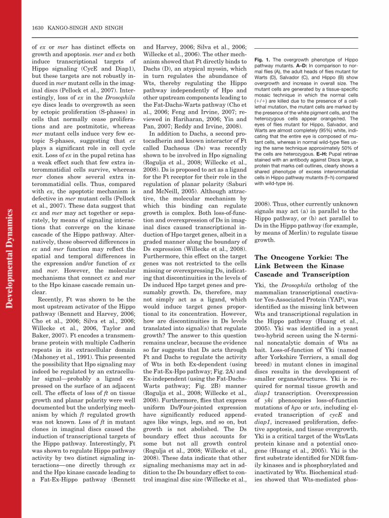
The adult heads of flies of Hippopathway mutants in which over 90% ofcells were homozygous mutant wereproportionately larger than the otherstructures but had a normal overall pat-tern. The mutant cells proliferatedfaster to out-compete the normal wild-type cells (Fig. 1). The underlying causeof overgrowth observed in the adulteyes, was studied in both the pupal ret-inae and the eye–antennal imaginaldiscs. In the adult eye sections, the pat-tern of ommatidial development waslargely unaffected except for the spac-ing of the ommatidial clusters. To traceback this phenotype, the mid-pupal ret-inae were studied in which cellular out-lines were marked using antibodies.Hippo pathway mutants exhibited adramatic increase in the numbers of in-terommatidial cells when comparedwith wild-type (Fig. 1).
The extra interommatidial cellscould be due to excessive cell prolifer-ation, increased spacing of photore-ceptor clusters during patterning,lack of apoptosis, or a combinationthereof. Systematic analyses revealedthat this phenotype results becauseHippo pathway mutant cells prolifer-ate faster than surrounding wild-typecells and because they do not termi-nate proliferation when imaginal tis-sues have reached their normal size.In fact, hpo mutant cells do not dereg-ulate cell proliferation of terminallyarrested cells, as cells differentiatenormally. In addition, Hippo affects
1628 KANGO-SINGH AND SINGH

cell survival. In the wild-type pupal ret-ina, extra interommatidial cells are re-moved by apoptosis, while extra hpomutant cells are not. Thus, Hippo has adual function in promoting cell prolifer-ation arrest and apoptosis (Tapon et al.,2002; Harvey et al., 2003; Jia et al.,2003; Udan et al., 2003; Wu et al., 2003;reviewed in Harvey and Tapon, 2007).These effects on proliferation arrest andapoptosis are not limited to the eye–antennal disc, but are observed inall disc-derived structures, includingwings, halteres, legs, and thorax, sug-gesting that Hippo function is ubiqui-tously required. The ubiquitous expres-sion pattern of several Hippo pathwaycomponents in imaginal discs supportsthis conclusion.
The molecular characterization ofHpo (Ste-20 kinase) and Sav (WW do-main adaptor), together with the pre-viously known Lats/Wts (Dbf-relatedkinase), shed important insights onthe interactions between the proteinsencoded by these genes, and a signal-ing pathway began to emerge (Jia etal., 2003; Udan et al., 2003; Wu et al.,2003; reviewed in Harvey and Tapon,2007). Since then, the addition of theMob super-family protein Mats (Lai etal., 2005), two four point one, ezrin,radixin, moesin (FERM) domain-con-taining proteins Ex and Mer (Hamar-atoglu et al., 2006), a transmembraneProto-Cadherin Ft (Bennett and Har-vey, 2006; Cho et al., 2006; Silva et al.,2006; Willecke et al., 2006), the tran-scriptional coactivator Yki (Huang etal., 2005), and the transcription factorScalloped (Sd; Goulev et al., 2008; Wuet al., 2008; Zhang et al., 2008b) laiddown the frame-work of a signalingnetwork from the membrane to thenucleus for the Hippo signaling path-way. Several targets of Hippo signal-ing were identified during the courseof characterization of mutant pheno-types of pathway components, whichinclude Cyclin E (CycE; Kango-Singhet al., 2002; Tapon et al., 2002), CyclinB (CycB; Tyler and Baker, 2007) andE2F (Nicolay and Frolov, 2008; all reg-ulating cell proliferation), Drosophilainhibitor of apoptosis protein 1(DIAP1; apoptosis; Tapon et al., 2002),the cytoskeletal proteins ex (cellshape; Hamaratoglu et al., 2006), themicroRNA bantam (Nolo et al., 2006),and target genes that function inother pathways and Hippo signaling
like four-jointed (fj; planar cell polarity;Fanto et al., 2003; Cho and Irvine,2004), division abnormally delayed(dally) and dally-like (dlp; DrosophilaHSPGs primarily function in the spreadof morphogen signals; Baena-Lopez etal., 2008), and wingless (wg; Cho andIrvine, 2004) involved in patterning andgrowth (reviewed in Edgar, 2006; Yinand Pan, 2007; Reddy and Irvine, 2008).
The Kinase Cascade in theHippo Pathway
The two protein kinases, Hpo and Wtsform a core kinase cascade in theHippo pathway. Hpo is a member ofthe Ste-20 superfamily of kinases, andacts upstream of Wts, a member of thenuclear Dbf2 related (NDR) family ofprotein kinases. Hpo activates Wts bydirect phosphorylation without affect-ing the levels of Wts expression (Wu etal., 2003). Hpo-mediated Wts activa-tion is facilitated by Sav, a WW do-main-containing protein that proba-bly functions as a scaffold, and also byMats, a protein that binds directly toWts and Hpo (Wu et al., 2003; Wei etal., 2007). When activated, Wts canphosphorylate Yki and reduce its ac-tivity, and as a consequence Yki isexcluded from the nucleus (Dong etal., 2007). The phosphorylation sitesimportant for activation of Hpo andWts kinases and inactivation of Ykihave been mapped in flies and inmammalian systems (Dong et al.,2007; Zhao et al., 2007; Hao et al.,2008; Oh and Irvine, 2008; Zhang etal., 2008a). Yki promotes, either di-rectly or indirectly, the transcriptionof genes that promote growth and cell-cycle progression as well as genes thatinhibit apoptosis (Huang et al., 2005;Dong et al., 2007; Zhao et al., 2008a).Thus, increased activity of Hpo andWts correlates with a reduction in tis-sue growth and a reduction of Hpo/Wts activity allows growth to occur.
Upstream Components of theHippo Pathway
Following the discovery of the core ki-nase cascade, an important questionthat remained unanswered was: doextracellular signals regulate Hpo sig-naling? To understand the regulationof the Hippo pathway, a search foradditional components of this path-
way was initiated. Several candidategenes were tested for their ability toinduce Hpo target genes in mutantclones induced in the imaginal discs,and for the signature “extra interom-matidial cell” phenotype in the pupalretinae. These efforts led to the discov-ery of two proteins with FERM do-mains, Mer and Ex (Hamaratoglu etal., 2006), and the protocadherin Ft asactivators of Hpo (Bennett and Har-vey, 2006; Cho et al., 2006; Silva et al.,2006; Willecke et al., 2006). Individu-ally ex, mer, or ft had very weak effectson the extra interommatidial cell phe-notype typical of the loss of Hippo sig-naling pathway (Willecke et al., 2006).However, loss-of-function clones ofmer, ex double mutants and ft stronglyinduced CycE and Diap1, two well-known targets of the Hippo pathway.
Of interest, simultaneous loss ofmer and ex resulted in (a) impairedendocytic trafficking, leading to up-regulation of multiple cell growth andproliferation pathways (Maitra et al.,2006); and (b) enormous overgrowth ofthe mutant tissue (more than eithersingle mutant), that is similar to thephenotypes of loss of Hpo signaling(Hamaratoglu et al., 2006). Further-more, characterization of the doublemutant phenotype revealed that cellsmutant for mer and ex showed all thedefects of reduction/down-regulationof Hpo signaling, for example, in-creased growth, delayed cell cycle exit,ectopic cell survival, and up-regula-tion of downstream target genes, in-cluding diap1, cycE, and ex (Hamar-atoglu et al., 2006). Further geneticand biochemical analysis of ex andmer double mutants showed thatthese two proteins act upstream of theHpo signaling cascade to regulate pro-liferation arrest and apoptosis.
However, recently distinct func-tional requirements of DrosophilaMer and Ex in the Hippo pathwaywere reported (Pellock et al., 2007).Merlin and Expanded both contain aFERM domain, but structurally Dro-sophila Ex is distinct from Mer, andphylogenetically distinct from theERM proteins (Boedigheimer et al.,1993; Bretscher et al., 2002). Earlierstudies showed the additive pheno-type of the double mutants, and hadproposed a redundant function for Exand Mer (McCartney et al., 2000). Un-like the mer, ex double mutants, loss
REGULATION OF ORGAN SIZE 1629
of ex or mer has distinct effects ongrowth and apoptosis. mer and ex bothinduce transcriptional targets ofHippo signaling (CycE and Diap1),but these targets are not robustly in-duced in mer mutant cells in the imag-inal discs (Pellock et al., 2007). Inter-estingly, loss of ex in the Drosophilaeye discs leads to overgrowth as seenby ectopic proliferation (S-phases) incells that normally cease prolifera-tions and are postmitotic, whereasmer mutant cells induce very few ec-topic S-phases, suggesting that explays a significant role in cell cycleexit. Loss of ex in the pupal retina hasa weak effect such that few extra in-terommatidial cells survive, whereasmer clones show several extra in-terommatidial cells. Thus, comparedwith ex, the apoptotic mechanism isdefective in mer mutant cells (Pellocket al., 2007). These data suggest thatex and mer may act together or sepa-rately, by means of signaling interac-tions that converge on the kinasecascade of the Hippo pathway. Alter-natively, these observed differences inex and mer function may reflect thespatial and temporal differences inthe expression and/or function of exand mer. However, the molecularmechanisms that connect ex and merto the Hpo kinase cascade remain un-clear.
Recently, Ft was shown to be themost upstream activator of the Hippopathway (Bennett and Harvey, 2006;Cho et al., 2006; Silva et al., 2006;Willecke et al., 2006, Taylor andBaker, 2007). Ft encodes a transmem-brane protein with multiple Cadherinrepeats in its extracellular domain(Mahoney et al., 1991). This presentedthe possibility that Hpo signaling mayindeed be regulated by an extracellu-lar signal—probably a ligand ex-pressed on the surface of an adjacentcell. The effects of loss of ft on tissuegrowth and planar polarity were welldocumented but the underlying mech-anism by which ft regulated growthwas not known. Loss of ft in mutantclones in imaginal discs caused theinduction of transcriptional targets ofthe Hippo pathway. Interestingly, Ftwas shown to regulate Hippo pathwayactivity by two distinct signaling in-teractions—one directly through exand the Hpo kinase cascade leading toa Fat-Ex-Hippo pathway (Bennett
and Harvey, 2006; Silva et al., 2006;Willecke et al., 2006). The other mech-anism showed that Ft directly binds toDachs (D), an atypical myosin, whichin turn regulates the abundance ofWts, thereby regulating the Hippopathway independently of Hpo andother upstream components leading tothe Fat-Dachs-Warts pathway (Cho etal., 2006; Feng and Irvine, 2007; re-viewed in Hariharan, 2006; Yin andPan, 2007; Reddy and Irvine, 2008).
In addition to Dachs, a second pro-tocadherin and known interactor of Ftcalled Dachsous (Ds) was recentlyshown to be involved in Hpo signaling(Rogulja et al., 2008; Willecke et al.,2008). Ds is proposed to act as a ligandfor the Ft receptor for their role in theregulation of planar polarity (Saburiand McNeill, 2005). Although attrac-tive, the molecular mechanism bywhich this binding can regulategrowth is complex. Both loss-of-func-tion and overexpression of Ds in imag-inal discs caused transcriptional in-duction of Hpo target genes, albeit in agraded manner along the boundary ofDs expression (Willecke et al., 2008).Furthermore, this effect on the targetgenes was not restricted to the cellsmissing or overexpressing Ds, indicat-ing that discontinuities in the levels ofDs induced Hpo target genes and pre-sumably growth. Ds, therefore, maynot simply act as a ligand, whichwould induce target genes propor-tional to its concentration. However,how are discontinuities in Ds levelstranslated into signal(s) that regulategrowth? The answer to this questionremains unclear, because the evidenceso far suggests that Ds acts throughFt and Dachs to regulate the activityof Wts in both Ex-dependent (usingthe Fat-Ex-Hpo pathway; Fig. 2A) andEx-independent (using the Fat-Dachs-Warts pathway; Fig. 2B) manner(Rogulja et al., 2008; Willecke et al.,2008). Furthermore, flies that expressuniform Ds/Four-jointed expressionhave significantly reduced append-ages like wings, legs, and so on, butgrowth is not abolished. The Dsboundary effect thus accounts forsome but not all growth control(Rogulja et al., 2008; Willecke et al.,2008). These data indicate that othersignaling mechanisms may act in ad-dition to the Ds boundary effect to con-trol imaginal disc size (Willecke et al.,
2008). Thus, other currently unknownsignals may act (a) in parallel to theHippo pathway, or (b) act parallel toDs in the Hippo pathway (for example,by means of Merlin) to regulate tissuegrowth.
The Oncogene Yorkie: TheLink Between the KinaseCascade and Transcription
Yki, the Drosophila ortholog of themammalian transcriptional coactiva-tor Yes-Associated Protein (YAP), wasidentified as the missing link betweenWts and transcriptional regulation inthe Hippo pathway (Huang et al.,2005). Yki was identified in a yeasttwo-hybrid screen using the N-termi-nal noncatalytic domain of Wts asbait. Loss-of-function of Yki (namedafter Yorkshire Terriers, a small dogbreed) in mutant clones in imaginaldiscs results in the development ofsmaller organs/structures. Yki is re-quired for normal tissue growth anddiap1 transcription. Overexpressionof yki phenocopies loss-of-functionmutations of hpo or wts, including el-evated transcription of cycE anddiap1, increased proliferation, defec-tive apoptosis, and tissue overgrowth.Yki is a critical target of the Wts/Latsprotein kinase and a potential onco-gene (Huang et al., 2005). Yki is thefirst substrate identified for NDR fam-ily kinases and is phosphorylated andinactivated by Wts. Biochemical stud-ies showed that Wts-mediated phos-
Fig. 1. The overgrowth phenotype of Hippopathway mutants. A–D: In comparison to nor-mal flies (A), the adult heads of flies mutant forWarts (D), Salvador (C), and Hippo (B) showovergrowth and increase in overall size. Themutant cells are generated by a tissue-specificmosaic technique in which the normal cells(�/�) are killed due to the presence of a cell-lethal mutation, the mutant cells are marked bythe presence of the white pigment cells, and theheterozygous cells appear orange/red. Theeyes of flies mutant for Hippo, Salvador, andWarts are almost completely (95%) white, indi-cating that the entire eye is composed of mu-tant cells, whereas in normal wild-type flies us-ing the same technique approximately 50% ofthe cells are heterozygous. E–H: Pupal retinaestained with an antibody against Discs large, aprotein that marks cell outlines, clearly shows ashared phenotype of excess interommatidialcells in Hippo pathway mutants (f–h) comparedwith wild-type (e).
1630 KANGO-SINGH AND SINGH

Fig. 1.
Fig. 2. Models of Hippo signaling. A: The Ft-Ex-Hpo model of Hippo signaling, where the transmembrane receptor Fat (Ft) interacts by means of Expanded(Ex) but not Merlin (Mer), which comprise the upstream regulators of Hippo signaling, interacts with the core kinase cascade of Hippo signaling (which includethe Hippo kinase (Hpo), the adaptor protein Salvador (Sav) and Mats, and the downstream kinase Warts (Wts)). The core kinase cascade functions byinhibiting the phosphorylation of the transcriptional coactivator Yorkie (Yki). Unphosphorylated form of Yki binds with its cognate transcription factorScalloped (Sd) and translocates to the nucleus to control expression of various Hippo target genes. Currently, all cell biological functions of Hippo signalingcannot be explained by the regulation of target genes controlled by the Yki/Sd complex. Thus, other transcription factors can partner with Yki to mediateHippo functions. The Ft binding partner Dachsous (Ds) is also involved in Hippo signaling by means of both modes of Hippo signaling. B: The Ft-Dachs-WtsModel of Hippo signaling contends that the transmembrane protein Ft does not interact with Ex (or Hpo, Sav and Mats), but interacts with Wts through theatypical myosin, Dachs. Dachs negatively regulates the levels and activity of the Wts kinase, which in turn can signal to Yki. Ft-Dachs pathway is directlyinvolved in the regulation of planar cell polarity. In addition to the Ft-Dachs-Wts interaction, another noncanonical interaction is that between Hippo and theDrosophila homolog of the RAS association factor 1 protein (RASSF1). Hpo and RASSF1 bind with each other by means of their SARAH domains to repressthe expression of Sav, and activate Hippo signaling. C: The current model of the Mammalian Hippo signaling pathway shows the widespread structural andfunctional conservation between the mammalian and Drosophila proteins. The core kinase cascade between MST1/2 (Hpo homologs), hWW45 (Savhomolog), MOB (Mats homolog), and the Lats 1/2 (Warts homolog) is conserved at the molecular level. As in flies, the mammalian Lats1/2 in turn preventthe phosphorylation of YAP and TAZ, the two Yki homologs, and allow YAP/TAZ to translocate to the nucleus to bind the TEAD (Sd homolog) transcriptionfactors to regulate Hippo signaling. The interactions upstream of the kinase cascade remain unclear (marked by dotted arrows and question marks); however,NF2 (Merlin homolog) has been shown to interact by means of YAP to regulate cell proliferation, survival, and contact inhibition. Database searches haveidentified Mammalian Ex but its role in vivo remains to be defined. The mammalian isoform of Fat (Ft4) has recently been implicated in breast cancer, butits functional interaction with mammalian Hippo pathway has not been worked out yet.
REGULATION OF ORGAN SIZE 1631

phorylation of Yki was stimulated byupstream components of the Hippopathway (Huang et al., 2005).
At the time of its characterization, itwas speculated that like other coacti-vators, Yki functions by interactingwith DNA binding transcription fac-tors. YAP, the mammalian homolog ofYki, is known to function as coactiva-tor for several transcription factors,such as the p53 family member p73(Strano et al., 2005), the Runt familymember PEBP2� (Yagi et al., 1999;Vassilev et al., 2001), and the fourTEAD/TEF transcription factors (Yagiet al., 1999; Vassilev et al., 2001;Strano et al., 2005). This interaction isgenerally mediated by WW domains ofYAP, and the PPxY motifs of the cog-nate transcription factors or by otherbinding interactions independent ofthe WW domain. Indeed, the Drosoph-ila TEAD domain transcription factor,scalloped (sd), was identified as thecognate factor that partnered with theN-terminal (TEAD binding) region ofYki to regulate Hpo target genes bythree different approaches: yeast two-hybrid screen (Zhang et al., 2008b),GST pull-down experiments (Goulevet al., 2008), and identification of aSd/TEAD-binding site in the HippoResponse Element (HRE) of the diap1gene (Wu et al., 2008; Zhang et al.,2008b).
Scalloped and theTranscriptional Regulationof Hippo Target Genes
In Drosophila, Sd belongs to a familyof evolutionarily conserved proteinscharacterized by the presence of aTEA/ATTS DNA-binding domain(Campbell et al., 1991, 1992). Sd hasbeen extensively characterized for itsrole in wing development and isknown to physically interact with theproduct of the vestigial (vg) gene,where the dimer functions as a mastergene controlling wing formation (Sim-monds et al., 1998; Paumard-Rigal etal., 1998; Halder et al., 1998; Halderand Carroll, 2001). The Vg-Sd dimeractivates the transcription of severalwing-specific genes, including sd andvg themselves. Sd binds with Yki toregulate the transcription of diap1, atarget gene of the Hippo pathway (Wuet al., 2008). The interaction betweenYki and Sd increases sd transcrip-
tional activity both ex vivo in Dro-sophila S2 cells and in vivo in Dro-sophila wing discs and promotes Ykinuclear localization (Wu et al., 2008;Zhang et al., 2008b). The diap1 locusharbors a minimal Sd-binding HippoResponsive Element (HRE) that medi-ates transcriptional regulation by theHippo pathway. Genetic and biochem-ical studies showed that sd is requiredfor yki-induced tissue overgrowth andtarget gene expression, and this sdfunction is conserved in its mamma-lian homolog (Goulev et al., 2008; Wuet al., 2008; Zhang et al., 2008b; Zhaoet al., 2008b; reviewed in Banduraand Edgar, 2008). Contrary to Yki, Sdis not expressed in all imaginal tis-sues. This indicates that Yki-Sd inter-action acts in a tissue-specific mannerand that other Yki partners must ex-ist that remain to be identified.
Two Mechanisms of HippoRegulation: The Fat-Ex-Hpoand the Fat-Dachs-WartsPathways
Three reports showed that Fat func-tions through a linear pathway thatinvolves Ft, Ex, Hpo, and Wts (Fig.2A; Bennett and Harvey, 2006; Silvaet al., 2006; Willecke et al., 2006). Thismodel is based on the following obser-vations: (1) ft mutant cells affected thestability and localization of Ex to theapical surface of the cells. (2) Overex-pression of an activated form of Ftcould induce Hpo and Wts phosphory-lation. (3) The ability of Ft to inhibitYki activity was reduced by knock-down of hpo or wts by means of RNAinterference using a transcriptionalassay of Yki activity in cell culture. (4)The most convincing evidence camefrom genetic epistasis experimentsthat showed that, as opposed to therequirement of Hpo components for Ftmediated growth regulation, activa-tion of the Hpo pathway at the level ofex or hpo mostly bypassed the require-ment for ft. This placed Ft upstream ofEx and other Hpo components. Inter-estingly, this interaction does not in-volve Mer (which is proposed to acttogether with Ex), suggesting thatMer may receive inputs from other up-stream signals. Together, these find-ings suggested that Ft (possibly actingas a receptor) signaled to Ex, which inturn signaled to the kinase cascade.
Warts then negatively regulated theactivity of the oncoprotein Yki, whichthen bound its cognate transcriptionfactor Sd to regulate the expression ofdiap1.
Cho et al. (2006) demonstrated thatFt promoted Wts-mediated Hippo sig-naling by a different mechanism thatinvolved an unconventional myosinDachs (Fig. 2B). In the Fat-Dachs-Wtsmodel, the path from Ft to Wts did notgo through Hpo or other upstreamcomponents of the pathway like Ex orMer. Three lines of evidence were pre-sented to support this conclusion. (1)The genetic analysis of double-mutantcombinations placed ft upstream ofdachs, and dachs upstream of bothhpo and wts. (2) The levels of Wts pro-tein, but not the levels of Hpo, Sav,Mer, or Mats were reduced in ft mu-tant tissue. (3) When overexpressed intissue culture cells, Dachs bound toWts. Thus, Ft regulated the abun-dance of Wts by means of Dachs, whileHpo independently regulated the en-zymatic activity of Wts. In this view,Wts was the point of convergence oftwo distinct upstream signals. Thismechanism of Fat action was also re-ported by Feng and Irvine, where Ftacts in parallel to Ex to signal to Wtsto regulate growth by means of Hipposignaling pathway (Feng and Irvine,2007).
Of interest, the experiments de-scribed in the two different models donot exclude or contradict each other.Indeed, many of the experimental re-sults are consistent with Ft being ableto regulate Wts activity in at least twodifferent ways and also with Wts be-ing regulated by signals that are inde-pendent of Ft (reviewed in Hariharan,2006). The observation that the phe-notype of ft ex double mutants is alittle more severe than that of ex mu-tants suggests that Ft can regulateWts independently of Ex. Also consis-tent with this conclusion are the re-sults that show that the suppressionof ft mutant phenotypes by the over-expression of Hippo pathway compo-nents is typically incomplete. In addi-tion, the observation of reducedgrowth and decreased transcription ofYki targets in cells doubly mutant forft and Dachs would seem to suggestthat the elevated levels of Wts in thesecells can still be activated by a path-way that is independent of Ft such as
1632 KANGO-SINGH AND SINGH

Mer. In addition, the fact that Sd isexpressed in a spatially defined pat-tern and is not expressed in all imag-inal discs, suggests that other tran-scription factors can bind with Yki toregulate the expression of other targetgenes in the Hippo pathway.
RASSF1 and the HippoPathway
Hippo signaling can also be regulatedby other Ft-independent mechanisms,for example, through the interactionof the Ras Association Factor 1(RASSF1) with Salvador and Hippo.RASSF1 belongs to a family of pro-teins frequently silenced in a varietyof solid tumors, mainly by promotermethylation. Mammalian RASSF pro-teins were known to interact withMst1 (the Hippo homolog), and afterthe discovery of the Hippo pathway inflies a homology domain was discov-ered between Sav, RASSF, and Hippocalled the SARAH (Sav-RASSF-Hippo) domain. RASSF proteins typi-cally contain a RA domain and a C-terminal SARAH domain (Polesello etal., 2006; Guo et al., 2007; Avruch etal., 2009). The dRASSF mutant fliesare viable, and under controlled con-ditions uncover a clear growth defectin comparison to wild-type animals.RASSF expression was reduced inclones mutant for a hpo allele thatlacked the SARAH domain. Furtherinvestigations reveal that the effect ondRASSF is posttranscriptional. Of in-terest, dRASSF levels were unaffectedin clones mutant for other Hippo-pathway members, such as ex, sav,and wts (Polesello et al., 2006; Guo etal., 2007). These results suggest thatdirect binding to Hpo through itsSARAH domain, rather than signal-ing through the Hippo pathway, isnecessary for dRASSF stability.RASSF1 binds to Hpo but not withSav, and does not form a ternary com-plex. Hpo was able to bind Sav anddRASSF, however, in two differentHpo complexes—a highly active Sav-associated pool and an inactivedRASSF-associated pool. Thus, Savcan promote Hpo activation. Salvadorand RASSF repress each other’s ex-pression, and in cell culture, increas-ing amounts of Sav can displaceRASSF bound to Hpo, suggesting thatmodulation of the inhibitory effects of
dRASSF and the activator effects ofSav determine the level of Hippo acti-vation, and organ size (Polesello et al.,2006; Guo et al., 2007).
THE MAMMALIAN HIPPOPATHWAY: REGULATIONOF ORGAN SIZE
Most components of the Hippo path-way identified to date have one ormore mammalian orthologs that prob-ably function in an analogous mannerto their Drosophila counterparts (Ta-ble 1). The components of the Hippopathway are highly conserved inmammals, including YAP (Yes-associ-ated protein), Lats1/2, Mob, Mst1/2,Sav, Mer, Ex1/2, and Ft4 (Yki, Wts,Mats, Hpo, Sav, Mer, Ex, and Ft ho-mologs, respectively). The YAP-likeprotein Transcriptional coactivatorwith PDZ binding motifs (TAZ orWWTR1), is also involved in the mam-malian Hippo pathway. Like Yki, YAPand TAZ are phosphorylated and in-hibited by the Hippo pathway throughcytoplasmic retention (Zhao et al.,2008ab). Although there is high struc-tural and functional similarity be-tween YAP and TAZ, these proteinshave distinct context-dependent func-tions (Hong and Yaffe, 2006; Lei et al.,2008).
Human YAP, Lats1, Mst2, andMob1 can functionally rescue the re-spective Drosophila mutants, suggest-ing the functional conservation ofthese proteins in mammals. Mst1/2,the Hpo homologs, can phosphorylateall three-core components and thusplay a key role in the mammalianHippo pathway. Mst1 and 2 phosphor-ylate (a) Lats1/2 on the activation loopand hydrophobic motif (Chan et al.,2005), (b) WW45 after physically in-teracting (binding) with WW45 (Cal-lus et al., 2006), and (c) Mob1, whichenhances its interaction with Lats1(Praskova et al., 2008). Not only arethe components and functional inter-actions conserved between the coreHippo pathway components from fliesto mammals, but the mechanism bywhich YAP and TAZ are regulated byHippo components is also conserved.Thus, Mst, WW45, Lats, and Mob in-duce YAP phosphorylation, cytoplas-mic translocation, and inhibition(Zhao et al., 2007; Hao et al., 2008; Leiet al., 2008; Oka et al., 2008; Zhang et
al., 2008a). As in flies, TEAD familytranscription factors, homologs of theDrosophila Sd, are key mediators ofYAP function in mammalian cells(Zhao et al., 2008a). However, inmammalian systems YAP/TAZ arealso known to function with othertranscription factors, such as the p53family member p73 (Strano et al.,2005), and the Runt family memberPEBP2�. Thus, Yki may partner withother transcription factors to regulateHpo target genes in a tissue-specific orcontext-dependent manner. Based oncurrent reports the conservation of theupstream components—Ft, Ex, andDs—remains unclear in mammalianHippo signaling. Therefore, the con-servation of the Hippo pathway is notlimited to structural homologies andsignaling interactions among pathwaycomponents but also to the function ofHippo signaling in the regulation oforgan size (Camargo et al., 2007; Donget al., 2007). The evidence for the func-tional conservation came from experi-ments in which overexpression of YAPin the mouse liver was shown to causean increase in liver size and eventu-ally to tumor formation (Dong et al.,2007).
Although mammalian Hippo signal-ing is very similar to that of Drosoph-ila, recent studies have also suggestedthat there are some differences be-tween mammalian and DrosophilaHippo signaling pathways. For exam-ple, the role of RASSF1 in the regula-tion of MST1 (Hippo) appears to beopposite between flies and mammals.In mammals, RASSF1A can increasethe MST1 activity and this activationis required for Fas-induced cell death(Oh et al., 2006). These differencesmay be due to the spatial and tempo-ral differences in the expression andactivity of the various RASSF iso-forms and MST1/2 proteins. Similarly,the level of CycE (a transcriptionaltarget of the Hippo pathway) is notsignificantly elevated in WW45 mu-tant mice (Lee et al., 2008). Despitethese dissimilarities, the core wiringdiagram of the Hippo signaling path-way is conserved between flies andmammals, and further studies in bothflies and mammalian systems will addto our understanding of growth andsize regulation.
REGULATION OF ORGAN SIZE 1633

Roles of Hippo Signaling inCancer, Apoptosis,Regulation of Cell Shape,and Contact Inhibition
The Hippo pathway is emerging as anetwork of several tumor suppressorgenes and (so far) one oncogene Yki.Interestingly, YAP has been shown re-cently to be a candidate oncogene in thehuman chromosome 11q22 amplicon(Overholtzer et al., 2006; Zender et al.,2006). In addition to genomic amplifica-
tion, YAP expression and nuclear local-ization was also shown to be elevated inmultiple types of human cancers (Ze-nder et al., 2006; Dong et al., 2007; Zhaoet al., 2007; Steinhardt et al., 2008).Furthermore, mutations of Lats1/2,Sav, and Mob have been implicated intumorigenesis (St John et al., 1999;Tapon et al., 2002; Lai et al., 2005; re-viewed in Harvey and Tapon, 2007).RASSF1, the constitutive binding part-ner of MST1/2, and MST1/2 are fre-quently mutated by hypermethylation
in several cancer types (Seidel et al.,2007; Avruch et al., 2009). NF2/ Merlinis a well-known tumor suppressor genein mammals (Okada et al., 2007). Fat4has recently been shown to be inactivat-ed/deleted in breast cancers and pri-mary tumor cell lines (Qi et al., 2009).Despite its conservation and intimaterelationship with cancer, the Hippopathway has not been systematicallystudied in mammalian cells.
Activation of Hippo signaling by over-expression of the Hpo kinase or Mst1/2
TABLE 1. Genes Involved in the Drosophila and Mammalian Hippo Pathway
Drosophilamelanogaster Human orthologs Protein
Role in Hippo SignalingPathway
Role in Human Cancer(Reference)
Fat Fat4 Proto-Cadherin Hippo Pathway Receptor(?)
Mutated in breast cancer(Qi et al., 2009)
Dachsous DCHS1 Proto-Cadherin Ligand for the Fatreceptor (?)
Unknown
Expanded FMRD6 (Willin)/ EX2 4.1 Superfamily FERMdomain protein
Upstream regulators ofthe Hippo kinasecascade
Unknown
Merlin NF2 4.1 Super-family FERMdomain protein
Upstream regulators ofthe Hippo kinasecascade
Mutated in familial andsporadic schwanomas(Evans et al., 2000)
Hippo MST1/2 Ste-20 family ProteinKinase
Bind Salvador and Mats,and phosphorylateMats and Warts inresponse to upstreamsignals
Hypermethylated in softtissue sarcoma (Seidelet al., 2007)
Salvador hWW45 WW domain Adaptorprotein
Binds Hpo, the Sav-Hpocomplex moreefficiently regulatesthe activity of theWarts kinase
Mutated in cancer celllines (Tapon et al.,2002)
Mats MOBK1B Mob superfamilycoactivator of proteinkinases
Phosphorylated by Hpoand Wts, and bindsHpo, and Wts, toregulate Wts activity
Mutated in cancer celllines (Lai et al., 2005)
Warts Lat1/2 NDR family ProteinKinase
Phosphorylates Mats,Wts and Yki toregulate Hippopathway activity
Silenced in breasttumors (Turenchalk etal., 1999; Zeng andHong, 2008)
Yorkie YAP TAZ TranscriptionalCoactivator
Unphosphorylated formbinds Sd, andtranslocates to thenucleus to regulateexpression oftranscriptional targetsof Hippo signaling
Amplified(overexpressed) inbreast tumors,colorectal cancer andseveral other solidtumors (Overholtzer etal., 2006; Zender et al.,2006)
Scalloped TEAD Transcription factor Binds with Yki toregulate target geneexpression.
Unknown
dRASSF1 RASSF1 RA domain containingRAS effector protein
Binds Hippo, complexmay be disrupted bySav in response topathway activation
Hypermethylated inLung and kidneycancers
Dachs Unknown Unconventional Myosin Binds with Warts Unknown
1634 KANGO-SINGH AND SINGH

has been shown to activate apoptosis inresponse to DNA damage or otherstress and apoptotic stimuli (Graves etal., 1998; Udan et al., 2003; Colombaniet al., 2006; reviewed in Hay and Guo,2003). Increased cell death results information of reduced/smaller organs orappendages. Although loss of Hippo sig-naling clearly makes cells resistant toapoptosis and promotes cell survival,the molecular mechanism by whichHippo signaling regulates apoptosis re-mains unclear. This is because activa-tion of Hippo signaling does not sup-press the transcriptional expression ofDiap1. Of interest, a Hpo-response ele-ment has been mapped in the Diap1gene that is induced in response to lossof Hippo signaling. Thus, even though amolecular mechanism by which Hipposignaling likely regulates the expres-sion of Diap1 gene has been reported,clearly the regulation of cell death byactivation of Hpo signaling is complex(Wu et al., 2008). Mammalian YAP hasbeen shown to have both pro- and anti-apoptotic functions (Oka et al., 2008);however, it is still possible that under cer-tain conditions such as DNA damage,YAP can be tyrosine phosphorylated byc-Abl, which selectively activates YAPtranscriptional activity on p73 to induceapoptosis (Zeng and Hong, 2008).
Hippo signaling is implicated in theregulation of cell shape because theplasma-membrane protein Mer/NF2 isinvolved in the regulation of cell shape,motility, and adhesion (Okada et al.,2007). Most studies conclude that mem-brane association is necessary for thegrowth-suppressing function of Mer.Mutant versions of Mer that cannot lo-calize to the membrane cannot inhibitcell proliferation. In addition, Mer canassociate with several membrane pro-teins, including both adhesion receptorsand membrane receptors that regulatecell proliferation and differentiation(Curto and McClatchey, 2008). Recentstudies suggest that Merlin directly con-trols the surface availability and functionof membrane receptors, and their traf-ficking by endocytosis in both Drosophilaand mammalian cells (Manchanda et al.,2005; Maitra et al., 2006). Ex and Mer arelocalized under the plasma membraneand are scaffolding proteins that providea regulated linkage between membraneproteins and the cortical cytoskeleton,and also participate in signal-transduc-tion pathways.
How normal cells in culture stop pro-liferating upon achieving confluencyhas long been a topic of intense investi-gations. This phenomenon called “con-tact inhibition” is a hallmark of cellsgrowing in culture. Contact inhibition islost in cancer cells, however, the mech-anism regulating this phenomenon re-mains poorly understood. Recently, sev-eral components of the Hippo pathwayhave been implicated in contact inhibi-tion. Mer becomes dephosphorylatedand activated in confluent cells (Shawet al., 1998; Morrison et al., 2001),which has been reported to be both nec-essary and sufficient for contact inhibi-tion. Lats2 and WW45 are also relatedto contact inhibition as their knockoutmouse embryonic fibroblast (MEF) cellsshow loss of contact inhibition. Finally,YAP is phosphorylated and translo-cated to the cytoplasm by the Hippopathway at high cell density in a Mer-dependent manner (Zhao et al., 2007).Moreover, a dominant-negative form ofYAP restores contact inhibition inACHN, a cancer cell line with activationof YAP due to WW45 mutation (Zhao etal., 2007). So far, the reports suggestthat, when cultured cells reach conflu-ence, cell–cell interactions lead to sig-naling events that activate the Hippopathway. The activation of the Hippopathway leads to the phosphorylation ofYAP and its retention in the cytoplasmwhere it interacts with 14-3-3 proteinsand reduced transcription of YAP/Hippo target genes. This likely sendsthe “stop proliferation” signal to cells inculture and thus play a crucial role incontact inhibition (Zhao et al., 2007,2008a). Identifying the upstream signalof this pathway might solve a long-standing mystery in cell biology.
FUTURE PERSPECTIVES
Several questions remain unansweredin the field of Hippo signaling and func-tion. For example, is the activity of theHippo pathway regulated by mecha-nisms beside Ft/Ds? Are there otherregulators of the pathway? Experimen-tal evidence so far suggests that severaladditional components of the Hippopathway (for example, the protein thatlinks Ex or Mer to Hpo, or the receptorthat acts in conjunction with Mer) re-main to be discovered. Given that Hipposignaling may play additional roles inaddition to its roles in regulation of cell
proliferation arrest, apoptosis, cell mor-phology, and contact inhibition, theremay be other downstream targets. Doother transcription factors bind withYki in tissues where Sd is not ex-pressed? In addition, several biochemi-cal mechanisms remain unresolved. Forexample, how Ft regulates Wts levelsand Hpo activity, and the functionalconsequences of either the abnormal Exlocalization in ft clones or the binding ofDs to Wts are still unknown. Can wemeasure normal levels of Hpo signaling(in physiological conditions instead of invitro)? Finally, are there other signalsthat instruct cells to cease proliferation?In the coming years, we can expect sev-eral interesting discoveries that willadd to our knowledge of Hippo signalingand further our understanding ofgrowth regulation.
ACKNOWLEDGMENTSThe authors thank Kevin Waits (Mer-cer University School of Medicine, Ma-con, Georgia) and Jaison J. Nain-aparampil (University of Dayton,Dayton, Ohio) for their help and com-ments. We apologize to all authorswhose work could not be cited due tospace limitations. A.S. is supported byStart-up support from University ofDayton, grants from Ohio Cancer Re-search Associates and University ofDayton Research Council. M.K.S. issupported by Strart-up Grant fromMercer University School of Medicine,a Seed Grant from Mercer University,and a MEDCEN Foundation grant.
REFERENCES
Avruch J, Xavier R, Bardeesy N, Zhang XF,Praskova M, Zhou D, Xia F. 2009. TheRassf family of tumor suppressor polypep-tides. J Biol Chem 284:11001–11005.
Baena-Lopez LA, Rodriguez I, Baonza A.2008. The tumor suppressor genesdachsous and fat modulate different sig-nalling pathways by regulating dallyand dally-like. Proc Natl Acad Sci U S A105:9645–9650.
Bandura JL, Edgar BA. 2008. Yorkie andScalloped: partners in growth activation.Dev Cell 14:315–316.
Bennett FC, Harvey KF. 2006. Fat cad-herin modulates organ size in Drosoph-ila via the Salvador/Warts/Hippo signal-ing pathway. Curr Biol 16:2101–2110.
Bretscher A, Edwards K, Fehon RG. 2002.ERM proteins and merlin: integrators atthe cell cortex. Nat Rev Mol Cell Biol3:586–599.
REGULATION OF ORGAN SIZE 1635

Boedigheimer M, Bryant P, Laughon A.1993. Expanded, a negative regulator ofcell proliferation in Drosophila, showshomology to the NF2 tumor suppressor.Mech Dev 44:83–84.
Bryant PJ, Simpson P. 1984. Intrinsic andextrinsic control of growth in developingorgans. Q Rev Biol 59:387–415.
Callus BA, Verhagen AM, Vaux DL. 2006.Association of mammalian sterile twentykinases, Mst1 and Mst2, with hSalvadorvia C-terminal coiled-coil domains, leadsto its stabilization and phosphorylation.FEBS J 273:4264–4276.
Camargo FD, Gokhale S, Johnnidis JB, FuD, Bell GW, Jaenisch R, BrummelkampTR. 2007. YAP1 increases organ size andexpands undifferentiated progenitorcells. Curr Biol 17:2054–2060.
Campbell SD, Duttaroy A, Katzen AL, Cho-vnick A. 1991. Cloning and characteriza-tion of the scalloped region of Drosophilamelanogaster. Genetics 127:367–380.
Campbell S, Inamdar M, Rodrigues V,Raghavan V, Palazzolo M, Chovnick A.1992. The scalloped gene encodes anovel, evolutionarily conserved tran-scription factor required for sensory or-gan differentiation in Drosophila. GenesDev 6:367–379.
Chan EH, Nousiainen M, ChalamalasettyRB, Schafer A, Nigg EA, Sillje HH. 2005.The Ste20-like kinase Mst2 activates thehuman large tumor suppressor kinaseLats1. Oncogene 24:2076–2086.
Cho E, Irvine KD. 2004. Action of fat, four-jointed, dachsous and dachs in distal-to-proximal wing signaling. Development131:4489–4500.
Cho E, Feng Y, Rauskolb C, Maitra S, Fe-hon R, Irvine KD. 2006. Delineation of aFat tumor suppressor pathway. NatGenet 38:1142–1150.
Cohen SM. 1993. Imaginal disc develop-ment. In: Bate, Martinez Arias. p 787–841.
Colombani J, Polesello C, Josue F, TaponN. 2006. Dmp53 activates the Hippopathway to promote cell death in re-sponse to DNA damage. Curr Biol 16:1453–1458.
Conlon I, Raff M. 1999. Size control in an-imal development. Cell 96:235–244.
Curto M, McClatchey AI. 2008. Nf2/Merlin:a coordinator of receptor signalling andintercellular contact. Br J Cancer98:256–262.
Dong J, Feldmann G, Huang J, Wu S,Zhang N, Comerford SA, Gayyed MF,Anders RA, Maitra A, Pan D. 2007. Elu-cidation of a universal size-control mech-anism in Drosophila and mammals. Cell130:1120–1133.
Edgar BA. 2006. From cell structure totranscription: Hippo forges a new path.Cell 124:267–273.
Evans DG, Sainio M, Baser ME. 2000.Neurofibromatosis type 2. J Med Genet37:897–904.
Fanto M, Clayton L, Meredith J, HardimanK, Charroux B, Kerridge S, McNeill H.2003. The tumor-suppressor and cell ad-hesion molecule Fat controls planar po-larity via physical interactions with At-
rophin, a transcriptional co-repressor.Development 130:763–774.
Feng Y, Irvine KD. 2007. Fat and ex-panded act in parallel to regulate growththrough warts. Proc Natl Acad Sci U S A104:20362–20367.
French V, Bryant PJ, Bryant SV. 1976.Pattern regulation in epimorphic fields.Science 193:969–981.
Goulev Y, Fauny JD, Gonzalez-Marti B,Flagiello D, Silber J, Zider A. 2008.SCALLOPED interacts with YORKIE,the nuclear effector of the hippo tumor-suppressor pathway in Drosophila. CurrBiol 18:435–441.
Graves JD, Gotoh Y, Draves KE, AmbroseD, Han DK, Wright M, Chernoff J, ClarkEA, Krebs EG. 1998. Caspase-mediatedactivation and induction of apoptosis bythe mammalian Ste20-like kinase Mst1.EMBO J 17:2224–2234.
Guo C, Tommasi S, Liu L, Yee JK, Dam-mann R, Pfeifer GP. 2007. RASSF1A ispart of a complex similar to the Drosoph-ila Hippo/Salvador/Lats tumor-suppres-sor network. Curr Biol 17:700–705.
Halder G, Carroll SB. 2001. Binding of theVestigial co-factor switches the DNA tar-get selectivity of the Scalloped selectorprotein. Development 128:3295–3305.
Halder G, Polzczyk P, Kraus ME, HudsonA, Kim J, Laughon A, Carroll S. 1998.The Vestigial and Scalloped proteins acttogether to directly regulate wing-spe-cific gene expression in Drosophila.Genes Dev 12:3900–3909.
Hamaratoglu F, Willecke M, Kango-SinghM, Nolo R, Hyun E, Tao C, Jafar-NejadH, Halder G. 2006. The tumour-suppres-sor genes NF2/Merlin and Expanded actthrough Hippo signalling to regulate cellproliferation and apoptosis. Nat Cell Biol8:27–36.
Hao Y, Chun A, Cheung K, Rashidi B,Yang X. 2008. Tumor suppressor LATS1is a negative regulator of oncogene YAP.J Biol Chem 283:5496–5509.
Hariharan IK. 2006. Growth regulation: abeginning for the hippo pathway. CurrBiol 16:R1037–R1039.
Harvey K, Tapon N. 2007. The Salvador-Warts-Hippo pathway - an emerging tu-mour-suppressor network. Nat Rev Can-cer 7:182–191.
Harvey KF, Pfleger CM, Hariharan IK.2003. The Drosophila Mst ortholog, hippo,restricts growth and cell proliferation andpromotes apoptosis. Cell 114:457–467.
Hay BA, Guo M. 2003. Coupling cellgrowth, proliferation, and death. Hippoweighs in. Dev Cell 5:361–363.
Hong JH, Yaffe MB. 2006. TAZ: a beta-catenin-like molecule that regulatesmesenchymal stem cell differentiation.Cell Cycle 5:176–179.
Huang J, Wu S, Barrera J, Matthews K,Pan D. 2005. The Hippo signaling path-way coordinately regulates cell prolifer-ation and apoptosis by inactivatingYorkie, the Drosophila Homolog of YAP.Cell 122:421–434.
Jia J, Zhang W, Wang B, Trinko R, Jiang J.2003. The Drosophila Ste20 family ki-nase dMST functions as a tumor sup-
pressor by restricting cell proliferationand promoting apoptosis. Genes Dev 17:2514–2519.
Justice RW, Zilian O, Woods DF, Noll M,Bryant PJ. 1995. The Drosophila tumorsuppressor gene warts encodes a ho-molog of human myotonic dystrophy ki-nase and is required for the control of cellshape and proliferation. Genes Dev9:534–546.
Kango-Singh M, Nolo R, Tao C, VerstrekenP, Hiesinger PR, Bellen HJ, Halder G.2002. Shar-pei mediates cell proliferationarrest during imaginal disc growth in Dro-sophila. Development 129:5719–5730.
Lai ZC, Wei X, Shimizu T, Ramos E, Rohr-baugh M, Nikolaidis N, Ho LL, Li Y.2005. Control of cell proliferation and ap-optosis by mob as tumor suppressor,mats. Cell 120:675–685.
Lee JH, Kim TS, Yang TH, Koo BK, Oh SP,Lee KP, Oh HJ, Lee SH, Kong YY, KimJM, Lim DS. 2008. A crucial role ofWW45 in developing epithelial tissues inthe mouse. EMBO J 27:1231–1242.
Lei QY, Zhang H, Zhao B, Zha ZY, Bai F,Pei XH, Zhao S, Xiong Y, Guan KL. 2008.TAZ promotes cell proliferation and epi-thelial-mesenchymal transition and isinhibited by the hippo pathway. Mol CellBiol 28:2426–2436.
Mahoney PA, Weber U, Onofrechuk P,Biessmann H, Bryant PJ, Goodman CS.1991. The fat tumor suppressor gene inDrosophila encodes a novel member ofthe cadherin gene superfamily. Cell 67:853–868.
Maitra S, Kulikauskas RM, Gavilan H, Fe-hon RG. 2006. The tumor suppressorsMerlin and Expanded function coopera-tively to modulate receptor endocytosisand signaling. Curr Biol 16:702–709.
Manchanda N, Lyubimova A, Ho HY,James MF, Gusella JF, Ramesh N, Snap-per SB, Ramesh V. 2005. The NF2 tumorsuppressor Merlin and the ERM proteinsinteract with N-WASP and regulate itsactin polymerization function. J BiolChem 280:12517–12522.
McCartney BM, Kulikauskas RM, LaJeu-nesse DR, Fehon RG. 2000. The Neuro-fibromatosis-2 homologue, Merlin, andthe tumor suppressor expanded functiontogether in Drosophila to regulate cellproliferation and differentiation. Devel-opment 127:1315–1324.
Morata G, Ripoll P. 1975. Minutes: mutantsof drosophila autonomously affecting celldivision rate. Dev Biol 42:211–221.
Morrison H, Sherman LS, Legg J, BanineF, Isacke C, Haipek CA, Gutmann DH,Ponta H, Herrlich P. 2001. The NF2 tu-mor suppressor gene product, merlin,mediates contact inhibition of growththrough interactions with CD44. GenesDev 15:968–980.
Neufeld TP, de la Cruz AF, Johnston LA,Edgar BA. 1998. Coordination of growthand cell division in the Drosophila wing.Cell 93:1183–1193.
Neufeld TP. 2003. Body building: regula-tion of shape and size by PI3K/TOR sig-naling during development. Mech Dev120:1283–1296.
1636 KANGO-SINGH AND SINGH

Nicolay BN, Frolov MV. 2008. Context-de-pendent requirement for dE2F duringoncogenic proliferation. PLoS Genet4:e1000205.
Nolo R, Morrison CM, Tao C, Zhang X,Halder G. 2006. The bantam microRNAis a target of the hippo tumor-suppressorpathway. Curr Biol 16:1895-1904.
Oh H, Irvine KD. 2008. In vivo regulationof Yorkie phosphorylation and localiza-tion. Development 135:1081–1088.
Oh HJ, Lee KK, Song SJ, Jin MS, Song MS,Lee JH, Im CR, Lee JO, Yonehara S, LimDS. 2006. Role of the tumor suppressorRASSF1A in Mst1-mediated apoptosis.Cancer Res 66:2562–2569.
Oka T, Mazack V, Sudol M. 2008. Mst2 andLats kinases regulate apoptotic functionof Yes kinase-associated protein (YAP).J Biol Chem 283:27534–27546.
Okada T, You L, Giancotti FG. 2007. Shed-ding light on Merlin’s wizardry. TrendsCell Biol 17:222–229.
Overholtzer M, Zhang J, Smolen GA, MuirB, Li W, Sgroi DC, Deng CX, Brugge JS,Haber DA. 2006. Transforming proper-ties of YAP, a candidate oncogene on thechromosome 11q22 amplicon. Proc NatlAcad Sci U S A 103:12405–12410.
Pan D. 2007. Hippo signaling in organ sizecontrol. Genes Dev 21:886–897.
Paumard-Rigal S, Zider A, Vaudin P, SilberJ. 1998. Specific interactions between ves-tigial and scalloped are required to pro-mote wing tissue proliferation in Dro-sophila melanogaster. Dev Genes Evol208:440–446.
Pellock BJ, Buff E, White K, Hariharan IK.2007. The Drosophila tumor suppressorsExpanded and Merlin differentially reg-ulate cell cycle exit, apoptosis, and Wing-less signaling. Dev Biol 304:102–115.
Polesello C, Huelsmann S, Brown NH,Tapon N. 2006. The Drosophila RASSFhomolog antagonizes the hippo pathway.Curr Biol 16:2459–2465.
Praskova M, Xia F, Avruch J. 2008.MOBKL1A/MOBKL1B phosphorylationby MST1 and MST2 inhibits cell prolif-eration. Curr Biol 18:311–321.
Qi C, Zhu YT, Hu L, Zhu YJ. 2009. Identi-fication of Fat4 as a candidate tumorsuppressor gene in breast cancers. Int JCancer 124:793–798.
Reddy BV, Irvine KD. 2008. The Fat andWarts signaling pathways: new insightsinto their regulation, mechanism and con-servation. Development 135:2827–2838.
Rogulja D, Rauskolb C, Irvine KD. 2008.Morphogen control of wing growththrough the Fat signaling pathway. DevCell 15:309–321.
Saburi S, McNeill H. 2005. Organisingcells into tissues: new roles for cell adhe-sion molecules in planar cell polarity.Curr Opin Cell Biol 17:482–488.
Saucedo LJ, Edgar BA. 2007. Filling outthe Hippo pathway. Nat Rev Mol CellBiol 8:613–621.
Seidel C, Schagdarsurengin U, Blumke K,Wurl P, Pfeifer GP, Hauptmann S, Taub-ert H, Dammann R. 2007. Frequent hyper-methylation of MST1 and MST2 in softtissue sarcoma. Mol Carcinog 46:865–871.
Shaw RJ, McClatchey AI, Jacks T. 1998. Lo-calization and functional domains of theneurofibromatosis type II tumor suppres-sor, merlin. Cell Growth Differ 9:287–296.
Silva E, Tsatskis Y, Gardano L, Tapon N,McNeill H. 2006. The tumor-suppressorgene fat controls tissue growth upstreamof expanded in the hippo signaling path-way. Curr Biol 16:2081–2089.
Simmonds AJ, Liu X, Soanes KH, KrauseHM, Irvine KD, Bell JB. 1998. Molecularinteractions between Vestigial and Scal-loped promote wing formation in Dro-sophila. Genes Dev 12:3815–3820.
St John MA, Tao W, Fei X, Fukumoto R,Carcangiu ML, Brownstein DG, ParlowAF, McGrath J, Xu T. 1999. Mice defi-cient of Lats1 develop soft-tissue sarco-mas, ovarian tumours and pituitary dys-function. Nat Genet 21:182–186.
St Johnston D. 2002. The art and design ofgenetic screens: Drosophila melano-gaster. Nat. Rev. Genet. 3:176–188.
Steinhardt AA, Gayyed MF, Klein AP,Dong J, Maitra A, Pan D, MontgomeryEA, Anders RA. 2008. Expression of Yes-associated protein in common solid tu-mors. Hum Pathol [Epub ahead of print].
Stocker H, Hafen E. 2000. Genetic controlof cell size. Curr Opin Genet Dev 10:529–535.
Strano S, Monti O, Pediconi N, BaccariniA, Fontemaggi G, Lapi E, Mantovani F,Damalas A, Citro G, Sacchi A, Del Sal G,Levrero M, Blandino G. 2005. The tran-scriptional coactivator Yes-associatedprotein drives p73 gene-target specificityin response to DNA Damage. Mol Cell18:447–459.
Tapon N, Harvey K, Bell D, Wahrer D,Schiripo T, Haber D, Hariharan I. 2002.salvador Promotes both cell cycle exitand apoptosis in Drosophila and is mu-tated in human cancer cell lines. Cell110:467.
Tyler DM, Li W, Zhuo N, Pellock B, BakerNE. 2007. Genes affecting cell competitionin Drosophila. Genetics 175:643–657.
Tyler DM, Baker NE. 2007. Expanded andfat regulate growth and differentiation inthe Drosophila eye through multiple sig-naling pathways. Dev Biol 305:187–201.
Udan RS, Kango-Singh M, Nolo R, Tao C,Halder G. 2003. Hippo promotes prolifer-ation arrest and apoptosis in the Salvador/Warts pathway. Nat Cell Biol 5:914–920.
Vassilev A, Kaneko KJ, Shu H, Zhao Y,DePamphilis ML. 2001. TEAD/TEFtranscription factors utilize the activa-tion domain of YAP65, a Src/Yes-associ-ated protein localized in the cytoplasm.Genes Dev 15:1229–1241.
Wei X, Shimizu T, Lai ZC. 2007. Mob astumor suppressor is activated by Hippokinase for growth inhibition in Drosoph-ila. EMBO J 26:1772–1781.
Weigmann K, Cohen SM, Lehner CF. 1997.Cell cycle progression, growth and pat-terning in imaginal discs despite inhibi-tion of cell division after inactivation ofDrosophila Cdc2 kinase. Development124:3555–3563.
Willecke M, Hamaratoglu F, Kango-SinghM, Udan R, Chen CL, Tao C, Zhang X,
Halder G. 2006. The fat cadherin actsthrough the hippo tumor-suppressorpathway to regulate tissue size. CurrBiol 16:2090–2100.
Willecke M, Hamaratoglu F, Sansores-Garcia L, Tao C, Halder G. 2008. Bound-aries of Dachsous Cadherin activity mod-ulate the Hippo signaling pathway toinduce cell proliferation. Proc Natl AcadSci U S A 105:14897–14902.
Wu S, Huang J, Dong J, Pan D. 2003. hippoencodes a Ste-20 family protein kinasethat restricts cell proliferation and pro-motes apoptosis in conjunction with sal-vador and warts. Cell 114:445–456.
Wu S, Liu Y, Zheng Y, Dong J, Pan D.2008. The TEAD/TEF family proteinScalloped mediates transcriptional out-put of the Hippo growth-regulatory path-way. Dev Cell 14:388–398.
Xu T, Rubin GM. 1993. Analysis of geneticmosaics in developing and adult Drosoph-ila tissues. Development 117:1223–1237.
Xu T, Wang W, Zhang S, Stewart RA, YuW. 1995. Identifying tumor suppressorsin genetic mosaics: the Drosophila latsgene encodes a putative protein kinase.Development 121:1053–1063.
Yagi R, Chen LF, Shigesada K, MurakamiY, Ito Y. 1999. A WW domain-containingyes-associated protein (YAP) is a noveltranscriptional co-activator. EMBO J 18:2551–2562.
Yin F, Pan D. 2007. Fat flies expanded thehippo pathway: a matter of size control.Sci STKE 2007:pe12.
Zender L, Spector MS, Xue W, FlemmingP, Cordon-Cardo C, Silke J, Fan ST, LukJM, Wigler M, Hannon GJ, Mu D, LucitoR, Powers S, Lowe SW. 2006. Identifica-tion and validation of oncogenes in livercancer using an integrative oncogenomicapproach. Cell 125:1253–1267.
Zeng Q, Hong W. 2008. The emerging role ofthe hippo pathway in cell contact inhibi-tion, organ size control, and cancer devel-opment in mammals. Cancer Cell 13:188–192.
Zhang J, Smolen GA, Haber DA. 2008a.Negative regulation of YAP by LATS1underscores evolutionary conservation ofthe Drosophila Hippo pathway. CancerRes 68:2789–2794.
Zhang L, Ren F, Zhang Q, Chen Y, Wang B,Jiang J. 2008b. The TEAD/TEF family oftranscription factor Scalloped mediate-sHippo signaling in organ size control.Dev Cell 14:377–387.
Zhao B, Wei X, Li W, Udan RS, Yang Q, KimJ, Xie J, Ikenoue T, Yu J, Li L, Zheng P, YeK, Chinnaiyan A, Halder G, Lai ZC, GuanKL. 2007. Inactivation of YAP oncoproteinby the Hippo pathway is involved in cellcontact inhibition and tissue growth con-trol. Genes Dev 21:2747–2761.
Zhao B, Lei QY, Guan KL. 2008a. TheHippo-YAP pathway: new connectionsbetween regulation of organ size andcancer. Curr Opin Cell Biol 20:638–646.
Zhao B, Ye X, Yu J, Li L, Li W, Li S, Yu J, LinJD, Wang CY, Chinnaiyan AM, Lai ZC,Guan KL. 2008b. TEAD mediates YAP-dependent gene induction and growth con-trol. Genes Dev 22:1962–1971.
REGULATION OF ORGAN SIZE 1637